-
The invention relates to immunogenic compositions able to induce a
protection against human immunodeficiency virus type 1 and type 2 (HIV-1 and HIV-2)
and its applications.
-
It has been found that infection with human immunodeficiency virus
type 1 and type 2 (HIV-1 and HIV-2) requires the interaction between the envelope
glycoprotein surface unit (SU) gp120 and other co-factors or co-receptors. Although
human immunodeficiency virus uses the T cell surface molecule CD4 (Klatzmann et
al., Nature, 1984, 312, 767-768 ; McDougal et al., J. Immunol., 1986, 137, 2937-2944)
as primary receptor, early studies suggested that CD4 was necessary but not sufficient
to allow HIV entry and that other co-factors are required to permit envelope-mediated
fusion of the viral and cellular membrane (Benkirane M. et al., J. Virol., 1994, 6332-6339).
-
Indeed, successful viral entry into and infection of a cell has been
found to require the presence of other molecules or co-factors (Clapham et al., Nature,
1997, 388, 230-231).
-
Several members of the family of chemokines receptors were identified
as co-factors or co-receptors needed for HIV entry. In general, T cell lines adapted
strains of HIV-1 (TCLA) use the CXC receptor 4 (CXCR4) (Dynamics of HIV infection,
Science and Medicine, 1998, 36-45) whereas macrophage tropic (M tropic)
strains use the co-receptor CCR5. Although the majority of HIV-1 strains use either
one of these two co-receptors, dual-tropic strains also have been identified as well as
strains which use alternative co-receptors, including CCR3 and CCR2b.
-
Major histocompatibility complex of class II (MHC-II) are able to
increase HIV infectivity: for instance, early events of viral entry are influenced by the
acquisition by the virus of host-derived cell proteins like Major Histocompatibility
Complex (MHC) class II glycoproteins (Cantin et al., Blood, 90, 3, 1091-1100, 1997;
Cantin et al., J . Virol. , 71, 3, 1922-1930, 1997) and ICAM-1 (Rizzuto et al., J. Virol.
71, 6, 4847-4851, 1997). MHC class I molecules are also incorporated within the virus
(Arthur et al., Science, 258, 1935-1938, 1992 and Frank et al., AIDS, 10, 1611-1620,
1996); however, their role in viral entry is still unknown. A role of these molecules in
HIV replication has been suggested by the observation that antibodies to HLA class I
antigens can suppress HIV replication in vitro (Briant et al., J. Virol., 70, 5213-5220,
1996; Corbeau et al., Eur. J. Immunol., 21, 865-871, 1991; Corbeau et al., J. Virol.,
64, 4, 1459-1464, 1990; Arthur et al., Science, 1992, 258, 1935-1938).
-
HLA class I molecules are trimeric complexes composed of a 45 kD
polymorphic heavy chain (HC) consisting of three structural domains, α1, α2 and α3
non-covalently associated with a monomorphic 12 kDa light chain, β-2 microglobulin
(β2m). The antigenic peptide fits into the highly polymorphic α1-α2 regions that
together comprise the peptide binding groove (Bjorkman, 1990). Other conformations
such as heavy chains not associated with β2m are also present at the cell membrane
(Schnabl, 1990). These conformations are predominantly expressed by HLA-C
encoded heavy chains (Zemmour, 1992).
-
It has been shown that the expression of different isoforms of HLA
class I antigens influences the extend of HIV induced cell to cell fusion (De Santis et
al., AIDS Res. Hum. Retrovir., 1996, 12, 11 1031-1040).
-
In primate models of lentivirus infection antibodies directed against
virionic HLA class I molecules can confer protection against infection (Stott et al.,
Nature, 1992; Chan et al., J. Exp. Med., 1992). This protection however, is limited to
immunisation with xenogeneic MHC antigens (Human HLA immunising monkeys)
and is not observed with allogeneic immunisation (monkeys MHC immunising
monkeys) (Plyanskaya et at., AIDS Res. Hum. Retrovir., 1997). Thus, the suggested
use of allogeneic immunisation as a possible strategy to HIV vaccination remains
controversial (Luscher et al., AIDS Res. Hum. retrovir., 14, 7, 541-544, 1998).
-
HIV infection down-modulates the surface expression of trimeric
HLA class I molecules (Schwartz, 1996), thus allowing escape from CD8 cell-mediated
immune surveillance (Collins, 1998). This effect depends on the presence of
a functional nef gene since the Nef protein affects the intracellular sorting of HLA-A
and B molecules.
-
In addition to be down-modulated by HIV Nef protein, HLA class I
molecules are incorporated by HIV during the process of assembly of viral particles.
The physiological relevance of this phenomenon is till unclear as is the underlying
mechanism. Furthermore, it is not known whether specific interactions between viral
proteins and HLA class I occur, nor if the pattern of HLA class I expression on HIV
infected cells is influenced by their association to HIV viral particles budding form the
cell membrane.
-
Cells devoid of cell surface HLA class I trimeric complexes are
permissive to the replication of some TCLA (T cell line adapted) viruses thus
suggesting that HLA class I molecules are not required for the generation of infective
viral particles (Clapham, 1991).
-
However, the Inventors have now found, unexpectedly, that incorporation
of HLA class I antigens and more specifically heavy chains of HLA class I anti-gens,
is needed for the generation of infective viral particles. Moreover they found that
anti-class I antibody alone could not inhibit cell-free infection whereas Env glycoprotein
or a fragment thereof and HLA class I heavy chain may form a multimolecular
complex able to induce an immune protection against HIV.
-
The subject of the present invention is an immunogenic composition,
characterised in that it essentially contains a multimolecular complex consisting
essentially in at least an immunogenic fragment of the Env glycoprotein of an HIV
and an HLA class I heavy chain (HC) or fragments thereof, said elements being
preferably non-covalently associated.
-
The subject of the present invention is also an immunogenic composition,
characterised in that it essentially contains particles containing preferably at
their surface, a multimolecular complex comprising essentially at least an immunogenic
fragment of the Env glycoprotein of HIV and an HLA class I heavy chain or a
fragment thereof.
-
According to an advantageous embodiment of said composition, said
particles are selected in the group consisting in viral particles and transfected cells.
-
According to an advantageous arrangement of this embodiment, said
viral particles are preferably selected in the group consisting in attenuated viruses,
recombinant attenuated viral particles and purified pseudoparticles. The viral particles
are preferably selected among MVA (modified virus Ankara) vaccinia virus, adenovirus,
canarypox, BCG, poliovirus, chickenpox, smallpox or baculovirus. More
preferably, attenuated MVA vaccinia expressing HIV Env (with or without gag pol)
may be used.
-
According to another an advantageous arrangement of this embodiment,
said transfected cells are preferably selected in the group consisting in human or
non-human cells, more preferably in the group consisting in B cells (.221 cells,
DAUDI cells, for instance) and CHO cells.
-
The subject of the present invention is also a composition consisting
in a mixture of at least a fragment of HIV envelope glycoprotein and virion associated
HLA class I heavy chains or a fragment thereof.
-
According to an advantageous embodiment of all the compositions
according to the invention, said HIV Env glycoprotein is selected in the group
consisting of gp120, gp160 or immunogenic fragments thereof either from HIV-1 or
HIV-2 ; said gp120 or gp160 may be recombinant envelope antigens.
-
According to another advantageous embodiment of all the composition
according to the invention, said HLA class I heavy chain is preferably a HLA-C
class I heavy chain or a fragment thereof, more preferably HLA-Cw4.
-
Also in conformity with the invention, said compositions are
advantageously combined with at least one pharmaceutically acceptable vehicle,
preferably with a suitable carrier and/or an acceptable adjuvant, in particular the
conventional adjuvants for human vaccines, such as, for instance alum.
-
Unexpectedly, compositions according to the invention are able to
induce an immune response against HIV.
-
A preferred protocol of immunisation may be as follows:
- priming is preferably carried out either with an appropriate composition
containing essentially at least an immunogenic fragment of HIV or a composition
according to the invention, i.e.:
- a composition essentially containing a multimolecular complex
consisting essentially in at least an immunogenic fragment of the Env glycoprotein of
an HIV and an HLA class I heavy chain (HC) or a fragment thereof, said elements
being preferably non-covalently associated; or
- a composition essentially containing particles containing, preferably
at their surface, a multimolecular complex comprising essentially at least an
immunogenic fragment of the Env glycoprotein of HIV and an HLA class I heavy
chain or a fragment thereof;
- boosting is preferably carried out with a composition according to
the invention, i.e. :
- a composition essentially containing a multimolecular complex
consisting essentially in at least an immunogenic fragment of the Env glycoprotein of
an HIV and an HLA class I heavy chain (HC) or a fragment thereof, said elements
being preferably non-covalently associated;
- a composition essentially containing particles containing, preferably
at their surface, a multimolecular complex comprising essentially at least an
immunogenic fragment of the Env glycoprotein of HIV and an HLA class I heavy
chain or a fragment thereof or
- a composition containing a mixture of at least a fragment of HIV
envelope glycoprotein and virion associated HLA class I heavy chains or a fragment
thereof.
-
The subject of the present invention is also a method of preparation
of a multimolecular complex according to the invention, said method comprising the
following steps:
- (a) transfecting an appropriate cell line with at least CD4, CXCR4 or
CCR5 and HLA class I genes or fragments thereof,
- (b) transfecting the cell obtained in (a) with a vector coding for at
least a fragment of an HIV envelope glycoprotein, for instance gp120 or gp160 gene
from any HIV and
- (c) isolating the complex consisting of at least a fragment of HIV
envelope glycoprotein and virion associated HLA class I heavy chains or a fragment
thereof.
-
-
According to a first embodiment of said method, said non-human
cell is selected in the group consisting in human or non-human cell lines, such as
LCC.721.221, C8166, DAUDI or CHO cell lines.
-
According to another embodiment of said method, said isolating step
is carried out by affinity chromatography.
-
The subject of the present invention is also another method of preparation
of a multimolecular complex according to the invention, said method
comprising the following steps:
- (a) infecting an appropriate cell line with LAI virus or any HIV-1 or
HIV-2 virus,
- (b) lysing the infected cells and
- (c) immunoprecipitating the obtained lysate with antibodies against
at least an HIV envelope glycoprotein or a fragment thereof, for separating said
multimolecular complex.
-
-
Apart from the foregoing arrangements, the invention also comprises
other arrangements which will emerge from the description which follows, which
refers to examples of implementation of the method which is the subject of the present
invention.
-
It should nevertheless be clearly understood that these examples are
given only by way of illustration of the subject of the invention and in no way constitute
a limitation of the latter.
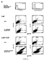
- Figure 1: Viral replication and surface modulation of HLA class I
in .221 cells transfected with three different HLA class I HC genes (HLA-A2, HLA-B51
and HLA-Cw4); said figure illustrates the kinetic of viral replication (virus used
was LAV-2/C) monitored by RT measurement and the determination of the infectivity
titers of supernatants. Five x 106 .221, 221.A2, .221.B51 or .221.Cw4 cells were
infected with the HIV-2 CD4-independent virus LAV-2/C (100 pg equivalent
RT/106cells). After 1.5 hour at 37°C in a minimal volume, cells were washed 3 times,
resuspended at 5.106 cells/ml in RPMI-10% FCS. Figure 1A: Every 3 days, cells were
passaged and supernatant assessed for RT production (Retrosys, Innovagen, Sweden)
to establish the kinetic of infection. Supernatants collected at the peak of infection
were titrated on the human T cell line C8166 to determine their 50% tissue culture
infectivity dose (TCID50). Cultures were routinely scored for the presence of syncitia.
After syncitia disappearance, cells were washed and stained with an anti-HIV-2 gp105
rabbit serum (NIBSC, British MRC) and with the anti-HLA class I mAbs BBM.1,
W6/32 and L31. Two x 105 cells in FACS buffer (PBS- BSA 0,2%, sodium azide
0,1%) were distributed in V bottom 96 well trays on ice, incubated with the appropriate
primary antibody for one hour at 4°C under agitation, washed 3 times in FACS
buffer and incubated for 45′ at 4°C with the secondary Ab : swine anti-rabbit-FITC
(Dako, Denmark) or Goat F(ab')2 anti-mouse IgG-PE (Immunotech, France). Cells
were washed again 3 times, resuspended in paraformaldehyde 2% and then analysed
on a FACSxalibur 4 colors instrument (Beckton Dickinson) using the Cell Quest
software.
Figure 1A illustrates that .221.Cw4 cells and .221.A2 cells are more
permissive to viral replication than .221.B51 cells and produce more infectious viruses
as demonstrated by the ratio between TCID50 and the amount of RT. Figure 1B illustrates
the expression of envelope antigen at the surface of infected cells and the down-modulation
of HLA class I trimeric molecules recognised by the two monoclonal antibodies
BBM1 and W6/32. More importantly, figure 1B illustrates the up-regulation of
HLA class I HCs (recognised by mAb L31) and the two cell lines (.221.A2 and
.221.Cw4) which sustain the strongest viral replication and produce the more infectious
viruses.
- Figure 2: Kinetic of infection of HIV-1 LAI, HIV-2 LAV-2/C and
three primary X4 isolates on .221.CD4 and .221.Cw4.CD4 cells. Five x 106 .221.CD4
or .221.Cw4.CD4 cells were infected as described in Fig 1 with either LAI, LAV-2/C,
215, 248 or ETS primary X4 isolates (2ng p24 equivalent/106cells). The kinetic of
infection was followed by measurement of HIV-1 p24 Ag (HIV-1 core profile
ELISA, Du Pont de Nemours) or viral RT (LAV-2/C) as in Fig. 1. The different
primary isolates 248, 215 and ETS were isolated from patient's PBMC by coculture
with PHA-activated PBMC from healthy blood donors. Infected cells were grown in
RPMI medium supplemented with 2 mM L-glutamine, 10% foetal calf serum,
500 IU/ml recombinant interleukin-2 (Proleukin, Chiron France, Suresnes), 1 IU/ml
sheep polyclonal anti-interferon-α antibody (Valbiotech, Chatillon, France) 2 µg/ml
polybrene (Sigma, St.Louis, MO) and antibiotics. For neutralisation experiments on
PBMC, 125 nM of human recombinant chemokines RANTES (Biodesign International,
Kennenbunk, ME, USA) or SDF-1α/BPSF (R&D Systems, Abingdon, UK)
were added to the cultures at the time of infection and every three days when culture
medium was replaced. The T cell line adapted (TCLA) strain HIV-1 LAI was propagated
in the T cell line CEM or in .221.CD4 and .221.Cw4.CD4 cell lines.
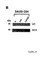
- Figure 3: figure 3A: Expression of HLA Class I heavy chains and
Env at the surface of infected Daudi cells. Five x 106 CD4-transfected Daudi cells
(Daudi.CD4) were infected with HIV-1 LAI or HIV-2 LAV-2/C and stained with the
anti-HLA class I HC mAbs L31 and HC10, and antisera specific for HIV-1 gp120 or
HIV-2 gp105 as described in Fig.1. figure 3B: Western blot analysis of HLA class I
heavy chains incorporated into LAI and LAV-2/C viruses grown on Daudi cells. LAI
and LAV-2/C virus supernatants from Daudi.CD4 cells were centrifuged and treated
as described in Fig.4B, hereafter for Western blot analysis with the two HLA class I
heavy chains specific mAbs L31 and HC10. Supernatant from uninfected cells were
used as control (0).
- Figure 4: PCR analysis of entry of LAI, LAV-2/C and 248 viruses
in HLA class I positive or negative cells (1st cycle) and entry of progeny viruses (2nd
cycle). Five x 105 .221.CD4 or .221.Cw4.CD4 cells were incubated either 2 hours at
0°C (negative control, In.V.: Inactive virus), or 2, 4 ,6, 18 or 30 hours at 37°C with
virus containing supernatant (MOI of 1 TCID50/cell) previously treated with 20 µg/ml
DNAse (Boehringer-Mannheim) in 10 mM MgCl2, 30′ at 30°C. The viruses used in
these experiments were : HIV-1 LAI, HIV-2 LAV-2/C or HIV-1 248 X4 primary isolate.
After the first step of 2 hours incubation, cells were washed twice in serum-free
medium, trypsinised (trypsine XIII, 15 µg/ml, Sigma) for 15′ at 4°C to remove any
bound virus particles from the cell surface, washed again three times in complete
medium and resuspended in 10% FCS-RPMI medium for a further incubation at 37°C.
At each time point, (2, 4, 6 and 18 hours) an aliquot of 0.5.106 cells from the assay
was centrifuged and lysed as follows: after careful resuspension in 400 µl of Tris-KCl
buffer (25 mM Tris-HCl pH 8, 125 mM KCl), 100 µl of NPK solution (1% NP40 plus
2.5 mg/ml proteinase K) (Boehringer-Mannheim) were added and the incubation done
at 56°C for at least 3 hours. DNA samples were heated at 95°C for 15 for DNAse
inactivation. Twenty five µl of the lysate were used for the PCR reaction performed in
a final volume of 50 µl containing 1.5 mM MgCl2, 100 ng of each amplification
primer, 0.2 mM of each dNTP and 2.5 U of Taq polymerase (Promega). Ctrl- was the
negative control of the PCR amplification. The primers and probes used were R, sense
primer, HIV-1 : 5'ggctaactagggaacccactg3' (LAI and 248), HIV-2 :
5'gaggctggcagattgagccctg3' (Lav2C); U5, anti-sense primer, HIV-1 :
5'ctgctagagattttccacactgac3', HIV-2 : 5'gcggcgactaggacagatggg3', and the Long
Terminal Repeat LTR3 (hybridisation probe) HIV-1: 5'gtgtgtgcccgtctgttgtgtg3', HIV-2
: 5'ggccccacgcttgcttgctt3'. The PCR reaction was run in a Perkin-Elmer instrument
as follows : 94°C, 2′, 1 cycle; 94°C,30″- 58°C (HIV-1) or 60°C (HIV-2), 1′- 72°C,
1′, 35 cycles; 72°C, 7′, 1 cycle. All PCR reactions were separated by electrophoresis
on a 2% agarose gel, transferred to a Gene Screen Plus membrane Amersham, UK
(alkaline transfer) and the 195 bp products hybridised with their 32P-labeled LTR
specific probe. Supernatants from the 30 hours incubation were collected, centrifuged
and filtered on a .45µm filter. An aliquot was centrifuged at 20 000 rpm for one hour
and titrated out in a RT assay (Retrosys, Innovagen, Sweden). For each virus,
equalised amounts of supernatants collected from .221.CD4 and .221.Cw4.CD4 cells
were used in a second round of entry PCR on .221.Cw4.CD4 cells as described above.
In addition, to further amplify the signal obtained, LAV-2/C PCR products were
subjected to a nested PCR with the following set of primers : 5'-ggtagagcctgggtgttc-3',
sens, and 5'-ggagagatgggagcacac-3', antisens. A photograph of the gel showing the
amplified product of 139 bp long is presented on the LAV2/C bottom panel. Figure
4B: Western blot analysis of virion-associated HLA class I heavy chains. Equal
amounts of LAI, LAV-2/C and 248 viruses generated on .221.Cw4.CD4 cells and LAI
virus generated on .221.CD4 cells were pelleted by centrifugation (1h at 22.000 rpm)
and resuspended in SDS-PAGE sample buffer [U.K. Laemmli, Nature, 1970, 227,
680-5]. Proteins were resolved by SDS-PAGE (12.5% gel) and transferred to a nitrocellulose
filter. Filters were blocked 16 hours at 4°C with 5% non-fat dry milk, 0.05%
Tween 20 in PBS (PBS-T-milk) and then incubated with anti HLA class I mAbs (L31
or HC10) for 2h in the same blocking buffer. After washing with 0.05% Tween 20 in
PBS (PBS-T), the filters were incubated with rabbit anti mouse-HRP complex
(DAKO, Denmark) diluted 1/1000 in PBS-T-milk. After ten washes with PBS-T,
peroxidase activity was revealed using an enhanced chemiluminescence system (ECL,
Amersham, U.K.). As a negative control, an identical volume of supernatant from
uninfected cells that was treated in the same way (lane 0) was used.
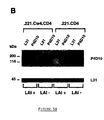
- Figure 5A: Co-precipitation of Env gp120/160 and HLA class I
heavy chains from LAI infected .221.Cw4.CD4 cells. .221.Cw4.CD4 and .221.CD4
cells were infected with LAI as described in Fig.2 and surface biotinylated by addition
of 0.15 mg/ml NHS-biotin (Pierce) to 1x10⁷ cells/ml in 0.1 M Hepes pH 8.0, 0.15 M
NaCl for 30 min on ice. The reaction was quenched with 10 mM NH4Cl and cells were
washed and lysed in 0.5% NP40. Lysates were immunoprecipitated with an anti-HLA-C
mAb (L31), an anti-LAI gp120 mAb (P4D10) and a control mAb using a procedure
previously described [A. Cosma, Anal. Biochem., 1997, 252, 10-4]. Immunoprecipitated
material was resolved by SDS-PAGE (7.5% gel), proteins were transferred to
nitrocellulose and probed with streptavidin-HRP for detection by ECL. Figure 5B:
Western blot detection of HLA class I heavy chains co-precipitated with Env. NP40
lysates of LAI infected or uninfected .221.Cw4.CD4 and .221.CD4 cells were
immunoprecipitated with L31 and P4D10 mAbs. After SDS-PAGE and transfer to
nitrocellulose membrane the immunoprecipitated material was probed with mAb L31
to detect HLA class I HC and mAb P4D10 to detect LAI Env glycoproteins as
described in Fig. 4B.
- Figure 6. Western blot analysis of Env gp120/160 glycoproteins
incorporated into virions generated on .221.Cw4.CD4 and .221.CD4 cells. Equal
amounts (50 ng of p24) of LAI grown on .221.Cw4.CD4 and .221.CD4 cells were
treated like in Fig. 4B and the nitrocellulose filter probed with a pool of anti-p24
mAbs (clones EH12E1 and LH104-E from British MRC) to control the quantity of
virus loaded on the gel, the anti-HLA class I HC mAb L31 to verify the presence of
HLA class I and the anti-LAI gp120 mAb P4D10 to estimate the level of virion-associated
Env glycoproteins.
EXAMPLE 1: Modulation of cell surface expression of HLA class I on infected
cells.
Material and methods
- Cell lines
-
- human cell lines (LCL.721.221) which lack chemical HLA class I
HC A, B and C genes but express β2m and become HLA class I surface positive upon
transfection with HC genes [Shimizu, 1989] were used.
- cells transfected with HLA-Cw4 (.221.Cw4) [Biassoni, 1995], -A2
(.221.A2) and -B51 (.221.B51) and
The cells were infected with the CD4-independent CXCR4
dependent TLCA virus, HIV-2 LAV2C.
- Monoclonal antibodies
-
- The surface expression of HLA class I was investigated using a
murine monoclonal antibody (L31) which had been obtained upon immunisation of
mice with purified cell-free HIV-1 LAI virus and recognises HLA C-encoded HC in
their β2m free conformation [Beretta, 1987] [Grassi, 1991].
- Two additional antibodies, W6-32 and BBM1, specific for HLA
class I trimeric complexes [Brodsky, 1979] and β2m [Parham, 1983] respectively were
also used as well as another anti-class I HC, HC10 (Stam, 1986).
-
Since L31 recognises, in addition to HLA-C some HLA-A and
HLA-B isotypes, including A2 and B51 [Setini, 1996], cells transfected with HLA-Cw4
(.221.Cw4), -A2 (.221.A2) and -B51 (.221.B51) were used [Biassoni, 1995].
.221 cells are CD4 negative and express the HIV co-receptor CXCR4. Lav2C which is
a CD4-independent, CXCR4-dependent HIV2 TCLA, was used in said experiments.
In order to minimise parameters incidence others than HLA class I in Lav2C replication
.221 transfected cells were subcloned and cells with similar amounts of HLA
class II and equivalent amounts of CXCR4 were expanded for infection experiments.
Non-transfected .221 cells were used as control.
- cell-surface is stained with the anti-HLA class I mAbs and an
antiserum specific for HIV-2 viral envelope glycoprotein, a few days ofter the peak of
viral replication corresponding to the disappearance of syncitia in culture. The kinetic
of viral replication was monitored by RT measurement and the supernatants infectivity
titers determined at the day of peak viral production.
Results
-
As shown in Figure 1A, .221 and .221.B51 cells are only marginally
permissive to the replication of LAV2/C. In contrast, LAV2/C replicated efficiently on
.221.A2 and .221.Cw4. In addition, there were significative differences between the
infectivity titers of cell-free supernatants (figure 1A): supernatants from .221.Cw4
cells and .221.A2 cells displayed a higher TCID50 compared to supernatants from .221
and .221.B51 cells. The comparison of the TCID50/ratios of each virus supernatant
revealed that .221.Cw4 cells generated the most infectious viruses. All cell lines were
surface positive for the viral envelope antigen gp105 (90 to 100 % fluorescent cells).
Maximum Env expression was seen on .221.Cw4 cells. The trimeric HLA class I
complexes recognised by BBM1 and W6-32 were down-modulated in the four cell
lines, including .221, which expresses non-classical HLA class I antigens (Shimizu Y
et al., 1989)decreases upon infection on -A2, -B51 as well as -Cw4 transfected cells.
In contrast, there is a significant up-modulation of L31-reactive molecules on
.221.Cw4 and .221.A2 but not .221.B51 cells.
-
Some level of viral replication occurs also on HLA class I negative
cells and lack of HLA class I does not prevent the expression of viral protein at the
cell surface (figure 1B) but rather results in a severe reduction of both the quantity of
viruses produced and their infectivity.
-
Viral replication in the three cell lines varied significantly: it was
maximal on .221.Cw4 cells, high on .221.A2 and low on .221.B51 cells (Figure 1b).
Therefore, the increased surface expression of L31-reactive HLA class I HC
apparently correlated to the extent of viral replication and to the infectivity of the
viruses generated on the three cell lines indicating an association of these molecules
with infectious viral particles.
-
In addition, there were evidences that viral replication could be
influenced by the transfected HLA class I allele since .221.Cw4 cells were more
permissive to LAV-2C replication than .221.A2 and much more than .221.B51 cells.
EXAMPLE 2: Permissiveness of HLA class I HC positive and negative cells to
the replication of TCLA and primary X4 viruses.
Material and methods
- cell lines
-
- cells transfected with HLA-Cw4 (.221.Cw4) [Biassoni, 1995],
HLA-Cw4 and CD4 (.221.Cw4.CD4)
Results
-
The results correspond to the evaluation of the permissiveness of
HLA class I HC positive and negative cells to the replication of viruses other than
LAV-2C.
-
The kinetic of replication of a CD4-dependent TCLA virus (HIV-1
LAI) and three primary HIV-1 isolates which use CXCR4 co-receptor was analysed.
LAI was used as a control of X4 dependent virus. Primary isolates (215, 248 and ETS)
were chosen on the basis of the capacity to replicate in CXCR4 expressing cell lines
(MT2 and C8166) and of being inhibited by SDF-1α (the natural ligand of CXCR4)
(E. Oberlin et al., Nature, 1996, 382, 833-5) in PBMC infection assays: LAI (100% of
inhibition), ETS (93% of inhibition), 215 (63% of inhibition) and 248 (45% of inhibition).
-
.221.Cw4 cells were used since they showed the highest permissivity
to LAV-2C, .221 and .221.Cw4 cells were transfected with the human CD4 gene and
tested for their capacity to support the replication of LAI and the three primary isolates
replication. As shown in Figure 2A, LAI was able to infect both .221.CD4
and.221.Cw4.CD4 cells, although the kinetic of replication was delayed in the absence
of HLA class I negative cells. In contrast, LAV-2C failed to replicate on .221.CD4
cells but replicated on .221.Cw4.CD4 cells.
-
The three isolates tested failed to replicate in the HLA class I negative
.221.CD4 cells although they all replicated efficiently in the HLA class I positive
cells .221.Cw4.CD4 (Figure 2B). Thus, the expression of HLA class I HC was found
to be indispensable for the replication of HIV-1 primary X4 isolates and LAV2C.
HIV-1 LAI differed from the other viruses since it could be propagated, although less
efficiently, in the HLA class I negative cells.
-
These data are in apparent contradiction with previous reports
concluding on the dispensability of HLA class I for virus replication which were based
on the permissivity of HLA class I surface negative cells, DAUDI, to HIV infection
[Clapham, 1991]. It has been reported that LAI virus replicates on DAUDI cells and
does not incorporated HLA class I molecules on HIV-LAI produced by DAUDI cells
[Le Gall, 1997].
-
DAUDI cells exhibit a genetic defect in the β2m gene, but they
express high intracellular levels of HLA class I HC transcripts [Ploegh, 1979].
-
Their permissivity to viral replication may be restricted to some
TCLA viruses, like LAI, which are less dependent on the presence of HLA class I for
replication. Alternatively, virus incorporation of HLA class I could occur even in the
absence of a functional β2m gene by recruitment of the intracellular pool of HLA class
I HC.
EXAMPLE 3: Expression of HLA class I HC and viral envelope glycoproteins on
CD4-transfected DAUDI cells, infected with LAV2C or LAI.
Material and methods
- Cell lines
-
- Daudi cells (Le Gall, 1997)
- Daudi-CD4 cells
-
- monoclonal antibodies (mAbs)
-
Two HLA class I HC specific mAbs, L31 and HC10 [Stam, 1986]
were used and antisera specific for HIV-1 or HIV-2 Env.
Results
-
Uninfected DAUDI-CD4 cells were weakly stained by the two anti-HLA
class I HC mAbs (Figure 3A). Infection with LAV2C (Figure 3A) but not LAI
(Figure 3A) induced a marked increase in the L31 (Figure 3) and HC10 fluorescence
(Figure 3A) which was evident on a population of Env-high and HLA class I HC-high
cells indicating the association of HLA class I HC with LAV-2C viral particles.
EXAMPLE 4: Western blot analysis on purified LAV2C and LAI viral particles
generated in DAUDI-CD4 cells.
-
The two HC specific mAbs are used (L31 and HC10) (Figure 3B).
-
A 45 kDa band corresponding to HLA class I HC within LAV2C but
not LAI viral lysates was detected. The protein was absent in the control supernatant
from uninfected cells.
-
The protein was absent in control supernatants from uninfected cells.
Of the two HLA class I HC mAbs used, HC10, but not L31, stained the 45 kD band.
This was unexpected since the surface reactivity of both mAbs on LAV2/C infected
cells was similar (Figure 3A). The reactivity of the two mAbs for native versus denatured
determinants may account for these differences. The absence of HLA class I HC
associated to LAI virus grown on DAUDI-CD4 cells was in agreement with results
previously published by other authors (Le Gall, 1997).
-
- i) DAUDI cells cannot be considered as bona fide HLA class I
negative cells since they express low levels of HLA class I HC at the cell surface and
become intensely positive following infection with LAV2C,
- ii) HLA class I HC can be incorporated within viral particles even in
the absence of β2m,
- iii) there is a clear association between the capacity of a given virus
(LAV2C versus LAI) to incorporate HLA class I HC and to induce their cell-surface
expression,
- iv) TCLA viruses differ in their dependency towards class I HC.
-
-
These data also bring further support to the conclusion that, on
infected cells, surface HLA class I HC are, at least in part, associated to infective viral
particles as also suggested by the data obtained on .221.Cw4 and A2 cells (Figure 1).
EXAMPLE 5 : PCR analysis of entry of LAI, LAV-2/C and 248 viruses in HLA
class I positive or negative cells (1st cycle) and entry of progeny viruses (2nd cycle).
-
To determine whether viral entry was hindered in Class I negative
cells, the ability of .221.CD4 and .221.Cw4.CD4 cells to support a single cycle of
viral replication was compared using an entry PCR assay [Zack, 1990]. Cells were
infected with equal amounts of either LAI, LAV-2/C or one of the primary isolates,
HIV-1 248, and the accumulation of strong stop cDNA was measured over 18 hours.
As shown in Fig.4A, entry was achieved by all three viruses, although there was a
significant delay in the accumulation of cDNA in the HLA class I negative cells. The
delay was more pronounced with LAI and was associated with lower viral RT production
after 30 hours. Viral RT production in LAV-2/C and 248 infected .221.CD4
and .221.Cw4.CD4 cells was identical, indicating there was no post-entry blockade in
.221.CD4 cells.
-
To determine whether HLA class I molecules acquired by virions
after the first cycle of replication influence viral entry, viruses generated from
.221.CD4 and .221.Cw4.CD4 cells were tested in a second cycle of replication in
.221.Cw4.CD4 cells. The results showed distinct patterns of virus entry for HLA class
I positive and negative cell-derived viruses (Fig.4A). LAI generated on .221.CD4 cells
displayed delayed entry kinetics and a decreased level of strong stop cDNA 18 hours
post-infection compared to LAI generated on .221.Cw4.CD4 cells. The HLA class I
negative-cell derived LAV2/C and 248 viruses showed no detectable accumulation of
strong stop cDNA, indicating a defect in a pre-retrotranscription step. However, when
generated on HLA class I positive cells, the same viruses were entry competent.
-
To confirm the presence of HLA class I heavy chains on the viruses
generated from .221.Cw4.CD4 cells, Western blot assays of the concentrated viral
lysates were performed using HLA class I heavy chain specific mAb L31 and another
heavy chain mAb HC10. LAI, LAV-2/C and 248 viruses expanded on .221.Cw4.CD4
cells were tested. As a control HLA negative virus, LAI generated from .221.CD4
cells was tested, since it could not be poossible to generate sufficient amounts of HLA
class I negative LAV-2/C and primary isolates for biochemical analysis. All viruses
obtained from .221.Cw4.CD4 cells, including LAI, displayed a 45 kDa band reactive
with both heavy chain specific mAbs (Figure 4B) which was absent on LAI grown on
the HLA class I negative cells.
-
These data showed that lack of virion-associated HLA class I results
in the generation of virions whose competence to enter cells is impaired. No major
blockade at the post-entry level occurs on HLA class I negative cells as indicated by
the levels of viral RT produced during the first cycle of replication. This is also
supported by the finding that LAI can efficiently replicate in .221.CD4 cells, even if at
a lower rate than in .221.Cw4.CD4 cells. The delayed kinetics of entry into .221.CD4.
cells during the first cycle PCR could be explained by the lower level of CD4
expressed by these cells. However, LAV-2/C, which is CD4 independent, was also
delayed indicating that other factors may be involved.
-
Said incorporation is required for the acquisition of the competence
to enter cells.
-
The two TCLA viruses LAI and LAV2C, and two primary X4 isolates
(248 and 215) were used to infect .221.Cw4 and .221.Cw4.CD4 cells and, after
one single cycle of replication, were concentrated by ultracentrifugation and quantified
in a RT assay.
-
This conclusion is also supported by the comparison of the infectivity
titers of LAI viruses generated on HLA class I negative and positive cells. LAI
cell-free supernatants from .221.CD4 and .221.Cw4.CD4. Infected cells were collected
at the peak of infection and titrated on PHA-activated peripheral blood mononuclear
cells. The infectivity titers were measured at day 6. There was a significative
difference between LAI viruses propagated on the two cell lines (13 versus 93
TCID50/ngp24 for LAI viruses propagated on .221.CD4 and 221.Cw4.CD4 cells
respectively) indicating that HIV-1 LAI acquires a higher infectivity upon replication
in HLA class I positive cells.
-
Table I illustrates the fact that viruses grown on HLA class I positive
cells are more infectious than viruses grown on HLA class I negative cells.
| | p24 ng/ml | RT cpm : 10-3/ml | Infectivity TCID50/ngp24 |
| LAI Class I Negative | 374 | 1570 | 9 |
| LAI Class I Positive | 826 | 1644 | 68 |
EXAMPLE 6: Co-precipitation of Env gp120/160 and HLA class I heavy chains
from LAI infected .221.Cw4.CD4 cells.
-
Since flow-cytometry data indicated the co-expression of Env and
HLA class I HC (see figure 1B and figure 3A) at the membrane of infected cells, and
the association of their surface expression with the production of infective viral
particles, the presence of Env/HLA class I complexes was tested on LAI-infected
.221.CD4 and .221.Cw4.CD4 cells.
-
Cells were surface biotinylated and the cell lysates immunoprecipitated
with either L31 or an anti-LAI-gp120 mAb (P4D10) (Figure 5A). Immunoprecipitation
of LAI infected .221.CD4 cells with the anti-gp120 mAb P4D10 revealed, in
addition to gp120 and gp160, two major bands of an apparent molecular weight
corresponding to CD4 (58 kDa) and CXCR4 (48 kDa). The P4D10 immunoprecipitates
of LAI infected .221.Cw4.CD4 cells exhibited an additional band of 45 kDa
whose molecular weight corresponded to that of the band precipitated by L31 on both
infected and non-infected cells. A weak band of an apparent molecular weight of 160
was evidenced in the L31 immunoprecipitate of .221.Cw4.CD4 LAI infected cells. To
assess the identity of the 45 kDa band with HLA class I HC, the same experiment was
performed without biotin surface labelling and we probed the nitrocellulose filter with
P4D10 and L31 (Figure 5B). The 45 kDa band precipitated by the anti-gp120 mAb
P4D10 on infected .221.Cw4.CD4 cells was stained by L31. This band was absent in
both the infected and non infected .221.CD4 cells as well as in the P4D10 immunoprecipitates
of uninfected .221.Cw4.CD4 cells. Similar data were obtained with
LAV2C-infected .221.Cw4 cells. Thus, a specific interaction between the HIV envelope
glycoprotein and the HLA class I HC occurs in infected cells.
EXAMPLE 7: Western blot analysis of Env gp120/160 glycoproteins incorporated
into virions generated on .221.Cw4.CD4 and .221.CD4 cells.
-
To test whether the pattern of Env expression by mature viral
particles was influenced by the presence or absence of virion-associated HLA class I
HC, a Western blot analysis of Env glycoproteins from purified HLA class I positive
or negative LAI viral particles was performed. The Env content of the HLA class I
positive virus, as estimated by densitometry analysis, was 4 folds higher compared to
that of HLA class I negative virus (Figure 6). The amount of p24 in virus preparations
from .221.CD4 and .221.Cw4.CD4 was equivalent.
-
This experiment could only be performed on HLA class I negative
and positive LAI viruses since we could not propagate the other viruses on the HLA
class I negative cells. Although not strictly dependent on HLA class I for replication,
LAI incorporates HLA class I HC when grown on .221.Cw4 cells and acquires a
higher infectivity. Thus, the difference in Env expression by HLA class I negative and
positive LAI viral particles probably reflects a general phenomenon associated to the
HLA class I-dependent acquisition or increase in infectivity. Such difference may be
due to reduced shedding or to enhanced incorporation of Env glycoproteins within
nascent HLA class I positive viral particles.
EXAMPLE 8: Immunoprotective role of the instant compositions.
-
Unexpectedly, the instant compositions given according to an
appropriate protocol, preferably during the boosting step, induce a immune protection
against HIV infection.
-
Primary and TCLA viruses display significative differences in their
susceptibility to neutralisation by anti-Env antibodies. The need for HLA class I HC
incorporation for the acquisition of entry-competence and the presence of an Env/HLA
class I complex on infected cells suggest that the expression of a functional viral
envelope requires the formation of such a complex. The expression of Env
determinants involved in the interactions with CD4 and the co-receptors may be
different in HLA negative or positive viruses as a consequence of Env SU conformational
modifications induced by the association with HLA class I HC.
-
Therefore, in order to induce antibodies to the relevant Env determinants,
it is preferable to use compositions according to the invention comprising
Env/HLA class I antigens rather than isolated Env antigens.
EXAMPLE 9: Preparation of a composition containing gp120 of HIV-1 and
HLA-C class I heavy chain.
-
.221.Cw4.CD4 cells are infected with LAI virus. The kinetic of viral
antigen measurement. A replication is followed by p24. At the day of peak viral replication,
cells are washed, and lysed in 0.5 % NP40 containing protease inhibitors
(aprotinine and PMSF, SIGMA, St Louis, USA). The lysates are immunoprecipitated
with an anti―LAI gp120 mAb (mAb P4D10, for instance (L. Akerblom et al., AIDS,
1990, 4, 953-960)), using the following procedure:
-
Protein A-Sepharose (10 µl) (Pharmacia) is incubated with 5 µg of
rabbit anti-mous IgG (RaMIgG) Dako) plus 200 µl of incubation buffer at 4°C on
rotation for one hour, and then free antibody is washed away with cold PBS. Successively
the protein A-Sepharose:RAMIgG complex is incubated with 5 µg of P4D10
mAb in the same condition as described above. The cell lysate is incubated with
protein A-Sepharose:RAMIgG:P4D10 for 3 hours at 4°C on rotation. Protein A-Sepharose:antibodies
complex is then washed seven times with cold "washing buffer
with protein" and three times with cold "washing buffer without protein".
-
The quality control analysis of the immunoprecipitated material is
performed by Western blot with mAb L31 and P4D10 on nitrocellulose filters after
transfer of the proteins resolved in SDS-PAGE (figure 5B).
EXAMPLE 10: Preparation of recombinant attenuated viral particles containing
a complex gp160 (or gp120) and HLA-Cw4 class I antigen.
-
The recombinant MVA vaccinia virus is, for instance as described in
US Patent 5 185 146.
-
The sequence coding for the gp160 or the gp120 is, for instance as
described in International PCT Application WO 86/02383 ; M. Alizon et al., Nature,
1984, 312, 757-760 ; M. Alizon et al., Cell, 1986, 46, 63-74; D. Klatzmann et al.,
Nature, 1984, 312, 767-768 or L.O. Arthur et al., Proc. Natl. Acad. Sci. USA, 1987,
84, 8583-8587.
-
The sequence coding for the HLA-Cw4 class I antigen is, for
instance described in Grassi, 1991.
REFERENCES
-
- P.J. Bjorkman et al., Annu. Rev. Biochem., 1990, 59, 253-88 ;
- E. Schnabl et al., J. Exp. Med., 1990, 171, 1431-42 ;
- J. Zemmour et al., J. Exp. Med., 1992, 176, 937-50 ;
- O. Schwartz et al., Nat. Med., 1996, 2, 338-42 ;
- K.L. Collins et al., Nature, 1998, 391, 397-401 ;
- S. Le Gall et al., Immunity, 1998, 8, 483-95 ;
- Y. Shimizu et al., J. Immunol., 1989, 142, 3320-8 ;
- A. Beretta et al., Eur J. Immunol., 1987, 17, 1793-8 ;
- F. Grassi et al., J. Exp. Med., 1991, 174, 53-62 ;
- F.M. Brodsky et al., Immunol. Rev., 1979, 47, 3-61 ;
- P. Parham et al., J. Biol Chem., 1983, 258, 6179-86 ;
- N.J. Stam et al., J. Immunol., 1986, 137, 2299-306 ;
- A. Setini et al., Hum. Immunol., 1996, 46, 69-81 ;
- R. Biassoni et al., J. Exp. Med., 1995, 182, 605-9 ,
- E. Oberlin et al., Nature, 1996, 382, 833-5 ;
- J.A. Zack et al., Cell, 1990, 61, 213-22 ;
- P.R. Clapham et al., Virology, 1991, 181, 703-15 ;
- S. Le Gall et al., Virology, 1997, 229, 295-301 ;
- H.L. Ploegh et al., Proc. Natl. Acad. Sci. USA, 1979, 76, 2273-7 ;
- C.D. Rizzuto et al., J. Virol, 1997, 71, 4847-51 ;
- R. Cantin et al., J Virol., 1997, 71, 1922-30 ;
- L.O. Arthur et al., Science, 1992, 258, 1935-8.
-
As is apparent from the foregoing description, the invention is in no
way limited to those modes of execution, embodiments and modes of application
which have now been described more explicitly; on the contrary, it encompasses all
the variants thereof which may occur to those skilled in the art, without deviating from
the framework or the scope of the present invention.