EP0999285B1 - Methods and compositions for amplifying detectable signals in specific binding assays - Google Patents
Methods and compositions for amplifying detectable signals in specific binding assays Download PDFInfo
- Publication number
- EP0999285B1 EP0999285B1 EP99250336A EP99250336A EP0999285B1 EP 0999285 B1 EP0999285 B1 EP 0999285B1 EP 99250336 A EP99250336 A EP 99250336A EP 99250336 A EP99250336 A EP 99250336A EP 0999285 B1 EP0999285 B1 EP 0999285B1
- Authority
- EP
- European Patent Office
- Prior art keywords
- nucleic acid
- receptor
- amplification reagent
- binding
- target
- Prior art date
- Legal status (The legal status is an assumption and is not a legal conclusion. Google has not performed a legal analysis and makes no representation as to the accuracy of the status listed.)
- Expired - Lifetime
Links
- 238000000034 method Methods 0.000 title claims abstract description 98
- 230000009870 specific binding Effects 0.000 title abstract description 8
- 238000000159 protein binding assay Methods 0.000 title abstract description 6
- 239000000203 mixture Substances 0.000 title description 4
- 150000007523 nucleic acids Chemical class 0.000 claims abstract description 159
- 230000027455 binding Effects 0.000 claims abstract description 147
- 239000003446 ligand Substances 0.000 claims abstract description 119
- 102000039446 nucleic acids Human genes 0.000 claims abstract description 118
- 108020004707 nucleic acids Proteins 0.000 claims abstract description 118
- 230000003321 amplification Effects 0.000 claims abstract description 114
- 238000003199 nucleic acid amplification method Methods 0.000 claims abstract description 114
- 239000003153 chemical reaction reagent Substances 0.000 claims abstract description 101
- 108020004414 DNA Proteins 0.000 claims abstract description 93
- YBJHBAHKTGYVGT-ZKWXMUAHSA-N (+)-Biotin Chemical compound N1C(=O)N[C@@H]2[C@H](CCCCC(=O)O)SC[C@@H]21 YBJHBAHKTGYVGT-ZKWXMUAHSA-N 0.000 claims abstract description 82
- 239000011159 matrix material Substances 0.000 claims abstract description 82
- 108010090804 Streptavidin Proteins 0.000 claims abstract description 56
- 239000000523 sample Substances 0.000 claims abstract description 54
- 235000020958 biotin Nutrition 0.000 claims abstract description 48
- 229960002685 biotin Drugs 0.000 claims abstract description 43
- 239000011616 biotin Substances 0.000 claims abstract description 43
- 108020004711 Nucleic Acid Probes Proteins 0.000 claims abstract description 38
- 239000002853 nucleic acid probe Substances 0.000 claims abstract description 38
- 108091028043 Nucleic acid sequence Proteins 0.000 claims abstract description 36
- 239000007850 fluorescent dye Substances 0.000 claims abstract description 9
- 238000009396 hybridization Methods 0.000 claims description 75
- 239000000872 buffer Substances 0.000 claims description 53
- 102000040430 polynucleotide Human genes 0.000 claims description 41
- 108091033319 polynucleotide Proteins 0.000 claims description 41
- 239000002157 polynucleotide Substances 0.000 claims description 41
- 239000000758 substrate Substances 0.000 claims description 32
- 125000003729 nucleotide group Chemical group 0.000 claims description 26
- 239000002773 nucleotide Substances 0.000 claims description 23
- 108090001008 Avidin Proteins 0.000 claims description 16
- 238000002372 labelling Methods 0.000 claims description 16
- BDHFUVZGWQCTTF-UHFFFAOYSA-M sulfonate Chemical compound [O-]S(=O)=O BDHFUVZGWQCTTF-UHFFFAOYSA-M 0.000 claims description 13
- 102000053602 DNA Human genes 0.000 claims description 9
- PCHJSUWPFVWCPO-UHFFFAOYSA-N gold Chemical compound [Au] PCHJSUWPFVWCPO-UHFFFAOYSA-N 0.000 claims description 8
- 102000004190 Enzymes Human genes 0.000 claims description 7
- 108090000790 Enzymes Proteins 0.000 claims description 7
- 108020004682 Single-Stranded DNA Proteins 0.000 claims description 7
- ZYGHJZDHTFUPRJ-UHFFFAOYSA-N coumarin Chemical compound C1=CC=C2OC(=O)C=CC2=C1 ZYGHJZDHTFUPRJ-UHFFFAOYSA-N 0.000 claims description 7
- VYPSYNLAJGMNEJ-UHFFFAOYSA-N Silicium dioxide Chemical compound O=[Si]=O VYPSYNLAJGMNEJ-UHFFFAOYSA-N 0.000 claims description 6
- 239000011521 glass Substances 0.000 claims description 6
- 239000010931 gold Substances 0.000 claims description 6
- 229910052737 gold Inorganic materials 0.000 claims description 6
- SXGZJKUKBWWHRA-UHFFFAOYSA-N 2-(N-morpholiniumyl)ethanesulfonate Chemical group [O-]S(=O)(=O)CC[NH+]1CCOCC1 SXGZJKUKBWWHRA-UHFFFAOYSA-N 0.000 claims description 5
- 235000001671 coumarin Nutrition 0.000 claims description 4
- GNBHRKFJIUUOQI-UHFFFAOYSA-N fluorescein Chemical compound O1C(=O)C2=CC=CC=C2C21C1=CC=C(O)C=C1OC1=CC(O)=CC=C21 GNBHRKFJIUUOQI-UHFFFAOYSA-N 0.000 claims description 4
- HSSLDCABUXLXKM-UHFFFAOYSA-N resorufin Chemical compound C1=CC(=O)C=C2OC3=CC(O)=CC=C3N=C21 HSSLDCABUXLXKM-UHFFFAOYSA-N 0.000 claims description 4
- PYWVYCXTNDRMGF-UHFFFAOYSA-N rhodamine B Chemical compound [Cl-].C=12C=CC(=[N+](CC)CC)C=C2OC2=CC(N(CC)CC)=CC=C2C=1C1=CC=CC=C1C(O)=O PYWVYCXTNDRMGF-UHFFFAOYSA-N 0.000 claims description 4
- 239000010703 silicon Substances 0.000 claims description 4
- 229910052710 silicon Inorganic materials 0.000 claims description 4
- JBRZTFJDHDCESZ-UHFFFAOYSA-N AsGa Chemical compound [As]#[Ga] JBRZTFJDHDCESZ-UHFFFAOYSA-N 0.000 claims description 3
- 229910005540 GaP Inorganic materials 0.000 claims description 3
- 229910001218 Gallium arsenide Inorganic materials 0.000 claims description 3
- 239000004793 Polystyrene Substances 0.000 claims description 3
- 229910052581 Si3N4 Inorganic materials 0.000 claims description 3
- 238000000137 annealing Methods 0.000 claims description 3
- 229960000956 coumarin Drugs 0.000 claims description 3
- HZXMRANICFIONG-UHFFFAOYSA-N gallium phosphide Chemical compound [Ga]#P HZXMRANICFIONG-UHFFFAOYSA-N 0.000 claims description 3
- 229910052732 germanium Inorganic materials 0.000 claims description 3
- GNPVGFCGXDBREM-UHFFFAOYSA-N germanium atom Chemical compound [Ge] GNPVGFCGXDBREM-UHFFFAOYSA-N 0.000 claims description 3
- 229920002223 polystyrene Polymers 0.000 claims description 3
- HQVNEWCFYHHQES-UHFFFAOYSA-N silicon nitride Chemical compound N12[Si]34N5[Si]62N3[Si]51N64 HQVNEWCFYHHQES-UHFFFAOYSA-N 0.000 claims description 3
- 229910052814 silicon oxide Inorganic materials 0.000 claims description 3
- BFKJFAAPBSQJPD-UHFFFAOYSA-N tetrafluoroethene Chemical group FC(F)=C(F)F BFKJFAAPBSQJPD-UHFFFAOYSA-N 0.000 claims description 3
- 238000003556 assay Methods 0.000 abstract description 42
- 238000001514 detection method Methods 0.000 abstract description 38
- 150000001875 compounds Chemical class 0.000 abstract description 7
- 238000012216 screening Methods 0.000 abstract description 4
- 108020003175 receptors Proteins 0.000 description 69
- 102000005962 receptors Human genes 0.000 description 69
- 239000007987 MES buffer Substances 0.000 description 39
- 208000037065 Subacute sclerosing leukoencephalitis Diseases 0.000 description 20
- 206010042297 Subacute sclerosing panencephalitis Diseases 0.000 description 20
- FAPWRFPIFSIZLT-UHFFFAOYSA-M Sodium chloride Chemical compound [Na+].[Cl-] FAPWRFPIFSIZLT-UHFFFAOYSA-M 0.000 description 19
- 239000000243 solution Substances 0.000 description 16
- 238000007899 nucleic acid hybridization Methods 0.000 description 15
- 238000003491 array Methods 0.000 description 14
- 239000000178 monomer Substances 0.000 description 13
- 108090000623 proteins and genes Proteins 0.000 description 12
- 108091034117 Oligonucleotide Proteins 0.000 description 11
- 239000011780 sodium chloride Substances 0.000 description 11
- KCXVZYZYPLLWCC-UHFFFAOYSA-N EDTA Chemical compound OC(=O)CN(CC(O)=O)CCN(CC(O)=O)CC(O)=O KCXVZYZYPLLWCC-UHFFFAOYSA-N 0.000 description 10
- 239000000412 dendrimer Substances 0.000 description 10
- 229920000736 dendritic polymer Polymers 0.000 description 10
- 238000010369 molecular cloning Methods 0.000 description 9
- 229920000642 polymer Polymers 0.000 description 9
- 239000007787 solid Substances 0.000 description 9
- 239000012192 staining solution Substances 0.000 description 9
- 229910019142 PO4 Inorganic materials 0.000 description 8
- 239000010410 layer Substances 0.000 description 8
- 230000015572 biosynthetic process Effects 0.000 description 7
- 150000001615 biotins Chemical class 0.000 description 7
- 229920001213 Polysorbate 20 Polymers 0.000 description 6
- 238000000338 in vitro Methods 0.000 description 6
- 239000000463 material Substances 0.000 description 6
- 239000008363 phosphate buffer Substances 0.000 description 6
- -1 poly(amino acid) Polymers 0.000 description 6
- 239000000256 polyoxyethylene sorbitan monolaurate Substances 0.000 description 6
- 235000010486 polyoxyethylene sorbitan monolaurate Nutrition 0.000 description 6
- 229920000136 polysorbate Polymers 0.000 description 6
- 230000035945 sensitivity Effects 0.000 description 6
- GPRLSGONYQIRFK-MNYXATJNSA-N triton Chemical compound [3H+] GPRLSGONYQIRFK-MNYXATJNSA-N 0.000 description 6
- 229920004890 Triton X-100 Polymers 0.000 description 5
- 239000002518 antifoaming agent Substances 0.000 description 5
- 238000006243 chemical reaction Methods 0.000 description 5
- 230000000295 complement effect Effects 0.000 description 5
- 230000000536 complexating effect Effects 0.000 description 5
- 238000013467 fragmentation Methods 0.000 description 5
- 238000006062 fragmentation reaction Methods 0.000 description 5
- 230000003993 interaction Effects 0.000 description 5
- 238000002203 pretreatment Methods 0.000 description 5
- 102000004169 proteins and genes Human genes 0.000 description 5
- 239000013598 vector Substances 0.000 description 5
- 108091032973 (ribonucleotides)n+m Proteins 0.000 description 4
- KDCGOANMDULRCW-UHFFFAOYSA-N 7H-purine Chemical compound N1=CNC2=NC=NC2=C1 KDCGOANMDULRCW-UHFFFAOYSA-N 0.000 description 4
- 108060003951 Immunoglobulin Proteins 0.000 description 4
- 241001529936 Murinae Species 0.000 description 4
- 239000000427 antigen Substances 0.000 description 4
- 210000003719 b-lymphocyte Anatomy 0.000 description 4
- 210000004958 brain cell Anatomy 0.000 description 4
- 230000000875 corresponding effect Effects 0.000 description 4
- 238000003500 gene array Methods 0.000 description 4
- 102000018358 immunoglobulin Human genes 0.000 description 4
- 239000013612 plasmid Substances 0.000 description 4
- ZCCUUQDIBDJBTK-UHFFFAOYSA-N psoralen Chemical compound C1=C2OC(=O)C=CC2=CC2=C1OC=C2 ZCCUUQDIBDJBTK-UHFFFAOYSA-N 0.000 description 4
- 239000000126 substance Substances 0.000 description 4
- 238000003786 synthesis reaction Methods 0.000 description 4
- 238000013518 transcription Methods 0.000 description 4
- 230000035897 transcription Effects 0.000 description 4
- XLYOFNOQVPJJNP-UHFFFAOYSA-N water Substances O XLYOFNOQVPJJNP-UHFFFAOYSA-N 0.000 description 4
- LLYYVNTYHIIBFL-UHFFFAOYSA-N 2-[2-[bis(carboxymethyl)amino]ethyl-(carboxymethyl)amino]acetic acid;phosphoric acid Chemical compound OP(O)(O)=O.OC(=O)CN(CC(O)=O)CCN(CC(O)=O)CC(O)=O LLYYVNTYHIIBFL-UHFFFAOYSA-N 0.000 description 3
- MIIIXQJBDGSIKL-UHFFFAOYSA-N 2-morpholin-4-ylethanesulfonic acid;hydrate Chemical compound O.OS(=O)(=O)CCN1CCOCC1 MIIIXQJBDGSIKL-UHFFFAOYSA-N 0.000 description 3
- 241000283707 Capra Species 0.000 description 3
- 239000007983 Tris buffer Substances 0.000 description 3
- JLCPHMBAVCMARE-UHFFFAOYSA-N [3-[[3-[[3-[[3-[[3-[[3-[[3-[[3-[[3-[[3-[[3-[[5-(2-amino-6-oxo-1H-purin-9-yl)-3-[[3-[[3-[[3-[[3-[[3-[[5-(2-amino-6-oxo-1H-purin-9-yl)-3-[[5-(2-amino-6-oxo-1H-purin-9-yl)-3-hydroxyoxolan-2-yl]methoxy-hydroxyphosphoryl]oxyoxolan-2-yl]methoxy-hydroxyphosphoryl]oxy-5-(5-methyl-2,4-dioxopyrimidin-1-yl)oxolan-2-yl]methoxy-hydroxyphosphoryl]oxy-5-(6-aminopurin-9-yl)oxolan-2-yl]methoxy-hydroxyphosphoryl]oxy-5-(6-aminopurin-9-yl)oxolan-2-yl]methoxy-hydroxyphosphoryl]oxy-5-(6-aminopurin-9-yl)oxolan-2-yl]methoxy-hydroxyphosphoryl]oxy-5-(6-aminopurin-9-yl)oxolan-2-yl]methoxy-hydroxyphosphoryl]oxyoxolan-2-yl]methoxy-hydroxyphosphoryl]oxy-5-(5-methyl-2,4-dioxopyrimidin-1-yl)oxolan-2-yl]methoxy-hydroxyphosphoryl]oxy-5-(4-amino-2-oxopyrimidin-1-yl)oxolan-2-yl]methoxy-hydroxyphosphoryl]oxy-5-(5-methyl-2,4-dioxopyrimidin-1-yl)oxolan-2-yl]methoxy-hydroxyphosphoryl]oxy-5-(5-methyl-2,4-dioxopyrimidin-1-yl)oxolan-2-yl]methoxy-hydroxyphosphoryl]oxy-5-(6-aminopurin-9-yl)oxolan-2-yl]methoxy-hydroxyphosphoryl]oxy-5-(6-aminopurin-9-yl)oxolan-2-yl]methoxy-hydroxyphosphoryl]oxy-5-(4-amino-2-oxopyrimidin-1-yl)oxolan-2-yl]methoxy-hydroxyphosphoryl]oxy-5-(4-amino-2-oxopyrimidin-1-yl)oxolan-2-yl]methoxy-hydroxyphosphoryl]oxy-5-(4-amino-2-oxopyrimidin-1-yl)oxolan-2-yl]methoxy-hydroxyphosphoryl]oxy-5-(6-aminopurin-9-yl)oxolan-2-yl]methoxy-hydroxyphosphoryl]oxy-5-(4-amino-2-oxopyrimidin-1-yl)oxolan-2-yl]methyl [5-(6-aminopurin-9-yl)-2-(hydroxymethyl)oxolan-3-yl] hydrogen phosphate Polymers Cc1cn(C2CC(OP(O)(=O)OCC3OC(CC3OP(O)(=O)OCC3OC(CC3O)n3cnc4c3nc(N)[nH]c4=O)n3cnc4c3nc(N)[nH]c4=O)C(COP(O)(=O)OC3CC(OC3COP(O)(=O)OC3CC(OC3COP(O)(=O)OC3CC(OC3COP(O)(=O)OC3CC(OC3COP(O)(=O)OC3CC(OC3COP(O)(=O)OC3CC(OC3COP(O)(=O)OC3CC(OC3COP(O)(=O)OC3CC(OC3COP(O)(=O)OC3CC(OC3COP(O)(=O)OC3CC(OC3COP(O)(=O)OC3CC(OC3COP(O)(=O)OC3CC(OC3COP(O)(=O)OC3CC(OC3COP(O)(=O)OC3CC(OC3COP(O)(=O)OC3CC(OC3COP(O)(=O)OC3CC(OC3COP(O)(=O)OC3CC(OC3CO)n3cnc4c(N)ncnc34)n3ccc(N)nc3=O)n3cnc4c(N)ncnc34)n3ccc(N)nc3=O)n3ccc(N)nc3=O)n3ccc(N)nc3=O)n3cnc4c(N)ncnc34)n3cnc4c(N)ncnc34)n3cc(C)c(=O)[nH]c3=O)n3cc(C)c(=O)[nH]c3=O)n3ccc(N)nc3=O)n3cc(C)c(=O)[nH]c3=O)n3cnc4c3nc(N)[nH]c4=O)n3cnc4c(N)ncnc34)n3cnc4c(N)ncnc34)n3cnc4c(N)ncnc34)n3cnc4c(N)ncnc34)O2)c(=O)[nH]c1=O JLCPHMBAVCMARE-UHFFFAOYSA-N 0.000 description 3
- 108091007433 antigens Proteins 0.000 description 3
- 102000036639 antigens Human genes 0.000 description 3
- 210000004027 cell Anatomy 0.000 description 3
- 210000000170 cell membrane Anatomy 0.000 description 3
- 238000010668 complexation reaction Methods 0.000 description 3
- 230000002708 enhancing effect Effects 0.000 description 3
- 230000014509 gene expression Effects 0.000 description 3
- 230000002068 genetic effect Effects 0.000 description 3
- 238000004519 manufacturing process Methods 0.000 description 3
- 239000003550 marker Substances 0.000 description 3
- 102000006240 membrane receptors Human genes 0.000 description 3
- 108020004084 membrane receptors Proteins 0.000 description 3
- 239000003068 molecular probe Substances 0.000 description 3
- 239000002245 particle Substances 0.000 description 3
- NBIIXXVUZAFLBC-UHFFFAOYSA-K phosphate Chemical compound [O-]P([O-])([O-])=O NBIIXXVUZAFLBC-UHFFFAOYSA-K 0.000 description 3
- 229920001308 poly(aminoacid) Polymers 0.000 description 3
- 238000002360 preparation method Methods 0.000 description 3
- 238000011160 research Methods 0.000 description 3
- 235000000346 sugar Nutrition 0.000 description 3
- 238000013519 translation Methods 0.000 description 3
- JHRPHASLIZOEBJ-UHFFFAOYSA-N 2-methylpyridine-3-carbaldehyde Chemical compound CC1=NC=CC=C1C=O JHRPHASLIZOEBJ-UHFFFAOYSA-N 0.000 description 2
- DVLFYONBTKHTER-UHFFFAOYSA-N 3-(N-morpholino)propanesulfonic acid Chemical compound OS(=O)(=O)CCCN1CCOCC1 DVLFYONBTKHTER-UHFFFAOYSA-N 0.000 description 2
- MJKVTPMWOKAVMS-UHFFFAOYSA-N 3-hydroxy-1-benzopyran-2-one Chemical compound C1=CC=C2OC(=O)C(O)=CC2=C1 MJKVTPMWOKAVMS-UHFFFAOYSA-N 0.000 description 2
- VXGRJERITKFWPL-UHFFFAOYSA-N 4',5'-Dihydropsoralen Natural products C1=C2OC(=O)C=CC2=CC2=C1OCC2 VXGRJERITKFWPL-UHFFFAOYSA-N 0.000 description 2
- LELMRLNNAOPAPI-UFLZEWODSA-N 5-[(3as,4s,6ar)-2-oxo-1,3,3a,4,6,6a-hexahydrothieno[3,4-d]imidazol-4-yl]pentanoic acid;aminophosphonous acid Chemical class NP(O)O.N1C(=O)N[C@@H]2[C@H](CCCCC(=O)O)SC[C@@H]21 LELMRLNNAOPAPI-UFLZEWODSA-N 0.000 description 2
- IAZDPXIOMUYVGZ-UHFFFAOYSA-N Dimethylsulphoxide Chemical compound CS(C)=O IAZDPXIOMUYVGZ-UHFFFAOYSA-N 0.000 description 2
- ZHNUHDYFZUAESO-UHFFFAOYSA-N Formamide Chemical compound NC=O ZHNUHDYFZUAESO-UHFFFAOYSA-N 0.000 description 2
- 102000008394 Immunoglobulin Fragments Human genes 0.000 description 2
- 108010021625 Immunoglobulin Fragments Proteins 0.000 description 2
- 238000012408 PCR amplification Methods 0.000 description 2
- 108010004729 Phycoerythrin Proteins 0.000 description 2
- CZPWVGJYEJSRLH-UHFFFAOYSA-N Pyrimidine Chemical compound C1=CN=CN=C1 CZPWVGJYEJSRLH-UHFFFAOYSA-N 0.000 description 2
- 239000012491 analyte Substances 0.000 description 2
- 230000008021 deposition Effects 0.000 description 2
- 239000000539 dimer Substances 0.000 description 2
- 229940079593 drug Drugs 0.000 description 2
- 239000003814 drug Substances 0.000 description 2
- 230000002255 enzymatic effect Effects 0.000 description 2
- 238000001914 filtration Methods 0.000 description 2
- MHMNJMPURVTYEJ-UHFFFAOYSA-N fluorescein-5-isothiocyanate Chemical compound O1C(=O)C2=CC(N=C=S)=CC=C2C21C1=CC=C(O)C=C1OC1=CC(O)=CC=C21 MHMNJMPURVTYEJ-UHFFFAOYSA-N 0.000 description 2
- 238000002866 fluorescence resonance energy transfer Methods 0.000 description 2
- 239000012634 fragment Substances 0.000 description 2
- 238000010438 heat treatment Methods 0.000 description 2
- 238000007901 in situ hybridization Methods 0.000 description 2
- 108020004999 messenger RNA Proteins 0.000 description 2
- 238000003499 nucleic acid array Methods 0.000 description 2
- 125000002467 phosphate group Chemical group [H]OP(=O)(O[H])O[*] 0.000 description 2
- 238000000206 photolithography Methods 0.000 description 2
- 229920001184 polypeptide Polymers 0.000 description 2
- SCVFZCLFOSHCOH-UHFFFAOYSA-M potassium acetate Chemical compound [K+].CC([O-])=O SCVFZCLFOSHCOH-UHFFFAOYSA-M 0.000 description 2
- 108090000765 processed proteins & peptides Proteins 0.000 description 2
- 102000004196 processed proteins & peptides Human genes 0.000 description 2
- 230000005855 radiation Effects 0.000 description 2
- 239000000376 reactant Substances 0.000 description 2
- 238000012163 sequencing technique Methods 0.000 description 2
- 239000007790 solid phase Substances 0.000 description 2
- 239000002904 solvent Substances 0.000 description 2
- 241000894007 species Species 0.000 description 2
- 238000010186 staining Methods 0.000 description 2
- 150000008163 sugars Chemical class 0.000 description 2
- 239000002344 surface layer Substances 0.000 description 2
- 239000001226 triphosphate Substances 0.000 description 2
- 235000011178 triphosphate Nutrition 0.000 description 2
- QRXMUCSWCMTJGU-UHFFFAOYSA-L (5-bromo-4-chloro-1h-indol-3-yl) phosphate Chemical compound C1=C(Br)C(Cl)=C2C(OP([O-])(=O)[O-])=CNC2=C1 QRXMUCSWCMTJGU-UHFFFAOYSA-L 0.000 description 1
- QKNYBSVHEMOAJP-UHFFFAOYSA-N 2-amino-2-(hydroxymethyl)propane-1,3-diol;hydron;chloride Chemical compound Cl.OCC(N)(CO)CO QKNYBSVHEMOAJP-UHFFFAOYSA-N 0.000 description 1
- SADPCIINLASURY-UHFFFAOYSA-N 2-morpholin-4-ylethanesulfonic acid;sodium Chemical compound [Na].OS(=O)(=O)CCN1CCOCC1 SADPCIINLASURY-UHFFFAOYSA-N 0.000 description 1
- YVTPVJDECOUTJW-UHFFFAOYSA-N 3-(2,5-dioxopyrrolidin-1-yl)oxycarbonyl-2-(3-hydroxy-6-oxoxanthen-9-yl)benzoic acid;4-(2,5-dioxopyrrolidin-1-yl)oxycarbonyl-2-(3-hydroxy-6-oxoxanthen-9-yl)benzoic acid Chemical compound C1=C(C2=C3C=CC(=O)C=C3OC3=CC(O)=CC=C32)C(C(=O)O)=CC=C1C(=O)ON1C(=O)CCC1=O.C12=CC=C(O)C=C2OC2=CC(=O)C=CC2=C1C=1C(C(=O)O)=CC=CC=1C(=O)ON1C(=O)CCC1=O YVTPVJDECOUTJW-UHFFFAOYSA-N 0.000 description 1
- QCVGEOXPDFCNHA-UHFFFAOYSA-N 5,5-dimethyl-2,4-dioxo-1,3-oxazolidine-3-carboxamide Chemical compound CC1(C)OC(=O)N(C(N)=O)C1=O QCVGEOXPDFCNHA-UHFFFAOYSA-N 0.000 description 1
- 102000002260 Alkaline Phosphatase Human genes 0.000 description 1
- 108020004774 Alkaline Phosphatase Proteins 0.000 description 1
- 108091003079 Bovine Serum Albumin Proteins 0.000 description 1
- 241001227713 Chiron Species 0.000 description 1
- 241000252203 Clupea harengus Species 0.000 description 1
- 108020004635 Complementary DNA Proteins 0.000 description 1
- 201000003883 Cystic fibrosis Diseases 0.000 description 1
- 108020003215 DNA Probes Proteins 0.000 description 1
- 239000003298 DNA probe Substances 0.000 description 1
- 102000004163 DNA-directed RNA polymerases Human genes 0.000 description 1
- 108090000626 DNA-directed RNA polymerases Proteins 0.000 description 1
- 108010000912 Egg Proteins Proteins 0.000 description 1
- 102000002322 Egg Proteins Human genes 0.000 description 1
- 241000588724 Escherichia coli Species 0.000 description 1
- 102000008857 Ferritin Human genes 0.000 description 1
- 108050000784 Ferritin Proteins 0.000 description 1
- 238000008416 Ferritin Methods 0.000 description 1
- 102000003886 Glycoproteins Human genes 0.000 description 1
- 108090000288 Glycoproteins Proteins 0.000 description 1
- 108010001336 Horseradish Peroxidase Proteins 0.000 description 1
- 208000026350 Inborn Genetic disease Diseases 0.000 description 1
- 108090001090 Lectins Proteins 0.000 description 1
- 102000004856 Lectins Human genes 0.000 description 1
- 239000007993 MOPS buffer Substances 0.000 description 1
- 206010028980 Neoplasm Diseases 0.000 description 1
- 102000003840 Opioid Receptors Human genes 0.000 description 1
- 108090000137 Opioid Receptors Proteins 0.000 description 1
- 108020004511 Recombinant DNA Proteins 0.000 description 1
- 108091028664 Ribonucleotide Proteins 0.000 description 1
- 240000004808 Saccharomyces cerevisiae Species 0.000 description 1
- BLRPTPMANUNPDV-UHFFFAOYSA-N Silane Chemical compound [SiH4] BLRPTPMANUNPDV-UHFFFAOYSA-N 0.000 description 1
- 241000186983 Streptomyces avidinii Species 0.000 description 1
- GYDJEQRTZSCIOI-UHFFFAOYSA-N Tranexamic acid Chemical compound NCC1CCC(C(O)=O)CC1 GYDJEQRTZSCIOI-UHFFFAOYSA-N 0.000 description 1
- 239000013504 Triton X-100 Substances 0.000 description 1
- DZGWFCGJZKJUFP-UHFFFAOYSA-N Tyramine Natural products NCCC1=CC=C(O)C=C1 DZGWFCGJZKJUFP-UHFFFAOYSA-N 0.000 description 1
- 208000037919 acquired disease Diseases 0.000 description 1
- 239000000654 additive Substances 0.000 description 1
- 239000000556 agonist Substances 0.000 description 1
- 239000005557 antagonist Substances 0.000 description 1
- 230000001745 anti-biotin effect Effects 0.000 description 1
- 230000005875 antibody response Effects 0.000 description 1
- 238000013459 approach Methods 0.000 description 1
- 230000001580 bacterial effect Effects 0.000 description 1
- 102000005936 beta-Galactosidase Human genes 0.000 description 1
- 108010005774 beta-Galactosidase Proteins 0.000 description 1
- 229940098773 bovine serum albumin Drugs 0.000 description 1
- 201000011510 cancer Diseases 0.000 description 1
- 150000001720 carbohydrates Chemical class 0.000 description 1
- 230000003197 catalytic effect Effects 0.000 description 1
- 238000012512 characterization method Methods 0.000 description 1
- 239000003593 chromogenic compound Substances 0.000 description 1
- 239000011248 coating agent Substances 0.000 description 1
- 238000000576 coating method Methods 0.000 description 1
- 230000002596 correlated effect Effects 0.000 description 1
- 150000004775 coumarins Chemical class 0.000 description 1
- 238000013480 data collection Methods 0.000 description 1
- 239000005549 deoxyribonucleoside Substances 0.000 description 1
- 239000005547 deoxyribonucleotide Substances 0.000 description 1
- 125000002637 deoxyribonucleotide group Chemical group 0.000 description 1
- 238000013461 design Methods 0.000 description 1
- 206010012601 diabetes mellitus Diseases 0.000 description 1
- 235000014103 egg white Nutrition 0.000 description 1
- 210000000969 egg white Anatomy 0.000 description 1
- 230000005284 excitation Effects 0.000 description 1
- 239000013604 expression vector Substances 0.000 description 1
- 239000005350 fused silica glass Substances 0.000 description 1
- 108020001507 fusion proteins Proteins 0.000 description 1
- 102000037865 fusion proteins Human genes 0.000 description 1
- 208000016361 genetic disease Diseases 0.000 description 1
- 238000003205 genotyping method Methods 0.000 description 1
- 150000004676 glycans Chemical class 0.000 description 1
- 235000019514 herring Nutrition 0.000 description 1
- 229940094991 herring sperm dna Drugs 0.000 description 1
- 238000004128 high performance liquid chromatography Methods 0.000 description 1
- 239000005556 hormone Substances 0.000 description 1
- 229940088597 hormone Drugs 0.000 description 1
- 210000004408 hybridoma Anatomy 0.000 description 1
- 229910052739 hydrogen Inorganic materials 0.000 description 1
- 239000001257 hydrogen Substances 0.000 description 1
- 238000003384 imaging method Methods 0.000 description 1
- 230000003053 immunization Effects 0.000 description 1
- 238000002649 immunization Methods 0.000 description 1
- 238000003018 immunoassay Methods 0.000 description 1
- 238000011031 large-scale manufacturing process Methods 0.000 description 1
- 239000002523 lectin Substances 0.000 description 1
- 229920002521 macromolecule Polymers 0.000 description 1
- UEGPKNKPLBYCNK-UHFFFAOYSA-L magnesium acetate Chemical compound [Mg+2].CC([O-])=O.CC([O-])=O UEGPKNKPLBYCNK-UHFFFAOYSA-L 0.000 description 1
- 235000011285 magnesium acetate Nutrition 0.000 description 1
- 239000011654 magnesium acetate Substances 0.000 description 1
- 229940069446 magnesium acetate Drugs 0.000 description 1
- YLJRCXSSKLWCDE-UHFFFAOYSA-N methyl ethanesulfonate Chemical compound CCS(=O)(=O)OC YLJRCXSSKLWCDE-UHFFFAOYSA-N 0.000 description 1
- 244000005700 microbiome Species 0.000 description 1
- 238000012544 monitoring process Methods 0.000 description 1
- 230000035772 mutation Effects 0.000 description 1
- 239000013642 negative control Substances 0.000 description 1
- FSVCQIDHPKZJSO-UHFFFAOYSA-L nitro blue tetrazolium dichloride Chemical compound [Cl-].[Cl-].COC1=CC(C=2C=C(OC)C(=CC=2)[N+]=2N(N=C(N=2)C=2C=CC=CC=2)C=2C=CC(=CC=2)[N+]([O-])=O)=CC=C1[N+]1=NC(C=2C=CC=CC=2)=NN1C1=CC=C([N+]([O-])=O)C=C1 FSVCQIDHPKZJSO-UHFFFAOYSA-L 0.000 description 1
- 229940127240 opiate Drugs 0.000 description 1
- 230000003287 optical effect Effects 0.000 description 1
- 210000003463 organelle Anatomy 0.000 description 1
- 108010011903 peptide receptors Proteins 0.000 description 1
- 102000014187 peptide receptors Human genes 0.000 description 1
- 230000000737 periodic effect Effects 0.000 description 1
- 102000013415 peroxidase activity proteins Human genes 0.000 description 1
- 108040007629 peroxidase activity proteins Proteins 0.000 description 1
- 150000008300 phosphoramidites Chemical class 0.000 description 1
- 229920001282 polysaccharide Polymers 0.000 description 1
- 239000005017 polysaccharide Substances 0.000 description 1
- 239000013641 positive control Substances 0.000 description 1
- 235000011056 potassium acetate Nutrition 0.000 description 1
- 238000012545 processing Methods 0.000 description 1
- 125000006239 protecting group Chemical group 0.000 description 1
- 238000010926 purge Methods 0.000 description 1
- 238000000746 purification Methods 0.000 description 1
- 230000002285 radioactive effect Effects 0.000 description 1
- 230000035484 reaction time Effects 0.000 description 1
- 108091008146 restriction endonucleases Proteins 0.000 description 1
- 239000002342 ribonucleoside Substances 0.000 description 1
- 239000002336 ribonucleotide Substances 0.000 description 1
- 125000002652 ribonucleotide group Chemical group 0.000 description 1
- 150000003839 salts Chemical class 0.000 description 1
- 229910000077 silane Inorganic materials 0.000 description 1
- 239000000377 silicon dioxide Substances 0.000 description 1
- 150000003384 small molecules Chemical class 0.000 description 1
- 150000003431 steroids Chemical class 0.000 description 1
- 239000011550 stock solution Substances 0.000 description 1
- 239000004094 surface-active agent Substances 0.000 description 1
- 239000003053 toxin Substances 0.000 description 1
- 231100000765 toxin Toxicity 0.000 description 1
- 108700012359 toxins Proteins 0.000 description 1
- 125000002264 triphosphate group Chemical class [H]OP(=O)(O[H])OP(=O)(O[H])OP(=O)(O[H])O* 0.000 description 1
- PIEPQKCYPFFYMG-UHFFFAOYSA-N tris acetate Chemical compound CC(O)=O.OCC(N)(CO)CO PIEPQKCYPFFYMG-UHFFFAOYSA-N 0.000 description 1
- 229960003732 tyramine Drugs 0.000 description 1
- DZGWFCGJZKJUFP-UHFFFAOYSA-O tyraminium Chemical compound [NH3+]CCC1=CC=C(O)C=C1 DZGWFCGJZKJUFP-UHFFFAOYSA-O 0.000 description 1
- 231100000611 venom Toxicity 0.000 description 1
- 239000002435 venom Substances 0.000 description 1
- 210000001048 venom Anatomy 0.000 description 1
- 230000003612 virological effect Effects 0.000 description 1
- 229940088594 vitamin Drugs 0.000 description 1
- 229930003231 vitamin Natural products 0.000 description 1
- 235000013343 vitamin Nutrition 0.000 description 1
- 239000011782 vitamin Substances 0.000 description 1
- 150000003722 vitamin derivatives Chemical class 0.000 description 1
Images
Classifications
-
- C—CHEMISTRY; METALLURGY
- C12—BIOCHEMISTRY; BEER; SPIRITS; WINE; VINEGAR; MICROBIOLOGY; ENZYMOLOGY; MUTATION OR GENETIC ENGINEERING
- C12Q—MEASURING OR TESTING PROCESSES INVOLVING ENZYMES, NUCLEIC ACIDS OR MICROORGANISMS; COMPOSITIONS OR TEST PAPERS THEREFOR; PROCESSES OF PREPARING SUCH COMPOSITIONS; CONDITION-RESPONSIVE CONTROL IN MICROBIOLOGICAL OR ENZYMOLOGICAL PROCESSES
- C12Q1/00—Measuring or testing processes involving enzymes, nucleic acids or microorganisms; Compositions therefor; Processes of preparing such compositions
- C12Q1/68—Measuring or testing processes involving enzymes, nucleic acids or microorganisms; Compositions therefor; Processes of preparing such compositions involving nucleic acids
- C12Q1/6813—Hybridisation assays
- C12Q1/6816—Hybridisation assays characterised by the detection means
- C12Q1/682—Signal amplification
-
- C—CHEMISTRY; METALLURGY
- C12—BIOCHEMISTRY; BEER; SPIRITS; WINE; VINEGAR; MICROBIOLOGY; ENZYMOLOGY; MUTATION OR GENETIC ENGINEERING
- C12Q—MEASURING OR TESTING PROCESSES INVOLVING ENZYMES, NUCLEIC ACIDS OR MICROORGANISMS; COMPOSITIONS OR TEST PAPERS THEREFOR; PROCESSES OF PREPARING SUCH COMPOSITIONS; CONDITION-RESPONSIVE CONTROL IN MICROBIOLOGICAL OR ENZYMOLOGICAL PROCESSES
- C12Q1/00—Measuring or testing processes involving enzymes, nucleic acids or microorganisms; Compositions therefor; Processes of preparing such compositions
- C12Q1/68—Measuring or testing processes involving enzymes, nucleic acids or microorganisms; Compositions therefor; Processes of preparing such compositions involving nucleic acids
- C12Q1/6804—Nucleic acid analysis using immunogens
-
- Y—GENERAL TAGGING OF NEW TECHNOLOGICAL DEVELOPMENTS; GENERAL TAGGING OF CROSS-SECTIONAL TECHNOLOGIES SPANNING OVER SEVERAL SECTIONS OF THE IPC; TECHNICAL SUBJECTS COVERED BY FORMER USPC CROSS-REFERENCE ART COLLECTIONS [XRACs] AND DIGESTS
- Y10—TECHNICAL SUBJECTS COVERED BY FORMER USPC
- Y10S—TECHNICAL SUBJECTS COVERED BY FORMER USPC CROSS-REFERENCE ART COLLECTIONS [XRACs] AND DIGESTS
- Y10S436/00—Chemistry: analytical and immunological testing
- Y10S436/807—Apparatus included in process claim, e.g. physical support structures
Definitions
- This invention relates generally to methods and compounds for amplifying detectable signals in specific binding assays, particularly nucleic acid hybridization assays.
- Nucleic acid hybridizations are commonly used in biochemical research and diagnostic assays. Generally a single stranded analyte nucleic acid is hybridized to labeled nucleic acid probe, and resulting nucleic acid duplexes are detected. Radioactive and nonradioactive labels have been used. Methods also have been developed to amplify the signal that is detected. For example, large comb-type branched polynucleotides, comprising a first oligonucleotide unit and branches including second oligonucleotide units, have been developed for signal amplification in nucleic acid detection assays.
- the branched polynucleotide is hybridized via the first oligonucleotide unit to single stranded analyte nucleic acid and then labeled oligonucleotide is hybridized to the branched polynucleotide via the second oligonucleotide units, as described in U.S. Patent No. 5,710,264 to Chiron Corporation.
- Dendrimers have been developed that are assembled by the sequential hybridization of single DNA strands. Pairwise hybridization of single strands produces monomers with a double stranded center and four single stranded "arms". The monomers can be grown exponentially in sequential hybridization steps, to produce a macromolecule including terminal single stranded arms.
- the dendrimers may range in size from hundreds to millions of bases.
- the arms that are not to be hybridized to target can be hybridized to covalently labeled oligonucleotide. These dendrimers are commerically available from Polyprobe, Bala Cynwyd, PA. Dendrimers for assaying nucleic acids are described in U.S. Patent Nos. 5,175,270, 5,487,973, and 5,484,904, the disclosures of which are incorporated herein.
- Avidin-biotin systems have been developed for use in a variety of detection assays. Methods for the detection and labeling of nucleic acids in biotin systems are described, for example, in “Nonradioactive Labeling and Detection Systems", C. Kessler, Ed., Springer-Verlag, New York, 1992, pp. 70-99; and in “Methods in Nonradioactive Detection,”, G. Howard, Ed., Appleton and Lange, Norwalk, Connecticut, 1993, pp. 11-27 and 137-150.
- nucleic acid sequences have suffered from drawbacks including background noise, time and labor requirements, lack of specificity and lack of sensitivity. It is an object of the invention to provide materials for the detection of polymers, particularly nucleic acids. It is a particular object of the invention to provide methods and compounds for amplifying labeling signals used in the detection of nucleic acid sequences in specific binding assays. It is a further object of the invention to provide methods and compounds which permit nucleic acid sequences to be detected specifically and rapidly with high sensitivity and high resolution.
- WO 98/04745 discloses a method of analyzing nucleic acid sequences by hybridization, wherein two probe sequences are hybridized to a target sequence and ligated to obtain a single polynucleotide which is subsequently detected.
- Detection of hybridization may comprise a hybridization signal amplification method, wherein target specific nucleic acid probe is labeled with a ligand, an excess of ligand binding moiety and a signal probe is added, thereby resulting in formation of a large complex which is said to be easily be detected.
- Kerstens et al. J. Histochem. and Cytochem., 1995, pp.347-352 also describes strategies for amplifying the signal in nucleic acid detection methods.
- a technique identified as CARD ISH catalyzed reporter deposition combined with in situ hybridization is described, wherein a DNA probe label is complexed with:
- Methods and compounds are provided for detecting target molecules using specific binding assays. Methods and compositions are provided that are useful in signal amplification in the detection of the target molecule.
- methods for detecting a nucleic acid target wherein the method comprises hybridizing a nucleic acid target, comprising a target nucleic acid sequence, to a nucleic acid probe immobilized on a surface, comprising a probe nucleic acid sequence, wherein a binding ligand is complexed to said target.
- the hydridized target is contacted with a receptor comprising multiple sites capable of binding the binding ligand to complex the receptor to the binding ligand, and the receptor is contacted with an amplification reagent, comprising a plurality of the binding ligands, wherein said binding ligands are covalently attached to said amplification reagent to complex the amplification reagent to the receptor, wherein the amplification reagent is selected from the group consisting of an antibody and a dendrimeric DNA matrix comprising layers of DNA and single stranded sequences at the surface of the matrix that are free to hybridize with a predetermined nucleic acid sequence.
- the presence of the complexed amplification reagent then is detected, for example, by detecting the presence of a detectable label on at least one of the receptor and the amplification reagent.
- the amplification reagent comprising a plurality of the binding ligands, is contacted with labeled receptor molecules thereby to complex a plurality of labeled receptor molecules to the amplification reagent, and the labeled receptor molecules complexed to the amplification reagent are detected.
- Labels which may be used include fluorescent labels, gold labels and enzyme labels.
- Exemplary fluorescent labels include fluorescein, rhodamine, resorufin, or a coumarin.
- the binding ligand is biotin and the receptor is avidin or streptavidin.
- the amplification reagent may comprise a polymer, such as a poly (amino acid) or a polynucleotide.
- the amplification reagent may be an antibody, such as an anti-receptor antibody capable of specifically binding the receptor.
- the amplification molecule may be a DNA matrix.
- the DNA matrix may include subunits of partially double stranded and partially single stranded DNA molecules.
- the DNA matrix molecule includes a plurality of molecules of a first partially double stranded polynucleotide, the polynucleotide having a first molecule end, a second molecule end and a double stranded body portion intermediate of the first and second ends, wherein the first and second ends each comprise at least one of first and second arms consisting of a single strand of polynucleotide, and wherein the single strands are hybridizable with a predetermined nucleic acid sequence of nucleotides in a nucleic acid, and the first and second arms of each of said first and second ends are nonhybridizable with each other.
- the DNA matrix further includes a plurality of molecules of a second partially double stranded polynucleotide, each polynucleotide including a first molecule end, a second molecule end and a double stranded body portion intermediate the first and second ends, the first and second ends each having at least one of first and second arms consisting of a single strand of polynucleotide that is hybridizable with a predetermined nucleic acid sequence of nucleotides in a nucleic acid, wherein the first and second arms of each of said first and second ends are non-hybridizable with each other.
- the plurality of molecules of the first polynucleotide and the second polynucleotide are joined together through annealing of one or more arms thereof, to form a matrix, wherein arms of the plurality of first and second polynucleotide molecules located on the outer surface of the matrix are hybridized to nucleic acids having the binding ligand attached thereto.
- the binding ligand is biotin and the receptor is avidin or streptavidin.
- the amplification reagent is an antibody the biotin may be covalently attached to the antibody.
- the antibody may be an anti-streptavidin antibody comprising a plurality of biotin molecules covalently attached to the antibody.
- the antibody after complexing of the antibody to a streptavidin receptor bound to the biotinylated target nucleic acid, the antibody may be contacted with labeled streptavidin, thereby to complex a plurality of labeled streptavidin molecules to the antibody, and the labeled streptavidin molecules complexed to the antibody then may be detected, thus providing signal amplification in the assay.
- a binding ligand consisting of biotin may be attached to the DNA matrix by hybridization of a plurality of biotinylated nucleic acids to single strands of the DNA matrix.
- the biotinylated DNA matrix is contacted with labeled streptavidin, thereby to complex a plurality of labeled streptavidin molecules to the biotins on the matrix, and the labeled strentavidin molecules complexed to the DNA matrix are detected, thus enhancing the detectable signal.
- the nucleic acid probe is immobilized on a surface.
- the surface may be, for example, Langmuir Blodgett film, glass, germanium, silicon, (poly)tetrafluorethylene, polystyrene, gallium arsenide, gallium phosphide, silicon oxide, silicon nitride, and combinations thereof.
- the hybridizing of a nucleic acid target, comprising a target nucleic acid sequence, to a nucleic acid probe, comprising a probe nucleic acid sequence is conducted in a hybridization solution comprising a sulfonate buffer.
- a method for detecting a nucleic acid target includes providing a surface comprising at least 100 nucleic acid probes, each in an area of less than about 0.1 cm 2 , and each nucleic acid probe having a defined sequence and location on the surface, and contacting the surface with a nucleic acid target, comprising a target nucleic acid sequence, to permit the nucleic acid target to hybridize with at least one selected nucleic acid probe that comprises a probe nucleic acid sequence capable of hybridizing to the target nucleic acid sequence, and wherein a binding ligand is complexed to said target.
- the probes are preferably immobilized on the surface, for example by covalent attachment.
- the method further includes contacting the hydridized target with a receptor comprising multiple sites capable of binding the binding ligand to complex the receptor to the binding ligand; contacting the receptor with an amplification reagent, comprising a plurality of the binding ligands, wherein said binding ligands are covalently attached to said amplification reagent to complex the amplification reagent to the receptor, wherein the amplification reagent is selected from the group consisting of an antibody and a dendrimeric DNA matrix comprising layers of DNA and single stranded sequences at the surface of the matrix that are free to hybridize with a predetermined nucleic acid sequence and detecting the presence of the complexed amplification reagent.
- the amplification reagent comprises an antibody or a DNA matrix
- the binding ligand comprises biotin
- the receptor comprises streptavidin.
- the surface comprising the probes is contacted with the nucleic acid target in a hybridization solution comprising a sulfonate buffer, such as 2-[N-morpholino]ethanesulfonic acid ("MES").
- MES 2-[N-morpholino]ethanesulfonic acid
- complexes comprising a binding ligand complexed to a nucleic acid, a receptor; and an amplification reagent comprising a plurality of the binding ligands, wherein said binding ligands are covalently attached to said amplification reagent.
- the binding ligand is biotin and the receptor is streptavidin or avidin.
- a substrate comprising a surface having immobilized thereon a nucleic acid probe, comprising a probe nucleic acid sequence, hybridized to a nucleic acid target, comprising a target nucleic acid sequence; wherein a binding ligand is complexed to said target, and wherein the binding ligand on the target is complexed with a receptor comprising multiple sites capable of binding the binding ligand, and wherein the receptor is complexed to an amplification reagent, comprising a plurality of the binding ligands wherein said binding ligands are covalently attached to said amplification reagent.
- Methods and compounds are provided for detecting target molecules using specific binding assays.
- methods are provided for detecting a nucleic acid target.
- the method compises hybridizing a nucleic acid target, comprising a target nucleic acid sequence, to a nucleic acid probe immobilized on a surface, comprising a probe nucleic acid sequence, wherein the target comprises a binding ligand is complexed to said target.
- the hydridized target is contacted with a receptor comprising multiple sites capable of binding the binding ligand to complex the receptor to the binding ligand, and the receptor is contacted with an amplification reagent, comprising a plurality of the binding ligands, wherein said binding ligands are covalently attached to said amplification reagent, to complex the amplification reagent to the receptor, wherein the amplification reagent is selected from the group consisting of an antibody and a dendrimeric DNA matrix comprising layers of DNA and single stranded sequences at the surface of the matrix that are free to hybridize with a predetermined nucleic acid sequence.
- the presence of the complexed amplification reagent then is detected, for example, by detecting the presence of a detectable label, such as a fluorescent label, for example, on the receptor or the amplification reagent.
- a detectable label such as a fluorescent label
- the amplification reagent comprising a plurality of the binding ligands, is contacted with labeled receptor molecules thereby to complex a plurality of labeled receptor molecules to the amplification reagent, and the labeled receptor molecules complexed to the amplification reagent are detected.
- the amplification reagent is an antibody or a DNA matrix
- the binding ligand is biotin
- the receptor is streptavidin.
- an array of different nucleic acid probes immobilized on a surface may be used in the assays disclosed herein, thus permitting screening and detection of binding of a large number of nucleic acids in a sample.
- nucleic acid targets and probes are discussed in detail herein by way of example, the methods and compounds disclosed herein may be used to detect the binding of other molecules including polypeptides.
- the amplification reagent may be any chemical substance that comprises a binding ligand capable of binding to a receptor.
- the amplification molecule includes a plurality of binding ligands capable of binding to a receptor.
- the ligands may be capable of binding the receptor, for example via non-covalent specific binding interactions.
- the amplification reagent may comprise, for example, a polymer, such as a poly(amino acid) or poly(ester).
- the amplification reagent may comprise an antibody.
- the term "antibody” refers to an immunoglobulin molecule or a fragment of an immunoglobulin molecule having the ability to specifically bind to a particular antigen.
- the antibody may be an anti-receptor antibody specific for the receptor used in the assay. Thus, the antibody may be capable of specifically binding the receptor as the antigen.
- Antibodies and methods for their manufacture are well known in the art of immunology.
- the antibody may be produced, for example, by hybridoma cell lines, by immunization to elicit a polyclonal antibody response, or by recombinant host cells that have been transformed with a recombinant DNA expression vector that encodes the antibody.
- Antibodies include but are not limited to immunoglobulin molecules of any isotype (IgA, IgG, IgE, IgD, IgM), and active fragments including Fab, Fab', F(ab') 2 , Facb, Fv, ScFv, Fd, V H and V L .
- Antibodies include but are not limited to single chain antibodies, chimeric antibodies, mutants, fusion proteins, humanized antibodies and any other modified configuration of an immunoglobulin molecule that comprises an antigen recognition site of the required specificity.
- the amplification reagent preferably comprises a plurality of the binding ligands.
- the binding ligands are covalently attached to the amplification reagent.
- the binding ligand comprises biotin
- the receptor is avidin or streptavidin
- the amplification reagent is an anti-streptavidin antibody.
- a plurality of biotin molecules e.g., about 3-10 biotin molecules, are covalently attached to the antibody.
- the amplification reagent is a polynucleotide matrix.
- polynucleotide or “nucleic acid” as used herein refers to a polymeric form of nucleotides of any length, either ribonucleotides or deoxyribonucleotides, that comprise purine and pyrimidine bases, or other natural, chemically or biochemically modified, non-natural, or derivatized nucleotide bases.
- the backbone of the polynucleotide can comprise sugars and phosphate groups (as may typically be found in RNA or DNA), or modified or substituted sugar or phosphate groups.
- a polynucleotide may comprise modified nucleotides, such as methylated nucleotides and nucleotide analogs.
- the sequence of nucleotides may be interrupted by non-nucleotide components.
- the amplification reagent is a deoxyribonucleic acid ("DNA") matrix.
- DNA matrix refers to a complex of DNA that comprises one or more deoxyribonucleic acids.
- the DNA may include purine and pyrimidine bases, or other natural, chemically or biochemically modified, non-natural, or derivatized nucleotide bases.
- the DNA matrix is a dendrimer that comprises subunits of partially double stranded and partially single stranded DNA molecules.
- the dendrimeric DNA matrix includes successive layers of polynucleotides of specific structure, including a double stranded waist and single stranded free arms at the molecule ends, formed by hybridization of the arms to adjacent molecule arms.
- Exemplary dendrimeric DNA matrices are commercially available from Polyprobe, Bala Cynwyd, PA, and are described in detail in U.S. Patent Nos. 5,175,270 and 5,487,973, the disclosures of which are incorporated herein.
- a set of nucleotide strand molecules are provided, e.g. , by synthesis on a DNA synthesizer or "gene machine” such as the Model 1500 SYSTEC synthesizer.
- the molecules may be cloned into specifically modified E. coli vectors for large scale production by known methods.
- the molecules are hybridized to form DNA matrices.
- the dendrimeric DNA matrices include layers of DNA.
- the outermost layer of the DNA matrix has single stranded sequences at the surface that can hybridize with a predetermined nucleic acid sequence.
- Each layer is composed of a specific class of matrix monomers. Sequential addition of matrix monomers yields a three dimensional DNA matrix.
- the matrix monomers for example, are DNA dimers having a middle, double stranded "waist” and four single stranded "arms", as shown in Figure 2 of U.S. Patent No. 5, 175,270.
- the matrix monomer and components thereof i.e. , waist and arms
- each waist can range from about 25 to about 2500 nucleotides.
- the single stranded arm can typically range between about 12 and about 1000 nucleotides, and preferably between about 20 and 100 nucleotides in length.
- FIG. 1 of U.S. Patent No. 5,175,270 illustrates seven single stranded DNA oligomers which may be assembled to form the matrix monomer.
- the solid lines represent (+) strands while the broken lines represent complementary strand sequences.
- Each lettered (a-e) segment denotes a unique sequence on the different oligomers.
- Figure 2 of U.S. Patent No. 5,175,270 depicts matrix monomers A, B', B", C' and C" assembled by hybridization of the single stranded DNA oligomer strands numbered 1-7 in Figure 1 of U.S. Pat. No. 5,175,270.
- Each matrix monomer thus is a dimer including double stranded (waist) regions and single stranded regions (arms).
- Sequential addition of matrix monomers A, B', B", C' and C" yields a three dimensional DNA matrix as illustrated in Figure 3 of U.S. Pat. No. 5,175,270, wherein the three dimensional matrix has a core and a surface layer.
- Further addition of matrix monomers can be implemented to permit growth of the DNA matrix to include the desired number of layers.
- DNA matrices are assembled by successive hybridization of the matrix monomers as described above using hybridization reaction conditions available in the art, as described in detail in U.S. Patent No. 5,175,270.
- the outermost shell or surface layer will have terminal oligonucleotide arms with a selected sequence.
- the DNA matrix thus can be conveniently hybridized to a ligand or receptor, or label, that is bound to an oligonucleotide that is the reverse complement to the sequence of the matrix arms.
- the DNA matrix includes a plurality of molecules of a first partially double stranded polynucleotide, the polynucleotide having a first molecule end, a second molecule end and a double stranded body portion intermediate of the first and second ends, wherein the first and second ends each comprise at least one of first and second arms consisting of a single strand of polynucleotide, and wherein the single strands are hybridizable with a predetermined nucleic acid sequence of nucleotides in a nucleic acid, and the first and second arms of each of said first and second ends are nonhybridizable with each other.
- the DNA matrix further includes a plurality of molecules of a second partially double stranded polynucleotide, each polynucleotide including a first molecule end, a second molecule end and a double stranded body portion intermediate the first and second ends, the first and second ends each having at least one of first and second arms consisting of a single strand of polynucleotide that is hybridizable with a predetermined nucleic acid sequence of nucleotides in a nucleic acid, wherein the first and second arms of each of said first and second ends are non-hybridizable with each other.
- the plurality of molecules of the first polynucleotide and the second polynucleotide are joined together through annealing of one or more arms thereof, to form a matrix, and at least one non-annealed arm of the plurality of first and second polynucleotide molecules located on the outer surface of the matrix is free to hybridize with a nucleic acid sequence.
- the non-annealed arm or arms are hybridizable with a specific nucleic acid sequence of nucleotides in a nucleic acid.
- a nucleic acid having a sequence complementary to the specific sequence of the dendrimer arms, which is attached to other moieties, such as ligands, receptors, and labels may be hybridized to the free arms, using hybridization conditions available in the art, and in one preferred embodiment, may be irreversibly crosslinked using commercially available reagents, such as psoralen.
- a nucleic acid having a sequence complementary to a specific sequence of the dendrimer arms is covalently attached to a binding ligand, and then a plurality of these nucleic acids are hybridized to the DNA matrix to provide the DNA matrix amplification reagent comprising a plurality of binding ligands.
- a method for detecting hybridization of a nucleic acid target comprising a target sequence to a nucleic acid probe comprising a probe sequence.
- the nucleic acid probe is preferably immobilized on a surface of a substrate.
- a binding ligand is complexed, preferably by covalent attachment, to the nucleic acid target.
- the immobilized nucleic acid probe is contacted, for example, sequentially, with the nucleic acid target comprising the binding ligand; a receptor comprising multiple sites capable of binding the binding ligand; and an anti-receptor antibody comprising a plurality of the binding ligands that are preferably covalently attached to the antibody. If hybridization of the nucleic acid probe to the target has occurred, then a complex is formed of the binding ligand of the target, the receptor and the antibody. The resulting complex can be detected, e.g.
- Detection of the label thus provides a positive indicator of the hybridization of the nucleic acid target and the probe.
- the ligand and receptor are biotin and streptavidin respectively.
- a method of determining the hybridization of a nucleic acid target comprising a target nucleotide sequence with an immobilized nucleic acid probe comprising a probe nucleotide sequence A biotinylated nucleic acid target is provided. The method comprises: contacting the immobilized nucleic acid probe, for example sequentially, with: the biotinylated nucleic acid target; streptavidin; a biotinylated anti-streptavidin antibody comprising a plurality of biotins; and labeled streptavidin molecules.
- the streptavidin is labeled with a detectable label, such as a fluorescent label.
- a detectable label such as a fluorescent label.
- the binding by hybridization of the nucleic acid target to the probe may be detected with high sensitivity.
- the target includes only one biotin moiety to which streptavidin may be complexed.
- the number of biotin molecules is greatly amplified.
- the number of detectable labels is greatly amplified, thus greatly enhancing the sensitivity of the assay.
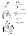
- FIG. 1 illustrates a nucleic acid probe 20, immobilized on the surface of a solid substrate 10, which is hybridized to a nucleic acid target 30 comprising a covalently attached biotin molecule.
- the biotin molecule on the target 30 is complexed with a streptavidin molecule which is optionally labeled with a detectable label.
- the labeled streptavidin 40 as shown in scheme 2 of Figure 1, is complexed with biotinylated anti-streptavidin antibody 50 which comprises multiple biotin molecules.
- the antibody thus can interact and complex with the immobilized biotinylated streptavidin via either or both of the antibody binding interaction and the biotin-streptavidin interaction.
- labeled streptavidin molecules 40 complex with the immobilized antibody 50 and provide multiple labels for detection and signal amplification.
- the antibody thus provides a bridge to permit multiple labeled streptavidin molecules to be complexed and detected. Additional steps of bridging and signal amplification also may be implemented within the scope of the invention, to enhance the detectable signal of the label.
- the detectable streptavidin which may be used is streptavidin phycoerythrin, which is commerically available, for example, from Molecular Probes (Eugene, Oregon). Biotinylated anti-streptavidin antibody is available, for example, from Vector Laboratories (Burlingame, CA).
- Nucleic acid hybridization assay procedures and conditions developed in the art may be used, as described, for example in in: Maniatis et al., "Molecular Cloning: A Laboratory Manual” 2nd Ed., Cold Spring Harbor, N.Y., 1989; Berger and Kimmel, "Methods in Enzymology," Vol. 152, “Guide to Molecular Cloning Techniques", Academic Press, Inc., San Diego, CA., 1987; Young and Davis, Proc. Natl. Acad. Sci., U.S.A., 80 :1194 (1983).
- Nucleic acid hybridization buffers that may be used include phosphate and TRIS buffers, for example, at a pH of about 6 to 8.
- a standard saline phosphate ethylenediaminetetraacetic acid (“SSPE”) buffer is used.
- An exemplary phosphate buffer includes: 0.06M H 2 PO 4 /HPO 4 , 1M Na + , 0.006M EDTA (ethylenediaminetetraacetic acid), 0.005% Triton®, at a pH of about 6.8, referred to herein as "6XSSPE-T".
- a sulfonate hybridization buffer is used, for example a buffer including 2-[N-morpholino]ethanesulfonic acid ("MES").
- the hybridization buffer may include about 0.01 M to about 2 M MES or more, e.g. , about 0.25 M MES, at a pH, for example, of about 6 to 7.
- the MES buffer includes: 0.25M MES, 1M Na + , and 0.005% Triton® X-100, at a pH of about 5.5 - 6.7, e.g. , 6.7 .
- the hybridization may be conducted, for example, at about 25 to 70°C, for example, about 45°C.
- the buffer may be filtered prior to use, for example, through a 2 ⁇ m filter.
- a method for detecting binding by hybridization of a nucleic acid target comprising a target sequence to a nucleic acid probe comprising a probe sequence.
- the nucleic acid probe is preferably immobilized on a surface.
- a binding ligand is complexed, preferably by covalent attachment, to the nucleic acid target.
- the immobilized nucleic acid probe then is contacted, for example sequentially, with the nucleic acid target comprising the binding ligand; a receptor comprising multiple sites capable of binding the binding ligand; and a DNA matrix comprising a plurality of the binding ligands.
- a complex is formed of the binding ligand of the target, the receptor and the DNA matrix.
- the resulting complex can be detected, e.g. , by providing and detecting a detectable label on the DNA matrix, or by contacting the complexed DNA matrix with, and detecting, labeled detectable molecules of the receptor that are capable of binding to the binding ligand molecules on the DNA matrix. Detection of the label thus provides a positive indicator of the hybridization of the nucleic acid target and the probe.
- the ligand and receptor are biotin and streptavidin respectively.
- a method of determining the hybridization of a nucleic acid target comprising a target nucleotide sequence with an immobilized nucleic acid probe comprising a probe nucleotide sequence A biotinylated nucleic acid target is provided.
- the method comprises contacting the immobilized nucleic acid probe, preferably sequentially, with: the biotinylated nucleic acid target; streptavidin; a biotinylated DNA matrix comprising a plurality of biotins; and labeled streptavidin molecules.
- the streptavidin is labeled with a detectable label, such as a fluorescent label.
- the binding by hybridization of the nucleic acid target to the probe may be detected with high sensitivity.
- the target includes only one biotin moiety to which streptavidin may be complexed. Streptavidin complexes with the biotinylated target. The biotinylated DNA matrix complexes with the complexed streptavidin. Thus, the number of biotin molecules is greatly amplified. Upon complexation of labeled streptavidin to the biotins on the DNA matrix, the number of detectable labels is greatly amplified, thus greatly enhancing the sensitivity of the assay.
- Nucleic acid hybridization assay procedures and conditions developed in the art may be used, as described, for example in in: Maniatis et al., "Molecular Cloning: A Laboratory Manual” 2nd Ed., Cold Spring Harbor, N.Y., 1989; Berger and Kimmel, "Methods in Enzymology," Vol. 152, “Guide to Molecular Cloning Techniques", Academic Press, Inc., San Diego, CA., 1987; Young and Davis, Proc. Natl. Acad. Sci., U.S.A., 80 :1194 (1983).
- Nucleic acid hybridization buffers that may be used include phosphate and TRIS buffers, for example, at a pH of about 6 to 8.
- a standard saline phosphate ethylenediaminetetraacetic acid (“SSPE”) buffer is used.
- An exemplary phosphate buffer includes: 0.06M H 2 PO 4 /HPO 4 , 1M Na + , 0.006M EDTA (ethylenediaminetetraacetic acid), 0.005% Triton®, at a pH of about 6.8, referred to herein as "6XSSPE-T".
- a sulfonate hybridization buffer is used, for example a buffer including 2-[N-morpholino]ethanesulfonic acid ("MES").
- the hybridization buffer may include about 0.01 M to about 2 M MES or more, e.g. , about 0.25 M MES, at a pH, for example, of about 6 to 7.
- the MES buffer includes: 0.25M MES, 1M Na + , and 0.005% Triton® X-100, at a pH of about 6.7.
- the hybridization may be conducted, for example, at about 25 to 70°C, for example, about 45°C.
- the buffer may be filtered prior to use, for example, through a 2 ⁇ m filter.
- an assay for hybridization of a nucleic acid target to a nucleic acid probe immobilized on a solid surface such as a biological chip
- the hybridization is conducted at about 45°C for 16 hrs.
- a manual wash with 1X ST-T (1M NaCl, 10 mM TRIS-HCl, 0.01% vol/vol Triton X-100, pH 8.0) three times
- a wash at a Genechip® Fluidics Station (Affymetrix, Santa Clara, CA) using (6X SSPE-T, 22°C, 10x2 (drain 10 cycles, drain and fill twice each cycle)
- the phrase "ligand-receptor pair" refers to a ligand and receptor that are chemical moieties capable of recognizing and binding to each other.
- the ligand and receptor can be any moieties that are capable of recognizing and binding to each other to form a complex. Additionally, the ligand and receptor may interact via the binding of a third intermediary substance.
- the ligand and receptor constituting the ligand-receptor pair are binding molecules that undergo a specific noncovalent binding interaction with each other.
- the ligand and receptor can be naturally occurring or artificially produced, and optionally may be aggregated with other species.
- ligands and/or receptors include, but are not limited to, agonists and antagonists for cell membrane receptors, toxins and venoms, viral epitopes, hormones such as steroids, hormone receptors, peptides, enzymes and other catalytic polypeptides, enzyme substrates, cofactors, drugs including small organic molecule drugs, opiates, opiate receptors, lectins, sugars, saccharides including polysaccharides, proteins, and antibodies including monoclonal antibodies and synthetic antibody fragments, cells, cell membranes and moieties therein including cell membrane receptors, and organelles.
- hormones such as steroids, hormone receptors, peptides, enzymes and other catalytic polypeptides, enzyme substrates, cofactors, drugs including small organic molecule drugs, opiates, opiate receptors, lectins, sugars, saccharides including polysaccharides, proteins, and antibodies including monoclonal antibodies and synthetic antibody fragments, cells, cell membranes and moieties therein including cell membrane
- ligand-receptor pairs include antibody-antigen; lectin-carbohydrate; peptide-cell membrane receptor; protein A-antibody; hapten-antihapten; digoxigenin-anti-digoxigenin; enzyme-cofactor and enzyme-substrate.
- a ligand-receptor pair includes a receptor that is capable of binding a plurality, e.g., 2, 3, 4 or more, molecules of the ligand.
- the ligand-receptor pair is biotin-avidin or biotin-streptavidin.
- the vitamin biotin is detected by binding of the indicator protein avidin, isolated from egg white, or streptavidin, isolated from Streptomyces avidinii bacteria.
- Amplification reagents used in the assay methods disclosed herein can be attached to any of a variety of members of ligand receptor binding pairs available in the art.
- the target nucleic acid is attached to a binding ligand constituting a member of a ligand-receptor binding pair.
- the amplification reagent includes a plurality of the binding ligands.
- the receptor of the ligand receptor pair is capable of binding to two or more molecules of ligand.
- the ligand may be biotin
- the receptor may be avidin or streptavidin, each of which are capable of binding four molecules of biotin.
- Hybridization of the target nucleic acid to the probe nucleotide is detected by detecting binding of the ligand of the target nucleic acid to the receptor, and binding of the receptor to the binding ligand on the amplification reagent.
- the amplification reagent is detected, e.g., by providing a label on the amplification reagent, or by complexing the amplification reagent with a plurality of molecules of labeled receptor.
- nucleic acids having a binding molecule covalently attached by can be synthesized using a DNA synthesizer and standard phosphoramidite reagents.
- a DNA synthesizer for example, a DNA synthesizer and standard phosphoramidite reagents.
- biotin phosphoramidites for direct labeling of synthetic oligonucleotides may be used. Biotin phosphoramidites are commercially available from Glen Research Corporation, Sterling, VA.
- biotinylated DNA targets can be prepared using nick translation and random primer extension, while biotinylated RNA targets can be synthesized by in vitro transcription using an RNA polymerase.
- Biotinylated deoxyribonucleoside triphosphates and ribonucleoside triphosphates have been used for the enzymatic preparation of biotinylated DNA and biotinylated RNA. Exemplary methods are disclosed in detail in Rashtchian and Mackey, Labeling and Detection of Nucleic Acids, in "Nonradioactive Labeling and Detection of Biomolecules", C.
- concentration of biotin molecules may be increased by the use of a psoralen biotin reagent, as described in Levenson et al., Methods Enzymol., 184 :577-583 (1990); and Cimono et al., Ann. Rev. Biochem. 54 :1151-1193 (1985). Background hybridization may be reduced by HPLC purification of biotinylated target nucleic acids.
- Binding ligands such as biotins
- binding ligands may be incorporated into dendrimeric DNA matrices, for example, by providing a nucleic acid having a sequence complementary to a specific sequence of the dendrimer arms to which a binding ligand such as biotin is covalently attached, and then hybridizing a plurality of the nucleic acids to the arms of the DNA matrix, thereby to produce a DNA matix amplification reagent comprising a plurality of binding ligands.
- Binding ligands such as biotins
- amplification molecules such as polymers, including poly(amino acids), such as antibodies
- exemplary methods are disclosed in detail in Bayer and Wilchek, Labeling and Detection of Proteins and Glycoproteins, in "Nonradioactive Labeling and Detection of Biomolecules", C. Kessler, Ed., Springer-Verlag, New York, 1992, pp. 91-100 and referenced cited therein.
- biotinylated antibodies such as biotinylated anti-streptavidin molecules, are available commercially, for example, from Vector Laboratories (Burlingame, CA).
- labels may be used in the assay methods disclosed herein.
- the label may be provided on the amplification reagent, the receptor and/or the binding ligand.
- Examples of labels include fluorescent labels, chemiluminescent labels, and inorganic labels, such as gold, as well as enzymatic labels.
- Labels may be used that are detectable, for example, by chromogenic detection, chemiluminescent detection and fluorescent detection. Labels that may be used include marker enzymes such as alkaline phosphatase ("AP"), ⁇ -galactosidase or horseradish peroxidase, which are detected using a chromogenic substrate. For example, AP may be detected using 5-bromo-4-chloro-3-indolyl phosphate or nitroblue tetrazolium salt.
- marker enzymes such as alkaline phosphatase (“AP"), ⁇ -galactosidase or horseradish peroxidase
- fluorescent tags such as fluorescein, rhodamine, and resorufin, and derivatives thereof, as well as coumarins such as hydroxycoumarin.
- fluorescence resonance energy transfer may be measured, as described in Cardullo, Nonradiative Fluorescence Resonance Energy Transfer in "Nonradioactive Labeling and Detection of Biomolecules", C. Kessler, Ed., Springer-Verlag, New York, 1992, pp. 414-423.
- inorganic labels may be used, such as colloidal gold particles or ferritin.
- colloidal gold particles as labels is described, for example, in Van de Plas and Leunissen, Colloidal Gold as a Marker in Molecular Biology: The Use of Ultra-Small Gold Particles, in "Nonradioactive Labeling and Detection of Biomolecules", C. Kessler, Ed., Springer-Verlag, New York, 1992, pp. 116-126.
- the binding ligand is biotin and the receptor is avidin or streptavidin
- the amplification reagent is an antibody or a DNA matrix.
- One or more labels may be conjugated to the avidin or streptavidin and/or the amplification reagent.
- Reagents for labeling streptavidin or avidin with a fluorescent tag are commercially available.
- the reagents 5(6)-Carboxyfluorescein-N-hydroxysuccinimide ester (FLUOS), 7-amino-4-methyl-coumarin-3-acetic acid-N'-hydroxysuccinimide ester (AMCA, acitvated) and fluorescein isothiocyanate (FITC) are available from Boehringer Mannheim, Indianapolis, IN. Methods for fluorescently labeling proteins with fluorescent labels, and methods for detection of the fluorescent labels, are described in Howard, G., Labeling Proteins with Fluorochromes, in "Methods in Nonradioactive Detection,”, G.
- streptavidin and avidin molecules there are a variety of commercially available labeled streptavidin and avidin molecules. Examples include streptavidin-gold, streptavidin-fluorochrome, streptavidin-AMCA, streptavidin-fluorescein, streptavidin-phycoerythrin (STPE), streptavidin-sulforhodamine 101, avidin-FITC and avidin-Texas red®, which are commercially available from Boehringer Mannheim, Indianapolis, IN.
- compositions disclosed herein may be used in a variety of applications for assaying for hybridization of a nucleic acid target comprising a target sequence and a nucleic acid probe comprising a probe sequence.
- one or more target nucleic acids comprising different target sequences may be screened for hybridization to a high density array of nucleic acid probes comprising different sequences.
- hybridization refers to the binding association between two nucleic acids, for example the non-covalent interaction via base pair hydrogen bonding and base stacking.
- the ability of two single stranded nucleic acids to hybridize will depend upon factors such as their degree of complementarity as well as the stringency of the hybridization reaction conditions.
- stringency refers to the conditions of a hybridization reaction that influence the degree to which nucleic acids hybridize. Stringent conditions can be selected that allow nucleic acid duplexes to be distinguished based on their degree of mismatch. High stringency is correlated with a lower probability for the formation of a duplex containing mismatched bases. Thus, the higher the stringency, the greater the probability that two single-stranded nucleic acids, capable of forming a mismatched duplex, will remain single-stranded. Conversely, at lower stringency, the probability of formation of a mismatched duplex is increased.
- the appropriate stringency that will allow selection of a perfectly-matched duplex, compared to a duplex containing one or more mismatches (or that will allow selection of a particular mismatched duplex compared to a duplex with a higher degree of mismatch) is generally determined empirically. Means for adjusting the stringency of a hybridization reaction are well-known to those of skill in the art.
- conditions that increase stringency include higher temperature, lower ionic strength and presence or absence of solvents; lower stringency is favored by lower temperature, higher ionic strength, and lower or higher concentrations of solvents (for example, lower concentrations of formamide or dimethyl sulfoxide).
- concentration of reactants i.e ., single stranded nucleic acid
- Hybridization assay procedures and conditions will vary depending on the application and are selected in accordance with the general binding methods known including those referred to in: Maniatis et al., "Molecular Cloning: A Laboratory Manual” 2nd Ed., Cold Spring Harbor, N.Y., 1989; Berger and Kimmel, "Methods in Enzymology," Vol. 152, “Guide to Molecular Cloning Techniques", Academic Press, Inc., San Diego, CA., 1987; Young and Davis, Proc. Natl. Acad. Sci., U.S.A., 80 :1194 (1983).
- Nucleic acid hybridization buffers that may be used include phosphate and TRIS buffers, for example, at a pH of about 6 to 8.
- a standard saline phosphate ethylenediaminetetraacetic acid (“SSPE”) buffer is used.
- An exemplary phosphate buffer includes: 0.06M H 2 PO 4 /HPO 4 , 1M Na + , 0.006M EDTA (ethylenediaminetetraacetic acid), 0.005% Triton®, at a pH of about 6.8, referred to herein as "6XSSPE-T".
- a method is provided of conducting nucleic acid hybridization assays, wherein the hybridization solution comprises a sulfonate buffer.
- Sulfonate hybridization buffers include 2-[N-morpholino]ethanesulfonic acid ("MES") and 3-[N-morpholino]propanesulfonic acid) ("MOPS").
- MES 2-[N-morpholino]ethanesulfonic acid
- MOPS 3-[N-morpholino]propanesulfonic acid
- the hybridization assay using a sulfonate buffer may be conducted with nucleic acid probes immobilized on a solid surface, such as a silicon or glass surface. The solid surface may be, for example, coated with a silane coating prior to immobilization of the nucleic acid probes.
- the hybridization assay in a solution comprising a sulfonate buffer may be conducted, for example, at a temperature of about 25 to 70°C, for example, at least about 35°C, or 45°C or more, and over a time period of, for example, about 10 minutes to 5 hours or more, e.g., about 16 hours or more.
- the sulfonate buffer may be used, for example in gene expression hybridization assays and polymorphism hybridization assays.
- the hybridization buffer may include about 0.01 M to about 2 M MES or more, e.g., about 0.25 M MES, at a pH, for example, of about 6 to 7.
- the MES buffer includes: 0.25M MES, 1M Na + , and 0.005% Triton® X-100, at a pH of about 5.5 - 6.7, e.g., 6.7.
- the hybridization may be conducted, for example, at about 25 to 70°C, for example, about 45°C.
- the buffer may be filtered prior to use, for example, through a 2 ⁇ m filter.
- the nucleic acid hybridization buffers may further include surfactants, such as Tween-20 and Triton-X100, as well as additives such as anti-foaming agents.
- substrates having a surface to which arrays of polynucleotides are attached are referred to herein as "biological chips".
- the substrate may be, for example, silicon or glass, and can have the thickness of a microscope slide or glass cover slip.
- substrates that are transparent to light are useful when the assay involves optical detection, as described, e.g., in U.S. Patent No. 5,545,531.
- Other substrates include Langmuir Blodgett film, germanium, (poly)tetrafluorethylene, polystyrene, gallium arsenide, gallium phosphide, silicon oxide, silicon nitride, and combinations thereof.
- the number of nucleic acid sequences may be selected for different applications, and may be, for example, about 100 or more, or, e.g., in some embodiments, more than 10 5 or 10 8 .
- the surface comprises at least 100 probe nucleic acids each preferably having a different sequence, each probe contained in an area of less than about 0.1 cm 2 , or, for example, between about 1 ⁇ m 2 and 10,000 ⁇ m 2 , and each probe nucleic acid having a defined sequence and location on the surface.
- at least 1,000 different nucleic acids are provided on the surface, wherein each nucleic acid is contained within an area less than about 10 -3 cm 2 , as described, for example, in U.S. Patent No. 5,510,270.
- arrays of nucleic acids for use in gene expression monitoring are described in PCT WO 97/10365.
- arrays of nucleic acid probes are immobilized on a surface, wherein the array comprises more than 100 different nucleic acids and wherein each different nucleic acid is localized in a predetermined area of the surface, and the density of the different oligonucleotides is greater than about 60 different oligonucleotides per 1 cm 2 .
- nucleic acids immobilized on a surface which may be used also are described in detail in U.S. Patent No. 5,744,305.
- nucleic acids with different sequences are immobilized each in a predefined area on a surface.
- 10, 50, 60, 100, 10 3 , 10 4 , 10 5 , 10 6 , 10 7 , or 10 8 different monomer sequences may be provided on the substrate.
- the nucleic acids of a particular sequence are provided within a predefined region of a substrate, having a surface area, for example, of about 1 cm 2 to 10 -10 cm 2 .
- the regions have areas of less than about 10 -1 , 10 -2 , 10 -3 , 10 -4 , 10 -5 , 10 -6 , 10 -7 , 10 -8 , 10 -9 , or 10 -10 cm 2 .
- a planar, non-porous support having at least a first surface, and a plurality of different nucleic acids attached to the first surface at a density exceeding about 400 different nucleic acids/cm 2 , wherein each of the different nucleic acids is attached to the surface of the solid support in a different predefined region, has a different determinable sequence, and is, for example, at least 4 nucleotides in length.
- the nucleic acids may be, for example, about 4 to 20 nucleotides in length.
- the number of different nucleic acids may be, for example, 1000 or more.
- fused silica substrates may be used which have a low glass fluorescence background.
- Silica substrates permit a low exposure print gap with little or no signal falloff in going from 50 ⁇ m to 20 ⁇ m features, and yields improved performance, including signal and discrimination, for feature sizes less than 50 ⁇ m, for example, 2 ⁇ m.
- a description of solid phase chemistry, photolithography, and data collecting methods, for the synthesis and use of arrays of materials attached to a solid substrate is provided in U.S. Patent No. 5,744,305.
- detection may be implemented by directing light to relatively small and precisely known locations on the substrate.
- the substrate is placed in a microscope detection apparatus for identification of locations where binding takes place.
- the microscope detection apparatus includes a monochromatic or polychromatic light source for directing light at the substrate, means for detecting fluoresced light from the substrate, and means for determining a location of the fluoresced light.
- the means for detecting light fluoresced on the substrate may in some embodiments include a photon counter.
- the means for determining a location of the fluoresced light may include an x/y translation table for the substrate. Translation of the substrate and data collection are recorded and managed by an appropriately programmed digital computer, as described in U.S. Patent No. 5,510,270, the disclosure of which is incorporated herein.
- Devices for concurrently processing multiple biological chip assays may be used as described in U.S. Patent No. 5,545,531.
- Methods and systems for detecting a labeled marker on a sample on a solid support, wherein the labeled material emits radiation at a wavelength that is different from the excitation wavelength, which radiation is collected by collection optics and imaged onto a detector which generates an image of the sample are disclosed in U.S. Patent No. 5,578,832. These methods permit a highly sensitive and resolved image to be obtained at high speed. Methods and apparatus for detection of fluorescently labeled materials are further described in U.S. Patent Nos. 5,631,734 and 5,324,633.
- the assay methods and compositions described herein may be used in a range of applications including biomedical and genetic research and clinical diagnostics.
- Arrays of polymers such as nucleic acids may be screened for specific binding to a target, such as a complementary nucleotide, for example, in screening studies for determination of binding affinity and in diagnostic assays.
- sequencing of polynucleotides can be conducted, as disclosed in U.S. Patent No. 5,547,839.
- the nucleic acid arrays may be used in many other applications including detection of genetic diseases such as cystic fibrosis, diabetes, and acquired diseases such as cancer, as disclosed in U.S. Patent Application Ser. No. 08/143,312. Genetic mutations may be detected by sequencing by hydridization.
- genetic markers may be sequenced and mapped using Type-IIs restriction endonucleases as disclosed in U.S. Patent No. 5,710,000.
- Gene expression may be monitored by hybridization of large numbers of mRNAs in parallel using high density arrays of nucleic acids in cells, such as in microorganisms such as yeast, as described in Lockhart et al., Nature Biotechnology, 14 :1675-1680 (1996).
- Bacterial transcript imaging by hybridization of total RNA to nucleic acid arrays may be conducted as described in Saizieu et al., Nature Biotechnology, 16 :45-48 (1998).
- kits for carrying out amplified nucleic acid hybridization assays that include in packaged combination the materialss: immobilized probe nucleic acids, amplification reagents, and labeled receptors. These reagents will be, for example, in separate containers in the kit.
- the kit may also include hybridization buffers, wash solutions, negative and positive controls and written instructions for carrying out the assay.
- a nucleic hybridization assay was conducted using a streptavidin-phycoerythrin ("SAPE”) and biotinylated anti-streptavidin antibody amplification detection method.
- SAPE streptavidin-phycoerythrin
- a fragmented cRNA sample was prepared as follows. PCR amplification of a plasmid library was conducted followed by in vitro transcription in the presence of biotinylated UTP and CTP to form biotinylated cRNA, which was then fragmented in 1x Fragmentation Buffer by heating at 95°C for 35 minutes. See general techniques described in Sambrook, et al., "Molecular Cloning: A Laboratory Manual," Second Edition, Cold Spring Harbor Laboratory Press, 1989; Ausubel, et al., "Current Protocols In Molecular Biology,” John Wiley & Sons, 1996; and Lockhart, D.J., et al., Nature Biotechnology, 14 : 1675-1680 (1996).
- Chip Pre-Treatment solution 200 ⁇ L was applied to a biological chip having immobilized thereon an array of probe nucleic acids of known sequences.
- the chip was placed on the rotisserie in the GeneChip Hybridization 320 Oven (Affymetrix, Santa Clara, CA) and rotated at 45°C, 60 rpm for 10 minutes.
- Chip Pre-Treatment solution was removed from the cartridge and 200 ⁇ L of sample at 45°C was added.
- the chip was placed on the rotisserie in the GeneChip Hybridization 320 Oven and hybridized at 45°C, 60 rpm for 16 hours.
- Sample was removed from the chip and replaced with 200 ⁇ L of 6X SSPE-Tween buffer.
- the chip was subjected to an automated wash/stain protocol on the GeneChip Fluidics Station 400 (Affymetrix, Santa Clara, CA) using the EukGE-WS2 fluidics script as follows.
- the chip was washed on the fluidics station at 25°C, with 6X SSPE-Tween buffer for 10 cycles with 2 mixes per cycle. This was followed by another wash in 0.1X MES Stringent buffer at 50°C for 4 cycles with 15 mixes per cycle.
- the chip was then stained for 10 minutes at 25°C on the fluidics station with the SAPE staining solution described above. Staining was followed by a wash with 6X SSPE-Tween buffer at 25°C for 10 cycles with 4 mixes per cycle.
- a first scan was conducted using the HP G2500A GeneArray Scanner (Affymetrix, Santa Clara, CA).
- the 2X multi-image scan was conducted at 570 nm, using a 3 ⁇ m pixel size.
- the dynamic range of detected intensity of fluorescence was 500-5000 counts (arbitrary units). The results are shown in Figure 2a, where the background was 707 counts and the standard deviation of background was 12.6.
- the chip was returned to the fluidics station and was stained for 10 minutes at 25°C with the biotinylated anti-streptavidin stain solution, followed by a purge (twice) with 6X SSPE-Tween buffer and a third 10 minute stain at 25°C with the SAPE stain solution.
- the final wash was with 6X SSPE-Tween buffer at 30°C for 15 cycles with 4 mixes per cycle.
- a nucleic hybridization assay was conducted using streptavidin-phycoerythrin and biotinylated anti-streptavidin antibody amplification.
- Buffers were made up in molecular biology grade water (BioWhittaker).
- a fragmented cRNA sample from a murine B-cell library was prepared by PCR amplification of a plasmid library (Clonetech) followed by in vitro transcription in the presence of biotinylated UTP and CTP to form biotinylated cRNA, which was then fragmented in 1x Fragmentation Buffer, as described in Example 1, by heating at 95°C for 35 minutes.
- a fragmented cRNA sample also was obtained from human brain cell mRNA (Clonetech).
- the biological chips had feature sizes of 24 microns.
- each hybridization assay 200 ⁇ l of chip pre-treatment solution was applied to the biological chip.
- the chip was placed on a rotisserie (Rotamix, Appropriate Technical Resources, Laurel, MD) at 40°C, at 60 rpm for 15 min.
- the chip was rinsed in 1x MES buffer.
- the sample was heated at 99°C for 5 min and then put in 45°C waterbath for less than about 10 minutes before putting it on the chip. 200 ⁇ l sample at 45°C was added to the chip. Hybridization on the rotisserie was conducted at 45°C, at 60 rpm, for 16 or 40 hr.
- the chip was washed on a fluidics station (GeneChip Fluidics Station 400, Affymetrix, Santa Clara, CA) with 6x SSPE-T at 22°C, 10x2 (10 cycles, drain and fill twice each cycle).
- the chip was rinsed with 0.1x MES buffer.
- 200 ⁇ l 0.1x MES buffer was then applied to wash the chip at 45°C, 60 rpm, for 30 min on the rotisserie.
- the chip was rinsed with 1x MES buffer.
- the chip was stained with 5x SAPE staining solution at 40°C, 60 rpm for 15 min. This was followed by a 6x SSPE-T wash on the fluidics station at 22°C, 10x2.
- a first scan was conducted using a HP G2500A GeneArray Scanner (Affymetrix, Santa Clara, CA).
- the 2X multi-image scan was conducted at 570 nm, using a 3 ⁇ m pixel size.
- the 6x SSPE-T solution was removed from the chip and 200 ⁇ l of 1x MES buffer was used to rinse the chip.
- the chip was stained with 200 ⁇ l of anti-SA (anti-streptavidin) staining solution at 40°C, 60 rpm, for 30 min.
- 200 ⁇ l of 1x MES buffer was used to wash the chip manually. This was followed by a 6x SSPE-T wash on the fluidics station at 22°C, 10x2. 1x MES buffer was then used to rinse the chip.
- Table 1 shows in columns 1-13 respectively: 1) concentration of spikes; 2) hybridization time; 3) use or nonuse of antibody; 4) number of genes detected; 5) percent of genes detected; 6) average difference in intensity between matches and mismatches for all signals; 7) average difference in intensity between matches and mismatches for positive signals; 8) background; 9) standard deviation; 10) the value Q corresponding to noise; 11) the value P/Q corresponding to signal/noise ratio; 12) number of spikes detected; 13) percent false negatives or positives.
- Table 2 shows in columns 1-13 respectively: 1) concentration of spikes; 2) hybridization time; 3) use or nonuse of antibody; 4) number of genes detected; 5) percent of genes detected; 6) average difference in intensity between matches and mismatches for all signals; 7) average difference in intensity between matches and mismatches for positive signals; 8) background; 9) standard deviation; 10) the value Q corresponding to noise; 11) the value P/Q corresponding to signal/noise ratio; 12) number of spikes detected; 13) percent false negatives or positives.
Landscapes
- Chemical & Material Sciences (AREA)
- Organic Chemistry (AREA)
- Life Sciences & Earth Sciences (AREA)
- Health & Medical Sciences (AREA)
- Zoology (AREA)
- Wood Science & Technology (AREA)
- Proteomics, Peptides & Aminoacids (AREA)
- Engineering & Computer Science (AREA)
- Analytical Chemistry (AREA)
- Immunology (AREA)
- Physics & Mathematics (AREA)
- Microbiology (AREA)
- Molecular Biology (AREA)
- Biotechnology (AREA)
- Biophysics (AREA)
- Biochemistry (AREA)
- Bioinformatics & Cheminformatics (AREA)
- General Engineering & Computer Science (AREA)
- General Health & Medical Sciences (AREA)
- Genetics & Genomics (AREA)
- Pathology (AREA)
- Measuring Or Testing Involving Enzymes Or Micro-Organisms (AREA)
Abstract
Description
- This application claims the benefit of U.S. Provisional Patent Application Serial No. 60/102,577, filed September 30, 1998, the disclosure of which is incorporated herein by reference in its entirety.
- This invention relates generally to methods and compounds for amplifying detectable signals in specific binding assays, particularly nucleic acid hybridization assays.
- Nucleic acid hybridizations are commonly used in biochemical research and diagnostic assays. Generally a single stranded analyte nucleic acid is hybridized to labeled nucleic acid probe, and resulting nucleic acid duplexes are detected. Radioactive and nonradioactive labels have been used. Methods also have been developed to amplify the signal that is detected. For example, large comb-type branched polynucleotides, comprising a first oligonucleotide unit and branches including second oligonucleotide units, have been developed for signal amplification in nucleic acid detection assays. In this application, the branched polynucleotide is hybridized via the first oligonucleotide unit to single stranded analyte nucleic acid and then labeled oligonucleotide is hybridized to the branched polynucleotide via the second oligonucleotide units, as described in U.S. Patent No. 5,710,264 to Chiron Corporation.
- Dendrimers have been developed that are assembled by the sequential hybridization of single DNA strands. Pairwise hybridization of single strands produces monomers with a double stranded center and four single stranded "arms". The monomers can be grown exponentially in sequential hybridization steps, to produce a macromolecule including terminal single stranded arms. The dendrimers may range in size from hundreds to millions of bases. The arms that are not to be hybridized to target can be hybridized to covalently labeled oligonucleotide. These dendrimers are commerically available from Polyprobe, Bala Cynwyd, PA. Dendrimers for assaying nucleic acids are described in U.S. Patent Nos. 5,175,270, 5,487,973, and 5,484,904, the disclosures of which are incorporated herein.
- Avidin-biotin systems have been developed for use in a variety of detection assays. Methods for the detection and labeling of nucleic acids in biotin systems are described, for example, in "Nonradioactive Labeling and Detection Systems", C. Kessler, Ed., Springer-Verlag, New York, 1992, pp. 70-99; and in "Methods in Nonradioactive Detection,", G. Howard, Ed., Appleton and Lange, Norwalk, Connecticut, 1993, pp. 11-27 and 137-150.
- Methods for the detection of nucleic acid sequences have suffered from drawbacks including background noise, time and labor requirements, lack of specificity and lack of sensitivity. It is an object of the invention to provide materials for the detection of polymers, particularly nucleic acids. It is a particular object of the invention to provide methods and compounds for amplifying labeling signals used in the detection of nucleic acid sequences in specific binding assays. It is a further object of the invention to provide methods and compounds which permit nucleic acid sequences to be detected specifically and rapidly with high sensitivity and high resolution.
- WO 98/04745 discloses a method of analyzing nucleic acid sequences by hybridization, wherein two probe sequences are hybridized to a target sequence and ligated to obtain a single polynucleotide which is subsequently detected. Detection of hybridization may comprise a hybridization signal amplification method, wherein target specific nucleic acid probe is labeled with a ligand, an excess of ligand binding moiety and a signal probe is added, thereby resulting in formation of a large complex which is said to be easily be detected.
- Kerstens et al. (J. Histochem. and Cytochem., 1995, pp.347-352) also describes strategies for amplifying the signal in nucleic acid detection methods. A technique identified as CARD ISH (catalyzed reporter deposition combined with in situ hybridization) is described, wherein a DNA probe label is complexed with:
- (1) mouse anti-biotin,
- (2) biotinylated horse anti-mouse,
- (3) avidin-biotin peroxidase,
- (4) deposition of biotinylated tyramine and
- (5) fluorochrom or enzyme labeled avidin.
-
- Secondary signal amplification in methods for nucleic acid detection was also achieved by in vitro transcription of the immobilized target molecule/amplifier construct complex in the presence of labels which are incorporated into the transcript (WO 93/24658).
- Methods and compounds are provided for detecting target molecules using specific binding assays. Methods and compositions are provided that are useful in signal amplification in the detection of the target molecule.
- In one embodiment, methods are provided for detecting a nucleic acid target wherein the method comprises hybridizing a nucleic acid target, comprising a target nucleic acid sequence, to a nucleic acid probe immobilized on a surface, comprising a probe nucleic acid sequence, wherein a binding ligand is complexed to said target. The hydridized target is contacted with a receptor comprising multiple sites capable of binding the binding ligand to complex the receptor to the binding ligand, and the receptor is contacted with an amplification reagent, comprising a plurality of the binding ligands, wherein said binding ligands are covalently attached to said amplification reagent to complex the amplification reagent to the receptor, wherein the amplification reagent is selected from the group consisting of an antibody and a dendrimeric DNA matrix comprising layers of DNA and single stranded sequences at the surface of the matrix that are free to hybridize with a predetermined nucleic acid sequence. The presence of the complexed amplification reagent then is detected, for example, by detecting the presence of a detectable label on at least one of the receptor and the amplification reagent.
- Optionally, after complexing the amplification reagent to the receptor complexed to the hybridized target, the amplification reagent, comprising a plurality of the binding ligands, is contacted with labeled receptor molecules thereby to complex a plurality of labeled receptor molecules to the amplification reagent, and the labeled receptor molecules complexed to the amplification reagent are detected. This permits the detectable signal to be enhanced and more easily detected.
- Labels which may be used include fluorescent labels, gold labels and enzyme labels. Exemplary fluorescent labels include fluorescein, rhodamine, resorufin, or a coumarin. In one preferred embodiment, the binding ligand is biotin and the receptor is avidin or streptavidin.
- The amplification reagent may comprise a polymer, such as a poly (amino acid) or a polynucleotide. In one embodiment, the amplification reagent may be an antibody, such as an anti-receptor antibody capable of specifically binding the receptor.
- In another embodiment, the amplification molecule may be a DNA matrix. The DNA matrix may include subunits of partially double stranded and partially single stranded DNA molecules. In one embodiment, the DNA matrix molecule includes a plurality of molecules of a first partially double stranded polynucleotide, the polynucleotide having a first molecule end, a second molecule end and a double stranded body portion intermediate of the first and second ends, wherein the first and second ends each comprise at least one of first and second arms consisting of a single strand of polynucleotide, and wherein the single strands are hybridizable with a predetermined nucleic acid sequence of nucleotides in a nucleic acid, and the first and second arms of each of said first and second ends are nonhybridizable with each other. The DNA matrix further includes a plurality of molecules of a second partially double stranded polynucleotide, each polynucleotide including a first molecule end, a second molecule end and a double stranded body portion intermediate the first and second ends, the first and second ends each having at least one of first and second arms consisting of a single strand of polynucleotide that is hybridizable with a predetermined nucleic acid sequence of nucleotides in a nucleic acid, wherein the first and second arms of each of said first and second ends are non-hybridizable with each other. In the DNA matrix, the plurality of molecules of the first polynucleotide and the second polynucleotide are joined together through annealing of one or more arms thereof, to form a matrix, wherein arms of the plurality of first and second polynucleotide molecules located on the outer surface of the matrix are hybridized to nucleic acids having the binding ligand attached thereto.
- In one embodiment, the binding ligand is biotin and the receptor is avidin or streptavidin. For example, in the embodiment wherein the amplification reagent is an antibody the biotin may be covalently attached to the antibody. For example, the antibody may be an anti-streptavidin antibody comprising a plurality of biotin molecules covalently attached to the antibody. In an assay, after complexing of the antibody to a streptavidin receptor bound to the biotinylated target nucleic acid, the antibody may be contacted with labeled streptavidin, thereby to complex a plurality of labeled streptavidin molecules to the antibody, and the labeled streptavidin molecules complexed to the antibody then may be detected, thus providing signal amplification in the assay.
- In the embodiment wherein the amplification reagent is a DNA matrix comprising single stranded DNA, a binding ligand consisting of biotin may be attached to the DNA matrix by hybridization of a plurality of biotinylated nucleic acids to single strands of the DNA matrix. In this embodiment, after complexing the DNA matrix to the receptor complexes to the hybridized target, the biotinylated DNA matrix is contacted with labeled streptavidin, thereby to complex a plurality of labeled streptavidin molecules to the biotins on the matrix, and the labeled strentavidin molecules complexed to the DNA matrix are detected, thus enhancing the detectable signal.
- In one preferred embodiment, the nucleic acid probe is immobilized on a surface. The surface may be, for example, Langmuir Blodgett film, glass, germanium, silicon, (poly)tetrafluorethylene, polystyrene, gallium arsenide, gallium phosphide, silicon oxide, silicon nitride, and combinations thereof.
- In one embodiment, the hybridizing of a nucleic acid target, comprising a target nucleic acid sequence, to a nucleic acid probe, comprising a probe nucleic acid sequence is conducted in a hybridization solution comprising a sulfonate buffer.
- In a further embodiment, a method for detecting a nucleic acid target is provided, wherein the method includes providing a surface comprising at least 100 nucleic acid probes, each in an area of less than about 0.1 cm2, and each nucleic acid probe having a defined sequence and location on the surface, and contacting the surface with a nucleic acid target, comprising a target nucleic acid sequence, to permit the nucleic acid target to hybridize with at least one selected nucleic acid probe that comprises a probe nucleic acid sequence capable of hybridizing to the target nucleic acid sequence, and wherein a binding ligand is complexed to said target. The probes are preferably immobilized on the surface, for example by covalent attachment. The method further includes contacting the hydridized target with a receptor comprising multiple sites capable of binding the binding ligand to complex the receptor to the binding ligand; contacting the receptor with an amplification reagent, comprising a plurality of the binding ligands, wherein said binding ligands are covalently attached to said amplification reagent to complex the amplification reagent to the receptor, wherein the amplification reagent is selected from the group consisting of an antibody and a dendrimeric DNA matrix comprising layers of DNA and single stranded sequences at the surface of the matrix that are free to hybridize with a predetermined nucleic acid sequence and detecting the presence of the complexed amplification reagent. In one embodiment, the amplification reagent comprises an antibody or a DNA matrix, the binding ligand comprises biotin and the receptor comprises streptavidin. In one embodiment, the surface comprising the probes is contacted with the nucleic acid target in a hybridization solution comprising a sulfonate buffer, such as 2-[N-morpholino]ethanesulfonic acid ("MES").
- Also provided are complexes comprising a binding ligand complexed to a nucleic acid, a receptor; and an amplification reagent comprising a plurality of the binding ligands, wherein said binding ligands are covalently attached to said amplification reagent. In one embodiment, the binding ligand is biotin and the receptor is streptavidin or avidin.
- In another embodiment, there is provided a substrate comprising a surface having immobilized thereon a nucleic acid probe, comprising a probe nucleic acid sequence, hybridized to a nucleic acid target, comprising a target nucleic acid sequence; wherein a binding ligand is complexed to said target, and wherein the binding ligand on the target is complexed with a receptor comprising multiple sites capable of binding the binding ligand, and wherein the receptor is complexed to an amplification reagent, comprising a plurality of the binding ligands wherein said binding ligands are covalently attached to said amplification reagent.
-
- Figure 1 is a scheme illustrating the detection of a nucleic acid target using an amplification reagent comprising an anti-streptavidin antibody covalently linked to a plurality of biotin molecules.
- Figures 2a and 2b show a comparison of fluoresence detection of nucleic acid hybridization without amplification and with amplification respectively.
-
- Methods and compounds are provided for detecting target molecules using specific binding assays. In particular, methods are provided for detecting a nucleic acid target.
- In one embodiment, the method compises hybridizing a nucleic acid target, comprising a target nucleic acid sequence, to a nucleic acid probe immobilized on a surface, comprising a probe nucleic acid sequence, wherein the target comprises a binding ligand is complexed to said target. The hydridized target is contacted with a receptor comprising multiple sites capable of binding the binding ligand to complex the receptor to the binding ligand, and the receptor is contacted with an amplification reagent, comprising a plurality of the binding ligands, wherein said binding ligands are covalently attached to said amplification reagent, to complex the amplification reagent to the receptor, wherein the amplification reagent is selected from the group consisting of an antibody and a dendrimeric DNA matrix comprising layers of DNA and single stranded sequences at the surface of the matrix that are free to hybridize with a predetermined nucleic acid sequence. The presence of the complexed amplification reagent then is detected, for example, by detecting the presence of a detectable label, such as a fluorescent label, for example, on the receptor or the amplification reagent. Optionally, after complexing the amplification reagent to the receptor complexed to the hybridized target, the amplification reagent, comprising a plurality of the binding ligands, is contacted with labeled receptor molecules thereby to complex a plurality of labeled receptor molecules to the amplification reagent, and the labeled receptor molecules complexed to the amplification reagent are detected. This permits the detectable signal to be enhanced and more easily detected. In one embodiment, the amplification reagent is an antibody or a DNA matrix, the binding ligand is biotin and the receptor is streptavidin.
- In another embodiment, an array of different nucleic acid probes immobilized on a surface, each having a defined sequence and location on the surface, may be used in the assays disclosed herein, thus permitting screening and detection of binding of a large number of nucleic acids in a sample.
- While nucleic acid targets and probes are discussed in detail herein by way of example, the methods and compounds disclosed herein may be used to detect the binding of other molecules including polypeptides.
- The amplification reagent may be any chemical substance that comprises a binding ligand capable of binding to a receptor. Preferably, the amplification molecule includes a plurality of binding ligands capable of binding to a receptor. The ligands may be capable of binding the receptor, for example via non-covalent specific binding interactions. The amplification reagent may comprise, for example, a polymer, such as a poly(amino acid) or poly(ester).
- In one embodiment, the amplification reagent may comprise an antibody. As used herein, the term "antibody" refers to an immunoglobulin molecule or a fragment of an immunoglobulin molecule having the ability to specifically bind to a particular antigen. The antibody may be an anti-receptor antibody specific for the receptor used in the assay. Thus, the antibody may be capable of specifically binding the receptor as the antigen. Antibodies and methods for their manufacture are well known in the art of immunology. The antibody may be produced, for example, by hybridoma cell lines, by immunization to elicit a polyclonal antibody response, or by recombinant host cells that have been transformed with a recombinant DNA expression vector that encodes the antibody. Antibodies include but are not limited to immunoglobulin molecules of any isotype (IgA, IgG, IgE, IgD, IgM), and active fragments including Fab, Fab', F(ab')2, Facb, Fv, ScFv, Fd, VH and VL. Antibodies include but are not limited to single chain antibodies, chimeric antibodies, mutants, fusion proteins, humanized antibodies and any other modified configuration of an immunoglobulin molecule that comprises an antigen recognition site of the required specificity.
- The preparation of antibodies including antibody fragments and other modified forms is described, for example, in "Immunochemistry in Practice," Johnstone and Thorpe, Eds., Blackwell Science, Cambridge, MA, 1996; "Antibody Engineering," 2nd edition, C. Borrebaeck, Ed., Oxford University Press, New York, 1995; "Immunoassay", E. P. Diamandis and T.K. Christopoulos, Eds., Academic Press, Inc., San Diego, 1996; "Handbook of Experimental Immunology," Herzenberg et al., Eds, Blackwell Science, Cambridge, MA, 1996; and "Current Protocols in Molecular Biology" F.M. Ausubel et al., Eds., Greene Pub. Associates and Wiley Interscience, 1987, the disclosures of which are incorporated herein. A wide variety of antibodies also are available commercially.
- The amplification reagent preferably comprises a plurality of the binding ligands. Preferably, the binding ligands are covalently attached to the amplification reagent. For example, in one embodiment, the binding ligand comprises biotin, the receptor is avidin or streptavidin, and the amplification reagent is an anti-streptavidin antibody. In this embodiment, for example, a plurality of biotin molecules, e.g., about 3-10 biotin molecules, are covalently attached to the antibody.
- In one embodiment, the amplification reagent is a polynucleotide matrix. The term "polynucleotide" or "nucleic acid" as used herein refers to a polymeric form of nucleotides of any length, either ribonucleotides or deoxyribonucleotides, that comprise purine and pyrimidine bases, or other natural, chemically or biochemically modified, non-natural, or derivatized nucleotide bases. The backbone of the polynucleotide can comprise sugars and phosphate groups (as may typically be found in RNA or DNA), or modified or substituted sugar or phosphate groups. A polynucleotide may comprise modified nucleotides, such as methylated nucleotides and nucleotide analogs. The sequence of nucleotides may be interrupted by non-nucleotide components.
- In one embodiment, the amplification reagent is a deoxyribonucleic acid ("DNA") matrix. As used herein, the phrase "DNA matrix" refers to a complex of DNA that comprises one or more deoxyribonucleic acids. The DNA may include purine and pyrimidine bases, or other natural, chemically or biochemically modified, non-natural, or derivatized nucleotide bases. In one embodiment, the DNA matrix is a dendrimer that comprises subunits of partially double stranded and partially single stranded DNA molecules. The dendrimeric DNA matrix includes successive layers of polynucleotides of specific structure, including a double stranded waist and single stranded free arms at the molecule ends, formed by hybridization of the arms to adjacent molecule arms. Exemplary dendrimeric DNA matrices are commercially available from Polyprobe, Bala Cynwyd, PA, and are described in detail in U.S. Patent Nos. 5,175,270 and 5,487,973, the disclosures of which are incorporated herein.
- For example, to form the dendrimeric DNA matrix, a set of nucleotide strand molecules are provided, e.g., by synthesis on a DNA synthesizer or "gene machine" such as the Model 1500 SYSTEC synthesizer. The molecules may be cloned into specifically modified E. coli vectors for large scale production by known methods. The molecules are hybridized to form DNA matrices.
- The dendrimeric DNA matrices include layers of DNA. The outermost layer of the DNA matrix has single stranded sequences at the surface that can hybridize with a predetermined nucleic acid sequence. Each layer is composed of a specific class of matrix monomers. Sequential addition of matrix monomers yields a three dimensional DNA matrix.
- One embodiment of the assembly of a DNA dendrimer is provided by reference to Figures 1-3 and the description of U.S. Patent No. 5,175,270. The matrix monomers, for example, are DNA dimers having a middle, double stranded "waist" and four single stranded "arms", as shown in Figure 2 of U.S. Patent No. 5, 175,270. The matrix monomer and components thereof (i.e., waist and arms) can be of any length. For example, each waist can range from about 25 to about 2500 nucleotides. The single stranded arm can typically range between about 12 and about 1000 nucleotides, and preferably between about 20 and 100 nucleotides in length.
- Figure 1 of U.S. Patent No. 5,175,270 illustrates seven single stranded DNA oligomers which may be assembled to form the matrix monomer. In each oligomer depicted, the solid lines represent (+) strands while the broken lines represent complementary strand sequences. Each lettered (a-e) segment denotes a unique sequence on the different oligomers. Figure 2 of U.S. Patent No. 5,175,270 depicts matrix monomers A, B', B", C' and C" assembled by hybridization of the single stranded DNA oligomer strands numbered 1-7 in Figure 1 of U.S. Pat. No. 5,175,270. Each matrix monomer thus is a dimer including double stranded (waist) regions and single stranded regions (arms). Sequential addition of matrix monomers A, B', B", C' and C" yields a three dimensional DNA matrix as illustrated in Figure 3 of U.S. Pat. No. 5,175,270, wherein the three dimensional matrix has a core and a surface layer. Further addition of matrix monomers can be implemented to permit growth of the DNA matrix to include the desired number of layers. Thus, in one embodiment, DNA matrices are assembled by successive hybridization of the matrix monomers as described above using hybridization reaction conditions available in the art, as described in detail in U.S. Patent No. 5,175,270.
- The outermost shell or surface layer will have terminal oligonucleotide arms with a selected sequence. The DNA matrix thus can be conveniently hybridized to a ligand or receptor, or label, that is bound to an oligonucleotide that is the reverse complement to the sequence of the matrix arms.
- Thus, in one embodiment, as described in U.S. Patent No. 5,175,270, the DNA matrix includes a plurality of molecules of a first partially double stranded polynucleotide, the polynucleotide having a first molecule end, a second molecule end and a double stranded body portion intermediate of the first and second ends, wherein the first and second ends each comprise at least one of first and second arms consisting of a single strand of polynucleotide, and wherein the single strands are hybridizable with a predetermined nucleic acid sequence of nucleotides in a nucleic acid, and the first and second arms of each of said first and second ends are nonhybridizable with each other. The DNA matrix further includes a plurality of molecules of a second partially double stranded polynucleotide, each polynucleotide including a first molecule end, a second molecule end and a double stranded body portion intermediate the first and second ends, the first and second ends each having at least one of first and second arms consisting of a single strand of polynucleotide that is hybridizable with a predetermined nucleic acid sequence of nucleotides in a nucleic acid, wherein the first and second arms of each of said first and second ends are non-hybridizable with each other. The plurality of molecules of the first polynucleotide and the second polynucleotide are joined together through annealing of one or more arms thereof, to form a matrix, and at least one non-annealed arm of the plurality of first and second polynucleotide molecules located on the outer surface of the matrix is free to hybridize with a nucleic acid sequence.
- Advantageously, the non-annealed arm or arms are hybridizable with a specific nucleic acid sequence of nucleotides in a nucleic acid. Thus, a nucleic acid having a sequence complementary to the specific sequence of the dendrimer arms, which is attached to other moieties, such as ligands, receptors, and labels, may be hybridized to the free arms, using hybridization conditions available in the art, and in one preferred embodiment, may be irreversibly crosslinked using commercially available reagents, such as psoralen. In one embodiment a nucleic acid having a sequence complementary to a specific sequence of the dendrimer arms is covalently attached to a binding ligand, and then a plurality of these nucleic acids are hybridized to the DNA matrix to provide the DNA matrix amplification reagent comprising a plurality of binding ligands.
- In one embodiment, a method is provided for detecting hybridization of a nucleic acid target comprising a target sequence to a nucleic acid probe comprising a probe sequence. The nucleic acid probe is preferably immobilized on a surface of a substrate. In one embodiment, a binding ligand is complexed, preferably by covalent attachment, to the nucleic acid target.
- In an assay, the immobilized nucleic acid probe is contacted, for example, sequentially, with the nucleic acid target comprising the binding ligand; a receptor comprising multiple sites capable of binding the binding ligand; and an anti-receptor antibody comprising a plurality of the binding ligands that are preferably covalently attached to the antibody. If hybridization of the nucleic acid probe to the target has occurred, then a complex is formed of the binding ligand of the target, the receptor and the antibody. The resulting complex can be detected, e.g., by providing and detecting a detectable label on the receptor or the antibody, or by contacting the complexed antibody with, and detecting, labeled detectable molecules of the receptor that are capable of binding to the binding ligand molecules on the antibody. Detection of the label thus provides a positive indicator of the hybridization of the nucleic acid target and the probe.
- In one embodiment, the ligand and receptor are biotin and streptavidin respectively. In this embodiment, there is provided a method of determining the hybridization of a nucleic acid target comprising a target nucleotide sequence with an immobilized nucleic acid probe comprising a probe nucleotide sequence. A biotinylated nucleic acid target is provided. The method comprises: contacting the immobilized nucleic acid probe, for example sequentially, with: the biotinylated nucleic acid target; streptavidin; a biotinylated anti-streptavidin antibody comprising a plurality of biotins; and labeled streptavidin molecules. The streptavidin is labeled with a detectable label, such as a fluorescent label. In this embodiment, the binding by hybridization of the nucleic acid target to the probe may be detected with high sensitivity. Upon hybridization of the probe and the target, the target includes only one biotin moiety to which streptavidin may be complexed. Upon complexation of streptavidin with the biotinylated target, the number of biotin molecules is greatly amplified. Upon complexation of labeled streptavidin to the biotins on the antibody, the number of detectable labels is greatly amplified, thus greatly enhancing the sensitivity of the assay.
- The assay is illustrated schematically in Figure 1. Figure 1,
scheme 1, illustrates anucleic acid probe 20, immobilized on the surface of asolid substrate 10, which is hybridized to anucleic acid target 30 comprising a covalently attached biotin molecule. The biotin molecule on thetarget 30 is complexed with a streptavidin molecule which is optionally labeled with a detectable label. The labeledstreptavidin 40, as shown inscheme 2 of Figure 1, is complexed with biotinylatedanti-streptavidin antibody 50 which comprises multiple biotin molecules. The antibody thus can interact and complex with the immobilized biotinylated streptavidin via either or both of the antibody binding interaction and the biotin-streptavidin interaction. As shown inscheme 3 of Figure 1, labeledstreptavidin molecules 40 complex with the immobilizedantibody 50 and provide multiple labels for detection and signal amplification. The antibody thus provides a bridge to permit multiple labeled streptavidin molecules to be complexed and detected. Additional steps of bridging and signal amplification also may be implemented within the scope of the invention, to enhance the detectable signal of the label. - In one embodiment, the detectable streptavidin which may be used is streptavidin phycoerythrin, which is commerically available, for example, from Molecular Probes (Eugene, Oregon). Biotinylated anti-streptavidin antibody is available, for example, from Vector Laboratories (Burlingame, CA).
- Nucleic acid hybridization assay procedures and conditions developed in the art may be used, as described, for example in in: Maniatis et al., "Molecular Cloning: A Laboratory Manual" 2nd Ed., Cold Spring Harbor, N.Y., 1989; Berger and Kimmel, "Methods in Enzymology," Vol. 152, "Guide to Molecular Cloning Techniques", Academic Press, Inc., San Diego, CA., 1987; Young and Davis, Proc. Natl. Acad. Sci., U.S.A., 80:1194 (1983).
- Nucleic acid hybridization buffers that may be used include phosphate and TRIS buffers, for example, at a pH of about 6 to 8. In one embodiment, a standard saline phosphate ethylenediaminetetraacetic acid ("SSPE") buffer is used. An exemplary phosphate buffer includes: 0.06M H2PO4/HPO4, 1M Na+, 0.006M EDTA (ethylenediaminetetraacetic acid), 0.005% Triton®, at a pH of about 6.8, referred to herein as "6XSSPE-T". In one preferred embodiment, in a nucleic acid hybridization assay, a sulfonate hybridization buffer is used, for example a buffer including 2-[N-morpholino]ethanesulfonic acid ("MES"). For example, the hybridization buffer may include about 0.01 M to about 2 M MES or more, e.g., about 0.25 M MES, at a pH, for example, of about 6 to 7. In one embodiment, the MES buffer includes: 0.25M MES, 1M Na+, and 0.005% Triton® X-100, at a pH of about 5.5 - 6.7, e.g., 6.7 . The hybridization may be conducted, for example, at about 25 to 70°C, for example, about 45°C. Optionally, the buffer may be filtered prior to use, for example, through a 2 µm filter.
- In one embodiment, a method is provided for detecting binding by hybridization of a nucleic acid target comprising a target sequence to a nucleic acid probe comprising a probe sequence. The nucleic acid probe is preferably immobilized on a surface. In this embodiment, a binding ligand is complexed, preferably by covalent attachment, to the nucleic acid target. The immobilized nucleic acid probe then is contacted, for example sequentially, with the nucleic acid target comprising the binding ligand; a receptor comprising multiple sites capable of binding the binding ligand; and a DNA matrix comprising a plurality of the binding ligands. If hybridization of the nucleic acid probe to the target has occurred, then a complex is formed of the binding ligand of the target, the receptor and the DNA matrix. The resulting complex can be detected, e.g., by providing and detecting a detectable label on the DNA matrix, or by contacting the complexed DNA matrix with, and detecting, labeled detectable molecules of the receptor that are capable of binding to the binding ligand molecules on the DNA matrix. Detection of the label thus provides a positive indicator of the hybridization of the nucleic acid target and the probe.
- In one embodiment, the ligand and receptor are biotin and streptavidin respectively. In this embodiment, there is provided a method of determining the hybridization of a nucleic acid target comprising a target nucleotide sequence with an immobilized nucleic acid probe comprising a probe nucleotide sequence. A biotinylated nucleic acid target is provided. The method comprises contacting the immobilized nucleic acid probe, preferably sequentially, with: the biotinylated nucleic acid target; streptavidin; a biotinylated DNA matrix comprising a plurality of biotins; and labeled streptavidin molecules. The streptavidin is labeled with a detectable label, such as a fluorescent label. In this embodiment, the binding by hybridization of the nucleic acid target to the probe may be detected with high sensitivity. After hybridization of the probe and the target, the target includes only one biotin moiety to which streptavidin may be complexed. Streptavidin complexes with the biotinylated target. The biotinylated DNA matrix complexes with the complexed streptavidin. Thus, the number of biotin molecules is greatly amplified. Upon complexation of labeled streptavidin to the biotins on the DNA matrix, the number of detectable labels is greatly amplified, thus greatly enhancing the sensitivity of the assay.
- Nucleic acid hybridization assay procedures and conditions developed in the art may be used, as described, for example in in: Maniatis et al., "Molecular Cloning: A Laboratory Manual" 2nd Ed., Cold Spring Harbor, N.Y., 1989; Berger and Kimmel, "Methods in Enzymology," Vol. 152, "Guide to Molecular Cloning Techniques", Academic Press, Inc., San Diego, CA., 1987; Young and Davis, Proc. Natl. Acad. Sci., U.S.A., 80:1194 (1983).
- Nucleic acid hybridization buffers that may be used include phosphate and TRIS buffers, for example, at a pH of about 6 to 8. In one embodiment, a standard saline phosphate ethylenediaminetetraacetic acid ("SSPE") buffer is used. An exemplary phosphate buffer includes: 0.06M H2PO4/HPO4, 1M Na+, 0.006M EDTA (ethylenediaminetetraacetic acid), 0.005% Triton®, at a pH of about 6.8, referred to herein as "6XSSPE-T". In one preferred embodiment, in a nucleic acid hybridization assay, a sulfonate hybridization buffer is used, for example a buffer including 2-[N-morpholino]ethanesulfonic acid ("MES"). For example, the hybridization buffer may include about 0.01 M to about 2 M MES or more, e.g., about 0.25 M MES, at a pH, for example, of about 6 to 7. In one embodiment, the MES buffer includes: 0.25M MES, 1M Na+, and 0.005% Triton® X-100, at a pH of about 6.7. The hybridization may be conducted, for example, at about 25 to 70°C, for example, about 45°C. Optionally, the buffer may be filtered prior to use, for example, through a 2 µm filter.
- In one embodiment, in an assay for hybridization of a nucleic acid target to a nucleic acid probe immobilized on a solid surface, such as a biological chip, and the hybridization is conducted at about 45°C for 16 hrs. This is followed by: a manual wash with 1X ST-T (1M NaCl, 10 mM TRIS-HCl, 0.01% vol/vol Triton X-100, pH 8.0) three times; a wash at a Genechip® Fluidics Station (Affymetrix, Santa Clara, CA) using (6X SSPE-T, 22°C, 10x2 (drain 10 cycles, drain and fill twice each cycle)); a wash at 50°C for 20 min. with 0.1X ST-T (pH 8.0); a stain at 40°C for 15 min with phycoerythrin ("PE") staining solution such as described in the Examples; a manual wash with 1X ST-T (pH 8.0) three times; a wash with fluidics station wash A (6X SSPE-T, 22°C, 10x2). A scan is taken using an automated confocal fluorescence scanner, the HP GeneArray® 2500 (Affymetrix, Santa Clara, CA). Subsequently a solution of Dendrimer with about 10 µl of BSA in ST-
T 40°C is applied for 20 min. This is followed by: a manual wash with 1X ST-T three times; a Fluidics Station wash (6X SSPE-T, 22°C, 10x2); staining at 40°C for 15 min with 5.0 X PE staining solution; a manual wash using 1X ST-T (pH8.0) x3; and a wash at the Fluidics Station (6X SSPE-T, 22°C, 10x2). - As used herein, the phrase "ligand-receptor pair" refers to a ligand and receptor that are chemical moieties capable of recognizing and binding to each other. The ligand and receptor can be any moieties that are capable of recognizing and binding to each other to form a complex. Additionally, the ligand and receptor may interact via the binding of a third intermediary substance. Typically, the ligand and receptor constituting the ligand-receptor pair are binding molecules that undergo a specific noncovalent binding interaction with each other. The ligand and receptor can be naturally occurring or artificially produced, and optionally may be aggregated with other species.
- Examples of ligands and/or receptors include, but are not limited to, agonists and antagonists for cell membrane receptors, toxins and venoms, viral epitopes, hormones such as steroids, hormone receptors, peptides, enzymes and other catalytic polypeptides, enzyme substrates, cofactors, drugs including small organic molecule drugs, opiates, opiate receptors, lectins, sugars, saccharides including polysaccharides, proteins, and antibodies including monoclonal antibodies and synthetic antibody fragments, cells, cell membranes and moieties therein including cell membrane receptors, and organelles. Examples of ligand-receptor pairs include antibody-antigen; lectin-carbohydrate; peptide-cell membrane receptor; protein A-antibody; hapten-antihapten; digoxigenin-anti-digoxigenin; enzyme-cofactor and enzyme-substrate.
- Preferably, a ligand-receptor pair includes a receptor that is capable of binding a plurality, e.g., 2, 3, 4 or more, molecules of the ligand. In one preferred embodiment, the ligand-receptor pair is biotin-avidin or biotin-streptavidin. The vitamin biotin is detected by binding of the indicator protein avidin, isolated from egg white, or streptavidin, isolated from Streptomyces avidinii bacteria. Avidin and streptavidin have four high affinity binding sites for biotin with a binding constant of about K = 1015 mol-1. Kessler, Overview of Nonradioactive Labeling Systems in "Nonradioactive Labeling and Detection of Biomolecules", C. Kessler, Ed., Springer-Verlag, New York, 1992, pp. 27-34.
- Amplification reagents used in the assay methods disclosed herein can be attached to any of a variety of members of ligand receptor binding pairs available in the art. In one preferred embodiment, in nucleic acid hydridization assays using an immobilized probe nucleic acid capable of hybridizing to a target nucleic acid, the target nucleic acid is attached to a binding ligand constituting a member of a ligand-receptor binding pair. Additionally, the amplification reagent includes a plurality of the binding ligands. Preferably, the receptor of the ligand receptor pair is capable of binding to two or more molecules of ligand. For example the ligand may be biotin, and the receptor may be avidin or streptavidin, each of which are capable of binding four molecules of biotin. Hybridization of the target nucleic acid to the probe nucleotide is detected by detecting binding of the ligand of the target nucleic acid to the receptor, and binding of the receptor to the binding ligand on the amplification reagent. The amplification reagent is detected, e.g., by providing a label on the amplification reagent, or by complexing the amplification reagent with a plurality of molecules of labeled receptor.
- Methods available in the art for attaching binding molecules to nucleic acids may be used to attach ligands or receptors to nucleic acid targets or amplification reagents. In one embodiment, nucleic acids having a binding molecule covalently attached by can be synthesized using a DNA synthesizer and standard phosphoramidite reagents. For example, biotin phosphoramidites for direct labeling of synthetic oligonucleotides may be used. Biotin phosphoramidites are commercially available from Glen Research Corporation, Sterling, VA.
- In one embodiment, in the case where the binding ligand is biotin, biotinylated DNA targets can be prepared using nick translation and random primer extension, while biotinylated RNA targets can be synthesized by in vitro transcription using an RNA polymerase. Biotinylated deoxyribonucleoside triphosphates and ribonucleoside triphosphates have been used for the enzymatic preparation of biotinylated DNA and biotinylated RNA. Exemplary methods are disclosed in detail in Rashtchian and Mackey, Labeling and Detection of Nucleic Acids, in "Nonradioactive Labeling and Detection of Biomolecules", C. Kessler, Ed., Springer-Verlag, New York, 1992, pp. 70-84. The concentration of biotin molecules may be increased by the use of a psoralen biotin reagent, as described in Levenson et al., Methods Enzymol., 184:577-583 (1990); and Cimono et al., Ann. Rev. Biochem. 54:1151-1193 (1985). Background hybridization may be reduced by HPLC purification of biotinylated target nucleic acids.
- Binding ligands, such as biotins, may be incorporated into dendrimeric DNA matrices, for example, by providing a nucleic acid having a sequence complementary to a specific sequence of the dendrimer arms to which a binding ligand such as biotin is covalently attached, and then hybridizing a plurality of the nucleic acids to the arms of the DNA matrix, thereby to produce a DNA matix amplification reagent comprising a plurality of binding ligands.
- Binding ligands, such as biotins, may be attached to amplification molecules, such as polymers, including poly(amino acids), such as antibodies, using methods available in the art. Exemplary methods are disclosed in detail in Bayer and Wilchek, Labeling and Detection of Proteins and Glycoproteins, in "Nonradioactive Labeling and Detection of Biomolecules", C. Kessler, Ed., Springer-Verlag, New York, 1992, pp. 91-100 and referenced cited therein. Additionally biotinylated antibodies, such as biotinylated anti-streptavidin molecules, are available commercially, for example, from Vector Laboratories (Burlingame, CA).
- A variety of labels may be used in the assay methods disclosed herein. The label may be provided on the amplification reagent, the receptor and/or the binding ligand. Examples of labels include fluorescent labels, chemiluminescent labels, and inorganic labels, such as gold, as well as enzymatic labels.
- Labels may be used that are detectable, for example, by chromogenic detection, chemiluminescent detection and fluorescent detection. Labels that may be used include marker enzymes such as alkaline phosphatase ("AP"), β-galactosidase or horseradish peroxidase, which are detected using a chromogenic substrate. For example, AP may be detected using 5-bromo-4-chloro-3-indolyl phosphate or nitroblue tetrazolium salt.
- Other labels include fluorescent tags such as fluorescein, rhodamine, and resorufin, and derivatives thereof, as well as coumarins such as hydroxycoumarin. Additionally fluorescence resonance energy transfer may be measured, as described in Cardullo, Nonradiative Fluorescence Resonance Energy Transfer in "Nonradioactive Labeling and Detection of Biomolecules", C. Kessler, Ed., Springer-Verlag, New York, 1992, pp. 414-423. Optionally inorganic labels may be used, such as colloidal gold particles or ferritin. The use of colloidal gold particles as labels is described, for example, in Van de Plas and Leunissen, Colloidal Gold as a Marker in Molecular Biology: The Use of Ultra-Small Gold Particles, in "Nonradioactive Labeling and Detection of Biomolecules", C. Kessler, Ed., Springer-Verlag, New York, 1992, pp. 116-126.
- In one preferred embodiment, in an assay to detect a nucleic acid target, using an immobilized probe or array of probes, the binding ligand is biotin and the receptor is avidin or streptavidin, and the amplification reagent is an antibody or a DNA matrix. One or more labels may be conjugated to the avidin or streptavidin and/or the amplification reagent.
- Reagents for labeling streptavidin or avidin with a fluorescent tag are commercially available. For example, the reagents, 5(6)-Carboxyfluorescein-N-hydroxysuccinimide ester (FLUOS), 7-amino-4-methyl-coumarin-3-acetic acid-N'-hydroxysuccinimide ester (AMCA, acitvated) and fluorescein isothiocyanate (FITC) are available from Boehringer Mannheim, Indianapolis, IN. Methods for fluorescently labeling proteins with fluorescent labels, and methods for detection of the fluorescent labels, are described in Howard, G., Labeling Proteins with Fluorochromes, in "Methods in Nonradioactive Detection,", G. Howard, Ed., Appleton and Lange, Norwalk, Connecticut, 1993, pp. 39-68. Additionally, there are a variety of commercially available labeled streptavidin and avidin molecules. Examples include streptavidin-gold, streptavidin-fluorochrome, streptavidin-AMCA, streptavidin-fluorescein, streptavidin-phycoerythrin (STPE), streptavidin-sulforhodamine 101, avidin-FITC and avidin-Texas red®, which are commercially available from Boehringer Mannheim, Indianapolis, IN.
- The methods and compositions disclosed herein may be used in a variety of applications for assaying for hybridization of a nucleic acid target comprising a target sequence and a nucleic acid probe comprising a probe sequence. In one embodiment, one or more target nucleic acids comprising different target sequences may be screened for hybridization to a high density array of nucleic acid probes comprising different sequences.
- As used herein, "hybridization" refers to the binding association between two nucleic acids, for example the non-covalent interaction via base pair hydrogen bonding and base stacking. The ability of two single stranded nucleic acids to hybridize will depend upon factors such as their degree of complementarity as well as the stringency of the hybridization reaction conditions.
- As used herein, "stringency" refers to the conditions of a hybridization reaction that influence the degree to which nucleic acids hybridize. Stringent conditions can be selected that allow nucleic acid duplexes to be distinguished based on their degree of mismatch. High stringency is correlated with a lower probability for the formation of a duplex containing mismatched bases. Thus, the higher the stringency, the greater the probability that two single-stranded nucleic acids, capable of forming a mismatched duplex, will remain single-stranded. Conversely, at lower stringency, the probability of formation of a mismatched duplex is increased. The appropriate stringency that will allow selection of a perfectly-matched duplex, compared to a duplex containing one or more mismatches (or that will allow selection of a particular mismatched duplex compared to a duplex with a higher degree of mismatch) is generally determined empirically. Means for adjusting the stringency of a hybridization reaction are well-known to those of skill in the art. See, for example, Sambrook, et al., "Molecular Cloning: A Laboratory Manual," Second Edition, Cold Spring Harbor Laboratory Press, 1989; Ausubel, et al., "Current Protocols In Molecular Biology," John Wiley & Sons, 1987, 1988, 1989, 1990, 1991, 1992, 1993, 1994, 1995, 1996 and periodic updates; and Hames et al., "Nucleic Acid Hybridization: A Practical Approach," IRL Press, Ltd., 1985. In general, conditions that increase stringency (i.e., select for the formation of more closely-matched duplexes) include higher temperature, lower ionic strength and presence or absence of solvents; lower stringency is favored by lower temperature, higher ionic strength, and lower or higher concentrations of solvents (for example, lower concentrations of formamide or dimethyl sulfoxide). The duration of the hybridization reaction and the concentration of reactants (i.e., single stranded nucleic acid) can also affect stringency, with short reaction times and low reactant concentrations favoring higher stringency.
- Methods for conducting nucleic acid hybridization assays have been well developed in the art. Hybridization assay procedures and conditions will vary depending on the application and are selected in accordance with the general binding methods known including those referred to in: Maniatis et al., "Molecular Cloning: A Laboratory Manual" 2nd Ed., Cold Spring Harbor, N.Y., 1989; Berger and Kimmel, "Methods in Enzymology," Vol. 152, "Guide to Molecular Cloning Techniques", Academic Press, Inc., San Diego, CA., 1987; Young and Davis, Proc. Natl. Acad. Sci., U.S.A., 80:1194 (1983).
- Nucleic acid hybridization buffers that may be used include phosphate and TRIS buffers, for example, at a pH of about 6 to 8. In one embodiment, a standard saline phosphate ethylenediaminetetraacetic acid ("SSPE") buffer is used. An exemplary phosphate buffer includes: 0.06M H2PO4/HPO4, 1M Na+, 0.006M EDTA (ethylenediaminetetraacetic acid), 0.005% Triton®, at a pH of about 6.8, referred to herein as "6XSSPE-T".
- In one embodiment, a method is provided of conducting nucleic acid hybridization assays, wherein the hybridization solution comprises a sulfonate buffer. Sulfonate hybridization buffers include 2-[N-morpholino]ethanesulfonic acid ("MES") and 3-[N-morpholino]propanesulfonic acid) ("MOPS"). In one embodiment, the hybridization assay using a sulfonate buffer may be conducted with nucleic acid probes immobilized on a solid surface, such as a silicon or glass surface. The solid surface may be, for example, coated with a silane coating prior to immobilization of the nucleic acid probes. The hybridization assay in a solution comprising a sulfonate buffer may be conducted, for example, at a temperature of about 25 to 70°C, for example, at least about 35°C, or 45°C or more, and over a time period of, for example, about 10 minutes to 5 hours or more, e.g., about 16 hours or more. The sulfonate buffer may be used, for example in gene expression hybridization assays and polymorphism hybridization assays.
- For example, the hybridization buffer may include about 0.01 M to about 2 M MES or more, e.g., about 0.25 M MES, at a pH, for example, of about 6 to 7. In one embodiment, the MES buffer includes: 0.25M MES, 1M Na+, and 0.005% Triton® X-100, at a pH of about 5.5 - 6.7, e.g., 6.7. The hybridization may be conducted, for example, at about 25 to 70°C, for example, about 45°C. Optionally, the buffer may be filtered prior to use, for example, through a 2 µm filter. The nucleic acid hybridization buffers may further include surfactants, such as Tween-20 and Triton-X100, as well as additives such as anti-foaming agents.
- Methods for screening using arrays of polymers, such as nucleic acids, immobilized on a solid substrate, are disclosed, for example, in U.S. Patent No. 5,510,270. In this method, an array of diverse nucleic acids is formed on a substrate. The fabrication of arrays of polymers, such as nucleic acids, on a solid substrate, and methods of use of the arrays in different assays, are described in: U.S. Patent Nos. 5,677,195, 5,624,711, 5,599,695, 5,451,683, 5,424,186, 5,412,087, 5,384,261, 5,252,743 and 5,143,854; PCT WO 92/10092; PCT WO 93/09668; PCT WO 97/10365. Accessing genetic information using high density DNA arrays is further described in Chee, Science 274:610-614 (1996). The combination of photolithographic and fabrication techniques allows each probe sequence to occupy a very small site on the support. The site may be as small as a few microns or even a small molecule. Such probe arrays may be of the type known as Very Large Scale Immobilized Polymer Synthesis (VLSIPS®). U.S. Patent No. 5,631,734.
- Substrates having a surface to which arrays of polynucleotides are attached are referred to herein as "biological chips". The substrate may be, for example, silicon or glass, and can have the thickness of a microscope slide or glass cover slip. Substrates that are transparent to light are useful when the assay involves optical detection, as described, e.g., in U.S. Patent No. 5,545,531. Other substrates include Langmuir Blodgett film, germanium, (poly)tetrafluorethylene, polystyrene, gallium arsenide, gallium phosphide, silicon oxide, silicon nitride, and combinations thereof.
- In the embodiment wherein arrays of nucleic acids are immobilized on a surface, the number of nucleic acid sequences may be selected for different applications, and may be, for example, about 100 or more, or, e.g., in some embodiments, more than 105 or 108. In one embodiment, the surface comprises at least 100 probe nucleic acids each preferably having a different sequence, each probe contained in an area of less than about 0.1 cm2, or, for example, between about 1 µm2 and 10,000 µm2, and each probe nucleic acid having a defined sequence and location on the surface. In one embodiment, at least 1,000 different nucleic acids are provided on the surface, wherein each nucleic acid is contained within an area less than about 10-3 cm2, as described, for example, in U.S. Patent No. 5,510,270.
- Arrays of nucleic acids for use in gene expression monitoring are described in PCT WO 97/10365. In one embodiment, arrays of nucleic acid probes are immobilized on a surface, wherein the array comprises more than 100 different nucleic acids and wherein each different nucleic acid is localized in a predetermined area of the surface, and the density of the different oligonucleotides is greater than about 60 different oligonucleotides per 1 cm2.
- Arrays of nucleic acids immobilized on a surface which may be used also are described in detail in U.S. Patent No. 5,744,305. As disclosed therein, on a substrate, nucleic acids with different sequences are immobilized each in a predefined area on a surface. For example, 10, 50, 60, 100, 103, 104, 105, 106, 107, or 108 different monomer sequences may be provided on the substrate. The nucleic acids of a particular sequence are provided within a predefined region of a substrate, having a surface area, for example, of about 1 cm2 to 10-10 cm2. In some embodiments, the regions have areas of less than about 10-1, 10-2, 10-3, 10-4, 10-5, 10-6, 10-7, 10-8, 10-9, or 10-10 cm2. For example, in one embodiment, there is provided a planar, non-porous support having at least a first surface, and a plurality of different nucleic acids attached to the first surface at a density exceeding about 400 different nucleic acids/cm2, wherein each of the different nucleic acids is attached to the surface of the solid support in a different predefined region, has a different determinable sequence, and is, for example, at least 4 nucleotides in length. The nucleic acids may be, for example, about 4 to 20 nucleotides in length. The number of different nucleic acids may be, for example, 1000 or more.
- In one embodiment, fused silica substrates may be used which have a low glass fluorescence background. Silica substrates permit a low exposure print gap with little or no signal falloff in going from 50 µm to 20 µm features, and yields improved performance, including signal and discrimination, for feature sizes less than 50 µm, for example, 2µm. A description of solid phase chemistry, photolithography, and data collecting methods, for the synthesis and use of arrays of materials attached to a solid substrate is provided in U.S. Patent No. 5,744,305.
- In the embodiment where polynucleotides of a known chemical sequence are synthesized at known locations on a substrate, and hybridization of a nucleic acid is detected, and wherein a fluorescent label is detected, detection may be implemented by directing light to relatively small and precisely known locations on the substrate. For example, the substrate is placed in a microscope detection apparatus for identification of locations where binding takes place. The microscope detection apparatus includes a monochromatic or polychromatic light source for directing light at the substrate, means for detecting fluoresced light from the substrate, and means for determining a location of the fluoresced light. The means for detecting light fluoresced on the substrate may in some embodiments include a photon counter. The means for determining a location of the fluoresced light may include an x/y translation table for the substrate. Translation of the substrate and data collection are recorded and managed by an appropriately programmed digital computer, as described in U.S. Patent No. 5,510,270, the disclosure of which is incorporated herein.
- Devices for concurrently processing multiple biological chip assays may be used as described in U.S. Patent No. 5,545,531. Methods and systems for detecting a labeled marker on a sample on a solid support, wherein the labeled material emits radiation at a wavelength that is different from the excitation wavelength, which radiation is collected by collection optics and imaged onto a detector which generates an image of the sample, are disclosed in U.S. Patent No. 5,578,832. These methods permit a highly sensitive and resolved image to be obtained at high speed. Methods and apparatus for detection of fluorescently labeled materials are further described in U.S. Patent Nos. 5,631,734 and 5,324,633.
- The assay methods and compositions described herein may be used in a range of applications including biomedical and genetic research and clinical diagnostics. Arrays of polymers such as nucleic acids may be screened for specific binding to a target, such as a complementary nucleotide, for example, in screening studies for determination of binding affinity and in diagnostic assays. In one embodiment, sequencing of polynucleotides can be conducted, as disclosed in U.S. Patent No. 5,547,839. The nucleic acid arrays may be used in many other applications including detection of genetic diseases such as cystic fibrosis, diabetes, and acquired diseases such as cancer, as disclosed in U.S. Patent Application Ser. No. 08/143,312. Genetic mutations may be detected by sequencing by hydridization. In one embodiment, genetic markers may be sequenced and mapped using Type-IIs restriction endonucleases as disclosed in U.S. Patent No. 5,710,000.
- Other applications include chip based genotyping, species identification and phenotypic characterization.
- Gene expression may be monitored by hybridization of large numbers of mRNAs in parallel using high density arrays of nucleic acids in cells, such as in microorganisms such as yeast, as described in Lockhart et al., Nature Biotechnology, 14:1675-1680 (1996). Bacterial transcript imaging by hybridization of total RNA to nucleic acid arrays may be conducted as described in Saizieu et al., Nature Biotechnology, 16:45-48 (1998).
- In the embodiment wherein solid phase chemistry, photolabile protecting groups and photolithography are used to create light directed spatially addressable parallel chemical synthesis of a large array of polynucleotides on the substrate, as described in U.S. Patent No. 5,527,681, computer tools may be used for forming arrays. For example, a computer system may be used to select nucleic acid or other polymer probes on the substrate, and design the layout of the array as described in U.S. Patent No. 5,571,639.
- In another embodiment, kits are provided for carrying out amplified nucleic acid hybridization assays that include in packaged combination the materialss: immobilized probe nucleic acids, amplification reagents, and labeled receptors. These reagents will be, for example, in separate containers in the kit. The kit may also include hybridization buffers, wash solutions, negative and positive controls and written instructions for carrying out the assay.
- The invention will be further understood by the following non-limiting examples.
- A nucleic hybridization assay was conducted using a streptavidin-phycoerythrin ("SAPE") and biotinylated anti-streptavidin antibody amplification detection method.
- The following materials were used. Solutions were made up in molecular biology grade water (Bio Whittaker, Walkersville, MD).
-
- 0.33M MES Free Acid (Sigma, St. Louis, MO)
- 0.89 M Na-MES Salt (Sigma)
- Filtered through 0.2 µm filter
- pH 6.5 to 6.8
-
-
- 2X MES Stock Buffer
- 1.77 M NaCl (BioWhittaker, Walkersville, MD)
- 0.04 M EDTA (Sigma, St. Louis, MO)
- 0.02 % Tween 20 (vol/vol) (Pierce, Rockford, IL)
- Filtered through 0.2 µm filter.
- pH 6.5 to 6.8
-
-
- 1X MES Stock Buffer
- 0.03 M NaCl (Bio Whittaker, Walkersville, MD)
- 0.01 % (vol/vol) Tween 20 (Pierce, Rockford, IL)
- Filtered through 0.2 µm filter
- pH 6.5 to 6.8
-
-
- 6X SSPE (0.06M H2PO4/HPO4, 1M Na+ , 0.006M EDTA) (Bio Whittaker, Walkersville, MD)
- 0.01 % (vol/vol) Tween 20 (Pierce, Rockford, IL)
- 0.005 % (vol/vol) Anti-Foam 0-30 (Sigma, St. Louis, MO)
- Filter through 0.2 µm filter, add Anti-Foam after filtering
- pH about 7.6
-
-
- 2X MES Stock Buffer
- 1.85 M NaCl (BioWhittaker, Walkersville, MD)
- 0.10 % (vol/vol) Tween 20 (Pierce, Rockford, IL)
- 0.010 % (vol/vol) Anti-Foam 0-30 (Sigma, St. Louis, MO)
- Filter through 0.2 µm, add Anti-Foam after filtering
-
-
- 1X Stain Buffer
- 2.0 mg/ml BSA (acetylated) (GibcoBRL, Gaithersburg, MD)
- 10.0 µg/ml SAPE (Molecular Probes, Eugene, OR)
- Stain Volume = 500.0 µL
-
-
- 1X Stain Buffer
- 2.0 mg/ml BSA (acetylated) (GibcoBRL, Gaithersburg, MD)
- 0.1 mg/ml normal goat IgG (Sigma, St. Louis, MO)
- 3.0 µg/ml biotinylated goat polyclonal anti-streptavidin antibody (Vector Laboratories, Burlingame, CA)
- Stain Volume = 500.0 µL
-
-
- 1X MES Stock Buffer
- 0.89 M NaCl (BioWhittaker, Walkersville, MD)
- 0.02 M EDTA (Sigma St. Louis, MO)
- 0.01 % (vol/vol) Tween 20 (Pierce, Rockford, IL)
-
-
- 200 mM TRIS Acetate pH 8.1
- 500 mM potassium acetate
- 150 mM Magnesium acetate
-
-
- 1X MES Hyb Buffer
- 0.5 mg/ml BSA (acetylated) (GibcoBRL, Gaithersburg, MD)
- 0.1 mg/ml herring sperm DNA (Promega Madison, WI)
- 50 pM biotinylated control oligomer
- 10 µg fragmented cRNA sample
- spike controls (ACTT, Rockville, MD) - in vitro transcripts (IVTs) of the following plasmids: pglks-BioB; pglks-BioC; pglks-BioD; pglks-Cre; pglbs-lys; pglbs-phe; pglbs-thr; pglbs-trp; pglbs-dap that are fragmented by heat in fragmentation buffer at 95°C for 35 minutes
-
- A fragmented cRNA sample was prepared as follows. PCR amplification of a plasmid library was conducted followed by in vitro transcription in the presence of biotinylated UTP and CTP to form biotinylated cRNA, which was then fragmented in 1x Fragmentation Buffer by heating at 95°C for 35 minutes. See general techniques described in Sambrook, et al., "Molecular Cloning: A Laboratory Manual," Second Edition, Cold Spring Harbor Laboratory Press, 1989; Ausubel, et al., "Current Protocols In Molecular Biology," John Wiley & Sons, 1996; and Lockhart, D.J., et al., Nature Biotechnology, 14: 1675-1680 (1996).
- 200 µL of Chip Pre-Treatment solution was applied to a biological chip having immobilized thereon an array of probe nucleic acids of known sequences. The chip was placed on the rotisserie in the GeneChip Hybridization 320 Oven (Affymetrix, Santa Clara, CA) and rotated at 45°C, 60 rpm for 10 minutes.
- The sample was heated at 99°C for 5 minutes and then placed in a 45°C water bath or heat block for 5 minutes. Chip Pre-Treatment solution was removed from the cartridge and 200 µL of sample at 45°C was added. The chip was placed on the rotisserie in the GeneChip Hybridization 320 Oven and hybridized at 45°C, 60 rpm for 16 hours.
- Sample was removed from the chip and replaced with 200 µL of 6X SSPE-Tween buffer. The chip was subjected to an automated wash/stain protocol on the GeneChip Fluidics Station 400 (Affymetrix, Santa Clara, CA) using the EukGE-WS2 fluidics script as follows.
- The chip was washed on the fluidics station at 25°C, with 6X SSPE-Tween buffer for 10 cycles with 2 mixes per cycle. This was followed by another wash in 0.1X MES Stringent buffer at 50°C for 4 cycles with 15 mixes per cycle.
- The chip was then stained for 10 minutes at 25°C on the fluidics station with the SAPE staining solution described above. Staining was followed by a wash with 6X SSPE-Tween buffer at 25°C for 10 cycles with 4 mixes per cycle.
- A first scan was conducted using the HP G2500A GeneArray Scanner (Affymetrix, Santa Clara, CA). The 2X multi-image scan was conducted at 570 nm, using a 3 µm pixel size. The dynamic range of detected intensity of fluorescence was 500-5000 counts (arbitrary units). The results are shown in Figure 2a, where the background was 707 counts and the standard deviation of background was 12.6.
- The chip was returned to the fluidics station and was stained for 10 minutes at 25°C with the biotinylated anti-streptavidin stain solution, followed by a purge (twice) with 6X SSPE-Tween buffer and a third 10 minute stain at 25°C with the SAPE stain solution.
- The final wash was with 6X SSPE-Tween buffer at 30°C for 15 cycles with 4 mixes per cycle.
- A second scan was conducted using the HP G2500A GeneArray Scanner as above. The results are shown in Figure 2b, where the background was 739.2 counts and the standard deviation of background was 14.2. The results showed that use of the antibody amplification enhanced the detectable signal substantially in comparison to the results obtained without amplification.
- A nucleic hybridization assay was conducted using streptavidin-phycoerythrin and biotinylated anti-streptavidin antibody amplification.
- The following materials were used. Buffers were made up in molecular biology grade water (BioWhittaker).
-
- 0.9 M NaCl (Bio Whittaker)
- 0.06 MNaH2PO4 (BioWhittaker)
- 6 mM EDTA(BioWhittaker)
- 0.01% (vol/vol) Triton x-100 (Sigma, St. Louis, MO)
-
-
- 0.1 M MES (MES-hydrate and MES-sodium (Sigma, St. Louis, MO)) (made from a stock solution of 12X MES made by combining 6.97 g MES hydrate and 19.39 g MES sodium salt per 100 mL water)
- 1.0 M NaCl (Ambion, Austin, TX)
- 0.01% (vol/vol) Triton (Sigma)
-
-
- 0.1 M MES (MES-hydrate and MES-sodium salt (Sigma)
- 0.1 M NaCl (Ambion)
- 0.01% (vol/vol) Triton x-100 (Sigma)
-
-
- 2
µl 1 mg/ml Streptavidin R-Phycoerythrin (Molecular Probes, Eugene, OR) - 10
µl 50 mg/ml acetylated Bovine Serum Albumin (GibcoBRL Gaithersburg, MD) - 188 µl 1x MES buffer
-
-
- 1 µg/ml biotinylated goat polyclonal anti-streptavidin antibody ("Anti-SA") Antibody (Vector Laboratories)
- 0.5 mg/ml Acetylated BSA (GibcoBRL)
- in 1x MES buffer
-
-
- 0.5 mg/ml acetylated BSA (GibcoBRL)
- 0.5 mg/ml HS DNA (Promega, Madison, WI)
- in 1x MES buffer
-
-
- 10 µg fragmented, labeled (biotinylated) cRNA sample
- 0.1 mg/ml HS (herring sperm) DNA (2 µl of 10 mg/ml HS DNA stock, Promega)
- 0.5 mg/ml acetylated BSA (2 µl of 50 mg/ml BSA stock) (GibcoBRL)
- spike controls (ACTT, Rockville, MD) (in vitro transcripts (IVTs) of the following plasmids: pglks-BioB; pglks-BioC; pglks-BioD; pglks-Cre; pglbs-lys; pglbs-phe; pglbs-thr; pglbs-trp; pglbs-dap that are fragmented by heat in fragmentation buffer, as described in Example 1, at 95°C for 35 minutes)
- 1x MES buffer
-
- A fragmented cRNA sample from a murine B-cell library was prepared by PCR amplification of a plasmid library (Clonetech) followed by in vitro transcription in the presence of biotinylated UTP and CTP to form biotinylated cRNA, which was then fragmented in 1x Fragmentation Buffer, as described in Example 1, by heating at 95°C for 35 minutes. A fragmented cRNA sample also was obtained from human brain cell mRNA (Clonetech). See general techniques described in Sambrook, et al., "Molecular Cloning: A Laboratory Manual," Second Edition, Cold Spring Harbor Laboratory Press, 1989; Ausubel, et al., "Current Protocols In Molecular Biology," John Wiley & Sons, 1996; and Lockhart, D.J., et al., Nature Biotechnology, 14: 1675-1680 (1996).
- Hybridization of the murine B-cell cRNA to a biological chip having thereon immobilized nucleic acids with murine B-cell gene sequences, and hybridization of the human brain cell cRNA to a biological chip having thereon immobilized nucleic acids with human brain cell gene sequences, were conducted in the presence of varying levels of added spike controls, and fluorescence was measured before and after antibody amplification of the detectable signal. The biological chips had feature sizes of 24 microns.
- In each hybridization assay, 200 µl of chip pre-treatment solution was applied to the biological chip. The chip was placed on a rotisserie (Rotamix, Appropriate Technical Resources, Laurel, MD) at 40°C, at 60 rpm for 15 min. The chip was rinsed in 1x MES buffer.
- The sample was heated at 99°C for 5 min and then put in 45°C waterbath for less than about 10 minutes before putting it on the chip. 200 µl sample at 45°C was added to the chip. Hybridization on the rotisserie was conducted at 45°C, at 60 rpm, for 16 or 40 hr.
- The chip was washed on a fluidics station (GeneChip Fluidics Station 400, Affymetrix, Santa Clara, CA) with 6x SSPE-T at 22°C, 10x2 (10 cycles, drain and fill twice each cycle). The chip was rinsed with 0.1x MES buffer. 200 µl 0.1x MES buffer was then applied to wash the chip at 45°C, 60 rpm, for 30 min on the rotisserie. The chip was rinsed with 1x MES buffer.
- The chip was stained with 5x SAPE staining solution at 40°C, 60 rpm for 15 min. This was followed by a 6x SSPE-T wash on the fluidics station at 22°C, 10x2.
- A first scan was conducted using a HP G2500A GeneArray Scanner (Affymetrix, Santa Clara, CA). The 2X multi-image scan was conducted at 570 nm, using a 3 µm pixel size.
- The 6x SSPE-T solution was removed from the chip and 200 µl of 1x MES buffer was used to rinse the chip. The chip was stained with 200 µl of anti-SA (anti-streptavidin) staining solution at 40°C, 60 rpm, for 30 min. 200 µl of 1x MES buffer was used to wash the chip manually. This was followed by a 6x SSPE-T wash on the fluidics station at 22°C, 10x2. 1x MES buffer was then used to rinse the chip.
- 200 µl 5x SAPE stain solution was used to stain the chip at 40°C, 60 rpm for 15 min. The chip was manually washed with 1x MES buffer. This was followed by a 6x SSPE-T wash on the fluidics station at 22°C, 10 x2. A second scan was conducted.
- The results of the hybridization of the human brain cell cRNA are shown below in Table 1. Table 1 shows in columns 1-13 respectively: 1) concentration of spikes; 2) hybridization time; 3) use or nonuse of antibody; 4) number of genes detected; 5) percent of genes detected; 6) average difference in intensity between matches and mismatches for all signals; 7) average difference in intensity between matches and mismatches for positive signals; 8) background; 9) standard deviation; 10) the value Q corresponding to noise; 11) the value P/Q corresponding to signal/noise ratio; 12) number of spikes detected; 13) percent false negatives or positives.

- The results of the hybridization of the murine B-cell cRNA are shown below in Table 2. Table 2 shows in columns 1-13 respectively: 1) concentration of spikes; 2) hybridization time; 3) use or nonuse of antibody; 4) number of genes detected; 5) percent of genes detected; 6) average difference in intensity between matches and mismatches for all signals; 7) average difference in intensity between matches and mismatches for positive signals; 8) background; 9) standard deviation; 10) the value Q corresponding to noise; 11) the value P/Q corresponding to signal/noise ratio; 12) number of spikes detected; 13) percent false negatives or positives.
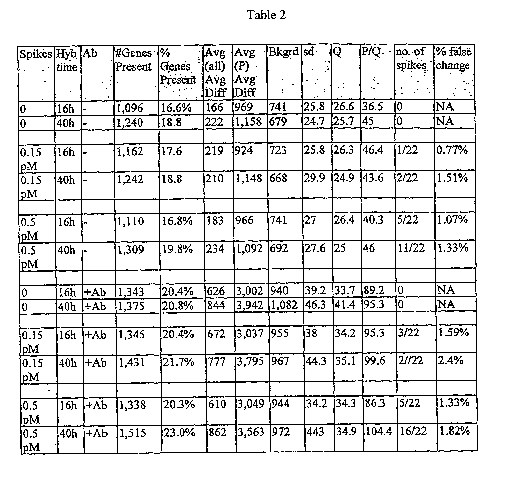
Claims (36)
- A method for detecting a nucleic acid target, the method comprising:a) hybridizing a nucleic acid target, comprising a target nucleic acid sequence, to a nucleic acid probe immobilized on a surface, comprising a probe nucleic acid sequence, wherein a binding ligand is complexed to said target;b) contacting the hydridized target with a receptor comprising multiple sites capable of binding the binding ligand to complex the receptor to the binding ligand;c) contacting the receptor with an amplification reagent, comprising a plurality of the binding ligands, wherein said binding ligands are covalently attached to said amplification reagent, to complex the amplification reagent to the receptor, wherein the amplification reagent is selected from the group consisting of an antibody and a dendrimeric DNA matrix comprising layers of DNA and single stranded sequences at the surface of the matrix that are free to hybridize with a predetermined nucleic acid sequence; andd) detecting the presence of the complexed amplification reagent.
- The method of claim 1, wherein the binding ligand is covalently attached to said target.
- The method of claim 1, wherein the amplification reagent comprises an anti-streptavidin antibody.
- The method of claim 1, wherein the DNA matrix comprises subunits of partially double stranded and partially single stranded DNA molecules.
- The method of claim 4, wherein the DNA matrix comprises:wherein the plurality of molecules of the first polynucleotide and the second polynucleotide are joined together through annealing of one or more arms thereof, to form a matrix, and wherein arms of the plurality of first and second polynucleotide molecules located on the outer surface of the matrix are hybridized to nucleic acids having the binding ligand attached thereto.a plurality of molecules of a first partially double stranded polynucleotide, the polynucleotide having a first molecule end, a second molecule end and a double stranded body portion intermediate of the first and second ends, wherein the first and second ends each comprise at least one of first and second arms consisting of a single strand of polynucleotide, and wherein the single strands are hybridizable with a predetermined nucleic acid sequence of nucleotides in a nucleic acid, and the first and second arms of each of said first and second ends are nonhybridizable with each other;a plurality of molecules of a second partially double stranded polynucleotide, each polynucleotide including a first molecule end, a second molecule end and a double stranded body portion intermediate the first and second ends, the first and second ends each having at least one of first and second arms consisting of a single strand of polynucleotide that is hybridizable with a predetermined nucleic acid sequence of nucleotides in a nucleic acid, wherein the first and second arms of each of said first and second ends are non-hybridizable with each other;
- The method of claim 1 wherein at least one of the receptor and the amplification reagent comprises a detectable label; and wherein step d) comprises detecting'the label.
- The method of claim 1 further comprising labeling at least one of the receptor and the amplification reagent with a detectable label prior to step d); and wherein step d) comprises detecting the label.
- The method of claim 1 wherein the method further comprises, after step c), and before step d), the step of contacting the amplification reagent, comprising a plurality of the binding ligands, with labeled receptor molecules thereby to complex a plurality of labeled receptor molecules to the amplification reagent; and wherein step d) comprises detecting the labeled receptor molecules complexed to the amplification reagent.
- The method of claim 8, wherein the label is selected from the group consisting of a fluorescent label, a gold label and an enzyme label.
- The method of claim 9, wherein the fluorescent label is selected from the group consisting of fluorescein, rhodamine, resorufin, and a coumarin.
- The method of claim 1, wherein the binding ligand comprises biotin and the receptor comprises avidin or streptavidin.
- The method of claim 1, wherein the amplification reagent comprises an antibody capable of specifically binding the receptor.
- The method of claim 12, wherein the binding ligand comprises biotin, the receptor comprises avidin or streptavidin, and wherein biotin is covalently attached to the antibody.
- The method of claim 13, wherein the receptor comprises streptavidin and the antibody is an anti-streptavidin antibody comprising a plurality of biotin molecules covalently attached to the antibody;
wherein the method further comprises, after step c), and before step d), the step of contacting the antibody with labeled streptavidin, thereby to complex a plurality of labeled streptavidin molecules to the antibody; and
wherein step d) comprises detecting the labeled streptavidin molecules complexed to the antibody. - The method of claim 11, wherein the amplification reagent comprises a DNA matrix comprising single stranded DNA; and wherein biotin is attached to the DNA matrix by hybridization of a plurality of biotinylated nucleic acids to single strands of the DNA matrix.
- The method of claim 15, wherein the method further comprises, after step c), and before step d), the step of contacting the DNA matrix with labeled streptavidin, thereby to complex a plurality of labeled streptavidin molecules to the DNA matrix; and wherein step d) comprises detecting the labeled streptavidin molecules complexed to the DNA matrix.
- The method of claim 16, wherein the label is selected from the group consisting of a fluorescent label, a gold label and an enzyme label.
- The method of claim 17, wherein the fluorescent label is selected from the group consisting of fluorescein, rhodamine, resorufin, and a coumarin.
- The method of claim 12, wherein the surface is selected from the group consisting of Langmuir Blodgett film, glass, germanium, silicon, (poly)tetrafluorethylene, polystyrene, gallium arsenide, gallium phosphide, silicon oxide, silicon nitride, and combinations thereof.
- The method of claim 5 wherein, in step (a), the hybridization is conducted in a hybridization solution comprising a sulfonate buffer.
- The method of claim 14 wherein, in step (a), the hybridization is conducted in a hybridization solution comprising a sulfonate buffer.
- A method for detecting a nucleic acid target, the methoda) providing a substrate comprising a surface, the surface comprising at least 100 nucleic acid probes, each nucleic acid probe contained in an area of less than about 0.1 cm2, and each nucleic acid probe having a defined sequence and location on the surface;b) contacting the surface with a nucleic acid target, comprising a target nucleic acid sequence, to permit the nucleic acid target to hybridize with at least one selected nucleic acid probe that comprises a probe nucleic acid sequence capable of hybridizing to the target nucleic acid sequence, and wherein a binding ligand is complexed to the target;c) contacting the hydridized target with a receptor comprising
multiple sites capable of binding the binding ligand to complex the receptor to the binding ligand;d) contacting the receptor with an amplification reagent, comprising a plurality of the binding ligands, wherein said binding ligands are covalently attached to said amplification reagent, to complex the amplification reagent to the receptor, wherein the amplification reagent is selected from the group consisting of an antibody and a dendrimeric DNA matrix comprising layers of DNA and single stranded sequences at the surface of the matrix that are free to hybridize with a predetermined nucleic acid sequence; ande) detecting the presence of the complexed amplification reagent. - The method of claim 22, wherein the binding ligand is covalently attached to said target.
- The method of claim 22, wherein, in step b), the surface is contacted with the nucleic acid target in a hybridization solution comprising a sulfonate buffer.
- The method of claim 24 wherein the sulfonate buffer is 2-[N-morpholino]ethanesulfonic acid ("MES").
- The method of claim 22, wherein the amplification reagent comprises a DNA matrix, the binding ligand comprises biotin and the receptor comprises streptavidin.
- The method of claim 22, wherein the binding ligand comprises biotin, the receptor comprises streptavidin and the amplification reagent comprises an anti-streptavidin antibody.
- A complex comprising:a binding ligand complexed to a nucleic acid;a receptor comprising multiple binding sites capable of binding the binding ligand; andan amplification reagent comprising a plurality of binding ligands, wherein said binding ligands are covalently attached to said amplification reagent, wherein the amplification reagent is selected from the group consisting of an antibody and a dendrimeric DNA matrix comprising layers of DNA and single stranded sequences at the surface of the matrix that are free to hybridize with a predetermined nucleic acid sequence.
- The complex of claim 28, wherein the binding ligand is covalently attached to said nucleic acid.
- The complex of claim 28, wherein the binding ligand comprises biotin and the receptor comprises streptavidin or avidin.
- The complex of claim 30, wherein the amplification reagent comprises a DNA matrix.
- The complex of claim 30, wherein the amplification reagent comprises an anti-streptavidin antibody.
- A substrate comprising a surface having immobilized thereon a nucleic acid probe, comprising a probe nucleic acid sequence, hybridized to a nucleic acid target, comprising a target nucleic acid sequence,
wherein a binding ligand is complexed to said target, and wherein the binding ligand on the target is complexed with a receptor comprising multiple sites capable of binding the binding ligand, and wherein the receptor is complexed to an amplification reagent, comprising a plurality of the binding ligands, wherein said binding ligands are covalently attached to said amplification reagent, wherein the amplification reagent is selected from the group consisting of an antibody and a dendrimeric DNA matrix comprising layers of DNA and single stranded sequences at the surface of the matrix that are free to hybridize with a predetermined nucleic acid sequence. - The method of claim 33, wherein the binding ligand is covalently attached to said target.
- The substrate of claim 33, wherein the binding ligand comprises biotin and the receptor comprises streptavidin or avidin.
- The substrate of claim 35, wherein the amplification reagent comprises an anti-streptavidin antibody.
Applications Claiming Priority (4)
| Application Number | Priority Date | Filing Date | Title |
|---|---|---|---|
| US10257798P | 1998-09-30 | 1998-09-30 | |
| US102577P | 1998-09-30 | ||
| US276774 | 1999-03-25 | ||
| US09/276,774 US6203989B1 (en) | 1998-09-30 | 1999-03-25 | Methods and compositions for amplifying detectable signals in specific binding assays |
Publications (2)
| Publication Number | Publication Date |
|---|---|
| EP0999285A1 EP0999285A1 (en) | 2000-05-10 |
| EP0999285B1 true EP0999285B1 (en) | 2005-12-07 |
Family
ID=26799529
Family Applications (1)
| Application Number | Title | Priority Date | Filing Date |
|---|---|---|---|
| EP99250336A Expired - Lifetime EP0999285B1 (en) | 1998-09-30 | 1999-09-22 | Methods and compositions for amplifying detectable signals in specific binding assays |
Country Status (7)
| Country | Link |
|---|---|
| US (3) | US6203989B1 (en) |
| EP (1) | EP0999285B1 (en) |
| JP (2) | JP2000106876A (en) |
| AT (1) | ATE312199T1 (en) |
| AU (1) | AU6275099A (en) |
| DE (1) | DE69928747T2 (en) |
| WO (1) | WO2000018962A1 (en) |
Families Citing this family (151)
| Publication number | Priority date | Publication date | Assignee | Title |
|---|---|---|---|---|
| US6506564B1 (en) | 1996-07-29 | 2003-01-14 | Nanosphere, Inc. | Nanoparticles having oligonucleotides attached thereto and uses therefor |
| US7098320B1 (en) | 1996-07-29 | 2006-08-29 | Nanosphere, Inc. | Nanoparticles having oligonucleotides attached thereto and uses therefor |
| US6582921B2 (en) | 1996-07-29 | 2003-06-24 | Nanosphere, Inc. | Nanoparticles having oligonucleotides attached thereto and uses thereof |
| US6750016B2 (en) | 1996-07-29 | 2004-06-15 | Nanosphere, Inc. | Nanoparticles having oligonucleotides attached thereto and uses therefor |
| WO1998004740A1 (en) | 1996-07-29 | 1998-02-05 | Nanosphere Llc | Nanoparticles having oligonucleotides attached thereto and uses therefor |
| US7169556B2 (en) | 1996-07-29 | 2007-01-30 | Nanosphere, Inc. | Nanoparticles having oligonucleotides attached thereto and uses therefor |
| US6984491B2 (en) * | 1996-07-29 | 2006-01-10 | Nanosphere, Inc. | Nanoparticles having oligonucleotides attached thereto and uses therefor |
| US6974669B2 (en) * | 2000-03-28 | 2005-12-13 | Nanosphere, Inc. | Bio-barcodes based on oligonucleotide-modified nanoparticles |
| US20050037397A1 (en) * | 2001-03-28 | 2005-02-17 | Nanosphere, Inc. | Bio-barcode based detection of target analytes |
| US7348181B2 (en) | 1997-10-06 | 2008-03-25 | Trustees Of Tufts College | Self-encoding sensor with microspheres |
| US6203989B1 (en) * | 1998-09-30 | 2001-03-20 | Affymetrix, Inc. | Methods and compositions for amplifying detectable signals in specific binding assays |
| EP2000803A1 (en) | 1998-11-30 | 2008-12-10 | Nanosphere, Inc. | Nanoparticles with Polymer Shells |
| JP2002543813A (en) * | 1999-05-07 | 2002-12-24 | イェール ユニバーシティ | Multiple label analysis |
| US6921638B2 (en) * | 1999-06-25 | 2005-07-26 | Amersham Biosciences Ab | Hydrogel-based microarray signal amplification methods and devices therefor |
| ES2273704T3 (en) | 1999-07-05 | 2007-05-16 | Novartis Ag | PROCEDURE FOR USING A DETECTION PLATFORM. |
| US6771376B2 (en) | 1999-07-05 | 2004-08-03 | Novartis Ag | Sensor platform, apparatus incorporating the platform, and process using the platform |
| US7211390B2 (en) | 1999-09-16 | 2007-05-01 | 454 Life Sciences Corporation | Method of sequencing a nucleic acid |
| US7244559B2 (en) | 1999-09-16 | 2007-07-17 | 454 Life Sciences Corporation | Method of sequencing a nucleic acid |
| US7167615B1 (en) | 1999-11-05 | 2007-01-23 | Board Of Regents, The University Of Texas System | Resonant waveguide-grating filters and sensors and methods for making and using same |
| EP1250451B1 (en) * | 1999-11-29 | 2006-03-29 | DiaSorin S.p.A. | Oligonucleotides and assemblies thereof useful in the detection of the presence or absence of target nucleic acid sequences in a sample |
| JP2004501340A (en) | 2000-01-13 | 2004-01-15 | ナノスフェアー インコーポレイテッド | Oligonucleotide-attached nanoparticles and methods of use |
| US20020094538A1 (en) * | 2001-01-13 | 2002-07-18 | Getts Robert C. | Methods for detecting and assaying nucleic acid sequences using temperature cycling |
| WO2001073117A1 (en) * | 2000-03-31 | 2001-10-04 | MEMOREC STOFFEL GmbH - MEDIZINISCH-MOLEKULARE ENTWICKLUNG, KÖLN | Use of particles which have signal-emitting characteristics and at least one group with an affinity for marked nucleic acids |
| WO2002004681A2 (en) * | 2000-07-11 | 2002-01-17 | Northwestern University | Method of detection by enhancement of silver staining |
| US20020019792A1 (en) * | 2000-08-11 | 2002-02-14 | Robert Maerz | Method and system for offering television pilots as a security |
| WO2002014867A2 (en) * | 2000-08-11 | 2002-02-21 | Agilix Corporation | Ultra-sensitive detection systems |
| EP1186671A3 (en) * | 2000-09-05 | 2003-12-17 | Agilent Technologies, Inc. (a Delaware corporation) | Method for hybridization of arrays on siliceous surfaces |
| US20050266494A1 (en) * | 2000-09-06 | 2005-12-01 | Hodge Timothy A | System and method for computer network ordering of biological testing |
| US20050239125A1 (en) * | 2000-09-06 | 2005-10-27 | Hodge Timothy A | Methods for genotype screening |
| US20050272085A1 (en) * | 2000-09-06 | 2005-12-08 | Hodge Timothy A | Methods for forensic and congenic screening |
| US7011943B2 (en) * | 2000-09-06 | 2006-03-14 | Transnetyx, Inc. | Method for detecting a designated genetic sequence in murine genomic DNA |
| US7494817B2 (en) * | 2000-09-06 | 2009-02-24 | Transnet Yx, Inc. | Methods for genotype screening using magnetic particles |
| US6803196B1 (en) * | 2000-10-13 | 2004-10-12 | Affymetrix, Inc. | Methods and compositions for detecting signals in binding assays using microparticles |
| GB0029154D0 (en) | 2000-11-30 | 2001-01-17 | Lee Helen | Signal enhancement with multiple labelled-antibodies |
| US20020165675A1 (en) * | 2001-03-03 | 2002-11-07 | Golovlev Valeri V. | Method and microelectronic device for multi-site molecule detection |
| AU2002255699A1 (en) * | 2001-03-09 | 2002-09-24 | Apollo Biotechnology, Inc. | Conjugate probes and optical detection of analytes |
| US20060040286A1 (en) * | 2001-03-28 | 2006-02-23 | Nanosphere, Inc. | Bio-barcode based detection of target analytes |
| EP1373467A4 (en) * | 2001-03-28 | 2007-06-06 | Genetech Biotechnology Shangai | Device and method for detection of multiple analytes |
| US7147687B2 (en) * | 2001-05-25 | 2006-12-12 | Nanosphere, Inc. | Non-alloying core shell nanoparticles |
| US7238472B2 (en) * | 2001-05-25 | 2007-07-03 | Nanosphere, Inc. | Non-alloying core shell nanoparticles |
| JP2003014746A (en) * | 2001-06-27 | 2003-01-15 | Fuji Photo Film Co Ltd | Structure having biological material fixed thereon and method for producing the same |
| US6662091B2 (en) * | 2001-06-29 | 2003-12-09 | Battelle Memorial Institute | Diagnostics/prognostics using wireless links |
| NZ530434A (en) | 2001-07-02 | 2005-01-28 | Battelle Memorial Institute | Intelligent microsensor module |
| US6753145B2 (en) * | 2001-07-05 | 2004-06-22 | Agilent Technologies, Inc. | Buffer composition and method for hybridization of microarrays on adsorbed polymer siliceous surfaces |
| US20050009022A1 (en) * | 2001-07-06 | 2005-01-13 | Weiner Michael P. | Method for isolation of independent, parallel chemical micro-reactions using a porous filter |
| WO2003006676A2 (en) * | 2001-07-13 | 2003-01-23 | Nanosphere, Inc. | Method for immobilizing molecules onto surfaces |
| US7297553B2 (en) * | 2002-05-28 | 2007-11-20 | Nanosphere, Inc. | Method for attachment of silylated molecules to glass surfaces |
| AU2002365115A1 (en) * | 2001-07-20 | 2003-09-02 | North Carolina State University | Light addressable electrochemical detection of duplex structures |
| US20060014186A1 (en) * | 2001-09-04 | 2006-01-19 | Hodge Timothy A | Methods for genotype screening of a strain disposed on an adsorbent carrier |
| US20030054396A1 (en) * | 2001-09-07 | 2003-03-20 | Weiner Michael P. | Enzymatic light amplification |
| WO2003027678A1 (en) * | 2001-09-26 | 2003-04-03 | Psychiatric Genomics, Inc. | Fluorescence proximity assay |
| US7132236B2 (en) * | 2001-10-25 | 2006-11-07 | Agilent Technologies, Inc. | Composition and method for optimized hybridization using modified solutions |
| US6956114B2 (en) | 2001-10-30 | 2005-10-18 | '454 Corporation | Sulfurylase-luciferase fusion proteins and thermostable sulfurylase |
| US6902921B2 (en) * | 2001-10-30 | 2005-06-07 | 454 Corporation | Sulfurylase-luciferase fusion proteins and thermostable sulfurylase |
| US20050124022A1 (en) * | 2001-10-30 | 2005-06-09 | Maithreyan Srinivasan | Novel sulfurylase-luciferase fusion proteins and thermostable sulfurylase |
| ATE403751T1 (en) | 2001-11-09 | 2008-08-15 | Nanosphere Inc | BIOCONJUGATE NANOPARTICLE PROBE |
| EP1451365A4 (en) | 2001-11-13 | 2006-09-13 | Rubicon Genomics Inc | Dna amplification and sequencing using dna molecules generated by random fragmentation |
| NZ535044A (en) | 2002-03-01 | 2008-12-24 | Ravgen Inc | Non-invasive method to determine the genetic sequence of foetal DNA from a sample from a pregnant female thereby detecting any alternation in gene sequence as compared with the wild type sequence |
| US6977162B2 (en) * | 2002-03-01 | 2005-12-20 | Ravgen, Inc. | Rapid analysis of variations in a genome |
| US20050266455A1 (en) * | 2002-03-02 | 2005-12-01 | Sci Tec, Inc. | Method and microelectronic device for multi-site molecule detection |
| US20030175828A1 (en) * | 2002-03-15 | 2003-09-18 | Lazar James G. | Signal amplification by Hybrid Capture |
| US20030211488A1 (en) | 2002-05-07 | 2003-11-13 | Northwestern University | Nanoparticle probs with Raman spectrocopic fingerprints for analyte detection |
| US20070178478A1 (en) * | 2002-05-08 | 2007-08-02 | Dhallan Ravinder S | Methods for detection of genetic disorders |
| US7727720B2 (en) * | 2002-05-08 | 2010-06-01 | Ravgen, Inc. | Methods for detection of genetic disorders |
| US7442506B2 (en) * | 2002-05-08 | 2008-10-28 | Ravgen, Inc. | Methods for detection of genetic disorders |
| ATE420357T1 (en) * | 2002-07-02 | 2009-01-15 | Nanosphere Inc | NANOPARTICLE-POLYANION CONJUGATES AND METHOD FOR USE THEREOF IN DETECTING ANALYTES |
| US20040101444A1 (en) * | 2002-07-15 | 2004-05-27 | Xeotron Corporation | Apparatus and method for fluid delivery to a hybridization station |
| ATE534748T1 (en) * | 2002-12-12 | 2011-12-15 | Nanosphere Inc | DIRECT SNP DETECTION USING NON-AMPLIFIED DNA |
| US20040180369A1 (en) * | 2003-01-16 | 2004-09-16 | North Carolina State University | Photothermal detection of nucleic acid hybridization |
| CA2513889A1 (en) | 2003-01-29 | 2004-08-19 | 454 Corporation | Double ended sequencing |
| US7575865B2 (en) * | 2003-01-29 | 2009-08-18 | 454 Life Sciences Corporation | Methods of amplifying and sequencing nucleic acids |
| US7192703B2 (en) | 2003-02-14 | 2007-03-20 | Intel Corporation, Inc. | Biomolecule analysis by rolling circle amplification and SERS detection |
| CA2897376A1 (en) * | 2003-02-26 | 2004-09-10 | Radoje T. Drmanac | Modular system and probes for dna analysis |
| WO2005001143A2 (en) * | 2003-02-27 | 2005-01-06 | Nanosphere, Inc. | Label-free gene expression profiling with universal nanoparticle probes in microarray assay format |
| AU2003268333A1 (en) * | 2003-02-28 | 2004-09-28 | Ravgen, Inc. | Methods for detection of genetic disorders |
| US20050250094A1 (en) * | 2003-05-30 | 2005-11-10 | Nanosphere, Inc. | Method for detecting analytes based on evanescent illumination and scatter-based detection of nanoparticle probe complexes |
| US20040259100A1 (en) | 2003-06-20 | 2004-12-23 | Illumina, Inc. | Methods and compositions for whole genome amplification and genotyping |
| US20050181394A1 (en) * | 2003-06-20 | 2005-08-18 | Illumina, Inc. | Methods and compositions for whole genome amplification and genotyping |
| EP1636337A4 (en) * | 2003-06-20 | 2007-07-04 | Illumina Inc | Methods and compositions for whole genome amplification and genotyping |
| US7642064B2 (en) | 2003-06-24 | 2010-01-05 | Ventana Medical Systems, Inc. | Enzyme-catalyzed metal deposition for the enhanced detection of analytes of interest |
| JP3899407B2 (en) * | 2003-06-26 | 2007-03-28 | 国立大学法人 鹿児島大学 | Antigen detection method by immunohistochemical staining method |
| WO2005024386A2 (en) * | 2003-09-09 | 2005-03-17 | The Regents Of The University Of Colorado, A Body Corporate | Use of photopolymerization for amplification and detection of a molecular recognition event |
| US7354706B2 (en) * | 2003-09-09 | 2008-04-08 | The Regents Of The University Of Colorado, A Body Corporate | Use of photopolymerization for amplification and detection of a molecular recognition event |
| US20090163375A1 (en) | 2003-09-09 | 2009-06-25 | Bowman Christopher N | Use of Photopolymerization for Amplification and Detection of a Molecular Recognition Event |
| KR100571817B1 (en) * | 2003-09-19 | 2006-04-17 | 삼성전자주식회사 | Target nucleic acid detection method using a detection probe that hybridizes to a tag sequence |
| US20050089874A1 (en) * | 2003-10-24 | 2005-04-28 | Torontali Suzanne M. | Amplification of signal using a bead-based oligonucleotide assay |
| US20050214796A1 (en) * | 2003-10-29 | 2005-09-29 | Hanna Michelle M | Compositions, methods and detection technologies for reiterative oligonucleotide synthesis |
| WO2005042785A1 (en) * | 2003-10-30 | 2005-05-12 | North Carolina State University | Electrochemical detection of nucleic acid hybridization |
| US20050191651A1 (en) * | 2003-10-30 | 2005-09-01 | North Carolina State University | Temperature-jump enhanced electrochemical detection of nucleic acid hybridization |
| GB0404187D0 (en) * | 2004-02-25 | 2004-03-31 | Biotransformations Ltd | Binding agents |
| US20050266432A1 (en) * | 2004-02-26 | 2005-12-01 | Illumina, Inc. | Haplotype markers for diagnosing susceptibility to immunological conditions |
| JP4476050B2 (en) * | 2004-06-30 | 2010-06-09 | 株式会社ニデック | Perimeter |
| EP1851551A2 (en) * | 2005-02-03 | 2007-11-07 | Perkinelmer Las, Inc. | Ultra-sensitive detection systems using multidimension signals |
| WO2007002567A2 (en) * | 2005-06-23 | 2007-01-04 | Nanosphere, Inc. | Selective isolation and concentration of nucleic acids from complex samples |
| US7368246B2 (en) * | 2005-09-15 | 2008-05-06 | Primera Biosystems, Inc. | Quantitative gene expression profiling |
| US20070275388A1 (en) * | 2006-05-25 | 2007-11-29 | Daniel Ryan | Dendrimers that possess a single target annealing site and uses thereof |
| US20090002703A1 (en) * | 2006-08-16 | 2009-01-01 | Craig Edward Parman | Methods and systems for quantifying isobaric labels and peptides |
| US20080153135A1 (en) * | 2006-12-20 | 2008-06-26 | Liu Timothy Z | Methods and apparatus for conducting amplification reactions on high density hydrophilic patterned microplates |
| US20080153134A1 (en) * | 2006-12-22 | 2008-06-26 | Willy Wiyatno | Methods and apparatus for generating hydrophilic patterning of high density microplates using an amphiphilic polymer |
| US20080241831A1 (en) * | 2007-03-28 | 2008-10-02 | Jian-Bing Fan | Methods for detecting small RNA species |
| US20080268440A1 (en) * | 2007-04-26 | 2008-10-30 | Liu Timothy Z | Biomolecule immobilization on surface via hydrophobic interactions |
| US20090304719A1 (en) | 2007-08-22 | 2009-12-10 | Patrick Daugherty | Activatable binding polypeptides and methods of identification and use thereof |
| US20090137405A1 (en) * | 2007-11-16 | 2009-05-28 | Christopher Bowman | Detection of nucleic acid biomarkers using polymerization-based amplification |
| US20090162863A1 (en) * | 2007-12-13 | 2009-06-25 | Hitachi High-Technologies Corporation | Nucleic acid detection probe |
| JP5504587B2 (en) * | 2008-05-27 | 2014-05-28 | 東レ株式会社 | Analysis chip |
| GB0814570D0 (en) | 2008-08-08 | 2008-09-17 | Diagnostics For The Real World | Isolation of nucleic acid |
| US8309306B2 (en) * | 2008-11-12 | 2012-11-13 | Nodality, Inc. | Detection composition |
| EP2189539B2 (en) * | 2008-11-21 | 2018-06-13 | Chimera Biotec GmbH | Conjugate complexes for analyte detection |
| WO2010081173A2 (en) | 2009-01-12 | 2010-07-15 | Cytomx Therapeutics, Llc | Modified antibody compositions, methods of making and using thereof |
| US8399219B2 (en) | 2009-02-23 | 2013-03-19 | Cytomx Therapeutics, Inc. | Protease activatable interferon alpha proprotein |
| EP2408905B1 (en) | 2009-03-16 | 2017-05-10 | Pangu Biopharma Limited | Compositions and methods comprising histidyl-trna synthetase splice variants having non-canonical biological activities |
| US9464319B2 (en) | 2009-03-24 | 2016-10-11 | California Institute Of Technology | Multivolume devices, kits and related methods for quantification of nucleic acids and other analytes |
| US9447461B2 (en) | 2009-03-24 | 2016-09-20 | California Institute Of Technology | Analysis devices, kits, and related methods for digital quantification of nucleic acids and other analytes |
| CA2756463C (en) | 2009-03-24 | 2019-01-22 | University Of Chicago | Slip chip device and methods |
| US10196700B2 (en) | 2009-03-24 | 2019-02-05 | University Of Chicago | Multivolume devices, kits and related methods for quantification and detection of nucleic acids and other analytes |
| AU2010236913B2 (en) | 2009-03-31 | 2016-03-24 | Atyr Pharma, Inc. | Compositions and methods comprising aspartyl-tRNA synthetases having non-canonical biological activities |
| EP2504448B1 (en) * | 2009-11-25 | 2016-10-19 | Bio-Rad Laboratories, Inc. | Methods and compositions for detecting genetic material |
| AU2011248625B2 (en) | 2010-04-26 | 2017-01-05 | Pangu Biopharma Limited | Innovative discovery of therapeutic, diagnostic, and antibody compositions related to protein fragments of cysteinyl-tRNA synthetase |
| JP6294074B2 (en) | 2010-04-27 | 2018-03-14 | エータイアー ファーマ, インコーポレイテッド | Innovative discovery of therapeutic, diagnostic and antibody compositions related to protein fragments of isoleucyl-tRNA synthetase |
| CN103097524B (en) | 2010-04-28 | 2016-08-03 | Atyr医药公司 | The innovation for the treatment of, diagnosis and the antibody compositions relevant to the protein fragments of Alanyl-tRNA synthetase finds |
| WO2011150279A2 (en) | 2010-05-27 | 2011-12-01 | Atyr Pharma, Inc. | Innovative discovery of therapeutic, diagnostic, and antibody compositions related to protein fragments of glutaminyl-trna synthetases |
| CA2797374C (en) | 2010-04-29 | 2021-02-16 | Pangu Biopharma Limited | Innovative discovery of therapeutic, diagnostic, and antibody compositions related to protein fragments of asparaginyl trna synthetases |
| AU2011248457B2 (en) | 2010-04-29 | 2017-02-16 | Pangu Biopharma Limited | Innovative discovery of therapeutic, diagnostic, and antibody compositions related to protein fragments of valyl tRNA synthetases |
| WO2011135459A2 (en) | 2010-04-29 | 2011-11-03 | Medical Prognosis Institute A/S | Methods and devices for predicting treatment efficacy |
| CA2797977C (en) | 2010-05-03 | 2019-08-20 | Atyr Pharma, Inc. | Innovative discovery of therapeutic, diagnostic, and antibody compositions related to protein fragments of phenylalanyl-alpha-trna synthetases |
| WO2011140135A2 (en) | 2010-05-03 | 2011-11-10 | Atyr Pharma, Inc. | Innovative discovery of therapeutic, diagnostic, and antibody compositions related to protein fragments of methionyl-trna synthetases |
| WO2011139986A2 (en) | 2010-05-03 | 2011-11-10 | Atyr Pharma, Inc. | Innovative discovery of therapeutic, diagnostic, and antibody compositions related to protein fragments of arginyl-trna synthetases |
| CA2798139C (en) | 2010-05-04 | 2019-09-24 | Atyr Pharma, Inc. | Innovative discovery of therapeutic, diagnostic, and antibody compositions related to protein fragments of p38 multi-trna synthetase complex |
| CN103200953B (en) | 2010-05-14 | 2017-02-15 | Atyr 医药公司 | Innovative discovery of therapeutic, diagnostic, and antibody compositions related to protein fragments of phenylalanyl-beta-trna synthetases |
| CN103096914B (en) | 2010-05-17 | 2015-08-12 | Atyr医药公司 | Innovative discovery of therapeutic, diagnostic and antibody compositions related to protein fragments of leucyl-tRNA synthetase |
| CA2800281C (en) | 2010-06-01 | 2021-01-12 | Atyr Pharma, Inc. | Innovative discovery of therapeutic, diagnostic, and antibody compositions related to protein fragments of lysyl-trna synthetases |
| AU2011289831C1 (en) | 2010-07-12 | 2017-06-15 | Pangu Biopharma Limited | Innovative discovery of therapeutic, diagnostic, and antibody compositions related to protein fragments of glycyl-tRNA synthetases |
| CN103108650A (en) | 2010-08-25 | 2013-05-15 | Atyr医药公司 | Innovative discovery of therapeutic, diagnostic, and antibody compositions related to protein fragments of tyrosyl-trna synthetases |
| EP2673380B1 (en) | 2011-02-09 | 2018-12-12 | Bio-Rad Laboratories, Inc. | Analysis of nucleic acids |
| WO2012122121A2 (en) * | 2011-03-04 | 2012-09-13 | Bio-Rad Laboratories, Inc. | Signal amplification for immunoassays by use of avidin-biotin linkages |
| US20140106986A1 (en) | 2011-06-01 | 2014-04-17 | Medical Prognosis Institute A/S | Methods and devices for prognosis of cancer relapse |
| WO2013021687A1 (en) | 2011-08-11 | 2013-02-14 | オリンパス株式会社 | Method for detecting target particles |
| EP2752655A4 (en) | 2011-08-30 | 2015-06-17 | Olympus Corp | Method for detecting target particles |
| IN2014MN01642A (en) | 2012-02-16 | 2015-07-03 | Atyr Pharma Inc | |
| EP2818850B1 (en) | 2012-02-22 | 2016-08-03 | Olympus Corporation | Method for detecting a target particle |
| JP6095645B2 (en) | 2012-03-21 | 2017-03-15 | オリンパス株式会社 | Method for detecting target nucleic acid molecule |
| MX367366B (en) | 2012-11-27 | 2019-08-16 | Univ Pontificia Catolica Chile | Compositions and methods for diagnosing thyroid tumors. |
| US20140308205A1 (en) * | 2013-04-12 | 2014-10-16 | University Of Massachusetts Medical School | Trifunctional and completely clearable specific targeting agents and methods thereof |
| WO2014195032A1 (en) | 2013-06-07 | 2014-12-11 | Medical Prognosis Institute A/S | Methods and devices for predicting treatment efficacy of fulvestrant in cancer patients |
| CN105555972B (en) | 2013-07-25 | 2020-07-31 | 伯乐生命医学产品有限公司 | Genetic assay |
| WO2016008048A1 (en) | 2014-07-15 | 2016-01-21 | Ontario Institute For Cancer Research | Methods and devices for predicting anthracycline treatment efficacy |
| EP3265587A4 (en) * | 2015-03-02 | 2018-08-08 | The Regents of the University of California | Homogenous entropy-driven biomolecular assay (heba) |
| EP3577233B1 (en) | 2017-03-31 | 2021-07-14 | Marcella Chiari | Genotyping of mutations by combination of in-tube hybridization and universal tag-microarray |
| EP3729090A4 (en) * | 2017-12-22 | 2021-09-22 | Pacific Biosciences Of California, Inc. | IMMOBILIZATION MODIFIED BIOTIN BINDING PROTEINS |
| WO2019200326A1 (en) | 2018-04-13 | 2019-10-17 | Rarecyte, Inc. | Kits for labeling of biomarkers and methods of using the same |
| CN116298265A (en) * | 2023-04-12 | 2023-06-23 | 上海品峰医疗科技有限公司 | hs-cTnT chemiluminescent assay kit applying multistage signal amplification technology |
Family Cites Families (48)
| Publication number | Priority date | Publication date | Assignee | Title |
|---|---|---|---|---|
| US4711955A (en) | 1981-04-17 | 1987-12-08 | Yale University | Modified nucleotides and methods of preparing and using same |
| US4748111A (en) | 1984-03-12 | 1988-05-31 | Molecular Diagnostics, Inc. | Nucleic acid-protein conjugate used in immunoassay |
| US4910300A (en) | 1985-12-11 | 1990-03-20 | Chiron Corporation | Method for making nucleic acid probes |
| US4882269A (en) | 1985-12-13 | 1989-11-21 | Princeton University | Amplified hybridization assay |
| US5175270A (en) * | 1986-09-10 | 1992-12-29 | Polyprobe, Inc. | Reagents for detecting and assaying nucleic acid sequences |
| US5124246A (en) | 1987-10-15 | 1992-06-23 | Chiron Corporation | Nucleic acid multimers and amplified nucleic acid hybridization assays using same |
| CA1339351C (en) | 1987-10-15 | 1997-08-26 | Michael S. Urdea | Nucleic acid multimers and amplified nucleic acid hybridization assays using same |
| US5359100A (en) | 1987-10-15 | 1994-10-25 | Chiron Corporation | Bifunctional blocked phosphoramidites useful in making nucleic acid mutimers |
| US5656731A (en) | 1987-10-15 | 1997-08-12 | Chiron Corporation | Nucleic acid-amplified immunoassay probes |
| JPH03501777A (en) | 1988-10-17 | 1991-04-18 | モレキユラー デヴアイシズ コーポレイシヨン | Hapten-induced capture membrane and diagnostic assay using the membrane |
| US5424186A (en) | 1989-06-07 | 1995-06-13 | Affymax Technologies N.V. | Very large scale immobilized polymer synthesis |
| US5547839A (en) | 1989-06-07 | 1996-08-20 | Affymax Technologies N.V. | Sequencing of surface immobilized polymers utilizing microflourescence detection |
| US6040138A (en) | 1995-09-15 | 2000-03-21 | Affymetrix, Inc. | Expression monitoring by hybridization to high density oligonucleotide arrays |
| US5744101A (en) | 1989-06-07 | 1998-04-28 | Affymax Technologies N.V. | Photolabile nucleoside protecting groups |
| US5527681A (en) | 1989-06-07 | 1996-06-18 | Affymax Technologies N.V. | Immobilized molecular synthesis of systematically substituted compounds |
| US5143854A (en) | 1989-06-07 | 1992-09-01 | Affymax Technologies N.V. | Large scale photolithographic solid phase synthesis of polypeptides and receptor binding screening thereof |
| DE69032847T2 (en) | 1989-06-30 | 1999-05-12 | Chiron Corp., Emeryville, Calif. | Hydrophobic nucleic acid probe |
| US5252743A (en) | 1989-11-13 | 1993-10-12 | Affymax Technologies N.V. | Spatially-addressable immobilization of anti-ligands on surfaces |
| US5629153A (en) | 1990-01-10 | 1997-05-13 | Chiron Corporation | Use of DNA-dependent RNA polymerase transcripts as reporter molecules for signal amplification in nucleic acid hybridization assays |
| JPH05507878A (en) | 1990-06-15 | 1993-11-11 | カイロン コーポレイション | Built-in assay assemblies and devices |
| US5849481A (en) | 1990-07-27 | 1998-12-15 | Chiron Corporation | Nucleic acid hybridization assays employing large comb-type branched polynucleotides |
| IE912662A1 (en) | 1990-07-27 | 1992-01-29 | Chiron Corp | Large comb-type branched polynucleotides |
| ES2155822T3 (en) | 1990-12-06 | 2001-06-01 | Affymetrix Inc | COMPOUNDS AND ITS USE IN A BINARY SYNTHESIS STRATEGY. |
| GB9114180D0 (en) * | 1991-07-01 | 1991-08-21 | Amersham Int Plc | Enhancement of polynucleotide hybridisation |
| US5677195A (en) | 1991-11-22 | 1997-10-14 | Affymax Technologies N.V. | Combinatorial strategies for polymer synthesis |
| US5384261A (en) | 1991-11-22 | 1995-01-24 | Affymax Technologies N.V. | Very large scale immobilized polymer synthesis using mechanically directed flow paths |
| US5412087A (en) | 1992-04-24 | 1995-05-02 | Affymax Technologies N.V. | Spatially-addressable immobilization of oligonucleotides and other biological polymers on surfaces |
| DE69231693T2 (en) | 1991-12-23 | 2001-05-31 | Bayer Corp., East Walpole | HBV AMPLIFICATION PROBE FOR USE IN HYBRIDIZATION SANDWICH ASSAY IN THE SOLUTION PHASE |
| AU4596893A (en) | 1992-05-29 | 1993-12-30 | Gen Trak, Inc. | Signal amplification probe and methods of use |
| US5681697A (en) | 1993-12-08 | 1997-10-28 | Chiron Corporation | Solution phase nucleic acid sandwich assays having reduced background noise and kits therefor |
| US5561043A (en) | 1994-01-31 | 1996-10-01 | Trustees Of Boston University | Self-assembling multimeric nucleic acid constructs |
| US5578832A (en) | 1994-09-02 | 1996-11-26 | Affymetrix, Inc. | Method and apparatus for imaging a sample on a device |
| US5631734A (en) | 1994-02-10 | 1997-05-20 | Affymetrix, Inc. | Method and apparatus for detection of fluorescently labeled materials |
| US5571639A (en) | 1994-05-24 | 1996-11-05 | Affymax Technologies N.V. | Computer-aided engineering system for design of sequence arrays and lithographic masks |
| US5876924A (en) | 1994-06-22 | 1999-03-02 | Mount Sinai School Of Medicine | Nucleic acid amplification method hybridization signal amplification method (HSAM) |
| US5597909A (en) | 1994-08-25 | 1997-01-28 | Chiron Corporation | Polynucleotide reagents containing modified deoxyribose moieties, and associated methods of synthesis and use |
| US5580731A (en) | 1994-08-25 | 1996-12-03 | Chiron Corporation | N-4 modified pyrimidine deoxynucleotides and oligonucleotide probes synthesized therewith |
| US5710000A (en) | 1994-09-16 | 1998-01-20 | Affymetrix, Inc. | Capturing sequences adjacent to Type-IIs restriction sites for genomic library mapping |
| US5599695A (en) | 1995-02-27 | 1997-02-04 | Affymetrix, Inc. | Printing molecular library arrays using deprotection agents solely in the vapor phase |
| US5624711A (en) | 1995-04-27 | 1997-04-29 | Affymax Technologies, N.V. | Derivatization of solid supports and methods for oligomer synthesis |
| US5545531A (en) | 1995-06-07 | 1996-08-13 | Affymax Technologies N.V. | Methods for making a device for concurrently processing multiple biological chip assays |
| WO1997029212A1 (en) | 1996-02-08 | 1997-08-14 | Affymetrix, Inc. | Chip-based speciation and phenotypic characterization of microorganisms |
| EP0960120A4 (en) | 1996-05-06 | 2001-05-02 | Chiron Corp | MAMMALIAN SEX COMB ON MIDLEG (MAMMALIAN Scm) ACTS AS AN ONCOGENE |
| EP0922053A4 (en) | 1996-05-08 | 2001-04-25 | Chiron Corp | MAMMALIAN ADDITIONAL SEX COMBS (MAMMALIAN Asx) ACTS AS A TUMOR SUPPRESSOR |
| US6072043A (en) | 1996-06-04 | 2000-06-06 | Polyprobe, Inc. | Optimally fluorescent oligonucleotides |
| US6117631A (en) | 1996-10-29 | 2000-09-12 | Polyprobe, Inc. | Detection of antigens via oligonucleotide antibody conjugates |
| US5804384A (en) * | 1996-12-06 | 1998-09-08 | Vysis, Inc. | Devices and methods for detecting multiple analytes in samples |
| US6203989B1 (en) * | 1998-09-30 | 2001-03-20 | Affymetrix, Inc. | Methods and compositions for amplifying detectable signals in specific binding assays |
-
1999
- 1999-03-25 US US09/276,774 patent/US6203989B1/en not_active Expired - Lifetime
- 1999-09-22 AT AT99250336T patent/ATE312199T1/en not_active IP Right Cessation
- 1999-09-22 EP EP99250336A patent/EP0999285B1/en not_active Expired - Lifetime
- 1999-09-22 DE DE69928747T patent/DE69928747T2/en not_active Expired - Lifetime
- 1999-09-28 WO PCT/US1999/022584 patent/WO2000018962A1/en not_active Ceased
- 1999-09-28 AU AU62750/99A patent/AU6275099A/en not_active Abandoned
- 1999-09-29 JP JP11277610A patent/JP2000106876A/en not_active Withdrawn
-
2001
- 2001-02-06 US US09/776,770 patent/US6806047B2/en not_active Expired - Lifetime
-
2004
- 2004-01-07 US US10/752,020 patent/US20040137493A1/en not_active Abandoned
-
2005
- 2005-10-25 JP JP2005310563A patent/JP2006042831A/en active Pending
Also Published As
| Publication number | Publication date |
|---|---|
| AU6275099A (en) | 2000-04-17 |
| WO2000018962A9 (en) | 2000-08-24 |
| ATE312199T1 (en) | 2005-12-15 |
| US20010041335A1 (en) | 2001-11-15 |
| DE69928747D1 (en) | 2006-01-12 |
| US20040137493A1 (en) | 2004-07-15 |
| JP2006042831A (en) | 2006-02-16 |
| DE69928747T2 (en) | 2006-09-07 |
| EP0999285A1 (en) | 2000-05-10 |
| JP2000106876A (en) | 2000-04-18 |
| WO2000018962A1 (en) | 2000-04-06 |
| US6806047B2 (en) | 2004-10-19 |
| US6203989B1 (en) | 2001-03-20 |
Similar Documents
| Publication | Publication Date | Title |
|---|---|---|
| US6203989B1 (en) | Methods and compositions for amplifying detectable signals in specific binding assays | |
| EP0633944B1 (en) | Method of multiplex ligase chain reaction | |
| US5869252A (en) | Method of multiplex ligase chain reaction | |
| US6100099A (en) | Test strip having a diagonal array of capture spots | |
| US6277579B1 (en) | Direct detection of RNA mediated by reverse transcriptase lacking RNAse H function | |
| EP1488000B1 (en) | Signal amplification by hybrid capture | |
| EP0625211B1 (en) | Amplification of assay reporters by nucleic acid replication | |
| US6803196B1 (en) | Methods and compositions for detecting signals in binding assays using microparticles | |
| JP2001518800A (en) | Hybridization assays destroy excess probe | |
| US20150344939A1 (en) | Methods and Compositions For Sorting and/or Determining Organisms | |
| AU2001269285B2 (en) | Improved binding interactions in dipstick assays | |
| EP0622464A2 (en) | Nucleic acid assay procedure | |
| US5871906A (en) | Method for detection of amplified nucleic acid products and related diagnostic assays | |
| HK1027838A1 (en) | Methods and compositions for amplifying detectable signals in specific binding assays | |
| HK1027838B (en) | Methods and compositions for amplifying detectable signals in specific binding assays | |
| US6306657B1 (en) | Polynucleotide probe and kit for amplifying signal detection in hybridization assays | |
| JP2001269197A (en) | Immobilized oligonucleotide probe | |
| EP0406280A1 (en) | Method for amplifying and detecting nucleic acid in a test liquid | |
| JP2007508849A (en) | Signal amplification using bead-based oligonucleotide assays | |
| WO2018180987A1 (en) | Nucleic acid detection method | |
| HK1003719B (en) | Amplification of assay reporters by nucleic acid replication |
Legal Events
| Date | Code | Title | Description |
|---|---|---|---|
| PUAI | Public reference made under article 153(3) epc to a published international application that has entered the european phase |
Free format text: ORIGINAL CODE: 0009012 |
|
| AK | Designated contracting states |
Kind code of ref document: A1 Designated state(s): AT BE CH CY DE DK ES FI FR GB GR IE IT LI LU MC NL PT SE |
|
| AX | Request for extension of the european patent |
Free format text: AL;LT;LV;MK;RO;SI |
|
| 17P | Request for examination filed |
Effective date: 20001030 |
|
| AKX | Designation fees paid |
Free format text: AT BE CH CY DE DK ES FI FR GB GR IE IT LI LU MC NL PT SE |
|
| 17Q | First examination report despatched |
Effective date: 20010614 |
|
| GRAP | Despatch of communication of intention to grant a patent |
Free format text: ORIGINAL CODE: EPIDOSNIGR1 |
|
| GRAS | Grant fee paid |
Free format text: ORIGINAL CODE: EPIDOSNIGR3 |
|
| GRAA | (expected) grant |
Free format text: ORIGINAL CODE: 0009210 |
|
| RAP1 | Party data changed (applicant data changed or rights of an application transferred) |
Owner name: AFFYMETRIX, INC. |
|
| AK | Designated contracting states |
Kind code of ref document: B1 Designated state(s): AT BE CH CY DE DK ES FI FR GB GR IE IT LI LU MC NL PT SE |
|
| PG25 | Lapsed in a contracting state [announced via postgrant information from national office to epo] |
Ref country code: FI Free format text: LAPSE BECAUSE OF FAILURE TO SUBMIT A TRANSLATION OF THE DESCRIPTION OR TO PAY THE FEE WITHIN THE PRESCRIBED TIME-LIMIT Effective date: 20051207 Ref country code: AT Free format text: LAPSE BECAUSE OF FAILURE TO SUBMIT A TRANSLATION OF THE DESCRIPTION OR TO PAY THE FEE WITHIN THE PRESCRIBED TIME-LIMIT Effective date: 20051207 |
|
| REG | Reference to a national code |
Ref country code: GB Ref legal event code: FG4D |
|
| REG | Reference to a national code |
Ref country code: CH Ref legal event code: EP |
|
| REG | Reference to a national code |
Ref country code: IE Ref legal event code: FG4D |
|
| REF | Corresponds to: |
Ref document number: 69928747 Country of ref document: DE Date of ref document: 20060112 Kind code of ref document: P |
|
| REG | Reference to a national code |
Ref country code: HK Ref legal event code: GR Ref document number: 1027838 Country of ref document: HK |
|
| PG25 | Lapsed in a contracting state [announced via postgrant information from national office to epo] |
Ref country code: SE Free format text: LAPSE BECAUSE OF FAILURE TO SUBMIT A TRANSLATION OF THE DESCRIPTION OR TO PAY THE FEE WITHIN THE PRESCRIBED TIME-LIMIT Effective date: 20060307 Ref country code: GR Free format text: LAPSE BECAUSE OF FAILURE TO SUBMIT A TRANSLATION OF THE DESCRIPTION OR TO PAY THE FEE WITHIN THE PRESCRIBED TIME-LIMIT Effective date: 20060307 Ref country code: DK Free format text: LAPSE BECAUSE OF FAILURE TO SUBMIT A TRANSLATION OF THE DESCRIPTION OR TO PAY THE FEE WITHIN THE PRESCRIBED TIME-LIMIT Effective date: 20060307 |
|
| PG25 | Lapsed in a contracting state [announced via postgrant information from national office to epo] |
Ref country code: ES Free format text: LAPSE BECAUSE OF FAILURE TO SUBMIT A TRANSLATION OF THE DESCRIPTION OR TO PAY THE FEE WITHIN THE PRESCRIBED TIME-LIMIT Effective date: 20060318 |
|
| REG | Reference to a national code |
Ref country code: CH Ref legal event code: NV Representative=s name: R. A. EGLI & CO. PATENTANWAELTE |
|
| PG25 | Lapsed in a contracting state [announced via postgrant information from national office to epo] |
Ref country code: PT Free format text: LAPSE BECAUSE OF FAILURE TO SUBMIT A TRANSLATION OF THE DESCRIPTION OR TO PAY THE FEE WITHIN THE PRESCRIBED TIME-LIMIT Effective date: 20060508 |
|
| ET | Fr: translation filed | ||
| PG25 | Lapsed in a contracting state [announced via postgrant information from national office to epo] |
Ref country code: MC Free format text: LAPSE BECAUSE OF NON-PAYMENT OF DUE FEES Effective date: 20060930 |
|
| PLBE | No opposition filed within time limit |
Free format text: ORIGINAL CODE: 0009261 |
|
| STAA | Information on the status of an ep patent application or granted ep patent |
Free format text: STATUS: NO OPPOSITION FILED WITHIN TIME LIMIT |
|
| 26N | No opposition filed |
Effective date: 20060908 |
|
| PG25 | Lapsed in a contracting state [announced via postgrant information from national office to epo] |
Ref country code: LU Free format text: LAPSE BECAUSE OF NON-PAYMENT OF DUE FEES Effective date: 20060922 |
|
| PG25 | Lapsed in a contracting state [announced via postgrant information from national office to epo] |
Ref country code: CY Free format text: LAPSE BECAUSE OF FAILURE TO SUBMIT A TRANSLATION OF THE DESCRIPTION OR TO PAY THE FEE WITHIN THE PRESCRIBED TIME-LIMIT Effective date: 20051207 |
|
| PGFP | Annual fee paid to national office [announced via postgrant information from national office to epo] |
Ref country code: CH Payment date: 20110926 Year of fee payment: 13 |
|
| PGFP | Annual fee paid to national office [announced via postgrant information from national office to epo] |
Ref country code: IE Payment date: 20120925 Year of fee payment: 14 |
|
| PGFP | Annual fee paid to national office [announced via postgrant information from national office to epo] |
Ref country code: IT Payment date: 20120924 Year of fee payment: 14 |
|
| PGFP | Annual fee paid to national office [announced via postgrant information from national office to epo] |
Ref country code: BE Payment date: 20120925 Year of fee payment: 14 |
|
| PGFP | Annual fee paid to national office [announced via postgrant information from national office to epo] |
Ref country code: NL Payment date: 20120924 Year of fee payment: 14 |
|
| BERE | Be: lapsed |
Owner name: *AFFYMETRIX INC. Effective date: 20130930 |
|
| REG | Reference to a national code |
Ref country code: NL Ref legal event code: V1 Effective date: 20140401 |
|
| REG | Reference to a national code |
Ref country code: CH Ref legal event code: PL |
|
| REG | Reference to a national code |
Ref country code: IE Ref legal event code: MM4A |
|
| PG25 | Lapsed in a contracting state [announced via postgrant information from national office to epo] |
Ref country code: IE Free format text: LAPSE BECAUSE OF NON-PAYMENT OF DUE FEES Effective date: 20130922 Ref country code: CH Free format text: LAPSE BECAUSE OF NON-PAYMENT OF DUE FEES Effective date: 20130930 Ref country code: LI Free format text: LAPSE BECAUSE OF NON-PAYMENT OF DUE FEES Effective date: 20130930 Ref country code: BE Free format text: LAPSE BECAUSE OF NON-PAYMENT OF DUE FEES Effective date: 20130930 |
|
| PG25 | Lapsed in a contracting state [announced via postgrant information from national office to epo] |
Ref country code: NL Free format text: LAPSE BECAUSE OF NON-PAYMENT OF DUE FEES Effective date: 20140401 Ref country code: IT Free format text: LAPSE BECAUSE OF NON-PAYMENT OF DUE FEES Effective date: 20130922 |
|
| REG | Reference to a national code |
Ref country code: FR Ref legal event code: PLFP Year of fee payment: 18 |
|
| REG | Reference to a national code |
Ref country code: FR Ref legal event code: PLFP Year of fee payment: 19 |
|
| PGFP | Annual fee paid to national office [announced via postgrant information from national office to epo] |
Ref country code: DE Payment date: 20170920 Year of fee payment: 19 Ref country code: GB Payment date: 20170920 Year of fee payment: 19 Ref country code: FR Payment date: 20170810 Year of fee payment: 19 |
|
| REG | Reference to a national code |
Ref country code: DE Ref legal event code: R119 Ref document number: 69928747 Country of ref document: DE |
|
| GBPC | Gb: european patent ceased through non-payment of renewal fee |
Effective date: 20180922 |
|
| PG25 | Lapsed in a contracting state [announced via postgrant information from national office to epo] |
Ref country code: DE Free format text: LAPSE BECAUSE OF NON-PAYMENT OF DUE FEES Effective date: 20190402 |
|
| PG25 | Lapsed in a contracting state [announced via postgrant information from national office to epo] |
Ref country code: FR Free format text: LAPSE BECAUSE OF NON-PAYMENT OF DUE FEES Effective date: 20180930 |
|
| PG25 | Lapsed in a contracting state [announced via postgrant information from national office to epo] |
Ref country code: GB Free format text: LAPSE BECAUSE OF NON-PAYMENT OF DUE FEES Effective date: 20180922 |