EP0991667B1 - Igg1 antibodies reactive to the surface of cryptosporidium oocysts - Google Patents
Igg1 antibodies reactive to the surface of cryptosporidium oocysts Download PDFInfo
- Publication number
- EP0991667B1 EP0991667B1 EP98922500A EP98922500A EP0991667B1 EP 0991667 B1 EP0991667 B1 EP 0991667B1 EP 98922500 A EP98922500 A EP 98922500A EP 98922500 A EP98922500 A EP 98922500A EP 0991667 B1 EP0991667 B1 EP 0991667B1
- Authority
- EP
- European Patent Office
- Prior art keywords
- oocyst
- oocysts
- cryptosporidium
- igg1
- animal
- Prior art date
- Legal status (The legal status is an assumption and is not a legal conclusion. Google has not performed a legal analysis and makes no representation as to the accuracy of the status listed.)
- Expired - Lifetime
Links
Images


Classifications
-
- C—CHEMISTRY; METALLURGY
- C07—ORGANIC CHEMISTRY
- C07K—PEPTIDES
- C07K16/00—Immunoglobulins [IG], e.g. monoclonal or polyclonal antibodies
- C07K16/18—Immunoglobulins [IG], e.g. monoclonal or polyclonal antibodies against material from animals or humans
- C07K16/20—Immunoglobulins [IG], e.g. monoclonal or polyclonal antibodies against material from animals or humans from protozoa
-
- G—PHYSICS
- G01—MEASURING; TESTING
- G01N—INVESTIGATING OR ANALYSING MATERIALS BY DETERMINING THEIR CHEMICAL OR PHYSICAL PROPERTIES
- G01N33/00—Investigating or analysing materials by specific methods not covered by groups G01N1/00 - G01N31/00
- G01N33/48—Biological material, e.g. blood, urine; Haemocytometers
- G01N33/50—Chemical analysis of biological material, e.g. blood, urine; Testing involving biospecific ligand binding methods; Immunological testing
- G01N33/53—Immunoassay; Biospecific binding assay; Materials therefor
- G01N33/569—Immunoassay; Biospecific binding assay; Materials therefor for microorganisms, e.g. protozoa, bacteria, viruses
- G01N33/56905—Protozoa
-
- G—PHYSICS
- G01—MEASURING; TESTING
- G01N—INVESTIGATING OR ANALYSING MATERIALS BY DETERMINING THEIR CHEMICAL OR PHYSICAL PROPERTIES
- G01N2333/00—Assays involving biological materials from specific organisms or of a specific nature
- G01N2333/435—Assays involving biological materials from specific organisms or of a specific nature from animals; from humans
- G01N2333/44—Assays involving biological materials from specific organisms or of a specific nature from animals; from humans from protozoa
Definitions
- the present invention relates to antibodies to Cryptosporidium and methods to raise suitable Cryptosporidium -specific antibodies in animals.
- Cryptosporidium The protozoan parasite Cryptosporidium is amongst the most common pathogens responsible for diarrhoeal disease in humans. Infection occurs when Cryptosporidium oocysts shed in the faeces of infected individuals are ingested by new hosts. Recently, several large outbreaks of cryptosporidiosis have occurred in which drinking water has been identified as the source of infection. Surveys have shown that many surface water supplies are contaminated with Cryptosporidium oocysts.
- Flow cytometric methods involve staining of samples with a fluorescently labelled monoclonal antibody specific to the surface of Cryptosporidium oocysts and then analysis with a sorter flow cytometer. Particles with the fluorescence and light scatter characteristics of Cryptosporidium oocysts are sorted onto a microscope slide and examined manually using epifluorescence microscopy to confirm their identity as oocysts.
- This confirmation step is necessary because with a single antibody the cytometer is unable to distinguish oocysts from all other particles present in water samples.
- the particles that the cytometer can mistake as oocysts are autofluorescent particles such as algae or particles that non-specifically bind the oocyst-specific antibody.
- Analysis-only flow cytometers are available which are simple to operate and relatively inexpensive. These cytometers are unable to perform sorting. To enable the detection of Cryptosporidium oocysts using an analysis-only cytometer the discrimination achieved by the cytometer must be improved so that non-oocyst particles are not mistaken as oocysts.
- the present inventors have shown previously that it is possible to detect a single specific microorganism in turbid water samples with an analysis cytometer if the microorganism is labelled with two different antibodies.
- Monoclonal antibodies that are specific to the surface of Cryptosporidium oocysts are used for detecting Cryptosporidium in clinical and environmental samples. All available mAbs that bind to the surface of Cryptosporidium oocysts are of the immunoglobulin M (IgM) or IgG3 subclass. Monoclonal antibodies of the IgG1 subclass would be preferable because they usually show less non-specific binding. Such mAbs would be more suitable in methods currently used for the detection and identification of Cryptosporidium.
- IgM immunoglobulin M
- IgG3 subclass Monoclonal antibodies of the IgG1 subclass would be preferable because they usually show less non-specific binding.
- Such mAbs would be more suitable in methods currently used for the detection and identification of Cryptosporidium.
- WO 97/08204 filed by the present inventors, monoclonal antibodies to a range of Cryptosporidium oocyst antigens were developed. Whole or excysted oocysts that were exposed to various treatments were used as antigens. From a total of eight fusions that included screening several thousand hybridomas only one hybridoma was identified that was specific to the surface of Cryptosporidium oocysts. This monoclonal antibody was of the IgM immunological subclass.
- the present inventors have now developed a new method that allows the production of IgG1 antibodies to the surface of Cryptosporidium oocysts.
- the present invention consists in a method of producing IgG1 subclass antibodies reactive to the surface of Cryptosporidium oocysts comprising:
- monoclonal antibodies of IgG1 subclass may be generated by standard techniques from that animal.
- the reagent used to prepare the antigen preparation is a detergent, preferably the detergent is sodium dodecyl sulphate (SDS).
- SDS sodium dodecyl sulphate
- One suitable pretreatment involves boiling the oocysts in the presence of SDS for a sufficient time to generate a suitable antigen preparation. When a concentration of 0.5% (w/v) SDS is used, boiling for 1 hour has been found to be particularly suitable.
- Suitable reagents include urea, detergents such as octoxynol known as Triton X-100 or nonident, enzymes such chitinase, oxidising agents such as sodium hypochlorite, sodium periodate, ozone and reducing agents such as mercaptol ethanol and 1,1,1-trichloro- 2,2-bis[4-chlorophenyl]ethane (DDT).
- urea detergents such as octoxynol known as Triton X-100 or nonident
- enzymes such chitinase
- oxidising agents such as sodium hypochlorite, sodium periodate
- ozone reducing agents
- the pretreatment removes antigens from the surface of the oocyst in a form that will allow the generation of IgG1 antibodies when injected into an animal.
- the animal may be immunised by any technique suitable for eliciting an immune response in an animal.
- Adjuvants may also be included with the antigen preparation prior to immunising the animal to promote a strong immune response in the animal.
- the antigen preparation also enhances the production of IgM antibodies when placed in an animal.
- the present invention consists in a method of producing IgG1 subclass antibodies reactive to the surface of Cryptosporidium oocysts, the method comprising:
- the present inventors have found that in order to obtain a suitable oocyst wall antigen preparation, the oocyst wall should be separated from internal sporozoite components. It appears that the internal sporozoite antigens are more immunodominant than oocyst wall antigens and their presence in an antigen preparation may mask the oocyst wall antigens. A mixed antigen preparation will usually result in raising antibodies to the sporozoite antigens.
- the separation of the oocyst wall from the internal sporozoite can be achieved by any means.
- the present inventors have found that causing the oocyst to excyst followed by immuno-separation of the wall components is particularly suitable. Separation can also be achieved by surface labelling of whole oocysts with a ligand, such as biotin, allowing separation of cell wall fragments from internal components by reaction with a reagent reactive to the ligand, such as avidin, on an insoluble matrix or beads.
- a ligand such as biotin
- the oocysts can be freeze-thawed for example to promote initial separation of the wall from the internal sporozoites. Furthermore, the oocyst may be physically broken up by crushing, sonication, or grinding followed by separation.
- the treating step (b) can be carried out by any means suitable.
- the present inventors have found that physically breaking up the cell wall can produce a good antigen preparation. This can be done by any means but the use of a bead beater is quite suitable.
- the treatment removes antigens from the surface of the oocyst wall in a form that will allow the generation of IgG1 antibodies when injected into an animal.
- monoclonal antibodies of IgG1 subclass may be generated by standard techniques from that animal.
- the animal may be immunised by any technique suitable for eliciting an immune response in an animal.
- Adjuvants may also be included with the antigen preparation prior to immunising the animal to promote a strong immune response in the animal.
- the antigen preparation also enhances the production of IgM antibodies when placed in an animal.
- the present invention consists in substantially isolated IgG1 antibodies reactive to the surface of Cryptosporidium oocysts produced by the method according to the first or second aspects of the present invention.
- the antibodies are monoclonal antibodies.
- Cryptosporidium parvum oocysts were purified from pooled faeces of naturally infected neonatal calves in Sydney. Faecal samples were centrifuged (2000 g, 10 min) and resuspended in water twice and then resuspended in 5 volumes of 1% (w/v) NaHCO 3 . Fatty substances were then extracted twice with 1 volume of ether, followed by centrifugation (2000 g for 10 min). Pellets were resuspended in water and filtered through a layer of pre-wetted non-adsorbent cotton wool. The eluate was then overlaid onto 10 volumes of 55% (w/v) sucrose solution and centrifuged (2000 g for 20 min).
- Oocysts were collected from the sucrose interface and the sucrose flotation step repeated until no visible contaminating material could be detected. Purified oocysts were surface sterilised with ice cold 70% (v/v) ethanol for 30 min, washed once in phosphate buffered saline (PBS; Oxoid, Sydney) and stored in PBS at 4°C for up to 2 weeks.
- PBS phosphate buffered saline
- the protein concentration was measured using the commercially available Biorad DC protein assay using the standard protocol and bovine serum albumin (BSA) as a standard.
- BSA bovine serum albumin
- Cryptosporidium oocysts were excysted as described by Robertson et al. (1993). A 1 ml sample of oocysts containing approximately 1 x 10 9 oocysts was centrifuged at 13000 g for 1 minute and the supernatant removed and discarded. Oocysts were resuspended in 1 ml of acidified Hank's balanced salt solution (HBSS), pH 2.7, and incubated at 37°C for 30 min.
- HBSS Hank's balanced salt solution
- the oocysts walls were purified from the excysted sample using immuno-magnetic separation.
- a 0.5 ml aliquot of magnet beads (approximately 5 x 10 7 beads) coated with a goat anti-mouse IgM antibody (Dynal Pty Ltd, Australia) were mixed with 10 ml of tissue culture supernatant of a Cryptosporidium oocyst-specific monoclonal antibody CRY26.
- the beads were incubated at 4°C for 4 hours and then placed next to a magnet so that the beads were drawn to the bottom of the tube.
- the supernatant was removed and discarded and the beads resuspended in 10 ml of PBS plus 2% (w/v) bovine serum albumin (BSA; Sigma fraction V)(PBS-BSA). This washing procedure was repeated twice and the beads resuspended in a final volume of 1 ml of PBS-BSA.
- the beads were then mixed with the sample of excysted oocysts and incubated on a rotary shaker at room temperature for 30 minutes. The tube was placed next to the magnet so that the beads and the attached oocysts were attracted to the bottom of the tube. The supernatant was removed and placed at 4°C.
- the beads were gently resuspended in 1 ml of PBS-BSA and then concentrated once more using the magnet. The supernatant was removed and discarded. The beads were resuspended in 1 ml of PBS and vortexed vigorously to unattached the beads from the oocyst walls. The beads were concentrated using the magnet and the supernatant containing the oocyst walls removed and kept on ice. The beads were then added to the original sample of excysted oocysts and the entire procedure repeated 10 times. The 10 samples of purified oocyst walls were then pooled and concentrated by centrifuging at 3000 g for 10 minutes.
- a fraction of the sample of oocyst walls was analysed using flow cytometry as described previously (Vesey et al., 1997). The sample was analysed in a tube containing an exact number of beads (TrueCount, Becton Dickinson, San Hose, USA) to determine the number of oocyst walls.
- mice Five Balb/C female mice were immunised by IP injection with either 200 ⁇ g of the oocyst surface extract (group E mice) or 4 x 10 4 oocyst walls (group W mice). Antigen preparations were emulsified in Freunds complete adjuvant. Mice were bled prior to receiving the primary injection to provide a negative control. Two further IP injections with the same amount of antigen but emulsified in Freunds incomplete adjuvant were given at 3 week intervals. Mice were bled after the second of these injections to check for immune response. Group E mice were given two final intravenous boosts of 200 ⁇ g of antigen were given 3 days and 1 day prior to the fusion.
- mice Five Balb/C female mice (group WP) were immunised by IP injection with 200 ⁇ l of smashed oocyst wall preparation emulsified in Freunds complete adjuvant. The preparation contained approximately 1 x 10 6 oocyst walls.
- a second group of mice (group WI) were immunised with approximately 1 x 10 6 intact purified oocyst walls emulsified in Freunds complete adjuvant. Mice were bled by tail bleeding prior to receiving the primary injection to provide a negative control. Two further IP injections with the same amount of antigen but emulsified in Freunds incomplete adjuvant were given at 3 week intervals. Mice were bled after the second of these injections to check for immune response.
- the mouse may be given either IP or IV booster injections to 7 days prior to sacrifice and fusion of spleen cells to assist in the development of appropriate antibodies.
- Samples (approximately 50 ⁇ l) of blood was collected by tail bleeding and then centrifuged at 13000 g for 30 seconds. The top layer of serum carefully removed and stored at -20°C until analysed. Serum was diluted to 1 in 100, 1 in 1000 and 1 in 10,000, in 1% (w/v) bovine serum albumin in PBS (BSA-PBS). Aliquots (50 ⁇ l) of diluted serum were mixed with 10 ⁇ l of oocyst suspension in PBS (containing approximately 1 x 10 6 oocysts) and incubated at room temperature for 20 minutes.
- BSA-PBS bovine serum albumin
- Samples were mixed with either 50 ⁇ l of goat anti-mouse IgM specific antibody conjugated with FITC (diluted 1 in 50 with BSA-PBS)(Sigma product number) or with 50 ⁇ l of goat anti-mouse IgG specific antibody conjugated with PE (diluted 1 in 200 with BSA-PBS)(Sigma). After a further 20 minutes incubation at room temperature samples were analysed using a FACScan flow cytometer. A negative control of PBS and an IgM positive control of tissue culture supernatant from a IgM monoclonal antibody specific to the surface of Cryptosporidium oocysts were analysed with each batch of samples. The mean fluorescence intensities of the FITC and the PE stained samples were recorded.
- mice were sacrificed, spleen cells dissected and fused with NS1 mouse myeloma cells and the resulting hybridomas cloned.
- a 50 ⁇ l volume of tissue culture supernatant from each hybridoma was mixed with 10 ⁇ l of oocyst suspension in PBS (containing approximately 1 x 10 6 oocysts). Samples were incubated at room temperature for 20 minutes and then mixed with 50 ⁇ l of goat anti-mouse antibody specific to both IgM and IgG antibodies and conjugated with FTTC (diluted 1 in 100 with BSA-PBS)(Silenus, Melbourne). After a further 20 minutes incubation at room temperature samples were analysed using a FACScan flow cytometer.
- a negative control of tissue culture supernatant from a Dictyostelium -specific antibody (MUD62) and a positive control of tissue culture supernatant from a IgM monoclonal antibody specific to the surface of Cryptosporidium oocysts (CRY26) were analysed with each batch of samples. Hybridomas that produced a higher mean fluorescence (FL1) than the negative control were cloned and tested once more.
- the immunological subclass of monoclonal antibody produced by clones was identified using the Sigma Immuno Type Kit.
- Hybridomas visible at 40x objective were marked, labelled and 100 ⁇ l of tissue culture supernatant aseptically removed without disturbing the hybridomas at the bottom. All hybridomas were then refed with 100 ⁇ l of fresh medium for continued growth.
- Avidity of binding (affinity constant of whole antibody)of the FITC labelled anti -Cryptosporidium antibodies CRy104, CRY26 and a commercial anti -Cryptosporidium FITC-labelled antibody (Immunocel) were measured as follows. Antibodies of a known concentration where serially diluted out from a 20 ⁇ g concentration down for 20 two-fold dilutions. Each concentration of mAb was then incubated with 1x10 7 oocysts for 20 minutes at room temperature. A negative control of oocysts in PBS was also prepared to provide and end point for binding. Fluorescence (FL-1) values for each dilution were recorded and plotted.
- a FACScan flow cytometer was used for analysis of mouse serum and hybridomas. Logarithmic signals were used for all detectors. The threshold was set on side scatter at a value of 500. The detectors were set at the following levels of sensitivity: 200 for side scatter (SSC); E00 for forward scatter (FALS); 600 for the green fluorescence detector (FL1) and 600 for the red fluorescence detector (FL2). A region (R1) was defined on a dot plot of FALS versus SSC that enclosed single oocysts but not clumps of oocysts. Histograms of FL1 and FL2 were gated so that the only particles that appeared in region R1 would appear on the histograms. The mean value of FL1 and or FL2 from the histograms were recorded for 2000 oocysts from each sample analysed.
- Tissue culture supernatant from an IgM Cryptosporidium-specific monoclonal antibody was included as a positive control.
- Samples were incubated for 1 hour at room temperature.
- the nitrocellulose strips were then rinsed for 3 x 5 min in 3% (w/v) skimmed milk powder in PBS; then incubated for 1 h in a goat anti-mouse antibody (specific to both IgM and IgG antibodies) conjugated to horse radish peroxidase (HRP, Tago, Inc. Burlingame, California, USA), diluted to 1:1500 with 3% (w/v) skimmed milk powder in PBS.
- Strips were then washed for 3 x 5 min in PBS and developed using a fresh solution of 4CN substrate and finally washed extensively under running tap water for at least 20 min.
- Samples (10 L) of untreated surface water were collected from locations around Australia and concentrated using a flocculation technique (Vesey et al. 1993a).
- a composite untreated surface water sample was prepared by mixing aliquots of samples from 15 different sites. The sample was centrifuged at 3000 g for 10 min and the pellet resuspended in PBS. Aliquots (50 ⁇ l) of water sample concentrate were seeded with 10 ⁇ l of oocyst suspension (containing approximately 1000 oocysts) and mixed thoroughly. The samples were then mixed with 50 ⁇ l of tissue culture supernatant from a Gryptosporidium- specific monoclonal antibody and incubated for 20 minutes at room temperature.
- the oocysts can be freeze-thawed instead of excysted.
- a purification step such as immuno-magnetic separation, flow cytometry or density gradient separation is required to purify the oocyst walls away from the sporozoites.
- the present inventors have prepared cell wall samples by surface labelling whole oocysts with biotin and after excystation binding the cell walls to avidin-labelled magnetic beads.
- One alternative approach would involve smashing the whole oocysts (sporozoites still inside) into small pieces and then using an immunological purification method such as immuno-magnetic separation or affinity chromatography.
- Cryptosporidium parvum oocysts were isolated from naturally infected calves as previously described. Oocysts were heat inactivated at 65°C for 15 minutes. Giardia Iamblia cysts were obtained formalin fixed from Waterborne, Inc. (New Jersey, USA). Cysts and oocysts were stored in phosphate buffered saline (0.01M phosphate, pH 7.3 ⁇ 0.2) (Oxoid) at 4°C.
- Samples (10 L) of untreated surface water were collected from sites around Australia and concentrated by flocculation or filtration.
- a composite untreated water sample was prepared by mixing aliquots of samples from a range of different sites. The sample was centrifuged at 13,000 rpm for 10 min and the pellet resuspended in PBS. The volume of the sample was adjusted so that 100 ⁇ l of concentrate was equivalent to 5 litres of original untreated water. Samples were also prepared from different water types which include raw river, effluent, filtered and backwash. All samples were pre-filtered through a 38-micron stainless steel mesh filter prior to use.
- FluorebriteTM beads (Polysciences, Inc. Warrington, PA, USA.) were used to standardise the assay. The beads were added at a high concentration into bovine serum albumin (BSA, Sigma Chemical Co, St Louis USA) (4% w/v) in PBS and azide (0.05% v/v). The same volume of beads was then added to every sample, making it possible to monitor the volume of concentrate analysed, by the number of beads detected. The volume of water concentrate analysed for each sample was then standardised by producing a result per bead analysed. Sample buffer for staining samples consisted of BSA (1% w/v) Tween20 (0.05%v/v) in PBS.
- BSA bovine serum albumin
- Tween20 0.05%v/v
- each mAb was diluted to the appropriate concentration in sample buffer. All samples were prepared in 5 ml Falcon tubes. Positive controls were prepared consisting of 50 ⁇ l of the respective high seed. Water samples to be tested were prepared in triplicate and consisted of 50 ⁇ l water concentrate. Each mAb was added to controls and water samples to a total volume of 150 ⁇ l in sample buffer and mixed. After incubation at room temperature for 30 minutes samples were seeded with 20 ⁇ l aliquots of the FluorbriteTM bead standard. Samples were then mixed by vortexing for 5 seconds and analysed.
- the Hydrofluor mAb kit contains unconjugated Cryptosporidium -specific and Giardia -specific antibodies as well as an anti-mouse FITC conjugated antibody.
- the reagents were used as recommended in the manufacturers instructions.
- Mouse number 4 from group E was sacrificed and the spleen cells fused with mouse myeloma cells. Screening of the resulting 230 clones identified six clones that were producing Cryptosporidium -specific antibody. Five of the clones (clones P9F1, P6G7, P6B11, P11D5 and P7D5) were found to be producing IgM antibody specific to the surface of Cryptosporidium oocysts. One clone (CRy104) was producing IgG1 antibody specific to the surface of Cryptosporidium oocysts.
- mice in group E have produced a large number of hybridomas specific for the specific to the surface of Cryptosporidium oocysts. Further IgG1 antibodies produced by these hybridomas have been characterised . This further demonstrated that the methods according to the present invention are particularly suitable to produce useful IgG1 antibodies specific to the surface of Cryptosporidium oocysts.
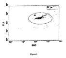
- the separation between the oocysts and the debris particles is greatest in the 8C12 (CRY104) stained sample. This suggests that this antibody is most useful for staining Cryptosporidium oocysts in water samples.
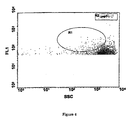
- the sample stained with 6G7 shows in increase in the fluorescence of some the debris particles (to the right of the oocysts) when compared to the unstained control. This is due to this antibody binding to some of the debris particles and suggests that this antibody may not be useful for staining Cryptosporidium oocysts in water samples.
- Antibody Antibody Type Supplier CRy104 IgG1 FITC conjugate MUCAB CRY26TM IgM FITC conjugate MUCAB CRY212 IgM FITC conjugate MUCAB Crypto-Cell IgM FITC conjugate CelLab, Australia Crypto-a-gloTM IgM FITC conjugate Waterborne. Inc. USA ImmucellTM IgG3 FITC conjugate Immucell, Portland. USA HydrofluorTM IgM Not conjugated Meridian, ENSYS Inc. USA Giardia -specific monoclonal antibodies evaluated.
- Immunoglobulin M antibodies exist as large mAbs containing ten binding sites, with a greater chance of binding non-specifically to particles compared with IgG.
- the surface of oocysts and cysts contain carbohydrates that induce the production of low affinity IgM and IgG3.
- Immunoglobulin G1 mAbs are generally produced following repeated exposure to an antigen. Isotype switching occurs from IgM to IgG, resulting in higher affinity mAbs containing two identical binding sites more specific to the antigen they are produced to.
- IgM, CRY212 Specificity of the IgM, CRY212 that was developed for water analysis was greater than commercial mAbs that were produced for clinical applications such as faecal analysis. This trend was observed following Giardia analysis of IgG1 mAbs, with G203 having higher affinity than Giardia-a-gloTM.
- mAbs produced specifically for water analysis have a high affinity for cysts and oocysts over extraneous particles present within water concentrates.
- Immunoglobulin G1 are smaller mAbs that bind specifically to antigenic sites that they are produced to with little background interference. Production of new IgG1 mAbs to different antigenic sites on cysts or oocysts may enable the development of highly specific assays for Cryptosporidium and Giardia detection within environmental samples.
- mice were monitored for both IgM and IgG levels in serum.
- the oocyst control group demonstrated a high IgM response with little or no IgG. This would suggest that there is no memory in the immune response (i.e. shift to IgG response) and that each immunisation is seen as a new antigen.
- the oocyst walls group (W) showed no immune response. This is probably due to the large size and structure of the whole oocyst wall which may have a desensitising effect on the immune response. The fact that no immune response was seen in the oocyst wall group would suggest that with no sporozoites present a decrease in immunogenicity is obtained.
- the oocyst extract group elicited strong IgM and IgG responses.
- IgM responses were not as strong as in the oocyst control group, again suggesting that sporozoites are more antigenic than the oocyst wall and are characteristic of the self limiting nature of Cryptosporidiosis.. It appears that the SDS digestion breaks up the oocyst wall and exposes more epitopes for an immune response. After removing the sporozoites and breaking up the oocyst wall structure into smaller complexes the oocyst wall or extract becomes an extremely immunogenic antigen as shown from the strong immune response seen in the extract mice. After further immunisations in extract mice the IgG response further increased as for a typical immunisation program. This was not observed in the Control or Wall group, which showed dominant IgM response or no response respectively.
- a strong immunological response to the surface of Cryptosporidium oocysts was produced by immunising mice with a partially purified sample of the outer layer of the oocyst wall. Immunising mice with purified oocyst walls did not produce a strong immunological response. This would suggest that there is either immune suppression by a component of the oocyst wall or that the natural presentation of the surface antigens on the oocyst walls does not produce an immunological response. The present inventors have overcome this lack of an immunological response to the surface of Cryptosporidium oocysts by partially purifying the surface antigen.
- Fusion of spleen cells from one mouse immunised with the purified sample of the outer layer of the oocyst wall resulted in six (6) hybridomas that produce antibody that is specific to the surface of the oocyst wall.
- One of these six antibodies is of the IgG1 immunological subclass, the remaining five antibodies are of the IgM subclass.
- the IgG1 antibody appears to be superior to the IgM antibodies for staining Cryptosporidium oocysts in water samples. Mice immunised with purified Cryptosporidium oocysts walls do not produce a strong immunological response to the surface of Cryptosporidium oocysts.
Landscapes
- Health & Medical Sciences (AREA)
- Life Sciences & Earth Sciences (AREA)
- Chemical & Material Sciences (AREA)
- Immunology (AREA)
- Molecular Biology (AREA)
- Engineering & Computer Science (AREA)
- Urology & Nephrology (AREA)
- General Health & Medical Sciences (AREA)
- Biochemistry (AREA)
- Medicinal Chemistry (AREA)
- Hematology (AREA)
- Organic Chemistry (AREA)
- Biomedical Technology (AREA)
- Genetics & Genomics (AREA)
- Biophysics (AREA)
- Tropical Medicine & Parasitology (AREA)
- Biotechnology (AREA)
- Cell Biology (AREA)
- Proteomics, Peptides & Aminoacids (AREA)
- Microbiology (AREA)
- Virology (AREA)
- Food Science & Technology (AREA)
- Physics & Mathematics (AREA)
- Analytical Chemistry (AREA)
- General Physics & Mathematics (AREA)
- Pathology (AREA)
- Peptides Or Proteins (AREA)
- Preparation Of Compounds By Using Micro-Organisms (AREA)
- Medicines Containing Antibodies Or Antigens For Use As Internal Diagnostic Agents (AREA)
Abstract
Description
| Comparison of the fluorescence intensity of Cryptosporidium oocysts stained with serum (diluted 1 in 1000) from group E and W mice and then stained with an anti-IgG or an anti-IgM fluorescently labelled antibody. Serum was tested prior to immunisation and then 21 days after the second immunisation. | ||||
| mouse number | IgG specific antibody | IgM specific antibody | ||
| Pre-immunisation | Post-immunisation | Pre-immunisation | Post-immunisation | |
| E1 | 12 | 1117 | 12 | 111 |
| E2 | 9 | 460 | 11 | 121 |
| | 10 | 195 | 11 | 74 |
| | 10 | 1762 | 13 | 258 |
| E5 | 12 | 550 | 12 | 33 |
| W1 | 36 | 39 | 16 | 17 |
| W2 | 45 | 32 | 27 | 14 |
| W3 | 36 | 37 | 27 | 13 |
| W4 | 42 | 30 | 25 | 27 |
| W5 | 47 | 40 | 16 | 15 |
| Positive control (IgM) | 42 | 978 | ||
| Negative Control | 27 | 17 |
| Comparison of the fluorescence intensity of Cryptosporidium oocysts stained with serum (diluted 1 in 100 or 1 in 1000) from group WI and group WP mice and then stained with an anti-IgG or an anti-IgM fluorescently labelled antibody. Serum was tested prior to immunisation and then 21 days after the second immunisation. | ||||
| IgG specific antibody | IgM specific antibody | |||
| mouse number | Pre-immunisation | Post-immunisation | Pre-immunisation | Post-immunisation |
| WI1 | 36 | 39 | 16 | 17 |
| WI2 | 45 | 32 | 27 | 14 |
| WI3 | 36 | 37 | 27 | 13 |
| WI4 | 42 | 30 | 25 | 27 |
| WI5 | 47 | 40 | 16 | 15 |
| WP1 | 230 | 248 | 404 | 420 |
| WP2 | 231 | 518 | 405 | 514 |
| WP3 | 229 | 306 | 403 | 475 |
| WP4 | 233 | 290 | 410 | 417 |
| WP5 | 232 | 350 | 407 | 603 |
| Cryptosporidium-specific monoclonal antibodies evaluated. | ||
| Antibody | Antibody Type | Supplier |
| CRy104 | IgG1 FITC conjugate | MUCAB |
| CRY26™ | IgM FITC conjugate | MUCAB |
| CRY212 | IgM FITC conjugate | MUCAB |
| Crypto-Cell | IgM FITC conjugate | CelLab, Australia |
| Crypto-a-glo™ | IgM FITC conjugate | Waterborne. Inc. USA |
| Immucell™ | IgG3 FITC conjugate | Immucell, Portland. USA |
| Hydrofluor™ | IgM Not conjugated | Meridian, ENSYS Inc. USA |
| Giardia-specific monoclonal antibodies evaluated. | ||
| Antibody | Antibody Type | Supplier |
| G203 | IgG1 FITC conjugate | MUCAB |
| Giardia-Cell | IgM FTTC conjugate | CelLab, Australia |
| Giardia-a-glo™ | IgG1 FITC conjugate | Waterborne. Inc. USA |
| Hydrofluor™ | Giardia IgG Not | Meridian, ENSYS Inc. |
| Combo | conjugated + Crypto IgG | USA |
| Comparison of the level non-specific binding to Cryptosporidium- specific mAbs | |
| Antibody | Non-specific Binding ratio |
| CRY104 | 0.045 ± 0.013 |
| CRY26 | 0.623 ± 0.023 |
| CRY212 | 0.112 ± 0.034 |
| Crypto-Cell | 0.177 ± 0.034 |
| Crypto-a-glo™ | 0.980 ±0.125 |
| Immucell™ | 0.155 ± 0.013 |
| Hydrofluor™ | 0.159 ± 0.058 |
| Unstained | 0.033 ± 0.010 |
| Comparison of the level of non-specific binding to Giardia specific mAbs | |
| Antibody | Non-specific Binding ratio |
| G203 | 0.029 ± 0.002 |
| Giardia-Cell | 0.163 ±0.024 |
| Giardia-a-glo™ | 0.201 ±0.032 |
| Hydrofluor™ | 0.044 ± 0.005 |
| Unstained | 0.020 ± 0.002 |
| Evaluation of the level of non-specific binding of Cryptosporidium specific mAbs in different water concentrates. | |||
| Water Type | Treatment mean ± SD | ||
| Unstained control | CRY104 | Immucell™ | |
| Raw | 0.023 ± 0.002 | 0.023 ±0.001 | 3.294 ± 0.327 |
| Backwash | 0.023 ± 0.003 | 0.020 ± 0.006 | 0.440 ± 0.049 |
| Raw | 0.021 ± 0.003 | 0.020 ± 0.004 | 0.385 ± 0.016 |
| Backwash | 0.022 ± 0.002 | 0.087 ± 0.038 | 0.233 ± 0.052 |
| Effluent | 0.035 ± 0.006 | 0.085 ± 0.01 | 1.801 ± 0.093 |
| Backwash | 0.023 ± 0.004 | 0.277 ± 0.031 | 0.869 ± 0.154 |
| Raw | 0.022 ± 0.006 | 0.025 ± 0.005 | 1.391 ± 0.036 |
| Raw | 0.036 ± 0.006 | 0.099 ± 0.013 | 0.651 ± 0.047 |
| Backwash | 0.023 ± 0.004 | 0.031 ± 0.002 | 0.156 ± 0.027 |
| Filtered | 0.026 ± 0.007 | 0.027 ± 0.002 | 0.166 ± 0.015 |
| Statistical difference (p<0.05) by ANOVA, N=3 |
| Evaluation of the level of non-specific binding of Giardia specific mAbs in different water concentrates. | |||
| Water Type | Treatment mean ± SD | ||
| Unstained control | G203 | Hydrofluor™ | |
| Raw | 0.022 ± 0.002 | 0.048 ± 0.007 | 0.645 ±0.01 |
| Backwash | 0.022 ± 0.003 | 0.034 ± 0.005 | 0.669 ± 0.008 |
| Raw | 0.020 ± 0.003 | 0.025 ± 0.002 | 0.696 ± 0.022 |
| Backwash | 0.021 ± 0.003 | 0.214 ± 0.094 | 14.63 ± 4.58 |
| Effluent | 0.032 ± 0.007 | 0.007 ± 0.001 | 1.021 ± 0.075 |
| Backwash | 0.021 ± 0.003 | 0.081 ± 0.014 | 10.36 ± 1.547 |
| Raw | 0.021 ± 0.006 | 0.023 ± 0.003 | 0.062 ± 0.005 |
| Raw | 0.031 ± 0.006 | 0.070 ± 0.022 | 1.637 ± 0.096 |
| Backwash | 0.022 ± 0.004 | 0.036 ± 0.009 | 0.111 ± 0.016 |
| Filtered | 0.024 ± 0.006 | 0.024 ± 0.003 | 0.043 ± 0.004 |
| Statistical difference (p<0.05) by ANOVA, N=3 |
| Comparison of the fluorescence intensity of Cryptosporidium oocysts stained with serum of three dilutions (1:1000, 1:10,000, 1:100,000) from the mouse groups E (Extract), W (Walls) oocyst control (OC - mice injected with whole oocysts) or negative control (C) and then stained with an anti-IgG or an anti-IgM fluorescently labelled antibody. Data was calculated by subtracting the fluorescent value (arb) obtained from second serum samples after two immunisations from the negative serum samples prior to immunisation. | ||||||
| IgM Specific Antibody | IgG Specific Antibody | |||||
| 1:1000 | 1:10,000 | 1:100,000 | 1:1000 | 1:10,000 | 1:100,000 | |
| Extract Mouse 1 | 280 | 129 | 64 | 353 | 370 | 229 |
| Extract Mouse 2 | 283 | 115 | 96 | 220 | 259 | 172 |
| Extract Mouse 3 | 278 | 129 | 90 | 67 | 84 | 73 |
| Extract Mouse 4 | 319 | 151 | 108 | 354 | 425 | 279 |
| Extract Mouse 5 | 45 | 92 | 99 | 103 | 324 | 44 |
| Walls Mouse 1 | 3 | 30 | 19 | -68 | -13 | 28 |
| Walls Mouse 2 | -3 | 36 | 2 | 29 | -31 | 10 |
| Walls Mouse 3 | 18 | 0 | 4 | 41 | 52 | 16 |
| Walls Mouse 4 | 38 | 38 | -3 | 24 | -3 | -4 |
| Walls mouse 5 | 22 | 16 | 15 | 3 | -11 | 27 |
| Oocyst Control 1 | 432 | 240 | 115 | 2 | -18 | 13 |
| Oocyst Control 2 | 339 | 189 | 26 | -53 | -5 | 42 |
| Oocyst Control 3 | 438 | 228 | 69 | 48 | 45 | 2 |
| Oocyst Control 4 | 581 | 457 | 235 | 124 | 46 | 57 |
| Oocyst Control 5 | 210 | 75 | 37 | 21 | 43 | 43 |
| Control | 0 | 0 | 0 | 0 | 0 | 0 |
| Shows the functional measurement of Avidity. | ||
| Monoclonal Antibody | Subclass | ka (affinity constant of whole antibody) |
| CRY104 | IgG1 | 7x10⁸ mol-1 |
| CRY26 | IgM | 3x10⁸ mol-1 |
| Immucell™ | IgG3 | 6x10⁷ mol-1 |
Claims (19)
- A method of producing isolated IgG1 subclass antibodies reactive to the surface of Cryptosporidium oocysts, the method comprising:(a) pretreating Cryptosporidium oocysts with a reagent so as to remove the surface layer of the oocysts to form an oocyst antigen preparation;(b) separating the oocysts from the oocyst antigen preparation so as to obtain a separated oocyst antigen preparation capable of eliciting a detectable IgG1 immune response in an animal to the surface of the oocyst;(c) immunising a non-human animal with the separated oocyst antigen preparation so as to elicit an IgG1 immune response in the animal; and(d) obtaining from the animal IgG1 antibodies reactive to the surface of Cryptosporidium oocysts.
- The method according to claim 1 wherein the reagent is a detergent.
- The method according to claim 2 wherein the detergent is sodium dodecyl sulphate (SDS).
- The method according to claim 3 wherein the pretreating is boiling the oocysts in the presence of SDS for a sufficient time to generate the oocyst antigen preparation.
- The method according to claim 4 wherein (a) is boiling the oocysts for 1 hour in the presence of 0.5% (w/v) SDS.
- The method according to claim 1 wherein the reagent is selected from the group consisting of urea, detergents including octoxynol known as Triton X-100 and nonident, enzymes including chitinase, oxidising agents including sodium hypochlorite, sodium periodate, and ozone; and reducing agents including mercaptol ethanol and 1,1,1-trichloro- 2,2-bis[4-chlorophenyl]ethane.
- The method according to any one of claims 1 to 6 wherein (c) further includes one or more adjuvants.
- The method according to any one of claims 1 to 7 wherein the animal is a mouse.
- A method of producing isolated IgG1 subclass antibodies reactive to the surface of Cryptosporidium oocysts, the method comprising:(a) separating at least a portion of the Cryptosporidium oocyst wall from the internal sporozoites to form an oocyst-wall preparation;(b) treating the separated oocyst-wall preparation so as to obtain an oocyst antigen preparation capable of eliciting a detectable IgG1 immune response in an animal to the surface of the oocyst;(c) immunising an animal with the oocyst antigen preparation so as to elicit an IgG1 immune response in the animal; and(d) obtaining from the animal IgG1 antibodies reactive to the surface of Cryptosporidium oocysts.
- The method according to claim 9 wherein the separation of the oocyst wall from the internal sporozoites is by causing the oocyst to excyst followed by immuno-separation of the oocyst wall components.
- The method according to claim 9 wherein the separation of the oocyst wall from the internal sporozoite is by causing the oocyst to excyst followed by separation of the wall components by the group of separation methods consisting of centrifugation, flow cytometry, density gradient separation, precipitation, immune-labelling, ligand-binding, biotin-labelling and separation by avidin, and chromatographic separation.
- The method according to claim 10 or 11 wherein causing the oocyst to excyst is by freeze-thawing or by physically breaking up by crushing, sonication, or grinding.
- The method according to any one of claims 9 to 12 wherein the treating step (b) is by physically breaking up the cell wall.
- The method according to any one of claims 9 to 13 wherein (c) further includes one or more adjuvants.
- The method according to any one of claims 9 to 15 wherein the animal is a mouse.
- An isolated IgG1 antibody reactive to the surface of Cryptosporidium oocysts obtainable by the method according to any one of claims 1 to 8.
- The antibody according to claim 16 being a monoclonal antibody.
- An isolated IgG1 antibody reactive to the surface of Cryptosporidium oocysts obtainable by the method according to any one of claims 9 to 15.
- The antibody according to claim 18 being a monoclonal antibody.
Applications Claiming Priority (5)
| Application Number | Priority Date | Filing Date | Title |
|---|---|---|---|
| AUPO696297 | 1997-05-19 | ||
| AUPO6962A AUPO696297A0 (en) | 1997-05-19 | 1997-05-19 | Antibodies to cryptosporidium |
| AUPO824297 | 1997-07-25 | ||
| AUPO8242A AUPO824297A0 (en) | 1997-07-25 | 1997-07-25 | Antibodies to cryptosporidium |
| PCT/AU1998/000368 WO1998052974A1 (en) | 1997-05-19 | 1998-05-19 | ANTIBODIES TO $i(CRYPTOSPORIDIUM) |
Publications (3)
| Publication Number | Publication Date |
|---|---|
| EP0991667A1 EP0991667A1 (en) | 2000-04-12 |
| EP0991667A4 EP0991667A4 (en) | 2000-12-20 |
| EP0991667B1 true EP0991667B1 (en) | 2003-04-16 |
Family
ID=25645429
Family Applications (1)
| Application Number | Title | Priority Date | Filing Date |
|---|---|---|---|
| EP98922500A Expired - Lifetime EP0991667B1 (en) | 1997-05-19 | 1998-05-19 | Igg1 antibodies reactive to the surface of cryptosporidium oocysts |
Country Status (6)
| Country | Link |
|---|---|
| US (1) | US6777222B1 (en) |
| EP (1) | EP0991667B1 (en) |
| AT (1) | ATE237639T1 (en) |
| DE (1) | DE69813555T2 (en) |
| ES (1) | ES2201490T3 (en) |
| WO (1) | WO1998052974A1 (en) |
Families Citing this family (4)
| Publication number | Priority date | Publication date | Assignee | Title |
|---|---|---|---|---|
| US6475747B2 (en) * | 1997-10-28 | 2002-11-05 | The United States Of America As Represented By The Department Of Health And Human Services | Method for detecting Cryptosporidium parvum oocysts |
| US20070141710A1 (en) * | 2005-12-20 | 2007-06-21 | Beckman Coulter, Inc. | Stable calibrators for immunoassays |
| US10429386B2 (en) | 2015-03-10 | 2019-10-01 | The Regents Of The University Of California | Antibodies to the surface of Toxoplasma gondii oocysts and methods of use thereof |
| CN116143875A (en) * | 2022-07-09 | 2023-05-23 | 吉林大学 | Cryptosporidium cgd2_3080 oocyst outer wall marker protein and its application |
Family Cites Families (1)
| Publication number | Priority date | Publication date | Assignee | Title |
|---|---|---|---|---|
| AUPN514695A0 (en) * | 1995-08-30 | 1995-09-21 | Australian Water Technologies Pty Ltd | Method for detection of cryptosporidium oocysts |
-
1998
- 1998-05-19 US US09/424,048 patent/US6777222B1/en not_active Expired - Lifetime
- 1998-05-19 EP EP98922500A patent/EP0991667B1/en not_active Expired - Lifetime
- 1998-05-19 DE DE69813555T patent/DE69813555T2/en not_active Expired - Lifetime
- 1998-05-19 AT AT98922500T patent/ATE237639T1/en not_active IP Right Cessation
- 1998-05-19 WO PCT/AU1998/000368 patent/WO1998052974A1/en not_active Ceased
- 1998-05-19 ES ES98922500T patent/ES2201490T3/en not_active Expired - Lifetime
Also Published As
| Publication number | Publication date |
|---|---|
| DE69813555T2 (en) | 2004-04-29 |
| ATE237639T1 (en) | 2003-05-15 |
| US6777222B1 (en) | 2004-08-17 |
| WO1998052974A1 (en) | 1998-11-26 |
| ES2201490T3 (en) | 2004-03-16 |
| EP0991667A1 (en) | 2000-04-12 |
| EP0991667A4 (en) | 2000-12-20 |
| DE69813555D1 (en) | 2003-05-22 |
Similar Documents
| Publication | Publication Date | Title |
|---|---|---|
| DE69321914T2 (en) | Antibodies against Plasmodium falciparum | |
| Gamble et al. | Diagnosis of swine trichinosis by enzyme-linked immunosorbent assay (ELISA) using an excretory-secretory antigen | |
| Wirtz et al. | Comparative testing of monoclonal antibodies against Plasmodium falciparum sporozoites for ELISA development | |
| EP1308507B1 (en) | HCV anti-core monoclonal antibody | |
| Sithigorngul et al. | Development of a monoclonal antibody specific to yellow head virus (YHV) from Penaeus monodon | |
| Li et al. | Recombinant antigens for immunodiagnosis of cystic echinococcosis | |
| DE19536166C1 (en) | Method for the determination of antibodies against Treponema pallidum (syphilis) | |
| US6777222B1 (en) | Antibodies to cryptosporidium | |
| Torian et al. | Tubulin and high-molecular-weight polypeptides as Giardia lamblia antigens | |
| DE69330164T2 (en) | HTLV-I / HTLV-II ASSAY AND METHOD | |
| AU738798B2 (en) | Antibodies to (Cryptosporidium) | |
| Secombes et al. | The development of an ELISPOT assay to quantify total and specific antibody-secreting cells in dab Limanda limanda (L.) | |
| Bradley-Dunlop et al. | Development of monoclonal antibodies fordetection of necrotizing hepatopancreatitisin penaeid shrimp | |
| Crosbie et al. | Production of polyclonal antisera against barramundi (Lates calcarifer Bloch) serum immunoglobulin derived from affinity columns containing mannan-binding protein or staphylococcal protein A | |
| Wonsit et al. | Enzyme-linked immunosorbent assay based on monoclonal and polyclonal antibodies for the detection of Entamoeba histolytica antigens in faecal specimens | |
| Winiecka-Krusnell et al. | Detection of Giardia lamblia cysts in stool samples by immunofluorescence using monoclonal antibody | |
| de Weil et al. | Isolation of Fasciola hepatica genus-specific antigens | |
| Souza et al. | Differences in reactivity of paracoccidioidomycosis sera with gp43 isoforms | |
| KR101884069B1 (en) | Monoclonal antibody against nervous necrosis virus and use thereof | |
| Lin et al. | Surface coat of Meloidogyne incognita | |
| WO1997039009A1 (en) | Recombinant antigens useful for the serodiagnosis of neosporosis | |
| DE60033241T2 (en) | COMPOSITIONS AND METHODS OF IDENTIFYING TREPONEMA PALLIDUM | |
| Andrade et al. | A direct agglutination test discriminative toward Chagas' disease for the diagnosis of visceral leishmaniasis in Brazil: preliminary results | |
| WO1997008204A1 (en) | Methods for detection of cryptosporidium oocysts | |
| Ferreira et al. | Toxoplasmosis in naturally infected deer from Brazil |
Legal Events
| Date | Code | Title | Description |
|---|---|---|---|
| PUAI | Public reference made under article 153(3) epc to a published international application that has entered the european phase |
Free format text: ORIGINAL CODE: 0009012 |
|
| 17P | Request for examination filed |
Effective date: 19991215 |
|
| AK | Designated contracting states |
Kind code of ref document: A1 Designated state(s): AT BE CH CY DE DK ES FI FR GB GR IE IT LI LU MC NL PT SE |
|
| A4 | Supplementary search report drawn up and despatched |
Effective date: 20001103 |
|
| AK | Designated contracting states |
Kind code of ref document: A4 Designated state(s): AT BE CH CY DE DK ES FI FR GB GR IE IT LI LU MC NL PT SE |
|
| 17Q | First examination report despatched |
Effective date: 20010305 |
|
| RTI1 | Title (correction) |
Free format text: IGG1 ANTIBODIES REACTIVE TO THE SURFACE OF CRYPTOSPORIDIUM OOCYSTS |
|
| RTI1 | Title (correction) |
Free format text: IGG1 ANTIBODIES REACTIVE TO THE SURFACE OF CRYPTOSPORIDIUM OOCYSTS |
|
| RTI1 | Title (correction) |
Free format text: IGG1 ANTIBODIES REACTIVE TO THE SURFACE OF CRYPTOSPORIDIUM OOCYSTS |
|
| GRAH | Despatch of communication of intention to grant a patent |
Free format text: ORIGINAL CODE: EPIDOS IGRA |
|
| GRAG | Despatch of communication of intention to grant |
Free format text: ORIGINAL CODE: EPIDOS AGRA |
|
| GRAH | Despatch of communication of intention to grant a patent |
Free format text: ORIGINAL CODE: EPIDOS IGRA |
|
| GRAG | Despatch of communication of intention to grant |
Free format text: ORIGINAL CODE: EPIDOS AGRA |
|
| GRAH | Despatch of communication of intention to grant a patent |
Free format text: ORIGINAL CODE: EPIDOS IGRA |
|
| GRAH | Despatch of communication of intention to grant a patent |
Free format text: ORIGINAL CODE: EPIDOS IGRA |
|
| GRAA | (expected) grant |
Free format text: ORIGINAL CODE: 0009210 |
|
| AK | Designated contracting states |
Designated state(s): AT BE CH CY DE DK ES FI FR GB GR IE IT LI LU MC NL PT SE |
|
| PG25 | Lapsed in a contracting state [announced via postgrant information from national office to epo] |
Ref country code: NL Free format text: LAPSE BECAUSE OF FAILURE TO SUBMIT A TRANSLATION OF THE DESCRIPTION OR TO PAY THE FEE WITHIN THE PRESCRIBED TIME-LIMIT Effective date: 20030416 Ref country code: LI Free format text: LAPSE BECAUSE OF FAILURE TO SUBMIT A TRANSLATION OF THE DESCRIPTION OR TO PAY THE FEE WITHIN THE PRESCRIBED TIME-LIMIT Effective date: 20030416 Ref country code: FI Free format text: LAPSE BECAUSE OF FAILURE TO SUBMIT A TRANSLATION OF THE DESCRIPTION OR TO PAY THE FEE WITHIN THE PRESCRIBED TIME-LIMIT Effective date: 20030416 Ref country code: CH Free format text: LAPSE BECAUSE OF FAILURE TO SUBMIT A TRANSLATION OF THE DESCRIPTION OR TO PAY THE FEE WITHIN THE PRESCRIBED TIME-LIMIT Effective date: 20030416 Ref country code: BE Free format text: LAPSE BECAUSE OF FAILURE TO SUBMIT A TRANSLATION OF THE DESCRIPTION OR TO PAY THE FEE WITHIN THE PRESCRIBED TIME-LIMIT Effective date: 20030416 Ref country code: AT Free format text: LAPSE BECAUSE OF FAILURE TO SUBMIT A TRANSLATION OF THE DESCRIPTION OR TO PAY THE FEE WITHIN THE PRESCRIBED TIME-LIMIT Effective date: 20030416 |
|
| REG | Reference to a national code |
Ref country code: GB Ref legal event code: FG4D |
|
| REG | Reference to a national code |
Ref country code: CH Ref legal event code: EP |
|
| PG25 | Lapsed in a contracting state [announced via postgrant information from national office to epo] |
Ref country code: LU Free format text: LAPSE BECAUSE OF NON-PAYMENT OF DUE FEES Effective date: 20030519 Ref country code: CY Free format text: LAPSE BECAUSE OF FAILURE TO SUBMIT A TRANSLATION OF THE DESCRIPTION OR TO PAY THE FEE WITHIN THE PRESCRIBED TIME-LIMIT Effective date: 20030519 |
|
| REF | Corresponds to: |
Ref document number: 69813555 Country of ref document: DE Date of ref document: 20030522 Kind code of ref document: P |
|
| REG | Reference to a national code |
Ref country code: IE Ref legal event code: FG4D |
|
| PG25 | Lapsed in a contracting state [announced via postgrant information from national office to epo] |
Ref country code: MC Free format text: LAPSE BECAUSE OF NON-PAYMENT OF DUE FEES Effective date: 20030531 |
|
| PG25 | Lapsed in a contracting state [announced via postgrant information from national office to epo] |
Ref country code: SE Free format text: LAPSE BECAUSE OF FAILURE TO SUBMIT A TRANSLATION OF THE DESCRIPTION OR TO PAY THE FEE WITHIN THE PRESCRIBED TIME-LIMIT Effective date: 20030716 Ref country code: PT Free format text: LAPSE BECAUSE OF FAILURE TO SUBMIT A TRANSLATION OF THE DESCRIPTION OR TO PAY THE FEE WITHIN THE PRESCRIBED TIME-LIMIT Effective date: 20030716 Ref country code: GR Free format text: LAPSE BECAUSE OF FAILURE TO SUBMIT A TRANSLATION OF THE DESCRIPTION OR TO PAY THE FEE WITHIN THE PRESCRIBED TIME-LIMIT Effective date: 20030716 Ref country code: DK Free format text: LAPSE BECAUSE OF FAILURE TO SUBMIT A TRANSLATION OF THE DESCRIPTION OR TO PAY THE FEE WITHIN THE PRESCRIBED TIME-LIMIT Effective date: 20030716 |
|
| NLV1 | Nl: lapsed or annulled due to failure to fulfill the requirements of art. 29p and 29m of the patents act | ||
| REG | Reference to a national code |
Ref country code: CH Ref legal event code: PL |
|
| ET | Fr: translation filed | ||
| PLBE | No opposition filed within time limit |
Free format text: ORIGINAL CODE: 0009261 |
|
| STAA | Information on the status of an ep patent application or granted ep patent |
Free format text: STATUS: NO OPPOSITION FILED WITHIN TIME LIMIT |
|
| REG | Reference to a national code |
Ref country code: ES Ref legal event code: FG2A Ref document number: 2201490 Country of ref document: ES Kind code of ref document: T3 |
|
| 26N | No opposition filed |
Effective date: 20040119 |
|
| REG | Reference to a national code |
Ref country code: FR Ref legal event code: PLFP Year of fee payment: 19 |
|
| REG | Reference to a national code |
Ref country code: FR Ref legal event code: PLFP Year of fee payment: 20 |
|
| PGFP | Annual fee paid to national office [announced via postgrant information from national office to epo] |
Ref country code: GB Payment date: 20170517 Year of fee payment: 20 Ref country code: IE Payment date: 20170510 Year of fee payment: 20 Ref country code: FR Payment date: 20170413 Year of fee payment: 20 Ref country code: DE Payment date: 20170516 Year of fee payment: 20 |
|
| PGFP | Annual fee paid to national office [announced via postgrant information from national office to epo] |
Ref country code: ES Payment date: 20170602 Year of fee payment: 20 Ref country code: IT Payment date: 20170522 Year of fee payment: 20 |
|
| REG | Reference to a national code |
Ref country code: DE Ref legal event code: R071 Ref document number: 69813555 Country of ref document: DE |
|
| REG | Reference to a national code |
Ref country code: GB Ref legal event code: PE20 Expiry date: 20180518 |
|
| REG | Reference to a national code |
Ref country code: IE Ref legal event code: MK9A |
|
| PG25 | Lapsed in a contracting state [announced via postgrant information from national office to epo] |
Ref country code: IE Free format text: LAPSE BECAUSE OF EXPIRATION OF PROTECTION Effective date: 20180519 Ref country code: GB Free format text: LAPSE BECAUSE OF EXPIRATION OF PROTECTION Effective date: 20180518 |
|
| REG | Reference to a national code |
Ref country code: ES Ref legal event code: FD2A Effective date: 20220103 |
|
| PG25 | Lapsed in a contracting state [announced via postgrant information from national office to epo] |
Ref country code: ES Free format text: LAPSE BECAUSE OF EXPIRATION OF PROTECTION Effective date: 20180520 |