Background of the Invention
Malignant transformation is characterized by uncoupling of proliferation
and differentiation, leading to continuing amplification of cells with loss of their
ability to progress to differentiation. Agents capable of restoring the
differentiation ability of cancer cells are thus potentially useful in cancer therapy.
Various extracts, proteins and chemicals have been shown to induce
differentiation of certain cancer cells in vitro and in vivo. For example, Sachs et al.
(1987) Cancer Research 47 : 1981 provide a review of induction of differentiation
of leukemia myeloid hematopoietic cells, including observations that myeloid
leukemia cells can be induced to differentiate in vitro and in vivo by a normal
differentiating protein. Tallman et al. (1992) J. Clin. Pharmacol. 32 : 868 review
the role of retinoids in cancer treatment. Retinoids have been investigated as
differentiating agents for the prevention and therapy of bladder and mammary
cancers and leukemias. Platica et al. (1992) Endocrinology 131 : 2573 report that
extracts of bovine pituitary and a rat mammosomatotropic tumor induce
differentiation of rat mammary tumor cells.
Differentiation agents identified by in vitro studies and in vivo rodent
studies have also been assessed clinically. For example, differentiation agents
including hexamethylene bisacetamide and retinotc acid have entered clinical trials
for cancer treatment and prevention and are reviewed by Linskey et al. (1995)
Neurosurgery 36 : 1. Successful use of differentiation agents for the treatment of
acute promyelocytic leukemia has been reported by Warrell et al. (1993) New
Engl. J. Med. 329 : 177. Retinoids have been shown to be therapeutically useful in
the treatment of cervical cancer by Lippman et al. (1993) J. Natl. Cancer Inst, 85 :
499.
The clinical use of differentiation agents to induce cancer cells to
differentiate and thus assume more normal characteristics has been termed
differentiation therapy. Differentiation therapy provides an alternative approach to
conventional cancer therapy such as cytotoxic chemotherapy. Accordingly, there
is a need in the art for the identification and isolation or synthesis of new agents
capable of promoting the differentiation of cancer cells.
Summary of the Invention
The present invention is directed to pituitary differentiation factor (PDF), a
pituitary factor that is capable of differentiating cells including breast cancer and
prostate cancer cells.
In one embodiment, the present invention provides isolated nucleic acids
encoding PDF. Vectors and host cells containing isolated nucleic acids encoding
PDF are further provided.
Another embodiment of the present invention provides isolated and purified
PDF and biologically active analogs and fragments thereof, and a method of
making PDF and biologically active analogs and fragments thereof.
The present invention further provides a method of promoting
differentiation of breast cancer or prostatic cancer cells comprising contacting the
breast or prostatic cancer cells with a differentiation-promoting effective amount of
PDF.
Another embodiment of the present invention provides a method of
treatment of breast cancer or prostatic cancer comprising administering a
therapeutically effective amount of PDF to a patient in need of such treatment.
In another embodiment of the present invention, pharmaceutical
compositions are provided that include PDF or biologically active analogs or
fragments thereof admixed with a pharmaceutically acceptable carrier.
Brief Description of the Drawings
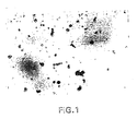
Fig. 1 depicts MCF-7 human breast cancer cells in conditioned medium in
the absence of PDF.
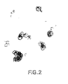
Fig. 2 depicts MCF-7 human breast cancer cells treated with lysate of
oocytes injected with cDNA encoding PDF, and illustrates aggregation induced by
PDF.
Fig. 3 depicts DU 145 prostate cancer cells cultured in the absence of PDF.
Fig. 4 illustrates the morphological changes, including aggregation and
spheroid formation, induced by PDF on DU145 cells.
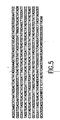
Fig. 5 provides the nucleotide sequence of SEQ ID NO:1.
Fig. 6 is a graph demonstrating the effect of oocyte lysate containing PDF
on spheroid formation in MCF-7 cells.
Fig. 7 is a graph demonstrating the effect of PDF on spheroid formation in
DU-145 cells.
Detailed Description of the Invention
The present invention is directed to pituitary differentiation factor (PDF).
PDF is a polypeptide obtainable from mammalian pituitary and from pituitary
tumors including MtTW10. PDF promotes the differentiation of cells including
breast cancer and prostatic cancer cells.
In one embodiment the present invention provides an isolated nucleic acid
encoding PDF. A plasmid designated pBS-PDF1 containing a 2.2kB cDNA
encoding PDF has been deposited with the American Type Culture Collection
(ATCC), 12301 Parklawn Drive, Rockville, Maryland 20852 and has been
accorded accession number ATCC 97648.
In accordance with the present invention, an isolated nucleic acid encoding
PDF may be obtained from mammalian pituitary by expression cloning. A
mammalian pituitary cDNA library may be prepared by methods known to one of
ordinary skill in the art, as described for example by Sambrook et al (1989)
Molecular Cloning: A Laboratory Manual, 2nd edition, Cold Spring Harbor
Laboratory Press, Cold Spring Harbor, NY. In addition, mammalian pituitary
cDNA libraries are available commercially, for example from Clontech, Palo Alto,
CA. A human pituitary library is preferred.
An isolated nucleic acid encoding PDF was obtained from a mammalian
cDNA library by expressing the cDNA clones of the library, and assessing the
expressed product for PDF activity in a functional assay. Various expression
systems are known to the ordinarily skilled artisan. In a preferred embodiment,
Xenopus oocytes are used as the host for expression of the pituitary cDNA. The
use of Xenopus oocytes for the expression of exogenous nucleic acids is known in
the art and described, for example, by Gurdon et al. (1983) Methods in
Enzymology 101 : 370. Expression vectors containing pituitary cDNA under the
control of a strong promoter can be injected into the nuclei of oocytes, after which
oocytes are incubated for from one to several days, followed by assessment of
oocyte lysates or conditioned media (CM) for PDF activity. Alternately, mRNA
can be synthesized in vitro from pituitary cDNA, and injected into oocytes,
followed by assessment of oocyte lysates or CM for PDF activity as described
hereinbelow. The pituitary cDNA may be divided into pools from which RNA is
synthesized, injected into oocytes, and tested for functional activity. Positive pools
are divided into subpools and the protocol is repeated until a single cDNA
encoding PDF is identified.
Bioassays useful for the identification of PDF are based upon the ability of
PDF to promote the differentiation of breast and prostate cancer cells. Any breast
or prostate cancer cells that are responsive to the differentiation-inducing activity
of PDF as described herein are suitable for use in the bioassay of the present
invention. Various cultured breast and prostate cancer cells are available from the
ATCC. In a preferred nonlimiting embodiment, the breast cancer cells used for the
bioassay are the rat mammary tumor cell line MTW9/P1 available from D.
Sirbascu, University of Texas Medical School, Houston, TX or MCF-7 human
breast cancer cells available from the ATCC. In another preferred embodiment,
the prostate cancer cells are from the human prostate cell line DU145 available
from the ATCC.
Treatment of breast or prostate cancer cells with PDF causes
undifferentiated cancer cells to differentiate. Differentiation can be measured by
morphological and biochemical parameters that are consistent with differentiation
toward the structure of a normal mammary or prostate gland. The cancer cells,
which normally grow in culture as single cell suspensions, aggregate and form
spheroids within 24 hours of treatment with PDF. Aggregation may be measured
by removing and counting suspended single cells, then detaching and counting the
remaining aggregated and adherent cells, and then determining the percentage of
total cells that have aggregated. A statistically significant increase in aggregation
of treated cells as compared to untreated cells is evidence of PDF activity. The
measurement of aggregation thus provides a simple and convenient bioassay for
PDF activity.
The aggregation bioassay may be performed as follows. About 1x10⁵
breast cancer cells, for example MTW9/P1 cells, arc grown in 1 ml serum-free
Dulbecco's modified Eagle's Medium (DMEM) in the presence or absence of the
expression product of the pituitary cDNA at a concentration of from about 10ng/ml
to about 10µg/ml. The cultures are incubated at 37°C in a 5% CO2 atmosphere for
about 72 hours. Suspended single cells are removed by rinsing with serum-free
DMEM and then counted. The remaining aggregated and adherent cells are
detached by trypsinization with trypsin-EDTA for five minutes and then counted.
Cells are conveniently counted by viewing cells by light microscopy on gridded
culture dishes. A dose-responsive increase in aggregation in response to treatment
with the pituitary cDNA expression product indicates that the cDNA encodes PDF.
A spheroid formation assay may be performed as follows. About 1x10⁵
prostate cancer cells, for example DU145 cells, are grown in RPMI 1640 medium
(Sigma) supplemented with 10% fetal bovine serum (FBS) (BioWhittacker,
Walkersville, MD), 10 IU penicillin/ml and 50mg streptomycin/ml at 37°C in a
5% CO2 atmosphere. Cultures are treated with various concentrations of PDF, for
example from 50-300 ug/ml culture. After 72 hours, cultures are scored for the
formation of spheroids. Spheroids are defined as multicellular aggregates with no
individual distinguishable cell morphology. Cultures are conveniently scored by
viewing gridded culture dishes by light microscopy and counting spheroids. A
dose-responsive increase in spheroid formation is indicative of PDF bioactivity.
In a modification of the foregoing aggregation bioassay, aggregation and
spheroid formation may be detected by light microscopy or electron microscopy of
fixed sections. After culturing and treating cells as described above, cultured cells
are fixed and sectioned for microscopy by methods known in the art. For example,
cultured cells are fixed in 1.5% glutaraldehyde in 0.1M cacodylate buffer for one
hour. Pellets obtained by low speed centrifugation are postfixed in 1.5% osmium
tetroxide in collidine buffer for 30 minutes, followed by 30 minutes in uranyl
acetate in maleate buffer, and then dehydrated and embedded in Epon 812. For
light microscopy, 1µm sections are stained with methylene blue, azure-II, and
basic fuchsin. For electron microscopy, 60 to 90 nm sections are cut and stained
with uranyl acetate-lead citrate. Aggregation and spheroid formation can then be
visualized by light or electron microscopy. A statistically significant increase in
aggregation and spheroid formation of treated cells as compared to untreated cells
is evidence of PDF bioactivity.
Treatment of breast cancer cells with PDF produces other effects that can
be observed by microscopy, thus providing further PDF assays. By light
microscopy, it can be observed that PDF-treated cells are smaller in size than
untreated cells, and are clustered in organoid structures consistent with gland
formation. By electron microscopy it can be observed that treated cells are
smaller, with smaller nuclei than untreated cells. Further, the cytoplasm is rich in
polarized organelles such as lysosomes and endoplasmic reticulum, as opposed to
untreated cells that have vacuolated cytoplasm, with few organelles other than
mitochondria.
Breast cancer cells treated with PDF also undergo biochemical changes that
provide additional bioassays for PDF. Specifically, lactalbumin, which is secreted
only in differentiated mammary cells, is produced by PDF-treated MTW9/P1 cells
but not by untreated cells. Thus the synthesis of lactalbumin, as detected, for
example, by conventional Northern or Western blotting, histochemical techniques,
or immunoassays provides another bioassay for PDF.
By the foregoing cloning methods and bioassays, an isolated cDNA
encoding PDF has been identified. It has thus been discovered in accordance with
the present invention that a single nucleic acid encoding a single polypeptide
directs the pituitary differentiating activity.
The isolated nucleic acid encoding PDF may be additionally characterized
by its nucleotide sequence. Nucleotide sequencing may be accomplished by
methods known to one of ordinary skill in the art, including for example the
dideoxy chain termination method of Sanger et al. (1977) Proc. Natl. Acad. Sci. 74
: 5463. An isolated nucleic acid encoding PDF in accordance with the present
invention contains the sequence set forth at SEQ ID NO:1 in Figure 5.
The present invention encompasses isolated nucleic acids that can be
obtained from mammalian pituitary and pituitary tumors and that encode PDF, a
polypeptide having the ability to promote the differentiation of breast cancer cells
as determined by any of the above-described bioassays. In a preferred
embodiment, the nucleic acid is the 2.2kB nucleic acid contained in plasmid pBS-PDF1
. In
another preferred embodiment, the isolated nucleic acid is a contiguous fragment
of the 2.2kB insert wherein the fragment encodes biologically active PDF. The
ordinarily skilled artisan can obtain fragments of the 2.2kB insert by conventional
molecular biological techniques, prepare expression vectors containing the
fragments, express the nucleic acid, and assay the resulting product for PDF
activity as described hereinabove to identify fragments that encode PDF.
In a preferred embodiment of the present invention, the isolated nucleic
acid encoding PDF is contained in the 2.2kB insert of plasmid pBS-PDF1. The
present invention further encompasses analogs of the nucleic acid contained in the
2.2kB insert of plasmid pBS-PDF1 wherein said analogs encode PDF. For
example, the ordinarily skilled artisan, with the knowledge of the degeneracy of the
genetic code, can determine nucleic acid sequences that encode the amino acid
sequence encoded by the insert of plasmid pBS-PDF1. Further, the sequence can
be selected to optimize expression in a particular host organism by utilizing known
preferred codons for a host organism of choice. In addition, analogs may be made
by making substitutions or deletions of residues that are not necessary for
biological activity. Such analogs may be identified by the bioassays described
above.
The present invention further encompasses nucleic acids isolatable from
mammalian pituitary or pituitary tumors and capable of hybridizing under
high stringency conditions to the 2.2kB insert of plasmid pBS-PDF1
and further capable of encoding biologically active PDF.
High stringency hybridization conditions are known to the skilled artisan and
described, for example, in Sambrook et al. and Beltz et al. (1983) Methods
Enzymol, 100: 226. High stringency conditions include, for example,
hybridization at 68°C in aqueous buffered solution or at 42°C in 50% formamide.
The ability of the isolated nucleic acid of the present invention to encode
biologically active PDF can be determined by the functional assays described
hereinabove.
The present invention is further directed to vectors comprising the isolated
nucleic acids of the present invention. The vectors are useful for the amplification
and/or expression of the nucleic acids encoding PDF. In one embodiment, the
vectors of the present invention comprise the nucleic acid encoding PDF operably
linked to suitable transcriptional and/or translational regulatory elements to effect
expression of PDF in a suitable host cell. The regulatory elements may be denved
from mammalian, microbial, viral or insect genes, and include, for example,
promoters, enhancers, transcription and transition initiation sequences,
termination sequences, origins of replication, and sequences encoding leader and
transport sequences. Suitable regulatory elements are selected for optimal
expression in a desired host cell. Useful expression vectors can be constructed by
methods known to one of ordinary skill m the art, and are also commercially
available. Recombinant viral vectors, including retrovirus, parvovirus, densovirus
and baculovirus vectors are particularly preferred.
In a preferred embodiment, the expression vector comprises a strong
constitutive or inducible promoter operatively linked to a nucleic acid encoding
PDF. Suitable promoters are well known and readily available to one of ordinary
skill in the art and include, for example, bacterial, yeast, viral, mammalian, and
insect promoters. Expression vectors compatible with insect and mammalian cells
are particularly preferred.
Another embodiment of the present invention provides host cells
comprising a nucleic acid encoding PDF. Host cells comprising the nucleic acid
are useful for replicating and expressing the nucleic acid encoding PDF. The host
cell may be procaryotic or eucaryotic, including bacterial, yeast, insect or
mammalian cells. Insect and mammalian cells are preferred. Particularly preferred
host cells are insect cell lines including, for example, Spodoptera frugiperda and
Trichoplusia ni cells.
The isolated nucleic acids or expression vectors may be introduced into the
host cells by methods known to one of ordinary skill in the art, including
transformation, transfection and infection. For example, transfection may be
accomplished by known methods such as liposome mediated transfection, calcium
phosphate mediated transfection, naked DNA transfection, microinjection and
electroporation. Transformation methods of procaryotic cells are described, for
example, by Cohen et al. (1972) Proc. Natl. Acad. Sci. USA 69: 2110.
Transformation of eucaryotic host cells is described, for example, by Sambrook et
al.
Expression systems utilizing baculovirus vectors and insect host cells are
also preferred. The use of baculoviruses as recombinant expression vectors to
infect lepidopteran insect cells is known in the art and described for example by
Luckow et al. (1988) BioTechnology 6 : 47.
The present invention is further directed to isolated and purified PDF and
biologically active analogs and fragments thereof. In a preferred embodiment of
the present invention, the isolated and purified PDF has an amino acid sequence
encoded by the DNA in plasmid pBS-PDF1.
Isolated and purified PDF may be made by introducing a nucleic acid
encoding PDF into a suitable host cell, for example by transformation, transfection
or injection, culturing the host cell under conditions suitable for expression, and
recovering recombinant PDF. Recombinant PDF may be recovered from cells or
culture medium by protein purification methods known in the art. In a preferred
embodiment, an expression vector comprising a nucleic acid encoding PDF under
the control of a suitable promoter is introduced into an insect or mammalian host
cell.
Biologically active analogs and fragments of PDF are similarly made
utilizing a nucleic acid encoding a biologically active analog or fragment of PDF.
The isolated recombinant analog or fragment may be identified by the bioassay
described above. The term "analogs" includes substitutions and alterations of the
amino acid sequence of PDF, which substitutions and alterations maintain the
biological activity of PDF. Amino acid insertional derivatives include amino and
carboxy terminal fusions and single or multiple intra-sequence insertions.
Deletional variants have one or more amino acids removed from the sequence. In
substitutional amino acid variants, at least one residue has been removed or
replaced by a different residue. Biologically active fragments are fragments of
PDF or PDF analogs that do not encompass the entire length of the PDF
polypeptide but which maintain the biological activity of PDF. The biologically
active analogs and fragments may be made by recombinant methods as described
for example by Sambrook et al, or by peptide synthetic techniques well known in
the art such as solid phase peptide synthesis.
The present invention provides a method of promoting differentiation of
breast cancer or prostatic cancer cells comprising contacting the breast or prostatic
cancer cells with a differentiation-promoting effective amount of PDF or an analog
or fragment thereof. A differentiation promoting effective amount of PDF is that
amount that promotes differentiation of cancer cells by any of the above-described
bioassays for differentiation.
Another embodiment of the present invention provides a method of
treatment of breast cancer comprising administering a therapeutically effective
amount of PDF or an analog or fragment thereof to a patient in need of such
treatment. A therapeutically effective amount of PDF for breast cancer treatment
is an amount that leads to change in the behavior of sentinel tumor masses such as
morphologic or biochemical differentiation, reduction in tumor markers, tumor
regression, apoptosis, or partial cessation of tumor growth or invasion. PDF is
administered as a pharmaceutical composition containing PDF or a biologically
active analog or fragment thereof and a pharmaceutically acceptable carrier.
Another embodiment of the present invention provides a method of
treatment of prostate cancer comprising administering a therapeutically effective
amount of PDF or an analog or fragment thereof to a patient in need of such
treatment. A therapeutically effective amount ofPDF for prostate cancer treatment
is an amount that results in change in behavior of sentinel tumor masses as
described hereinabove. PDF is administered as a pharmaceutical composition
containing PDF or a biologically active analog or fragment thereof and a
pharmaceutically acceptable carrier.
The formulation of pharmaceutical compositions is generally known in the
art and reference can conveniently be made to Remington's Pharmaceutical
Sciences, 17th ed., Mack Publishing Co., Easton, Pennsylvania. Formulation of
PDF and biologically active analogs and fragments thereof for use in present
invention must be stable under the conditions of manufacture and storage and must
also be preserved against the contaminating action of microorganisms such as
bacteria and fungi. Prevention against microorganism contamination can be
achieved through the addition of various antibacterial and antifungal agents.
The pharmaceutical forms of PDF suitable for administration include sterile
aqueous solutions or dispersions and sterile powders for the extemporaneous
preparation of sterile injectable solutions or dispersions. In all cases, the form
must be sterile and must be fluid to the extent that easy syringability exists.
Typical carriers include a solvent or dispersion medium containing, for example,
water buffered aqueous solutions (i.e., biocompatible buffers), ethanol, polyols
such as glycerol, propylene glycol, polyethylene glycol, suitable mixtures thereof,
surfactants, or vegetable oils. Sterilization can be accomplished by an art-recognized
technique, including but not limited to filtration or addition of
antibacterial or antifungal agents, for example, paraben, chlorobutanol, phenol,
sorbic acid or thimerosal. Further, isotonic agents such as sugars or sodium
chloride may be incorporated in the subject compositions.
Production of sterile injectable solutions containing the subject PDF is
accomplished by incorporating these compounds in the required amount in the
appropriate solvent with various ingredients enumerated above, as required,
followed by sterilization, preferably filter sterilization. To obtain a sterile powder,
the above solutions are vacuum-dried or freeze-dried as necessary.
The subject PDF or analogs and fragments thereof are thus compounded for
convenient and effective administration in pharmaceutically effective amounts with
a suitable pharmaceutically acceptable carrier and/or diluent in a therapeutically
effective dose.
As used herein, the term "pharmaceutically acceptable carrier and/or
diluent" includes any and all solvents, dispersion media, antibacterial and
antifungal agents, microcapsules, liposomes, cationic lipid carriers, isotonic and
absorption delaying agents and the like which are not incompatible with the active
ingredients. The use of such media and agents for pharmaceutical active
substances is well known in the art. Supplementary active ingredients may also be
incorporated into the compositions and used in the methods of present invention.
The precise therapeutically effective amount of PDF, analog or fragment
thereof to be used in the methods of this invention applied to humans can be
determined by the ordinary skilled artisan with consideration of individual
differences in age, weight, extent of disease and condition of the patient. It can
generally be stated that the PDF pharmaceutical preparation of the present
invention should be preferably administered in an amount of at least about
1mg per infusion dose, and more preferably m an amount up to about 10mg per
dose, or at a dose that achieves a local breast or prostate tissue concentration of
from about 10-9M to 10-6M.
It is especially advantageous to formulate parenteral compositions in
dosage unit form for ease of administration and uniformity of dosage. Dosage unit
form as used herein refers to physically discrete units suited as unitary dosages for
the mammalian subjects to be treated, each unit containing a predetermined
quantity of active material calculated to produce the desired therapeutic effect in
association with the required pharmaceutical carrier. The specification for the
novel dosage unit forms of the invention are dictated by and directly depend on the
unique characteristics of the active material (i.e., PDF, analogs, or fragments
thereof), and the limitations inherent in the art of compounding such an active
material for the treatment of breast or prostatic cell cancer.
The principal active ingredient is compounded for convenient and effective
administration in effective amounts with a suitable pharmaceutically acceptable
carrier in dosage unit form as hereinabove disclosed. In the case of compositions
containing supplementary active ingredients, the dosages are determined by
reference to the usual dose and manner of administration of the ingredients.
In the method of treatment according to the present invention, the PDF,
analogs or fragments thereof may be administered in a manner compatible with the
dosage formulation, in such amount as will be therapeutically effective, and in any
way which is medically acceptable for the treatment of breast or prostatic cell
cancer. Possible administration routes include injections by parenteral routes such
as intravascular, intravenous, intraarterial, subcutaneous, intramuscular, intratumor,
intraperitoneal, intraventricular or intraepidural. The compositions may also be
directly applied to tissue surfaces, for example, during surgery. Sustained release
administration is also specifically included in the invention, by such means as
depot injections or erodible implants.
The invention is further illustrated by the following specific examples
which are not intended in any way to limit the scope of the invention.
Example 1
Pituitary Extracts Induce Differentiation
Of Breast Cancer Cells and Prostatic Cancer Cells
Extracts prepared from bovine pituitary and from a mammosomatotropic
pituitary tumor were assessed for ability to induce differentiation of breast cancer
cells.
Alkaline pituitary extracts of the mammosomatotropic tumor MtTW10 and
the pituitary tumor MTW9-0M obtained from Dr. Untae Kim, Roswell Park
Memorial Institute, Buffalo, NY were prepared as described by Platica et al. (1992)
Endocrinology 131 : 2573. Bovine pituitary extract was obtained commercially
from Collaborative Research, Bedford, MA.
Pituitary extracts were added to serum-free cultures of MTW9/P1 rat
mammary tumor cells, MCF-7 human breast cancer cells, normal epithelial breast
cells, and myelocytic and lymphocytic leukemic cells. After twenty-four hours,
breast cancer cells, which normally grew as single cell suspensions, aggregated and
formed spheroids. Electron microscopy demonstrated changes indicating
differentiation toward the structure of a normal mammary gland including
polarization of organelles, lumen-like formation, junction formation, and
appearance of intracellular secretory granules. Northern and Western blots
performed by standard methods demonstrated that pituitary extract induced
expression of laminin, casein and lactalbumin, and overexpression of E-cadherin,
in breast cancer cells. Normal epithelial cells and myelocytic and lymphocytic
leukemic cells were unaffected by treatment with pituitary extract.
Prostatic cancer cells were obtained from the ATCC. Serum-free cultures
of the prostatic cancer cells were treated with pituitary extract prepared by Platica
et al. as described above and assessed morphologically and biochemically for
evidence of differentiation. Pituitary extract induced differentiation of prostatic
cancer cells as measured by bioassay.
Extracts were similarly prepared from rat liver and kidney and added to cell
cultures as described above. Rat liver and kidney extracts had no effect on
differentiation of breast cancer or prostatic cancer cells.
Various hormones and growth factors, including epidermal growth factor
(EGF), transforming growth factor (TGF), platelet-derived growth factor (PDGF),
fibroblast growth factor (FGF), insulin-like growth factor (IGF) -I and -II,
estradiol, growth hormone and prolactin, were added to cultures of breast cancer
cells and assessed for differentiation activity as described above. None of the
hormones or growth factors exhibited the ability to induce differentiation.
Example 2
Identification of a Cloning System for
Pituitary Differentiating Activity
An expression cloning method was devised to further characterize the
pituitary differentiation activity. To determine whether Xenopus oocytes were an
appropriate expression system, Xenopus oocytes were assessed for absence of
pituitary differentiation-like activity, toxicity for breast cancer cells in the selected
functional assay, and presence of factors that may destroy or interfere with
pituitary differentiation activity.
The aggregation bioassay for pituitary differentiation activity measured
spheroid formation of MCF-7 breast cancer cells. MCF-7 cells aggregate and form
spheroids in response to the pituitary differentiation-activity of pituitary extract.
Spheroids were visualized by light microscopy.
Oocyte lysate was obtained by homogenization of oocytes in 0.15M NaCl,
followed by centrifugation for 30 minutes at 15,000xg at 4°C and collection of
supernatant. Various amounts of oocyte lysate containing 50-400µg of protein and
conditioned medium in which oocytes were kept for 24 hours, were added to 1 ml.
cultures containing 1x10⁵ MCF-7 cells, followed by incubation at 37°C for 72
hours. No morphological changes or toxic effects on MCF-7 cells were observed
at any concentration of lysate or medium, indicating that Xenopus oocytes do not
contain a pituitary differentiation-like activity, and that oocyte lysate is not toxic to
breast cancer cells.
To determine whether the pituitary differentiation activity remains active in
the presence of Xenopus oocyte lysate, cultures containing 1 x 10⁵ MCF-7 cells
were incubated with 150µg/ml pituitary extract at 37°C for 72 hours in the
presence or absence of varying amounts of oocyte lysate (50-400µg/ml). The
aggregation effect induced by pituitary extract was unaffected by the presence of
oocyte lysate. These results indicated that the pituitary differentiation activity
remains active in the presence of Xenopus oocyte lysate.
The ability of Xenopus oocytes to express the pituitary differentiating
activity in amounts sufficient for detection by the aggregation bioassay was
determined. Poly (A)+ RNA from the rat pituitary tumor MtTW10 was prepared
by the guanidinium/cesium chloride (CsCl) centrifugation method as described by
Sambrook et al. in an RNAse free environment. Briefly, one gram of tissue was
homogenized in 5 ml of 4M guanidinium thiocyanate, 0.1 Tris-HCl (pH 7.5) and
1% 2-ME, at room temperature. Then 9.7 ml homogenate was layered on a 3.3 ml
pad of 5.7M CsCl and 4mM EDTA Ph 7.5 and centrifuged at 30,000 rpm for 24
hours at room temperature. The RNA, pelleted at the bottom of the tube, was
dissolved in 10mM Tris pH 7.4, 1mM EDTA, 0.1% SDS and then ethanol
precipitated. The poly(A)+RNA was obtained by twice passing the total RNA on
an oligo dT cellulose column. Seventeen micrograms of poly(A)+RNA were
obtained from 900µg total RNA. Then, 20 fully grown oocytes (stage V and VI),
kept in sterile MBS solution at 19°C, were injected with 5ml containing 50 ng
mRNA per oocyte, using an automatic syringe. The oocytes were then placed in
MBS solution containing penicillin and streptomycin and incubated at 19°C. After
3 days the supernatants from oocyte lysates (prepared as described above) and
conditioned medium (CM) from RNA-injected oocytes were tested for pituitary
differentiating activity. Cultures containing 1x10⁵ MCF-7 cells in 1 ml serum free
RPMI were incubated in the presence of various protein concentrations of oocyte
lysate (10-300µg) or CM (50-200µl/culture), at 37°C in a 5% CO2 atmosphere.
After 72 hours, aggregation was seen in MCF-7 cultures treated with the oocyte
lysate, but not in CM-treated MCF-7 cells. A linear relationship between the
number of aggregates obtained and the lysate protein concentration was seen, as
shown in Figure 6.
Similar experiments were performed using rat liver poly(A)+RNA. Neither
the conditioned medium, nor the lysates from oocytes injected with liver mRNA
had any effect on MCF-7 cells.
These results indicated that pituitary differentiation activity can be detected
by the aggregation bioassay even when the whole population of pituitary mRNA
was expressed in Xenopus oocytes.
The foregoing results demonstrate that Xenopus occytes do not contain a
pituitary differentiating-like activity, are not toxic for MCF-7 cells, and do not
destroy the pituitary differentiating activity. Further, Xenopus oocytes injected
with pituitary mRNA expressed pituitary differentiating activity at a level
detectable by the aggregation bioassay.
Example 3
Characterization of Pituitary
Differentiating Activity
A human pituitary cDNA library was obtained from Clontech, Palo Alto,
CA and tested for the presence of cDNA encoding pituitary differentiating activity.
The cDNA library was directionally cloned in the EcoRI-HindIII site of lambda
Bluemid phage, which allows the transcription of either strand of DNA inserts with
T7 or T3 RNA polymerases. Five µl of serial dilutions of phage library were
mixed with 200 µl K802 E. coli (OD600 =0.25) and incubated at 37°C for 20
minutes. Then, 3 ml of 0.7 % agar molten at 48°C were added and the mixture
was spread on top of a 100 mm plate containing 1.5% LB agar. After incubation at
37°C overnight the plaques were counted for each phage dilution and the library
titer determined to be 1x10¹⁰pfu/ml. Then a pool of 400,000 pfu from the library
was plated on 20 plates (20,000 pfu per plate) and incubated at 37°C until the
plaques reached about 1mm in diameter. The top agar was then harvested in SM
buffer (0.1M NaCl, 10mM MgSO4.7H2O, 10mM Tris-HCl, pH 7.5, 2% gelatin)
and the bacterial cells lysed with chloroform. The agar and bacterial debris were
pelleted by centrifugation at 8,000xg for 20 minutes. The supernatant was treated
with 1µg/ml DNase I and 5µg/ml RNase A for 1 hour at 37 °C and then
recentrifuged at 8,000 rpm for 20 minutes. The supernatant, containing the phage
particles, was centrifuged at 25,000 rpm (Beckman, SW27 ROTOR) for 2 hours at
20°C. The pelleted phages were then resuspended in 0.5M Tris buffer pH 8,
incubated with 100µg/ml proteinase K for 30 minutes at 37°C, followed by three
phenol, one phenol/chloroform and one chloroform extraction and ethanol
precipitation.
Since in this library the T3 RNA polymerase synthesizes the (+) strand of
cloned inserts, the phage DNA was linearized by digestion with Sal I which cuts in
the polycloning region on the site of the insert opposite to T3 RNA polymerase
promoter. For efficient translation, a CAP site was added to the 5' end of
transcripts. The transcription followed Melton's protocol (Krieg et al., 1987,
Methods Enzymol. 155 : 397) with minor modifications as described by Regec et
al. (1995) Blood 85 : 2711 using a mMessage mMachine kit from Ambion which
can generate 30-50µg of capped RNA per each µg of plasmid DNA. The reaction
was carried out in 20 µl volume using 5µg linearized phage DNA and the protocol
and reagents provided by manufacturer. After a one hour incubation at 37°C, IU
RNase free DNase I was added for each µg of DNA and the incubation continued
for 15 minutes. Then the reaction mixture was phenol/chloroform extracted,
followed by precipitation with half volume 7.5 MNH4 acetate and three volumes of
ethanol. With three such successive ethanol precipitations, 99% of unincorporated
nucleotides were removed.
Twenty oocytes were injected with 50 ng RNA per oocyte, as described
above, placed in MBS solution containing penicillin and streptomycin, and
incubated at 19°C for 3 days. The oocyte lysate, prepared as described above, was
then tested for differentiating activity on MCF-7 cells using the above-described
bioassay. The treated cells formed spheroids, similar to those formed by these
human breast cancer cells in the presence of pituitary extract. A linear relationship
between the number of spheroids formed and the protein lysate used in the
bioassay was seen. (Please provide data). These data show that the Clontech
human pituitary cDNA library contains one or more clones encoding for pituitary
differentiating activity.
The above-described pool of 400,000 pfu from the pituitary cDNA library
was used for sib selection. From the 400,000 pfu pool, ten subpools of about
40,000 plaques each were plated separately and grown until they reached about 1
mm in size. The phage DNA from each pool was prepared, and capped transcripts
were synthesized with T3 RNA polymerase as described. RNA from each subpool
was injected into 20 frog oocytes (50ng/oocyte) and the lysates were tested for
differentiating activity on MCF-7 cells. For each bioassay, controls with mRNA
from pituitary tumors, with lysate from non-injected oocytes and with pituitary
extracts were prepared. Of the ten subpools analyzed, three showed aggregating
activity in the bioassay. Subpool #2 displayed the strongest biological activity and
was selected for further sib selection. This subpool was divided in 10 subpools of
about 4,000 pfu each, which were processed as above. From these subpools, the
subpool #6 was shown to have the strongest differentiating activity, and was
further divided in ten subpools (each containing above 400pfu) which were
processed similarly. Subpool #4 was found to contain the highest differentiating
specific activity,and was used for further sib selection. This process was continued
by further dividing positive pools and screening for differentiation activity until a
single positive clone encoding pituitary differentiating factor (PDF) was identified.
The identified PDF cDNA phage clone was converted into a plasmid clone
following the procedure of clontech. The phage DNA was digested with NotI to
release the pBLUESCRIPT plasmid DNA containing the cDNA insert. After
phenol chloroform extraction and ethanol precipitation, the digested plasmid DNA
was ligated and used to transform competent DM5 alpha E. coli (Gibco, BRL).
The colonies were grown on agar plates with 50µg/ml ampicillin, and contained
the Bluescript plasmid with the cloned PDF cDNA.
To ensure that the plasmid clone contained cDNA encoding PDF, the
transcript from the cDNA was synthesized with T3 RNA polymerase and
translated in Xenopus oocytes. The oocyte lysate was tested for PDF activity as
described above. Figure 1 depicts MCF-7 cells incubated with control CM. Figure
2 depicts MCF-7 cells treated with lysate of oocytes containing the cDNA
transcript. The treated cells aggregated and formed spheroids, thus confirming that
the cDNA clone encoded PDF. The plasmid DNA was then prepared for
sequencing.
Five hundred ml ampicillin/LB media inoculated with 0.5 ml stock
suspension of E. coli carrying the plasmid containing the PDF cDNA was
incubated, with shaking, at 37°C overnight. The next day the cells were pelleted at
8,000xg for 20 minutes at 4°C and plasmid DNA prepared by alkaline lysis
method of Bimboim and Dolly (1979) Nucleic Acid Res. 7 : 1573. The bacterial
pellet was resuspended in 10 ml 50mM glucose, 25mM Tris-HCl (pH 8.0) and
10mM EDTA (pH 8.0). After treatment with lysozyme, the bacterial cells were
lysed with 0.2N NaOH, 1% SDS, for 10 minutes at room temperature. Then, 15
ml of 3M cold potassium acetate was added. The mixture was stored on ice for ten
minutes and centrifuged at 4000xg for 15 minutes at 4°C. The supernatant
containing primarily plasmid DNA was filtered and precipitated with 0.6 volumes
of isopropanol. The nucleic acid was sedimented by centrifugation at 5.000 rpm
for 15 minutes, washed with 70% ethanol and dissolved in TE buffer.
CsCl/ethidium bromide gradients were prepared according to Sambrook et
al. (1989) Molecular Cloning: A Laboratory Manual, 2nd edition, Cold Spring
Harbor Laboratory Press, Cold Spring Harbor, NY. One gram of CsCl was
dissolved in each ml of plasmid preparation and 80 µl of ethidium bromide
(10mg/ml) were added to each ml of CsCl DNA solution. After the centrifugation
at 60,000 rpm for 24 hours at 20°C in a Ti 65 rotor, the bacterial DNA was
separated in two bands: the upper band containing the linear, chromosomal and
nicked circular plasmid DNA and the lower band consisting of closed circular
supercoiled plasmid DNA. The plasmid DNA was recovered, ethidium bromide
extracted with 1-butanol and after removing CsCl by dialysis, the DNA was
precipitated with ethanol.
The PDF cDNA sequencing was performed in the Core Facility of Mount
Sinai Medical School, New York, using the dideoxy chain termination method of
Sanger et al. (1977) Proc. Natl. Acad. Sci. 74 : 5463. Each portion of the clone
was sequenced from both forward and reverse orientations. 530 nucleotides of the
2.2 kB insert were sequenced using M13-20 and reverse primers from Bluescript
plasmid sequence. The 530 base pairs of the PDF cDNA have the nucleotide
sequence set forth in SEQ ID NO: 1. The sequence was analyzed with the
DNASIS program (Hitachi Software Engineering Co.) for homology with other
sequences from GENBANK and EMBL. No simple homology of this sequence
with any sequence from the GENBANK database was found. The greatest
maximum matching found was 72.5% over a stretch of 98 base pairs, indicating
that PDF is a novel polypeptide.
Example 4
Effect of PDF on Prostatic Cancer Cells
The human prostatic cell line DU145 was obtained from the American
Type Culture Collection and grown in RPMI 1640 medium supplemented with
10% fetal bovine serum (FBS) (BioWhittacker, Walkersville, MC.), 10 IU
penicillin/ml 50mg streptomycin/ml, at 37°C in a humidified atmosphere
containing 5% CO2
To look for the effect of PDF on these cells, cultures containing 1x10⁵ DU-145
cells in serum free-RPMI 1640 medium were treated with various
concentrations of PDF (50-300µg/ml culture). Three days later, the cultures were
scored for the formation of spheroids. A correlation between PDF concentration
and the number of spheroids formed was found, as depicted by the graph in Figure
7.
Figure 3 depicts DU145 cells cultured in the absence of PDF. Figure 4
illustrates the morphological changes induced by 150µg PDF/ml on DU145
prostate cancer cells after 72 hours. The PDF-treated cells aggregated and formed
spheroids similar to those produced by breast cancer cells treated with PDF.
Example 5
Sequencing and Expression of PDF
The complete nucleotide sequence of the PDF cDNA described in Example
3 is determined by a primer extension strategy as described, for example, by
Sambrook et al. From the complete sequence, the start and stop codons, open
reading frame and deduced amino acid sequence are determined.
Baculovirus expression vectors containing the cDNA encoding PDF are
prepared. The PDF cDNA is cloned into the baculovirus vector AcNPv, and the
recombinant baculovirus vector is used to transform cells of the insect cell line
Sf21. The baculovirus/insect system is cultured to express recombinant PDF,
which is then purified from the culture media or cell extracts. Biological activity
of recombinant PDF is confirmed by the in vitro aggregation assay.
Example 6
Effect of Recombinant PDF on Tumor Growth
Nude mice bearing the mammary tumor MTW9 are obtained by
transplantation as described by Diamond et al. (1976) Cancer Research 36 : 77.
Recombinant PDF is administered to the tumor-bearing mice by intraperitoneal
injection for 21 days at a dosage of 50 µg/kg per day. Reduction in tumor size in
response to treatment with PDF is assessed at regular intervals by measuring tumor
length, width and height with calipers and calculating tumor volume.
Various publications are cited herein, the contents of which are hereby
incorporated by reference in their entireties.
SEQUENCE LISTING
(1) GENERAL INFORMATION
(i) APPLICANT: PLATICA ET AL. (ii) TITLE OF THE INVENTION: PITUITARY DIFFERENTIATION
FACTOR AND METHODS OF USE THEREOF (iii) NUMBER OF SEQUENCES: 1 (iv) CORRESPONDENCE ADDRESS:
(A) ADDRESSEE: Brumbaugh, Graves, Donohue &
Raymond (B) STREET: 30 Rockefeller Plaza (C) CITY. New York (D) STATE NY (E) COUNTRY. USA (F) ZIP: 10112-0228 (v) COMPUTER READABLE FORM
(A) MEDIUM TYPE: Diskette (B) COMPUTER: IBM Compatible (C) OPERATING SYSTEM DOS (D) SOFTWARE: FastSEQ Version 1.5 (vi) CURRENT APPLICATION DATA:
(A) APPLICATION NUMBER: (B) FILING DATE. (C) CLASSIFICATION: (vii) PRIOR APPLICATION DATA:
(A) APPLICATION NUMBER: USSN 60/021,589 (B) FILING DATE. July 11, 1996 (viii) ATTORNEY/AGENT INFORMATION
(A) NAME: Clark, Richard S (B) REGISTRATION NUMBER. 26,154 (C) REFERENCE/DOCKET NUMBER: AP30487 165/33281 (ix) TELECOMMUNICATION INFORMATION:
(A) TELEPHONE. 212-408-2558 (B) TELEFAX 212-765-2519 (C) TELEX:
(2) INFORMATION FOR SEQ ID NO:1
(i) SEQUENCE CHARACTERISTICS:
(A) LENGTH: 538 nucleic acids (B) TYPE: nucleic acid (C) STRANDEDNESS. single (D) TOPOLOGY: linear (ii) MOLECULE TYPE: peptide (m) ORIGINAL SOURCE
(A) ORGANISM: human (iv) FEATURE
(A) NAME/KEY: (B) LOCATION. (C) OTHER INFORMATION Pituitary Differentiation Factor (v) SEQUENCE DESCRIPTION: SEQ ID NO: 1: