Field of the Invention
-
The present invention relates to Salmosin for use as
an anti-tumor agent. The invention also relates to
polynucleotides encoding Salmosin and to the use of such
polynucleotides in the treatment of tumors.
Background of the Invention
-
Tumor invasion and metastasis are the biological
phenomena in which cancer cells lethally spread
throughout the body. First, cancer cells detach from the
primary site (e.g., epithelial tissue) and cross the
basement membrane separating them from other tissue
layers. Some of these invasive cells penetrate the
basement membrane and endothelial cells surrounding a
blood vessel and circulate via the bloodstream. The
circulating cancer cells may adhere to and penetrate a
capillary wall to create a secondary tumor. Perhaps,
fewer than one in 10,000 cancer cells that escape the
primary site survives to form a colony in another tissue
(see: Erkki Ruoslahti, Scientific American, 72-77, Sep.,
1996).
-
Therefore, tumor metastasis and invasion require
adhesive interaction between cells and extracellular
matrix ("ECM"). In the course of tumor metastasis, tumor
cells can cause endothelial cells to retract, exposing
the subendothelial basement membrane and allowing the
tumor cells to adhere efficiently to ECM proteins of the
surrounding stroma (see: Hynes, R. O., Cell, 48:549,
1987). These matrix proteins promote cells adhesion by
binding to specific cell surface receptors, including a
member of integrin family.
-
In terms of structure, each integrin is a
heterodimer consisting of α and β subunits which are
noncovalently associated with each other. The β1
subfamily is a primary mediator of extracellular matrix
adhesions. It has been reported that β1 integrins may
have other functions, such as to mediate cell-cell
adhesion directly (see: Larjava, H., et al,. J. Cell.
Biol., 110:803-815, 1990). Integrins of the β2 subfamily
are found on leukocytes where they mediate cell-cell
interactions. The β3 subfamily, which includes the
platelet glycoprotein IIb/IIIa complex and the
vitronectin receptor, may play an important role in the
development of tumor invasiveness and malignancy (see:
Albelda, S. M., et al., Cancer Res., 50:6757-6764, 1990).
-
The integrin receptor complex that spans the plasma
membrane links the integral cytoskeletal network of a
cell with the extracellular environment. Common or
characteristic core sequences in cell adhesion molecules
such as fibrinogen, vitronectin and laminin have been
considered to contribute to cell adhesion and to the
spread or integration of cells.
-
It has been suggested that tumorigenesis and
metastasis are closely associated with the biological
role of integrins (see: Giancotti, F. G. and Rouslahti,
E., Cell, 60:849-859, 1990: Hynes, R. O., Cell, 69:11-25,
1992; Nip, J., et al., J. Clin. Invest., 96:2096-2103,
1995).
-
It has been reported that overexpression of the
fibronectin receptor, α5β1, suppresses the transformed
phenotype of Chinese hamster ovary cells, that integrin
α5β1 is reduced in ras-transformed rodent cells (see:
Plantefaben, L. C. and Hynes, R. O., Cell, 56:281-290,
1989) and that superfibronectin, a polymeric fibrillar
form of fibronectin, prevents tumor metastasis and tumor
formation (see: Pasqualini, R., et al., Nature Medicine,
2:1197-1203, 1996).
-
Integrin αvβ3 is a specific marker of the most
malignant cells, suggesting a crucial role of this
adhesion receptor in the malignant growth of human
melanoma (see: Albelda, S. M.. Et al., Cancer Res.,
50:6757-6764, 1990). Integrin αvβ3 gene expression and
the resulting adhesive phenotype are directly involved in
the proliferation of human melanoma in vivo (see:
Felding-Habermann, J. Clin. Invest., 89:2018-2022, 1992).
-
Angiogenesis is a biological process of forming new
blood vessels as outgrowths from preexisting blood
vessels (see: Folkman, J. and D'Amore, P. A, Cell,
87:1153-1155, 1996). This process plays a key role in
the progression of a solid tumor, as well as in normal
development, wound healing and inflammation and vascular
cell adhesion molecules in smooth muscle and endothelial
cells contribute to its regulation (see: Nguyen, M., et
al., Nature, 365-267, 1993).
-
The switch of angiogenic phenotype of a tumor may be
caused by losing balance between positive and negative
modulators involved in neovascularization. Recently, it
has been reported that two cytokine-dependent pathways of
angiogenesis were shown to exist and were defined by
distinct vascular cell integrins, αvβ3 and αvβ5 that
become expressed on angiogenic vascular cells where they
play a critical role in angiogenesis induced by basic
fibroblast growth factor ("bFGF"), tumor necrosis factor-alpha
(TNF-α), vascular endothelial growth factor (VEGF),
and fragments of human tumors (see: Friedlander, M., et
al., Science, 270:1500-502, 1995). Activation of αvβ3
integrin stimulates a survival signal that facilitates
blood vessel growth and differentiation indicating that
signalling events by both cytokine and integrin receptors
are closely associated with the growth of new blood
vessels (see: Brooks. P. C. et al., Cell, 79:1157-1164,
1994).
-
Several endogenous angiogenic inhibitors have been
identified as following: interferon-α, -γ (see: Friesel,
R., et al., J. Cell. Biol., 104:689-696, 1987; Ezekowitz,
R. A., et al., N. Engl. J. Med., 324:1456-1463, 1992);
interferon-inducible protein 10 (see: Angiolillo, A. L.,
et al., J. Exp. Med., 182:155-162, 1995; Strieter, R. M.,
et al., Biochem. Biophys. Res. Comm., 210:51-57, 1995);
angiostatin and endostatin that specifically suppress
endothelial cell proliferation (see: O'Reilly, M. S., et
al., Cell, 79:315-328, 1994; O'Reilly M. S., et al.,
Cell, 88:277-285, 1997); gro-β (see: Cao, Y., et al., J.
Exp. Med., 182:2069-2077, 1995); the 16kDa N-terminal
fragment of prolactin (see: Clapp, C., et al.,
Endocrinology, 133:1292-1299, 1993); and, platelet
factor-4 (see: Maione, T., et al., Science, 247:77-79,
1990; Gupta, S. K., et al., Proc. Natl. Acad. Sci., USA,
92:7799-7803, 1995).
-
It has been well known that disintegrins are a
family of small proteins mainly derived from snake venom
(see: Niewiarowski, S., et al., Semin. Hematol., 31:289-300,
1994). Most of the disintegrins contain Arg-Gly-Asp
(RGD) or Lys-Gly-Asp (KGD) sequence which is the
structural motif recognized by a platelet fibrogen
receptor α2bβ3. Disintegrins also act as potent
antagonists of several integrins including αvβ3 and α5β1.
There are several reports demonstrating that disintegrins
inhibit tumor metastasis by blocking tumor cell adhesion
to ECM (see: Trika, M., et al., Cancer Res., 54 (8):4993-4998,
1994).
-
Integrin αvβ3 was identified as a marker of
angiogenic blood vessels in chick embryo and human (see:
Brooks. P. C., et al., Science, 264:569-571, 1994).
Monoclonal antibody against αvβ3 was able to perturb
angiogenesis by inducing apoptosis in the endothelial
cells of the newly formed blood vessels. Application of
synthetic peptides containing the RGD sequence that
inhibit ligand binding to integrin αvβ3 suppressed tumor-induced
angiogenesis on chick chorioallantoic membrane
("CAM") (see: Brooks. P. C., et al., Cell, 99:1157-1164,
1994), and also suppressed the function of angiogenin
which assists adhesion and diffusion of endothelial
cells. Recently, triflavin, a disintegrin derived from
snake venom, is reported to inhibit angiogenesis induced
by TNF-α.
-
The present inventors isolated Salmosin derived from
venom of Korean snake, Agkistrodon halys brevicaudus, and
characterized that: it is a novel protein of 7.5kDa
having a strong inhibitory activity against platelet
agglutination (see: Korean Patent No. 142606, SEQ ID NO:
1), and it contains RGD sequence which is known to
inhibit ligand binding to integrin αvβ3.
-
The present invention relates to an anti-tumor agent
comprising a disintegrin derived from snake venom, more
specifically, to an anti-tumor agent comprising Salmosin
which is a novel disintegrin containing Arg-Gly-Asp (RGD)
sequence and derived from venom of Korean snake,
Agkistrodon halys brevicaudus, as an active ingredient.
Summary of the Invention
-
Here we report the finding that Salmosin can
strongly inhibit tumor angiogenesis which is essential to
cancer cell growth and metastasis without affecting
proliferation of normal endothelial cells. We also report
the isolation and purification of Salmosin from the venom
of Korean snake Agkistrodon halys brevicaudus, the
cloning of the cDNA encoding Salmosin and the
overexpression of recombinant Salmosin in E.coli.
-
Accordingly, the invention provides:
- a polypeptide which comprises the sequence of SEQ ID
NO: 1, a polypeptide substantially homologous
thereto, or a fragment of the polypeptides of, or
homologue of SEQ ID NO: 1 for use in a method of
treatment of the human or animal body.
- use of a polypeptide of the invention in the
manufacture of a medicament for anti-tumor therapy.
- use of a polypeptide of the invention in the
manufacture of a medicament for the inhibition or
reduction of tumor cell angiogenesis.
- use of a polypeptide of the invention in the
manufacture of a medicament for the inhibition of
metastatic tumor formation.
- use of a polypeptide of the invention in the
manufacture of a medicament for the inhibition or
reduction of metastatic tumor growth.
- use of a polypeptide of the invention in the
manufacture of a medicament for the treatment of
skin cancer, laryngeal cancer, uterine cancer, colon
cancer, lung cancer or bone marrow cancer.
- a polynucleotide selected from:
- (a) a polynucleotide comprising the nucleotide
sequence of SEQ ID NO: 3 or the complement
thereof
- (b) a polynucleotide comprising a nucleotide
sequence capable of hybridizing to the
nucleotide sequence defined in (a)
- (c) a polynucleotide comprising a nucleotide
sequence which is degenerate as a result of the
genetic code to the polynucleotides defined in
(a) and (b)
- (d) a polynucleotide which encodes a polypeptide of
the invention.
- an expression vector comprising a polynucleotide of
the invention operably linked to regulatory
sequences capable of directing expression of said
polynucleotide in a host cell.
- a pharmaceutical composition comprising a
polypeptide, polynucleotide or vector of the
invention and a pharmaceutically acceptable carrier.
- a polynucleotide or vector of the invention for use
in a method of treatment of the human or animal
body, for example in anti-tumor therapy.
Brief Description of Drawings
-
The objects and features of the present invention
will become apparent from the following description given
in conjunction with the accompanying drawings, in which:
-
Figure 1 is a genetic map of a recombinant
expression vector ApMA-PRK153 encoding phosphoribulose
kinase (PRK).
-
Figure 2A is a genetic map of a recombinant
expression vector ΔpMASIN1 encoding a fusion protein
which consists of PRK153, urokinase cleavage site and
Salmosin.
-
Figure 2B is a photograph of SDS-PAGE pattern
showing recombinant Salmosin and isolated Salmosin.
-
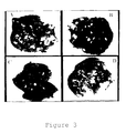
Figure 3 shows photographs of mouse lung in which
B16 melanoma cell metastasis is suppressed by Salmosin.
-
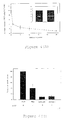
Figure 4A is a graph showing the suppressive effect
of Salmosin on proliferation of BCE (bovine capillary
endothelial) cell stimulated by bFGF (basic fibroblast
growth factor).
-
Figure 4B is a graph showing the suppressive effect
of anti-αvβ3 monoclonal antibody and Salmosin on
proliferation of BCE cells stimulated by bFGF.
-
Figure 5A is a photograph showing CAM (chick
choriollantoic membrane) angiogenesis induced by bFGF.
-
Figure 5B is a photograph showing the suppressive
effect of anti-αvβ3 monoclonal antibody on CAM
angiogenesis induced by bFGF.
-
Figure 5C is a photograph showing the suppressive
effect of Salmosin on CAM angiogenesis induced by bFGF.
-
Figure 6A is a graph showing the suppressive effect
of Salmosin or other antagonists against αvβ3 integrin
(i.e. anti-αvβ3 antibody or GRGDSP) on adhesion of
vitronectin and BCE cell.
-
Figure 6B is a graph showing the suppressive effect
of Salmosin or other antagonists against αvβ3 integrin
(i.e. anti-αvβ3 antibody or GRGDSP) on adhesion of
Salmosin and BCE cell.
-
Figure 7 shows photographs of mouse lungs showing
the suppressive effect of Salmosin on growth of
metastatic Lewis lung carcinoma (top: PBS treatment,
bottom: Salmosin treatment).
-
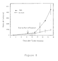
Figure 8 is a graph showing the suppressive effect
of Salmosin on growth of solid Lewis lung carcinoma.
Detailed Description of the Invention
-
The present inventors have isolated and purified
Salmosin from venom of Korean snake, Agkistrodon halys
brevicaudus, screened cDNA thereof and overexpressed
recombinant Salmosin in E.coli. They have investigated
the biological effect of both isolated and recombinant
Salmosin on tumor metastasis and tumor growth both in
vivo and in vitro. As a result, they found that:
Salmosin strongly inhibits the function of αvβ3 integrin
which is crucial to tumor angiogenesis; and, it
effectively suppresses metastasis and growth of tumors.
-
The present invention is further illustrated as
follows.
-
Salmosin was isolated and purified from venom of
Korean snake, Agkistrodon halys brevicaudus, using a
series of column chromatographies and the cDNA encoding
Salmosin was cloned from the venom gland cDNA library of
Agkistrodon halys brevicaudus. To investigate whether
Salmosin may suppress metastasis of tumor cells, Salmosin
was mixed with melanoma cells and injected into the
lateral tail vein of mouse. Salmosin did not inhibit the
proliferation of tumor cell itself in vitro, while it
inhibited tumor cell angiogenesis in a dose-dependent
manner. These findings strongly suggested that the
suppressive effect of Salmosin on tumor metastasis is
grounded on inhibition of the function of integrin
relating to tumor metastasis, not on its cytotoxicity.
In this connection, the present inventors investigated
whether Salmosin may inhibit the function of αvβ3
integrin which is associated with tumor angiogenesis
using a BCE cell proliferation test. As a result, it was
found that: Salmosin inhibits tumor-induced angiogenesis
without affecting the preexisting blood vessels or normal
angiogenesis; and, the suppressive effect of Salmosin on
BCE cell proliferation results from the direct binding of
Salmosin to the vitronectin receptor, αvβ3 integrin, on
the surface of BCE cells.
-
It was known that disintegrins can inhibit colony
formation of a metastatic tumor, but is was not clear
whether disintegrins can inhibit the growth of a
metastatic tumor that has already formed. Thus, to
examine the effect of Salmosin on the growth of
metastatic tumors, metastatic Lewis lung carcinoma cells
were injected to the mouse and after colony formation,
Salmosin was injected. As a result, it was demonstrated
that metastatic tumor growth was effectively suppressed
without any symptom of cytotoxicity and Salmosin also
inhibited the growth of a solid tumor.
A. Polypeptides
-
Polypeptides of the invention or for use in the
invention are the Salmosin polypeptide which comprises
the sequences set out in SEQ ID NO: 1, a homologue
thereof or a fragment of Salmosin or a Salmosin
homologue.
-
Polypeptides provided by the invention include
variants of SEQ ID NO: 1, including naturally occurring
allelic variants and synthetic variants which are
substantially homologous to said polypeptides and contain
RGD or KGD at positions homologous to amino acids 51 to
53 of SEQ ID No. 1. In this context, substantial
homology is regarded as a sequence which has at least
70%, e.g. 80% or 90% amino acid homology (identity) over
30 amino acids with the sequence of SEQ ID No. 1.
-
Polypeptides also include other Salmosin homologues,
and variants thereof as defined above, from other species
including animals such as snakes.
-
Polypeptides of the invention also include fragments
of the above mentioned full length polypeptides and
variants thereof, including fragments of the sequences
set out in SEQ ID No. 1. Preferred fragments include
those which include the RGD tripeptide. Suitable
fragments will be at least about 5, e.g. 10, 12, 15 or 20
amino acids in size. Polypeptide fragments of the
Salmosin proteins and allelic and species variants
thereof may contain one or more (e.g. 2, 3, 5, or 10)
substitutions, deletions or insertions, including
conserved substitutions.
-
Conserved substitutions may be made according to the
following table which indicates conservative
substitutions, where amino acids on the same block in the
second column and preferably in the same line in the
third column may be substituted for each other:
| ALIPHATIC | Non-polar | G A P |
| I L V |
| Polar - uncharged | C S T M |
| N Q |
| Polar - charged | D E |
| K R |
| AROMATIC | | H F W Y |
| OTHER | | N Q D E |
-
Polypeptides of the invention may be in a substantially
isolated form. It will be understood that the polypeptide
may be mixed with carriers or diluents which will not
interfere with the intended purpose of the polypeptide and
still be regarded as substantially isolated. A polypeptide
of the invention may also be in a substantially purified
form, in which case it will generally comprise the
polypeptide in a preparation in which more than 90%, e.g.
95%, 98% or 99% of the polypeptide in the preparation is a
polypeptide of the invention. Polypeptides of the invention
may be modified for example by the addition of histidine
residues to assist their purification or by the addition of
a signal sequence to promote their secretion from a cell.
-
Polypeptides provided by the invention may be made by
synthetic means (e.g. as described by Geysen et al., 1996) or
recombinantly, as described below. Alternatively, Salmosin
may be isolated and purified from the venom of Agkistrodon
halys brevicaudus, for example, by column chromatography.
-
Particularly preferred polypeptides of the invention
include those spanning the RGD sequence represented as amino
acids 51 to 53 in SEQ ID No. 1. Fragments as defined above
from this region containing the RGD sequence are particularly
preferred. The polypeptides and fragments thereof may
contain amino acid alterations as defined above.
-
The polypeptides of the invention may be introduced into
a cell by in situ expression of the polypeptide from a
recombinant expression vector (see below). The expression
vector optionally carries an inducible promoter to control
the expression of the polypeptide. The use of mammalian host
cells is expected to provide for such post-translational
modifications (e.g. myristolation, glycosylation, truncation,
lapidation and tyrosine, serine or threonine phosphorylation)
as may be needed to confer optimal biological activity on
recombinant expression products of the invention. Such cell
culture systems in which polypeptide of the invention are
expressed may be used in assay systems to identify candidate
substances which interfere with or enhance the functions of
the polypeptides of the invention in the cell.
B. Polynucleotides
-
In the following description polynucleotides of the
invention may comprise DNA or RNA. They may be single or
double stranded. They may also be polynucleotides which
include within them synthetic or modified nucleotides. A
number of different types of modification to oligonucleotides
are known in the art. These include methylphosphonate and
phosphorothioate backbones, addition of acridine or
polylysine chains at the 3' and/or 5' ends of the molecule.
For the purposes of the present invention, it is to be
understood that the polynucleotides described herein may be
modified by any method available in the art. Such
modifications may be carried out in order to enhance the in
vivo activity or life span of polynucleotides of the
invention.
-
Polynucleotides of the invention capable of selectively
hybridising to the DNA of SEQ ID No. 3 will be generally at
least 70%, preferably at least 80 or 90% and more preferably
at least 95% homologous to the corresponding DNA of SEQ ID
No. 3 over a region of at least 20, preferably at least 25 or
30, for instance at least 40, 60 or 100 or more contiguous
nucleotides. Preferred polynucleotides of the invention will
comprise regions encompassing the region encoding the RGD
amino acid sequence of Salmosin at nucleotides 151 to 159 of
SEQ ID No. 3. Such preferred polynucleotides will encode RGD
or KGD at nucleotides 151 to 159 of SEQ ID No. 3.
-
It is to be understood that skilled persons may, using
routine techniques, make nucleotide substitutions that do not
affect the polypeptide sequence encoded by the
polynucleotides of the invention to reflect the codon usage
of any particular host organism in which the polypeptides of
the invention are to be expressed.
-
Any combination of the above mentioned degrees of
homology and minimum sizes may be used to define
polynucleotides of the invention, with the more stringent
combinations (i.e. higher homology over longer lengths) being
preferred. Thus for example a polynucleotide which is at
least 80% homologous over 25, preferably 30 nucleotides forms
one aspect of the invention, as does a polynucleotide which
is at least 90% homologous over 40 nucleotides.
-
Polynucleotides of the invention may be used to produce
a primer, e.g. a PCR primer, a primer for an alternative
amplification reaction, a probe e.g. labelled with a
revealing label by conventional means using radioactive or
non-radioactive labels, or the polynucleotides may be cloned
into vectors. Such primers, probes and other fragments will
be at least 15, preferably at least 20, for example at least
25, 30 or 40 nucleotides in length, and are also encompassed
by the term polynucleotides of the invention as used herein.
-
Polynucleotides such as a DNA polynucleotide and primers
according to the invention may be produced recombinantly,
synthetically, or by any means available to those of skill in
the art. They may also be cloned by standard techniques.
-
In general, primers will be produced by synthetic means,
involving a step wise manufacture of the desired nucleic acid
sequence one nucleotide at a time. Techniques for
accomplishing this using automated techniques are readily
available in the art.
-
Longer polynucleotides will generally be produced using
recombinant means, for example using a PCR (polymerase chain
reaction) cloning techniques. This will involve making a
pair of primers (e.g. of about 15-30 nucleotides) to a region
of the Salmosin gene which it is desired to clone, bringing
the primers into contact with mRNA or cDNA obtained from an
animal cell, performing a polymerase chain reaction under
conditions which bring about amplification of the desired
region, isolating the amplified fragment (e.g. by purifying
the reaction mixture on an agarose gel) and recovering the
amplified DNA. The primers may be designed to contain
suitable restriction enzyme recognition sites so that the
amplified DNA can be cloned into a suitable cloning vector.
-
Such techniques may be used to obtain all or part of the
Salmosin sequence described herein. Genomic clones
containing the Salmosin gene and its introns and promoter
regions may also be obtained in an analogous manner, starting
with genomic DNA from an animal cell.
-
Although in general the techniques mentioned herein are
well known in the art, reference may be made in particular to
Sambrook et al., Molecular Cloning, A Laboratory Manual
(1989) and Ausubel et al., Current Protocols in Molecular
Biology (1995), John Wiley & Sons, Inc.
-
Polynucleotides which are not 100% homologous to the
sequences of the present invention but fall within the scope
of the invention can be obtained in a number of ways. Other
Agkistrodon halys brevicaudus allelic variants of the
Salmosin sequence described herein may be obtained for
example by probing genomic DNA libraries made from a range of
individuals, for example individuals from different
populations. In addition, other animal, particularly reptile
(e.g. snake), homologues of Salmosin may be obtained and such
homologues and fragments thereof in general will be capable
of selectively hybridising to SEQ ID No. 3. Such sequences
may be obtained by probing cDNA libraries made from dividing
cells or tissues or genomic DNA libraries from other animal
species, and probing such libraries with probes comprising
all or part of SEQ ID. 3 under conditions of medium to high
stringency (for example 0.03M sodium chloride and 0.03M
sodium citrate at from about 50°C to about 60°C). Nucleic
acid probes comprising all or part of SEQ ID No. 3 may be
used to probe cDNA libraries, preferably from toxin glands,
to obtain homologues of Salmosin.
-
Allelic variants and species homologues may also be
obtained using degenerate PCR which will use primers designed
to target sequences within the cDNA encoding Salmosin. The
primers will contain one or more degenerate positions and
will be used at stringency conditions lower than those used
for cloning sequences with single sequence primers against
known sequences. In particular, primers can be designed to
target the RGD tripeptide encoding domains described above.
-
Alternatively, such polynucleotides may be obtained by
site directed mutagenesis of the Salmosin sequence or allelic
variants thereof. This may be useful where for example
silent codon changes are required to sequences to optimise
codon preferences for a particular host cell in which the
polynucleotide sequences are being expressed. Other sequence
changes may be desired in order to introduce restriction
enzyme recognition sites, or to alter the property or
function of the polypeptides encoded by the polynucleotides.
For example, it may be desirable to introduce changes to
enhance the integrin binding properties of Salmosin encoded
by the polynucleotides.
-
The invention further provides double stranded
polynucleotides comprising a polynucleotide of the invention
and its complement.
-
Polynucleotides or primers of the invention may carry
a revealing label. Suitable labels include radioisotopes
such as 32P or 35S, enzyme labels, or other protein labels such
as biotin. Such labels may be added to polynucleotides or
primers of the invention and may be detected using by
techniques known per se.
-
The present invention also provides polynucleotides
encoding the polypeptides of the invention described below.
Because such polynucleotides will be useful as sequences for
recombinant production of polypeptides of the invention, it
is not necessary for them to be selectively hybridisable to
the sequence of SEQ ID No. 3 although this will generally be
desirable. Otherwise, such polynucleotides may be labelled,
used, and made as described above if desired. Polypeptides
of the invention are described below.
C. Vectors.
-
Polynucleotides of the invention can be incorporated
into a recombinant replicable vector. The vector may be used
to replicate the nucleic acid in a compatible host cell.
Thus in a further embodiment, the invention provides a method
of making polynucleotides of the invention by introducing a
polynucleotide of the invention into a replicable vector,
introducing the vector into a compatible host cell, and
growing the host cell under conditions which bring about
replication of the vector. The vector may be recovered from
the host cell. Suitable host cells are described below in
connection with expression vectors.
Expression Vectors.
-
Preferably, a polynucleotide of the invention in a
vector is operably linked to a regulatory sequence which is
capable of providing for the expression of the coding
sequence by the host cell, i.e. the vector is an expression
vector. The term "operably linked" refers to a juxtaposition
wherein the components described are in a relationship
permitting them to function in their intended manner. A
regulatory sequence "operably linked" to a coding sequence is
ligated in such a way that expression of the coding sequence
is achieved under condition compatible with the control
sequences.
-
The term "regulatory sequences" includes promoters and
enhancers and other expression regulation signals. These may
be selected to be compatible with the host cell for which the
expression vector is designed. For example, yeast regulatory
sequences include S. cerevisiae GAL4 and ADH promoters, S.
pombe nmt1 and adh promoters. Mammalian promoters, such as
a-actin promoters, may be used. Mammalian promoters also
include the metallothionein promoter which can upregulate
expression in response to heavy metals such as cadmium and is
thus an inducible promoter. Tissue-specific promoters, for
example neuronal cell specific may be used. Viral promoters
may also be used, for example the Moloney murine leukaemia
virus long terminal repeat (MMLV LTR), the promoter rous
sarcoma virus (RSV) LTR promoter, the SV40 promoter, the
human cytomegalovirus (CMV) IE promoter, herpes simplex virus
promoters or adenovirus promoters. All these promoters are
readily available in the art.
-
Such vectors may be transformed into a suitable host
cell as described above to provide for expression of a
polypeptide of the invention. Thus, in a further aspect the
invention provides a process for preparing polypeptides
according to the invention which comprises cultivating a host
cell transformed or transfected with an expression vector as
described above under conditions to provide for expression by
the vector of a coding sequence encoding the polypeptides,
and recovering the expressed polypeptides.
-
The vectors may be for example, plasmid, virus or phage
vectors provided with an origin of replication, optionally a
promoter for the expression of the said polynucleotide and
optionally a regulator of the promoter. The vectors may
contain one or more selectable marker genes, for example an
ampicillin resistance gene in the case of a bacterial plasmid
or a neomycin resistance gene for a mammalian vector.
Vectors may be used in vitro, for example for the production
of RNA or used to transfect or transform a host cell. The
vector may also be adapted to be used in vivo, for example in
a method of gene therapy.
-
A further embodiment of the invention provides host
cells transformed or transfected with the vectors for the
replication and expression of polynucleotides of the
invention. The cells will be chosen to be compatible with
the said vector and may for example be bacterial, yeast,
insect or mammalian.
E. Therapeutic uses
-
The invention is based on the finding that Salmosin can
inhibit the function of αvβ3 integrin and strongly inhibit
tumour angiogenesis which is essential for cancer cell growth
and metastasis without affecting proliferation of normal
endothelial cells.
-
Salmosin and fragments and homologues thereof which
inhibit integrin function may therefore be used as antitumour
agents. Polynucleotides encoding these polypeptides
may also be used inhibit tumour growth and metastasis by gene
therapy.
-
Thus the polypeptides and polynucleotides of the
invention may be used as outlined above in compounds for
treating tumours in animals or humans. Typically the
compounds are formulated for clinical administration by
mixing them with
a pharmaceutically acceptable carrier or diluent. For
example they can be formulated for topical, parenteral,
intravenous, intramuscular, subcutaneous, intraocular or
transdermal administration. Preferably, the compound is
used in an injectable form. Direct injection into the
patient's tumour is advantageous because it makes it
possible to concentrate the therapeutic effect at the level
of the affected tissues. It may therefore be mixed with
any vehicle which is pharmaceutically acceptable for an
injectable formulation, preferably for a direct injection
at the site to be treated. The pharmaceutically carrier or
diluent may be, for example, sterile or isotonic solutions.
-
The dose of compound used may be adjusted according
to various parameters, especially according to the compound
used, the age, weight and condition of the patient to be
treated, the mode of administration used, pathology of the
tumour and the required clinical regimen. As a guide, the
amount of compound administered by injection is suitably
from 0.01 mg/kg to 30 mg/kg, preferably from 0.1 mg/kg to
10 mg/kg.
-
The routes of administration and dosages described
are intended only as a guide since a skilled practitioner
will be able to determine readily the optimum route of
administration and dosage for any particular patient and
condition.
-
Compounds to be administered may include polypeptides
or nucleic acids that encode polypeptides. Nucleic acids
may be administered by, for example, lipofection or by
viral vectors. For example, the nucleic acid may form part
of a viral vector such as an adenovirus. When viral
vectors are used, in general the dose administered is
between 104 and 1014 pfu/ml, preferably 106 to 1010 pfu/ml.
The term pfu ("plaque forming unit") corresponds to the
infectivity of a virus solution and is determined by
infecting an appropriate cell culture and measuring,
generally after 48 hours, the number of plaques of infected
cells. The techniques for determining the pfu titre of a
viral solution are well documented in the literature.
-
Any cancer types may be treated by these methods, for
example leukaemias, and solid tumours such as breast,
ovary, lung, colon, pancreas, testes, liver, brain, muscle
and bone tumour.
-
The following examples illustrate the invention:
Example 1: Isolation of Salmosin from venom
-
To purify Salmosin from venom of Korean snake,
Agkistrodon halys brevicaudus, 1ml of venom was suspended
with 20mM Tris·HCl-buffer(pH 7.5, "buffer A") to a volume
of 10ml, and applied into benzamidine-Sepharose(Pharmacia-LKB,
Sweden) column which is
equilibrated with buffer A, at a flow rate of 30ml/hr.
The unbound fractions of the column were collected and
suspended with buffer A, and applied again into DEAE-Toyopearl
column (2.5×5.0cm)(Pharmacia-LKB, Sweden) at a
flow rate of 60ml/hr. The step elution was carried out
at concentrations of 25mM, 50mM, 100mM and 1M NaCl and
the active fractions were concentrated with
ultrafiltration system(Amicon, U.S.A.). The concentrated
material was applied onto TSK-G2000 column(7.5×300mm)
(Toyosoda, Japan) and eluted with PBS(phosphate buffered
saline). Active fractions were pooled, concentrated and
applied onto reverse-phase Vydac C18
column(2.5×300mm) (Vydac, U.S.A.) equilibrated with 0.1%
TFA(trifluoroacetic acid) solution. And then, the column
was alternately washed with the said TFA solution and
20%(v/v) acetonitrile solution and the protein was eluted
with a linear gradient of 20 - 60%(v/v) acetonitrile in
the said TFA solution.
Example 2: Expression and purification of recombinant
Salmosin
-
To clone cDNA encoding Salmosin, polymerase chain
reaction(PCR) was performed with the venom gland cDNA
library of Agkistrodon halys brevicaudus as a template.
In carrying out PCR, oligo d(T) was employed as 3' primer
and the nucleotide sequence(SEQ ID NO: 2) deduced from N-terminal
amino acid sequence(-EECDCG-) of Salmosin was
employed as 5' primer. PCR product of 290bp was purified
by agarose gel electrophoresis and cloned into a vector
pCRII (Invitrogen, U.S.A.). The cloned DNA sequence was
analyzed(SEQ ID NO: 3) and the amino acid sequence
translated from the said DNA sequence was consistent with
that of isolated Salmosin. For the expression of
recombinant Salmosin, an E.coli expression vector, ΔpMA-PRK153
was employed(see: Figure 1). In this vector, the
protein of interest is expressed as a fusion protein to
the phosphoribulose kinase(PRK) under the control of the
araB promoter. For the facilitation of the subcloning,
the DNA fragment generated by PCR was modified by
introducing BamHI and XhoI site. Urokinase cleavage site
was also introduced between the coding sequences of
phosphoribulose kinase and Salmosin and an expression
vector thus prepared was designated as ΔpMASIN1(see:
Figure 2A) . In Figure 2A, P araB represents araB
promoter, PRK(1-153) represents the protein comprising
amino acid sequence from 1 to 153 and UK represents the
cleavage site of urokinase. The said expression vector
was transformed in the E.coli strain MC1061. The
transformants were grown in 1L of 2X YT medium to an
absorbance of 0.3 at a wavelength of 600nm, induced with
1% arabinose(w/v) and incubated at 37 °C for 18hr.
Salmosin was expressed as an insoluble inclusion body and
the inclusion body was resuspended in a pellet wash
solution(0.5% Triton X-100, lmM EDTA, lmM DTT) and
centrifuged at 12,000rpm for 20min to remove contaminated
E.coli proteins. After washing 3 more times, the pellet
was solubilized in 20ml of 8M urea solution(8M urea, 20mM
Tris·HCl, pH 7.8, 20 µM DTT) and incubated at 4°C for
24hr. After centrifugation, the clear supernatant was
dialyzed against 4L of dialysis buffer (80mM NaCl, 20mM
Tris·HCl, pH 7.8, 0.03% SDS) at 4°C. The refolded fusion
protein was then cleaved with urokinase at a ratio of
400mg of fusion protein and 1 U of urokinase at room
temperature for 30min. 10mg of cleaved protein was
loaded onto DEAE-Toyopearl column equilibrated with 20mM
Tris·HCl(pH 8.0) buffer and eluted with the same buffer
containing 50mM NaCl. Active fractions were pooled,
concentrated and applied onto TSK-G200 HPLC column(2.15 X
30cm) and eluted with PBS. Subsequently, to remove the
inappropriately cleaved Salmosin, active fractions were
applied onto semi-preparative C18 HPLC column(1.0 X 20cm)
and eluted with a linear gradient of 20-40%(v/v)
acetonitrile in 0.1% TFA solution.
Example 3: Comparative assay of recombinant Salmosin and
isolated Salmosin
-
To compare physicochemical natures and biological
activities of Salmosin prepared in Examples 1 and 2
described above, N-terminal sequencing, the platelet
aggregation assay, and SDS-gel electrophoresis were
carried out, respectively. The results showed that N-terminal
sequence of recombinant Salmosin is initiated
with the sequence of EAGEEC, which is consistent with
that of isolated Salmosin and the suppressive acitivity
of platelet aggregation is also identical. And, the
result of SDS-PAGE indicated that the size of the
recombianant Salmosin is exactly the same as that of the
isolated salmosin(see: Figure 2B). In Figure 2B, lane 1
is the recombinant Salmosin, lane M is size marker, and
lane 2 is the isolated Salmosin. Accordingly, it was
clearly demonstrated that the recombinant Salmosin is
correctly refolded and has the same biological activity
as the isolated Salmosin.
Example 4: Inhibition of metastasis by Salmosin
-
To investigate the inhibitory effect of Salmosin on
tumor metastasis
in vivo, Salmosin was coinjected with
B16 melanoma cells into C57BL/6 mouse(Charles river,
Japan). B16 melanoma cells were detached with 0.02% EDTA
and resuspended gently to 7.5×10
5/ml in RPMI-1640 medium
without serum. Salmosin(250, 500, 1,250µg/kg mouse) was
then mixed with the cells to prepare single-cell
suspension containing the indicated amount of Salmosin or
PBS, and the single-cell suspension of 200µl aliquots
were injected into the lateral tail veins of mice. 14
days later, the mice were sacrificed and the number of
lung melanoma colonies was counted by dissecting
microscope. As a result, it was detected that metastatic
colonies were dramatically reduced when compared with the
PBS-treated control group in the lungs of C57BL/6
mice(
see: Table 1). As shown in Table 1, the inhibition
of colony formation by Salmosin was in dose-dependent
manner; lower dose(250µg/kg mouse) of Salmosin was able
to inhibit the colony formation in the lung, but few
colonies were detectable when salmosin was administered
with higher dose. These findings are consistent with
histochemical analysis(
see: Figure 3: A, normal lung; B,
PBS treatment; C, 250µg Salmosin/kg treatment; D, 1,250µg
Salmosin/kg treatment)
| Inhibition of metastasis by the treatment of Salmosin |
| Salmosin (µg/kg mouse) | Number of mouse | Average number of lung tumor colony | Percentage of inhibition (%) |
| 0 | 8 | 144 ± 40 | 0 |
| 250 | 7 | 49 ± 22 | 66 |
| 500 | 7 | 3 ± 2 | 98 |
| 1,250 | 6 | 1 ± 1 | 99 |
-
Further, it was demonstrated that the inhibition
of metastasis by Salmosin is not caused by cytotoxicity,
based on the experimental fact that incubation of the B16
melanoma cells in vitro with Salmosin did not affect
their subsequent proliferation rate.
-
These experimental evidences strongly suggested
that Salmosin inhibits tumor metastasis by acting as an
antagonist of integrin receptors on the surface of tumor
cells, based on the suppressive effect of Salmosin on
adhesion and invasion of B16 melanoma cell to ECM.
Example 5: Inhibition of angiogenesis by Salmosin
Example 5-1: BCE(bovine capillary endothelial) cell
proliferation assay
-
The suppressive effect of Salmosin on angiogensis
was examined by employing BCE cell proliferation assay
system. Primary culture of BCE cell from bovine adrenal
was prepared, and then the cells were maintained in
Dulbeco's minimum essential medium("DMEM") containing
3ng/ml of bFGF supplemented with 10% fetal calf serum.
The said BCE cells were plated onto gelatinized 24-well
culture plates and incubated at 37°C, 5% CO2 for 24hr.
After incubation, the medium was replaced with DMEM
containing 5% fetal calf serum, and Salmosin was added to
the medium. After 20 min of incubation, bFGF(1ng/ml)
dissolved in the said medium was treated to the cells and
after 72hr, cells were detached with trypsin and the
number of cells was counted. As a result, it was found
that Salmosin is able to inhibit the proliferation of BCE
cells stimulated by bFGF in a dose-dependent manner.
Half-maximal inhibition was observed with Salmosin
concentration of 0.1-0.2µg/ml corresponding to 13-27nM(see:
Figure 4A). In addition, the bFGF-stimulated
BCE cells undergo remarkable morphological change into
spherical shape by the treatment of Salmosin. However,
it was observed that the cell proliferation is not
inhibited in the absence of bFGF when the cells were
treated with 20µg/ml Salmosin which was required for
maximal inhibition of bFGF-induced proliferation(see:
graph in box of Figure 4A). On the other hand, it was
examined that anti-αvβ3 monoclonal antibody effectively
inhibits bFGF-stimulated proliferation of BCE cells(see:
Figure 4B).
Example 5-2: CAM(chick chorioallantoic membrane) assay
-
The suppressive effect of Salmosin on tumor-induced
angiogenesis in vivo, was examined by CAM (chick
chorioallantoic membrane) assay: Three-day-old fertilized
eggs were carefully cracked and sealed with transparent
tapes. For 10 days of incubation at 37°C with 60%
humidity, the said fertilized eggs were developed into
embryo. After that, 6ng of bFGF was implanted on the CAM
of individual embryo to induce neovascularization. After
24hr of incubation, 5µg of Salmosin, 5µg of anti-αvβ3
moniclonal antibody(positive control) or PBS(phosphate
buffered saline, negative control) was treated on the CAM
of individual embryo. And after 72hr, blood vessels on
CAM were observed under the stereomicroscope(see: Figures
5A, 5B and 5C). It was observed that: there was no
visible change in the neovascularization when PBS was
treated(see: Figure 5A), while it was inhibited when
Salmosin or anti-αvβ3 moniclonal antibody was
treated(see: Figures 5B and 5C). Especially, it was
found that Salmosin breaks new blood vessels which are
induced by bFGF, indicating that Salmosin inhibits
tumor-induced angiogenesis, without affecting the
preexisting blood vessels or the angiogenesis that is a
normal physiological phenomenon.
Example 6: Inhibition of BCE cell adhesion by Salmosin
-
To investigate whether the BCE cell proliferation
is inhibited by direct-binding of Salmosin to αvβ3
integrin which is a vitronectin receptor on the surface
of BCE cell, the capability of Salmosin which inhibits
BCE cell adhesion to vitronectin was examined: 96-well
microplates were coated with Salmosin(1µg/well) and
vitronectin(0.5µg/well) in PBS at 4°C for 16hr. The said
microplates were washed and incubated for lhr with
10mg/ml of heat-denatured bovine serum albumin to block
remaining protein binding sites, and washed with PBS
before use. BCE cells were detached with trypsin-EDTA,
washed three times in PBS, and resuspended in serum-free
DMEM. 5×10⁴ of cells were preincubated with Salmosin,
anti-αvβ3 monoclonal antibody, synthetic RGD
peptide(GRGDSP) or synthetic RGE peptide(GRGETP) for
20min and aliquoted to each well of the said plate, then
further incubated at 37°C for lhr in 5% CO2, 95% air.
Unbound cells were removed by washing with PBS, then the
cells which bound to the plate were fixed and stained
with Coomassie Blue. Absorbance at 540nm of the
individual well was measured to determine the relative
cell number. As a result, it was found that: adhesion of
BCE cell to vitronectin can be highly suppressed by
preincubating the bFGF-stimulated cells with
Salmosin(see: Figure 6A); anti-αvβ3 monoclonal antibody
strongly inhibits the cell adhesion to Salmosin(see:
Figure 6B); and, synthetic RGD peptide(GRGDSP) is also
able to prevent the cells from adhering to either
vitronectin or Salmosin(see: Figures 6A and 6B). These
results clearly demonstrated that Salmosin binds to αvβ3
integrin on BCE cells and thereby blocks the integrin-mediated
cell adhesion.
Example 7: Inhibition of metastatic tumor growth by
Salmosin
-
It has been reported that disintegrins suppress
the colony formation of lung tumor by inhibiting the
attachment of tumor cells to endothelium. However, it is
not clear whether the disintegrins inhibit the growth of
lung metastatic tumor or not. To investigate the
suppressive effect of Salmosin on metastatic tumor growth,
1.5×10
5 of Lewis lung carcinoma cells obtained from
ATCC(Rockville, U.S.A.) were injected into the lateral
tail veins of 8-week-old male C57BL/6 mice, and
metastatic colonies were developed in mice. 4 days later,
Salmosin was administered intravenously to the mice once
a day with a dose of 1.25mg/kg/day; 4 weeks later, mice
were sacrificed and lungs were removed; the number of
lung tumor colony was counted under the dissecting
microscope. and there was no recognizable symptom of
toxicity in any mouse tested. As a result, it was
demostrated that Salmosin remarkably inhibits the
metastatic tumor growth(
see: Table 2). In this regard,
the suppressive effect of Salmosin on the metastatic
tumor growth could be explained by binding of Salmosin to
αvβ3 integrin which is essential for tumor angiogenesis.
| Growth inhibition of pulmonary metastatic Lewis lung carcinoma by Salmosin treatment |
| Salmosin (mg/kg mouse) | Number of mouse | Average number of lung tumor colony | Inhibition (%) |
| o | 4 | 15 ± 6 | 0 |
| 4 | 4 | 0.8 ± 0.7 | 93 |
-
Further, histochemical analysis was carried out to
investigate a suppressive effect of Salmosin on the
pulmonary metastatic tumor. The said lung tissue was
fixed for 4hr in Bouin's soution and embedded in paraffin
in accordance with the standard procedure. Sections of 4
µm thick were permeabilized with trypsin at 37°C for 10
min and washed with PBS. The said sections were stained
with hematoxylin and eosin, and then mounted. It was
observed that few of tumor colonies were detected in the
lungs of Salmosin-treated animal(see: Figure 7), which is
quite different from those of PBS-treated control mice.
Figure 7 shows photographs of ×400 magnification(left)
and ×200 magnification(right), respectively, suggesting
that the suppressive effect of Salmosin on metastatic
tumor growth is closely related to the suppression of
angiogenesis which is necessary for secondary tumor
growth.
Example 8: Inhibition of solid tumor growth by Salmosin
-
To examine the role of Salmosin in the growth of
solid tumor, Lewis lung carcinoma cells(1×10⁶) were
injected subcutaneously into dorsal midline of C57BL/6
mice and grown to a mass of at least 100-200mm3. After
the mice were randomized into two groups, one group
received injections of Salmosin(lOmg/kg mouse) in PBS via
subcutaneous injection at a site distant from the tumor
once daily and the other group as a control received
injections of PBS alone. The size of the tumors in all
groups were measured at the same time everyday and the
experiments were terminated when the control mice began
to die. As a result, it was observed that the growth of
solid tumor is strongly inhibited by the treatment of
Samosin(see: Figure 8)
Administration and effective dose
-
Though the effective dose of Salmosin is variable
depending on age, body weight of patient and progression
of disease, it is preferable to administer parenterally
0.5 to 10mg/kg/day in a single dose, and which may be
individualized by experience of the skilled in the art.
Acute toxicity test
-
To examine the acute toxicity of Salmosin, Salmosin
was injected into male C57BL/6 mouse subcutaneously and
the deads were counted to determine LD50 for 7days. As a
result, LD50 is determined as about 1,000mg/kg,
indicating that the anti-tumor agent comprising Salmosin
is sufficiently safe within the range of effective dose.
-
The pharmaceutical composition of the present
invention which comprises an active ingredient of
Salmosin and pharmaceutically acceptable carrier may be
administrated as an injection formula. The injection
formula may further comprise isotonic aqueous solution or
suspension, and the pharmaceutically acceptable carrier
covers preservatives, stabilizers, wetting agents,
emulsifiers, salts for changing osmotic pressure or
buffers.
-
While the present invention has been shown and
described with reference to the particular embodiments
employing melanoma cells and Lewis lung carcinoma cells
as tested tumor cells, the anti-tumor agent of the
invention may be effective for various tumors such as
skin cancer, laryngeal cancer, uterine cancer, colon
cancer, lung cancer and bone marrow cancer.
-
As clearly illustrated and demonstrated as above,
the present invention provides an anti-tumor agent
comprising an active ingredient of Samosin. In
accordance with the present invention, it was
demonstrated that Salmosin is a novel disintegrin which
blocks the function of αvβ3 integrin and strongly
inhibits tumor angiogenesis, tumor metastasis as well as
growth of solid tumor. Salmosin does not exhibit
cytotoxicity within an effective dose range where tumor
growth is efficiently suppressed without any untoward
effect on preexisting blood vessels and normal
angiogenesis. Accordingly, Salmosin can be applied for
the development of potent anti-tumor drugs which are
effective for various types of cancers.
Annex to the description
-