EP0950710A2 - Zellen, die ein chimärischen Rezeptor exprimieren, und deren Verwendungen - Google Patents
Zellen, die ein chimärischen Rezeptor exprimieren, und deren Verwendungen Download PDFInfo
- Publication number
- EP0950710A2 EP0950710A2 EP99106794A EP99106794A EP0950710A2 EP 0950710 A2 EP0950710 A2 EP 0950710A2 EP 99106794 A EP99106794 A EP 99106794A EP 99106794 A EP99106794 A EP 99106794A EP 0950710 A2 EP0950710 A2 EP 0950710A2
- Authority
- EP
- European Patent Office
- Prior art keywords
- gene
- cells
- receptor
- chimera
- hgh
- Prior art date
- Legal status (The legal status is an assumption and is not a legal conclusion. Google has not performed a legal analysis and makes no representation as to the accuracy of the status listed.)
- Withdrawn
Links
- 108010000521 Human Growth Hormone Proteins 0.000 claims abstract description 61
- 102000002265 Human Growth Hormone Human genes 0.000 claims abstract description 61
- 239000000854 Human Growth Hormone Substances 0.000 claims abstract description 61
- 108090000623 proteins and genes Proteins 0.000 claims abstract description 55
- 108020003175 receptors Proteins 0.000 claims abstract description 31
- 102000005962 receptors Human genes 0.000 claims abstract description 29
- 230000001419 dependent effect Effects 0.000 claims abstract description 24
- 230000001086 cytosolic effect Effects 0.000 claims abstract description 16
- 238000012216 screening Methods 0.000 claims abstract description 10
- 210000004102 animal cell Anatomy 0.000 claims abstract description 8
- 239000013613 expression plasmid Substances 0.000 claims abstract description 8
- 239000005556 hormone Substances 0.000 claims abstract description 8
- 229940088597 hormone Drugs 0.000 claims abstract description 8
- 108010070774 Thrombopoietin Receptors Proteins 0.000 claims abstract description 5
- 102100034196 Thrombopoietin receptor Human genes 0.000 claims abstract description 5
- 102000004169 proteins and genes Human genes 0.000 claims abstract description 5
- 239000000556 agonist Substances 0.000 claims abstract description 4
- 239000005557 antagonist Substances 0.000 claims abstract description 4
- 210000004027 cell Anatomy 0.000 claims description 119
- 102100020948 Growth hormone receptor Human genes 0.000 claims description 46
- 108010068542 Somatotropin Receptors Proteins 0.000 claims description 46
- 239000013612 plasmid Substances 0.000 claims description 31
- 238000000034 method Methods 0.000 claims description 29
- 230000000694 effects Effects 0.000 claims description 11
- 238000010276 construction Methods 0.000 claims description 8
- 230000002068 genetic effect Effects 0.000 claims description 2
- 102000018997 Growth Hormone Human genes 0.000 claims 1
- 108010051696 Growth Hormone Proteins 0.000 claims 1
- 150000001413 amino acids Chemical class 0.000 claims 1
- 238000012217 deletion Methods 0.000 claims 1
- 230000037430 deletion Effects 0.000 claims 1
- 239000000122 growth hormone Substances 0.000 claims 1
- 108090000765 processed proteins & peptides Proteins 0.000 claims 1
- 108010005905 delta-hGHR Proteins 0.000 abstract description 5
- 239000000243 solution Substances 0.000 description 39
- 239000002609 medium Substances 0.000 description 36
- 108091003079 Bovine Serum Albumin Proteins 0.000 description 25
- 239000012091 fetal bovine serum Substances 0.000 description 24
- 108020004414 DNA Proteins 0.000 description 21
- 238000006243 chemical reaction Methods 0.000 description 20
- 238000004458 analytical method Methods 0.000 description 18
- 239000000203 mixture Substances 0.000 description 17
- 239000002299 complementary DNA Substances 0.000 description 16
- 108010006025 bovine growth hormone Proteins 0.000 description 14
- 210000004185 liver Anatomy 0.000 description 14
- 108091008146 restriction endonucleases Proteins 0.000 description 14
- 241000588724 Escherichia coli Species 0.000 description 13
- 239000012980 RPMI-1640 medium Substances 0.000 description 13
- 238000002156 mixing Methods 0.000 description 13
- 239000000047 product Substances 0.000 description 12
- 238000012408 PCR amplification Methods 0.000 description 11
- 230000006539 extracellular acidification Effects 0.000 description 10
- 230000002062 proliferating effect Effects 0.000 description 9
- 238000001712 DNA sequencing Methods 0.000 description 8
- 102000036693 Thrombopoietin Human genes 0.000 description 8
- 108010041111 Thrombopoietin Proteins 0.000 description 8
- 230000001605 fetal effect Effects 0.000 description 8
- 229920000936 Agarose Polymers 0.000 description 7
- 229920002307 Dextran Polymers 0.000 description 7
- 239000011543 agarose gel Substances 0.000 description 7
- 229960000723 ampicillin Drugs 0.000 description 7
- AVKUERGKIZMTKX-NJBDSQKTSA-N ampicillin Chemical compound C1([C@@H](N)C(=O)N[C@H]2[C@H]3SC([C@@H](N3C2=O)C(O)=O)(C)C)=CC=CC=C1 AVKUERGKIZMTKX-NJBDSQKTSA-N 0.000 description 7
- 230000001580 bacterial effect Effects 0.000 description 7
- 239000000872 buffer Substances 0.000 description 7
- 239000003153 chemical reaction reagent Substances 0.000 description 7
- 230000012010 growth Effects 0.000 description 7
- 238000012163 sequencing technique Methods 0.000 description 7
- 230000004663 cell proliferation Effects 0.000 description 6
- 230000029087 digestion Effects 0.000 description 6
- 238000001962 electrophoresis Methods 0.000 description 6
- ZMMJGEGLRURXTF-UHFFFAOYSA-N ethidium bromide Chemical compound [Br-].C12=CC(N)=CC=C2C2=CC=C(N)C=C2[N+](CC)=C1C1=CC=CC=C1 ZMMJGEGLRURXTF-UHFFFAOYSA-N 0.000 description 6
- 229960005542 ethidium bromide Drugs 0.000 description 6
- 238000002360 preparation method Methods 0.000 description 6
- 239000000126 substance Substances 0.000 description 6
- 239000006228 supernatant Substances 0.000 description 6
- 108091028043 Nucleic acid sequence Proteins 0.000 description 5
- 230000009471 action Effects 0.000 description 5
- 239000002775 capsule Substances 0.000 description 5
- 238000010367 cloning Methods 0.000 description 5
- 238000010494 dissociation reaction Methods 0.000 description 5
- 230000005593 dissociations Effects 0.000 description 5
- 108020004999 messenger RNA Proteins 0.000 description 5
- 239000002773 nucleotide Substances 0.000 description 5
- 125000003729 nucleotide group Chemical group 0.000 description 5
- 230000020477 pH reduction Effects 0.000 description 5
- 230000004044 response Effects 0.000 description 5
- 238000000926 separation method Methods 0.000 description 5
- 210000002966 serum Anatomy 0.000 description 5
- 238000009331 sowing Methods 0.000 description 5
- 238000010186 staining Methods 0.000 description 5
- 238000009281 ultraviolet germicidal irradiation Methods 0.000 description 5
- 108010014303 DNA-directed DNA polymerase Proteins 0.000 description 4
- 102000016928 DNA-directed DNA polymerase Human genes 0.000 description 4
- 108010006785 Taq Polymerase Proteins 0.000 description 4
- 238000003556 assay Methods 0.000 description 4
- 230000015572 biosynthetic process Effects 0.000 description 4
- 230000022131 cell cycle Effects 0.000 description 4
- 239000013256 coordination polymer Substances 0.000 description 4
- 238000012869 ethanol precipitation Methods 0.000 description 4
- BRZYSWJRSDMWLG-CAXSIQPQSA-N geneticin Natural products O1C[C@@](O)(C)[C@H](NC)[C@@H](O)[C@H]1O[C@@H]1[C@@H](O)[C@H](O[C@@H]2[C@@H]([C@@H](O)[C@H](O)[C@@H](C(C)O)O2)N)[C@@H](N)C[C@H]1N BRZYSWJRSDMWLG-CAXSIQPQSA-N 0.000 description 4
- 239000005457 ice water Substances 0.000 description 4
- 238000010348 incorporation Methods 0.000 description 4
- 239000006166 lysate Substances 0.000 description 4
- 239000011535 reaction buffer Substances 0.000 description 4
- 238000003786 synthesis reaction Methods 0.000 description 4
- 230000009466 transformation Effects 0.000 description 4
- 238000005406 washing Methods 0.000 description 4
- 229920001817 Agar Polymers 0.000 description 3
- 102000004127 Cytokines Human genes 0.000 description 3
- 108090000695 Cytokines Proteins 0.000 description 3
- 102000012410 DNA Ligases Human genes 0.000 description 3
- 108010061982 DNA Ligases Proteins 0.000 description 3
- 108091034057 RNA (poly(A)) Proteins 0.000 description 3
- 239000008272 agar Substances 0.000 description 3
- 238000003776 cleavage reaction Methods 0.000 description 3
- 102000003675 cytokine receptors Human genes 0.000 description 3
- 108010057085 cytokine receptors Proteins 0.000 description 3
- 238000000605 extraction Methods 0.000 description 3
- 239000012634 fragment Substances 0.000 description 3
- 239000000499 gel Substances 0.000 description 3
- 238000003780 insertion Methods 0.000 description 3
- 230000037431 insertion Effects 0.000 description 3
- 239000000463 material Substances 0.000 description 3
- 238000005259 measurement Methods 0.000 description 3
- 239000012528 membrane Substances 0.000 description 3
- 238000010899 nucleation Methods 0.000 description 3
- 230000035755 proliferation Effects 0.000 description 3
- 238000011160 research Methods 0.000 description 3
- 230000007017 scission Effects 0.000 description 3
- 230000019491 signal transduction Effects 0.000 description 3
- 210000001519 tissue Anatomy 0.000 description 3
- 108091032973 (ribonucleotides)n+m Proteins 0.000 description 2
- NKDFYOWSKOHCCO-YPVLXUMRSA-N 20-hydroxyecdysone Chemical compound C1[C@@H](O)[C@@H](O)C[C@]2(C)[C@@H](CC[C@@]3([C@@H]([C@@](C)(O)[C@H](O)CCC(C)(O)C)CC[C@]33O)C)C3=CC(=O)[C@@H]21 NKDFYOWSKOHCCO-YPVLXUMRSA-N 0.000 description 2
- 235000010585 Ammi visnaga Nutrition 0.000 description 2
- 244000153158 Ammi visnaga Species 0.000 description 2
- 241000283690 Bos taurus Species 0.000 description 2
- 108020004635 Complementary DNA Proteins 0.000 description 2
- 241000701022 Cytomegalovirus Species 0.000 description 2
- KCXVZYZYPLLWCC-UHFFFAOYSA-N EDTA Chemical compound OC(=O)CN(CC(O)=O)CCN(CC(O)=O)CC(O)=O KCXVZYZYPLLWCC-UHFFFAOYSA-N 0.000 description 2
- 241000283073 Equus caballus Species 0.000 description 2
- LFQSCWFLJHTTHZ-UHFFFAOYSA-N Ethanol Chemical compound CCO LFQSCWFLJHTTHZ-UHFFFAOYSA-N 0.000 description 2
- 208000034454 F12-related hereditary angioedema with normal C1Inh Diseases 0.000 description 2
- ZHNUHDYFZUAESO-UHFFFAOYSA-N Formamide Chemical compound NC=O ZHNUHDYFZUAESO-UHFFFAOYSA-N 0.000 description 2
- 229930182566 Gentamicin Natural products 0.000 description 2
- CEAZRRDELHUEMR-URQXQFDESA-N Gentamicin Chemical compound O1[C@H](C(C)NC)CC[C@@H](N)[C@H]1O[C@H]1[C@H](O)[C@@H](O[C@@H]2[C@@H]([C@@H](NC)[C@@](C)(O)CO2)O)[C@H](N)C[C@@H]1N CEAZRRDELHUEMR-URQXQFDESA-N 0.000 description 2
- WQZGKKKJIJFFOK-GASJEMHNSA-N Glucose Natural products OC[C@H]1OC(O)[C@H](O)[C@@H](O)[C@@H]1O WQZGKKKJIJFFOK-GASJEMHNSA-N 0.000 description 2
- 102000042092 Glucose transporter family Human genes 0.000 description 2
- 108091052347 Glucose transporter family Proteins 0.000 description 2
- 108090000723 Insulin-Like Growth Factor I Proteins 0.000 description 2
- 102100034343 Integrase Human genes 0.000 description 2
- 101001033276 Mus musculus Interleukin-3 Proteins 0.000 description 2
- 108091007960 PI3Ks Proteins 0.000 description 2
- UBXIJOJXUFYNRG-RJKBCLGNSA-N PIP[3'](17:0/20:4(5Z,8Z,11Z,14Z)) Chemical compound CCCCC\C=C/C\C=C/C\C=C/C\C=C/CCCC(=O)O[C@H](COC(=O)CCCCCCCCCCCCCCCC)COP(O)(=O)O[C@H]1C(O)C(O)C(O)[C@@H](OP(O)(O)=O)C1O UBXIJOJXUFYNRG-RJKBCLGNSA-N 0.000 description 2
- ISWSIDIOOBJBQZ-UHFFFAOYSA-N Phenol Chemical compound OC1=CC=CC=C1 ISWSIDIOOBJBQZ-UHFFFAOYSA-N 0.000 description 2
- 102000003993 Phosphatidylinositol 3-kinases Human genes 0.000 description 2
- 108090000430 Phosphatidylinositol 3-kinases Proteins 0.000 description 2
- 102000004022 Protein-Tyrosine Kinases Human genes 0.000 description 2
- 108090000412 Protein-Tyrosine Kinases Proteins 0.000 description 2
- 239000006146 Roswell Park Memorial Institute medium Substances 0.000 description 2
- 239000007984 Tris EDTA buffer Substances 0.000 description 2
- 238000002835 absorbance Methods 0.000 description 2
- 230000004913 activation Effects 0.000 description 2
- 238000010804 cDNA synthesis Methods 0.000 description 2
- 238000011088 calibration curve Methods 0.000 description 2
- 238000004113 cell culture Methods 0.000 description 2
- 230000011712 cell development Effects 0.000 description 2
- 238000005119 centrifugation Methods 0.000 description 2
- 229960005091 chloramphenicol Drugs 0.000 description 2
- WIIZWVCIJKGZOK-RKDXNWHRSA-N chloramphenicol Chemical compound ClC(Cl)C(=O)N[C@H](CO)[C@H](O)C1=CC=C([N+]([O-])=O)C=C1 WIIZWVCIJKGZOK-RKDXNWHRSA-N 0.000 description 2
- 230000000052 comparative effect Effects 0.000 description 2
- 230000000295 complement effect Effects 0.000 description 2
- 239000000539 dimer Substances 0.000 description 2
- 238000009585 enzyme analysis Methods 0.000 description 2
- 238000012215 gene cloning Methods 0.000 description 2
- 229960002518 gentamicin Drugs 0.000 description 2
- 239000008103 glucose Substances 0.000 description 2
- 208000016861 hereditary angioedema type 3 Diseases 0.000 description 2
- 230000010354 integration Effects 0.000 description 2
- 239000003446 ligand Substances 0.000 description 2
- 230000007246 mechanism Effects 0.000 description 2
- 239000011259 mixed solution Substances 0.000 description 2
- 238000006366 phosphorylation reaction Methods 0.000 description 2
- 238000000746 purification Methods 0.000 description 2
- 238000003757 reverse transcription PCR Methods 0.000 description 2
- 238000013391 scatchard analysis Methods 0.000 description 2
- 230000035939 shock Effects 0.000 description 2
- 238000003756 stirring Methods 0.000 description 2
- 239000000725 suspension Substances 0.000 description 2
- 238000010998 test method Methods 0.000 description 2
- 238000012360 testing method Methods 0.000 description 2
- 230000002463 transducing effect Effects 0.000 description 2
- XLYOFNOQVPJJNP-UHFFFAOYSA-N water Chemical compound O XLYOFNOQVPJJNP-UHFFFAOYSA-N 0.000 description 2
- DGVVWUTYPXICAM-UHFFFAOYSA-N β‐Mercaptoethanol Chemical compound OCCS DGVVWUTYPXICAM-UHFFFAOYSA-N 0.000 description 2
- BFSVOASYOCHEOV-UHFFFAOYSA-N 2-diethylaminoethanol Chemical compound CCN(CC)CCO BFSVOASYOCHEOV-UHFFFAOYSA-N 0.000 description 1
- 229930024421 Adenine Natural products 0.000 description 1
- GFFGJBXGBJISGV-UHFFFAOYSA-N Adenine Chemical compound NC1=NC=NC2=C1N=CN2 GFFGJBXGBJISGV-UHFFFAOYSA-N 0.000 description 1
- 238000002965 ELISA Methods 0.000 description 1
- 230000035519 G0 Phase Effects 0.000 description 1
- 101150020003 GHR gene Proteins 0.000 description 1
- 108700039691 Genetic Promoter Regions Proteins 0.000 description 1
- 108091006905 Human Serum Albumin Proteins 0.000 description 1
- 102000008100 Human Serum Albumin Human genes 0.000 description 1
- 206010062767 Hypophysitis Diseases 0.000 description 1
- 101710203526 Integrase Proteins 0.000 description 1
- ZDXPYRJPNDTMRX-VKHMYHEASA-N L-glutamine Chemical compound OC(=O)[C@@H](N)CCC(N)=O ZDXPYRJPNDTMRX-VKHMYHEASA-N 0.000 description 1
- 229930182816 L-glutamine Natural products 0.000 description 1
- OUYCCCASQSFEME-QMMMGPOBSA-N L-tyrosine Chemical compound OC(=O)[C@@H](N)CC1=CC=C(O)C=C1 OUYCCCASQSFEME-QMMMGPOBSA-N 0.000 description 1
- 102000018697 Membrane Proteins Human genes 0.000 description 1
- 108010052285 Membrane Proteins Proteins 0.000 description 1
- 102000016943 Muramidase Human genes 0.000 description 1
- 108010014251 Muramidase Proteins 0.000 description 1
- 108010062010 N-Acetylmuramoyl-L-alanine Amidase Proteins 0.000 description 1
- 229920002274 Nalgene Polymers 0.000 description 1
- 229930193140 Neomycin Natural products 0.000 description 1
- 102000016978 Orphan receptors Human genes 0.000 description 1
- 108070000031 Orphan receptors Proteins 0.000 description 1
- 241000283973 Oryctolagus cuniculus Species 0.000 description 1
- 108091000080 Phosphotransferase Proteins 0.000 description 1
- 108010021757 Polynucleotide 5'-Hydroxyl-Kinase Proteins 0.000 description 1
- 102000008422 Polynucleotide 5'-hydroxyl-kinase Human genes 0.000 description 1
- 108010002519 Prolactin Receptors Proteins 0.000 description 1
- 102100029000 Prolactin receptor Human genes 0.000 description 1
- 108010092799 RNA-directed DNA polymerase Proteins 0.000 description 1
- 241000700159 Rattus Species 0.000 description 1
- 102100037968 Ribonuclease inhibitor Human genes 0.000 description 1
- FAPWRFPIFSIZLT-UHFFFAOYSA-M Sodium chloride Chemical compound [Na+].[Cl-] FAPWRFPIFSIZLT-UHFFFAOYSA-M 0.000 description 1
- 102000013275 Somatomedins Human genes 0.000 description 1
- 229920004890 Triton X-100 Polymers 0.000 description 1
- 239000013504 Triton X-100 Substances 0.000 description 1
- 208000021017 Weight Gain Diseases 0.000 description 1
- 238000011481 absorbance measurement Methods 0.000 description 1
- 239000013543 active substance Substances 0.000 description 1
- 229960000643 adenine Drugs 0.000 description 1
- 210000001789 adipocyte Anatomy 0.000 description 1
- 230000003321 amplification Effects 0.000 description 1
- 230000003698 anagen phase Effects 0.000 description 1
- 230000008901 benefit Effects 0.000 description 1
- 230000033228 biological regulation Effects 0.000 description 1
- 229940098773 bovine serum albumin Drugs 0.000 description 1
- 238000004364 calculation method Methods 0.000 description 1
- 210000000845 cartilage Anatomy 0.000 description 1
- 230000024245 cell differentiation Effects 0.000 description 1
- 230000010261 cell growth Effects 0.000 description 1
- 210000000170 cell membrane Anatomy 0.000 description 1
- 238000001516 cell proliferation assay Methods 0.000 description 1
- 230000001413 cellular effect Effects 0.000 description 1
- 230000036755 cellular response Effects 0.000 description 1
- 210000004978 chinese hamster ovary cell Anatomy 0.000 description 1
- 238000013377 clone selection method Methods 0.000 description 1
- 150000001875 compounds Chemical class 0.000 description 1
- 238000012790 confirmation Methods 0.000 description 1
- 238000004132 cross linking Methods 0.000 description 1
- 230000037029 cross reaction Effects 0.000 description 1
- 239000012228 culture supernatant Substances 0.000 description 1
- 125000000151 cysteine group Chemical group N[C@@H](CS)C(=O)* 0.000 description 1
- SUYVUBYJARFZHO-RRKCRQDMSA-N dATP Chemical compound C1=NC=2C(N)=NC=NC=2N1[C@H]1C[C@H](O)[C@@H](COP(O)(=O)OP(O)(=O)OP(O)(O)=O)O1 SUYVUBYJARFZHO-RRKCRQDMSA-N 0.000 description 1
- SUYVUBYJARFZHO-UHFFFAOYSA-N dATP Natural products C1=NC=2C(N)=NC=NC=2N1C1CC(O)C(COP(O)(=O)OP(O)(=O)OP(O)(O)=O)O1 SUYVUBYJARFZHO-UHFFFAOYSA-N 0.000 description 1
- 230000008021 deposition Effects 0.000 description 1
- 230000004069 differentiation Effects 0.000 description 1
- 238000007865 diluting Methods 0.000 description 1
- 238000010790 dilution Methods 0.000 description 1
- 239000012895 dilution Substances 0.000 description 1
- 239000012154 double-distilled water Substances 0.000 description 1
- 239000013604 expression vector Substances 0.000 description 1
- 239000000706 filtrate Substances 0.000 description 1
- 230000005251 gamma ray Effects 0.000 description 1
- 230000003450 growing effect Effects 0.000 description 1
- 238000010438 heat treatment Methods 0.000 description 1
- 108091008039 hormone receptors Proteins 0.000 description 1
- 238000000338 in vitro Methods 0.000 description 1
- 238000001727 in vivo Methods 0.000 description 1
- 230000003993 interaction Effects 0.000 description 1
- 230000003834 intracellular effect Effects 0.000 description 1
- 238000011835 investigation Methods 0.000 description 1
- 150000002632 lipids Chemical class 0.000 description 1
- 210000005229 liver cell Anatomy 0.000 description 1
- 239000012160 loading buffer Substances 0.000 description 1
- 229960000274 lysozyme Drugs 0.000 description 1
- 239000004325 lysozyme Substances 0.000 description 1
- 235000010335 lysozyme Nutrition 0.000 description 1
- 238000012423 maintenance Methods 0.000 description 1
- 238000004519 manufacturing process Methods 0.000 description 1
- 230000004060 metabolic process Effects 0.000 description 1
- 230000021332 multicellular organism growth Effects 0.000 description 1
- 229960004927 neomycin Drugs 0.000 description 1
- 238000003199 nucleic acid amplification method Methods 0.000 description 1
- 150000002989 phenols Chemical class 0.000 description 1
- 230000026731 phosphorylation Effects 0.000 description 1
- 102000020233 phosphotransferase Human genes 0.000 description 1
- 210000003635 pituitary gland Anatomy 0.000 description 1
- 108010024226 placental ribonuclease inhibitor Proteins 0.000 description 1
- 229920002401 polyacrylamide Polymers 0.000 description 1
- 239000011148 porous material Substances 0.000 description 1
- 230000003389 potentiating effect Effects 0.000 description 1
- 239000000843 powder Substances 0.000 description 1
- 230000001737 promoting effect Effects 0.000 description 1
- 235000018102 proteins Nutrition 0.000 description 1
- 230000002285 radioactive effect Effects 0.000 description 1
- 238000011084 recovery Methods 0.000 description 1
- 230000001105 regulatory effect Effects 0.000 description 1
- 230000010076 replication Effects 0.000 description 1
- 230000035945 sensitivity Effects 0.000 description 1
- FQENQNTWSFEDLI-UHFFFAOYSA-J sodium diphosphate Chemical compound [Na+].[Na+].[Na+].[Na+].[O-]P([O-])(=O)OP([O-])([O-])=O FQENQNTWSFEDLI-UHFFFAOYSA-J 0.000 description 1
- 229940048086 sodium pyrophosphate Drugs 0.000 description 1
- 238000005063 solubilization Methods 0.000 description 1
- 230000007928 solubilization Effects 0.000 description 1
- 108010033419 somatotropin-binding protein Proteins 0.000 description 1
- 125000006850 spacer group Chemical group 0.000 description 1
- 238000001228 spectrum Methods 0.000 description 1
- 230000000638 stimulation Effects 0.000 description 1
- 239000012089 stop solution Substances 0.000 description 1
- 238000003860 storage Methods 0.000 description 1
- 235000000346 sugar Nutrition 0.000 description 1
- 150000008163 sugars Chemical class 0.000 description 1
- 230000009469 supplementation Effects 0.000 description 1
- 235000019818 tetrasodium diphosphate Nutrition 0.000 description 1
- 239000001577 tetrasodium phosphonato phosphate Substances 0.000 description 1
- 210000002303 tibia Anatomy 0.000 description 1
- 230000005030 transcription termination Effects 0.000 description 1
- 230000014616 translation Effects 0.000 description 1
- GPRLSGONYQIRFK-MNYXATJNSA-N triton Chemical compound [3H+] GPRLSGONYQIRFK-MNYXATJNSA-N 0.000 description 1
- OUYCCCASQSFEME-UHFFFAOYSA-N tyrosine Natural products OC(=O)C(N)CC1=CC=C(O)C=C1 OUYCCCASQSFEME-UHFFFAOYSA-N 0.000 description 1
- 230000028973 vesicle-mediated transport Effects 0.000 description 1
- 230000004584 weight gain Effects 0.000 description 1
- 235000019786 weight gain Nutrition 0.000 description 1
Images


Classifications
-
- C—CHEMISTRY; METALLURGY
- C07—ORGANIC CHEMISTRY
- C07K—PEPTIDES
- C07K14/00—Peptides having more than 20 amino acids; Gastrins; Somatostatins; Melanotropins; Derivatives thereof
- C07K14/435—Peptides having more than 20 amino acids; Gastrins; Somatostatins; Melanotropins; Derivatives thereof from animals; from humans
- C07K14/705—Receptors; Cell surface antigens; Cell surface determinants
- C07K14/72—Receptors; Cell surface antigens; Cell surface determinants for hormones
-
- C—CHEMISTRY; METALLURGY
- C07—ORGANIC CHEMISTRY
- C07K—PEPTIDES
- C07K14/00—Peptides having more than 20 amino acids; Gastrins; Somatostatins; Melanotropins; Derivatives thereof
- C07K14/435—Peptides having more than 20 amino acids; Gastrins; Somatostatins; Melanotropins; Derivatives thereof from animals; from humans
- C07K14/705—Receptors; Cell surface antigens; Cell surface determinants
- C07K14/715—Receptors; Cell surface antigens; Cell surface determinants for cytokines; for lymphokines; for interferons
-
- C—CHEMISTRY; METALLURGY
- C07—ORGANIC CHEMISTRY
- C07K—PEPTIDES
- C07K2319/00—Fusion polypeptide
-
- C—CHEMISTRY; METALLURGY
- C07—ORGANIC CHEMISTRY
- C07K—PEPTIDES
- C07K2319/00—Fusion polypeptide
- C07K2319/01—Fusion polypeptide containing a localisation/targetting motif
- C07K2319/02—Fusion polypeptide containing a localisation/targetting motif containing a signal sequence
Definitions
- the present invention relates to the construction of a chimera gene GHR/Mp1 by fusing the gene in the extracellular region of human growth hormone receptor with the genes in the transmembrane and cytoplasmic regions of the thrombopoietin receptor c-Mp1, to its expression plasmid and to animal cells capable of expressing the protein for the gene. Furthermore, the invention provides a method for constructing various receptors and is concerned with a method for functionally screening hormone compounds which show affinity for the chimera receptor as well as their agonists and antagonists.
- hGH Human growth hormone
- IGF-1 somatomedins
- GHR growth hormone receptor
- the titer assay for hGH includes the in vivo neck-bone apophysis cartilage amplification-increase test method (the tibia test) in pituitary-excised rats using as an index a weight-gain and cartilage-elongation, and the in vitro method for measuring Nb2-cell proliferative activity by taking advantage of crossreaction of hGH with prolactin receptor [Tanaka, T. et al., I. Clin. Endocrinol. Metab., 51: 1058 (1980)].
- the method for measuring the IM-9 receptor regulation activity Rosenfeld R. G. et al., J. Clin. Endocrinol. Metab.
- Miscellaneous test methods include, for example, a radioreceptor assay (RRA) which comprises allowing GHR on the membrane fraction from IM-9 cells or liver cells of a pregnant house rabbit to compete with 126 I-labeled hGH and ELISA based on the use of an antibody specific to hGH [Nederman T. et. al., J. Biol. Stand 15: 199, (1987), Campino c., J. Clin. Endocrinol. Metab. 70: 601 (1990)].
- RRA radioreceptor assay
- the hGH receptor belonging to the cytokine receptor superfamily, expresses in the liver and many other tissues [Leung D.W. et al., Nature 330: 537 (1987)]. It is widely known that when hGH, or a GHR-binding protein (ligand), binds to GHR, there is produced the GHR dimer, resulting in activation of lots of intracellular signal transducing molecules inclusive of non-receptor type tyrosine kinase to thereby bring about diverse physiologic effects.
- GHR GHR
- GHR in its extracellular region, possesses a unit which consists of a domain having four preserved cysteine residues and a domain comprising a characteristic sequence (WSXWS motif) of tryptophan-serine-X-tryptophan-serine, whereas it lacks a tyrosine-kinase like domain in its cytoplasmic region, and has the highly homologous regions, Box1 and Box2 [Koezuka, N., Igakuno Ayumi, 161: 74 (1992)].
- TPO thrombopoietin
- the cell line and assaying method could be established to functionally analyze the actions of the hormones with their receptors at the cellular level, this would offer a novel technical means for investigating the physiologic effects of the hormones and screening useful substances.
- preparation of a hGH-dependent cell line capable of proliferating only in the presence of hGH is expected to be of use in screening the agonist-like or antagonist-like active substances for hGH.
- Such hGH-dependent cells would desirably be detectable by means of cytosenser analysis in which a cell response to hGH is assayed in terms of changes in extracellular acidification ratio or rate as an index.
- TPO thrombopoietin-dependent cell line
- the present inventors constructed a gene for the chimera receptor consisting of the extracellular region of GHR and the transmembrane and cytoplasmic regions of c-Mp1, the receptor for thrombopoietin (TPO), and succeeded in the establishment of the novel hGH-dependent cell line BaF/GM by introducing the gene into the mouse IL-3 dependent cell line BaF3.
- the construction of the chimera gene may be exemplified as follows:
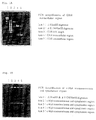
- RT-PCR technique By using the RT-PCR technique with poly A + RNA originated from the human liver, the extracellular region of human growth hormone receptor was amplified to synthesize cDNA with ca. 0.8 kbp (Fig. 1). By means of the RT-PCR technique with poly A + RNA from the human fetal liver, the genes for thetransmembrane region and intraplasmic region of c-Mp1 were amplified to synthesize cDNA with ca. 0.7 kbp (Fig. 1B).
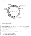
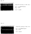
- cDNAs were inserted into an expression vector for animal cells pRC3-Uni (Fig. 2), followed by integration into TOP10F' competent cells of E . coli to generate transformants. Out of them, colony PCR was effected with 16 clones to select 8 clones (Fig. 3 A, B). Then, the selected clones were determined for the total nucleotide sequences, and selection was made of the clones CHR-EX and c-Mp1-TM+CP which are usable in the present invention.
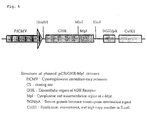
- Plasmid GHR-Ex was double-digested with the restriction enzymes M1u1 and Mp1-CP to open the strands, into which the M1u1-Xho1 treated fragment of Mp1-CP encoding the transmembrane and cytoplasmic regions of Mp1 was inserted to thereby construct the recombinant plasmid containing the GHR/Mp1 receptor gene and this gene. (Fig. 4A, B).
- the clone pCR3-Uni/GM was selected and then inserted into E. coli to generate pCR3-uni/GM in large quantities. Furthermore, the generated clone was integrated into BaF3 cells by means of the DEAE-dextran method, and the resultant transformed cells were subjected to selection on a medium containing neomycin (G418), followed by additional selection on a medium containing hGH to establish the hGH-dependent cell line BaF/GM.
- the present invention encompasses the construction of the genes for chimera receptors consisting of the extracellular regions of various cytokine receptors and the transmembrane and cytoplasmic regions of c-Mp1 and their expression plasmids, as well as animal cells having such chimera genes expressed therein.
- the cells which are utilizable as a host the CHO cells, COS cells and others may be arbitrarily selected.
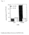
- the BaF/GM cell of the present invention is able to proliferate in the presence of hGH or mIL-3 alone, and also shows potent growth on a medium containing 10% of fetal bovine serum (FBS), although it fails to grow on the fetal bovine serum medium containing 10 % of equine serum (HS), revealing that some factor contained in FBS exerts functional effects on the GHR/Mp1 chimera receptor expressed in BaF/GM cells.
- FBS fetal bovine serum
- HS equine serum
- the BaF3 cell does not exhibit growth on a medium containing FBS and HS (Fig. 5). Proliferation of the BaF/GM cell after FBS supplementation is assumed to be attributed to the action of bovine growth hormone (bGH) contained in FBS.
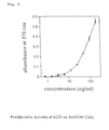
- a comparative growth analysis for the BaF/GM cell indicates that the growth curve can be determined at concentrations in the range of 10 to 100 ng/ml for bGH and 1 to 200 ng/ml for hGH (Fig. 7), leading to the conclusion that bGH can exerts about 1/10 or so less the functional effect as than hGH (Fig. 6).
- the present invention is useful in the analysis of the species-specificity of hormones to cytokine receptors.
- the expressed number of the GHR/Mp1 chimera receptor in the BaF/GM cell and the dissociation constant of the chimera receptor to hGH were calculated at about 3,700 receptors per cell and 0.16 nM, respectively (Fig. 9).
- the BaF/GM cell line established according to the present invention which possesses the GHR/Mp1 receptor exactly equivalent to GHR in terms of interaction with hGH and exhibits hGH-concentration-dependent cell growth and rises in extracellular acidification ratio, can therefore be used in screening the agonists or antagonists.
- the present invention can find practical application in establishing the chimera receptor genes for various cytokines and their expression plasmids as well as their expression cells.
- GR-2 and Mp1-A primers were provided with restriction enzyme M1u1 (indicated by underlining), which site served as a binding site for GHR and c-Mp1:
- GR-1 and Mp1-A primer were phosphorylated at their 5' ends with use of Kination Kit.
- the phosphorylation reaction of the primers was carried out by adding 40 ul in total volume of a solution mixture consisting of 10-times diluted protruding end kinase buffer (4.0 ul), 10 mM dATP (4.0 ul), 15 u/ ul T4 polynucleotide kinase (2.0 ul), 10 uM GR-1 or 10 uM Mp1-A primer (20.0 ul), sterilized double distilled water (SDDW) (10.0 ul) into an Eppendorf tube with 0.5 ml capacity, mixing the solution and allowing the reaction to proceed at 37°C for 1 hr, followed by treatment at 94°C for 5 min. to terminate the reaction.
- SDDW sterilized double distilled water
- reaction solution was admixed with 100 ul in total volume consisting of first strand cDNA reaction buffer (20 ul), second strand synthesis buffer (40 ul), 0.8 unit/ul ribonuclease H (1 ul), 3.5 units/ul DNA polymerase (5.6 ul) and SDDW (32.4 ul), and the reaction was allowed to proceed for 1 hr at 12°C and then at 22°C for 1 hr, followed by heating treatment for 10 min at 70°C to terminate the reaction to synthesize the second strand DNA.
- reaction-terminated solution was admixed with 1 ul of T4 DNA polymerase (4 units/ul), and the reaction was allowed to proceed at 37°C for 10 min and then terminated by adding 2 ul of 0.5M EDTA (pH 8.0). Subsequently, the solution was subjected to the procedures of phenol extraction and ethanol precipitation in accordance with the method described in Current Protocols in Molecular Biology (2.1.1) to purify cDNA, which was then dissolved in 50 ul of TE buffer.
- PCR Amplification was conducted by placing 50 ul in total volume of a solution mixture consisting of the 10-times diluted reaction solution (5.0 ul), 2.5 mM dNTP mixture (8.0 ul), 5 uM phosphorylated forward primer (4.0 ul), 10 uM reverse primer (2.0 ul), 5 units/ul LATaq DNA polymerase (0.5 ul), cDNA (5.0 ul) and SDDW (25.5 ul) into a MicroAmp reaction tube, followed by mixing, and treating the solution at 94°C for 1 min, at 60°C for 1 min and then at 72°C for 10 min to add adenine to the 3' end, whereby Gene Amp PCR System 9600 (PERKIN-ELMER) was employed as a thermal cycler.
- PERKIN-ELMER Gene Amp PCR System 9600
- Ligation was effected by placing 10 ul in total volume of the solution mixture consisting of 10-times diluted ligation buffer (1.0 ul), plasmid pCR3-Uni (2.0 ul), GHR cDNA or c-Mp1 cDNA (3.0 ul), T4 DNA ligase (1.0 ul) and SDDW (3.0 ul) into an Eppendorf tube with a 0.5-ml capacity to allow the reaction to proceed at 14°C overnight.
- 10-times diluted ligation buffer 1.0 ul
- plasmid pCR3-Uni 2.0 ul
- GHR cDNA or c-Mp1 cDNA 3.0 ul
- T4 DNA ligase 1.0 ul
- SDDW 3.0 ul
- Transformation of E. coli was carried out by mixing gently 50 ul of E . coli TOP10F' competent cells with added cold 0.5M ⁇ -mercaptoethanol (2 ul) and furthermore adding 2 ul of the ligation product for gentle mixing, followed by standing in ice-water for 30 min; then providing thermal shock at 42°C for 30 sec to the E. coli mixed solution, which was quickly transferred into ice-water to thereby cool for 2 min; and admixing the solution with 900 ul of SOC medium warmed in advance at 37°C, followed by shake culture at 37°C for 1 hr, and sowing the culture broth on L-broth agar medium containing 50 ug/ml of ampicillin, followed by standing at 37°C overnight.
- Bacterial cells were taken from 16 clones with a toothpick and suspended in 10 ul of 1 % Triton x-100, and the resultant suspension was heat-treated at 100°C for 5 min to melt the bacterial cells, which was to be used as a template for PCR.
- PCR Amplification was conducted by placing 20 ul in total volume of a solution mixture consisting of the 10-times diluted reaction buffer (2.0 ul), 2.5 mM dNTP mixture (2.0 ul), 10 uM T7 sequencing primer (1.0 ul), 10 uM GR-4 or 10 uM Mp1-B primer (1.0 ul), 0.5 units/ul Ampli Taq DNA polymerase (1.0 ul), E.
- Preparation of plasmid DNA was carried out by picking up a single colony of each clone, followed by sowing on a L-broth medium containing 50 ug/ml concentrated ampicillin to conduct shake culture at 37°C overnight, and recovering the bacterial cells from 1.5 ml of each culture broth to purify plasmid with use of RPM Kit; The plasmid prepared in this manner was subjected to DNA sequencing.
- PCR Amplification was conducted by placing 20 ul in total volume of a solution mixture consisting of the Reaction premix (8.0 ul), 5 uM sequencing primer for T7, SP6 or Gr4 (1.0 ul), plasmid DNA (3.5 ul) and SDDW (12.0 ul) into a MicroAmp reaction tube, followed by mixing, and repeating 25 times a cycle of treating the solution at 96°C for 30 sec, at 50°C for 15 sec and at 60°C for 4 min, whereby Gene Amp PCR System 9600 (PERKIN-ELMER) was employed as a thermal cycler.
- Reaction premix 8.0 ul
- 5 uM sequencing primer for T7, SP6 or Gr4 1.0 ul
- plasmid DNA 3.5 ul
- SDDW 12.0 ul
- ethanol precipitation was effected to purify the PCR product, and the resultant DNA was dissolved in 3 ul of loading buffer (a buffer prepared by mixing deionized formamide with 25 mM EDTA (pH 8.0) containing 50 mg/ml concentrated blue dextran at a volume ratio of 5:1), and the solution was heat-treated at 90°C for 2 min and quickly cooled with ice water to denature DNA into single-strand.
- loading buffer a buffer prepared by mixing deionized formamide with 25 mM EDTA (pH 8.0) containing 50 mg/ml concentrated blue dextran at a volume ratio of 5:1
- a polyacrylamide gel for DNA sequencing was prepared from Gel-Mix6 and set onto the DNA sequencer, to which the DNA sample was then applied.
- the miscellaneous steps were effected in accordance with the procedure described in the Instructions for Use attached to the kit.
- Double digestion with restriction enzymes Mlul and Xhol was conducted by placing 10 ul in total volume of a solution mixture consisting of the 10-times diluted H universal buffer (1.0 ul), 10 units/ul Mlul (0.5 ul), 14 units/ul Xhol (0.5 ul), GHR-Ex#4 or Mp1-Cp#7 plasmid DNA (2 ul) and SDDW (6 ul) into an Eppendorf tube with a 0.5 ml capacity, followed by mixing, and allowing the reaction to proceed at 96°C for 4 hrs, followed by treatment at 70°C for 10 min to deactivate the restriction enzymes.
- the sample prepared in this manner was applied to a 2.2 % agarose gel (Sea Kem GTG agarose gel used) to conduct electrophoresis. After staining with ethidium bromide, subsequently, the electrophoretic separation was photographed under UV irradiation, and the objective band was cut out to thereby purify DNA with Gene Clean II.
- Ligation was effected by placing 20 ul in total volume of the solution mixture consisting of 10-times diluted ligation buffer (2.0 ul), plasmid GHR-Ex#4 (2.0 ul), Mp1-Cp#7 fragment (8.0 ul), T4 DNA ligase (2.0 ul) and SDDW (6.0 ul) into an Eppendorf tube with a 0.5-ml capacity to allow the reaction to proceed at 14°C overnight.
- Transformation of E . coli was carried out by mixing gently 50 ul of E. coli TOP10F' competent cells with added cold 0.5M ⁇ -mercaptoethanol (2 ul) and furthermore adding 2 ul of the ligation product for gentle mixing, followed by standing in ice-water for 30 min; then providing thermal shock at 42°C for 30 sec to the E. coli mixed solution, which was quickly transferred into ice to thereby cool for 2 min; and admixing the solution with 900 ul of SOC medium warmed in advance at 37°C, followed by shake culture at 37°C for 1 hr, and sowing the culture broth on L-broth agar medium containing 50 ug/ml of ampicillin, followed by standing at 37°C overnight for cultivation.
- Bacterial cells were taken from 16 clones with a toothpick and suspended in 10 ul of 1 % Triton X-100, and the resultant suspension was heat-treated at 100°C for 5 min to melt the bacterial cells, which was to be used as a template for PCR.
- PCR Amplification was conducted by placing 20 ul in total volume of a solution mixture consisting of the 10-times diluted reaction buffer (2.0 ul), 10 uM MGR-1 primer (1.0 ul), 10 uM Mp1-primer (1.0 ul), 0.5 units/ul Ampli Taq DNA polymerase (1.0 ul), E . coli lysates (2.0 ul) and SDDW (12.0 ul) into a MicroAmp reaction tube, followed by mixing, and repeating 25 times a cycle of treating the solution at 94°C for 1 min and at 55°C for 2 min, whereby Gene Amp PCR System 9600 (PERKIN-ELMER) was employed as a thermal cycler.
- PERKIN-ELMER Gene Amp PCR System 9600
- Preparation of the objective plasmid DNA was carried out by picking up a single colony of each clone, followed by sowing on a L-broth medium containing 50 ug/ml concentrated ampicillin to conduct shake culture at 37°C overnight, and recovering the bacterial cells from 1.5 ml of each culture broth to purify plasmid with use of RPM Kit; The plasmid prepared in this manner was subjected to restriction enzyme analysis and DNA sequencing.
- Restriction enzyme analysis was performed by placing 10 ul in total volume of a solution mixture consisting of the 10-times diluted H universal buffer (1.0 ul), 12 units/ul HindIII (0.5 ul), 10 units/ul Mlul (0.5 ul), 14 units/ul Xhol (0.5 ul), plasmid DNA (3.0 ul) and SDDW (4.5 ul) into an Eppendorf tube with a 0.5-ml capacity, followed by mixing, and allowing the reaction to proceed at 96°C for 4 hrs, followed by treatment at 70°C for 10 min to deactivate the restriction enzymes.
- Example 3 Establishment of a hGH-dependent cell line BaF/GM
- Plasmid DNA was prepared in large quantities by picking up a single colony of selected clones, followed by sowing on L-broth medium containing 50-ug/ml concentrated ampicillin to conduct shake culture at 37°C overnight, then transferring the resultant culture broth into 500 ml of L-broth medium containing 500-ug/ml concentrated ampicillin to thereby conduct shake culture at 37°C until the culture broth showed 1.0 of absorbance at a wavelength of 600 nm, and adding 2.5 ml of a 34-ng/ml concentrated solution of chloramphenicol in ethanol to effect shake culture at 37°C overnight. The bacterial cells were recovered and the plasmid DNA was purified in accordance with the procedure described in Current Protocols in Molecular Biology (9.1.5).
- the said cells were cultivated on RPMI 1640 medium (containing 10 % of bovine fetal serum and 50 ug/ml of gentamicin) to separate out a culture supernatant.
- the resultant supernatant was filtered through a membrane of 0.22 um in pore size (0.22 um disposable filterware: Nalgene), and the filtrate was frozen for storage.
- BaF3 cells were cultivated on a medium consisting of RPMI 1640 medium supplemented with 10 volume % of the supernatant of WEHI-3 culture as a source for mIL-3.
- Transformation of BaF3 cells was carried out in accordance with the DEAE-dextran method; BaF3 cells during the logarithmic growth phase were sowed on a medium at a density of 5 x 10 5 cells/ml and cultivated overnight in a CO 2 incubator. Then, Plasmid pCR3-Uni/GM sterilized through the ethanol precipitation treatment was diluted with PBS to a concentration of 10 ug/ml, and 170 ul of the resultant plasmid solution was admixed with 170 ul of a 1:1 solution mixture of a 2 mg/ml-concentrated solution of DEAE-dextran in 0.9 % NaCl with PBS.
- BaF3 Cells after being cultivated overnight were washed with PBS twice, and 2 x 10 6 cells were separated out by counting. After the supernatant was removed by centrifugation, the plasmid/DEAE dextran solution mixture was added to the cells, followed by gentle stirring and treatment at room temperature for 5 min. The cells were washed with PBS twice and cultivated on a medium for BaF3 cells in a CO 2 incubator for 48 hrs.
- Selection of the transformed cells with G 418 and hGH was performed by firstly cultivating the transformant cells on a medium for BaF3 cells containing 400 ug/ml concentrated G 418 for 2 weeks, during which the medium was exchanged every several days, and then cultivating G-418 resistant cells for 1 week on a medium supplemented with 100-ng/ml concentrated hGH in place of a supernatant of wEHI-3 cell culture as a source of mIL-3 to select the cells capable of proliferating in a hGH-dependent manner, during which period the medium was exchanged every two days.
- the finally obtained hGH-dependent cells were considered as the BaF/GM cell (Deposition No. FERM P-16255).
- the cell proliferation analysis was effected by cultivating BaF/GM cells on RPMI 1640 medium containing 100 ng/ml of hGH overnight, with BaF3 cells as a control being cultivated on a medium for BaF3 cells overnight, then washing the cells with PBS three times, followed by further cultivation on serum-free RPMI 1640 medium for 5 hrs, diluting the cells with RPMI 1640 medium containing individually 10 % of FBS, 10 % of HS and 1 % of BSA to the cell concentration of 300,000 cells/ml, respectively, and seeding the diluted cell solutions in 100 ul portions into 96 wells for cultivation for 22 hrs., followed by determination with 3 wells by the following procedure:
- the BaF/GM cells were cultivated overnight on RPMI 1640 medium (containing 10 % of FBS) containing 100 ng/ml of hGH, and the grown cells were washed with PBS three times and cultivated for 16 hrs. on RPMI 1640 medium containing 10 % HS in place of FBS to thereby synchronize with the G 0 stage of cell cycle, followed by seeding into a 96-well flat-bottom plate for cultivation at a density of 30,000 cells/well.
- RPMI 1640 medium containing 10 % of FBS
- bGH was distributed in 10-ul portion into wells to the different concentrations (ng/ml) of 0.39, 0.78, 1.56, 3.12, 6.25, 12.5, 25.0, 50.0, 100.0 and 200.0 to prepare standard samples, while FBS as such and after being diluted 10 times and 100 times, respectively, was also added in 10-ul portion for dilution, and all measurements were taken with every 3 wells whereby the amount of bGH contained in PBS was determined by plotting FBS values on the calibration curve.
- concentrations ng/ml
- the BaF/GM cells were cultivated overnight on RPMI 1460 medium (containing 10 % of FBS) containing 100-ng/ml concentrated hGH, and the grown cells were washed with PBS three times and were furthermore cultivated for 16 hrs on RPMI 1460 medium containing 10 % of HS in place of FBS to thereby synchronize with the G 0 stage of cell cycle followed by seeding into a 96-well flat-bottom plate at a density of 30,000 cells/well.
- hGH was distributed in 10-ul portion into wells to the different concentrations (ng/ml) of 0.39, 0.78, 1.56, 3.12, 6.25, 12.5, 25.0, 50.0, 100.0 and 200.0 . All measurements were taken with every 3 wells.
- Synchronization of the BaF/GM cells with the G 0 stage of cell cycle was regulated by cultivating the BaF/GM cells overnight on PRMI 1460 medium (containing 10 % of FBS) containing 100 ng/ml of hGH, and recovering the grown cells, followed by washing with PBS three times and cultivation for 16 hrs on RPMI 1640 medium containing 10 % of HS.
- agarose entrapment medium was dissolved over a warm-water bath and placed into a heat block at 37°C, followed by standing.
- the cells were washed with a measuring medium (RPMI 1640 medium , pH 7.4, containing 4.1 % of human serum albumin) for cytosensor and adjusted to a cell density of 1.3 x 10 7 cells/ml.
- the cell solution was mixed with the agarose solution at a ratio of 3 : 1, and 7 ul of the solution mixture was immediately placed in the center of a capsule cup having a spacer intervened.
- the measuring media containing individually 3.90, 15.6, 62.5, 250.0 and 1,000.0 (ng/ml) concentrated hGH were flown through the cells for 15 min, followed by switching to the measuring medium, whereby 100 ng/ml TPO was used as a control.
- BaF/GM cells were adjusted to a density of 2.8 x 10 7 cells/ml by cultivating the BaF/GM cells overnight on RPMI 1640 medium (10 % FBS) containing 100 ng/ml of hGH, recovering the grown cells, followed by washing with PBS three times, and further cultivating the washed cells on RPMI 1640 medium containing 10 % of HS, followed by recovery and washing with RRA medium (RPMI 1640 medium containing 0.5 % of PBS) once.
- RPMI 1640 medium 10 % FBS
- RRA medium RPMI 1640 medium containing 0.5 % of PBS
- Radioreceptor analysis was carried out by preparing solutions containing different amounts of hGH per 100 ul (i.e., 2,000, 1,000, 500, 250, 125, 62.5, 31.25, 15.62, 7.81, 3.91, 1.95, 0.98, 0.49 and 0.24 ng) with use of RRA medium, then placing 100 ul each of the resultant solutions of hGH in individual Kohken tubes, adding 100 ul of a 125 I-labeled hGH solution (3.33 kBq/ml) to each tube, followed by shake culture at 4°C for 2 hrs to allow the reaction to go to conclusion, adding 2 ml of cold RRA medium to each tube, followed by stirring and centrifugation at 3,000 rpm at 4°C for 30 min, removing the supernatant and determining radioactivity per 30 sec for each tube with use of a gamma-ray counter, wherein every measured amounts of radioactivity were expressed as a mean value for two radioactivity determinations.
- the resultant data were analyzed by resistance plotting and Scatchard plotting to determine IC 50 and dissociation constant Kd value as well as the number of receptors per cell.
Landscapes
- Health & Medical Sciences (AREA)
- Chemical & Material Sciences (AREA)
- Life Sciences & Earth Sciences (AREA)
- Organic Chemistry (AREA)
- Biophysics (AREA)
- General Health & Medical Sciences (AREA)
- Zoology (AREA)
- Gastroenterology & Hepatology (AREA)
- Immunology (AREA)
- Biochemistry (AREA)
- Cell Biology (AREA)
- Toxicology (AREA)
- Genetics & Genomics (AREA)
- Medicinal Chemistry (AREA)
- Molecular Biology (AREA)
- Proteomics, Peptides & Aminoacids (AREA)
- Endocrinology (AREA)
- Peptides Or Proteins (AREA)
- Preparation Of Compounds By Using Micro-Organisms (AREA)
- Micro-Organisms Or Cultivation Processes Thereof (AREA)
Applications Claiming Priority (2)
| Application Number | Priority Date | Filing Date | Title |
|---|---|---|---|
| JP10108769A JPH11285388A (ja) | 1998-04-03 | 1998-04-03 | キメラレセプター発現細胞とその用途 |
| JP10876998 | 1998-04-03 |
Publications (2)
| Publication Number | Publication Date |
|---|---|
| EP0950710A2 true EP0950710A2 (de) | 1999-10-20 |
| EP0950710A3 EP0950710A3 (de) | 1999-10-27 |
Family
ID=14493029
Family Applications (1)
| Application Number | Title | Priority Date | Filing Date |
|---|---|---|---|
| EP99106794A Withdrawn EP0950710A3 (de) | 1998-04-03 | 1999-04-06 | Zellen, die ein chimärischen Rezeptor exprimieren, und deren Verwendungen |
Country Status (2)
| Country | Link |
|---|---|
| EP (1) | EP0950710A3 (de) |
| JP (1) | JPH11285388A (de) |
Cited By (4)
| Publication number | Priority date | Publication date | Assignee | Title |
|---|---|---|---|---|
| WO2001083551A1 (en) * | 2000-05-04 | 2001-11-08 | Sahltech I Göteborg AB | Reagent for detection of biomolecules, and use thereof |
| WO2001096565A3 (en) * | 2000-06-16 | 2002-08-01 | Asterion Ltd | Binding agents: chimeric ligand/receptor proteins |
| WO2017081440A1 (en) * | 2015-11-13 | 2017-05-18 | Asterion Limited | Fusion polypeptide comprising the extracellular binding domain of growth hormone receptor |
| US11344606B2 (en) | 2017-04-28 | 2022-05-31 | University Of Sheffield | Parathyroid hormone fusion polypeptide |
Families Citing this family (2)
| Publication number | Priority date | Publication date | Assignee | Title |
|---|---|---|---|---|
| JPWO2007043200A1 (ja) * | 2005-09-30 | 2009-04-16 | 独立行政法人理化学研究所 | T細胞分化調節剤 |
| GB0523954D0 (en) * | 2005-11-24 | 2006-01-04 | Ucb Celltech | Bioassays |
-
1998
- 1998-04-03 JP JP10108769A patent/JPH11285388A/ja active Pending
-
1999
- 1999-04-06 EP EP99106794A patent/EP0950710A3/de not_active Withdrawn
Non-Patent Citations (4)
| Title |
|---|
| GURNEY A.L. ET AL.: "Distinct regions of c-Mpl cytoplasmic domain are coupled to the JAK-STAT signal transduction pathway and Shc phosphorylation" PROCEEDINGS OF THE NATIONAL ACADEMY OF SCIENCES OF THE USA, vol. 92, June 1995 (1995-06), pages 5292-5296, XP002114223 NATIONAL ACADEMY OF SCIENCE, WASHINGTON, US ISSN: 0027-8424 * |
| LEUNG D.W. ET AL.: "Growth Hormone Receptor and serum binding protein: cloning and expression" NATURE, vol. 330, 10 December 1987 (1987-12-10), pages 537-543, XP002114224 * |
| TAKAHASHI Y. ET AL.: "Biologically inactive Growth Hormone caused by an aminoacid substitution" THE JOURNAL OF CLINICAL INVESTIGATION, vol. 100, no. 5, 1 September 1997 (1997-09-01), pages 1159-1165, XP002114222 NEW YORK, NY, US ISSN: 0021-9738 * |
| VIGON I. ET AL.: "Molecular cloning and characterization of MPL, the human homolog of the v-MPL oncogene: identification of a member of the hematopoietic growth factor receptor superfamily" PROCEEDINGS OF THE NATIONAL ACADEMY OF SCIENCES OF USA, vol. 89, June 1992 (1992-06), pages 5640-5644, XP002915951 ISSN: 0027-8424 * |
Cited By (6)
| Publication number | Priority date | Publication date | Assignee | Title |
|---|---|---|---|---|
| WO2001083551A1 (en) * | 2000-05-04 | 2001-11-08 | Sahltech I Göteborg AB | Reagent for detection of biomolecules, and use thereof |
| WO2001096565A3 (en) * | 2000-06-16 | 2002-08-01 | Asterion Ltd | Binding agents: chimeric ligand/receptor proteins |
| US7446183B2 (en) | 2000-06-16 | 2008-11-04 | Asterion Limited | Fusion protein comprising growth hormone and growth hormone receptor |
| WO2017081440A1 (en) * | 2015-11-13 | 2017-05-18 | Asterion Limited | Fusion polypeptide comprising the extracellular binding domain of growth hormone receptor |
| GB2549351A (en) * | 2015-11-13 | 2017-10-18 | Asterion Ltd | Fusion polypeptide |
| US11344606B2 (en) | 2017-04-28 | 2022-05-31 | University Of Sheffield | Parathyroid hormone fusion polypeptide |
Also Published As
| Publication number | Publication date |
|---|---|
| EP0950710A3 (de) | 1999-10-27 |
| JPH11285388A (ja) | 1999-10-19 |
Similar Documents
| Publication | Publication Date | Title |
|---|---|---|
| Burke et al. | Dimerization of the p185neu transmembrane domain is necessary but not sufficient for transformation | |
| Lin et al. | Expression cloning of the TGF-β type II receptor, a functional transmembrane serine/threonine kinase | |
| Ohashi et al. | Ligand-induced activation of chimeric receptors between the erythropoietin receptor and receptor tyrosine kinases. | |
| Morris et al. | ALK, the chromosome 2 gene locus altered by the t (2; 5) in non-Hodgkin's lymphoma, encodes a novel neural receptor tyrosine kinase that is highly related to leukocyte tyrosine kinase (LTK) | |
| Fraser | Site-directed mutagenesis of β-adrenergic receptors: identification of conserved cysteine residues that independently affect ligand binding and receptor activation | |
| US5843678A (en) | Osteoprotegerin binding proteins | |
| Gurney et al. | Distinct regions of c-Mpl cytoplasmic domain are coupled to the JAK-STAT signal transduction pathway and Shc phosphorylation. | |
| EP1131333B1 (de) | Isolierte nukleinsäuremoleküle die t-zellen induzierbare faktoren kodieren (tif), kodierte proteine und ihre anwendung | |
| JP2001512680A (ja) | 哺乳動物のサイトカインレセプター−11 | |
| AU718795B2 (en) | Netrin receptors | |
| Ozenberger et al. | Functional interaction of ligands and receptors of the hematopoietic superfamily in yeast | |
| LEFKOWITZ et al. | Molecular and regulatory properties of adrenergic receptors | |
| KR20160135738A (ko) | 폴리펩티드를 발현하는 숙주 세포의 선택을 위한 발현 구조물 및 방법 | |
| Gourdou et al. | Development of a constitutively active mutant form of the prolactin receptor, a member of the cytokine receptor family. | |
| Minami et al. | Signal transduction mediated by the reconstituted IL-2 receptor. Evidence for a cell type-specific function of IL-2 receptor beta-chain. | |
| US20040014024A1 (en) | Screening assay for antagonists of FGFR-mediated malignant cell transformation and tumor formation | |
| US20020160451A1 (en) | Novel orphan receptors | |
| Tajima et al. | Role of Dimerization of the Membrane-associated Growth Factor Kit Ligand in Juxtacrine Signaling: The Sl 17H Mutation Affects Dimerization and Stability—Phenotypes in Hematopoiesis | |
| Tsuruta et al. | Insulin-like growth factor-II/cation-independent mannose 6-phosphate receptor mediates paracrine interactions during spermatogonial development | |
| Pacifici et al. | Hybrid tyrosine kinase/cytokine receptors transmit mitogenic signals in response to ligand. | |
| TWI405850B (zh) | 經改良之宿主細胞及培養方法 | |
| US5789237A (en) | Nucleic acid sequences coding for or complementary to nucleic acid sequences coding for interleukin 9 receptor | |
| Muthukumaran et al. | Chimeric erythropoietin-interferon γ receptors reveal differences in functional architecture of intracellular domains for signal transduction | |
| US20010024652A1 (en) | Isolated nucleic acid molecules which encode T cell inducible factors (TIFs), the proteins encoded, and used thereof | |
| AU772932B2 (en) | Novel guanosine triphosphate (gtp) binding protein-coupled receptor proteins |
Legal Events
| Date | Code | Title | Description |
|---|---|---|---|
| PUAI | Public reference made under article 153(3) epc to a published international application that has entered the european phase |
Free format text: ORIGINAL CODE: 0009012 |
|
| PUAL | Search report despatched |
Free format text: ORIGINAL CODE: 0009013 |
|
| AK | Designated contracting states |
Kind code of ref document: A2 Designated state(s): DE FR GB |
|
| AX | Request for extension of the european patent |
Free format text: AL;LT;LV;MK;RO;SI |
|
| AK | Designated contracting states |
Kind code of ref document: A3 Designated state(s): AT BE CH CY DE DK ES FI FR GB GR IE IT LI LU MC NL PT SE |
|
| AX | Request for extension of the european patent |
Free format text: AL;LT;LV;MK;RO;SI |
|
| RTI1 | Title (correction) |
Free format text: CELLS EXPRESSING A CHIMERA RECEPTOR AND THEIR USES |
|
| 17P | Request for examination filed |
Effective date: 19991122 |
|
| AKX | Designation fees paid |
Free format text: DE FR GB |
|
| 17Q | First examination report despatched |
Effective date: 20030729 |
|
| STAA | Information on the status of an ep patent application or granted ep patent |
Free format text: STATUS: THE APPLICATION IS DEEMED TO BE WITHDRAWN |
|
| 18D | Application deemed to be withdrawn |
Effective date: 20031209 |