EP0932665B1 - Method for sorting antifungal molecules acting on the glucanosyltransferase activity - Google Patents
Method for sorting antifungal molecules acting on the glucanosyltransferase activity Download PDFInfo
- Publication number
- EP0932665B1 EP0932665B1 EP97938964A EP97938964A EP0932665B1 EP 0932665 B1 EP0932665 B1 EP 0932665B1 EP 97938964 A EP97938964 A EP 97938964A EP 97938964 A EP97938964 A EP 97938964A EP 0932665 B1 EP0932665 B1 EP 0932665B1
- Authority
- EP
- European Patent Office
- Prior art keywords
- protein
- seq
- activity
- sequence
- glucanosyltransferase
- Prior art date
- Legal status (The legal status is an assumption and is not a legal conclusion. Google has not performed a legal analysis and makes no representation as to the accuracy of the status listed.)
- Expired - Lifetime
Links
Images


Classifications
-
- C—CHEMISTRY; METALLURGY
- C12—BIOCHEMISTRY; BEER; SPIRITS; WINE; VINEGAR; MICROBIOLOGY; ENZYMOLOGY; MUTATION OR GENETIC ENGINEERING
- C12Q—MEASURING OR TESTING PROCESSES INVOLVING ENZYMES, NUCLEIC ACIDS OR MICROORGANISMS; COMPOSITIONS OR TEST PAPERS THEREFOR; PROCESSES OF PREPARING SUCH COMPOSITIONS; CONDITION-RESPONSIVE CONTROL IN MICROBIOLOGICAL OR ENZYMOLOGICAL PROCESSES
- C12Q1/00—Measuring or testing processes involving enzymes, nucleic acids or microorganisms; Compositions therefor; Processes of preparing such compositions
- C12Q1/02—Measuring or testing processes involving enzymes, nucleic acids or microorganisms; Compositions therefor; Processes of preparing such compositions involving viable microorganisms
- C12Q1/18—Testing for antimicrobial activity of a material
-
- A—HUMAN NECESSITIES
- A61—MEDICAL OR VETERINARY SCIENCE; HYGIENE
- A61P—SPECIFIC THERAPEUTIC ACTIVITY OF CHEMICAL COMPOUNDS OR MEDICINAL PREPARATIONS
- A61P31/00—Antiinfectives, i.e. antibiotics, antiseptics, chemotherapeutics
- A61P31/10—Antimycotics
-
- C—CHEMISTRY; METALLURGY
- C12—BIOCHEMISTRY; BEER; SPIRITS; WINE; VINEGAR; MICROBIOLOGY; ENZYMOLOGY; MUTATION OR GENETIC ENGINEERING
- C12N—MICROORGANISMS OR ENZYMES; COMPOSITIONS THEREOF; PROPAGATING, PRESERVING, OR MAINTAINING MICROORGANISMS; MUTATION OR GENETIC ENGINEERING; CULTURE MEDIA
- C12N9/00—Enzymes; Proenzymes; Compositions thereof; Processes for preparing, activating, inhibiting, separating or purifying enzymes
- C12N9/10—Transferases (2.)
- C12N9/1048—Glycosyltransferases (2.4)
- C12N9/1051—Hexosyltransferases (2.4.1)
-
- C—CHEMISTRY; METALLURGY
- C12—BIOCHEMISTRY; BEER; SPIRITS; WINE; VINEGAR; MICROBIOLOGY; ENZYMOLOGY; MUTATION OR GENETIC ENGINEERING
- C12Q—MEASURING OR TESTING PROCESSES INVOLVING ENZYMES, NUCLEIC ACIDS OR MICROORGANISMS; COMPOSITIONS OR TEST PAPERS THEREFOR; PROCESSES OF PREPARING SUCH COMPOSITIONS; CONDITION-RESPONSIVE CONTROL IN MICROBIOLOGICAL OR ENZYMOLOGICAL PROCESSES
- C12Q1/00—Measuring or testing processes involving enzymes, nucleic acids or microorganisms; Compositions therefor; Processes of preparing such compositions
- C12Q1/48—Measuring or testing processes involving enzymes, nucleic acids or microorganisms; Compositions therefor; Processes of preparing such compositions involving transferase
-
- H—ELECTRICITY
- H01—ELECTRIC ELEMENTS
- H01L—SEMICONDUCTOR DEVICES NOT COVERED BY CLASS H10
- H01L28/00—Passive two-terminal components without a potential-jump or surface barrier for integrated circuits; Details thereof; Multistep manufacturing processes therefor
- H01L28/40—Capacitors
- H01L28/60—Electrodes
- H01L28/82—Electrodes with an enlarged surface, e.g. formed by texturisation
- H01L28/90—Electrodes with an enlarged surface, e.g. formed by texturisation having vertical extensions
- H01L28/92—Electrodes with an enlarged surface, e.g. formed by texturisation having vertical extensions made by patterning layers, e.g. by etching conductive layers
-
- G—PHYSICS
- G01—MEASURING; TESTING
- G01N—INVESTIGATING OR ANALYSING MATERIALS BY DETERMINING THEIR CHEMICAL OR PHYSICAL PROPERTIES
- G01N2333/00—Assays involving biological materials from specific organisms or of a specific nature
- G01N2333/37—Assays involving biological materials from specific organisms or of a specific nature from fungi
- G01N2333/39—Assays involving biological materials from specific organisms or of a specific nature from fungi from yeasts
-
- G—PHYSICS
- G01—MEASURING; TESTING
- G01N—INVESTIGATING OR ANALYSING MATERIALS BY DETERMINING THEIR CHEMICAL OR PHYSICAL PROPERTIES
- G01N2333/00—Assays involving biological materials from specific organisms or of a specific nature
- G01N2333/90—Enzymes; Proenzymes
- G01N2333/91—Transferases (2.)
- G01N2333/91091—Glycosyltransferases (2.4)
- G01N2333/91097—Hexosyltransferases (general) (2.4.1)
-
- G—PHYSICS
- G01—MEASURING; TESTING
- G01N—INVESTIGATING OR ANALYSING MATERIALS BY DETERMINING THEIR CHEMICAL OR PHYSICAL PROPERTIES
- G01N2500/00—Screening for compounds of potential therapeutic value
- G01N2500/04—Screening involving studying the effect of compounds C directly on molecule A (e.g. C are potential ligands for a receptor A, or potential substrates for an enzyme A)
-
- H—ELECTRICITY
- H10—SEMICONDUCTOR DEVICES; ELECTRIC SOLID-STATE DEVICES NOT OTHERWISE PROVIDED FOR
- H10B—ELECTRONIC MEMORY DEVICES
- H10B12/00—Dynamic random access memory [DRAM] devices
- H10B12/01—Manufacture or treatment
- H10B12/02—Manufacture or treatment for one transistor one-capacitor [1T-1C] memory cells
- H10B12/03—Making the capacitor or connections thereto
- H10B12/033—Making the capacitor or connections thereto the capacitor extending over the transistor
Landscapes
- Chemical & Material Sciences (AREA)
- Life Sciences & Earth Sciences (AREA)
- Health & Medical Sciences (AREA)
- Engineering & Computer Science (AREA)
- Organic Chemistry (AREA)
- Zoology (AREA)
- Wood Science & Technology (AREA)
- Genetics & Genomics (AREA)
- Bioinformatics & Cheminformatics (AREA)
- Proteomics, Peptides & Aminoacids (AREA)
- General Health & Medical Sciences (AREA)
- General Engineering & Computer Science (AREA)
- Power Engineering (AREA)
- Microbiology (AREA)
- Molecular Biology (AREA)
- Biotechnology (AREA)
- Biochemistry (AREA)
- Biophysics (AREA)
- Analytical Chemistry (AREA)
- Physics & Mathematics (AREA)
- Immunology (AREA)
- Medicinal Chemistry (AREA)
- Toxicology (AREA)
- Computer Hardware Design (AREA)
- Biomedical Technology (AREA)
- Microelectronics & Electronic Packaging (AREA)
- Nuclear Medicine, Radiotherapy & Molecular Imaging (AREA)
- General Chemical & Material Sciences (AREA)
- Communicable Diseases (AREA)
- Pharmacology & Pharmacy (AREA)
- Animal Behavior & Ethology (AREA)
- Public Health (AREA)
- Veterinary Medicine (AREA)
- Chemical Kinetics & Catalysis (AREA)
- Oncology (AREA)
- Measuring Or Testing Involving Enzymes Or Micro-Organisms (AREA)
- Peptides Or Proteins (AREA)
- Enzymes And Modification Thereof (AREA)
- Preparation Of Compounds By Using Micro-Organisms (AREA)
- Measuring Pulse, Heart Rate, Blood Pressure Or Blood Flow (AREA)
Abstract
Description
La présente invention concerne une protéine ayant une activité de type glucanosyltransférase, et plus particulièrement une activité de type β-(1-3) glucanosyltransférase.The present invention relates to a protein having a glucanosyltransferase type activity, and more particularly a β- (1-3) glucanosyltransferase type activity.
La présente invention décrit également des oligonucléotides codant pour cette protéine ayant une activité enzymatique.The present invention also discloses oligonucleotides encoding this protein having enzymatic activity.
De même elle décrit des molécules ayant un effet sur l'activité de cette enzyme.Likewise, it describes molecules having an effect on the activity of this enzyme.
Les infections opportunistes fongiques dûes à Candida, Aspergillus. Cryptococcus et Pneumocystis sont responsables de la croissance de la morbidité et de la mortalité parmi les patients atteints du SIDA et autres patients présentant une immunité compromise cliniquement. De plus, la levure Candida et les dermatophytes demeurent aujourd'hui un problème médical important parmi les patients présentant une immunité suffisante. Malgré l'augmentation du nombre des infections dues aux champignons pathogènes et opportunistes, la thérapie contre les mycoses ne s'est pas améliorée durant ces dernières années. Deux familles de médicaments sont utilisés: les azoles et l' Amphotéricine B. Ces médicaments présentent certains inconvénients puisque le traitement à base d'Amphotéricine B est associé à une néphrotoxicité et celui à base d'azole est plus fongistatique que fongicide.Fungal opportunistic infections due to Candida, Aspergillus. Cryptococcus and Pneumocystis are responsible for the growth of morbidity and mortality among patients with AIDS and other patients with clinically compromised immunity. In addition, Candida yeast and dermatophytes remain an important medical problem in patients with sufficient immunity. Despite the increase in infections due to pathogenic and opportunistic fungi, mycosis therapy has not improved in recent years. Two families of drugs are used: azoles and Amphotericin B. These drugs have certain drawbacks since treatment with Amphotericin B is associated with nephrotoxicity and that with azole is more fungistatic than fungicidal.
Les champignons sont des micro-organismes de type eucaryote qui partagent la plupart des voies biochimiques avec leur hôte avec une exception importante : la biosynthèse de la paroi cellulaire. La paroi cellulaire est une enveloppe rigide qui protège la cellule contre l'environnement et les stress mécaniques, mais également une structure dynamique qui est impliquée dans le transport des ions et des macromolécules et dans la localisation d'enzymes impliquées dans la croissance fongique. Par conséquent, la désorganisation de l'organisation de la paroi cellulaire doit être préjudiciable aux champignons.Fungi are eukaryotic microorganisms that share most of the biochemical pathways with their host with one important exception: cell wall biosynthesis. The cell wall is a rigid envelope that protects the cell against the environment and mechanical stress, but also a dynamic structure that is involved in the transport of ions and macromolecules and in the localization of enzymes involved in fungal growth. Therefore, the disorganization of the organization of the cell wall must be detrimental to the fungi.
Le squelette de la paroi cellulaire fongique est principalement constituée de polymères de type polysaccharide (β (1-3) glucanes, mannanes, chitine) que l'on ne trouve pas chez les humains. Pour cette raison, la biosynthèse de la paroi cellulaire a été une cible pour la recherche de nouveaux médicaments antifongiques. Les pénicilllines et les céphalosporines qui sont tous deux des inhibiteurs de la paroi cellulaire bactérienne et des antibiotiques potentiels apportent une crédibilité à cette hypothèse. De plus, beaucoup de molécules qui inhibent le développement de la paroi cellulaire fongique ont des propriétés antifongiques (
- 1) Les familles des lipopeptides des échinocandins et des glycopeptides des palulacandins qui sont des inhibiteurs non compétitifs du complexe de la glucane synthétase.
- 2) Les polyoxines et les nikkomycines qui sont des analogues de l'UDP-GlcNac et des inhibiteurs compétitifs potentiels de la chitine synthétase, et
- 3) Les pradimicines liant le mannane et les bénanomicines.
- 1) The families of lipopeptides of echinocandins and glycopeptides of palulacandins which are noncompetitive inhibitors of the glucan synthetase complex.
- 2) polyoxins and nikkomycins which are UDP-GlcNac analogs and potential competitive inhibitors of chitin synthetase, and
- 3) Pradimicins binding mannan and benanomicins.
La synthèse du β (1-3) glucane et de la chitine est sous le contrôle de complexes d'enzymes (Glucane synthétase et chitine synthétase) qui sont localisés au niveau de la membrane plasmique. Une fois que les polymères sont relargés dans l'espace périplasmique, des liaisons réticulantes s'effectuent entre les polymères et sont responsables de la rigidité de la paroi cellulaire. Les protéines et les gènes des glucane et chitine synthétases commencent à être plutôt bien compris.The synthesis of β (1-3) glucan and chitin is under the control of enzyme complexes (Glucan synthetase and chitin synthetase) which are located at the level of the plasma membrane. Once the polymers are released into the periplasmic space, crosslinking bonds are made between the polymers and are responsible for the rigidity of the cell wall. The proteins and genes of glucan and chitin synthetases are beginning to be fairly well understood.
Mais l'inhibition des chitine et glucane synthétases pour une molécule nécessite trois étapes: le transport à travers la paroi cellulaire, la traversée de la membrane plasmique et le transport à l'intérieur de la cellule vers la cible, chaque étape pouvant être responsable de l'échec de l'inhibiteur enzymatique à être un médicament antifongique efficace de même que de la sélection de résistants contre le médicament.But the inhibition of chitin and glucan synthetases for a molecule requires three steps: the transport through the cell wall, the crossing of the plasma membrane and the transport inside the cell towards the target, each step being able to be responsible for the failure of the enzyme inhibitor to be an effective antifungal drug as well as the selection of resistant against the drug.
Les transférases qui sont responsables de l'établissement des liaisons covalentes entre les différents polymères de la paroi ont été très peu étudiées jusqu'à maintenant.The transferases that are responsible for establishing the covalent bonds between the different polymers of the wall have been little studied until now.
Ces enzymes représentent une meilleure cible que les complexes de chitine et glucane synthétases puisqu'elles sont d'un accès plus facile pour un médicament antifongique putatif.These enzymes represent a better target than chitin and glucan synthetase complexes since they are of easier access for a putative antifungal drug.
Il ressort clairement de cette analyse de l'art antérieur qu'il existait un problème résidant dans l'obtention de molécules ayant une activité antifongique efficace.It is clear from this analysis of the prior art that there was a problem residing in obtaining molecules having antifungal activity effective.
Les inventeurs ont résolu ce problème.The inventors solved this problem.
Ils ont montré que l'introduction de mutations dans une glucanosyltransférase provenant d'Aspergillus fumigatus interfère avec le développement de ce micro-organisme.They showed that the introduction of mutations in a glucanosyltransferase from Aspergillus fumigatus interferes with the development of this microorganism.
Ils ont de plus déterminés la séquence de plusieurs de ces enzymes.They further determined the sequence of several of these enzymes.
La présente invention concerne de ce fait une protéine ayant une activité de type β-(1-3)-glucanosyltransférase caractérisée en ce qu'elle présente au moins 85% d'homologie avec la protéine ayant une des séquences SEQ ID N°2 ou SEQ ID N°:3 suivantes :


De préférence, cette protéine présente un poids moléculaire d'environ 44 kD, ou d'environ 49 kD si elle porte au moins un radical de type N-glycosyle.Preferably, this protein has a molecular weight of about 44 kD, or about 49 kD if it carries at least one N-glycosyl type radical.
La présente invention décrit de plus des séquences nucléotidiques codant pour la proteine telle que décrit ci-dessus, et plus particulièrement des séquences d'ADN (ADNc ou ADN génomique) ou des séquences d'ARN.The present invention further discloses nucleotide sequences encoding the protein as described above, and more particularly DNA sequences (cDNA or genomic DNA) or RNA sequences.
Une telle séquence d'ADN peut être celle présentant une homologie d'au moins 50%, de préférence 60%, et encore plus préférentiellement de 85% avec la séquence génomique SEQ ID N° 1, ou une partie de la séquence suivante

Cette séquence a été incluse dans un fragment de 2,2 kb, qui a lui-même été inclus dans le site X baI du vecteur pUC 19 (Maniatis et al, 1989, Cold Spring Harbor Laboratoires Press). La souche E.coli DH5α portant le vecteur modifié de cette façon, a été déposée auprès de la Collection Nationale de Culture de Micro-organismes de l'Institut Pasteur (CNCM) le 26 juillet 1996 sous le numéro I-1763.This sequence was included in a 2.2 kb fragment, which itself was included in the XI site of the
Une telle séquence peut également être celle présentant une homologie d'au moins 50%, de préférence 60%, et encore plus préférentiellement de 85% avec la séquence d'ADN complémentaire comprise dans un fragment de 1,4 kb qui a été inclus dans le vecteur pCRII (In Vitrogen). L'ensemble, porté par la souche E.coli DH5α, a été déposé auprès de la Collection Nationale de Culture de Micro-organismes de l'Institut Pasteur (CNCM) le 26 juillet 1996 sous le numéro I-1762.Such a sequence may also be that having a homology of at least 50%, preferably 60%, and even more preferably 85% with the complementary DNA sequence included in a 1.4 kb fragment which has been included in the vector pCRII (In Vitrogen). The whole, carried by the strain E.coli DH 5 α, was deposited with the National Collection of Microorganisms Culture of the Pasteur Institute (CNCM) on July 26, 1996 under the number I-1762.
Ces deux souches sont des objets de la présente invention.These two strains are objects of the present invention.
La présente invention a en outre pour objet une méthode pour la détection de mutations dans la séquence SEQ ID N°1, , dans un échantillon biologique contenant des séquences nucléotidiques, comprenant les étapes suivantes :
- a) mise en contact de l'échantillon biologique avec les amorces nucléotidiques P3 et P4 présentant respectivement les séquences SEQ ID N°7 et SEQ ID N°8 suivantes :
- SEQ ID NO:7:
GSYTTCTTCK CYGGCAACGA GGTT - SEQ ID NO: 8:
GTTGCAGCCG WATTCGGASA YGAA
- SEQ ID NO:7:
- a) bringing the biological sample into contact with the nucleotide primers P3 and P4 respectively having the following sequences SEQ ID No. 7 and SEQ ID No. 8:
- SEQ ID NO: 7:
GSYTTCTTCK CYGGCAACGA GGTT - SEQ ID NO: 8:
GTTGCAGCCG WATTCGGASA YGAA
- SEQ ID NO: 7:
les séquences nucléotidiques contenues dans l'échantillon ayant été éventuellement mises sous une forme permettant leur hybridation dans des conditions permettant l'hybridation des amorces avec les séquences nucléotidiques.
- b) amplification de séquences nucléotidiques
- c) mise en évidence des produits d'amplification, et
- d) détection des mutations par des techniques appropriées
- b) amplification of nucleotide sequences
- c) highlighting the amplification products, and
- d) detection of mutations by appropriate techniques
Les protéines selon l'invention peuvent être obtenues par purification à partir d'un autolysat d'Aspergillus fumigatus. La protéine peut être purifiée par quatre étapes de chromatographie d'échange d'ions et une étape de filtration sur gel.The proteins according to the invention can be obtained by purification from an autolysate of Aspergillus fumigatus . The protein can be purified by four steps of ion exchange chromatography and a gel filtration step.
Lesdites protéines peuvent également être obtenues par des méthodes du génie génésique. Par exemple, la séquence SEQ ID N° 1, privée si possible de sa partie C terminale, peut être clonée dans un vecteur approprié, et être exprimée dans un système d'expression, tel que le système de Pichia pastoris, commercialisé par In Vitrogen.Said proteins can also be obtained by methods of reproductive engineering. For example, the sequence SEQ ID No. 1, if possible deprived of its C-terminal portion, may be cloned in a suitable vector, and expressed in an expression system, such as the Pichia pastoris system, marketed by In Vitrogen .
Dans ce système la séquence du gène codant pour la protéine est clonée dans un vecteur d'expression, puis linéarisé. Des protoplastes provenant de P. pastoris sont transformés avec le vecteur linéarisé.In this system, the sequence of the gene coding for the protein is cloned into an expression vector and then linearized. Protoplasts from P. pastoris are transformed with the linearized vector.
Les clones, dans lesquels une recombinaison s'effectue et permet le remplacement de la séquence aox1 par la séquence du gène de la protéine que l'on cherche à produire, sont choisis pour leur capacité à pousser sur un milieu déficient en histidine. L'home de l'art peut se référer au « Manual of methods for expression of recombinant proteins in Pichia pastoris », édité par In Vitrogen.Clones, in which recombination is carried out and allows the replacement of the aox1 sequence by the gene sequence of the protein to be produced, are selected for their ability to grow on a histidine deficient medium. The art of the art can refer to the "Manual of methods for expression of recombinant proteins in Pichia pastoris ", edited by In Vitrogen.
La protéine, exprimée de cette façon, sécrétée si possible dans le milieu de culture, est récupérée par des procédés connus par l'homme de l'art.The protein, expressed in this way, secreted if possible in the culture medium, is recovered by methods known to those skilled in the art.
Une telle protéine peut être utilisée, en particulier pour le criblage de molécules pour identifier leur activité antifongique.Such a protein can be used, in particular for the screening of molecules to identify their antifungal activity.
Ainsi, un autre objet de la présente invention est un procédé pour la detection de l'activité antiforgique de molécules, par inhibition de l'activité de la β-(1,3) glcanosyltransférase, comprenant les étapes suivantes:
- mettre en présence la molécule à détecter et la protéine telle que décrite ci-dessus, ou codée par la séquence SEQ ID N°1 , et
- déterminer l'effet inhibiteur d'une telle molécule.
- bringing into contact the molecule to be detected and the protein as described above, or encoded by the sequence SEQ ID No. 1, and
- determine the inhibitory effect of such a molecule.
Le produit, résultant de l'activité de la protéine est sous forme de produits de couplage de deux laminorioligosaccharides. Ce produit peut être détecté par tout procédé permettant la séparation des oligosaccharides ayant des degrés différents de polymérisation, en particulier par des méthodes de chromatographie, telles que la chromatographie liquide à haute pression (HPLC) ou la chromatographie en couche mince (CCM). Cette méthode, bien que moins précise que la première, est la méthode la plus facile à mettre en oeuvre.The product resulting from the activity of the protein is in the form of coupling products of two laminorioligosaccharides. This product can be detected by any process which allows the separation of oligosaccharides having different degrees of polymerization, in particular by chromatography methods, such as high pressure liquid chromatography (HPLC) or thin layer chromatography (TLC). This method, although less precise than the first, is the easiest method to implement.
Pour la mise en oeuvre de ces méthodes de chromatographie, l'homme de l'art peut se référer au manuel suivant:
Cette méthode de détection permet de déterminer si les molécules détectées possèdent une activité antifongique.This detection method makes it possible to determine whether the detected molecules have antifungal activity.
Ces molécules ayant une activité antifongique présentent des effets sur l'activité de la β-(1-3)glucanosyltransférase desdites protéines. Ces effets peuvent être par exemple l'inhibition de cette activité.These molecules having antifungal activity have effects on the activity of the β- (1-3) glucanosyltransferase of said proteins. These effects may be, for example, the inhibition of this activity.
La présente invention est illustrée, sans en être limitée, par les exemples suivants:
- La
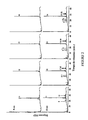
figure 1 illustre une analyse de type SDS-PAGE de la protéine purifiée de 49 kDa: ligne a, standards des poids moléculaires; ligne b, protéine purifiée de 49 kDa (1,5 µg); ligne c, protéine purifiée de 49 kDa (1,5 µg) après traitement avec de la N-glycosidase F, ligne d, N-glycosidase seulement. Le poids moléculaire des bandes des protéines et de la N-glycosidase F sont indiqués. - La
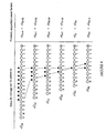
figure 2 représente l'analyse de type HPAEC des produits provenant de l'incubation de l'enzyme de 49 kDa avec des laminarioligosaccharides réduits. On a incubé la protéine purifiée de 49 kDaavec 8 mM de laminarioligosaccharides réduits de taille G11, G12, G13 ou G14 (laminarioligosaccharides réduits des résidus respectifs 11, 12, 13ou 14 de glucose) et on a représenté les profils HPAEC provenant d'échantillons aux temps zéro et 15 min, en indiquant la taille des produits principaux. - La
figure 3 représente une analyse de type HPAEC de produits provenant de l'incubation de l'enzyme de 49 kDaavec 8 mM de rG16. On a représenté les profils HPAEC provenant d'échantillons pris aux temps zéro, 30 min et 120 min, en indiquant la taille des produits principaux. - La
figure 4 illustre l'action de la transférase et des produits formés provenant des laminarioligosaccharides réduits. - La
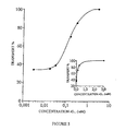
figure 5 illustre l'effet des concentrations variantes des substrats. On a incubé la transférase de 49 kDaavec 3 µM de [3H]-rG11 (1x10⁶ cpm) plus des quantités variantes de rG11 non marqués. On a déterminé le % du transfert en comparant la proportion du marquage formé sous forme de rG6 et rG16. Le supplément présente les même données avec la concentration des substrats présentée sous forme d'une échelle linéaire. - La
figure 6 illustre les effets du pH sur la vitesse du transfert. On a incubé la transférase de 49 kDaavec 8 mM de [3H]-rG11 (1x10⁶ cpm). On a déterminé les vitesses des réactions en mesurant la quantité de marquage formé sous forme de produit rG16. Les tampons utilisés étaient: •, citrate de sodium/acide citrique; o, imidazole/acide citrique; X, acétate de sodium/acide acétique; ■, Tris/glycine, ▲, phosphate/NaOH; □, Tris/acide acétique; Δ, glycine/HCl. - La
figure 7 représente la séquence provenant du gène BGT2 de A.fumigatus (A) comparée aux gènes PHR1 et GAS1 isolés de C.albicans (C) et S. cerevisiae (S). L'acide aminé surligné correspond au supposé intron. - La
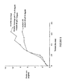
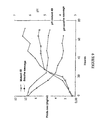
figure 8 illustre la vitesse de croissance du mutant 49 comparé à la souche sauvage. L'indication d'une valeur de pH signifie que la culture a été faite à un pH régulé (4 ou 7). - La
figure 9 illustre la croissance de la souche sauvage et celle dumutant Δ 49 et l'évolution du pH du milieu de culture au cours de la croissance des deux souches. - La
figure 10 est une comparaison des séquences de la protéine de séquence SEQ ID N°2 et des cinq protéines suivantes : Phr 1p, Gas 2p, Gas 3p, Gas4p et Gas 5p. - Les
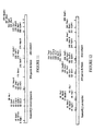
figures 11 et 12 sont des cartes de restriction des séquences de gènes homologues au gène BGT2, appelés respectivement BGT4 et BGT3.
- The
figure 1 illustrates an SDS-PAGE analysis of the purified 49 kDa protein: a-line, standard molecular weights; b line, purified 49 kDa protein (1.5 μg); lane c, purified 49 kDa protein (1.5 μg) after treatment with N-glycosidase F, d-line, N-glycosidase only. The molecular weight of protein bands and N-glycosidase F are indicated. - The
figure 2 represents the HPAEC type analysis of products from the incubation of the 49 kDa enzyme with reduced laminarioligosaccharides. The purified 49 kDa protein was incubated with 8 mM laminarioligosaccharides reduced in size G 11 , G 12 , G 13 or G 14 (reduced laminarioligosaccharides of therespective glucose residues - The
figure 3 represents an HPAEC type analysis of products from the incubation of the 49 kDa enzyme with 8 mM rG 16 . The HPAEC profiles from samples taken at zero, 30 min and 120 min times are shown, indicating the size of the main products. - The
figure 4 illustrates the action of transferase and products formed from reduced laminarioligosaccharides. - The
figure 5 illustrates the effect of variant concentrations of substrates. The 49 kDa transferase was incubated with 3 μM [ 3 H] -rG 11 (1x10 6 cpm) plus varying amounts of unlabeled rG 11 . The% transfer was determined by comparing the proportion of the labeling formed as rG 6 and rG 16 . The supplement presents the same data with the concentration of the substrates presented in the form of a linear scale. - The
figure 6 illustrates the effects of pH on the transfer speed. The 49 kDa transferase was incubated with 8 mM [ 3 H] -RG 11 (1x10 6 cpm). Reaction rates were determined by measuring the amount of labeling formed as product rG 16 . The buffers used were: • sodium citrate / citric acid; o, imidazole / citric acid; X, sodium acetate / acetic acid; Tris / glycine, ▲, phosphate / NaOH; □, Tris / acetic acid; Δ, glycine / HCl. - The
figure 7 represents the sequence from the A. fumigatus BGT2 gene (A) compared to the PHR1 and GAS1 genes isolated from C.albicans (C) and S. cerevisiae (S). The highlighted amino acid corresponds to the supposed intron. - The
figure 8 illustrates the growth rate of the mutant 49 compared to the wild-type strain. The indication of a pH value means that the culture was made at a regulated pH (4 or 7). - The
figure 9 illustrates the growth of the wild-type strain and that of themutant Δ 49 and the evolution of the pH of the culture medium during the growth of the two strains. - The
figure 10 is a comparison of the sequences of the protein of sequence SEQ ID No. 2 and the following five proteins: Phr 1p, Gas 2p, Gas 3p, Gas 4p andGas 5p. - The
Figures 11 and 12 are restriction maps of gene sequences homologous to the BGT2 gene, called BGT4 and BGT3, respectively.
On a fait pousser la souche CBS 144-89 de type A. fumigatus (disponible à la Collection Centralbureau voor Schimmelculture) dans un fermenteur de 15 L dans du glucose à 2%, de la mycopeptone à 1% (Biokar Diagnostics) plus du silicone antimousse 426R à 0,1% (Rhodorsil) à 25°C (agitation à 500 tpm, aération à 0,5 vol.vol-1 .min-1) pendant 42 h. On a utilisé en tant qu'inoculum une culture ayant poussée pendant 3 jours dans un fermenteur de 2 L sous les mêmes conditions (8% (v/v)). Les mycélium ont été collectés par filtration sous vide et cassés en les passant à travers un broyeur de type Dyno en présence de billes de verre (W.A. Bachofen AG, Basel, Suisse) (0,5-0,75 mm de diamètre). On a suivi la progression de la dislocation des cellules par examen au microscope. La suspension de mycélium cassée a été centrifugée (8000 g, 15 min) et on a lavé le culot contenant les parois cellulaires 3 fois avec de l'eau et une fois dans de l'acétate de Na 50 mM, Ph 5,6 contenant de l'azide de Na 5 mM avant de le remettre en suspension dans le même tampon (250 g de poids humide par L de tampon) et on l'a incubé (agitation à 200 tpm) à 37°C. Après 72 h, on a centrifugé la suspension (10000g 15 min) et on a placé le surnageant dans une canalisation pour dialyse, on l'a concentré 5 à 10 fois avec du polyéthylèneglycol 20000 dialysé contre de l'acétate de Na 5 mM, pH 5,6, recentrifugé (10000g, 15 min) et filtré (filtre de 0,45 µm). Cette préparation est référencée comme autolysat.CBS 144-89 strain A. fumigatus (available from the Centralbureau voor Schimmelculture Collection) was grown in a 15 L fermentor in 2% glucose, 1% mycopeptone (Biokar Diagnostics) plus silicone. 0.1% 426R antifoam (Rhodorsil) at 25 ° C. (shaking at 500 rpm, aeration at 0.5 vol.vol -1 .min -1 ) for 42 h. As inoculum, a culture was grown for 3 days in a 2 L fermentor under the same conditions (8% (v / v)). The mycelia were collected by vacuum filtration and broken by passing them through a Dyno-type grinder in the presence of glass beads (WA Bachofen AG, Basel, Switzerland) (0.5-0.75 mm in diameter). The progression of cell dislocation was monitored by microscopic examination. The broken mycelium suspension was centrifuged (8000 g, 15 min) and the pellet containing the cell walls was washed 3 times with water and once in 50 mM Na acetate, pH 5.6 containing 5 mM Na azide before resuspension in the same buffer (250 g wet weight per L buffer) and incubated (shaking at 200 rpm) at 37 ° C. After 72 h, the suspension was centrifuged (10000g 15 min) and the supernatant was placed in a dialysis line, concentrated 5-10 times with 20000 polyethylene glycol dialysed against 5 mM Na acetate, pH 5.6, recentrifuged (10000g, 15 min) and filtered (0.45 μm filter). This preparation is referenced as an autolysate.
On a testé des fractions collectées au cours de chaque étape de la chromatographie pour l'activité enzymatique en utilisant le test non radioactif de la transférase (voir ci-dessous). On a appliqué l'autolysat dialysé et concentré sur une colonne (4 x 18 cm) de DEAE-Sepharose Fast-Flow (Pharmacia) équilibrée avec de l'acétate de Na 5 mM, pH 5,6 et on a élué avec un gradient linéaire de NaCl 0 à 1 M (2000 ml) à une vitesse d'écoulement de 240 ml.h-1. On a collecté les fractions contenant l'activité de la transférase, on les a dialysées contre un tampon contenant du β-mercaptoéthanol 10 mM, de l'EDTA 5 mM, de l'acétate de Na 10 mM, pH 4,0 et on les a appliquées sur une colonne Mono S (HR 5/5 Pharmacia) et éluées avec un gradient linéaire de NaCl (0 à 300 mM en 40 min) à une vitesse d'écoulement de 0,8 ml.min-1. On a collecté la fraction contenant l'activité de la transférase, on l'a dialysée contre du Tris/HCl 10 mM, pH 7,0 et on l'a déposée sur une colonne DEAE-5PW (8 x 75 mm, TosoHaas) et on l'a éluée avec un gradient linéaire de NaCl (0 à 300 mM en 60 min) avec une vitesse d'écoulement de 0,75 ml.min-1. On a collecté les fractions contenant l'activité de la transférase, on les a dialysées contre un tampon contenant du β-mercaptoéthanol 10 mM, de l'EDTA 5 mM, de l'acétate de Na 10 mM, pH 4,0 et on les a déposées sur une colonne CM-5PW (8 x 75 mm, TosoHaas) et on a élué avec un gradient linéaire de NaCl (0 à 300 mM en 60 min) avec une vitesse d'écoulement de 0,8 ml.min-1. On a collecté les tractions contenant la transférase et on les a concentrées par un speed-vac et fractionnées sur une colonne superdex HR75 (Pharmacia) équilibrée avec du Tris/HCl 10 mM, pH 7,0 contenant du NaCl 150 mM, à une vitesse d'écoulement de 0,75 ml.min-1. On a collecté les fractions contenant la transférase purifiée, dialysé contre du citrate de Na 5 mM, pH 5,0, concentré par speedvac et conservé à -20°C jusqu'à utilisation.Fractions collected during each chromatography step for enzymatic activity were tested using the non-radioactive transferase assay (see below). The dialyzed and concentrated autolysate was applied on a DEAE-Sepharose Fast-Flow (Pharmacia) column (4 x 18 cm) equilibrated with 5 mM Na acetate, pH 5.6 and eluted with a gradient.
On a dosé les fractions d'enzymes pour la présence de l'activité de la transférase en incubant dans du citrate de Na 50 mM, pH 5.0 à 37°C (10 µl de volume de dosage) avec un laminarioligosaccharide réduit au borohydrure (8 mM final) d'au moins une taille G10. On a pris des échantillons (3 µl) à des temps différents et ajouté du NaOH 50 mM refroidi dans de la glace (47 µl) pour terminer la réaction et on a congelé jusqu'à l'analyse par chromatographie échangeuse d'anions à haute performante (HPAEC). A cause des variations de l'intensité des pics de jour en jour avec une détection par Pulsed Electrochemical Detector (PED), on a quantifié l'activité de la transférase en utilisant des laminarioligosaccharides réduits marqués au 3H en tant que substrats et en mesurant l'apparition du marquage dans les produits après la séparation par chromatographie HPAEC, en utilisant un appareil en ligne 150TR d'analyse de vitesse de scintillation Radiomatic (Packard). A moins que cela ne soit mentionné, les dosages pour les études de la caractérisation des enzymes sont effectuées comme ci-dessus avec 0,25 µg de transférase purifiée.Enzyme fractions were assayed for the presence of transferase activity by incubating in 50 mM Na citrate, pH 5.0 at 37 ° C (10 μl assay volume) with a borohydride reduced laminarioligosaccharide (8). final mM) of at least one size G 10 . Samples (3 μl) were taken at different times and ice-cold 50 mM NaOH (47 μl) added to terminate the reaction and frozen until analysis by high-pressure anion exchange chromatography. performance (HPAEC). Due to variations in day-to-day peak intensity with Pulsed Electrochemical Detector (PED) detection, transferase activity was quantified using reduced 3 H-labeled laminarioligosaccharides as substrates and measuring the appearance of labeling in the products after separation by HPAEC chromatography, using a Radiomatic scintillation rate analysis device 150TR (Packard). Unless mentioned, assays for enzyme characterization studies are performed as above with 0.25 μg of purified transferase.
On a mesuré l'activité de le β-glucanase dans les fractions protéiques par un test réducteur à base de sucre en utilisant le réactif hydrazide de l'acide hydroxybenzoique avec de la laminarine réduite au borohydrure à la place de la carboxyméthyl pachymane an tant que substrat (
On a analysé les échantillons provenant des tests de la transférase sur une colonne analytique Dionex CarboPac PA1 (4 x 250 mm) (avec une colonne de contrôle PA1) sur un système Dionex de type HPAEC avec une détection pulsée électrochimique (cellule PED-2), équipé avec une combinaison d'électrodes de référence pH-Ag/AgCl et en utilisant un potentiel de 0,4 V pour la première 0,5 s de détection. On a élué les oligosaccharides dans les conditions suivantes: vitesse d'écoulement de 1 ml/min, tampon A: NaOH 50 mM; tampon B: acétate de sodium 500 mM dans du NaOH 50 mM;gradient: 0 à 2 min, 98% A 2% B (isocratique), 2 à 15min 75% A 25% B (linéaire), 15 à 45 min 60% A 40% B (linéaire).The samples from the transferase assay were analyzed on a Dionex CarboPac PA1 analytical column (4 x 250 mm) (with a PA1 control column) on a Dionex HPAEC system with pulsed electrochemical detection (PED-2 cell). , equipped with a combination of pH-Ag / AgCl reference electrodes and using a potential of 0.4 V for the first 0.5 s of detection. The oligosaccharides were eluted under the following conditions: flow rate of 1 ml / min, buffer A: 50 mM NaOH; buffer B: 500 mM sodium acetate in 50 mM NaOH, gradient: 0-2 min, 98
Les standards de laminarioligosaccharides ont été obtenus chez Seikagaku (Japon).Laminarioligosaccharide standards were obtained from Seikagaku (Japan).
On a visualisé les laminariologosaccharides par une chromatographie en couche mince sur un gel de silice 60 (Kieselgel, Merck) en utilisant du n-butanol/acide acétique/eau; (2/1/1,5) en tant qu'éluant et une coloration à base d'orcinol sulfurique.Laminariologosaccharides were visualized by thin layer chromatography on silica gel 60 (Kieselgel, Merck) using n-butanol / acetic acid / water; (2/1 / 1.5) as eluent and a staining based on orcinol sulfuric acid.
On a également déterminé le degré de polymérisation (dp) des oligosaccharides par chromatographie de type HPAE en utilisant un détecteur pulsé électrochimique et une colonne échangeuse d'anions (Carbo6PAC PA1, 4,6 x 250 mm, Dionex).The degree of polymerization (dp) of the oligosaccharides was also determined by HPAE chromatography using an electrochemical pulsed detector and an anion exchange column (Carbo6PAC PA1, 4.6x250 mm, Dionex).
On a obtenu les laminarioligosaccharides par une hydrolyse partielle acide (TFA 6,5 M, 15 min, 100°C, suivi par du TFA 1 M, 45 min, 100°C) de curdlan (Serva). On a éliminé le TFA par une évaporation rotatoire en présence de méthanol. On a réduit les oligosaccharides toute la nuit avec du NaBH4 (1:0,5 (m/m) dans NaOH 0,1 M à température ambiante). On a préparé de manière similaire les extrémités réduites des laminarioligosaccharides marquées au 3H par réduction avec du NaB3H4 (Amersham, 20-40 Ci/mmol, 10 mCi par mg d'oligosaccharides) toute la nuit suivi par une réduction supplémentaire avec du NaBH4 comme avant. On a détruit l'excès de NaBH4 en ajoutant de l'acide acétique jusqu'à un pH de 5-6, et on a éliminé les sels de borate par une évaporation rotatoire en présence de méthanol. On a dessalé les oligosaccharides réduits par une filtration sur gel sur une colonne de Sephadex G15 (1,2 x 80 cm, 8 ml.h-1, équilibrée en eau) et on les a collecté après détection par la méthode à l'orcinol-acide sulfurique (
On a analysé des échantillons de protéines par SDS-PAGE (
On a analysé deux échantillons: le laminarioligosaccharide réduit G10 utilisé comme standard et un oligosaccharide G16 réduit qui a été obtenu après incubation du rG10 avec la transférase et purifié par HPAEC. On a remplacé le deutérium dans les échantillons secs par hyophilisation en dissolvant dans D2O (99,95%, Solvents Documentation Synthese, France). On a enregistré les spectres à 300 K et 318 K sur une spectrométrie 500 de type Variant Unity opérant à une fréquence des protons de 500 MHz. La résonance des OH de l'eau résiduelle a été supprimée par une rayonnement sélectif au cours du temps de relaxation. On a utilisé de l'acide 3-triméthylsilylpropionique de sodium comme témoin externe.Both samples were analyzed: the reduced laminarioligosaccharide G 10 used as a standard and a reduced oligosaccharide G 16 which was obtained after incubation of rG 10 with the transferase and purified by HPAEC. Deuterium was replaced in the dry samples by hyophilization by dissolving in D 2 O (99.95%, Solvents Documentation Synthese, France). Spectra at 300 K and 318 K were recorded on 500 Variant Unity spectrometry operating at a 500 MHz proton frequency. The OH resonance of the residual water was suppressed by selective radiation during the relaxation time. Sodium 3-trimethylsilylpropionic acid was used as an external control.
Afin d'étudier les activités de la β-glucanosyl transférase associée à la paroi cellulaire dans A. fumigatus, on a développé un test de chromatographie échangeuse d'anions à haute performance (HPAEC) en utilisant des laminarioligosaccharides réduits au borohydrure comme substrats. On a détecté une nouvelle activité de la β-(1-3)-glucosyl transférase dans les fractions semi-purifiées provenant de l'autolysat de la paroi cellulaire de A.fumigatus, qui reste associée à une protéine de 49 kDa tout le long de sa purification.In order to study the activities of cell wall-associated β-glucanosyl transferase in A. fumigatus, a high performance anion exchange chromatography (HPAEC) test was developed using borohydride reduced laminarioligosaccharides as substrates. A new β- (1-3) -glucosyl transferase activity was detected in the semi-purified fractions from A.fumigatus cell wall autolysate, which remains associated with a 49 kDa protein all along of his purification.
On a purifié la protéine à une homogénéité apparente avec quatre étapes de chromatographie échangeuse d'ions et une étape de filtration sur gel.The protein was purified to apparent homogeneity with four ion exchange chromatography steps and one gel filtration step.
L'activité de la transférase était clairement détectable après seulement la seconde étape de chromatographie (Mono S). L'analyse par SDS-PAGE de la fraction purifiée a montré une bande principale à 49 kDa (
L'analyse par HPAEC des produits résultant de l'incubation de la protéine de 49 kDa avec un laminarioligosaccharide réduit au borohydrure (rGn) de taille G10 ou plus grand a permis la caractérisation d'une nouvelle activité de type glucanosyl transférase.HPAEC analysis of the products resulting from the incubation of the 49 kDa protein with a reduced boron hydride laminarioligosaccharide (rG n ) of size G 10 or greater allowed the characterization of a new glucanosyl transferase activity.
Les produits initiaux principaux provenant de l'incubation avec rG11 étaient rG6 et rG16; rG12 donnait rG6 + rG7 et rG17 + rG18, rG13 donnait rG6 à rG8 et rG18 à rG20, et rG14 résultait de la formation de rG6 à rG9 et rG19 à rG22 (
Le profil des produits obtenus (
E + rG11 → E.G5 + rG6
E.G5 + rG11 → E + rG16
où E représente l'enzyme. La transférase coupe rG12 dans deux endroits différents engendrent deux produits différents de type transférase :
E = rG12 → E.G6 + rG6 → E.G5 + rG7
E. G6 + rG12 → E + rG18
E.G5 + rG12 → E + rG17
The profile of the products obtained (
E + rG 11 → EG 5 + rG 6
EG 5 + rG 11 → E + rG 16
where E represents the enzyme. The trG 12 transferase in two different locations generates two different transferase products:
E = rG 12 → EG 6 + rG 6 → EG 5 + rG 7
E.G 6 + rG 12 → E + rG 18
EG 5 + rG 12 → E + rG 17
De manière similaire avec rG13 et rG14, la transférase coupe dans trois ou quatre endroits différents, respectivement, chaque fois transférant la partie de l'extrémité non réduite à une autre molécule acceptrice rG13 ou rG14.Similarly with rG 13 and rG 14, the transferase cuts in three or four different locations, respectively, each time transferring the non-reduced end portion to another acceptor molecule rG 13 or rG 14 .
Des analyses supplémentaires d'incubations de la transférase de 49 kDa avec des laminarioligosaccharides réduits et plus petits ont montré que la réaction avec rG10 donnait rG5 + rG6 et rG14 + rG15 comme produits initiaux majeurs, alors que la réaction avec rG9 était extrêmement lente, formant des petits pics de rG5 à rG8 et rC10 à rG13. On n'a pas détecté de produits par incubation avec des laminarioligosaccharides réduits de taille G8 et plus petit.Additional analyzes of 49 kDa transferase incubations with reduced and smaller laminarioligosaccharides showed that the reaction with rG 10 gave rG 5 + rG 6 and rG 14 + rG 15 as major initial products, whereas the reaction with rG 9 was extremely slow, forming small peaks from rG 5 to rG 8 and rC 10 to rG 13 . No products were detected by incubation with reduced laminarioligosaccharides of size G 8 and smaller.
Pour déterminer la vitesse de la réaction relative des enzymes avec des laminarioligosaccharides de tailles variables, on a incubé l'enzyme de 49 kDa (0,25 µg) séparément avec 8 mM de rG10 à rG15 marqués au 3H et on a mesuré la vitesse de formation des produits marqués. La vitesse avec rG10 (328 nmol.min-1) était approximativement égale à 50% de celle avec des substrats plus gros et il n'apparaît aucune différence significative entre les vitesses de réaction pour rG11 à rG15 (648 ± 46 nmol.min-1 .mg protéine-1).To determine the rate of relative enzyme reaction with laminarioligosaccharides of varying sizes, the 49 kDa enzyme (0.25 μg) was incubated separately with 8 mM rG 10 to rG 15 labeled at 3 H and measured. the speed of formation of marked products. The rate with rG 10 (328 nmol.min -1 ) was approximately 50% of that with larger substrates and there is no significant difference in reaction rates for rG 11 to rG 15 (648 ± 46 nmol .min -1 .mg protein -1 ).
L'analyse d'incubations plus longues d'enzymes purifiées avec des laminarioligosaccharides réduits de taille au moins égale à G10 a montré que les produits de la transférase initiale peuvent être ultérieurement ré-utilisés soit comme donneurs soit comme accepteurs, engendrant la formation de produits de taille croissante, jusqu'à ce qu'ils soient éliminés de la solution à cause de leur insolubilité dans le tampon aqueux. Une incubation de 30 min avec rG16 (contenant certains contaminants rG15) a engendré la formation de produits initiaux majeurs réduits, de tailles G6 à G11 et G21 à G26 (
Pour déterminer le plus petit laminarioligosaccharides qui puisse agir comme accepteur, la transférase purifiée a été incubée avec 4 mM de rG11 comme donneur plus 16 mM de rG8 ou plus petit comme accepteur. Les analyses des incubations contenant rG11 plus rG4 ou plus petit ont montré la formation de rG6 et rG16 comme seuls produits initiaux majeurs, indiquant que seul rG11 a été utilisé comme accepteur. Cependant, des incubations contenant rG11 plus rG5 à rG8 ont montré des produits additionnels de la transférase en conformité avec les derniers oligosaccharides ayant été utilisés comme accepteurs. Par exemple, la réaction de rG11 plus rG7 a engendré la formation initiale de rG6, rG12 et rG16 en conformité avec:
E + rG11 → E.G5 + rG6
E.G5 + rG11 →E+rG16
E.G5 + rG7 → E + rG12
La vitesse relative de la réaction avec ces accepteurs a été déterminée en utilisant 2 mM de rG11 comme donneur plus 32 mM d'accepteur réduit marqué au 3H et en mesurant la formation du produit marqué de la transférase (
E + rG 11 → EG 5 + rG 6
EG 5 + rG 11 → E + rG 16
EG 5 + rG 7 → E + rG 12
The relative rate of reaction with these acceptors was determined using 2 mM of rG 11 as donor plus 32 mM reduced acceptor labeled with 3 H and measuring the formation of labeled product transferase (
La transférase n'a montré aucune activité envers les gentiooligosaccharides (taille G3-8), la chitohexaose, la cellopentaose ou la maltoheptaose, ni en présence ni en absence de rG11, suggérant que l'enzyme utilisant exclusivement le β-(I-3) glucane comme un donneur a été démontré en utilisant un G10 réduit branche (rG10*) similaire de la laminaridécaose, sauf que la sixième liaison à partir de l'extrémité réduite est une liaison de type β-(1-6). L'incubation de l'enzyme de 49 kDa avec 8 mM de rG10* n'a donné aucun produits indiquant que ce n'était pas un donneur. Cependant, une incubation similaire en présence de 2 mM de rG11 a engendré la formation de rG6 et un pic éluant en position de rG15 comme produits initiaux majeurs, montrant que rG10* peut agir comme accepteur.The transferase showed no activity towards gentiooligosaccharides (size G 3-8 ), chitohexaose, cellopentaose or maltoheptaose, neither in the presence nor absence of rG 11 , suggesting that the enzyme using exclusively β- (I -3) glucan as a donor was demonstrated using a 10 G lowered leg (10 rG *) of similar laminaridécaose except that the sixth link from the reduced end is a β- type linkage (1-6 ). Incubation of the 49 kDa enzyme with 8 mM rG 10 * gave no evidence that it was not a donor. However, a similar incubation in the presence of 2 mM rG 11 has caused the formation of rG 6 and a peak eluting in the position of rG 15 as initial major products, showing that 10 rG * can act as acceptor.
Pour déterminer si la transférase de 49 kDa a produit un nouveau type de liaison au cours du transfert, le produit rG16 de la transférase a été purifié du milieu d'incubation de la transférase avec rG11. On a analysé approximativement 300 µg de produit par RMN-1H. Le spectre 1D du produit rG16 de la transférase présentait trois déplacements chimiques dans la région anomérique:
- δ = 4,68 ppm correspondant au résidu de glucose lié au groupe glucitol;
- δ = 4,75 ppm correspondant au résidu de glucose de l'extrémité non réduite;
- δ = 4,80 ppm correspondant aux résidus intrachaîne de glucose lié en β-(1-3).
- δ = 4.68 ppm corresponding to the glucose residue bound to the glucitol group;
- δ = 4.75 ppm corresponding to the glucose residue of the non-reduced end;
- δ = 4.80 ppm corresponding to intrachain residues of β- (1-3) -linked glucose.
L'intensité relative des signaux anomériques indiquait 1,1 et 13 protons respectivement. Puisque le glucitol ne donne aucun signal dans la région anomérique, cela confirme la longueur de l'oligosaccharide (16 résidus). Les constantes de couplage mesurées sur ces signaux sont en accord avec les résidus de glucose liés en β (3J1,2 = 7,9 Hz). La présence d'un seul motif de type glucose à l'extrémité non réduite indiquait que la protéine de 49 kDa a été transférée sur l'extrémité non réduite de l'accepteur β-(1-3) glucane.The relative intensity of the anomeric signals indicated 1.1 and 13 protons respectively. Since glucitol gives no signal in the anomeric region, this confirms the length of the oligosaccharide (16 residues). The coupling constants measured on these signals are in agreement with the glucose residues bound in β ( 3 J 1.2 = 7.9 Hz). The presence of a single non-reduced end glucose unit indicated that the 49 kDa protein was transferred to the non-reduced end of the β- (1-3) glucan acceptor.
Le spectre 1D du produit rG16 était identique, sauf pour l'intensité relative du signal de 4,80 ppm par rapport à celui du standard des laminarioligosaccharides rG10. En plus, aucune caractéristique de déplacement chimique de résidu glucose lié en (1-2), (1-4), ou (1-6) n'a été vue confirmant que le produit rG16 était une laminari-hexadécaose. L'élution conjointe du produit rG16 avec la référence de rG16 sur HPAEC et l'insolubilité du produit le plus gros sont en confirmité avec la production de liaison de type β-(1-3) au cours du transfert.The 1D spectrum of the rG 16 product was identical except for the relative signal intensity of 4.80 ppm compared to that of the rG 10 laminarioligosaccharide standard. In addition, no chemical shift characteristics of glucose residue bound in (1-2), (1-4), or (1-6) were seen to confirm that the product rG 16 was a laminari-hexadecaose. The elution of the product rG 16 with the reference of rG 16 on HPAEC and the insolubility of the larger product are in agreement with the production of β- (1-3) type binding during the transfer.
Pour déterminer si l'abaissement de la concentration des accepteurs stimulerait les réactions d'hydrolyse, on a incubé la transférase de 49 kDa avec 3 µl de [3H]-r-G11 plus des quantités décroissantes de rG11 non marqué. On a observé un déplacement du transfert (i) à l'hydrolyse (ii) (
E + [3H]-rG11 → E.G5 +[3H]-rG6
- (i)
E.G5 + [3H)-rG11 → E +[3H]-rG16
- (ii)
E.G5 + H2O → E + G5
E + [3 H] -rg 11 EG 5 → + [3 H] -rg 6
- (I)
5 EG + [3 H) -Rg 11 → E + [3 H] -rg 16
- (Ii)
EG 5 + H 2 O → E + G 5
On a testé l'enzyme de 49 kDa à des valeurs de pH différentes, vérifié la stabilité au cours du stockage et on a testé l'activité de l'enzyme N-glycosylée. L'enzyme était active dans une large gamme de pH acide, montrant une activité supérieure à 50% du maximum entre pH 2,5 et 6,0. L'enzyme a manifiestée un pH optimum à environ 5,0 dans un tampon citrate (
L'enzyme de 49 kDa catalyse sa réaction de type transférase par un mécanisme de type bi-réaction (deux étapes) avec une hydrolyse initiale du substrat pour libérer la partie de l'extrémité réduite, et un transfert ultérieur du restant à l'extrémité non réduite d'une molécule de substrat jouant le rôle de molécule acceptrice.The 49 kDa enzyme catalyzes its transferase type reaction by a bi-reaction type mechanism (two steps) with initial hydrolysis of the substrate to release the reduced end portion, and subsequent transfer of the remainder to the end not reduced by a substrate molecule acting as an acceptor molecule.
En utilisant rG11 en tant que substrat, il était impossible de calculer un Km apparent correspondant aux deux étapes de la réaction. Pour déterminer un Km pour le site donneur, nous avons utilisé un accepteur qui n'était pas un donneur. On a utilisé [3H]-rG7(1x10⁶cpm) comme accepteur à une concentration élevée (64 mM) avec des concentrations variées de rG11 gardées en dessous de 8 mM. Dans ces conditions, la réaction s'est effectuée avec rG11 comme donneur et rG7 était utilisé plus que rG11 comme accepteur, comme cela est déterminé par l'absence de formation du produit de la transférase rG16. La vitesse initiale de la réaction a été déterminée en mesurant l'apparition du rG12 marqué.Using rG 11 as a substrate, it was impossible to calculate an apparent Km corresponding to the two stages of the reaction. To determine a Km for the donor site, we used an acceptor that was not a donor. [ 3 H] -rG 7 (1x10 6 cpm) was used as an acceptor at a high concentration (64 mM) with varying concentrations of rG 11 kept below 8 mM. Under these conditions, the reaction was carried out with rG 11 as a donor and rG 7 was used more than rG 11 as an acceptor, as determined by the lack of formation of the rG 16 transferase product. The initial rate of the reaction was determined by measuring the appearance of the labeled rG 12 .
On a obtenu un Km apparent de 5,3 mM à partir des doubles taches de type réciproque (valeur de r2 = 0,997).An apparent Km of 5.3 mM was obtained from the reciprocal double spots (r 2 value = 0.997).
On a obtenu deux séquences d'acides aminés de la protéine BGT2 purifiée. La séquence NH2 terminale était DVTPITVKGNAFFKGDERFY et une séquence interne était DAPNWDVDNDALP. Un oligonucléotide de 38 mer sur la partie N-terminale ayant la séquence suivante SEQ ID N°4:
- (AAA GG(T/C) AA(C/T) GC(T/C) TTC TT(C/T) AAG GG(T/C) GA(T/C) GAG CG (T/C) TTC TA)
- (AAA GG (T / C) AA (C / T) GC (T / C) TTC TT (C / T) AAG GG (T / C) GA (T / C) GAG CG (T / C) TTC TA)
Le transfert est effectué sur des membranes de type Zetaprobe. On a préhybridé et hybridé les membranes à 50°C dans une solution contenant du SSC 5X, Na2HPO4 20 mM, pH 7, SDS 7%, Denhardt 10X et du sperme de saumon à 1%. On a effectué le lavage des membranes à 42°C deux fois dans une solution contenant du SSC 3X, Denhardt 10X, SDS 5%, Na2HPO4 25 mM et deux fois dans une solution de SDS à 1% SSC 1X.The transfer is carried out on membranes of Zetaprobe type. The membranes were prehybridized and hybridized at 50 ° C in a solution containing 5X SSC,
Le clonage et le séquençage du gène codant pour la protéine BGT2 a montré des homologies importantes avec les gènes PHR1 et GAS 1 identifiés auparavant dans C.albicans et S.cerecisiae respectivement (
La dislocation du gène BGT2 a été effectué en utilisant le vecteur pAN7-1 (
Le mutant Δ 49 de A.fumigatus obtenu a été déposé le 30 juillet 1996 à la CNCM sous le numéro de dépôt I-1764.The
Il ne présente aucun phénotype distinct de la souche sauvage, à l'exception d'une inhibition totale de la croissance en fermenteur après 24 heures de croissance.It shows no phenotype distinct from the wild type, except for a total inhibition of growth in fermenters after 24 hours of growth.
On a obtenu l'ADNc de BGT2 par amplification avec deux amorces de type ADNc 5' GAATTCGACGACGTTACTCCCATCACT 3' de P1 et 5'TCTAGAGGGTATGAGAAGAACAAATCA 3' de P2 provenant de 10 ng d'ADNc, 1U de taq polymérase, 200 mM de chaque amorces. On a effectué 30 cycles d'amplification, composés d'une minute à 95°C, une minute à 55°C et une minute à 72°C.The BGT2 cDNA was obtained by amplification with two 3 'cDNA and 5'TCTAGAGGGTATGAGAAGAACAAATCA 3' cDNA primers of P2 from 10 ng of cDNA, 1u of taq polymerase, 200 mM of each primers. 30 amplification cycles, consisting of one minute at 95 ° C, one minute at 55 ° C and one minute at 72 ° C were performed.
On a ensuite cloné la préparation amplifiée dans un vecteur avec l'aide d'un kit de Clonage TA (In Vitrogen).The amplified preparation was then cloned into a vector with the aid of a TA cloning kit (In Vitrogen).
Des expériences utilisant des partages de Triton X114 avec ou sans traitement à la GPI-phospholipase C a montré que la protéine BGT2 de A.fumigatus est accrochée par un résidu GPI à la membrane plasmique.Experiments using shares of Triton X114 with or without GPI-phospholipase C treatment showed that the A. fumigatus BGT2 protein is attached by a GPI residue to the plasma membrane.
L'accrochage de la protéine à la membrane n'est pas nécessaire pour conserver l'activité enzymatique. On a démontré que dans A. fumigatus la même activité était présente lorsque la protéine était soit libérée dans le milieu de culture (en absence de la liaison GPI) soit accrochée à la membrane plasmique.The attachment of the protein to the membrane is not necessary to maintain enzymatic activity. It was demonstrated that in A. fumigatus the same activity was present when the protein was either released into the culture medium (in the absence of GPI binding) or attached to the plasma membrane.
Ces résultats suggèrent que l'expression de la glucanosyltransférase peut être faite dans des vecteurs par une expression sécrétée. Le système d'expression de Pichia pastoris par Invitrogen a été choisi. On a utilisé ce système auparavant avec une autre protéine de A.fumigatus de 88 kDa et il a été confirmé que Pichia conservait très bien le site de glycosylation de la protéine native.These results suggest that expression of glucanosyltransferase can be made in vectors by secreted expression. The expression system of Pichia pastoris by Invitrogen was chosen. This system was previously used with another 88 kDa A.fumigatus protein and it was confirmed that Pichia maintained the glycosylation site of the native protein very well.
Le vecteur utilisé est pPICZα (in Vitrogen) pour la sécrétion avec un épitope myc et six résidus histidine en tandem pour une purification facile. La séquence C-tenninale responsable de l'accrochage par GPI est éliminée avant le sous clonage dans pPICZα afin d'obtenir une protéine sécrétée tronquée active du point de vue enzymatique.The vector used is pPICZα (in Vitrogen) for secretion with a myc epitope and six histidine residues in tandem for easy purification. The C-terminal sequence responsible for GPI binding is removed before subcloning into pPICZα in order to obtain an enzymatically active truncated secreted protein.
Cette protéine recombinante est utilisée pour la détection de médicaments antifongiques. L'inhibition de l'activité enzymatique peut être mesurée par une détection de type HPLC en absence de clivage et une élongation supplémentaire de tout β-(1-3) laminarioligosaccharides de dp>= 10 en présence de p49 de A.fumigatus. This recombinant protein is used for the detection of antifungal drugs. Inhibition of enzymatic activity can be measured by HPLC detection in the absence of cleavage and further elongation of any β- (1-3) laminarioligosaccharides of dp> = 10 in the presence of p49 of A. fumigatus.
L'absence de motilité du substrat laminariologosaccharide mesuré par une chromatographie en couche mince visualisé par des techniques classiques ou directement après le marquage de l'extrémité réduite avec un radical chromogène ou fluorogène peut être une technique de base pour effectuer une détection automatisée.The absence of motility of the laminariologosaccharide substrate measured by thin layer chromatography visualized by conventional techniques or directly after the labeling of the reduced end with a chromogenic or fluorogenic radical may be a basic technique for performing automated detection.
Puisque le produit de la β-(1-3)glucanosyltransférase devient insoluble dans un milieu aqueux, du fait de l'élongation de la chaîne β-glucan, l'absence de précipitation de tout produit après une incubation prolongée en utilisant un substrat marqué de façon radiocative peut également être utilisé pour la détection de médicaments.Since the product of β- (1-3) glucanosyltransferase becomes insoluble in an aqueous medium, due to the elongation of the β-glucan chain, the absence of precipitation of any product after prolonged incubation using a labeled substrate radiocatively can also be used for the drug detection.
Des amorces oligonucléotidiques dégénérées correspondant aux régions conservées de la séquence de la
- SEQ ID N°7 (P3) : 5' GSYTTCTTCKCYGGCAACGAGGTT 3'
- SEQ ID N°8 (P4') : 5' GTTGCAGCCGWATTCGGASAYGAA 3'
K est T ou G
S est C ou G
W est A ou TDegenerate oligonucleotide primers corresponding to the conserved regions of the sequence of the
- SEQ ID NO: 7 (P3): 5 'GSYTTCTTCKCYGGCAACGAGGTT 3'
- SEQ ID NO: 8 (P4 '): 5' GTTGCAGCCGWATTCGGASAYGAA 3 '
K is T or G
S is C or G
W is A or T
Les réactions PCR ont été mises en oeuvre dans un volume de 100 µl contenant 1,5 mM MgC12, 50 mM Kcl, 10 mM TrisCl (pH8), 250 µM de dATP, dGTP, dCTP et d TTP (Pharmacia), 1 µM de chaque amorce, 2,5 unités d'Amplitaq DNA polymérase (Pharmacia) et 50 ng d'ADN génomique. L'amplification a été effectuée dans un appareil (OmniGene) tout d'abord durant 5 min à 93°C, puis durant 30 cycles de 1 min à 93°C, puis de 1 min à 50 °C et de 1 min à 72°C. Les produits de la réaction PCR ont été analysés par électrophorèse sur un gel d'agarose à 1 % puis visualisés à l'aide des ultraviolets après coloration par du bromure d'éthidium.The PCR reactions were carried out in a volume of 100 μl containing 1.5 mM MgCl2, 50 mM KCl, 10 mM TrisCl (pH8), 250 μM dATP, dGTP, dCTP and TTP (Pharmacia), 1 μM of each primer, 2.5 units of Amplitaq DNA polymerase (Pharmacia) and 50 ng of genomic DNA. The amplification was carried out in an apparatus (OmniGene) first for 5 min at 93 ° C, then for 30 cycles of 1 min at 93 ° C, then 1 min at 50 ° C and 1 min at 72 ° C. ° C. The products of the PCR reaction were analyzed by electrophoresis on a 1% agarose gel and then visualized using ultraviolet light after staining with ethidium bromide.
Les fragments résultant de la réaction PCR ont été ligaturés dans pCR2.1 (TA cloning kit, In Vitrogen). Les inserts des plasmides recombinants ont été séquencés par la méthode de terminaison de chaîne à la didéoxy (Sanger et al, 1977) en utilisant la Sequenase, Version 2 (US Biochemicals) conformément aux instructions du fabricant.The fragments resulting from the PCR reaction were ligated into pCR2.1 (TA cloning kit, In Vitrogen). The inserts of the recombinant plasmids were sequenced by the dideoxy chain termination method (Sanger et al, 1977) using Sequenase, Version 2 (US Biochemicals) according to the manufacturer's instructions.
Deux séquences nucléotidiques ont été ainsi amplifiées : les séquences SEQ ID N°9 et SEQ ID N°11. Les séquences d'acides aminés déduites des séquences nucléotidiques sont les séquences SEQ ID N°10 et SEQ ID N°12, correspondant aux gènes appelés BGT4 et BGT3.Two nucleotide sequences were thus amplified: the sequences SEQ ID No. 9 and SEQ ID No. 11. The amino acid sequences deduced from the nucleotide sequences are the sequences SEQ ID No. 10 and SEQ ID No. 12, corresponding to the genes designated BGT4 and BGT3.
Ces séquences sont en particulier décrites dans la liste ci-après. Elles présentent des pourcentages d'identité avec BGT2 respectivement de 41 % et 37%.These sequences are in particular described in the list below. They have percentages of identity with BGT2 respectively of 41% and 37%.
Les cartes de restriction des deux séquences SEQ ID N°10 et SEQ ID N°12 sont représentées respectivement sur les
Les séquences SEQ ID N°9 et N°11 ont été insérées dans le plasmide PCRllqui a été introduit dans E. Coli. Ces bactéries ont été déposées le 22 août 1997 auprès de la CNCM, respectivement sous les n°I-1914 et I-1913.The sequences SEQ ID No. 9 and No. 11 were inserted into the plasmid PCR11 that was introduced into E. coli. These bacteria were deposited on August 22, 1997 with the CNCM, respectively under No. I-1914 and I-1913.
-
(1) INFORMATIONS GENERALES:
- (i) DEPOSANT:
- (A) NOM: INSTITUT PASTEUR
- (B) RUE: 25 RUE DU DOCTEUR ROUX
- (C) VILLE: PARIS
- (E) PAYS: FRANCE
- (F) CODE POSTAL: 75724
- (ii) TITRE DE L' INVENTION: BETA(1-3)GLUCANOSYLTRANSFERASE
- (iii) NOMBRE DE SEQUENCES: 12
- (iv) FORME DECHIFFRABLE PAR ORDINATEUR:
- (A) TYPE DE SUPPORT: Floppy disk
- (B) ORDINATEUR: IBM PC compatible
- (C) SYSTEME D' EXPLOITATION: PC-DOS/MS-DOS
- (D) LOGICIEL: PatentIn Release #1.0, Version #1.30 (OEB)
- (i) DEPOSITOR:
- (A) NAME: INSTITUT PASTEUR
- (B) STREET: 25 RUE DU DOCTEUR ROUX
- (C) CITY: PARIS
- (E) COUNTRY: FRANCE
- (F) POSTAL CODE: 75724
- (ii) TITLE OF THE INVENTION: BETA (1-3) GLUCANOSYLTRANSFERASE
- (iii) NUMBER OF SEQUENCES: 12
- (iv) COMPUTER-DEPENDABLE FORM:
- (A) SUPPORT TYPE: Floppy disk
- (B) COMPUTER: IBM PC compatible
- (C) OPERATING SYSTEM: PC-DOS / MS-DOS
- (D) SOFTWARE: PatentIn Release # 1.0, Version # 1.30 (EPO)
- (i) DEPOSANT:
-
(2) INFORMATIONS POUR LA SEQ ID NO: 1:
- (i) CARACTERISTIQUES DE LA SEQUENCE:
- (A) LONGUEUR: 1359 paires de bases
- (B) TYPE: nuclotide
- (C) NOMBRE DE BRINS: simple
- (D) CONFIGURATION: linaire
- (ii) TYPE DE MOLECULE: ADN (gnomique)
- (ix) CARACTERISTIQUE:
- (A) NOM/CLE: CDS
- (B) EMPLACEMENT:1..1357
- (xi) DESCRIPTION DE LA SEQUENCE: SEQ ID NO: 1:


- (i) CHARACTERISTICS OF THE SEQUENCE:
- (A) LENGTH: 1359 base pairs
- (B) TYPE: nuclotide
- (C) NUMBER OF BRINS: simple
- (D) CONFIGURATION: linear
- (ii) MOLECULE TYPE: DNA (gnomic)
- (ix) CHARACTERISTIC:
- (A) NAME / KEY: CDS
- (B) LOCATION: 1..1357
- (xi) DESCRIPTION OF THE SEQUENCE: SEQ ID NO: 1:
- (i) CARACTERISTIQUES DE LA SEQUENCE:
-
(2) INFORMATIONS POUR LA SEQ ID NO: 2:
- (i) CARACTERISTIQUES DE LA SEQUENCE:
- (A) LONGUEUR: 452 acides amins
- (B) TYPE: acide amin
- (D) CONFIGURATION: linaire
- (ii) TYPE DE MOLECULE: protine
- (xi) DESCRIPTION DE LA SEQUENCE: SEQ ID NO: 2:


- (i) CHARACTERISTICS OF THE SEQUENCE:
- (A) LENGTH: 452 amino acids
- (B) TYPE: amino acid
- (D) CONFIGURATION: linear
- (ii) TYPE OF MOLECULE: Protein
- (xi) DESCRIPTION OF THE SEQUENCE: SEQ ID NO: 2:
- (i) CARACTERISTIQUES DE LA SEQUENCE:
-
(2) INFORMATIONS POUR LA SEQ ID NO:3:
- (i) CARACTERISTIQUES DE LA SEQUENCE:
- (A) LONGUEUR: 426 acides amins
- (B) TYPE: acide amin
- (C) NOMBRE DE BRINS:
- (D) CONFIGURATION: linaire
- (ii) TYPE DE MOLECULE: protine
- (ix) DESCRIPTION DE LA SEQUENCE: SEQ ID NO: 3:

- (i) CHARACTERISTICS OF THE SEQUENCE:
- (A) LENGTH: 426 amino acids
- (B) TYPE: amino acid
- (C) NUMBER OF STRANDS:
- (D) CONFIGURATION: linear
- (ii) TYPE OF MOLECULE: Protein
- (ix) DESCRIPTION OF THE SEQUENCE: SEQ ID NO: 3:
- (i) CARACTERISTIQUES DE LA SEQUENCE:
-
(2) INFORMATIONS POUR LA SEQ ID NO: 4:
- (i) CARACTERISTIQUES DE LA SEQUENCE:
- (A) LONGUEUR: 38 paires de bases
- (B) TYPE: nuclotide
- (C) NOMBRE DE BRINS: simple

- (D) CONFIGURATION: linaire
- (ii) TYPE DE MOLECULE: ADN (gnomique)
- (xi) DESCRIPTION DE LA SEQUENCE: SEQ ID NO: 7:
GSYTTCTTCK CYGGCAACGA GGTT 24
- (i) CHARACTERISTICS OF THE SEQUENCE:
- (A) LENGTH: 38 base pairs
- (B) TYPE: nuclotide
- (C) NUMBER OF BRINS: simple
- (D) CONFIGURATION: linear
- (ii) MOLECULE TYPE: DNA (gnomic)
- (xi) DESCRIPTION OF THE SEQUENCE: SEQ ID NO: 7:
GSYTTCTTCK CYGGCAACGA GGTT 24
- (i) CARACTERISTIQUES DE LA SEQUENCE:
-
(2) INFORMATIONS POUR LA SEQ ID NO: 8:
- (i) CARACTERISTIQUES DE LA SEQUENCE:
- (A) LONGUEUR: 24 paires de bases
- (B) TYPE: nuclotide
- (C) NOMBRE DE BRINS: simple
- (D) CONFIGURATION: linaire
- (ii) TYPE DE MOLECULE: ADN (gnomique)
- (xi) DESCRIPTION DE LA SEQUENCE: SEQ ID NO: 8:
GTTGCAGCCG WATTCGGASA YGAA 24
- (i) CHARACTERISTICS OF THE SEQUENCE:
- (A) LENGTH: 24 base pairs
- (B) TYPE: nuclotide
- (C) NUMBER OF BRINS: simple
- (D) CONFIGURATION: linear
- (ii) MOLECULE TYPE: DNA (gnomic)
- (xi) DESCRIPTION OF THE SEQUENCE: SEQ ID NO: 8:
GTTGCAGCCG WATTCGGASA YGAA 24
- (i) CARACTERISTIQUES DE LA SEQUENCE:
-
(2) INFORMATION POUR LA SEQ ID NO: 9:
- (i) CARACTERISTIQUES DE LA SEQUENCE:
- (A) LONGUEUR: 394 paires de bases
- (B) TYPE: nuclotide
- (C) NOMBRE DE BRINS: simple
- (D) CONFIGURATION: linaire
- (ii) TYPE DE MOLECULE: ADN (gnomique)
- (xi) DESCRIPTION DE LA SEQUENCE: SEQ ID NO: 9:


- (i) CHARACTERISTICS OF THE SEQUENCE:
- (A) LENGTH: 394 base pairs
- (B) TYPE: nuclotide
- (C) NUMBER OF BRINS: simple
- (D) CONFIGURATION: linear
- (ii) MOLECULE TYPE: DNA (gnomic)
- (xi) DESCRIPTION OF THE SEQUENCE: SEQ ID NO: 9:
- (i) CARACTERISTIQUES DE LA SEQUENCE:
-
(2) INFORMATIONS POUR LA SEQ ID NO: 10:
- (i) CARACTERISTIQUES DE LA SEQUENCE:
- (A) LONGUEUR: 36 acides amins
- (B) TYPE: acide amin
- (C) NOMBRE DE BRINS:
- (D) CONFIGURATION: linéaire
- (ii) TYPE DE MOLECULE: protine
- (xi) DESCRIPTION DE LA SEQUENCE: SEQ ID NO: 10:

- (i) CHARACTERISTICS OF THE SEQUENCE:
- (A) LENGTH: 36 amino acids
- (B) TYPE: amino acid
- (C) NUMBER OF STRANDS:
- (D) CONFIGURATION: linear
- (ii) TYPE OF MOLECULE: Protein
- (xi) DESCRIPTION OF THE SEQUENCE: SEQ ID NO: 10:
- (i) CARACTERISTIQUES DE LA SEQUENCE:
-
(2) INFORMATIONS POUR LA SEQ ID NO: 11:
- (i) CARACTERISTIQUES DE LA SEQUENCE:
- (A) LONGUEUR: 339 paires de bases
- (B) TYPE: nuclotide
- (C) NOMBRE DE BRINS: simple
- (D) CONFIGURATION: linaire
- (ii) TYPE DE MOLECULE: ADN (gnomique)
- DESCRIPTION DE LA SEQENCE: SEQ ID NO: 11:


- (i) CARACTERISTIQUES DE LA SEQUENCE:
- (A) LONGUEUR: 113 acides amins
- (B) TYPE: acide amin
- (C) NOMBRE DE BRINS:
- (D) CONFIGURATION: linaire
- (ii) TYPE DE MOLECULE: protine
- (xi) DESCRIPTION DE LA SEQUENCE: SEQ ID NO: 12:

- (i) CHARACTERISTICS OF THE SEQUENCE:
- (A) LENGTH: 339 base pairs
- (B) TYPE: nuclotide
- (C) NUMBER OF BRINS: simple
- (D) CONFIGURATION: linear
- (ii) MOLECULE TYPE: DNA (gnomic)
- DESCRIPTION OF THE SEQENCE: SEQ ID NO: 11:
- (i) CHARACTERISTICS OF THE SEQUENCE:
- (A) LENGTH: 113 amino acids
- (B) TYPE: amino acid
- (C) NUMBER OF STRANDS:
- (D) CONFIGURATION: linear
- (ii) TYPE OF MOLECULE: Protein
- (xi) DESCRIPTION OF THE SEQUENCE: SEQ ID NO: 12:
- (i) CARACTERISTIQUES DE LA SEQUENCE:
Claims (12)
- Protein having an activity of the β(1-3)-glucanosyltransferase type characterized in that it has at least 85% of homology with the protein having one of the sequences SEQ ID N°2 or SEQ ID N°3 as follows:


- Non-glycosylated protein having the sequence SEQ ID N°2 according to claim 1 characterized in that it has a molecular weight of 44 kD.
- Protein according to claim 1 characterized in that it carries an N-glycosyl radical.
- Glycosylated protein having the sequence SEQ ID N°3 characterized in that it has a molecular weight of 49 kD.
- Protein according to any of claims 3 and 4 characterized in that it is attached to the membrane by a glycosylphosphatidylinositol radical.
- E. coli strain deposited at the CNCM under the deposit number I-1762 carrying the sequence SEQ ID N° 1.
- E. coli strain deposited at the CNCM under the deposit number I-1763 carrying a complementary DNA sequence able to code for a protein of SEQ ID N°2.
- Method for detecting mutations of the sequence SED ID N°1, in a biological sample containing nucleotide sequences, comprising the following steps:a) placing the biological sample in contact with the nucleotide primers P3 and P4 having the sequences SEQ ID N° 7 and SEQ ID N° 8, respectively, as follows:SEQ ID N° 7:
GSYTTCTTCKCYGGCAACGAGGTTSEQ ID N°8:the nucleotide sequences contained in the sample having been optionally put into a form enabling their hybridization under conditions enabling the hybridization of the primers with the nucleotide sequences;
GTTGCAGCCGWATTCGGASAYGAA:b) amplificating the nucleotide sequencesc) revealing the amplification products, andd) detecting the mutations in relation with the sequence SEQ ID N°1 by appropriate methods. - Process for detecting the antifungal activity of molecules by the inhibition of the β(1-3)-glucanosyltransferase activity comprising the following steps:- placing the molecule to be detected in contact of a protein according to claim 4, or coded by the sequence SEQ ID N°1, and- determining the inhibition effect of such molecule on said protein by measuring the β(1-3)-glucanosyltransferase activity of such protein.
- Process according to claim 9 characterized in that the β-(1-3) glucanosyltransferase is determined by placing said protein in the presence of a substrate of β-(1-3) glucanosyltransferase.
- Process according to claim 10 characterized in that said substrate is composed of at least one laminarioligosaccharide comprising at least 10 glucosyl radicals.
- Process according to claim 11 characterized in that the activity of the protein on its substrate is detected by chromatography, in particular by HPLC or TLC, or by any method which enables the separation of oligosaccharides having different degrees of polymerization.
Applications Claiming Priority (3)
| Application Number | Priority Date | Filing Date | Title |
|---|---|---|---|
| US2491096P | 1996-08-30 | 1996-08-30 | |
| US24910P | 1996-08-30 | ||
| PCT/FR1997/001540 WO1998008937A1 (en) | 1996-08-30 | 1997-08-29 | Method for sorting antifungal molecules acting on the glucanosyltransferase activity |
Publications (2)
| Publication Number | Publication Date |
|---|---|
| EP0932665A1 EP0932665A1 (en) | 1999-08-04 |
| EP0932665B1 true EP0932665B1 (en) | 2008-08-27 |
Family
ID=21822985
Family Applications (1)
| Application Number | Title | Priority Date | Filing Date |
|---|---|---|---|
| EP97938964A Expired - Lifetime EP0932665B1 (en) | 1996-08-30 | 1997-08-29 | Method for sorting antifungal molecules acting on the glucanosyltransferase activity |
Country Status (8)
| Country | Link |
|---|---|
| US (5) | US6551811B1 (en) |
| EP (1) | EP0932665B1 (en) |
| JP (1) | JP2001501814A (en) |
| AT (1) | ATE406437T1 (en) |
| AU (1) | AU4121997A (en) |
| DE (1) | DE69738945D1 (en) |
| ES (1) | ES2312178T3 (en) |
| WO (1) | WO1998008937A1 (en) |
Families Citing this family (3)
| Publication number | Priority date | Publication date | Assignee | Title |
|---|---|---|---|---|
| ATE406437T1 (en) * | 1996-08-30 | 2008-09-15 | Pasteur Institut | METHOD FOR IDENTIFYING ANTIFUNGIC MOLECULES ACTING ON GLUCANOSYL TRANSFERASE ACTIVITY |
| JP4657726B2 (en) * | 2002-05-20 | 2011-03-23 | マサチューセッツ インスティテュート オブ テクノロジー | A novel method for sequencing using NMR |
| KR102144998B1 (en) | 2013-08-30 | 2020-08-14 | 삼성전자주식회사 | A polypeptide confering acid tolerant property to a yeast cell, polynucleotide encoding the same, a yeast cell having increased amount of the polypeptide, a method of producing a product using the yeast cell and a method of producing acid tolerant yeast cell |
Family Cites Families (1)
| Publication number | Priority date | Publication date | Assignee | Title |
|---|---|---|---|---|
| ATE406437T1 (en) * | 1996-08-30 | 2008-09-15 | Pasteur Institut | METHOD FOR IDENTIFYING ANTIFUNGIC MOLECULES ACTING ON GLUCANOSYL TRANSFERASE ACTIVITY |
-
1997
- 1997-08-29 AT AT97938964T patent/ATE406437T1/en not_active IP Right Cessation
- 1997-08-29 ES ES97938964T patent/ES2312178T3/en not_active Expired - Lifetime
- 1997-08-29 DE DE69738945T patent/DE69738945D1/en not_active Expired - Lifetime
- 1997-08-29 JP JP10511344A patent/JP2001501814A/en not_active Ceased
- 1997-08-29 EP EP97938964A patent/EP0932665B1/en not_active Expired - Lifetime
- 1997-08-29 AU AU41219/97A patent/AU4121997A/en not_active Abandoned
- 1997-08-29 US US09/242,913 patent/US6551811B1/en not_active Expired - Lifetime
- 1997-08-29 WO PCT/FR1997/001540 patent/WO1998008937A1/en active IP Right Grant
- 1997-09-02 US US08/922,171 patent/US5989895A/en not_active Expired - Lifetime
-
2003
- 2003-01-21 US US10/347,278 patent/US7060454B2/en not_active Expired - Fee Related
- 2003-01-21 US US10/347,252 patent/US7022495B2/en not_active Expired - Fee Related
-
2006
- 2006-01-06 US US11/326,609 patent/US20060211013A1/en not_active Abandoned
Also Published As
| Publication number | Publication date |
|---|---|
| US5989895A (en) | 1999-11-23 |
| AU4121997A (en) | 1998-03-19 |
| EP0932665A1 (en) | 1999-08-04 |
| ES2312178T3 (en) | 2009-02-16 |
| JP2001501814A (en) | 2001-02-13 |
| US7060454B2 (en) | 2006-06-13 |
| ATE406437T1 (en) | 2008-09-15 |
| US20030152986A1 (en) | 2003-08-14 |
| US7022495B2 (en) | 2006-04-04 |
| DE69738945D1 (en) | 2008-10-09 |
| US20030147856A1 (en) | 2003-08-07 |
| US6551811B1 (en) | 2003-04-22 |
| WO1998008937A1 (en) | 1998-03-05 |
| US20060211013A1 (en) | 2006-09-21 |
Similar Documents
| Publication | Publication Date | Title |
|---|---|---|
| Aufauvre-Brown et al. | Aspergillus fumigatus chsE: A Gene Related toCHS3ofSaccharomyces cerevisiaeand Important for Hyphal Growth and Conidiophore Development but Not Pathogenicity | |
| Jigami et al. | Mannosylphosphate transfer to yeast mannan | |
| Munro et al. | Chs1 of Candida albicans is an essential chitin synthase required for synthesis of the septum and for cell integrity | |
| Schmalhorst et al. | Contribution of galactofuranose to the virulence of the opportunistic pathogen Aspergillus fumigatus | |
| Li et al. | Glycosylphosphatidylinositol (GPI) anchor is required in Aspergillus fumigatus for morphogenesis and virulence | |
| He et al. | Characterization of A spergillus nidulans α‐glucan synthesis: roles for two synthases and two amylases | |
| Southard et al. | Molecular analysis of the Candida albicans homolog of Saccharomyces cerevisiae MNN9, required for glycosylation of cell wall mannoproteins | |
| Ragni et al. | GAS2 and GAS4, a pair of developmentally regulated genes required for spore wall assembly in Saccharomyces cerevisiae | |
| EP0932665B1 (en) | Method for sorting antifungal molecules acting on the glucanosyltransferase activity | |
| De Medina‐Redondo et al. | The β‐1, 3‐glucanosyltransferase gas4p is essential for ascospore wall maturation and spore viability in Schizosaccharomyces pombe | |
| Roh et al. | Rho1p mutations specific for regulation of β (1→ 3) glucan synthesis and the order of assembly of the yeast cell wall | |
| US5194600A (en) | Genes which participate in β-glucan assembly and use thereof | |
| Daher et al. | The Candida albicans Dse1 protein is essential and plays a role in cell wall rigidity, biofilm formation, and virulence | |
| Elagöz et al. | Although calnexin is essential in S. pombe, its highly conserved central domain is dispensable for viability | |
| Encinar del Dedo et al. | β-glucanase Eng2 is required for ascus wall endolysis after sporulation in the fission yeast Schizosaccharomyces pombe | |
| JPH10501408A (en) | DNA encoding GLS1 | |
| EP0783578B1 (en) | Yeast with modified permeability | |
| Ichinomiya et al. | Different functions of the class I and class II chitin synthase genes, chsC and chsA, are revealed by repression of chsB expression in Aspergillus nidulans | |
| Goldman et al. | Targeting cell wall synthesis and assembly in microbes: similarities and contrasts between bacteria and fungi | |
| CA2489194A1 (en) | Polypeptides involved in candida biofilm formation and uses thereof | |
| Cervera et al. | Molecular cloning and characterization of a Candida albicans gene coding for cytochrome c haem lyase and a cell wall‐related protein | |
| Mouyna et al. | Cell wall of Aspergillus fumigatus: a dynamic structure | |
| López-Ramírez et al. | Functional characterization of the Sporothrix schenckii Ktr4 and Ktr5, mannosyltransferases involved in the N-linked glycosylation pathway | |
| Dols-Lafargue et al. | Polysaccharide production by grapes, must, and wine microorganisms | |
| NISHIKAWA | A Study of the GDP-mannose Transporter |
Legal Events
| Date | Code | Title | Description |
|---|---|---|---|
| PUAI | Public reference made under article 153(3) epc to a published international application that has entered the european phase |
Free format text: ORIGINAL CODE: 0009012 |
|
| 17P | Request for examination filed |
Effective date: 19990330 |
|
| AK | Designated contracting states |
Kind code of ref document: A1 Designated state(s): AT BE CH DE DK ES FI FR GB GR IE IT LI LU MC NL PT SE |
|
| 17Q | First examination report despatched |
Effective date: 20030925 |
|
| 17Q | First examination report despatched |
Effective date: 20030925 |
|
| GRAP | Despatch of communication of intention to grant a patent |
Free format text: ORIGINAL CODE: EPIDOSNIGR1 |
|
| GRAS | Grant fee paid |
Free format text: ORIGINAL CODE: EPIDOSNIGR3 |
|
| GRAA | (expected) grant |
Free format text: ORIGINAL CODE: 0009210 |
|
| AK | Designated contracting states |
Kind code of ref document: B1 Designated state(s): AT BE CH DE DK ES FI FR GB GR IE IT LI LU MC NL PT SE |
|
| REG | Reference to a national code |
Ref country code: GB Ref legal event code: FG4D Free format text: NOT ENGLISH |
|
| REG | Reference to a national code |
Ref country code: CH Ref legal event code: EP |
|
| REG | Reference to a national code |
Ref country code: IE Ref legal event code: FG4D Free format text: LANGUAGE OF EP DOCUMENT: FRENCH |
|
| REF | Corresponds to: |
Ref document number: 69738945 Country of ref document: DE Date of ref document: 20081009 Kind code of ref document: P |
|
| PG25 | Lapsed in a contracting state [announced via postgrant information from national office to epo] |
Ref country code: NL Free format text: LAPSE BECAUSE OF FAILURE TO SUBMIT A TRANSLATION OF THE DESCRIPTION OR TO PAY THE FEE WITHIN THE PRESCRIBED TIME-LIMIT Effective date: 20080827 |
|
| REG | Reference to a national code |
Ref country code: ES Ref legal event code: FG2A Ref document number: 2312178 Country of ref document: ES Kind code of ref document: T3 |
|
| PG25 | Lapsed in a contracting state [announced via postgrant information from national office to epo] |
Ref country code: FI Free format text: LAPSE BECAUSE OF FAILURE TO SUBMIT A TRANSLATION OF THE DESCRIPTION OR TO PAY THE FEE WITHIN THE PRESCRIBED TIME-LIMIT Effective date: 20080827 Ref country code: AT Free format text: LAPSE BECAUSE OF FAILURE TO SUBMIT A TRANSLATION OF THE DESCRIPTION OR TO PAY THE FEE WITHIN THE PRESCRIBED TIME-LIMIT Effective date: 20080827 |
|
| PG25 | Lapsed in a contracting state [announced via postgrant information from national office to epo] |
Ref country code: MC Free format text: LAPSE BECAUSE OF NON-PAYMENT OF DUE FEES Effective date: 20080831 |
|
| REG | Reference to a national code |
Ref country code: CH Ref legal event code: PL |
|
| REG | Reference to a national code |
Ref country code: IE Ref legal event code: FD4D |
|
| PG25 | Lapsed in a contracting state [announced via postgrant information from national office to epo] |
Ref country code: IE Free format text: LAPSE BECAUSE OF FAILURE TO SUBMIT A TRANSLATION OF THE DESCRIPTION OR TO PAY THE FEE WITHIN THE PRESCRIBED TIME-LIMIT Effective date: 20080827 Ref country code: DK Free format text: LAPSE BECAUSE OF FAILURE TO SUBMIT A TRANSLATION OF THE DESCRIPTION OR TO PAY THE FEE WITHIN THE PRESCRIBED TIME-LIMIT Effective date: 20080827 |
|
| PG25 | Lapsed in a contracting state [announced via postgrant information from national office to epo] |
Ref country code: PT Free format text: LAPSE BECAUSE OF FAILURE TO SUBMIT A TRANSLATION OF THE DESCRIPTION OR TO PAY THE FEE WITHIN THE PRESCRIBED TIME-LIMIT Effective date: 20090127 |
|
| PG25 | Lapsed in a contracting state [announced via postgrant information from national office to epo] |
Ref country code: LI Free format text: LAPSE BECAUSE OF NON-PAYMENT OF DUE FEES Effective date: 20080831 Ref country code: CH Free format text: LAPSE BECAUSE OF NON-PAYMENT OF DUE FEES Effective date: 20080831 |
|
| PLBE | No opposition filed within time limit |
Free format text: ORIGINAL CODE: 0009261 |
|
| STAA | Information on the status of an ep patent application or granted ep patent |
Free format text: STATUS: NO OPPOSITION FILED WITHIN TIME LIMIT |
|
| PG25 | Lapsed in a contracting state [announced via postgrant information from national office to epo] |
Ref country code: BE Free format text: LAPSE BECAUSE OF NON-PAYMENT OF DUE FEES Effective date: 20080831 |
|
| 26N | No opposition filed |
Effective date: 20090528 |
|
| PG25 | Lapsed in a contracting state [announced via postgrant information from national office to epo] |
Ref country code: IT Free format text: LAPSE BECAUSE OF NON-PAYMENT OF DUE FEES Effective date: 20080829 |
|
| PG25 | Lapsed in a contracting state [announced via postgrant information from national office to epo] |
Ref country code: SE Free format text: LAPSE BECAUSE OF FAILURE TO SUBMIT A TRANSLATION OF THE DESCRIPTION OR TO PAY THE FEE WITHIN THE PRESCRIBED TIME-LIMIT Effective date: 20081127 |
|
| PG25 | Lapsed in a contracting state [announced via postgrant information from national office to epo] |
Ref country code: LU Free format text: LAPSE BECAUSE OF NON-PAYMENT OF DUE FEES Effective date: 20080829 |
|
| PG25 | Lapsed in a contracting state [announced via postgrant information from national office to epo] |
Ref country code: GR Free format text: LAPSE BECAUSE OF FAILURE TO SUBMIT A TRANSLATION OF THE DESCRIPTION OR TO PAY THE FEE WITHIN THE PRESCRIBED TIME-LIMIT Effective date: 20081128 |
|
| PGRI | Patent reinstated in contracting state [announced from national office to epo] |
Ref country code: IT Effective date: 20110616 |
|
| REG | Reference to a national code |
Ref country code: FR Ref legal event code: PLFP Year of fee payment: 19 |
|
| PGFP | Annual fee paid to national office [announced via postgrant information from national office to epo] |
Ref country code: ES Payment date: 20150826 Year of fee payment: 19 Ref country code: GB Payment date: 20150813 Year of fee payment: 19 Ref country code: DE Payment date: 20150807 Year of fee payment: 19 |
|
| PGFP | Annual fee paid to national office [announced via postgrant information from national office to epo] |
Ref country code: FR Payment date: 20150831 Year of fee payment: 19 |
|
| PGFP | Annual fee paid to national office [announced via postgrant information from national office to epo] |
Ref country code: IT Payment date: 20150813 Year of fee payment: 19 |
|
| REG | Reference to a national code |
Ref country code: DE Ref legal event code: R119 Ref document number: 69738945 Country of ref document: DE |
|
| GBPC | Gb: european patent ceased through non-payment of renewal fee |
Effective date: 20160829 |
|
| REG | Reference to a national code |
Ref country code: FR Ref legal event code: ST Effective date: 20170428 |
|
| PG25 | Lapsed in a contracting state [announced via postgrant information from national office to epo] |
Ref country code: DE Free format text: LAPSE BECAUSE OF NON-PAYMENT OF DUE FEES Effective date: 20170301 Ref country code: FR Free format text: LAPSE BECAUSE OF NON-PAYMENT OF DUE FEES Effective date: 20160831 Ref country code: GB Free format text: LAPSE BECAUSE OF NON-PAYMENT OF DUE FEES Effective date: 20160829 |
|
| PG25 | Lapsed in a contracting state [announced via postgrant information from national office to epo] |
Ref country code: IT Free format text: LAPSE BECAUSE OF NON-PAYMENT OF DUE FEES Effective date: 20160829 |
|
| REG | Reference to a national code |
Ref country code: ES Ref legal event code: FD2A Effective date: 20180507 |
|
| PG25 | Lapsed in a contracting state [announced via postgrant information from national office to epo] |
Ref country code: ES Free format text: LAPSE BECAUSE OF NON-PAYMENT OF DUE FEES Effective date: 20160830 |