EP0925504B1 - Verfahren zur detektion und/oder quantitativen bestimmung von einem gliotoxischen faktor - Google Patents
Verfahren zur detektion und/oder quantitativen bestimmung von einem gliotoxischen faktor Download PDFInfo
- Publication number
- EP0925504B1 EP0925504B1 EP97919095A EP97919095A EP0925504B1 EP 0925504 B1 EP0925504 B1 EP 0925504B1 EP 97919095 A EP97919095 A EP 97919095A EP 97919095 A EP97919095 A EP 97919095A EP 0925504 B1 EP0925504 B1 EP 0925504B1
- Authority
- EP
- European Patent Office
- Prior art keywords
- cells
- macroglial
- detecting
- apoptosis
- sample
- Prior art date
- Legal status (The legal status is an assumption and is not a legal conclusion. Google has not performed a legal analysis and makes no representation as to the accuracy of the status listed.)
- Expired - Lifetime
Links
- 238000000034 method Methods 0.000 title claims abstract description 54
- 230000001509 gliotoxic effect Effects 0.000 title claims abstract description 30
- 231100000424 gliotoxic Toxicity 0.000 title claims abstract description 27
- 230000006907 apoptotic process Effects 0.000 claims abstract description 37
- 239000000523 sample Substances 0.000 claims abstract description 36
- 230000001640 apoptogenic effect Effects 0.000 claims abstract description 35
- 210000002700 urine Anatomy 0.000 claims abstract description 27
- 238000000684 flow cytometry Methods 0.000 claims abstract description 25
- 230000001464 adherent effect Effects 0.000 claims abstract description 24
- 239000012472 biological sample Substances 0.000 claims abstract description 20
- 239000001963 growth medium Substances 0.000 claims abstract description 14
- 239000002609 medium Substances 0.000 claims abstract description 14
- 238000011282 treatment Methods 0.000 claims abstract description 11
- 230000002588 toxic effect Effects 0.000 claims abstract description 9
- 231100000331 toxic Toxicity 0.000 claims abstract description 8
- 230000034994 death Effects 0.000 claims abstract description 6
- 210000004027 cell Anatomy 0.000 claims description 187
- 238000001514 detection method Methods 0.000 claims description 30
- 108020004414 DNA Proteins 0.000 claims description 26
- 230000008569 process Effects 0.000 claims description 26
- 230000017074 necrotic cell death Effects 0.000 claims description 17
- XJMOSONTPMZWPB-UHFFFAOYSA-M propidium iodide Chemical compound [I-].[I-].C12=CC(N)=CC=C2C2=CC=C(N)C=C2[N+](CCC[N+](C)(CC)CC)=C1C1=CC=CC=C1 XJMOSONTPMZWPB-UHFFFAOYSA-M 0.000 claims description 16
- 239000012634 fragment Substances 0.000 claims description 15
- 230000001338 necrotic effect Effects 0.000 claims description 14
- LFQSCWFLJHTTHZ-UHFFFAOYSA-N Ethanol Chemical compound CCO LFQSCWFLJHTTHZ-UHFFFAOYSA-N 0.000 claims description 13
- 210000001130 astrocyte Anatomy 0.000 claims description 12
- 238000002372 labelling Methods 0.000 claims description 12
- 201000006417 multiple sclerosis Diseases 0.000 claims description 12
- 108090000623 proteins and genes Proteins 0.000 claims description 12
- 102000004169 proteins and genes Human genes 0.000 claims description 12
- 210000000170 cell membrane Anatomy 0.000 claims description 11
- 238000000605 extraction Methods 0.000 claims description 9
- 230000003140 astrocytic effect Effects 0.000 claims description 8
- 230000008823 permeabilization Effects 0.000 claims description 8
- 230000003833 cell viability Effects 0.000 claims description 6
- 239000003795 chemical substances by application Substances 0.000 claims description 6
- 230000003834 intracellular effect Effects 0.000 claims description 6
- 238000011534 incubation Methods 0.000 claims description 5
- 239000003550 marker Substances 0.000 claims description 5
- 230000035755 proliferation Effects 0.000 claims description 5
- 230000003013 cytotoxicity Effects 0.000 claims description 4
- 231100000135 cytotoxicity Toxicity 0.000 claims description 4
- 238000002270 exclusion chromatography Methods 0.000 claims description 4
- 108010062580 Concanavalin A Proteins 0.000 claims description 3
- 102000053171 Glial Fibrillary Acidic Human genes 0.000 claims description 3
- 101710193519 Glial fibrillary acidic protein Proteins 0.000 claims description 3
- 108090001090 Lectins Proteins 0.000 claims description 3
- 102000004856 Lectins Human genes 0.000 claims description 3
- BFNBIHQBYMNNAN-UHFFFAOYSA-N ammonium sulfate Chemical compound N.N.OS(O)(=O)=O BFNBIHQBYMNNAN-UHFFFAOYSA-N 0.000 claims description 3
- 229910052921 ammonium sulfate Inorganic materials 0.000 claims description 3
- 235000011130 ammonium sulphate Nutrition 0.000 claims description 3
- 210000005046 glial fibrillary acidic protein Anatomy 0.000 claims description 3
- 238000004255 ion exchange chromatography Methods 0.000 claims description 3
- 239000002523 lectin Substances 0.000 claims description 3
- 238000001556 precipitation Methods 0.000 claims description 3
- 238000010186 staining Methods 0.000 claims description 3
- XIBOSSQRSQSBDM-UHFFFAOYSA-N 1-methyltetrazole;hydrobromide Chemical compound [Br-].C[NH+]1C=NN=N1 XIBOSSQRSQSBDM-UHFFFAOYSA-N 0.000 claims description 2
- 108091092356 cellular DNA Proteins 0.000 claims description 2
- 230000001472 cytotoxic effect Effects 0.000 claims description 2
- 230000007717 exclusion Effects 0.000 claims description 2
- 230000003287 optical effect Effects 0.000 claims description 2
- 230000000717 retained effect Effects 0.000 claims description 2
- 238000000539 two dimensional gel electrophoresis Methods 0.000 claims description 2
- 239000001166 ammonium sulphate Substances 0.000 claims 1
- 231100000433 cytotoxic Toxicity 0.000 claims 1
- 230000001939 inductive effect Effects 0.000 claims 1
- 210000003470 mitochondria Anatomy 0.000 claims 1
- 239000003145 cytotoxic factor Substances 0.000 abstract description 3
- 230000001988 toxicity Effects 0.000 abstract description 2
- 231100000419 toxicity Toxicity 0.000 abstract description 2
- KFZMGEQAYNKOFK-UHFFFAOYSA-N Isopropanol Chemical compound CC(C)O KFZMGEQAYNKOFK-UHFFFAOYSA-N 0.000 description 14
- 238000012360 testing method Methods 0.000 description 13
- 239000000243 solution Substances 0.000 description 12
- 235000018102 proteins Nutrition 0.000 description 9
- 239000006144 Dulbecco’s modified Eagle's medium Substances 0.000 description 8
- 238000011002 quantification Methods 0.000 description 7
- 239000000725 suspension Substances 0.000 description 6
- 208000012902 Nervous system disease Diseases 0.000 description 5
- 208000025966 Neurological disease Diseases 0.000 description 5
- 230000001413 cellular effect Effects 0.000 description 5
- 210000001175 cerebrospinal fluid Anatomy 0.000 description 5
- 201000010099 disease Diseases 0.000 description 5
- 208000037265 diseases, disorders, signs and symptoms Diseases 0.000 description 5
- 230000002518 glial effect Effects 0.000 description 5
- FAPWRFPIFSIZLT-UHFFFAOYSA-M Sodium chloride Chemical compound [Na+].[Cl-] FAPWRFPIFSIZLT-UHFFFAOYSA-M 0.000 description 4
- 239000002253 acid Substances 0.000 description 4
- 230000002378 acidificating effect Effects 0.000 description 4
- 230000000694 effects Effects 0.000 description 4
- 230000004907 flux Effects 0.000 description 4
- 238000013467 fragmentation Methods 0.000 description 4
- 238000006062 fragmentation reaction Methods 0.000 description 4
- KCXVZYZYPLLWCC-UHFFFAOYSA-N EDTA Chemical compound OC(=O)CN(CC(O)=O)CCN(CC(O)=O)CC(O)=O KCXVZYZYPLLWCC-UHFFFAOYSA-N 0.000 description 3
- 208000034189 Sclerosis Diseases 0.000 description 3
- 102000004142 Trypsin Human genes 0.000 description 3
- 108090000631 Trypsin Proteins 0.000 description 3
- 230000022131 cell cycle Effects 0.000 description 3
- 238000004587 chromatography analysis Methods 0.000 description 3
- KRKNYBCHXYNGOX-UHFFFAOYSA-N citric acid Chemical compound OC(=O)CC(O)(C(O)=O)CC(O)=O KRKNYBCHXYNGOX-UHFFFAOYSA-N 0.000 description 3
- 238000003745 diagnosis Methods 0.000 description 3
- 239000008188 pellet Substances 0.000 description 3
- 229920000729 poly(L-lysine) polymer Polymers 0.000 description 3
- 230000001681 protective effect Effects 0.000 description 3
- 239000000126 substance Substances 0.000 description 3
- 239000012588 trypsin Substances 0.000 description 3
- 241001465754 Metazoa Species 0.000 description 2
- 241000283973 Oryctolagus cuniculus Species 0.000 description 2
- 108010059712 Pronase Proteins 0.000 description 2
- 229920004890 Triton X-100 Polymers 0.000 description 2
- 239000013504 Triton X-100 Substances 0.000 description 2
- 238000004458 analytical method Methods 0.000 description 2
- 208000034615 apoptosis-related disease Diseases 0.000 description 2
- 239000013060 biological fluid Substances 0.000 description 2
- 239000000872 buffer Substances 0.000 description 2
- 238000005119 centrifugation Methods 0.000 description 2
- 238000004040 coloring Methods 0.000 description 2
- 238000004163 cytometry Methods 0.000 description 2
- 229940079593 drug Drugs 0.000 description 2
- 239000003814 drug Substances 0.000 description 2
- 238000001962 electrophoresis Methods 0.000 description 2
- 238000010348 incorporation Methods 0.000 description 2
- 230000006698 induction Effects 0.000 description 2
- 230000005764 inhibitory process Effects 0.000 description 2
- 230000002452 interceptive effect Effects 0.000 description 2
- 210000003963 intermediate filament Anatomy 0.000 description 2
- 239000007791 liquid phase Substances 0.000 description 2
- 210000004698 lymphocyte Anatomy 0.000 description 2
- 239000012528 membrane Substances 0.000 description 2
- 238000012544 monitoring process Methods 0.000 description 2
- 210000004498 neuroglial cell Anatomy 0.000 description 2
- 230000000926 neurological effect Effects 0.000 description 2
- 230000004987 nonapoptotic effect Effects 0.000 description 2
- 210000004248 oligodendroglia Anatomy 0.000 description 2
- 210000000056 organ Anatomy 0.000 description 2
- 230000007170 pathology Effects 0.000 description 2
- 230000001012 protector Effects 0.000 description 2
- 238000000746 purification Methods 0.000 description 2
- 238000011160 research Methods 0.000 description 2
- 239000011780 sodium chloride Substances 0.000 description 2
- UCSJYZPVAKXKNQ-HZYVHMACSA-N streptomycin Chemical compound CN[C@H]1[C@H](O)[C@@H](O)[C@H](CO)O[C@H]1O[C@@H]1[C@](C=O)(O)[C@H](C)O[C@H]1O[C@@H]1[C@@H](NC(N)=N)[C@H](O)[C@@H](NC(N)=N)[C@H](O)[C@H]1O UCSJYZPVAKXKNQ-HZYVHMACSA-N 0.000 description 2
- 239000006228 supernatant Substances 0.000 description 2
- 210000001519 tissue Anatomy 0.000 description 2
- 238000012546 transfer Methods 0.000 description 2
- 230000035899 viability Effects 0.000 description 2
- 238000005406 washing Methods 0.000 description 2
- OMAFFHIGWTVZOH-UHFFFAOYSA-O 1-methyl-2h-tetrazol-1-ium Chemical compound C[N+]1=CN=NN1 OMAFFHIGWTVZOH-UHFFFAOYSA-O 0.000 description 1
- BFSVOASYOCHEOV-UHFFFAOYSA-N 2-diethylaminoethanol Chemical compound CCN(CC)CCO BFSVOASYOCHEOV-UHFFFAOYSA-N 0.000 description 1
- DLNAGMLXUYEHQS-UHFFFAOYSA-N 3-O-beta-D-glucopyranosylserjanic acid Natural products COC(=O)C1(C)CCC2(CCC3(C)C(=CCC4C5(C)CCC(OC6OC(CO)C(O)C(O)C6O)C(C)(C)C5CCC34C)C2C1)C(=O)O DLNAGMLXUYEHQS-UHFFFAOYSA-N 0.000 description 1
- HRPVXLWXLXDGHG-UHFFFAOYSA-N Acrylamide Chemical compound NC(=O)C=C HRPVXLWXLXDGHG-UHFFFAOYSA-N 0.000 description 1
- 102000004121 Annexin A5 Human genes 0.000 description 1
- 108090000672 Annexin A5 Proteins 0.000 description 1
- CPELXLSAUQHCOX-UHFFFAOYSA-M Bromide Chemical compound [Br-] CPELXLSAUQHCOX-UHFFFAOYSA-M 0.000 description 1
- 229920000742 Cotton Polymers 0.000 description 1
- 229920002271 DEAE-Sepharose Polymers 0.000 description 1
- 108010067770 Endopeptidase K Proteins 0.000 description 1
- 230000035519 G0 Phase Effects 0.000 description 1
- 230000010190 G1 phase Effects 0.000 description 1
- 102000003886 Glycoproteins Human genes 0.000 description 1
- 108090000288 Glycoproteins Proteins 0.000 description 1
- FBOZXECLQNJBKD-ZDUSSCGKSA-N L-methotrexate Chemical compound C=1N=C2N=C(N)N=C(N)C2=NC=1CN(C)C1=CC=C(C(=O)N[C@@H](CCC(O)=O)C(O)=O)C=C1 FBOZXECLQNJBKD-ZDUSSCGKSA-N 0.000 description 1
- 241000699660 Mus musculus Species 0.000 description 1
- 229910019142 PO4 Inorganic materials 0.000 description 1
- 229930182555 Penicillin Natural products 0.000 description 1
- JGSARLDLIJGVTE-MBNYWOFBSA-N Penicillin G Chemical compound N([C@H]1[C@H]2SC([C@@H](N2C1=O)C(O)=O)(C)C)C(=O)CC1=CC=CC=C1 JGSARLDLIJGVTE-MBNYWOFBSA-N 0.000 description 1
- 241001505332 Polyomavirus sp. Species 0.000 description 1
- 102000006382 Ribonucleases Human genes 0.000 description 1
- 108010083644 Ribonucleases Proteins 0.000 description 1
- PMZURENOXWZQFD-UHFFFAOYSA-L Sodium Sulfate Chemical compound [Na+].[Na+].[O-]S([O-])(=O)=O PMZURENOXWZQFD-UHFFFAOYSA-L 0.000 description 1
- 101100289792 Squirrel monkey polyomavirus large T gene Proteins 0.000 description 1
- 241001080024 Telles Species 0.000 description 1
- 239000007983 Tris buffer Substances 0.000 description 1
- XSQUKJJJFZCRTK-UHFFFAOYSA-N Urea Chemical compound NC(N)=O XSQUKJJJFZCRTK-UHFFFAOYSA-N 0.000 description 1
- 240000008042 Zea mays Species 0.000 description 1
- MZVQCMJNVPIDEA-UHFFFAOYSA-N [CH2]CN(CC)CC Chemical group [CH2]CN(CC)CC MZVQCMJNVPIDEA-UHFFFAOYSA-N 0.000 description 1
- 230000009471 action Effects 0.000 description 1
- APKFDSVGJQXUKY-INPOYWNPSA-N amphotericin B Chemical compound O[C@H]1[C@@H](N)[C@H](O)[C@@H](C)O[C@H]1O[C@H]1/C=C/C=C/C=C/C=C/C=C/C=C/C=C/[C@H](C)[C@@H](O)[C@@H](C)[C@H](C)OC(=O)C[C@H](O)C[C@H](O)CC[C@@H](O)[C@H](O)C[C@H](O)C[C@](O)(C[C@H](O)[C@H]2C(O)=O)O[C@H]2C1 APKFDSVGJQXUKY-INPOYWNPSA-N 0.000 description 1
- 230000000843 anti-fungal effect Effects 0.000 description 1
- 229940121375 antifungal agent Drugs 0.000 description 1
- 238000013459 approach Methods 0.000 description 1
- 230000001363 autoimmune Effects 0.000 description 1
- 230000008901 benefit Effects 0.000 description 1
- 238000004166 bioassay Methods 0.000 description 1
- 230000003115 biocidal effect Effects 0.000 description 1
- 230000015572 biosynthetic process Effects 0.000 description 1
- 210000004958 brain cell Anatomy 0.000 description 1
- 230000009172 bursting Effects 0.000 description 1
- BQRGNLJZBFXNCZ-UHFFFAOYSA-N calcein am Chemical compound O1C(=O)C2=CC=CC=C2C21C1=CC(CN(CC(=O)OCOC(C)=O)CC(=O)OCOC(C)=O)=C(OC(C)=O)C=C1OC1=C2C=C(CN(CC(=O)OCOC(C)=O)CC(=O)OCOC(=O)C)C(OC(C)=O)=C1 BQRGNLJZBFXNCZ-UHFFFAOYSA-N 0.000 description 1
- 239000004202 carbamide Substances 0.000 description 1
- 230000015556 catabolic process Effects 0.000 description 1
- 230000030833 cell death Effects 0.000 description 1
- 230000004663 cell proliferation Effects 0.000 description 1
- 230000001684 chronic effect Effects 0.000 description 1
- 238000007398 colorimetric assay Methods 0.000 description 1
- 239000000470 constituent Substances 0.000 description 1
- 239000003246 corticosteroid Substances 0.000 description 1
- 239000013078 crystal Substances 0.000 description 1
- 239000012228 culture supernatant Substances 0.000 description 1
- 230000009089 cytolysis Effects 0.000 description 1
- 230000003583 cytomorphological effect Effects 0.000 description 1
- 230000001086 cytosolic effect Effects 0.000 description 1
- 238000006731 degradation reaction Methods 0.000 description 1
- XLMALTXPSGQGBX-GCJKJVERSA-N dextropropoxyphene Chemical compound C([C@](OC(=O)CC)([C@H](C)CN(C)C)C=1C=CC=CC=1)C1=CC=CC=C1 XLMALTXPSGQGBX-GCJKJVERSA-N 0.000 description 1
- 229960004193 dextropropoxyphene Drugs 0.000 description 1
- 230000004069 differentiation Effects 0.000 description 1
- 238000007865 diluting Methods 0.000 description 1
- 238000010790 dilution Methods 0.000 description 1
- 239000012895 dilution Substances 0.000 description 1
- 210000001840 diploid cell Anatomy 0.000 description 1
- BNIILDVGGAEEIG-UHFFFAOYSA-L disodium hydrogen phosphate Chemical compound [Na+].[Na+].OP([O-])([O-])=O BNIILDVGGAEEIG-UHFFFAOYSA-L 0.000 description 1
- 229910000397 disodium phosphate Inorganic materials 0.000 description 1
- 238000010828 elution Methods 0.000 description 1
- 230000002255 enzymatic effect Effects 0.000 description 1
- GTSMOYLSFUBTMV-UHFFFAOYSA-N ethidium homodimer Chemical compound [H+].[H+].[Cl-].[Cl-].[Cl-].[Cl-].C12=CC(N)=CC=C2C2=CC=C(N)C=C2C(C)=[N+]1CCCNCCNCCC[N+](C1=CC(N)=CC=C1C1=CC=C(N)C=C11)=C1C1=CC=CC=C1 GTSMOYLSFUBTMV-UHFFFAOYSA-N 0.000 description 1
- 238000001704 evaporation Methods 0.000 description 1
- 230000008020 evaporation Effects 0.000 description 1
- 238000002474 experimental method Methods 0.000 description 1
- 239000011536 extraction buffer Substances 0.000 description 1
- 230000002349 favourable effect Effects 0.000 description 1
- GNBHRKFJIUUOQI-UHFFFAOYSA-N fluorescein Chemical compound O1C(=O)C2=CC=CC=C2C21C1=CC=C(O)C=C1OC1=CC(O)=CC=C21 GNBHRKFJIUUOQI-UHFFFAOYSA-N 0.000 description 1
- MHMNJMPURVTYEJ-UHFFFAOYSA-N fluorescein-5-isothiocyanate Chemical compound O1C(=O)C2=CC(N=C=S)=CC=C2C21C1=CC=C(O)C=C1OC1=CC(O)=CC=C21 MHMNJMPURVTYEJ-UHFFFAOYSA-N 0.000 description 1
- 238000000265 homogenisation Methods 0.000 description 1
- XMBWDFGMSWQBCA-UHFFFAOYSA-N hydrogen iodide Chemical compound I XMBWDFGMSWQBCA-UHFFFAOYSA-N 0.000 description 1
- 230000003301 hydrolyzing effect Effects 0.000 description 1
- 238000000338 in vitro Methods 0.000 description 1
- 238000011065 in-situ storage Methods 0.000 description 1
- 230000006882 induction of apoptosis Effects 0.000 description 1
- 238000005342 ion exchange Methods 0.000 description 1
- 150000002540 isothiocyanates Chemical class 0.000 description 1
- 239000007788 liquid Substances 0.000 description 1
- 230000004807 localization Effects 0.000 description 1
- 229960000485 methotrexate Drugs 0.000 description 1
- 210000000274 microglia Anatomy 0.000 description 1
- 238000000386 microscopy Methods 0.000 description 1
- 230000002438 mitochondrial effect Effects 0.000 description 1
- 239000000203 mixture Substances 0.000 description 1
- 238000012986 modification Methods 0.000 description 1
- 230000004048 modification Effects 0.000 description 1
- 210000003097 mucus Anatomy 0.000 description 1
- VMGAPWLDMVPYIA-HIDZBRGKSA-N n'-amino-n-iminomethanimidamide Chemical compound N\N=C\N=N VMGAPWLDMVPYIA-HIDZBRGKSA-N 0.000 description 1
- 230000000626 neurodegenerative effect Effects 0.000 description 1
- 239000006174 pH buffer Substances 0.000 description 1
- 229940049954 penicillin Drugs 0.000 description 1
- 239000012071 phase Substances 0.000 description 1
- NBIIXXVUZAFLBC-UHFFFAOYSA-K phosphate Chemical compound [O-]P([O-])([O-])=O NBIIXXVUZAFLBC-UHFFFAOYSA-K 0.000 description 1
- 239000010452 phosphate Substances 0.000 description 1
- 150000003904 phospholipids Chemical class 0.000 description 1
- 238000002264 polyacrylamide gel electrophoresis Methods 0.000 description 1
- 239000011148 porous material Substances 0.000 description 1
- 238000004321 preservation Methods 0.000 description 1
- 239000001397 quillaja saponaria molina bark Substances 0.000 description 1
- 230000021419 recognition of apoptotic cell Effects 0.000 description 1
- 238000011084 recovery Methods 0.000 description 1
- 230000009467 reduction Effects 0.000 description 1
- BOLDJAUMGUJJKM-LSDHHAIUSA-N renifolin D Natural products CC(=C)[C@@H]1Cc2c(O)c(O)ccc2[C@H]1CC(=O)c3ccc(O)cc3O BOLDJAUMGUJJKM-LSDHHAIUSA-N 0.000 description 1
- 229930182490 saponin Natural products 0.000 description 1
- 150000007949 saponins Chemical class 0.000 description 1
- 230000035945 sensitivity Effects 0.000 description 1
- 238000000926 separation method Methods 0.000 description 1
- 238000012163 sequencing technique Methods 0.000 description 1
- 239000011734 sodium Substances 0.000 description 1
- 238000002415 sodium dodecyl sulfate polyacrylamide gel electrophoresis Methods 0.000 description 1
- 229910052938 sodium sulfate Inorganic materials 0.000 description 1
- 235000011152 sodium sulphate Nutrition 0.000 description 1
- 229960005322 streptomycin Drugs 0.000 description 1
- 125000003831 tetrazolyl group Chemical group 0.000 description 1
- 230000001225 therapeutic effect Effects 0.000 description 1
- 238000002560 therapeutic procedure Methods 0.000 description 1
- 238000011830 transgenic mouse model Methods 0.000 description 1
- LENZDBCJOHFCAS-UHFFFAOYSA-N tris Chemical compound OCC(N)(CO)CO LENZDBCJOHFCAS-UHFFFAOYSA-N 0.000 description 1
- 230000002485 urinary effect Effects 0.000 description 1
- 230000000007 visual effect Effects 0.000 description 1
- 238000012800 visualization Methods 0.000 description 1
- XLYOFNOQVPJJNP-UHFFFAOYSA-N water Substances O XLYOFNOQVPJJNP-UHFFFAOYSA-N 0.000 description 1
Images

Classifications
-
- G—PHYSICS
- G01—MEASURING; TESTING
- G01N—INVESTIGATING OR ANALYSING MATERIALS BY DETERMINING THEIR CHEMICAL OR PHYSICAL PROPERTIES
- G01N33/00—Investigating or analysing materials by specific methods not covered by groups G01N1/00 - G01N31/00
- G01N33/48—Biological material, e.g. blood, urine; Haemocytometers
- G01N33/50—Chemical analysis of biological material, e.g. blood, urine; Testing involving biospecific ligand binding methods; Immunological testing
- G01N33/53—Immunoassay; Biospecific binding assay; Materials therefor
- G01N33/564—Immunoassay; Biospecific binding assay; Materials therefor for pre-existing immune complex or autoimmune disease, i.e. systemic lupus erythematosus, rheumatoid arthritis, multiple sclerosis, rheumatoid factors or complement components C1-C9
-
- G—PHYSICS
- G01—MEASURING; TESTING
- G01N—INVESTIGATING OR ANALYSING MATERIALS BY DETERMINING THEIR CHEMICAL OR PHYSICAL PROPERTIES
- G01N33/00—Investigating or analysing materials by specific methods not covered by groups G01N1/00 - G01N31/00
- G01N33/48—Biological material, e.g. blood, urine; Haemocytometers
- G01N33/50—Chemical analysis of biological material, e.g. blood, urine; Testing involving biospecific ligand binding methods; Immunological testing
- G01N33/5005—Chemical analysis of biological material, e.g. blood, urine; Testing involving biospecific ligand binding methods; Immunological testing involving human or animal cells
- G01N33/5008—Chemical analysis of biological material, e.g. blood, urine; Testing involving biospecific ligand binding methods; Immunological testing involving human or animal cells for testing or evaluating the effect of chemical or biological compounds, e.g. drugs, cosmetics
- G01N33/5014—Chemical analysis of biological material, e.g. blood, urine; Testing involving biospecific ligand binding methods; Immunological testing involving human or animal cells for testing or evaluating the effect of chemical or biological compounds, e.g. drugs, cosmetics for testing toxicity
-
- G—PHYSICS
- G01—MEASURING; TESTING
- G01N—INVESTIGATING OR ANALYSING MATERIALS BY DETERMINING THEIR CHEMICAL OR PHYSICAL PROPERTIES
- G01N33/00—Investigating or analysing materials by specific methods not covered by groups G01N1/00 - G01N31/00
- G01N33/48—Biological material, e.g. blood, urine; Haemocytometers
- G01N33/50—Chemical analysis of biological material, e.g. blood, urine; Testing involving biospecific ligand binding methods; Immunological testing
- G01N33/68—Chemical analysis of biological material, e.g. blood, urine; Testing involving biospecific ligand binding methods; Immunological testing involving proteins, peptides or amino acids
- G01N33/6893—Chemical analysis of biological material, e.g. blood, urine; Testing involving biospecific ligand binding methods; Immunological testing involving proteins, peptides or amino acids related to diseases not provided for elsewhere
- G01N33/6896—Neurological disorders, e.g. Alzheimer's disease
Definitions
- the present invention relates to the determination, detection, and quantification in a sample biology of a gliotoxic factor, as associated with multiple sclerosis.
- biological sample in particular a biological fluid type sample, a living tissue or fragment of tissue, mucus, organ or fragment organs, or any culture supernatant obtained using of an aforementioned direct debit.
- gliotoxic factor a cytotoxic factor against cells glial (astrocytes, oligodendrocytes, microgliocytes), hereinafter referred to as gliotoxic factor.
- glial astrocytes, oligodendrocytes, microgliocytes
- this factor can be characterized either from a process (among others) for isolating or purify, either from different properties or biological, biochemical or chemical characteristics.
- Such a method, implemented for detection and therapeutic monitoring of multiple sclerosis supposes a so-called "invasive" sample, for example of CSF, that is to say requiring a prior medical act or surgical.
- a first object of the invention is a method for detecting and / or quantifying, in a biological sample, a gliotoxic factor, according to which has a starting fraction of said sample, possibly enriched in said factor gliotoxic by a preliminary treatment, one incubates said starting fraction with a reference culture medium comprising macroglial cells, for example immortalized, such as astrocyte cells, and macroglial cells are detected and / or quantified dead and / or alive, said biological sample being a urine sample.
- a reference culture medium comprising macroglial cells, for example immortalized, such as astrocyte cells, and macroglial cells are detected and / or quantified dead and / or alive, said biological sample being a urine sample.
- a method is provided. to detect and / or quantify, in a sample biological, a cytotoxic factor towards cells target adherents and whose cytotoxicity induces death by apoptosis of said cells, according to which one has of a starting fraction of said sample, possibly enriched in said toxic factor by treatment beforehand, the said starting fraction is incubated with a reference culture medium comprising cells target adherents, and we detect and / or quantify the adherent cells died by apoptosis, by cytometry of flow, at least one direct or indirect characteristic, associated with apoptotic adherent cells of all or part of the incubated medium, which, if it exists and / or is quantified, qualifies said biological sample as positive, that is to say containing said toxic factor.
- gliotoxic factor and target adherent cells are the magroglial cells, including astrocytic cells.
- said method of the invention comprises at least one of the following protocols (1) to (3); advantageously protocols (1) and (2) or (1) and (3) are used in combination.
- the cell fixing agent and permeabilization of the cytoplasmic membrane of cells is ethanol and the DNA marker is propidium iodide (PI).
- necrotic cells it is possible to distinguish apoptotic cells from necrotic cells.
- necrotic cells We know that the phenomena of necrosis materialize by a bursting of the wall cell and the release of the constituents of the cell. Necrotic cells are therefore distinguished from others cells, living or apoptotic, in that the integrity of their cytoplasmic membrane is not preserved.
- necrotic cells and apoptotic are necessary especially in the case drug interference in patients, said substances being able to induce phenomena of recovery of the necrotic cell population and of the population of apoptotic cells, materialized in particular by generating false results positive for the apoptosis detection test.
- This interference has indeed been observed from biological samples, for example urine, from of patients who have undergone treatment with substances interfering medication, for example methotrexate or dextropropoxyphene.
- the application of the method of the invention for specifically detect and / or quantify cells apoptotic macroglials, to a urine sample provides a particularly simple bioassay, sensitive and specific to the gliotoxic factor, for example with regard to assistance in the diagnosis of sclerosis in plates, which today requires a lot of testing or different exams over a period of time which can be relatively long.
- this may include, before the step of detection and / or quantification of macroglial cells apoptotics, a viability detection step cellular.
- the starting fraction is inoculated in the reference culture medium, then we observe the macroglial cell proliferation rate by relative to a reference value, below which said so-called positive fraction is retained to be then subjected to the stage of detection of apoptosis of macroglial cells specifically, to qualify or not the biological sample as true positive.
- the rate of proliferation of macroglial cells can be obtained by staining mitochondrial macroglial cells in the middle of culture, for example with methyltetrazolium bromide, then by measuring the optical density of the culture medium colored.
- MTT dosage methyl tetrazolium colorimetric of living cells
- GFAP glial fibrillar acid protein
- a determination of the changes occurring at level of phospholipids in the cytoplasmic membrane of apoptotic cells, without affecting its integrity, and put highlighted by annexin V can also be predicted for complete the process of the invention.
- the starting fraction of the biological sample in which the factor is detected and / or quantified gliotoxic can be obtained by enrichment or factor biological sample purification gliotoxic.
- This enrichment or purification is in particular carried out by at least one of the treatments following, namely precipitation of the protein fraction, for example with ammonium sulfate, chromatography by exclusion and / or ionic, one or two electrophoresis dimensions, contact with protein A or a lectin, for example concanavalin-A.
- the reference culture medium in which said starting fraction is incubated comprises of preferably an immortalized cell line of cells macroglials, for example a cell line immortalized by astrocytes.
- the macroglial cells considered according to the present invention include, in particular, astrocytes and oligodendrocytes.
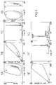
- the letters A to L appearing on the histograms correspond to the location of cells visualized in flow cytometry.
- Example 1 Step of detection of apoptosis, by flow cytometry.
- the immortalized astrocyte line is obtained according to the article by E. Galiana, I. Borde, P. Marin, M. Rassoulzadegan, F. Chuzin, F. Gros, P. Rouget and C. Evrard, Establishement of permanent astroglial cell lines, able to differentiate in vitro, from transgenic mice carrying the polyoma virus large-T gene: an alternative approach to brain cell immortalization, Journal of Neuroscience Research, 1990; 26; 269-277, article whose content is incorporated by reference to the present description.
- the medium called medium M is constituted as follows: DMEM (400 ⁇ l / 200 ml) / F12 (1/1) + 10% FCS no decomplemented + Penicillin / Streptomycin (antibiotic) + Fungizone (antifungal) (20 ⁇ l / 40 ml).
- the cells contained in a 250 cm 2 box are trypsinized (solution prepared from a 1 / 250th trypsin solution (S1: 45 ml) and a 1% EDTA solution (S2: 5 ml) and kept at +4 ° C):
- the box came out of the oven.
- the medium that covers the cells is aspirated (10 ml pipette) and eliminated.
- the adherent cells are rinsed with 10 to 15 ml of DMEM / F12 (1/1) which are aspirated.
- We add 1 at 1.2 ml of trypsin / EDTA the cells are returned to the oven for 10 min. They are taken off their mechanically support using a 10 ml pipette medium M. After homogenization they are washed in 20 ml of additional DMEM / F12 then centrifuged 10 min at 1500 rpm (18 ° C).
- the supernatant is carefully removed.
- the cells are taken up in 2 ml of DMEM / F12 and long homogenized. They are washed again in 25 ml of DMEM / F12 then centrifuged (10 min, 1500 rpm, 18 ° C). The supernatant is removed. The cells are taken up in 2 ml of the medium and homogenized long to obtain the most uniform suspension possible. 1 to 5 ml of the medium are added as a function of the importance of the base.
- Cells are scanned directly into a Tomas cell.
- Cell concentration is adjusted at 10,000 cells / ml by dilution in medium M in order to deposit 20,000 cells / well, or 2 ml / well for one 6-well plate.
- 8 x 6 x 2 ml of suspension either 96 ml; 120 ml (3 x 40 ml) of suspension are prepared; he about 25 ml of suspension remain which will serve to put the remaining cells back in culture, adding in the remaining 25 ml.
- the plates thus prepared are covered with protective film to prevent evaporation.
- Gliotoxic factor is added to each well at a rate of 60 to 80 ⁇ l / well depending on the quantity suspected toxic factor in sample (10 ⁇ l / well for urine, 10 to 50 ⁇ l / well for urine purified fractions and other samples).
- the box came out of the oven.
- the protective film is removed.
- the factor is added in triplicate in 2 or 3 successive wells.
- a new protective film is put in place.
- the plates are returned to the oven (37 ° C CO 2 5% H 2 O 95%) for 72 hours.
- the state of living cells present in well is evaluated by flow cytometry after labeling with propidium iodide to highlight cells apoptotic.
- the boxes are taken out of the oven.
- the film protector is removed.
- the medium is aspirated (pasteur pipettes straight cotton) taking care not to vacuum the adherent cells at the bottom of the wells. Cells are rinsed with 2 ml of PBS.
- the cells are then gently peeled off (non-enzymatic) by incubation with a PBS EDTA solution (1 to 2 ml / well) (37 ° C CO 2 5% H 2 O 95% for 10 min) and then dissociated mechanically.
- PBS EDTA solution 1 to 2 ml / well
- the wells are rinsed with PBS in order to recover the last cells at the bottom of these. 2 washes in PBS are carried out.
- the cells are fixed with 70% ethanol which generates pore formation in the cell membrane and induces its permeabilization.
- 70% ethanol for this purpose, 2 ml / well 70% ethanol (frozen) are added.
- the cells are fixed for 1 hour at -20 ° C. After centrifugation for remove the alcohol, washing in PBS is carried out.
- the cell pellet obtained in the next step is taken up in 2 ml of this buffer, and incubated for 1 hour at + 4 ° C.
- a anti-GFAP labeling can be performed.
- the cell pellet is taken up in 50 ⁇ l of 5% BSA + 50 ⁇ l 1% saponin in PBS + 20 ⁇ l anti-GFAP antibody (prepared in rabbits) diluted 1 / 100th then incubated 30 min at room temperature in the dark. After 2 washes in PBS countermarking is carried out (addition of 50 ⁇ l of 5% BSA + 50 ⁇ l of saponin 1% in PBS + 20 ⁇ l of anti-IgG rabbit labeled with fluorescein (FITC) diluted 1 / 10th), incubation for 15 min at room temperature at darkness. 2 washes in PBS are carried out.
- FITC fluorescein
- the suspensions can be stored for 1 to 2 days in PBS.
- PI labeling a DNA marker
- a DNA marker is made by addition on the pellet cell (in order) of 100 ⁇ l RNase (i.e. 50 IU) and 250 ⁇ l of PI at 50 ⁇ g / ml.
- the cytometer reading is taken within 15 min.
- IP marking possibly associated with a GFAP marking, is used to identify and locate astrocytes.
- the analysis of each sample is performed on 3000 cells (see Figures 1A, 1B, 1C and 2A, 2B, 2C).
- the nuclei marked with the IP of living cells are visualized as 3 peaks of fluorescence respectively representing the G0 / G1 phases, S and G2 / M of the cell cycle, as shown in the figure 1G.
- the nuclei labeled with the IP of the cells apoptotics, from which fragmented DNA was extracted at are displayed as a single peak relatively diffuse positioned in sub-G1, as shown on Figure 2G.
- sub-G1 sub-diploid peak
- the cells are first analyzed in experimental conditions allowing to induce a death by necrosis (Wyllie et al., J.
- MTT bromide of tetrazolium
- a solution of MTT at 0.5 mg / ml is prepared in DMEM / F12 by diluting MTT (frozen) to 1 / 10th in the DMEM / F12.
- the boxes are taken out of the oven.
- the film protector is removed.
- the medium is vacuumed taking care not to suck the adherent cells at the bottom of the well.
- 250 ⁇ l / well of the MTT solution are added (in DMEM / F12). Incubate at least 3 hours at the oven (37 ° C).
- Isopropanol acid is prepared (addition of 40 ⁇ l / ml of 1N HCl in pure isopropanol), providing 100 ⁇ l / well for 48-well plates.
- each well is transferred to a 96-well plate so that it can be read on the microplate reader.
- the DOs are read at 570 nm (ref 630 nm).
- Example 3 Techniques for Enriching the Biological Sample with Gliotoxic Factor to Obtain a Fraction Subjected to the Step According to Example 1, and Optionally the Determination Step According to Example 2
- a TSK G 2000 column is used (sold by SUPELCO), in high pressure liquid phase, with a pH buffer at 6.8, with 0.1 M phosphate, and 0.1 M sodium sulfate
- the acquisition time is 60 minutes, and the sample injected is 1 ml.
- DEAE-Sepharose column (DEAE: diethylaminoethyl) (marketed by PHARMACIA), in high pressure, fast flow liquid phase with a 20 mM Tris buffer, pH 7.5, elution by steps with a 100 mM to 1 M NaCl solution, the useful fraction being at 200 mM.
- Example 4 Biological equivalence of urine and CSF of a patient with multiple sclerosis, for the determination of the gliotoxic factor.
- EXAMPLE 5 Urinary determination of the gliotoxic factor in a large population, by detection of cytotoxicity.
- Urine samples are tested according to the protocol identified in Example 2.
- results obtained highlight a positivity of the test in 98 patients with sclerosis in plaques and a negative test in 10 patients with multiple sclerosis. But however, this test generates 40 false positives in patients with other diseases neurological and 2 false positives in healthy controls.
- test defined in example 2 is therefore not totally specific to him alone.
- Example 1 allows both judge the state of astrocytes and detect death cellular (in particular differentiation between necrosis and apoptosis), and ploidy.
- This method is based on detection by flow cytometry (CMF) i) cell morphology (the FALS ("Forward Light Angle Scatter”) allows the viewing cell size, the SS (“Side Scatter ”) allows the visualization of their structure), ii) astrocyte characteristics (anti-GFAP labeling ("Glial Fibrillary Acidic Protein”), anti-Ig-FITC revelation), iii) ploidy (iodide labeling of propidium (PI) after fixing with 70% ethanol and extraction of any DNA fragments) and as a result of cell cycle.
- This last step helps differentiate cellular debris, necrotic cells, apoptotic cells and diploid cells capable of dividing normally.
- Example 7 Combination of the methods according to examples 1 and 2
Landscapes
- Health & Medical Sciences (AREA)
- Life Sciences & Earth Sciences (AREA)
- Engineering & Computer Science (AREA)
- Immunology (AREA)
- Biomedical Technology (AREA)
- Hematology (AREA)
- Chemical & Material Sciences (AREA)
- Urology & Nephrology (AREA)
- Molecular Biology (AREA)
- Medicinal Chemistry (AREA)
- General Health & Medical Sciences (AREA)
- Microbiology (AREA)
- Biotechnology (AREA)
- Pathology (AREA)
- General Physics & Mathematics (AREA)
- Food Science & Technology (AREA)
- Cell Biology (AREA)
- Physics & Mathematics (AREA)
- Analytical Chemistry (AREA)
- Biochemistry (AREA)
- Toxicology (AREA)
- Rehabilitation Therapy (AREA)
- Tropical Medicine & Parasitology (AREA)
- Neurology (AREA)
- Neurosurgery (AREA)
- Proteomics, Peptides & Aminoacids (AREA)
- Bioinformatics & Cheminformatics (AREA)
- Rheumatology (AREA)
- Investigating Or Analysing Biological Materials (AREA)
- Measuring Or Testing Involving Enzymes Or Micro-Organisms (AREA)
- Investigating Or Analysing Materials By Optical Means (AREA)
- Nitrogen And Oxygen Or Sulfur-Condensed Heterocyclic Ring Systems (AREA)
- Medicines Containing Plant Substances (AREA)
Claims (14)
- Verfahren, um in einer biologischen Probe die gegenüber Makrogliazielzellen qliotoxische Aktivität eines gliotoxischen Faktors nachzuweisen und/oder zu quantifizieren, dadurch gekennzeichnet, daß die biologische Probe eine Urinprobe ist.
- Verfahren, um in einer biologischen Probe einen gegenüber adhärenten Makrogliazielzellen cytotoxischen Faktor nachzuweisen und/oder zu quantifizieren, dessen Cytoxizität das Absterben dieser Zellen durch Apoptose induziert, bei dem eine Ausgangsfraktion dieser Probe bereitgestellt wird, die eventuell durch vorherige Behandlung bezüglich des toxischen Faktors angereichert ist, die Ausgangsfraktion mit einem Referenzkulturmedium inkubiert wird, das adhärente Zielzellen aufweist, und die durch Apoptose abgestorbenen adhärenten Zellen detektiert und/oder quantifiziert werden, dadurch gekennzeichnet, daß zum Nachweis und/oder zur Quantifizierung der durch Apoptose abgestorbenen adhärenten Zellen mittels Fluxcytometrie zumindest ein direktes oder indirektes Merkmal nachgewiesen wird, das mit apoptotischen adhärenten Zellen des gesamten oder eines Teils des inkubierten Mediums assoziiert ist, und das, wenn es existiert und/oder quantifiziert wird, die biologische Probe als positiv d.h. als den toxischen Faktor enthaltend kennzeichnet, wobei die Probe eine Urinprobe ist.
- Verfahren nach Anspruch 2, dadurch gekennzeichnet, daß die adhärenten Makrogliazielzellen astrocytäre Zellen sind.
- Verfahren nach Anspruch 2 oder 3, dadurch gekennzeichnet, daß zum Nachweis und/oder zur Quantifizierung der durch spezifische Apoptose abgestorbenen Makrogliazellen mittels Fluxcytometrie das Verfahren die folgenden Schritte aufweist:Ablösung der adhärenten Makrogliazellen;Behandlung der so abgelösten Zellen mit einer Substanz zur zellulären Fixierung und Permeabilisierung der Cytoplasmamembran der Zellen;Extraktion von aus der Apoptose resultierenden intrazellulären DNA-Fragmenten;Markierung der noch vorhandenen zellulären DNA durch einen geeigneten Marker, undNachweis der Ploidie der Makrogliazellen mittels Fluxcytometrie.
- Verfahren nach Anspruch 4, dadurch gekennzeichnet, daß die Substanz zur zellulären Fixierung und Permeabilisierung der Cytoplasmamembran der Zellen Ethanol ist.
- Verfahren nach Anspruch 4 oder 5, dadurch gekennzeichnet, daß der Marker der DNA Propidiumjodid ist (PJ).
- Verfahren nach Anspruch 2 oder 3, dadurch gekennzeichnet, daß zum Nachweis und/oder zur Quantifizierung der durch spezifische Apoptose abgestorbenen Makrogliazellen mittels Fluxcytometrie das Verfahren die folgenden Schritte aufweist:Induktion einer Nekrose auf einer Probe lebender Makrogliazellen, die sich von dem Inkubationsmedium, das die Ausgangsfraktion und das Referenzkulturmedium enthält, unterscheidet;Markierung der aus der Nekrose resultierenden DNA-Trümmer, wobei die Trümmer mit Fragmenten der Cytoplasmamembran der nekrotischen Makrogliazellen assoziiert sind, und die Markierung nach Ablösung der Zellen durchgeführt wird;Lokalisation der durch Nekrose abgestorbenen Zellen mittels Fluxcytometrie und geeignete Einstellung des Cytometers, so daß bei dem Schritt des Nachweisens der durch Apoptose abgestorbenen Zellen der Aus-schluß der durch Nekrose abgestorbenen Zellen möglich ist;Nachweis der durch Apoptose abgestorbenen Zellen in der zu analysierenden Probe.
- Verfahren nach Anspruch 2 oder 3, dadurch gekennzeichnet, daß zum Nachweis und/oder zur Quantifizierung der durch spezifische Apoptose abgestorbenen Makrogliazellen mittels Fluxcytometrie das Verfahren die folgenden Schritte aufweist:Aufteilung der Zellen des Inkubationsmediums, das die Ausgangsfraktion und das Referenzkulturmedium enthält, in zwei gleiche Teile, und Ablösung der Zellen;Nachweis in einem der beiden Teile mittels Fluxcytometrie einerseits von durch Apoptose oder Nekrose abgestorbenen Makrogliazellen und/oder andererseits von lebenden Makrogliazellen, ein Schritt, bei dem die Fraktion mit einer Substanz zur zellulären Fixierung und Permeabilisierung der Cytoplasmamembran behandelt wird; Extrahieren der DNA-Fragmente; Markieren intrazellulär verbleibender DNA mittels eines Markers, und Nachweisen des letzteren;Nachweis in dem anderen der beiden Teile mittels Fluxcytometrie nach Fixierung ohne Extraktion einerseits von lebenden Makrogliazellen und apoptotischen Makrogliazellen und/oder andererseits von nekrotischen Makrogliazellen, undSchlußfolgerung aus den für jeden der beiden Teile erfolgten Nachweisen auf die Menge der apoptotischen Makrogliazellen.
- Verfahren nach Anspruch 2 oder 3, dadurch gekennzeichnet, daß das Referenzkulturmedium eine immortalisierte Zellinie von Makrogliazellen aufweist, z.B. eine immortalisierte Astrocytenzellinie.
- Verfahren nach einem der Ansprüche 1 bis 9, dadurch gekennzeichnet, daß es vor dem Schritt des Nachweisens und/oder Quantifizierens der apoptotischen Mikrogliazellen einen Schritt umfaßt, um die zelluläre Lebensfähigkeit nachzuweisen, bei dem die Ausgangsfraktion in das Referenzkulturmedium eingeimpft wird, dann der Proliferationsgrad der Makrogliazellen im Vergleich zu einem Referenzwert beobachtet wird, diesseits dessen eine als positiv bezeichnete Fraktion zurückbehalten wird, um sie anschließend einem Schritt zum Nachweis makrogliazellspezifischer Apoptose zu unterziehen, um zu qualifizieren, ob die biologische Probe wirklich positiv ist oder nicht.
- Verfahren nach Anspruch 10, dadurch gekennzeichnet, daß während des Schrittes des Nachweisens der zellulären Lebensfähigkeit der Proliferationsgrad der Makrogliazellen durch mitochondriale Anfärbung der Makrogliazellen des Kulturmediums, z.B. durch Methyltetrazoliumbromid, und danach durch Messung der optischen Dichte des gefärbten Kulturmediums erhalten wird.
- Verfahren nach einem der Ansprüche 2 bis 11, dadurch gekennzeichnet, daß zusätzlich während des Schrittes des Nachweisens der makrogliazellspezifischen Apoptose eine Bestimmung des Vorliegens und/oder eine Quantifizierung eines Proteins durchgeführt wird, das es erlaubt, die zellulären Unterpopulationen zu unterscheiden, wie z.B. das saure Gliafaserprotein (GFAP), z.B. mit einem direkt oder indirekt identifizierbaren Anti-GFAP-Antikörper.
- Verfahren nach einem der vorherigen Ansprüche, dadurch gekennzeichnet, daß die Ausgangsfraktion durch Anreicherung oder Aufreinigung des gliotoxischen Faktors in bzw. aus der biologischen Probe nach zumindest einer der nachfolgenden Behandlungen erhalten wird: Präzipitation der Proteinfraktion, z.B. mit Ammoniumsulfat, Ausschluß- und/oder Ionenchromatographie, ein- oder zweidimensionale Elektrophorese, in-Kontakt-bringen mit Protein A oder einem Lektin, z.B. dem Concanavalin-A.
- Verwendung einer Urinprobe, um dort einen solchen gliotoxischen Faktor nachzuweisen, der mit multipler Sklerose assoziiert ist.
Applications Claiming Priority (3)
| Application Number | Priority Date | Filing Date | Title |
|---|---|---|---|
| FR9611347 | 1996-09-12 | ||
| FR9611347 | 1996-09-12 | ||
| PCT/FR1997/001620 WO1998011439A1 (fr) | 1996-09-12 | 1997-09-12 | Procede de detection et/ou de quantification d'un facteur gliotoxique |
Publications (2)
| Publication Number | Publication Date |
|---|---|
| EP0925504A1 EP0925504A1 (de) | 1999-06-30 |
| EP0925504B1 true EP0925504B1 (de) | 2001-03-14 |
Family
ID=9495830
Family Applications (1)
| Application Number | Title | Priority Date | Filing Date |
|---|---|---|---|
| EP97919095A Expired - Lifetime EP0925504B1 (de) | 1996-09-12 | 1997-09-12 | Verfahren zur detektion und/oder quantitativen bestimmung von einem gliotoxischen faktor |
Country Status (9)
| Country | Link |
|---|---|
| US (1) | US6270953B1 (de) |
| EP (1) | EP0925504B1 (de) |
| JP (1) | JP2001504686A (de) |
| AT (1) | ATE199782T1 (de) |
| CA (1) | CA2266124A1 (de) |
| DE (1) | DE69704279T2 (de) |
| DK (1) | DK0925504T3 (de) |
| ES (1) | ES2157569T3 (de) |
| WO (1) | WO1998011439A1 (de) |
Families Citing this family (12)
| Publication number | Priority date | Publication date | Assignee | Title |
|---|---|---|---|---|
| FR2797402B1 (fr) * | 1999-07-15 | 2004-03-12 | Biomerieux Stelhys | Utilisation d'un polypeptide pour detecter, prevenir ou traiter un etat pathologique associe a une maladie degenerative, neurologique ou autoimmune |
| DE10053521B4 (de) * | 2000-10-27 | 2004-07-08 | november Aktiengesellschaft Gesellschaft für Molekulare Medizin | Verfahren zum Nachweis apoptotischer und nekrotischer Zellen |
| ES2661168T3 (es) * | 2003-07-12 | 2018-03-27 | Accelerate Diagnostics, Inc. | Biodetección sensible y rápida |
| US20120077206A1 (en) | 2003-07-12 | 2012-03-29 | Accelr8 Technology Corporation | Rapid Microbial Detection and Antimicrobial Susceptibility Testing |
| US7341841B2 (en) * | 2003-07-12 | 2008-03-11 | Accelr8 Technology Corporation | Rapid microbial detection and antimicrobial susceptibility testing |
| US10254204B2 (en) | 2011-03-07 | 2019-04-09 | Accelerate Diagnostics, Inc. | Membrane-assisted purification |
| US9434937B2 (en) | 2011-03-07 | 2016-09-06 | Accelerate Diagnostics, Inc. | Rapid cell purification systems |
| US9442092B2 (en) | 2011-06-20 | 2016-09-13 | Kerry Lane | Methods for treatment of autism |
| US9677109B2 (en) | 2013-03-15 | 2017-06-13 | Accelerate Diagnostics, Inc. | Rapid determination of microbial growth and antimicrobial susceptibility |
| US10023895B2 (en) | 2015-03-30 | 2018-07-17 | Accelerate Diagnostics, Inc. | Instrument and system for rapid microogranism identification and antimicrobial agent susceptibility testing |
| US10253355B2 (en) | 2015-03-30 | 2019-04-09 | Accelerate Diagnostics, Inc. | Instrument and system for rapid microorganism identification and antimicrobial agent susceptibility testing |
| CN110196219B (zh) * | 2019-05-16 | 2022-06-03 | 扬州大学 | 一种基于流式细胞术检测鸡脾脏cd8+t细胞特异性杀伤作用的方法 |
Family Cites Families (2)
| Publication number | Priority date | Publication date | Assignee | Title |
|---|---|---|---|---|
| FR2716198B1 (fr) * | 1994-02-15 | 1996-04-19 | Bio Merieux | Facteur cytotoxique tel qu'associé à la sclérose en plaques, sa détection et sa quantification. |
| IT1278044B1 (it) * | 1995-03-09 | 1997-11-17 | Salvatore Petraroli | Metodologia per la realizzazione di un esame di laboratorio dell' integrita' in vitro delle cellule ematiche non fissate e/o |
-
1997
- 1997-09-12 EP EP97919095A patent/EP0925504B1/de not_active Expired - Lifetime
- 1997-09-12 WO PCT/FR1997/001620 patent/WO1998011439A1/fr not_active Ceased
- 1997-09-12 DE DE69704279T patent/DE69704279T2/de not_active Expired - Fee Related
- 1997-09-12 AT AT97919095T patent/ATE199782T1/de active
- 1997-09-12 DK DK97919095T patent/DK0925504T3/da active
- 1997-09-12 US US09/202,118 patent/US6270953B1/en not_active Expired - Fee Related
- 1997-09-12 JP JP51332998A patent/JP2001504686A/ja not_active Ceased
- 1997-09-12 ES ES97919095T patent/ES2157569T3/es not_active Expired - Lifetime
- 1997-09-12 CA CA002266124A patent/CA2266124A1/fr not_active Abandoned
Also Published As
| Publication number | Publication date |
|---|---|
| CA2266124A1 (fr) | 1998-03-19 |
| EP0925504A1 (de) | 1999-06-30 |
| DK0925504T3 (da) | 2001-07-23 |
| ES2157569T3 (es) | 2001-08-16 |
| DE69704279T2 (de) | 2001-10-31 |
| WO1998011439A1 (fr) | 1998-03-19 |
| US6270953B1 (en) | 2001-08-07 |
| JP2001504686A (ja) | 2001-04-10 |
| DE69704279D1 (de) | 2001-04-19 |
| ATE199782T1 (de) | 2001-03-15 |
Similar Documents
| Publication | Publication Date | Title |
|---|---|---|
| EP0925504B1 (de) | Verfahren zur detektion und/oder quantitativen bestimmung von einem gliotoxischen faktor | |
| Habela et al. | ClC3 is a critical regulator of the cell cycle in normal and malignant glial cells | |
| Alice et al. | Subtoxic oxidative stress induces senescence in retinal pigment epithelial cells via TGF-β release | |
| Alderete et al. | The vagina of women infected with Trichomonas vaginalis has numerous proteinases and antibody to trichomonad proteinases. | |
| He et al. | Munc13-4 interacts with syntaxin 7 and regulates late endosomal maturation, endosomal signaling, and TLR9-initiated cellular responses | |
| Braga et al. | Sub-MIC concentrations of cefodizime interfere with various factors affecting bacterial virulence | |
| Schwartz et al. | Oral cytology assessment by flow cytometry of DNA adducts, aneuploidy, proliferation and apoptosis shows differences between smokers and non-smokers | |
| Turner et al. | Loss of variable antigen during transformation of Trypanosoma brucei rhodesiense from bloodstream to procyclic forms in the tsetse fly | |
| Bansal et al. | PIM2 Induced COX-2 and MMP-9 expression in macrophages requires PI3K and Notch1 signaling | |
| Bar-Chama et al. | Infection and pyospermia in male infertility | |
| Schenkels et al. | In VivoBinding of the Salivary Glycoprotein EP-GP (Identical to GCDFP-15) to Oral and Non-Oral Bacteria Detection and Identification of EP-GP Binding Species | |
| Jia et al. | Malassezia globosa induces differentiation of pathogenic Th17 cells by inducing IL-23 secretion by keratinocytes | |
| Bisson et al. | Presence of trichomonads in subgingival biofilm of patients with periodontitis: preliminary results | |
| Cooper et al. | Localization of gonococcal lipopolysaccharide and its relationship to toxic damage in human fallopian tube mucosa | |
| King et al. | There is substantial nuclear and cellular disintegration before detectable phosphatidylserine exposure during the camptothecin‐induced apoptosis of HL‐60 cells | |
| Sharma et al. | Diagnosis of acanthamoeba keratitis-A report of four cases and review of literature | |
| Ma et al. | Flow cytometry with crystal violet to detect intracytoplasmic fluorescence in viable human lymphocytes demonstration of antibody entering living cells | |
| Gnarpe et al. | Studies in venereal disease. I. Isolation of L-phase organisms of N. gonorrhoeae from patients with gonorrhoea | |
| Jakob et al. | Intestinal inflammation associated with flagellates in snakes | |
| Ueno et al. | Microsporidial keratitis retrospectively diagnosed by ultrastructural study of formalin-fixed paraffin-embedded corneal tissue: a case report | |
| JP4117563B2 (ja) | 皮膚糸状菌の検出方法、検出用試薬および抗原性賦活化方法 | |
| Kaur et al. | Receptor mediated amelioration of the detrimental effects of sperm agglutinating factor on sperm parameters | |
| Biddlestone et al. | A clinico-pathological study of cutaneous leishmaniasis in British troops from Belize | |
| Webberley et al. | Commercially available fluorescein-conjugated monoclonal antibody for determining the in vitro activity of antimicrobial agents against Chlamydia trachomatis | |
| Valigurová et al. | Eudiplozoon nipponicum: morphofunctional adaptations of diplozoid monogeneans for confronting their host |
Legal Events
| Date | Code | Title | Description |
|---|---|---|---|
| PUAI | Public reference made under article 153(3) epc to a published international application that has entered the european phase |
Free format text: ORIGINAL CODE: 0009012 |
|
| 17P | Request for examination filed |
Effective date: 19990222 |
|
| AK | Designated contracting states |
Kind code of ref document: A1 Designated state(s): AT BE CH DE DK ES FI FR GB GR IE IT LI LU MC NL PT SE |
|
| GRAG | Despatch of communication of intention to grant |
Free format text: ORIGINAL CODE: EPIDOS AGRA |
|
| 17Q | First examination report despatched |
Effective date: 20000207 |
|
| GRAG | Despatch of communication of intention to grant |
Free format text: ORIGINAL CODE: EPIDOS AGRA |
|
| GRAH | Despatch of communication of intention to grant a patent |
Free format text: ORIGINAL CODE: EPIDOS IGRA |
|
| GRAH | Despatch of communication of intention to grant a patent |
Free format text: ORIGINAL CODE: EPIDOS IGRA |
|
| GRAA | (expected) grant |
Free format text: ORIGINAL CODE: 0009210 |
|
| AK | Designated contracting states |
Kind code of ref document: B1 Designated state(s): AT BE CH DE DK ES FI FR GB GR IE IT LI LU MC NL PT SE |
|
| PG25 | Lapsed in a contracting state [announced via postgrant information from national office to epo] |
Ref country code: FI Free format text: LAPSE BECAUSE OF FAILURE TO SUBMIT A TRANSLATION OF THE DESCRIPTION OR TO PAY THE FEE WITHIN THE PRESCRIBED TIME-LIMIT Effective date: 20010314 Ref country code: AT Free format text: LAPSE BECAUSE OF FAILURE TO SUBMIT A TRANSLATION OF THE DESCRIPTION OR TO PAY THE FEE WITHIN THE PRESCRIBED TIME-LIMIT Effective date: 20010314 |
|
| REF | Corresponds to: |
Ref document number: 199782 Country of ref document: AT Date of ref document: 20010315 Kind code of ref document: T |
|
| REG | Reference to a national code |
Ref country code: CH Ref legal event code: EP |
|
| GRAG | Despatch of communication of intention to grant |
Free format text: ORIGINAL CODE: EPIDOS AGRA |
|
| REG | Reference to a national code |
Ref country code: IE Ref legal event code: FG4D Free format text: FRENCH |
|
| REF | Corresponds to: |
Ref document number: 69704279 Country of ref document: DE Date of ref document: 20010419 |
|
| ITF | It: translation for a ep patent filed | ||
| PG25 | Lapsed in a contracting state [announced via postgrant information from national office to epo] |
Ref country code: PT Free format text: LAPSE BECAUSE OF FAILURE TO SUBMIT A TRANSLATION OF THE DESCRIPTION OR TO PAY THE FEE WITHIN THE PRESCRIBED TIME-LIMIT Effective date: 20010615 Ref country code: GR Free format text: LAPSE BECAUSE OF FAILURE TO SUBMIT A TRANSLATION OF THE DESCRIPTION OR TO PAY THE FEE WITHIN THE PRESCRIBED TIME-LIMIT Effective date: 20010615 |
|
| REG | Reference to a national code |
Ref country code: CH Ref legal event code: NV Representative=s name: MICHELI & CIE INGENIEURS-CONSEILS |
|
| GBT | Gb: translation of ep patent filed (gb section 77(6)(a)/1977) |
Effective date: 20010612 |
|
| REG | Reference to a national code |
Ref country code: DK Ref legal event code: T3 |
|
| REG | Reference to a national code |
Ref country code: ES Ref legal event code: FG2A Ref document number: 2157569 Country of ref document: ES Kind code of ref document: T3 |
|
| REG | Reference to a national code |
Ref country code: GB Ref legal event code: IF02 |
|
| PLBE | No opposition filed within time limit |
Free format text: ORIGINAL CODE: 0009261 |
|
| STAA | Information on the status of an ep patent application or granted ep patent |
Free format text: STATUS: NO OPPOSITION FILED WITHIN TIME LIMIT |
|
| 26N | No opposition filed | ||
| PGFP | Annual fee paid to national office [announced via postgrant information from national office to epo] |
Ref country code: LU Payment date: 20060817 Year of fee payment: 10 |
|
| PGFP | Annual fee paid to national office [announced via postgrant information from national office to epo] |
Ref country code: NL Payment date: 20060818 Year of fee payment: 10 Ref country code: MC Payment date: 20060818 Year of fee payment: 10 |
|
| PGFP | Annual fee paid to national office [announced via postgrant information from national office to epo] |
Ref country code: GB Payment date: 20060822 Year of fee payment: 10 |
|
| PGFP | Annual fee paid to national office [announced via postgrant information from national office to epo] |
Ref country code: IE Payment date: 20060823 Year of fee payment: 10 |
|
| PGFP | Annual fee paid to national office [announced via postgrant information from national office to epo] |
Ref country code: DE Payment date: 20060907 Year of fee payment: 10 |
|
| PGFP | Annual fee paid to national office [announced via postgrant information from national office to epo] |
Ref country code: CH Payment date: 20060911 Year of fee payment: 10 |
|
| PGFP | Annual fee paid to national office [announced via postgrant information from national office to epo] |
Ref country code: ES Payment date: 20060912 Year of fee payment: 10 |
|
| PGFP | Annual fee paid to national office [announced via postgrant information from national office to epo] |
Ref country code: DK Payment date: 20060921 Year of fee payment: 10 |
|
| PGFP | Annual fee paid to national office [announced via postgrant information from national office to epo] |
Ref country code: IT Payment date: 20060930 Year of fee payment: 10 |
|
| PGFP | Annual fee paid to national office [announced via postgrant information from national office to epo] |
Ref country code: BE Payment date: 20061010 Year of fee payment: 10 |
|
| PGFP | Annual fee paid to national office [announced via postgrant information from national office to epo] |
Ref country code: SE Payment date: 20060821 Year of fee payment: 10 |
|
| BERE | Be: lapsed |
Owner name: *BIO MERIEUX Effective date: 20070930 |
|
| PG25 | Lapsed in a contracting state [announced via postgrant information from national office to epo] |
Ref country code: SE Free format text: LAPSE BECAUSE OF NON-PAYMENT OF DUE FEES Effective date: 20070913 Ref country code: MC Free format text: LAPSE BECAUSE OF NON-PAYMENT OF DUE FEES Effective date: 20070930 |
|
| REG | Reference to a national code |
Ref country code: DK Ref legal event code: EBP |
|
| REG | Reference to a national code |
Ref country code: CH Ref legal event code: PL |
|
| EUG | Se: european patent has lapsed | ||
| GBPC | Gb: european patent ceased through non-payment of renewal fee |
Effective date: 20070912 |
|
| PG25 | Lapsed in a contracting state [announced via postgrant information from national office to epo] |
Ref country code: NL Free format text: LAPSE BECAUSE OF NON-PAYMENT OF DUE FEES Effective date: 20080401 |
|
| NLV4 | Nl: lapsed or anulled due to non-payment of the annual fee |
Effective date: 20080401 |
|
| REG | Reference to a national code |
Ref country code: IE Ref legal event code: MM4A |
|
| PG25 | Lapsed in a contracting state [announced via postgrant information from national office to epo] |
Ref country code: LI Free format text: LAPSE BECAUSE OF NON-PAYMENT OF DUE FEES Effective date: 20070930 Ref country code: DE Free format text: LAPSE BECAUSE OF NON-PAYMENT OF DUE FEES Effective date: 20080401 Ref country code: CH Free format text: LAPSE BECAUSE OF NON-PAYMENT OF DUE FEES Effective date: 20070930 |
|
| PG25 | Lapsed in a contracting state [announced via postgrant information from national office to epo] |
Ref country code: BE Free format text: LAPSE BECAUSE OF NON-PAYMENT OF DUE FEES Effective date: 20070930 |
|
| REG | Reference to a national code |
Ref country code: FR Ref legal event code: ST Effective date: 20080531 |
|
| PG25 | Lapsed in a contracting state [announced via postgrant information from national office to epo] |
Ref country code: IE Free format text: LAPSE BECAUSE OF NON-PAYMENT OF DUE FEES Effective date: 20070912 Ref country code: FR Free format text: LAPSE BECAUSE OF NON-PAYMENT OF DUE FEES Effective date: 20071001 Ref country code: DK Free format text: LAPSE BECAUSE OF NON-PAYMENT OF DUE FEES Effective date: 20071001 |
|
| PGFP | Annual fee paid to national office [announced via postgrant information from national office to epo] |
Ref country code: FR Payment date: 20060929 Year of fee payment: 10 |
|
| PG25 | Lapsed in a contracting state [announced via postgrant information from national office to epo] |
Ref country code: GB Free format text: LAPSE BECAUSE OF NON-PAYMENT OF DUE FEES Effective date: 20070912 |
|
| REG | Reference to a national code |
Ref country code: ES Ref legal event code: FD2A Effective date: 20070913 |
|
| PG25 | Lapsed in a contracting state [announced via postgrant information from national office to epo] |
Ref country code: ES Free format text: LAPSE BECAUSE OF NON-PAYMENT OF DUE FEES Effective date: 20070913 |
|
| PG25 | Lapsed in a contracting state [announced via postgrant information from national office to epo] |
Ref country code: LU Free format text: LAPSE BECAUSE OF NON-PAYMENT OF DUE FEES Effective date: 20070912 Ref country code: IT Free format text: LAPSE BECAUSE OF NON-PAYMENT OF DUE FEES Effective date: 20070912 |