EP0920522B1 - A vector for polynucleotide vaccines - Google Patents
A vector for polynucleotide vaccines Download PDFInfo
- Publication number
- EP0920522B1 EP0920522B1 EP97937243A EP97937243A EP0920522B1 EP 0920522 B1 EP0920522 B1 EP 0920522B1 EP 97937243 A EP97937243 A EP 97937243A EP 97937243 A EP97937243 A EP 97937243A EP 0920522 B1 EP0920522 B1 EP 0920522B1
- Authority
- EP
- European Patent Office
- Prior art keywords
- sequence
- polynucleotide vector
- antigen
- target
- tumor
- Prior art date
- Legal status (The legal status is an assumption and is not a legal conclusion. Google has not performed a legal analysis and makes no representation as to the accuracy of the status listed.)
- Expired - Lifetime
Links
- 239000013598 vector Substances 0.000 title claims abstract description 151
- 108700001237 Nucleic Acid-Based Vaccines Proteins 0.000 title description 43
- 108091007433 antigens Proteins 0.000 claims abstract description 147
- 102000036639 antigens Human genes 0.000 claims abstract description 147
- 239000000427 antigen Substances 0.000 claims abstract description 145
- 102000040430 polynucleotide Human genes 0.000 claims abstract description 126
- 108091033319 polynucleotide Proteins 0.000 claims abstract description 126
- 239000002157 polynucleotide Substances 0.000 claims abstract description 126
- 206010028980 Neoplasm Diseases 0.000 claims abstract description 106
- 239000013612 plasmid Substances 0.000 claims abstract description 86
- 150000007523 nucleic acids Chemical group 0.000 claims abstract description 61
- 229940126580 vector vaccine Drugs 0.000 claims abstract description 38
- 230000028993 immune response Effects 0.000 claims abstract description 32
- 210000000612 antigen-presenting cell Anatomy 0.000 claims abstract description 12
- 210000004027 cell Anatomy 0.000 claims description 66
- 241000282414 Homo sapiens Species 0.000 claims description 57
- 239000002299 complementary DNA Substances 0.000 claims description 43
- 230000014509 gene expression Effects 0.000 claims description 42
- 238000000034 method Methods 0.000 claims description 39
- 210000001519 tissue Anatomy 0.000 claims description 37
- 230000000890 antigenic effect Effects 0.000 claims description 30
- 102000001327 Chemokine CCL5 Human genes 0.000 claims description 26
- 108010055166 Chemokine CCL5 Proteins 0.000 claims description 26
- 238000010367 cloning Methods 0.000 claims description 26
- 230000010076 replication Effects 0.000 claims description 25
- 102100034256 Mucin-1 Human genes 0.000 claims description 21
- 108010008707 Mucin-1 Proteins 0.000 claims description 21
- 108091028043 Nucleic acid sequence Proteins 0.000 claims description 21
- 239000003795 chemical substances by application Substances 0.000 claims description 17
- 102100030086 Receptor tyrosine-protein kinase erbB-2 Human genes 0.000 claims description 14
- 229960003150 bupivacaine Drugs 0.000 claims description 14
- 101100446506 Mus musculus Fgf3 gene Proteins 0.000 claims description 11
- 239000008194 pharmaceutical composition Substances 0.000 claims description 11
- 101001012157 Homo sapiens Receptor tyrosine-protein kinase erbB-2 Proteins 0.000 claims description 10
- 230000001580 bacterial effect Effects 0.000 claims description 10
- -1 glyU Proteins 0.000 claims description 10
- 108091008146 restriction endonucleases Proteins 0.000 claims description 10
- 230000012010 growth Effects 0.000 claims description 9
- 238000004519 manufacturing process Methods 0.000 claims description 9
- 239000002773 nucleotide Substances 0.000 claims description 9
- 125000003729 nucleotide group Chemical group 0.000 claims description 9
- 240000004808 Saccharomyces cerevisiae Species 0.000 claims description 8
- 230000002708 enhancing effect Effects 0.000 claims description 8
- 230000003612 virological effect Effects 0.000 claims description 8
- 210000003205 muscle Anatomy 0.000 claims description 7
- 241000894006 Bacteria Species 0.000 claims description 6
- 102000019034 Chemokines Human genes 0.000 claims description 6
- 108010012236 Chemokines Proteins 0.000 claims description 6
- 102000004127 Cytokines Human genes 0.000 claims description 6
- 108090000695 Cytokines Proteins 0.000 claims description 6
- 108020005038 Terminator Codon Proteins 0.000 claims description 6
- 230000002068 genetic effect Effects 0.000 claims description 6
- 108091035707 Consensus sequence Proteins 0.000 claims description 5
- WQZGKKKJIJFFOK-GASJEMHNSA-N Glucose Natural products OC[C@H]1OC(O)[C@H](O)[C@@H](O)[C@@H]1O WQZGKKKJIJFFOK-GASJEMHNSA-N 0.000 claims description 5
- 108091081024 Start codon Proteins 0.000 claims description 5
- 230000003115 biocidal effect Effects 0.000 claims description 5
- 239000008121 dextrose Substances 0.000 claims description 5
- 238000000338 in vitro Methods 0.000 claims description 5
- 231100000363 mycotoxic Toxicity 0.000 claims description 5
- 230000000357 mycotoxic effect Effects 0.000 claims description 5
- 108010000521 Human Growth Hormone Proteins 0.000 claims description 4
- 108010002350 Interleukin-2 Proteins 0.000 claims description 4
- 102000000588 Interleukin-2 Human genes 0.000 claims description 4
- 102000004388 Interleukin-4 Human genes 0.000 claims description 4
- 108090000978 Interleukin-4 Proteins 0.000 claims description 4
- 206010073150 Multiple endocrine neoplasia Type 1 Diseases 0.000 claims description 4
- 108020004566 Transfer RNA Proteins 0.000 claims description 4
- 239000003242 anti bacterial agent Substances 0.000 claims description 4
- WQZGKKKJIJFFOK-VFUOTHLCSA-N beta-D-glucose Chemical compound OC[C@H]1O[C@@H](O)[C@H](O)[C@@H](O)[C@@H]1O WQZGKKKJIJFFOK-VFUOTHLCSA-N 0.000 claims description 4
- 239000003814 drug Substances 0.000 claims description 4
- 239000003937 drug carrier Substances 0.000 claims description 4
- 108010087914 epidermal growth factor receptor VIII Proteins 0.000 claims description 4
- 210000004698 lymphocyte Anatomy 0.000 claims description 4
- 230000003071 parasitic effect Effects 0.000 claims description 4
- 210000003705 ribosome Anatomy 0.000 claims description 4
- 230000004936 stimulating effect Effects 0.000 claims description 4
- 241000124008 Mammalia Species 0.000 claims description 3
- 108060008682 Tumor Necrosis Factor Proteins 0.000 claims description 3
- 102000003390 tumor necrosis factor Human genes 0.000 claims description 3
- QEVHRUUCFGRFIF-UHFFFAOYSA-N 6,18-dimethoxy-17-[oxo-(3,4,5-trimethoxyphenyl)methoxy]-1,3,11,12,14,15,16,17,18,19,20,21-dodecahydroyohimban-19-carboxylic acid methyl ester Chemical compound C1C2CN3CCC(C4=CC=C(OC)C=C4N4)=C4C3CC2C(C(=O)OC)C(OC)C1OC(=O)C1=CC(OC)=C(OC)C(OC)=C1 QEVHRUUCFGRFIF-UHFFFAOYSA-N 0.000 claims description 2
- 102000000541 Defensins Human genes 0.000 claims description 2
- 108010002069 Defensins Proteins 0.000 claims description 2
- 101100533283 Dictyostelium discoideum serp gene Proteins 0.000 claims description 2
- 101710121996 Hexon protein p72 Proteins 0.000 claims description 2
- 102000002265 Human Growth Hormone Human genes 0.000 claims description 2
- 108010050904 Interferons Proteins 0.000 claims description 2
- 102000014150 Interferons Human genes 0.000 claims description 2
- 108010065805 Interleukin-12 Proteins 0.000 claims description 2
- 102000013462 Interleukin-12 Human genes 0.000 claims description 2
- 102000003812 Interleukin-15 Human genes 0.000 claims description 2
- 108090000172 Interleukin-15 Proteins 0.000 claims description 2
- 108010002386 Interleukin-3 Proteins 0.000 claims description 2
- 102000000646 Interleukin-3 Human genes 0.000 claims description 2
- 108010002586 Interleukin-7 Proteins 0.000 claims description 2
- 102000000704 Interleukin-7 Human genes 0.000 claims description 2
- 108090001007 Interleukin-8 Proteins 0.000 claims description 2
- 102000004890 Interleukin-8 Human genes 0.000 claims description 2
- 101100312053 Lactococcus lactis subsp. lactis (strain IL1403) glyS gene Proteins 0.000 claims description 2
- 101710125418 Major capsid protein Proteins 0.000 claims description 2
- 108091060545 Nonsense suppressor Proteins 0.000 claims description 2
- 231100000433 cytotoxic Toxicity 0.000 claims description 2
- 230000001472 cytotoxic effect Effects 0.000 claims description 2
- 229940079322 interferon Drugs 0.000 claims description 2
- 229940117681 interleukin-12 Drugs 0.000 claims description 2
- 229940076264 interleukin-3 Drugs 0.000 claims description 2
- 229940028885 interleukin-4 Drugs 0.000 claims description 2
- 229940100994 interleukin-7 Drugs 0.000 claims description 2
- 229940096397 interleukin-8 Drugs 0.000 claims description 2
- XKTZWUACRZHVAN-VADRZIEHSA-N interleukin-8 Chemical compound C([C@H](NC(=O)[C@H](CC(O)=O)NC(=O)[C@H](CC=1C2=CC=CC=C2NC=1)NC(=O)[C@@H](NC(C)=O)CCSC)C(=O)N[C@@H](CC(O)=O)C(=O)N[C@@H](CC(O)=O)C(=O)N[C@@H](CC(C)C)C(=O)N[C@@H](CC(N)=O)C(=O)N[C@@H](CC=1C=CC=CC=1)C(=O)N[C@@H]([C@@H](C)O)C(=O)NCC(=O)N[C@@H](CCSC)C(=O)N1[C@H](CCC1)C(=O)N1[C@H](CCC1)C(=O)N[C@@H](C)C(=O)N[C@H](CC(O)=O)C(=O)N[C@H](CCC(O)=O)C(=O)N[C@H](CC(O)=O)C(=O)N[C@H](CC=1C=CC(O)=CC=1)C(=O)N[C@H](CO)C(=O)N1[C@H](CCC1)C(N)=O)C1=CC=CC=C1 XKTZWUACRZHVAN-VADRZIEHSA-N 0.000 claims description 2
- 108700026220 vif Genes Proteins 0.000 claims description 2
- 210000000107 myocyte Anatomy 0.000 claims 3
- 101710151805 Mitochondrial intermediate peptidase 1 Proteins 0.000 claims 2
- SIEYLFHKZGLBNX-UHFFFAOYSA-N bupivacaine hydrochloride (anhydrous) Chemical group [Cl-].CCCC[NH+]1CCCCC1C(=O)NC1=C(C)C=CC=C1C SIEYLFHKZGLBNX-UHFFFAOYSA-N 0.000 claims 2
- 108010017213 Granulocyte-Macrophage Colony-Stimulating Factor Proteins 0.000 claims 1
- 102100039620 Granulocyte-macrophage colony-stimulating factor Human genes 0.000 claims 1
- 239000000854 Human Growth Hormone Substances 0.000 claims 1
- 108020004414 DNA Proteins 0.000 abstract description 58
- 210000002027 skeletal muscle Anatomy 0.000 abstract description 10
- 108091061960 Naked DNA Proteins 0.000 abstract description 4
- 238000005516 engineering process Methods 0.000 abstract description 4
- 210000004962 mammalian cell Anatomy 0.000 abstract description 3
- 230000003362 replicative effect Effects 0.000 abstract description 3
- 230000010473 stable expression Effects 0.000 abstract description 2
- 238000002255 vaccination Methods 0.000 description 55
- 108090000623 proteins and genes Proteins 0.000 description 53
- 238000002347 injection Methods 0.000 description 42
- 239000007924 injection Substances 0.000 description 42
- 239000005090 green fluorescent protein Substances 0.000 description 35
- 229940090044 injection Drugs 0.000 description 35
- 238000002360 preparation method Methods 0.000 description 33
- 102000004169 proteins and genes Human genes 0.000 description 32
- 241000700159 Rattus Species 0.000 description 29
- 206010006187 Breast cancer Diseases 0.000 description 28
- 102000039446 nucleic acids Human genes 0.000 description 28
- 108020004707 nucleic acids Proteins 0.000 description 28
- 239000000047 product Substances 0.000 description 27
- 229960005486 vaccine Drugs 0.000 description 27
- 208000026310 Breast neoplasm Diseases 0.000 description 26
- 239000012634 fragment Substances 0.000 description 26
- FAPWRFPIFSIZLT-UHFFFAOYSA-M Sodium chloride Chemical compound [Na+].[Cl-] FAPWRFPIFSIZLT-UHFFFAOYSA-M 0.000 description 25
- 238000011156 evaluation Methods 0.000 description 24
- 241001465754 Metazoa Species 0.000 description 23
- 230000000692 anti-sense effect Effects 0.000 description 23
- 101150029707 ERBB2 gene Proteins 0.000 description 21
- 210000001151 cytotoxic T lymphocyte Anatomy 0.000 description 21
- 230000035772 mutation Effects 0.000 description 21
- 108090000765 processed proteins & peptides Proteins 0.000 description 17
- LEBVLXFERQHONN-UHFFFAOYSA-N 1-butyl-N-(2,6-dimethylphenyl)piperidine-2-carboxamide Chemical compound CCCCN1CCCCC1C(=O)NC1=C(C)C=CC=C1C LEBVLXFERQHONN-UHFFFAOYSA-N 0.000 description 15
- 230000001413 cellular effect Effects 0.000 description 15
- 239000007927 intramuscular injection Substances 0.000 description 15
- 238000011282 treatment Methods 0.000 description 15
- 102100025064 Cellular tumor antigen p53 Human genes 0.000 description 13
- 238000001964 muscle biopsy Methods 0.000 description 13
- 230000001988 toxicity Effects 0.000 description 13
- 231100000419 toxicity Toxicity 0.000 description 13
- 101000721661 Homo sapiens Cellular tumor antigen p53 Proteins 0.000 description 12
- 108700020796 Oncogene Proteins 0.000 description 12
- 210000001744 T-lymphocyte Anatomy 0.000 description 12
- 150000001413 amino acids Chemical group 0.000 description 12
- 108020004999 messenger RNA Proteins 0.000 description 12
- 102000004196 processed proteins & peptides Human genes 0.000 description 12
- 101100314454 Caenorhabditis elegans tra-1 gene Proteins 0.000 description 11
- 230000003834 intracellular effect Effects 0.000 description 11
- 102000016914 ras Proteins Human genes 0.000 description 11
- 108010014186 ras Proteins Proteins 0.000 description 11
- 230000004044 response Effects 0.000 description 11
- LFQSCWFLJHTTHZ-UHFFFAOYSA-N Ethanol Chemical compound CCO LFQSCWFLJHTTHZ-UHFFFAOYSA-N 0.000 description 10
- 230000002519 immonomodulatory effect Effects 0.000 description 10
- 238000002955 isolation Methods 0.000 description 10
- 210000003141 lower extremity Anatomy 0.000 description 10
- 239000011780 sodium chloride Substances 0.000 description 10
- 210000004881 tumor cell Anatomy 0.000 description 10
- 231100000371 dose-limiting toxicity Toxicity 0.000 description 9
- 238000001890 transfection Methods 0.000 description 9
- 230000004075 alteration Effects 0.000 description 8
- 230000005975 antitumor immune response Effects 0.000 description 8
- 238000003556 assay Methods 0.000 description 8
- 238000010255 intramuscular injection Methods 0.000 description 8
- 230000003902 lesion Effects 0.000 description 8
- 210000003819 peripheral blood mononuclear cell Anatomy 0.000 description 8
- 230000000717 retained effect Effects 0.000 description 8
- 239000000523 sample Substances 0.000 description 8
- 241000588724 Escherichia coli Species 0.000 description 7
- 108700025716 Tumor Suppressor Genes Proteins 0.000 description 7
- 102000044209 Tumor Suppressor Genes Human genes 0.000 description 7
- 230000008901 benefit Effects 0.000 description 7
- 230000000694 effects Effects 0.000 description 7
- 210000000987 immune system Anatomy 0.000 description 7
- 230000036961 partial effect Effects 0.000 description 7
- 229920001184 polypeptide Polymers 0.000 description 7
- 102000001301 EGF receptor Human genes 0.000 description 6
- 108060006698 EGF receptor Proteins 0.000 description 6
- 102000004190 Enzymes Human genes 0.000 description 6
- 108090000790 Enzymes Proteins 0.000 description 6
- 108010043121 Green Fluorescent Proteins Proteins 0.000 description 6
- 102000004144 Green Fluorescent Proteins Human genes 0.000 description 6
- 201000008274 breast adenocarcinoma Diseases 0.000 description 6
- 201000011510 cancer Diseases 0.000 description 6
- 201000010099 disease Diseases 0.000 description 6
- 208000037265 diseases, disorders, signs and symptoms Diseases 0.000 description 6
- 229940088598 enzyme Drugs 0.000 description 6
- 210000000981 epithelium Anatomy 0.000 description 6
- 239000000203 mixture Substances 0.000 description 6
- 210000001616 monocyte Anatomy 0.000 description 6
- 238000002560 therapeutic procedure Methods 0.000 description 6
- 108020004705 Codon Proteins 0.000 description 5
- 238000011887 Necropsy Methods 0.000 description 5
- 230000000259 anti-tumor effect Effects 0.000 description 5
- 210000004369 blood Anatomy 0.000 description 5
- 239000008280 blood Substances 0.000 description 5
- 210000000481 breast Anatomy 0.000 description 5
- 210000004443 dendritic cell Anatomy 0.000 description 5
- 210000003414 extremity Anatomy 0.000 description 5
- 230000006870 function Effects 0.000 description 5
- 230000000977 initiatory effect Effects 0.000 description 5
- 238000012986 modification Methods 0.000 description 5
- 239000002243 precursor Substances 0.000 description 5
- 238000012163 sequencing technique Methods 0.000 description 5
- 210000002363 skeletal muscle cell Anatomy 0.000 description 5
- 210000003491 skin Anatomy 0.000 description 5
- 230000004083 survival effect Effects 0.000 description 5
- 238000011144 upstream manufacturing Methods 0.000 description 5
- CURLTUGMZLYLDI-UHFFFAOYSA-N Carbon dioxide Chemical compound O=C=O CURLTUGMZLYLDI-UHFFFAOYSA-N 0.000 description 4
- WSFSSNUMVMOOMR-UHFFFAOYSA-N Formaldehyde Chemical compound O=C WSFSSNUMVMOOMR-UHFFFAOYSA-N 0.000 description 4
- 101000643895 Homo sapiens Ubiquitin carboxyl-terminal hydrolase 6 Proteins 0.000 description 4
- KFZMGEQAYNKOFK-UHFFFAOYSA-N Isopropanol Chemical compound CC(C)O KFZMGEQAYNKOFK-UHFFFAOYSA-N 0.000 description 4
- 241000699666 Mus <mouse, genus> Species 0.000 description 4
- 108091034117 Oligonucleotide Proteins 0.000 description 4
- 206010033128 Ovarian cancer Diseases 0.000 description 4
- 101710100968 Receptor tyrosine-protein kinase erbB-2 Proteins 0.000 description 4
- 239000004098 Tetracycline Substances 0.000 description 4
- 102100021015 Ubiquitin carboxyl-terminal hydrolase 6 Human genes 0.000 description 4
- 229960000723 ampicillin Drugs 0.000 description 4
- AVKUERGKIZMTKX-NJBDSQKTSA-N ampicillin Chemical compound C1([C@@H](N)C(=O)N[C@H]2[C@H]3SC([C@@H](N3C2=O)C(O)=O)(C)C)=CC=CC=C1 AVKUERGKIZMTKX-NJBDSQKTSA-N 0.000 description 4
- 238000010171 animal model Methods 0.000 description 4
- 238000010276 construction Methods 0.000 description 4
- 230000001461 cytolytic effect Effects 0.000 description 4
- 230000007547 defect Effects 0.000 description 4
- 238000013461 design Methods 0.000 description 4
- 108700020302 erbB-2 Genes Proteins 0.000 description 4
- 210000002950 fibroblast Anatomy 0.000 description 4
- 239000000499 gel Substances 0.000 description 4
- 230000002962 histologic effect Effects 0.000 description 4
- 238000003780 insertion Methods 0.000 description 4
- 230000037431 insertion Effects 0.000 description 4
- 230000000670 limiting effect Effects 0.000 description 4
- 230000007246 mechanism Effects 0.000 description 4
- 230000002018 overexpression Effects 0.000 description 4
- 239000008188 pellet Substances 0.000 description 4
- 230000002829 reductive effect Effects 0.000 description 4
- 210000000952 spleen Anatomy 0.000 description 4
- 229960002180 tetracycline Drugs 0.000 description 4
- 229930101283 tetracycline Natural products 0.000 description 4
- 235000019364 tetracycline Nutrition 0.000 description 4
- 150000003522 tetracyclines Chemical class 0.000 description 4
- 229920000936 Agarose Polymers 0.000 description 3
- 108700020463 BRCA1 Proteins 0.000 description 3
- 102000036365 BRCA1 Human genes 0.000 description 3
- 101150072950 BRCA1 gene Proteins 0.000 description 3
- 244000038022 Chenopodium capitatum Species 0.000 description 3
- 235000004391 Chenopodium capitatum Nutrition 0.000 description 3
- 108010017826 DNA Polymerase I Proteins 0.000 description 3
- 102000004594 DNA Polymerase I Human genes 0.000 description 3
- 206010015548 Euthanasia Diseases 0.000 description 3
- 238000012413 Fluorescence activated cell sorting analysis Methods 0.000 description 3
- 241000282412 Homo Species 0.000 description 3
- 101000898505 Homo sapiens Histatin-3 Proteins 0.000 description 3
- 241000699670 Mus sp. Species 0.000 description 3
- 208000015914 Non-Hodgkin lymphomas Diseases 0.000 description 3
- 241000700605 Viruses Species 0.000 description 3
- 230000002159 abnormal effect Effects 0.000 description 3
- 230000003321 amplification Effects 0.000 description 3
- 238000004458 analytical method Methods 0.000 description 3
- 230000030741 antigen processing and presentation Effects 0.000 description 3
- 238000013459 approach Methods 0.000 description 3
- 230000001363 autoimmune Effects 0.000 description 3
- 231100000504 carcinogenesis Toxicity 0.000 description 3
- 238000004113 cell culture Methods 0.000 description 3
- 238000006243 chemical reaction Methods 0.000 description 3
- 238000012790 confirmation Methods 0.000 description 3
- 238000001514 detection method Methods 0.000 description 3
- 238000003745 diagnosis Methods 0.000 description 3
- 238000010790 dilution Methods 0.000 description 3
- 239000012895 dilution Substances 0.000 description 3
- 238000010828 elution Methods 0.000 description 3
- ZMMJGEGLRURXTF-UHFFFAOYSA-N ethidium bromide Chemical compound [Br-].C12=CC(N)=CC=C2C2=CC=C(N)C=C2[N+](CC)=C1C1=CC=CC=C1 ZMMJGEGLRURXTF-UHFFFAOYSA-N 0.000 description 3
- 229960005542 ethidium bromide Drugs 0.000 description 3
- 238000007387 excisional biopsy Methods 0.000 description 3
- 238000002474 experimental method Methods 0.000 description 3
- 230000012953 feeding on blood of other organism Effects 0.000 description 3
- 210000004602 germ cell Anatomy 0.000 description 3
- 210000002443 helper t lymphocyte Anatomy 0.000 description 3
- 230000002489 hematologic effect Effects 0.000 description 3
- 230000028996 humoral immune response Effects 0.000 description 3
- 230000001900 immune effect Effects 0.000 description 3
- 238000003364 immunohistochemistry Methods 0.000 description 3
- 238000009169 immunotherapy Methods 0.000 description 3
- 238000001727 in vivo Methods 0.000 description 3
- 230000002757 inflammatory effect Effects 0.000 description 3
- 230000002601 intratumoral effect Effects 0.000 description 3
- 210000001165 lymph node Anatomy 0.000 description 3
- 230000003211 malignant effect Effects 0.000 description 3
- 201000001441 melanoma Diseases 0.000 description 3
- 230000001394 metastastic effect Effects 0.000 description 3
- 206010061289 metastatic neoplasm Diseases 0.000 description 3
- 238000000386 microscopy Methods 0.000 description 3
- 230000004048 modification Effects 0.000 description 3
- 231100000956 nontoxicity Toxicity 0.000 description 3
- 230000037311 normal skin Effects 0.000 description 3
- 238000003199 nucleic acid amplification method Methods 0.000 description 3
- 231100000590 oncogenic Toxicity 0.000 description 3
- 230000002246 oncogenic effect Effects 0.000 description 3
- 230000002085 persistent effect Effects 0.000 description 3
- 239000013600 plasmid vector Substances 0.000 description 3
- 231100000683 possible toxicity Toxicity 0.000 description 3
- 239000002244 precipitate Substances 0.000 description 3
- 238000002203 pretreatment Methods 0.000 description 3
- 230000019491 signal transduction Effects 0.000 description 3
- 238000010186 staining Methods 0.000 description 3
- 238000007920 subcutaneous administration Methods 0.000 description 3
- 230000001225 therapeutic effect Effects 0.000 description 3
- 238000013519 translation Methods 0.000 description 3
- 210000003462 vein Anatomy 0.000 description 3
- 230000003442 weekly effect Effects 0.000 description 3
- 102000007469 Actins Human genes 0.000 description 2
- 108010085238 Actins Proteins 0.000 description 2
- 108700028369 Alleles Proteins 0.000 description 2
- 102000017420 CD3 protein, epsilon/gamma/delta subunit Human genes 0.000 description 2
- 108050005493 CD3 protein, epsilon/gamma/delta subunit Proteins 0.000 description 2
- 208000005623 Carcinogenesis Diseases 0.000 description 2
- 238000007399 DNA isolation Methods 0.000 description 2
- 108010054576 Deoxyribonuclease EcoRI Proteins 0.000 description 2
- 238000002965 ELISA Methods 0.000 description 2
- 102000018233 Fibroblast Growth Factor Human genes 0.000 description 2
- 108050007372 Fibroblast Growth Factor Proteins 0.000 description 2
- 102000003886 Glycoproteins Human genes 0.000 description 2
- 108090000288 Glycoproteins Proteins 0.000 description 2
- 102000025850 HLA-A2 Antigen Human genes 0.000 description 2
- 108010074032 HLA-A2 Antigen Proteins 0.000 description 2
- 108010062347 HLA-DQ Antigens Proteins 0.000 description 2
- 108010058597 HLA-DR Antigens Proteins 0.000 description 2
- 102000006354 HLA-DR Antigens Human genes 0.000 description 2
- 101001057504 Homo sapiens Interferon-stimulated gene 20 kDa protein Proteins 0.000 description 2
- 101001055144 Homo sapiens Interleukin-2 receptor subunit alpha Proteins 0.000 description 2
- 101000917858 Homo sapiens Low affinity immunoglobulin gamma Fc region receptor III-A Proteins 0.000 description 2
- 101000917839 Homo sapiens Low affinity immunoglobulin gamma Fc region receptor III-B Proteins 0.000 description 2
- 101000581981 Homo sapiens Neural cell adhesion molecule 1 Proteins 0.000 description 2
- 101000738771 Homo sapiens Receptor-type tyrosine-protein phosphatase C Proteins 0.000 description 2
- 101000914484 Homo sapiens T-lymphocyte activation antigen CD80 Proteins 0.000 description 2
- 241000725303 Human immunodeficiency virus Species 0.000 description 2
- 108060003951 Immunoglobulin Proteins 0.000 description 2
- 102100026878 Interleukin-2 receptor subunit alpha Human genes 0.000 description 2
- 102100029185 Low affinity immunoglobulin gamma Fc region receptor III-B Human genes 0.000 description 2
- 108060001084 Luciferase Proteins 0.000 description 2
- 239000005089 Luciferase Substances 0.000 description 2
- 101100346932 Mus musculus Muc1 gene Proteins 0.000 description 2
- 102100027347 Neural cell adhesion molecule 1 Human genes 0.000 description 2
- 102000043276 Oncogene Human genes 0.000 description 2
- 206010061535 Ovarian neoplasm Diseases 0.000 description 2
- 108010076504 Protein Sorting Signals Proteins 0.000 description 2
- 101100501691 Rattus norvegicus Erbb2 gene Proteins 0.000 description 2
- 102100037422 Receptor-type tyrosine-protein phosphatase C Human genes 0.000 description 2
- 108700008625 Reporter Genes Proteins 0.000 description 2
- 102100027222 T-lymphocyte activation antigen CD80 Human genes 0.000 description 2
- JLCPHMBAVCMARE-UHFFFAOYSA-N [3-[[3-[[3-[[3-[[3-[[3-[[3-[[3-[[3-[[3-[[3-[[5-(2-amino-6-oxo-1H-purin-9-yl)-3-[[3-[[3-[[3-[[3-[[3-[[5-(2-amino-6-oxo-1H-purin-9-yl)-3-[[5-(2-amino-6-oxo-1H-purin-9-yl)-3-hydroxyoxolan-2-yl]methoxy-hydroxyphosphoryl]oxyoxolan-2-yl]methoxy-hydroxyphosphoryl]oxy-5-(5-methyl-2,4-dioxopyrimidin-1-yl)oxolan-2-yl]methoxy-hydroxyphosphoryl]oxy-5-(6-aminopurin-9-yl)oxolan-2-yl]methoxy-hydroxyphosphoryl]oxy-5-(6-aminopurin-9-yl)oxolan-2-yl]methoxy-hydroxyphosphoryl]oxy-5-(6-aminopurin-9-yl)oxolan-2-yl]methoxy-hydroxyphosphoryl]oxy-5-(6-aminopurin-9-yl)oxolan-2-yl]methoxy-hydroxyphosphoryl]oxyoxolan-2-yl]methoxy-hydroxyphosphoryl]oxy-5-(5-methyl-2,4-dioxopyrimidin-1-yl)oxolan-2-yl]methoxy-hydroxyphosphoryl]oxy-5-(4-amino-2-oxopyrimidin-1-yl)oxolan-2-yl]methoxy-hydroxyphosphoryl]oxy-5-(5-methyl-2,4-dioxopyrimidin-1-yl)oxolan-2-yl]methoxy-hydroxyphosphoryl]oxy-5-(5-methyl-2,4-dioxopyrimidin-1-yl)oxolan-2-yl]methoxy-hydroxyphosphoryl]oxy-5-(6-aminopurin-9-yl)oxolan-2-yl]methoxy-hydroxyphosphoryl]oxy-5-(6-aminopurin-9-yl)oxolan-2-yl]methoxy-hydroxyphosphoryl]oxy-5-(4-amino-2-oxopyrimidin-1-yl)oxolan-2-yl]methoxy-hydroxyphosphoryl]oxy-5-(4-amino-2-oxopyrimidin-1-yl)oxolan-2-yl]methoxy-hydroxyphosphoryl]oxy-5-(4-amino-2-oxopyrimidin-1-yl)oxolan-2-yl]methoxy-hydroxyphosphoryl]oxy-5-(6-aminopurin-9-yl)oxolan-2-yl]methoxy-hydroxyphosphoryl]oxy-5-(4-amino-2-oxopyrimidin-1-yl)oxolan-2-yl]methyl [5-(6-aminopurin-9-yl)-2-(hydroxymethyl)oxolan-3-yl] hydrogen phosphate Polymers Cc1cn(C2CC(OP(O)(=O)OCC3OC(CC3OP(O)(=O)OCC3OC(CC3O)n3cnc4c3nc(N)[nH]c4=O)n3cnc4c3nc(N)[nH]c4=O)C(COP(O)(=O)OC3CC(OC3COP(O)(=O)OC3CC(OC3COP(O)(=O)OC3CC(OC3COP(O)(=O)OC3CC(OC3COP(O)(=O)OC3CC(OC3COP(O)(=O)OC3CC(OC3COP(O)(=O)OC3CC(OC3COP(O)(=O)OC3CC(OC3COP(O)(=O)OC3CC(OC3COP(O)(=O)OC3CC(OC3COP(O)(=O)OC3CC(OC3COP(O)(=O)OC3CC(OC3COP(O)(=O)OC3CC(OC3COP(O)(=O)OC3CC(OC3COP(O)(=O)OC3CC(OC3COP(O)(=O)OC3CC(OC3COP(O)(=O)OC3CC(OC3CO)n3cnc4c(N)ncnc34)n3ccc(N)nc3=O)n3cnc4c(N)ncnc34)n3ccc(N)nc3=O)n3ccc(N)nc3=O)n3ccc(N)nc3=O)n3cnc4c(N)ncnc34)n3cnc4c(N)ncnc34)n3cc(C)c(=O)[nH]c3=O)n3cc(C)c(=O)[nH]c3=O)n3ccc(N)nc3=O)n3cc(C)c(=O)[nH]c3=O)n3cnc4c3nc(N)[nH]c4=O)n3cnc4c(N)ncnc34)n3cnc4c(N)ncnc34)n3cnc4c(N)ncnc34)n3cnc4c(N)ncnc34)O2)c(=O)[nH]c1=O JLCPHMBAVCMARE-UHFFFAOYSA-N 0.000 description 2
- HMNZFMSWFCAGGW-XPWSMXQVSA-N [3-[hydroxy(2-hydroxyethoxy)phosphoryl]oxy-2-[(e)-octadec-9-enoyl]oxypropyl] (e)-octadec-9-enoate Chemical compound CCCCCCCC\C=C\CCCCCCCC(=O)OCC(COP(O)(=O)OCCO)OC(=O)CCCCCCC\C=C\CCCCCCCC HMNZFMSWFCAGGW-XPWSMXQVSA-N 0.000 description 2
- 230000005856 abnormality Effects 0.000 description 2
- 230000004913 activation Effects 0.000 description 2
- 238000001994 activation Methods 0.000 description 2
- 238000000246 agarose gel electrophoresis Methods 0.000 description 2
- 230000000735 allogeneic effect Effects 0.000 description 2
- 238000005349 anion exchange Methods 0.000 description 2
- 238000005571 anion exchange chromatography Methods 0.000 description 2
- 239000003957 anion exchange resin Substances 0.000 description 2
- 230000006023 anti-tumor response Effects 0.000 description 2
- 238000001574 biopsy Methods 0.000 description 2
- 210000001185 bone marrow Anatomy 0.000 description 2
- 210000004556 brain Anatomy 0.000 description 2
- 201000008275 breast carcinoma Diseases 0.000 description 2
- 229940013767 bupivacaine injection Drugs 0.000 description 2
- 230000036952 cancer formation Effects 0.000 description 2
- 239000001569 carbon dioxide Substances 0.000 description 2
- 229910002092 carbon dioxide Inorganic materials 0.000 description 2
- 229940030156 cell vaccine Drugs 0.000 description 2
- 230000036755 cellular response Effects 0.000 description 2
- 238000012512 characterization method Methods 0.000 description 2
- 230000002596 correlated effect Effects 0.000 description 2
- 230000000875 corresponding effect Effects 0.000 description 2
- 238000012864 cross contamination Methods 0.000 description 2
- 230000009089 cytolysis Effects 0.000 description 2
- 238000012217 deletion Methods 0.000 description 2
- 230000037430 deletion Effects 0.000 description 2
- 238000011161 development Methods 0.000 description 2
- 230000018109 developmental process Effects 0.000 description 2
- 230000029087 digestion Effects 0.000 description 2
- 230000008482 dysregulation Effects 0.000 description 2
- 238000001962 electrophoresis Methods 0.000 description 2
- 229940126864 fibroblast growth factor Drugs 0.000 description 2
- 210000001035 gastrointestinal tract Anatomy 0.000 description 2
- 210000002216 heart Anatomy 0.000 description 2
- 230000001744 histochemical effect Effects 0.000 description 2
- 102000051957 human ERBB2 Human genes 0.000 description 2
- 210000005260 human cell Anatomy 0.000 description 2
- 230000008348 humoral response Effects 0.000 description 2
- 102000018358 immunoglobulin Human genes 0.000 description 2
- 238000010348 incorporation Methods 0.000 description 2
- 238000007918 intramuscular administration Methods 0.000 description 2
- 210000003734 kidney Anatomy 0.000 description 2
- 210000004185 liver Anatomy 0.000 description 2
- 210000004072 lung Anatomy 0.000 description 2
- 238000010841 mRNA extraction Methods 0.000 description 2
- 210000002540 macrophage Anatomy 0.000 description 2
- 230000011987 methylation Effects 0.000 description 2
- 238000007069 methylation reaction Methods 0.000 description 2
- LXCFILQKKLGQFO-UHFFFAOYSA-N methylparaben Chemical compound COC(=O)C1=CC=C(O)C=C1 LXCFILQKKLGQFO-UHFFFAOYSA-N 0.000 description 2
- 230000002906 microbiologic effect Effects 0.000 description 2
- 238000010172 mouse model Methods 0.000 description 2
- 210000000056 organ Anatomy 0.000 description 2
- 230000002611 ovarian Effects 0.000 description 2
- 239000002245 particle Substances 0.000 description 2
- 230000001575 pathological effect Effects 0.000 description 2
- 230000007170 pathology Effects 0.000 description 2
- 210000001986 peyer's patch Anatomy 0.000 description 2
- 238000001556 precipitation Methods 0.000 description 2
- 230000008569 process Effects 0.000 description 2
- 238000012545 processing Methods 0.000 description 2
- 208000037821 progressive disease Diseases 0.000 description 2
- 230000002062 proliferating effect Effects 0.000 description 2
- 229940023143 protein vaccine Drugs 0.000 description 2
- 238000011552 rat model Methods 0.000 description 2
- 230000009467 reduction Effects 0.000 description 2
- 230000001105 regulatory effect Effects 0.000 description 2
- 238000011160 research Methods 0.000 description 2
- 230000003307 reticuloendothelial effect Effects 0.000 description 2
- 230000001177 retroviral effect Effects 0.000 description 2
- 238000010839 reverse transcription Methods 0.000 description 2
- 238000005070 sampling Methods 0.000 description 2
- 230000035939 shock Effects 0.000 description 2
- 150000003384 small molecules Chemical class 0.000 description 2
- 210000000278 spinal cord Anatomy 0.000 description 2
- 230000003393 splenic effect Effects 0.000 description 2
- 108700017804 supF tRNA Proteins 0.000 description 2
- 238000001356 surgical procedure Methods 0.000 description 2
- 230000002459 sustained effect Effects 0.000 description 2
- 210000001541 thymus gland Anatomy 0.000 description 2
- 238000013518 transcription Methods 0.000 description 2
- 230000035897 transcription Effects 0.000 description 2
- 230000004614 tumor growth Effects 0.000 description 2
- 238000002211 ultraviolet spectrum Methods 0.000 description 2
- 210000000689 upper leg Anatomy 0.000 description 2
- 108091032973 (ribonucleotides)n+m Proteins 0.000 description 1
- BFSVOASYOCHEOV-UHFFFAOYSA-N 2-diethylaminoethanol Chemical compound CCN(CC)CCO BFSVOASYOCHEOV-UHFFFAOYSA-N 0.000 description 1
- DVGKRPYUFRZAQW-UHFFFAOYSA-N 3 prime Natural products CC(=O)NC1OC(CC(O)C1C(O)C(O)CO)(OC2C(O)C(CO)OC(OC3C(O)C(O)C(O)OC3CO)C2O)C(=O)O DVGKRPYUFRZAQW-UHFFFAOYSA-N 0.000 description 1
- 238000010600 3H thymidine incorporation assay Methods 0.000 description 1
- 102000002260 Alkaline Phosphatase Human genes 0.000 description 1
- 108020004774 Alkaline Phosphatase Proteins 0.000 description 1
- 208000000058 Anaplasia Diseases 0.000 description 1
- 102100021569 Apoptosis regulator Bcl-2 Human genes 0.000 description 1
- 101100163849 Arabidopsis thaliana ARS1 gene Proteins 0.000 description 1
- 102100024222 B-lymphocyte antigen CD19 Human genes 0.000 description 1
- 102100022005 B-lymphocyte antigen CD20 Human genes 0.000 description 1
- 102000052609 BRCA2 Human genes 0.000 description 1
- 108700020462 BRCA2 Proteins 0.000 description 1
- 108010077805 Bacterial Proteins Proteins 0.000 description 1
- 101150008921 Brca2 gene Proteins 0.000 description 1
- 102100025475 Carcinoembryonic antigen-related cell adhesion molecule 5 Human genes 0.000 description 1
- 102000001326 Chemokine CCL4 Human genes 0.000 description 1
- 108010055165 Chemokine CCL4 Proteins 0.000 description 1
- 108020004638 Circular DNA Proteins 0.000 description 1
- 102000029816 Collagenase Human genes 0.000 description 1
- 108060005980 Collagenase Proteins 0.000 description 1
- 206010009944 Colon cancer Diseases 0.000 description 1
- 208000001333 Colorectal Neoplasms Diseases 0.000 description 1
- 208000035473 Communicable disease Diseases 0.000 description 1
- 108050006400 Cyclin Proteins 0.000 description 1
- 102000016736 Cyclin Human genes 0.000 description 1
- 241000701022 Cytomegalovirus Species 0.000 description 1
- 102000012410 DNA Ligases Human genes 0.000 description 1
- 108010061982 DNA Ligases Proteins 0.000 description 1
- 230000004544 DNA amplification Effects 0.000 description 1
- 101100239693 Dictyostelium discoideum myoD gene Proteins 0.000 description 1
- 101150039808 Egfr gene Proteins 0.000 description 1
- 102100031780 Endonuclease Human genes 0.000 description 1
- 102100038595 Estrogen receptor Human genes 0.000 description 1
- 108091029865 Exogenous DNA Proteins 0.000 description 1
- 101150034814 F gene Proteins 0.000 description 1
- 102100024785 Fibroblast growth factor 2 Human genes 0.000 description 1
- 108090000379 Fibroblast growth factor 2 Proteins 0.000 description 1
- 108090000331 Firefly luciferases Proteins 0.000 description 1
- 102000011786 HLA-A Antigens Human genes 0.000 description 1
- 108010075704 HLA-A Antigens Proteins 0.000 description 1
- 101150101014 HST1 gene Proteins 0.000 description 1
- 208000017604 Hodgkin disease Diseases 0.000 description 1
- 208000021519 Hodgkin lymphoma Diseases 0.000 description 1
- 208000010747 Hodgkins lymphoma Diseases 0.000 description 1
- 101000971171 Homo sapiens Apoptosis regulator Bcl-2 Proteins 0.000 description 1
- 101000980825 Homo sapiens B-lymphocyte antigen CD19 Proteins 0.000 description 1
- 101000897405 Homo sapiens B-lymphocyte antigen CD20 Proteins 0.000 description 1
- 101000797762 Homo sapiens C-C motif chemokine 5 Proteins 0.000 description 1
- 101000914324 Homo sapiens Carcinoembryonic antigen-related cell adhesion molecule 5 Proteins 0.000 description 1
- 101000914321 Homo sapiens Carcinoembryonic antigen-related cell adhesion molecule 7 Proteins 0.000 description 1
- 101001053946 Homo sapiens Dystrophin Proteins 0.000 description 1
- 101000851181 Homo sapiens Epidermal growth factor receptor Proteins 0.000 description 1
- 101001034652 Homo sapiens Insulin-like growth factor 1 receptor Proteins 0.000 description 1
- 101000946889 Homo sapiens Monocyte differentiation antigen CD14 Proteins 0.000 description 1
- 101000617725 Homo sapiens Pregnancy-specific beta-1-glycoprotein 2 Proteins 0.000 description 1
- 101000716102 Homo sapiens T-cell surface glycoprotein CD4 Proteins 0.000 description 1
- 101000946843 Homo sapiens T-cell surface glycoprotein CD8 alpha chain Proteins 0.000 description 1
- 102100039688 Insulin-like growth factor 1 receptor Human genes 0.000 description 1
- 102000003814 Interleukin-10 Human genes 0.000 description 1
- 108090000174 Interleukin-10 Proteins 0.000 description 1
- 108090000171 Interleukin-18 Proteins 0.000 description 1
- 102000003810 Interleukin-18 Human genes 0.000 description 1
- 108090001005 Interleukin-6 Proteins 0.000 description 1
- 102000004889 Interleukin-6 Human genes 0.000 description 1
- 206010058467 Lung neoplasm malignant Diseases 0.000 description 1
- 102000043129 MHC class I family Human genes 0.000 description 1
- 108091054437 MHC class I family Proteins 0.000 description 1
- 102000043131 MHC class II family Human genes 0.000 description 1
- 108091054438 MHC class II family Proteins 0.000 description 1
- 241000712079 Measles morbillivirus Species 0.000 description 1
- 208000007054 Medullary Carcinoma Diseases 0.000 description 1
- 102100035877 Monocyte differentiation antigen CD14 Human genes 0.000 description 1
- 241000713333 Mouse mammary tumor virus Species 0.000 description 1
- 101000686985 Mouse mammary tumor virus (strain C3H) Protein PR73 Proteins 0.000 description 1
- 241001529936 Murinae Species 0.000 description 1
- 101710135898 Myc proto-oncogene protein Proteins 0.000 description 1
- 102100038895 Myc proto-oncogene protein Human genes 0.000 description 1
- 241000186359 Mycobacterium Species 0.000 description 1
- 206010029260 Neuroblastoma Diseases 0.000 description 1
- 108700026244 Open Reading Frames Proteins 0.000 description 1
- 238000012408 PCR amplification Methods 0.000 description 1
- 206010061902 Pancreatic neoplasm Diseases 0.000 description 1
- 241001631646 Papillomaviridae Species 0.000 description 1
- 208000030852 Parasitic disease Diseases 0.000 description 1
- 241000224016 Plasmodium Species 0.000 description 1
- 206010060862 Prostate cancer Diseases 0.000 description 1
- 208000000236 Prostatic Neoplasms Diseases 0.000 description 1
- 108010092799 RNA-directed DNA polymerase Proteins 0.000 description 1
- 241000711798 Rabies lyssavirus Species 0.000 description 1
- 101710090029 Replication-associated protein A Proteins 0.000 description 1
- 201000000582 Retinoblastoma Diseases 0.000 description 1
- 241000283984 Rodentia Species 0.000 description 1
- 206010039491 Sarcoma Diseases 0.000 description 1
- 101100097319 Schizosaccharomyces pombe (strain 972 / ATCC 24843) ala1 gene Proteins 0.000 description 1
- MTCFGRXMJLQNBG-UHFFFAOYSA-N Serine Natural products OCC(N)C(O)=O MTCFGRXMJLQNBG-UHFFFAOYSA-N 0.000 description 1
- 230000024932 T cell mediated immunity Effects 0.000 description 1
- 108091008874 T cell receptors Proteins 0.000 description 1
- 230000005867 T cell response Effects 0.000 description 1
- 102000016266 T-Cell Antigen Receptors Human genes 0.000 description 1
- 102100036011 T-cell surface glycoprotein CD4 Human genes 0.000 description 1
- 102100034922 T-cell surface glycoprotein CD8 alpha chain Human genes 0.000 description 1
- IQFYYKKMVGJFEH-XLPZGREQSA-N Thymidine Chemical compound O=C1NC(=O)C(C)=CN1[C@@H]1O[C@H](CO)[C@@H](O)C1 IQFYYKKMVGJFEH-XLPZGREQSA-N 0.000 description 1
- 101710150448 Transcriptional regulator Myc Proteins 0.000 description 1
- 208000036142 Viral infection Diseases 0.000 description 1
- 108010006886 Vitrogen Proteins 0.000 description 1
- 230000001594 aberrant effect Effects 0.000 description 1
- 239000002253 acid Substances 0.000 description 1
- 150000007513 acids Chemical class 0.000 description 1
- 230000003213 activating effect Effects 0.000 description 1
- 125000000539 amino acid group Chemical group 0.000 description 1
- 230000036592 analgesia Effects 0.000 description 1
- 230000000840 anti-viral effect Effects 0.000 description 1
- 229940088710 antibiotic agent Drugs 0.000 description 1
- 230000010056 antibody-dependent cellular cytotoxicity Effects 0.000 description 1
- 229940121375 antifungal agent Drugs 0.000 description 1
- 230000003416 augmentation Effects 0.000 description 1
- 108010058966 bacteriophage T7 induced DNA polymerase Proteins 0.000 description 1
- 210000003651 basophil Anatomy 0.000 description 1
- XEGGRYVFLWGFHI-UHFFFAOYSA-N bendiocarb Chemical compound CNC(=O)OC1=CC=CC2=C1OC(C)(C)O2 XEGGRYVFLWGFHI-UHFFFAOYSA-N 0.000 description 1
- 238000007469 bone scintigraphy Methods 0.000 description 1
- 108010006025 bovine growth hormone Proteins 0.000 description 1
- 244000309466 calf Species 0.000 description 1
- 125000002091 cationic group Chemical group 0.000 description 1
- 238000001516 cell proliferation assay Methods 0.000 description 1
- 230000033077 cellular process Effects 0.000 description 1
- 230000008859 change Effects 0.000 description 1
- 239000013599 cloning vector Substances 0.000 description 1
- 229960002424 collagenase Drugs 0.000 description 1
- 230000002281 colonystimulating effect Effects 0.000 description 1
- 231100000026 common toxicity Toxicity 0.000 description 1
- 238000011443 conventional therapy Methods 0.000 description 1
- 238000009109 curative therapy Methods 0.000 description 1
- 230000001086 cytosolic effect Effects 0.000 description 1
- 239000000824 cytostatic agent Substances 0.000 description 1
- 230000001085 cytostatic effect Effects 0.000 description 1
- 230000003013 cytotoxicity Effects 0.000 description 1
- 231100000135 cytotoxicity Toxicity 0.000 description 1
- 238000002784 cytotoxicity assay Methods 0.000 description 1
- 231100000263 cytotoxicity test Toxicity 0.000 description 1
- 230000034994 death Effects 0.000 description 1
- 231100000517 death Toxicity 0.000 description 1
- 230000007423 decrease Effects 0.000 description 1
- 230000003247 decreasing effect Effects 0.000 description 1
- 230000006735 deficit Effects 0.000 description 1
- 230000001419 dependent effect Effects 0.000 description 1
- 230000008021 deposition Effects 0.000 description 1
- 230000004069 differentiation Effects 0.000 description 1
- 230000008034 disappearance Effects 0.000 description 1
- 230000003828 downregulation Effects 0.000 description 1
- 102000052116 epidermal growth factor receptor activity proteins Human genes 0.000 description 1
- 108010038795 estrogen receptors Proteins 0.000 description 1
- 210000003527 eukaryotic cell Anatomy 0.000 description 1
- 238000001400 expression cloning Methods 0.000 description 1
- 238000000684 flow cytometry Methods 0.000 description 1
- MHMNJMPURVTYEJ-UHFFFAOYSA-N fluorescein-5-isothiocyanate Chemical compound O1C(=O)C2=CC(N=C=S)=CC=C2C21C1=CC=C(O)C=C1OC1=CC(O)=CC=C21 MHMNJMPURVTYEJ-UHFFFAOYSA-N 0.000 description 1
- 238000009472 formulation Methods 0.000 description 1
- 238000002825 functional assay Methods 0.000 description 1
- 238000001415 gene therapy Methods 0.000 description 1
- 231100000118 genetic alteration Toxicity 0.000 description 1
- 230000004077 genetic alteration Effects 0.000 description 1
- 230000009395 genetic defect Effects 0.000 description 1
- 210000003714 granulocyte Anatomy 0.000 description 1
- 230000036541 health Effects 0.000 description 1
- 102000045341 human CCL5 Human genes 0.000 description 1
- 230000037451 immune surveillance Effects 0.000 description 1
- 230000036039 immunity Effects 0.000 description 1
- 230000003053 immunization Effects 0.000 description 1
- 238000002649 immunization Methods 0.000 description 1
- 238000003018 immunoassay Methods 0.000 description 1
- 238000011532 immunohistochemical staining Methods 0.000 description 1
- 230000001024 immunotherapeutic effect Effects 0.000 description 1
- 238000005470 impregnation Methods 0.000 description 1
- 230000006872 improvement Effects 0.000 description 1
- 230000005917 in vivo anti-tumor Effects 0.000 description 1
- 230000002779 inactivation Effects 0.000 description 1
- 230000006698 induction Effects 0.000 description 1
- 208000015181 infectious disease Diseases 0.000 description 1
- 230000008595 infiltration Effects 0.000 description 1
- 238000001764 infiltration Methods 0.000 description 1
- 230000028709 inflammatory response Effects 0.000 description 1
- 230000002401 inhibitory effect Effects 0.000 description 1
- 238000011221 initial treatment Methods 0.000 description 1
- 238000011081 inoculation Methods 0.000 description 1
- 230000010354 integration Effects 0.000 description 1
- 230000003993 interaction Effects 0.000 description 1
- 208000030776 invasive breast carcinoma Diseases 0.000 description 1
- 238000004255 ion exchange chromatography Methods 0.000 description 1
- 230000002147 killing effect Effects 0.000 description 1
- 210000001821 langerhans cell Anatomy 0.000 description 1
- 208000032839 leukemia Diseases 0.000 description 1
- 201000007270 liver cancer Diseases 0.000 description 1
- 208000014018 liver neoplasm Diseases 0.000 description 1
- 230000007774 longterm Effects 0.000 description 1
- 238000003670 luciferase enzyme activity assay Methods 0.000 description 1
- 238000004020 luminiscence type Methods 0.000 description 1
- 201000005202 lung cancer Diseases 0.000 description 1
- 208000020816 lung neoplasm Diseases 0.000 description 1
- 239000006166 lysate Substances 0.000 description 1
- 208000015486 malignant pancreatic neoplasm Diseases 0.000 description 1
- 210000004216 mammary stem cell Anatomy 0.000 description 1
- 229940106885 marcaine Drugs 0.000 description 1
- 239000000463 material Substances 0.000 description 1
- 230000001404 mediated effect Effects 0.000 description 1
- 208000023356 medullary thyroid gland carcinoma Diseases 0.000 description 1
- 239000004292 methyl p-hydroxybenzoate Substances 0.000 description 1
- 235000010270 methyl p-hydroxybenzoate Nutrition 0.000 description 1
- 229960002216 methylparaben Drugs 0.000 description 1
- 230000003278 mimic effect Effects 0.000 description 1
- 210000000865 mononuclear phagocyte system Anatomy 0.000 description 1
- 210000000663 muscle cell Anatomy 0.000 description 1
- 231100000302 myotoxic Toxicity 0.000 description 1
- 230000003630 myotoxic effect Effects 0.000 description 1
- 230000001613 neoplastic effect Effects 0.000 description 1
- 230000010309 neoplastic transformation Effects 0.000 description 1
- 238000011275 oncology therapy Methods 0.000 description 1
- 201000002528 pancreatic cancer Diseases 0.000 description 1
- 208000008443 pancreatic carcinoma Diseases 0.000 description 1
- 239000012188 paraffin wax Substances 0.000 description 1
- 244000045947 parasite Species 0.000 description 1
- 244000052769 pathogen Species 0.000 description 1
- 230000037361 pathway Effects 0.000 description 1
- 229940023041 peptide vaccine Drugs 0.000 description 1
- 230000000737 periodic effect Effects 0.000 description 1
- 230000002688 persistence Effects 0.000 description 1
- 239000002504 physiological saline solution Substances 0.000 description 1
- 238000013492 plasmid preparation Methods 0.000 description 1
- 230000002265 prevention Effects 0.000 description 1
- 238000004393 prognosis Methods 0.000 description 1
- 238000000746 purification Methods 0.000 description 1
- 230000005855 radiation Effects 0.000 description 1
- 230000003439 radiotherapeutic effect Effects 0.000 description 1
- 239000011347 resin Substances 0.000 description 1
- 229920005989 resin Polymers 0.000 description 1
- 238000003757 reverse transcription PCR Methods 0.000 description 1
- 229940063629 sensorcaine Drugs 0.000 description 1
- 210000002966 serum Anatomy 0.000 description 1
- 210000001626 skin fibroblast Anatomy 0.000 description 1
- 239000000243 solution Substances 0.000 description 1
- 238000010561 standard procedure Methods 0.000 description 1
- 231100000617 superantigen Toxicity 0.000 description 1
- 208000024891 symptom Diseases 0.000 description 1
- 238000012360 testing method Methods 0.000 description 1
- 208000008732 thymoma Diseases 0.000 description 1
- 239000003440 toxic substance Substances 0.000 description 1
- 230000002103 transcriptional effect Effects 0.000 description 1
- 238000012546 transfer Methods 0.000 description 1
- 230000009466 transformation Effects 0.000 description 1
- 238000003146 transient transfection Methods 0.000 description 1
- 230000005909 tumor killing Effects 0.000 description 1
- 231100000402 unacceptable toxicity Toxicity 0.000 description 1
- 241001529453 unidentified herpesvirus Species 0.000 description 1
- 241000712461 unidentified influenza virus Species 0.000 description 1
- 230000003827 upregulation Effects 0.000 description 1
- 230000009385 viral infection Effects 0.000 description 1
- 238000012800 visualization Methods 0.000 description 1
- XLYOFNOQVPJJNP-UHFFFAOYSA-N water Substances O XLYOFNOQVPJJNP-UHFFFAOYSA-N 0.000 description 1
- 238000001262 western blot Methods 0.000 description 1
Images

Classifications
-
- C—CHEMISTRY; METALLURGY
- C07—ORGANIC CHEMISTRY
- C07K—PEPTIDES
- C07K14/00—Peptides having more than 20 amino acids; Gastrins; Somatostatins; Melanotropins; Derivatives thereof
- C07K14/435—Peptides having more than 20 amino acids; Gastrins; Somatostatins; Melanotropins; Derivatives thereof from animals; from humans
- C07K14/705—Receptors; Cell surface antigens; Cell surface determinants
- C07K14/71—Receptors; Cell surface antigens; Cell surface determinants for growth factors; for growth regulators
-
- C—CHEMISTRY; METALLURGY
- C12—BIOCHEMISTRY; BEER; SPIRITS; WINE; VINEGAR; MICROBIOLOGY; ENZYMOLOGY; MUTATION OR GENETIC ENGINEERING
- C12N—MICROORGANISMS OR ENZYMES; COMPOSITIONS THEREOF; PROPAGATING, PRESERVING, OR MAINTAINING MICROORGANISMS; MUTATION OR GENETIC ENGINEERING; CULTURE MEDIA
- C12N15/00—Mutation or genetic engineering; DNA or RNA concerning genetic engineering, vectors, e.g. plasmids, or their isolation, preparation or purification; Use of hosts therefor
- C12N15/09—Recombinant DNA-technology
- C12N15/63—Introduction of foreign genetic material using vectors; Vectors; Use of hosts therefor; Regulation of expression
- C12N15/79—Vectors or expression systems specially adapted for eukaryotic hosts
- C12N15/85—Vectors or expression systems specially adapted for eukaryotic hosts for animal cells
-
- A—HUMAN NECESSITIES
- A61—MEDICAL OR VETERINARY SCIENCE; HYGIENE
- A61K—PREPARATIONS FOR MEDICAL, DENTAL OR TOILETRY PURPOSES
- A61K39/00—Medicinal preparations containing antigens or antibodies
- A61K2039/51—Medicinal preparations containing antigens or antibodies comprising whole cells, viruses or DNA/RNA
-
- C—CHEMISTRY; METALLURGY
- C12—BIOCHEMISTRY; BEER; SPIRITS; WINE; VINEGAR; MICROBIOLOGY; ENZYMOLOGY; MUTATION OR GENETIC ENGINEERING
- C12N—MICROORGANISMS OR ENZYMES; COMPOSITIONS THEREOF; PROPAGATING, PRESERVING, OR MAINTAINING MICROORGANISMS; MUTATION OR GENETIC ENGINEERING; CULTURE MEDIA
- C12N2830/00—Vector systems having a special element relevant for transcription
-
- C—CHEMISTRY; METALLURGY
- C12—BIOCHEMISTRY; BEER; SPIRITS; WINE; VINEGAR; MICROBIOLOGY; ENZYMOLOGY; MUTATION OR GENETIC ENGINEERING
- C12N—MICROORGANISMS OR ENZYMES; COMPOSITIONS THEREOF; PROPAGATING, PRESERVING, OR MAINTAINING MICROORGANISMS; MUTATION OR GENETIC ENGINEERING; CULTURE MEDIA
- C12N2830/00—Vector systems having a special element relevant for transcription
- C12N2830/38—Vector systems having a special element relevant for transcription being a stuffer
-
- C—CHEMISTRY; METALLURGY
- C12—BIOCHEMISTRY; BEER; SPIRITS; WINE; VINEGAR; MICROBIOLOGY; ENZYMOLOGY; MUTATION OR GENETIC ENGINEERING
- C12N—MICROORGANISMS OR ENZYMES; COMPOSITIONS THEREOF; PROPAGATING, PRESERVING, OR MAINTAINING MICROORGANISMS; MUTATION OR GENETIC ENGINEERING; CULTURE MEDIA
- C12N2830/00—Vector systems having a special element relevant for transcription
- C12N2830/80—Vector systems having a special element relevant for transcription from vertebrates
- C12N2830/85—Vector systems having a special element relevant for transcription from vertebrates mammalian
-
- C—CHEMISTRY; METALLURGY
- C12—BIOCHEMISTRY; BEER; SPIRITS; WINE; VINEGAR; MICROBIOLOGY; ENZYMOLOGY; MUTATION OR GENETIC ENGINEERING
- C12N—MICROORGANISMS OR ENZYMES; COMPOSITIONS THEREOF; PROPAGATING, PRESERVING, OR MAINTAINING MICROORGANISMS; MUTATION OR GENETIC ENGINEERING; CULTURE MEDIA
- C12N2840/00—Vectors comprising a special translation-regulating system
- C12N2840/20—Vectors comprising a special translation-regulating system translation of more than one cistron
- C12N2840/203—Vectors comprising a special translation-regulating system translation of more than one cistron having an IRES
-
- C—CHEMISTRY; METALLURGY
- C12—BIOCHEMISTRY; BEER; SPIRITS; WINE; VINEGAR; MICROBIOLOGY; ENZYMOLOGY; MUTATION OR GENETIC ENGINEERING
- C12N—MICROORGANISMS OR ENZYMES; COMPOSITIONS THEREOF; PROPAGATING, PRESERVING, OR MAINTAINING MICROORGANISMS; MUTATION OR GENETIC ENGINEERING; CULTURE MEDIA
- C12N2840/00—Vectors comprising a special translation-regulating system
- C12N2840/44—Vectors comprising a special translation-regulating system being a specific part of the splice mechanism, e.g. donor, acceptor
-
- Y—GENERAL TAGGING OF NEW TECHNOLOGICAL DEVELOPMENTS; GENERAL TAGGING OF CROSS-SECTIONAL TECHNOLOGIES SPANNING OVER SEVERAL SECTIONS OF THE IPC; TECHNICAL SUBJECTS COVERED BY FORMER USPC CROSS-REFERENCE ART COLLECTIONS [XRACs] AND DIGESTS
- Y02—TECHNOLOGIES OR APPLICATIONS FOR MITIGATION OR ADAPTATION AGAINST CLIMATE CHANGE
- Y02A—TECHNOLOGIES FOR ADAPTATION TO CLIMATE CHANGE
- Y02A50/00—TECHNOLOGIES FOR ADAPTATION TO CLIMATE CHANGE in human health protection, e.g. against extreme weather
- Y02A50/30—Against vector-borne diseases, e.g. mosquito-borne, fly-borne, tick-borne or waterborne diseases whose impact is exacerbated by climate change
Definitions
- the present invention relates generally to polynucleotide vaccines and a novel vector useful for same.
- the present invention relates more specifically to a humanized polynucleotide vector vaccine which is useful in generating an immune response to a selected target antigen, in particular, to a tumor antigen.
- Cancer is a leading cause of morbidity and mortality.
- Conventional therapies such as surgery, radiation treatment and the like have resulted in a modest improvement of survival.
- the upregulation of the immune system provides an alternative approach to treatment of cancers.
- the process of oncogenesis involves multiple steps and predominantly involves intracellular protein products of either oncogenies or tumor suppressor genes (3-7). These intracellular proteins frequently have mutations or alterations leading to dysregulation or altered activity which can result in 'altered self' protein sequences and therefore, potential tumor-specific antigens (8,9).
- the immune response to any tumor requires recognition of such tumor associated antigens.
- These more subtle, 'altered self', antigenic differences are likely to be much less immunodominant than truly foreign antigens such as those from infectious disease pathogens or allograft tissues.
- the recent work defining antigen presentation has revealed that antibodies generally recognize antigens in the form of whole molecules, T cells typically recognize antigens in the form of small peptides combined with MHC molecules.
- breast cancer is the most common cancer affecting women in the United States of America, with 183,400 new cases annually (1). Breast cancer is also the second most frequent single case of female cancer mortality with 46,000 deaths annually (1).
- stage III or metastatic the majority of patients with advanced breast carcinoma.
- patients with significant inflammatory infiltrates in the primary tumor, i.e. medullary carcinoma have significantly improved survival (2) despite a higher degree of cellular anaplasia.
- the recent identification of tumor suppressor gene loss, oncogene mutations and oncogene dysregulation in breast cancers has opened new horizons for the treatment of the extraordinarily common cancer.
- oncogene alterations have been documented in patients with breast adenocarcinoma including p53, HER2/neu (C-erbB-2), Rb, ras, PEM/MUC 1, BRCA1, BRCA2, Int-2, and hst. These particular oncogene alterations yield a panel of potential tumor associated targets for immunomodulatory therapies. Up to 80% of advanced breast adenocarcinomas will have p53 alterations (10-14) with most of the mutations occurring in the so called "hot spot", between codons 130 and 286, (15-20). The C-erbB-2 or HER-2/neu gene product shares homology with the epidermal growth factor receptor (21-25) and is expressed in up to 40% breast carcinomas (26).
- the tumor suppressor gene Rb has been noted to be altered or absent in 24% to 42% of breast cancer patients (27-29) and this percentage appears to be increased in metastatic lesions over primary tumors.
- the role of ras alterations appears to be more complex in breast cancer than for most other tumors (30-33).
- H-ras is the member of this family of proteins which is most frequently altered in breast cancers although, either K ras and/or H ras are overexpressed in excess of 70% of breast cancers.
- Polymorphic Epithelial Mucin (PEM) or MUC-1 is a tumor-associated antigen which is now known to represent an underglycosylated and dysregulated glycoprotein (34-36) and is diffusely overexpressed on 94% of breast cancers (37).
- Int-2 and Hst are oncogene sequences encoding proteins which are members of the fibroblast growth factor (FGF) supergene family (44). Amplification of Int-2 and Hst is significantly correlated with invasive breast carcinoma (45, 46) and estrogen receptor positivity (47).
- Breast carcinomas have other alterations including overexpression of non-mutated ubiquitous cellular proteins such at c-myc, Bcl-1, Bcl-2, EGFR, PRADI (cyclin Dl), IGF1R, bFGF, PDGF-B, TGF-b and others (48, 49).
- Oncogene and tumor suppressor genes can have mutations within confined domains, such as p53 and ras (15-20, 64, 65), while others have mutations which inactivate the protein product and/or can have total loss of the normal alleles, such as RB-1 and BRCA1 (5-7, 66-70).
- allelic deletions provide no protein product but, mutated inactive gene products can be the source of peptides recognized as tumor associated antigens.
- humoral and CTL immune responses to polypeptides encompassing the mutated sequences for both p53 and ras have been identified (50-52, 55, 71, 72, 73).
- regions of dissimilarity are potential antigenic targets for immunomodulation of the anti-tumor immune response.
- Intratumoral heterogeneity dictates that antigen specific immunomodulatory maneuvers be individualized or face severe limitations on the patient population for which they are applicable. Single antigen based immunomodulatory strategies do not account for intratumoral heterogeneity and thus, are at risk for tumor escape from immune surveillance even if effective augmentation of the anti-tumor immune response is accomplished. Based on our current understanding of the biology of neoplasms, including the multitude of cellular processes which must be deranged for transformation and tumorigenesis, each patient's tumor offers multiple potential targets for antigen-specific, immunomodulatory therapies. A more effective and efficacious strategy should take advantage of this multiplicity of potential, highly specific, tumor antigens. However, there is no current immunomodulatory strategy which allows for this individualized 'polyclonal' approach.
- the preeminent role of the TH1 phenotype of T helper cells in both humans and animal models supports the critical role of multiple arms of the cellular immune system for an effective anti-tumor immune response (97-101) and provides further evidence for the critical role of CTL anti-tumor immune responses.
- the recently described polynucleotide vaccine strategy has been demonstrated to yield humoral and cellular (both helper and cytotoxic T lymphocyte) immune responses to numerous encoded antigens (102, 103, 104-131). This is in contrast to the overwhelming humoral response to exogenous protein or cellular vaccine preparations.
- This strategy uses covalent closed circular (CCC) plasmid DNA, 'naked DNA', to express the target antigen (117).
- CCC covalent closed circular
- plasmids are non-replicating but, are capable of extended stable expression of the target sequences in skeletal muscle and potentially in professional antigen presenting cells such as members of the macrophage lineage or dendritic cells (117).
- the plasmids are non-replicating but, capable of stable mRNA expression within skeletal muscle and reticuloendothelial cells.
- antigens encoded by these mRNA sequences are processed as endogenous proteins and presented to the immune system in a manner analogous to presentation of virus encoded protein antigens.
- tumor antigens are recognized as peptides derived from intracellular proteins, some of which are dysregulated or mutated oncogene products (103, 132a, 133a, 134a, 104-112, 118, 119).
- Polynucleotide vaccine strategy provides an avenue for eliciting immune responses to such intracellular oncogene or dysregulated products.
- the existing vectors use an antibiotic resistance gene as the selection agent. These genes generate a foreign protein, raise the possibility of an anti-vector immune response, and have the theoretical risk of transferring antibiotic resistance to normal host flora.
- These vectors use the CMV promoter, b actin promoter, or other retroviral LTR elements to drive transcription of the target sequence. These promoter elements have ubiquitous activity but, are large in the case of CMV and b actin, and in the case of the LTR have the theoretical risk of integration and potential oncogenic event. All of the current vectors are designed with a single target antigen in place with little or no flexibility for changing target sequences. The functional size limitation of all plasmid vectors limits the size of insertable elements.
- the present invention is a vector which accommodates both monomorphic and polymorphic target antigens via PCR technology. This vector is useful in generating patient-specific, target-specific multiple antigen, anti-target polynucleotide vaccines.
- An object of the invention is a humanized, polynucleotide vector.
- An object of the invention is a kit comprising a humanized, polynucleotide vector alone, or in combination with at least one nucleic acid sequence encoding one or more target antigens or antigenic epitopes thereof.
- Another object of the invention is a humanized polynucleotide vector vaccine useful in eliciting an immune response against one or more target antigens or antigenic epitope thereof.
- Yet another object of the invention is a pharmaceutical composition
- a pharmaceutical composition comprising the humanized polynucleotide vector vaccine and a pharmaceutically acceptable carrier.
- a further object of the invention is a kit comprising a humanized polynucleotide vector vaccine alone or in combination with an expression enhancing agent.
- Another object of the invention is the use of a sequence acceptance site which accepts cDNA target products from rt-PCR cloning.
- One aspect of the invention is a method of preparing a humanized polynucleotide vector.
- Another aspect of the invention is a method of preparing a humanized polynucleotide vector vaccine.
- Yet another aspect of the invention is an in vitro or ex vivo method for expressing at least one target antigen or antigenic epitope thereof in cells comprising introducing a humanized polynucleotide vector into cells under conditions for' expression of the target antigen or antigenic epitope thereof.
- Another object of the invention is a host cell expressing at least one target antigen or antigenic epitope thereof provided by a polynucleotide vector.
- a further aspect of the invention is the use of a polynucleotide vector vaccine for the preparation of a pharmaceutical composition for stimulating a specific immune response to at least one target antigen or antigenic epitope thereof in a mammal.
- Yet another aspect of the invention are target antigen specific cytotoxic lymphocytes and target antigen specific helper T lymphocytes elicited by administration of a polynucleotide vector vaccine.
- the present invention is a "humanized" vector which has the necessary elements to express mRNA for a target antigen.
- the resultant translated polypeptides are available for processing into presentable antigens to the immune system.
- the vector accommodates monomorphic and polymorphic nucleic acid sequences of a target antigen or antigens.
- the vector of the present invention is useful for constructing polynucleotide vector vaccines or "naked DNA" vaccines containing a nucleic acid sequence encoding one or more target antigens.
- the polynucleotide vaccines vector of the present invention has the following characteristics:
- a polynucleotide vaccine of the invention offers multiple advantages over other vaccine strategies for immunomodulation.
- Polynucleotide vaccine strategies appear to elicit brisk CTL responses in all models in which this aspect of the immune response has been evaluated as seen in Table 1.
- the primary advantage of the polynucleotide vaccine of the invention is that the target antigen(s) is expressed as an intracellular polypeptide or peptide and, as such, is precessed as a self polypeptide or peptide and appropriately presented on antigen presenting cells.
- polynucleotide vector as a vaccine is much less labor and time intensive than cellular or protein vaccine preparations.
- Polynucleotide vaccine preparations are readily purified, sterilized, and DNA preparations are known to have a long shelf life when stored as a precipitate.
- Technical expertise in standard sterile tissue handling procedures, standard molecular techniques, and established PCR technology is all that is required for generation of the polynucleotide vaccine preparation.
- Administration is by standard medical procedures performed routinely. Safety concerns are minimal as no retroviral or oncogenic viral elements are used in the present invention.
- the only protein to be transcribed and translated is the target antigen thus, unlike the pox virus vectors or existing polynucleotide vaccine vectors there is no acquired immunity to the vector of the present invention.
- the polynucleotide vector of the present invention has minimal exogenous, non-human DNA and has been maximally "humanized".
- a 'humanized' vector is one in which the majority of the functional elements are derived from or synthesized based on a human element or a mammalian homolog of the human element.
- the intent of humanization is to achieve sustained expression of a target antigen(s) with no or minimal risk of methylation and genetic downregulation which would prevent or inhibit translation of the target antigen(s). Therefore, the vector does not contain sequences which may increase inactivation by methylation or changes in tertiary structure.
- the polynucleotide vector selectively elicits immune responses to the target sequence(s) with little or no immune response to the other components of the polynucleotide vector.
- the humanized polynucleotide vector comprises a human derived promoter or mammalian homolog thereof which is functional in a mammalian target tissue and mammalian target cells and a sequence acceptance site which accepts cDNA target products from rtPCR cloning.
- non-human components there are minimal non-human components in the polynucleotide vector. These components of the polynucleotide vector which are non-human derived components are necessary for production of the vector. These non-human components are the origin of replication which allows replication and growth of the vector in bacterial or yeast host cells and a nucleic acid sequence which allows for selection of recombinant plasmids in the bacterial or yeast host cells.
- the plasmid comprises an origin for plasmid replication and growth in bacterial cells, a nucleic acid sequence which allows for selection of recombinant plasmids which is operably linked to a human-derived promoter which is functional in a mammalian target tissue and mammalian target cells, a cloning site for insertion of sequences encoding target antigen(s), a stuffer sequence, a human derived 3' splice sequence, and a human derived poly A sequence.
- the origin of replication may be non-human and may be derived from several sources including bacteria and yeast and the like. Such origins of replication include, but are not limited to PMB1, pUB110, pBC16, pSAQS01, pX012, pE194, pC194, pS174pSA2100, pSE3, pAM330, pCG1, pCG4, pHM1519, pSa151, pLS103, pTA1060, pBs81/6, pSC101, R1, RK2, RSF1010, ARS1, R6K, Sa, R300B, Rep, RepA, pR01614, OriT, OriV, OriW, OriC, OriF, OriP, OriT, OriT, CdE1, pEW27, pE194, fd, F1, F, NR1, p15A, a colE1 origin, functional portions thereof, and the like which are available from plasmids deposited
- the origin of replication is a colE1 origin or functional portion thereof.
- the origin of replication for use in constructing the polynucleotide vector is a minimal colE1 origin isolated from the vector pBR327 (ATCC) (Oka, A. et al Molec. Gen. Genet. Vol. 172, 151-159, '1979) comprising the sequence:
- the minimal colE1 origin is a minimal size yet functional and provides plasmid replication and growth within permissive strains of E. coli .
- the colE1 in antiparallel as it reads in the polynucleotide vector and comprises the sequence:
- the colE1 origin of replication in the polynucleotide vector comprises the sequence:
- This fragment of the colE1 replicon was isolated by digesting pBR327 with Bst Yl and Ava I. After isolation of the fragment by agarose gel electrophoresis the ends were polished to blunt with klenow fragment (large fragment) of DNA polymerase I. Then this fragment was ligated into pBluescript SK at the Sma I site and orientation was determined such that the orientation of the replicon was anti-parallel to the origin of replication of the pBluescript. The SupF fragment was isolated and ligated as described herein, orientation was again determined such that the SupF was read in an anti-parallel direction to the RANTES promoter and the putative target sequence in the completed vector.
- a sequence which will provide a mechanism for selection and growth of recombinant plasmids in bacteria or yeast is provided in the vector construct.
- the polynucleotide vectors does not contain foreign antibiotic resistance genes.
- the sequences may be non-human derived sequences.
- the polynucleotide vector uses sequences such as suppressor tRNA genes including but not limited to as SupE, SupP, SupD, SupU, SupF, SupZ, glyT, glyU, SerP, psu + / 1, psu + / 2-C34, psu + / 3am, psu + / 2OC (Eggertson, G. et al, Am. Soc. Microbiology, vol.
- the synthetic supF complementation tRNA gene (143) provides the mechanism for selection and growth of recombinant plasmids in a manner analogous to that used in eukaryotic expression cloning using the 'Seed' vector (pCDM8 or derivatives). This selection is dependent upon the presence of the 60kb p3 helper plasmid which contains inactive tetracycline and ampicillin resistance genes due to amber stop codon mutations which are complimented by the supF tRNA.
- the p3 helper plasmid is derived from pLM-2 as described in Mindich, L. et al. J. Bacteriology vol. 126, pp. 177-182, 1976.
- the supF tRNA is not functional in eukaryotic cells.
- a Sup F gene sequence for use in the polynucleotide vector comprises: , and analogs thereof.
- the synthetic supF complementation tRNA gene may be isolated from the vector, pVX, which is present in the bacterial strain 39083 available at the American Type Culture Collection.
- the Sup F sequence as it reads in the polynucleotide vector is antiparallel and comprises: , and analogs thereof.
- Promoters for use in the vector are human promoters or functional portions thereof, promoters derived from or synthesized based on a human promoter, and mammalian homologs of human promoters, or portions thereof.
- the promoters for use in the polynucleotide vector of the present invention do not encompass viral promoters or viral derived promoters.
- the human promoters are selected on the basis of the tissue and cells to be targeted and should provide optimal expression of the target antigen(s) or antigenic epitopes thereof in the selected mammalian target tissue and mammalian cells.
- Embodiments of promoters which may be used in constructing the polynucleotide vector of the present invention include but are not limited to the human derived RANTES promoter (Nelson, P.J. et al. J. Immunol. Vol. 151:2601-33, 1993; Ortiz; B.D. et al. Molecular Cell. Biol. pp 202-210, 1996 (Jan); Nelson, P.J. Immunol Vol. 157, No. 3, 1996 (March 1)), truncated RANTES promoters and derivatives thereof.
- Truncated RANTES promoters include but are not limited to a 249 base pair fragment, a 440 base pair fragment and a 900 base pair fragment.
- the truncated RANTES promoter is a base pair fragment of approximately 440 base pairs.
- a preferred truncated RANTES promoter is a fragment that spans the region approximately between the NCO restriction endonuclease site through the KpnI site of the RANTES promoter as described in GenBank Accession No. S64885.
- the human derived promoter is selected based on the mammalian target tissue and mammalian target cell in which expression of the target antigen(s) is desired.
- the target tissue is one containing antigen presenting cells or easily accessible to antigen presenting cells. Such tissue include but is not limited to muscle, lymph nodes, epithelium, subepithelium, skin, and the like.
- the promoter is active in muscle or skin. Promoters may express one or more target antigen(s) or antigenic epitope thereof in muscles cells or professional antigen presenting cells. Preferred are promoters which express target antigen(s) in professional antigen presenting cells such as monocytes, macrophages, dendritic cells, Langerhans cells and the like.
- the 3' splice sequence and the poly A sequences in the polynucleotide vector are a mammalian sequence, or a synthetic sequence based on a mammalian sequence, preferably a human sequence or mammalian homolog thereof. Possible sources of these elements include but are not limited to bovine growth hormone, human growth hormone gene, and the like.
- the 3' splice (intron) and the poly A sequences are synthetic sequences based on sequences from the human growth hormone gene (DeNoto, F.M. et al. Nuc. Acids. Res. Vol. 9(15):371930, 1981).
- Oligonucleotides may be synthesized based on the published sequence or modified to condense the third intron and poly A tail sequences along with the appropriate splice functions.
- Poly A-signal sequences which may be used in constructing the polynucleotide vector include but are not limited to: and the like.
- a combined 3' splice sequence and poly A tail sequence for incorporation into the polynucleotide vector construct includes but is not limited to:
- the sequence acceptance site is functional in mammalian cells, preferably human cells.
- the sequence acceptance site is synthetically constructed, however, the sequences are derived from sequences that function in human cells.
- the sequence acceptance site is designed to directionally accept sequence-specific products from rtPCR-based cloning strategies via unique sequences within the interrupted palindrome recognition sequence for the Bgl I restriction endonuclease, which is incorporated into the PCR primers.
- the 5' cloning site of the vector was designed in such a way as to provide an integral Kozak consensus sequence (145) and an in-frame initiation codon.
- Oligonucleotides may be synthesized for the sequence acceptance site so as to provide an initiation codon, Kozak consensus sequence and downstream termination codon.
- Kozak consensus sequence which may be incorporated into the polynucleotide vector includes but is not limited to:
- GCCCGCC (either A or G) CCAUGG and the like, as are known in the art (Kozak, M. J. Cell Biol. vol. 108, pp 229-241, 1989).
- the polynucleotide vector may be constructed to allow for insertion of a internal ribosomal entry site (IRES).
- IRES internal ribosomal entry site
- Such a site allows for translation of a second open reading frame from a polycistronic mRNA molecule.
- IRES elements are described by Sachs et al., Cell June 13, 1997 Vol. 89: 831-838; Chen, C-Y et al. Science Vol. 268, pp. 415-417, 1995; Tahara, H. et al. J Immunol pp. 6467-6474, 1995; Joshi, C. et al. Nucleic Acids Research Vol. 23, No. 4, pp. 541-549, 1995; Ting, J. et al. DNA Vol. 7, No.
- a polynucleotide vector comprising an IRES allows for translation of more than one target antigen or epitope from the polycistronic mRNA.
- a polynucleotide vector comprising an IRES allows expression of a target antigen and a cytokine or chemokine from a polycistronic mRNA.
- a stuffer site is provided in the vector as a space occupying, non-coding fragment of DNA.
- a stuffer site may be synthetically synthesized.
- the stuffer site has the nucleic acid sequence: , and variants thereof.
- This stuffer region sequence is based on the stuffer region in vector pCDM8 (In Vitrogen).
- the sequence of the stuffer is not critical as long as it is one which does not interfere with cloning of the rt-PCR target sequence and does not contain a Bgll site.
- the stuffer site is not present in the final polynucleotide vector vaccine, as it is excised.
- the polynucleotide vector comprises pITL as schematically depicted in Figure 1.
- cDNA encoding one or more target antigen(s) or antigenic epitopes thereof may be ligated into the cloning site or sequence acceptance site depicted in Figure 1.
- the sequence acceptance site is designed to directionally accept sequence specific products from rtPCR based cloning strategies via unique sites within an interrupted palindrome recognition sequence for a restriction endonuclease which is incorporated into the PCR primer.
- the pallidrome recognition sequence is for the Bgl I restriction endonuclease.
- sequence acceptance site is as depicted in Figure 2 in which the 5' acceptance site reads on the (+) strand as the sequence GCCA/CCATGGCc wherein the GCC sequence is recognized by Bgl I and ATG is the start codon. GCc keeps the sequence in-frame and encodes the amino acid serine. The 3' acceptance site reads on the (+) strand as GCCTTAAGGGC.
- exogenous gene may be inserted into the polynucleotide vector and expressed in a host cell or host tissue.
- exogenous genes which are associated with diseases or pathological conditions in humans.
- antigens or antigenic epitopes thereof which are useful in stimulating an immune response in a mammal, preferably a human for the purpose of prevention or treatment of a disease or pathological condition.
- the polynucleotide vaccine vector is useful as a single target antigen vaccine and as a multi-target antigen vaccine.
- target antigens derived from intracellular organisms and the host intracellular compartment.
- the target antigen includes but is not limited to tumor antigen, bacterial, viral, parasitic antigen and the like.
- antigens include but are not limited to antigens or antigenic epitopes derived from rabies virus, plasmodium, Herpes Virus, HIV 1, HIV 2, influenza virus, HBV, HCV, SIV, Cytomegalovirus, Mycobacterium, Measles virus, papillomavirus, and the like.
- the polynucleotide vector is particularly well suited for expression of tumor associated genetic derangements, which encompass aberrant transcription regulatory controls on oncogene mutation, a dysregulated developmental or oncofetal gene, a mutated tumor suppressor gene, dysregulated cellular enzymes coding sequences such as metaloproteases or combinations thereof.
- Tumor antigen or antigenic epitopes thereof which may be expressed by the polynucleotide vector include but are not limited to p53, RB, ras, int-2, Hst, Trel7, BRCA-1, BRCA-2, MUC-1, HER-2/neu, PEM/MUC-1, and the like.
- the polynucleotide vector is also well suited for insertion of one or more target antigens or antigenic epitopes thereof derived for an individual's tumor.
- the nucleic acid sequences encoding one or more target antigens is ligated into the cloning site of the polynucleotide vector thereby forming the polynucleotide vector vaccine.
- the nucleic acid sequence encoding a cytokine and/or chemokine may also be incorporated into the polynucleotide vector vaccine for enhancement of an immune response.
- cytokines include but are not limited to interleukin 2, interleukin 4, interleukin 7, granulocyte-monocyte colony stimulating, factor (GM-CSF), tumor necrosis factor (TNF), interferon, interleukin 12, interleukin 3, interleukin 15, interleukin 8, interleukin 18 and the like.
- Nucleic acid sequences encoding chemokines which may incorporate into the cloning site of the polynucleotide vector vaccine include those that recruit lymphocytes and antigen presenting cells into a target tissue. These include but are not limited to RANTES, MCP, MIP- ⁇ , MIP-1 ⁇ , defensins and the like.
- the polynucleotide vector vaccine comprises a gene encoding a target antigen or epitope thereof and a gene encoding GM-CSF or functional portion thereof.
- the polynucleotide vector allows expression of both the target antigen or epitope thereof and GM-CSF in the target cell or target tissue resulting in an enhanced immune response to the target antigen or epitope thereof.
- the present invention encompasses methods of making a humanized polynucleotide vector which comprises operably linking an origin of replication with a nucleotide sequence which provides a mechanism for selection, which in turn is operably linked to a human derived promoter or mammalian homolog thereof which is functional in a mammalian target tissue and mammalian target cells.
- the promoter is operably linked with a cloning site containing a sequence acceptance site which directionally accepts target sequence specific products from rtPCR cloning.
- the 5' sequence acceptance site comprises an initiation codon and Kozak consensus sequence.
- the 3' sequence acceptance site comprises termination codons ( Figure 2).
- the method also provides a stuffer region, two separate in1 frame termination codons, a human derived 3' splice (intron) or mammalian homolog thereof and a human derived poly A sequence.
- the present invention also provides a method of preparing a polynucleotide vector vaccine in which one or more nucleic acid sequences encoding one or more target antigens or antigenic epitopes thereof are incorporated.
- the method of making the substantially humanized polynucleotide vector vaccine provides a product able to express stable mRNA for one or more target sequences.
- the construction of the polynucleotide vector vaccine is tailored to the individual's tumor antigen or antigens.
- Most potential tumor antigens are recognized as peptides derived from intracellular proteins, some of which are dysregulated or mutated oncogene products (74, 8, 9, 30, 53, 71, 132b, 133b, 134b, 135b, 136, 138) as opposed to unique, intact cell surface molecules.
- Therapies such as monoclonal antibodies or peptide vaccine strategies are generally directed against a single tumor associated antigen and as such are confronted by the fundamental issue of intratumoral antigenic heterogeneity and are limited in their scope of applicability due to both intertumoral antigenic heterogeneity and the nature of most tumor-associated antigens.
- the multiple and diverse set of molecular events involved in neoplastic transformation, in the evolution to a malignant/metastatic phenotype, and in resistance to therapy provides a foundation for devising novel therapeutic strategies as each molecular event has the potential to generate a tumor specific antigen (139-141).
- RT-PCR technology it is possible to isolate target cDNA sequences encoding antigenic polypeptides from individual tumors and to use this nucleic acid template in the polynucleotide vector to induce an immune response to the uniquely mutated or dysregulated intracellular tumor associated proteins in individuals immunized with the vaccine.
- mRNA isolated from a patient's tumor is subjected to rtPCR by standard techniques.
- PCR primers are used to amplify, clone into the vector of the present invention and subsequently evaluate, by thermocycle sequencing, the resultant cDNA for mutations. If mutated sequences are identified and present in at least about 20% of clones analyzed, the sequences are used as a component of the polynucleotide vaccine of the present invention.
- oncofetal proteins such as int-2 and Hst
- demonstration of the presence of mRNA is sufficient for constructs with the oncofetal sequences to form part of the polynucleotide vaccine.
- Tumors which express MUC-1 or HER-2/neu antigens by immunohistochemistry have monomorphic constructs for these two tumor antigens which are ligated into the polynucleotide vector.
- each individual patient may receive a unique set of plasmid constructs depending upon the repertoire of mutated oncogenies, mutated tumor suppressor genes, expressed oncofetal genes, and disregulated monomorphic antigens.
- Examples of such include but are not limited to nucleic acid sequences for c-erb- ⁇ -2/HER2/neu, PEM/MUC-1, Int-2, Hst, BRCA-1, BRCA-2, truncated EGFRvIII, MUC-1,CEA, p53, ras, RK, Myc, Myb, OB-1, OB-2, BCR/ABL, GIP, GSP, RET, ROS, FIS, SRC, TRC, WT], DCC, NF1, FAP, MEN-1, ERB-B1 ( Cell vol. 64:pp235-326, Jan. 25, 1991) and the like, which are inserted into the polynucleotide vector.
- the polynucleotide vector vaccine of the present invention include but are not limited to: pITL-HER2/neu, pITL-PEM/MUC-1, pITL-Int-2, pITL-Hst, pITL-BRCA-1, pITL-BRCA-2, pITL-p53, pITL-ras, pITL-RB, pITL-TRE17 and the like.
- the polynucleotide vector vaccine of the invention may be formulated into a pharmaceutical composition comprising the vector vaccine and a pharmaceutically acceptable carrier such as physiological saline and the like, as known in the formulation art.
- the pharmaceutical composition may comprise one or more different polynucleotide vectors depending on the needs of the patient to be treated.
- the pharmaceutical composition may also comprise an agent which enhances the uptake and expression of the polynucleotide vaccine into the target tissue and target cells.
- agents include but are not limited to mycotoxic agents.
- mycotoxic agents include but are not limited to dextrose, bupivacaine HCl (1-butyl-N-[2,6-dimethylphenyl]-2-piperidine carboxamide) (Sigma, St. Louis, MO), and the like.
- the present invention also encompasses the use of the polynucleotide vector vaccine for the preparation of a pharmaceutical composition for stimulating an immune response, cellular and humoral, against a target antigen or antigenic epitope with little or no toxicity or autoimmune reactions.
- the target antigen is a tumor antigen or antigens
- the immune response thus generated should result in generation of cytotoxic T lymphocytes which are capable of inhibiting or killing tumor cells or premalignant cells expressing the tumor antigen(s).
- At least one polynucleotide vector vaccine comprising a nucleotide sequence of the target antigen or multiple antigens is administered to a patient in a dose of at least about 1 ⁇ g per component, preferably at doses of about 1 ⁇ g to about 1 mg per DNA per construct, although higher or lower doses may be used.
- the dose is administered intramuscularly (IM) or by cutaneous ballistic impregnation in a volume of about 0.1 ml to a volume of about 1 ml. Preferably the volume is about 0.5 ml per injection.
- the dose of vaccine is administered at three week intervals for a total of three doses.
- the intramuscular injection of the polynucleotide vaccine may be preceded by IM injection of an expression enhancing agent approximately 24 hours before vaccine administration.
- the enhancing agent may be bupivacaine-HCl, dextrose, and the like. In the case of dextrose, a concentration of about 10 to about 50% is administered, preferably a concentration of about 50%.
- the enhancing agent is bupivacaine-HCl and functional equivalents. Bupivacaine-HCl is administered in a concentration of about 0.25% to about 1.0%, preferably a concentration of about 0.5% in a volume of 0.5 ml.
- the pharmaceutical composition with at least one polynucleotide vector vaccine comprising a nucleotide sequence of the target antigen or multiple antigens may be administered using gene gun techniques. Then, the polynucleotide vector vaccine is adsorbed on to particles and administered by bombardment of the particles into the target tissue such as skin.
- the target tissue such as skin.
- Patients may also receive conventional cancer therapy prior to the polynucleotide vaccine use. Treatments that induce or result in immunodepression are discontinued a minimum of 4 weeks prior to immunization with the polynucleotide vaccine.
- Treatments that induce or result in immunodepression are discontinued a minimum of 4 weeks prior to immunization with the polynucleotide vaccine.
- appropriate antiviral, antibiotics, antifungals and the like may be administered prior to, concurrent with, or after administration of the polynucleotide vaccine.
- Patients are monitored at periodic intervals to access the efficacy of the treatment. Tumor burden of the individual is monitored.
- the immune response to each component of each indivudal's vaccines preparation is evaluated. This is accomplished by evaluating the proliferative and cytolytic response to autologous cells transfected with single individual components (plasmid constructs). Tumor specific CTLp frequency is assessed for each patient.
- Anti-tumor humoral responses are assayed by staining of autologous tumor cells and analysis by fluorescent cell scanning (FACS) or the whole cell ELISA technique.
- Immune responses induced by polynucleotide vector vaccines containing nucleic acid sequences encoding virus, bacterial, yeast, parasite target antigens may be measured using standard immunoassays as are known in the art.
- Polynucleotide vaccine therapy is applicable to any cancer in which a genetic defect is identified.
- cancers include but are not limited to breast cancer, prostate cancer, lung cancer, liver cancer, melanoma, colorectal cancer, pancreatic cancer, thymoma, sarcoma, non-Hodgkins lymphoma, Hodgkins lymphoma, leukemia, ovarian cancer and the like.
- breast adenocarcinoma Of particular interest is breast adenocarcinoma.
- a plasmid, pITL was designes to contain minimal exogenous DNA and to be maximally "humanized", with elements of the plasmid except the origin of replication and selection element (supF) being derived from human genetic elements (Figure 1).
- This blunt end, 'polished' fragment is ligated into the Sma I site of pBluescript II KS in which the SupF gene has previously been ligated into the ECORI site.
- the resultant product of these 2 ligations consists of pBluescript KS with the SupF gene and a second Coil origin of replication, both located within the multiple cloning site.
- the splice poly A sequence was synthesized and inserted into the vector pGEM 7f+ between the Hind II and Xho I sites.
- the unique directional cloning site or sequence acceptance site is upstream of two separate in frame termination codons, a 3-prime splice (intron), and poly-A sequences (the latter two synthetic sequences based on the human growth hormone gene) (144). These synthetic sequences were directionally inserted into pGEM-7F at the Xho and HindIII sites (Genosys, The Woodlands, TX). This vector is digested with Bam HI and HIND III. Likewise, the product from the minimal colE1, SupF, pBluescriptII KS is digested with these same enzymes. These two resultant purified, double digests are ligated together. The product of this ligation contains pGEM7F+ with the synthetic splice poly A sequence, minimal col E1 origin of replication and SupF gene all with the multiple cloning site.
- the stuffer region is isolated from pCDM8 by using the following PCR primers to amplify an approximately 200 base pair portion of the stuffer (the exact PCR product will be 253 bases but, incorporates sequence modifications as detailed below).
- the upper primer positive strand or 5' is CCTCGGTACCT GCCACCATGGCGCGG ATTCTTTAT (SEQ. ID. NO. 13) and spans the 2217 to 2252 bases of pCDM8 but, diverges at 2235 to accommodate linking restriction sites in this case the 5' Bgl I cloning site ( Figure 2) and a Kpn I site at the upstream, 5' end. This is important later for promoter ligation.
- the lower primer (negative strand or 3 ' ) is CGGCATATGGCCTTAAGGCGCCCGCTTGTTCCTGAAGT (SEQ. ID. NO. 14) and spans the 2394 to 2433 bases of pCDM8 but, diverges at 2454 to accommodate linking restriction sites in this case the 3' Bgl I cloning site (see Figure 2) and a Nde I site at the upstream, 5' end. This allows isolation of a fragment without a Bgl I site or any other conflicting sites.
- This fragment is digested with Nde I and gel purified.
- the pGEM product from above is digested with Nde I (site engineered into synthetic sequence) and Stu I which results in a blunt end on the 5' end.
- the fragment isolated above is ligated into this digested construct, i.e., Nde I end to Nde I end and blunt to blunt.
- the RANTES promoter construct has a Kpn I site at its 3' end and depending upon the size of the truncation, various restriction sites at the 5' end.
- From pGL-RANTES the 249 base fragment is excised using Kpn I and Sac I enzymes.
- the product of the above paragraph is likewise digested with Kpn I and Sac I.
- the two digests, 249 bases and 1176 bases respectively, are gel purified an ligated together resulting in the intact vector pITL-1.
- the choice of promoter sequence included in the vectors is critical as the promoter must be functional in the mammalian target tissues preferably human tissues and drive the expression of the mRNA at an appropriate level.
- the human RANTES promoter is one promoter useful in the present invention because it is smaller than the CMV or b-actin promoters, is functionally between the two in terms of activity, and is functional in the desired tissues (146).
- This promoter is highly conserved between human, rat and mouse (146, 147).
- This promoter has been well characterized and contains myoD like elements functional in skeletal muscle cells along with elements known to be active in cells of the reticuloendothelial system.
- the minimal promoter elements necessary for expression in lymphocytes has been determined (146, 147).
- truncated RANTES promoters include a 249,440 and 990 base pair fragments.
- skeletal muscle cells Primary cultures of skeletal muscle cells were purchased (Clonetics, San Diego, CA) and cultured according to manufacturers directions. These cells can be transiently transfected with the control plasmid pGreen Lantern (Gibco, BRL, Gaithersburg, MD) using the cationic lipid mixtures Lipofectin and Lipofectamine (Gibco, BRL, Gaithersburg, MD) with only slight modifications from the manufacturer's suggested protocol. These modifications include trypsinizing cells and replanting 20 minutes before transfection and exposing the cells to the lower concentrations of cationic lipids.
- pGL-RANTES promoter constructs pGL-basic and pGL-control vectors are transfected similarly and luciferase activity is assayed after 72 hours using the Luciferase Assay System (Promega, Madison, WI).
- pCMV- ⁇ Gal (Clontech, San Francisco, CA) is cotransfected with the above plasmids and assayed using Galactolight Plus (Tropix Inc., Bedford, MA). Luminescence is determined for both assays on an existing LKB scintillation counter.
- Monocytes are isolated by elutriation from normal donor leukapharesis packs.
- the elutriation protocols provides an 90-95% pure monocyte population as evidenced by FACS analysis for CD1-a, CD3, CD14, CD16, CD19, CD45RO, CD56, CD80, HLA-DR and HLA-DQ.
- the remaining 5% of cells are granulocytes/basophils.
- FACS analysis of MHC class II and CD25 expression reveals that these cells are not activated after the isolation procedure.
- Routinely 5 ⁇ 10 8 monocytes are isolated from a given leukopharesis pack.
- the same series of constructs and assay conditions are evaluated using these cellular preparation.
- the transfection procedure are modified for suspended cells, but utilize cationic lipid mixtures are above.
- DCs dendritic cells
- phenotype and functional assays peripheral blood mononuclear cells
- monocytes are placed into culture with IL-4 and GM-CSF as reported by Sallusto and Lanzavecchia (148).
- IL-4 and GM-CSF as reported by Sallusto and Lanzavecchia (148).
- FACS FACS for DC phenotype, i.e., CD3-, CD19-, CD14-, CD1a-c+, CD80+, HLA-DR+, and HLA-DQ+.
- cationic lipid transient transfections are carried out as above with identical assay procedures followed.
- the appropriate promoter fragment is determined based on the activities in the above three cellular preparations.
- the promoter fragment is ligated into the pITL vector. This constitutes the base vector.
- the vector is transfected using the standard heat shock method into frozen competent DH10 ⁇ /p3 E. coli bacteria (Gibco BRL, Gaithersburg, MD), grown under ampicillin and tetracycline selection as previously described.
- the cloning site of the base vector is the location of all subsequent cloning of reporter gene sequences or target antigen sequences.
- the computer generated nucleic acid sequence which approximates the sequence of one base vector, pITL is as follows: and variants thereof.
- the minimal ColE1 origin of replication comprises base pairs 712 through 1164 of pITL
- the SupF sequence comprises base pairs 495 through 701
- the minimal RANTES promoter comprises base pairs 1177 through 1425
- the stuffer sequence comprises base pairs 1 through 221
- the combined splice and poly A sequences comprises base pairs 222 through 481.
- Base pairs 482 through 494, 602 through 611, and 1165 through 1176 are extraneous, noncoding sequences derived from plasmids from which the component sequences were excised from (sequences in lower case in pITL sequence).
- the vector, pITL requires the presence of the p3 helper plasmid for appropriate selection and subsequent isolation.
- the p3 helper plasmid is not part of the therapeutic product.
- Standard plasmid DNA isolation techniques cannot selectively isolate pITL (1.5 - 2.0 kb) from the much larger p3 plasmid (60kb).
- a separate isolation procedure is established based on anion exchange chromatography. Anion exchange resins have a higher affinity for large DNA molecules than small molecules at any given NaCI concentration.
- the 30 fold larger p3 plasmid will be retained at a NaCI concentration in the elution gradient which releases the pITL.
- Small disposable Nucleobond AX columns (Nest Group Inc., Southborough, MA) or MonoQ column (Pharmacia Biotech, Piscataway, NJ) are run on an FPLC system (Pharmacia Biotech, Piscataway, NJ) with a NaCI gradient from 0.01M to 3M.
- the covalently closed circular (CCC) form of the pITL construct is eluted at less than 1M NaCl while the p3 plasmid is retained.
- the open circular (OC) form of the pITL plasmid is eluted at a lower NaCI concentration in the gradient and is discarded. Columns are used only once to avoid cross contamination.
- the resultant retained (CCC) fractions are precipitated with ethanol at -20°C.
- the pellet is resuspended with sterile PBS and stored aseptically at-20°C until use. Purity of the preparation is assessed by agarose electrophoresis and ethidium bromide staining of undigested and HindIII digested samples.
- the polynucleotide vector of the present invention is greater than 95% pure, preferably greater than 99% pure, more preferably greater than 99.9% for use in humans.
- a rat model using Fisher 344 rats is used to evaluate the polynucleotide vaccine vector.
- the rat model was chosen over mouse to avoid the complication of the mouse mammary tumor virus which is present in most mouse mammary tumor lines and which can function as a superantigen causing significant alterations in the immune system.
- Cohorts of 45 rats, vaccinated with a given dose of DNA are used, for evaluation of toxicity and target sequence expression over a period of 60 days.
- Three individual members of each cohort are sacrificed and submitted for necropsy on days; 0, 1, 2, 3, 5, 7, 14, 21, 28, 35, 42, 49, and 56 in order to evaluate sequence expression and toxicity.
- the injection site is identified by a cutaneous tattoo, ⁇ 1 mm diameter, with sterile Indian Ink (autoclaved 20 min.) placed at the time of bupivacaine injection.
- a cutaneous tattoo ⁇ 1 mm diameter
- sterile Indian Ink autoclaved 20 min.
- the closed circular DNA preparation in a volume of 200 microlitres of injection grade normal saline is injected into one limb only at the same site as the bupivacaine injection as noted by the tattoo.
- the opposite limb will be used as a control with an equal volume of sterile injection grade normal saline, without any DNA, administered in the same fashion as above.
- the "humanized" nucleic acid sequence encoding a green fluorescent protein sequence portion or variant thereof present in the Green Lantern vector (Gibco BRL, Gaithersburg, MD) is utilized.
- This vector results in production of a green fluorescent protein in human skeletal muscle cells in vitro and in vivo.
- This reporter protein has the advantage of not requiring any treatment of tissue to visualize expression and it is stable in both frozen section and formalin fixed paraffin embedded tissues. Using fluorescent microscopy trace expression of this protein is accurately identified using conditions used for evaluation of FITC labeled antibodies. Therefore, this reporter also provides the opportunity to evaluate reticuloendothelial cells by FACS to evaluate possible uptake and expression of the reporter vector.
- PCR primers to the green lantern protein are used with the appropriate Bgl I 5' extensions to amplify and clone the cDNA encoding green fluorescent protein into pITL, in a manner identical to that used to insert tumor target antigen sequences.
- the construct containing the green fluorescent protein, pITL-GFP is sequenced by standard dideoxy sequencing techniques to confirm the fidelity of the cloning process.
- the nucleic acid sequence for GFP target sequence contained in pITL is as follows:
- the pITL-GFP DNA is prepared as described above, for the vaccination procedure.
- the computer generated sequence which approximates the full pITL-GFP sequence comprises: ID NO. 18) and analogs thereof.
- the nucleic acid sequence encoding the green fluorescent protein sequence are in lower case and bolded.
- Toxicity from polynucleotide vaccination has been limited to minimal local inflammatory responses at the site of injection.
- Expression of the target protein, GFP is expected through 60 days.
- the literature supports extended expression, upwards of 18 months, with other conventional vectors.
- Cohort #1 consists of 45 rats each 200 microlitres of normal saline without DNA.
- Cohort #2 consists of 45 rats each receiving 1 microgram of pITL-GFP closed circular plasmid DNA.
- Cohort # 3 consists of 45 rats each receiving 10 micrograms of pITL-GFP closed circular plasmid DNA.
- Cohort # 4 consists of 45 rats each receiving 100 micrograms of pITL-GFP closed circular plasmid DNA.
- Cohort # 5 consists of 45 rats each receiving 1 milligram of pITL-GFP closed circular plasmid DNA.
- Tissues from these same organs are prepared and frozen in OCM media for frozen section evaluation of GFP expression by fluorescent microscopy and stored at -70°C for future studies.
- Blood specimens not to exceed 2 ml are collected by retro-orbital plexus or tail vein phlebotomy no more than weekly for evaluation of hematologic parameters.
- T cell receptor and signal transduction defects 149
- the presence of tumor might alter the stability and expression by the polynucleotide vaccine. It is possible that these defects will be manifest in this model and may result in alterations of the immune response to the reporter protein. This maybe manifest by increased and persistent expression or a skewing of the immune response with a predominance of antibodies and subsequent ADCC or immune complex deposition. Therefore, these parameters are evaluated in tumor bearing animals.
- the syngeneic rat mammary tumor line 13762 (ATCC #1666-CRL) which is known to express rat HER2/neu is utilized.
- Cohort #6 receive 200 microlitres of normal saline without DNA.
- Cohort #7 receive 1 microgram of pITL-GFP closed circular plasmid DNA.
- Cohort #8 receive 10 micrograms of pITL-GFP closed circular plasmid DNA.
- Cohort #9 receive 100 micrograms of pITL-GFP closed circular plasmid DNA.
- Cohort #10 receive 1 milligram of pITL-GFP closed circular plasmid DNA.
- Tissue specimens are identical to cohorts #1 through 4 with the exception that sections of the tumor nodule is submitted for H&E stained histochemistry and a specimen is frozen as above. Blood specimens not to exceed 2 ml is collected by retro-orbital plexus or tail vein phlebotomy no more than weekly for evaluation of hematologic parameters. Clearly, if tumor growth is either accelerated or sustained this will cause morbidity to the animals.
- the C-erbB-2 or HER-2/neu gene product originally isolated from neuroblastoma tumor lines, is homologous to the epidermal growth factor receptor (150-154) and expressed in up to 40% of breast carcinomas (26). Since the original characterization of this sequence the amplification and overexpression of the protein has been variably associated with prognosis in both ovarian and breast carcinomas (155-165). In murine cell lines, activation of HER-2/neu has been associated with mutations in the transmembrane domain. However, to date most tumor specimens demonstrate gene amplification rather than point mutations (155, 157, 158, 166). HER2/neu amplification occurs in up to one third of breast carcinomas.
- Monoclonal antibodies recognizing the c-erbB-2 or HER2/neu protein have been shown to be cytostatic in vitro for cell lines overexpressing the c-erbB-2 or HER2/neu protein (167-170). Additionally, CTL lines isolated from patients with ovarian carcinoma have been shown to recognize synthetic peptides corresponding to two regions on the HER2/neu protein (74) despite the fact that c-erbB-2 has been reported to be expressed in the aerodigestive and urologic tract (171). These findings suggest that HER2/neu can be a source of tumor antigens for an anti-tumor immune response.
- HER2/neu as the initial target, tumor-associated, antigen is predicated upon considerations of the animal model and the human clinical trial.
- a concern of polynucleotide vaccination is the theoretical induction of an autoimmune phenomena to related normal proteins.
- HER2/neu shares the high degree of homology with other normal cellular proteins, i.e. with other members of the EGFR family (150, 154) as shown in Figure 3. Therefore, a phase 1 clinical trial with this target antigen is most likely to expose this potential toxicity.
- the human Her2/neu target antigen corresponds with amino acid residues in Figure 3. Additionally, it has been demonstrated that the rat mammary tumor 13762 expresses rat neu.
- a limited partial sequence including the transmembrane domain (the site of the activating mutation in rat neu), and a segment of the cytoplasmic domain with limited homology to EGFRs is constructed using rtPCR techniques from rat 13762 and the human breast cancer cell line SK-BR-3 (ATCC # HTB-30). These sequences share very limited homology with other EGFR members. Identical sections of the rat and human sequence are utilized. The target amino acid sequence for both human Her2/neu and rat Her2/neu is depicted in Figure 4.
- the nucleic acid sequence encoding the partial human Her 2/neu target sequence is ligated at the initiation site of the polynucleotide vector.
- the nucleic acid sequences encode at least one or a combination of the following amino acid sequences or portion thereof of Her 2/neu: PDLSYMPIWKF PDEEGACQPC PINCTHSCVD LDDKGCPAEQ RASPLTSIIS AVVGILLVVV LGVVFGIL (SEQ. ID NO.
- nucleic acid sequences encode at least one region corresponding to the transmembrane domain of Her 2/neu comprising the amino acid sequence: IISAVVGILLVVVLGVVFGILI (SEQ. ID NO. 21), or portion or mammalian homolog thereof.
- nucleic acid sequences encoding the target antigen are ligated into pITL. Subsequent clones are sequenced by standard dideoxy sequencing techniques (Sequenase, USB/Amersham, Arlington Heights, IL) to assure fidelity of the insert. Appropriate clones of the vector are transformed into DH10 b/p3, amplified, purified, and characterized as described above. These preparations are used in the following animal experiments.
- the polynucleotide vaccine preparation consist of pITL-rHER2/neu, i.e., pITL with a partial rat HER2/neu cDNA sequence.
- Two weeks after the final injection the rats are challenged with 5 ⁇ 10 5 13762 tumor cells in no more than 0.5 ml sterile normal saline administered into the subcutaneous space.
- Cohort #11 receives only sterile saline and no closed circular plasmid DNA per injection.
- Cohort #12 receives 1 microgram of pITL-rHER2/neu closed circular plasmid DNA per injection.
- Cohort #13 receives 10 micrograms of pITL-rHER2/neu closed circular plasmid DNA per injection.
- Cohort #14 receives 100 micrograms of pITL-rHER2/neu closed circular plasmid DNA per injection.
- Cohort #15 receives 1 milligram of pITL-rHER2/neu closed circular plasmid DNA per injection.
- Cohorts #16 through #20, 20 rats per cohort have 5 ⁇ 10 5 cells in no more than 0.5 ml sterile normal saline administered into the subcutaneous space 10 to 14 days prior to initiating a polynucleotide vaccine sequence of three injections administered every three weeks so as to have an established tumor not to exceed 1.0 cm in diameter.
- Cohort #16 receives only sterile saline and no closed circular plasmid DNA per injection.
- Cohort #17 receives 1 microgram of pITL-rHER2/neu closed circular plasmid DNA per injection.
- Cohort #18 receives 10 micrograms of pITL-rHER2/neu closed circular plasmid DNA per injection.
- Cohort #19 receives 100 micrograms of pITL-rHER2/neu closed circular plasmid DNA per injection.
- Cohort #20 receives 1 milligram of pITL-rHER2/neu closed circular plasmid DNA per injection.
- Animals from cohorts #11 through 15 have venous blood samples obtained pre-vaccine initiation, at the time of each vaccination, at the time of tumor challenge, and every three weeks thereafter. These samples are used to assess humoral immune responses by FACS analysis on both 13762 tumor cells and RAT2 cells transiently transfected with the vaccine construct, pITL-rHER2/neu, using cationic lipid techniques as described above for promoter evaluation. A 'sandwich' ELISA is also used to evaluate the development of anti-HER2/neu antibodies, i.e.
- test serum as the capture antibody
- SKBR3 HER2/neu + human breast cancer cell line
- 13762 cellular lysates detection with anti-rat or anti-human HER2/neu antibody ( Ab-4 and Ab-2 or Ab-5 respectively, Oncogene Sciences) and followed by an appropriate anti-isotope enzyme labeled detection antibody and calorimetric determination.
- Three individual animals from each cohort are euthanized at the following time points: at the time of tumor challenge, two weeks after tumor challenge, four weeks after tumor challenge, six weeks after tumor challenge, and the remaining animals when moribund or no later than one year after tumor challenge.
- Euthanized animals have spleens harvested for isolation of T lymphocytes, tumor nodule sampling for histochemical examination (both fixed and frozen section), and necropsied with gross abnormalities directing further tissue section submission.
- Splenic T lymphocytes are isolated used to assay tumor specific CTL precursor frequency using limiting dilution assays (89) and signal transduction defects as previously described for mouse model systems (149, 172).
- Animals from cohorts #16 through #20 have venous blood samples obtained pre-tumor inoculation, pre-vaccine initiation, at the time of each vaccination, and every three weeks thereafter. These samples are used to assess humoral immune responses as described above. Three individual animals from each cohort are euthanized at the following time points: at the time of first vaccination (pre treatment), at the time of second vaccination, at the time of the third vaccination, 3 weeks after completion of vaccination, 6 weeks after completion of the vaccination sequence, 12 weeks after completion of the vaccination sequence, and the remaining animals when moribund or no later than one year after completion of the vaccination sequence.
- Euthanized animals have spleens harvested for isolation of T lymphocytes, tumor nodule sampling for histochemical examination (both fixed and frozen section), and necropsied with gross abnormalities directing further tissue section submission.
- Splenic T lymphocytes are isolated used to assay tumor specific CTL precursor frequency and signal transduction defects.
- Cohorts #21 through 25, 45 rats per cohort receive three repeated injections in the same limb, every three weeks, administered exactly as described above with the exception that the tattoo is placed only once.
- the construct used is the base vector with the human partial HER2/neu sequence construct, pITL-hHER2/neu.
- the nucleic acid sequence of one embodiment of a pITL-hHer2/neu polynucleotide vector vaccine comprises: and analogs thereof.
- the nucleic acid sequence encoding the human Her 2/neu target sequence are in lower case and bolded.
- Tissues from these same organs are prepared and frozen in OCM media for frozen section evaluation if indicated and stored at -70°C for future studies.
- Blood specimens not to exceed 2 ml are collected by retro-orbital plexus or tail vein phlebotomy no more than weekly for evaluation of hematologic parameters.
- the Phase I study is designed to determine the maximal tolerated dose of a polynucleotide vaccine, evaluate potential toxicities, and immunological effect of the polynucleotide vector vaccine.
- pITL plasmid vector and constructs containing the various target sequences are detailed herein. Individual plasmid constructs have been sequenced to confirm target sequence fidelity. These vectors are transfected using the standard heat shock method into frozen competent DH10 ⁇ /p3 E. Coli. bacteria (Gibco BRL, Gaithersburg, MD), grown under ampicillin and tetracycline selection as previously described. The vector pITL requires the presence of the p3 helper plasmid for appropriate selection and subsequent isolation. However, all current plasmid DNA isolation techniques cannot selectively isolate pITL (1.5 - 2.0 kb) from the much larger p3 plasmid (60kb).
- Anion exchange resins have a higher affinity for large DNA molecules than small molecules at any given NaCI concentration.
- the 30 fold larger p3 plasmid is retained at a NaCI concentration in the elution gradient which releases the pITL.
- the covalently closed circular (CCC) form of the pITL construct is eluted at less than 1M NaCl while the p3 plasmid is retained and the open circular (OC) form of the pITL plasmid is eluted at a lower NaCl concentration in the gradient and is discarded.
- Columns are used only once to avoid cross contamination.
- the resultant retained fractions are precipitated with ethanol at -20°C.
- the pellet is resuspended with sterile PBS and stored aseptically at -20°C until use. Purity of the preparation is assessed by agarose electrophoresis and ethidium bromide staining of undigested and Hind III digested samples. Hind III digests linearizes both pITL and the 3 helper plasmid. An ultraviolet spectra across the 240nm to 320nm range is also used. Sample aliquots are retained for microbiological evaluation as needed.
- the patients have histopathological confirmation of the diagnosis of adenocarcinoma of the breast.
- the specimen submitted to pathology is evaluated for surface expression of HER-2/neu immunohistochemistry. If tumor is available for biopsy the immunotherapy laboratory receives 2 cm 3 (minimum of 1 cm 3 ) of fresh tumor transported on ice. The sample is used to generate tumor cell and autologous normal breast epithelium or normal fibroblast cell cultures and for mRNA isolation for rtPCR analysis.
- IM intramuscular
- the vaccine consists of the Her 2/neu polynucleotide vaccine construct as detailed herein at the appropriate dose level per cohort, resuspended in 0.5 ml injection grade, sterile, normal saline and administered in the lateral quadriceps with the patient in the supine position.
- Cohort #1 receives three initial IM injections of normal saline control (no DNA) prior to receiving three polynucleotide vaccinations.
- the injection site is marked with a sterile indian ink pinpoint tattoo at the time of the Bupivacaine administration.
- Patients receive IM bupivacaine followed by IM injection of sterile saline x 3 at three week intervals. Subsequently, patients receive a dose of 100 microgram of basal plasmid DNA, pITL without the Her 2/neu target sequence insert, per vaccination administered in the contralateral lower extremity at three week intervals x 3.
- the first three patients have a muscle biopsy of the sterile saline injection site at week 12.
- the last three patients have a muscle biopsy of the basal plasmid injection site three weeks after the last vaccination, i.e. week 21.
- the patients are vaccinated with the pITL-her 2/neu construct, in the lower extremity originally injected with bupivacaine and saline.
- the dose of the pITL-Her 2/neu construct is the highest dose at which two patients have been vaccinated at least twice without observed toxicity greater than grade 2.
- the initial vaccination consists of 1 microgram of the reporter plasmid, pITL with the PCR amplified GFP sequence.
- Patients receive a dose of 1 micrograms of pITL-Her 2/neu DNA per vaccination administered in the contralateral lower extremity at three week intervals x 3.
- Groups of 2 patients have a muscle biopsy of the reporter plasmid vaccination site at weeks 3, 6 and 9 respectively.
- the initial vaccination consists of 10 micrograms of the reporter plasmid, pITL with the PCR amplified GFP sequence.
- a dose of 25 micrograms pITL-Her 2/neu DNA per vaccination is administered in the contralateral lower extremity at three week intervals x 3.
- Groups of 2 patients have a muscle biopsy of the reporter plasmid vaccination site at weeks 3, 6, and 9 respectively.
- the initial vaccination consists of 25 micrograms of the reporter plasmid, pITL with the PCR amplified GFP sequence.
- a dose of 100 micrograms pITL-Her 2/neu DNA per vaccination is administered in the contralateral lower extremity at three week intervals x 3.
- Groups of 2 patients have a muscle biopsy of the reporter plasmid vaccination site at weeks 3, 6, and 9 respectively.
- the initial vaccination consists of 100 micrograms of the reporter plasmid, pITL with the PCR amplified GFP sequence.
- a dose of 1000 micrograms pITL-Her 2/neu DNA per vaccination is administered in the contralateral lower extremity at three week intervals x 3.
- Groups of 2 patients have a muscle biopsy of the reporter plasmid vaccination site at weeks 3, 6, and 9 respectively.
- T lymphocyte function T lymphocyte function, fluorescent cell sorting, antigen specific responses are performed as detailed in Example 9 below.
- the maximally tolerated dose (MTD) of total DNA per vaccination is defined as that dose at which 0 or 1/6 patients experience polynucleotide vaccine related dose limiting toxicity (DLT) with the next higher dose level provoking DLT in any 2 patients from that cohort.
- DLT dose limiting toxicity
- the dose is reduced by 50% and the equivalent of a full cohort enrolled at this reduced dose unless dose limiting toxicity again encountered at this new dose.
- the specimen submitted to pathology is also used for evaluation of surface expression of HER-2/neu and MUC-1 by immunohistochemistry.
- the immunotherapy laboratory receives 2 cm3 (minimum of lcm3) of fresh tumor transported on ice. This specimen is divided with 50% of the sample dispersed with collagenase and the resultant cellular preparation cryopreserved for use as described below. 10-15% of the sample is used to generate tumor cell and autologous normal breast epithelium or normal fibroblast cell cultures. The remainder is used for mRNA isolation and vaccine preparation with an immediately adjacent slice/section held for histologic confirmation and correlation.
- RNA is isolated with oligo dT agarose (Pharmacia) and subjected to rtPCR with first strand reverse transcription initiated from oligo d(T)12N using StrataScript Rnase H- Reverse Transcriptase (Stratagene) and the high fidelity pfu (Stratagene) thermostable polymerase for the PCR reaction.
- StrataScript Rnase H- Reverse Transcriptase (Stratagene) and the high fidelity pfu (Stratagene) thermostable polymerase for the PCR reaction.
- rtPCR primers for the individual transcripts of interest are used in individual PCR reactions using an aliquot of the cDNA from the reverse transcription of the mRNA.
- An embodiment of rtPCR primers for the target sequence p53 is as follows:
- primer extension on the P53 cDNA using primer 1 and primer 2 may be depicted as follows:
- Primers from cDNA of ras, RB1, BRCA-1, and TRE17 which incorporate the underlined mutated sequences as shown above for the p53 primers allow the target sequences to be ligated into the polynucleotide vector.
- the amplified fragments is digested with the restriction endonuclease Bgl I and ligated, using T4 DNA ligase, into a similarly digested pITL vector which has been dephosphorylated with calf alkaline phosphatase.
- Ligation products is electroporated into DH10/p3 bacteria and selected on LB ampicillin and tetracycline plates. Ten to twenty individual colonies from each ligation/transfection are randomly selected and subjected to PCR-based sequencing. The nucleic acid sequences from clones positive for mutations are used in preparing a polynucleotide vector vaccine.
- a mutation from germline is considered a true mutation and selected for use if present in 20% or greater of the colonies evaluated. If two or more mutations meet the above criteria all clones considered valid is used in the polynucleotide vaccine preparation. Individual clones are isolated from the strain DH10/p3 of E. coli using standard molecular biological techniques.
- the plasmid of interest is separated from the p3 helper plasmid, required for selection, via ion exchange chromatography using the DEAE 4000-7 resin of the Nest Group Inc.
- An aliquot of each component of the vaccine preparation is analyzed by agarose gel electrophoresis and ethidium bromide visualization to assure purity of DNA.
- An aliquot of these large scale preparations is evaluated for nucleotide sequence to confirm the inserted target sequence.
- Individual aliquots of plasmid preparations is stored at -20°C as an ethanol precipitate (under 70% ethanol in water).
- these precipitates Prior to use, these precipitates are resuspended at a concentration of 1 milligram per milliliter (mg/ml) in sterile, injection grade, normal saline. Appropriate volumes are diluted as required to match the individual and total dose parameters of each cohort.
- IM intramuscular
- the vaccine consist of individual polynucleotide vaccine construct mixtures at the appropriate dose level per cohort, resuspended in 0.5 ml injection grade, sterile, normal saline and administered in the lateral quadriceps with the patient in the supine position.
- Cohort #1 receives three initial IM injections of normal saline control (no DNA) prior to receiving three polynucleotide vaccinations.
- All intramuscular injections are preceded by IM injection of 0.5 ml. 0.5 % Bupivacaine 24 hours before vaccine administration.
- the injection site are marked with a sterile indian ink pinpoint tatoo at the time of the bupivacaine HCl administration.
- Patients receive IM bupivacaine HCl followed by IM injection of sterile saline x 3 at three week intervals.
- the initial vaccination consists of 10 micrograms of the control plasmid, pITL without any PCR amplified sequences.
- Full compliment polynucleotide vaccination a total dose of 10 micrograms of DNA per construct per vaccination with a total dose of DNA not to exceed 50 micrograms per injection, is administered in the contralateral lower extremity at three week intervals x 3.
- the first three patients have a muscle biopsy of the control plasmid injection site at week three.
- the second three patients have a muscle biopsy of the control plasmid injection site at week 6.
- the final four patients have a muscle biopsy of the control plasmid injection site at week 9.
- the initial vaccination consists of 10 micrograms of the reporter plasmid, pITL with the PCR amplified GFP sequence.
- Full compliment polynucleotide vaccination a total dose of 25 micrograms of DNA per construct per vaccination with a total dose of DNA not to exceed 125 micrograms per injection, will be administered in the contralateral lower extremity at three week intervals x 3.
- the first three patients have a muscle biopsy of the control plasmid injection site at week three.
- the second three patients have a muscle biopsy of the control plasmid injection site at week 6.
- the final four patients have a muscle biopsy of the control plasmid injection site at week 9.
- Full compliment polynucleotide vaccination at a total dose of 100 micrograms of DNA per construct per vaccination with the total dose of DNA per injection not to exceed 500 micrograms, is administered in one lower extremity at three week intervals x 4.
- Freshly excised tumor is used not only to generate individual patient polynucleotide vaccine preparation but, also for the establishment of tumor cell lines using previously described techniques (173-175) and cryopreserved to evaluate tumor specific CTLp.
- Normal skin fibroblasts or normal autologous breast epithelium derived from the excisional biopsy is used in transfection studies for analysis of immune responses to individual vaccine components.
- autologous PBMC is used in the same fashion although, transfection procedures require non-specific activation and will be adjusted accordingly.
- Patients of cohort #1 have leukapheresis performed pre-treatment, after the first three injections (just prior to first polynucleotide vaccination), at the first post treatment follow-up visit, and at the 6 month time point.
- Patients of cohorts #2, #3 and #4 have leukopharesis performed pre-treatment, at the first post treatment follow-up visit, and at the 6 month time point.
- Manual leukapheresis of not less than 100 cc is performed for the purpose of evaluation of tumor-specific CTLp and peripheral blood mononuclear cell phenotype at one year after initiating treatment in all surviving patients.
- the parameters monitored are as follows:
- the phenotype of peripheral blood mononuclear cells is evaluated by flow cytometry from pretreatment, pre-third vaccination, first post-vaccination follow-up visit, and 6 month samples.
- the cell markers to be determined are; CD3, CD4, CD8, CD16, CD25, CD56, TCR ⁇ / ⁇ , TCR ⁇ / ⁇ , CD45 RO, CD45 RA, CD20.
- Non-malignant autologous cell cultures either fibroblast or breast epithelium depending upon materials obtained from the excisional biopsy, is transiently transfected with a mixture of pITL-GFP and an individual pITL-target sequence construct. Either lipofectin or other established transfection procedures (176) are used. A control transfection of pITL-GFP is utilized for background, non-target sequence, immune responses while the mixture assures that transfection efficiency can be controlled for.
- Standard proliferation assays using 3 H-thymidine incorporation are performed using PBMC from individual time points.
- PBMC is placed into culture with irradiated (10-20cGy) transfected cells as stimulators and after 48, 3H-thymidine is added for a 16 hour pulse. Cells is harvested and counted in the standard fashion.
- CTLp CTL precursors
- Tumor specific CTL precursors are analyzed using irradiated (10-20cGy) autologous tumor cells as stimulators in limiting dilution cultures of PBMC from individual time points, using a previously established technique (178). Standard cytotoxicity assays again using autologous tumor cells and K562 as target cells, are scored and tumor specific CTLp frequency calculated.
- Muscle biopsies is performed as out patient minor surgery procedures.
- the tissue samples is multiply divided cross sectionally and separated in an alternating fashion into two groups; the first for standard formalin fixation with H&E histologic examination along with fluorescent microscopy on unstained sections for detection of GFP, the second for frozen section examination and use for immunohistochemical staining. Standard post biopsy care with adequate analgesia and wound care is administered.
- a minimum of 50% reduction in the sum of the bidimensional product of measurable lesions persisting for a minimum period of four weeks. Although every lesion need not demonstrate a full 50% reduction in bidimensional product, no lesion may grow nor can there be new lesions demonstrated in this category of response.
- Toxicities is graded according to the NCI Common Toxicity Criteria.
- the maximally tolerated dose (MTD) of total DNA per vaccination is defined as that dose at which 0 or 1/6 patients experience polynucleotide vaccine related dose limiting toxicity (DLT) with the next higher dose level provoking DLT in any 2 patients from that cohort.
- DLT dose limiting toxicity
- constructs included in the vaccine preparation is prioritized as follows: 1.) constructs expressing mutated sequences isolated from the patient's tumor, 2.) sequences for Int-2 and Hst and 3.) muc-1 and c-erbB-2/HER2/neu.
- the polynucleotide vector comprises a 440 base pair region from the RANTES promoter in combination with a 635 base pair region of colE1 origin of replication.
- the polynucleotide vector was constructed as follows:
- the resulting vector is designated herein as pITL-1.
- the pITL-1 vector contains the fragment of the RANTES promoter that corresponds with the region in genomic RANTES promoter from about NCO site through the Kpnl site.
- the plasmid polynucleotide vector in Escherichia Coli strain DH 10 ⁇ /P3 was deposited under terms of the Budapest treaty with the American Type Culture Collection, 12301 Parklawn Drive, Rockville MD 20852 USA on April 9, 1997 under ATCC Designation 98400.
- the computer generated sequence which approximates the sequence of pITL-1 comprises the sequence:
- nucleic acid sequence of the ATCC deposited plasmid may vary slightly from the computer generated sequence due to slight variations in the ligation sites of the various components that comprise the vector. Such variations will not affect the functioning of the vector.
- the actual sequence of the deposited vector including the ligation sites may be performed using routine sequencing methods as are known in the art.
- a smaller polynucleotide vector was constructed, differing from pITL-1 by about 200 base pairs, was constructed as described above with the exception that a base pair region of approximately 220 base pairs of the RANTES promoter was used.
- the resulting vector is referred to herein as pITL-A.
- This smaller vector in E. coli strain DH-10 ⁇ /P3 was deposited under terms of the Budapest treaty with the ATCC on April 9, 1997 under ATCC designation 98401.
- the computer generated sequence which approximates the sequence of pITL-A comprises the sequence:
- the target reporter gene encoding green fluorescent protein was inserted into the above pITL-1 polynucleotide vector forming the vector, pITL-1GFP.
- the computer generated sequence which approximates the sequence of pITL-1 GFP comprises:
- the pITL-1-GFP vector was tested for functional expression in primary human skeletal muscle cells.
- the vector pITL-GFP expressed the reporter protein, GFP in the human skeletal muscle cells in vitro.
Landscapes
- Health & Medical Sciences (AREA)
- Life Sciences & Earth Sciences (AREA)
- Genetics & Genomics (AREA)
- Chemical & Material Sciences (AREA)
- Organic Chemistry (AREA)
- Engineering & Computer Science (AREA)
- Zoology (AREA)
- Biotechnology (AREA)
- Biochemistry (AREA)
- Molecular Biology (AREA)
- General Engineering & Computer Science (AREA)
- Bioinformatics & Cheminformatics (AREA)
- General Health & Medical Sciences (AREA)
- Wood Science & Technology (AREA)
- Biophysics (AREA)
- Biomedical Technology (AREA)
- Physics & Mathematics (AREA)
- Microbiology (AREA)
- Plant Pathology (AREA)
- Cell Biology (AREA)
- Immunology (AREA)
- Toxicology (AREA)
- Gastroenterology & Hepatology (AREA)
- Medicinal Chemistry (AREA)
- Proteomics, Peptides & Aminoacids (AREA)
- Medicines Containing Antibodies Or Antigens For Use As Internal Diagnostic Agents (AREA)
- Micro-Organisms Or Cultivation Processes Thereof (AREA)
- Medicines That Contain Protein Lipid Enzymes And Other Medicines (AREA)
- Pharmaceuticals Containing Other Organic And Inorganic Compounds (AREA)
Abstract
Description

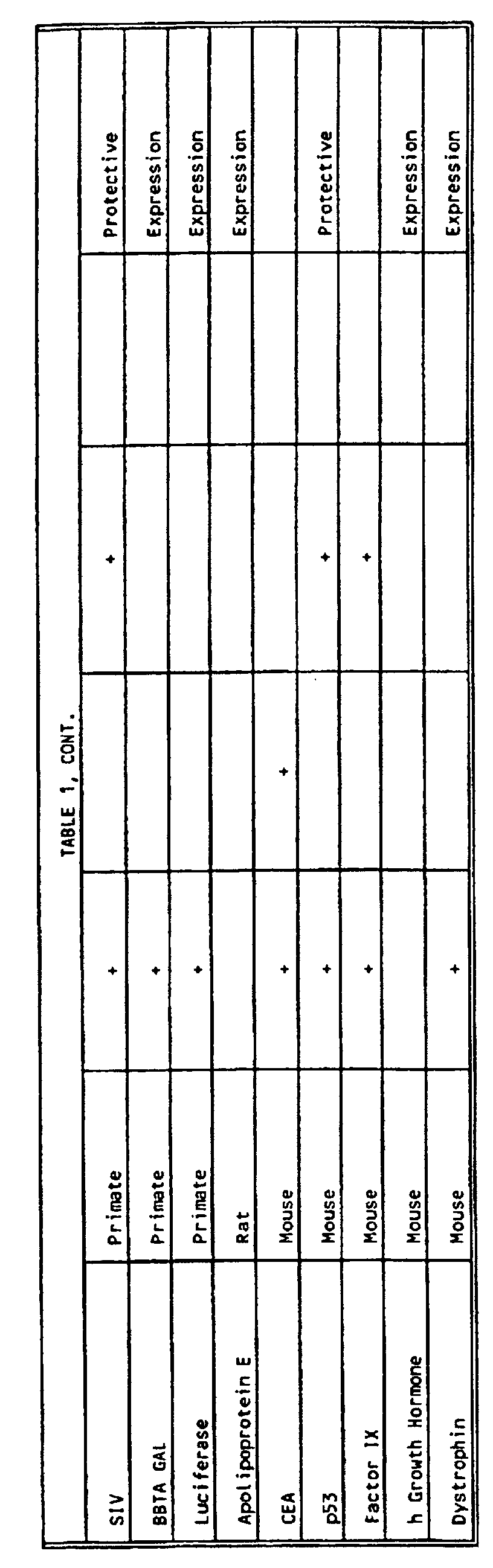


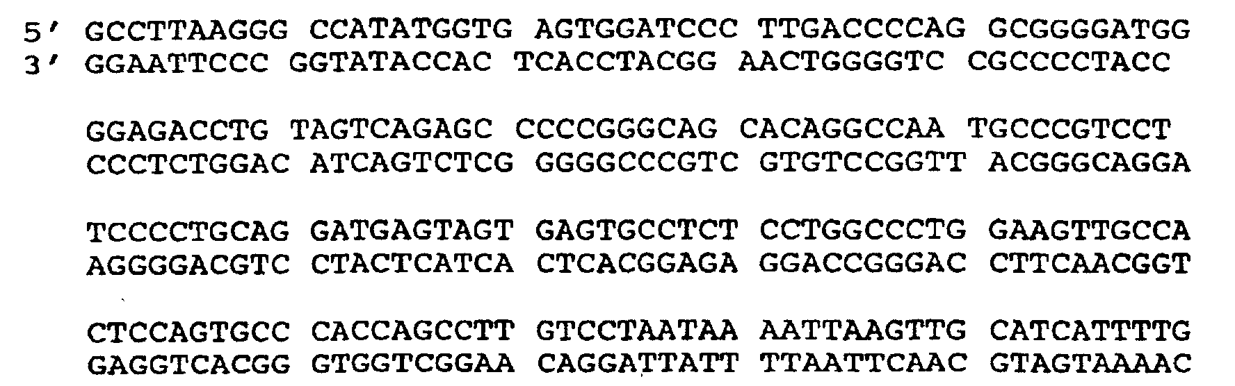



CCTCGGTACCTGCCACCATGGCGCGGATTCTTTAT (SEQ. ID. NO. 13) and spans the 2217 to 2252 bases of pCDM8 but, diverges at 2235 to accommodate linking restriction sites in this case the 5' Bgl I cloning site (Figure 2) and a Kpn I site at the upstream, 5' end. This is important later for promoter ligation. The lower primer (negative strand or 3 ' ) is CGGCATATGGCCTTAAGGCGCCCGCTTGTTCCTGAAGT (SEQ. ID. NO. 14) and spans the 2394 to 2433 bases of pCDM8 but, diverges at 2454 to accommodate linking restriction sites in this case the 3' Bgl I cloning site (see Figure 2) and a Nde I site at the upstream, 5' end. This allows isolation of a fragment without a Bgl I site or any other conflicting sites. This fragment is digested with Nde I and gel purified. The pGEM product from above is digested with Nde I (site engineered into synthetic sequence) and Stu I which results in a blunt end on the 5' end. The fragment isolated above is ligated into this digested construct, i.e., Nde I end to Nde I end and blunt to blunt.


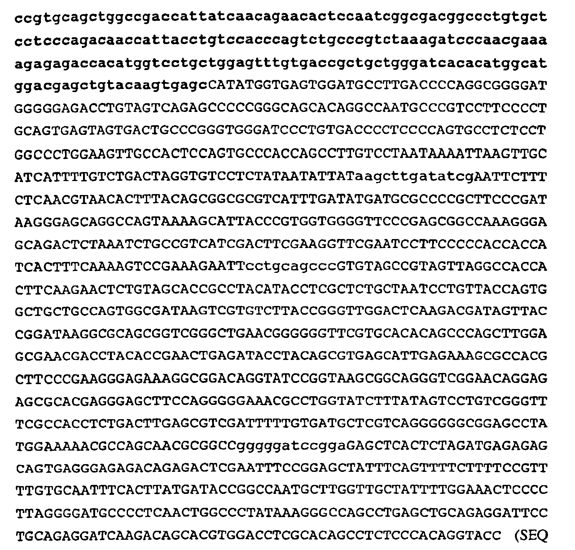
PDLSYMPIWKF PDEEGACQPC PINCTHSCVD LDDKGCPAEQ
RASPLTSIIS AVVGILLVVV LGVVFGIL (SEQ. ID NO. 19), or portion or mammalian homolog thereof and,
PAPGAGGMVH HRHRSSSTRS GGGDLTLGLE PSEEEAPRSP
LAPSEGAGSD VFDGDLGMGA AKGLOSLPTH DPSPLQRYSE
DPTVPLPSET DGYVAPLTCS PQPEYVNQPD VRPOPPSPRE
GPLPAARPAG ATLERPKTLS PGKNGVVKDV FAFGGAVENP
EYLTPQGTCS PQPEYVNQPD VRPQPPSPRE GPLPAARPAG
ATLERPKLSP GKNGVVKDVF AFGGAVENPE YLTPQGGAAP
QPHPPPAFSP AFDNLYYWDO DPPERGAPPS TFKGTPTAEN
PEYLGLDVPV (SEQ. ID NO. 20), or portion or mammalian homolog thereof.
IISAVVGILLVVVLGVVFGILI (SEQ. ID NO. 21), or portion or mammalian homolog thereof.


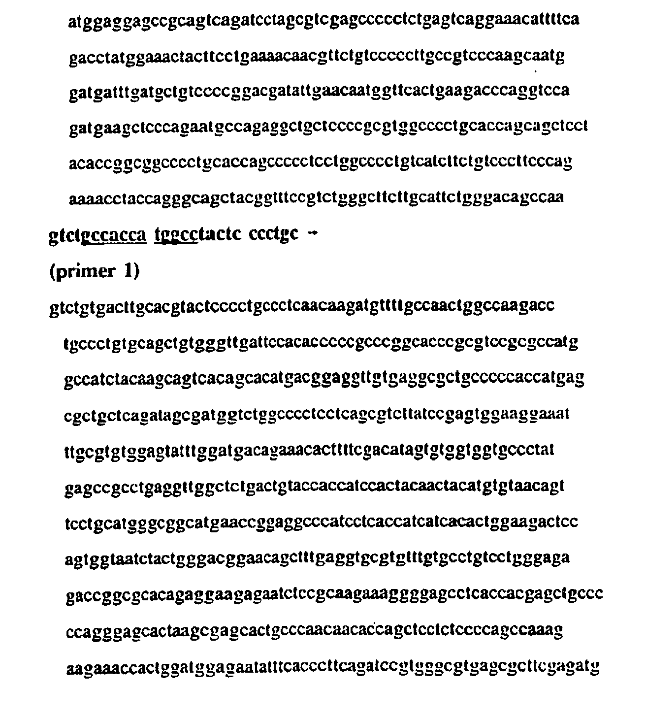

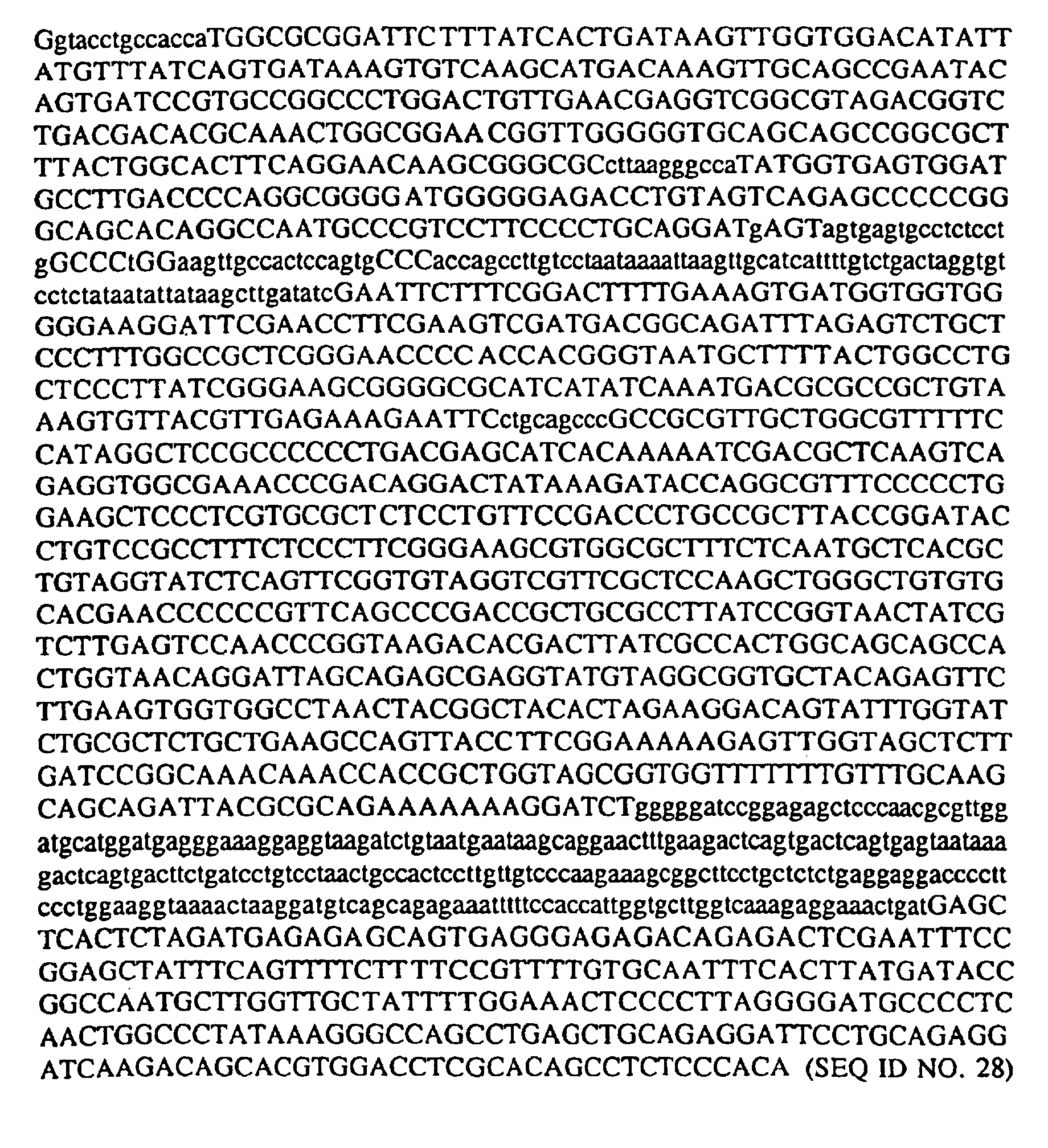
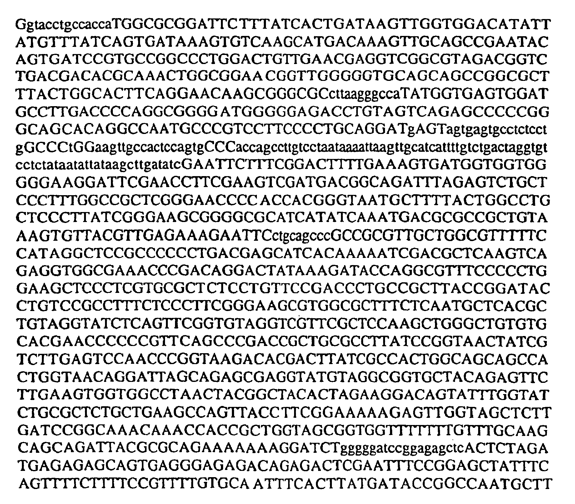














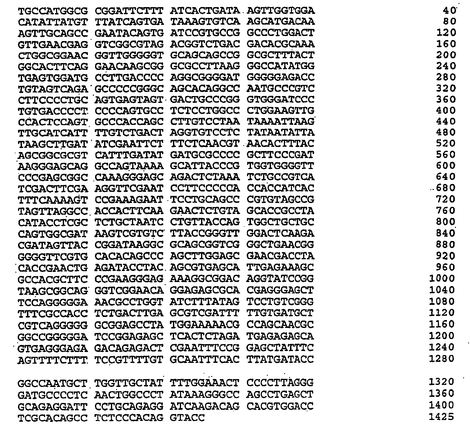
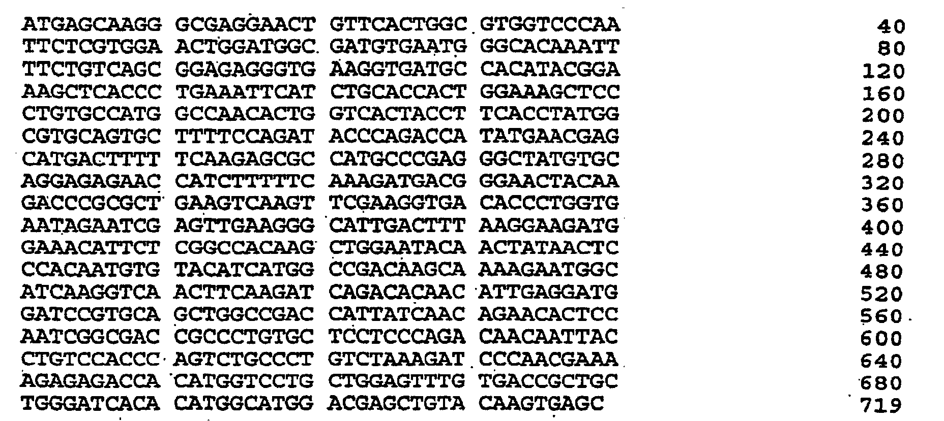
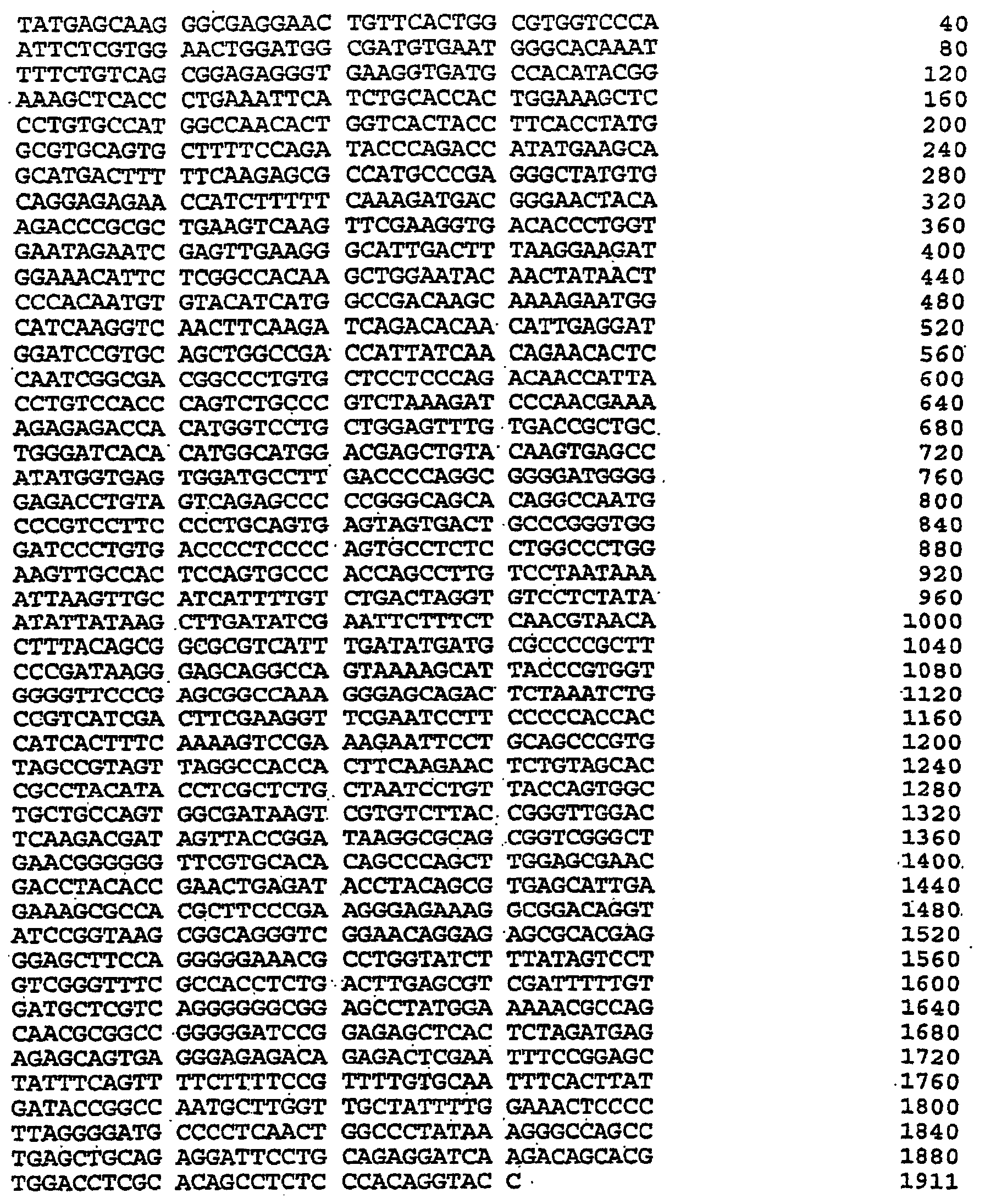
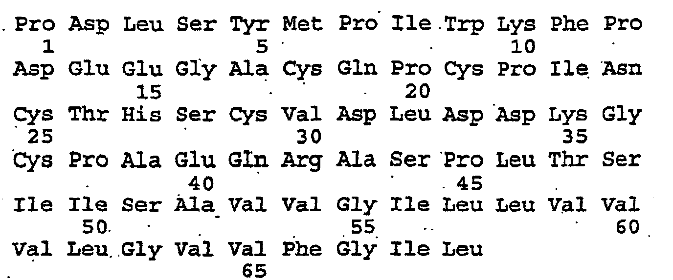







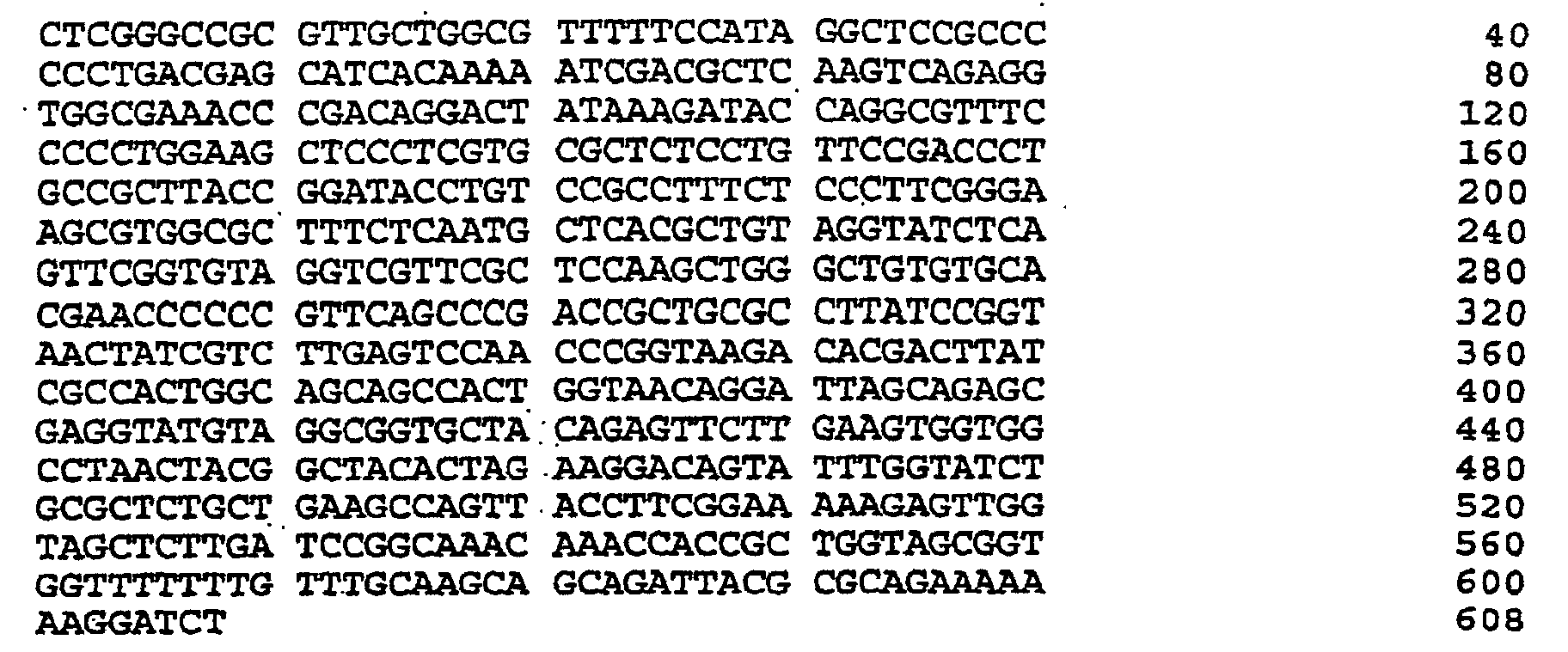
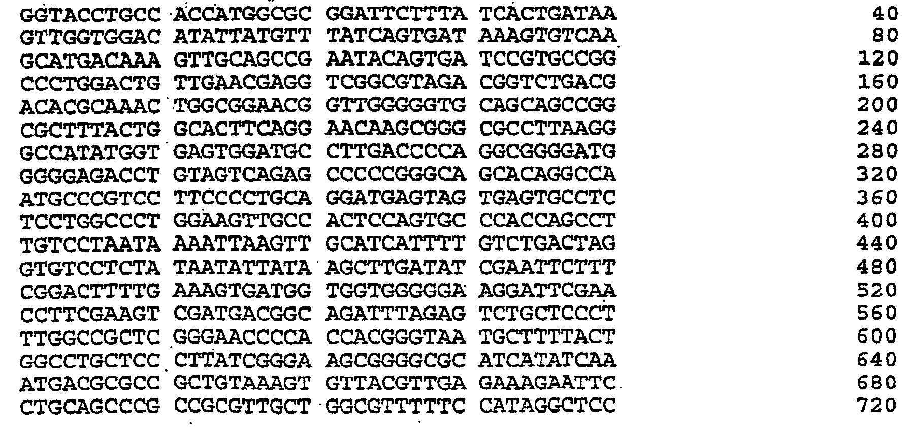

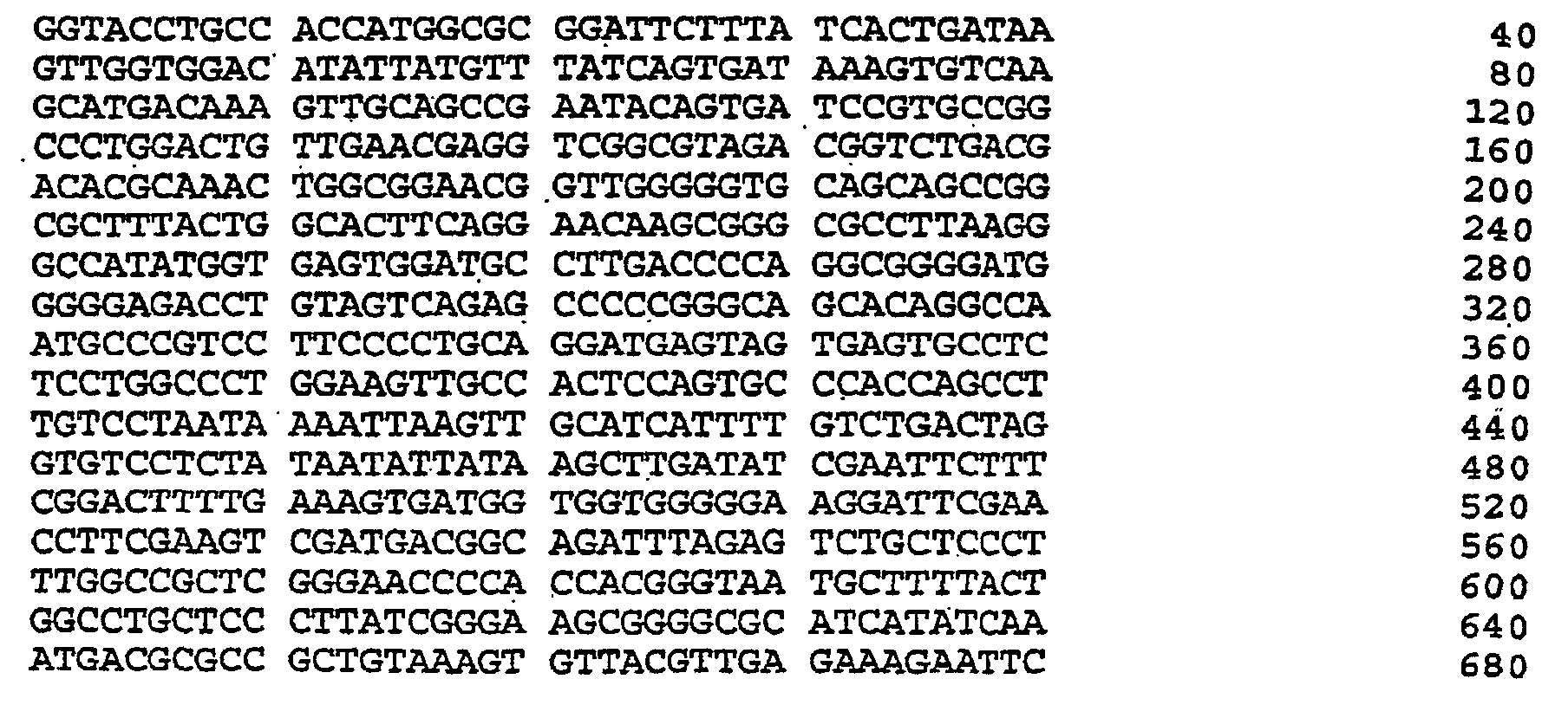
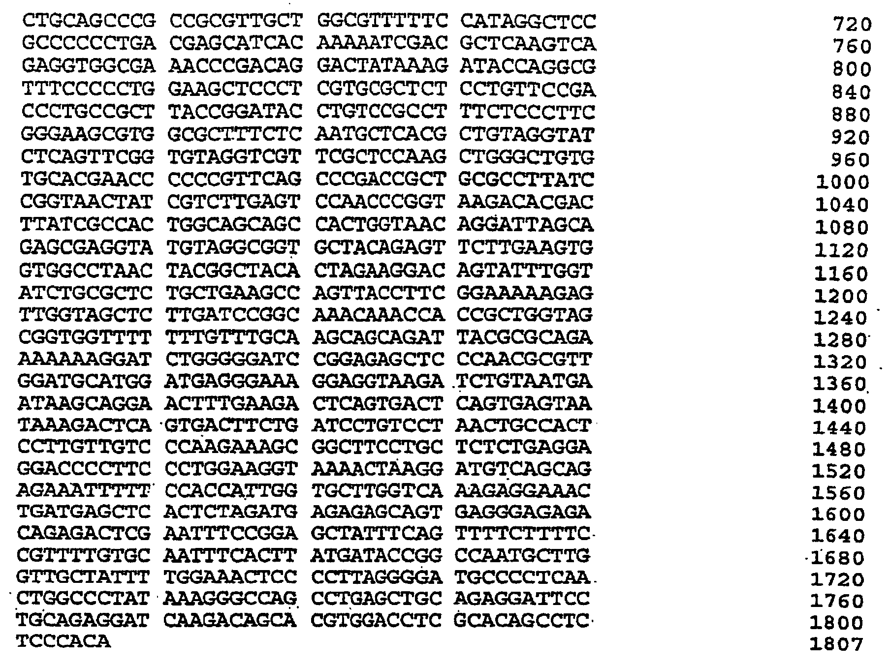


Claims (52)
- A humanized polynucleotide vector comprising:a human derived promotor or mammalian homolog thereof which is functional in a target tissue or target cells, said promotor operably linked to a sequence acceptance site which directionally accepts cDNA target products from rtPCR cloning via unique sites within an interrupted palindrome recognition sequence for a restriction endonuclease, wherein said vector lacks an antibiotic resistance encoding nucleic acid sequence, and comprises minimal non-human components necessary for production of the vector, wherein said minimal non-human components are an origin of replication which allows replication and growth of the vector in bacteria or yeast and a nucleic acid sequence which allows for selection of recombinant plasmids in bacteria or yeast.
- The humanized polynucleotide vector according to claim 1 wherein the target cells are selected from the group consisting of myocytes and professional antigen presenting cells.
- The humanized polynucleotide vector according to claim 1 or 2 wherein the target cells or target tissue are human.
- The humanized polynucleotide vector according to claims 1 to 3 wherein the human derived promotor is a RANTES promotor or portion thereof.
- The humanized polynucleotide vector according to claim 4 wherein the promotor has approximately 440 base pairs.
- The humanized polynucleotide vector according to claims 4 or 5 wherein the portion corresponds to a region spanning the NCO site through the KpnI site of the genomic RANTES promotor.
- The humanized polynucleotide vector according to claim 6 wherein the origin for replication is colE1 or functional portion thereof.
- The humanized polynucleotide vector according to claim 6 wherein the origin for replication comprises a 635 base pair region of the colE1 origin of replication.
- The humanized polynucleotide vector according to claims 1 to 8 further comprising a human-derived 3' splice sequence and a human-derived poly A sequence, both sequences located downstream of the sequence acceptance site.
- The humanized polynucleotide vector according to claim 9 wherein the human derived 3' splice and poly A sequence are derived from human growth hormone.
- A polynucleotide vector according to claims 1 to 10 wherein a 5' sequence acceptance site reads on the positive strand as GCCACCATGGCC or wherein a 3' sequence acceptance site reads on the positive strand as GCCTTAAGGGC.
- A polynucleotide vector comprising SEQ ID NO:16, SEQ ID NO:27 or SEQ ID NO:28.
- A polynucleotide vector contained within a host cell deposited with the ATCC under the ATCC designation 98400 or ATCC designation 98401.
- A polynucleotide vector according to claims 1 to 13 further comprising cDNA target products, and an optional internal ribosomal entry site, said cDNA target products integrated into said sequence acceptance site, said cDNA target products comprising a nucleotide sequence encoding at least one target antigen or antigenic epitope thereof alone or in combination with a nucleotide sequence encoding a cytokine or chemokine.
- The humanized polynucleotide vector according to claims 1 to 11 wherein the sequence acceptance site comprises the nucleotide sequence as depicted in Figure 2.
- The humanized polynucleotide vector according to claims 1 to 14 wherein the recognition sequence is recognized by BglI restriction endonuclease.
- The humanized polynucleotide vector according to claim 6 wherein the nucleic acid sequence which allows for selection is a suppressor tRNA gene, a synthetic SupF complementation tRNA gene, or functional derivatives thereof.
- The humanized polynucleotide vector according to claim 17, wherein the nucleic acid sequence is selected from the group consisting of SupE, SupP, SupD, SupU, SupF, SupZ, glyT, glyU, SerP, psu1 +, psu2 +-C34, psu3 +AM and psu3 +OC.
- A polynucleotide vector vaccine according to claim 1 further comprising an optional internal ribosomal entry site, and cDNA target products, said cDNA target products integrated into said sequence acceptance site, said cDNA target products comprising a nucleotide sequence encoding at least one target antigen or antigenic epitope thereof.
- A polynucleotide vector vaccine according to claim 19 wherein the target antigen is a product of a tumor associated genetic derangement.
- The polynucleotide vector vaccine of claim 19 wherein the target antigen is a tumor antigen, bacterial antigen, viral antigen, or parasitic antigen.
- The polynucleotide vector vaccine of claim 19 wherein the tumor antigen is p53, RB, ras, int-2, Hst, Tre 17, BRCA-1, BRCA-2, MUC-1, HER-2/neu, truncated EGFRvIII, CEA, MyC, Myb, OB-1, OB-2, BCR/ABL, GIP, GSP, RET, ROS, FIS, SRC, TRC, WT1, DCC, NF1, FAB, MEN-1, ERB-B1 or combinations thereof.
- A polynucleotide vector vaccine according to claim 19, 20, 21 or 22 further comprising an additional cDNA target product comprising a nucleic acid sequence encoding a cytokine or chemokine.
- A polynucleotide vector vaccine according to claim 23 wherein the cytokine is selected from the group consisting of interleukin 2, interleukin 3, interleukin 4, interleukin 7, interleukin 8, interleukin 12, interleukin 15, GM-CSF, tumor necrosis factor, and interferon.
- A polynucleotide vector vaccine according to claim 23 wherein the chemokine is selected from the group consisting of RANTES, MCP, MIP-1α, MIP-1β, defensins and combinations thereof.
- A pharmaceutical composition comprising at least one polynucleotide vector according to claims 1 to 14 and a pharmaceutically acceptable carrier.
- A pharmaceutical composition comprising the polynucleotide vector vaccine according to claims 19-20 or 21-25 and a pharmaceutically acceptable carrier.
- A kit comprising the polynucleotide vector according to claims 1 to 14.
- A kit comprising the polynucleotide vector vaccine according to claims 19-20 or 21-25.
- A kit according to claim 29, further comprising an expression enhancing agent.
- The kit according to claim 30 wherein the expression enhancing agent is a mycotoxic agent.
- The kit according to claim 31 wherein the mycotoxic agent is bupivacaine-HCl and dextrose.
- A host cell comprising:the polynucleotide vector of claims 19-20 or 21-25, wherein the host cell is capable of expressing the target antigen or antigenic epitope.
- The host cell according to claim 33 wherein the host cell is a myocyte or professional antigen presenting cell.
- An in vitro or ex vivo method for expressing at least one target antigen or antigenic epitope thereof in cells comprising:introducing a humanized polynucleotide vector according to claim 1 or 15 into said cells, under conditions for expression of the target antigen or antigenic epitope thereof.
- The method of claim 35 wherein the cells are selected from the group consisting of myocytes and professional antigen presenting cells.
- A method of making a humanized polynucleotide vector according claim 1 or 4 comprising:operably linking a human derived promotor or mammalian homolog thereof which is functional in a target tissue or target cells to a sequence acceptance site, said site directionally accepts cDNA target products from rtPCR cloning via unique sites within an interrupted palindrome recognition sequence for a restriction endonuclease.
- The method of claim 35 wherein the target antigen is a tumor antigen, bacterial antigen, viral antigen, or parasitic antigen.
- The method of claim 38 wherein the tumor antigen is p53, RB, ras, int-2, Hst, Tre 17, BRCA-1, BRCA-2, MUC-1, HER-2/neu, truncated EGFRvIII, CEA, MyC, Myb, OB-1, OB-2, BCR/ABL, GIP, GSP, RET, ROS, FIS, SRC, TRC, WT1, DCC, NF1, FAB, MEN-1, ERB-B1, or combinations thereof.
- The use of a sequence acceptance site having a 5' acceptance site and a 3' acceptance site comprising nucleic acid sequences for accepting cDNA target products from rtPCR cloning wherein the sequence acceptance site directionally accepts target sequence specific products from rtPCR cloning via unique sites within an interrupted palindrome recognition sequence for a restriction enconuclease, wherein the 5'acceptance site comprises an initiation codon and Kozak consensus sequence and the 3' acceptance site comprises a termination codon.
- The use according to claim 40 wherein the restriction endonuclease is BglI.
- The use according to claims 40 or 41 wherein a 5' acceptance site reads on the positive strand as GCCACCATGGCC.
- The use according to claim 42 wherein a 3' acceptance site reads on the positive strand as GCCTTAAGGGC.
- The use according to claim 40 wherein the site comprises the nucleotide sequence as depicted in Figure 2.
- A use of a polynucleotide vector vaccine according to claims 19-20 or 21-25 in the manufacture of a medicament for use in a method of stimulating a specific immune response to at least one target antigen or antigenic epitope thereof in a mammal.
- A use according to claim 45, wherein the medicament can be administered to the muscle or skin.
- A use according to claim 45 or 46 wherein the medicament further comprises an expression enhancing agent.
- The use according to claim 47, wherein the expression enhancing agent is a mycotoxic agent.
- The use according to claim 48, wherein the mycotocix agent is bupivacaine-HCl or dextrose.
- The use according to claims 45 to 48 or 49 wherein the target antigen is a tumor antigen, bacterial antigen, viral antigen or parasitic antigen.
- The use according to claim 50, wherein the tumor antigen is selected for the group consisting of p53, RB, ras, int-2, Hst, Tre 17, BRCA-1, BRCA-2, MUC-1, HER-2/neu, truncated EGFRvIII, CEA, MyC, Myb, OB-1, OB-2, BCR/ABL, GIP, GSP, RET, ROS, FIS, SRC, TRC, WT1, DCC, NF1, FAB, MEN-1, ERB-B1 and combinations thereof.
- The use according to claim 51, wherein the method generates antigen specific cytotoxic lymphocytes to the tumor antigen or antigenic epitopes thereof.
Applications Claiming Priority (3)
| Application Number | Priority Date | Filing Date | Title |
|---|---|---|---|
| US2393196P | 1996-08-14 | 1996-08-14 | |
| US23931P | 1996-08-14 | ||
| PCT/US1997/014306 WO1998006863A1 (en) | 1996-08-14 | 1997-08-14 | A vector for polynucleotide vaccines |
Publications (2)
| Publication Number | Publication Date |
|---|---|
| EP0920522A1 EP0920522A1 (en) | 1999-06-09 |
| EP0920522B1 true EP0920522B1 (en) | 2003-10-29 |
Family
ID=21817994
Family Applications (1)
| Application Number | Title | Priority Date | Filing Date |
|---|---|---|---|
| EP97937243A Expired - Lifetime EP0920522B1 (en) | 1996-08-14 | 1997-08-14 | A vector for polynucleotide vaccines |
Country Status (5)
| Country | Link |
|---|---|
| EP (1) | EP0920522B1 (en) |
| AT (1) | ATE253122T1 (en) |
| AU (1) | AU3980497A (en) |
| DE (1) | DE69725857T2 (en) |
| WO (1) | WO1998006863A1 (en) |
Families Citing this family (9)
| Publication number | Priority date | Publication date | Assignee | Title |
|---|---|---|---|---|
| AU3755800A (en) * | 1999-03-15 | 2000-10-04 | Introgen Therapeutics, Inc. | Dendritic cells transduced with a wild-type self gene elicit potent antitumor immune responses |
| US8668905B2 (en) | 2005-05-12 | 2014-03-11 | University Of South Florida | P53 vaccines for the treatment of cancers |
| PT2222697E (en) | 2007-11-01 | 2013-02-15 | Perseid Therapeutics Llc | Immunosuppressive polypeptides and nucleic acids |
| ES2342646B1 (en) * | 2008-06-02 | 2011-04-26 | Institut De Recerca Hospital Universitari Vall Hebron | METHOD OF DIAGNOSIS OF CANCERES THAT EXPRESS THE RECEIVER HER-2 OR ITS TRUNCATED VARIANTS. |
| SG177252A1 (en) | 2008-12-01 | 2012-03-29 | Lab Corp America Holdings | METHODS AND ASSAYS FOR MEASURING p95 AND/OR p95 IN A SAMPLE AND ANTIBODIES SPECIFIC FOR p95 |
| KR20130087517A (en) | 2010-06-25 | 2013-08-06 | 백시바디 에이에스 | Homodimeric protein constructs |
| KR20140069222A (en) | 2011-09-23 | 2014-06-09 | 유니버시티 오브 오슬로 | Vaccibodies targeted to cross-presenting dendritic cells |
| KR102833333B1 (en) | 2016-01-08 | 2025-07-11 | 니코데 테라퓨틱스 에이에스에이 | Therapeutic anticancer neoepitope vaccine |
| RU2734726C1 (en) * | 2020-01-15 | 2020-10-22 | Генетик Диагностикс Энд Терапи 21 Лтд | Gene-therapeutic dna vector based on the gene-therapeutic dna vector gdtt1_8nas12, carrying the target gene selected from a group of genes ddc, il10, il13, ifnb1, tnfrsf4, tnfsf10, bcl2, hgf, il2 to increase the expression level of said target genes, a method for production and use thereof, a strain escherichia coli jm110-nas/gdtt1_8nas12-ddc or escherichia coli jm110-nas/gdtt1_8nas12-il10 or escherichia coli jm110-nas/gdtt1_8nas12-il13 or escherichia coli jm110-nas/gdtt1_8nas12-ifnb1 or escherichia coli jm110-nas/gdtt1_8nas12-tnfrsf4 or escherichia coli jm110-nas/gdtt1_8nas12-tnfsf10 or escherichia coli jm110-nas/gdtt1_8nas12-bcl2 or escherichia coli jm110-nas/gdtt1_8nas12-hgf or escherichia coli jm110-nas/gdtt1_8nas12-il2, carrying a gene-therapeutic dna vector, method for production thereof, a method for industrial production of a gene-therapeutic dna vector |
Citations (2)
| Publication number | Priority date | Publication date | Assignee | Title |
|---|---|---|---|---|
| WO1992001307A1 (en) * | 1990-07-12 | 1992-01-23 | Gte Products Corporation | Lamp with an oxygen detector |
| WO1996040987A1 (en) * | 1995-06-07 | 1996-12-19 | Affymax Technologies N.V. | Peptide library and screening method |
Family Cites Families (2)
| Publication number | Priority date | Publication date | Assignee | Title |
|---|---|---|---|---|
| DK0538300T3 (en) * | 1990-07-10 | 1994-10-10 | Boehringer Ingelheim Int | O-Glycosylated IFN-alpha |
| US5426039A (en) * | 1993-09-08 | 1995-06-20 | Bio-Rad Laboratories, Inc. | Direct molecular cloning of primer extended DNA containing an alkane diol |
-
1997
- 1997-08-14 EP EP97937243A patent/EP0920522B1/en not_active Expired - Lifetime
- 1997-08-14 WO PCT/US1997/014306 patent/WO1998006863A1/en not_active Ceased
- 1997-08-14 DE DE69725857T patent/DE69725857T2/en not_active Expired - Fee Related
- 1997-08-14 AT AT97937243T patent/ATE253122T1/en not_active IP Right Cessation
- 1997-08-14 AU AU39804/97A patent/AU3980497A/en not_active Abandoned
Patent Citations (2)
| Publication number | Priority date | Publication date | Assignee | Title |
|---|---|---|---|---|
| WO1992001307A1 (en) * | 1990-07-12 | 1992-01-23 | Gte Products Corporation | Lamp with an oxygen detector |
| WO1996040987A1 (en) * | 1995-06-07 | 1996-12-19 | Affymax Technologies N.V. | Peptide library and screening method |
Also Published As
| Publication number | Publication date |
|---|---|
| EP0920522A1 (en) | 1999-06-09 |
| DE69725857T2 (en) | 2004-07-29 |
| ATE253122T1 (en) | 2003-11-15 |
| WO1998006863A1 (en) | 1998-02-19 |
| DE69725857D1 (en) | 2003-12-04 |
| AU3980497A (en) | 1998-03-06 |
Similar Documents
| Publication | Publication Date | Title |
|---|---|---|
| Inoue et al. | Induction of interleukin 2 receptor gene expression by p40x encoded by human T‐cell leukemia virus type 1. | |
| Hodge et al. | Induction of antitumor immunity by recombinant vaccinia viruses expressing B7-1 or B7-2 costimulatory molecules | |
| Haber et al. | WT1-mediated growth suppression of Wilms tumor cells expressing a WT1 splicing variant | |
| CN107847577B (en) | Cancer vaccine comprising mRNA encoding M-like protein | |
| JP3907698B2 (en) | A composition comprising a recombinant virus expressing an antigen and a recombinant virus expressing an immunostimulatory molecule | |
| KR20010024585A (en) | Tumor-specific antigens, methods for their production and their use for immunization and diagnosis | |
| US20160235787A1 (en) | Epitope Spreading Associated with CAR T-Cells | |
| JP2002531133A (en) | Recombinant vector expressing multiple costimulatory molecules and use thereof | |
| US20100316603A1 (en) | Combination Immunogene Therapy | |
| KR19980703453A (en) | Intracellular Domain of Heer-2 / Neu Protein for Prevention or Treatment of Malignant Tumors | |
| JP2009165490A (en) | Compositions and methods for enhancing anti-tumor immunity and gene therapy | |
| JP2002509716A (en) | Methods and compositions for raising an immune response to a telomerase antigen | |
| CN108603171A (en) | Antigen-specificity TCR of new generation | |
| KR100255582B1 (en) | Genes and Genetic Factors Associated with the Regulation of Oncogenic Transformation in Mammalian Cells | |
| DE69725857T2 (en) | VECTOR FOR POLYNUCLEOT VACCINE | |
| KR20210080372A (en) | Novel cancer antigens and methods | |
| JP2007523610A (en) | Synthetic genes encoding carcinoembryonic antigen and uses thereof | |
| KR20050004211A (en) | Muc-1 antigen with reduced number of vntr repeat units | |
| JP2000506379A (en) | Cell lines and clones of human prostate epithelial immortal cells and their research and application for prostate cancer treatment | |
| JP2009195235A (en) | Immune effector cell hybrid | |
| Temme et al. | Efficient transduction and long-term retroviral expression of the melanoma-associated tumor antigen tyrosinase in CD34+ cord blood-derived dendritic cells | |
| EP0599077A2 (en) | Anti-metastatic vaccine | |
| DE60023891T2 (en) | PEPTIDE COMPOUND DERIVED FROM A DELAYED OPEN READING OF THE ICE GENE | |
| US20070042047A1 (en) | Vaccines | |
| Lucansky et al. | DNA vaccination against bcr-abl-positive cells in mice |
Legal Events
| Date | Code | Title | Description |
|---|---|---|---|
| PUAI | Public reference made under article 153(3) epc to a published international application that has entered the european phase |
Free format text: ORIGINAL CODE: 0009012 |
|
| 17P | Request for examination filed |
Effective date: 19990301 |
|
| AK | Designated contracting states |
Kind code of ref document: A1 Designated state(s): AT BE CH DE DK ES FI FR GB GR IE IT LI LU MC NL PT SE |
|
| 17Q | First examination report despatched |
Effective date: 19990727 |
|
| GRAH | Despatch of communication of intention to grant a patent |
Free format text: ORIGINAL CODE: EPIDOS IGRA |
|
| GRAS | Grant fee paid |
Free format text: ORIGINAL CODE: EPIDOSNIGR3 |
|
| GRAA | (expected) grant |
Free format text: ORIGINAL CODE: 0009210 |
|
| AK | Designated contracting states |
Kind code of ref document: B1 Designated state(s): AT BE CH DE DK ES FI FR GB GR IE IT LI LU MC NL PT SE |
|
| PG25 | Lapsed in a contracting state [announced via postgrant information from national office to epo] |
Ref country code: NL Free format text: LAPSE BECAUSE OF FAILURE TO SUBMIT A TRANSLATION OF THE DESCRIPTION OR TO PAY THE FEE WITHIN THE PRESCRIBED TIME-LIMIT Effective date: 20031029 Ref country code: IT Free format text: LAPSE BECAUSE OF FAILURE TO SUBMIT A TRANSLATION OF THE DESCRIPTION OR TO PAY THE FEE WITHIN THE PRE;WARNING: LAPSES OF ITALIAN PATENTS WITH EFFECTIVE DATE BEFORE 2007 MAY HAVE OCCURRED AT ANY TIME BEFORE 2007. THE CORRECT EFFECTIVE DATE MAY BE DIFFERENT FROM THE ONE RECORDED.SCRIBED TIME-LIMIT Effective date: 20031029 Ref country code: FI Free format text: LAPSE BECAUSE OF FAILURE TO SUBMIT A TRANSLATION OF THE DESCRIPTION OR TO PAY THE FEE WITHIN THE PRESCRIBED TIME-LIMIT Effective date: 20031029 Ref country code: BE Free format text: LAPSE BECAUSE OF FAILURE TO SUBMIT A TRANSLATION OF THE DESCRIPTION OR TO PAY THE FEE WITHIN THE PRESCRIBED TIME-LIMIT Effective date: 20031029 Ref country code: AT Free format text: LAPSE BECAUSE OF FAILURE TO SUBMIT A TRANSLATION OF THE DESCRIPTION OR TO PAY THE FEE WITHIN THE PRESCRIBED TIME-LIMIT Effective date: 20031029 |
|
| REG | Reference to a national code |
Ref country code: GB Ref legal event code: FG4D |
|
| REG | Reference to a national code |
Ref country code: CH Ref legal event code: EP |
|
| REG | Reference to a national code |
Ref country code: IE Ref legal event code: FG4D |
|
| REF | Corresponds to: |
Ref document number: 69725857 Country of ref document: DE Date of ref document: 20031204 Kind code of ref document: P |
|
| PG25 | Lapsed in a contracting state [announced via postgrant information from national office to epo] |
Ref country code: SE Free format text: LAPSE BECAUSE OF FAILURE TO SUBMIT A TRANSLATION OF THE DESCRIPTION OR TO PAY THE FEE WITHIN THE PRESCRIBED TIME-LIMIT Effective date: 20040129 Ref country code: GR Free format text: LAPSE BECAUSE OF FAILURE TO SUBMIT A TRANSLATION OF THE DESCRIPTION OR TO PAY THE FEE WITHIN THE PRESCRIBED TIME-LIMIT Effective date: 20040129 Ref country code: DK Free format text: LAPSE BECAUSE OF FAILURE TO SUBMIT A TRANSLATION OF THE DESCRIPTION OR TO PAY THE FEE WITHIN THE PRESCRIBED TIME-LIMIT Effective date: 20040129 |
|
| REG | Reference to a national code |
Ref country code: CH Ref legal event code: NV Representative=s name: RITSCHER & PARTNER AG PATENTANWAELTE |
|
| PG25 | Lapsed in a contracting state [announced via postgrant information from national office to epo] |
Ref country code: ES Free format text: LAPSE BECAUSE OF FAILURE TO SUBMIT A TRANSLATION OF THE DESCRIPTION OR TO PAY THE FEE WITHIN THE PRESCRIBED TIME-LIMIT Effective date: 20040209 |
|
| NLV1 | Nl: lapsed or annulled due to failure to fulfill the requirements of art. 29p and 29m of the patents act | ||
| ET | Fr: translation filed | ||
| PLBE | No opposition filed within time limit |
Free format text: ORIGINAL CODE: 0009261 |
|
| STAA | Information on the status of an ep patent application or granted ep patent |
Free format text: STATUS: NO OPPOSITION FILED WITHIN TIME LIMIT |
|
| PGFP | Annual fee paid to national office [announced via postgrant information from national office to epo] |
Ref country code: DE Payment date: 20040930 Year of fee payment: 8 |
|
| 26N | No opposition filed |
Effective date: 20040730 |
|
| PGFP | Annual fee paid to national office [announced via postgrant information from national office to epo] |
Ref country code: MC Payment date: 20050725 Year of fee payment: 9 |
|
| PGFP | Annual fee paid to national office [announced via postgrant information from national office to epo] |
Ref country code: GB Payment date: 20050810 Year of fee payment: 9 |
|
| PGFP | Annual fee paid to national office [announced via postgrant information from national office to epo] |
Ref country code: FR Payment date: 20050817 Year of fee payment: 9 |
|
| PGFP | Annual fee paid to national office [announced via postgrant information from national office to epo] |
Ref country code: CH Payment date: 20050819 Year of fee payment: 9 |
|
| PGFP | Annual fee paid to national office [announced via postgrant information from national office to epo] |
Ref country code: IE Payment date: 20050824 Year of fee payment: 9 |
|
| PGFP | Annual fee paid to national office [announced via postgrant information from national office to epo] |
Ref country code: LU Payment date: 20051005 Year of fee payment: 9 |
|
| PG25 | Lapsed in a contracting state [announced via postgrant information from national office to epo] |
Ref country code: DE Free format text: LAPSE BECAUSE OF NON-PAYMENT OF DUE FEES Effective date: 20060301 |
|
| PG25 | Lapsed in a contracting state [announced via postgrant information from national office to epo] |
Ref country code: IE Free format text: LAPSE BECAUSE OF NON-PAYMENT OF DUE FEES Effective date: 20060814 |
|
| PG25 | Lapsed in a contracting state [announced via postgrant information from national office to epo] |
Ref country code: MC Free format text: LAPSE BECAUSE OF NON-PAYMENT OF DUE FEES Effective date: 20060831 Ref country code: LI Free format text: LAPSE BECAUSE OF NON-PAYMENT OF DUE FEES Effective date: 20060831 Ref country code: CH Free format text: LAPSE BECAUSE OF NON-PAYMENT OF DUE FEES Effective date: 20060831 |
|
| REG | Reference to a national code |
Ref country code: CH Ref legal event code: PL |
|
| GBPC | Gb: european patent ceased through non-payment of renewal fee |
Effective date: 20060814 |
|
| REG | Reference to a national code |
Ref country code: IE Ref legal event code: MM4A |
|
| REG | Reference to a national code |
Ref country code: FR Ref legal event code: ST Effective date: 20070430 |
|
| PG25 | Lapsed in a contracting state [announced via postgrant information from national office to epo] |
Ref country code: GB Free format text: LAPSE BECAUSE OF NON-PAYMENT OF DUE FEES Effective date: 20060814 |
|
| PG25 | Lapsed in a contracting state [announced via postgrant information from national office to epo] |
Ref country code: PT Free format text: LAPSE BECAUSE OF NON-PAYMENT OF DUE FEES Effective date: 20040329 |
|
| PG25 | Lapsed in a contracting state [announced via postgrant information from national office to epo] |
Ref country code: FR Free format text: LAPSE BECAUSE OF NON-PAYMENT OF DUE FEES Effective date: 20060831 |
|
| PG25 | Lapsed in a contracting state [announced via postgrant information from national office to epo] |
Ref country code: LU Free format text: LAPSE BECAUSE OF NON-PAYMENT OF DUE FEES Effective date: 20060814 |