EP0918867B1 - Substrate trapping protein tyrosine phosphatases - Google Patents
Substrate trapping protein tyrosine phosphatases Download PDFInfo
- Publication number
- EP0918867B1 EP0918867B1 EP97937017A EP97937017A EP0918867B1 EP 0918867 B1 EP0918867 B1 EP 0918867B1 EP 97937017 A EP97937017 A EP 97937017A EP 97937017 A EP97937017 A EP 97937017A EP 0918867 B1 EP0918867 B1 EP 0918867B1
- Authority
- EP
- European Patent Office
- Prior art keywords
- ptp
- protein
- tyrosine phosphatase
- cas
- pest
- Prior art date
- Legal status (The legal status is an assumption and is not a legal conclusion. Google has not performed a legal analysis and makes no representation as to the accuracy of the status listed.)
- Expired - Lifetime
Links
Images


Classifications
-
- C—CHEMISTRY; METALLURGY
- C12—BIOCHEMISTRY; BEER; SPIRITS; WINE; VINEGAR; MICROBIOLOGY; ENZYMOLOGY; MUTATION OR GENETIC ENGINEERING
- C12N—MICROORGANISMS OR ENZYMES; COMPOSITIONS THEREOF; PROPAGATING, PRESERVING, OR MAINTAINING MICROORGANISMS; MUTATION OR GENETIC ENGINEERING; CULTURE MEDIA
- C12N9/00—Enzymes; Proenzymes; Compositions thereof; Processes for preparing, activating, inhibiting, separating or purifying enzymes
- C12N9/14—Hydrolases (3)
- C12N9/16—Hydrolases (3) acting on ester bonds (3.1)
-
- A—HUMAN NECESSITIES
- A61—MEDICAL OR VETERINARY SCIENCE; HYGIENE
- A61P—SPECIFIC THERAPEUTIC ACTIVITY OF CHEMICAL COMPOUNDS OR MEDICINAL PREPARATIONS
- A61P35/00—Antineoplastic agents
-
- A—HUMAN NECESSITIES
- A61—MEDICAL OR VETERINARY SCIENCE; HYGIENE
- A61K—PREPARATIONS FOR MEDICAL, DENTAL OR TOILETRY PURPOSES
- A61K38/00—Medicinal preparations containing peptides
-
- C—CHEMISTRY; METALLURGY
- C12—BIOCHEMISTRY; BEER; SPIRITS; WINE; VINEGAR; MICROBIOLOGY; ENZYMOLOGY; MUTATION OR GENETIC ENGINEERING
- C12N—MICROORGANISMS OR ENZYMES; COMPOSITIONS THEREOF; PROPAGATING, PRESERVING, OR MAINTAINING MICROORGANISMS; MUTATION OR GENETIC ENGINEERING; CULTURE MEDIA
- C12N2799/00—Uses of viruses
- C12N2799/02—Uses of viruses as vector
- C12N2799/021—Uses of viruses as vector for the expression of a heterologous nucleic acid
- C12N2799/026—Uses of viruses as vector for the expression of a heterologous nucleic acid where the vector is derived from a baculovirus
Definitions
- the protein tyrosine phosphatase (PTP) family of enzymes consists of more than 500 structurally diverse proteins which have in common the highly conserved 250 amino acid PTP catalytic domain, but which display considerable variation in their non-catalytic segments (Charbonneau and Tonks, Annu. Rev. Cell Biol. 8 :463-493 (1992); Tonks, Semin. Cell Biol. 4 :373-453 (1993)).
- Protein Science, vol. 5, no. 1, Jan 1996, 5-12 discloses that the reaction mechanism of protein tyrosine phosphatases (PTPases) and dual specificity protein phosphatases is thought to involve a catalytic aspartic acid residue.
- Aspartic acid 383 is described to be identified as a potential candidate for the catalytic acid in human Cdc25A protein phosphatase, using sequence alignment, structural information, and site directed mutagenesis.
- a D383N mutant exhibits a 150-fold reduction in k cat , with K m more than doubled.
- substrate specificity of mammalian protein tyrosine phosphatases has been investigated using a novel substrate trapping approach in which mutant or altered forms of the mammalian PTP, also referred to as substrate trapping PTPs, are used to bind (trap) one or more substrates of the PTP. Binding of the substrate trapping PTP with a substrate of the PTP results in the formation of a complex which can be readily observed, and, if desired, isolated, and characterized.
- the mutant forms of the PTPs have attenuated catalytic activity (lack catalytic activity or have reduced catalytic activity) relative to the wild type PTP but retain the ability to bind tyrosine phosphorylated substrate(s) of the wild type PTP.
- the methods of the present invention are specifically exempliefied herein with respect to the phosphatases PTP1B and PTP-PEST; however, it is understood that the invention is not limited to these specific PTPs but is applicable to all members of the PTP family.
- mutant (i.e., altered or substrate trapping) forms of PTP1B and PTP-PEST were generated which were catalytically attenuated but retained the ability to bind substrates.
- These mutant PTPs associated in stable complexes with proteins which were identified by immunoblotting as p210 bcr:abl and p130 cas , respectively.
- These associations were observed in lysates from several cell lines and in transfected COS cells, indicating that p210 bcr:abl and p130 cas represent major physiologically relevant substrates for PTP1B and PTP-PEST.
- One embodiment of the invention relates to novel mutant PTPs in which the invariant aspartate residue is replaced with an amino acid which does not cause significant alteration of the Km of the enzyme but which results in a reduction in Kcat to less than 1 per minute (less than 1 min -1 ). These PTPs retain the ability to form a complex with, or bind, their tyrosine phosphorylated substrates, but are catalytically attenuated.
- the invention relates to the phosphatase PTP1B in which the invariant aspartate residue at position 181 is replaced with alanine (D181A).
- the invention in another embodiment relates to the phosphatase PTP-PEST in which the invariant aspartate residue at position 199 is replaced with an alanine (D199A).
- Another embodiment of the invention relates to a PTP-PEST phosphatase in which the cysteine residue at position 231 is replaced with a serine (C231S).
- the invention also relates to other mutant or substrate trapping PTPs in which the invariant aspartate residue is replaced with or changed to another amino acid residue, such as alanine.
- the invariant aspartate residue can be identified in other PTPs by aligning the PTP nucleotide sequence with the nucleotide sequence of a PTP for which the location of the invariant aspartate residue is known.
- the invention also relates to a method of identifying a tyrosine phosphorylated substrate of a protein tyrosine phosphatase.
- a tyrosine phosphorylated protein of interest is combined with one or more PTP(s) in which the invariant aspartate residue is replaced with an amino acid which does not cause significant alteration of the Km of the enzyme but which results in a reduction in Kcat to less than 1 per minute (less than 1 min -1 ), and the presence or absence of a complex between the protein and the PTP(s) is determined. Presence of a complex in the combination indicates that the tyrosine phosphorylated protein is a substrate of the PTP.
- the PTP DA mutant binds to or complexes with its substrate but does not dephosphorylate it (or does so very slowly), thereby allowing the complex to be observed and, optionally, isolated and identified.
- the invariant aspartate is replaced with an alanine residue (a PTP DA mutation or alteration)
- the PTP DA mutant binds to or complexes with its substrate but does not dephosphorylate it (or does so very slowly), thereby allowing the complex to be observed, and, optionally, isolated and identified.
- the invariant aspartate residue is replaced with an alanine residue (a PTP DA mutation or alteration)
- the present invention also relates to a method of identifying a tyrosine phosphorylated substrate of a protein tyrosine phosphatase wherein more than one tyrosine phosphorylated protein of interest is combined with more than one PTP of interest in which the invariant aspartate residue is replaced with an amino acid which does not cause significant alteration of the Km of the enzyme but which results in a reduction in Kcat to less than 1 per minute (less than 1 min -1 ) (e.g., the invariant aspartate is replaced with an alanine residue).
- Complexes formed in the combination can be isolated and the component PTP and substrate can be identified.
- the invention also pertains to a method of reducing the activity of a tyrosine phosphorylated protein, comprising administering to a mammal a PTP in which the invariant aspartate residue is replaced with an amino acid which does not cause significant alteration of the Km of the enzyme but which results in a reduction in Kcat to less than 1 per minute (less than 1 min -1 ) (e.g., the invariant aspartate is replaced with an alanine residue) and which forms a complex with the tyrosine phosphorylated protein.
- the PTP mutant binds to the phosphorylated protein without dephosphorylating it, thereby inhibiting the activity of the protein and reducing its downstream effects.
- the invention relates to a method of reducing the transforming effects of oncogenes associated with p130 cas , a substrate of PTP-PEST, comprising administering to a mammal wild type PTP-PEST or PTP-PEST in which the invariant aspartate residue is replaced with an alanine residue.
- Wild type PTP-PEST binds and dephosphorylates p130 cas , thereby negatively regulating its downstream effects.
- DA mutants of PTP-PEST bind but do not dephosphorylate p130 cas (or dephosphorylate it at a reduced rate); the substrate is thus tied up in the complex with the substrate trapping form of PTP-PEST and cannot exert its downstream effects.
- the invention relates to a method of reducing the formation of signalling complexes associated with p130 cas , particularly those signalling complexes which induce mitogenic pathways, comprising administering to a mammal wild type PTP-PEST or PTP-PEST in which the invariant aspartate residue is replaced with an alanine residue.
- the present invention also relates to assays for identifying agents which alter, e.g., enhance or inhibit, the interaction between a PTP and its phosphorylated substrate.
- Agents identified by these assays can be agonists (e.g., agents which enhance or increase the activity of the PTP) or antagonists (e.g., agents which inhibit or decrease the activity of the PTP) of PTP activity.
- the agent may be an endogenous physiological substance or may be a natural or synthetic drug, including small organic molecules.
- the tyrosine phosphorylated substrate of a PTP can be identified by the methods described herein.
- An enzymatic activity assay utilizing the wild type PTP can be carried out in the presence of an agent to be tested, and the resulting amount of enzyme activity can be compared with the amount of enzyme activity in the absence of the agent to be tested.
- a decrease in the enzymatic activity in the presence of the agent to be tested indicates that the agent inhibits the interaction between the PTP and its substrate.
- an increase in the enzymatic activity in the presence of the agent to be tested indicates that the agent enhances the interaction between the PTP and its substrate.
- a competitive binding assay can be carried out utilizing the mutant PTP in the presence of an agent to be tested, and the resulting extent of binding of the mutant PTP to its substrate can be compared with the extent of binding in the absence of the agent to be tested.
- a decrease in the extent of binding in the presence of the agent to be tested indicates that the agent inhibits the interaction between the PTP and its substrate.
- an increase in the extent of binding in the presence of the agent to be tested indicates that the agent enhances the interaction between the PTP and its substrate.
- compositions and methods described herein are useful in identifying the tyrosine phosphorylated substrates of members of the PTP family of phosphatases, as well as in regulating the activity of identified substrates.
- compositions and methods described herein are also useful for identifying tyrosine-phosphorylated proteins which are related to a particular disease or disorder, and to methods of screening for modulators which enhance or inhibit the PTP/substrate interaction for use in therapeutic applications.
- the PTP family of enzymes contains a common evolutionarily conserved segment of approximately 250 amino acids known as the PTP catalytic domain. Within this conserved domain is a unique signature sequence motif, [I/V]HCXAGXXR[S/T]G (SEQ ID NO: 36), that is invariant among all PTPs. The cysteine residue in this motif is invariant in members of the family and is known to be essential for catalysis. It functions as a nucleophile to attack the phosphate moiety of the incoming substrate.
- cysteine residue is altered by site-directed mutagenesis to serine (CS mutants) or alanine (CA mutants)
- the resulting PTP is catalytically attenuated but retains the ability to complex with, or bind, its substrate, at least in vitro.
- MKP-1 a member of the PTP family (Sun et al., Cell 75 : 487-493 (1993)), as well as other PTPs.
- these CS mutants can in general bind effectively to phosphotyrosyl substrates in vitro, in many cases such complexes cannot be isolated in vivo.
- the CS mutants are limited in their applicability and cannot be used to isolate all combinations of PTPs and substrates.
- mutant PTPs other than those specifically described herein can readily be made by aligning the amino acid sequence of the PTP catalytic domain with those described herein, identifying the invariant aspartate residue, and changing the residue by site-directed mutagenesis.
- specific examples of PTP mutants described herein are aspartate to alanine mutants (DA mutants), it is understood that the invention is not limited to changes of aspartate to alanine.
- the invariant aspartate residue can be changed, e.g., by site-directed mutagenesis, to any amino acid which does not cause significant alteration of the Km of the enzyme but which results in a reduction in Kcat to less than 1 per minute (less than 1 min -1 ).
- the invariant aspartate residue can be changed or mutated to an alanine, valine, leucine, isoleucine, proline, phenylalanine, tryptophan, methionine, glycine, serine, threonine, cysteine, tyrosine, asparagine, glutamine, lysine, arginine or histidine.
- PTP-PEST is an 88 kDa cytosolic PTP (Charest et al., Biochem. J. 308 :425-432 (1995); den Hertog et al., Biochem. Biophys. Res. Commun. 184 :1241-1249 (1992); Takekawa et al., Biochem. Biophys. Res. Commun. 189 :1223-1230 (1992); Yang et al., J. Biol. Chem. 268 :6622-6628 (1993); Yang et al., J. Biol.
- the substrate specificity of PTP1B was investigated utilizing the same methods outlined for PTP-PEST, with the exception that the cells were not treated with pervanadate.
- a combination of in vitro dephosphorylation and substrate trapping experiments were used to study the substrate interactions of PTP1B and PTP-PEST.
- the substrate trapping methods outlined herein are generally applicable to any PTP by virtue of the shared invariant aspartate residue, and should therefore prove useful in delineating the substrate preference of other PTP family members.
- the use of mutant, catalytically impaired PTPs to trap, and thereby isolate, potential substrates will greatly facilitate the identification of physiologically important substrates for individual PTPs, leading to improved understanding of the roles of these enzymes in regulation of cellular processes.
- One embodiment of the invention relates to novel PTPs in which the invariant aspartate residue is replaced with an amino acid which does not cause significant alteration of the Km of the enzyme but which results in a reduction in Kcat to less than 1 per minute (less than 1 min -1 ).
- These PTPs retain the ability to form a complex with, or bind, their tyrosine phosphorylated substrates but are catalytically attenuated.
- "attenuated" activity is intended to mean that the phosphatase retains a similar Km to that of the wild type phosphatase but has a Vmax which is reduced by a factor of at least 10 4 relative to the wild type enzyme.
- the invariant aspartate residue can be changed or mutated to an alanine, valine, leucine, isoleucine, proline, phenylalanine, tryptophan, methionine, glycine, serine, threonine, cysteine, tyrosine, asparagine, glutamine, lysine, arginine or histidine.
- novel PTPs described herein in which the invariant aspartate residue is replaced with an amino acid which does not cause significant alteration of the Km of the enzyme but which results in a reduction in Kcat to less than 1 per minute (less than 1 min -1 ), can also comprise other mutations, particularly those which assist in stabilizing the PTP/substrate complex.
- a mutation of the [serine/threonine] residue in the signature motif to an alanine residue changes the rate-determining step of the dephosphorylation reaction from the formation of the transition state to the break, down of the transition state, thereby stabilizing the PTP/substrate complex.
- Such mutations may be valuably combined with the replacement of the invariant aspartate residue, particularly assisting in stabilizing the complex and facilitating the observation and isolation of the complex.
- PTPs suitable for use in the invention include any PTP which has an invariant aspartate residue in a corresponding position.
- a phosphatase is a member of the PTP family if it contains the signature motif [I/V]HCXAGXXR[S/T]G (SEQ ID NO: 36).
- Dual specificity PTPs i.e., PTPs which dephosphorylate both phosphorylated tyrosine and phosphorylated serine or threonine, are also suitable for use in the invention.
- Appropriate PTPs include, but are not limited to, PTP1B, PTP-PEST, PTP ⁇ , MKP-1, DEP-1, PTP ⁇ , PTPX1, PTPX10 and PTPH1.
- the invention relates to the phosphatase PTP1B in which the aspartate residue at position 181 is replaced with alanine (D181A). In another embodiment the invention relates to the phosphatase PTP-PEST in which the invariant aspartate residue at position 199 is replaced with an alanine (D199A). Another embodiment of the invention relates to a PTP-PEST phosphatase in which the cysteine residue at position 231 is replaced with a serine (C231S) .
- the invention also relates to a method of identifying a tyrosine phosphorylated protein which is a substrate of a particular protein tyrosine phosphatase.
- a tyrosine phosphorylated protein of interest is combined with at least one PTP in which the invariant aspartate residue is replaced with an amino acid which does not cause significant alteration of the Km of the enzyme but which results in a reduction in Kcat to less than 1 per minute (less than 1 min -1 ) (e.g., an alanine residue), and the presence or absence of a complex between the protein and the PTP is determined.
- Presence of a complex in the combination indicates that the tyrosine phosphorylated protein is a substrate of the PTP.
- the PTP DA mutant substrate trapping mutant binds to or complexes with its substrate but does not dephosphorylate it (or does so very slowly), thereby allowing the complex to be isolated and identified.
- the phosphorylated protein/PTP complex may be isolated by conventional isolation techniques as described in U.S. Patent No. 5,352,660 to Pawson, including salting out, chromatography, electrophoresis, gel filtration, fractionation, absorption, polyacrylamide gel electrophoresis, agglutination, or combinations thereof.
- antibodies against the PTP or the phosphorylated protein can be used, as well as labelled PTPs and/or labelled phosphorylated substrates.
- the PTP or phosphorylated protein can be labelled with various enzymes, fluorescent materials, luminescent materials and radioactive materials.
- suitable enzymes include, but are not limited to, horseradish peroxidase, biotin, alkaline phosphatase, ⁇ -galactosidase and acetylcholinesterase.
- suitable fluorescent materials include, but are not limited to, umbelliferone, fluorescein, fluorescein isothiocyanate, rhodamine, dichlorotriazinylamine fluorescein, dansyl chloride and phycoerythrin.
- Appropriate luminescent materials include luminol, and suitable radioactive material include radioactive phosphorous 32 P, iodine I 125 , I 131 or tritium.
- the invention pertains to a method of identifying a tyrosine phosphorylated protein which is a substrate of a PTP, comprising combining a PTP of interest in which the invariant aspartate residue is replaced with an amino acid which does not cause significant alteration of the Km of the enzyme but which results in a reduction in Kcat to less than 1 per minute (less than 1 min- 1 ) (e.g., an alanine residue), with at least one tyrosine phosphorylated protein, thereby producing a combination; and determining the presence or absence of a complex in the combination, wherein presence of a complex in the combination between a tyrosine phosphorylated protein and the PTP indicates that the tyrosine phosphorylated protein is a substrate of the PTP.
- the substrate trapping PTPs of the present invention can also be used in place of wild type PTPs to screen phosphotyrosyl peptide libraries for peptides which bind to the PTP as described in Songyang et al. (Nature 373 :536-539 (1995); Cell 72 :767-778 (1993)). Peptides identified from such peptide libraries can then be assessed to determine whether tyrosine phosphorylated proteins containing these peptides exist in nature.
- tyrosine phosphorylated protein is suitable as a potential substrate in the present invention.
- Tyrosine phosphorylated proteins are well known in the art. Specific examples of appropriate substrates include, without limitation, p130 cas , the EGF receptor, p210 bcr:abl, MAP kinase and the insulin receptor. Of particular interest are tyrosine phosphorylated proteins which have been implicated in a mammalian disease or disorder.
- the invention also pertains to a method of reducing the activity of a tyrosine phosphorylated protein, comprising administering to a mammal a PTP in which the invariant aspartate residue is replaced with an amino acid which does not cause significant alteration of the Km of the enzyme but which results in a reduction in Kcat to less than 1 per minute (less than 1 min -1 ) (e.g., an alanine residue) and which forms a complex with the tyrosine phosphorylated protein.
- the PTP DA mutant binds to the phosphorylated protein without dephosphorylating it (or causing dephsophorylation at a greatly reduced rate), thereby inhibiting the activity of the protein and reducing its downstream effects.
- "reducing” includes both reduction and complete abolishment, e.g., of one or more activities or functions of the phosphorylated protein.
- the invention relates to a method of reducing the transforming effects of oncogenes associated with p130 cas , a substrate of PTP-PEST, comprising administering to a mammal wild type PTP-PEST or PTP-PEST in which the invariant aspartate residue is replaced with an alanine residue.
- Wild type PTP-PEST binds and dephosphorylates p130 cas , thereby negatively regulating its downstream effects.
- DA mutants of PTP-PEST bind but do not dephosphorylate p130 cas (or do so at a greatly reduced rate); the substrate is thus tied up in the complex with the substrate trapping form of PTP-PEST and cannot exert its downstream effects.
- the invention relates to a method of reducing the formation of signalling complexes associated with p130 cas , particularly those signalling complexes which induce mitogenic pathways, comprising administering to a mammal wild type PTP-PEST or PTP-PEST in which the invariant aspartate residue is replaced with an alanine residue.
- the PTP binds to and/or dephosphorylates p130 cas , thereby negatively regulating the downstream effects of p130 cas and reducing the formation of signalling complexes associated with p130 cas .
- the substrate trapping mutant PTPs of the present invention can be used in virtually any application in place of, or in addition to, a corresponding wild type PTP.
- the advantages of such a utility lie in the ability of the mutant PTP to mimic the function of the wild type enzyme, e.g., to decrease the activity of its tyrosine phosphorylated substrate, without inducing the harmful cytotoxic effects commonly observed with administration or overexpression of the wild type PTP.
- the invention also pertains to a method of reducing the cytotoxic effects associated with administration or overexpression of wild type PTPs.
- CS mutants of MKP-1 have been shown to have the same functional effect as wild type MKP-1 without induction of potentially harmful side effects.
- PTPs described herein in which the invariant aspartate residue is replaced with an amino acid which does not cause significant alteration of the Km of the enzyme but which results in a reduction in Kcat to less than 1 per minute (less than 1 min -1 ) (e.g., an alanine residue), can be used in many applications in place of the corresponding wild type enzyme.
- a "corresponding" enzyme is one which is the same as the mutant PTP (e.g., PTP-PEST and PTP-PEST D199A) or one which is different from the mutant PTP but recognizes the same substrate as the mutant PTP.
- the mutant PTPs described herein can also be used therapeutically to reduce the activity of a tyrosine phosphorylated protein, such as by a gene therapy method in which the mutant PTP described herein, or a functional portion thereof which retains the ability to bind to its tyrosine phosphorylated substrate, is introduced into a subject and in whom the mutant PTP is expressed.
- the mutant PTP replaces, either partially or totally, the wild type enzyme which is normally produced or competes with the wild type PTP for binding to the substrate.
- a specific tyrosine phosphorylated protein can be identified which is implicated in a particular disease or disorder (such as a protein tyrosine kinase).
- At least one PTP which acts to dephosphorylate the selected tyrosine phosphorylated protein of the present invention can be identified by the methods described herein.
- the wild type or mutant form of the PTP can be administered to a subject in need of treatment in order to tie up or bind the tyrosine phosphorylated substrate, thereby inhibiting or reducing the function of the phosphorylated protein.
- the mutant PTP is administered in place of the wild type enzyme in order to reduce the cytotoxic effects associated with overexpression of the wild type enzyme.
- Procedures for gene therapy are known in the art (see U.S. Patent No. 5,399,346 to Anderson et al.) and can be modified by methods known in the art to appropriately express the specific mutant and wild type PTPs of the present invention.
- the present invention also relates to assays for identifying agents which alter, e.g., enhance or inhibit, the interaction between a PTP and its phosphorylated substrate.
- Agents identified by these assays can be agonists (e.g., agents which enhance or increase the activity of the PTP) or antagonists (e.g., agents which inhibit or decrease the activity of the PTP) of PTP activity.
- the agent may be an endogenous physiological substance or may be a natural or synthetic drug, including small organic molecules.
- the tyrosine phosphorylated substrate of a PTP can be identified by the methods described herein.
- An enzymatic activity assay utilizing the wild type PTP can be carried out in the presence of an agent to be tested, and the resulting amount of enzyme activity can be compared with the amount of enzyme activity in the absence of the agent to be tested.
- Enzymatic activity assays are known in the art; for example, assays of PTP activity using a tyrosine phosphorylated 32 P-labelled substrate are described in Flint et al. (EMBO J. 12: 1937-1946 (1993)).
- a decrease in the enzymatic activity in the presence of the agent to be tested indicates that the agent inhibits the interaction between the PTP and its substrate.
- an increase in the enzymatic activity in the presence of the agent to be tested indicates that the agent enhances the interaction between the PTP and its substrate.
- a competitive binding assay can be carried out utilizing the mutant PTP in the presence of an agent to be tested, and the resulting extent of binding of the mutant PTP to its substrate can be compared with the extent of binding in the absence of the agent to be tested.
- Competitive binding assays are known in the art; for example, U.S. Patent No. 5,352,660 to Pawson describes methods suitable for use in this invention.
- a decrease in the extent of binding in the presence of the agent to be tested indicates that the agent inhibits the interaction between the PTP and its substrate.
- an increase in the extent of binding in the presence of the agent to be tested indicates that the agent enhances the interaction between the PTP and its substrate.
- tyrosine phosphorylated peptides identified with mutant PTPs from peptide libraries by the methods of Songyang et al. can be used herein in place of the complete tyrosine phosphorylated protein in competitive binding assays.
- the present invention also pertains to pharmaceutical compositions comprising a PTP in which the invariant aspartate residue is replaced with an amino acid which does not cause significant alteration of the Km of the enzyme but which results in a reduction in Kcat to less than 1 per minute (less than 1 min -1 ) (e.g., an alanine residue).
- the PTP of the present invention can be formulated with a physiologically acceptable medium to prepare a pharmaceutical composition.
- the particular physiological medium may include, but is not limited to, water, buffered saline, polyols (e.g., glycerol, propylene glycol, liquid polyethylene glycol) and dextrose solutions.
- the optimum concentration of the active ingredient(s) in the chosen medium can be determined empirically, according to procedures well known to medicinal chemists, and will depend on the ultimate pharmaceutical formulation desired.
- Methods of introduction of exogenous PTPs at the site of treatment include, but are not limited to, intradermal, intramuscular, intraperitoneal, intravenous, subcutaneous, oral and intranasal. Other suitable methods of introduction can also include rechargeable or biodegradable devices and slow release polymeric devices.
- the pharmaceutical compositions of this invention can also be administered as part of a combinatorial therapy with other agents.
- Point mutations within the catalytic domains of PTP-PEST (D199A, C231S) and PTP1B (D181A, C215S) were introduced by site-directed mutagenesis using the Muta-Gene TM in vitro mutagenesis kit (Bio-Rad, Richmond, CA). Regions containing the required point mutation were then exchanged with the wild type sequences within appropriate expression vectors, and the replaced mutant regions were sequenced in their entirety to verify the absence of additional mutations.
- Wild length PTP-PEST proteins wild type and mutant proteins, containing either Asp199 to Ala or Cys231 to Ser mutations
- wild type PTP-PEST catalytic domain amino acids 1-305
- Sf9 cells using recombinant baculovirus (BaculoGold TM , Pharmingen, San Diego, CA), and purified as described in Garton and Tonks (EMBO J. 13 :3763-3771 (1994)).
- Truncated forms of wild type and mutant PTP-PEST proteins, comprising amino acid residues 1-305 of PTP-PEST were also expressed in E.
- E. coli as GST fusion proteins following subcloning of PTP-PEST DNA in-frame downstream of GST in pGEX vectors (Pharmacia Biotech Inc., Uppsala, Sweden). Twenty-five ml of E. coli transformed with the appropriate vector were grown to log phase (OD 600 approximately 0.5). Fusion protein expression was then induced by addition of 0.2 mM isopropyl-1-thio-b-D-galactopyranoside, and the cells were grown for 2-4 hours at 30°C.
- Cells were harvested by centrifugation, incubated with 50 mg/ml lysozyme in 3 ml buffer containing 50 mM Tris-HCl, pH 7.4, 5mM EDTA, 1 mM PMSF, 1 mM benzamidine, 5 mg/ml leupeptin, 5 mg/ml aprotinin, 0.1 o Triton X-100 and 150 mM NaCl, then lysed by sonication (3 x 10s).
- fusion proteins were isolated by incubation for 30 min at 4°C with 100 ml glutathione-Sepharose beads (Pharmacia Biotech Inc., Uppsala, Sweden), and the beads were then collected by centrifugation and washed three times with buffer A (20 mM Tris-HCl, pH 7.4, 1 mM EDTA, 1 mM benzamidine, 1 mg/ml leupeptin, 1 mg/ml aprotinin, 10 % glycerol, 1 % Triton X-100 and 100 mM NaCl).
- buffer A (20 mM Tris-HCl, pH 7.4, 1 mM EDTA, 1 mM benzamidine, 1 mg/ml leupeptin, 1 mg/ml aprotinin, 10 % glycerol, 1 % Triton X-100 and 100 mM NaCl).
- PTP1B proteins wild type and mutant forms comprising amino acids 1-321 were expressed in E.coli and purified to homogeneity as described in Barford et al. ( J. Mol. Biol. 239 :726-730 (1994)).
- HeLa and COS cells were grown in Dulbecco's modified Eagle's medium (DMEM), containing 5% fetal bovine serum (FBS); Wi38, C2C12 and MvLu cells were grown in DMEM containing 10% FBS; 293 cells were grown in DMEM containing 10% calf serum; MCF10A cells were grown in 50% DMEM, 50% Ham's F-12 containing 5% horse serum, 20 ng/ml epidermal growth factor, 10 mg/ml insulin, 0.5 mg/ml hydrocortisone and 0.25 mg/ml fungizone. All media also contained penicillin and streptomycin at 100 U/ml and 100 mg/ml, respectively, and all cells were grown at 37°C.
- DMEM Dulbecco's modified Eagle's medium
- FBS fetal bovine serum
- Wi38 C2C12 and MvLu cells were grown in DMEM containing 10% FBS
- 293 cells were grown in DMEM containing 10% calf
- Pervanadate-treated HeLa cell lysate was fractionated by anion exchange chromatography using a Mono Q FPLC column (Pharmacia).
- the sample 50 mg total protein at 3 mg/ml in buffer A
- the sample was diluted in three volumes of buffer B (20 mM tris-HCl, pH 7.4, 1 mM EDTA, 1 mM benzamidine, 1 mg/ml leupeptin, 1 mg/ml aprotinin and 0.1% Triton X-100) prior to loading.
- Proteins were eluted at a flow rate of 1 ml/min with a linear gradient of 0-0.5 M NaCl in buffer B over 20 fractions (1 ml fraction volume), followed by a second gradient of 0.5-1.0 M NaCl in buffer B over 5 fractions. Phosphotyrosine-containing proteins were detected within fractions 7-21 according to anti-phosphotyrosine immunoblotting. The same procedures were followed for PTP1B, with the exception that the cells were not treated with pervanadate.
- Lysates of pervanadate-treated HeLa cells 1-2 mg protein/ml containing tyrosine phosphorylated proteins were incubated on ice in the absence or presence of purified active PTPs at a concentration of 2 nM. Dephosphorylation was terminated by the removal of aliquots (30 mg protein) into SDS-PAGE sample buffer, and the extent of dephosphorylation was determined by immunoblotting using the monoclonal antibody G104. Assays of PTP activity using tyrosine phosphorylated 32 P-labelled RCM-lysozyme as substrate were performed as described in Flint et al. (EMBO J. 12 :1937-1946 (1993)).
- the PTP-PEST monoclonal antibody AG25 was raised against baculovirus-expressed purified full-length PTP-PEST.
- the anti-phosphotyrosine monoclonal antibody G104 was generated using as antigen phosphotyrosine, alanine and glycine, in a 1:1:1 ratio, polymerized in the presence of keyhole limpet hemocyanin with 1-ethyl-3-(3'-dimethylaminopropyl)carbodiimide, a method originally described in Kamps and Sefton ( Oncogene 2 :305-315 (1988)).
- p130 cas monoclonal antibody was from Transduction Laboratories (Lexington, Ky).
- Monoclonal antibody FG6 against PTP1B was provided by Dr David Hill (Calbiochem Oncogene Research Products, Cambridge, MA). Visualization of proteins by immunoblotting was achieved by enhanced chemiluminescence (ECL) using HRP-conjugated secondary antibodies (Amersham Life Science Inc., Arlington Heights, Il) and the SuperSignal TM CL-HRP substrate system (Pierce, Rockford, Il).
- Immunoprecipitation of PTP-PEST from transfected COS cells was performed following covalent coupling of monoclonal antibody AG25 to protein A-Sepharose beads (Pharmacia Biotech Inc., Uppsala, Sweden) using the chemical cross-linking agent dimethyl pimelimidate (Schneider et al., J. Biol. Chem. 257 :10766-10769 (1982)).
- Antibody was first bound to protein A-Sepharose at a concentration of 1 mg/ml bead volume, and unbound material was then removed by three washes with 0.2 M sodium borate, pH 9.
- Covalent coupling was achieved by incubation at room temperature for 30 minutes in the presence of 20 mM dimethyl pimelimidate in 0.2 M sodium borate, pH 9. The beads were then incubated for 1 hour with an excess of 0.2 M ethanolamine, pH 8, to block any unreacted cross-linker, and washed three times with PBS prior to storage at 4°C. Ten ml of AG25 beads were used to precipitate transfected PTP-PEST from lysates containing approximately 0.375 mg protein.
- Substrate trapping was performed using various PTP affinity matrices.
- the full-length PTP-PEST matrix utilized covalent coupled AG25-protein A-Sepharose beads to which purified baculovirus-expressed PTP-PEST protein was bound. Aliquots (10ml) of AG25 beads were incubated for 2 hours at 4°C in 100 ml buffer A in the presence of 5 mg of purified PTP-PEST (wild type or mutant forms); unbound PTP-PEST was then removed by washing three times with 1 ml buffer A. The resultant PTP-PEST-AG25-protein A-Sepharose beads contained approximately 2 mg of PTP-PEST per 10 ml aliquot. Substrate trapping was also carried out with glutathione-Sepharose beads bound to bacterially-expressed GST fusion proteins containing the catalytic domain of PTP-PEST.
- Pervanadate-treated cell lysates, or column fractions were used as a source of phosphotyrosine-containing proteins for substrate trapping experiments.
- lysates containing 0.25-0.5 mg protein in 0.5 ml buffer A including 5 mM iodoacetic acid, 10 mM DTT
- 10 mM DTT 5 mM iodoacetic acid
- affinity matrix containing approximately 2 mg of the appropriate PTP protein.
- Unbound proteins were then removed from the samples by washing three times with 1 ml buffer A, and bound material was collected by addition of 50 ml SDS-PAGE sample buffer followed by heating at 95°C for 5 minutes; proteins bound to the beads were then analyzed by SDS-PAGE followed by immunoblotting.
- Chronic myelogenous leukemia is a clonal disorder of the haematopoietic stem cell that is characterized by the Philadelphia chromosome, in which the c-Abl proto-oncogene on chromosome 9, encoding a PTK, becomes linked to the bcr gene on chromosome 22.
- This results in the generation of a bcr:able fusion protein, p210 bcr:abl in which the PTK activity is enhanced relative to that of c-Abl.
- Current data indicates that this cytogenetic abnormality is the primary and sole causative event in CML.
- Expression of p210 bcr:abl produces abnormal patterns of tyrosine phosphorylation that result in the aberrant maturation of the haematopoietic stem cell that is characteristic of CML.
- PTP1B mRNA and protein is enhanced as a consequence of p210 bcr:abl expression in Rat1, Mo7 and BaF3 cells. Changes in PTP1B activity, which were commensurate with the change in enzyme protein, were also observed. These changes are specific for PTP1B and are not seen in closely related homologue (65% identity) TC-PTP or in other tested PTPs, including SHP-1, SHP-2 and PTP-PEST. The increase in expression of PTP1B was also observed in Ph+ B-lymphoid cells derived from a CML patient relative to Ph-cells from the same patient.
- PTP1B levels were induced specifically by p210 bcr:abl and were not seen in cells expressing other PTKs including v-abl, v-src or other oncoproteins such as myc.
- the PTK activity of p210 bcr:abl was essential for the increase in expression of PTP1B, since expression of an inactive lysine to arginine mutant form of p210 bcr:abl in Rat1 cells did not alter PTP1B levels.
- the increase in PTP1B levels is a rapid response to induction of p210 bcr:abl.
- PTP1B dephosphorylates p210 bcr:abl but not v-abl.
- the PTP1B D181A mutant was expressed as a GST fusion protein, purified and incubated with lysates of Mo7-p210 cells (which overexpress p210 bcr:abl), a complex of the mutant PTP and p210 bcr:abl was isolated.
- tyrosine phosphorylated c-abl which was also present in the lysates, did not bind to the mutant PTP.
- the interaction between PTP1B D181A and p210 bcr:abl was blocked by vanadate, suggesting that the interaction involved the active site of the PTP.
- PTP1B D181A formed a complex with p210 bcr:abl.
- Preliminary data indicate that the Y177F mutant form of p210 bcr:abl did not interact with PTP1b D181A, suggesting that this tyrosine residue is a component of the binding site in the PTK.
- This tyrosine residue in p210 bcr:abl is phosphorylated in vivo and has been demonstrated to serve as a docking site for GRB2. Direct interaction of the pTyr in p210 bcr:abl and the SH2 domain of GRB2 is essential for the transforming activity of the PTK.
- PTP1B D181A in COS cells leads to enhanced phosphorylation of tyrosyl residues in a 180 kDa protein and in proteins of 120 and 70 kDa.
- a GST-PTP1B D181A fusion protein is expressed in COS cells and precipitated on Glutathione-Sepharose, the 180 kDa, and smaller quantities of p120 and p70, were coprecipitated.
- the p180 protein was identified as the epidermal growth factor (EGF) receptor by immunoblotting.
- EGF epidermal growth factor
- PTP1B D181A in COS cells induces tyrosine phosphorylation of the EGF receptor in the absence of its ligand, EGF, indicating that the mutant PTP is exerting its effects in the intact cell and not post-lysis.
- the equivalent D199A PTP-PEST mutant does not interact with the EGF receptor, indicating the specificity of this substrate interaction.
- PTP1B can modulate EGF-induced signalling pathways, perhaps including the pathways of many diseases, including breast cancer.
- PTP-PEST In order to investigate the substrate specificity of PTP-PEST in vitro, aliquots of pervanadate-treated HeLa cell lysates were incubated on ice, yielding 50-100 distinct phosphotyrosine-containing proteins as judged by immunoblotting of the cell lysate using the monoclonal anti-phosphotyrosine antibody G104. Purified full-length PTP-PEST (expressed in Sf9 cells using recombinant baculovirus), PTP-PEST catalytic domain, or PTP1B catalytic domain (37 kDa form) was then added to the lysate, and aliquots were removed at various time points for analysis by SDS-PAGE followed by anti-phosphotyrosine immunoblotting.
- p130 a prominent 130 kDa phosphotyrosine band (p130) was selectively dephosphorylated by PTP-PEST within 10 minutes, whereas the intensity of all the other bands was essentially unchanged even after 60 minutes of incubation with PTP-PEST. Long incubations with higher concentrations of PTP-PEST (greater than 100-fold) resulted in the complete removal of all phosphotyrosine bands from the lysate. However, under all conditions tested, p130 was found to be dephosphorylated more rapidly than all other bands present.
- Pervanadate-treated HeLa cell lysate was fractionated by anion exchange chromatography and aliquots of the fractions were analyzed by SDS-PAGE followed by immunoblotting with anti-phosphotyrosine or anti-p130 cas antibodies. Aliquots of all samples analyzed were then incubated with an affinity matrix containing a substrate trapping PTP-PEST mutant, comprising full length PTP-PEST in which Asp199 is changed to alanine (D199A), bound to covalently coupled protein A-Sepharose/antibody (AG25) beads. Proteins associated with PTP-PEST were then analyzed by SDS-PAGE followed by immunoblotting with anti-phosphotyrosine or anti-p130 cas antibodies.
- p130 cas has been identified as a particularly prominent phosphotyrosine band in a wide variety of systems, including v-crk (Mayer and Hanafusa, Proc. Natl. Acad. Sci. USA 87 : 2638-2642 (1990); Mayer et al., Nature 332 :272-275 (1988)) and src (Kanner et al., Proc. Natl. Acad. Sci. USA 87 :3328-3332 (1990); Reynolds et al., Mol. Cell. Biol. 9 : 3951-3958 (1989)) transformed fibroblasts, integrin-mediated cell adhesion (Nojima et al., J. Biol.
- the possibility that the p130 phosphotyrosine band corresponds to p130 cas was tested by immunoblotting the Mono Q fractions using an antibody to p130 cas .
- the 130 kDa band corresponding to p130 cas eluted in the same fractions as the p130 tyrosine phosphorylated band, and displayed a similar apparent molecular weight, suggesting that they might represent the same protein.
- p130 cas immunoprecipitated from these fractions was found to be phosphorylated on tyrosyl residues.
- a mutant form of PTP-PEST (D199A) was generated by site-directed mutagenesis, and the mutant enzyme was purified following expression using recombinant baculovirus.
- the purified mutant enzyme When assayed using tyrosine phosphorylated RCM-Lysozyme as substrate, the purified mutant enzyme exhibited a specific activity which was approximately 10,000 fold lower than that of the wild type enzyme (Garton and Tonks, unpublished data).
- This purified protein was bound to an affinity matrix comprised of an anti-PTP-PEST monoclonal antibody (AG25) covalently coupled to Protein A-Sepharose beads, then incubated with each of the Mono Q fractions.
- proteins associating with the mutant PTP-PEST were collected by centrifugation, the beads were washed, and SDS-PAGE sample buffer was added. Associated proteins were then analyzed by immunoblotting using the monoclonal anti-phosphotyrosine antibody G104.
- the mutant PTP-PEST protein was found to associate with a single phosphotyrosine-containing protein, the molecular weight (130 kDa) and Mono Q elution position (fractions 11-14) of which coincided with those of p130 cas .
- Immunoblotting of the PTP-PEST-associated proteins using the p130 cas antibody demonstrated that the 130 kDa tyrosine phosphorylated protein trapped by the mutant PTP-PEST is indeed p130 cas .
- the wild type full-length phosphatase was found to be incapable of stable association with tyrosine phosphorylated p130 cas whereas both the PTP-PEST (D199A) mutant protein and a mutant lacking the active site cysteine residue (C231S) specifically precipitated p130 cas from the lysate.
- the inability of the wild type phosphatase to precipitate tyrosine phosphorylated p130 cas presumably reflects the transient nature of the normal interaction between PTP-PEST and tyrosine phosphorylated p130 cas , which is likely to be concluded as soon as p130 cas is dephosphorylated by PTP-PEST.
- the mutant catalytic domain of PTP-PEST fused to GST was found to precipitate the p130 cas phosphotyrosine band specifically, whereas both the wild type fusion protein and GST alone failed to precipitate p130 cas .
- the specific interaction between PTP-PEST and p130 cas observed in these experiments therefore appears to be an intrinsic property of the catalytic domain of PTP-PEST, emulating the observed preference of the active PTP-PEST catalytic domain for dephosphorylation of p130 cas in vitro.
- mutant variants of the PTP1B phosphatase domain (comprising Cys or Asp mutations analogous to those described above for PTP-PEST) associated with many tyrosine phosphorylated proteins. This was especially apparent for the aspartic acid mutant of PTP1B (D181A), which appeared to precipitate essentially all phosphotyrosine-containing proteins from the lysate with similar efficacy.
- the D199A mutant PTP-PEST protein precipitated a single broad phosphotyrosine band with an apparent molecular weight between 120 and 150 kDa in different cell lines, whereas the affinity matrix alone failed to precipitate any phosphotyrosine-containing protein.
- Immunoblotting of the precipitates with a p130 cas antibody revealed that the protein precipitated from all cell lysates corresponded to p130 cas ; the observed molecular weight variation between different cell lines presumably reflects either species differences in the molecular weight of p130 cas or expression of different alternatively spliced forms (Sakai et al., EMBO J. 13 :3748-3756 (1994)).
- mutant PTP-PEST (D199A) was incubated with the PTP inhibitor vanadate at various concentrations prior to addition of pervanadate-treated HeLa cell lysate. The extent of association of p130 cas with PTP-PEST was then analyzed.
- PTP-PEST affinity matrix comprising full length PTP-PEST (D199A) bound to covalently coupled protein A-Sepharose/antibody (AG25) beads, was incubated for 10 minutes on ice in the presence of varying concentrations of sodium orthovanadate. The samples were then incubated with aliquots of pervanadate-treated HeLa cell lysate; associated proteins were analyzed by SDS-PAGE and immunoblotting with anti-phosphotyrosine or anti-p130 cas antibodies. The activity of wild type PTP-PEST was also determined under the same conditions, using tyrosine phosphorylated 32 P-labelled RCM-lysozyme as substrate.
- p130 cas represents a potential physiologically significant substrate for PTP-PEST.
- COS cells were transfected with plasmids encoding wild type or mutant forms of PTP-PEST (D199A or C215S). The cells were treated with pervanadate 30 minutes prior to lysis, PTP-PEST proteins were immunoprecipitated, and associated tyrosine phosphorylated proteins were analyzed by anti-phosphotyrosine immunoblotting of the resultant precipitates. Lysates were also incubated with covalently coupled protein A-Sepharose/anti-PTP-PEST (AG25) beads and associated proteins were analyzed by SDS-PAGE and immunoblotting with anti-phosphotyrosine antibody.
- A-Sepharose/anti-PTP-PEST AG25
- the phosphotyrosine-containing band corresponding to p130 cas was again unique in its ability to associate with the C231S PTP-PEST protein, indicating that p130 cas can be specifically selected by PTP-PEST as a substrate in an intracellular context in the presence of a large number of alternative possible substrates.
- p130 cas can be specifically selected by PTP-PEST as a substrate in an intracellular context in the presence of a large number of alternative possible substrates.
- Neither the wild type nor the D199A form of PTP-PEST was capable of a stable interaction with tyrosine phosphorylated p130 cas in pervanadate-treated COS cells.
- results described herein implicate p130 cas as a physiologically relevant substrate for PTP-PEST.
- p130 cas may be a unique high affinity substrate for PTP-PEST, although the possibility that other significant PTP-PEST substrates may exist cannot be excluded at present.
- pervanadate-treated cells display a complete spectrum of all possible tyrosine phosphorylated proteins; in fact this appears unlikely since pervanadate treatment presumably results only in an increase in tyrosine phosphorylation of proteins which are to some extent constitutively phosphorylated, but which are normally rapidly dephosphorylated, within the cell.
- PTP-PEST protein tyrosine kinases
- PTKs protein tyrosine kinases
- PYK2 calcium regulated kinase
- PTP1B which is localized to the cytoplasmic face of the endoplasmic reticulum (Frangioni et al., Cell 68 :545-560 (1992))
- TCPTP which is either nuclear (Tillmann et al., Mol. Cell. Biol. 14 :3030-3040 (1994)) or localized to the endoplasmic reticulum, depending upon which alternative spliced form is expressed (Lorenzen et al., J. Cell Biol. 131 :631-643 (1995)).
- certain PTPs appear to be highly regulated, requiring activation before appreciable activity can be demonstrated.
- the SH2 domain-containing PTPs, SHP1 and SHP2 display relatively low activity in vitro, but can be considerably activated by several mechanisms including C-terminal truncation (Zhao et al ., J. Biol. Chem. 268 :2816-2820 (1993)), addition of certain phospholipids (Zhao et al ., Proc. Natl. Acad. Sci. USA 90 :4251-4255 (1993)), or SH2 domain-mediated binding of appropriate phosphotyrosine-containing peptides (Lechleider et al., J. Biol. Chem. 268 :21478-21481 (1993)).
- PTP-PEST exhibits high specific activity in vitro (35,000 U/mg), and is a predominantly (90-95%) soluble PTP within cells (Garton and Tonks, unpublished data); in principle, therefore, it may act potently on any substrate accessible to the cytoplasm. This accessibility may partly underlie the necessity for PTP-PEST to possess an inherently constrained substrate specificity.
- mutant PTP-PEST is capable of exclusively associating with p130 cas in an intracellular context in the presence of many other tyrosine phosphorylated proteins, is an indication that the narrow substrate specificity of the enzyme may result in PTP-PEST having a negligible influence on the phosphorylation state of the majority of tyrosine phosphorylated proteins within the cell, even though those substrates are largely accessible to PTP-PEST.
- Tyrosine phosphorylation of p130 cas has been observed in fibroblasts following integrin-mediated cell adhesion to extracellular matrix proteins (Nojima et al., J. Biol. Chem. 270 :15398-15402 (1995); Petch et al., J. Cell Science 108 :1371-1379 (1995); Vuori and Ruoslahti, J. Biol. Chem. 270 :22259-22262 (1995)). Under these conditions, using an antibody (4F4) that predominantly recognizes tyrosine phosphorylated p130 cas (Kanner et al ., EMBO J. 10 :1689-1698 (1991); Petch et al ., J.
- tyrosine phosphorylated p130 cas may be driven by its association with FAK, which is constitutively associated with focal adhesions due to its C-terminal focal adhesion targeting domain (Hildebrand et al., J. Cell Biol. 123 :993-1005 (1993); Schaller et al., Proc. Natl. Acad. Sci. USA 89 :5192-5196 (1992)).
- mitogenic factors potently stimulate tyrosine phosphorylation of p130 cas .
- These include agents acting through heterotrimeric G protein-coupled receptors such as lysophosphatidic acid (Seufferlein and Rozengurt, J. Biol. Chem. 269 :9345-9351 (1994)), bradykinin (Leeb-Lundberg et al., J. Biol. Chem. 269: 24328-24344 (1994)), and bombesin (Zachary et al., J. Biol. Chem.
Abstract
Description
- The protein tyrosine phosphatase (PTP) family of enzymes consists of more than 500 structurally diverse proteins which have in common the highly conserved 250 amino acid PTP catalytic domain, but which display considerable variation in their non-catalytic segments (Charbonneau and Tonks, Annu. Rev. Cell Biol. 8:463-493 (1992); Tonks, Semin. Cell Biol. 4:373-453 (1993)). This structural diversity presumably reflects the diversity of physiological roles of individual PTP family members, which in certain cases have been demonstrated to have specific functions in growth, development and differentiation (Desai et al., Cell 84:599-609 (1996); Kishihara et al., Cell 74:143-156 (1993); Perkins et al., Cell 70:225-236 (1992); Pingel and Thomas, Cell 58:1055-1065 (1989); Schultz et al., Cell 73:1445-1454 (1993)). Although recent studies have also generated considerable information regarding the structure, expression and regulation of PTPs, the nature of the tyrosine phosphorylated substrates through which the PTPs exert their effects remains to be determined. Studies with a limited number of synthetic phosphopeptide substrates have demonstrated some differences in substrate selectivity of different PTPs (Cho et al., Protein Sci. 2: 977-984 (1993); Dechert et al., Eur. J. Biochem. 231:673-681 (1995)), and have indicated preferences for certain amino acid residues at particular positions around the phosphorylated tyrosine residue (Ruzzene et al., Eur. J. Biochem. 211:289-295 (1993); Zhang et al., Biochemistry 33:2285-2290 (1994)). This indicates that PTPs display a certain level of substrate selectivity in vitro, although the physiological relevance of the substrates used in these studies is unclear.
- Protein Science, vol. 5, no. 1, Jan 1996, 5-12 discloses that the reaction mechanism of protein tyrosine phosphatases (PTPases) and dual specificity protein phosphatases is thought to involve a catalytic aspartic acid residue. Aspartic acid 383 is described to be identified as a potential candidate for the catalytic acid in human Cdc25A protein phosphatase, using sequence alignment, structural information, and site directed mutagenesis. A D383N mutant exhibits a 150-fold reduction in kcat, with Km more than doubled. Using analysis of sequence homologies and based on the alignment of catalytic residues and secondary structure elements, a three-dimensional model of the core region of Cdc25 is presented and a general architecture of the protein phosphatase core region is proposed encompassing the active site loop motif HCXXXXXR and the catalytic aspartic acid residue.
- As described herein, the substrate specificity of mammalian protein tyrosine phosphatases (PTPs) has been investigated using a novel substrate trapping approach in which mutant or altered forms of the mammalian PTP, also referred to as substrate trapping PTPs, are used to bind (trap) one or more substrates of the PTP. Binding of the substrate trapping PTP with a substrate of the PTP results in the formation of a complex which can be readily observed, and, if desired, isolated, and characterized. The mutant forms of the PTPs have attenuated catalytic activity (lack catalytic activity or have reduced catalytic activity) relative to the wild type PTP but retain the ability to bind tyrosine phosphorylated substrate(s) of the wild type PTP.
- The methods of the present invention are specifically exempliefied herein with respect to the phosphatases PTP1B and PTP-PEST; however, it is understood that the invention is not limited to these specific PTPs but is applicable to all members of the PTP family. In order to identify potential substrates of PTP1B and PTP-PEST, mutant (i.e., altered or substrate trapping) forms of PTP1B and PTP-PEST were generated which were catalytically attenuated but retained the ability to bind substrates. These mutant PTPs associated in stable complexes with proteins which were identified by immunoblotting as p210 bcr:abl and p130cas, respectively. These associations were observed in lysates from several cell lines and in transfected COS cells, indicating that p210 bcr:abl and p130cas represent major physiologically relevant substrates for PTP1B and PTP-PEST.
- These results provide the first demonstration of PTPs having inherently restricted substrate specificity in vivo. The methods used to identify p210 bcr:abl and p130cas as specific substrates for PTP1B and PTP-PEST, respectively, are generally applicable to any member of the PTP family, of which approximately 500 members have currently been reported, and can be used to determine the physiological substrates of other members of the PTP family.
- One embodiment of the invention relates to novel mutant PTPs in which the invariant aspartate residue is replaced with an amino acid which does not cause significant alteration of the Km of the enzyme but which results in a reduction in Kcat to less than 1 per minute (less than 1 min-1). These PTPs retain the ability to form a complex with, or bind, their tyrosine phosphorylated substrates, but are catalytically attenuated. In one embodiment, the invention relates to the phosphatase PTP1B in which the invariant aspartate residue at position 181 is replaced with alanine (D181A). In another embodiment the invention relates to the phosphatase PTP-PEST in which the invariant aspartate residue at position 199 is replaced with an alanine (D199A). Another embodiment of the invention relates to a PTP-PEST phosphatase in which the cysteine residue at position 231 is replaced with a serine (C231S). The invention also relates to other mutant or substrate trapping PTPs in which the invariant aspartate residue is replaced with or changed to another amino acid residue, such as alanine. The invariant aspartate residue can be identified in other PTPs by aligning the PTP nucleotide sequence with the nucleotide sequence of a PTP for which the location of the invariant aspartate residue is known.
- The invention also relates to a method of identifying a tyrosine phosphorylated substrate of a protein tyrosine phosphatase. According to one embodiment of the present invention, a tyrosine phosphorylated protein of interest is combined with one or more PTP(s) in which the invariant aspartate residue is replaced with an amino acid which does not cause significant alteration of the Km of the enzyme but which results in a reduction in Kcat to less than 1 per minute (less than 1 min-1), and the presence or absence of a complex between the protein and the PTP(s) is determined. Presence of a complex in the combination indicates that the tyrosine phosphorylated protein is a substrate of the PTP. The PTP DA mutant binds to or complexes with its substrate but does not dephosphorylate it (or does so very slowly), thereby allowing the complex to be observed and, optionally, isolated and identified. In a particular embodiment of the invention, the invariant aspartate is replaced with an alanine residue (a PTP DA mutation or alteration)
- In an alternative embodiment of the present invention, a PTP of interest in which the invariant aspartate residue is replaced with an amino acid which does not cause significant alteration of the Km of the enzyme but which results in a reduction in Kcat to less than 1 per minute (less than 1 min-1), is combined with one or more tyrosine phosphorylated proteins, and the presence or absence of a complex between the protein(s) and the PTP is determined. Presence of a complex in the combination indicates that the tyrosine phosphorylated protein is a substrate of the PTP. The PTP DA mutant binds to or complexes with its substrate but does not dephosphorylate it (or does so very slowly), thereby allowing the complex to be observed, and, optionally, isolated and identified. In one embodiment of the invention, the invariant aspartate residue is replaced with an alanine residue (a PTP DA mutation or alteration)
- The present invention also relates to a method of identifying a tyrosine phosphorylated substrate of a protein tyrosine phosphatase wherein more than one tyrosine phosphorylated protein of interest is combined with more than one PTP of interest in which the invariant aspartate residue is replaced with an amino acid which does not cause significant alteration of the Km of the enzyme but which results in a reduction in Kcat to less than 1 per minute (less than 1 min-1) (e.g., the invariant aspartate is replaced with an alanine residue). Complexes formed in the combination can be isolated and the component PTP and substrate can be identified.
- The invention also pertains to a method of reducing the activity of a tyrosine phosphorylated protein, comprising administering to a mammal a PTP in which the invariant aspartate residue is replaced with an amino acid which does not cause significant alteration of the Km of the enzyme but which results in a reduction in Kcat to less than 1 per minute (less than 1 min-1) (e.g., the invariant aspartate is replaced with an alanine residue) and which forms a complex with the tyrosine phosphorylated protein. The PTP mutant binds to the phosphorylated protein without dephosphorylating it, thereby inhibiting the activity of the protein and reducing its downstream effects.
- For example, the invention relates to a method of reducing the transforming effects of oncogenes associated with p130cas, a substrate of PTP-PEST, comprising administering to a mammal wild type PTP-PEST or PTP-PEST in which the invariant aspartate residue is replaced with an alanine residue. Wild type PTP-PEST binds and dephosphorylates p130cas, thereby negatively regulating its downstream effects. DA mutants of PTP-PEST bind but do not dephosphorylate p130cas (or dephosphorylate it at a reduced rate); the substrate is thus tied up in the complex with the substrate trapping form of PTP-PEST and cannot exert its downstream effects. Similarly, the invention relates to a method of reducing the formation of signalling complexes associated with p130cas, particularly those signalling complexes which induce mitogenic pathways, comprising administering to a mammal wild type PTP-PEST or PTP-PEST in which the invariant aspartate residue is replaced with an alanine residue.
- The present invention also relates to assays for identifying agents which alter, e.g., enhance or inhibit, the interaction between a PTP and its phosphorylated substrate. Agents identified by these assays can be agonists (e.g., agents which enhance or increase the activity of the PTP) or antagonists (e.g., agents which inhibit or decrease the activity of the PTP) of PTP activity. The agent may be an endogenous physiological substance or may be a natural or synthetic drug, including small organic molecules.
- For example, the tyrosine phosphorylated substrate of a PTP can be identified by the methods described herein. An enzymatic activity assay utilizing the wild type PTP can be carried out in the presence of an agent to be tested, and the resulting amount of enzyme activity can be compared with the amount of enzyme activity in the absence of the agent to be tested. A decrease in the enzymatic activity in the presence of the agent to be tested indicates that the agent inhibits the interaction between the PTP and its substrate. Conversely, an increase in the enzymatic activity in the presence of the agent to be tested indicates that the agent enhances the interaction between the PTP and its substrate.
- Alternatively, a competitive binding assay can be carried out utilizing the mutant PTP in the presence of an agent to be tested, and the resulting extent of binding of the mutant PTP to its substrate can be compared with the extent of binding in the absence of the agent to be tested. A decrease in the extent of binding in the presence of the agent to be tested indicates that the agent inhibits the interaction between the PTP and its substrate. Conversely, an increase in the extent of binding in the presence of the agent to be tested indicates that the agent enhances the interaction between the PTP and its substrate.
- Thus, the compositions and methods described herein are useful in identifying the tyrosine phosphorylated substrates of members of the PTP family of phosphatases, as well as in regulating the activity of identified substrates. The compositions and methods described herein are also useful for identifying tyrosine-phosphorylated proteins which are related to a particular disease or disorder, and to methods of screening for modulators which enhance or inhibit the PTP/substrate interaction for use in therapeutic applications.
-
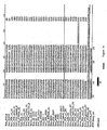
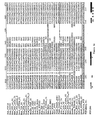
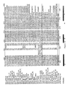
- Figures 1A-1E show a multiple sequence alignment of the catalytic domains of PTPs (SEQ ID NOS: 1-35). Cytosolic eukaryotic PTPs and
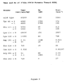
domain 1 of RPTPs are combined into one group, domains 2 of RPTPs are in a second group, and the Yersinia PTP is in a third. Invariant residues shared among all three groups are shown in lower case. Invariant and highly conserved residues within a group are shown in italics and bold, respectively. Within the Yersinia PTP sequence, residues that are either invariant or highly conserved between the cytosolic and RPTP domain sequences are shown in italics and bold, respectively. The position of residues of PTP1B that interact with the peptide are indicated with a small arrowhead, and the residue numbering at the bottom of the alignment corresponds to that for PTP1B. - Figure 2 shows the Vmax and Km of various PTP1B mutants toward RCML.
- The PTP family of enzymes contains a common evolutionarily conserved segment of approximately 250 amino acids known as the PTP catalytic domain. Within this conserved domain is a unique signature sequence motif, [I/V]HCXAGXXR[S/T]G (SEQ ID NO: 36), that is invariant among all PTPs. The cysteine residue in this motif is invariant in members of the family and is known to be essential for catalysis. It functions as a nucleophile to attack the phosphate moiety of the incoming substrate. If the cysteine residue is altered by site-directed mutagenesis to serine (CS mutants) or alanine (CA mutants), the resulting PTP is catalytically attenuated but retains the ability to complex with, or bind, its substrate, at least in vitro. These results have been confirmed relative to MKP-1, a member of the PTP family (Sun et al., Cell 75: 487-493 (1993)), as well as other PTPs. However, although these CS mutants can in general bind effectively to phosphotyrosyl substrates in vitro, in many cases such complexes cannot be isolated in vivo. Thus, the CS mutants are limited in their applicability and cannot be used to isolate all combinations of PTPs and substrates.
- The crystal structures of PTP1B alone (Barford, et al., Science 263:1397-1404 (1994)) and in a complex with a phosphotyrosine-containing peptide (Jia et al., Science 268:1754-1758 (1995)) were recently determined. These structures indicated twenty seven invariant residues (Barford et al., 1994), one of which is an aspartate residue. This aspartate residue is invariant across the catalytic domains of PTP family members. That is, if the amino acid sequences of the PTP family members are aligned, the aspartate residue is present in each PTP at a corresponding location, although the position numbers may be different due to the shifts required to maximize alignment (see the Figure (from Barford et al., Nature Struc. Biol. 2:1043-1053 (1995)) for an alignment of various PTP sequences). Sequences for which the alignment has not yet been published can readily be aligned with other known PTP sequences, e.g., utilizing available computer software such as GENEWORKS.
- Thus, mutant PTPs other than those specifically described herein can readily be made by aligning the amino acid sequence of the PTP catalytic domain with those described herein, identifying the invariant aspartate residue, and changing the residue by site-directed mutagenesis. Although the specific examples of PTP mutants described herein are aspartate to alanine mutants (DA mutants), it is understood that the invention is not limited to changes of aspartate to alanine. The invariant aspartate residue can be changed, e.g., by site-directed mutagenesis, to any amino acid which does not cause significant alteration of the Km of the enzyme but which results in a reduction in Kcat to less than 1 per minute (less than 1 min-1). For example, the invariant aspartate residue can be changed or mutated to an alanine, valine, leucine, isoleucine, proline, phenylalanine, tryptophan, methionine, glycine, serine, threonine, cysteine, tyrosine, asparagine, glutamine, lysine, arginine or histidine.
- As described herein, pervanadate-treated cells were used as an abundant source of tyrosine phosphorylated proteins to investigate the substrate specificity of PTP-PEST. PTP-PEST is an 88 kDa cytosolic PTP (Charest et al., Biochem. J. 308:425-432 (1995); den Hertog et al., Biochem. Biophys. Res. Commun. 184:1241-1249 (1992); Takekawa et al., Biochem. Biophys. Res. Commun. 189:1223-1230 (1992); Yang et al., J. Biol. Chem. 268:6622-6628 (1993); Yang et al., J. Biol. Chem. 268:17650 (1993)) which is expressed ubiquitously in mammalian tissues (Yi et al., Blood 78: 2222-2228 (1991)), and which exhibits high specific activity when assayed in vitro using artificial tyrosine phosphorylated substrates (Garton and Tonks, EMBO J. 13:3763-3771 (1994)). It has previously been demonstrated that PTP-PEST is subject to regulation via phosphorylation of Ser39 in vitro and in vivo. This modification is catalyzed by both protein kinase C (PKC) and protein kinase A (PKA), and results in reduced enzyme activity as a consequence of an increase in the Km of the dephosphorylation reaction (Garton and Tonks, EMBO J. 13:3763-3771 (1994)). It appears likely that further regulatory mechanisms exist for PTP-PEST, since this enzyme would be expected to exert a considerable negative influence on the tyrosine phosphorylation state of cytosolic substrates of tyrosine kinases. One possibility is that this influence could be limited by the substrate specificity of PTP-PEST.
- The substrate specificity of PTP1B was investigated utilizing the same methods outlined for PTP-PEST, with the exception that the cells were not treated with pervanadate. A combination of in vitro dephosphorylation and substrate trapping experiments were used to study the substrate interactions of PTP1B and PTP-PEST. The substrate trapping methods outlined herein are generally applicable to any PTP by virtue of the shared invariant aspartate residue, and should therefore prove useful in delineating the substrate preference of other PTP family members. In particular, the use of mutant, catalytically impaired PTPs to trap, and thereby isolate, potential substrates will greatly facilitate the identification of physiologically important substrates for individual PTPs, leading to improved understanding of the roles of these enzymes in regulation of cellular processes.
- One embodiment of the invention relates to novel PTPs in which the invariant aspartate residue is replaced with an amino acid which does not cause significant alteration of the Km of the enzyme but which results in a reduction in Kcat to less than 1 per minute (less than 1 min-1). These PTPs retain the ability to form a complex with, or bind, their tyrosine phosphorylated substrates but are catalytically attenuated. As defined herein, "attenuated" activity is intended to mean that the phosphatase retains a similar Km to that of the wild type phosphatase but has a Vmax which is reduced by a factor of at least 104 relative to the wild type enzyme. This includes catalytic activity which is either reduced or abolished relative to the wild type PTP. For example, the invariant aspartate residue can be changed or mutated to an alanine, valine, leucine, isoleucine, proline, phenylalanine, tryptophan, methionine, glycine, serine, threonine, cysteine, tyrosine, asparagine, glutamine, lysine, arginine or histidine.
- The novel PTPs described herein, in which the invariant aspartate residue is replaced with an amino acid which does not cause significant alteration of the Km of the enzyme but which results in a reduction in Kcat to less than 1 per minute (less than 1 min-1), can also comprise other mutations, particularly those which assist in stabilizing the PTP/substrate complex. For example, a mutation of the [serine/threonine] residue in the signature motif to an alanine residue changes the rate-determining step of the dephosphorylation reaction from the formation of the transition state to the break, down of the transition state, thereby stabilizing the PTP/substrate complex. Such mutations may be valuably combined with the replacement of the invariant aspartate residue, particularly assisting in stabilizing the complex and facilitating the observation and isolation of the complex.
- PTPs suitable for use in the invention include any PTP which has an invariant aspartate residue in a corresponding position. As defined herein, a phosphatase is a member of the PTP family if it contains the signature motif [I/V]HCXAGXXR[S/T]G (SEQ ID NO: 36). Dual specificity PTPs, i.e., PTPs which dephosphorylate both phosphorylated tyrosine and phosphorylated serine or threonine, are also suitable for use in the invention. Appropriate PTPs include, but are not limited to, PTP1B, PTP-PEST, PTPγ, MKP-1, DEP-1, PTPµ, PTPX1, PTPX10 and PTPH1.
- In one embodiment, the invention relates to the phosphatase PTP1B in which the aspartate residue at position 181 is replaced with alanine (D181A). In another embodiment the invention relates to the phosphatase PTP-PEST in which the invariant aspartate residue at position 199 is replaced with an alanine (D199A). Another embodiment of the invention relates to a PTP-PEST phosphatase in which the cysteine residue at position 231 is replaced with a serine (C231S) .
- The invention also relates to a method of identifying a tyrosine phosphorylated protein which is a substrate of a particular protein tyrosine phosphatase. According to one embodiment of the present invention, a tyrosine phosphorylated protein of interest is combined with at least one PTP in which the invariant aspartate residue is replaced with an amino acid which does not cause significant alteration of the Km of the enzyme but which results in a reduction in Kcat to less than 1 per minute (less than 1 min-1) (e.g., an alanine residue), and the presence or absence of a complex between the protein and the PTP is determined. Presence of a complex in the combination indicates that the tyrosine phosphorylated protein is a substrate of the PTP. The PTP DA mutant (substrate trapping mutant) binds to or complexes with its substrate but does not dephosphorylate it (or does so very slowly), thereby allowing the complex to be isolated and identified.
- The phosphorylated protein/PTP complex may be isolated by conventional isolation techniques as described in U.S. Patent No. 5,352,660 to Pawson, including salting out, chromatography, electrophoresis, gel filtration, fractionation, absorption, polyacrylamide gel electrophoresis, agglutination, or combinations thereof. Furthermore, to facilitate the determination of the presence of the protein/PTP complex, antibodies against the PTP or the phosphorylated protein can be used, as well as labelled PTPs and/or labelled phosphorylated substrates. The PTP or phosphorylated protein can be labelled with various enzymes, fluorescent materials, luminescent materials and radioactive materials. Examples of suitable enzymes include, but are not limited to, horseradish peroxidase, biotin, alkaline phosphatase, β-galactosidase and acetylcholinesterase. Examples of suitable fluorescent materials include, but are not limited to, umbelliferone, fluorescein, fluorescein isothiocyanate, rhodamine, dichlorotriazinylamine fluorescein, dansyl chloride and phycoerythrin. Appropriate luminescent materials include luminol, and suitable radioactive material include radioactive phosphorous 32P, iodine I125, I131 or tritium.
- Alternatively, the invention pertains to a method of identifying a tyrosine phosphorylated protein which is a substrate of a PTP, comprising combining a PTP of interest in which the invariant aspartate residue is replaced with an amino acid which does not cause significant alteration of the Km of the enzyme but which results in a reduction in Kcat to less than 1 per minute (less than 1 min-1) (e.g., an alanine residue), with at least one tyrosine phosphorylated protein, thereby producing a combination; and determining the presence or absence of a complex in the combination, wherein presence of a complex in the combination between a tyrosine phosphorylated protein and the PTP indicates that the tyrosine phosphorylated protein is a substrate of the PTP.
- The substrate trapping PTPs of the present invention can also be used in place of wild type PTPs to screen phosphotyrosyl peptide libraries for peptides which bind to the PTP as described in Songyang et al. (Nature 373:536-539 (1995); Cell 72:767-778 (1993)). Peptides identified from such peptide libraries can then be assessed to determine whether tyrosine phosphorylated proteins containing these peptides exist in nature.
- Any tyrosine phosphorylated protein is suitable as a potential substrate in the present invention. Tyrosine phosphorylated proteins are well known in the art. Specific examples of appropriate substrates include, without limitation, p130cas, the EGF receptor, p210 bcr:abl, MAP kinase and the insulin receptor. Of particular interest are tyrosine phosphorylated proteins which have been implicated in a mammalian disease or disorder.
- The invention also pertains to a method of reducing the activity of a tyrosine phosphorylated protein, comprising administering to a mammal a PTP in which the invariant aspartate residue is replaced with an amino acid which does not cause significant alteration of the Km of the enzyme but which results in a reduction in Kcat to less than 1 per minute (less than 1 min-1) (e.g., an alanine residue) and which forms a complex with the tyrosine phosphorylated protein. The PTP DA mutant binds to the phosphorylated protein without dephosphorylating it (or causing dephsophorylation at a greatly reduced rate), thereby inhibiting the activity of the protein and reducing its downstream effects. As used herein, "reducing" includes both reduction and complete abolishment, e.g., of one or more activities or functions of the phosphorylated protein.
- For example, the invention relates to a method of reducing the transforming effects of oncogenes associated with p130cas, a substrate of PTP-PEST, comprising administering to a mammal wild type PTP-PEST or PTP-PEST in which the invariant aspartate residue is replaced with an alanine residue. Wild type PTP-PEST binds and dephosphorylates p130cas, thereby negatively regulating its downstream effects. DA mutants of PTP-PEST bind but do not dephosphorylate p130cas (or do so at a greatly reduced rate); the substrate is thus tied up in the complex with the substrate trapping form of PTP-PEST and cannot exert its downstream effects. Similarly, the invention relates to a method of reducing the formation of signalling complexes associated with p130cas, particularly those signalling complexes which induce mitogenic pathways, comprising administering to a mammal wild type PTP-PEST or PTP-PEST in which the invariant aspartate residue is replaced with an alanine residue. The PTP binds to and/or dephosphorylates p130cas, thereby negatively regulating the downstream effects of p130cas and reducing the formation of signalling complexes associated with p130cas.
- The substrate trapping mutant PTPs of the present invention can be used in virtually any application in place of, or in addition to, a corresponding wild type PTP. The advantages of such a utility lie in the ability of the mutant PTP to mimic the function of the wild type enzyme, e.g., to decrease the activity of its tyrosine phosphorylated substrate, without inducing the harmful cytotoxic effects commonly observed with administration or overexpression of the wild type PTP. Thus, the invention also pertains to a method of reducing the cytotoxic effects associated with administration or overexpression of wild type PTPs. For example, CS mutants of MKP-1 have been shown to have the same functional effect as wild type MKP-1 without induction of potentially harmful side effects. Thus, PTPs described herein, in which the invariant aspartate residue is replaced with an amino acid which does not cause significant alteration of the Km of the enzyme but which results in a reduction in Kcat to less than 1 per minute (less than 1 min-1) (e.g., an alanine residue), can be used in many applications in place of the corresponding wild type enzyme. As used herein, a "corresponding" enzyme is one which is the same as the mutant PTP (e.g., PTP-PEST and PTP-PEST D199A) or one which is different from the mutant PTP but recognizes the same substrate as the mutant PTP.
- The mutant PTPs described herein can also be used therapeutically to reduce the activity of a tyrosine phosphorylated protein, such as by a gene therapy method in which the mutant PTP described herein, or a functional portion thereof which retains the ability to bind to its tyrosine phosphorylated substrate, is introduced into a subject and in whom the mutant PTP is expressed. The mutant PTP replaces, either partially or totally, the wild type enzyme which is normally produced or competes with the wild type PTP for binding to the substrate. For example, a specific tyrosine phosphorylated protein can be identified which is implicated in a particular disease or disorder (such as a protein tyrosine kinase). At least one PTP which acts to dephosphorylate the selected tyrosine phosphorylated protein of the present invention can be identified by the methods described herein. The wild type or mutant form of the PTP can be administered to a subject in need of treatment in order to tie up or bind the tyrosine phosphorylated substrate, thereby inhibiting or reducing the function of the phosphorylated protein. In a preferred embodiment, the mutant PTP is administered in place of the wild type enzyme in order to reduce the cytotoxic effects associated with overexpression of the wild type enzyme. Procedures for gene therapy are known in the art (see U.S. Patent No. 5,399,346 to Anderson et al.) and can be modified by methods known in the art to appropriately express the specific mutant and wild type PTPs of the present invention.
- The present invention also relates to assays for identifying agents which alter, e.g., enhance or inhibit, the interaction between a PTP and its phosphorylated substrate. Agents identified by these assays can be agonists (e.g., agents which enhance or increase the activity of the PTP) or antagonists (e.g., agents which inhibit or decrease the activity of the PTP) of PTP activity. The agent may be an endogenous physiological substance or may be a natural or synthetic drug, including small organic molecules.
- For example, the tyrosine phosphorylated substrate of a PTP can be identified by the methods described herein. An enzymatic activity assay utilizing the wild type PTP can be carried out in the presence of an agent to be tested, and the resulting amount of enzyme activity can be compared with the amount of enzyme activity in the absence of the agent to be tested. Enzymatic activity assays are known in the art; for example, assays of PTP activity using a tyrosine phosphorylated 32P-labelled substrate are described in Flint et al. (EMBO J. 12:1937-1946 (1993)). A decrease in the enzymatic activity in the presence of the agent to be tested indicates that the agent inhibits the interaction between the PTP and its substrate. Conversely, an increase in the enzymatic activity in the presence of the agent to be tested indicates that the agent enhances the interaction between the PTP and its substrate.
- Alternatively, a competitive binding assay can be carried out utilizing the mutant PTP in the presence of an agent to be tested, and the resulting extent of binding of the mutant PTP to its substrate can be compared with the extent of binding in the absence of the agent to be tested. Competitive binding assays are known in the art; for example, U.S. Patent No. 5,352,660 to Pawson describes methods suitable for use in this invention. A decrease in the extent of binding in the presence of the agent to be tested indicates that the agent inhibits the interaction between the PTP and its substrate. Conversely, an increase in the extent of binding in the presence of the agent to be tested indicates that the agent enhances the interaction between the PTP and its substrate.
- According to the present invention, tyrosine phosphorylated peptides identified with mutant PTPs from peptide libraries by the methods of Songyang et al. (Nature 373:536-539 (1995); Cell 72:767-778 (1993)) can be used herein in place of the complete tyrosine phosphorylated protein in competitive binding assays.
- The present invention also pertains to pharmaceutical compositions comprising a PTP in which the invariant aspartate residue is replaced with an amino acid which does not cause significant alteration of the Km of the enzyme but which results in a reduction in Kcat to less than 1 per minute (less than 1 min-1) (e.g., an alanine residue). For instance, the PTP of the present invention can be formulated with a physiologically acceptable medium to prepare a pharmaceutical composition. The particular physiological medium may include, but is not limited to, water, buffered saline, polyols (e.g., glycerol, propylene glycol, liquid polyethylene glycol) and dextrose solutions. The optimum concentration of the active ingredient(s) in the chosen medium can be determined empirically, according to procedures well known to medicinal chemists, and will depend on the ultimate pharmaceutical formulation desired. Methods of introduction of exogenous PTPs at the site of treatment include, but are not limited to, intradermal, intramuscular, intraperitoneal, intravenous, subcutaneous, oral and intranasal. Other suitable methods of introduction can also include rechargeable or biodegradable devices and slow release polymeric devices. The pharmaceutical compositions of this invention can also be administered as part of a combinatorial therapy with other agents.
- The following Examples are offered for the purpose of illustrating the present invention and are not to be construed to limit the scope of this invention. The teachings of all references cited herein are hereby incorporated by reference in their entirety.
- The following is a description of the materials and methods used in the work described herein.
- Point mutations within the catalytic domains of PTP-PEST (D199A, C231S) and PTP1B (D181A, C215S) were introduced by site-directed mutagenesis using the Muta-Gene™ in vitro mutagenesis kit (Bio-Rad, Richmond, CA). Regions containing the required point mutation were then exchanged with the wild type sequences within appropriate expression vectors, and the replaced mutant regions were sequenced in their entirety to verify the absence of additional mutations.
- Full length PTP-PEST proteins (wild type and mutant proteins, containing either Asp199 to Ala or Cys231 to Ser mutations) and the wild type PTP-PEST catalytic domain (amino acids 1-305) were expressed in Sf9 cells using recombinant baculovirus (BaculoGold™, Pharmingen, San Diego, CA), and purified as described in Garton and Tonks (EMBO J. 13:3763-3771 (1994)). Truncated forms of wild type and mutant PTP-PEST proteins, comprising amino acid residues 1-305 of PTP-PEST were also expressed in E. coli as GST fusion proteins following subcloning of PTP-PEST DNA in-frame downstream of GST in pGEX vectors (Pharmacia Biotech Inc., Uppsala, Sweden). Twenty-five ml of E. coli transformed with the appropriate vector were grown to log phase (OD600 approximately 0.5). Fusion protein expression was then induced by addition of 0.2 mM isopropyl-1-thio-b-D-galactopyranoside, and the cells were grown for 2-4 hours at 30°C. Cells were harvested by centrifugation, incubated with 50 mg/ml lysozyme in 3 ml buffer containing 50 mM Tris-HCl, pH 7.4, 5mM EDTA, 1 mM PMSF, 1 mM benzamidine, 5 mg/ml leupeptin, 5 mg/ml aprotinin, 0.1 o Triton X-100 and 150 mM NaCl, then lysed by sonication (3 x 10s). Following removal of insoluble material by centrifugation (20 minutes at 300,000 x g), fusion proteins were isolated by incubation for 30 min at 4°C with 100 ml glutathione-Sepharose beads (Pharmacia Biotech Inc., Uppsala, Sweden), and the beads were then collected by centrifugation and washed three times with buffer A (20 mM Tris-HCl, pH 7.4, 1 mM EDTA, 1 mM benzamidine, 1 mg/ml leupeptin, 1 mg/ml aprotinin, 10 % glycerol, 1 % Triton X-100 and 100 mM NaCl). This procedure yielded essentially homogeneous fusion protein at a concentration of 1 mg protein/ml glutathione-Sepharose beads. PTP1B proteins (wild type and mutant forms) comprising amino acids 1-321 were expressed in E.coli and purified to homogeneity as described in Barford et al. (J. Mol. Biol. 239:726-730 (1994)).
- HeLa and COS cells were grown in Dulbecco's modified Eagle's medium (DMEM), containing 5% fetal bovine serum (FBS); Wi38, C2C12 and MvLu cells were grown in DMEM containing 10% FBS; 293 cells were grown in DMEM containing 10% calf serum; MCF10A cells were grown in 50% DMEM, 50% Ham's F-12 containing 5% horse serum, 20 ng/ml epidermal growth factor, 10 mg/ml insulin, 0.5 mg/ml hydrocortisone and 0.25 mg/ml fungizone. All media also contained penicillin and streptomycin at 100 U/ml and 100 mg/ml, respectively, and all cells were grown at 37°C. Calcium phosphate-mediated transfection was used to introduce cDNA encoding wild type and mutant PTP-PEST proteins into COS cells. These were encoded by PTP-PEST cDNA subcloned into the plasmid PMT2, from which expression was driven by an adenovirus major late promoter; 20 mg DNA was used for transfection of each 10 cm plate of cells. The level of expression of PTP-PEST constructs was similar in all cases.
- Prior to cell lysis, 70-90% confluent cultures of cells were treated for 30 minutes with 0.1 mM pervanadate (20 ml of a fresh solution containing 50 mM sodium metavanadate (NaVO3) and 50 mM H2O2 were added to 10 ml medium). Treatment of cells with H2O2 and vanadate leads to a synergistic increase in phosphotyrosine levels, presumably due to inhibition of intracellular PTPs by vanadate. The synergism between H2O2 and vanadate has previously been suggested to result from improved accumulation of the resultant oxidized vanadate (pervanadate) within the cells when compared to vanadate itself (Heffetz et al., J. Biol. Chem. 265:2896-2902 (1990)). It is important to note that during the preparation of cell lysates, dilution occurs such that the inhibitory effect of vanadate on PTP action is lost. Pervanadate treatment resulted in the appearance of at least 50 prominent phosphotyrosine protein bands in all cell types, whereas untreated cells contained virtually undetectable levels of phosphotyrosine (data not shown).
- Cells were lysed in Buffer A containing 5 mM iodoacetic acid, which was included in order to inhibit irreversibly cellular PTPs. Following incubation at 4°C for 30 minutes, 10 mM DTT was added to inactivate any unreacted iodoacetic acid. Insoluble material was then removed by centrifugation for 20 minutes at 300,000 x g. The resultant lysates were stable with regard to their phosphotyrosine content during long term (several months) storage at -70°C and during prolonged (at least 20 hours) incubation at 4°C, in the absence of exogenous added PTPs.
- Pervanadate-treated HeLa cell lysate was fractionated by anion exchange chromatography using a Mono Q FPLC column (Pharmacia). The sample (50 mg total protein at 3 mg/ml in buffer A) was diluted in three volumes of buffer B (20 mM tris-HCl, pH 7.4, 1 mM EDTA, 1 mM benzamidine, 1 mg/ml leupeptin, 1 mg/ml aprotinin and 0.1% Triton X-100) prior to loading. Proteins were eluted at a flow rate of 1 ml/min with a linear gradient of 0-0.5 M NaCl in buffer B over 20 fractions (1 ml fraction volume), followed by a second gradient of 0.5-1.0 M NaCl in buffer B over 5 fractions. Phosphotyrosine-containing proteins were detected within fractions 7-21 according to anti-phosphotyrosine immunoblotting. The same procedures were followed for PTP1B, with the exception that the cells were not treated with pervanadate.
- Lysates of pervanadate-treated HeLa cells (1-2 mg protein/ml) containing tyrosine phosphorylated proteins were incubated on ice in the absence or presence of purified active PTPs at a concentration of 2 nM. Dephosphorylation was terminated by the removal of aliquots (30 mg protein) into SDS-PAGE sample buffer, and the extent of dephosphorylation was determined by immunoblotting using the monoclonal antibody G104. Assays of PTP activity using tyrosine phosphorylated 32P-labelled RCM-lysozyme as substrate were performed as described in Flint et al. (EMBO J. 12:1937-1946 (1993)).
- The PTP-PEST monoclonal antibody AG25 was raised against baculovirus-expressed purified full-length PTP-PEST. The anti-phosphotyrosine monoclonal antibody G104 was generated using as antigen phosphotyrosine, alanine and glycine, in a 1:1:1 ratio, polymerized in the presence of keyhole limpet hemocyanin with 1-ethyl-3-(3'-dimethylaminopropyl)carbodiimide, a method originally described in Kamps and Sefton (Oncogene 2:305-315 (1988)). p130cas monoclonal antibody was from Transduction Laboratories (Lexington, Ky). Monoclonal antibody FG6 against PTP1B was provided by Dr David Hill (Calbiochem Oncogene Research Products, Cambridge, MA). Visualization of proteins by immunoblotting was achieved by enhanced chemiluminescence (ECL) using HRP-conjugated secondary antibodies (Amersham Life Science Inc., Arlington Heights, Il) and the SuperSignal™ CL-HRP substrate system (Pierce, Rockford, Il).
- Immunoprecipitation of PTP-PEST from transfected COS cells was performed following covalent coupling of monoclonal antibody AG25 to protein A-Sepharose beads (Pharmacia Biotech Inc., Uppsala, Sweden) using the chemical cross-linking agent dimethyl pimelimidate (Schneider et al., J. Biol. Chem. 257:10766-10769 (1982)). Antibody was first bound to protein A-Sepharose at a concentration of 1 mg/ml bead volume, and unbound material was then removed by three washes with 0.2 M sodium borate,
pH 9. Covalent coupling was achieved by incubation at room temperature for 30 minutes in the presence of 20 mM dimethyl pimelimidate in 0.2 M sodium borate,pH 9. The beads were then incubated for 1 hour with an excess of 0.2 M ethanolamine, pH 8, to block any unreacted cross-linker, and washed three times with PBS prior to storage at 4°C. Ten ml of AG25 beads were used to precipitate transfected PTP-PEST from lysates containing approximately 0.375 mg protein. - Substrate trapping was performed using various PTP affinity matrices. The full-length PTP-PEST matrix utilized covalent coupled AG25-protein A-Sepharose beads to which purified baculovirus-expressed PTP-PEST protein was bound. Aliquots (10ml) of AG25 beads were incubated for 2 hours at 4°C in 100 ml buffer A in the presence of 5 mg of purified PTP-PEST (wild type or mutant forms); unbound PTP-PEST was then removed by washing three times with 1 ml buffer A. The resultant PTP-PEST-AG25-protein A-Sepharose beads contained approximately 2 mg of PTP-PEST per 10 ml aliquot. Substrate trapping was also carried out with glutathione-Sepharose beads bound to bacterially-expressed GST fusion proteins containing the catalytic domain of PTP-PEST.
- PTP1B was also used in substrate trapping experiments. In this case, the monoclonal antibody FG6 was precoupled to protein A-Sepharose in the absence of cross-linker (2 mg antibody/10 ml beads), then purified PTP1B proteins were added in excess and incubated at 4°C for 2 hours. Following removal of unbound PTP1B, 10 ml beads contained approximately 2 mg PTP1B.
- Pervanadate-treated cell lysates, or column fractions, were used as a source of phosphotyrosine-containing proteins for substrate trapping experiments. In general, lysates containing 0.25-0.5 mg protein in 0.5 ml buffer A (including 5 mM iodoacetic acid, 10 mM DTT) were incubated at 4°C for 2 hours in the presence of 10 ml of affinity matrix containing approximately 2 mg of the appropriate PTP protein. Unbound proteins were then removed from the samples by washing three times with 1 ml buffer A, and bound material was collected by addition of 50 ml SDS-PAGE sample buffer followed by heating at 95°C for 5 minutes; proteins bound to the beads were then analyzed by SDS-PAGE followed by immunoblotting.
- The following details the results of the work described herein carried out as described above.
- Chronic myelogenous leukemia (CML) is a clonal disorder of the haematopoietic stem cell that is characterized by the Philadelphia chromosome, in which the c-Abl proto-oncogene on
chromosome 9, encoding a PTK, becomes linked to the bcr gene on chromosome 22. This results in the generation of a bcr:able fusion protein, p210 bcr:abl, in which the PTK activity is enhanced relative to that of c-Abl. Current data indicates that this cytogenetic abnormality is the primary and sole causative event in CML. Expression of p210 bcr:abl produces abnormal patterns of tyrosine phosphorylation that result in the aberrant maturation of the haematopoietic stem cell that is characteristic of CML. - Expression of PTP1B mRNA and protein is enhanced as a consequence of p210 bcr:abl expression in Rat1, Mo7 and BaF3 cells. Changes in PTP1B activity, which were commensurate with the change in enzyme protein, were also observed. These changes are specific for PTP1B and are not seen in closely related homologue (65% identity) TC-PTP or in other tested PTPs, including SHP-1, SHP-2 and PTP-PEST. The increase in expression of PTP1B was also observed in Ph+ B-lymphoid cells derived from a CML patient relative to Ph-cells from the same patient.
- The changes in PTP1B levels were induced specifically by p210 bcr:abl and were not seen in cells expressing other PTKs including v-abl, v-src or other oncoproteins such as myc. The PTK activity of p210 bcr:abl was essential for the increase in expression of PTP1B, since expression of an inactive lysine to arginine mutant form of p210 bcr:abl in Rat1 cells did not alter PTP1B levels. The increase in PTP1B levels is a rapid response to induction of p210 bcr:abl. When BaF3 cells expressing a temperature-sensitive mutant form of p210 bcr:abl were shifted to the permissive temperature for the PTK, PTP1B levels were observed to increase within 12-24 hours coincident with the appearance of the active form of the PTK. THese data indicate that the alteration PTP1B levels is a relatively rapid response to the appearance of p210 bcr:abl, rather than a long-term adaptive response of the cells.
- In transient cotransfection experiments in COS cells, PTP1B dephosphorylates p210 bcr:abl but not v-abl. When the PTP1B D181A mutant was expressed as a GST fusion protein, purified and incubated with lysates of Mo7-p210 cells (which overexpress p210 bcr:abl), a complex of the mutant PTP and p210 bcr:abl was isolated. In contrast, tyrosine phosphorylated c-abl, which was also present in the lysates, did not bind to the mutant PTP. The interaction between PTP1B D181A and p210 bcr:abl was blocked by vanadate, suggesting that the interaction involved the active site of the PTP.
- Following transient coexpression in COS cells, PTP1B D181A formed a complex with p210 bcr:abl. Preliminary data indicate that the Y177F mutant form of p210 bcr:abl did not interact with PTP1b D181A, suggesting that this tyrosine residue is a component of the binding site in the PTK. This tyrosine residue in p210 bcr:abl is phosphorylated in vivo and has been demonstrated to serve as a docking site for GRB2. Direct interaction of the pTyr in p210 bcr:abl and the SH2 domain of GRB2 is essential for the transforming activity of the PTK. Interaction of PTP1B D181A with p210 bcr:abl interferes with the association of the PTK with GRB2. Taken together, these data suggest that p210 bcr:abl is a physiological substrate of PTP1B and that PTP1B may function as an antagonist of the oncoprotein PTK in vivo. The Vmax, Km and Kcat of 37 kDa PTP1B mutants toward RCML are shown in Figure 2.
- Expression of PTP1B D181A in COS cells leads to enhanced phosphorylation of tyrosyl residues in a 180 kDa protein and in proteins of 120 and 70 kDa. When a GST-PTP1B D181A fusion protein is expressed in COS cells and precipitated on Glutathione-Sepharose, the 180 kDa, and smaller quantities of p120 and p70, were coprecipitated. The p180 protein was identified as the epidermal growth factor (EGF) receptor by immunoblotting. The identity of the p120 and p70 proteins is unclear; however, the latter is not src, p62 or paxillin.
- Expression of PTP1B D181A in COS cells induces tyrosine phosphorylation of the EGF receptor in the absence of its ligand, EGF, indicating that the mutant PTP is exerting its effects in the intact cell and not post-lysis. The equivalent D199A PTP-PEST mutant does not interact with the EGF receptor, indicating the specificity of this substrate interaction.
- Autophosphorylation of the EGF receptor is required for the interaction with PTP1B D181A. Mutants of the receptor that are either kinase-dead or in which the autophosphorylation sites have been deleted do not interact with PTP1B D181A. In v-src-expressing cells, a plethora of tyrosine phosphorylated proteins were observed, but phosphorylation of the EGF receptor was not detected. Under these conditions, PTP1B D181A bound predominantly to a 70 kDa tyrosine phosphorylated protein.
- As a result of this work, it appears that PTP1B can modulate EGF-induced signalling pathways, perhaps including the pathways of many diseases, including breast cancer.
- In order to investigate the substrate specificity of PTP-PEST in vitro, aliquots of pervanadate-treated HeLa cell lysates were incubated on ice, yielding 50-100 distinct phosphotyrosine-containing proteins as judged by immunoblotting of the cell lysate using the monoclonal anti-phosphotyrosine antibody G104. Purified full-length PTP-PEST (expressed in Sf9 cells using recombinant baculovirus), PTP-PEST catalytic domain, or PTP1B catalytic domain (37 kDa form) was then added to the lysate, and aliquots were removed at various time points for analysis by SDS-PAGE followed by anti-phosphotyrosine immunoblotting.
- Surprisingly, a prominent 130 kDa phosphotyrosine band (p130) was selectively dephosphorylated by PTP-PEST within 10 minutes, whereas the intensity of all the other bands was essentially unchanged even after 60 minutes of incubation with PTP-PEST. Long incubations with higher concentrations of PTP-PEST (greater than 100-fold) resulted in the complete removal of all phosphotyrosine bands from the lysate. However, under all conditions tested, p130 was found to be dephosphorylated more rapidly than all other bands present.
- The selective dephosphorylation of p130 by PTP-PEST was also observed using a truncated form of the phosphatase (amino acid residues 1-305) which essentially contains only the catalytic domain of the enzyme. This result suggests that the striking substrate preference displayed by PTP-PEST in this analysis is an inherent property of the phosphatase catalytic domain, whereas the C-terminal 500 amino acid residues have little discernible effect on the substrate specificity of the enzyme.
- The specificity of the interaction between PTP-PEST and p130 was addressed using the catalytic domain of PTP1B (amino acid residues 1-321) in dephosphorylation reactions. When added at a similar molar concentration to that used for PTP-PEST, PTP1B was found to dephosphorylate fully and rapidly (within 15 minutes) most of the phosphotyrosine-containing proteins present in the pervanadate-treated HeLa lysate. In addition, the time course of dephosphorylation of p130 was not significantly more rapid than that of the other phosphotyrosine bands dephosphorylated by PTP1B. It should be noted, however, that these in vitro dephosphorylation results are not truly illustrative of the substrate specificity of PTP1B in vivo for several reasons. First, only the isolated catalytic subunit was used in this particular experiment. Furthermore, in vivo substrate specificity may be quite different due to the intracellular distribution of both the PTP and potential substrates. That is, in vitro dephosphorylation experiemnts may utilize substrates which the PTP is capable of dephosphorylating but which it would not have access to in vivo. The phenomenon of differing substrate specificity depending upon different physiologic contexts is illustrated by a comparison of this data with the in vivo PTP1B work described above, wherein PTP1B showed specificity for only three proteins.
- Pervanadate-treated HeLa cell lysate was fractionated by anion exchange chromatography and aliquots of the fractions were analyzed by SDS-PAGE followed by immunoblotting with anti-phosphotyrosine or anti-p130cas antibodies. Aliquots of all samples analyzed were then incubated with an affinity matrix containing a substrate trapping PTP-PEST mutant, comprising full length PTP-PEST in which Asp199 is changed to alanine (D199A), bound to covalently coupled protein A-Sepharose/antibody (AG25) beads. Proteins associated with PTP-PEST were then analyzed by SDS-PAGE followed by immunoblotting with anti-phosphotyrosine or anti-p130cas antibodies.
- Anti-phosphotyrosine immunoblotting of the column fractions showed that the p130 phosphotyrosine band eluted as a single peak in fractions 11-14 (approx. 0.3 M NaCl). In view of the abundance of tyrosine phosphorylated p130 in HeLa lysates, it appeared likely that p130 represents a previously identified phosphotyrosine-containing 130 kDa protein. Several potential candidates were identified in the literature, including the focal adhesion kinase p125FAK, ras-GAP, gp130 and p130cas. Of these candidates, p130cas has been identified as a particularly prominent phosphotyrosine band in a wide variety of systems, including v-crk (Mayer and Hanafusa, Proc. Natl. Acad. Sci. USA 87: 2638-2642 (1990); Mayer et al., Nature 332:272-275 (1988)) and src (Kanner et al., Proc. Natl. Acad. Sci. USA 87:3328-3332 (1990); Reynolds et al., Mol. Cell. Biol. 9: 3951-3958 (1989)) transformed fibroblasts, integrin-mediated cell adhesion (Nojima et al., J. Biol. Chem. 270:15398-15402 (1995); Petch et al., J. Cell Science 108:1371-1379 (1995); Vuori and Ruoslahti, J. Biol. Chem. 270:22259-22262 (1995)) and PDGF stimulated 3T3 cells (Rankin and Rozengurt, J. Biol. Chem. 269:704-710 (1994)).
- Therefore, the possibility that the p130 phosphotyrosine band corresponds to p130cas was tested by immunoblotting the Mono Q fractions using an antibody to p130cas. The 130 kDa band corresponding to p130cas eluted in the same fractions as the p130 tyrosine phosphorylated band, and displayed a similar apparent molecular weight, suggesting that they might represent the same protein. Furthermore, p130cas immunoprecipitated from these fractions was found to be phosphorylated on tyrosyl residues.
- A mutant form of PTP-PEST (D199A) was generated by site-directed mutagenesis, and the mutant enzyme was purified following expression using recombinant baculovirus. When assayed using tyrosine phosphorylated RCM-Lysozyme as substrate, the purified mutant enzyme exhibited a specific activity which was approximately 10,000 fold lower than that of the wild type enzyme (Garton and Tonks, unpublished data). This purified protein was bound to an affinity matrix comprised of an anti-PTP-PEST monoclonal antibody (AG25) covalently coupled to Protein A-Sepharose beads, then incubated with each of the Mono Q fractions. After 45 minutes of incubation, proteins associating with the mutant PTP-PEST were collected by centrifugation, the beads were washed, and SDS-PAGE sample buffer was added. Associated proteins were then analyzed by immunoblotting using the monoclonal anti-phosphotyrosine antibody G104.
- The mutant PTP-PEST protein was found to associate with a single phosphotyrosine-containing protein, the molecular weight (130 kDa) and Mono Q elution position (fractions 11-14) of which coincided with those of p130cas. Immunoblotting of the PTP-PEST-associated proteins using the p130cas antibody demonstrated that the 130 kDa tyrosine phosphorylated protein trapped by the mutant PTP-PEST is indeed p130cas. These data further support the hypothesis that p130cas is a potential physiologically relevant substrate for PTP-PEST.
- The interaction between p130cas and PTP-PEST was investigated further in substrate trapping experiments using various purified mutant forms of PTP-PEST to precipitate proteins from pervanadate-treated HeLa lysates. Several affinity matrices were incubated with pervanadate-treated HeLa cell lysate, and proteins associated with the beads were analyzed by SDS-PAGE followed by immunoblotting with anti-phosphotyrosine or anti-p130cas antibodies.
- The wild type full-length phosphatase was found to be incapable of stable association with tyrosine phosphorylated p130cas whereas both the PTP-PEST (D199A) mutant protein and a mutant lacking the active site cysteine residue (C231S) specifically precipitated p130cas from the lysate. The inability of the wild type phosphatase to precipitate tyrosine phosphorylated p130cas presumably reflects the transient nature of the normal interaction between PTP-PEST and tyrosine phosphorylated p130cas, which is likely to be concluded as soon as p130cas is dephosphorylated by PTP-PEST.
- Since the C-terminal 500 amino acids of PTP-PEST contain several proline-rich regions which resemble src homology-3 (SH3) domain binding sequences, it appeared plausible that the specificity of the interaction between PTP-PEST and p130cas might depend to some extent on association of these segments with the SH3 domain of p130cas. The possible contribution of the C-terminal segment of PTP-PEST in the observed specific interaction of PTP-PEST with p130cas was therefore addressed in further substrate trapping experiments using GST fusion proteins containing the catalytic domain of PTP-PEST alone, in both wild type and mutant (D199A) forms. The mutant catalytic domain of PTP-PEST fused to GST was found to precipitate the p130cas phosphotyrosine band specifically, whereas both the wild type fusion protein and GST alone failed to precipitate p130cas. The specific interaction between PTP-PEST and p130cas observed in these experiments therefore appears to be an intrinsic property of the catalytic domain of PTP-PEST, emulating the observed preference of the active PTP-PEST catalytic domain for dephosphorylation of p130cas in vitro.
- In view of the relative abundance of tyrosine phosphorylated p130cas in the pervanadate-treated HeLa cell lysate, the possibility that the observed selective binding of PTP-PEST inactive mutant proteins to p130cas was substrate-directed (reflecting the abundance of this potential substrate relative to the other phosphotyrosine-containing proteins present in the lysate) rather than enzyme-directed (reflecting a genuine substrate preference of PTP-PEST) was considered; this possibility was addressed in two ways. First, inactive mutant forms of the catalytic domain of PTP1B were used to trap potential substrates for this enzyme from the pervanadate-treated HeLa lysates. Again it was found that the wild type phosphatase was incapable of stable interaction with any phosphotyrosine-containing protein, whereas mutant variants of the PTP1B phosphatase domain (comprising Cys or Asp mutations analogous to those described above for PTP-PEST) associated with many tyrosine phosphorylated proteins. This was especially apparent for the aspartic acid mutant of PTP1B (D181A), which appeared to precipitate essentially all phosphotyrosine-containing proteins from the lysate with similar efficacy. These data emphasize the specific nature of the interaction between PTP-PEST and p130cas, which appears to be a property peculiar to the PTP-PEST catalytic domain, rather than a feature shared by all PTP catalytic domains.
- The specificity of the interaction between PTP-PEST and p130cas was addressed further following pervanadate-treatment of several different cell lines (Wi38, 293, COS, MCF10A, C2C12, MvLu), yielding a different array of tyrosine phosphorylated proteins in each case; the resultant lysates were analyzed by SDS-PAGE followed by anti-phosphotyrosine immunoblotting. Aliquots were incubated with PTP-PEST (D199A) affinity matrix or control matrix, and tyrosine phosphorylated proteins associating with PTP-PEST were analyzed by SDS-PAGE and immunoblotting with anti-phosphotyrosine or anti-p130cas antibodies as described above.
- In each case, the D199A mutant PTP-PEST protein precipitated a single broad phosphotyrosine band with an apparent molecular weight between 120 and 150 kDa in different cell lines, whereas the affinity matrix alone failed to precipitate any phosphotyrosine-containing protein. Immunoblotting of the precipitates with a p130 cas antibody revealed that the protein precipitated from all cell lysates corresponded to p130cas; the observed molecular weight variation between different cell lines presumably reflects either species differences in the molecular weight of p130cas or expression of different alternatively spliced forms (Sakai et al., EMBO J. 13:3748-3756 (1994)).
- The relative abundance of tyrosine phosphorylated p130cas, in the PTP-PEST precipitates appeared to correlate approximately with the abundance of p130cas protein in the lysates (data not shown). Surprisingly, regardless of the abundance of tyrosine phosphorylated p130cas in the lysates, p130cas was invariably the only phosphotyrosine-containing protein in the precipitates, even in 293 cell lysates which contained very little p130cas protein but which displayed a wide variety of other abundantly tyrosine phosphorylated proteins. Similarly, when lysates of pervanadate-treated 293 cells (containing tyrosine phosphorylated p130cas in amounts which are undetectable by anti-phosphotyrosine immunoblotting of the lysate) were incubated with active PTP-PEST, no visible dephosphorylation of any phosphotyrosine band occurred (Garton and Tonks, unpublished data). These results indicate that the affinity of PTP-PEST for p130cas is substantially greater than for any other substrate present, and further emphasizes the remarkable substrate selectivity of PTP-PEST for p130cas
- A consistent observation of this work was that, in contrast to the inactive mutant PTP-PEST, the wild type enzyme failed to associate in a stable complex with tyrosine phosphorylated p130cas, suggesting that the observed association is active site-directed. In order to investigate this possibility, mutant PTP-PEST (D199A) was incubated with the PTP inhibitor vanadate at various concentrations prior to addition of pervanadate-treated HeLa cell lysate. The extent of association of p130cas with PTP-PEST was then analyzed. PTP-PEST affinity matrix, comprising full length PTP-PEST (D199A) bound to covalently coupled protein A-Sepharose/antibody (AG25) beads, was incubated for 10 minutes on ice in the presence of varying concentrations of sodium orthovanadate. The samples were then incubated with aliquots of pervanadate-treated HeLa cell lysate; associated proteins were analyzed by SDS-PAGE and immunoblotting with anti-phosphotyrosine or anti-p130cas antibodies. The activity of wild type PTP-PEST was also determined under the same conditions, using tyrosine phosphorylated 32P-labelled RCM-lysozyme as substrate.
- The association was found to be potently disrupted by vanadate, with a concentration-dependence similar to that of vanadate inhibition of wild type PTP-PEST, and complete disruption being observed at 10 mM vanadate. Since PTP inhibition by vanadate presumably results from a direct interaction of vanadate with the active site cysteine residue of the enzyme (Denu et al., Proc. Natl. Acad. Sci. USA 93:2493-2498 (1996)), this result supports the hypothesis that the stable association of mutant PTP-PEST with tyrosine phosphorylated p130cas is mediated by direct interactions between active site residues within PTP-PEST, in particular the active site cysteine residue, and phosphotyrosine moieties within p130cas.
- The work described above strongly suggests that p130cas represents a potential physiologically significant substrate for PTP-PEST. In order to assess whether PTP-PEST interacts with p130cas in intact cells, COS cells were transfected with plasmids encoding wild type or mutant forms of PTP-PEST (D199A or C215S). The cells were treated with
pervanadate 30 minutes prior to lysis, PTP-PEST proteins were immunoprecipitated, and associated tyrosine phosphorylated proteins were analyzed by anti-phosphotyrosine immunoblotting of the resultant precipitates. Lysates were also incubated with covalently coupled protein A-Sepharose/anti-PTP-PEST (AG25) beads and associated proteins were analyzed by SDS-PAGE and immunoblotting with anti-phosphotyrosine antibody. - Under these conditions, the phosphotyrosine-containing band corresponding to p130 cas was again unique in its ability to associate with the C231S PTP-PEST protein, indicating that p130 cas can be specifically selected by PTP-PEST as a substrate in an intracellular context in the presence of a large number of alternative possible substrates. Neither the wild type nor the D199A form of PTP-PEST was capable of a stable interaction with tyrosine phosphorylated p130cas in pervanadate-treated COS cells.
- The binding of both wild type and D199A PTP-PEST to tyrosine phosphorylated p130cas under these conditions is most likely prohibited by the presence of pervanadate bound to the active site cysteine residue of PTP-PEST (Denu et al., Proc. Natl. Acad. Sci. USA 93:2493-2498 (1996)), which effectively excludes the binding of phosphotyrosine residues of p130cas. The ability of the C231S mutant PTP-PEST to associate in a stable complex with p130cas in the presence of pervanadate suggests that this mutant protein is largely unaffected by pervanadate, indicating that the normal mode of inhibition of PTPs by vanadate ions depends critically on direct interactions between vanadate and the thiolate anion of the PTP active-site cysteine residue. These observations therefore lend further support to the existence of an exclusive interaction between PTP-PEST and p130cas which appears to be entirely active site-directed, and therefore reflects the genuine, inherent, highly restricted substrate preference of PTP-PEST for p130cas.
- Results described herein implicate p130cas as a physiologically relevant substrate for PTP-PEST. Furthermore, the observed stringency and exclusivity of the interaction between PTP-PEST and p130cas in a wide variety of cell lines suggest that p130 cas may be a unique high affinity substrate for PTP-PEST, although the possibility that other significant PTP-PEST substrates may exist cannot be excluded at present. In particular, it is unclear whether pervanadate-treated cells display a complete spectrum of all possible tyrosine phosphorylated proteins; in fact this appears unlikely since pervanadate treatment presumably results only in an increase in tyrosine phosphorylation of proteins which are to some extent constitutively phosphorylated, but which are normally rapidly dephosphorylated, within the cell. Potential substrates lacking from pervanadate-treated cells therefore presumably include substrates of protein tyrosine kinases (PTKs) which are normally present in an inactive state, such as ligand-stimulated receptor PTKs, and the recently described calcium regulated kinase PYK2 (Lev et al., Nature 376: 737-745 (1995)). Regardless of these considerations, the ability of PTP-PEST to select p130cas exclusively as a substrate from lysates of several different cell lines, containing a combined total of at least one hundred different potential substrates (many of which presumably contain multiple sites of phosphorylation), clearly demonstrates that the substrate specificity of PTP-PEST is highly restricted.
- Many intracellular PTPs are limited in their substrate availability due to strict confinement within a particular subcellular location; examples include PTP1B, which is localized to the cytoplasmic face of the endoplasmic reticulum (Frangioni et al., Cell 68:545-560 (1992)), and TCPTP which is either nuclear (Tillmann et al., Mol. Cell. Biol. 14:3030-3040 (1994)) or localized to the endoplasmic reticulum, depending upon which alternative spliced form is expressed (Lorenzen et al., J. Cell Biol. 131:631-643 (1995)). Alternatively, certain PTPs appear to be highly regulated, requiring activation before appreciable activity can be demonstrated. For example, the SH2 domain-containing PTPs, SHP1 and SHP2, display relatively low activity in vitro, but can be considerably activated by several mechanisms including C-terminal truncation (Zhao et al., J. Biol. Chem. 268:2816-2820 (1993)), addition of certain phospholipids (Zhao et al., Proc. Natl. Acad. Sci. USA 90:4251-4255 (1993)), or SH2 domain-mediated binding of appropriate phosphotyrosine-containing peptides (Lechleider et al., J. Biol. Chem. 268:21478-21481 (1993)).
- However, PTP-PEST exhibits high specific activity in vitro (35,000 U/mg), and is a predominantly (90-95%) soluble PTP within cells (Garton and Tonks, unpublished data); in principle, therefore, it may act potently on any substrate accessible to the cytoplasm. This accessibility may partly underlie the necessity for PTP-PEST to possess an inherently constrained substrate specificity. The demonstration that mutant PTP-PEST is capable of exclusively associating with p130cas in an intracellular context in the presence of many other tyrosine phosphorylated proteins, is an indication that the narrow substrate specificity of the enzyme may result in PTP-PEST having a negligible influence on the phosphorylation state of the majority of tyrosine phosphorylated proteins within the cell, even though those substrates are largely accessible to PTP-PEST.
- The role of p130cas in cellular transformation by the v-crk and v-src oncogenes is unclear, although there is a general correlation between the level of tyrosine phosphorylation of p130cas and the degree of transformation in cells expressing different forms of crk or src (Kanner et al., EMBO J. 10:1689-1698 (1991); Mayer and Hanafusa, J. Virol. 64:3581-3589 (1990)). Furthermore, enhanced tyrosine phosphorylation of p130cas has also been observed in cells transformed by c-Ha-ras and by ornithine decarboxylase overexpression (Auvinen et al., Mol. Cell. Biol. 15: 6513-6525 (1995)). Expression of antisense cDNA encoding p130cas in these cells results in a partial reversion of the transformed phenotype. These observations suggest that aberrant tyrosine phosphorylation of p130cas is a common feature of cells transformed by several disparate mechanisms and that p130cas may be required for full manifestation of the transformed state. Dephosphorylation of p130cas by PTP-PEST is therefore a potentially important regulatory mechanism for counteracting the transforming effects of various oncogenes.
- Tyrosine phosphorylation of p130cas has been observed in fibroblasts following integrin-mediated cell adhesion to extracellular matrix proteins (Nojima et al., J. Biol. Chem. 270:15398-15402 (1995); Petch et al., J. Cell Science 108:1371-1379 (1995); Vuori and Ruoslahti, J. Biol. Chem. 270:22259-22262 (1995)). Under these conditions, using an antibody (4F4) that predominantly recognizes tyrosine phosphorylated p130cas (Kanner et al., EMBO J. 10:1689-1698 (1991); Petch et al., J. Cell Science 108:1371-1379 (1995)) it was shown that phosphorylated p130cas is localized to focal adhesions (Petch et al., J. Cell Science 108:1371-1379 (1995)), whereas fractionation studies have demonstrated that the normal cellular location of the majority of non-phosphorylated p130cas is the cytosol (Sakai et al., EMBO J. 13:3748-3756 (1994)). Furthermore, in crk-transformed fibroblasts, tyrosine phosphorylated p130cas is detected only in insoluble fractions (Sakai et al., EMBO J. 13:3748-3756 (1994)), suggesting that both cell adhesion- and transformation-mediated phosphorylation of p130cas, is associated with redistribution of the protein from the cytosol to focal adhesions.
- It is plausible that the redistribution of tyrosine phosphorylated p130cas may be driven by its association with FAK, which is constitutively associated with focal adhesions due to its C-terminal focal adhesion targeting domain (Hildebrand et al., J. Cell Biol. 123:993-1005 (1993); Schaller et al., Proc. Natl. Acad. Sci. USA 89:5192-5196 (1992)). The sequestration of tyrosine phosphorylated p130cas in focal adhesions both in transformed cells, and following integrin-mediated cell adhesion, strongly suggests a role for p130cas in signalling events in this region of the cell. One consequence of the redistribution of tyrosine phosphorylated p130cas is likely to be that, in addition to localizing p130cas to a region of the cell containing abundant protein tyrosine kinase activity, the phosphorylated protein will be relatively inaccessible to the cytosolic phosphatase PTP-PEST. This raises the possibility that the role of PTP-PEST in dephosphorylating p130cas may be to prevent inappropriate tyrosine phosphorylation of the cytosolic pool of p130cas, thus preventing formation of signalling complexes assembled around tyrosine phosphorylated p130cas in inappropriate cellular locations.
- Several mitogenic factors potently stimulate tyrosine phosphorylation of p130cas. These include agents acting through heterotrimeric G protein-coupled receptors such as lysophosphatidic acid (Seufferlein and Rozengurt, J. Biol. Chem. 269:9345-9351 (1994)), bradykinin (Leeb-Lundberg et al., J. Biol. Chem. 269: 24328-24344 (1994)), and bombesin (Zachary et al., J. Biol. Chem. 267:19031-19034 (1992)), as well as growth factors that activate receptor tyrosine kinases, namely PDGF (Rankin and Rozengurt, J. Biol. Chem. 269:704-710 (1994)), EGF and NGF (Ribon and Saltiel, J. Biol. Chem. 271:7375-7380 (1996)). These observations suggest roles for p130cas in regulation of mitogenic signalling pathways, presumably involving assembly of signalling complexes based on tyrosine phosphorylated p130cas. The identities of the proteins involved in these complexes are not established, but are likely to include SH2 domain-containing adaptor proteins such as crk (Ribon and Saltiel, J. Biol. Chem. 271:7375-7380 (1996)), and its associated proteins (Feller et al., Oncogene 10:1465-1473 (1995); Hasegawa et al., Mol. Cell. Biol. 16:1770-1776 (1996); Knudsen et al., J. Biol. Chem. 269:32781-32787 (1994); Matsuda et al., Mol. Cell. Biol. 14: 5495-5500 (1994); Tanaka et al., Proc. Natl. Acad. Sci. USA 91:3443-3447 (1994)). Therefore tyrosine phosphorylation and dephosphorylation of p130cas potentially plays a central role in regulating the formation of such complexes, thereby influencing downstream events in mitogenic signalling.
-
- (1) GENERAL INFORMATION:
- (i) APPLICANT: Harbor, Cold Spring
- (ii) TITLE OF INVENTION: Substrate Trapping Protein Tyrosine Phosphatases
- (iii) NUMBER OF SEQUENCES: 36
- (iv) CORRESPONDENCE ADDRESS:
- (A) ADDRESSEE: HAMILTON, BROOK, SMITH & REYNOLDS, P.C.
- (B) STREET: Two Militia Drive
- (C) CITY: Lexington
- (D) STATE: MA
- (E) COUNTRY: USA
- (F) ZIP: 02173
- (v) COMPUTER READABLE FORM:
- (A) MEDIUM TYPE: Floppy disk
- (B) COMPUTER: IBM PC compatible
- (C) OPERATING SYSTEM: PC-DOS/MS-DOS
- (D) SOFTWARE: PatentIn Release #1.0, Version #1.30
- (vi) CURRENT APPLICATION DATA:
- (A) APPLICATION NUMBER: PCT/US97/13016
- (B) FILING DATE: 24-JUL-1997
- (C) CLASSIFICATION:
- (viii) ATTORNEY/AGENT INFORMATION:
- (A) NAME: Warren, Lisa
- (B) REGISTRATION NUMBER: 41,368
- (C) REFERENCE/DOCKET NUMBER: CSHL96-03 PCT
- (ix) TELECOMMUNICATION INFORMATION:
- (A) TELEPHONE: (781) 861-6240
- (B) TELEFAX: (781) 861-9540
- (2) INFORMATION FOR SEQ ID NO:1:
- (i) SEQUENCE CHARACTERISTICS:
- (A) LENGTH: 196 amino acids
- (B) TYPE: amino acid
- (C) STRANDEDNESS: single
- (D) TOPOLOGY: linear
- (ii) MOLECULE TYPE: peptide
- (xi) SEQUENCE DESCRIPTION: SEQ ID NO:1:

- (i) SEQUENCE CHARACTERISTICS:
- (2) INFORMATION FOR SEQ ID NO:2:
- (i) SEQUENCE CHARACTERISTICS:
- (A) LENGTH: 194 amino acids
- (B) TYPE: amino acid
- (C) STRANDEDNESS: single
- (D) TOPOLOGY: linear
- (ii) MOLECULE TYPE: peptide
- (xi) SEQUENCE DESCRIPTION: SEQ ID NO:2:

- (i) SEQUENCE CHARACTERISTICS:
- (2) INFORMATION FOR SEQ ID NO:3:
- (i) SEQUENCE CHARACTERISTICS:
- (A) LENGTH: 261 amino acids
- (B) TYPE: amino acid
- (C) STRANDEDNESS: single
- (D) TOPOLOGY: linear
- (ii) MOLECULE TYPE: peptide
- (xi) SEQUENCE DESCRIPTION: SEQ ID NO:3:


- (i) SEQUENCE CHARACTERISTICS:
- (2) INFORMATION FOR SEQ ID NO:4:
- (i) SEQUENCE CHARACTERISTICS:
- (A) LENGTH: 260 amino acids
- (B) TYPE: amino acid
- (C) STRANDEDNESS: single
- (D) TOPOLOGY: linear
- (ii) MOLECULE TYPE: peptide
- (xi) SEQUENCE DESCRIPTION: SEQ ID NO:4:


- (i) SEQUENCE CHARACTERISTICS:
- (2) INFORMATION FOR SEQ ID NO:5:
- (i) SEQUENCE CHARACTERISTICS:
- (A) LENGTH: 263 amino acids
- (B) TYPE: amino acid
- (C) STRANDEDNESS: single
- (D) TOPOLOGY: linear
- (ii) MOLECULE TYPE: peptide
- (xi) SEQUENCE DESCRIPTION: SEQ ID NO:5:


- (i) SEQUENCE CHARACTERISTICS:
- (2) INFORMATION FOR SEQ ID NO:6:
- (i) SEQUENCE CHARACTERISTICS:
- (A) LENGTH: 257 amino acids
- (B) TYPE: amino acid
- (C) STRANDEDNESS: single
- (D) TOPOLOGY: linear
- (ii) MOLECULE TYPE: peptide
- (xi) SEQUENCE DESCRIPTION: SEQ ID NO:6:


- (i) SEQUENCE CHARACTERISTICS:
- (2) INFORMATION FOR SEQ ID NO:7:
- (i) SEQUENCE CHARACTERISTICS:
- (A) LENGTH: 250 amino acids
- (B) TYPE: amino acid
- (C) STRANDEDNESS: single
- (D) TOPOLOGY: linear
- (ii) MOLECULE TYPE: peptide
- (xi) SEQUENCE DESCRIPTION: SEQ ID NO:7:


- (i) SEQUENCE CHARACTERISTICS:
- (2) INFORMATION FOR SEQ ID NO:8:
- (i) SEQUENCE CHARACTERISTICS:
- (A) LENGTH: 249 amino acids
- (B) TYPE: amino acid
- (C) STRANDEDNESS: single
- (D) TOPOLOGY: linear
- (ii) MOLECULE TYPE: peptide
- (xi) SEQUENCE DESCRIPTION: SEQ ID NO:8:


- (i) SEQUENCE CHARACTERISTICS:
- (2) INFORMATION FOR SEQ ID NO:9:
- (i) SEQUENCE CHARACTERISTICS:
- (A) LENGTH: 254 amino acids
- (B) TYPE: amino acid
- (C) STRANDEDNESS: single
- (D) TOPOLOGY: linear
- (ii) MOLECULE TYPE: peptide
- (xi) SEQUENCE DESCRIPTION: SEQ ID NO:9:


- (i) SEQUENCE CHARACTERISTICS:
- (2) INFORMATION FOR SEQ ID NO:10:
- (i) SEQUENCE CHARACTERISTICS:
- (A) LENGTH: 253 amino acids
- (B) TYPE: amino acid
- (C) STRANDEDNESS: single
- (D) TOPOLOGY: linear
- (ii) MOLECULE TYPE: peptide
- (xi) SEQUENCE DESCRIPTION: SEQ ID NO:10:


- (i) SEQUENCE CHARACTERISTICS:
- (2) INFORMATION FOR SEQ ID NO:11:
- (i) SEQUENCE CHARACTERISTICS:
- (A) LENGTH: 253 amino acids
- (B) TYPE: amino acid
- (C) STRANDEDNESS: single
- (D) TOPOLOGY: linear
- (ii) MOLECULE TYPE: peptide
- (xi) SEQUENCE DESCRIPTION: SEQ ID NO:11:

- (i) SEQUENCE CHARACTERISTICS:
- (2) INFORMATION FOR SEQ ID NO:12:
- (i) SEQUENCE CHARACTERISTICS:
- (A) LENGTH: 266 amino acids
- (B) TYPE: amino acid
- (C) STRANDEDNESS: single
- (D) TOPOLOGY: linear
- (ii) MOLECULE TYPE: peptide
- (xi) SEQUENCE DESCRIPTION: SEQ ID NO:12:


- (i) SEQUENCE CHARACTERISTICS:
- (2) INFORMATION FOR SEQ ID NO:13:
- (i) SEQUENCE CHARACTERISTICS:
- (A) LENGTH: 263 amino acids
- (B) TYPE: amino acid
- (C) STRANDEDNESS: single
- (D) TOPOLOGY: linear
- (ii) MOLECULE TYPE: peptide
- (xi) SEQUENCE DESCRIPTION: SEQ ID NO:13:


- (i) SEQUENCE CHARACTERISTICS:
- (2) INFORMATION FOR SEQ ID NO:14:
- (i) SEQUENCE CHARACTERISTICS:
- (A) LENGTH: 254 amino acids
- (B) TYPE: amino acid
- (C) STRANDEDNESS: single
- (D) TOPOLOGY: linear
- (ii) MOLECULE TYPE: peptide
- (xi) SEQUENCE DESCRIPTION: SEQ ID NO:14:


- (i) SEQUENCE CHARACTERISTICS:
- (2) INFORMATION FOR SEQ ID NO:15:
- (i) SEQUENCE CHARACTERISTICS:
- (A) LENGTH: 251 amino acids
- (B) TYPE: amino acid
- (C) STRANDEDNESS: single
- (D) TOPOLOGY: linear
- (ii) MOLECULE TYPE: peptide
- (xi) SEQUENCE DESCRIPTION: SEQ ID NO:15:

- (i) SEQUENCE CHARACTERISTICS:
- (2) INFORMATION FOR SEQ ID NO:16:
- (i) SEQUENCE CHARACTERISTICS:
- (A) LENGTH: 255 amino acids
- (B) TYPE: amino acid
- (C) STRANDEDNESS: single
- (D) TOPOLOGY: linear
- (ii) MOLECULE TYPE: peptide
- (xi) SEQUENCE DESCRIPTION: SEQ ID NO:16:


- (i) SEQUENCE CHARACTERISTICS:
- (2) INFORMATION FOR SEQ ID NO:17:
- (i) SEQUENCE CHARACTERISTICS:
- (A) LENGTH: 243 amino acids
- (B) TYPE: amino acid
- (C) STRANDEDNESS: single
- (D) TOPOLOGY: linear
- (ii) MOLECULE TYPE: peptide
- (xi) SEQUENCE DESCRIPTION: SEQ ID NO:17:


- (i) SEQUENCE CHARACTERISTICS:
- (2) INFORMATION FOR SEQ ID NO:18:
- (i) SEQUENCE CHARACTERISTICS:
- (A) LENGTH: 257 amino acids
- (B) TYPE: amino acid
- (C) STRANDEDNESS: single
- (D) TOPOLOGY: linear
- (ii) MOLECULE TYPE: peptide
- (xi) SEQUENCE DESCRIPTION: SEQ ID NO:18:


- (i) SEQUENCE CHARACTERISTICS:
- (2) INFORMATION FOR SEQ ID NO:19:
- (i) SEQUENCE CHARACTERISTICS:
- (A) LENGTH: 258 amino acids
- (B) TYPE: amino acid
- (C) STRANDEDNESS: single
- (D) TOPOLOGY: linear
- (ii) MOLECULE TYPE: peptide
- (xi) SEQUENCE DESCRIPTION: SEQ ID NO:19:

- (i) SEQUENCE CHARACTERISTICS:
- (2) INFORMATION FOR SEQ ID NO:20:
- (i) SEQUENCE CHARACTERISTICS:
- (A) LENGTH: 253 amino acids
- (B) TYPE: amino acid
- (C) STRANDEDNESS: single
- (D) TOPOLOGY: linear
- (ii) MOLECULE TYPE: peptide
- (xi) SEQUENCE DESCRIPTION: SEQ ID NO:20:


- (i) SEQUENCE CHARACTERISTICS:
- (2) INFORMATION FOR SEQ ID NO:21:
- (i) SEQUENCE CHARACTERISTICS:
- (A) LENGTH: 242 amino acids
- (B) TYPE: amino acid
- (C) STRANDEDNESS: single
- (D) TOPOLOGY: linear
- (ii) MOLECULE TYPE: peptide
- (xi) SEQUENCE DESCRIPTION: SEQ ID NO:21:


- (i) SEQUENCE CHARACTERISTICS:
- (2) INFORMATION FOR SEQ ID NO:22:
- (i) SEQUENCE CHARACTERISTICS:
- (A) LENGTH: 277 amino acids
- (B) TYPE: amino acid
- (C) STRANDEDNESS: single
- (D) TOPOLOGY: linear
- (ii) MOLECULE TYPE: peptide
- (xi) SEQUENCE DESCRIPTION: SEQ ID NO:22:


- (i) SEQUENCE CHARACTERISTICS:
- (2) INFORMATION FOR SEQ ID NO:23:
- (i) SEQUENCE CHARACTERISTICS:
- (A) LENGTH: 254 amino acids
- (B) TYPE: amino acid
- (C) STRANDEDNESS: single
- (D) TOPOLOGY: linear
- (ii) MOLECULE TYPE: peptide
- (xi) SEQUENCE DESCRIPTION: SEQ ID NO:23:

- (i) SEQUENCE CHARACTERISTICS:
- (2) INFORMATION FOR SEQ ID NO:24:
- (i) SEQUENCE CHARACTERISTICS:
- (A) LENGTH: 250 amino acids
- (B) TYPE: amino acid
- (C) STRANDEDNESS: single
- (D) TOPOLOGY: linear
- (ii) MOLECULE TYPE: peptide
- (xi) SEQUENCE DESCRIPTION: SEQ ID NO:24:


- (i) SEQUENCE CHARACTERISTICS:
- (2) INFORMATION FOR SEQ ID NO:25:
- (i) SEQUENCE CHARACTERISTICS:
- (A) LENGTH: 260 amino acids
- (B) TYPE: amino acid
- (C) STRANDEDNESS: single
- (D) TOPOLOGY: linear
- (ii) MOLECULE TYPE: peptide
- (xi) SEQUENCE DESCRIPTION: SEQ ID NO:25:


- (i) SEQUENCE CHARACTERISTICS:
- (2) INFORMATION FOR SEQ ID NO:26:
- (i) SEQUENCE CHARACTERISTICS:
- (A) LENGTH: 245 amino acids
- (B) TYPE: amino acid
- (C) STRANDEDNESS: single
- (D) TOPOLOGY: linear
- (ii) MOLECULE TYPE: peptide
- (xi) SEQUENCE DESCRIPTION: SEQ ID NO:26:


- (i) SEQUENCE CHARACTERISTICS:
- (2) INFORMATION FOR SEQ ID NO:27:
- (i) SEQUENCE CHARACTERISTICS:
- (A) LENGTH: 232 amino acids
- (B) TYPE: amino acid
- (C) STRANDEDNESS: single
- (D) TOPOLOGY: linear
- (ii) MOLECULE TYPE: peptide
- (xi) SEQUENCE DESCRIPTION: SEQ ID NO:27:

- (i) SEQUENCE CHARACTERISTICS:
- (2) INFORMATION FOR SEQ ID NO:28:
- (i) SEQUENCE CHARACTERISTICS:
- (A) LENGTH: 249 amino acids
- (B) TYPE: amino acid
- (C) STRANDEDNESS: single
- (D) TOPOLOGY: linear
- (ii) MOLECULE TYPE: peptide
- (xi) SEQUENCE DESCRIPTION: SEQ ID NO:28:


- (i) SEQUENCE CHARACTERISTICS:
- (2) INFORMATION FOR SEQ ID NO:29:
- (i) SEQUENCE CHARACTERISTICS:
- (A) LENGTH: 249 amino acids
- (B) TYPE: amino acid
- (C) STRANDEDNESS: single
- (D) TOPOLOGY: linear
- (ii) MOLECULE TYPE: peptide
- (xi) SEQUENCE DESCRIPTION: SEQ ID NO:29:


- (i) SEQUENCE CHARACTERISTICS:
- (2) INFORMATION FOR SEQ ID NO:30:
- (i) SEQUENCE CHARACTERISTICS:
- (A) LENGTH: 277 amino acids
- (B) TYPE: amino acid
- (C) STRANDEDNESS: single
- (D) TOPOLOGY: linear
- (ii) MOLECULE TYPE: peptide
- (xi) SEQUENCE DESCRIPTION: SEQ ID NO:30:


- (i) SEQUENCE CHARACTERISTICS:
- (2) INFORMATION FOR SEQ ID NO:31:
- (i) SEQUENCE CHARACTERISTICS:
- (A) LENGTH: 247 amino acids
- (B) TYPE: amino acid
- (C) STRANDEDNESS: single
- (D) TOPOLOGY: linear
- (ii) MOLECULE TYPE: peptide
- (xi) SEQUENCE DESCRIPTION: SEQ ID NO:31:

- (i) SEQUENCE CHARACTERISTICS:
- (2) INFORMATION FOR SEQ ID NO:32:
- (i) SEQUENCE CHARACTERISTICS:
- (A) LENGTH: 252 amino acids
- (B) TYPE: amino acid
- (C) STRANDEDNESS: single
- (D) TOPOLOGY: linear
- (ii) MOLECULE TYPE: peptide
- (xi) SEQUENCE DESCRIPTION: SEQ ID NO:32:


- (i) SEQUENCE CHARACTERISTICS:
- (2) INFORMATION FOR SEQ ID NO:33:
- (i) SEQUENCE CHARACTERISTICS:
- (A) LENGTH: 252 amino acids
- (B) TYPE: amino acid
- (C) STRANDEDNESS: single
- (D) TOPOLOGY: linear
- (ii) MOLECULE TYPE: peptide
- (xi) SEQUENCE DESCRIPTION: SEQ ID NO:33:


- (i) SEQUENCE CHARACTERISTICS:
- (2) INFORMATION FOR SEQ ID NO:34:
- (i) SEQUENCE CHARACTERISTICS:
- (A) LENGTH: 278 amino acids
- (B) TYPE: amino acid
- (C) STRANDEDNESS: single
- (D) TOPOLOGY: linear
- (ii) MOLECULE TYPE: peptide
- (xi) SEQUENCE DESCRIPTION: SEQ ID NO:34:


- (i) SEQUENCE CHARACTERISTICS:
- (2) INFORMATION FOR SEQ ID NO:35:
- (i) SEQUENCE CHARACTERISTICS:
- (A) LENGTH: 243 amino acids
- (B) TYPE: amino acid
- (C) STRANDEDNESS: single
- (D) TOPOLOGY: linear
- (ii) MOLECULE TYPE: peptide
- (xi) SEQUENCE DESCRIPTION: SEQ ID NO:35:

- (i) SEQUENCE CHARACTERISTICS:
- (2) INFORMATION FOR SEQ ID NO:36:
- (i) SEQUENCE CHARACTERISTICS:
- (A) LENGTH: 11 amino acids
- (B) TYPE: amino acid
- (C) STRANDEDNESS: single
- (D) TOPOLOGY: linear
- (ii) MOLECULE TYPE: peptide
- (xi) SEQUENCE DESCRIPTION: SEQ ID NO:36:

- (i) SEQUENCE CHARACTERISTICS:
Claims (17)
- A mutant protein tyrosine phosphatase wherein the invariant aspartate residue is replaced with an amino acid which:a) results in attenuated catalytic activity of the phosphatase and in a reduction in Kcat to less than 1 per minute; andb) is selected from the group consisting of: alanine, valine, leucine, isoleucine, proline, phenylalanine, tryptophan, methionine, glycine, serine, threonine, cysteine, tyrosine, glutamine, lysine, arginine, and histidine,wherein the mutant phosphatase retains the ability to bind to a tyrosine phosphorylated substrate, and wherein the invariant aspartate residue to be replaced is identified by (i) aligning the amino acid sequence of the phosphatase catalytic domain of said phosphatase with the amino acid sequence of at least one protein tyrosine phosphatase catalytic domain in Figures 1A to 1E, and (ii) locating the invariant aspartate residue of the phosphatase at a position corresponding to the position of the invariant aspartate residue in the at least one protein tyrosine phosphatase catalytic domain in Figures 1A to 1E.
- A mutant protein tyrosine phosphatase according to claim 1 which is either(I) selected from: PTP1B, wherein the invariant aspartate residue is located at position 181; PTP-PEST, wherein the invariant aspartate residue is located at position 199; PTPγ; MKP-1; DEP-1 ; PTPµ; PTPX1; PTPX10; and PTPH1; or(II) a PTP-PEST phosphatase, wherein the invariant aspartate residue is located at position 199, and in which the amino acid at position 231 is replaced with a serine residue.
- The mutant protein tyrosine phosphatase according to claim 1 or claim 2 wherein the invariant aspartate residue is replaced with an alanine residue.
- A method of identifying a tyrosine phosphorylated protein which is a substrate of a protein tyrosine phosphatase, comprising the steps of:a) combining at least one tyrosine phosphorylated protein with at least one mutant protein tyrosine phosphatase according to any one of claims 1 to 3, under conditions appropriate for formation of a complex between the tyrosine phosphorylated protein and the mutant protein tyrosine phosphatase, thereby producing a combination; andb) determining the presence or absence of a complex in the combination wherein the presence of a complex in the combination indicates that the tyrosine phosphorylated protein is a substrate of the protein tyrosine phosphatase.
- The method according to claim 4 wherein the tyrosine phosphorylated protein is selected from: p130cas, the EGF receptor, p210 bcr:abl, MAP kinase and the insulin receptor.
- A kit for identifying a tyrosine phosphorylated protein substrate of a protein tyrosine phosphatase comprising:a) at least one mutant protein tyrosine phosphatase according to any one of claims I to 3; andb) ancillary reagents suitable for use in detecting the presence or absence of a complex between the mutant protein tyrosine phosphatase and a tyrosine phosphorylated protein.
- A method of identifying an agent which alters the interaction between a protein tyrosine phosphatase and a tyrosine phosphorylated protein, comprising the steps of:a) identifying according to the method of claim 4 or 5 a tyrosine phosphorylated protein which is a substrate of a protein tyrosine phosphatase;b) combining the tyrosine phosphorylated protein and the wild type protein tyrosine phosphatase and an agent to be tested under conditions suitable for interaction between the tyrosine phosphorylated protein and the wild type protein tyrosine phosphatase, thereby forming a combination;c) determining the amount of enzymatic activity in the combination; andd) comparing the amount of enzymatic activity determined in (c) with the amount of enzymatic activity in the absence of the agent to be tested, under conditions suitable for interaction between the tyrosine phosphorylated protein and the wild type protein tyrosine phosphatase,wherein a difference in the enzymatic activity indicates that the agent alters the interaction between the wild type protein tyrosine phosphatase and the tyrosine phosphorylated protein.
- A method of identifying an agent which alters the interaction between a protein tyrosine phosphatase and a tyrosine phosphorylated protein which is a substrate of the protein tyrosine phosphatase, comprising the steps of:a) identifying according to the method of claim 4 or 5 a tyrosine phosphorylated protein which is a substrate of a protein tyrosine phosphatase;b) combining the tyrosine phosphorylated protein, a mutant protein tyrosine phosphatase according to any one of claims 1 to 3, and an agent to be tested, under conditions suitable for interaction between the tyrosine phosphorylated protein and the mutant protein tyrosine phosphatase, thereby forming a combination;c) determining the extent of binding between the tyrosine phosphorylated protein and the mutant protein tyrosine phosphatase in the combination; andd) comparing the extent of binding determined in (c) with the extent of binding in the absence of the agent be tested, under conditions suitable for interaction between the tyrosine phosphorylated protein and the mutant protein tyrosine phosphatase,wherein a difference in the extent of binding indicates that the agent alters the interaction between the protein tyrosine phosphatase and the tyrosine phosphorylated protein.
- A method according to claim 7 or 8 wherein if the amount of enzymatic activity or the extent of binding, respectively, is:a) greater in the presence of the agent to be tested than in the absence of the agent, then the agent enhances the interaction between the protein tyrosine phosphatase and the tyrosine phosphorylated protein; orb) less in the presence of the agent to be tested than in the absence of the agent, then the agent inhibits the interaction between the protein tyrosine phosphatase and the tyrosine phosphorylated protein.
- A mutant protein tyrosine phosphatase as defined in any one of claims I to 3 for use in therapy, prophylaxis or diagnosis.
- A mutant protein tyrosine phosphatase according to claim 10 for use in any one of:a) reducing the activity of a tyrosine phosphorylated protein that is a substrate of the protein tyrosine phosphatase; and/orb) a method of reducing the activity of a tyrosine phosphorylated protein that is a substrate of the protein tyrosine phosphatase, comprising administering to a mammal a mutant protein tyrosine phosphatase according to any one of claims 1 to 3, whereby formation of a complex between the tyrosine phosphorylated protein and the mutant protein tyrosine phosphatase reduces the activity of the tyrosine phosphorylated protein; and/orc) reducing the transforming effects of oncogenes associated with p130cas phosphorylation; and/ord) a method of reducing the transforming effects of oncogenes associated with p130cas phosphorylation comprising administering to a mammal a protein tyrosine phosphatase (PTP) which is PTP-PEST or mutant PTP-PEST according to claim 2, whereby the PTP binds to and/or dephosphorylates p130cas, reducing the transforming effects of oncogenes associated with p130cas phosphorylation; and/ore) the treatment, therapy, diagnosis, or prophylaxis of cancer including cancers associated with p 130cas phosphorylation; and/orf) reducing the formation of signalling complexes associated with p130cas; and/org) a method of reducing the formation of signalling complexes associated with p130cas comprises administering to a mammal a protein tyrosine phosphatase (PTP) which is PTP-PEST or mutant PTP-PEST according to claim 2, whereby the PTP binds to and/or dephosphorylates p130cas, reducing the formation of signalling complexes associated with p130cas, and/orh) the prevention of the induction of mitogenic pathways; and/ori) reducing the cytotoxic effects associated with protein tyrosine phosphatase administration or over-expression; and/orj) a method for reducing the cytotoxic effects associated with protein tyrosine phosphatase administration or over-expression comprising administering to a mammal a mutant protein tyrosine phosphatase according to any one of claims 1 to 3, in place of a corresponding wild type protein tyrosine phosphatase.
- Use of a protein tyrosine phosphatase according to any one of claims 1 to 3 for the manufacture of a medicament for treatment, prophylaxis or diagnosis.
- The use of claim 12 for the treatment of cancer, wherein the treatment:(a) comprises administering to a mammal a mutant protein tyrosine phosphatase according to any one of claims 1 to 3, whereby formation of a complex between the mutant protein tyrosine phosphatase and a tyrosine phosphorylated protein that is a substrate of the protein tyrosine phosphatase reduces the activity of the tyrosine phosphorylated protein; or(b) reduces the transforming effects of oncogenes associated with p130cas phosphorylation comprising of administering to a mammal a protein tyrosine phosphatase (PTP) which is PTP-PEST or mutant PTP-PEST according to claim 2, whereby the PTP binds to and/or dephosphorylates p130cas, thereby negatively regulating the downstream effects of p130cas and reducing the transforming effects of oncogenes associated with p130cas phosphorylation; or(c) reduces formation of signalling complexes associated with p130cas comprising administering to a mammal a protein tyrosine phosphatase (PTP) which is PTP-PEST or mutant PTP-PEST according to claim 2, whereby the PTP binds to and/or dephosphorylates p130cas, thereby negatively regulating the downstream effects of p130cas and reducing the formation of signalling complexes associated with p130cas; or(d) reduces cytotoxic effects associated with protein tyrosine phosphatase administration or over expression, comprising administering to a mammal a mutant protein tyrosine phosphatase according to any one of claims 1 to 3, in place of a corresponding wild type protein tyrosine phosphatase.
- The use of claim 13(a) wherein the tyrosine phosphorylated protein is selected from the group consisting of: p130cas, the EGF receptor, p210 bcr:abl, MAP kinase, and the insulin receptor and/or the protein tyrosine phosphatase is selected from the group consisting of: PTP1B, PTP-PEST, PTPγ, MKP-1, DEP-1, PTPµ, PTPX1, PTPX10, and PTPH1.
- The use of claim 13(b) wherein the oncogene is selected from the group consisting of: v-crk, v-src, and c-Ha-ras.
- The use of claim 13(c) wherein the treatment prevents the induction of mitogenic pathways.
- The use of any one of claims 11 or 13 to 16 wherein the mammal is a human.
Applications Claiming Priority (3)
| Application Number | Priority Date | Filing Date | Title |
|---|---|---|---|
| US685992 | 1996-07-25 | ||
| US08/685,992 US5912138A (en) | 1996-07-25 | 1996-07-25 | Substrate trapping protein tyrosine phosphatases |
| PCT/US1997/013016 WO1998004712A2 (en) | 1996-07-25 | 1997-07-24 | Substrate trapping protein tyrosine phosphatases |
Publications (2)
| Publication Number | Publication Date |
|---|---|
| EP0918867A2 EP0918867A2 (en) | 1999-06-02 |
| EP0918867B1 true EP0918867B1 (en) | 2007-01-24 |
Family
ID=24754471
Family Applications (1)
| Application Number | Title | Priority Date | Filing Date |
|---|---|---|---|
| EP97937017A Expired - Lifetime EP0918867B1 (en) | 1996-07-25 | 1997-07-24 | Substrate trapping protein tyrosine phosphatases |
Country Status (9)
| Country | Link |
|---|---|
| US (2) | US5912138A (en) |
| EP (1) | EP0918867B1 (en) |
| JP (1) | JP2000515760A (en) |
| AT (1) | ATE352625T1 (en) |
| AU (1) | AU728405B2 (en) |
| CA (1) | CA2262440A1 (en) |
| DE (1) | DE69737290T2 (en) |
| ES (1) | ES2281915T3 (en) |
| WO (1) | WO1998004712A2 (en) |
Families Citing this family (27)
| Publication number | Priority date | Publication date | Assignee | Title |
|---|---|---|---|---|
| US5891646A (en) * | 1997-06-05 | 1999-04-06 | Duke University | Methods of assaying receptor activity and constructs useful in such methods |
| US6528271B1 (en) | 1997-06-05 | 2003-03-04 | Duke University | Inhibition of βarrestin mediated effects prolongs and potentiates opioid receptor-mediated analgesia |
| US7541151B2 (en) | 1997-06-05 | 2009-06-02 | Duke University | Single-cell biosensor for the measurement of GPCR ligands in a test sample |
| EP1077997A2 (en) * | 1998-05-21 | 2001-02-28 | THE ROYAL INSTITUTION FOR THE ADVANCEMENT OF LEARNING (McGILL UNIVERSITY) | Inhibition of the binding of protein tyrosine phosphatase pest to domains of signalling proteins |
| US6156551A (en) * | 1998-06-05 | 2000-12-05 | Beth Israel Deaconess Medical Center | Activated mutants of SH2-domain-containing protein tyrosine phosphatases and methods of use thereof |
| US6248327B1 (en) | 1998-09-11 | 2001-06-19 | Vanderbilt University | Modulation of endothelial cell surface receptor activity in the regulation of angiogenesis |
| US6355786B1 (en) | 1998-10-30 | 2002-03-12 | Vanderbilt University | Purified and isolated protein zero related (PZR) and therapeutic and screening methods using same |
| EP1161558A1 (en) | 1999-03-08 | 2001-12-12 | Merck Frosst Canada & Co. | Intact cell assay for protein tyrosine phosphatases |
| US6852520B1 (en) | 1999-03-24 | 2005-02-08 | Ceptyr, Inc. | DSP-2 dual-specificity phosphatase |
| US6492157B1 (en) | 1999-04-07 | 2002-12-10 | Ceptyr, Inc. | DSP-9 dual-specificity phosphatase |
| US6841369B1 (en) | 1999-04-07 | 2005-01-11 | Ceptyr, Inc. | DSP-4 dual specificity phosphatase |
| CA2377135A1 (en) | 1999-04-23 | 2000-11-02 | Ceptyr, Inc. | Dsp-10 dual-specificity map kinsase phosphatase |
| US6645753B1 (en) | 1999-04-27 | 2003-11-11 | Ceptyr, Inc. | DSP-5 dual-specificity phosphatase |
| EP1187924A1 (en) * | 1999-06-03 | 2002-03-20 | Cold Spring Harbor Laboratory | Substrate trapping protein tyrosine phosphatases |
| US7078210B2 (en) | 1999-07-02 | 2006-07-18 | Ceptyr, Inc. | DSP-3 dual-specificity phosphatase |
| US6649391B1 (en) | 1999-07-20 | 2003-11-18 | Ceptyr, Inc. | DSP-11 dual-specificity phosphatase |
| AU2001233252A1 (en) | 2000-02-02 | 2001-08-14 | Ceptyr, Inc. | Dsp-12 and dsp-13 dual-specificity phosphatases |
| CA2435507A1 (en) * | 2000-02-14 | 2001-08-23 | Ceptyr, Inc. | Improved assay for protein tyrosine phosphatases |
| US6825021B2 (en) | 2000-09-19 | 2004-11-30 | Ceptyr, Inc. | DSP-15 dual-specificity phosphatase |
| US8071348B2 (en) * | 2002-01-14 | 2011-12-06 | Albert Einstein College Of Medicine Of Yeshiva University | Protein tyrosine phosphatase substrate-trapping double mutant and uses thereof |
| US20030215899A1 (en) * | 2002-02-13 | 2003-11-20 | Ceptyr, Inc. | Reversible oxidation of protein tyrosine phosphatases |
| US20030182669A1 (en) * | 2002-03-19 | 2003-09-25 | Rockman Howard A. | Phosphoinositide 3-kinase mediated inhibition of GPCRs |
| AU2003239897A1 (en) * | 2002-05-23 | 2003-12-12 | Ceptyr, Inc. | Modulation of ptp1b signal transduction by rna interference |
| US7290215B2 (en) * | 2002-06-03 | 2007-10-30 | Microsoft Corporation | Dynamic wizard interface system and method |
| AU2003298761A1 (en) * | 2002-11-26 | 2004-06-18 | Cold Spring Harbor Laboratory | Dep-1 receptor protein tyrosine phosphatase interacting proteins and related methods |
| US20070238129A1 (en) * | 2006-03-29 | 2007-10-11 | Moyer Susanne C | Catalytically inactive enzymes for affinity binding |
| EP2499256A4 (en) * | 2009-11-10 | 2013-09-11 | Amgen Inc | Anti-c-mpl antibodies |
Family Cites Families (5)
| Publication number | Priority date | Publication date | Assignee | Title |
|---|---|---|---|---|
| CA2054602C (en) * | 1991-10-31 | 2003-04-22 | Anthony Pawson | Method for assaying for a substance that affects an sh2-phosphorylated ligand regulatory system |
| US5589375A (en) * | 1992-10-06 | 1996-12-31 | Max-Planck-Gesellschaft Zur Forderung Der Wissenschaften E. V. | PTP 1D: a novel protein tyrosine phosphatase |
| US5585233A (en) * | 1993-03-23 | 1996-12-17 | Max-Planck Gesellschaft Zur Forderung Der Wissenschaften E.V. | PTP-S31: A novel protein tyrosine phosphatase |
| JPH09510861A (en) * | 1993-09-01 | 1997-11-04 | ザ・ルドウィッグ・インスティチュート・フォー・キャンサー・リサーチ | Primary structure and functional expression of nucleotide sequences for a novel protein tyrosine phosphatase |
| US5744313A (en) * | 1994-12-09 | 1998-04-28 | The Regents Of The University Of California | Assay employing novel protein domain which binds tyrosine phosphorylated proteins |
-
1996
- 1996-07-25 US US08/685,992 patent/US5912138A/en not_active Expired - Fee Related
-
1997
- 1997-07-24 JP JP10508989A patent/JP2000515760A/en active Pending
- 1997-07-24 EP EP97937017A patent/EP0918867B1/en not_active Expired - Lifetime
- 1997-07-24 CA CA002262440A patent/CA2262440A1/en not_active Abandoned
- 1997-07-24 AU AU59395/98A patent/AU728405B2/en not_active Ceased
- 1997-07-24 DE DE69737290T patent/DE69737290T2/en not_active Expired - Fee Related
- 1997-07-24 ES ES97937017T patent/ES2281915T3/en not_active Expired - Lifetime
- 1997-07-24 WO PCT/US1997/013016 patent/WO1998004712A2/en active IP Right Grant
- 1997-07-24 AT AT97937017T patent/ATE352625T1/en not_active IP Right Cessation
-
1998
- 1998-09-01 US US09/144,925 patent/US5951979A/en not_active Expired - Fee Related
Non-Patent Citations (11)
| Title |
|---|
| BARFORD D. ET AL: "Protein tyrosine phosphatases take off.", NATURE STRUCTURE BIOLOGY, vol. 2, no. 12, December 1995 (1995-12-01), pages 1043 - 1053 * |
| BRADY-KALNAY ET AL, J. CELL. BIOL., vol. 122, 1993, pages 961 - 972 * |
| BRADY-KALNAY ET AL, J. CELL. BIOL., vol. 141, 1998, pages 287 * |
| DEL VECCHIO ET AL: "Characterization of two structurally related Xenopus laevis protein tyrosine phosphatases with homology to lipid-binding proteins.", J. BIOL. CHEM,, vol. 269, 1994, pages 19639 - 19645 * |
| GEBBINK ET AL, J. BIOL. CHEM., vol. 268, 1993, pages 16101 - 16104 * |
| HONDA ET AL: "Molecular cloning, characterization, and chromosomal localization of a novel protein-tyrosine phosphatase, HPTP eta.", BLOOD, vol. 84, 1994, pages 4186 - 4194, XP001010296 * |
| MOLLER ET AL, PROC. NATL. ACAD. SCI. USA, vol. 91, 1994, pages 7477 * |
| OSTMAN ET AL: "Expression of DEP-1, a receptor-like protein-tyrosine-phosphatase, is enhanced with increasing cell density.", PROC. NATL. ACAD. SCI. USA, vol. 91, 1994, pages 9680 - 9684, XP002174299 * |
| SAKAGUCHI ET AL, GENOMICS, vol. 12, 1992, pages 151 * |
| SUN ET AL: "MKP-1 (3CH134), an immediate early gene product, is a dual specificity phosphatase that dephosphorylates MAP kinase in vivo.", CELL, vol. 75, 1994, pages 487 - 493, XP000611983 * |
| YANG; TONKS, PROC. NAT. ACAD. SCI. USA, vol. 88, 1991, pages 5949 * |
Also Published As
| Publication number | Publication date |
|---|---|
| EP0918867A2 (en) | 1999-06-02 |
| ATE352625T1 (en) | 2007-02-15 |
| AU728405B2 (en) | 2001-01-11 |
| US5912138A (en) | 1999-06-15 |
| ES2281915T3 (en) | 2007-10-01 |
| WO1998004712A3 (en) | 1998-03-12 |
| WO1998004712A2 (en) | 1998-02-05 |
| JP2000515760A (en) | 2000-11-28 |
| DE69737290D1 (en) | 2007-03-15 |
| DE69737290T2 (en) | 2007-11-08 |
| US5951979A (en) | 1999-09-14 |
| CA2262440A1 (en) | 1998-02-05 |
| AU5939598A (en) | 1999-02-16 |
Similar Documents
| Publication | Publication Date | Title |
|---|---|---|
| EP0918867B1 (en) | Substrate trapping protein tyrosine phosphatases | |
| Garton et al. | Identification of p130 cas as a substrate for the cytosolic protein tyrosine phosphatase PTP-PEST | |
| Garton et al. | Association of PTP-PEST with the SH3 domain of p130cas; a novel mechanism of protein tyrosine phosphatase substrate recognition | |
| US6326469B1 (en) | Megakaryocytic protein tyrosine kinases | |
| US6579972B1 (en) | Extracellular signal-regulated kinase, sequences, and methods of production and use | |
| KR20010052712A (en) | Antibody against LAR phosphatase subunit | |
| EP0839194B1 (en) | Protein p53 variants and their therapeutic uses | |
| AU755251B2 (en) | Compositions and methods for sensitizing and inhibiting growth of human tumor cells | |
| JP2003501098A (en) | Substrate-capturing protein tyrosine phosphatase | |
| WO1999002704A2 (en) | Dual specifically phosphatase and methods of use | |
| CA2325228C (en) | Catalytic domain of the human effector cell cycle checkpoint protein kinase, chk1, materials and methods for identification of inhibitors thereof | |
| US20020022023A1 (en) | Treatment of diabetes mellitus and insulin receptor signal transduction | |
| US9068174B2 (en) | Method for sensitizing tumor cells to chemotherapeutic prodrug CPT-11 | |
| US6849438B1 (en) | Isolated and purified human soluble guanylyl cyclase α1/β1 (hsgcα1/β1) | |
| US6506578B1 (en) | Nucelotide encoding megakaryocytic protein tyrosine kinases | |
| US9492510B2 (en) | Composition and method for inhibiting tumor cell growth | |
| WO2005025610A1 (en) | Use of markk or markk antagonists for the treatment of pathologies characterised by increased or reduced phosphorylation of mark or tau protein | |
| CA2362936A1 (en) | A caspase activated protein kinase | |
| EP1071759A1 (en) | Assay methods for modulators of hematopoietic cell phosphatase |
Legal Events
| Date | Code | Title | Description |
|---|---|---|---|
| PUAI | Public reference made under article 153(3) epc to a published international application that has entered the european phase |
Free format text: ORIGINAL CODE: 0009012 |
|
| 17P | Request for examination filed |
Effective date: 19990216 |
|
| AK | Designated contracting states |
Kind code of ref document: A2 Designated state(s): AT BE CH DE DK ES FI FR GB GR IE IT LI LU MC NL PT SE |
|
| 17Q | First examination report despatched |
Effective date: 20010307 |
|
| GRAP | Despatch of communication of intention to grant a patent |
Free format text: ORIGINAL CODE: EPIDOSNIGR1 |
|
| GRAS | Grant fee paid |
Free format text: ORIGINAL CODE: EPIDOSNIGR3 |
|
| GRAA | (expected) grant |
Free format text: ORIGINAL CODE: 0009210 |
|
| AK | Designated contracting states |
Kind code of ref document: B1 Designated state(s): AT BE CH DE DK ES FI FR GB GR IE IT LI LU MC NL PT SE |
|
| PG25 | Lapsed in a contracting state [announced via postgrant information from national office to epo] |
Ref country code: NL Free format text: LAPSE BECAUSE OF FAILURE TO SUBMIT A TRANSLATION OF THE DESCRIPTION OR TO PAY THE FEE WITHIN THE PRESCRIBED TIME-LIMIT Effective date: 20070124 Ref country code: FI Free format text: LAPSE BECAUSE OF FAILURE TO SUBMIT A TRANSLATION OF THE DESCRIPTION OR TO PAY THE FEE WITHIN THE PRESCRIBED TIME-LIMIT Effective date: 20070124 Ref country code: DK Free format text: LAPSE BECAUSE OF FAILURE TO SUBMIT A TRANSLATION OF THE DESCRIPTION OR TO PAY THE FEE WITHIN THE PRESCRIBED TIME-LIMIT Effective date: 20070124 |
|
| REG | Reference to a national code |
Ref country code: GB Ref legal event code: FG4D |
|
| REG | Reference to a national code |
Ref country code: CH Ref legal event code: EP |
|
| REG | Reference to a national code |
Ref country code: IE Ref legal event code: FG4D |
|
| REF | Corresponds to: |
Ref document number: 69737290 Country of ref document: DE Date of ref document: 20070315 Kind code of ref document: P |
|
| PG25 | Lapsed in a contracting state [announced via postgrant information from national office to epo] |
Ref country code: SE Free format text: LAPSE BECAUSE OF FAILURE TO SUBMIT A TRANSLATION OF THE DESCRIPTION OR TO PAY THE FEE WITHIN THE PRESCRIBED TIME-LIMIT Effective date: 20070424 |
|
| PG25 | Lapsed in a contracting state [announced via postgrant information from national office to epo] |
Ref country code: PT Free format text: LAPSE BECAUSE OF FAILURE TO SUBMIT A TRANSLATION OF THE DESCRIPTION OR TO PAY THE FEE WITHIN THE PRESCRIBED TIME-LIMIT Effective date: 20070625 |
|
| NLV1 | Nl: lapsed or annulled due to failure to fulfill the requirements of art. 29p and 29m of the patents act | ||
| ET | Fr: translation filed | ||
| REG | Reference to a national code |
Ref country code: ES Ref legal event code: FG2A Ref document number: 2281915 Country of ref document: ES Kind code of ref document: T3 |
|
| PLBE | No opposition filed within time limit |
Free format text: ORIGINAL CODE: 0009261 |
|
| STAA | Information on the status of an ep patent application or granted ep patent |
Free format text: STATUS: NO OPPOSITION FILED WITHIN TIME LIMIT |
|
| 26N | No opposition filed |
Effective date: 20071025 |
|
| PG25 | Lapsed in a contracting state [announced via postgrant information from national office to epo] |
Ref country code: MC Free format text: LAPSE BECAUSE OF NON-PAYMENT OF DUE FEES Effective date: 20070731 Ref country code: GR Free format text: LAPSE BECAUSE OF FAILURE TO SUBMIT A TRANSLATION OF THE DESCRIPTION OR TO PAY THE FEE WITHIN THE PRESCRIBED TIME-LIMIT Effective date: 20070425 |
|
| PG25 | Lapsed in a contracting state [announced via postgrant information from national office to epo] |
Ref country code: LU Free format text: LAPSE BECAUSE OF NON-PAYMENT OF DUE FEES Effective date: 20070724 |
|
| PGFP | Annual fee paid to national office [announced via postgrant information from national office to epo] |
Ref country code: IE Payment date: 20090728 Year of fee payment: 13 Ref country code: FR Payment date: 20090717 Year of fee payment: 13 Ref country code: ES Payment date: 20090727 Year of fee payment: 13 |
|
| PGFP | Annual fee paid to national office [announced via postgrant information from national office to epo] |
Ref country code: GB Payment date: 20090727 Year of fee payment: 13 Ref country code: DE Payment date: 20090729 Year of fee payment: 13 Ref country code: CH Payment date: 20090728 Year of fee payment: 13 Ref country code: AT Payment date: 20090721 Year of fee payment: 13 |
|
| PGFP | Annual fee paid to national office [announced via postgrant information from national office to epo] |
Ref country code: BE Payment date: 20090818 Year of fee payment: 13 |
|
| PGFP | Annual fee paid to national office [announced via postgrant information from national office to epo] |
Ref country code: IT Payment date: 20090729 Year of fee payment: 13 |
|
| BERE | Be: lapsed |
Owner name: COLD SPRING HARBOR LABORATORY Effective date: 20100731 |
|
| REG | Reference to a national code |
Ref country code: CH Ref legal event code: PL |
|
| GBPC | Gb: european patent ceased through non-payment of renewal fee |
Effective date: 20100724 |
|
| REG | Reference to a national code |
Ref country code: FR Ref legal event code: ST Effective date: 20110331 |
|
| PG25 | Lapsed in a contracting state [announced via postgrant information from national office to epo] |
Ref country code: LI Free format text: LAPSE BECAUSE OF NON-PAYMENT OF DUE FEES Effective date: 20100731 Ref country code: CH Free format text: LAPSE BECAUSE OF NON-PAYMENT OF DUE FEES Effective date: 20100731 Ref country code: DE Free format text: LAPSE BECAUSE OF NON-PAYMENT OF DUE FEES Effective date: 20110201 |
|
| REG | Reference to a national code |
Ref country code: DE Ref legal event code: R119 Ref document number: 69737290 Country of ref document: DE Effective date: 20110201 |
|
| PG25 | Lapsed in a contracting state [announced via postgrant information from national office to epo] |
Ref country code: FR Free format text: LAPSE BECAUSE OF NON-PAYMENT OF DUE FEES Effective date: 20100802 Ref country code: IT Free format text: LAPSE BECAUSE OF NON-PAYMENT OF DUE FEES Effective date: 20100724 Ref country code: AT Free format text: LAPSE BECAUSE OF NON-PAYMENT OF DUE FEES Effective date: 20100724 |
|
| PG25 | Lapsed in a contracting state [announced via postgrant information from national office to epo] |
Ref country code: BE Free format text: LAPSE BECAUSE OF NON-PAYMENT OF DUE FEES Effective date: 20100731 |
|
| PG25 | Lapsed in a contracting state [announced via postgrant information from national office to epo] |
Ref country code: GB Free format text: LAPSE BECAUSE OF NON-PAYMENT OF DUE FEES Effective date: 20100724 Ref country code: IE Free format text: LAPSE BECAUSE OF NON-PAYMENT OF DUE FEES Effective date: 20100726 |
|
| REG | Reference to a national code |
Ref country code: ES Ref legal event code: FD2A Effective date: 20110818 |
|
| PG25 | Lapsed in a contracting state [announced via postgrant information from national office to epo] |
Ref country code: ES Free format text: LAPSE BECAUSE OF NON-PAYMENT OF DUE FEES Effective date: 20100725 |