EP0917569B1 - Antitumor antisense sequences directed against r1 and r2 components of ribonucleotide reductase - Google Patents
Antitumor antisense sequences directed against r1 and r2 components of ribonucleotide reductase Download PDFInfo
- Publication number
- EP0917569B1 EP0917569B1 EP97932690A EP97932690A EP0917569B1 EP 0917569 B1 EP0917569 B1 EP 0917569B1 EP 97932690 A EP97932690 A EP 97932690A EP 97932690 A EP97932690 A EP 97932690A EP 0917569 B1 EP0917569 B1 EP 0917569B1
- Authority
- EP
- European Patent Office
- Prior art keywords
- cells
- antisense oligonucleotide
- oligonucleotide according
- antisense
- neoplastic
- Prior art date
- Legal status (The legal status is an assumption and is not a legal conclusion. Google has not performed a legal analysis and makes no representation as to the accuracy of the status listed.)
- Expired - Lifetime
Links
Images


Classifications
-
- C—CHEMISTRY; METALLURGY
- C12—BIOCHEMISTRY; BEER; SPIRITS; WINE; VINEGAR; MICROBIOLOGY; ENZYMOLOGY; MUTATION OR GENETIC ENGINEERING
- C12N—MICROORGANISMS OR ENZYMES; COMPOSITIONS THEREOF; PROPAGATING, PRESERVING, OR MAINTAINING MICROORGANISMS; MUTATION OR GENETIC ENGINEERING; CULTURE MEDIA
- C12N15/00—Mutation or genetic engineering; DNA or RNA concerning genetic engineering, vectors, e.g. plasmids, or their isolation, preparation or purification; Use of hosts therefor
- C12N15/09—Recombinant DNA-technology
- C12N15/11—DNA or RNA fragments; Modified forms thereof; Non-coding nucleic acids having a biological activity
- C12N15/113—Non-coding nucleic acids modulating the expression of genes, e.g. antisense oligonucleotides; Antisense DNA or RNA; Triplex- forming oligonucleotides; Catalytic nucleic acids, e.g. ribozymes; Nucleic acids used in co-suppression or gene silencing
- C12N15/1137—Non-coding nucleic acids modulating the expression of genes, e.g. antisense oligonucleotides; Antisense DNA or RNA; Triplex- forming oligonucleotides; Catalytic nucleic acids, e.g. ribozymes; Nucleic acids used in co-suppression or gene silencing against enzymes
-
- A—HUMAN NECESSITIES
- A61—MEDICAL OR VETERINARY SCIENCE; HYGIENE
- A61P—SPECIFIC THERAPEUTIC ACTIVITY OF CHEMICAL COMPOUNDS OR MEDICINAL PREPARATIONS
- A61P35/00—Antineoplastic agents
-
- C—CHEMISTRY; METALLURGY
- C12—BIOCHEMISTRY; BEER; SPIRITS; WINE; VINEGAR; MICROBIOLOGY; ENZYMOLOGY; MUTATION OR GENETIC ENGINEERING
- C12N—MICROORGANISMS OR ENZYMES; COMPOSITIONS THEREOF; PROPAGATING, PRESERVING, OR MAINTAINING MICROORGANISMS; MUTATION OR GENETIC ENGINEERING; CULTURE MEDIA
- C12N9/00—Enzymes; Proenzymes; Compositions thereof; Processes for preparing, activating, inhibiting, separating or purifying enzymes
- C12N9/0004—Oxidoreductases (1.)
- C12N9/0093—Oxidoreductases (1.) acting on CH or CH2 groups (1.17)
-
- A—HUMAN NECESSITIES
- A61—MEDICAL OR VETERINARY SCIENCE; HYGIENE
- A61K—PREPARATIONS FOR MEDICAL, DENTAL OR TOILETRY PURPOSES
- A61K38/00—Medicinal preparations containing peptides
-
- C—CHEMISTRY; METALLURGY
- C07—ORGANIC CHEMISTRY
- C07K—PEPTIDES
- C07K2319/00—Fusion polypeptide
-
- C—CHEMISTRY; METALLURGY
- C12—BIOCHEMISTRY; BEER; SPIRITS; WINE; VINEGAR; MICROBIOLOGY; ENZYMOLOGY; MUTATION OR GENETIC ENGINEERING
- C12N—MICROORGANISMS OR ENZYMES; COMPOSITIONS THEREOF; PROPAGATING, PRESERVING, OR MAINTAINING MICROORGANISMS; MUTATION OR GENETIC ENGINEERING; CULTURE MEDIA
- C12N2310/00—Structure or type of the nucleic acid
- C12N2310/10—Type of nucleic acid
- C12N2310/11—Antisense
- C12N2310/111—Antisense spanning the whole gene, or a large part of it
-
- C—CHEMISTRY; METALLURGY
- C12—BIOCHEMISTRY; BEER; SPIRITS; WINE; VINEGAR; MICROBIOLOGY; ENZYMOLOGY; MUTATION OR GENETIC ENGINEERING
- C12N—MICROORGANISMS OR ENZYMES; COMPOSITIONS THEREOF; PROPAGATING, PRESERVING, OR MAINTAINING MICROORGANISMS; MUTATION OR GENETIC ENGINEERING; CULTURE MEDIA
- C12N2310/00—Structure or type of the nucleic acid
- C12N2310/30—Chemical structure
- C12N2310/31—Chemical structure of the backbone
- C12N2310/315—Phosphorothioates
-
- C—CHEMISTRY; METALLURGY
- C12—BIOCHEMISTRY; BEER; SPIRITS; WINE; VINEGAR; MICROBIOLOGY; ENZYMOLOGY; MUTATION OR GENETIC ENGINEERING
- C12N—MICROORGANISMS OR ENZYMES; COMPOSITIONS THEREOF; PROPAGATING, PRESERVING, OR MAINTAINING MICROORGANISMS; MUTATION OR GENETIC ENGINEERING; CULTURE MEDIA
- C12N2799/00—Uses of viruses
- C12N2799/02—Uses of viruses as vector
- C12N2799/021—Uses of viruses as vector for the expression of a heterologous nucleic acid
- C12N2799/027—Uses of viruses as vector for the expression of a heterologous nucleic acid where the vector is derived from a retrovirus
Definitions
- the field of this invention relates to the use of antisense sequences directed against the R1 and R2 components of ribonucleotide reductase.
- the first unique step leading to DNA synthesis is the conversion of ribonucleotides to their corresponding deoxyribonucleotides, a reaction that is catalyzed in a cell cycle specific manner by the housekeeping gene ribonucleotide reductase [Lewis et al., 1973; Reichard, 1993; Wright, 1989a; Wright et al., 1990a; Stubbe, 1989].
- the mammalian enzyme is composed of two dissimilar dimeric protein components often called R1 and R2, which are encoded by two different genes located on different chromosomes [Björklund et al., 1993; Tonin et al., 1987].
- Mammalian protein R1 is a homodimeric structure, with a molecular weight of about 170 kDa, and has substrate sites and allosteric effector sites that control enzyme activity and substrate specificity [Wright, 1989; Thelander et al., 1980; Caras et al., 1985; Wright et al., 1990a].
- Protein R2 is a homodimer, with a molecular weight of 88 kDa, and forms two equivalent dinuclear iron centers that stabilizes a tyrosyl free radical required for catalysis (Wright et al., 1990a; Thelander et al., 1985; McClarty et al., 1990].
- R1 and R2 proteins interact at their C-terminal ends to form an active holoenzyme [Reichard, 1993; Wright et al., 1990a; Davis et al., 1994].
- R1 and R2 are differentially regulated during the cell cycle. There is an 5-phase correlated increase in the R2 protein resulting from its de novo synthesis [Lewis et al., 1978; Mann et al, 1988].
- the activity of ribonucleotide reductase, and therefore DNA synthesis and cell proliferation, is controlled in proliferating cells during the cell cycle by the synthesis and degradation of the R2 component [Eriksson et al., 1984].
- the rate-limiting R2 component is a phosphoprotein capable of being phosphorylated by the CDC2 and CDK2 protein kinase mediators of cell cycle progression [Chan et al., 1993], and contains non-heme iron that stabilizes an unique tyrosyl free radical required for enzyme activity [Reichard, 1993; McClarty et al., 1990].
- the levels of the R1 protein do not appear to change substantially during the cell cycle of proliferating cells and can be detected throughout the cell cycle. Synthesis of R1 mRNA, like R2 mRNA appears to occur mainly during S phase [Eriksson et al., 1984; Choy et al., 1988; Mann et al., 1988]. The broader distribution of the R1 protein during the cell cycle is attributed to its longer half life as compared to the R2 protein [Choy et al., 1988; Mann et al., 1988].
- ribonucleotide reductase and particularly the R2 component, is altered in malignant cells exposed to tumor promoters or to the growth factor TGF- ⁇ [Amara, et al., 1994; Chen et al., 1993; Amara et al., 1995b; Hurta and Wright, 1995; Hurta et al., 1991].
- ribonucleotide reductase and in particular the R2 component, is elevated in transformed cells exposed to tumor promoters, or to transforming growth factor ⁇ in growth factor mediated mechanisms of tumor progression [Amara et al., 1996; Chen et al., 1993; Amara et al, 1995b].
- These studies are in tumor cells obtained from rodent and human tissues [Weber, 1983; Wright et al., 1989a; Saeki, et al., 1995; Jenson et al, 1994], and in cultured cells selected for resistance to anti-tumor agents such as hydroxyurea [Lewis et al., 1978; Wright et al., 1989b].
- Antisense (AS) oligonucleotides designed to hybridize with specific sequences within a targeted mRNA are one example of such targeted intervention.
- antisense oligonucleotides interact well with phospholipid membranes [Akhter et al ., 1991].
- antisense oligonucleotides available to control tumorigenicity and/or metastatic potential in premalignant or malignant cells wherein the R1 and R2 components of ribonucleotide reductase were utilized.
- Document WO-A-9 800 532 is a prior art in the sense of Art. 54(3) EPC. It discloses oligonucleotides from untranslated region (UTR) of the R1 and R2 genes of ribonucleotide reductase which are demonstrated as significantly reducing tumor growth rates in animals. It is also shown that untranslated regions of ribonucleotide R2 reduces the ability of tumor cells to metastasise. The disclosed oligonucleotides encompass antisense sequences complementary to the UTR of the ribonucleotide reductase R1 and R2.
- a disclaimer has been introduced in claim 1 of the present application in order to exclude the gene region consisting of a UTR sequence segment and sequences 5'-AATGAACTCTGAAGATGTGCCC-3' and 5'-CTAAATGAACTGAAGATGTGCCCT-3' disclosed in this Art. 54(3) EPC document.
- the present inventors have shown that aberrant expression of the R2 gene can determine the malignant characteristics of cells. Altered R2 gene expression was found to cooperate with ras in mechanisms of malignant progression, and recombinant R2 expression resulted in increased membrane associated Raf-1 protein. These results suggest that R2 cooperates with Raf-1 and Rac-1 thereby affecting ras pathways and accoidingly cell proliferation and in particular malignant progression.
- the present inventors also showed that suppression of R2 gene expression reduced transformed properties of tumor cells.
- the present inventors demonstrated that novel R2 antisense decreased transformation.
- R1 antisense also suppressed transformed properties of tumor cells.
- the R1 and R2 antisense are effective at low concentrations, and surprisingly normal cells were less sensitive to the antisense molecules.
- R2 antisense decreased resistance of tumor cells to chemotherapeutic agents at concentrations of antisense that alone did not kill the neoplastic cells.
- the present invention relates to compounds for modulating cell proliferation, preferably inhibiting the proliferation of tumor cells.
- Compounds that may be used to modulate cell proliferation include inhibitors of ribonucleotide reductase expression i.e. inhibitors of transcription or translation of the gene encoding ribonucleotide reductase.
- Antisense oligonucleotides complementary to regions of the ribonucleotide reductase gene are particularly useful inhibitors.
- the present invention provides an antisense oligonucleotide comprising a sequence of at least 7 nucleotides complementary to a region of a human ribonucleotide reductase gene or mRNA, wherein said oligonucleotide comprises at least seven to about thirty-five nucleotides and inhibits neoplastic cell proliferation in a mammal, with the proviso that said region is other than: (a) a region consisting of a UTR sequence segment; and (b) a region consisting of a sequence segment selected from 5'-AATGAACTGAAGATGTGCCC-3', and 5'-CTAAATGAACTGAAGATGTGCCCT-3'.
- the oligonucleotide is complementary to an mRNA region from a ribonucleotide reductase gene, more preferably the ribonucleotide reductase gene R1 and R2. Further detailed embodiments are defined in the annexed dependant claims.
- the present invention also provides a pharmaceutical composition for modulating cell proliferation, preferably tumor cell proliferation, comprising at least one inhibitor of expression of R1 or R2, preferably an antisense oligonucleotide according to the present invention, or a compound identified in accordance with a method of the invention, in admixture with a physiologically acceptable carrier or diluent.
- the present invention also provides a pharmaceutical composition for increasing the sensitivity of a tumor cell to a chemotherapeutic drug comprising at least one inhibitor of expression of R1 or R2, preferably an antisense oligonucleotide according to the present invention, or a compound identified in accordance with a method of the invention, in admixture with a physiologically acceptable carrier or diluent.
- the present invention further provides a pharmaceutical composition for modulating the growth of a tumor cell that is resistant to a chemotherapeutic drug comprising at least one inhibitor of expression of R1 or R2, preferably an antisense oligonucleotide according to the present invention, or a compound identified in accordance with a method of the invention, in admixture with a physiologically acceptable carrier or diluent.
- a chemotherapeutic drug comprising at least one inhibitor of expression of R1 or R2, preferably an antisense oligonucleotide according to the present invention, or a compound identified in accordance with a method of the invention, in admixture with a physiologically acceptable carrier or diluent.
- the invention also contemplates the use of an antisense oligonucleotide according to the present invention, to prepare a medicament for modulating cell proliferation.
- the present invention provides compounds that inhibit the expression of a ribonucleotide reductase protein and thereby modulate cell proliferation.
- the compounds may inhibit the expression of the ribonucleotide reductase by inhibiting the transcription of the gene, or the translation of the mRNA to protein.
- Such compounds may include antisense oligonucleotides and ribozymes.
- antisense oligonucleotide as used herein means a nucleotide sequence that is complementary to its target.
- oligonucleotide refers to an oligomer or polymer of nucleotide or nucleoside monomers consisting of naturally occurring bases, sugars, and intersugar (backbone) linkages.
- the term also includes modified or substituted oligomers comprising non-naturally occurring monomers or portions thereof, which function similarly. Such modified or substituted oligonucleotides may be preferred over naturally occurring forms because of properties such as enhanced cellular uptake, or increased stability in the presence of nucleases.
- the term also includes chimeric oligonucleotides which contain two or more chemically distinct regions. For example, chimeric oligonudeotides may contain at least one region of modified nudeotides that confer beneficial properties (e.g. increased nuclease resistance, increased uptake into cells), or two or more oligonucleotides of the invention may be joined to form a chimeric oligonudeotide.
- the antisense oligonucleotides of the present invention may be ribonucleic or deoxyribonucleic acids and may contain naturally occurring bases including adenine, guanine, cytosine, thymidine and uracil.
- the oligonucleotides may also contain modified bases such as xanthine, hypoxanthine, 2-aminoadenine, 6-methyl, 2-propyl and other alkyl adenines, 5-halo uracil, 5-halo cytosine, 6-aza uracil, 6-aza cytosine and 6-aza thymine, pseudo uracil, 4-thiouracil, 8-halo adenine, 8-aminoadenine, 8-thiol adenine, 8-thiolalkyl adenines, 8-hydroxyl adenine and other 8-substituted adenines, 8-halo guanines, 8-amino guanine, 8-thiol guanine, 8-thiolalkyl guanines, 8-hydroxyl guanine and other 8-substituted guanines, other aza and deaza uracils, thymidines, cytosines, adenines,
- antisense oligonudeotides of the invention may contain modified phosphorous, oxygen heteroatoms in the phosphate backbone, short chain alkyl or cycloalkyl intersugar linkages or short chain heteroatomic or heterocyclic intersugar linkages.
- the antisense oligonucleotides may contain phosphorothioates, phosphotriesters, methyl phosphonates, and phosphorodithioates.
- phosphorothioate bonds link all the nucleotides.
- the antisense oligonucleotides of the invention may also comprise nudeotide analogs that may be better suited as therapeutic or experimental reagents.
- An example of an oligonucleotide analogue is a peptide nucleic acid (PNA) wherein the deoxyribose (or ribose) phosphate backbone in the DNA (or RNA), is replaced with a polyamide backbone which is similar to that found in peptides (P.E. Nielsen, et al Science 1991, 254, 1497). PNA analogues have been shown to be resistant to degradation by enzymes and to have extended lives in vivo and in vitro .
- PNA peptide nucleic acid
- oligonucleotides may contain nudeotides containing polymer backbones, cyclic backbones, or acyclic backbones.
- the nucleotides may have morpholino backbone structures (U.S. Pat. Nol 5,034, 506).
- Oligonucleotides may also contain groups such as reporter groups, a group for improving the pharmacokinetic properties of an oligonucleotide, or a group for improving the pharmacodynamic properties of an antisense oligonucleotide.
- Antisense oligonucleotides may also have sugar mimetics.
- the antisense oligonucleotides may be selected such that they exhibit the least likelihood of dimer formation, self-complementary interactions, and binding potential to the ribonucleotide reductase mRNA other than target sequence. These properties may be determined using the computer modeling program OLIGO Primer Analysis software Version 3.4 (National Biosciences). The program allows the determination of a qualitative estimation of these three parameters and indicates "no potential”; "some potential”; or “essentially complete potential”. Oligonucleotides are preferably selected that have estimates of "some potential” or “no potential”, most preferably "no potential", in all three parameters as described in Tables 7 and 11. The oligonucleotides are also selected so that their function is not substantially affected by any modifications or substitutions.
- the antisense oligonucleotides of the present invention are preferably complimentary to the mRNA region from the ribonucleotide reductase gene. More preferably, the antisense oligonucleotide is complimentary to an mRNA region from the ribonucleotide reductase R2 gene.
- the antisense oligonucleotides generally comprise at least seven nucleotides or nucleotides analogs, more preferably, at least 20 nucleotides or nucleotide analogs, most preferably 30-35 nucleotides or nucleotide analogs.
- the sequences of preferred antisense oligonucleotides according to the present invention can be found in Tables 11 and 7 and are SEQ. ID. NOS. 1-102 and SEQ. ID. NOS. 103-161. More preferred oligonucleotides are shown in Table 12.
- oligonucleotides have the SEQ.ID.NOS 1, 2, 12, 16, 18, 21, 25, 29, 34, 42, 44, 45, 46, 52, 53, 59, 60, 64, 65, 66, 68, 69, 70, 72, 73, 74, 76, 78, 79, 80. 90, 91, 92, 96, 99, 100 and 102 as shown in Table 7.
- the antisense oligonucleotides of the invention may be prepared by conventional and well-known techniques.
- the oligonucleotides may be prepared using solid-phase synthesis and in particular using commercially available equipment such as the equipment available from Applied Biosystems. It is also preferred to substantially purify the oligonucleotides so that they are free of any other factors which would interfere with their activity.
- Oligonucleotides of the invention may also be identified using genetic complementation techniques, or using the probes described herein. It is also well within the skill in the art to prepare modified or substituted antisense oligonucleotides.
- a ribozyme sequence may also be used to modulate cell proliferation.
- the ribozyme has homologous or complementary sequences to an antisense oligonucleotide of the invention and the necessary catalytic centre for cleaving the oligonucleotide.
- the ribozyme type utilized in the present invention may be selected from types known in the art. Several ribozyme structural families have been identified including Group I introns, RNase P, the hepatitis delta virus ribozyme, hammerhead ribozymes, and the hairpin ribozyme originally derived from the negative strand of the tobacco ringspot virus satellite RNA (sTRSV) (Sullivan, 1994, U.S. Patent No. 5,225,347, columns 4 to 5).
- sTRSV tobacco ringspot virus satellite RNA
- ribozyme is believed to separate monomers form oligomers created during rolling circle replication (Symons, 1989 and 1992).
- Hammerhead and hairpin ribozyme motifs are most commonly adapted for trans-cleavage of mRNAs for gene therapy (Sullivan, 1994).
- Hairpin ribozymes which are presently in clinical trials are preferably used in the present invention. In general the ribozyme is from 30 to 100 nucleotides in length.
- the antisense oligonucleotides of the invention modulate cell proliferation and in particular tumor cell proliferation when said tissues or cells are in contact with one or more of antisense oligonucleotides of the invention.
- an antisense oligonucleotide as shown in Table 7 or 11 or 12 is administered.
- contact refers to the addition of an antisense oligonucleotide, ribozyme etc, in a liquid carrier to a cell suspension or tissue sample, or to administering the oligonucleotides etc. directly or indirectly to cells or tissues within an animal.
- the present invention may be used to treat proliferative disorders including various forms of cancer such as leukemias, lymphomas (Hodgkins and non-Hodgkins), sarcomas, melanomas, adenomas, carcinomas of solid tissue, hypoxic tumors, squamous cell carcinomas of the mouth, throat, larynx, and lung, genitourinary cancers such as cervical and bladder cancer, hematopoietic cancers, colon cancer, breast cancer, pancreatic cancer, head and neck cancers, and nervous system cancers, benign lesions such as papillomas, arthrosclerosis, psoriasis, primary and secondary polythemia, mastocytosis, autoimmune diseases, angiogenesis, bacterial infections, and viral infections, such as HIV infections, hepatitis or herpes infections.
- cancer such as leukemias, lymphomas (Hodgkins and non-Hodgkins), sarcomas, melanomas, adenomas,
- the antisense oligonucleotides of the present invention may also be used to treat drug resistant tumors.
- drug resistant tumors are tumors resistant to hydroxyurea; tumors expressing high levels of P-glycoprotein which is known to confer resistance to multiple anticancer drugs such as colchicine, vinblastine and doxorubicin; or, tumors expressing the multi-drug resistance protein as described in R. Deeley et al., Science, 258:1650-1654, 1992.
- Antisense oligonudeotides of the invention have also been found to reduce metastasis by administering an amount of an antisense oligonucleotide of the invention effective to reduce metastasis.
- the antisense oligonucleotide has the sequence shown in SEQ. ID. NOS. 1-102 or SEQ. ID. NOS. 103-161, most preferably a sequence shown in Table 12.
- Selected antisense oligonucleotides, ribozymes, and compounds may be tested for their ability to modulate cell growth and in particular tumor cell growth, or to reduce metastasis in vitro and in vivo systems as described herein.
- the antisense oligonucleotides of the present invention may be formulated into pharmaceutical compositions.
- the pharmaceutical compositions may comprise one or more antisense oligonucleotides, ribozymes, and compounds identified using the methods of the invention for adminstration to subjects in a biologically compatible form suitable for administration to a subject.
- the compositions of the invention can be intended for administration to humans and various other mammals, such as ovines, bovines, equines, swine, canines, and felines.
- compositions of the invention may be administered in different ways depending upon whether local or systemic treatment is desired, and upon the area to be treated.
- the compositions can be administered orally, subcutaneously or parenterally including intravenous, intraarterial, intramuscular, intraperitoneally, and intranasal administration as well as intrathecal and infusion techniques as required by the malignant cells being treated.
- intrathecal delivery can be used with for example an Ommaya reservoir or other methods known in the art.
- the pharmaceutically acceptable carriers, diluents, adjuvants and vehicles as well as implant carriers generally refer to inert, non-toxic solid or liquid fillers, diluents or encapsulating material not reacting with the active ingredients of the invention.
- Cationic lipids e.g. Lipofectin, Life Technologies
- Implants of the compounds are also useful. In general the pharmaceutical compositions are sterile.
- the antisense oligonucleotides and ribozymes of the invention may be delivered using viral or non-viral vectors. Sequences may be incorporated into cassettes or constructs such that an antisense oligonucleotide or ribozyme of the invention is expressed in a cell. Generally the construct contains the proper transcriptional control region to allow the oligonucleotide or antisense oligonucleotide to be transcribed in the cell.
- the invention provides vectors comprising a transcription control sequence operatively linked to a sequence which encodes an antisense oligonucleotide or ribozyme of the invention.
- the present invention further provides host cells, selected from suitable eucaryotic and procaryotic cells, which are transformed with these vectors. Such transformed cells allow the study of the function and the regulation of malignancy and the treatments of the present invention.
- Vectors are known or can be constructed by those skilled in the art and should contain all expression elements necessary to achieve the desired transcription of the sequences. Other beneficial characteristics can also be contained within the vectors such as mechanisms for recovery of the nucleic acids in a different form. Phagemids are a specific example of such beneficial vectors because they can be used either as plasmids or as bacteriophage vectors. Examples of other vectors include viruses such as bacteriophages, baculoviruses and retroviruses, DNA viruses, liposomes and other recombination vectors. The vectors can also contain elements for use in either procaryotic or eucaryotic host systems. One of ordinary skill in the art will know which host systems are compatible with a particular vector.
- the vectors can be introduced into cells or tissues by any one of a variety of known methods within the art. Such methods can be found generally described in Sambrook et al., Molecular Cloning: A Laboratory Manual, Cold Springs Harbor Laboratory, New York (1989, 1992), in Ausubel et al., Current Protocols in Molecular Biology, John Wiley and Sons, Baltimore, Maryland (1989), Chang et al., Somatic Gene Therapy, CRC Press, Ann Arbor, MI (1995), Vega et al., Gene Targeting, CRC Press, Ann Arbor, MI (1995), Vectors: A Survey of Molecular Cloning Vectors and Their Uses, Butterworths, Boston MA (1988) and Gilboa et al (1986) and include, for example, stable or transient transfection, lipofection, electroporation and infection with recombinant viral vectors.
- nucleic acids by infection offers several advantages. Higher efficiency can be obtained due to their infectious nature. Moreover, viruses are very specialized and typically infect and propagate in specific cell types. Thus, their natural specificity can be used to target the vectors to specific cell types in vivo or within a tissue or mixed culture of cells. Viral vectors can also be modified with specific receptors or ligands to alter target specificity through receptor mediated events.
- Additional features can be added to the vector to ensure its safety and/or enhance its therapeutic efficacy.
- Such features include, for example, markers that can be used to negatively select against cells infected with the recombinant virus.
- An example of such a negative selection marker is the TK gene that confers sensitivity to the anti-viral gancyclovir. Negative selection is therefore a means by which infection can be controlled because it provides inducible suicide through the addition of antibiotic. Such protection ensures that if, for example, mutations arise that produce altered forms of the viral vector or sequence, cellular transformation will not occur.
- Features that limit expression to particular cell types can also be included. Such features include, for example, promoter and regulatory elements that are specific for the desired cell type.
- Recombinant viral vectors are another example of vectors useful for in vivo introduction of a desired nucleic acid because they offer advantages such as lateral infection and targeting specificity.
- Lateral infection is inherent in the life cycle of, for example, retrovirus and is the process by which a single infected cell produces many progeny virions that bud off and infect neighboring cells. The result is that a large area becomes rapidly infected, most of which was not initially infected by the original viral particles. This is in contrast to vertical-type of infection in which the infectious agent spreads only through daughter progeny.
- Viral vectors can also be produced that are unable to spread laterally. This characteristic can be useful if the desired purpose is to introduce a specified gene into only a localized number of targeted cells.
- a vector to be used in the present invention may be selected depending on the desired cell type to be targeted. For example, if breast cancer is to be treated, then a vector specific for such epithelial cells should be used. Similarly, if cells of the hematopoietic system are to be treated, then a viral vector that is specific for blood cells and their precursors, preferably for the specific type of hematopoietic cell, should be used.
- Retroviral vectors can be constructed to function either as infectious particles or to undergo only a single initial round of infection.
- the genome of the virus is modified so that it maintains all the necessary genes, regulatory sequences and packaging signals to synthesize new viral proteins and RNA. Once these molecules are synthesized, the host cell packages the RNA into new viral particles which are capable of undergoing further rounds of infection.
- the vector's genome is also engineered to encode and express the desired recombinant gene.
- the vector genome is usually mutated to destroy the viral packaging signal that is required to encapsulate the RNA into viral particles. Without such a signal, any particles that are formed will not contain a genome and therefore cannot proceed through subsequent rounds of infection.
- the specific type of vector will depend upon the intended application.
- the actual vectors are also known and readily available within the art or can be constructed by one skilled in the art using well-known methodology.
- the procedure can take advantage of their target specificity and consequently, do not have to be administered locally at the diseased site.
- local administration may provide a quicker and more effective treatment
- administration can also be performed by, for example, intravenous or subcutaneous injection into the subject.
- Injection of the viral vectors into a spinal fluid can also be used as a mode of administration.
- the viral vectors will circulate until they recognize host cells with the appropriate target specificity for infection.
- Transfection vehicles such as liposomes can also be used to introduce the non-viral vectors described above into recipient cells within the inoculated area. Such transfection vehicles are known by one skilled within the art.
- compositions and vectors of the invention may be administered in combination with other drugs or singly, consistent with good medical practice and treatment modalities that are known in the art.
- other drugs which may be administered in combination with the compositions etc. of the invention are cytotoxic agents, immunotoxins, alkylating agents, anti-metabolites, antitumor antibiotics and other anti-cancer drugs
- Dosing of the antisense oligonucleotides, ribozymes, and compounds will depend on the severity and responsiveness of the condition to be treated with a course of treatment lasting from several days to several months or until diminution of the disease is achieved. Optimal dosing schedules may be calculated using measurements of drug accumulation in the body. Persons of ordinary skill in the art can readily determine optimum dosages, dosing methodologies, and repetition rates. Optimum dosages may vary depending on the relative potency of individual oligonucleotides, and can generally be determined based on ED 50 s in in vitro and in vivo animal studies.
- compositions or vectors of the invention, and combination drugs may each be administered at non-cytotoxic or cytotoxic doses, or one may be administered at a cytotoxic dose and the other at a non-cytotoxic dose.
- the doses may be selected to provide a synergistic effect.
- the examples provide an analysis of malignancy related characteristics of cells containing deregulated R2 expression achieved by gene transfer techniques.
- Overexpression of R2 leads to an increased frequency of transformed foci formation by mouse fibroblasts following transfection with activated H- ras.
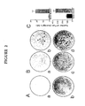
- expression of recobminant R2 in ras -transformed cells resulted in enhanced colony forming efficiency in soft agar, and markedly elevated tumorigenic and metastatic potential in vivo.
- deregulated R2 expression can cooperate with other oncogenes like rac-1 in mechanisms of transformation.
- results herein demonstrate for the first time that the R2 component of mammalian ribonucleotide reductase is a malignancy determinant that can synergize with activated oncogenes to modify malignant potential, and supports a model in which these effects are mediated through alterations in major Ras pathways that are brought about by deregulated R2 gene expression.
- the observations presented here indicated that R2 can also participate in other critical cellular functions, and can play a direct role in determining malignant potential through oncogene cooperativity.
- ribonucleotide reductase R2 gene expression can play a significant role in determining drug sensitivity characteristics, and that this appeas to occur at least in part through a mechanism involving genomic instability.
- the mechanism through which aberrant R2 expression modifies drug sensitivities does not appear to require the direct involvement of p53 mutation or loss of wild type p53 function, although it is possible that genetic events downstream of a p53 regulated pathway are involved.
- Recombinant R2 gene expression in Balb/c 3T3 and NIH-3T3 cells significantly increases both Raf-2 protein activation and mitogen-activating protein kinase (MAPK) activity.
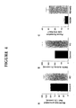
- Example 3 demonstrates that short antisense sequences directed against the R1 and R2 components have anti-tumor activity and are cytotoxic to the neoplastic cells. Further, the R2 antisense sequences can also act synergistically with well known chemotherapeutic agents. Very low concentrations (non-toxic) of short antisense sequences reduced the resistance of the neoplastic cells to chemotherapeutic agents such as N-(phosphonacetyl)-L-aspartate (PALA) and methotrexate (MTX) as well as hydroxyurea. As shown in the Example, cells were transfected with a vector containing the R2 sequence in an antisense oritentation. These cells were more sensitive to the chemotherapeutic agents.
- chemotherapeutic agents such as N-(phosphonacetyl)-L-aspartate (PALA) and methotrexate (MTX)
- mouse 10T 1 / 2 cells which are drug resistant, when transfected with R2 sequence in the antisense orientation, were found to have significantly reduced resistance (increased sensitivity) to the chemotherapeutic agents.
- Short synthetic antisense sequence complementary to the R2 sequence also provided increases sensitivity.
- Vectors can be constructed for the present invention by those skilled in the art and should contain all expression elements necessary to achieve the desired transcription of the sequences.
- the expression elements can be selected to allow expression only in the cell being targeted. Other beneficial characteristics can also be contained within the vectors such as mechanisms for recovery of the nucleic acids in a different form.
- One of ordinary skill in the art will know which expression elements are compatible with a particular cell type.
- the vectors can be introduced into cells or tissues by any one of a variety of known methods within the art as described herein above.
- tumorigenicity tumor latency assay
- 1 x 10 5 cells in a 0.1 ml volume were injected subcutaneously into the back of mice and the time required to form a tumor (2 X 2 mm) detectable by palpation was recorded.
- the growth of tumors was also evaluated by measuring tumor diameters, and estimating tumor base area each day following tumor appearance [ Indianapolisn et al., 1989].
- Tumor size was determined by multiplying the dimensions of the cross-section of the tumor. Tumors were removed from the mice and tumor weight was recorded 21 days later. In the case of no tumor formation, mice were kept for 2 months after injection and then sacrificed.
- RIBONUCLEOTIDE REDUCTASE ASSAY Ribonucleotide reductase activity in crude extracts prepared from cells is assayed as previously described [Lewis et al., 1978; Hurta and Wright, 1992; Hurta et al., 1995]. Enzyme preparations are obtained from logarithmically growing cells lysed in phosphate buffered saline, pH 7.2, containing 1 mM dithiothreitol and 1 mM protease inhibitor, AEBSF (Calbiochem, San Francisco, CA), by three cycles of freeze-thawing.
- AEBSF Calbiochem, San Francisco, CA
- WESTERN BLOT ANALYSIS The procedures used have been reported [Fan et al., 1996a; 1996b; Choy et al, 1988]. Briefly, following cell extract preparation, total protein content was determined, and an aliquot was analyzed on 10% linear SDS-polyacrylamide gel. After protein transfer and blocking, membranes were incubated with anti-R2 rabbit polyclonal antibody. Alkaline phosphatase conjugated goat anti-rabbit IgG (Sigma) was used for protein R2 detection.
- mice and Cell Culture The mouse cell lines, BALB/c 3T3, NIH 3T3, four lines of T24 H- ras transformed 10T 1 / 2 cells, named C1, NR4, r-2 and r-3 have been previously used as recipients of the R2 retroviral vector [Fan et al., 1996b].
- Cells were routinely cultured in ⁇ -minimal essential medium ( ⁇ -MEM)(Gibco, Grand Island, NY) supplemented with 10% calf serum (Fetalclone III, Hyclone, Logan, UT).
- ⁇ -MEM ⁇ -minimal essential medium
- calf serum Fetalclone III, Hyclone, Logan, UT.
- the membrane fraction was prepared as described by Qui et al. [1995], and used for Western analysis with a polyclonal antibody specific for Raf-1 protein (Santa Cruz Biotechnology Inc., Santa Cruz, CA), after the protein content was determined by the standard Bio-Rad assay. Densitometry analysis of the Raf-1 band was performed, and the amount of Raf-1 protein from each sample was corrected by densitometry analysis of a well separated band on a parallel gel stained with Coomassie blue.
- Ribonucleotide Reductase Assay The Assay was performed as described herein above. In some experiments enzyme assays were performed by combining purified recombinant R1 protein [Salem et al, 1993] with 9E10 antibody-precipitated R2 protein [Hurta and Wright, 1992]. In this Example, 20 ⁇ g of the 9E10 antibody and 50 ⁇ l of Staphylococcal protein A-agarose (Sigma Chem. Co., St. Louis, MO) were added to 1 ml of the supernatant of centrifuged lysed cells, and placed on a rocker at 4°C for 2 hours.
- the Staphylococcal protein A agarose-immunocomplex was washed three times with 1 ml of cold phosphate buffer containing 1 mg/ml bovine serum albumin. The immunocomplex was then assayed for ribonucleotide reductase activity [Lewis et al, 1978; Hurta and Wright, 1992; Fan et al., 1996b; Choy et al, 1988].
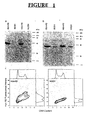
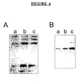
- Figure 1A shows that Western blots with the 9E10 antibody that specifically recognizes the Myc-epitope sequence detects the R2 protein of approximately 45 kDa in SH/mR2 stably infected BALB/c 3T3 and NIH 3T3 cells (named B3/mR2 and N3/mR2, respectively), but not in control vector (LXSH) infected B3/SH or N3/SH cells.
- R2 specific antibodies detected the endogenous as well as the recombinant R2 protein in expression vector infected cells, and as expected only the endogenous protein was observed in control vector infected cells (Fig. 1B).
- ribonucleotide reductase activity was assayed and found that the CDP reductase activities in B3/mR2 and N3/mR2 cells in three independent experiments were 1.96 ⁇ 0.32 and 1.71 ⁇ 0.11 nmoles/mg protein/hour, respectively, which was 2.6 and 2.1 times higher than observed with B3/SH and N3/SH cells (0.74 ⁇ 0.14 and 0.83 ⁇ 0.08 nmoles/mg/hour, respectively).
- enzyme assays were carried out by combining purified recombinant R1 protein [Salem et al, 1993], with 9E10 antibody precipitated R2 protein.
- Ras Malignancy Potential Determined by Aberrant R2 Gene Expression Since combinations of altered R2 gene expression and activated H- ras were synergistic in focus forming experiments in which ras was transfected into altered R2 expressing cells, this gene combination was tested further by infecting four independent H- ras transformed 10T 1 / 2 cell lines, C1, NR4, r-2 and r-3 that were previously characterized [Egan et al., 1987a, 1987b; Taylor et al., 1992; Stokoe et al., 1994], with the retroviral vector SH/mR2. Stable infectants were selected with hygromycin, and Western blot analyses and enzyme activity assays confirmed that these infectants expressed biologically active Myc-tagged R2 protein.
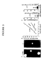
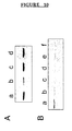
- C1/mR2 and C1/SH cells were compared in syngeneic C3H/HeN mice. Marked differences in malignant potential were observed. C1/mR2 cells exhibited shorter tumor latency and greater tumor growth when compared to C1/SH cells (Fig. 3B). Furthermore, metastasis assays clearly indicated that C1/mR2 cells were more malignant than C1/SH cells and produced significantly more lung tumors (Fig. 3C).
- R2 Gene Expression and Oncogene Cooperativity The above results indicate that altered R2 expression can cooperate with activated H-ras in in vitro transformation and in in vivo malignancy assays. Since no obvious differences in growth rates or cell cycle phase distributions were found that may account for this cooperation, as for example changes in cell cycle regulation, the following idea was tested. Does deregulated R2 expression synergize with ras by elevating the activity of a Ras signal pathway? This would be consistent with studies showing a direct correlation between ras expression and malignant potential [Egan et al., 1987a, 1987b; Wright et al, 1993; Bradley et al, 1986]. A major Ras pathway for regulating gene expression involves the Raf-1 protein kinase. Activated Ras recruits Raf to the plasma membrane where Raf and downstream signalling molecules like MAPKs become activated [Stokoe et al, 1994; Jelinek et al, 1994; Leevers et al, 1994].
- This Example indicates for the first time that the R2 component of mammalian ribonucleotide reductase is a novel malignancy determinant that can synergize with activated oncogenes to modify malignant potential. It is important to note that the only role ascribed to R2 in the cell prior to this Example is as a rate-limiting component of ribonucleotide reductase. This Example demonstrates that R2 can also participate in other critical cellular functions and can play a direct role in determining malignant potential through oncogenic cooperativity.
- mice The hydroxyurea resistant mouse cell lines, H-2, H-4, LHF and SC2 were derived from mouse L cells and have been characterized in Choy et al [1988] and McClarty et al [1986].
- BALB/c 3T3 cells were used as recipients of an R2 retroviral expression vector (B3/mR2 and B3/R2c2 cell lines), or of the same retroviral vector lacking the R2 sequence (B3/SH cells)[Fan et al., 1996a; 1996b].
- NIH-3T3 cells were also used as recipients of the R2 retroviral expression vector (N/R2-4 cell line) or of this retroviral vector lacking the R2 sequence (N/SH cells), as described previously [Fan et al., 1996a; 1996b].
- the N/R2+ASR2 cell line was the recipient through co-transfection using LipofectAmine (Life Technologies, N.Y) [Damen et al., 1991] of retroviral vectors containing the R2 coding sequence and the R2 sequence in the antisense orientation.
- RP3 and RP6 cells are 10T 1 / 2 mouse cells that have been transfected with the T-24 H- ras oncogene and a mutant oncogenic form of the p53 gene [Taylor et al., 1992], and they were also used as recipients through transfection using LipofectAmine reagent, of a retroviral vector containing the R2 coding region in an antisense orientation [Fan et al., 1996b], to obtain RP3/ASR2 and RP6/ASR2 cells.
- 1B cells are p53 -/- and were derived from embryonic fibroblasts [Lowe et al., 1994].
- All cells were cultured in ⁇ -minimal essential medium (Gibco, Grand Island, NY) containing 10% fetal bovine serum (Intergen, Purchase, NY) and antibiotics (100 units/ml penicillin and 100 ⁇ g/ml streptomycin) at 37°C in a humidified atmosphere containing 5% CO 2 .
- ⁇ -minimal essential medium Gibco, Grand Island, NY
- antibiotics 100 units/ml penicillin and 100 ⁇ g/ml streptomycin
- Drug Selections Cells ranging in numbers from 500 to 1-2 x 10 5 were added to 100 mm tissue culture plates in growth medium containing 10% dialyzed fetal bovine serum, and in the absence or presence of drug [Huang et al., 1995a; Choy et al., 1988]. The culture medium was replaced with fresh medium every week for two to three weeks. Surviving cells were visualized by methylene blue staining, and colonies of about 50 cells or more were scored [Huang et al., 1995a]. The relative colony forming efficiency was defined as the ability to produce colonies in the presence of a drug divided by that ability in the absence of drug.
- Genomic DNA was extracted from logarithmically growing cells by the phenol-chloroform extraction method [Blin and Stafford, 1976], and potential gene amplification events were determined by Southern blot analysis as described [Huang et al., 1995a; Choy et al., 1988], using the cDNA fragments as probes noted below.
- the 1487 bp Sal I/Pst I probe for ribonucleotide reductase R2 was prepared from cDNA clone 10 [Huang et al., 1995a; Choy et al., 1988].
- Electrophoretic Gel Mobility Shift Assay (EMSA) was used to determine the presence of wild type p53. Assays were performed essentially as described [Price and Calderwood, 1993], with the following modifications. Cells on 150 mm plates were washed once with ice cold phosphate buffered saline (PBS) and scraped into 1 ml PBS. Cells were pelleted by centrifugation at 1300 g at 4°C for 10 minutes and stored at -80°C.
- PBS ice cold phosphate buffered saline
- Nuclei were prepared by lysing the pellets in 300 ⁇ l buffer A (20 mM HEPES ⁇ pH 7.6 ⁇ , 20% glycerol, 10 mM NaCl, 1.5 mM MgCl 2 , 0.2 mM EDTA and 0.1% Triton X-100) for 20 minutes on ice. Buffer A also contained 1 mM phenylmethylsulfonyl fluoride (PMSF) and 10 mM dithiothreitol (DTT). Nuclei were isolated by centrifugation at 1300 g at 4°C for 10 minutes.
- PMSF phenylmethylsulfonyl fluoride
- DTT dithiothreitol
- Nuclear lysates were prepared by adding 20-40 ⁇ l of buffer A containing 500 mM NaCl, 1 mM PMSF and 10 mM DTT to the nuclear pellet and incubating 20 minutes on ice.
- the extracted nuclei were pelleted by centrifugation at 16,000 g at 4°C; the supernatant was removed and an aliquot was used for protein determination using the Biorad protein assay procedure (Biorad).
- the nuclear lysate was incubated with an excess of double stranded p53 consensus binding sequence (GGACATGCCCGGGCATGTCC)(SEQ ID No:162) end labeled with [ ⁇ - 32 P]-ATP using T4 polynucleotide kinase (Boehringer).
- DNA binding was carried out in buffer containing 20 mM HEPES (pH 7.6), 20% glycerol, 1.5 mM MgCl 2 , 0.2 mM EDTA, 1 mM PMSF and 10 mM DTT.
- Each binding reaction contained 5 ⁇ g cell lysate, 10 ⁇ g double stranded poly (dI-dC)(Pharmacia), 1.4 ng labeled consensus probe and 100 ng of monoclonal antibody 421 (Santa Cruz) in a total volume of 20 ⁇ l.
- DNA binding was allowed to proceed for 30 minutes at room temperature and the mixture was separated by electrophoresis on 5% nondenaturing polyacrylamide gels. Electrophoresis was carried out at room temperature until the xylene cyanol tracking dye had run to the bottom of the gel and the free probe had run off the gel.
- H-2, H-4, LHF and SC2 are mouse L cell lines selected for resistance to the cytotoxic effects of the antitumor agent, hydroxyurea. These four cell lines exhibited resistance to hydroxyurea in colony forming efficiency experiments, that ranged between approximately 18 (H-2) to 30 (SC2) fold higher than the wild type mouse L cell line from which they were derived [Choy et al., 1998; McClarty et al., 1988].
- ribonucleotide reductase activity that ranged between 2.2 fold (H-2) to 17 fold (LHF and SC2), which was primarily due to increases in the R2 component of ribonucleotide reductase that is limiting for enzyme activity and cell division in proliferating mouse cells.
- Table 2 shows that the four hydroxyurea resistant cell lines were also less sensitive to the cytotoxic effects of N-(phosphonacetyl)-L-aspartate (PALA) and methotrexate (MTX) in colony forming experiments, when compared to parental wild type mouse L cells. These differences in drug sensitivity are highly significant, with p values of ⁇ 0.0001 for each of the cell lines when compared to the parental wild type mouse cells.
- DHFR dihydrofolate reductase
- CAD a multifunctional polypeptide containing carbamyl phosphate synthetase, aspartate transcarbamylase and dihydrooratase
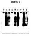
- Figure 5 shows that cells that proliferated in the presence of PALA or MTX exhibited increased CAD or DHFR gene copy numbers.
- PALA or MTX exhibited increased CAD or DHFR gene copy numbers.
- all colonies that developed in PALA and tested (10/10) showed CAD gene amplification.
- some but not all colonies that developed in the presence of MTX (3/6) showed DHFR gene amplification.
- B3/mR2 is a population of BALB/c 3T3 cells containing elevated R2 protein due to the presence of a retroviral expression vector encoding R2
- B3/SH is a cell population that has wildtype levels of R2 protein and contains the empty vector as a control.
- B3/R2c2 is a cloned line with elevated R2 protein selected from the B3/mR2 population.
- B3/mR2 and B3/R2c2 cells are significantly more resistant to the cytotoxic effects of hydroxyurea, at a range of concentrations, when compared to B3/SH cells.
- B3/mR2 and B3/R2c2 cells express increased levels of an active R2 component of ribonucleotide reductase.
- B3/mR2 and B3/R2c2 cells were also significantly less sensitive to the cytotoxic effects of PALA and MTX, which act at sites other than ribonucleotide reductase (Table 3). Resistance to these two drugs ranged between approximately 10 fold with 100 nM MTX to more than 100 fold at most concentrations of PALA tested.
- ANTISENSE DEOXYRIBONUCLEOTIDE SEQUENCES THAT TARGET RIBONUCLEOTIDE REDUCTASE AND ARE CYTOTOXIC FOR HUMAN TUMOR CELLS.
- Colony Forming Efficiency was determined as previously reported [Huang and Wright, 1994]. The cells were cultured for 24 hours at 37°C in growth medium with 10% fetal bovine serum. The cells were washed in 5ml phosphate buffered saline, pH 7.2, once prior to lipofectin +/- oligonucleotide treatment.
- oligonucleotides being tested were added to cell cultures in the presence of 2.5 ⁇ g of DOTMA/DOPE (Lipofectin; Life Technologies, Inc.) for four hours. The oligonucleotide was tested at 0.2 ⁇ M unless otherwise indicated. Controls were the cultures treated with lipofectin but without the oligonucleotide. After 4 hours the medium containing the oligonucleotide was removed and washed with 5 ml of growth medium. The cells were then cultured in growth medium containing 10% fetal bovine serum for seven to ten days. Surviving cells were visualized by methylene blue staining, and colonies were scored. In some experiments cell aliquotes were removed from the culture and viability was determined using the trypan blue exclusion test [Phillips, 1973]. Results were analyzed as percent of surviving cells compared to control cells.
- Antisense molecules were identified that target ribonucleotide reductase. As shown below they were cytotoxic for a variety of human tumor cells. Sequences were found that facilitated drug-cytotoxicity for drug resistant tumor cells. That is, at very low non-cytotoxic concentrations, antisense sequences targeting ribonucleotide reductase can sensitize tumor cells to the cytotoxic activity of clinically important chemotherapeutic compounds.
- AS-II-336-20 has the sequence 5'-TCC TGG AAG ATC CTC CTC GC-3'(SEQ ID No:1), and targets the R2 message of human ribonucleotide reductase at nucleotides 336-355, based on the numbering of R2 nucleotides [Pavloff et al., 1992].
- the AS-II-2229B-20 sequence is: 5'-TCC CAC ATA TGA GAA AAC TC-3' (SEQ ID No:2), and targets the R2 message at nucleotides 2229-2248.
- Both AS-II-336-20 and AS-II-2229B-20 were constructed as phosphorothioate sequences to protect against nuclease activity [Anazodo et al., 1995].
- Antisense construct AS-II-336-20 was tested for the ability to inhibit the proliferation of human tumor cells (Hela) in relative colony forming efficiency experiments as described herein above.
- Hela S3 cells American Type Culture Collection, Rockville, Maryland, ATCC
- Hela 1mM Hela cell line
- Two experiments were undertaken with Hela S3 cells. With a 4 hour treatment of 0.2 ⁇ M antisense construct AS-II-336-20, inhibition of 92% and 82% was seen in colony forming efficiency in two experiments, respectively.
- the same experiment was repeated with the Hela 1mM cell line and with varying concentrations of the antisense construct AS-II-336-20 (Table 6) with similar results, 0.2 ⁇ M was an effective concentration for inhibiting colony formation.
- AS-II-336-20 is a very effective inhibitor of human tumor cell colony forming ability, and it is effective both in inhibiting the proliferation of human tumor cell colony forming ability and in inhibiting the proliferation of human tumor cells that exhibit resistance to another chemotherapeutic compound.
- antisense construct AS-II-336-20 is an effective antitumor compound in experiments performed with the mouse tumor cell line, SC2, which is a highly hydroxyurea resistant mouse L cell line [McClarty et al., 1988].
- the antisense sequence AS-II-2229B-20 was also tested for the ability to inhibit the proliferation of human Hela tumor cells in relative colony forming efficiency experiments with results similar to that of AS-II-336-20 as shown in Table 6. These data show that AS-II-2229B-20 is a potent antitumor agent when tested with Hela S3 cells and with the drug resistant Hela 1mM cell line.
- the antisense construct AS-II-2229B-20 was also tested for the ability to inhibit the proliferation of the human breast cancer cell line MDA435 and found to be very effective (Table 8).
- the ribonucleotide reductase R2 antisense construct designated AS-II-2229B-20 was tested for tumor cell cytotoxicity by comparing the results obtained with human tumor and non-tumor cell populations. Hela S3 tumor cells and WI 38 normal non-tumorigenic human cells were used. Tumor cells were found to be much more sensitive to the cytotoxic effects of AS-II-2229B-20 than normal non-tumorigenic cells. For example, analysis of cells three days after antisense exposure indicated that tumor cells were approximately 5-times more sensitive to the cytotoxic effects of AS-II-2229B-20 than normal non-tumorigenic cells averaged over 4-8 determinations.
- Example 2 Treatment of human tumor cells with very low concentrations of short antisense sequences was tested to determine if these constructs could sensitize the tumor cells to inhibitory effects of other chemotherapeutic drugs.
- concentration used was not cytotoxic in itself as shown in Table 6.
- the treatment of Hela S3 and Hela 1mM cells with 0.02 ⁇ M of the AS-II-2229B-20 antisense construct increases the sensitivity of these cells to N-(phosphonacetyl)-L-aspartate (PALA) and to methotrexate (MTX) as shown in Table 9.
- Ribonucleotide reductase is composed of two dissimilar protein components coded by two distinct genes, R1 and R2. Therefore, the results described hereinabove suggest that the R1 message may also be an appropriate target for designing short antisense molecules that have potent antitumor activity.
- R1 20-mer deoxyribonucleotide phosphorothioate sequence in antisense orientation, designated AS-1-1395-20, was constructed and its antitumor abilities were tested.
- the antisense construct AS-1-1395-20 has the sequence 5'-ACA GGA ATC TTT GTA GAG CA-3' (SEQ ID No:103), and targets the R1 message at nucleotides 1395-1414.
- colony efficiency assays performed in soft agar as described herein demonstrated similar results. Colony forming efficiencies of 15.6 ⁇ 6.73 for NIH-3T3 mouse cells containing the H- ras oncogene, 4.4 ⁇ 2.62 for NIH-3T3 mouse cells containing the H- ras oncogene and the R2 antisense sequence, and 51 ⁇ 12.29 for NIH-3T3 mouse cells containing the H- ras oncogene and the coding region sequence for R2 were seen.
- PALA CONC B3/SH B3/mR2 B3/R2c2 10 ⁇ M 17.9 ⁇ 11.0 965.0 ⁇ 529.7 1230.0 ⁇ 97.0 20 ⁇ M 0.39 ⁇ 0.18 120.1 ⁇ 28.4 55.1 ⁇ 15.6 40 ⁇ M 0.35 ⁇ 0.01 25.0 ⁇ 4.6 20.2 ⁇ 6.8 50 ⁇ M 0.24 ⁇ 0.14 27.6 ⁇ 8.9 15.9 ⁇ 4.0 60 ⁇ M 0.12 ⁇ 0.05 25.0 ⁇ 6.4 18.7 ⁇ 5.3 80 ⁇ M 0.17 ⁇ 0.08 27.1 ⁇ 6.75 20.0 ⁇ 4.9 C.
- Ribonucleotide reductase R2 component is a novel malignancy determinant that cooperates with activated oncogenes to determine transformation and malignant potential. Proc. Natl. Acad. Sci. USA 93:14036-14040.
- Wild-type p53 restores cell cycle control and inhibits gene amplification in cells with mutant p53 alleles.
Abstract
Description
| INCREASED COLONY FORMATION IN SOFT AGAR BY ras -TRANSFORMED CELLS CONTAINING THE RECOMBINANT R2 VECTOR | |||
| Cell Line | Colonies (average ± SE) formed in soft agar with varying | ||
| 103 | 104 | 105 | |
| C1/ | 0 | 4 ± 3 | 66 ± 9 |
| C1/ | 3 ± 3 | 28 ± 7 | 347 ± 45 |
| r-2/SH | ND | 9 ± 2 | 105 ± 7 |
| r-2/mR2 | ND | 24 ± 1 | 298 ± 11 |
| NR4/ | 0 | 3 ± 1 | 32 ± 4 |
| NR4/ | 2 ± 1 | 14 ± 2 | 127 ± 10 |
| r-3 | 7 ± 1 | 100 ± 11 | ND |
| r-3/mR2 | 31 ± 4 | 309 ± 17 | ND |
| DRUG SENSITIVITIES DETERMINED BY RELATIVE COLONY FORMING EFFICIENCIES x 10 -4 | ||||||
| A. PALA | ||||||
| DRUG CONC. | CELL LINES | |||||
| W.T. | H2 | | LHF | SC2 | ||
| 20 µM | 172.3 ± 126.3 | 406.7 ± 202.2 | 322.5 ± 36.4 | 233.3 ± 3.6 | 850.1 ± 325.2 | |
| 30 µM | 50.3 ± 20.5 | 39.4 ± 16.4 | 84.0 ± 30.0 | 78.8 ± 7.9 | 187.6 ± 46.4 | |
| 40 µM | 15.0 ± 7.0 | 23.3 ± 10.4 | 43.3 ± 9.6 | 46.5 ± 9.9 | 37.5 ± 8.7 | |
| 50 µM | 3.6 ± 1.1 | 7.9 ± 1.7 | 23.2 ± 0.5 | 25.0 ± 6.8 | 47.5 ± 35.8 | |
| 60 µM | 1.3 ± 0.4 | 3.6 ± 0.6 | 11.1 ± 1.4 | 10.7 ± 3.0 | 17.6 ± 1.2 | |
| B. MTX | ||||||
| CONC. | W.T. | H2 | | LHF | SC2 | |
| 40 nM | 11.2 ± 7.2 | 52.6 ± 25.2 | 44.2 ± 20.9 | 143.4 ± 41.3 | 880.4 ± 147.4 | |
| 60 nM | 12.3 ± 7.2 | 73.7 ± 16.6 | 34.7 ± 11.2 | 63.5 ± 18.6 | 566.8 ± 66.2 | |
| 80 nM | 2.2 ± 1.6 | 67.7 ± 20.0 | 39.3 ± 18.7 | 68.2 ± 19.2 | 306.6 ± 61.5 | |
| 100 nM | 0.8 ± 0.4 | 75.3 ± 10.0 | 15.1 ± 8.8 | 60.8 ± 16.7 | 261.8 ± 39.7 | |
| 150 nM | 0.5 ± 0.2 | 53.3 ± 9.4 | 32.3 ± 13.7 | 63.9 ± 16.0 | 301.6 ± 76.8 | |
| The relative colony forming efficiencies are shown ± se, and the values presented are from 4 to 8 determinations. Statistically significant differences were observed when data obtained with H2 (p = 0.0004), H4 (p ≤ = 0.0001), LHF (p ≤ 0.0001), and SC2 (p ≤ 0.0001) were each compared to data obtained with the parental wild type (W.T.) cell line. |
| DRUG SENSITIVITIES DETERMINED BY RELATIVE COLONY FORMING EFFICIENCIES x10 -4 | |||
| A. HYDROXYUREA | |||
| DRUG CONC | CELL LINES | ||
| B3/SH | B3/mR2 | B3/R2c2 | |
| 0.1 mM | 3.3 ± 1.4 | 1310 ± 319.0 | 830.8 ± 97.0 |
| 0.4mM | 0.17 ± 0.19 | 14.6 ± 4.0 | 33.7 ± 11.0 |
| 0.5mM | 0.21 ± 0.14 | 6.5 ± 4.6 | 26.9 ± 11.9 |
| 0.6 mM | 0.41 ± 0.22 | 5.2 ± 3.7 | 12.5 ± 4.6 |
| 0.8 mM | 0.19 ± 0.62 | 2.6 ± 1.4 | 13.2 ± 6.4 |
| B. PALA | |||
| CONC | B3/SH | B3/mR2 | B3/R2c2 |
| 10 µM | 17.9 ± 11.0 | 965.0 ± 529.7 | 1230.0 ± 97.0 |
| 20 µM | 0.39 ± 0.18 | 120.1 ± 28.4 | 55.1 ± 15.6 |
| 40 µM | 0.35 ± 0.01 | 25.0 ± 4.6 | 20.2 ± 6.8 |
| 50 µM | 0.24 ± 0.14 | 27.6 ± 8.9 | 15.9 ± 4.0 |
| 60 µM | 0.12 ± 0.05 | 25.0 ± 6.4 | 18.7 ± 5.3 |
| 80 µM | 0.17 ± 0.08 | 27.1 ± 6.75 | 20.0 ± 4.9 |
| C. MTX | |||
| CONC | B3/SH | B3/mR2 | B3/R2c2 |
| 20 nM | 192.6 ± 44.6 | 1055.0 ± 239.0 | 382.4 ± 71.3 |
| 40nM | 15.7 ± 2.9 | 62.1 ± 8.8 | 60.8 ± 13.0 |
| 60 nM | 6.1 ± 2.0 | 76.7 ± 21.6 | 64.1 ± 20.5 |
| 80nM | 2.2 ± 0.7 | 17.5 ± 3.6 | 20.1 ± 5.5 |
| 100nM | 1.5 ± 0.5 | 12.3 ± 2.8 | 21.0 ± 7.2 |
| 150 nM | 3.0 ± 1.1 | 23.0 ± 7.6 | 33.4 ± 14.3 |
| The relative colony forming efficiencies are shown ±SE, and the values presented are from 4 to 12 determinations. Statistically significant differences were observed when data obtained with B3/mR2 or with B3/R2c2 were compared with data obtained with B3/SH (all p values were ≤ 0.0001 for data obtained in the presence of hydroxyurea, PALA or MTX). |
| DRUG SENSITIVITIES DETERMINED BY RELATIVE COLONY FORMING EFFICIENCIES x10 -4 | |||
| A. HYDROXYUREA | |||
| DRUG CONC | CELL LINES | ||
| N/SH | N/R2-4 | N/R2+ASR2 | |
| 0.3mM | 1.14 ± 0.12 | 46.1 ± 9.8 | 0.49 ± 0.34 |
| 0.4mM | 0.71 ± 0.17 | 18.0 ± 6.7 | 0.14 ± 0.14 |
| B. PALA | |||
| CONC | N/SH | N/R2-4 | N/ |
| 10 µM | 5.28 ± 1.5 | 6.22 ± 3.3 | 1.81 ± 0.8 |
| 15 µM | 5.83 ± 2.7 | 10.0 ± 5.5 | 0.58 ± 0.3 |
| 20 µM | 0.30 ± 0.1 | 1.71 ± 1.2 | 0.04 ± 0.04 |
| 25 µM | 0.53 ± 0.3 | 0.8 ± 0.7 | 0.04 ± 0.04 |
| 30 µM | 0.48 ± 0.08 | 1.03 ± 0.07 | 0.12 ± 0.12 |
| 40 µM | 0.27 ± 0.2 | 0.14 ± 0.08 | 0.04 ± 0.04 |
| C. MTX | |||
| CONC | N/SH | N/R2-4 | N/R2+ASR2 |
| 20nM | 655 ± 74.8 | 540 ± 25.1 | 423 ± 119 |
| 40nM | 21 ± 12.1 | 147 ± 4.2 | 3.5 ± 1.9 |
| 60nM | 3.4 ± 2.2 | 62.2 ± 30.7 | 1.9 ± 1.3 |
| 80nM | 5.0 ± 5.0 | 50.4 ± 23.9 | 2.5 ± 1.5 |
| 100nM | 4.2 ± 2.5 | 66.1 ± 32.8 | 1.1 ± 0.6 |
| 150nM | 1.4 ± 0.9 | 21.0 ± 11.5 | 0, n=4 |
| The relative colony forming effeciencies are shown ± SE, and the values presented are from 4 to 6 determinations. Where 0 is shown the number of determinations using 1 x 10⁵ cells per test is shown as 4 (n=4). Statistically significant differences were observed when data obtained with N/SH in the presence of PALA was compared to data obtained with N/R2-4 or with N/R2+ASR2 in the presence of hydroxyurea (p = 0.0001 in both cases) or in the presence of MTX (p = 0.0002 and 0.032, respectively). Statistically significant differences were also observed when data obtained with N/SH in the presence of PALA was compared to data obtained with N/R2+ASR2 (p = 0.002), but not with data obtained with N/R2-4. |
| DRUG SENSITIVITIES DETERMINED BY RELATIVE COLONY FORMING EFFICIENCIES x10 -4 | ||||
| A. HYDROXYUREA | ||||
| DRUG | CELL LINES | |||
| CONC | RP3/SH | RP3/ASR2 | RP6/SH | RP6/ASR2 |
| 0.1 mM | 263.6 ± 19.3 | 109.8 ± 43 | 201.3 ± 27.2 | 43.8 ± 12.3 |
| 0.2 mM | 53.6 ± 13.7 | 22.9 ± 3.1 | 35.5 ± 8.4 | 8.6 ± 2.5 |
| 0.3mM | 20.8 ± 7.5 | 6.6 ± 2.5 | 12.6 ± 2.4 | 4.5 ± 1.1 |
| 0.4mM | 5.8 ± 1.9 | 1.0 ± 0.2 | 10.8 ± 4.1 | 1.2 ± 0.5 |
| 0.5mM | 4.8 ± 1.9 | 0.2 ± 0.1 | 12.1 ± 3.9 | 1.8 ± 0.9 |
| 0.6mM | 0.7 ± 0.3 | 0.3 ± 0.1 | 6.6 ± 2.9 | 1.5 ± 0.7 |
| 0.8mM | 0.8 ± 0.3 | 0.1 ± 0.05 | 1.7 ± 1.2 | 0.4 ± 0.3 |
| B. PALA | ||||
| CONC | RP3/SH | RP3/ASR2 | RP6/SH | RP6/ASR2 |
| 10µM | 2569 ± 338 | 1183 ± 384 | 4619 ± 648 | 2083 ± 960 |
| 20µM | 123.4 ± 19.3 | 86.1 ± 32.9 | 1220 ± 255 | 368 ± 154 |
| 30µM | 45.2 ± 7.8 | 19.5 ± 4.7 | 450 ± 129 | 316 ± 171 |
| 40µM | 15.0 ± 4.9 | 4.7 ± 0.6 | 271 ± 68 | 116 ± 54 |
| 50µM | 9.3 ± 3.6 | 2.1 ± 0.8 | 109 ± 23 | 41.7 ± 23 |
| 60µM | 3.9 ± 1.6 | 0.3 ± 0.2 | 555 ± 13 | 13.2 ± 6.3 |
| C. MTX | ||||
| CONC | RP3/SH | RP3/ASR2 | RP6/SH | RP6/ASR2 |
| 20nM | 961.7 ± 134 | 485.9 ± 165 | 1856 ± 464 | 1504 ± 486 |
| 40nM | 347.1 ± 154 | 77.8 ± 18 | 172 ± 41.3 | 91.5 ± 28.1 |
| 60nM | 123.8 ± 64 | 18.1 ± 6.2 | 77.3 ± 15.6 | 49.9 ± 14.1 |
| 80nM | 66.5 ± 37 | 4.4 ± 0.8 | 68.7 ± 16.7 | 36.0 ± 6.0 |
| 100nM | 34.8 ± 21 | 0.6 ± 0.06 | 46.6 ± 5.6 | 14.4 ± 3.8 |
| 150nM | 4.7 ± 3 | 0.2 ± 0.1 | 11.1 ± 4.4 | 3.5 ± 0.9 |
| The relative colony forming effeciencies are shown ± SE, and the values presented are from 4 to 10 determinations. Statistically significant differences were observed when data obtained with RP6/SH was compared with data obtained with RP6/ASR2 (p = 0.0001, 0.0001 and 0.0001 in the presence of hydroxyurea, PALA and MTX, respectively). Significant differences were also observed when data obtained with RP3/SH was compared with data obtained with RP3/ASR2 (p = 0.04, 0.0001 and 0.004 in the presence of hydroxyurea, PALA and MTX, respectively). |
| REDUCED COLONY FORMING EFFICIENCY FOLLOWING TREATMENT WITH R2 ANTISENSE CONSTRUCTS | |||
| CELL LINE: Hela S3 | |||
| Conc. AS-II-336-20a | % Inhib. | Conc. AS-II-2229B-20b | % Inhib. |
| 0 | - | 0 | - |
| 0.2 µM | 92% | 0.05 µM | 50% |
| 0.2 µM | 82% | 0.10 µM | 80% |
| 0.20 µM | 95% | ||
| 0.20 µM | 97% | ||
| CELL LINE: Hela 1mM | |||
| Conc. AS-II-336-20a | % Inhib. | Conc. AS-II-2229B-20b | % Inhib. |
| 0 | - | 0 | - |
| 0.01 µM | 15% | 0.01 | 0% |
| 0.05 µM | 25% | 0.02 | 0% |
| 0.10 µM | 60% | 0.03 µM | 21% |
| 0.20 µM | 85% | 0.04 µM | 34% |
| 0.05 µM | 48% | ||
| 0.05 µM | 50% | ||
| 0.10 µM | 78% | ||
| 0.20 µM | 97% | ||
| 0.20 µM | 90% | ||
| CELL LINE: Mouse SC2 | |||
| Conc. | AS-II-336-20a | % Inhib. | |
| 0 | - | ||
| 0.2 µM | 95% |


| TREATMENT WITH AN R2 ANTISENSE CONSTRUCTS | ||
| CONSTRUCT | CONC. (µm) | COLONY FORMING INHIBITION OF: MDA435 |
| AS-II-2229B-20 | 0.02 | 25% |
| 0.03 | 56% | |
| 0.05 | 78% | |
| 0.10 | 94% | |
| 0.20 | 99% |
| SYNERGISTIC EFFECT OF AS-II-2229B-20 ANTISENSE CONSTRUCT | ||||
| Cells | Drug | Drug Conc. | AS-II-2229B-20a 0.02_M | Relative Colony Forming Efficiency |
| Hela S3 | PALA | 20µM | - | 350 ± 50 |
| PALA | 20µM | + | 90 ± 10 | |
| Hela S3 | MTX | 40µM | - | 118 ± 32 |
| MTX | 60µM | - | 116 ± 13 | |
| MTX | 40µM | + | 25 ± 5 | |
| MTX | 60µM | + | 0 | |
| Hela 1mM | PALA | 20µM | - | 377 ± 21 |
| PALA | 30µM | - | 311 ± 9.5 | |
| PALA | 20µM | + | 108 ± 7.5 | |
| PALA | 30µM | + | 101 ± 2.0 | |
| Hela 1mM | MTX | 40µM | - | 28 ± 10 |
| MTX | 60µM | - | 12 ± 0.5 | |
| MTX | 40µM | + | 6.5 ± 5.5 | |
| MTX | 60µM | + | 3.5 ± 0.5 |
| REDUCED COLONY FORMING EFFICIENCY FOLLOWING TREATMENT WITH R1 ANTISENSE CONSTRUCT | |
| CELL LINE: Hela S3 | |
| Conc. AS-I-1395-20a % Inhib. | |
| 0 | - |
| 0.2 µM | 75% (Exp. 1) |
| 0.2 µM | 77% (Exp. 2) |
| CELL LINE: Hela 1mM | |
| Conc. AS-I-1395-20a | % Inhib. |
| 0 | - |
| 0.01 | 0 |
| 0.05 µM | 30% |
| 0.10 µM | 60% |
| CELL LINE: Mouse SC2 | |
| Conc. AS-I-1395-20a | % Inhib. |
| 0 | - |
| 0.2 µM | 76% |


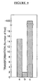
| Metastatic Characteristics of r-3 Mouse 10T1/2 Tumor Cells in Syngeneic Mice Following Treatment with the Antisense Oligonucleotides, AS-II-626-20 | ||
| Oligonucleotide Treatment | Frequency of Mice with Tumors | Number of Lung Tumors (mean ± SE) |
| none | 4/4 | 6.0 ± 1.58 |
| 0.2 | 1/4 | 0.25 ± 0.25 |
Lowe et al., 1994. Abrogation of oncogene-associated apoptosis allows transformation of p53-deficient cells. Proc. Natl. Acad. Sci. USA, 91: 2026-2030.
Claims (82)
- An antisense oligonucleotide comprising a sequence of at least 7 nucleotides complementary to a region of a human ribonucleotide reductase gene or mRNA, wherein said oligonucleotide comprises at least seven to about thirty-five nucleotides and inhibits neoplastic cell proliferation in a mammal, with the proviso that said region is other than:a) a region consisting of a UTR sequence segment; andb) a region consisting of a sequence segment selected fromand


- The antisense oligonucleotide according to claim 1, wherein said oligonucleotide consists of twelve to about thirty-five nucleotides.
- The antisense oligonucleotide according to claim 1, wherein said oligonucleotide consists of twenty to about thirty-five nucleotides.
- The antisense oligonucleotide according to any one of claims 1 to 3, wherein said region is a coding region.
- The antisense oligonucleotide according to any one of claims 1 to 4, wherein the human ribonucleotide reductase gene or mRNA encodes an R1 ribonucleotide reductase component.
- The antisense oligonucleotide according to any one of claims 1 to 4, wherein the human ribonucleotide reductase gene or mRNA encodes an R2 ribonucleotide reductase component.
- An antisense oligonucleotide comprising a sequence complementary to a region of a human ribonucleotide reductase gene or mRNA, wherein said oligonucleotide comprises at least seven to about thirty-five nucleotides and inhibits neoplastic cell proliferation in a mammal, and wherein said sequence is selected from SEQ ID NOs: 1, 3, 4, 5, 6, 7, 8, 9, 10, 11, 12, 13, 14, 15, 16, 17, 18, 19, 20, 21, 22, 23, 24, 25, 26, 27, 28, 29, 30, 31, 32, 33, 34, 35, 36, 37, 38, 39, 40, 41, 42, 43, 44, 45, 46, 47, 48, 49, 50, 51, 52, 53, 54, 55, 56, 57, 58, 59, 60, 61, 62, 63, 64, 65, 66, 67, 68, 69, 70, 71, 73, 74, 75, 77, 78, 79, 81, 82, 83, 84, 85, 86, 87, 88, 89, 90, 91, 92, 93, 94, 97, 98, 99, 100, 102, 103, 104, 105, 106, 107, 108, 109, 110, 111, 112, 113, 114, 115, 116, 117, 118, 119, 120, 121, 122, 123, 124, 125, 126, 127, 128, 129, 130, 131, 132, 133, 134, 135, 136, 137, 138, 139, 140, 141, 142, 143, 144, 145, 146, 147, 148, 149, 150, 151, 152, 153, 154, 155, 156, 158, 159, 160, and 161.
- The antisense oligonucleotide according to claim 7, wherein the oligonucleotide comprises a nucleic acid sequence selected from SEQ ID NOs: 1, 3, 4, 5, 6, 7, 8, 9, 10, 11, 12, 13, 14, 15, 16, 17, 18, 19, 20, 21, 22, 23, 24, 25, 26, 27, 28, 29, 30, 31, 32, 33, 34, 35, 36, 37, 38, 39, 40, 41, 42, 43, 44, 45, 46, 47, 48, 49, 50, 51, 52, 53, 54, 55, 56, 57, 58, 59, 60, 61, 62, 63, 64, 65, 66, 67, 68, 69, 70, 71, 73, 74, 75, 77, 78, 79, 81, 82, 83, 84, 85, 86, 87, 88, 89, 90, 91, 92, 93, 94, 97, 98, 99, 100, and 102.
- The antisense oligonucleotide according to any one of claims 1, 2, 3, 4, 7 or 8, wherein the oligonucleotide comprises a nucleic acid sequence selected from SEQ ID NOS: 1, 19, 20, 21, 22, 23, 24, 25, 26, 27, 28, 29, 30, 31, 32, 33, 34, 35, 36, 37, 38, 39, 40, 41, 42, 43, 44, 45, 46, 47, 48, 49, 50, 51, 52, 53, 54, 55, 56, 57, 58, 59, 60, 61, 62, 63, 64, 65, 66, 67, 68, 69, 70, and 71.
- The antisense oligonucleotide according to any one of claims 1, 3, 4, 7 or 8, wherein the oligonucleotide comprises a nucleic acid sequence selected from SEQ ID NOS: 1, 12, 16, 18, 21, 25, 29, 34, 42, 44, 45, 46, 52, 53, 59, 60, 64, 65, 66, 68, 69, 70, 73, 74, 78, 79, 90, 91, 92, 99, 100, and 102.
- The antisense oligonucleotide according to any one of claims 1, 3, 4, 7, 8, 9 or 10, wherein the oligonucleotide comprises the nucleic acid sequence set forth in SEQ ID NO: 42.
- The antisense oligonucleotide according to claim 7, wherein the oligonucleotide comprises a nucleic acid sequence selected from SEQ ID NOs: 103, 104, 105, 106, 107, 108, 109, 110, 111, 112, 113, 114, 115, 116, 117, 118, 119, 120, 121, 122, 123, 124, 125, 126, 127, 128, 129, 130, 131, 132, 133, 134, 135, 136, 137, 138, 139, 140, 141, 142, 143, 144, 145, 146, 147, 148, 149, 150, 151, 152, 153, 154, 155, 156, 158, 159, 160, and 161.
- The antisense oligonucleotide according to any one of claims 1, 2, 3, 4, 7 or 12 , wherein the oligonucleotide comprises a nucleic acid sequence selected from SEQ ID NOS: 103, 109, 110, 111, 112, 113, 114, 115, 116, 117, 118, 119, 120, 121, 122, 123, 124, 125, 126, 127, 128, 129, 130, 131, 132, 133, 134, 135, 136, 137, 138, 139, 140, 141, 142, 143, 144, 145, 146, 147, 148, 149, 150, 151, 152, 153, 154, and 155.
- The antisense oligonucleotide according to any one of claims 1, 2, 3, 4, 7, 12 or 13, wherein the oligonucleotide comprises a nucleic acid sequence as set forth in SEQ ID NO: 103.
- The antisense oligonucleotide according to any one of claims 1 to 14, wherein said oligonucleotide is a ribonucleic acid (RNA) oligonucleotide.
- The antisense oligonucleotide according to any one of claims 1 to 14, wherein said oligonucleotide is a deoxyribonucleic acid (DNA) oligonucleotide.
- The antisense oligonucleotide according to any one of claims 1 to 16, wherein said oligonucleotide is a peptide nucleic acid.
- The antisense oligonucleotide according to any one of claims 1 to 16, wherein said oligonucleotide comprises a morpholino backbone structure.
- The antisense oligonucleotide according to any one of claims 1 to 16, wherein said oligonucleotide comprises one or more modified bases selected from the group of: xanthine, hypoxanthine, 2-aminoadenine, 6-methyl, 2-propyl and other alkyl adenines, 5-halo uracil, 5-halo cytosine, 6-aza uracil, 6-aza cytosine, 6-aza thymine, pseudo uracil, 4-thiouracil, 8-halo adenine, 8-aminoadenine, 8-thiol adenine, 8-thiolalkyl adenines, 8-hydroxyl adenine, 8-halo guanines, 8-amino guanine, 8-thiol guanine, 8-thioalkyl guanines, 8-hydroxyl guanine, 5-trifluoromethyl uracil and 5-trifluoro cytosine.
- The antisense oligonucleotide according to any one of claims 1 to 16, wherein said oligonucleotide comprises one or more modified internucleotide linkages in the phosphate backbone selected from the group of: methyl phosphonate, phosphorothioate, phosphorodithioate and phosphotriester internucleotide linkages.
- The antisense oligonucleotide according to claim 20, wherein said phosphate backbone comprises one or more phosphorothioate internucleotide linkages.
- The antisense oligonucleotide according to claim 21, wherein the phosphorothioate internucleotide linkages link the four, five or six 3'-terminal nucleotides of said oligonucleotide.
- The antisense oligonucleotide according to claim 21, wherein the phosphorothioate internucleotide linkages link all the nucleotides of said oligonucleotide.
- The antisense oligonucleotide according to any one of claims 1 to 16, wherein said oligonucleotide comprises one or more alkyl, cycloalkyl or short chain heterocyclic intersugar linkages.
- The antisense oligonucleotide according to any one of claims 1 to 16, wherein said oligonucleotide is a chimeric oligonucleotide comprising two chemically distinct regions.
- The antisense oligonucleotide according to claim 25, wherein one of said chemically distinct regions comprises at least one modified nucleotide.
- The antisense oligonucleotide according to any one of claims 1 to 16 wherein the oligonucleotide exhibits reduced dimer formation and reduced self-complementary interactions.
- The antisense oligonucleotide according to any one of claims 1 to 27, wherein said oligonucleotide is nuclease resistant.
- The antisense oligonucleotide according to any one of claims 1 to 28, wherein said mammal is a human.
- A pharmaceutical composition comprising at least one antisense oligonucleotide according to any one of claims 1 to 29 in admixture with a physiologically acceptable carrier or diluent.
- Use of at least one antisense oligonucleotide according to any one of claims 1 to 29 to prepare a medicament.
- The use according to claim 31, wherein said medicament is for inhibiting neoplastic cell proliferation in a mammal.
- The use according to claim 31, wherein said medicament is for increasing the sensitivity of neoplastic cells to a chemotherapeutic drug in a mammal.
- The use according to claim 31, wherein said medicament is for inhibiting the growth of neoplastic cells that are resistant to a chemotherapeutic drug in a mammal.
- The use according to claim 33 or 34, wherein said chemotherapeutic drug is colchicine, vinblastine, doxorubicin, PALA, MTX or hydroxyurea.
- The use according to claim 33 or 34, wherein said chemotherapeutic drug is PALA, MTX, or hydroxyurea.
- The use according to claim 31, wherein said medicament is for reducing the metastasis of neoplastic cells in a mammal.
- The use according to any one of claims 32 to 37, wherein said mammal is a human.
- A DNA comprising a transcription initiation region and a sequence encoding at least one antisense oligonucleotide according to any one of claims 1 to 16.
- A vector comprising the DNA according to claim 39.
- A pharmaceutical composition comprising at least one vector according to claim 40 in admixture with a physiologically acceptable carrier or diluent.
- Use of at least one vector according to claim 40 to prepare a medicament.
- The use according to claim 42, wherein said medicament is for inhibiting neoplastic cell proliferation in a mammal.
- The use according to claim 42, wherein said medicament is for increasing the sensitivity of neoplastic cells to a chemotherapeutic drug in a mammal.
- The use according to claim 42, wherein said medicament is for inhibiting the growth of neoplastic cells that are resistant to a chemotherapeutic drug in a mammal.
- The use according to claim 44 or 45, wherein said chemotherapeutic drug is colchicine, vinblastine, doxorubicin, PALA, MTX or hydroxyurea.
- The use according to claim 44 or 45, wherein said chemotherapeutic drug is PALA, MTX or hydroxyurea.
- The use according to claim 42, wherein said medicament is for reducing the metastasis of neoplastic cells in a mammal.
- The use according to any one of claims 43 to 48, wherein said mammal is a human.
- An antisense oligonucleotide according to any one of claims 1 to 29 for use in the treatment of a mammal in need of such therapy.
- The antisense oligonucleotide according to claim 50, wherein said treatment is inhibition of neoplastic cell proliferation in a mammal.
- The antisense oligonucleotide according to claim 51, wherein said inhibition of neoplastic cell proliferation is by increased sensitivity of neoplastic cells to a chemotherapeutic drug.
- The antisense oligonucleotide according to claim 51, wherein said inhibition of neoplastic cell proliferation is by inhibition of the growth of neoplastic cells that are resistant to a chemotherapeutic drug.
- The antisense oligonucleotide according to claim 52 or 53, wherein the chemotherapeutic drug is colchicine, vinblastine, doxorubicin, PALA, MTX or hydroxyurea.
- The antisense oligonucleotide according to claim 52 or 53, wherein the chemotherapeutic drug is PALA, MTX or hydroxyurea.
- The antisense oligonucleotide according to claim 51, wherein said inhibition of neoplastic cell proliferation is by inhibition of the metastasis of neoplastic cells.
- The antisense oligonucleotide according to any one of claims 50 to 56, wherein said treatment is in combination with one or more chemotherapeutic drug.
- The antisense oligonucleotide according to claim 57, wherein said chemotherapeutic drug is hydroxyurea, MTX or PALA.
- The antisense oligonucleotide according to any one of claims 51 to 58, wherein said treatment comprises contacting the neoplastic cells with an effective amount of said oligonucleotide.
- The antisense oligonucleotide according to claim 57 or claim 58, wherein said treatment comprises contacting the neoplastic cells with said oligonucleotide at a non-cytotoxic dose and the chemotherapeutic drug at a non-cytotoxic dose.
- The antisense oligonucleotide according to claim 57 or claim 58, wherein said treatment comprises contacting the neoplastic cells with said oligonucleotide at a cytotoxic dose and the chemotherapeutic drug at a cytotoxic dose.
- The antisense oligonucleotide according to claim 57 or claim 58, wherein said treatment comprises contacting the neoplastic cells with said oligonucleotide at a non-cytotoxic dose and the chemotherapeutic drug at a cytotoxic dose.
- The antisense oligonucleotide according to claim 57 or claim 58, wherein said treatment comprises contacting the neoplastic cells with said oligonucleotide at a cytotoxic dose and the chemotherapeutic drug at a non-cytotoxic dose.
- The antisense oligonucleotide according to claim 57 or claim 58, wherein said treatment comprises contacting the neoplastic cells with said oligonucleotide at a dose sufficient to inhibit neoplastic cell growth.
- The antisense oligonucleotide according to any one of claims 57, 58, or 64, wherein said treatment comprises contacting the neoplastic cells with said chemotherapeutic drug at a dose sufficient to inhibit neoplastic cell growth.
- The antisense oligonucleotide according to any one of claims 51 to 65, wherein the neoplastic cells are selected from the group of: leukemia cells, lymphoma cells (Hodgkins and non-Hodgkins), sarcoma cells, melanoma cells, adenoma cells, carcinoma cells of solid tissue, hypoxic tumor cells, squamous cell carcinoma cells of the mouth, throat, larynx, and lung, genitourinary cancer cells, hematopoietic cancer cells, colon cancer cells, breast cancer cells, pancreatic cancer cells, head and neck cancer cells and nervous system cancer cells.
- The antisense oligonucleotide according to claim 66, wherein said genitourinary cancer cells are selected from the group of bladder cancer cells and cervical cancer cells.
- The antisense oligonucleotide according to any one of claims 51 to 65, wherein the neoplastic cells are selected from the group of: breast cancer cells, liver cancer cells, pancreatic cancer cells, bladder cancer cells, colon cancer cells, prostate cancer cells, cervical cancer cells, lung cancer cells, and leukemia cells.
- The antisense oligonucleotide according to claim 68, wherein said breast cancer cells are breast ductal cancer cells.
- The antisense oligonucleotide according to claim 68, wherein said lung cancer cells are metastatic lung cancer cells.
- The antisense oligonucleotide according to any one of claims 51 to 65, wherein said neoplastic cells are in a solid tumour in a mammal.
- The antisense oligonucleotide according to claim 51 to 65, wherein said tumour is derived from a cancer selected from the group of: breast cancer, lung cancer, colon cancer, and prostate cancer.
- The antisense oligonucleotide according to any one of claims 51 to 65, wherein the neoplastic cells are leukemia cells.
- The antisense oligonucleotide according to any one of claims 50 to 73, wherein said mammal is a human.
- A vector according to claim 40 for use in the treatment of a mammal in need of such therapy.
- The vector according to claim 75, wherein said treatment is inhibition of neoplastic cell proliferation in a mammal.
- The vector according to claim 76, wherein said inhibition of neoplastic cell proliferation is by increased sensitivity of neoplastic cells to a chemotherapeutic drug.
- The vector according to claim 76, wherein said inhibition of neoplastic cell proliferation is by inhibition of the growth of neoplastic cells that are resistant to a chemotherapeutic drug.
- The vector according to claim 77 or claim 78, wherein the chemotherapeutic drug is colchicine, vinblastine, doxorubicin, PALA, MTX or hydroxyurea.
- The vector according to claim 77 or 78, wherein the chemotherapeutic drug is PALA, MTX or hydroxyurea.
- The vector according to claim 76, wherein said inhibition of neoplastic cell proliferation is by the inhibition of the metastasis of neoplastic cells.
- The vector according to any one of claims 75 to 81, wherein said mammal is a human.
Applications Claiming Priority (5)
| Application Number | Priority Date | Filing Date | Title |
|---|---|---|---|
| US2304096P | 1996-08-02 | 1996-08-02 | |
| US23040P | 1996-08-02 | ||
| US3995997P | 1997-03-07 | 1997-03-07 | |
| PCT/CA1997/000540 WO1998005769A2 (en) | 1996-08-02 | 1997-08-01 | Antitumor antisense sequences directed against r1 and r2 components of ribonucleotide reductase |
| US39959P | 2008-03-27 |
Publications (2)
| Publication Number | Publication Date |
|---|---|
| EP0917569A2 EP0917569A2 (en) | 1999-05-26 |
| EP0917569B1 true EP0917569B1 (en) | 2005-11-09 |
Family
ID=26696655
Family Applications (1)
| Application Number | Title | Priority Date | Filing Date |
|---|---|---|---|
| EP97932690A Expired - Lifetime EP0917569B1 (en) | 1996-08-02 | 1997-08-01 | Antitumor antisense sequences directed against r1 and r2 components of ribonucleotide reductase |
Country Status (12)
| Country | Link |
|---|---|
| US (1) | US5998383A (en) |
| EP (1) | EP0917569B1 (en) |
| JP (1) | JP4301576B2 (en) |
| CN (1) | CN1189562C (en) |
| AT (1) | ATE309345T1 (en) |
| AU (1) | AU738592C (en) |
| CA (1) | CA2262776C (en) |
| DE (1) | DE69734589T2 (en) |
| ES (1) | ES2256893T3 (en) |
| IL (2) | IL128124A0 (en) |
| NZ (1) | NZ333802A (en) |
| WO (1) | WO1998005769A2 (en) |
Families Citing this family (47)
| Publication number | Priority date | Publication date | Assignee | Title |
|---|---|---|---|---|
| CA1223831A (en) | 1982-06-23 | 1987-07-07 | Dean Engelhardt | Modified nucleotides, methods of preparing and utilizing and compositions containing the same |
| US7223849B1 (en) * | 1996-07-01 | 2007-05-29 | Genesense Technologies Inc. | Oligonucleotides from the untranslated regions of housekeeping genes and methods of using same to modulate cell growth |
| US6593305B1 (en) | 1996-08-02 | 2003-07-15 | Genesense Technologies Inc. | Antitumor antisense sequences directed against R1 and R2 components of ribonucleotide reductase |
| JP2002501486A (en) | 1997-03-19 | 2002-01-15 | ジェネセンス・テクノロジーズ・インコーポレーテッド | Suppression of malignant tumors using ribonucleotide reductase R1 |
| US6835395B1 (en) | 1997-05-14 | 2004-12-28 | The University Of British Columbia | Composition containing small multilamellar oligodeoxynucleotide-containing lipid vesicles |
| EP1584681A3 (en) * | 1997-07-10 | 2005-11-09 | GeneSense Technologies Inc. | Antisense oligonucleotide sequences as inhibitors of microorganisms |
| US6610539B1 (en) * | 1997-07-10 | 2003-08-26 | Genesense Technologies, Inc. | Antisense oligonucleotide sequences as inhibitors of microorganisms |
| US6506559B1 (en) | 1997-12-23 | 2003-01-14 | Carnegie Institute Of Washington | Genetic inhibition by double-stranded RNA |
| CN1796556A (en) * | 1998-03-20 | 2006-07-05 | 联邦科学和工业研究组织 | Control of gene expression |
| AUPP249298A0 (en) | 1998-03-20 | 1998-04-23 | Ag-Gene Australia Limited | Synthetic genes and genetic constructs comprising same I |
| WO2000044914A1 (en) * | 1999-01-28 | 2000-08-03 | Medical College Of Georgia Research Institute, Inc. | Composition and method for in vivo and in vitro attenuation of gene expression using double stranded rna |
| US6121000A (en) * | 1999-02-11 | 2000-09-19 | Genesense Technologies, Inc. | Antitumor antisense sequences directed against R1 and R2 components of ribonucleotide reductase |
| US20040138168A1 (en) * | 1999-04-21 | 2004-07-15 | Wyeth | Methods and compositions for inhibiting the function of polynucleotide sequences |
| US6642033B1 (en) * | 1999-07-20 | 2003-11-04 | V.I. Technologies, Inc. | Nucleic acids for detecting parvovirus and methods of using same |
| US6423885B1 (en) | 1999-08-13 | 2002-07-23 | Commonwealth Scientific And Industrial Research Organization (Csiro) | Methods for obtaining modified phenotypes in plant cells |
| CN1301816A (en) * | 1999-12-27 | 2001-07-04 | 上海博德基因开发有限公司 | New polypeptide-nucleotide reductase 10 and polynucleotide codign such polypeptide |
| BR0109269A (en) * | 2000-03-17 | 2002-12-17 | Benitec Australia Ltd | Genetic construct and its use, vertebrate animal cell, methods of altering the vertebrate animal cell phenotype and gene therapy in genetically modified vertebrate, animal and murine animal |
| EP1229134A3 (en) * | 2001-01-31 | 2004-01-28 | Nucleonics, Inc | Use of post-transcriptional gene silencing for identifying nucleic acid sequences that modulate the function of a cell |
| DE10131148A1 (en) * | 2001-06-28 | 2003-01-16 | I P L Internat Pharmaceutics L | Xenogenic oligo- and / or polyribonucleotides as agents for the treatment of malignant tumors |
| EP1597365B1 (en) * | 2003-02-10 | 2013-10-09 | Lorus Therapeutics Inc. | Antisense oligonucleotides directed to ribonucleotide reductase r2 and uses thereof in the treatment of cancer |
| AU2003225410A1 (en) * | 2003-03-21 | 2004-10-11 | Academisch Ziekenhuis Leiden | Modulation of exon recognition in pre-mrna by interfering with the secondary rna structure |
| EP1636363A1 (en) * | 2003-05-21 | 2006-03-22 | GeneSense Technologies Inc. | Antisense oligonucleotides directed to ribonucleotide reductase r1 and uses thereof in the treatment of cancer |
| WO2004106518A1 (en) * | 2003-05-31 | 2004-12-09 | Genesense Technologies Inc. | Antisense oligonucleotides directed to ribonucleotide reductase r2 and uses thereof in the treatment of cancer |
| CN102174538B (en) | 2003-10-23 | 2013-06-19 | 奇尼塔二有限责任公司 | Detection of mutations in a gene associated with resistance to viral infection, oas1 |
| US20080311126A1 (en) * | 2004-01-12 | 2008-12-18 | Genesense Technologies, Inc | Antisense Oligonucleotides Directed to Ribonucleotide Reducatase R2 and Uses Thereof in Combination Therapies for the Treatment of Cancer |
| WO2005115405A1 (en) * | 2004-04-28 | 2005-12-08 | Molecules For Health, Inc. | Methods for treating or preventing restenosis and other vascular proliferative disorders |
| AU2005243410B2 (en) * | 2004-05-14 | 2010-04-22 | Rosetta Genomics Ltd. | Micronas and uses thereof |
| US7968762B2 (en) * | 2004-07-13 | 2011-06-28 | Van Andel Research Institute | Immune-compromised transgenic mice expressing human hepatocyte growth factor (hHGF) |
| WO2006017932A1 (en) * | 2004-08-18 | 2006-02-23 | Genesense Technologies Inc. | Small interfering rna molecules against ribonucleotide reductase and uses thereof |
| US20060185027A1 (en) * | 2004-12-23 | 2006-08-17 | David Bartel | Systems and methods for identifying miRNA targets and for altering miRNA and target expression |
| DK1866414T3 (en) * | 2005-03-31 | 2012-04-23 | Calando Pharmaceuticals Inc | Inhibitors of ribonucleotide reductase subunit 2 and uses thereof. |
| JO2660B1 (en) | 2006-01-20 | 2012-06-17 | نوفارتيس ايه جي | PI-3 Kinase inhibitors and methods of their use |
| AR060358A1 (en) | 2006-04-06 | 2008-06-11 | Novartis Vaccines & Diagnostic | QUINAZOLINS FOR THE INHIBITION OF PDK 1 |
| EP2030615A3 (en) * | 2007-08-13 | 2009-12-02 | ELFORD, Howard L. | Ribonucleotide reductase inhibitors for use in the treatment or prevention of neuroinflammatory or autoimmune diseases |
| US20100204305A1 (en) * | 2007-12-11 | 2010-08-12 | Lorus Therapeutics Inc. | Small interfering rna molecules against ribonucleotide reductase and uses thereof |
| UY31545A1 (en) | 2007-12-20 | 2009-08-03 | NEW DERIVATIVES OF 2-CARBOXAMIDE CIANOAMINOUREA, ITS SALTS AND PHARMACEUTICALLY ACCEPTABLE PROFARMS, PREPARATION PROCESSES AND APPLICATIONS | |
| WO2010027279A2 (en) * | 2008-09-04 | 2010-03-11 | Genesis Research And Development Corporation Limited | Compositions and methods for the treatment and prevention of neoplastic disorders |
| WO2010054221A2 (en) | 2008-11-06 | 2010-05-14 | The Johns Hopkins University | Treatment of chronic inflammatory respiratory disorders |
| US8293753B2 (en) | 2009-07-02 | 2012-10-23 | Novartis Ag | Substituted 2-carboxamide cycloamino ureas |
| AR082418A1 (en) | 2010-08-02 | 2012-12-05 | Novartis Ag | CRYSTAL FORMS OF 1- (4-METHYL-5- [2- (2,2,2-TRIFLUORO-1,1-DIMETHYL-Ethyl) -PIRIDIN-4-IL] -TIAZOL-2-IL) -AMIDE OF 2 -AMIDA OF THE ACID (S) -PIRROLIDIN-1,2-DICARBOXILICO |
| WO2012052258A1 (en) * | 2010-10-18 | 2012-04-26 | Arrowhead Research Corporation | Compositions and methods for inhibiting expression of rrm2 genes |
| AU2012213080B2 (en) | 2011-01-31 | 2014-03-27 | Novartis Ag | Novel heterocyclic derivatives |
| KR20140090218A (en) | 2011-10-28 | 2014-07-16 | 노파르티스 아게 | Novel purine derivatives and their use in the treatment of disease |
| NZ700924A (en) | 2012-05-16 | 2016-02-26 | Novartis Ag | Dosage regimen for a pi-3 kinase inhibitor |
| MX2016007376A (en) | 2013-12-06 | 2017-05-11 | Novartis Ag | Dosage regimen for an alpha-isoform selective phosphatidylinositol 3-kinase inhibitor. |
| AU2016347881A1 (en) | 2015-11-02 | 2018-05-10 | Novartis Ag | Dosage regimen for a phosphatidylinositol 3-kinase inhibitor |
| WO2018060833A1 (en) | 2016-09-27 | 2018-04-05 | Novartis Ag | Dosage regimen for alpha-isoform selective phosphatidylinositol 3-kinase inhibitor alpelisib |
Family Cites Families (2)
| Publication number | Priority date | Publication date | Assignee | Title |
|---|---|---|---|---|
| US5681747A (en) * | 1992-03-16 | 1997-10-28 | Isis Pharmaceuticals, Inc. | Nucleic acid sequences encoding protein kinase C and antisense inhibition of expression thereof |
| EP0690869A1 (en) * | 1993-03-23 | 1996-01-10 | The Board Of Trustees Of The Leland Stanford Junior University | Cellular regulation with riboregulators |
-
1997
- 1997-08-01 CA CA002262776A patent/CA2262776C/en not_active Expired - Fee Related
- 1997-08-01 JP JP50741098A patent/JP4301576B2/en not_active Expired - Fee Related
- 1997-08-01 DE DE69734589T patent/DE69734589T2/en not_active Expired - Lifetime
- 1997-08-01 EP EP97932690A patent/EP0917569B1/en not_active Expired - Lifetime
- 1997-08-01 AT AT97932690T patent/ATE309345T1/en not_active IP Right Cessation
- 1997-08-01 US US08/904,901 patent/US5998383A/en not_active Expired - Lifetime
- 1997-08-01 IL IL12812497A patent/IL128124A0/en active IP Right Grant
- 1997-08-01 ES ES97932690T patent/ES2256893T3/en not_active Expired - Lifetime
- 1997-08-01 WO PCT/CA1997/000540 patent/WO1998005769A2/en active IP Right Grant
- 1997-08-01 AU AU36175/97A patent/AU738592C/en not_active Ceased
- 1997-08-01 CN CNB971981639A patent/CN1189562C/en not_active Expired - Fee Related
- 1997-08-01 NZ NZ333802A patent/NZ333802A/en not_active IP Right Cessation
-
1999
- 1999-01-19 IL IL128124A patent/IL128124A/en not_active IP Right Cessation
Also Published As
| Publication number | Publication date |
|---|---|
| IL128124A0 (en) | 1999-11-30 |
| AU738592B2 (en) | 2001-09-20 |
| AU738592C (en) | 2002-07-25 |
| WO1998005769A3 (en) | 1998-06-25 |
| ES2256893T3 (en) | 2006-07-16 |
| CN1231694A (en) | 1999-10-13 |
| DE69734589T2 (en) | 2006-08-10 |
| CA2262776C (en) | 2005-03-08 |
| CA2262776A1 (en) | 1998-02-12 |
| EP0917569A2 (en) | 1999-05-26 |
| WO1998005769A2 (en) | 1998-02-12 |
| US5998383A (en) | 1999-12-07 |
| DE69734589D1 (en) | 2005-12-15 |
| CN1189562C (en) | 2005-02-16 |
| JP4301576B2 (en) | 2009-07-22 |
| AU3617597A (en) | 1998-02-25 |
| ATE309345T1 (en) | 2005-11-15 |
| NZ333802A (en) | 2001-09-28 |
| IL128124A (en) | 2006-12-31 |
| JP2000517167A (en) | 2000-12-26 |
Similar Documents
| Publication | Publication Date | Title |
|---|---|---|
| EP0917569B1 (en) | Antitumor antisense sequences directed against r1 and r2 components of ribonucleotide reductase | |
| US20070032442A1 (en) | Vector utilizing ribonuclease reductase UTR and use for inhibiting neoplastic cells | |
| EP1153128B1 (en) | Antitumor antisense sequences directed against r1 and r2 components of ribonucleotide reductase | |
| EP0748383B1 (en) | Targeted cleavage of rna using ribonuclease p targeting and cleavage sequences | |
| Halatsch et al. | Marked inhibition of glioblastoma target cell tumorigenicity in vitro by retrovirus-mediated transfer of a hairpin ribozyme against deletion-mutant epidermal growth factor messenger RNA | |
| US7405205B2 (en) | Antitumor antisense sequences directed against R1 and R2 components of ribonucleotide reductase | |
| EP1090146B1 (en) | Uses of dna-pk | |
| EP0971731B1 (en) | Suppression of malignancy utilizing ribonucleotide reductase r1 | |
| Kyo et al. | How to inhibit telomerase activity for cancer therapy | |
| AU9732101A (en) | Antitumor antisense sequences directed against R1 and R2 components of ribonucleotide reductase | |
| AU2004201285B2 (en) | Tissue-specific and target RNA-specific ribozymes |
Legal Events
| Date | Code | Title | Description |
|---|---|---|---|
| PUAI | Public reference made under article 153(3) epc to a published international application that has entered the european phase |
Free format text: ORIGINAL CODE: 0009012 |
|
| 17P | Request for examination filed |
Effective date: 19990205 |
|
| AK | Designated contracting states |
Kind code of ref document: A2 Designated state(s): AT BE CH DE DK ES FI FR GB GR IE IT LI LU MC NL PT SE |
|
| AX | Request for extension of the european patent |
Free format text: AL PAYMENT 19990204;LT PAYMENT 19990204;LV PAYMENT 19990204;RO PAYMENT 19990204;SI PAYMENT 19990204 |
|
| RIN1 | Information on inventor provided before grant (corrected) |
Inventor name: YOUNG, AIPING, H. Inventor name: WRIGHT, JIM, A. |
|
| RAP3 | Party data changed (applicant data changed or rights of an application transferred) |
Owner name: GENESENSE TECHNOLOGIES INC. |
|
| 17Q | First examination report despatched |
Effective date: 20010719 |
|
| GRAP | Despatch of communication of intention to grant a patent |
Free format text: ORIGINAL CODE: EPIDOSNIGR1 |
|
| GRAS | Grant fee paid |
Free format text: ORIGINAL CODE: EPIDOSNIGR3 |
|
| GRAA | (expected) grant |
Free format text: ORIGINAL CODE: 0009210 |
|
| AK | Designated contracting states |
Kind code of ref document: B1 Designated state(s): AT BE CH DE DK ES FI FR GB GR IE IT LI LU MC NL PT SE |
|
| AX | Request for extension of the european patent |
Extension state: AL LT LV RO SI |
|
| PG25 | Lapsed in a contracting state [announced via postgrant information from national office to epo] |
Ref country code: NL Free format text: LAPSE BECAUSE OF FAILURE TO SUBMIT A TRANSLATION OF THE DESCRIPTION OR TO PAY THE FEE WITHIN THE PRESCRIBED TIME-LIMIT Effective date: 20051109 Ref country code: LI Free format text: LAPSE BECAUSE OF FAILURE TO SUBMIT A TRANSLATION OF THE DESCRIPTION OR TO PAY THE FEE WITHIN THE PRESCRIBED TIME-LIMIT Effective date: 20051109 Ref country code: FI Free format text: LAPSE BECAUSE OF FAILURE TO SUBMIT A TRANSLATION OF THE DESCRIPTION OR TO PAY THE FEE WITHIN THE PRESCRIBED TIME-LIMIT Effective date: 20051109 Ref country code: CH Free format text: LAPSE BECAUSE OF FAILURE TO SUBMIT A TRANSLATION OF THE DESCRIPTION OR TO PAY THE FEE WITHIN THE PRESCRIBED TIME-LIMIT Effective date: 20051109 Ref country code: BE Free format text: LAPSE BECAUSE OF FAILURE TO SUBMIT A TRANSLATION OF THE DESCRIPTION OR TO PAY THE FEE WITHIN THE PRESCRIBED TIME-LIMIT Effective date: 20051109 Ref country code: AT Free format text: LAPSE BECAUSE OF FAILURE TO SUBMIT A TRANSLATION OF THE DESCRIPTION OR TO PAY THE FEE WITHIN THE PRESCRIBED TIME-LIMIT Effective date: 20051109 |
|
| REG | Reference to a national code |
Ref country code: GB Ref legal event code: FG4D |
|
| REG | Reference to a national code |
Ref country code: CH Ref legal event code: EP |
|
| REG | Reference to a national code |
Ref country code: IE Ref legal event code: FG4D |
|
| REF | Corresponds to: |
Ref document number: 69734589 Country of ref document: DE Date of ref document: 20051215 Kind code of ref document: P |
|
| RAP2 | Party data changed (patent owner data changed or rights of a patent transferred) |
Owner name: GENESENSE TECHNOLOGIES INC. |
|
| PG25 | Lapsed in a contracting state [announced via postgrant information from national office to epo] |
Ref country code: SE Free format text: LAPSE BECAUSE OF FAILURE TO SUBMIT A TRANSLATION OF THE DESCRIPTION OR TO PAY THE FEE WITHIN THE PRESCRIBED TIME-LIMIT Effective date: 20060209 Ref country code: GR Free format text: LAPSE BECAUSE OF FAILURE TO SUBMIT A TRANSLATION OF THE DESCRIPTION OR TO PAY THE FEE WITHIN THE PRESCRIBED TIME-LIMIT Effective date: 20060209 Ref country code: DK Free format text: LAPSE BECAUSE OF FAILURE TO SUBMIT A TRANSLATION OF THE DESCRIPTION OR TO PAY THE FEE WITHIN THE PRESCRIBED TIME-LIMIT Effective date: 20060209 |
|
| NLT2 | Nl: modifications (of names), taken from the european patent patent bulletin |
Owner name: GENESENSE TECHNOLOGIES INC. Effective date: 20051228 |
|
| PG25 | Lapsed in a contracting state [announced via postgrant information from national office to epo] |
Ref country code: PT Free format text: LAPSE BECAUSE OF FAILURE TO SUBMIT A TRANSLATION OF THE DESCRIPTION OR TO PAY THE FEE WITHIN THE PRESCRIBED TIME-LIMIT Effective date: 20060410 |
|
| LTIE | Lt: invalidation of european patent or patent extension |
Effective date: 20051109 |
|
| NLV1 | Nl: lapsed or annulled due to failure to fulfill the requirements of art. 29p and 29m of the patents act | ||
| REG | Reference to a national code |
Ref country code: CH Ref legal event code: PL |
|
| REG | Reference to a national code |
Ref country code: ES Ref legal event code: FG2A Ref document number: 2256893 Country of ref document: ES Kind code of ref document: T3 |
|
| PG25 | Lapsed in a contracting state [announced via postgrant information from national office to epo] |
Ref country code: IE Free format text: LAPSE BECAUSE OF NON-PAYMENT OF DUE FEES Effective date: 20060801 |
|
| ET | Fr: translation filed | ||
| PG25 | Lapsed in a contracting state [announced via postgrant information from national office to epo] |
Ref country code: MC Free format text: LAPSE BECAUSE OF NON-PAYMENT OF DUE FEES Effective date: 20060831 |
|
| PLBE | No opposition filed within time limit |
Free format text: ORIGINAL CODE: 0009261 |
|
| STAA | Information on the status of an ep patent application or granted ep patent |
Free format text: STATUS: NO OPPOSITION FILED WITHIN TIME LIMIT |
|
| 26N | No opposition filed |
Effective date: 20060810 |
|
| REG | Reference to a national code |
Ref country code: HK Ref legal event code: WD Ref document number: 1020353 Country of ref document: HK |
|
| REG | Reference to a national code |
Ref country code: IE Ref legal event code: MM4A |
|
| REG | Reference to a national code |
Ref country code: GB Ref legal event code: 732E |
|
| REG | Reference to a national code |
Ref country code: FR Ref legal event code: TP |
|
| PG25 | Lapsed in a contracting state [announced via postgrant information from national office to epo] |
Ref country code: LU Free format text: LAPSE BECAUSE OF NON-PAYMENT OF DUE FEES Effective date: 20060801 |
|
| PGFP | Annual fee paid to national office [announced via postgrant information from national office to epo] |
Ref country code: DE Payment date: 20140228 Year of fee payment: 17 |
|
| PGFP | Annual fee paid to national office [announced via postgrant information from national office to epo] |
Ref country code: FR Payment date: 20140211 Year of fee payment: 17 Ref country code: ES Payment date: 20140207 Year of fee payment: 17 Ref country code: IT Payment date: 20140224 Year of fee payment: 17 |
|
| PGFP | Annual fee paid to national office [announced via postgrant information from national office to epo] |
Ref country code: GB Payment date: 20140130 Year of fee payment: 17 |
|
| REG | Reference to a national code |
Ref country code: DE Ref legal event code: R119 Ref document number: 69734589 Country of ref document: DE |
|
| GBPC | Gb: european patent ceased through non-payment of renewal fee |
Effective date: 20140801 |
|
| PG25 | Lapsed in a contracting state [announced via postgrant information from national office to epo] |
Ref country code: IT Free format text: LAPSE BECAUSE OF NON-PAYMENT OF DUE FEES Effective date: 20140801 |
|
| REG | Reference to a national code |
Ref country code: DE Ref legal event code: R119 Ref document number: 69734589 Country of ref document: DE Effective date: 20150303 |
|
| REG | Reference to a national code |
Ref country code: FR Ref legal event code: ST Effective date: 20150430 |
|
| PG25 | Lapsed in a contracting state [announced via postgrant information from national office to epo] |
Ref country code: DE Free format text: LAPSE BECAUSE OF NON-PAYMENT OF DUE FEES Effective date: 20150303 Ref country code: GB Free format text: LAPSE BECAUSE OF NON-PAYMENT OF DUE FEES Effective date: 20140801 |
|
| PG25 | Lapsed in a contracting state [announced via postgrant information from national office to epo] |
Ref country code: FR Free format text: LAPSE BECAUSE OF NON-PAYMENT OF DUE FEES Effective date: 20140901 |
|
| REG | Reference to a national code |
Ref country code: ES Ref legal event code: FD2A Effective date: 20150925 |
|
| PG25 | Lapsed in a contracting state [announced via postgrant information from national office to epo] |
Ref country code: ES Free format text: LAPSE BECAUSE OF NON-PAYMENT OF DUE FEES Effective date: 20140802 |