EP0904016B1 - Radially expandable access system having disposable and reusable components - Google Patents
Radially expandable access system having disposable and reusable components Download PDFInfo
- Publication number
- EP0904016B1 EP0904016B1 EP97925698A EP97925698A EP0904016B1 EP 0904016 B1 EP0904016 B1 EP 0904016B1 EP 97925698 A EP97925698 A EP 97925698A EP 97925698 A EP97925698 A EP 97925698A EP 0904016 B1 EP0904016 B1 EP 0904016B1
- Authority
- EP
- European Patent Office
- Prior art keywords
- cannula
- needle
- hub
- obturator
- lumen
- Prior art date
- Legal status (The legal status is an assumption and is not a legal conclusion. Google has not performed a legal analysis and makes no representation as to the accuracy of the status listed.)
- Expired - Lifetime
Links
Images


Classifications
-
- A—HUMAN NECESSITIES
- A61—MEDICAL OR VETERINARY SCIENCE; HYGIENE
- A61B—DIAGNOSIS; SURGERY; IDENTIFICATION
- A61B17/00—Surgical instruments, devices or methods
- A61B17/34—Trocars; Puncturing needles
- A61B17/3417—Details of tips or shafts, e.g. grooves, expandable, bendable; Multiple coaxial sliding cannulas, e.g. for dilating
- A61B17/3421—Cannulas
- A61B17/3439—Cannulas with means for changing the inner diameter of the cannula, e.g. expandable
-
- A—HUMAN NECESSITIES
- A61—MEDICAL OR VETERINARY SCIENCE; HYGIENE
- A61B—DIAGNOSIS; SURGERY; IDENTIFICATION
- A61B50/00—Containers, covers, furniture or holders specially adapted for surgical or diagnostic appliances or instruments, e.g. sterile covers
- A61B50/30—Containers specially adapted for packaging, protecting, dispensing, collecting or disposing of surgical or diagnostic appliances or instruments
-
- A—HUMAN NECESSITIES
- A61—MEDICAL OR VETERINARY SCIENCE; HYGIENE
- A61B—DIAGNOSIS; SURGERY; IDENTIFICATION
- A61B17/00—Surgical instruments, devices or methods
- A61B17/34—Trocars; Puncturing needles
- A61B17/3498—Valves therefor, e.g. flapper valves, slide valves
Definitions
- the present invention relates generally to apparatus and methods for providing access to an internal operative site during a surgical procedure. More particularly, the present invention relates to an access system which can be percutaneously or otherwise introduced while in a narrow diameter configuration and which after introduction can be radially expanded to accommodate passage of larger diameter surgical instruments.
- Minimally invasive surgical procedures rely on obtaining percutaneous access to an internal surgical site using small-diameter access tubes (typically 5 to 12 mm), usually referred to as trocars, which penetrate through the skin and which open to the desired surgical site.
- a viewing scope is introduced through one such trocar, and the surgeon operates using instruments introduced through other appropriately placed trocars while viewing the operative site on a video monitor connected to the viewing scope.
- the surgeon is thus able to perform a wide variety of surgical procedures requiring only several 5 to 12 mm punctures at the surgical site. Patient trauma and recovery time are thus greatly reduced.
- laparoscopic procedures procedures in the abdominal area, which rely on a laparoscope for viewing, are referred to as laparoscopic procedures.
- the patient's abdominal region is typically insufflated (filled with pressured gas) to raise the abdominal wall and create sufficient operating space to perform a desired procedure.
- the trocars used in laparoscopic procedures must therefore include a valve at their proximal end to allow passage of the scope or surgical instruments while inhibiting leakage of the insufflating gas. It has also been proposed to perform laparoscopic procedures by mechanically expanding the abdomen rather than using insufflation.
- thoracoscopic procedures performed in the region of the chest
- arthroscopic procedures performed in body joints, particularly the knee
- gynecological laparoscopic procedures and endoscopic surgical procedures performed in various regions of the body, typically with a flexible scope.
- endoscopic surgical procedures do not always employ pressurization and the trocars used may not include pressure valves at their proximal ends.
- the STEPTM access system includes a pneumoperitoneum needle, an expandable sleeve component which is percutaneously introduced while positioned over the pneumoperitoneum needle, a cannula having a pneumostasis valve permanently affixed at its proximal end, and an obturator which is removably inserted into the cannula to form an expansion member for the sleeve.
- the sleeve which initially has a diameter in the range of 2-3 mm, is thus expanded to a final diameter depending on the cannula size, which can be selected from 5 mm, 10 mm, or 12 mm.
- Use of the STEPTM access system has many advantages, including reduced trauma to the patient and the ability to replace a cannula with a larger diameter cannula through a previously introduced sleeve.
- STEPTM access system represents a substantial advance over conventional trocars, the large number of components required to assemble all configurations of the system can present inventory problems for hospitals and other users. Moreover, it may be desirable to further increase the number of components in order to reduce the "disposable" content of the system, thus reducing costs associated with using the system.
- U.S. Patent Nos. 5,183,464 and 5,431,676, have been described above.
- WO 94/20026, published on September 15, 1994 corresponds to U.S. Patent No. 5,431,676.
- the STEPTM access system is described in a package insert published by InnerDyne Medical, Sunnyvale, California.
- US-A-5,496,280. discloses a trocar having a cannula and a housing.
- One of the cannula and housing is disposed of a sterilizable metal and the other of the cannula and housing is formed of a plastic material. This document has been used for the delineation in the two-part form of claim 10.
- the access system of the present invention is specified in claim 10.
- the pneumoperitoneum needle preferably comprises an assembly of a tubular needle and an internal stylet which is. removably received in the tubular needle.
- the valve cap will be disposable after each use. After an initial expansion, the sleeve material is generally unsuitable for reuse in another patient.
- both the valve cap and the radially expandable sleeve will be disposable after each use.
- Other components such as the tubular needle of the pneumoperitoneum needle will usually have a limited reuse, typically from 5 times to 30 times, while the remaining components will be reusable for a larger number of times, typically more than 100 times. Of course, any component which becomes damaged or excessively worn will be disposed of and replaced.
- Methods according to the present invention for reusing the access system will comprise replacing at least the radially expandable sleeve, and usually both the sleeve and the valve cap, and sterilizing or replacing all other components of the system.
- the cannula hub will be sterilized and the cannula tube replaced, while in other cases the entire cannula assembly will be sterilized and reused.
- the tubular needle will be replaced and the stylet sterilized, while in other cases both the tubular needle and stylet will be sterilized and reused.
- the method will further comprise reconstructing an expansion assembly by attaching a replacement valve cap onto the sterilized cannula tube and hub and inserting the sterilized obturator therethrough.
- the expansion assembly is reconstructed by attaching the replacement valve cap onto the sterilized cannula hub, attaching the replacement cannula tube to the cannula hub, and inserting the sterilized obturator through the cannula tube and hub.
- the method preferably further comprises reconstructing a dilation assembly by inserting a sterilized or partly sterilized pneumoperitoneum needle into a central lumen of the replaced radially expandable sleeve.
- a sterilized or partly sterilized pneumoperitoneum needle may have been sterilized prior to insertion into the radially expandable sleeve.
- at least one of the needle and stylet may have been replaced with the other component having been sterilized prior to inserting the needle into the radially expandable sleeve.
- the present invention further provides kits for use in reconstructing the access system of the present invention as specified in claim 13.
- the package may further include the stylet of the pneumoperitoneum needle in the same package.
- the package may be sterile or non-sterile, usually being sterile for the disposable components (the sleeve and usually the valve cap), and may comprise a pouch, box, tray, foam insert, envelope, or other. conventional packing material of a type employed for medical instruments.
- the access system of the present invention is useful for forming and enlarging percutaneous penetrations into a variety of target locations within a patient's body for a multiplicity of purposes.
- Such purposes include drainage, intra-organ drug administration, feeding, perfusion, aspiration, and the like, most usually being the introduction of viewing scopes and surgical instruments for use in minimally invasive surgical procedures, such as laparoscopic procedures, thoracoscopic procedures, arthroscopic procedures, endoscopic procedures, and the like.
- the access system of the present invention will find use in hysteroscopic, colonoscopic, and other procedures where access is established through existing body orifices.
- the access systems of the present invention are particularly valuable in percutaneous procedures since they will create a very small initial penetration, usually being below about 5 mm, more usually being below about 4 mm, frequently being below about 3.5 mm, and preferably being 3 mm or below.
- the penetration will be subsequently enlarged to a desired final size, usually having a final diameter in the range from about 5 mm to 15 mm, more usually being from about 5 mm to 12 mm, and typically being from about 5 mm to 10 mm.
- the enlarged penetration will define an access lumen from the outside of the patient's body to the desired internal location, and it is a particular advantage of the present invention that the diameter of the access lumen can be changed as will be described in more detail hereinafter.
- the access system is valuable since it can pass through the existing body orifice in its narrow-diameter configuration and be subsequently expanded with minimum discomfort and trauma to the patient.
- the access system of the present invention comprises a number of individual components that can be assembled into different size configurations.
- the assembled components can also be disassembled after use, and the components selectively sterilized or replaced prior to reassembling the access system for further use with a different patient.
- the different components and component assemblies and subassemblies will be described in greater detail below.
- Sterilization of the components of the trocar system can be accomplished by any suitable conventional sterilization technique, including heat, e.g., steam and autoclaving; chemical treatment, e.g., ethylene oxide exposure; radiation, and the like.
- heat e.g., steam and autoclaving
- chemical treatment e.g., ethylene oxide exposure
- radiation e.g., UV light
- reusable components will be washed to remove blood and other contaminating substances and then sterilized, preferably by exposure to steam.
- Disposable components will usually be radiation sterilized in their packages prior to distribution. Thus, disposable components will usually be ready to use out of the package.
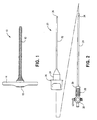
- a first component of the access system of the present invention comprises radially expandable sleeve 10.
- the sleeve 10 includes a sleeve body, typically comprising a radially expandable braid covered by an elastomeric layer, e.g., polyurethane.
- the braid initially has an inner diameter of about 2 mm and an outer diameter of about 3.5 mm.
- passage of an expansion assembly therethrough causes radial expansion of the sleeve, typically to a final diameter of 5 mm, 10 mm, or 12 mm.
- the sleeve 10 further comprises a handle at its proximal end.
- the handle includes a passage 16 therethrough, typically including a female bayonet connector coaxially located about the passage.
- the radially expandable sleeve 10 may be constructed in accordance with the details set forth in U.S. Patent No. 5,431,676, the full disclosure of which is incorporated herein by reference.
- a pneumoperitoneum needle assembly 20 comprising a tubular needle body 22 and a stylet 24 is illustrated in Fig. 2.
- the tubular needle body 22 includes a hub 24 having a male bayonet connector 26 at a proximal end thereof.
- Stylet 24 is spring-loaded in a proximal connector 28 which comprises a male bayonet fitting 30 which is receivably mounted in a female bayonet fitting (not illustrated) in the hub 24.
- An insufflation valve 32 is connected to the proximal end of the stylet 24 and a port 34 is disposed in the distal end of the stylet, permitting insufflation gas introduced through the valve 32 to be released through the port 34.
- the stylet 24 will be mounted within the tubular needle body 22 with the bayonet connector 30 attached to the hub 24.
- the distal end of the stylet will extend from distal end 36 of the needle, and the stylet will retract into the needle body 22 when the needle is engaged against tissue, as described in more detail below.
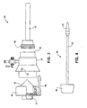
- Cannula assembly 40 constructed in accordance with the principles of the present invention is illustrated in Fig. 3.
- Cannula assembly 40 comprises a cannula tube 42, a proximal hub 44, and a removable valve cap 46.
- the cannula tube 42 includes a threaded connector 48 at its proximal end which can be removably secured to a fitting 50 in the distal end of the cannula hub 44.
- the valve cap 46 includes a pneumostasis valve element 52 and mates with a male bayonet fitting 54 at the proximal end of the cannula hub 44.
- a second disk valve element 56 is mounted in tandem with the pneumostasis valve element 52 to engage against a surgical instrument (not shown) when introduced through the cannula assembly 40.
- the valve element 56 is generally sized for a large instrument, e.g., 12 mm in diameter.
- a reducing element 58 is provided for reducing the port size of valve element 56 to accommodate smaller instruments, e
- An obturator 60 comprising a shaft 62, tapered distal end 64, and handle 66 is illustrated in Fig. 4.
- the obturator 60 is intended to be placed within a central lumen of the cannula assembly 40 in order to form an expansion assembly for use as described below.
- the access system components just described will generally differ in both size and reusability, with exemplary sizes and extent of reusability for each of the components set forth below in Table 1.
- the radially expandable sleeve, pneumoperitoneum needle (including both the tubular needle body and the needle stylet), and the cannula hub will preferably come in just one size which is compatible with each of the three exemplary system sizes. It will be appreciated, of course, that the radially expandable sleeve is intended to conform to cannula tubes having each of the three sizes.
- the cannula hub will be sized sufficiently large to be compatible with each of the three sizes, and the valve cap will come in two sizes, with the larger size accommodating both 10 and 12 mm instruments, as described above.
- the cannula tube and obturator will be provided in each of the three system sizes.
- kits containing various combinations of system components are illustrated. Providing such kits is a particularly convenient way to facilitate inventory maintenance of the components necessary to reconstruct the access systems of the present invention. It will be appreciated, of course, that complete systems could be sold in kits, as well as each of the individual components can be sold in kits. In many cases, it will be desirable to combine pairs of components or multiple pieces of a single component together in one package, particularly where the components are sized to match each other.
- the kits will include conventional package elements, typically pouches, envelopes, trays, boxes, foam inserts and other containers of a type commonly used for sterile or non-sterile packing of surgical instruments.
- the packages will typically also include a "package insert" which is a written instruction sheet with instructions on use, warnings, etc.
- the kit 70 comprises a package, typically non-sterile since the reusable components can be subsequently sterilized, including a 5 mm cannula assembly 72 and a 5 mm obturator 74.
- the cannula assembly 72 includes both the cannula hub and the removable cannula tube, as described previously.
- the package 102 of the kit may be sterile or non-sterile.
- Kits comprising individual components may also be provided.
- a kit 110 including a 5 mm cannula body 112 in a sterile pack 114 is illustrated in Fig. 9.
- Similar kits 116 and 118 comprising a 10 mm cannula body 120 and a 12 mm cannula body 122 in sterile packs 124 and 126, respectively, are illustrated in Figs. 10 and 11.
- a kit 130 comprising a tubular needle body 22, a sterile package 132 is illustrated in Fig. 12.
- Kits comprising disposable components are illustrated in Figs. 13-17.
- a kit 140 comprising a radially expandable sleeve 10 and a 5 mm valve cap 142 in a sterile package 144 is illustrated in Fig. 13.
- a similar kit 150 comprising a radially expandable sleeve 10 and a 10/12 mm valve cap 152 present in a sterile pack 154 is illustrated in Fig. 14.
- a kit 160 comprising one or more 5 mm valve caps 142 in a sterile package 162 is illustrated in Fig. 15.
- a kit 170 comprising one or more sterile 10/12 mm valve caps 152 in a sterile package 172 is illustrated in Fig. 16.
- a kit 180 comprising one or more radially expandable sleeves 10 in a sterile package 182 is illustrated in Fig. 17.
- a radially expandable sleeve 10 having a pneumoperitoneum needle 20 inserted therein is introduced through patient's abdomen A (or other body location) by engaging the sharpened distal end 36 of the needle against the tissue and advancing the assembly until the body 12 of the sleeve extends across the tissue.
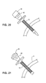
- the pneumoperitoneum needle 20 is then removed, and an expansion assembly comprising the cannula 40 and obturator 60 is introduced through the sleeve 10, resulting in radial expansion of the sleeve body 12, as illustrated in Fig. 20.
- the obturator 60 is then removed, leaving an access channel through the abdominal wall A, as illustrated in Fig. 21.
- valve cap 46 and radially expandable sleeve 10 will usually be discarded. Because of the expanded or spent configuration of the sleeve, the sleeve cannot be reused. The valve cap will usually be discarded since cleaning of the internal components is difficult and the cost of a disposable valve cap is relatively low. The remaining components of the system, however, can be reused after appropriate cleaning and sterilization. As set forth in Table 1 above, the tubular needle body and cannula tube are available for a limited reuse, but will typically wear sufficiently after from 5-30 additional uses, that they should be discarded. The needle stylet, cannula hub, and obturator, however, may be reused a relatively unlimited number of times, usually more than 100, before needing replacement. The components, however, are of course subject to wear and tear and breakage, at which times these system components will also have to be replaced.
Landscapes
- Health & Medical Sciences (AREA)
- Life Sciences & Earth Sciences (AREA)
- Surgery (AREA)
- Heart & Thoracic Surgery (AREA)
- Engineering & Computer Science (AREA)
- Biomedical Technology (AREA)
- Nuclear Medicine, Radiotherapy & Molecular Imaging (AREA)
- Medical Informatics (AREA)
- Molecular Biology (AREA)
- Animal Behavior & Ethology (AREA)
- General Health & Medical Sciences (AREA)
- Public Health (AREA)
- Veterinary Medicine (AREA)
- Pathology (AREA)
- Surgical Instruments (AREA)
Description
| ACCESS SYSTEM COMPONENTS | ||
| Component | Size(s) | Reusability |
| Radially Expandable Sleeve | One | Disposable |
| Tubular Needle Body | One | Limited reuse |
| Needle Stylet | One | Unlimited reuse |
| Cannula Tube | 5 mm, 10 mm, 12 mm | Limited reuse |
| Cannula Hub | One | Unlimited reuse |
| Valve cap | 5 mm, 10/12 mm | Disposable or limited reuse |
| Obturator | 5 mm, 10 mm, 12 mm | Unlimited reuse |
Claims (19)
- A method for reusing an access system, wherein the access system comprises the following components each of which have been previously used to access a target location in a patient:wherein the method includes the steps of:a radially expandable sleeve (10) having an axial sleeve lumen therethrough;a cannula assembly (40) removably insertable into said sleeve lumen and including a cannula tube (42) having a connector (48) at its proximal end, a distal end, and a lumen therethrough, and a cannula hub (44) being removably attached to the connector (48) at the proximal end of the cannula tube (42) and having a removable valve cap (46) proximal of the hub and having a lumen spanned by a pneumostasis valve (52) mounted to the cap; andand obturator (60), and the lumen of the valve cap, said obturator (6) having a tapered distal end (64) which extends distally from the distal end of the cannula tube (42) when the obturator (6) is therein;replacing at least the radially expandable sleeve (10) and the valve cap (46); andsterilizing at least one sterilizable system component other that the expandable sleeve (10) and the valve cap (46) andsterilizing or replacing all components of the system other than the expandable sleeve (10), the valve cap (46) and the at least one sterilizable system component.
- A method as in claim 1, wherein the cannula tube (42) is removably attached to the cannula hub (44), the method further comprising the steps of separating the tube (42) and hub (44), cleaning and sterilizing the cannula hub (44), and replacing the cannula tube (42).
- A method as in claim 1 or claim 2, wherein the access system further comprises a needle component (20) including a tubular needle (22) and a stylet (24) removably received in the needle (22), the method further comprising the step of cleaning and sterilizing the stylet (24) and replacing the needle (22).
- A method as in claim 1, wherein the cannula tube (42) and hub (44) and the obturator (60) are cleaned.and sterilized, the method further comprising the steps of reconstructing an expansion assembly by attaching the replacement valve cap (46) on the cleaned and sterilized cannula tube (42) and hub (44), and inserting the cleaned and sterilized obturator (60) through the cannula tube (42) and hub (44).
- A method as in claim 1, wherein the cannula tube (42) is replaced and the cannula hub (42) and obturator (44) are cleaned and sterilized, the method further comprising the steps of reconstructing an expansion assembly by attaching the replacement valve cap (46) on the sterilized cannula hub (44), attaching the replacement cannula tube (42) to the cannula hub (44), and inserting the sterilized obturator (6) through the cannula tube (42) and hub (44).
- A method as in claim 1, wherein the access system further comprises a needle component (20) including a tubular needle (22) and a stylet (24) removably received in the needle (22), the method further comprising the steps of reconstructing an expansion assembly by inserting the needle component (22) in a central lumen of the replaced radially expandable sleeve (10).
- A method as in claim 6, further comprising cleaning and sterilizing both the needle (22) and the stylet (24), and inserting the stylet (24) through the needle (22) before inserting the needle component (20) in the radially expandable sleeve (10).
- A method as in claim 6, further comprising replacing at least the needle (22), and inserting the stylet (24) through the needle (22) before inserting the needle components (20) in the radially expandable sleeve (10).
- A method as in claim 6, further comprising replacing both the needle (22) and the stylet (24), and inserting the stylet (24) through the needle (22) before inserting the needle component (20) in the radially expandable sleeve (10).
- An access system comprising:a radially expandable sleeve (10) having an axial sleeve lumen therethrough;a cannula assembly (40) removably insertable into said sleeve lumen and including a cannula tube (42) having a connector (48) at its proximal end, a distal end, and a lumen therethrough, and a cannula hub (44) removably attached to the connector (48) at the proximal end of the cannula tube (42) ;a valve cap (46) proximal of the hub and having a lumen spanned by a pnemostasis valve (52) mounted therein, said valve cap (46) being removably attached to the cannula hub (44); andan obturator (60) removably receivable in the lumen of the cannula tube (42), and the lumen of the valve cap, said obturator (60) having a tapered distal end (64) which extends distally from the distal end of the cannula tube (42) when the obturator (60) is therein.
- An access system as in claim 10 and further comprising a pneumoperitoneum needle assembly (20).
- An access system as in claim 11, wherein the pneumoperitoneum needle assembly (20) comprises a tubular needle (22) and an internal stylet (24) removably received in the tubular needle (22).
- A kit with which an access system can be assembled, wherein the kit comprises the following components:a radially expandable sleeve (10) having an axial lumen therethrough;a valve cap having a lumen spanned by a pneumostasis valve (52) mounted thereto;a cannula assembly (40) including a cannula tube (42) having a connector (48) at its proximal end, a distal end, and a lumen therethrough, and a cannula hub (44) removably attachable to, and removable from, the connector (48) at the proximal end of the cannula tube (42);an obturator (60) removably receivable in the lumen of the cannula tube (42), and the lumen of the calve cap, said obturator (60) having a tapered distal end (64) which extends distally from the distal end of the cannula tube (42) when the obturator (60) is therein; anda package enclosing the valve cap (46), wherein the valve cap is attachable to, and removable from, the proximal end of the cannula hub.
- A kit as claimed in claim 13 wherein the kit further comprises:wherein the package encloses the radially expandable sleeve (10) and the valve cap (46).a radially expandable sleeve (10); and
- A kit as in claim 13 or claim 14, wherein the kit further comprises:wherein the package also encloses the cannula tube (42) and the cannula hub (44).a cannula tube (42) and a cannula hub (44); and
- A kit as in claim 15, further comprising an obturator (60) disposed in the package.
- A kit as in claim 13, wherein the kit further comprises:wherein the package also encloses the cannula body (112).a cannula body (112); and
- A kit as in claim 17, further comprising a stylet (24) disposed in the package.
- A kit as in claim 13, wherein the kit further comprises:wherein the package encloses the tubular needle (22).a tubular needle (22); and
Applications Claiming Priority (3)
| Application Number | Priority Date | Filing Date | Title |
|---|---|---|---|
| US08/650,387 US5827319A (en) | 1996-05-20 | 1996-05-20 | Radially expandable access system having disposable and reusable components |
| US650387 | 1996-05-20 | ||
| PCT/US1997/008709 WO1997043958A1 (en) | 1996-05-20 | 1997-05-19 | Radially expandable access system having disposable and reusable components |
Publications (3)
| Publication Number | Publication Date |
|---|---|
| EP0904016A1 EP0904016A1 (en) | 1999-03-31 |
| EP0904016A4 EP0904016A4 (en) | 1999-08-04 |
| EP0904016B1 true EP0904016B1 (en) | 2004-11-10 |
Family
ID=24608685
Family Applications (1)
| Application Number | Title | Priority Date | Filing Date |
|---|---|---|---|
| EP97925698A Expired - Lifetime EP0904016B1 (en) | 1996-05-20 | 1997-05-19 | Radially expandable access system having disposable and reusable components |
Country Status (5)
| Country | Link |
|---|---|
| US (1) | US5827319A (en) |
| EP (1) | EP0904016B1 (en) |
| JP (1) | JP2000511792A (en) |
| DE (1) | DE69731531T2 (en) |
| WO (1) | WO1997043958A1 (en) |
Cited By (1)
| Publication number | Priority date | Publication date | Assignee | Title |
|---|---|---|---|---|
| DE102007026235A1 (en) * | 2007-05-31 | 2008-12-04 | Karl Storz Gmbh & Co. Kg | Device for the sterile enclosure of a sterilization-sensitive control panel |
Families Citing this family (123)
| Publication number | Priority date | Publication date | Assignee | Title |
|---|---|---|---|---|
| US6175758B1 (en) | 1997-07-15 | 2001-01-16 | Parviz Kambin | Method for percutaneous arthroscopic disc removal, bone biopsy and fixation of the vertebrae |
| DE69837611T2 (en) | 1997-10-01 | 2007-12-27 | Boston Scientific Ltd., St. Michael | dilation |
| US6245052B1 (en) | 1998-07-08 | 2001-06-12 | Innerdyne, Inc. | Methods, systems, and kits for implanting articles |
| US7799036B2 (en) * | 1998-08-20 | 2010-09-21 | Zimmer Spine, Inc. | Method and apparatus for securing vertebrae |
| US6187000B1 (en) | 1998-08-20 | 2001-02-13 | Endius Incorporated | Cannula for receiving surgical instruments |
| US7641670B2 (en) | 1998-08-20 | 2010-01-05 | Zimmer Spine, Inc. | Cannula for receiving surgical instruments |
| US8527094B2 (en) | 1998-11-20 | 2013-09-03 | Intuitive Surgical Operations, Inc. | Multi-user medical robotic system for collaboration or training in minimally invasive surgical procedures |
| US6951535B2 (en) | 2002-01-16 | 2005-10-04 | Intuitive Surgical, Inc. | Tele-medicine system that transmits an entire state of a subsystem |
| US6398726B1 (en) | 1998-11-20 | 2002-06-04 | Intuitive Surgical, Inc. | Stabilizer for robotic beating-heart surgery |
| US6659939B2 (en) | 1998-11-20 | 2003-12-09 | Intuitive Surgical, Inc. | Cooperative minimally invasive telesurgical system |
| US6852107B2 (en) | 2002-01-16 | 2005-02-08 | Computer Motion, Inc. | Minimally invasive surgical training using robotics and tele-collaboration |
| JP4225697B2 (en) * | 1999-05-19 | 2009-02-18 | インナーダイン, インコーポレイテッド | System for establishing access to vessels |
| US7217240B2 (en) | 1999-10-01 | 2007-05-15 | Intuitive Surgical, Inc. | Heart stabilizer |
| US6471659B2 (en) | 1999-12-27 | 2002-10-29 | Neothermia Corporation | Minimally invasive intact recovery of tissue |
| US6277083B1 (en) | 1999-12-27 | 2001-08-21 | Neothermia Corporation | Minimally invasive intact recovery of tissue |
| US6626859B2 (en) * | 2000-01-18 | 2003-09-30 | Coraflo Ltd. | High performance cannulas |
| US6648906B2 (en) | 2000-04-06 | 2003-11-18 | Innercool Therapies, Inc. | Method and apparatus for regulating patient temperature by irrigating the bladder with a fluid |
| US7985247B2 (en) | 2000-08-01 | 2011-07-26 | Zimmer Spine, Inc. | Methods and apparatuses for treating the spine through an access device |
| US7056321B2 (en) | 2000-08-01 | 2006-06-06 | Endius, Incorporated | Method of securing vertebrae |
| US6726699B1 (en) * | 2000-08-15 | 2004-04-27 | Computer Motion, Inc. | Instrument guide |
| EP2441394B1 (en) | 2000-11-28 | 2017-04-05 | Intuitive Surgical Operations, Inc. | Irrigator for an endoscopic instrument |
| US6958069B2 (en) * | 2001-01-17 | 2005-10-25 | Mark LoGuidice | Instruments and methods for use in laparoscopic surgery |
| US20060025781A1 (en) * | 2001-01-17 | 2006-02-02 | Young Wayne P | Laparoscopic instruments and methods utilizing suction |
| US7041055B2 (en) * | 2002-10-07 | 2006-05-09 | Mark LoGuidice | Instruments and methods for use in laparoscopic surgery |
| US20020165524A1 (en) | 2001-05-01 | 2002-11-07 | Dan Sanchez | Pivot point arm for a robotic system used to perform a surgical procedure |
| JP2004526539A (en) * | 2001-05-11 | 2004-09-02 | アプライド メディカル リソーシーズ コーポレイション | Traction trocar device and method |
| US7144393B2 (en) * | 2001-05-15 | 2006-12-05 | Dipoto Gene P | Structure for receiving surgical instruments |
| AU2002317605B2 (en) * | 2001-08-01 | 2008-09-25 | Covidien Lp | Radially dilatable percutaneous access apparatus with introducer seal in handle |
| US6728599B2 (en) | 2001-09-07 | 2004-04-27 | Computer Motion, Inc. | Modularity system for computer assisted surgery |
| US6839612B2 (en) | 2001-12-07 | 2005-01-04 | Institute Surgical, Inc. | Microwrist system for surgical procedures |
| US6793653B2 (en) | 2001-12-08 | 2004-09-21 | Computer Motion, Inc. | Multifunctional handle for a medical robotic system |
| WO2003071926A2 (en) * | 2002-02-21 | 2003-09-04 | Persidsky Maxim D | Apparatus and method for making a percutaneous access for port of variable size |
| US7377897B1 (en) | 2002-05-02 | 2008-05-27 | Kunkel Sanford S | Portal device |
| US7004947B2 (en) * | 2002-06-24 | 2006-02-28 | Endius Incorporated | Surgical instrument for moving vertebrae |
| US6648888B1 (en) | 2002-09-06 | 2003-11-18 | Endius Incorporated | Surgical instrument for moving a vertebra |
| FI20021817A7 (en) * | 2002-10-14 | 2004-04-15 | Mirhava Ltd | Cannula |
| WO2004039235A2 (en) | 2002-10-25 | 2004-05-13 | Endius Incorporated | Apparatus and methods for shielding body structures during surgery |
| AU2003295878B2 (en) * | 2002-11-22 | 2008-07-10 | Covidien Lp | Sheath introduction apparatus and method |
| DK1466563T3 (en) * | 2003-03-28 | 2006-03-13 | Wolf Gmbh Richard | Dilation system |
| US20050059934A1 (en) * | 2003-04-25 | 2005-03-17 | Thomas Wenchell | Surgical access apparatus |
| US20050143690A1 (en) * | 2003-05-01 | 2005-06-30 | High Kenneth A. | Cystotomy catheter capture device and methods of using same |
| US7645232B2 (en) | 2003-05-16 | 2010-01-12 | Zimmer Spine, Inc. | Access device for minimally invasive surgery |
| PL1651121T3 (en) * | 2003-07-07 | 2008-02-29 | Coraflo Ltd Liab Co | High performance cannulas |
| ATE416678T1 (en) | 2003-08-26 | 2008-12-15 | Zimmer Spine Inc | ACCESS SYSTEMS FOR MINIMALLY INVASIVE SURGERY |
| US7226451B2 (en) | 2003-08-26 | 2007-06-05 | Shluzas Alan E | Minimally invasive access device and method |
| US7479150B2 (en) | 2003-09-19 | 2009-01-20 | Tyco Healthcare Group Lp | Trocar insertion apparatus |
| US20050070947A1 (en) * | 2003-09-30 | 2005-03-31 | Franer Paul T. | Rotational latching system for a trocar |
| US20050090899A1 (en) * | 2003-10-24 | 2005-04-28 | Dipoto Gene | Methods and apparatuses for treating the spine through an access device |
| US7655012B2 (en) | 2003-10-02 | 2010-02-02 | Zimmer Spine, Inc. | Methods and apparatuses for minimally invasive replacement of intervertebral discs |
| US7731737B2 (en) * | 2003-10-24 | 2010-06-08 | Zimmer Spine, Inc. | Methods and apparatuses for fixation of the spine through an access device |
| US20050090822A1 (en) * | 2003-10-24 | 2005-04-28 | Dipoto Gene | Methods and apparatus for stabilizing the spine through an access device |
| AU2004220757B2 (en) * | 2003-10-17 | 2010-06-17 | Covidien Lp | Surgical access device and manufacture thereof |
| US8673021B2 (en) * | 2003-11-26 | 2014-03-18 | Depuy Mitek, Llc | Arthroscopic tissue scaffold delivery device |
| US7195592B2 (en) * | 2004-01-27 | 2007-03-27 | Sundaram Ravikumar | Surgical retractor apparatus for use with a surgical port |
| US7344495B2 (en) * | 2004-01-27 | 2008-03-18 | Arvik Enterprises, Llc | Surgical retractor apparatus for use with a surgical port |
| US20050251091A1 (en) * | 2004-05-10 | 2005-11-10 | Usgi Medical Inc. | Apparatus and methods for transgastric tissue manipulation |
| US8475476B2 (en) * | 2004-06-01 | 2013-07-02 | Cook Medical Technologies Llc | System and method for accessing a body cavity |
| US20060052812A1 (en) * | 2004-09-07 | 2006-03-09 | Michael Winer | Tool for preparing a surgical site for an access device |
| US7329233B2 (en) * | 2004-10-05 | 2008-02-12 | Tyco Healthcare Group Lp | Surgical system for laparoscopic surgery |
| ATE393602T1 (en) * | 2004-11-25 | 2008-05-15 | Luigi Carro | GRIPPING LIMB FOR BIOMEZIDINIC NEEDLE |
| US20060212062A1 (en) * | 2005-03-16 | 2006-09-21 | David Farascioni | Radially expandable access system including trocar seal |
| US7837612B2 (en) | 2005-04-08 | 2010-11-23 | Ethicon Endo-Surgery, Inc. | Tissue suspension device |
| EP1898771A2 (en) * | 2005-06-24 | 2008-03-19 | Power Ten, LLC | Expandable surgical site access system |
| WO2007038429A1 (en) | 2005-09-27 | 2007-04-05 | Endius, Inc. | Methods and apparatuses for stabilizing the spine through an access device |
| US20070233089A1 (en) * | 2006-02-17 | 2007-10-04 | Endius, Inc. | Systems and methods for reducing adjacent level disc disease |
| US8430811B2 (en) | 2008-09-30 | 2013-04-30 | Ethicon Endo-Surgery, Inc. | Multiple port surgical access device |
| US8206294B2 (en) | 2008-09-30 | 2012-06-26 | Ethicon Endo-Surgery, Inc. | Surgical access device with flexible seal channel |
| US8485970B2 (en) | 2008-09-30 | 2013-07-16 | Ethicon Endo-Surgery, Inc. | Surgical access device |
| US8425410B2 (en) | 2008-09-30 | 2013-04-23 | Ethicon Endo-Surgery, Inc. | Surgical access device with protective element |
| US8961406B2 (en) | 2009-03-06 | 2015-02-24 | Ethicon Endo-Surgery, Inc. | Surgical access devices and methods providing seal movement in predefined movement regions |
| US8926506B2 (en) | 2009-03-06 | 2015-01-06 | Ethicon Endo-Surgery, Inc. | Methods and devices for providing access into a body cavity |
| US8251900B2 (en) | 2009-03-06 | 2012-08-28 | Ethicon Endo-Surgery, Inc. | Surgical access devices and methods providing seal movement in predefined paths |
| US8821391B2 (en) | 2009-03-06 | 2014-09-02 | Ethicon Endo-Surgery, Inc. | Methods and devices for providing access into a body cavity |
| US8357085B2 (en) * | 2009-03-31 | 2013-01-22 | Ethicon Endo-Surgery, Inc. | Devices and methods for providing access into a body cavity |
| WO2008012787A2 (en) * | 2006-07-27 | 2008-01-31 | Atropos Limited | An apparatus for inserting a surgical device at least partially through a wound opening |
| US7666074B2 (en) | 2006-08-31 | 2010-02-23 | Mpsc, Inc. | Meat processing |
| US8795235B2 (en) | 2006-10-06 | 2014-08-05 | Surgiquest, Inc. | Devices for and methods of performing minimally-invasive surgical procedures through a single incision |
| US20080171984A1 (en) * | 2007-01-11 | 2008-07-17 | Miller Peter C | Cannula driver and system |
| US20080177257A1 (en) * | 2007-01-23 | 2008-07-24 | Smith Ronald T | Thermally robust illumination probe tip |
| US8372131B2 (en) | 2007-07-16 | 2013-02-12 | Power Ten , LLC | Surgical site access system and deployment device for same |
| US8118826B2 (en) * | 2007-09-27 | 2012-02-21 | Swan Valley Medical, Incorporated | Method of performing a suprapubic transurethral cystostomy and associated procedures and apparatus therefor |
| US8382785B2 (en) * | 2007-09-27 | 2013-02-26 | Swan Valley Medical Incorporated | Apparatus and method for performing cystotomy procedures |
| US8795326B2 (en) | 2007-10-05 | 2014-08-05 | Covidien Lp | Expanding seal anchor for single incision surgery |
| US8142467B2 (en) | 2008-04-21 | 2012-03-27 | Applied Medical Resources Corporation | Tamponade trocar device and method |
| US20090292172A1 (en) * | 2008-05-21 | 2009-11-26 | Boston Scientific Scimed, Inc. | Expandable Delivery Devices and Methods of Use |
| US20090312645A1 (en) * | 2008-06-16 | 2009-12-17 | Boston Scientific Scimed, Inc. | Methods and Devices for Accessing Anatomic Structures |
| US8328761B2 (en) | 2008-09-30 | 2012-12-11 | Ethicon Endo-Surgery, Inc. | Variable surgical access device |
| US8992558B2 (en) | 2008-12-18 | 2015-03-31 | Osteomed, Llc | Lateral access system for the lumbar spine |
| US7988669B2 (en) * | 2009-02-17 | 2011-08-02 | Tyco Healthcare Group Lp | Port fixation with filament actuating member |
| US9737334B2 (en) | 2009-03-06 | 2017-08-22 | Ethicon Llc | Methods and devices for accessing a body cavity |
| US20100249520A1 (en) | 2009-03-31 | 2010-09-30 | Shelton Iv Frederick E | Method Of Surgical Access |
| US8353824B2 (en) | 2009-03-31 | 2013-01-15 | Ethicon Endo-Surgery, Inc. | Access method with insert |
| US8945163B2 (en) | 2009-04-01 | 2015-02-03 | Ethicon Endo-Surgery, Inc. | Methods and devices for cutting and fastening tissue |
| US8137267B2 (en) * | 2009-04-08 | 2012-03-20 | Ethicon Endo-Surgery, Inc. | Retractor with flexible sleeve |
| US8419635B2 (en) * | 2009-04-08 | 2013-04-16 | Ethicon Endo-Surgery, Inc. | Surgical access device having removable and replaceable components |
| US8257251B2 (en) | 2009-04-08 | 2012-09-04 | Ethicon Endo-Surgery, Inc. | Methods and devices for providing access into a body cavity |
| US20100261972A1 (en) * | 2009-04-08 | 2010-10-14 | Ethicon Endo-Surgery, Inc. | Surgical Access Device with One Time Seal |
| US9078695B2 (en) | 2009-06-05 | 2015-07-14 | Ethicon Endo-Surgery, Inc. | Methods and devices for accessing a body cavity using a surgical access device with modular seal components |
| US8795163B2 (en) | 2009-06-05 | 2014-08-05 | Ethicon Endo-Surgery, Inc. | Interlocking seal components |
| US8361109B2 (en) | 2009-06-05 | 2013-01-29 | Ethicon Endo-Surgery, Inc. | Multi-planar obturator with foldable retractor |
| US8475490B2 (en) | 2009-06-05 | 2013-07-02 | Ethicon Endo-Surgery, Inc. | Methods and devices for providing access through tissue to a surgical site |
| US8033995B2 (en) * | 2009-06-05 | 2011-10-11 | Ethicon Endo-Surgery, Inc. | Inflatable retractor with insufflation and method |
| US8241209B2 (en) | 2009-06-05 | 2012-08-14 | Ethicon Endo-Surgery, Inc. | Active seal components |
| US8465422B2 (en) | 2009-06-05 | 2013-06-18 | Ethicon Endo-Surgery, Inc. | Retractor with integrated wound closure |
| US20100312189A1 (en) * | 2009-06-05 | 2010-12-09 | Ethicon Endo-Surgery, Inc. | Flexible cannula devices and methods |
| US9474540B2 (en) | 2009-10-08 | 2016-10-25 | Ethicon-Endo-Surgery, Inc. | Laparoscopic device with compound angulation |
| US8728162B2 (en) | 2010-04-15 | 2014-05-20 | Osteomed, Llc | Direct lateral spine system instruments, implants and associated methods |
| US8562592B2 (en) | 2010-05-07 | 2013-10-22 | Ethicon Endo-Surgery, Inc. | Compound angle laparoscopic methods and devices |
| US9226760B2 (en) | 2010-05-07 | 2016-01-05 | Ethicon Endo-Surgery, Inc. | Laparoscopic devices with flexible actuation mechanisms |
| US8460337B2 (en) | 2010-06-09 | 2013-06-11 | Ethicon Endo-Surgery, Inc. | Selectable handle biasing |
| US8480710B2 (en) | 2010-11-04 | 2013-07-09 | Covidien Lp | Wound closure device including suction step sleeve |
| US9700288B2 (en) | 2011-03-29 | 2017-07-11 | Covidien Lp | System and method for performing surgical procedures with a modular surgical system having a rail mechanism |
| US10307038B2 (en) | 2011-03-29 | 2019-06-04 | Covidien Lp | System and method for performing surgical procedures with a modular surgical system |
| US8535339B2 (en) | 2011-12-18 | 2013-09-17 | Via Surgical Ltd. | Apparatus and method for suturing |
| US9232958B2 (en) | 2012-05-16 | 2016-01-12 | Smith & Nephew, Inc. | Reusable blade hub assembly |
| US9888913B2 (en) | 2012-05-31 | 2018-02-13 | Via Surgical Ltd. | Variable depth surgical fixation |
| US9271752B2 (en) | 2013-03-13 | 2016-03-01 | Swan Valley Medical Incorporated | Method and apparatus for placing a cannula in a bladder |
| EP2997913A1 (en) | 2014-09-18 | 2016-03-23 | AprioMed AB | Medical device |
| US10117648B2 (en) | 2015-04-23 | 2018-11-06 | Via Surgical Ltd. | Surgical fastener delivery and locking mechanism |
| ITUB20152603A1 (en) | 2015-07-29 | 2017-01-29 | Ab Medica Holding Spa | STRUCTURE OF TROCAR FOR ABDOMINAL SURGERY |
| EP3503814B1 (en) | 2016-08-23 | 2024-07-10 | Stryker European Operations Holdings LLC | Instrumentation for the implantation of spinal implants |
| EP3545857B1 (en) | 2018-03-30 | 2024-01-03 | Stryker European Operations Holdings LLC | Lateral access retractor and core insertion |
| US11564674B2 (en) | 2019-11-27 | 2023-01-31 | K2M, Inc. | Lateral access system and method of use |
Family Cites Families (10)
| Publication number | Priority date | Publication date | Assignee | Title |
|---|---|---|---|---|
| US3788318A (en) * | 1972-06-12 | 1974-01-29 | S Kim | Expandable cannular, especially for medical purposes |
| US3789852A (en) * | 1972-06-12 | 1974-02-05 | S Kim | Expandable trochar, especially for medical purposes |
| FR2625429A1 (en) * | 1988-01-06 | 1989-07-07 | Loire Const Meca Precision | Mechanised trocar, for performing surgical operations |
| NZ272209A (en) * | 1991-05-01 | 2001-02-23 | Univ Columbia | Myocardial revascularisation of the heart by a laser |
| US5183464A (en) * | 1991-05-17 | 1993-02-02 | Interventional Thermodynamics, Inc. | Radially expandable dilator |
| US5324300A (en) * | 1991-10-25 | 1994-06-28 | Elias Elias G | Device for the controlled excision of tissue from a living body |
| US5496280A (en) * | 1992-07-02 | 1996-03-05 | Applied Medical Resources Corporation | Trocar valve assembly |
| US5431676A (en) * | 1993-03-05 | 1995-07-11 | Innerdyne Medical, Inc. | Trocar system having expandable port |
| US5453094A (en) * | 1993-09-17 | 1995-09-26 | Minnesota Mining And Manufacturing Company | Kit assembly for use during a laparoscopic surgical procedure |
| US5490860A (en) * | 1993-12-08 | 1996-02-13 | Sofamor Danek Properties, Inc. | Portable power cutting tool |
-
1996
- 1996-05-20 US US08/650,387 patent/US5827319A/en not_active Expired - Lifetime
-
1997
- 1997-05-19 DE DE69731531T patent/DE69731531T2/en not_active Expired - Lifetime
- 1997-05-19 EP EP97925698A patent/EP0904016B1/en not_active Expired - Lifetime
- 1997-05-19 JP JP09542732A patent/JP2000511792A/en not_active Ceased
- 1997-05-19 WO PCT/US1997/008709 patent/WO1997043958A1/en not_active Ceased
Cited By (2)
| Publication number | Priority date | Publication date | Assignee | Title |
|---|---|---|---|---|
| DE102007026235A1 (en) * | 2007-05-31 | 2008-12-04 | Karl Storz Gmbh & Co. Kg | Device for the sterile enclosure of a sterilization-sensitive control panel |
| US8613699B2 (en) | 2007-05-31 | 2013-12-24 | Karl Storz Gmbh & Co. Kg | Device for the sterile sheathing of a sterilization-sensitive operating part |
Also Published As
| Publication number | Publication date |
|---|---|
| EP0904016A4 (en) | 1999-08-04 |
| JP2000511792A (en) | 2000-09-12 |
| US5827319A (en) | 1998-10-27 |
| EP0904016A1 (en) | 1999-03-31 |
| DE69731531T2 (en) | 2005-10-27 |
| DE69731531D1 (en) | 2004-12-16 |
| WO1997043958A1 (en) | 1997-11-27 |
Similar Documents
| Publication | Publication Date | Title |
|---|---|---|
| US5827319A (en) | Radially expandable access system having disposable and reusable components | |
| EP2098180B1 (en) | Radially expandable access system including trocar seal | |
| US8033995B2 (en) | Inflatable retractor with insufflation and method | |
| EP2113212B1 (en) | Fluid removal in a surgical access device | |
| EP2113215B1 (en) | Scraping fluid removal in a surgical access device | |
| EP2476384B1 (en) | Trocar cannula with atraumatic tip | |
| EP2113214B1 (en) | Absorbing fluids in a surgical access device | |
| EP2113213B1 (en) | Wicking fluid management in a surgical access device | |
| JP6400682B2 (en) | Replaceable surgical access port assembly | |
| US5453094A (en) | Kit assembly for use during a laparoscopic surgical procedure | |
| US20030225432A1 (en) | Soft tissue retraction device for an endoscopic instrument | |
| JP5452816B2 (en) | Single port device with multi-lumen cap | |
| US20090030375A1 (en) | Surgical access device | |
| US20120089093A1 (en) | Seal arrangement for minimally invasive diagnostic or surgical instruments | |
| CA2771407A1 (en) | Wound protector including pocket for reusable distal ring | |
| US7553297B2 (en) | Access cannula for endoscopic operations | |
| US20180014908A1 (en) | Trocar device | |
| AU2022320701B2 (en) | Gas sealed access cap for a robotic cannula and method of performing a robotically assisted surgical procedure | |
| CN117729894A (en) | Gas sealing access cap for robotic cannula and method of performing robotic assisted surgery | |
| HK1136181B (en) | Wicking fluid management in a surgical access device |
Legal Events
| Date | Code | Title | Description |
|---|---|---|---|
| PUAI | Public reference made under article 153(3) epc to a published international application that has entered the european phase |
Free format text: ORIGINAL CODE: 0009012 |
|
| 17P | Request for examination filed |
Effective date: 19981209 |
|
| AK | Designated contracting states |
Kind code of ref document: A1 Designated state(s): DE FR GB IT |
|
| A4 | Supplementary search report drawn up and despatched |
Effective date: 19990622 |
|
| AK | Designated contracting states |
Kind code of ref document: A4 Designated state(s): DE FR GB IT |
|
| RIC1 | Information provided on ipc code assigned before grant |
Free format text: 6A 61B 17/00 A, 6A 61B 17/34 B |
|
| RAP3 | Party data changed (applicant data changed or rights of an application transferred) |
Owner name: INNERDYNE, INC. |
|
| 17Q | First examination report despatched |
Effective date: 20021112 |
|
| GRAP | Despatch of communication of intention to grant a patent |
Free format text: ORIGINAL CODE: EPIDOSNIGR1 |
|
| GRAS | Grant fee paid |
Free format text: ORIGINAL CODE: EPIDOSNIGR3 |
|
| RAP1 | Party data changed (applicant data changed or rights of an application transferred) |
Owner name: INNERDYNE, INC. |
|
| GRAA | (expected) grant |
Free format text: ORIGINAL CODE: 0009210 |
|
| AK | Designated contracting states |
Kind code of ref document: B1 Designated state(s): DE FR GB IT |
|
| REG | Reference to a national code |
Ref country code: GB Ref legal event code: FG4D |
|
| REF | Corresponds to: |
Ref document number: 69731531 Country of ref document: DE Date of ref document: 20041216 Kind code of ref document: P |
|
| PLBE | No opposition filed within time limit |
Free format text: ORIGINAL CODE: 0009261 |
|
| STAA | Information on the status of an ep patent application or granted ep patent |
Free format text: STATUS: NO OPPOSITION FILED WITHIN TIME LIMIT |
|
| 26N | No opposition filed |
Effective date: 20050811 |
|
| ET | Fr: translation filed | ||
| REG | Reference to a national code |
Ref country code: FR Ref legal event code: TP |
|
| PGFP | Annual fee paid to national office [announced via postgrant information from national office to epo] |
Ref country code: IT Payment date: 20141127 Year of fee payment: 18 |
|
| PG25 | Lapsed in a contracting state [announced via postgrant information from national office to epo] |
Ref country code: IT Free format text: LAPSE BECAUSE OF NON-PAYMENT OF DUE FEES Effective date: 20150519 |
|
| REG | Reference to a national code |
Ref country code: FR Ref legal event code: PLFP Year of fee payment: 20 |
|
| PGFP | Annual fee paid to national office [announced via postgrant information from national office to epo] |
Ref country code: GB Payment date: 20160426 Year of fee payment: 20 Ref country code: DE Payment date: 20160421 Year of fee payment: 20 |
|
| PGFP | Annual fee paid to national office [announced via postgrant information from national office to epo] |
Ref country code: FR Payment date: 20160422 Year of fee payment: 20 |
|
| REG | Reference to a national code |
Ref country code: DE Ref legal event code: R071 Ref document number: 69731531 Country of ref document: DE |
|
| REG | Reference to a national code |
Ref country code: GB Ref legal event code: PE20 Expiry date: 20170518 |
|
| PG25 | Lapsed in a contracting state [announced via postgrant information from national office to epo] |
Ref country code: GB Free format text: LAPSE BECAUSE OF EXPIRATION OF PROTECTION Effective date: 20170518 |