EP0880700B1 - Method of determining the hepatic status of a liver transplant recipient. - Google Patents
Method of determining the hepatic status of a liver transplant recipient. Download PDFInfo
- Publication number
- EP0880700B1 EP0880700B1 EP96901946A EP96901946A EP0880700B1 EP 0880700 B1 EP0880700 B1 EP 0880700B1 EP 96901946 A EP96901946 A EP 96901946A EP 96901946 A EP96901946 A EP 96901946A EP 0880700 B1 EP0880700 B1 EP 0880700B1
- Authority
- EP
- European Patent Office
- Prior art keywords
- πgst
- level
- biological fluid
- human
- immunoassay
- Prior art date
- Legal status (The legal status is an assumption and is not a legal conclusion. Google has not performed a legal analysis and makes no representation as to the accuracy of the status listed.)
- Expired - Lifetime
Links
Images


Classifications
-
- G—PHYSICS
- G01—MEASURING; TESTING
- G01N—INVESTIGATING OR ANALYSING MATERIALS BY DETERMINING THEIR CHEMICAL OR PHYSICAL PROPERTIES
- G01N33/00—Investigating or analysing materials by specific methods not covered by groups G01N1/00 - G01N31/00
- G01N33/48—Biological material, e.g. blood, urine; Haemocytometers
- G01N33/50—Chemical analysis of biological material, e.g. blood, urine; Testing involving biospecific ligand binding methods; Immunological testing
- G01N33/53—Immunoassay; Biospecific binding assay; Materials therefor
- G01N33/573—Immunoassay; Biospecific binding assay; Materials therefor for enzymes or isoenzymes
-
- Y—GENERAL TAGGING OF NEW TECHNOLOGICAL DEVELOPMENTS; GENERAL TAGGING OF CROSS-SECTIONAL TECHNOLOGIES SPANNING OVER SEVERAL SECTIONS OF THE IPC; TECHNICAL SUBJECTS COVERED BY FORMER USPC CROSS-REFERENCE ART COLLECTIONS [XRACs] AND DIGESTS
- Y10—TECHNICAL SUBJECTS COVERED BY FORMER USPC
- Y10S—TECHNICAL SUBJECTS COVERED BY FORMER USPC CROSS-REFERENCE ART COLLECTIONS [XRACs] AND DIGESTS
- Y10S435/00—Chemistry: molecular biology and microbiology
- Y10S435/975—Kit
Definitions
- This invention relates to a method of determining the hepatic status of a subject, including a liver transplant recipient and, thereby, deciding on appropriate therapy or corrective action, if required, dependent on said hepatic status.
- Glutathione S-transferases comprise a multigene family of proteins consisting mainly of alpha ( ⁇ GST), mu ( ⁇ GST), pi ( ⁇ GST) and theta-class ( ⁇ GST) isoforms as defined by isoelectric point and are responsible for the detoxification of a range of xenobiotics, mainly via conjugation to glutathione (Beckett, G.J and Hayes, J.D., Advances in Clinical Chemistry (1993); 30, 281-380).
- the proteins are dimeric in nature consisting of two 25-27kDa subunits and may exist in homodimeric or heterodimeric forms.
- Pi Glutathione S-transferase is a homodimer, and is located in the cytoplasm of bile duct epithelial cells within the liver (Beckett G.J. and Hayes, J.D., (1993) supra).
- ⁇ GST is known to be present in hepatocytes within the liver and exists in both homodimeric and heterodimeric states (Campbell, J.A.H., et. al., Cancer (Philadelphia) (1991) 67, 1608-1613; Howie, A.F., et. al., Clin. Chem. Acta., (1988) 177, 65-76).
- This heterogenous GST distribution of ⁇ and ⁇ GST suggests that the different isoenzymes have unique in vivo functions in different hepatic regions (Campbell, J.A. H., et. al ., (1991) supra ).
- EP-A 0 640 145 discloses a method which assists in the early diagnosis of rejection in a liver transplant recipient and which comprises measuring an increase in plasma or serum ⁇ GST from the recipient in the absence of or preceding any change in plasma or serum transaminase.
- ⁇ GST has received no attention as a potential marker of graft rejection, a fact possibly due to the low levels of enzyme present in the biliary epithelial cells of the liver.
- ⁇ and ⁇ GST are present in bile from both normal individuals and people suffering from specific cancers (e.g.,cholangiocarcinoma) as measured by radio-immunoassay (Howie. A.F., et. al., Clin. Chem. Acta. (1989) 184, 269-278).
- ⁇ GST may have a role in the prediction of transplanted liver rejection or other liver/biliary disorders.
- C re-infection or cytomegalovirus C re-infection or cytomegalovirus (CMV)
- immunosuppresive agents e.g., cyclosporin A or FK506
- FK506 cytomegalovirus
- the recipient is a human.
- the immunoassay is preferably an enzyme immunoassay, more especially a sandwich enzyme immunoassay.
- the method according to the invention facilitates for the first time detection of the ⁇ GST isoenzyme level in bile.
- the normal ⁇ GST level is less than 15 ⁇ g/L.
- the normal ⁇ GST level is less than 100 ⁇ g/L.
- the sample is diluted with a diluent which contains an effective amount of a protein which optimises antibody-antigen reactions.
- the immunoassay method according to the invention can be completed within 2.5 hours as hereinafter described in Examples. This is considerably faster than any commercially available assay for the quantitation of ⁇ GST.
- the invention thus provides in one embodiment an immunoassay capable of being completed in under 2.5 hours which is based on the sequential addition of sample, antibody-enzyme conjugate and substrate to microtitre wells or other surface coated with monoclonal anti- ⁇ GST IgG.
- the resultant colour intensity is proportional to the amount of ⁇ GST present in the sample and the assay range is 0-100 ⁇ g/L.
- the assay range is readily extended by increased sample dilution.
- the level of the alpha glutathione S-transferase ( ⁇ GST) isoform is measured in a sample of a biological fluid from said recipient so as to facilitate differentiation between graft rejection and non-specific hepatocellular damage in said recipient.
- the method used for immunoblot detection was as follows:
- Anti ⁇ GST IgG-HRP conjugates were synthesised using thioether conjugation methodology.
- Reactive maleimide groups were introduced onto IgG molecules using SMCC (succinimidyl 4-(N-maleimidomethyl) cyclohexane 1-carboxylate) and masked sulphydryl groups were linked to HRP.
- SMCC succinimidyl 4-(N-maleimidomethyl) cyclohexane 1-carboxylate
- HRP masked sulphydryl groups were linked to HRP.
- the maleimide-activated IgG and HRP-SH were mixed together and allowed to react for 4.5 hours.
- the resultant IgG-HRP conjugate, formed by covalent thioether linkage was brought to 50% (v/v) glycerol and stored at -20°C for use in the EIA of Example 1.
- Fig. 3 illustrates the purity of human ⁇ GST obtained by the procedure of Preparatory Example A prior to immunisation into rabbits and confirms the absence of any other human derived proteins which might otherwise contribute to reduced assay specificity.
- Fig. 3 illustrates the purity of human ⁇ GST obtained by the procedure of Preparatory Example A prior to immunisation into rabbits and confirms the absence of any other human derived proteins which might otherwise contribute to reduced assay specificity.
- HCC hepatocellular carcinoma
- PBC primary biliary cirrhosis
- BDS bile duct stones
- Serial bile and plasma samples were collected from patients following liver transplant operations and assayed for both ⁇ and ⁇ GST respectively.
- the patients exhibited a range of post-operative conditions, from uneventful recovery, to acute rejection and Hepatitis C re-infection, which are normally associated with transplantation.
- ⁇ GST concentrations of both ⁇ and ⁇ GST in bile were monitored simultaneously.
- ⁇ GST was measured according to the procedure described in EP-A 0 640 145. During an uneventful recovery, ⁇ GST could be detected as soon as 2 hours post transplantation, and levels remained low ( i . e . below 50 ⁇ g/L). ⁇ GST levels were initially high due to reperfusion injury, but returned to baseline levels within 2 days as shown in Fig. 5. This figure shows the typical course of ⁇ and ⁇ GST during and after human liver transplantation. ⁇ GST levels remain low. Complications associated with liver transplantation could also be identified. One of the major risks is acute rejection, or the even more serious, steroid-resistant rejection.
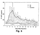
- ⁇ GST levels sustained over a period of days as shown in Fig. 6.
- SR steroid-resistant rejection
- AR acute rejection
- Tween-20 a standard reagent
- Table 4 Comparison of the titration of a PBC bile sample in diluents with and without Tween-20. Values are given in ⁇ g/L. Dilution - Tween-20 + Tween-20 1/50 2344 3074 1/100 2374 5187 1/200 2410 7059 Linear titration was observed only in the diluent without Tween-20.
Abstract
Description
- This invention relates to a method of determining the hepatic status of a subject, including a liver transplant recipient and, thereby, deciding on appropriate therapy or corrective action, if required, dependent on said hepatic status.
- The ability to differentiate between the various types of hepatic injury is of great significance in the treatment of both transplant patients and also individuals who suffer from other hepatic diseases which may affect the biliary system.
- Glutathione S-transferases (GSTs) comprise a multigene family of proteins consisting mainly of alpha (αGST), mu (µGST), pi (πGST) and theta-class (θGST) isoforms as defined by isoelectric point and are responsible for the detoxification of a range of xenobiotics, mainly via conjugation to glutathione (Beckett, G.J and Hayes, J.D., Advances in Clinical Chemistry (1993); 30, 281-380). Generally, the proteins are dimeric in nature consisting of two 25-27kDa subunits and may exist in homodimeric or heterodimeric forms. Pi Glutathione S-transferase (πGST) is a homodimer, and is located in the cytoplasm of bile duct epithelial cells within the liver (Beckett G.J. and Hayes, J.D., (1993) supra). αGST is known to be present in hepatocytes within the liver and exists in both homodimeric and heterodimeric states (Campbell, J.A.H., et. al., Cancer (Philadelphia) (1991) 67, 1608-1613; Howie, A.F., et. al., Clin. Chem. Acta., (1988) 177, 65-76). This heterogenous GST distribution of α and πGST suggests that the different isoenzymes have unique in vivo functions in different hepatic regions (Campbell, J.A. H., et. al., (1991) supra).
- EP-
A 0 640 145 discloses a method which assists in the early diagnosis of rejection in a liver transplant recipient and which comprises measuring an increase in plasma or serum αGST from the recipient in the absence of or preceding any change in plasma or serum transaminase. Thus, it has been conclusively demonstrated that measurement of the plasma αGST level facilitates monitoring of the post-transplant hepatic status by acting as an extremely sensitive, although not totally specific marker of graft rejection. - It is notable that πGST has received no attention as a potential marker of graft rejection, a fact possibly due to the low levels of enzyme present in the biliary epithelial cells of the liver. There is some evidence, however, that α and πGST are present in bile from both normal individuals and people suffering from specific cancers (e.g.,cholangiocarcinoma) as measured by radio-immunoassay (Howie. A.F., et. al., Clin. Chem. Acta. (1989) 184, 269-278). Additionally, some authors have referenced the fact that measurement of serum and plasma πGST levels may facilitate diagnosis of malignant tumours since πGST appears to be specifically expressed in malignant tissue (Niitsu, Y., et al., Cancer (1989) 63, 317-323; Howie, A.F., et. al., Clin. Chem. (1990) 36(3), 453-456, and Hida, T., et al., Cancer (1994) 73(5), 1377-1382. None of the aforementioned authors allude to the fact that πGST may have a role in the prediction of transplanted liver rejection or other liver/biliary disorders.
- Since it is known that primary graft rejection generally occurs in the biliary tree within the liver (Ascher, N., (1993) In 'Immunology of liver transplantation' Neuberger, J. and Adams, D. (eds)), it would appear that specific measurement of biliary or plasma πGST levels may allow diagnosis of early rejection or facilitate discrimination between post-transplant hepatocellular or biliary damage. The importance of distinguishing between non-specific hepatic injury and graft rejection cannot be overstated since the treatment for each condition is entirely different. Furthermore, initiation of the incorrect treatment could be extremely deleterious to the health of an individual already severely ill. For example, if graft injury occurs due to viral infection (e.g.,
- C re-infection or cytomegalovirus (CMV), it is necessary to carefully monitor the levels of anti-rejection immunosuppression treatment since excess immunosuppresive agents (e.g., cyclosporin A or FK506) would significantly impair the ability to fight viral infection. Conversely, failure to recognise genuine rejection from non-specific graft injury could lead to delay in augmentation of immunosuppressive therapy and ultimately lead to graft removal.
- Accordingly, there is a need for methods of determining the hepatic status of an individual in various disease states or abnormal conditions of the liver.
- The invention provides a method of determining the hepatic status of a liver transplant recipient for graft rejection after liver transplantation, which method comprises measuring the level of the pi glutathione S-transferase (πGST) isoform in a sample of a biological fluid from said subject by an immunoassay specific for the πGST isoform, comparing the level of πGST measured with the normal range of πGST in said biological fluid and, when an increase in πGST level relative to said normal range is detected, determining the hepatic status of the subject based on the level of πGST in said biological fluid.
- By providing a further method for determining hepatic status based on a marker specific to a particular hepatic site greatly facilitates the treatment of patients with various disease states and other abnormal conditions of the liver post-transplantation as hereinafter described in greater detail.
- The invention has particular application in the case of liver transplantation because it enables one to determine at a very early stage post-transplantation a likelihood of rejection because the primary graft rejection generally occurs in the biliary tree within the liver as stated above. Accordingly, even earlier detection of liver transplant rejection is possible With the method according to the invention relative to the method described and claimed in EP-
A 0 640 145. - Preferably, the recipient is a human.
- The immunoassay is preferably an enzyme immunoassay, more especially a sandwich enzyme immunoassay.
- The method according to the invention can be used to measure πGST in a range of media, but especially in bile, plasma and serum.
- By biological fluid herein is meant for example body fluids such as bile, plasma, serum and urine as well as tissue support media and perfusates. The biological fluids herein are also referred to generally as matrices.
- The method according to the invention facilitates for the first time detection of the πGST isoenzyme level in bile.
- When the biological fluid is bile, the normal πGST level is less than 15 µg/L.
- When the biological fluid is plasma, the normal πGST level is less than 100 µg/L.
- As demonstrated hereinbelow care should be taken when the method is carried out on plasma that the plasma is collected and stored prior to the determination in the presence of an anti-coagulant under conditions which permit substantially no haemolysis to occur during said storage period.
- We have found that use of fluoro-oxylate tubes results in a high degree of haemolysis releasing πGST from erythrocytes which gives falsely elevated levels of πGST. Other GST isoenzymes are either not found in the blood or are present at extremely low levels. For example, µGST is present in leucocytes. However, it is not clear from the literature as to whether it is present in erythrocytes. In any event, µGST is only present in 50% of the population. θGST expresses a similar inter-individual variability as does µGST and if present in blood is present at extremely low levels. αGST is not present in blood to any great extent.
- In a preferred embodiment, the sample is diluted with a diluent which contains an effective amount of a protein which optimises antibody-antigen reactions.
- We have found that if the diluent includes Tween 20 conventionally used as a standard reagent in such immunometric methods that incorrect πGST concentrations are detected. We have found that if one uses an effective amount of a protein which optimises antibody-antigen reactions, one can achieve a linear titration as shown in Example 6.
- Suitably, the protein is a serum albumin such as bovine serum albumin or human serum albumin.
- The immunoassay method according to the invention can be completed within 2.5 hours as hereinafter described in Examples. This is considerably faster than any commercially available assay for the quantitation of πGST.
- The invention thus provides in one embodiment an immunoassay capable of being completed in under 2.5 hours which is based on the sequential addition of sample, antibody-enzyme conjugate and substrate to microtitre wells or other surface coated with monoclonal anti-πGST IgG. The resultant colour intensity is proportional to the amount of πGST present in the sample and the assay range is 0-100 µg/L. The assay range is readily extended by increased sample dilution.
- According to another embodiment of the invention, additionally the level of the alpha glutathione S-transferase (αGST) isoform is measured in a sample of a biological fluid from said recipient so as to facilitate differentiation between graft rejection and non-specific hepatocellular damage in said recipient.
- In the accompanying drawings:
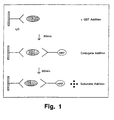
- Fig. 1 is a schematic diagram of the sandwich enzyme immunoassay of Example 1;
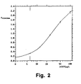
- Fig. 2 is a plot of absorbance at 450/630 nm versus log πGST concentration (µg/L) according to the enzyme immunoassay for human πGST described in Example 1;
- Fig. 3 is an SDS-PAGE analysis of human µ, α and πGST;
- Fig. 4 is an immunoblot analysis of human πGST;
- Fig. 5 is a plot of αGST and πGST (ng/ml) in bile versus time after reperfusion (hours) for a number of patients;
- Fig. 6 is a plot of πGST concentration versus time (days) for a number of patients; and
- Fig. 7 is a plot of AST/ALT (U/L) and αGST and πGST (ng/ml) versus days after transplantation for a single patient.
- The invention will be further illustrated by the following Examples.
- πGST was purified from human placenta by affinity chromatography. Precise details of the purification procedure are as follows:
- a. 325g of human placenta was homogenised for 2 minutes in homogenisation buffer, at a ratio of one part placenta to three parts buffer, using a Waring (Waring is a Trade Mark) blender. The homogenisation buffer had the following composition:
- 20mM Tris-HCl
- 250mM Sucrose
- 5mM EDTA pH 7.8
- 2µg/ml Leupeptin
- 2µg/ml Pepstatin.
- b. The placenta homogenate was centrifuged at 10000g for 60 minutes.
- c. The supernatant was then loaded on a Glutathione (GSH)-Sepharose Affinity column previously equilibrated in 20mM Tris-HCl with 200mM NaCl, pH 7.8. Equilibration buffer was reapplied to elute unbound protein. Finally 50mM Tris-HCl pH 9.5 containing 5mM GSH was used to elute bound GST from the affinity column.
- d. The eluted material was then dialysed against 0.1M PBS.
- Purified human πGST was injected into New Zealand White rabbits subcutaneously (s.c.) according to the time schedule given below and serum evaluated for anti-πGST reactivity. Once the IgG [anti-human πGST] titre was sufficient as determined by semiquantitative dot blot analysis, the animals were exsanguinated and serum collected. Total IgG was purified from rabbit serum by Protein A affinity chromatography and was used for conjugation to horseradish peroxidase (HRP). Monoclonal IgG [anti-human πGST] as ascites, was obtained from The University Hospital, Nijmegen, The Netherlands and was not purified further prior to use.
- Day 1: A test bleed of 5ml of preserum was taken from the ear of the rabbit. 0.5ml of human πGST antigen (100µg) was mixed with an equal volume of Freund's Complete Adjuvant. The mixture of antigen and adjuvant was homogenised to ensure a good emulsion. This mixture was then injected subcutaneously into multiple sites on the back of the rabbit which had previously been shaved.
- Day 28: A test bleed of 5ml of serum was taken from the ear of the rabbit. 0.5ml antigen (100µg) was mixed with an equal volume of Freund's Incomplete Adjuvant. The antigen/adjuvant mixture was homogenised to ensure a good emulsion. This mixture was then injected subcutaneously into multiple sites on the back of the rabbit.
- Day 42 A test bleed of 10ml of blood was taken from the rabbit's ear.
- Day 56: A second boost was given to the rabbit as described on Day 28.
- Day 70: A test bleed of 10ml of blood was taken from the ear of the rabbit. When the titre was sufficiently high, the rabbit was sacrificed and as much blood as possible collected.
- All polyclonal and monoclonal IgGs for use in the followings Examples were checked for πGST reactivity and potential cross-reactivity against human α and µGST respectively, via the following immunoblot combinations:-
- (a) Rabbit IgG [anti-human πGST] was used to probe nitrocellulose membranes containing immobilised human α. π and µGST.
- (b) Murine IgG [anti-human πGST] was used to probe nitrocellulose membranes containing immobilised human α, π and µGST.
- The method used for immunoblot detection was as follows:
- 1. Human α, π and µGST (0.5µg/track) were electrophoresed on 15% SDS-PAGE with molecular weight markers also included.
- 2. After electrophoresis, the polyacrylamide gel was cut and one half stained for protein while the remainder was used for electrophoretic transfer onto nitrocellulose.
- 3. After electrophoretic transfer, the nitrocellulose membranes were blocked for 1 hour with 5%(w/v) Marvel (Marvel is a Trade Mark) in phosphate buffered saline containing 0.05%(w/v) TWEEN-20 (PBST)- blocking buffer.
- 4. The following solutions were then prepared:
- (i) Rabbit IgG [anti-human πGST] in 1%(w/v) Marvel in PBST
- (ii) Murine IgG [anti-human πGST] in 1%(w/v) Marvel in PBST and added to the membranes once blocking buffer was decanted.
Incubation with antibody solutions was allowed to proceed for one hour. - 5. The nitrocellulose membranes were then washed in PBST (2x for 5 min each).
- 6. Anti rabbit IgG-HRP conjugate was then prepared (1/1000 in 1% (w/v) Marvel in PBST and added to 4(i) above. Anti murine IgG-HRP conjugate was also prepared (1/1000) and added to 4(ii) above.
- 7. After one hour incubation with anti-species conjugates, the reagents were discarded and the membranes washed as in 5 above.
- 8. Diaminobenzidine substrate was then prepared and added to the membrane.
- A positive reaction was indicated by a brown precipitate on the nitrocellulose membrane.
- Anti πGST IgG-HRP conjugates were synthesised using thioether conjugation methodology. (Duncan, R.J.S., et al., (1983); Anal. Biochem. 132, 68-73) Reactive maleimide groups were introduced onto IgG molecules using SMCC (succinimidyl 4-(N-maleimidomethyl) cyclohexane 1-carboxylate) and masked sulphydryl groups were linked to HRP. After a demasking step to produce reactive sulphydryl groups, the maleimide-activated IgG and HRP-SH were mixed together and allowed to react for 4.5 hours. The resultant IgG-HRP conjugate, formed by covalent thioether linkage, was brought to 50% (v/v) glycerol and stored at -20°C for use in the EIA of Example 1.
- The format of the immunoassay for the quantitative detection of human πGST is a conventional sandwich format as depicted schematically in Fig. 1, and described below.
- a. A Nunc Maxisorp (Nunc Maxisorp is a Trade Mark) microtitre plate was coated with murine monoclonal IgG [anti-human πGST] (referred to in Preparatory Example B) immobilised via goat F(ab)2 fragments [anti-mouse IgG]. This method of antibody coating serves to orientate Mab binding sites and also improves assay sensitivity by minimising adherence - induced denaturation of the capture antibody.
- b. Human πGST, purified from placenta as described in Preparatory Example A, was used as the assay calibrator.
- c. IgG [anti-human πGST]-HRP conjugates, in association with tetramethylbenzidine substrate (TMB), were used to facilitate detection of captured/immobilised πGST.
- d. The enzyme reaction was stopped by the addition of IN H2SO4 and the absorbance measured at 450nm using 630nm as a reference wavelength. Colour intensity was proportional to πGST concentration and after generating a plot of A450/630nm versus concentration (µg/L), the concentration of unknown samples can be determined (see Fig. 2). Total assay time was less than 2.5 hours.
- The total assay time was found to be 2
hours 15 minutes and assay conditions included microtitre plate shaking at fixed temperature during the sample and conjugate incubation steps, respectively. The TMB substrate incubation required fixed temperature conditions only. - The effect of a variety of commonly used anti-coagulants (ethylenediaminetetraacetic acid (EDTA), lithium heparin, sodium citrate and fluoro-oxylate) and other plasma collection tubes (containing platelet inhibitors) on πGST levels in plasma, was examined. Samples were collected by venipuncture into tubes containing the particular anticoagulant. Plasma was separated by centrifugation (6,000g for 10 min), and residual platelets were removed by an additional centrifugation step (10,000g for 10 min). The supernatant was removed, and samples were assayed using the protocol described in Example 1.
- Plasma was collected from a number of individuals into a series of plasma collection tubes. Each sample was handled as described above, and the release of πGST into plasma was monitored over a 24 hour period. Table 1 below shows the πGST levels in plasma from the same individual collected into four different plasma collection tubes, assayed at T0 and T24. The results show that there is no significant difference in πGST concentrations in plasma collected in the presence of any of the above anti-coagulants. There does not appear to be a significant increase in πGST concentrations (caused by release of πGST from erythrocytes or platelets) when the unseparated plasma is stored for up to 24 hours. The exception is the fluoro-oxylate tubes where a large degree of haemolysis occurred, releasing πGST from erythrocytes, and giving falsely elevated levels of the protein as shown in Table 1.
Table 1 Comparison of the influence of anti-coagulants in plasma collection tubes for πGST analysis in plasma. Values are given in µg/L. Patient EDTA Li/Hep Na citrate F1 oxylate T0 T24 T0 T24 T0 T24 T0 T24 1 171.9 220.2 199.4 194.7 167.5 220.3 170.0 >500 2 65.6 70.5 62.8 88.4 53.2 65.4 55.4 >500 3 59.6 78.6 65.4 88.6 53.7 68.4 59.9 >500 4 81.7 92.8 90.2 119.5 74.9 122.1 81.2 >500 5 46.7 67.5 44.6 73.3 28.9 68.6 60.9 >500 - It can be seen from Table 1 that there is no significant difference between the concentration of πGST in plasma collected in the presence of any of the above anti-coagulants.
- Fig. 3 illustrates the purity of human πGST obtained by the procedure of Preparatory Example A prior to immunisation into rabbits and confirms the absence of any other human derived proteins which might otherwise contribute to reduced assay specificity. In Fig. 3:
-
Lane 1 = µGST -
Lane 2 = αGST -
Lane 3 = πGST -
Lane 4 = molecular weight markers. - Immunoblot analysis of the monoclonal antibody reactivity revealed that the IgG[anti-human πGST] was highly specific for human πGST and did not exhibit any significant cross-reactivity with human α or µGST. The results are shown in Fig. 4.
-
Lane 1 = πGST -
Lane 2 = molecular weight markers. - The significance of this fact is of utmost importance since it implies that the enzyme immunoassay for human πGST quantitation is specific for the detection of human πGST. Thus, any human πGST present in samples can be specifically detected without cross-contamination from other GSTs.
- A number of bile samples from patients with specific liver/biliary damage were tested in the assay for human πGST. Patients with hepatocellular carcinoma (HCC) and primary biliary cirrhosis (PBC) were found to contain very high levels of πGST. A patient with bile duct stones (BDS) was also found to have increased πGST levels, but not as elevated as those for HCC and PBC as shown in Table 3.
Table 3 πGST concentrations in bile from patients with hepatocellular carcinoma (HCC), primary biliary cirrhosis (PBC) and bile duct stone (BDS). Condition [πGST] (µg/L) HCC 5357 PBC 2432 BDS 506.6 - All of these samples show significantly elevated πGST levels.
- Serial bile and plasma samples were collected from patients following liver transplant operations and assayed for both π and αGST respectively. The patients exhibited a range of post-operative conditions, from uneventful recovery, to acute rejection and Hepatitis C re-infection, which are normally associated with transplantation.
- A number of significant trends were observed when the concentrations of both π and αGST in bile were monitored simultaneously. αGST was measured according to the procedure described in EP-
A 0 640 145. During an uneventful recovery, πGST could be detected as soon as 2 hours post transplantation, and levels remained low (i.e. below 50 µg/L). αGST levels were initially high due to reperfusion injury, but returned to baseline levels within 2 days as shown in Fig. 5. This figure shows the typical course of α and πGST during and after human liver transplantation. πGST levels remain low. Complications associated with liver transplantation could also be identified. One of the major risks is acute rejection, or the even more serious, steroid-resistant rejection. We have found that in these particular cases, there was a significant increase in πGST levels sustained over a period of days as shown in Fig. 6. This figure shows πGST levels during episodes of steroid-resistant rejection (SR) and acute rejection (AR). Levels are elevated and remain high over a period of at least 20 days. αGST levels were also high, but they returned to baseline levels within a period of 5-8 days. - The possibility of infection or re-infection is another serious risk involved in transplantation. We have seen that in episodes of HCV re-infection, very high levels of αGST were observed over a sustained period of time (i.e. at least 25 days). However, πGST levels remained near normal as shown in Fig. 7. This contrasts sharply with the previous cases of acute rejection, where the reverse was true. Thus, by the simultaneous quantitation of α and πGST, it was possible to successfully differentiate between acute rejection and HCV re-infection, something which has previously proved to be very difficult, and has posed a therapeutic dilemma to transplant surgeons.
- A number of bile samples were obtained from patients with specific liver/biliary damage (hepatocellular carcinoma and primary biliary cirrhosis), as well as samples from donor bile, and post liver transplantation. These samples were assayed for human πGST according to the protocol of Example 1.
- A number of standard diluents were used as sample diluents for the titration of the bile samples. These diluents are routinely used in many assay systems, with Tween-20 being the most commonly used detergent. We have found however that the presence of this particular detergent in the sample diluent caused erroneous results. Falsely high concentrations of πGST were observed in samples diluted in Tween-20-containing diluents, caused by insufficient titration as shown in Table 4. In the absence of Tween-20, linear titration was observed. Therefore, a critical factor of this assay is the absence of Tween-20 (a standard reagent) in the sample diluent, as falsely elevated levels of πGST would be observed if it were used.
Table 4 Comparison of the titration of a PBC bile sample in diluents with and without Tween-20. Values are given in µg/L. Dilution - Tween-20 + Tween-20 1/50 2344 3074 1/100 2374 5187 1/200 2410 7059 - Polyclonal and monoclonal anti-human πGST IgG were immobilised onto Nunc Maxisorp plates either directly or via a linker (F(ab)2 fragments of goat anti-species IgG). Standards of known concentrations of human πGST were then run as described in Example 1 and the absorbances at similar immobilised IgG concentrations compared.
- Both polyclonal and monoclonal antibodies were coated onto the solid phase for use as a capture antibody. Direct coating of the both the polyclonal and the monoclonal antibodies resulted in very low absorbance readings for the standard curve (see Table 5). When immobilisation was achieved via a linker antibody (goat anti-mouse/anti-rabbit IgG) a significant increase in O.D. values was obtained for the monoclonal antibody. However, no such increase was observed for the polyclonal antibody.
Table 5 Comparison of coating methods for the detection of human πGST. [πGST] DIRECT COATING COATING via LINKER ANTIBODY Polyclonal Monoclonal Polyclonal Monoclonal Ab. 100 0.625 0.421 0.352 1.557 0 0.044 0.038 0.235 0.095
| GST Conc. | A450/630nm | ||
| πGST | αGST | µGST | |
| 0.00 | 0.022 | 0.022 | 0.022 |
| 3.12 | 0.087 | 0.024 | 0.016 |
| 12.5 | 0.299 | 0.020 | 0.022 |
| 25.0 | 0.688 | 0.036 | 0.055 |
| 50.0 | 1.204 | 0.018 | 0.034 |
| 100 | 1.735 | 0.040 | 0.043 |
Claims (10)
- A method of determining the hepatic status of a liver transplant recipient for graft rejection after liver transplantation, which method comprises measuring the level of the pi glutathione S-transferase (πGST) isoform in a sample of a biological fluid from said subject by an immunoassay specific for the πGST isoform, comparing the level of πGST measured with the normal range of πGST in said biological fluid and, when an increase in πGST level relative to said normal range is detected, determining the hepatic status of the subject based on the level of πGST in said biological fluid.
- A method according to Claim 1, wherein the subject is a human.
- A method according to Claim 1 or 2, wherein the immunoassay is an enzyme immunoassay.
- A method according to any preceding claim, wherein the biological fluid is bile and the normal πGST level is less than 15 µg/L.
- A method according to Claim 3, wherein the biological fluid is plasma and the normal πGST level is less than 100 µg/L.
- A method according to Claim 5, wherein the plasma is collected and stored prior to the determination in the presence of an anti-coagulant under conditions which permit substantially no haemolysis to occur during said storage period.
- A method according to any preceding claim, wherein the sample is diluted with a diluent which contains an effective amount of a protein which optimises antibody-antigen reactions.
- A method according to Claim 7, wherein the protein is a serum albumin.
- A method according to any preceding claim, wherein the entire immunoassay is completed within 2.5 hours.
- A method according to any one of Claims 1-9, wherein additionally the level of the alpha glutathione S-transferase (αGST) isoform is measured in a sample of a biological fluid from said recipient so as to facilitate differentiation between graft rejection and non-specific hepatocellular damage in said recipient.
Applications Claiming Priority (2)
| Application Number | Priority Date | Filing Date | Title |
|---|---|---|---|
| CA002246072A CA2246072A1 (en) | 1996-02-02 | 1996-02-02 | Method of determining the hepatic status of an individual, including a liver transplant recipient |
| PCT/IE1996/000003 WO1997028449A1 (en) | 1996-02-02 | 1996-02-02 | Method of determining the hepatic status of an individual, including a liver transplant recipient |
Publications (2)
| Publication Number | Publication Date |
|---|---|
| EP0880700A1 EP0880700A1 (en) | 1998-12-02 |
| EP0880700B1 true EP0880700B1 (en) | 2006-03-29 |
Family
ID=25680448
Family Applications (1)
| Application Number | Title | Priority Date | Filing Date |
|---|---|---|---|
| EP96901946A Expired - Lifetime EP0880700B1 (en) | 1996-02-02 | 1996-02-02 | Method of determining the hepatic status of a liver transplant recipient. |
Country Status (13)
| Country | Link |
|---|---|
| US (1) | US6183977B1 (en) |
| EP (1) | EP0880700B1 (en) |
| JP (1) | JP2000505544A (en) |
| AT (1) | ATE322013T1 (en) |
| AU (1) | AU712180B2 (en) |
| BR (1) | BR9612475A (en) |
| CA (1) | CA2246072A1 (en) |
| CZ (1) | CZ287488B6 (en) |
| DE (1) | DE69635979T2 (en) |
| ES (1) | ES2262153T3 (en) |
| HU (1) | HUP9903578A2 (en) |
| PL (1) | PL328518A1 (en) |
| WO (1) | WO1997028449A1 (en) |
Families Citing this family (7)
| Publication number | Priority date | Publication date | Assignee | Title |
|---|---|---|---|---|
| US6977140B1 (en) | 1998-09-29 | 2005-12-20 | Organ Recovery Systems, Inc. | Method for maintaining and/or restoring viability of organs |
| US6492103B1 (en) | 2000-01-31 | 2002-12-10 | Organ Recovery Systems, Inc. | System for organ and tissue preservation and hypothermic blood substitution |
| US20030022909A1 (en) * | 2001-06-05 | 2003-01-30 | University Of Chicago | Use of methylnaltrexone to treat immune suppression |
| AU2003297028A1 (en) * | 2002-12-13 | 2004-07-09 | St. Jude Children's Research Hospital | Glutathione-s-transferase test for susceptibility to parkinson's |
| US8048638B2 (en) * | 2005-04-01 | 2011-11-01 | University Of Florida Research Foundation, Inc. | Biomarkers of liver injury |
| JP5202296B2 (en) * | 2005-04-01 | 2013-06-05 | ユニバーシティ オブ フロリダ リサーチ ファウンデーション,インコーポレイティド | Biomarkers for liver injury |
| AU2011354597B2 (en) | 2010-01-26 | 2016-09-22 | Bioregency Inc. | Compositions and methods relating to argininosuccinate synthetase |
Family Cites Families (4)
| Publication number | Priority date | Publication date | Assignee | Title |
|---|---|---|---|---|
| AU5435990A (en) | 1989-04-13 | 1990-11-05 | United States of America, as represented by the Secretary, U.S. Department of Commerce, The | Monoclonal antibodies to human glutathione s transferase pi |
| JPH0425763A (en) | 1990-05-21 | 1992-01-29 | Maruko Seiyaku Kk | Method for determining glutathione s-transferase pi and reagent for diagnosis |
| USRE35419E (en) * | 1992-05-01 | 1997-01-07 | Cormac G. Kilty | Measurement of an enzyme marker as an aid to diagnosis of liver transplant rejection |
| US5217868A (en) * | 1992-05-01 | 1993-06-08 | Syncor Limited | Measurement of an enzyme marker as an aid to diagnosis of liver transplant rejection |
-
1996
- 1996-02-02 ES ES96901946T patent/ES2262153T3/en not_active Expired - Lifetime
- 1996-02-02 AT AT96901946T patent/ATE322013T1/en not_active IP Right Cessation
- 1996-02-02 US US09/117,476 patent/US6183977B1/en not_active Expired - Fee Related
- 1996-02-02 DE DE69635979T patent/DE69635979T2/en not_active Expired - Lifetime
- 1996-02-02 WO PCT/IE1996/000003 patent/WO1997028449A1/en active IP Right Grant
- 1996-02-02 HU HU9903578A patent/HUP9903578A2/en unknown
- 1996-02-02 PL PL96328518A patent/PL328518A1/en unknown
- 1996-02-02 BR BR9612475A patent/BR9612475A/en not_active Application Discontinuation
- 1996-02-02 AU AU46323/96A patent/AU712180B2/en not_active Ceased
- 1996-02-02 CZ CZ19982288A patent/CZ287488B6/en not_active IP Right Cessation
- 1996-02-02 EP EP96901946A patent/EP0880700B1/en not_active Expired - Lifetime
- 1996-02-02 CA CA002246072A patent/CA2246072A1/en not_active Abandoned
- 1996-02-02 JP JP09527453A patent/JP2000505544A/en active Pending
Also Published As
| Publication number | Publication date |
|---|---|
| ATE322013T1 (en) | 2006-04-15 |
| US6183977B1 (en) | 2001-02-06 |
| BR9612475A (en) | 1999-07-13 |
| ES2262153T3 (en) | 2006-11-16 |
| WO1997028449A1 (en) | 1997-08-07 |
| AU712180B2 (en) | 1999-10-28 |
| CA2246072A1 (en) | 1997-08-07 |
| DE69635979T2 (en) | 2006-11-16 |
| CZ287488B6 (en) | 2000-12-13 |
| PL328518A1 (en) | 1999-02-01 |
| JP2000505544A (en) | 2000-05-09 |
| AU4632396A (en) | 1997-08-22 |
| DE69635979D1 (en) | 2006-05-18 |
| EP0880700A1 (en) | 1998-12-02 |
| HUP9903578A2 (en) | 2000-02-28 |
| CZ228898A3 (en) | 1999-03-17 |
Similar Documents
| Publication | Publication Date | Title |
|---|---|---|
| US7462495B2 (en) | Methods and compositions for use in diagnosing and characterizing chronic immune disease | |
| CA2088050A1 (en) | Cell necrosis detection through assays for spectrum and breakdown products thereof | |
| EP0880700B1 (en) | Method of determining the hepatic status of a liver transplant recipient. | |
| Michalski et al. | A modified double antibody sandwich enzyme-linked immunosorbent assay for measurement of alpha-1-antitrypsin in biologic fluids | |
| EP0968429A1 (en) | Method and kit for determining the phenotype of a haptoglobin and use thereof | |
| KR101396300B1 (en) | Biomarkers for diagnosing the addiction of ethylbenzene | |
| US6080554A (en) | Methods and compositions for use in characterizing multiple sclerosis disease activity in a subject | |
| RU2164027C2 (en) | Method for determining liver state | |
| US6733980B1 (en) | Diagnostic method for detection of molecular forms of eosinophilic cationic protein (ISO-ECPS) | |
| AU710668B2 (en) | Method of determining or detecting donor organ damage following xenotransplantation based on donor-organ derived analytes | |
| Lannfelt et al. | ELISA for measuring porphobilinogen deaminase in human erythrocytes | |
| AU2006318919A1 (en) | Method for identifying the genotype in position 171 of the sheep prion protein as well as kits for implementing said method | |
| JP4681175B2 (en) | Method for detecting disseminated intravascular coagulation syndrome and its pre-onset stage | |
| RU2157540C2 (en) | Method for identifying injuries in donor organ after xenotransplantation by analyzing substances obtained from the donor organ | |
| EP0764849B1 (en) | Immunological ELISA test for inter-alpha-trypsin inhibitor | |
| CA2056449A1 (en) | Method for the detection of anti-streptokinase antibodies | |
| US20100151451A1 (en) | Method for Identifying the Genotype in Position 171 of the Ovine Prion Protein as well as Kits for Implementing said Method | |
| JPH0833398B2 (en) | Method and kit for measuring human tissue plasminogen activator / human plasminogen activator inhibitor complex | |
| JPS62503123A (en) | Methods and articles for assaying immune complexes | |
| JPH06249852A (en) | D-amino acid oxidizing enzyme polychlornal antibody and measurement of d-amino acid oxidizing enzyme using the antibody |
Legal Events
| Date | Code | Title | Description |
|---|---|---|---|
| PUAI | Public reference made under article 153(3) epc to a published international application that has entered the european phase |
Free format text: ORIGINAL CODE: 0009012 |
|
| 17P | Request for examination filed |
Effective date: 19980804 |
|
| AK | Designated contracting states |
Kind code of ref document: A1 Designated state(s): AT BE CH DE DK ES FR GB GR IE IT LI LU MC NL PT SE |
|
| 17Q | First examination report despatched |
Effective date: 20040823 |
|
| GRAP | Despatch of communication of intention to grant a patent |
Free format text: ORIGINAL CODE: EPIDOSNIGR1 |
|
| RTI1 | Title (correction) |
Free format text: METHOD OF DETERMINING THE HEPATIC STATUS OF A LIVER TRANSPLANT RECIPIENT. |
|
| GRAS | Grant fee paid |
Free format text: ORIGINAL CODE: EPIDOSNIGR3 |
|
| GRAA | (expected) grant |
Free format text: ORIGINAL CODE: 0009210 |
|
| AK | Designated contracting states |
Kind code of ref document: B1 Designated state(s): AT BE CH DE DK ES FR GB GR IE IT LI LU MC NL PT SE |
|
| PG25 | Lapsed in a contracting state [announced via postgrant information from national office to epo] |
Ref country code: NL Free format text: LAPSE BECAUSE OF FAILURE TO SUBMIT A TRANSLATION OF THE DESCRIPTION OR TO PAY THE FEE WITHIN THE PRESCRIBED TIME-LIMIT Effective date: 20060329 Ref country code: IT Free format text: LAPSE BECAUSE OF FAILURE TO SUBMIT A TRANSLATION OF THE DESCRIPTION OR TO PAY THE FEE WITHIN THE PRESCRIBED TIME-LIMIT;WARNING: LAPSES OF ITALIAN PATENTS WITH EFFECTIVE DATE BEFORE 2007 MAY HAVE OCCURRED AT ANY TIME BEFORE 2007. THE CORRECT EFFECTIVE DATE MAY BE DIFFERENT FROM THE ONE RECORDED. Effective date: 20060329 |
|
| REG | Reference to a national code |
Ref country code: GB Ref legal event code: FG4D |
|
| REG | Reference to a national code |
Ref country code: CH Ref legal event code: EP |
|
| REG | Reference to a national code |
Ref country code: IE Ref legal event code: FG4D |
|
| REF | Corresponds to: |
Ref document number: 69635979 Country of ref document: DE Date of ref document: 20060518 Kind code of ref document: P |
|
| PG25 | Lapsed in a contracting state [announced via postgrant information from national office to epo] |
Ref country code: SE Free format text: LAPSE BECAUSE OF FAILURE TO SUBMIT A TRANSLATION OF THE DESCRIPTION OR TO PAY THE FEE WITHIN THE PRESCRIBED TIME-LIMIT Effective date: 20060629 Ref country code: DK Free format text: LAPSE BECAUSE OF FAILURE TO SUBMIT A TRANSLATION OF THE DESCRIPTION OR TO PAY THE FEE WITHIN THE PRESCRIBED TIME-LIMIT Effective date: 20060629 |
|
| REG | Reference to a national code |
Ref country code: CH Ref legal event code: NV Representative=s name: KIRKER & CIE SA |
|
| PG25 | Lapsed in a contracting state [announced via postgrant information from national office to epo] |
Ref country code: PT Free format text: LAPSE BECAUSE OF FAILURE TO SUBMIT A TRANSLATION OF THE DESCRIPTION OR TO PAY THE FEE WITHIN THE PRESCRIBED TIME-LIMIT Effective date: 20060829 |
|
| ET | Fr: translation filed | ||
| NLV1 | Nl: lapsed or annulled due to failure to fulfill the requirements of art. 29p and 29m of the patents act | ||
| REG | Reference to a national code |
Ref country code: ES Ref legal event code: FG2A Ref document number: 2262153 Country of ref document: ES Kind code of ref document: T3 |
|
| PG25 | Lapsed in a contracting state [announced via postgrant information from national office to epo] |
Ref country code: MC Free format text: LAPSE BECAUSE OF NON-PAYMENT OF DUE FEES Effective date: 20070228 |
|
| PLBE | No opposition filed within time limit |
Free format text: ORIGINAL CODE: 0009261 |
|
| STAA | Information on the status of an ep patent application or granted ep patent |
Free format text: STATUS: NO OPPOSITION FILED WITHIN TIME LIMIT |
|
| 26N | No opposition filed |
Effective date: 20070102 |
|
| PG25 | Lapsed in a contracting state [announced via postgrant information from national office to epo] |
Ref country code: GR Free format text: LAPSE BECAUSE OF FAILURE TO SUBMIT A TRANSLATION OF THE DESCRIPTION OR TO PAY THE FEE WITHIN THE PRESCRIBED TIME-LIMIT Effective date: 20060630 |
|
| REG | Reference to a national code |
Ref country code: CH Ref legal event code: PUE Owner name: ARGUTUS INTELLECTUAL PROPERTIES LIMITED Free format text: BIOTRIN INTELLECTUAL PROPERTIES LIMITED#93 THE RISE#MOUNT MERRION, COUNTY DUBLIN (IE) -TRANSFER TO- ARGUTUS INTELLECTUAL PROPERTIES LIMITED#UNIT 9 TRINITY TECHNOLOGY & ENTERPRISE CAMPUS PEARSE ST.#DUBLIN 2 (IE) |
|
| REG | Reference to a national code |
Ref country code: GB Ref legal event code: 732E Free format text: REGISTERED BETWEEN 20091029 AND 20091104 |
|
| REG | Reference to a national code |
Ref country code: FR Ref legal event code: TP |
|
| PGFP | Annual fee paid to national office [announced via postgrant information from national office to epo] |
Ref country code: AT Payment date: 20100208 Year of fee payment: 15 |
|
| PGFP | Annual fee paid to national office [announced via postgrant information from national office to epo] |
Ref country code: LU Payment date: 20110228 Year of fee payment: 16 Ref country code: IT Payment date: 20110211 Year of fee payment: 16 Ref country code: CH Payment date: 20110228 Year of fee payment: 16 |
|
| PGFP | Annual fee paid to national office [announced via postgrant information from national office to epo] |
Ref country code: BE Payment date: 20110218 Year of fee payment: 16 |
|
| PG25 | Lapsed in a contracting state [announced via postgrant information from national office to epo] |
Ref country code: AT Free format text: LAPSE BECAUSE OF NON-PAYMENT OF DUE FEES Effective date: 20110202 |
|
| PG25 | Lapsed in a contracting state [announced via postgrant information from national office to epo] |
Ref country code: LU Free format text: LAPSE BECAUSE OF NON-PAYMENT OF DUE FEES Effective date: 20120229 |
|
| PGFP | Annual fee paid to national office [announced via postgrant information from national office to epo] |
Ref country code: IE Payment date: 20120127 Year of fee payment: 17 Ref country code: FR Payment date: 20120221 Year of fee payment: 17 |
|
| PGFP | Annual fee paid to national office [announced via postgrant information from national office to epo] |
Ref country code: DE Payment date: 20120228 Year of fee payment: 17 |
|
| PGFP | Annual fee paid to national office [announced via postgrant information from national office to epo] |
Ref country code: GB Payment date: 20120127 Year of fee payment: 17 |
|
| BERE | Be: lapsed |
Owner name: AUGUSTUS INTELLECTUAL PROPERTIES LTD Effective date: 20120228 |
|
| REG | Reference to a national code |
Ref country code: CH Ref legal event code: PL |
|
| PG25 | Lapsed in a contracting state [announced via postgrant information from national office to epo] |
Ref country code: LI Free format text: LAPSE BECAUSE OF NON-PAYMENT OF DUE FEES Effective date: 20120229 Ref country code: CH Free format text: LAPSE BECAUSE OF NON-PAYMENT OF DUE FEES Effective date: 20120229 |
|
| PG25 | Lapsed in a contracting state [announced via postgrant information from national office to epo] |
Ref country code: IT Free format text: LAPSE BECAUSE OF NON-PAYMENT OF DUE FEES Effective date: 20120202 |
|
| PG25 | Lapsed in a contracting state [announced via postgrant information from national office to epo] |
Ref country code: BE Free format text: LAPSE BECAUSE OF NON-PAYMENT OF DUE FEES Effective date: 20120228 |
|
| PGFP | Annual fee paid to national office [announced via postgrant information from national office to epo] |
Ref country code: ES Payment date: 20120206 Year of fee payment: 17 |
|
| GBPC | Gb: european patent ceased through non-payment of renewal fee |
Effective date: 20130202 |
|
| REG | Reference to a national code |
Ref country code: FR Ref legal event code: ST Effective date: 20131031 |
|
| REG | Reference to a national code |
Ref country code: IE Ref legal event code: MM4A |
|
| REG | Reference to a national code |
Ref country code: DE Ref legal event code: R119 Ref document number: 69635979 Country of ref document: DE Effective date: 20130903 |
|
| PG25 | Lapsed in a contracting state [announced via postgrant information from national office to epo] |
Ref country code: GB Free format text: LAPSE BECAUSE OF NON-PAYMENT OF DUE FEES Effective date: 20130202 Ref country code: DE Free format text: LAPSE BECAUSE OF NON-PAYMENT OF DUE FEES Effective date: 20130903 Ref country code: IE Free format text: LAPSE BECAUSE OF NON-PAYMENT OF DUE FEES Effective date: 20130202 Ref country code: FR Free format text: LAPSE BECAUSE OF NON-PAYMENT OF DUE FEES Effective date: 20130228 |
|
| REG | Reference to a national code |
Ref country code: ES Ref legal event code: FD2A Effective date: 20140408 |
|
| PG25 | Lapsed in a contracting state [announced via postgrant information from national office to epo] |
Ref country code: ES Free format text: LAPSE BECAUSE OF NON-PAYMENT OF DUE FEES Effective date: 20130203 |