EP0866075B1 - Method to treat microbial infections by uncoupling of phosphotransferase system and appropriate agents therefor - Google Patents
Method to treat microbial infections by uncoupling of phosphotransferase system and appropriate agents therefor Download PDFInfo
- Publication number
- EP0866075B1 EP0866075B1 EP19980101704 EP98101704A EP0866075B1 EP 0866075 B1 EP0866075 B1 EP 0866075B1 EP 19980101704 EP19980101704 EP 19980101704 EP 98101704 A EP98101704 A EP 98101704A EP 0866075 B1 EP0866075 B1 EP 0866075B1
- Authority
- EP
- European Patent Office
- Prior art keywords
- seq
- peptides
- enzyme
- peptide
- pts
- Prior art date
- Legal status (The legal status is an assumption and is not a legal conclusion. Google has not performed a legal analysis and makes no representation as to the accuracy of the status listed.)
- Expired - Lifetime
Links
- 108091000080 Phosphotransferase Proteins 0.000 title claims description 23
- 102000020233 phosphotransferase Human genes 0.000 title claims description 23
- 238000000034 method Methods 0.000 title description 6
- 208000015181 infectious disease Diseases 0.000 title 1
- 230000000813 microbial effect Effects 0.000 title 1
- 108090000765 processed proteins & peptides Proteins 0.000 claims description 92
- 102000004190 Enzymes Human genes 0.000 claims description 55
- 108090000790 Enzymes Proteins 0.000 claims description 55
- 102000004196 processed proteins & peptides Human genes 0.000 claims description 41
- 230000000694 effects Effects 0.000 claims description 30
- 230000005764 inhibitory process Effects 0.000 claims description 19
- 230000002401 inhibitory effect Effects 0.000 claims description 12
- 230000001580 bacterial effect Effects 0.000 claims description 11
- 238000003255 drug test Methods 0.000 claims description 8
- 102000004160 Phosphoric Monoester Hydrolases Human genes 0.000 claims description 6
- 108090000608 Phosphoric Monoester Hydrolases Proteins 0.000 claims description 6
- 239000004599 antimicrobial Substances 0.000 claims description 6
- 239000008194 pharmaceutical composition Substances 0.000 claims 1
- 150000001413 amino acids Chemical group 0.000 description 70
- 235000001014 amino acid Nutrition 0.000 description 69
- 108090000623 proteins and genes Proteins 0.000 description 20
- 150000001875 compounds Chemical class 0.000 description 17
- 235000018102 proteins Nutrition 0.000 description 17
- 108010055837 phosphocarrier protein HPr Proteins 0.000 description 16
- DTBNBXWJWCWCIK-UHFFFAOYSA-N phosphoenolpyruvic acid Chemical compound OC(=O)C(=C)OP(O)(O)=O DTBNBXWJWCWCIK-UHFFFAOYSA-N 0.000 description 16
- 102000004169 proteins and genes Human genes 0.000 description 16
- 229930029653 phosphoenolpyruvate Natural products 0.000 description 13
- 238000003556 assay Methods 0.000 description 11
- 230000026731 phosphorylation Effects 0.000 description 11
- 238000006366 phosphorylation reaction Methods 0.000 description 11
- 235000000346 sugar Nutrition 0.000 description 11
- 241000894006 Bacteria Species 0.000 description 10
- 238000006243 chemical reaction Methods 0.000 description 10
- 108010078791 Carrier Proteins Proteins 0.000 description 9
- 241000588724 Escherichia coli Species 0.000 description 9
- 230000027455 binding Effects 0.000 description 8
- 210000004027 cell Anatomy 0.000 description 8
- 239000003112 inhibitor Substances 0.000 description 8
- 239000012528 membrane Substances 0.000 description 8
- 239000000758 substrate Substances 0.000 description 7
- 150000005846 sugar alcohols Polymers 0.000 description 7
- WQZGKKKJIJFFOK-GASJEMHNSA-N Glucose Natural products OC[C@H]1OC(O)[C@H](O)[C@@H](O)[C@@H]1O WQZGKKKJIJFFOK-GASJEMHNSA-N 0.000 description 6
- OVRNDRQMDRJTHS-UHFFFAOYSA-N N-acelyl-D-glucosamine Natural products CC(=O)NC1C(O)OC(CO)C(O)C1O OVRNDRQMDRJTHS-UHFFFAOYSA-N 0.000 description 6
- MBLBDJOUHNCFQT-LXGUWJNJSA-N N-acetylglucosamine Natural products CC(=O)N[C@@H](C=O)[C@@H](O)[C@H](O)[C@H](O)CO MBLBDJOUHNCFQT-LXGUWJNJSA-N 0.000 description 6
- 230000012010 growth Effects 0.000 description 6
- 238000002823 phage display Methods 0.000 description 6
- OVRNDRQMDRJTHS-RTRLPJTCSA-N N-acetyl-D-glucosamine Chemical compound CC(=O)N[C@H]1C(O)O[C@H](CO)[C@@H](O)[C@@H]1O OVRNDRQMDRJTHS-RTRLPJTCSA-N 0.000 description 5
- LCTONWCANYUPML-UHFFFAOYSA-M Pyruvate Chemical compound CC(=O)C([O-])=O LCTONWCANYUPML-UHFFFAOYSA-M 0.000 description 5
- 230000015572 biosynthetic process Effects 0.000 description 5
- 229910052799 carbon Inorganic materials 0.000 description 5
- 230000001419 dependent effect Effects 0.000 description 5
- 239000008103 glucose Substances 0.000 description 5
- 230000009036 growth inhibition Effects 0.000 description 5
- 229920001817 Agar Polymers 0.000 description 4
- OKTJSMMVPCPJKN-UHFFFAOYSA-N Carbon Chemical compound [C] OKTJSMMVPCPJKN-UHFFFAOYSA-N 0.000 description 4
- 241000192125 Firmicutes Species 0.000 description 4
- 102000003855 L-lactate dehydrogenase Human genes 0.000 description 4
- 108700023483 L-lactate dehydrogenases Proteins 0.000 description 4
- 125000002707 L-tryptophyl group Chemical group [H]C1=C([H])C([H])=C2C(C([C@](N([H])[H])(C(=O)[*])[H])([H])[H])=C([H])N([H])C2=C1[H] 0.000 description 4
- TWRXJAOTZQYOKJ-UHFFFAOYSA-L Magnesium chloride Chemical compound [Mg+2].[Cl-].[Cl-] TWRXJAOTZQYOKJ-UHFFFAOYSA-L 0.000 description 4
- 239000000020 Nitrocellulose Substances 0.000 description 4
- 229910019142 PO4 Inorganic materials 0.000 description 4
- 108010067902 Peptide Library Proteins 0.000 description 4
- 239000002253 acid Substances 0.000 description 4
- 239000008272 agar Substances 0.000 description 4
- 150000001720 carbohydrates Chemical class 0.000 description 4
- 235000014633 carbohydrates Nutrition 0.000 description 4
- 239000002609 medium Substances 0.000 description 4
- 229920001220 nitrocellulos Polymers 0.000 description 4
- 241000894007 species Species 0.000 description 4
- 101710092648 Catabolite control protein A Proteins 0.000 description 3
- GHXZTYHSJHQHIJ-UHFFFAOYSA-N Chlorhexidine Chemical compound C=1C=C(Cl)C=CC=1NC(N)=NC(N)=NCCCCCCN=C(N)N=C(N)NC1=CC=C(Cl)C=C1 GHXZTYHSJHQHIJ-UHFFFAOYSA-N 0.000 description 3
- 241000724791 Filamentous phage Species 0.000 description 3
- 102000045595 Phosphoprotein Phosphatases Human genes 0.000 description 3
- 108700019535 Phosphoprotein Phosphatases Proteins 0.000 description 3
- 108060000200 adenylate cyclase Proteins 0.000 description 3
- 102000030621 adenylate cyclase Human genes 0.000 description 3
- 238000000376 autoradiography Methods 0.000 description 3
- 230000004071 biological effect Effects 0.000 description 3
- 229960003260 chlorhexidine Drugs 0.000 description 3
- RAXXELZNTBOGNW-UHFFFAOYSA-N imidazole Natural products C1=CNC=N1 RAXXELZNTBOGNW-UHFFFAOYSA-N 0.000 description 3
- 238000000338 in vitro Methods 0.000 description 3
- 239000003446 ligand Substances 0.000 description 3
- 238000004519 manufacturing process Methods 0.000 description 3
- 230000002503 metabolic effect Effects 0.000 description 3
- 229930027945 nicotinamide-adenine dinucleotide Natural products 0.000 description 3
- BOPGDPNILDQYTO-NNYOXOHSSA-N nicotinamide-adenine dinucleotide Chemical compound C1=CCC(C(=O)N)=CN1[C@H]1[C@H](O)[C@H](O)[C@@H](COP(O)(=O)OP(O)(=O)OC[C@@H]2[C@H]([C@@H](O)[C@@H](O2)N2C3=NC=NC(N)=C3N=C2)O)O1 BOPGDPNILDQYTO-NNYOXOHSSA-N 0.000 description 3
- 235000021317 phosphate Nutrition 0.000 description 3
- 239000010452 phosphate Substances 0.000 description 3
- NLKNQRATVPKPDG-UHFFFAOYSA-M potassium iodide Chemical compound [K+].[I-] NLKNQRATVPKPDG-UHFFFAOYSA-M 0.000 description 3
- 238000012552 review Methods 0.000 description 3
- 238000012216 screening Methods 0.000 description 3
- 150000008163 sugars Chemical class 0.000 description 3
- 238000012546 transfer Methods 0.000 description 3
- YBJHBAHKTGYVGT-ZKWXMUAHSA-N (+)-Biotin Chemical compound N1C(=O)N[C@@H]2[C@H](CCCCC(=O)O)SC[C@@H]21 YBJHBAHKTGYVGT-ZKWXMUAHSA-N 0.000 description 2
- FWMNVWWHGCHHJJ-SKKKGAJSSA-N 4-amino-1-[(2r)-6-amino-2-[[(2r)-2-[[(2r)-2-[[(2r)-2-amino-3-phenylpropanoyl]amino]-3-phenylpropanoyl]amino]-4-methylpentanoyl]amino]hexanoyl]piperidine-4-carboxylic acid Chemical compound C([C@H](C(=O)N[C@H](CC(C)C)C(=O)N[C@H](CCCCN)C(=O)N1CCC(N)(CC1)C(O)=O)NC(=O)[C@H](N)CC=1C=CC=CC=1)C1=CC=CC=C1 FWMNVWWHGCHHJJ-SKKKGAJSSA-N 0.000 description 2
- IJGRMHOSHXDMSA-UHFFFAOYSA-N Atomic nitrogen Chemical compound N#N IJGRMHOSHXDMSA-UHFFFAOYSA-N 0.000 description 2
- 208000035473 Communicable disease Diseases 0.000 description 2
- 206010010144 Completed suicide Diseases 0.000 description 2
- 241001646716 Escherichia coli K-12 Species 0.000 description 2
- 241001524679 Escherichia virus M13 Species 0.000 description 2
- 125000000174 L-prolyl group Chemical group [H]N1C([H])([H])C([H])([H])C([H])([H])[C@@]1([H])C(*)=O 0.000 description 2
- GUBGYTABKSRVRQ-QKKXKWKRSA-N Lactose Natural products OC[C@H]1O[C@@H](O[C@H]2[C@H](O)[C@@H](O)C(O)O[C@@H]2CO)[C@H](O)[C@@H](O)[C@H]1O GUBGYTABKSRVRQ-QKKXKWKRSA-N 0.000 description 2
- PXHVJJICTQNCMI-UHFFFAOYSA-N Nickel Chemical compound [Ni] PXHVJJICTQNCMI-UHFFFAOYSA-N 0.000 description 2
- 238000011203 antimicrobial therapy Methods 0.000 description 2
- 239000002981 blocking agent Substances 0.000 description 2
- 230000006860 carbon metabolism Effects 0.000 description 2
- 239000005018 casein Substances 0.000 description 2
- BECPQYXYKAMYBN-UHFFFAOYSA-N casein, tech. Chemical compound NCCCCC(C(O)=O)N=C(O)C(CC(O)=O)N=C(O)C(CCC(O)=N)N=C(O)C(CC(C)C)N=C(O)C(CCC(O)=O)N=C(O)C(CC(O)=O)N=C(O)C(CCC(O)=O)N=C(O)C(C(C)O)N=C(O)C(CCC(O)=N)N=C(O)C(CCC(O)=N)N=C(O)C(CCC(O)=N)N=C(O)C(CCC(O)=O)N=C(O)C(CCC(O)=O)N=C(O)C(COP(O)(O)=O)N=C(O)C(CCC(O)=N)N=C(O)C(N)CC1=CC=CC=C1 BECPQYXYKAMYBN-UHFFFAOYSA-N 0.000 description 2
- 235000021240 caseins Nutrition 0.000 description 2
- 239000001913 cellulose Substances 0.000 description 2
- 229920002678 cellulose Polymers 0.000 description 2
- 239000007795 chemical reaction product Substances 0.000 description 2
- 238000010276 construction Methods 0.000 description 2
- 235000018417 cysteine Nutrition 0.000 description 2
- XUJNEKJLAYXESH-UHFFFAOYSA-N cysteine Natural products SCC(N)C(O)=O XUJNEKJLAYXESH-UHFFFAOYSA-N 0.000 description 2
- 238000011161 development Methods 0.000 description 2
- VHJLVAABSRFDPM-QWWZWVQMSA-N dithiothreitol Chemical compound SC[C@@H](O)[C@H](O)CS VHJLVAABSRFDPM-QWWZWVQMSA-N 0.000 description 2
- 230000009977 dual effect Effects 0.000 description 2
- 210000003527 eukaryotic cell Anatomy 0.000 description 2
- 230000004151 fermentation Effects 0.000 description 2
- 238000000855 fermentation Methods 0.000 description 2
- 230000006870 function Effects 0.000 description 2
- 230000002414 glycolytic effect Effects 0.000 description 2
- HNDVDQJCIGZPNO-UHFFFAOYSA-N histidine Natural products OC(=O)C(N)CC1=CN=CN1 HNDVDQJCIGZPNO-UHFFFAOYSA-N 0.000 description 2
- 239000000543 intermediate Substances 0.000 description 2
- 239000008101 lactose Substances 0.000 description 2
- 229910001629 magnesium chloride Inorganic materials 0.000 description 2
- 239000000463 material Substances 0.000 description 2
- 238000000329 molecular dynamics simulation Methods 0.000 description 2
- 230000000269 nucleophilic effect Effects 0.000 description 2
- -1 phosphoryl groups Chemical group 0.000 description 2
- 230000009822 protein phosphorylation Effects 0.000 description 2
- 239000011541 reaction mixture Substances 0.000 description 2
- 230000019491 signal transduction Effects 0.000 description 2
- 239000000304 virulence factor Substances 0.000 description 2
- 230000007923 virulence factor Effects 0.000 description 2
- XLYOFNOQVPJJNP-UHFFFAOYSA-N water Substances O XLYOFNOQVPJJNP-UHFFFAOYSA-N 0.000 description 2
- YBEPDTOTOCUWHF-REOHCLBHSA-N (2r)-2-(phosphonoamino)-3-sulfanylpropanoic acid Chemical compound OC(=O)[C@H](CS)NP(O)(O)=O YBEPDTOTOCUWHF-REOHCLBHSA-N 0.000 description 1
- OWEGMIWEEQEYGQ-UHFFFAOYSA-N 100676-05-9 Natural products OC1C(O)C(O)C(CO)OC1OCC1C(O)C(O)C(O)C(OC2C(OC(O)C(O)C2O)CO)O1 OWEGMIWEEQEYGQ-UHFFFAOYSA-N 0.000 description 1
- 102100020928 14 kDa phosphohistidine phosphatase Human genes 0.000 description 1
- 101710082470 14 kDa phosphohistidine phosphatase Proteins 0.000 description 1
- ZPDKTVJZFVWAOC-UHFFFAOYSA-N 4-hydroxy-1,3,2,4lambda5-dioxathiaphosphetane 4-oxide Chemical class S1OP(O1)(O)=O ZPDKTVJZFVWAOC-UHFFFAOYSA-N 0.000 description 1
- 108010063198 6-phospho-beta-galactosidase Proteins 0.000 description 1
- ZCYVEMRRCGMTRW-UHFFFAOYSA-N 7553-56-2 Chemical compound [I] ZCYVEMRRCGMTRW-UHFFFAOYSA-N 0.000 description 1
- 229920000936 Agarose Polymers 0.000 description 1
- GUBGYTABKSRVRQ-XLOQQCSPSA-N Alpha-Lactose Chemical compound O[C@@H]1[C@@H](O)[C@@H](O)[C@@H](CO)O[C@H]1O[C@@H]1[C@@H](CO)O[C@H](O)[C@H](O)[C@H]1O GUBGYTABKSRVRQ-XLOQQCSPSA-N 0.000 description 1
- 235000014469 Bacillus subtilis Nutrition 0.000 description 1
- 208000035143 Bacterial infection Diseases 0.000 description 1
- 102000008169 Co-Repressor Proteins Human genes 0.000 description 1
- 108010060434 Co-Repressor Proteins Proteins 0.000 description 1
- RYGMFSIKBFXOCR-UHFFFAOYSA-N Copper Chemical compound [Cu] RYGMFSIKBFXOCR-UHFFFAOYSA-N 0.000 description 1
- WQZGKKKJIJFFOK-QTVWNMPRSA-N D-mannopyranose Chemical compound OC[C@H]1OC(O)[C@@H](O)[C@@H](O)[C@@H]1O WQZGKKKJIJFFOK-QTVWNMPRSA-N 0.000 description 1
- 108020004414 DNA Proteins 0.000 description 1
- 230000004543 DNA replication Effects 0.000 description 1
- 206010012735 Diarrhoea Diseases 0.000 description 1
- 206010059866 Drug resistance Diseases 0.000 description 1
- 102000002322 Egg Proteins Human genes 0.000 description 1
- 108010000912 Egg Proteins Proteins 0.000 description 1
- 102000018711 Facilitative Glucose Transport Proteins Human genes 0.000 description 1
- 230000005526 G1 to G0 transition Effects 0.000 description 1
- 108091052347 Glucose transporter family Proteins 0.000 description 1
- 102000057621 Glycerol kinases Human genes 0.000 description 1
- 108700016170 Glycerol kinases Proteins 0.000 description 1
- 102000006771 Gonadotropins Human genes 0.000 description 1
- 108010086677 Gonadotropins Proteins 0.000 description 1
- 241000606790 Haemophilus Species 0.000 description 1
- RAXXELZNTBOGNW-UHFFFAOYSA-O Imidazolium Chemical compound C1=C[NH+]=CN1 RAXXELZNTBOGNW-UHFFFAOYSA-O 0.000 description 1
- JVTAAEKCZFNVCJ-UHFFFAOYSA-M Lactate Chemical compound CC(O)C([O-])=O JVTAAEKCZFNVCJ-UHFFFAOYSA-M 0.000 description 1
- 239000006142 Luria-Bertani Agar Substances 0.000 description 1
- 239000006137 Luria-Bertani broth Substances 0.000 description 1
- GUBGYTABKSRVRQ-PICCSMPSSA-N Maltose Natural products O[C@@H]1[C@@H](O)[C@H](O)[C@@H](CO)O[C@@H]1O[C@@H]1[C@@H](CO)OC(O)[C@H](O)[C@H]1O GUBGYTABKSRVRQ-PICCSMPSSA-N 0.000 description 1
- 241000699670 Mus sp. Species 0.000 description 1
- 241000204051 Mycoplasma genitalium Species 0.000 description 1
- OVRNDRQMDRJTHS-BLTUATFCSA-N N-[(3R,4R,5S,6R)-2,4,5-trihydroxy-6-(hydroxymethyl)oxan-3-yl]acetamide Chemical compound [14C](C)(=O)N[C@H]1C(O)O[C@@H]([C@H]([C@@H]1O)O)CO OVRNDRQMDRJTHS-BLTUATFCSA-N 0.000 description 1
- OVRNDRQMDRJTHS-FMDGEEDCSA-N N-acetyl-beta-D-glucosamine Chemical compound CC(=O)N[C@H]1[C@H](O)O[C@H](CO)[C@@H](O)[C@@H]1O OVRNDRQMDRJTHS-FMDGEEDCSA-N 0.000 description 1
- 201000005702 Pertussis Diseases 0.000 description 1
- 101710188351 Phosphoenolpyruvate-dependent phosphotransferase system Proteins 0.000 description 1
- 108010089430 Phosphoproteins Proteins 0.000 description 1
- 102000007982 Phosphoproteins Human genes 0.000 description 1
- 102000001253 Protein Kinase Human genes 0.000 description 1
- 108010001267 Protein Subunits Proteins 0.000 description 1
- 102000002067 Protein Subunits Human genes 0.000 description 1
- MUPFEKGTMRGPLJ-RMMQSMQOSA-N Raffinose Natural products O(C[C@H]1[C@@H](O)[C@H](O)[C@@H](O)[C@@H](O[C@@]2(CO)[C@H](O)[C@@H](O)[C@@H](CO)O2)O1)[C@@H]1[C@H](O)[C@@H](O)[C@@H](O)[C@@H](CO)O1 MUPFEKGTMRGPLJ-RMMQSMQOSA-N 0.000 description 1
- 206010057190 Respiratory tract infections Diseases 0.000 description 1
- 241001138501 Salmonella enterica Species 0.000 description 1
- 241000293869 Salmonella enterica subsp. enterica serovar Typhimurium Species 0.000 description 1
- 229920005654 Sephadex Polymers 0.000 description 1
- 239000012507 Sephadex™ Substances 0.000 description 1
- 108010017898 Shiga Toxins Proteins 0.000 description 1
- 239000004098 Tetracycline Substances 0.000 description 1
- 102000040945 Transcription factor Human genes 0.000 description 1
- 108091023040 Transcription factor Proteins 0.000 description 1
- MUPFEKGTMRGPLJ-UHFFFAOYSA-N UNPD196149 Natural products OC1C(O)C(CO)OC1(CO)OC1C(O)C(O)C(O)C(COC2C(C(O)C(O)C(CO)O2)O)O1 MUPFEKGTMRGPLJ-UHFFFAOYSA-N 0.000 description 1
- ATBOMIWRCZXYSZ-XZBBILGWSA-N [1-[2,3-dihydroxypropoxy(hydroxy)phosphoryl]oxy-3-hexadecanoyloxypropan-2-yl] (9e,12e)-octadeca-9,12-dienoate Chemical compound CCCCCCCCCCCCCCCC(=O)OCC(COP(O)(=O)OCC(O)CO)OC(=O)CCCCCCC\C=C\C\C=C\CCCCC ATBOMIWRCZXYSZ-XZBBILGWSA-N 0.000 description 1
- LUVHQTUYWUPFAS-UHFFFAOYSA-N [Cr].[Hg] Chemical compound [Cr].[Hg] LUVHQTUYWUPFAS-UHFFFAOYSA-N 0.000 description 1
- 230000002378 acidificating effect Effects 0.000 description 1
- 150000007513 acids Chemical class 0.000 description 1
- GZCGUPFRVQAUEE-SLPGGIOYSA-N aldehydo-D-glucose Chemical compound OC[C@@H](O)[C@@H](O)[C@H](O)[C@@H](O)C=O GZCGUPFRVQAUEE-SLPGGIOYSA-N 0.000 description 1
- AWUCVROLDVIAJX-UHFFFAOYSA-N alpha-glycerophosphate Natural products OCC(O)COP(O)(O)=O AWUCVROLDVIAJX-UHFFFAOYSA-N 0.000 description 1
- 238000005571 anion exchange chromatography Methods 0.000 description 1
- 239000003242 anti bacterial agent Substances 0.000 description 1
- 230000000844 anti-bacterial effect Effects 0.000 description 1
- 229940088710 antibiotic agent Drugs 0.000 description 1
- 208000022362 bacterial infectious disease Diseases 0.000 description 1
- GUBGYTABKSRVRQ-QUYVBRFLSA-N beta-maltose Chemical compound OC[C@H]1O[C@H](O[C@H]2[C@H](O)[C@@H](O)[C@H](O)O[C@@H]2CO)[C@H](O)[C@@H](O)[C@@H]1O GUBGYTABKSRVRQ-QUYVBRFLSA-N 0.000 description 1
- DLRVVLDZNNYCBX-ZZFZYMBESA-N beta-melibiose Chemical compound O[C@@H]1[C@@H](O)[C@@H](O)[C@@H](CO)O[C@@H]1OC[C@@H]1[C@@H](O)[C@H](O)[C@@H](O)[C@H](O)O1 DLRVVLDZNNYCBX-ZZFZYMBESA-N 0.000 description 1
- 230000003115 biocidal effect Effects 0.000 description 1
- 230000001851 biosynthetic effect Effects 0.000 description 1
- 229960002685 biotin Drugs 0.000 description 1
- 235000020958 biotin Nutrition 0.000 description 1
- 239000011616 biotin Substances 0.000 description 1
- 229910052793 cadmium Inorganic materials 0.000 description 1
- BDOSMKKIYDKNTQ-UHFFFAOYSA-N cadmium atom Chemical compound [Cd] BDOSMKKIYDKNTQ-UHFFFAOYSA-N 0.000 description 1
- 230000003197 catalytic effect Effects 0.000 description 1
- 238000006555 catalytic reaction Methods 0.000 description 1
- 238000004113 cell culture Methods 0.000 description 1
- 230000032823 cell division Effects 0.000 description 1
- 230000010261 cell growth Effects 0.000 description 1
- 239000006285 cell suspension Substances 0.000 description 1
- 238000012512 characterization method Methods 0.000 description 1
- 239000003795 chemical substances by application Substances 0.000 description 1
- 230000035605 chemotaxis Effects 0.000 description 1
- 230000002860 competitive effect Effects 0.000 description 1
- 229910052802 copper Inorganic materials 0.000 description 1
- 239000010949 copper Substances 0.000 description 1
- 230000002596 correlated effect Effects 0.000 description 1
- 125000004122 cyclic group Chemical group 0.000 description 1
- 125000000151 cysteine group Chemical group N[C@@H](CS)C(=O)* 0.000 description 1
- 230000034994 death Effects 0.000 description 1
- 230000005595 deprotonation Effects 0.000 description 1
- 238000010537 deprotonation reaction Methods 0.000 description 1
- 238000001514 detection method Methods 0.000 description 1
- 239000003596 drug target Substances 0.000 description 1
- 235000013345 egg yolk Nutrition 0.000 description 1
- 210000002969 egg yolk Anatomy 0.000 description 1
- 230000037149 energy metabolism Effects 0.000 description 1
- 230000004110 gluconeogenesis Effects 0.000 description 1
- 230000004190 glucose uptake Effects 0.000 description 1
- 239000002622 gonadotropin Substances 0.000 description 1
- 229940094892 gonadotropins Drugs 0.000 description 1
- 229910001385 heavy metal Inorganic materials 0.000 description 1
- 150000002391 heterocyclic compounds Chemical class 0.000 description 1
- 125000002883 imidazolyl group Chemical group 0.000 description 1
- 238000000099 in vitro assay Methods 0.000 description 1
- 238000011534 incubation Methods 0.000 description 1
- 229910052816 inorganic phosphate Inorganic materials 0.000 description 1
- 230000009545 invasion Effects 0.000 description 1
- 108091005979 iodinated proteins Proteins 0.000 description 1
- 229910052740 iodine Inorganic materials 0.000 description 1
- 239000011630 iodine Substances 0.000 description 1
- 239000011133 lead Substances 0.000 description 1
- 230000000670 limiting effect Effects 0.000 description 1
- 238000004949 mass spectrometry Methods 0.000 description 1
- 230000007246 mechanism Effects 0.000 description 1
- 230000001404 mediated effect Effects 0.000 description 1
- 229950006780 n-acetylglucosamine Drugs 0.000 description 1
- 229910052759 nickel Inorganic materials 0.000 description 1
- 229910052757 nitrogen Inorganic materials 0.000 description 1
- 230000009871 nonspecific binding Effects 0.000 description 1
- 239000012038 nucleophile Substances 0.000 description 1
- 238000010534 nucleophilic substitution reaction Methods 0.000 description 1
- 235000015097 nutrients Nutrition 0.000 description 1
- 239000003960 organic solvent Substances 0.000 description 1
- KHPXUQMNIQBQEV-UHFFFAOYSA-L oxaloacetate(2-) Chemical compound [O-]C(=O)CC(=O)C([O-])=O KHPXUQMNIQBQEV-UHFFFAOYSA-L 0.000 description 1
- 239000000863 peptide conjugate Substances 0.000 description 1
- NBIIXXVUZAFLBC-UHFFFAOYSA-K phosphate Chemical compound [O-]P([O-])([O-])=O NBIIXXVUZAFLBC-UHFFFAOYSA-K 0.000 description 1
- 125000002467 phosphate group Chemical group [H]OP(=O)(O[H])O[*] 0.000 description 1
- 108010045020 phosphoenolpyruvate-protein phosphotransferase Proteins 0.000 description 1
- FDIKHVQUPVCJFA-UHFFFAOYSA-N phosphohistidine Chemical compound OP(=O)(O)NC(C(=O)O)CC1=CN=CN1 FDIKHVQUPVCJFA-UHFFFAOYSA-N 0.000 description 1
- 150000003013 phosphoric acid derivatives Chemical class 0.000 description 1
- LFGREXWGYUGZLY-UHFFFAOYSA-N phosphoryl Chemical group [P]=O LFGREXWGYUGZLY-UHFFFAOYSA-N 0.000 description 1
- 230000035790 physiological processes and functions Effects 0.000 description 1
- 230000004260 plant-type cell wall biogenesis Effects 0.000 description 1
- 230000004983 pleiotropic effect Effects 0.000 description 1
- 231100000614 poison Toxicity 0.000 description 1
- 239000002574 poison Substances 0.000 description 1
- 231100000572 poisoning Toxicity 0.000 description 1
- 230000000607 poisoning effect Effects 0.000 description 1
- 229920001184 polypeptide Polymers 0.000 description 1
- 229920000136 polysorbate Polymers 0.000 description 1
- XAEFZNCEHLXOMS-UHFFFAOYSA-M potassium benzoate Chemical compound [K+].[O-]C(=O)C1=CC=CC=C1 XAEFZNCEHLXOMS-UHFFFAOYSA-M 0.000 description 1
- 108060006633 protein kinase Proteins 0.000 description 1
- MUPFEKGTMRGPLJ-ZQSKZDJDSA-N raffinose Chemical compound O[C@H]1[C@H](O)[C@@H](CO)O[C@@]1(CO)O[C@@H]1[C@H](O)[C@@H](O)[C@H](O)[C@@H](CO[C@@H]2[C@@H]([C@@H](O)[C@@H](O)[C@@H](CO)O2)O)O1 MUPFEKGTMRGPLJ-ZQSKZDJDSA-N 0.000 description 1
- 230000009257 reactivity Effects 0.000 description 1
- 230000002829 reductive effect Effects 0.000 description 1
- 230000001105 regulatory effect Effects 0.000 description 1
- 230000009711 regulatory function Effects 0.000 description 1
- 230000010076 replication Effects 0.000 description 1
- 230000000630 rising effect Effects 0.000 description 1
- 150000003839 salts Chemical class 0.000 description 1
- 238000007423 screening assay Methods 0.000 description 1
- 230000006641 stabilisation Effects 0.000 description 1
- 238000010561 standard procedure Methods 0.000 description 1
- 239000011550 stock solution Substances 0.000 description 1
- 239000000126 substance Substances 0.000 description 1
- 238000006467 substitution reaction Methods 0.000 description 1
- 230000004152 substrate-level phosphorylation Effects 0.000 description 1
- 238000003786 synthesis reaction Methods 0.000 description 1
- 230000008685 targeting Effects 0.000 description 1
- 206010043378 tetanus neonatorum Diseases 0.000 description 1
- 229960002180 tetracycline Drugs 0.000 description 1
- 229930101283 tetracycline Natural products 0.000 description 1
- 235000019364 tetracycline Nutrition 0.000 description 1
- 150000003522 tetracyclines Chemical class 0.000 description 1
- 238000004809 thin layer chromatography Methods 0.000 description 1
- 150000003573 thiols Chemical class 0.000 description 1
- 231100000331 toxic Toxicity 0.000 description 1
- 231100000167 toxic agent Toxicity 0.000 description 1
- 230000002588 toxic effect Effects 0.000 description 1
- 238000013518 transcription Methods 0.000 description 1
- 230000035897 transcription Effects 0.000 description 1
- 230000005945 translocation Effects 0.000 description 1
- 201000008827 tuberculosis Diseases 0.000 description 1
- 230000001018 virulence Effects 0.000 description 1
- 238000005406 washing Methods 0.000 description 1
- 150000008495 β-glucosides Chemical class 0.000 description 1
Classifications
-
- C—CHEMISTRY; METALLURGY
- C07—ORGANIC CHEMISTRY
- C07K—PEPTIDES
- C07K7/00—Peptides having 5 to 20 amino acids in a fully defined sequence; Derivatives thereof
- C07K7/04—Linear peptides containing only normal peptide links
- C07K7/06—Linear peptides containing only normal peptide links having 5 to 11 amino acids
-
- C—CHEMISTRY; METALLURGY
- C07—ORGANIC CHEMISTRY
- C07K—PEPTIDES
- C07K5/00—Peptides containing up to four amino acids in a fully defined sequence; Derivatives thereof
- C07K5/04—Peptides containing up to four amino acids in a fully defined sequence; Derivatives thereof containing only normal peptide links
- C07K5/10—Tetrapeptides
- C07K5/1024—Tetrapeptides with the first amino acid being heterocyclic
-
- C—CHEMISTRY; METALLURGY
- C07—ORGANIC CHEMISTRY
- C07K—PEPTIDES
- C07K7/00—Peptides having 5 to 20 amino acids in a fully defined sequence; Derivatives thereof
- C07K7/04—Linear peptides containing only normal peptide links
- C07K7/08—Linear peptides containing only normal peptide links having 12 to 20 amino acids
-
- A—HUMAN NECESSITIES
- A61—MEDICAL OR VETERINARY SCIENCE; HYGIENE
- A61K—PREPARATIONS FOR MEDICAL, DENTAL OR TOILETRY PURPOSES
- A61K38/00—Medicinal preparations containing peptides
Definitions
- the invention concerns a drug assay for the identification of novel and highly specific antimicrobials.
- Infectious diseases affect all age groups and cause one-third of all death worldwide. Respiratory infections, diarrheal diseases and tuberculosis, neonatal tetanus and whooping cough are the most prominent among the bacterial infections. Infectious diseases are still rising due to population growth, urbanization, and not the least due to the widespread use of antibiotics that led to the development of antibiotic resistance. The increasing incidence of bacterial drug-resistance requires that new targets of antimicrobial therapy be identified and that inhibitors of these systems be discovered. It was found that the bacterial phosphotransferase system (PTS) is one such target. (compare Fig. 1) for the identification of novel and specific antimicrobials.
- PTS bacterial phosphotransferase system
- European Patent Application 0 664 450 A1 proposes to use PTS-activity of intact bacteria as a readout (a measurable parameter of an organism) to monitor the presence of cadmium, chromium mercury, copper, nickel, lead and other heavy metals as well as of water-insoluble organic solvents at toxicologically relevant concentrations in samples from the environment. It is nowhere implied that these compounds specifically inhibit any of the protein subunits of the PTS, nor that they could be used as antimicrobials.
- the PTS is not the target of these compounds but part of a reporter system (the other part is the 6-phospho-beta-galactosidase) to monitor the reaction of living bacteria under the assault of non-specifically toxic compounds.
- the present invention relates to a drug assay for the identification of peptides which are specific antimicrobials comprising uncoupling or inhibition of the bacterial phosphotransferase system by either
- Fig. 1 The bacterial phosphoenolpyruvate dependent phosphotransferase system. Phosphorylgroups are sequentially transferred from phosphoenolpyruvate (P-Prv) to HPr and hence to the different transporters for carbohydrates and polyalcohols (IIA sugar , IIB & IIC. Enzyme I, the IIA crr subunit of the glucose transporter, the IIB subunit of the ⁇ -glucoside transporter of E. coli and of possibly other gram negative bacteria, and HPr of B . subtilis and other gram positive bacteria have regulatory functions.
- X is the hypothetical inhibitor. Ideally, X should be phosphorylated by enzyme I and then hydrolyse spontaneously, thereby uncoupling the protein phosphorylation.
- the PTS plays a dual role in transport and phosphorylation of carbohydrates and hexitols on one hand and in signal transduction on the other.
- the molecular basis of its dual function is a protein phosphorylation cascade comprising four phosphoprotein units which sequentially transfer phosphoryl groups from phosphoenolpyruvate to the transported carbohydrates or polyalcohols (Fig. 1).
- Phosphoryltransfer occurs through phosphohistidine and phosphocysteine intermediates (for review see Meadow et al., 1990; Erni, 1992; Postma et al., 1993; Saier and Reizer, 1994: Saier et al., 1995).
- HPr itself then serves as phosphoryldonor to the different PTS transporters (sugar and / or polyalcohol specific IIA IIB, IIC subunits, called phosphotransferase enzyme IIA, phosphotransferase enzyme II BC, all numbered EC 2.7.1.69; Lengeler et al., 1994).
- the transporters mediate Sugar and polyalcohol uptake by a mechanism that couples translocation with 5 phosphorylation of the transported solute. (Fig. 1).
- the PTS In addition to the transport and the phosphorylation of sugars and / or polyalcohols the PTS also regulates a variety of physiological processes in both gram-negative and gram-positive bacteria, e. g . catabolite repression, transport of carbohydrates which are not substrates of the PTS, carbon and energy metabolism, carbon storage, the coordination of nitrogen with carbon metabolism, and chemotaxis (Powell, et al., 1995; Saier and Reizer, 1994). Signal transduction by the PTS is based on the cyclic interconversion of the proteins between phosphorylated and non-phosphorylated forms. The two forms regulate allosterically and by phosphorylation the activity of metabolic enzymes and transcription factors (for review see Postma et al., 1993).
- Phosphorylated IIA of gram-negative bacteria stimulates adenylate cyclase and thereby the cAMP dependent transcription of the catabolite controlled genes (Fig. 1).
- Dephosphorylated IIA inhibits glycerolkinase (Hurley et al., 1993), the transporters for maltose, lactose, melibiose, raffinose, and most likely other as yet unknown enzymes (for reviews see Saier, 1993; Titgemeyer et al., 1994; Saier et al., 1995).
- HPr In gram-positive bacteria HPr instead of IIA is involved in catabolite repression (Saier et al., 1995). HPr of gram-positive bacteria is phosphorylated at Ser46 by an ATP dependent HPr specific protein kinase. Phosphorylated HPr acts as corepressor by binding to the Catabolite control protein A (CcpA) repressor thereby increasing the a ffi n i ty of CcpA for the regulatory DNA region (reviewed in Saier et al., 1995).
- CcpA Catabolite control protein A
- PTS Enz I mutants cannot grow an defined media containing PTS substrates as the only carbon source. They also do not grow or grow with difficulty on other defined media containing non-PTS substrates.
- the bacterial phosphotransferase system is a target of antimicrobial therapy, in particular, since the PTS is ubiquitous in bacteria but does not occur in eukaryotic cells. This minimizes the toxic side effects.
- the PTS unlike cell wall synthesis or DNA replication, continues to be active and therefore vulnerable in non growing and slow-growing cells. Metabolic energy is required in stationary phase cells long alter replication and cell division are arrested.
- An antimicrobial agent targeting the PTS should have the following functional properties: (i) It should interrupt the flow of phosphorylgroups along the phosphorylation cascade with the effect that PTS proteins remain dephosphorylated. Dephosphorylated proteins erroneously signal that PTS sugars are abundant even if they are not present in the medium. As a consequence virulence factors under catabolite control cannot be expressed and alternative nutrients that might be present in the host cannot be metabolized efficiently. (ii) It should act as protein histidine phosphatase towards enzyme I or any other PTS protein. This way, it not only block but also uncouples the phosphoryl transfer chain with the effect that phosphoenolpyruvate would be consumed-in a futile cycle.
- enzyme I is preferably - but not exclusively - the target of choice for the following reasons:
- Fig. 2 Assays for the screening, selection and characterization of inhibitory compounds from combinatorial libraries. Reaction products that can be determined experimentally are boxed.
- the inhibitory compound X can be in soluble form, immobilized on a filamentous phage, or bound to a microtiter well.
- the transporter for GlcNAc (shaded in grey) contains the IIA, IIB and IIC functional units as domains of a single polypeptide chain.
- M13 phage display libraries expressing 6-mer, 9-mer, 10-mer and 15-mer inserts were panned with enzyme I to enrich for enzyme I binding phages.
- E. coli clones transfected with "high affinity" phages were grown on nitrocellulose sheets, and the secreted and nitrocellulose bound phages were incubated over night with enzyme I in the presence of [ 32 P]phosphoenolpyruvate.
- Phosphorylated phages were identified with the PhosphorImager (commercially available from e. g. Molecular Dynamics) and sequenced. The sequences of the peptides which were phosphorylated 25 to 30 fold over the average are shown in Table I. Fig. 3 on the next page illustrates this procedure.
- All the phage displayed peptides contain either a histidine or a cysteine residue and all the peptides contain one or several basic residues. This is indicative of electrostatic stabilisation of the enzyme I-ligand complex between the positively charged peptide and the active site pocket of enzyme I which contains a high proportion of acidic residues (Seok et al., 1996). It is of particular interest that cysteine containing sequences appeared in addition to the expected His containing peptides. Indeed, certain PTS proteins (IIB domains of the PTS transporters, Fig. 1) are phosphorylated at cysteine (Meins et al. 1991, Eberstadt et al. 1996). It appears that the active site of enzyme I recognizes not only His but also Cys in an appropriate sequence context.
- Fig. 3 Selection of Enzyme I-binding peptides from phage display libraries. M13 phages were panned with enzyme I (top of figure) to enrich for enzyme I binding phages. E . coli clones transfected with "high affinity" phages were then grown on nitrocellulose sheets, and the secreted and nitrocellulose bound phages were incubated over night with an overlay of enzyme I in the presence of [ 32 P]phosphoenolpyruvate. Phosphorylated phage were identified with the PhosphorImager and sequenced. The phage displayed sequences which became phosphorylated 25 to 30 fold over average were chemically synthesized and their biological activity was characterized. Results are summarized in Table 1.
- Enzyme I was radiolabeled with 125 I by the lactoperoxidase-catalyzed reaction following the procedure reported for gonadotropins (Sairam, M.R. 1979). Briefly, 1 mg of Enzyme I in 200 ⁇ l of buffer was iodinated with 10 ⁇ l (0.5 mCi) of Na 125 I. The reaction was conducted at 34°C for 60 min in a water bath with gentle shaking throughout. The reaction was terminated by the addition of potassium iodide. Iodinated protein was separated from free iodine on a Sephadex G-50 column presaturated with BSA and equilibrated with 50 mM PBS (pH 7.2).
- Enzyme I dependent phosphorylation of immobilized peptides [ 32 P]- PEP was prepared as described by Roossien et al ., 1983. Cellulose membranes with immobilized peptide libraries were treated with the blocking agents as described above. These libraries were then incubated for 30 min at room temperature with [ 32 P]- PEP in the presence of catalytic amounts of enzyme I. The phosphorylated species were identified with a PhosphoImager ( Molecular Dynamics).
- the reaction mixture contained per 100 ⁇ l, 100 ⁇ g phosphatidyl glycerol from egg yolk (Sigma), 50 mM KPi, pH 7.5, 2.5 mM dithiothreitol, 2.5 mM NaF, 5 mM MgCl 2 , mM phosphoenolpyruvate (potassium salt, Sigma), 0.5 mM [U- 14 C]GlcNAc (New England Nuclear; 56.3 mCi/mmol, diluted to 1000 cpm/nmol), and 3 ⁇ M each of purified HPr and enzyme I (Mao et al ., 1995).
- the reaction was started by addition of 0.1 ⁇ g of II GlcNAc .
- Pyruvate formation coupled to lactate dehydrogenase Pyruvate formed from phosphoenolpyruvate by enzyme I was converted to lactate in the presence of lactate dehydrogenase, and the concomittant consumption of NADH was followed spectrophotometrically at 340 nm.
- 1 ml of the reaction mixture contained 0.8 mM PEP, 0.2 mM NADH, 30 ⁇ g lactate dehydrogenase, 50 mM KPi pH 7.5, 2.5 mM dithiothreitol, 2.5 mM NaF, 5 mM MgCl 2 , and as substrate either 22.5 ⁇ M HPr or 50 to 150 ⁇ M of the peptide.
- the reaction was started by the addition of 0.75 ⁇ M enzyme I.
- enzyme I 0.75 ⁇ M
- peptides were added at different concentrations.
- the inhibitory peptides are phosphorylated by enzyme I at a much slower rate than HPr.
- Fig. 4 Growth inhibition of E. coli K12 UT580. Peptides were added to filter discs and placed onto a cell culture in LB soft agar. The clear zones are indicative of growth inhibition (panel A). The growth inhibitory activity of the peptides is inversely correlated with their IC 50 (panel B). The numbers refer to the peptides listed in Table 1. Biological activities of peptides identified from support-based combinatorial libraries.
Landscapes
- Chemical & Material Sciences (AREA)
- Organic Chemistry (AREA)
- Genetics & Genomics (AREA)
- Biochemistry (AREA)
- Biophysics (AREA)
- General Health & Medical Sciences (AREA)
- Health & Medical Sciences (AREA)
- Medicinal Chemistry (AREA)
- Molecular Biology (AREA)
- Proteomics, Peptides & Aminoacids (AREA)
- Life Sciences & Earth Sciences (AREA)
- Peptides Or Proteins (AREA)
- Medicines That Contain Protein Lipid Enzymes And Other Medicines (AREA)
- Measuring Or Testing Involving Enzymes Or Micro-Organisms (AREA)
Description
- The invention concerns a drug assay for the identification of novel and highly specific antimicrobials.
- Infectious diseases affect all age groups and cause one-third of all death worldwide. Respiratory infections, diarrheal diseases and tuberculosis, neonatal tetanus and whooping cough are the most prominent among the bacterial infections. Infectious diseases are still rising due to population growth, urbanization, and not the least due to the widespread use of antibiotics that led to the development of antibiotic resistance. The increasing incidence of bacterial drug-resistance requires that new targets of antimicrobial therapy be identified and that inhibitors of these systems be discovered. It was found that the bacterial phosphotransferase system (PTS) is one such target. (compare Fig. 1) for the identification of novel and specific antimicrobials.
- European Patent Application 0 664 450 A1 proposes to use PTS-activity of intact bacteria as a readout (a measurable parameter of an organism) to monitor the presence of cadmium, chromium mercury, copper, nickel, lead and other heavy metals as well as of water-insoluble organic solvents at toxicologically relevant concentrations in samples from the environment. It is nowhere implied that these compounds specifically inhibit any of the protein subunits of the PTS, nor that they could be used as antimicrobials. The PTS is not the target of these compounds but part of a reporter system (the other part is the 6-phospho-beta-galactosidase) to monitor the reaction of living bacteria under the assault of non-specifically toxic compounds.
- In Arch. Oral. Microbiol. Vol. 28, no. 3, 1983, pages 233 to 240, Marsh et al demonstrate that chlorhexidine at concentrations of 0.05 mM to 0.29 mM inhibits glucose PTS activity of toluene-permeabilized oral streptococci. They report (but do not show results) that it also inhibits sugar transport by whole cells. They further show, that addition of chlorhexidine results in quick stop of acid production. Since acids are the metabolic endproducts of glucose fermentation, and since fermentation is contingent on prior uptake of glucose, cessation of acid production is taken as a readout for inhibition of PTS-mediated glucose uptake. However, cessation of acid production could also be caused by inhibition of one or several glycolytic enzymes or leakage of glycolytic intermediates across a chlorhexidine-damaged membrane.
- The claim by Marsh et al. (1983) that chlorhexidine inhibits the PTS is not conclusive based on their own experimental data and is disproved by more recent literature. If the PTS is inhibited at all by this compound it is by the indirect effect of general cell poisoning.The effect upon the PTS is non-specific and comparable to effects induced by other membrane active cell poisons, such as uncouplers of the membrane potential. Such more recent published literature references are:
- Iwami Y, Schachtele CF, Yamada T. (1995) Mechanism of inhibition of glycolysis in Streptococcus mutans NCIB 11723 by chlorhexidine. Oral Microbiol Immunol 10:360-4.
- Kuyyakanond T, Quesnel LB. (1992) The mechanism of action of chlorhexidine. FEMS Microbiol Lett 79:211-5.
- Fitzgerald KA, Davies A, Russell AD. (1992) Mechanism of action of chlorhexidine diacetat and phenoxyethanol singly and in combination against gram-negative bacteria. Microbios 70:215-30.
-
- The present invention relates to a drug assay for the identification of peptides which are specific antimicrobials comprising uncoupling or inhibition of the bacterial phosphotransferase system by either
- a) using such peptides as a phosphorylgroup acceptor which is not part of the system and preferably hydrolyses spontaneously or under the influence of non-specific endogenous phosphohydrolases or
- b) inhibiting the phosphotransferase system activity with such peptides which block or inactivate at least one of the enzymes of the phosphotransferase system.
-
- Furthermore, the invention relates to novel antimicrobials identified by using said drug assay.Fig. 1. The bacterial phosphoenolpyruvate dependent phosphotransferase system. Phosphorylgroups are sequentially transferred from phosphoenolpyruvate (P-Prv) to HPr and hence to the different transporters for carbohydrates and polyalcohols (IIAsugar, IIB & IIC. Enzyme I, the IIAcrr subunit of the glucose transporter, the IIB subunit of the β-glucoside transporter of E. coli and of possibly other gram negative bacteria, and HPr of B. subtilis and other gram positive bacteria have regulatory functions. X is the hypothetical inhibitor. Ideally, X should be phosphorylated by enzyme I and then hydrolyse spontaneously, thereby uncoupling the protein phosphorylation.

- The PTS plays a dual role in transport and phosphorylation of carbohydrates and hexitols on one hand and in signal transduction on the other. The molecular basis of its dual function is a protein phosphorylation cascade comprising four phosphoprotein units which sequentially transfer phosphoryl groups from phosphoenolpyruvate to the transported carbohydrates or polyalcohols (Fig. 1). Phosphoryltransfer occurs through phosphohistidine and phosphocysteine intermediates (for review see Meadow et al., 1990; Erni, 1992; Postma et al., 1993; Saier and Reizer, 1994: Saier et al., 1995). The first unit in the cascade phosphoenolpyruvate-protein-phosphatase termed enzyme I (Enz 1) (EC 2.7.3.9.), is a soluble two-domain protein (LiCalsi et al., 1991; Seok et al., 1996). It catalyses the phosphoryltransfer from phosphoenolpyruvate to the second component, the small (9 kD) soluble phosphorylcarrier protein HPr. HPr itself then serves as phosphoryldonor to the different PTS transporters (sugar and / or polyalcohol specific IIA IIB, IIC subunits, called phosphotransferase enzyme IIA, phosphotransferase enzyme II BC, all numbered EC 2.7.1.69; Lengeler et al., 1994). The transporters mediate Sugar and polyalcohol uptake by a mechanism that couples translocation with 5 phosphorylation of the transported solute. (Fig. 1).
- In addition to the transport and the phosphorylation of sugars and / or polyalcohols the PTS also regulates a variety of physiological processes in both gram-negative and gram-positive bacteria, e. g . catabolite repression, transport of carbohydrates which are not substrates of the PTS, carbon and energy metabolism, carbon storage, the coordination of nitrogen with carbon metabolism, and chemotaxis (Powell, et al., 1995; Saier and Reizer, 1994). Signal transduction by the PTS is based on the cyclic interconversion of the proteins between phosphorylated and non-phosphorylated forms. The two forms regulate allosterically and by phosphorylation the activity of metabolic enzymes and transcription factors (for review see Postma et al., 1993). The concentration of the dephosphorylated form of PTS proteins increases when PTS sugars are present and taken up by the cell. The phosphorylated species predominate in the absence of PTS substrates. Phosphorylated IIA of gram-negative bacteria stimulates adenylate cyclase and thereby the cAMP dependent transcription of the catabolite controlled genes (Fig. 1). Dephosphorylated IIA inhibits glycerolkinase (Hurley et al., 1993), the transporters for maltose, lactose, melibiose, raffinose, and most likely other as yet unknown enzymes (for reviews see Saier, 1993; Titgemeyer et al., 1994; Saier et al., 1995).
In gram-positive bacteria HPr instead of IIA is involved in catabolite repression (Saier et al., 1995). HPr of gram-positive bacteria is phosphorylated at Ser46 by an ATP dependent HPr specific protein kinase. Phosphorylated HPr acts as corepressor by binding to the Catabolite control protein A (CcpA) repressor thereby increasing the a ffi n i ty of CcpA for the regulatory DNA region (reviewed in Saier et al., 1995). - Recent studies have uncovered that catabolite repression plays a role in the expression of virulence factors. S. typhimurium lacking adenylate cyclase are avirulent for mice (Curtiss and Kelly, 1987; Curtiss et al., 1988). Similarly a S. choleraesuis mutant lacking a functional adenylate cyclase was avirulent indicating that genes involved in cell attachment or invasion may be under catabolite control (Kelly et al., 1992). Vero cytotoxin-producing E. coli showed marked inhibition of adhesion when grown in the presence of the PTS sugar mannose, further suggesting that the expression of virulence genes might be controlled by the PTS via catabolite repression (Nishikawa et al., 1995).
- However, it must be mentioned that the PTS functions are not necessary for growth of E. coli on a complex medium in the laboratory. But PTS Enz I mutants cannot grow an defined media containing PTS substrates as the only carbon source. They also do not grow or grow with difficulty on other defined media containing non-PTS substrates. As stated above, the bacterial phosphotransferase system is a target of antimicrobial therapy, in particular, since the PTS is ubiquitous in bacteria but does not occur in eukaryotic cells. This minimizes the toxic side effects. The PTS, unlike cell wall synthesis or DNA replication, continues to be active and therefore vulnerable in non growing and slow-growing cells. Metabolic energy is required in stationary phase cells long alter replication and cell division are arrested.
- An antimicrobial agent targeting the PTS should have the following functional properties: (i) It should interrupt the flow of phosphorylgroups along the phosphorylation cascade with the effect that PTS proteins remain dephosphorylated. Dephosphorylated proteins erroneously signal that PTS sugars are abundant even if they are not present in the medium. As a consequence virulence factors under catabolite control cannot be expressed and alternative nutrients that might be present in the host cannot be metabolized efficiently. (ii) It should act as protein histidine phosphatase towards enzyme I or any other PTS protein. This way, it not only block but also uncouples the phosphoryl transfer chain with the effect that phosphoenolpyruvate would be consumed-in a futile cycle. This will deprive the cell of an energy carrier for substrate level phosphorylation and of an important building block for biosynthetic reactions (anaplerotic synthesis of oxalacetate, gluconeogenesis, aromatic amino acid biosynthesis). The two effects, depletion of phosphoenolpyruvate and repression of catabolite controlled genes are enough to compromise cell growth and infectivity.
- Of all the PTS proteins, enzyme I is preferably - but not exclusively - the target of choice for the following reasons:
- Enzyme I has been found in all bacteria containing a PTS system. There are at least seventeen enzyme I sequences listed in the EMBL protein data bank. A comparison of the complete genomes of e. g. Mycoplasma genitalium, Haemophilus influnezae and the almost complete genome of E. coli confirms, that enzyme I and HPr are always present. For comparison, the number and types of sugar specific PTS transporters strongly varies between different species.
- Enzyme I of the PTS appears to be one of the best conserved proteins with maximal amino acid sequence similarity in the prokaryotic kingdom and minimal aminoacid sequence similarity to any protein from the eukaryotic kingdom. For this reason it can be expected, that an inhibitor / uncoupler of Enzyme I should have a strong selectivity for its target and, hence, a very reduced - if at all - side effect profile regarding eukaryotic cells.
- Enzyme I transfers phosphorylgroups from phosphoenolpyruvate to the phosphorylcarrier protein HPr. This is the first reaction in the divergent cascade (compare Fig. 1). Therefore, its inhibition has the most pleiotropic effect.
- Using the above explained drug target system antibacterial compounds directed against the bacterial phosphotransferase system, preferentially, but not exclusively directed against Enzyme I have been identified. To achieve this all the relevant PTS proteins of E. coli shown in Fig. 1 have been purified and in vitro reconstituted. With these proteins at hand the following objectives have been achieved:
- Development of in vitro assays for the rapid screening of peptides (hereinafter also called chemical compound or compound) and peptide libraries for binding to PTS proteins, inhibition of PTS activity, and for protein phosphatase activity.
- Search for chemical compounds and peptides inhibiting the phosphoryltransfer between enzyme I and HPr (compare Table I on page 16, in which examples of such peptides are listed).
- Search for peptides with protein phosphatase activity.
- Conversion of peptides (which act as strong competitive inhibitors of enzyme I) into suicide-inhibitors and uncouplers. The construction of uncouplers involves the Substitution of the nucleophilic imidazole ring of the active histidine with an N-alkylimidazole, a thiazoliumring and / or other heterocyclic compounds of similar reactivity. Such groups react with the enzyme I bound phosphorylgroup like imidazole. However, unlike imidazolium
- phosphate, the N-alkylimidazolium/thiazolium phosphates cannot be stabilized by deprotonation and are hydrolyzed immediately. The construction of suicide inhibitors involves suitably substituted a, β- unsaturated thiols and other nucleophilic groups, which upon phosphorylation become good leaving groups (e. g. phosphate thioesters). Such leaving groups accelerate vinylogous nucleophilic substitutions by protein nucleophiles at the β-carbon.
- Search for peptides or peptide conjugates inhibiting the PTS in bacteria using a bacterial screening assay for the discovery of chemical compounds which specifically inhibit the PTS.
- The following exemplification illustrates the invention
- Assays for the screening of chemical compound libraries for binding to PTS proteins, inhibition of PTS activity, and for protein phosphatase activity.
- Several assays (Fig. 2 on the next page) were developed to identify chemical compounds which (a) bind to Enzyme I and (b) are phosphorylated by Enzyme I. A further assay (c) to detect ligands with phosphatase activity towards Enzyme I has also been developed (for details see Materials and Methods).

- Fig. 2. Assays for the screening, selection and characterization of inhibitory compounds from combinatorial libraries. Reaction products that can be determined experimentally are boxed. The inhibitory compound X can be in soluble form, immobilized on a filamentous phage, or bound to a microtiter well. The transporter for GlcNAc (shaded in grey) contains the IIA, IIB and IIC functional units as domains of a single polypeptide chain.
- (a) Binding of Enzyme I to immobilized ligands, e.g. filamentous phages from a phage display library or synthetic peptides from a combinatorial peptide library was detected using [1251]-Enzyme I and autoradiography.
- (b) Phosphorylation by Enzyme I. To identify peptides and filamentous phages displaying a peptide which can be phosphorylated by Enzyme 1, support based peptide libraries and immobilized peptide-displaying phages were incubated over night with enzyme I in the presence of [32P]phosphoenolpyruvate, and the phosphorylated species were detected with a PhosphorImager. The enzyme I dependent phosphorylation was confirmed by thin layer chromatography, autoradiography and by electrospray mass spectroscopy of the phosphorylated soluble peptides.
- c) Phosphatase activity. A conventional and well established assay to colorimetrically detect the formation of inorganic phosphate has been adapted to the specific needs. The assay can be performed in microtiter plates and quantitated with a microplate photoreader. Another assay to monitor phosphatase activity of soluble compounds is based on the detection of pyruvate which is continuously produced from phosphoenolpyruvate whenever enzyme I is dephosphorylated. The enzyme I-specific phosphatase activity of soluble compounds can be detected from the pyruvate burst. The rate of pyruvate formation is monitored with lactate dehydrogenase and NADH. The assay can be performed in microtiter plates and quantitated with a microplate photoreader.
- (d) Inhibition of in vitro PTS activity. The inhibitory activity of soluble compounds can be measured quantitatively in the sugar phosphotransferase assay in the presence of the purified PTS proteins and with [14C]GlcNAc ([14C]N-acetyl-glucosamine) or (14C] Glc ([14C]glucose) as terminal phosphate acceptor (Emi et al., 1982).
- (e) Inhibition of PTS activity in bacteria. Compounds that specifically interfere with the PTS can also be detected in bacteria. Indicator E. coli (or any other bacteria) will be plated an two defined minimal salts media containing either glucose or a nonPTS Substrate e.g. lactose as the only carbon source. Compounds from chemical libraries are applied to both cultures. Agents that inhibit growth an glucose only but do not affect growth an non-PTS substrates are candidate PTS inhibitors.
-
- M13 phage display libraries expressing 6-mer, 9-mer, 10-mer and 15-mer inserts (Scott and Smith, 1990; Christian et al., 1992) were panned with enzyme I to enrich for enzyme I binding phages. E. coli clones transfected with "high affinity" phages were grown on nitrocellulose sheets, and the secreted and nitrocellulose bound phages were incubated over night with enzyme I in the presence of [32P]phosphoenolpyruvate. Phosphorylated phages were identified with the PhosphorImager (commercially available from e. g. Molecular Dynamics) and sequenced. The sequences of the peptides which were phosphorylated 25 to 30 fold over the average are shown in Table I. Fig. 3 on the next page illustrates this procedure.
- All the phage displayed peptides contain either a histidine or a cysteine residue and all the peptides contain one or several basic residues. This is indicative of electrostatic stabilisation of the enzyme I-ligand complex between the positively charged peptide and the active site pocket of enzyme I which contains a high proportion of acidic residues (Seok et al., 1996). It is of particular interest that cysteine containing sequences appeared in addition to the expected His containing peptides. Indeed, certain PTS proteins (IIB domains of the PTS transporters, Fig. 1) are phosphorylated at cysteine (Meins et al. 1991, Eberstadt et al. 1996). It appears that the active site of enzyme I recognizes not only His but also Cys in an appropriate sequence context.
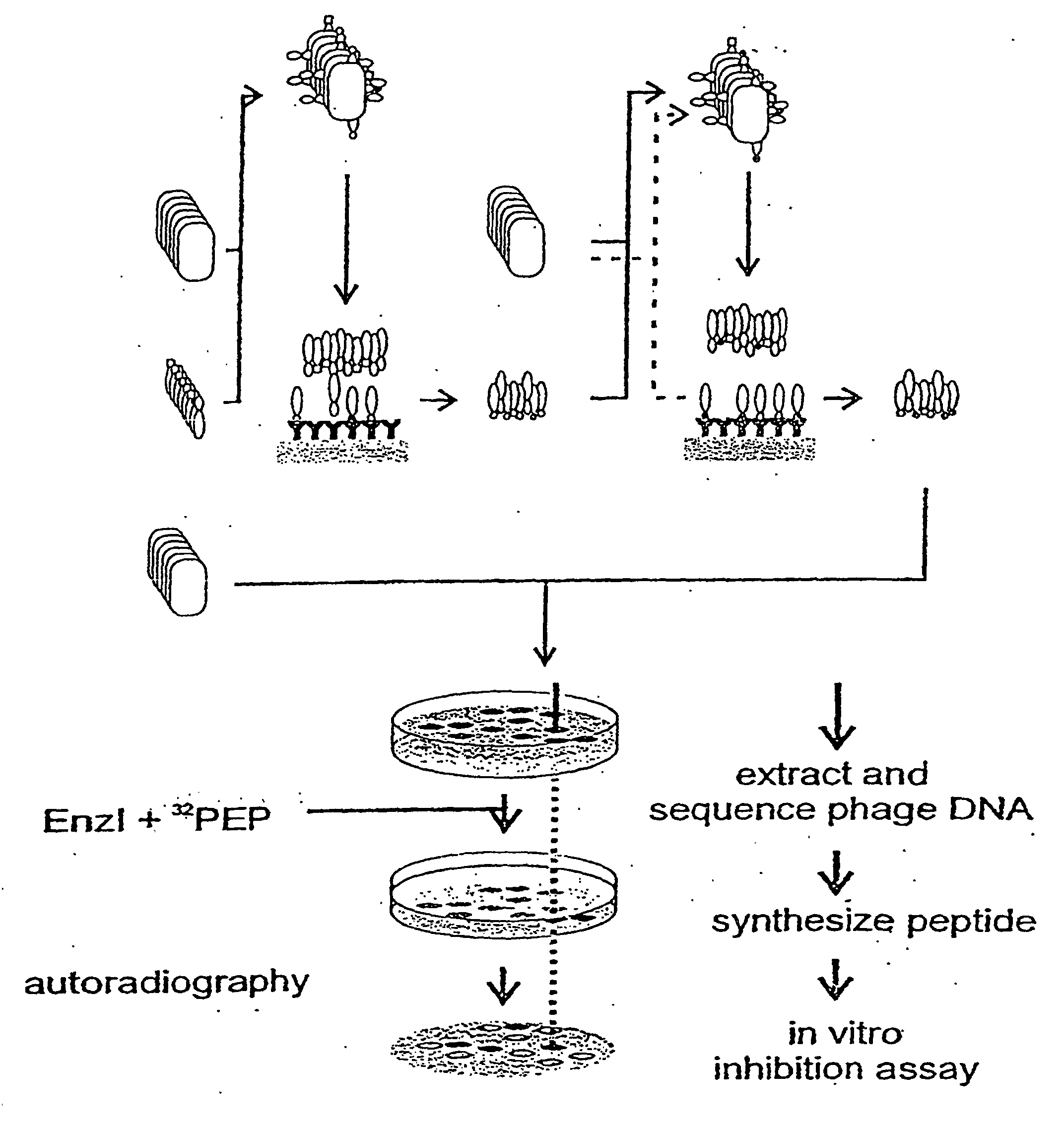
- Fig. 3. Selection of Enzyme I-binding peptides from phage display libraries. M13 phages were panned with enzyme I (top of figure) to enrich for enzyme I binding phages. E. coli clones transfected with "high affinity" phages were then grown on nitrocellulose sheets, and the secreted and nitrocellulose bound phages were incubated over night with an overlay of enzyme I in the presence of [32P]phosphoenolpyruvate. Phosphorylated phage were identified with the PhosphorImager and sequenced. The phage displayed sequences which became phosphorylated 25 to 30 fold over average were chemically synthesized and their biological activity was characterized. Results are summarized in Table 1.
- Some of the peptides from the phage display library which became phosphorylated by Enzyme I have been synthesized by standard methods in soluble form and characterized. They inhibit the in vitro phosphotransferase activity with 1C50 of between 60 µM and over 10 mM (Table 1). The second part of Fig. 3 an the previous page illustrates this procedure.
- Binding of enzyme I to immobilized peptides, monitored with [125I]-enzymel. Enzyme I was radiolabeled with 125I by the lactoperoxidase-catalyzed reaction following the procedure reported for gonadotropins (Sairam, M.R. 1979). Briefly, 1 mg of Enzyme I in 200 µl of buffer was iodinated with 10 µl (0.5 mCi) of Na 125I. The reaction was conducted at 34°C for 60 min in a water bath with gentle shaking throughout. The reaction was terminated by the addition of potassium iodide. Iodinated protein was separated from free iodine on a Sephadex G-50 column presaturated with BSA and equilibrated with 50 mM PBS (pH 7.2).
- Cellulose membranes with immobilized peptides were incubated with 50 mM TBS containing 3% BSA for 1 hr with shaking at room temperature. Membranes were subsequently washed and further incubated for 1 hr with 50 mM PBS containing 0.15% casein, another blocking agent to block the non-specific binding sites. These membranes were then incubated with [125I]-enzyme I in PBS containing 0.15% casein for 2 hr. Non-specifically and weakly bound [125I]-enzyme I was removed from the membranes by washing them with PBS containing 0.1% Tween. Radioactivity was determined by autoradiography. Enzyme I dependent phosphorylation of immobilized peptides [32P]- PEP was prepared as described by Roossien et al., 1983. Cellulose membranes with immobilized peptide libraries were treated with the blocking agents as described above. These libraries were then incubated for 30 min at room temperature with [32P]- PEP in the presence of catalytic amounts of enzyme I. The phosphorylated species were identified with a PhosphoImager ( Molecular Dynamics).
- Inhibition of phosphotransferase activity by soluble peptides. This assay measures the inhibition of enzyme I dependent sugar phosphorylation activity. The IC50 is defined as the concentration of the inhibitor required for 50% inhibition of the PTS activity. The purified N-acetylglucosamine transporter (IIGlcNAc) of E.coli (Mukhija and Erni, 1996) was used as the sugar specific component. The reaction mixture contained per 100 µl, 100 µg phosphatidyl glycerol from egg yolk (Sigma), 50 mM KPi, pH 7.5, 2.5 mM dithiothreitol, 2.5 mM NaF, 5 mM MgCl2, mM phosphoenolpyruvate (potassium salt, Sigma), 0.5 mM [U-14C]GlcNAc (New England Nuclear; 56.3 mCi/mmol, diluted to 1000 cpm/nmol), and 3 µM each of purified HPr and enzyme I (Mao et al., 1995). The reaction was started by addition of 0.1 µg of IIGlcNAc. After incubation for 30 min at 37°C, the reaction was stopped by adding 1 ml ice-cold water. GlcNAc-6-Phosphate was separated from free GlcNAc by anion-exchange chromatography on AG-1-X2 (50-100 mesh, Biorad). Activity is expressed as nmoles of sugar-P formed after 30 min. The HPr concentration was adjusted such that it was rate limiting in the overall phosphotransferase reaction. The inhibitory peptides were added to concentrations of 25 µM to 10 mM. Phosphorylation of GlcNAc in the absences of peptide was taken as 100%.
- Pyruvate formation coupled to lactate dehydrogenase. Pyruvate formed from phosphoenolpyruvate by enzyme I was converted to lactate in the presence of lactate dehydrogenase, and the concomittant consumption of NADH was followed spectrophotometrically at 340 nm. 1 ml of the reaction mixture contained 0.8 mM PEP, 0.2 mM NADH, 30 µg lactate dehydrogenase, 50 mM KPi pH 7.5, 2.5 mM dithiothreitol, 2.5 mM NaF, 5 mM MgCl2, and as substrate either 22.5 µM HPr or 50 to 150 µM of the peptide. The reaction was started by the addition of 0.75 µM enzyme I. To measure the inhibition of enzyme I by the peptides, peptides were added at different concentrations. The inhibitory peptides are phosphorylated by enzyme I at a much slower rate than HPr.
- Growth inhibition assay. An over night culture of E. coli K-12 UT580 grown in LB-medium was diluted 1:30 into soft agar (0.7% agarose in LB broth) at 40°C, and 3 ml of the diluted cell suspension were spread an LB agar plates. Filter discs (4.4 mm diameter) were placed in regular intervals on the solidified top agar. To each filter was added 10 µl of one of the different peptide stock solutions (20 µM to 80 µM). The agar plates were incubated over night at 37°C. The area of the clear zone around each filter disc was determined. The clear area is taken as a measure for the growth inhibitory activity of the peptide. Compare Fig 4 and Table 1 below and on the following two pages.


- Fig. 4. Growth inhibition of E. coli K12 UT580. Peptides were added to filter discs and placed onto a cell culture in LB soft agar. The clear zones are indicative of growth inhibition (panel A). The growth inhibitory activity of the peptides is inversely correlated with their IC50 (panel B). The numbers refer to the peptides listed in Table 1.
Biological activities of peptides identified from support-based combinatorial libraries. ID peptide sequence inhibition IC50 (µM) uncoupling P;Ipeptide/h growth inhibition MIC (µM) 1 KKFHLRK 150 0.49 125.8 2 KKFALRK none 3 KKFDLRK 820 0.05 5 KKWHLRKR 30 0.3 63.0 6 KGWHKRKK 530 0.23 7 KKWHRRKK 300 0.29 8 KKWHKRKK 440 0.215 9 KKFHIRKR 160 0.31 10 PNGLHTRPA 2600 0.07 11 Bio-PNGLHTRPA 4300 - 12 W H KR 4900 - 13 Ac-WH KR 5800 - HPr protein 2.70 tetracycline 2.5 Biological activities of peptides identified by phage display ID peptide sequence inhibition IC50 (µM) uncoupling Pi/peptide (x IC50/h) growth inhibition (area/µM) 14 GLRFGKTRVHYLVLG 25 3.2 3.65 15 SGRKSTRVHHWLLVL 60 6.6 3.05 16 KMSRHRKPGA 100 22.2 17 SSLRGHRWVY 120 50.3 18 KISRHGKRGK 140 1.9 19 KISRHGRPTG 1065 479.3 20 RIHFIPRRGR 280 39.6 0.77 21 RLHYLF 375 64.2 0.90 22 DGARLHYLF 1200 132.3 23 AcDGARLHYLF 7600 15.2 24 RHWSIF 400 1.30 25 RHRTLF 410 26 RHYLLF 500 0.83 27 RHITSL 650 28 RHITLF 1380 0.27 29 MSRHRN 760 143.8 30 PNGLHTRPA 2600 31 MRLLKTLCFGLCG 3500 32 AcMRLLKTLCFVGLCG >10'000 -
- Ammer, J., Brennenstuhl, M., Schindler, P., Höltje, J.V., and Zähler, H. (1979) Phosphorylation of streptozotocin during uptake via the phosphoenolpyruvate:sugar phosphotransferase system in Escherichia coli. Antimicrob. Agents Chemother. 16, 801-807
- Amster-Choder, O. and Wright, A. (1993) Transcriptional Regulation of the bgl Operon of Escherichia-Coli involves Phosphotransferase system-mediated phosphorylation of a Transcriptional Antiterminator. J. Cell. Bioch. 51, 83-90
- Buhr, A., Daniels, G.A., and Erni, B. (1992) The glucose transporter of Escherichia coli. mutants with impaired translocation activity that retain phosphorylation activity. J. Biol. Chem. 267, 3847-3851
- Christian, R.B., Zuckermann,R.N., Kerr,J.M., Wang, L., and Malcolm, B.A. (1992) Simplified methods for construction, assessment and rapid screening of peptide libraries in bacteriophages. J. Mol. Biol. 227, 711-718.
- Curtiss, R. and Kelly, S.M. (1987) Salmonella typhimurium deletion mutants lacking adenylate cyclase and cyclic AMP receptor protein are avirulent and immunogenic. Infect. Immun. 55,30355-3043.
- Curtiss, R., Goldschmidt, R.M., Fletchall, N.B., and Kelly, S.M. (1988) A virulent Salmonella typhimurium delta cya delta crp oral vaccine strains expressing a streptococcal colonization and virulence antigen. Vacine 6, 155-160.
- Eberstadt, M., Golic Grdadolnik, S., Gemmecker, G. Kessler, H., Buhr, A. and Erni, B (1996) Solution structure of the IIB domain of the glucose transporter of Escherichia coli. Biochemistry, in press
- Eberstadt, M., Gemmecker, G. Golic Grdadolnik, S.,Kessler, H., Buhr, A., Lanz, R. and Erni, B. (1996) The glucose trasnporter of Escherichia coli: NMR characterization of the phosphocysteine form of the IIBGlc domain and its binding interface with the IIAGlc subunit. submitted
- Erni, B. (1992) Grouptranslocation of glucose and other carbohydrates by the bacterial phosphotransferase system. Int. Rev. Cytol. 137A, 127-148
- Emi, B., Trachsel, H., Postma, P.W., and Rosenbusch, J.P. (1982) Bacterial phosphotransferase system. Solubilization and purification of the glucose-specific enzyme II from membranes of Salmonella typhimurium. J. Biol. Chem. 257, 13726-13730
- Felici, F., Luzzago, A., Folgori, A., and Cortese, R. (1993) Mimicking of discontinuous epitopes by phage-displayed peptides, II. Selection of clones recognized by a protective monoclonal antibody against the Bordetella pertussis toxin from phage peptide libraries. Gene 128, 21-27
- Hurley, J.H., Faber, H.R., Worthylake, D., Meadow, N.D., Roseman, S., Pettigrew, D.W., and Remington, S.J. (1993) Structure of the regulatory romplex of Escherichia coli IIGlc with glycerol kinase. Science 259, 673-677.
- Jia, Z.C., Quail, J.W., Waygood, E.B., and Delbaere, L.T.J. (1993a). The 2.0-angstrom resolution structure of Escherichia coli Histidine-Containing Phosphocarrier Protein HPr - A redetermination. J. Biol. Chem. 268, 22490-22501.
- Jia, Z.C., Vandonselaar, M., Quail, J.W., and Delbaere, L.T.J. (1993b) Active-Centre torsion-angle strain revealed in 1.6 Angstrom-resolution structure of histidine containing phosphocarrier protein. Nature 361, 94-97.
- Kalbitzer, H.R. and Hengstenberg, W. (1993) The Solution Structure of the histidine containing protein (HPr) from Staphylococcus-aureus as determined by 2 dimensional H-1-NMR Spectroscopy. Eur. J. Biochem. 216, 205-214.
- Kelly, S.M., Bosecker, B.A. Curtiss, R. (1992) Characterization of protective properties of attenuated mutants of Salmonella choleraesuis. Infect. Immun. 60, 4881-4890
- Lengeler, J.W., Jahreis, K. and Wehmeier, U.F. (1994) Enzymes II of the phosphoenol pyruvate-dependent phosphotransferase systems: their structure and function in carbohydrate transport. Biochem. Biophys. Acta 1188, 1-28.
- Liao, D. I., Silverton, E., Seok, Y.J., Lee B. R., Peterkofsky, A., Davies. D.R. (1996) The first step in sugar transport: crystal structure of the amino terminal domain of enzyme I of the E. coli PEP: sugar phosphotransferase system and a model of the phosphotransfer complex with HPr. Structure, 4, 861-872.
- LiCalsi, C., Crocenzi, T. S., Freire, E.and Roseman, S. (1991) Sugar transport by the bacterial phosphotransferase system. Structural and thermodynamic domains of Enzyme I of Salmonella typhimurium. J. Biol. Chem. 266, 19519-19527
- Lux, R., Jahreis, K., Bettenbrook, K., Parkinson, J.S. and Lengeler, J.W. (1995) A communication link between the PTS carbohydrate uptake System and the chemotaxis signaling pathway of Escherichia coli. Proc. Natl. Acad. Sci. USA 92, 11583-11587
- Mao, Q., Schunk, T., Flükiger, K., and Erni, B. (1995) Functional reconstitution of the purified mannose phosphotransferase system of Escherichia coli into phospholipid vesicles. J. Biol. Chem. 270, 5258-5265
- Martin-Verstraete, I., Debarbouille, M., Klier, A., and Rapoport, G. (1994) Interactions of wild-type and truncated LevR of Bacillus subtilis with the upstream activating sequence of the levanase operon. J. Mol. Biol. 241, 178-192.
- Mayahara, H., Hirano, H., Saitoh,T., and Ogawa, K. (1967) The new lead citrate method for the ultrachemical demonstration of activity of non-specific alkaline phosphatase. Histochemie 11, 88.
- Meins, M., Jenö, P., Müller, D., Richter, W.J., Rosenbusch, J.P., and Erni, B. (1993) Cysteine phosphorylation of the glucose transporter of Escherichia coli. J. Biol. Chem. 268, 11604-11609
- Meadow, N.D., Fox, D.K. and Roseman, S. (1990) The bacterial phosphoenolpyruvate:glycose phosphotransferase system. Annu. Rev. Biochem. 59, 497-542.
- Mukhija, S. and Erni, B. (1996) Purification by Ni2+ affinity chromatography, and functional reconstitution of the transporter for N-acetylglucosamine of Escherichia coli. J. Biol. Chem. 271, 14819-14824
- Nishikawa,Y., Scotland, S.M., Smith, H.R. Willshaw, G.A. and Rowe, B. (1995) Catabolite repression of the adhesion of Vero cytotoxin-producing Escherichia coli serogroups 01557 and 0111. Microb. Pathog. 18, 223-229.
- Postma, P.W., Lengeler, J.W. and Jacobson, G.R. (1993) Phosphoenolpyruvate: carbohydrate phosphotransferase systems in bacteria. Microbiol. Rev. 57, 543-594.
- Powell, B. S., Court, D. L., Inada, T., Nakamura, Y., Michotey, V., Cui, X., Reizer, A., Saier, M. H., and Reizer, J. (1995) Novel proteins of the phosphotransferase system encoded within the rpoN operon of Escherichia coli. Enzyme IIANtr affects growth an organic nitrogen and the conditional lethality of the erats mutant. J. Biol. Chem. 270,4822-4839
- Rajagopal, P., Waygood, E.B., and Klevit, R.E. (1994) Structural consequences of histidine phosphorylation: NMR characterization of the phosphohistidine form of histidine-containing protein from Bacillus subtilis and Escherichia coli. Biochemistry 33, 15271-15282.
- Roossien, F. F., Brink, J., and Robillard, G. T. (1983) A simple method for the synthesis of 32P phosphosphoenolpyruvate via the pyruvate kinase exchange reaction at equilibrium. Biochim. Biophys. Acta. 760,185-187
- Reizer, J., Hoischen, C., Reizer, A., Pham, T.N., and Saier, M.H. (1993) Sequence Analyses and Evolutionary Relationships Among the Energy-Coupling Proteins Enzyme-I and HPr of the Bacterial Phosphoenolpyruvate-Sugar Phosphotransferase System. Protein Sci. 2, 506-521.
- Saier, M.H. (1993). Regulatory Interactions Involving the Proteins of the Phosphotransferase System in Enteric Bacteria. Journal. of Cellular. Biochemistry 51, 62-68..
- Saier, M. H. and Reizer, J.(1994) The bacterial phosphotransferase system: New frontiers 30 years later. Molecular. Microbiology. 13, 755-764
- Saier, M.H., Chavaux, S., Deutscher, J., Reizer, J., and Ye, JJ. (1995) Protein phosphorylation and regulation of carbon metabolism in Gram-negative versus Gram-positive bacteria. TIB S 20, 267-271.
- Scott, J.K., and Smith, G.P. (1990) Searching for peptide ligands with an epitope library. Science 249, 386-390.
- Seok, Y. J., Lee, B. R., Zhu, P. P.and Peterkofsky, A.(1996) Importance of the carboxyl-terminal domain of enzyme 1 of the Escherichia. coli phosphoenolpyruvate: Sugar phosphotransferase System for phosphoryl donor specificity. Proc. Natl. Acad. Sci. USA 93,347-351
- Titgemeyer, F., Mason, R.E. and Saier, M.H. (1994) Regulation of the raffinose permease of Escherichia coli by the glucose-specific enzyme IIA of the phosphoenolpyruvate:sugar phosphotransferase system. J. Bacteriol. 176, 543-546
- van Nuland, N.A.J., Boelens, R., Scheek, R.M., and Robillard, G.T. (1995) High-resolution structure of the phosphorylated form of the histidine-containing phosphocarrier protein HPr from Escherichia coli determined by restrained molecular dynamics from NMR-NOE data. J. Mol. Biol. 246, 180-193.
- van Nuland, N.A.J., Grotzinger, J., Dijkstra, K., Scheek, R.M., and Robillard, G.T. (1992) Determination of the 3 -dimensional solution structure of the histidine containing phosphocarrier protein HPr from Escherichia coli using multidimensional NMR spectroscopy. Eur. J. Bioch. 210, 881-891.
- van Nuland, N.A.J., Hangyi, I.W., van Schaik, R.C., Berendsen, H.J.C., van Gunsteren, W.F., Scheek, R.M., and Robillard, G.T. (1994) The High-Resolution Structure of the Histidine-Containing Phosphocarrier Protein HPr from Escherichia coli determined by restrained molecular dynamics from Nuclear Magnetic Resonance Nuclear Overhauser effect data. J. Mol. Biol
- Phosphorylation of a Transcriptional Antiterminator. J. Cell. Bioch. 51, 83-90.
- Weber, L., Wallbaum, S., Broger, C., and Gubernator, K. (1995) Optimierung der biologischen Aktivität von kombinatorischen Verbindungsbibliotheken durch einen genetischen Algorithmus. Angew. Chem. 107, 2452-2454
- Wittekind, M., Rajagopal, P., Branchini, B. R., Reizer, J., Saier, M. H. Jr. & Klevit, R. E. (1992) Solution structure of the phosphocarrier protein HPr from Bacillus subtilis by two-dimensional NMR spectroscopy, Protein Sci. 1, 1363-1376.
- Yamamoto, K (1988) Alkaline phosphatase in Electron microscopic cytochemistry and immunochemistry in biomedicine (Ogawa, K., and Barka, T. eds.) CRC Press, Boca Raton.
-
-
- (1) GENERAL INFORMATION:
- (i) APPLICANT:
- (A) NAME: ARPIDA
- (B) STREET: Dammstrasse 36
- (C) CITY: Muenchenstein
- (D) STATE: BL
- (E) COUNTRY: Switzerland
- (F) POSTAL CODE (ZIP): CH-4142
- (G) TELEPHONE: +41 61 417 96 60
- (H) TELEFAX: +41 61 417 96 61
- (ii) TITLE OF INVENTION: Target System
- (iii) NUMBER OF SEQUENCES: 32
- (iv) COMPUTER READABLE FORM:
- (A) MEDIUM TYPE: Floppy disk
- (B) COMPUTER: IBM PC compatible
- (C) OPERATING SYSTEM: PC-DOS/MS-DOS
- (D) SOFTWARE: PatentIn Release #1.0, Version #1.30 (EPO)
- (2) INFORMATION FOR SEQ NO: 1:
- (i) SEQUENCE CHARACTERISTICS:
- (A) LENGTH: 7 amino acids
- (B) TYPE: amino acid
- (D) TOPOLOGY: linear
- (ii) MOLECULE TYPE: peptide
- (xi) SEQUENCE DESCRIPTION: SEQ ID NO: 1:

- (2) INFORMATION FOR SEQ ID NO: 2:
- (i) SEQUENCE CHARACTERISTICS:
- (A) LENGTH: 7 amino acids
- (B) TYPE: amino acid
- (D) TOPOLOGY: linear
- (ii) MOLECULE TYPE: peptide
- (xi) SEQUENCE DESCRIPTION: SEQ ID NO: 2:

- (2) INFORMATION FOR SEQ ID NO: 3:
- (i) SEQUENCE CHARACTERISTICS:
- (A) LENGTH: 7 amino acids
- (B) TYPE: amino acid
- (D) TOPOLOGY: linear
- (ii) MOLECULE TYPE: peptide
- (xi) SEQUENCE DESCRIPTION: SEQ ID NO: 3:

- (2) INFORMATION FOR SEQ ID NO: 4:
- (i) SEQUENCE CHARACTERISTICS:
- (A) LENGTH: 12 amino acids
- (B) TYPE: amino acid
- (D) TOPOLOGY: linear
- (ii) MOLECULE TYPE: peptide
- (ix) FEATURE
- (A) NAME/KEY: peptide
- (B) LOCATION: 1
- (C) OTHER INFORMATION: /label= Xaa /note= "wherein Xaa can be any amino acid"
- (ix) FEATURE
- (A) NAME/KEY: peptide
- (B) LOCATION: 2
- (C) OTHER INFORMATION: /label= Xaa /note= "wherein Xaa can be any amino acid"
- (ix) FEATURE
- (A) NAME/KEY: peptide
- (B) LOCATION: 11
- (C) OTHER INFORMATION: /label= Xaa /note= "wherein Xaa can be any amino acid"
- (ix) FEATURE
- (A) NAME/KEY: peptide
- (B) LOCATION: 12
- (C) OTHER INFORMATION: /label= Xaa /note= "wherein Xaa can be any amino acid"
- (xi) SEQUENCE DESCRIPTION: SEQ ID NO: 4:

- (2) INFORMATION FOR SEQ ID NO: 5:
- (i) SEQUENCE CHARACTERISTICS:
- (A) LENGTH: 8 amino acids
- (B) TYPE: amino acid
- (D) TOPOLOGY: linear
- (ii) MOLECULE TYPE: peptide
- (xi) SEQUENCE DESCRIPTION: SEQ ID NO: 5:
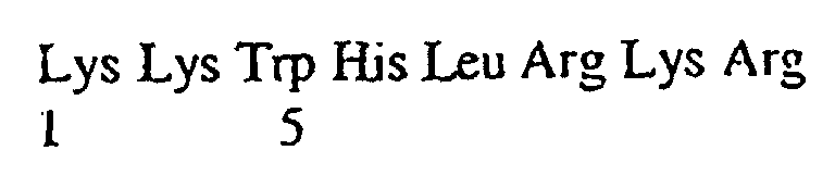
- (2) INFORMATION FOR SEQ ID NO: 6:
- (i) SEQUENCE CHARACTERISTICS:
- (A) LENGTH: 8 amino acids
- (B) TYPE: amino acid
- (D) TOPOLOGY: linear
- (ii) MOLECULE TYPE: peptide
- (xi) SEQUENCE DESCRIPTION: SEQ ID NO: 6:

- (2) INFORMATION FOR SEQ ID NO: 7:
- (i) SEQUENCE CHARACTERISTICS:
- (A) LENGTH: 8 amino acids
- (B) TYPE: amino acid
- (D) TOPOLOGY: linear
- (ii) MOLECULE TYPE: peptide
- (xi) SEQUENCE DESCRIPTION: SEQ ID NO: 7:
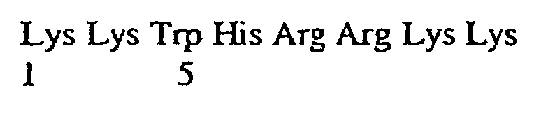
- (2) INFORMATION FOR SEQ ID NO: 8:
- (i) SEQUENCE CHARACTERISTICS:
- (A) LENGTH: 8 amino acids
- (B) TYPE: amino acid
- (D) TOPOLOGY: linear
- (ii) MOLECULE TYPE: peptide
- (xi) SEQUENCE DESCRIPTION: SEQ ID NO: 8:
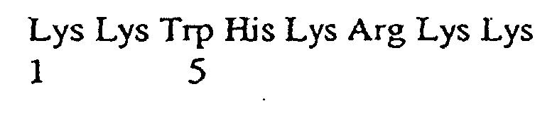
- (2) INFORMATION FOR SEQ ID NO: 9:
- (i) SEQUENCE CHARACTERISTICS:
- (A) LENGTH: 8 amino acids
- (B) TYPE: amino acid
- (D) TOPOLOGY: linear
- (ii) MOLECULE TYPE: peptide
- (xi) SEQUENCE DESCRIPTION: SEQ ID NO: 9:

- (2) INFORMATION FOR SEQ ID NO: 10:
- (i) SEQUENCE CHARACTERISTICS:
- (A) LENGTH: 9 amino acids
- (B) TYPE: amino acid
- (D) TOPOLOGY: linear
- (ii) MOLECULE TYPE: peptide
- (xi) SEQUENCE DESCRIPTION: SEQ NO: 10:

- (2) INFORMATION FOR SEQ ID NO: 11:
- (i) SEQUENCE CHARACTERISTICS:
- (A) LENGTH: 9 amino acids
- (B) TYPE: amino acid
- (D) TOPOLOGY: linear
- (ii) MOLECULE TYPE: peptide
- (ix) FEATURE
- (A) NAME/KEY: peptide
- (B) LOCATION: 1
- (C) OTHER INFORMATION: /label= Pro /note= "wherein a biotin molecule is covalently linked to Pro at position 1"
- (xi) SEQUENCE DESCRIPTION: SEQ ID NO: 11:

- (2) INFORMATION FOR SEQ ID NO: 12:
- (i) SEQUENCE CHARACTERISTICS:
- (A) LENGTH: 4 amino acids
- (B) TYPE: amino acid
- (D) TOPOLOGY: linear
- (ii) MOLECULE TYPE: peptide
- (xi) SEQUENCE DESCRIPTION: SEQ ID NO: 12

- (2) INFORMATION FOR SEQ ID NO: 13
- (i) SEQUENCE CHARACTERISTICS:
- (A) LENGTH: 4 amino acids
- (B) TYPE: amino acid
- (D) TOPOLOGY: linear
- (ii) MOLECULE TYPE: peptide
- (ix) FEATURE
- (A) NAME/KEY: peptide
- (B) LOCATION: 1
- (C) OTHER INFORMATION: /label= Trp /note= "wherein Trp is acetylated"
- (xi) SEQUENCE DESCRIPTION: SEQ ID NO: 13:

- (2) INFORMATION FOR SEQ NO: 14:
- (i) SEQUENCE CHARACTERISTICS:
- (A) LENGTH: 15 amino acids
- (B) TYPE: amino acid
- (D) TOPOLOGY: linear
- (ii) MOLECULE TYPE: peptide
- (xi) SEQUENCE DESCRIPTION: SEQ ID NO: 14:

- (2) INFORMATION FOR SEQ ID NO: 15:
- (i) SEQUENCE CHARACTERISTICS:
- (A) LENGTH: 15 amino acids
- (B) TYPE: amino acid
- (D) TOPOLOGY: linear
- (ii) MOLECULE TYPE: peptide
- (ix) FEATURE
- (A) NAME/KEY: peptide
- (B) LOCATION: 11
- (C) OTHER INFORMATION: /label= Trp /note= "wherein Trp is acetylated"
- (xi) SEQUENCE DESCRIPTION: SEQ ID NO: 15:

- (2) INFORMATION FOR SEQ ID NO: 16:
- (i) SEQUENCE CHARACTERISTICS:
- (A) LENGTH: 10 amino acids
- (B) TYPE: amino acid
- (D) TOPOLOGY: linear
- (ii) MOLECULE TYPE: peptide
- (xi) SEQUENCE DESCRIPTION: SEQ ID NO: 16:

- (2) INFORMATION FOR SEQ ID NO: 17:
- (i) SEQUENCE CHARACTERISTICS:
- (A) LENGTH: 10 amino acids
- (B) TYPE: amino acid
- (D) TOPOLOGY: linear
- (ii) MOLECULE TYPE: peptide
- (xi) SEQUENCE DESCRIPTION: SEQ NO: 17:

- (2) INFORMATION FOR SEQ ID NO: 18:
- (i) SEQUENCE CHARACTERISTICS:
- (A) LENGTH: 10 amino acids
- (B) TYPE: amino acid
- (D) TOPOLOGY: linear
- (xi) SEQUENCE DESCRIPTION: SEQ ID NO: 18:

- (2) INFORMATION FOR SEQ ID NO: 19:
- (i) SEQUENCE CHARACTERISTICS:
- (A) LENGTH: 10 amino acids
- (B) TYPE: amino acid
- (D) TOPOLOGY: linear
- (ii) MOLECULE TYPE: peptide
- (xi) SEQUENCE DESCRIPTION: SEQ NO: 19:

- (2) INFORMATION FOR SEQ ID NO: 20:
- (i) SEQUENCE CHARACTERISTICS:
- (A) LENGTH: 10 amino acids
- (B) TYPE: amino acid
- (D) TOPOLOGY: linear
- (ii) MOLECULE TYPE: peptide
- (xi) SEQUENCE DESCRIPTION: SEQ ID NO: 20:

- (2) INFORMATION FOR SEQ ID NO: 21:
- (i) SEQUENCE CHARACTERISTICS:
- (A) LENGTH: 6 amino acids
- (B) TYPE: amino acid
- (D) TOPOLOGY: linear
- (ii) MOLECULE TYPE: peptide
- (xi) SEQUENCE DESCRIPTION: SEQ ID NO: 21:
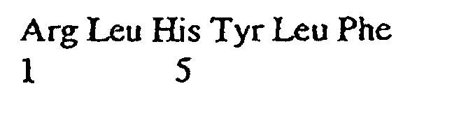
- (2) INFORMATION FOR SEQ ID NO: 22:
- (i) SEQUENCE CHARACTERISTICS:
- (A) LENGTH: 9 amino acids
- (B) TYPE: amino acid
- (D) TOPOLOGY: linear
- (ii) MOLECULE TYPE: peptide
- (xi) SEQUENCE DESCRIPTION: SEQ ID NO: 22:

- (2) INFORMATION FOR SEQ ID NO: 23:
- (i) SEQUENCE CHARACTERISTICS:
- (A) LENGTH: 9 amino acids
- (B) TYPE: amino acid
- (D) TOPOLOGY: linear
- (ii) MOLECULE TYPE: peptide
- (ix) FEATURE
- (A) NAME/KEY: peptide
- (B) LOCATION: 1
- (C) OTHER INFORMATION: /label= Xaa /note= "wherein Asp is acetylated"
- (xi) SEQUENCE DESCRIPTION: SEQ ID NO: 23:

- (2) INFORMATION FOR SEQ ID NO: 24:
- (i) SEQUENCE CHARACTERISTICS:
- (A) LENGTH: 6 amino acids
- (B) TYPE: amino acid
- (D) TOPOLOGY: linear
- (ii) MOLECULE TYPE: peptide
- (xi) SEQUENCE DESCRIPTION: SEQ ID NO: 24:
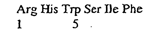
- (2) INFORMATION FOR SEQ ID NO: 25:
- (i) SEQUENCE CHARACTERISTICS:
- (A) LENGTH: 6 amino acids
- (B) TYPE: amino acid
- (D) TOPOLOGY: linear
- (ii) MOLECULE TYPE: peptide
- (xi) SEQUENCE DESCRIPTION: SEQ ID NO: 25:

- (2) INFORMATION FOR SEQ ID NO: 26:
- (i) SEQUENCE CHARACTERISTICS:
- (A) LENGTH: 6 amino acids
- (B) TYPE: amino acid
- (D) TOPOLOGY: linear
- (ii) MOLECULE TYPE: peptide
- (xi) SEQUENCE DESCRIPTION: SEQ ID NO: 26:

- (2) INFORMATION FOR SEQ ID NO: 27:
- (i) SEQUENCE CHARACTERISTICS:
- (A) LENGTH: 6 amino acids
- (B) TYPE: amino acid
- (D) TOPOLOGY: linear
- (ii) MOLECULE TYPE: peptide
- (xi) SEQUENCE DESCRIPTION: SEQ ID NO: 27:

- (2) INFORMATION FOR SEQ NO: 28:
- (i) SEQUENCE CHARACTERISTICS:
- (A) LENGTH: 6 amino acids
- (B) TYPE: amino acid
- (D) TOPOLOGY: linear
- (ii) MOLECULE TYPE: peptide
- (xi) SEQUENCE DESCRIPTION: SEQ ID NO: 28:

- (2) INFORMATION FOR SEQ ID NO: 29:
- (i) SEQUENCE CHARACTERISTICS:
- (A) LENGTH: 6 amino acids
- (B) TYPE: amino acid
- (D) TOPOLOGY: linear
- (ii) MOLECULE TYPE: peptide
- (xi) SEQUENCE DESCRIPTION: SEQ NO: 29:
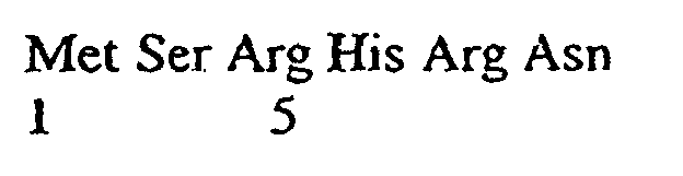
- (2) INFORMATION FOR SEQ ID NO: 30:
- (i) SEQUENCE CHARACTERISTICS:
- (A) LENGTH: 9 amino acids
- (B) TYPE: amino acid
- (D) TOPOLOGY: linear
- (ii) MOLECULE TYPE: peptide
- (xi) SEQUENCE DESCRIPTION: SEQ ID NO: 30:

- (2) INFORMATION FOR SEQ ID NO: 31:
- (i) SEQUENCE CHARACTERISTICS:
- (A) LENGTH: 13 amino acids
- (B) TYPE: amino acid
- (D) TOPOLOGY: linear
- (ii) MOLECULE TYPE: peptide
- (xi) SEQUENCE DESCRIPTION: SEQ ID NO: 31:

- (2) INFORMATION FOR SEQ ID NO: 32:
- (i) SEQUENCE CHARACTERISTICS:
- (A) LENGTH: 14 amino acids
- (B) TYPE: amino acid
- (D) TOPOLOGY: linear
- (ii) MOLECULE TYPE: peptide
- (ix) FEATURE
- (A) NAME/KEY: peptide
- (B) LOCATION: 1
- (C) OTHER INFORMATION: /label= Met
/note= "wherein Met is acetylated" - (xi) SEQUENCE DESCRIPTION: SEQ ID NO: 32:

-
Claims (9)
- Drug assay for the identification of peptides which are specific antimicrobials comprising uncoupling or inhibition of the bacterial phosphotransferase system by eithera) using such peptides as a phosphorylgroup acceptor which is not part of the system and hydrolyses spontaneously or under the influence of non-specific endogenous phosphohydrolases orb) inhibiting the phosphotransferase system activity with such peptides which block or inactivate at least one of the enzymes of the phosphotransferase system.
- Drug assay according to claim 1 comprising uncoupling or inhibition of the bacterial phosphotransferase system by using such peptides as a phosphorylgroup acceptor which is not part of the system and hydrolyses spontaneously.
- Drug assay according to claim 1 or 2 comprising uncoupling of the bacterial phosphotransferase system by using such peptides as a phosphorylgroup acceptor which is not part of the system and hydrolyses under the influence of non-specific endogenous phosphohydrolases.
- Drug assay according to claim 1 comprising inhibiting the phosphotransferase system activity with said peptides which block or inactivate at least one of the enzymes of the phosphotransferase system.
- Drug assay according to claim 1 or 4 comprising inhibition of the bacterial phosphotransferase system with said peptides which block at least one of the enzymes of the phosphotransferase system.
- Drug assay according to claim 1 or 4 comprising inhibition of the bacterial phosphotransferase system with said peptides which inactivate at least one of the enzymes of the phosphotransferase system.
- Peptide sequences of the formulae:










- Peptide sequences of the formulae:
















- Pharmaceutical composition comprising one or more of the peptides according to any one of claims 7 and 8.
Priority Applications (1)
| Application Number | Priority Date | Filing Date | Title |
|---|---|---|---|
| EP19980101704 EP0866075B1 (en) | 1997-02-19 | 1998-02-02 | Method to treat microbial infections by uncoupling of phosphotransferase system and appropriate agents therefor |
Applications Claiming Priority (3)
| Application Number | Priority Date | Filing Date | Title |
|---|---|---|---|
| EP97102616 | 1997-02-19 | ||
| EP97102616 | 1997-02-19 | ||
| EP19980101704 EP0866075B1 (en) | 1997-02-19 | 1998-02-02 | Method to treat microbial infections by uncoupling of phosphotransferase system and appropriate agents therefor |
Publications (3)
| Publication Number | Publication Date |
|---|---|
| EP0866075A2 EP0866075A2 (en) | 1998-09-23 |
| EP0866075A3 EP0866075A3 (en) | 2000-06-07 |
| EP0866075B1 true EP0866075B1 (en) | 2004-10-27 |
Family
ID=26145245
Family Applications (1)
| Application Number | Title | Priority Date | Filing Date |
|---|---|---|---|
| EP19980101704 Expired - Lifetime EP0866075B1 (en) | 1997-02-19 | 1998-02-02 | Method to treat microbial infections by uncoupling of phosphotransferase system and appropriate agents therefor |
Country Status (1)
| Country | Link |
|---|---|
| EP (1) | EP0866075B1 (en) |
Families Citing this family (2)
| Publication number | Priority date | Publication date | Assignee | Title |
|---|---|---|---|---|
| WO2001098540A2 (en) * | 2000-06-22 | 2001-12-27 | San Diego State University Foundation | Recombination modulators and methods for their production and use |
| US20040110963A1 (en) * | 2001-01-22 | 2004-06-10 | Kaspar Burri | Novel hydrazones |
Family Cites Families (1)
| Publication number | Priority date | Publication date | Assignee | Title |
|---|---|---|---|---|
| DE4401868C1 (en) * | 1994-01-22 | 1995-02-16 | Lobbe Xenex Gmbh | Microbial method for detecting pollutants |
-
1998
- 1998-02-02 EP EP19980101704 patent/EP0866075B1/en not_active Expired - Lifetime
Also Published As
| Publication number | Publication date |
|---|---|
| EP0866075A2 (en) | 1998-09-23 |
| EP0866075A3 (en) | 2000-06-07 |
Similar Documents
| Publication | Publication Date | Title |
|---|---|---|
| Matsushita et al. | Histidine kinases as targets for new antimicrobial agents | |
| Boudreau et al. | Messenger functions of the bacterial cell wall-derived muropeptides | |
| Giefing et al. | The pneumococcal eukaryotic-type serine/threonine protein kinase StkP co-localizes with the cell division apparatus and interacts with FtsZ in vitro | |
| US8575306B2 (en) | Target and method for inhibition of bacterial RNA polymerase | |
| Han et al. | The two-component system Ihk/Irr contributes to the virulence of Streptococcus suis serotype 2 strain 05ZYH33 through alteration of the bacterial cell metabolism | |
| Reizer et al. | Sequence analyses and evolutionary relationships among the energy‐coupling proteins enzyme I and HPr of the bacterial phosphoenolpyruvate: Sugar phosphotransferase system | |
| S Paparella et al. | Structure guided design of biotin protein ligase inhibitors for antibiotic discovery | |
| Mukhija et al. | Phage display selection of peptides against enzyme I of the phosphoenolpyruvate–sugar phosphotransferase system (PTS) | |
| Jukič et al. | Towards discovery of inhibitors of the undecaprenyl-pyrophosphate phosphatase BacA by virtual high-throughput screening | |
| Schlösser et al. | A Lipid‐Anchored Binding Protein is a Component of an ATP‐Dependent Cellobiose/Cellotriose‐Transport System from the Cellulose Degrader Streptomyces reticuli | |
| US20120322056A1 (en) | Rna-exit-channel: target and method for inhibition of bacterial rna polymerase | |
| US9890111B2 (en) | Crystal structure of bifunctional transglycosylase PBP1b from E. coli and inhibitors thereof | |
| Stephenson et al. | Histidine kinase-mediated signal transduction systems of pathogenic microorganisms as targets for therapeutic intervention | |
| WO2005074986A2 (en) | Bioactive species capable of interfering with a microbial toxin-antitoxin complex and methods for evaluation and use of said bioactive species | |
| EP0866075A2 (en) | Method to treat microbial infections by uncoupling of phosphotransferase system and appropriate agents therefor | |
| US6777176B1 (en) | Target system | |
| US20030023032A1 (en) | LuxO-sigma54 interactions and methods of use | |
| US6448224B1 (en) | Antibiotics and methods of using the same | |
| US6245502B1 (en) | Target system | |
| Hsu et al. | Advances and prospects of analytic methods for bacterial transglycosylation and inhibitor discovery | |
| US7604975B2 (en) | Glycosylated LPXTGases and uses thereof | |
| Narita et al. | Biogenesis and membrane targeting of lipoproteins | |
| US20080057494A1 (en) | Ribosomal Rna Methyltransferases Rima:Target Validation and Processes for Deleloping an Inhibitor Assay and Identification of Candidate Inhibitors | |
| Jones | Structure, Function, and Inhibition of Peptidoglycan O-Acetyltransferase A from Staphylococcus aureus | |
| Vernet et al. | The Glycosyltransferase Domain of |
Legal Events
| Date | Code | Title | Description |
|---|---|---|---|
| PUAI | Public reference made under article 153(3) epc to a published international application that has entered the european phase |
Free format text: ORIGINAL CODE: 0009012 |
|
| AK | Designated contracting states |
Kind code of ref document: A2 Designated state(s): AT BE CH DE DK ES FI FR GB GR IE IT LI LU MC NL PT SE |
|
| AX | Request for extension of the european patent |
Free format text: AL;LT;LV;MK;RO;SI |
|
| PUAL | Search report despatched |
Free format text: ORIGINAL CODE: 0009013 |
|
| RIC1 | Information provided on ipc code assigned before grant |
Free format text: 7C 07K 7/06 A, 7C 07K 7/08 B, 7C 07K 5/10 B, 7A 61K 38/00 B, 7C 12Q 1/02 B, 7C 12Q 1/48 B, 7C 12R 1/19 B |
|
| AK | Designated contracting states |
Kind code of ref document: A3 Designated state(s): AT BE CH DE DK ES FI FR GB GR IE IT LI LU MC NL PT SE |
|
| AX | Request for extension of the european patent |
Free format text: AL;LT;LV;MK;RO;SI |
|
| AKX | Designation fees paid | ||
| 17P | Request for examination filed |
Effective date: 20001126 |
|
| RBV | Designated contracting states (corrected) |
Designated state(s): AT BE CH DE DK ES FI FR GB GR IE IT LI LU MC NL PT SE |
|
| REG | Reference to a national code |
Ref country code: DE Ref legal event code: 8566 |
|
| 17Q | First examination report despatched |
Effective date: 20021021 |
|
| GRAP | Despatch of communication of intention to grant a patent |
Free format text: ORIGINAL CODE: EPIDOSNIGR1 |
|
| GRAS | Grant fee paid |
Free format text: ORIGINAL CODE: EPIDOSNIGR3 |
|
| RAP1 | Party data changed (applicant data changed or rights of an application transferred) |
Owner name: ARPIDA AG, |
|
| GRAA | (expected) grant |
Free format text: ORIGINAL CODE: 0009210 |
|
| AK | Designated contracting states |
Kind code of ref document: B1 Designated state(s): AT BE CH DE DK ES FI FR GB GR IE IT LI LU MC NL PT SE |
|
| PG25 | Lapsed in a contracting state [announced via postgrant information from national office to epo] |
Ref country code: NL Free format text: LAPSE BECAUSE OF FAILURE TO SUBMIT A TRANSLATION OF THE DESCRIPTION OR TO PAY THE FEE WITHIN THE PRESCRIBED TIME-LIMIT Effective date: 20041027 Ref country code: FI Free format text: LAPSE BECAUSE OF FAILURE TO SUBMIT A TRANSLATION OF THE DESCRIPTION OR TO PAY THE FEE WITHIN THE PRESCRIBED TIME-LIMIT Effective date: 20041027 Ref country code: BE Free format text: LAPSE BECAUSE OF FAILURE TO SUBMIT A TRANSLATION OF THE DESCRIPTION OR TO PAY THE FEE WITHIN THE PRESCRIBED TIME-LIMIT Effective date: 20041027 Ref country code: AT Free format text: LAPSE BECAUSE OF FAILURE TO SUBMIT A TRANSLATION OF THE DESCRIPTION OR TO PAY THE FEE WITHIN THE PRESCRIBED TIME-LIMIT Effective date: 20041027 |
|
| REG | Reference to a national code |
Ref country code: GB Ref legal event code: FG4D |
|
| REG | Reference to a national code |
Ref country code: CH Ref legal event code: EP |
|
| REG | Reference to a national code |
Ref country code: CH Ref legal event code: NV Representative=s name: PATENTANWAELTE SCHAAD, BALASS, MENZL & PARTNER AG |
|
| REG | Reference to a national code |
Ref country code: IE Ref legal event code: FG4D |
|
| REF | Corresponds to: |
Ref document number: 69827179 Country of ref document: DE Date of ref document: 20041202 Kind code of ref document: P |
|
| PG25 | Lapsed in a contracting state [announced via postgrant information from national office to epo] |
Ref country code: SE Free format text: LAPSE BECAUSE OF FAILURE TO SUBMIT A TRANSLATION OF THE DESCRIPTION OR TO PAY THE FEE WITHIN THE PRESCRIBED TIME-LIMIT Effective date: 20050127 Ref country code: GR Free format text: LAPSE BECAUSE OF FAILURE TO SUBMIT A TRANSLATION OF THE DESCRIPTION OR TO PAY THE FEE WITHIN THE PRESCRIBED TIME-LIMIT Effective date: 20050127 Ref country code: DK Free format text: LAPSE BECAUSE OF FAILURE TO SUBMIT A TRANSLATION OF THE DESCRIPTION OR TO PAY THE FEE WITHIN THE PRESCRIBED TIME-LIMIT Effective date: 20050127 |
|
| PG25 | Lapsed in a contracting state [announced via postgrant information from national office to epo] |
Ref country code: ES Free format text: LAPSE BECAUSE OF FAILURE TO SUBMIT A TRANSLATION OF THE DESCRIPTION OR TO PAY THE FEE WITHIN THE PRESCRIBED TIME-LIMIT Effective date: 20050207 |
|
| PGFP | Annual fee paid to national office [announced via postgrant information from national office to epo] |
Ref country code: SE Payment date: 20050501 Year of fee payment: 8 |
|
| NLV1 | Nl: lapsed or annulled due to failure to fulfill the requirements of art. 29p and 29m of the patents act | ||
| PGFP | Annual fee paid to national office [announced via postgrant information from national office to epo] |
Ref country code: MC Payment date: 20050526 Year of fee payment: 8 |
|
| PGFP | Annual fee paid to national office [announced via postgrant information from national office to epo] |
Ref country code: LU Payment date: 20050531 Year of fee payment: 8 |
|
| ET | Fr: translation filed | ||
| PLBE | No opposition filed within time limit |
Free format text: ORIGINAL CODE: 0009261 |
|
| STAA | Information on the status of an ep patent application or granted ep patent |
Free format text: STATUS: NO OPPOSITION FILED WITHIN TIME LIMIT |
|
| 26N | No opposition filed |
Effective date: 20050728 |
|
| PG25 | Lapsed in a contracting state [announced via postgrant information from national office to epo] |
Ref country code: MC Free format text: LAPSE BECAUSE OF NON-PAYMENT OF DUE FEES Effective date: 20060228 Ref country code: LU Free format text: LAPSE BECAUSE OF NON-PAYMENT OF DUE FEES Effective date: 20060228 |
|
| PGFP | Annual fee paid to national office [announced via postgrant information from national office to epo] |
Ref country code: DE Payment date: 20070129 Year of fee payment: 10 |
|
| PGFP | Annual fee paid to national office [announced via postgrant information from national office to epo] |
Ref country code: GB Payment date: 20070221 Year of fee payment: 10 |
|
| PGFP | Annual fee paid to national office [announced via postgrant information from national office to epo] |
Ref country code: CH Payment date: 20070222 Year of fee payment: 10 |
|
| PGFP | Annual fee paid to national office [announced via postgrant information from national office to epo] |
Ref country code: IE Payment date: 20070502 Year of fee payment: 10 |
|
| PG25 | Lapsed in a contracting state [announced via postgrant information from national office to epo] |
Ref country code: PT Free format text: LAPSE BECAUSE OF NON-PAYMENT OF DUE FEES Effective date: 20050327 |
|
| PGFP | Annual fee paid to national office [announced via postgrant information from national office to epo] |
Ref country code: IT Payment date: 20070531 Year of fee payment: 10 |
|
| PGFP | Annual fee paid to national office [announced via postgrant information from national office to epo] |
Ref country code: FR Payment date: 20070216 Year of fee payment: 10 |
|
| REG | Reference to a national code |
Ref country code: CH Ref legal event code: PL |
|
| GBPC | Gb: european patent ceased through non-payment of renewal fee |
Effective date: 20080202 |
|
| PG25 | Lapsed in a contracting state [announced via postgrant information from national office to epo] |
Ref country code: LI Free format text: LAPSE BECAUSE OF NON-PAYMENT OF DUE FEES Effective date: 20080229 Ref country code: CH Free format text: LAPSE BECAUSE OF NON-PAYMENT OF DUE FEES Effective date: 20080229 |
|
| REG | Reference to a national code |
Ref country code: IE Ref legal event code: MM4A |
|
| REG | Reference to a national code |
Ref country code: FR Ref legal event code: ST Effective date: 20081031 |
|
| PG25 | Lapsed in a contracting state [announced via postgrant information from national office to epo] |
Ref country code: IE Free format text: LAPSE BECAUSE OF NON-PAYMENT OF DUE FEES Effective date: 20080204 Ref country code: DE Free format text: LAPSE BECAUSE OF NON-PAYMENT OF DUE FEES Effective date: 20080902 |
|
| PG25 | Lapsed in a contracting state [announced via postgrant information from national office to epo] |
Ref country code: FR Free format text: LAPSE BECAUSE OF NON-PAYMENT OF DUE FEES Effective date: 20080229 |
|
| PG25 | Lapsed in a contracting state [announced via postgrant information from national office to epo] |
Ref country code: GB Free format text: LAPSE BECAUSE OF NON-PAYMENT OF DUE FEES Effective date: 20080202 |
|
| PG25 | Lapsed in a contracting state [announced via postgrant information from national office to epo] |
Ref country code: IT Free format text: LAPSE BECAUSE OF NON-PAYMENT OF DUE FEES Effective date: 20080202 |