EP0857780A1 - Novel proteins remarkably expressed in liver cancer, genes encoding the same, antibodies against the same, and method for detecting the expression of the same - Google Patents
Novel proteins remarkably expressed in liver cancer, genes encoding the same, antibodies against the same, and method for detecting the expression of the same Download PDFInfo
- Publication number
- EP0857780A1 EP0857780A1 EP96930419A EP96930419A EP0857780A1 EP 0857780 A1 EP0857780 A1 EP 0857780A1 EP 96930419 A EP96930419 A EP 96930419A EP 96930419 A EP96930419 A EP 96930419A EP 0857780 A1 EP0857780 A1 EP 0857780A1
- Authority
- EP
- European Patent Office
- Prior art keywords
- protein
- dna
- base sequence
- rna
- encoding
- Prior art date
- Legal status (The legal status is an assumption and is not a legal conclusion. Google has not performed a legal analysis and makes no representation as to the accuracy of the status listed.)
- Withdrawn
Links
Images


Classifications
-
- C—CHEMISTRY; METALLURGY
- C07—ORGANIC CHEMISTRY
- C07K—PEPTIDES
- C07K14/00—Peptides having more than 20 amino acids; Gastrins; Somatostatins; Melanotropins; Derivatives thereof
- C07K14/435—Peptides having more than 20 amino acids; Gastrins; Somatostatins; Melanotropins; Derivatives thereof from animals; from humans
- C07K14/46—Peptides having more than 20 amino acids; Gastrins; Somatostatins; Melanotropins; Derivatives thereof from animals; from humans from vertebrates
- C07K14/47—Peptides having more than 20 amino acids; Gastrins; Somatostatins; Melanotropins; Derivatives thereof from animals; from humans from vertebrates from mammals
-
- G—PHYSICS
- G01—MEASURING; TESTING
- G01N—INVESTIGATING OR ANALYSING MATERIALS BY DETERMINING THEIR CHEMICAL OR PHYSICAL PROPERTIES
- G01N33/00—Investigating or analysing materials by specific methods not covered by groups G01N1/00 - G01N31/00
- G01N33/48—Biological material, e.g. blood, urine; Haemocytometers
- G01N33/50—Chemical analysis of biological material, e.g. blood, urine; Testing involving biospecific ligand binding methods; Immunological testing
- G01N33/53—Immunoassay; Biospecific binding assay; Materials therefor
- G01N33/574—Immunoassay; Biospecific binding assay; Materials therefor for cancer
- G01N33/57407—Specifically defined cancers
- G01N33/57438—Specifically defined cancers of liver, pancreas or kidney
-
- A—HUMAN NECESSITIES
- A61—MEDICAL OR VETERINARY SCIENCE; HYGIENE
- A61K—PREPARATIONS FOR MEDICAL, DENTAL OR TOILETRY PURPOSES
- A61K38/00—Medicinal preparations containing peptides
-
- G—PHYSICS
- G01—MEASURING; TESTING
- G01N—INVESTIGATING OR ANALYSING MATERIALS BY DETERMINING THEIR CHEMICAL OR PHYSICAL PROPERTIES
- G01N2333/00—Assays involving biological materials from specific organisms or of a specific nature
- G01N2333/82—Translation products from oncogenes
Definitions
- This invention relates to proteins expressed markedly in hepatic cancer, genes encoding the proteins, antibodies to the proteins, and methods for detecting the expressions of the proteins or the genes.
- Carcinoma or cancer is known to occur because of some abnormality in a gene.
- a change or abnormality in the gene at its transcription level is regarded as a major cause of cancer ("Science" Vo1. 222, 1983, pp. 765-771).
- the acquisition of a protein variedly expressed during carcinogenesis and a gene encoding the protein, or a protein different in the state of expression among tissues and a gene encoding the protein has been performed eagerly since about 1980.
- cancer-specific proteins, ⁇ -fetoprotein and CEA are known which were acquired by biochemically analyzing a carcinomatous tissue, and searching for its difference from a normal tissue.
- Methods used to identify these substances comprised preparing monoclonal antibodies to a carcinomatous tissue, and sorting antibodies reactive only with the cancer tissue from the resulting monoclonal antibodies, and then identifying a substance which would serve as an antigen reactive with the monoclonal antibodies.
- Information obtained by these methods was mostly concerned with a protein, and no gene was obtained directly. To acquire a gene from the protein obtained, therefore, the gene was selected from a population of genes, called a gene library, by a genetic engineering technique.
- this method of selection is described in "Molecu1ar C1oning Second Edition” (Co1d Spring Harbor Laboratory Press, 1989), chapters 8, 9 and 12.
- the method prepares a gene probe on the basis of information on the resulting protein, and acquires a gene corresponding to this gene probe from the gene library by a method called colony/plaque hybridization.
- the desired protein should be large in amount, or the use of the monoclonal antibodies requires an antigen protein of the cell surface type and with high antigenicity.
- a protein which obviates these requirements has been very difficult to obtain. Consequently, it has also been extremely difficult to yield a gene encoding the protein.
- the present invention uses methods entirely different from the foregoing some known methods, to isolate genes whose expression in a hepatic cancer tissue is increased compared with a normal hepatic tissue.
- an object of the present invention is to provide novel proteins increasing in expression during a carcinogenic process, novel genes encoding the proteins, and antibodies to the proteins.
- genes specifically expressed in rat hepatic cancer were extracted by the subtraction method, and genes whose expression was increased in hepatic cancer were isolated by dot screening. From a hepatic cancer cDNA library, the full length cDNA's of the genes, and the base sequences of the genes were determined. Further, amino acid sequences encoded by the genes were determined.
- Northern blot hybridization confirmed the cDNA's to be hepatic cancer specific genes. Also, proteins encoded by the cDNA's were expressed in recombinant Escherichia coli (E. coli), and the proteins were confirmed to have the same functions as those of their naturally occurring equivalents.
- Novel rat proteins CRTI or HTF
- HRPI or GADII
- GADII or CSAD
- genes encoding these proteins were isolated by the above-described means.
- a novel human protein, GADII, specific for hepatic cancer, and a gene encoding this protein were also isolated.
- the above proteins were each inoculated into a mammal other than a species from which the protein was derived (excluding human), to produce antibodies against the protein and confirm the antigenicity of the protein.
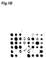
- Figs. 1A and 1B are views showing the results of dot blot screening performed by fixing to a membrane DNA that was extracted from a gene library obtained from hepatic cancer by the subtraction method in Example 1.
- Fig. 1A is a view showing the results of screening with a cDNA probe prepared from hepatic cancer polyA RNA
- Fig. 1B is a view showing the results of screening with a cDNA probe prepared from normal liver polyA RNA.
- An arrow 1 indicates a dot of a gene whose expression is increased in hepatic cancer
- an arrow 2 a dot of a gene whose expression is increased in hepatic cancer.
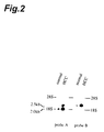
- Fig. 2 is a photograph showing the results of analysis by Northern blot hybridization in systems of the rat normal liver and 7-month hepatic cancer by use of probes prepared in Examples 3 and 4 (probe A and probe B), respectively, with the left lane showing the normal liver (normal), and the right lane showing hepatic cancer (HCC).
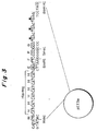
- Fig. 3 is a view showing pET3a vector used in Examples 3, 14 and 16.
- Fig. 4 is a photograph showing the results of analysis by Northern blot hybridization using gel electrophoresis of total RNA's prepared from a human hepatic cancer tissue and a non-cancerous hepatic tissue of the same patient.
- Lane 1 gives the results with the electrophoresed human hepatic cancer RNA
- lane 2 gives the results with the electrophoresed human non-cancerous RNA (a normal portion of the same patient).
- the band indicated by an arrow is the targeted band.
- Fig. 5 is a photograph showing the results of Western blotting (protein level analysis) of various hepatic cancer tissues prepared in Example 1. From the bottom of the drawing upwards lie a marker (for a ladder), rec (positive control, CRTI prepared in Example 4), N (normal liver), 1 (liver one month after onset of cancer), 3 (liver 3 months after cancer onset), 5 (liver 5 months after cancer onset) and 7 (liver 7 months after cancer onset).
- Fig. 6 is a photograph showing the results of Western blotting using various human cancer cells. From right to left in the drawing, lie a positive control, Li21, LiHM, LiNM (human hepatic cancer cells), RERF (human pulmonary cancer cells), AZ (human gastric cancer cells), Hecl (human uterine cancer cells), Alex (human hepatic cancer cells), MEWO (human melanoma), and PaCa (human pancreatic cancer cells).
- a band indicated by an arrow in the drawing represents CRTI.
- Fig. 7 is a photograph showing the results of Northern blot hybridization of CRTI gene distributed in various rat tissues.
- a band indicated by an arrow in the drawing is a band derived from CRTI gene.
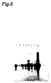
- Fig. 8 is a photograph showing the results of Northern blot hybridization of CRTI gene distributed in various human tissues.
- a band indicated by an arrow in the drawing is a band derived from CRTI gene.
- Lane 1 represents heart, 2 brain, 3 placenta, 4 lung, 5 liver, 6 skeletal muscle, 7 kidney, and 8 pancreas.
- Fig. 9 is a view showing the results of analysis by Northern blot hybridization of mRNA's from normal liver and hepatic cancer with the use of HRPI gene as a probe.
- Lane 1 is a lane for normal liver mRNA
- Lane 2 is a lane for mRNA of the hepatic cancer 12 hours after administration of DEN
- Lane 3 is a lane for mRNA of the hepatic cancer 24 hours after DEN administration
- Lane 4 is a lane for mRNA of the hepatic cancer 48 hours after DEN administration
- Lane 5 is a lane for mRNA of the hepatic cancer 1 month after DEN administration
- Lane 6 is a lane for mRNA of the hepatic cancer 3 months after DEN administration
- Lane 7 is a lane for mRNA of the hepatic cancer 5 months after DEN administration
- Lane 8 is a lane for mRNA of the hepatic cancer 7 months after DEN administration
- arrow 9 shows the position of
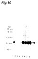
- Fig. 10 is a view showing the results of analysis by Northern blot hybridization of mRNA's from normal liver and hepatic cancer with the use of GADII gene as a probe.
- Lane 1 is a lane for normal liver mRNA
- Lane 2 is a lane for mRNA of the hepatic cancer 12 hours after administration of DEN
- Lane 3 is a lane for mRNA of the hepatic cancer 24 hours after DEN administration
- Lane 4 is a lane for mRNA of the hepatic cancer 48 hours after DEN administration
- Lane 5 is a lane for mRNA of the hepatic cancer 1 month after DEN administration
- Lane 6 is a lane for mRNA of the hepatic cancer 3 months after DEN administration
- Lane 7 is a lane for mRNA of the hepatic cancer 5 months after DEN administration
- Lane 8 is a lane for mRNA of the hepatic cancer 7 months after DEN administration
- arrow 9 shows the position
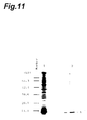
- Fig. 11 is a view showing the results of SDS-PAGE electrophoresis of a recombinant HRPI partial protein.
- Lane 1 is a lane for a protein prepared by recombinant E. coli.
- Lane 2 is a lane for purified recombinant HRPI partial protein.
- An arrow 3 represents the position of the band of the HRPI partial protein.
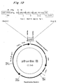
- Fig. 12 is a view showing pBluebacIII vector as used in Example 15.
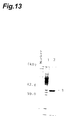
- Fig. 13 is a view showing the results of SDS-PAGE electrophoresis of recombinant HRPI.
- Lane 1 is a lane for a protein expressed by wild type Sf9 cells.
- Lane 2 is a lane for a protein expressed by recombinant Sf9 cells.
- An arrow 3 represents the position of the band of the recombinant HRPI.
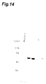
- Fig. 14 is a view showing the results of 10%SDS-PAGE electrophoresis of recombinant GADII (Lane 1). An arrow represents the position of the band of the recombinant GADII.
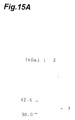
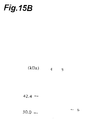
- Figs. 15A and 15B are views showing the results of Western blot analysis of the reaction product between a sample containing HRPI full length protein and anti-HRPI antibodies.
- Fig. 15A is a view showing the results of Western blot analysis of the reaction product between a sample containing recombinant HRPI full length protein and anti-HRPI antibodies.
- Fig. 15B is a view showing the results of Western blot analysis of the reaction product between the extract of a rat liver and anti-HRPI antibodies.
- lane 1 is a lane for the electrophoresed recombinant HRPI
- lane 2 is a lane for the electrophoresed hepatic cancer tissue extract
- an arrow 3 represents the position of the band of the recombinant HRPI
- lane 4 is a lane for the electrophoresed normal liver extract
- lane 5 is a lane for the electrophoresed extract of hepatic cancer 7 months after administration of DEN
- an arrow 6 represents the position of the band of HRPI.
- Fig. 16 is a view showing the expression of HRPI gene in various organs.
- Lane 1 is a lane for electrophoresed heart mRNA
- lane 2 is a lane for electrophoresed brain mRNA
- lane 3 is a lane for electrophoresed spleen mRNA
- lane 4 is a lane for electrophoresed lung mRNA
- lane 5 is a lane for electrophoresed liver mRNA
- lane 6 is a lane for electrophoresed skeletal muscle mRNA
- lane 7 is a lane for electrophoresed kidney mRNA
- lane 8 is a lane for electrophoresed testicle mRNA
- an arrow 9 represents the position of the band of HRPI mRNA.
- Fig. 17 is a view showing the expression of GADII gene in various organs.
- Lane 1 is a lane for electrophoresed heart mRNA
- lane 2 is a lane for electrophoresed brain mRNA
- lane 3 is a lane for electrophoresed spleen mRNA
- lane 4 is a lane for electrophoresed lung mRNA
- lane 5 is a lane for electrophoresed liver mRNA
- lane 6 is a lane for electrophoresed skeletal muscle mRNA
- lane 7 is a lane for electrophoresed kidney mRNA
- lane 8 is a lane for electrophoresed testicle mRNA
- an arrow 9 represents the position of the band of GADII mRNA.
- the present invention concerns proteins whose expression is increased in a hepatic cancer tissue compared with a normal hepatic tissue, genes encoding these proteins, antibodies specific for the proteins, and a method for monitoring cancer by detecting the proteins or the genes.
- the present invention is a protein specifically expressed in hepatic cancer, and a gene encoding the protein.
- the protein and the gene were obtained by isolating a gene, present specifically in hepatic cancer, from the liver of a rat by the subtraction method, and determining its base sequence. From this gene, a protein expressed specifically in hepatic cancer was confirmed and isolated.
- proteins specific for hepatic cancer were named rat CRTI, rat HRPI, rat GADII and human GADII, respectively.
- the amino acid sequences of these proteins, and gene sequences encoding them are shown in Seq. ID Nos. 1 and 2, 3 and 4, 5 and 6, and 7 and 8, respectively, in the Sequence Listing.
- Heterologous proteins with the same function are generally known to have homology of 30-40% or more, although they are somewhat different in amino acid sequence depending on species. It can be fully presumed, therefore, that mammals other than rat also have a protein having the same function as the protein isolated in the present invention (CRTI or HRPI), and having homology of 30% or more (preferably 40% or more) to this protein.
- the presence of homology is generally determined by a calculation method which counts homologous amino acids as positive. When two proteins are different in the length of the amino acid chain, the homology rate (%) is expressed as the proportion of the homologous portion to the length of the protein with the shorter amino acid chain.
- proteins relevant to the present invention are intended to mean those having the following two features:
- Spontaneous or artificial mutation can alter part of the structure of a polynucleotide, and accordingly, can mutate a protein encoded by the polynucleotide.
- the protein of the present invention too, it is possible to produce a mutant protein in which one or more amino acids of the amino acid sequence described as Seq. ID No. 1, 3, 5 or 7 in the Sequence Listing has or have been replaced, deleted or added.
- the protein of the present invention is recognized specifically by antibodies to the protein, so that the mutant protein of the present invention is characterized by being recognized by the antibodies (being mutated to a degree recognizable by the antibodies).
- genes encoding CRTI, HRPI and GADII concerned with the present invention are genes coding for the CRTI, HRPI and GADII defined earlier.
- the present invention clarifies the antigenicity of the above-mentioned proteins, i.e. CRTI, HRPI and GADII, such that these proteins easily give antibodies when used to immunize mammals other than the species of origin and other than human, as will be exemplified in the Examples.
- the antibodies of the present invention against CRTI, HRPI and GADII include, in their range, antisera and polyclonal antibodies which are obtained by the same method, namely, immunization of an animal other than the species from which the proteins were derived.
- the use, as an immunogen, of part of the protein or part of the protein bound to a carrier protein such as bovine serum albumin is a method in common use by those skilled in the art.
- the part of the protein may be synthesized by a peptide synthesizer.
- the part of the protein is 8 amino acid residues or more for sufficient use as an immunogen.
- monoclonal antibodies can be produced by hybridomas using lymphocytes of the immunized animal (e.g., see Antibodies, A Laboratory Manual (Cold Spring Harbor Laboratory Press, 1988), Chapter 6).
- the antibodies of the present invention to CRTI, HRPI and GADII include polyclonal antibodies and monoclonal antibodies in their scope.
- Methods for detecting CRTI, HRPI or GADII include, for example, the use of antibodies, the use of antibodies relying on an enzyme reaction, and the detection of the gene for the protein.
- Methods using antibodies are concretely (1) a method of detecting each protein by use of antibodies to the protein, and (2) a method of detecting each protein by using antibodies to the protein and labeled secondary antibodies to the antibodies.
- the labels are, for instance, radioisotopes (RI), enzymes, avidin or biotin, and fluorescent materials (FITC or rhodamine).
- Methods using antibodies relying on an enzyme reaction include, for example, ELISA.
- Methods for detecting genes are, for instance, Northern blot hybridization, RT-PCR ( Current Protocols in Molecular Biology (Greene Publishing Associates and Wiley-Interscience), Chapter 15), and in situ hybridization (ibid. Chapter 14).
- the optimum conditions for hybridization are known to be different according to the length of the probe or the membrane used. What conditions to be set can be easily chosen by those skilled in the art.
- hybridization needless to say, can be achieved under different hybridization conditions.
- hybridization may be effected in the absence of sodium pyrophosphate.
- a preferred embodiment is as follows:
- the number of bases of the human genome is said to be 3x10 9 . Since DNA of 16 bases comes in 4 16 types, this length of DNA enables human proteins to be all recognized.
- the length required of a probe is, theoretically, 16 bases.
- a length longer than it is desirable needless to say.
- Practically, a length of 12 bases or more is often used because of synthesis efficiency, operability and so on.
- the site used as the probe may be a noncoding region or a coding region.
- the site used as the probe has a GC content of 30 to 70%, it may be a noncoding region or a coding region.
- the antisense DNA or antisense RNA of the present invention (may be collectively called the antisense polynucleotide) includes all of nucleotides comprising bases, phosphates and sugars combined in a plurality of numbers, including those which are not present in nature.
- the antisense polynucleotide derivative of the present invention includes all of those which are similar to polynucleotide in conformation and function. Examples are polynucleotides linked at the 3'-terminal or 5'-terminal to other substances, polynucleotides having undergone modification, such as replacement, deletion, or addition, in at least part of the bases, sugars or phosphates, polynucleotides having bases, sugars or phosphates that do not naturally occur, and polynucleotides having a skeleton other than sugar-phosphate skeleton.
- the antisense polynucleotide or its derivative may be the one which hybridizes with any portion of a polynucleotide encoding the protein of the present invention whose expression is increased in hepatic cancer.
- the antisense polynucleotide or its derivative is preferably the one which has a base sequence complementary for part of mRNA encoding all or part of the protein, and which hybridizes with the mRNA.
- the particularly preferred one is that which hybridizes with mRNA encoding at least CRTI, HRPI or GADII.
- the antisense polynucleotide or its derivative is immediately usable as a research polynucleotide probe for investigating the presence and the status of expression, in a tissue or cells, of a polynucleotide encoding the protein of the present invention whose expression is increased in hepatic cancer.
- the antisense polynucleotide or its derivative is also usable as a diagnostic polynucleotide probe.
- the probe preferably has 12 bases or more and a GC content of 30 to 70%, more preferably, 16 bases or more and a GC content of 30 to 70%.
- an antisense pharmaceutical can be developed from the antisense polynucleotide or its derivative.
- the antisense polynucleotide derivative are known to be the derivatives with at least one of nuclease resistance, tissue selectivity, cell permeation, and binding capacity.
- the polynucleotide derivative is known to be the derivative having a phosphorothioate bond as a skeletal structure.
- the polynucleotide and its derivative of the present invention also include derivatives having such function or structure.
- the antisense polynucleotide derivative of the present invention can be prepared, for example, by the method described in Antisense Research and Applications (CRC Publishing, Florida, 1993).
- Wild type DNA or RNA can be synthesized by a chemical synthesizer.
- the antisense polynucleotide derivative of the present invention can be obtained by PCR using as a template a gene encoding the protein of the invention whose expression is increased in hepatic cancer.
- Some of the derivatives of the methyl phosphonate type or the phosphorothioate type can be synthesized by a chemical synthesizer (e.g. Model 394, Perkin-Elmer Japan).
- the procedure is performed in accordance with an Instruction Manual attached to the chemical synthesizer, and the resulting synthesis product is purified by HPLC using a reversed phase chromatograph, whereby the desired polynucleotide or polynucleotide derivative can be obtained.
- DNA or RNA When DNA or RNA is chemically or enzymatically synthesized, it is well known to subject it to a chemical modification, such as methylation or biotinylation of the side chain, or substitution of oxygen of the phosphate portion by sulfur.
- a chemical modification such as methylation or biotinylation of the side chain, or substitution of oxygen of the phosphate portion by sulfur.
- Main examples of the modification to be introducible during chemical synthesis are 1) biotinylation, 2) methylation, 3) conversion to digoxigenin derivative, 4) dephosphorylation, 5)fluorescence labeling (fluorescein, rhodamine, Texas Red and derivatives thereof), 6) amination, and 7) substitution of O of the phosphate group by S.
- Main examples of chemical modification to be introducible enzymatically are 1) biotinylation, 2) methylation, 3) conversion to digoxigenin derivative, 4) dephosphorylation, 5)fluorescence labeling (fluorescein, rhodamine, Texas Red and derivatives thereof), and 6) enzyme labeling (alkaline phosphatase).
- DNA described as Seq. ID No. 2, 4, 6 or 8 in the Sequence Listing is to be chemically synthesized, for instance, it is possible to synthesize DNA different from the DNA of the Sequence Listing itself by performing the chemical modification.
- the DNA and RNA of the present invention include in their scopes the chemically modified DNA and RNA.
- Monitoring of the progress of cancer by detection of CRTI, HRPI or GADII can be performed by investigating whether the protein is present or not in a tissue or cells sampled from subjects. If CRTI, HRPI or GADII is secreted or liberated outside cells, the progress of cancer can be monitored by examining whether CRTI, HRPI or GADII is present or absent in the blood of the subject. Concretely, the monitoring may be done in the same manner as the aforementioned method of detecting CRTI, HRPI or GADII (the method using antibodies, the method using antibodies relying on enzyme reaction, or the method detecting the relevant gene).
- Monitoring of the progress of cancer by use of gene can be done by checking for the gene in a tissue or cells sampled from the subject.
- the methods of detecting the gene include, for example, Northern blot hybridization, RT-PCR, and in situ hybridization, as stated previously.
- Hepatic cancer-bearing rats were prepared based on the Salt-Farber method ( Nature , Vol. 263, 1976, pp. 701-703). Actually, 5-week Wistar rats (Funabashi Farm) were intraperitoneally administered diethylnitrosamine (DEN). Two weeks later, oral administration of M Feed (Oriental Yeast) containing 0.02% of 2-aminoacetylfluorene (AAF) was started. One week later, a regenerative hepatic operation was performed. The livers were removed 12, 24, 48 hours and 1, 3, 5 and 7 months after DEN administration, and used for subsequent RNA preparation.
- DEN diethylnitrosamine
- AAF 2-aminoacetylfluorene
- livers 12, 24 and 48 hours after regenerative hepatic operation were acquired by removing the livers 12, 24 and 48 hours after regenerative hepatic operation. These livers were used later for analysis.
- CRTI gene was analyzed for hepatic cancer-derived polyA RNA by Northern blot hybridization using the probe of Example 3. As shown in Fig. 2 (probe A), two bands were detected. The band observed at about 2.0 kb corresponds to a gene of the base sequence from the 500th base to the 2,520th base of Seq. ID No. 2 in the Sequence Listing. A gene corresponding to the other band at about 2.5 kb was acquired by the method described in 11. of Example 1.
- the base sequence of the resulting gene was determined by the method described in 1. to 4. of Example 2. This gene was found to be an extension of the gene of about 2.0 kb by 499 bp on the 5' side.
- the CRTI gene base sequence of the resulting portion of about 2.5 kb is shown as Seq. ID No. 2 in the Sequence Listing.
- Northern blot hybridization was performed in accordance with the method described in Example 3 by using, as a probe, DNA ranging from the 1st base to the 372nd base of the 499 bp portion obtained above.
- the film was Kodak's XAR film, the screen used Du Pont's Lightening Plus(+), and exposure was performed for 24 hours at - 80°C.
- Fig. 2 (probe B) the presence of a band of 2.5 kb specific for rat hepatic cancer was confirmed (Fig. 2 (probe B)). This result showed that hepatic cancer could be detected based on the above-described DNA of 499 bp.
- TREB5 or XBP
- XBP is reported in Science, vol. 247, pp. 1581-1584 (1990).
- Rat MTN BlotTM (CLONTECH) was used. This product is a commercially available membrane blotted with polyA RNA's of rat tissues to be used in the above-mentioned Northern blot hybridization. This membrane was subjected to Northern blot hybridization by the method described in Molecular Cloning Second Edition , (Cold Spring Harbor Laboratory Press, 1989) pp. 7.3-7.84 in accordance with the product's instruction manual with the use of the same CRTI gene probe as prepared in Example 3. In autoradiography, the film was Kodak's XAR film, the screen used Du Pont's Lightening Plus(+), and exposure was performed for 24 hours at -80°C.
- CRTI gene was expressed in all organs examined, and intensely expressed in the liver and the lung, in particular.
- Example 3 suggest that if any organs become cancerous, the expression of CRTI gene may increase. Thus, CRTI gene can be expect to monitor the outbreak and progress of cancer in various organs.
- CRTI protein in various organs permits monitoring of the outbreak and progress of cancer in those organs.
- CRTI gene was expressed in all organs examined, and intensely expressed in the pancreas and liver, in particular.
- Example 6 suggest that if any human organs become cancerous, the expression of CRTI gene may increase. Thus, the use of CRTI gene can be expect to monitor the outbreak and progress of cancer in various organs in human.
- CRTI protein in various organs in human permits monitoring of the outbreak and progress of cancer in those organs.
- the detection of the CRTI or CRTI gene of the present invention in a tissue such as the liver makes it possible to diagnose cancer of the tissue, such as hepatic cancer.
- the use of a part or the whole of the CRTI gene enables the occurrence of cancer, such as hepatic cancer, to be diagnosed at the gene expression level.
- the use of the anti-CRTI antibodies also permits the immunohistological diagnosis of the outbreak of cancer such as hepatic cancer.
- the application of the gene or the antibodies to the treatment of cancer can be expected in the future.
- Rat MTN Blot (registered trademark, CLONTECH) was used. This product is a commercially available membrane blotted with polyA RNA's of rat tissues to be used in the above-mentioned Northern blot hybridization. This membrane was subject to Northern blot hybridization by the method described in Molecular Cloning Second Edition , (Cold Spring Harbor Laboratory Press, 1989) pp. 7.3-7.84 in accordance with the product's instruction manual with the use of the aforementioned HRPI gene probe and GADII gene probe.
- the detection of the HRPI or the HRPI gene or GADII or GADII gene of the present invention in the liver permits the monitoring of the progress of hepatic cancer, and diagnosis of hepatic cancer, especially the early diagnosis of hepatic cancer.
- the use of a part or the whole of the HRPI gene or GADII gene makes it possible to diagnose the onset of hepatic cancer at the gene expression level.
- the use of the anti-HRPI or anti-GADII antibodies permits the immunohistological diagnosis of hepatic cancer occurrence.
- genes, antisense genes to the genes, or antibodies to the genes can be expected to find use in the treatment of hepatic cancer.
Abstract
Novel proteins (CRTI, HRPI and GADII) showing elevated expression in the course of carcinogenesis; novel genes encoding these
proteins; antisense genes of these genes; antibodies against these proteins; and a method for monitoring liver cancer by using the
above-mentioned proteins, genes or antibodies. Genes expressed specifically in rat liver cancer are extracted and those showing elevated
expression in liver cancer are isolated. Their cDNAs of the full length are obtained and thus the base sequences of these genes are
determined. Thus it is confirmed that these genes are those specific to liver cancer. Further, the amino acid sequences encoded by the
above-mentioned genes are determined and proteins having these amino acid sequences are expressed in recombinant Escherichia coli .
Furthermore, antibodies against these proteins are constructed. Then it is clarified that the expression of these proteins can be detected by
using the above-mentioned antibodies and the onset of liver cancer can be monitored by detecting the above-mentioned genes.
Description
This invention relates to proteins expressed
markedly in hepatic cancer, genes encoding the
proteins, antibodies to the proteins, and methods for
detecting the expressions of the proteins or the genes.
Carcinoma or cancer is known to occur because of
some abnormality in a gene. A change or abnormality in
the gene at its transcription level, in particular, is
regarded as a major cause of cancer ("Science" Vo1.
222, 1983, pp. 765-771). To elucidate the mechanism of
onset of carcinoma, the acquisition of a protein
variedly expressed during carcinogenesis and a gene
encoding the protein, or a protein different in the
state of expression among tissues and a gene encoding
the protein has been performed eagerly since about
1980. For example, cancer-specific proteins, α-fetoprotein
and CEA, are known which were acquired by
biochemically analyzing a carcinomatous tissue, and
searching for its difference from a normal tissue.
Methods used to identify these substances
comprised preparing monoclonal antibodies to a
carcinomatous tissue, and sorting antibodies reactive
only with the cancer tissue from the resulting
monoclonal antibodies, and then identifying a substance
which would serve as an antigen reactive with the
monoclonal antibodies.
Information obtained by these methods was mostly
concerned with a protein, and no gene was obtained
directly. To acquire a gene from the protein obtained,
therefore, the gene was selected from a population of
genes, called a gene library, by a genetic engineering
technique.
Concretely, this method of selection is described
in "Molecu1ar C1oning Second Edition" (Co1d Spring
Harbor Laboratory Press, 1989), chapters 8, 9 and 12.
As shown there, the method prepares a gene probe on the
basis of information on the resulting protein, and
acquires a gene corresponding to this gene probe from
the gene library by a method called colony/plaque
hybridization.
The same book also shows in chapters 8, 9, 11 and
12 a method for screening the desired gene from the
gene library by use of monoclonal antibodies.
With these methods, however, the desired protein
should be large in amount, or the use of the monoclonal
antibodies requires an antigen protein of the cell
surface type and with high antigenicity. A protein
which obviates these requirements has been very
difficult to obtain. Consequently, it has also been
extremely difficult to yield a gene encoding the
protein.
The present invention uses methods entirely
different from the foregoing some known methods, to
isolate genes whose expression in a hepatic cancer
tissue is increased compared with a normal hepatic
tissue.
That is, an object of the present invention is to
provide novel proteins increasing in expression during
a carcinogenic process, novel genes encoding the
proteins, and antibodies to the proteins.
It is another object of the invention to provide
methods for detecting the expressions of the proteins
or the genes to monitor the onset and progress of
hepatic carcinoma.
To attain the above objects, the inventors
conducted extensive studies. As a result, genes
specifically expressed in rat hepatic cancer were
extracted by the subtraction method, and genes whose
expression was increased in hepatic cancer were
isolated by dot screening. From a hepatic cancer cDNA
library, the full length cDNA's of the genes, and the
base sequences of the genes were determined. Further,
amino acid sequences encoded by the genes were
determined.
Moreover, Northern blot hybridization confirmed
the cDNA's to be hepatic cancer specific genes. Also,
proteins encoded by the cDNA's were expressed in
recombinant Escherichia coli (E. coli), and the
proteins were confirmed to have the same functions as
those of their naturally occurring equivalents.
Novel rat proteins CRTI (or HTF), HRPI and GADII
(or CSAD) specific for hepatic cancer, and genes
encoding these proteins were isolated by the above-described
means. A novel human protein, GADII,
specific for hepatic cancer, and a gene encoding this
protein were also isolated.
The above proteins were each inoculated into a
mammal other than a species from which the protein was
derived (excluding human), to produce antibodies
against the protein and confirm the antigenicity of the
protein.
It was also found that the above genes, CRTI gene,
HRPI gene and GADII gene, were detected in tissues of
the rat and the human by Northern blot hybridization.
These findings showed that the onset and progress
of cancer, especially hepatic cancer, could be
monitored by detecting the expression of the proteins
by use of those antibodies, and that the onset and
progress of cancer, especially hepatic cancer, could be
monitored by detecting the genes encoding the proteins.
Figs. 1A and 1B are views showing the results of
dot blot screening performed by fixing to a membrane
DNA that was extracted from a gene library obtained
from hepatic cancer by the subtraction method in
Example 1. Fig. 1A is a view showing the results of
screening with a cDNA probe prepared from hepatic
cancer polyA RNA, while Fig. 1B is a view showing the
results of screening with a cDNA probe prepared from
normal liver polyA RNA. An arrow 1 indicates a dot of
a gene whose expression is increased in hepatic cancer,
and an arrow 2, a dot of a gene whose expression is
increased in hepatic cancer.
Fig. 2 is a photograph showing the results of
analysis by Northern blot hybridization in systems of
the rat normal liver and 7-month hepatic cancer by use
of probes prepared in Examples 3 and 4 (probe A and
probe B), respectively, with the left lane showing the
normal liver (normal), and the right lane showing
hepatic cancer (HCC).
Fig. 3 is a view showing pET3a vector used in
Examples 3, 14 and 16.
Fig. 4 is a photograph showing the results of
analysis by Northern blot hybridization using gel
electrophoresis of total RNA's prepared from a human
hepatic cancer tissue and a non-cancerous hepatic
tissue of the same patient. Lane 1 gives the results
with the electrophoresed human hepatic cancer RNA,
while lane 2 gives the results with the electrophoresed
human non-cancerous RNA (a normal portion of the same
patient). The band indicated by an arrow is the
targeted band.
Fig. 5 is a photograph showing the results of
Western blotting (protein level analysis) of various
hepatic cancer tissues prepared in Example 1. From the
bottom of the drawing upwards lie a marker (for a
ladder), rec (positive control, CRTI prepared in
Example 4), N (normal liver), 1 (liver one month after
onset of cancer), 3 (liver 3 months after cancer
onset), 5 (liver 5 months after cancer onset) and 7
(liver 7 months after cancer onset).
Fig. 6 is a photograph showing the results of
Western blotting using various human cancer cells.
From right to left in the drawing, lie a positive
control, Li21, LiHM, LiNM (human hepatic cancer cells),
RERF (human pulmonary cancer cells), AZ (human gastric
cancer cells), Hecl (human uterine cancer cells), Alex
(human hepatic cancer cells), MEWO (human melanoma),
and PaCa (human pancreatic cancer cells). A band
indicated by an arrow in the drawing represents CRTI.
Fig. 7 is a photograph showing the results of
Northern blot hybridization of CRTI gene distributed in
various rat tissues. A band indicated by an arrow in
the drawing is a band derived from CRTI gene.
Fig. 8 is a photograph showing the results of
Northern blot hybridization of CRTI gene distributed in
various human tissues. A band indicated by an arrow in
the drawing is a band derived from CRTI gene. Lane 1
represents heart, 2 brain, 3 placenta, 4 lung, 5 liver,
6 skeletal muscle, 7 kidney, and 8 pancreas.
Fig. 9 is a view showing the results of analysis
by Northern blot hybridization of mRNA's from normal
liver and hepatic cancer with the use of HRPI gene as a
probe. Lane 1 is a lane for normal liver mRNA, Lane 2
is a lane for mRNA of the hepatic cancer 12 hours after
administration of DEN, Lane 3 is a lane for mRNA of the
hepatic cancer 24 hours after DEN administration, Lane
4 is a lane for mRNA of the hepatic cancer 48 hours
after DEN administration, Lane 5 is a lane for mRNA of
the hepatic cancer 1 month after DEN administration,
Lane 6 is a lane for mRNA of the hepatic cancer 3
months after DEN administration, Lane 7 is a lane for
mRNA of the hepatic cancer 5 months after DEN
administration, Lane 8 is a lane for mRNA of the
hepatic cancer 7 months after DEN administration, and
arrow 9 shows the position of the band of HRPI mRNA.
Fig. 10 is a view showing the results of analysis
by Northern blot hybridization of mRNA's from normal
liver and hepatic cancer with the use of GADII gene as
a probe. Lane 1 is a lane for normal liver mRNA, Lane
2 is a lane for mRNA of the hepatic cancer 12 hours
after administration of DEN, Lane 3 is a lane for mRNA
of the hepatic cancer 24 hours after DEN
administration, Lane 4 is a lane for mRNA of the
hepatic cancer 48 hours after DEN administration, Lane
5 is a lane for mRNA of the hepatic cancer 1 month
after DEN administration, Lane 6 is a lane for mRNA of
the hepatic cancer 3 months after DEN administration,
Lane 7 is a lane for mRNA of the hepatic cancer 5
months after DEN administration, Lane 8 is a lane for
mRNA of the hepatic cancer 7 months after DEN
administration, and arrow 9 shows the position of the
band of GADII mRNA.
Fig. 11 is a view showing the results of SDS-PAGE
electrophoresis of a recombinant HRPI partial protein.
Lane 1 is a lane for a protein prepared by recombinant
E. coli. Lane 2 is a lane for purified recombinant
HRPI partial protein. An arrow 3 represents the
position of the band of the HRPI partial protein.
Fig. 12 is a view showing pBluebacIII vector as
used in Example 15.
Fig. 13 is a view showing the results of SDS-PAGE
electrophoresis of recombinant HRPI. Lane 1 is a lane
for a protein expressed by wild type Sf9 cells. Lane 2
is a lane for a protein expressed by recombinant Sf9
cells. An arrow 3 represents the position of the band
of the recombinant HRPI.
Fig. 14 is a view showing the results of 10%SDS-PAGE
electrophoresis of recombinant GADII (Lane 1). An
arrow represents the position of the band of the
recombinant GADII.
Figs. 15A and 15B are views showing the results of
Western blot analysis of the reaction product between a
sample containing HRPI full length protein and anti-HRPI
antibodies. Fig. 15A is a view showing the
results of Western blot analysis of the reaction
product between a sample containing recombinant HRPI
full length protein and anti-HRPI antibodies. Fig. 15B
is a view showing the results of Western blot analysis
of the reaction product between the extract of a rat
liver and anti-HRPI antibodies. In Fig. 15A, lane 1 is
a lane for the electrophoresed recombinant HRPI, lane 2
is a lane for the electrophoresed hepatic cancer tissue
extract, and an arrow 3 represents the position of the
band of the recombinant HRPI. In Fig. 15B, lane 4 is a
lane for the electrophoresed normal liver extract, lane
5 is a lane for the electrophoresed extract of hepatic
cancer 7 months after administration of DEN, and an
arrow 6 represents the position of the band of HRPI.
Fig. 16 is a view showing the expression of HRPI
gene in various organs. Lane 1 is a lane for
electrophoresed heart mRNA, lane 2 is a lane for
electrophoresed brain mRNA, lane 3 is a lane for
electrophoresed spleen mRNA, lane 4 is a lane for
electrophoresed lung mRNA, lane 5 is a lane for
electrophoresed liver mRNA, lane 6 is a lane for
electrophoresed skeletal muscle mRNA, lane 7 is a lane
for electrophoresed kidney mRNA, lane 8 is a lane for
electrophoresed testicle mRNA, and an arrow 9
represents the position of the band of HRPI mRNA.
Fig. 17 is a view showing the expression of GADII
gene in various organs. Lane 1 is a lane for
electrophoresed heart mRNA, lane 2 is a lane for
electrophoresed brain mRNA, lane 3 is a lane for
electrophoresed spleen mRNA, lane 4 is a lane for
electrophoresed lung mRNA, lane 5 is a lane for
electrophoresed liver mRNA, lane 6 is a lane for
electrophoresed skeletal muscle mRNA, lane 7 is a lane
for electrophoresed kidney mRNA, lane 8 is a lane for
electrophoresed testicle mRNA, and an arrow 9
represents the position of the band of GADII mRNA.
The present invention concerns proteins whose
expression is increased in a hepatic cancer tissue
compared with a normal hepatic tissue, genes encoding
these proteins, antibodies specific for the proteins,
and a method for monitoring cancer by detecting the
proteins or the genes.
The characteristics of the present invention are
as follows:
The present invention is a protein specifically
expressed in hepatic cancer, and a gene encoding the
protein. As disclosed in the Examples to be described
later on, the protein and the gene were obtained by
isolating a gene, present specifically in hepatic
cancer, from the liver of a rat by the subtraction
method, and determining its base sequence. From this
gene, a protein expressed specifically in hepatic
cancer was confirmed and isolated.
In this manner, the inventors identified and
isolated four proteins and four genes newly. These
proteins specific for hepatic cancer were named rat
CRTI, rat HRPI, rat GADII and human GADII,
respectively. The amino acid sequences of these
proteins, and gene sequences encoding them are shown in
Seq. ID Nos. 1 and 2, 3 and 4, 5 and 6, and 7 and 8,
respectively, in the Sequence Listing.
Heterologous proteins with the same function are
generally known to have homology of 30-40% or more,
although they are somewhat different in amino acid
sequence depending on species. It can be fully
presumed, therefore, that mammals other than rat also
have a protein having the same function as the protein
isolated in the present invention (CRTI or HRPI), and
having homology of 30% or more (preferably 40% or more)
to this protein. The presence of homology, referred to
herein, is generally determined by a calculation method
which counts homologous amino acids as positive. When
two proteins are different in the length of the amino
acid chain, the homology rate (%) is expressed as the
proportion of the homologous portion to the length of
the protein with the shorter amino acid chain.
Thus, the proteins relevant to the present
invention are intended to mean those having the
following two features:
To know whether the protein is specifically
observed (expressed) in hepatic cancer, various known
methods are usable. For example, those in the Examples
to be described later on are preferred methods.
Spontaneous or artificial mutation (see, e.g.,
 Molecular Cloning 2nd Edition
 , Cold Spring Harbor
Laboratory Press, 1989) pp. 15.1-15.113) can alter part
of the structure of a polynucleotide, and accordingly,
can mutate a protein encoded by the polynucleotide. In
regard to the protein of the present invention, too, it
is possible to produce a mutant protein in which one or
more amino acids of the amino acid sequence described
as Seq. ID No. 1, 3, 5 or 7 in the Sequence Listing has
or have been replaced, deleted or added. The protein
of the present invention is recognized specifically by
antibodies to the protein, so that the mutant protein
of the present invention is characterized by being
recognized by the antibodies (being mutated to a degree
recognizable by the antibodies).
, Cold Spring Harbor
Laboratory Press, 1989) pp. 15.1-15.113) can alter part
of the structure of a polynucleotide, and accordingly,
can mutate a protein encoded by the polynucleotide. In
regard to the protein of the present invention, too, it
is possible to produce a mutant protein in which one or
more amino acids of the amino acid sequence described
as Seq. ID No. 1, 3, 5 or 7 in the Sequence Listing has
or have been replaced, deleted or added. The protein
of the present invention is recognized specifically by
antibodies to the protein, so that the mutant protein
of the present invention is characterized by being
recognized by the antibodies (being mutated to a degree
recognizable by the antibodies).
The genes encoding CRTI, HRPI and GADII concerned
with the present invention are genes coding for the
CRTI, HRPI and GADII defined earlier.
The present invention clarifies the antigenicity
of the above-mentioned proteins, i.e. CRTI, HRPI and
GADII, such that these proteins easily give antibodies
when used to immunize mammals other than the species of
origin and other than human, as will be exemplified in
the Examples. Hence, the antibodies of the present
invention against CRTI, HRPI and GADII include, in
their range, antisera and polyclonal antibodies which
are obtained by the same method, namely, immunization
of an animal other than the species from which the
proteins were derived. The use, as an immunogen, of
part of the protein or part of the protein bound to a
carrier protein such as bovine serum albumin is a
method in common use by those skilled in the art. The
part of the protein may be synthesized by a peptide
synthesizer. Preferably, the part of the protein is 8
amino acid residues or more for sufficient use as an
immunogen.
If polyclonal antibodies against the substances
whose antigenicity has been elucidated are obtained by
immunization, monoclonal antibodies can be produced by
hybridomas using lymphocytes of the immunized animal
(e.g., see Antibodies, A Laboratory Manual (Cold
Spring Harbor Laboratory Press, 1988), Chapter 6).
Thus, the antibodies of the present invention to CRTI,
HRPI and GADII include polyclonal antibodies and
monoclonal antibodies in their scope.
Methods for detecting CRTI, HRPI or GADII include,
for example, the use of antibodies, the use of
antibodies relying on an enzyme reaction, and the
detection of the gene for the protein.
Methods using antibodies are concretely (1) a
method of detecting each protein by use of antibodies
to the protein, and (2) a method of detecting each
protein by using antibodies to the protein and labeled
secondary antibodies to the antibodies. The labels
are, for instance, radioisotopes (RI), enzymes, avidin
or biotin, and fluorescent materials (FITC or
rhodamine).
Methods using antibodies relying on an enzyme
reaction include, for example, ELISA.
Methods for detecting genes are, for instance,
Northern blot hybridization, RT-PCR ( Current Protocols
in Molecular Biology (Greene Publishing Associates and
Wiley-Interscience), Chapter 15), and in situ
hybridization (ibid. Chapter 14).
The optimum conditions for hybridization are known
to be different according to the length of the probe or
the membrane used. What conditions to be set can be
easily chosen by those skilled in the art.
The Examples of the present invention disclose
optimum conditions worked out in view of the nature of
the membrane used and the length of the probe aimed at.
If the membrane and the probe length differ,
hybridization, needless to say, can be achieved under
different hybridization conditions. For instance,
hybridization may be effected in the absence of sodium
pyrophosphate. A preferred embodiment is as follows:
The number of bases of the human genome is said to
be 3x10⁹. Since DNA of 16 bases comes in 416 types,
this length of DNA enables human proteins to be all
recognized.
That is, the length required of a probe is,
theoretically, 16 bases. For practical purposes, a
length longer than it is desirable needless to say.
Practically, a length of 12 bases or more is often used
because of synthesis efficiency, operability and so on.
The site used as the probe may be a noncoding region or
a coding region.
When the site used as the probe has a GC content
of 30 to 70%, it may be a noncoding region or a coding
region.
The antisense DNA or antisense RNA of the present
invention (may be collectively called the antisense
polynucleotide) includes all of nucleotides comprising
bases, phosphates and sugars combined in a plurality of
numbers, including those which are not present in
nature.
The antisense polynucleotide derivative of the
present invention includes all of those which are
similar to polynucleotide in conformation and function.
Examples are polynucleotides linked at the 3'-terminal
or 5'-terminal to other substances, polynucleotides
having undergone modification, such as replacement,
deletion, or addition, in at least part of the bases,
sugars or phosphates, polynucleotides having bases,
sugars or phosphates that do not naturally occur, and
polynucleotides having a skeleton other than sugar-phosphate
skeleton.
The antisense polynucleotide or its derivative may
be the one which hybridizes with any portion of a
polynucleotide encoding the protein of the present
invention whose expression is increased in hepatic
cancer. The antisense polynucleotide or its derivative
is preferably the one which has a base sequence
complementary for part of mRNA encoding all or part of
the protein, and which hybridizes with the mRNA. The
particularly preferred one is that which hybridizes
with mRNA encoding at least CRTI, HRPI or GADII.
The antisense polynucleotide or its derivative is
immediately usable as a research polynucleotide probe
for investigating the presence and the status of
expression, in a tissue or cells, of a polynucleotide
encoding the protein of the present invention whose
expression is increased in hepatic cancer. The
antisense polynucleotide or its derivative is also
usable as a diagnostic polynucleotide probe. The probe
preferably has 12 bases or more and a GC content of 30
to 70%, more preferably, 16 bases or more and a GC
content of 30 to 70%.
It is also possible to adjust the expression of
the protein of the present invention whose expression
is increased in hepatic cancer, by using the antisense
polynucleotide or its derivative. This antisense
polynucleotide or derivative is expected to prevent the
expression of the protein by hybridizing with a gene or
mRNA encoding the protein. Thus, it can be used as a
therapeutic agent for a disease based on the disorder
of function which the protein takes part in. That is,
an antisense pharmaceutical can be developed from the
antisense polynucleotide or its derivative.
Generally, preferred examples of the antisense
polynucleotide derivative are known to be the
derivatives with at least one of nuclease resistance,
tissue selectivity, cell permeation, and binding
capacity. Particularly preferably, the polynucleotide
derivative is known to be the derivative having a
phosphorothioate bond as a skeletal structure. The
polynucleotide and its derivative of the present
invention also include derivatives having such function
or structure.
The antisense polynucleotide derivative of the
present invention can be prepared, for example, by the
method described in Antisense Research and
Applications (CRC Publishing, Florida, 1993).
Wild type DNA or RNA, for instance, can be
synthesized by a chemical synthesizer. Alternatively,
the antisense polynucleotide derivative of the present
invention can be obtained by PCR using as a template a
gene encoding the protein of the invention whose
expression is increased in hepatic cancer. Some of the
derivatives of the methyl phosphonate type or the
phosphorothioate type can be synthesized by a chemical
synthesizer (e.g. Model 394, Perkin-Elmer Japan). In
this case, the procedure is performed in accordance
with an Instruction Manual attached to the chemical
synthesizer, and the resulting synthesis product is
purified by HPLC using a reversed phase chromatograph,
whereby the desired polynucleotide or polynucleotide
derivative can be obtained.
When DNA or RNA is chemically or enzymatically
synthesized, it is well known to subject it to a
chemical modification, such as methylation or
biotinylation of the side chain, or substitution of
oxygen of the phosphate portion by sulfur.
Main examples of the modification to be
introducible during chemical synthesis are 1)
biotinylation, 2) methylation, 3) conversion to
digoxigenin derivative, 4) dephosphorylation,
5)fluorescence labeling (fluorescein, rhodamine, Texas
Red and derivatives thereof), 6) amination, and 7)
substitution of O of the phosphate group by S.
Main examples of chemical modification to be
introducible enzymatically are 1) biotinylation, 2)
methylation, 3) conversion to digoxigenin derivative,
4) dephosphorylation, 5)fluorescence labeling
(fluorescein, rhodamine, Texas Red and derivatives
thereof), and 6) enzyme labeling (alkaline
phosphatase).
When DNA described as Seq. ID No. 2, 4, 6 or 8 in
the Sequence Listing is to be chemically synthesized,
for instance, it is possible to synthesize DNA
different from the DNA of the Sequence Listing itself
by performing the chemical modification. Thus, the DNA
and RNA of the present invention include in their
scopes the chemically modified DNA and RNA.
Monitoring of the progress of cancer by detection
of CRTI, HRPI or GADII can be performed by
investigating whether the protein is present or not in
a tissue or cells sampled from subjects. If CRTI, HRPI
or GADII is secreted or liberated outside cells, the
progress of cancer can be monitored by examining
whether CRTI, HRPI or GADII is present or absent in the
blood of the subject. Concretely, the monitoring may
be done in the same manner as the aforementioned method
of detecting CRTI, HRPI or GADII (the method using
antibodies, the method using antibodies relying on
enzyme reaction, or the method detecting the relevant
gene).
Monitoring of the progress of cancer by use of
gene can be done by checking for the gene in a tissue
or cells sampled from the subject. The methods of
detecting the gene include, for example, Northern blot
hybridization, RT-PCR, and in situ hybridization, as
stated previously.
The present invention will now be described in
detail by reference to Examples, which do not limit the
present invention.
Hepatic cancer-bearing rats were prepared based on
the Salt-Farber method ( Nature , Vol. 263, 1976, pp.
701-703). Actually, 5-week Wistar rats (Funabashi
Farm) were intraperitoneally administered
diethylnitrosamine (DEN). Two weeks later, oral
administration of M Feed (Oriental Yeast) containing
0.02% of 2-aminoacetylfluorene (AAF) was started. One
week later, a regenerative hepatic operation was
performed. The livers were removed 12, 24, 48 hours
and 1, 3, 5 and 7 months after DEN administration, and
used for subsequent RNA preparation.
As controls, regenerated livers showing normal
growth were acquired by removing the livers 12, 24 and
48 hours after regenerative hepatic operation. These
livers were used later for analysis.
Total RNA was prepared based on the method
described in Methods in enzymology , Vol. 154
(Academic Press Inc., 1987), pp. 3-28. The actual
procedure was as follows:
This method was performed in accordance with the
method of Hara et al. ( Analytical Biochemistry , Vol.
214, 1993, pp. 58-64 (this reference is a part of the
instant specification as a result of its citation
herein). The actual procedure was as follows:
| cDNA solution | 69 µl |
| 10xTaq buffer (Perkin-Elmer) | 10 µl |
| 1.25 mM dNTP | 16 µl |
| EcoRI-(dG)15 primer (2 µg/µl) | 2 µl |
| XhoI-(dT)30 primer (2 µg/µl) | 2 µl |
| Taq polymerase (Perkin-Elmer)(5 µ/µl) | 1 µl |
| Total 100 µl |
| Gene solution | 10 µl |
| 10xH buffer (TAKARA SHUZO) | 10 µl |
| EcoRI (TAKARA SHUZO) | 5 µl |
| Sterilized water | 75 µl |
| Total 100 µl |
| Gene solution | 75 |
| 1% BSA (TAKARA SHUZO) | 10 µl |
| 10xH buffer (TAKARA SHUZO) | 10 µl |
| XhoI (TAKARA SHUZO) | 5 µl |
| Total 100 µl |
| Ampicillin (Wako Pure Chemical) | 100 µg/ml |
| IPTG (TAKARA SHUZO) | 0.1 mM |
| X-gal (TAKARA SHUZO) | 0.004% |
| DNA solution (prepared in 1.) | 20 µl |
| 0.1% BSA | 10 µl |
| 0.1% TritonX100 | 10 µl |
| NotI (TAKARA SHUZO) | 2 µl |
| Rnase (Nippon Gene) | 1 µl |
| 10xH buffer (Takara) | 10 µl |
| Sterilized water | 47 µl |
| Total 100 µl |
| pBluescriptII (1 µg/µl) | 3 |
| 10xH buffer | |
| 2 µl | |
| 0.1 | 2 µl |
| 0.1 | 2 |
| NotI | |
| 2 µl | |
| Sterilized water | 10 µl |
| Total 20 µl |
| DNA (prepared in (2)) | 5 µl |
| NotI-cleaved pBluescriptII (prepared in (3)) | 1 µl |
| 10-fold ligation buffer (Nippon Gene) | 2 µl |
| T4 ligase (Nippon Gene) Sterilized | 1 |
| Total 20 µl |
CRTI gene was analyzed for hepatic cancer-derived
polyA RNA by Northern blot hybridization using the
probe of Example 3. As shown in Fig. 2 (probe A), two
bands were detected. The band observed at about 2.0 kb
corresponds to a gene of the base sequence from the
500th base to the 2,520th base of Seq. ID No. 2 in the
Sequence Listing. A gene corresponding to the other
band at about 2.5 kb was acquired by the method
described in 11. of Example 1.
The base sequence of the resulting gene was
determined by the method described in 1. to 4. of
Example 2. This gene was found to be an extension of
the gene of about 2.0 kb by 499 bp on the 5' side. The
CRTI gene base sequence of the resulting portion of
about 2.5 kb is shown as Seq. ID No. 2 in the Sequence
Listing.
Recombinant E. coli having CRTI gene (R3)
containing the 499 bp portion introduced therein
(JM109) was deposited June 12, 1996 at the National
Institute of Bioscience and Human Technology (Address:
1-3, Higashi 1-chome, Tsukuba-shi, Ibaraki-ken, Japan),
Agency of Industrial Science and Technology, Japan,
with the accession number FERM P-15685. Then, the
microorganism was transferred to international
deposition on September 9, 1996 (Accession No. FERM BP-5660).
Furthermore, Northern blot hybridization was
performed in accordance with the method described in
Example 3 by using, as a probe, DNA ranging from the
1st base to the 372nd base of the 499 bp portion
obtained above. In autoradiography, the film was
Kodak's XAR film, the screen used Du Pont's Lightening
Plus(+), and exposure was performed for 24 hours at -
80°C. In systems using polyA RNA from a rat's normal
liver and hepatic cancer, the presence of a band of 2.5
kb specific for rat hepatic cancer was confirmed (Fig.
2 (probe B)). This result showed that hepatic cancer
could be detected based on the above-described DNA of
499 bp.
Homology search of the resulting CRTI through a
database revealed TREB5 (or XBP) as a homologous
sequence, which had homology of about 79% to CRTI of
the present invention. TREB5 is reported in The EMBO
Journal, vol. 9, No. 8, pp. 2537-2542 (1990), while XBP
is reported in Science, vol. 247, pp. 1581-1584 (1990).
The results are shown in Fig. 4. In human hepatic
cancer, the cancerous area showed a significant
increase in the amount of expression compared with the
non-cancerous area. This finding demonstrates that the
use of the CRTI gene permits monitoring of human
hepatic cancer.
To confirm the distribution of the gene in various
tissues, Rat MTN Blot™ (CLONTECH) was used. This
product is a commercially available membrane blotted
with polyA RNA's of rat tissues to be used in the
above-mentioned Northern blot hybridization. This
membrane was subjected to Northern blot hybridization
by the method described in Molecular Cloning Second
Edition , (Cold Spring Harbor Laboratory Press, 1989)
pp. 7.3-7.84 in accordance with the product's
instruction manual with the use of the same CRTI gene
probe as prepared in Example 3. In autoradiography,
the film was Kodak's XAR film, the screen used Du
Pont's Lightening Plus(+), and exposure was performed
for 24 hours at -80°C.
The results are shown in Fig. 7, which indicates
that CRTI gene was expressed in all organs examined,
and intensely expressed in the liver and the lung, in
particular.
These results and the results of Example 3 suggest
that if any organs become cancerous, the expression of
CRTI gene may increase. Thus, CRTI gene can be expect
to monitor the outbreak and progress of cancer in
various organs.
Furthermore, the detection of CRTI protein in
various organs permits monitoring of the outbreak and
progress of cancer in those organs.
To confirm the distribution of the gene in various
tissues, Human MTN Blot™ (CLONTECH) was used. This
product is a commercially available membrane blotted
with polyA RNA's of human tissues to be used in the
aforementioned Northern blot hybridization. This
membrane was subjected to Northern blot hybridization
by the method described in Molecular Cloning Second
Edition , (Cold Spring Harbor Laboratory Press, 1989)
pp. 7.3-7.84 in accordance with the product's
instruction manual with the use of the same CRTI gene
probe as prepared in Example 3. In autoradiography,
the film was Kodak's XAR film, the screen used Du
Pont's Lightening Plus(+), and exposure was performed
for 24 hours at -80°C.
The results are shown in Fig. 8, which indicates
that CRTI gene was expressed in all organs examined,
and intensely expressed in the pancreas and liver, in
particular.
These results and the results of Example 6 suggest
that if any human organs become cancerous, the
expression of CRTI gene may increase. Thus, the use of
CRTI gene can be expect to monitor the outbreak and
progress of cancer in various organs in human.
Furthermore, the detection of CRTI protein in
various organs in human permits monitoring of the
outbreak and progress of cancer in those organs.
Based on the above experimental results, the
detection of the CRTI or CRTI gene of the present
invention in a tissue such as the liver makes it
possible to diagnose cancer of the tissue, such as
hepatic cancer. The use of a part or the whole of the
CRTI gene enables the occurrence of cancer, such as
hepatic cancer, to be diagnosed at the gene expression
level. The use of the anti-CRTI antibodies also
permits the immunohistological diagnosis of the
outbreak of cancer such as hepatic cancer.
Furthermore, the application of the gene or the
antibodies to the treatment of cancer can be expected
in the future.
| DNA solution (prepared in 1.) | 20 µl |
| 0.1% BSA | 10 µl |
| 0.1% TritonX100 | 10 µl |
| NotI (TAKARA SHUZO) | 2 µl |
| Rnase (Nippon Gene) | 1 µl |
| 10xH buffer (Takara) | 10 µl |
| Sterilized water | 47 µl |
| Total 100 µl |
| pBluescriptII (1 µg/µl) | 3 |
| 10xH buffer | |
| 2 µl | |
| 0.1 | 2 µl |
| 0.1 | 2 |
| NotI | |
| 2 µl | |
| Sterilized water | 10 µl |
| Total 20 µl |
| DNA (prepared in (2)) | 5 µl |
| NotI-cleaved pBluescriptII (prepared in (3)) | 1 µl |
| 10-fold ligation buffer (Nippon Gene) | 2 µl |
| T4 ligase (Nippon Gene) | 1 µl |
| Sterilized | 11 µl |
| Total 20 µl |
| DNA solution (prepared in 1.) | 20 µl |
| EcoRI (TAKARA SHUZO) | 2 µl |
| Rnase (Nippon Gene) | 1 µl |
| 10xH buffer (TAKARA SHUZO) | 10 µl |
| Sterilized water | 67 µl |
| Total 100 µl |
| pBluescriptII (1 µg/µl) | 3 |
| 10xH buffer | |
| 2 | |
| EcoRI | |
| 2 µl | |
| Sterilized water | 14 µl |
| Total 20 µl |
| DNA (prepared in (2)) | 5 µl |
| EcoRI-cleaved pBluescriptII (prepared in (3)) | 1 µl |
| 10-fold ligation buffer (Nippon Gene) | 2 µl |
| T4 ligase (Nippon Gene) | 1 µl |
| Sterilized | 11 µl |
| Total 20 µl |
| DNA solution (prepared in 1.) | 20 µl |
| EcoRI (TAKARA SHUZO) | 2 µl |
| Rnase (Nippon Gene) | 1 µl |
| 10xH buffer (TAKARA SHUZO) | 10 µl |
| Sterilized water | 67 µl |
| Total 100 µl |
| pBluescriptII (1 µg/µl) | 3 |
| 10xH buffer | |
| 2 | |
| EcoRI | |
| 2 µl | |
| Sterilized water | 14 µl |
| Total 20 µl |
| DNA (prepared in 2)) | 5 µl |
| EcoRI-cleaved pBluescriptII (prepared in 3)) | 1 µl |
| 10-fold ligation buffer (Nippon Gene) | 2 µl |
| T4 ligase (Nippon Gene) | 1 µl |
| Sterilized | 11 µl |
| Total 20 µl |
5' GAA TTC CAT ATG TTC TAT ATA CAG AGT TCT GAG GC 3' ... Formula (1) 5' TCC AGT TAG TGA CGT CTG ATG 3' ... Formula (2)
5' GAA TTC CCC ATG GCT GAC TCA AAA CCA CTC AGA A 3' ... Formula (3)
5' GCA CTG ACC AGA AAT GGC AC 3' ... Formula (4)
To confirm the distribution of the genes in various
tissues, Rat MTN Blot (registered trademark, CLONTECH)
was used. This product is a commercially available
membrane blotted with polyA RNA's of rat tissues to be
used in the above-mentioned Northern blot
hybridization. This membrane was subject to Northern
blot hybridization by the method described in
Molecular Cloning Second Edition , (Cold Spring Harbor
Laboratory Press, 1989) pp. 7.3-7.84 in accordance with
the product's instruction manual with the use of the
aforementioned HRPI gene probe and GADII gene probe.
The results with the HRPI gene probe are shown in
Fig. 16, while the results with the GADII gene probe
are shown in Fig. 17. These results indicate that the
HRPI gene was intensely expressed in the liver and
kidney, and in the lung as well. Whereas the GADII
gene was intensely expressed in the liver and kidney,
and in the lung as well, although different in length.
Thus, the detection of the HRPI or the HRPI gene or
GADII or GADII gene of the present invention in the
liver permits the monitoring of the progress of hepatic
cancer, and diagnosis of hepatic cancer, especially the
early diagnosis of hepatic cancer.
Furthermore, the use of a part or the whole of the
HRPI gene or GADII gene makes it possible to diagnose
the onset of hepatic cancer at the gene expression
level.
Moreover, the use of the anti-HRPI or anti-GADII
antibodies permits the immunohistological diagnosis of
hepatic cancer occurrence.
In the future, the genes, antisense genes to the
genes, or antibodies to the genes can be expected to
find use in the treatment of hepatic cancer.


Claims (105)
- A protein represented by an amino acid sequence described as Seq. ID No. 1 in the Sequence Listing.
- A protein represented by an amino acid sequence described as Seq. ID No. 1 in the Sequence Listing in which one or more amino acids has been or have been added, deleted or replaced, and whose expression in a hepatic tissue is increased in cancer cells compared with normal cells.
- The protein of claim 2, wherein mRNA encoding the protein is increased in cancer cells compared with normal cells at a level detectable by the subtraction method.
- DNA containing a base sequence encoding the protein of any one of claims 1 to 3.
- DNA which contains a base sequence encoding at least part of the protein of any one of claims 1 to 3, and which hybridizes with RNA encoding whole length of the protein.
- DNA containing a base sequence described as Seq. ID No. 2 in the Sequence Listing.
- DNA containing a base sequence ranging from the 500th base A to the 2520th base A in the base sequence described as Seq. ID No. 2 in the Sequence Listing.
- DNA containing a base sequence ranging from the 515th base A to the 1315th base C in the base sequence described as Seq. ID No. 2 in the Sequence Listing.
- DNA containing a base sequence ranging from the 1st base C to the 499th base T in the base sequence described as Seq. ID No. 2 in the Sequence Listing.
- DNA which comprises consecutive 12 bases or more in a base sequence encoding the protein of any one of claims 1 to 3, and whose GC content is 30 to 70%.
- DNA which comprises consecutive 16 bases or more in a base sequence encoding the protein of any one of claims 1 to 3, and whose GC content is 30 to 70%.
- DNA which comprises consecutive 12 bases or more in the base sequence described as Seq. ID No. 2 in the Sequence Listing, and whose GC content is 30 to 70%.
- DNA which comprises consecutive 16 bases or more in the base sequence described as Seq. ID No. 2 in the Sequence Listing, and whose GC content is 30 to 70%.
- The DNA of any one of claims 4 to 13, which has been chemically modified.
- Antisense DNA to the DNA of any one of claims 4 to 13.
- RNA containing a base sequence encoding the protein of any one of claims 1 to 3.
- RNA which comprises consecutive 12 bases or more in a base sequence encoding the protein of any one of claims 1 to 3, and whose GC content is 30 to 70%.
- RNA which comprises consecutive 16 bases or more in a base sequence encoding the protein of any one of claims 1 to 3, and whose GC content is 30 to 70%.
- The RNA of any one of claims 16 to 18, which has been chemically modified.
- Antisense RNA to the RNA of any one of claims 16 to 18.
- Antibodies specifically reactive with the protein of any one of claims 1 to 3.
- The antibodies of claim 21 which react with human CRTI and rat CRTI.
- A method for detecting the protein of any one of claims 1 to 3, said method using the antibodies of claim 21 or 22.
- A method for detecting cancer, comprising detecting the protein of any one of claims 1 to 3, which is present in a tissue of a mammal, by use of the antibodies of claim 21 or 22.
- The method for detecting cancer of claim 24, wherein the tissue of the mammal is a hepatic tissue.
- A method for detecting RNA encoding the protein of any one of claims 1 to 3, said method using the DNA of any one of claims 4 to 14 as a probe.
- A method for detecting cancer, comprising detecting RNA encoding the protein of any one of claims 1 to 3, which is present in a tissue of a mammal, by use of the DNA of any one of claims 4 to 14 as a probe.
- The method for detecting cancer of claim 27, wherein the tissue of the mammal is a hepatic tissue.
- A protein represented by an amino acid sequence described as Seq. ID No. 3 in the Sequence Listing.
- A protein represented by an amino acid sequence described as Seq. ID No. 3 in the Sequence Listing, wherein one or more amino acids has been or have been added, deleted or replaced, and whose expression in a hepatic tissue is increased in cancer cells compared with normal cells.
- The protein of claim 30, wherein mRNA encoding the protein is increased in cancer cells compared with normal cells at a level detectable by the subtraction method.
- DNA containing a base sequence encoding the protein of any one of claims 29 to 31.
- DNA which contains a base sequence encoding at least part of the protein of any one of claims 29 to 31, and which hybridizes with RNA encoding whole length of the protein.
- DNA containing a base sequence described as Seq. ID No. 4 in the Sequence Listing.
- DNA containing a base sequence ranging from the 25th base A to the 924th base G in the base sequence described as Seq. ID No. 4 in the Sequence Listing.
- DNA which comprises consecutive 12 bases or more in a base sequence encoding the protein of any one of claims 29 to 31, and whose GC content is 30 to 70%.
- DNA which comprises consecutive 16 bases or more in a base sequence encoding the protein of any one of claims 29 to 31, and whose GC content is 30 to 70%.
- DNA which comprises consecutive 12 bases or more in the base sequence described as Seq. ID No. 4 in the Sequence Listing, and whose GC content is 30 to 70%.
- DNA which comprises consecutive 16 bases or more in the base sequence described as Seq. ID No. 4 in the Sequence Listing, and whose GC content is 30 to 70%.
- The DNA of any one of claims 32 to 39, which has been chemically modified.
- Antisense DNA to the DNA of any one of claims 32 to 39.
- RNA containing a base sequence encoding the protein of any one of claims 29 to 31.
- RNA which comprises consecutive 12 bases or more in a base sequence encoding the protein of any one of claims 29 to 31, and whose GC content is 30 to 70%.
- RNA which comprises consecutive 16 bases or more in a base sequence encoding the protein of any one of claims 29 to 31, and whose GC content is 30 to 70%.
- The RNA of any one of claims 42 to 44, which has been chemically modified.
- Antisense RNA to the RNA of any one of claims 42 to 44.
- Antibodies specifically reactive with the protein of any one of claims 29 to 31.
- A method for detecting the protein of any one of claims 29 to 31, said method using the antibodies of claim 47.
- A method for detecting cancer, comprising detecting the protein of any one of claims 29 to 31, which is present in a tissue of a mammal, by use of the antibodies of claim 47.
- The method for detecting cancer of claim 49, wherein the tissue of the mammal is a hepatic tissue.
- A method for detecting RNA encoding the protein of any one of claims 29 to 31, said method using the DNA of any one of claims 32 to 40 as a probe.
- A method for detecting cancer, comprising detecting RNA encoding the protein of any one of claims 29 to 31, which is present in a tissue of a mammal, by use of the DNA of any one of claims 32 to 40 as a probe.
- The method for detecting cancer of claim 52, wherein the tissue of the mammal is a hepatic tissue.
- A protein represented by an amino acid sequence described as Seq. ID No. 5 in the Sequence Listing.
- A protein represented by an amino acid sequence described as Seq. ID No. 5 in the Sequence Listing, wherein one or more amino acids has been or have been added, deleted or replaced, and whose expression in a hepatic tissue is increased in cancer cells compared with normal cells.
- The protein of claim 55, wherein mRNA encoding the protein is increased in cancer cells compared with normal cells at a level detectable by the subtraction method.
- DNA containing a base sequence encoding the protein of any one of claims 54 to 56.
- DNA which contains a base sequence encoding at least part of the protein of any one of claims 54 to 56, and which hybridizes with RNA encoding whole length of the protein.
- DNA containing a base sequence described as Seq. ID No. 6 in the Sequence Listing.
- DNA containing a base sequence ranging from the 65th base A to the 1582nd base A in the base sequence described as Seq. ID No. 6 in the Sequence Listing.
- DNA which comprises consecutive 12 bases or more in a base sequence encoding the protein of any one of claims 54 to 56, and whose GC content is 30 to 70%.
- DNA which comprises consecutive 16 bases or more in a base sequence encoding the protein of any one of claims 54 to 56, and whose GC content is 30 to 70%.
- DNA which comprises consecutive 12 bases or more in the base sequence described as Seq. ID No. 6 in the Sequence Listing, and whose GC content is 30 to 70%.
- DNA which comprises consecutive 16 bases or more in the base sequence described as Seq. ID No. 6 in the Sequence Listing, and whose GC content is 30 to 70%.
- The DNA of any one of claims 57 to 64, which has been chemically modified.
- Antisense DNA to the DNA of any one of claims 57 to 64.
- RNA containing a base sequence encoding the protein of any one of claims 54 to 56.
- RNA which comprises consecutive 12 bases or more in a base sequence encoding the protein of any one of claims 54 to 56, and whose GC content is 30 to 70%.
- RNA which comprises consecutive 16 bases or more in a base sequence encoding the protein of any one of claims 54 to 56, and whose GC content is 30 to 70%.
- The RNA of any one of claims 67 to 69, which has been chemically modified.
- Antisense RNA to the RNA of any one of claims 67 to 69.
- Antibodies specifically reactive with the protein of any one of claims 54 to 56.
- The antibodies of claim 72, which are reactive with rat GADII.
- A method for detecting the protein of any one of claims 54 to 56, said method using the antibodies of claim 72 or 73.
- A method for detecting cancer, comprising detecting the protein of any one of claims 54 to 56, which is present in a tissue of a mammal, by use of the antibodies of claim 72 or 73.
- The method for detecting cancer of claim 75, wherein the tissue of the mammal is a hepatic tissue.
- A method for detecting RNA encoding the protein of any one of claims 54 to 56, said method using the DNA of any one of claims 57 to 65 as a probe.
- A method for detecting cancer, comprising detecting RNA encoding the protein of any one of claims 54 to 56, which is present in a tissue of a mammal, by use of the DNA of any one of claims 57 to 65 as a probe.
- The method for detecting cancer of claim 78, wherein the tissue of the mammal is a hepatic tissue.
- A protein represented by an amino acid sequence described as Seq. ID No. 7 in the Sequence Listing.
- A protein represented by an amino acid sequence described as Seq. ID No. 7 in the Sequence Listing, wherein one or more amino acids has been or have been added, deleted or replaced, and whose expression in a hepatic tissue is increased in cancer cells compared with normal cells.
- The protein of claim 81, wherein mRNA encoding the protein is increased in cancer cells compared with normal cells at a level detectable by the subtraction method.
- DNA containing a base sequence encoding the protein of any one of claims 80 to 82.
- DNA which contains a base sequence encoding at least part of the protein of any one of claims 80 to 82, and which hybridizes with RNA encoding whole length of the protein.
- DNA containing a base sequence described as Seq. ID No. 8 in the Sequence Listing.
- DNA containing a base sequence ranging from the 72nd base A to the 1550th base G in the base sequence described as Seq. ID No. 8 in the Sequence Listing.
- DNA which comprises consecutive 12 bases or more in a base sequence encoding the protein of any one of claims 80 to 82, and whose GC content is 30 to 70%.
- DNA which comprises consecutive 16 bases or more in a base sequence encoding the protein of any one of claims 80 to 82, and whose GC content is 30 to 70%.
- DNA which comprises consecutive 12 bases or more in the base sequence described as Seq. ID No. 8 in the Sequence Listing, and whose GC content is 30 to 70%.
- DNA which comprises consecutive 16 bases or more in the base sequence described as Seq. ID No. 8 in the Sequence Listing, and whose GC content is 30 to 70%.
- The DNA of any one of claims 83 to 90, which has been chemically modified.
- Antisense DNA to the DNA of any one of claims 83 to 90.
- RNA containing a base sequence encoding the protein of any one of claims 80 to 82.
- RNA which comprises consecutive 12 bases or more in a base sequence encoding the protein of any one of claims 80 to 82, and whose GC content is 30 to 70%.
- RNA which comprises consecutive 16 bases or more in a base sequence encoding the protein of any one of claims 80 to 82, and whose GC content is 30 to 70%.
- The RNA of any one of claims 93 to 95, which has been chemically modified.
- Antisense RNA to the RNA of any one of claims 93 to 95.
- Antibodies specifically reactive with the protein of any one of claims 80 to 82.
- The antibodies of claim 98, which are reactive with human GADII.
- A method for detecting the protein of any one of claims 80 to 82, said method using the antibodies of claim 98 or 99.
- A method for detecting cancer, comprising detecting the protein of any one of claims 80 to 82, which is present in a tissue of a mammal, by use of the antibodies of claim 98 or 99.
- The method for detecting cancer of claim 102, wherein the tissue of the mammal is a hepatic tissue.
- A method for detecting RNA encoding the protein of any one of claims 80 to 82, said method using the DNA of any one of claims 83 to 91 as a probe.
- A method for detecting cancer, comprising detecting RNA encoding the protein of any one of claims 80 to 82, which is present in a tissue of a mammal, by use of the DNA of any one of claims 83 to 91 as a probe.
- The method for detecting cancer of claim 104, wherein the tissue of the mammal is a hepatic tissue.
Applications Claiming Priority (9)
| Application Number | Priority Date | Filing Date | Title |
|---|---|---|---|
| JP23626495 | 1995-09-14 | ||
| JP236264/95 | 1995-09-14 | ||
| JP331023/95 | 1995-11-27 | ||
| JP33102395 | 1995-11-27 | ||
| JP179885/96 | 1996-06-20 | ||
| JP17988596 | 1996-06-20 | ||
| JP22946996 | 1996-08-30 | ||
| JP229469/96 | 1996-08-30 | ||
| PCT/JP1996/002654 WO1997010333A1 (en) | 1995-09-14 | 1996-09-17 | Novel proteins remarkably expressed in liver cancer, genes encoding the same, antibodies against the same, and method for detecting the expression of the same |
Publications (2)
| Publication Number | Publication Date |
|---|---|
| EP0857780A1 true EP0857780A1 (en) | 1998-08-12 |
| EP0857780A4 EP0857780A4 (en) | 2001-12-05 |
Family
ID=27474911
Family Applications (1)
| Application Number | Title | Priority Date | Filing Date |
|---|---|---|---|
| EP96930419A Withdrawn EP0857780A4 (en) | 1995-09-14 | 1996-09-17 | Novel proteins remarkably expressed in liver cancer, genes encoding the same, antibodies against the same, and method for detecting the expression of the same |
Country Status (2)
| Country | Link |
|---|---|
| EP (1) | EP0857780A4 (en) |
| WO (1) | WO1997010333A1 (en) |
Cited By (7)
| Publication number | Priority date | Publication date | Assignee | Title |
|---|---|---|---|---|
| WO2001049717A2 (en) * | 1999-12-30 | 2001-07-12 | President And Fellows Of Harvard College | Methods and compositions relating to modulation of hepatocyte growth, plasma cell differentiation or t cell subset activity by modulation of xbp-1 activity |
| WO2002029103A2 (en) * | 2000-10-02 | 2002-04-11 | Gene Logic, Inc. | Gene expression profiles in liver cancer |
| US7812128B2 (en) | 2003-08-08 | 2010-10-12 | Hiroyuki Aburatani | Gene overexpressed in cancer |
| US8940479B2 (en) | 2008-01-14 | 2015-01-27 | Cornell University | Methods for modulating de novo hepatic lipogenesis by modulating XBP-1 activity |
| US9956236B2 (en) | 2011-02-07 | 2018-05-01 | Cornell University | Methods for increasing immune responses using agents that directly bind to and activate IRE-1 |
| US9957506B2 (en) | 2013-09-25 | 2018-05-01 | Cornell University | Compounds for inducing anti-tumor immunity and methods thereof |
| US10655130B2 (en) | 2012-03-09 | 2020-05-19 | Cornell University | Modulation of breast cancer growth by modulation of XBP1 activity |
Families Citing this family (5)
| Publication number | Priority date | Publication date | Assignee | Title |
|---|---|---|---|---|
| DE69739396D1 (en) * | 1996-10-02 | 2009-06-18 | Orient Chemical Ind | Metal phthalocyanine-containing deodorant |
| WO1999000495A1 (en) * | 1997-06-26 | 1999-01-07 | Sumitomo Electric Industries, Ltd. | Novel protein remarkably expressed in hepatic cancer, gene encoding the same, antibody thereto, and method for detecting the expression thereof |
| CA2321160A1 (en) * | 1998-02-13 | 1999-08-19 | Oxford Glycosciences (Uk) Ltd. | Methods and compositions for diagnosis of hepatoma |
| WO1999057269A1 (en) * | 1998-04-30 | 1999-11-11 | Sumitomo Electric Industries, Ltd. | Human hrpi |
| EP1652923B1 (en) * | 2003-08-08 | 2011-10-12 | Perseus Proteomics Inc. | Gene overexpressed in cancer |
Citations (1)
| Publication number | Priority date | Publication date | Assignee | Title |
|---|---|---|---|---|
| EP0886142A1 (en) * | 1996-01-26 | 1998-12-23 | Sumitomo Electric Industries, Ltd. | Method for detecting anti-gadii antibody and method for diagnosing cancer using said detection method |
-
1996
- 1996-09-17 EP EP96930419A patent/EP0857780A4/en not_active Withdrawn
- 1996-09-17 WO PCT/JP1996/002654 patent/WO1997010333A1/en not_active Application Discontinuation
Patent Citations (1)
| Publication number | Priority date | Publication date | Assignee | Title |
|---|---|---|---|---|
| EP0886142A1 (en) * | 1996-01-26 | 1998-12-23 | Sumitomo Electric Industries, Ltd. | Method for detecting anti-gadii antibody and method for diagnosing cancer using said detection method |
Non-Patent Citations (2)
| Title |
|---|
| LIOU, H.C. ET AL.: "A new member of the leucine zipper class of proteins that binds to the HLA DR alpha promoter" SCIENCE, vol. 247, 1990, XP002171237 * |
| See also references of WO9710333A1 * |
Cited By (15)
| Publication number | Priority date | Publication date | Assignee | Title |
|---|---|---|---|---|
| WO2001049717A2 (en) * | 1999-12-30 | 2001-07-12 | President And Fellows Of Harvard College | Methods and compositions relating to modulation of hepatocyte growth, plasma cell differentiation or t cell subset activity by modulation of xbp-1 activity |
| WO2001049717A3 (en) * | 1999-12-30 | 2001-12-06 | Harvard College | Methods and compositions relating to modulation of hepatocyte growth, plasma cell differentiation or t cell subset activity by modulation of xbp-1 activity |
| US6632608B2 (en) | 1999-12-30 | 2003-10-14 | President And Fellows Of Harvard College | Methods and compositions relating to modulation of hepatocyte growth, plasma cell differentiation or T cell subset activity by modulation of XBP-1 activity |
| US7358415B2 (en) | 1999-12-30 | 2008-04-15 | President And Fellows Of Harvard College | Methods and compositions relating to modulation of hepatocyte growth, plasma cell differentiation or T cell subset activity by modulation of XBP-1 activity |
| WO2002029103A2 (en) * | 2000-10-02 | 2002-04-11 | Gene Logic, Inc. | Gene expression profiles in liver cancer |
| WO2002029103A3 (en) * | 2000-10-02 | 2003-09-04 | Gene Logic Inc | Gene expression profiles in liver cancer |
| US7812128B2 (en) | 2003-08-08 | 2010-10-12 | Hiroyuki Aburatani | Gene overexpressed in cancer |
| US9376475B2 (en) | 2003-08-08 | 2016-06-28 | Hiroyuki Aburatani | Gene overexpressed in cancer |
| US9434775B2 (en) | 2003-08-08 | 2016-09-06 | Hiroyuki Aburatani | Gene overexpressed in cancer |
| US8940479B2 (en) | 2008-01-14 | 2015-01-27 | Cornell University | Methods for modulating de novo hepatic lipogenesis by modulating XBP-1 activity |
| US9956236B2 (en) | 2011-02-07 | 2018-05-01 | Cornell University | Methods for increasing immune responses using agents that directly bind to and activate IRE-1 |
| US10655130B2 (en) | 2012-03-09 | 2020-05-19 | Cornell University | Modulation of breast cancer growth by modulation of XBP1 activity |
| US9957506B2 (en) | 2013-09-25 | 2018-05-01 | Cornell University | Compounds for inducing anti-tumor immunity and methods thereof |
| US10421965B2 (en) | 2013-09-25 | 2019-09-24 | Cornell University | Compounds for inducing anti-tumor immunity and methods thereof |
| US10450566B2 (en) | 2013-09-25 | 2019-10-22 | Cornell University | Compounds for inducing anti-tumor immunity and methods thereof |
Also Published As
| Publication number | Publication date |
|---|---|
| EP0857780A4 (en) | 2001-12-05 |
| WO1997010333A1 (en) | 1997-03-20 |
Similar Documents
| Publication | Publication Date | Title |
|---|---|---|
| JP4106391B2 (en) | Neuronal apoptosis inhibitor protein and its gene sequence, and mutation of the gene causing spinal muscular atrophy | |
| US8101349B2 (en) | Gene products differentially expressed in cancerous cells and their methods of use II | |
| EP0728217B1 (en) | Nucleic acid mutation detection by analysis of sputum | |
| EP0857780A1 (en) | Novel proteins remarkably expressed in liver cancer, genes encoding the same, antibodies against the same, and method for detecting the expression of the same | |
| JP2004521602A (en) | Novel prostate-specific or testis-specific nucleic acid molecules, polypeptides, and diagnostic and therapeutic methods | |
| JP2004528810A (en) | Nucleic acid sequences differentially expressed in cancer tissues | |
| US6251613B1 (en) | Method for detecting anti-GADII antibody and method for diagnosing cancer using said detection method | |
| JP4146353B2 (en) | Mouse spermatogenesis gene and human male infertility-related gene, and diagnostic system using them | |
| JP2002519027A (en) | Nucleic acids encoding retinoblastoma binding protein (RBP-7) and polymorphic markers associated with said nucleic acids | |
| EP1016717A1 (en) | Novel protein remarkably expressed in hepatic cancer, gene encoding the same, antibody thereto, and method for detecting the expression thereof | |
| JP2000511765A (en) | Purified SR-p70 protein | |
| WO1999037764A2 (en) | New members of the glypican gene family | |
| JPH0956380A (en) | Asialoglycoprotein receptor derivative and its use | |
| BIRDSEY et al. | Evolution of alanine: glyoxylate aminotransferase intracellular targeting: structural and functional analysis of the guinea pig gene | |
| US5935783A (en) | Genes mapping in the digeorge and velocardiofacial syndrome minimal critical region | |
| WO2000011157A1 (en) | Dysferlin, a gene mutated in distal myopathy and limb girdle muscular dystrophy | |
| JP2002503952A (en) | Polycystic kidney disease gene | |
| EP0926157A1 (en) | Protein capable of combining with tbp to form complex, polynucleotide coding for said protein, anti-sense polynucleotide of said polynucleotide, and antibody capable of recognizing said protein | |
| JP3298901B2 (en) | Method for detecting carbamyl phosphate synthase I mutant gene and DNA probe used therefor | |
| JP2002369696A (en) | Treatment and diagnosis of disease of kiaa0172 gene and utilization of the same gene to drug | |
| JP3824700B2 (en) | Human SIM gene | |
| WO2000011016A1 (en) | Dysferlin mutations | |
| JP2000226400A (en) | Protein having apoptosis controlling ability, its gene and use thereof | |
| EP2459717A1 (en) | Wd repeat 79 protein targeted therapy | |
| JP5354484B2 (en) | Cancer detection method |
Legal Events
| Date | Code | Title | Description |
|---|---|---|---|
| PUAI | Public reference made under article 153(3) epc to a published international application that has entered the european phase |
Free format text: ORIGINAL CODE: 0009012 |
|
| 17P | Request for examination filed |
Effective date: 19980302 |
|
| AK | Designated contracting states |
Kind code of ref document: A1 Designated state(s): CH DE FR GB LI |
|
| A4 | Supplementary search report drawn up and despatched |
Effective date: 20011022 |
|
| AK | Designated contracting states |
Kind code of ref document: A4 Designated state(s): CH DE FR GB LI |
|
| STAA | Information on the status of an ep patent application or granted ep patent |
Free format text: STATUS: THE APPLICATION HAS BEEN WITHDRAWN |
|
| 18W | Application withdrawn |
Withdrawal date: 20011126 |