EP0842426B1 - Improvements in or relating to methods of screening substances - Google Patents
Improvements in or relating to methods of screening substances Download PDFInfo
- Publication number
- EP0842426B1 EP0842426B1 EP96926484A EP96926484A EP0842426B1 EP 0842426 B1 EP0842426 B1 EP 0842426B1 EP 96926484 A EP96926484 A EP 96926484A EP 96926484 A EP96926484 A EP 96926484A EP 0842426 B1 EP0842426 B1 EP 0842426B1
- Authority
- EP
- European Patent Office
- Prior art keywords
- sbp
- cells
- viral
- nucleic acid
- infection
- Prior art date
- Legal status (The legal status is an assumption and is not a legal conclusion. Google has not performed a legal analysis and makes no representation as to the accuracy of the status listed.)
- Expired - Lifetime
Links
Images

Classifications
-
- G—PHYSICS
- G01—MEASURING; TESTING
- G01N—INVESTIGATING OR ANALYSING MATERIALS BY DETERMINING THEIR CHEMICAL OR PHYSICAL PROPERTIES
- G01N33/00—Investigating or analysing materials by specific methods not covered by groups G01N1/00 - G01N31/00
- G01N33/48—Biological material, e.g. blood, urine; Haemocytometers
- G01N33/50—Chemical analysis of biological material, e.g. blood, urine; Testing involving biospecific ligand binding methods; Immunological testing
- G01N33/5005—Chemical analysis of biological material, e.g. blood, urine; Testing involving biospecific ligand binding methods; Immunological testing involving human or animal cells
- G01N33/5008—Chemical analysis of biological material, e.g. blood, urine; Testing involving biospecific ligand binding methods; Immunological testing involving human or animal cells for testing or evaluating the effect of chemical or biological compounds, e.g. drugs, cosmetics
- G01N33/502—Chemical analysis of biological material, e.g. blood, urine; Testing involving biospecific ligand binding methods; Immunological testing involving human or animal cells for testing or evaluating the effect of chemical or biological compounds, e.g. drugs, cosmetics for testing non-proliferative effects
-
- G—PHYSICS
- G01—MEASURING; TESTING
- G01N—INVESTIGATING OR ANALYSING MATERIALS BY DETERMINING THEIR CHEMICAL OR PHYSICAL PROPERTIES
- G01N33/00—Investigating or analysing materials by specific methods not covered by groups G01N1/00 - G01N31/00
- G01N33/48—Biological material, e.g. blood, urine; Haemocytometers
- G01N33/50—Chemical analysis of biological material, e.g. blood, urine; Testing involving biospecific ligand binding methods; Immunological testing
- G01N33/5005—Chemical analysis of biological material, e.g. blood, urine; Testing involving biospecific ligand binding methods; Immunological testing involving human or animal cells
- G01N33/5008—Chemical analysis of biological material, e.g. blood, urine; Testing involving biospecific ligand binding methods; Immunological testing involving human or animal cells for testing or evaluating the effect of chemical or biological compounds, e.g. drugs, cosmetics
Definitions
- This invention relates to a method of screening substances for reduced ability to bind to a specified target molecule.
- genes encoding proteins with known binding properties has recently been facilitated by selection technologies utilising genetic display packages.
- the genes encoding the protein are packaged such that the encoded protein is displayed on the outside of the package.
- the package is then selected by its binding affinity (for example an encoded antigen by binding to solid phase antibody), and replicated.
- binding affinity for example an encoded antigen by binding to solid phase antibody
- Replicable packages have included mammalian cells - used to isolate the genes encoding lymphocyte surface markers from cDNA libraries (Strengelin et al , 1988 EMBO J 7 p1053-1059), and filamentous bacteriophage - used to isolate the genes encoding antibody fragments and other proteins (McCafferty et al , 1990 Nature 348 p552-554; Bass et al , 1990 Proteins: Struct. Funct. Genet. 8 p309-314; Marks et al , 1991 J. Mol. Biol. 222 p581-597; Hoogenboom & Winter, 1992 J. Mol. Biol. 227 p381-388).
- retroviral display libraries More recently, recombinant retroviruses have been used as replicable display packages, constructs suitable for the generation of retroviral display libraries have been disclosed, and the selection of retroviral display libraries has been proposed to facilitate the isolation of genes encoding ligands that can target retroviral entry into mammalian cells through novel cell surface receptors (WO94/06920. Medical Research Council).
- phage display relies on bacterial expression of the displayed polypeptide ligand which is often problematic since polypeptide ligands originating from mammalian cells frequently fail to fold correctly or function normally when expressed in bacteria.
- This deficiency stems largely from the lack of a bacterial compartment equivalent to the endoplasmic reticulum of mammalian cells which is equipped with enzyme systems for post-translational modification (e.g. glycosyl transferases) and folding (e.g. chaperonins, disulphide and proline isomerases) of polypeptide ligands, as well as the sorting and expulsion of misfolded variants.
- packages displaying ligands with high binding affinity for a specified target antigen or receptor are readily selected from diverse libraries of genetic display packages.
- the converse is not true.
- Retroviral envelope glycoproteins mediate specific viral attachment to cell surface receptors and subsequently trigger fusion between the viral envelope and the target cell membrane.
- Retroviral envelope glycoproteins consist of an external glycoprotein moiety (SU) noncovalently attached at its C-terminus to a smaller transmembrane polypeptide moiety (TM).
- SU external glycoprotein moiety
- TM transmembrane polypeptide moiety
- Each surface projection (or spike), visible by electron microscopy on the viral surface is a trimer of identical envelope glycoprotein subunits.
- SU comprises two domains connected by a proline-rich hinge, the N-terminal domain conferring receptor specificity and exhibiting a high degree of conservation between Murine Leukaemia Viruses (MLVs, which are generally typical retroviruses) with different host ranges (Battini et al , 1992 J. Virol. 66 p1468-1475).
- MMVs Murine Leukaemia Viruses
- a polypeptide ligand may be fused (by genetic engineering) to the envelope protein of an MLV-based retroviral vector such that the envelope protein to which it has been grafted remains substantially intact and capable of binding to its natural receptor, and the fused nonviral polypeptide ligand is displayed on the viral surface.
- the virus displaying the fused non-viral polypeptide ligand is then capable of multivalent attachment to the natural virus receptor and to the cognate receptor for the non-viral ligand; attachment to the natural virus receptor leads to infection of the target cell, whereas attachment to the cellular receptor for the displayed non-viral ligand may not lead to infection of the target cell.
- the target cell expresses both species of receptor and attachment through the displayed non-viral ligand does not lead to infection, the two binding reactions (envelope protein to natural receptor and non-viral ligand to its cognate receptor) proceed in competition and the infectivity of the virus for the target cells is reduced in proportion to the efficiency with which the second binding reaction competes virus away from the natural virus receptor.
- EGF epidermal growth factor
- amphotropic retroviral vector the engineered vector bound preferentially to EGF receptors present on EGF receptor-positive human cells and gene transfer did not occur.
- EGF receptor-negative cells were fully susceptible to the engineered retroviral vector but showed reduced susceptibility when they were genetically modified to express EGF receptors. The reduction in susceptibility was in proportion to the level of EGF receptor expression.
- soluble EGF was added to competitively inhibit virus capture by the EGF receptors, gene transfer was restored (see WO 96/00294). The loss of infectivity of the EGF-displaying virus on EGF receptor expressing cells is believed to reflect a block to membrane fusion between the viral envelope and the target cell plasma membrane.
- the degree to which gene transfer is inhibited depends on the relative affinities of the two binding reactions (envelope protein to natural receptor and non-viral ligand to its cognate receptor), the relative densities of the two receptors on the target cell surface, and the relative densities of the non-viral ligand and the intact envelope protein on the viral surface. Inhibition of gene transfer is additionally influenced by intrinsic properties of the receptor for the non-viral ligand, such as the distance it projects from the target cell membrane, its mobility within the target cell membrane and its half life on the cell surface after engagement of ligand.
- the invention provides a method of screening a plurality of altered first members of a specific binding pair ("sbp") for reduced ability to bind to the second member of the sbp compared to an unaltered first member; the method comprising introducing a plurality of nucleic acid sequences, each encoding a respective altered first member of the sbp, into a plurality of viral display packages so as to form a viral display package library in which each viral display package displays on its surface an altered first member of the sbp encoded by a respective nucleic acid sequence comprised within the viral display package and an infection-competent binding moiety capable of mediating infection of a susceptible target cell; contacting the viral display packages with a plurality of target cells which express on their surface a receptor molecule for the infection-competent binding moiety, such that binding to the receptor molecule tends to facilitate infection of a target cell by the viral display package, said target cells also expressing on their surface the second member of the sbp, binding to which, via
- the viral display package could be based on filamentous bacteriophage or, more preferably, any virus capable of infecting a eukaryotic cell, such as a retrovirus (in particular, a C type retrovirus, preferably MLV).
- a retrovirus in particular, a C type retrovirus, preferably MLV.
- suitable viruses may include Vesicular Stomatitis virus (VSV), influenza virus, Semliki Forest virus, Sindbis virus, Sendai virus or Adenoviruses.
- VSV Vesicular Stomatitis virus
- influenza virus influenza virus
- Semliki Forest virus Semliki Forest virus
- Sindbis virus Sendai virus or Adenoviruses.
- Viral display packages capable of infecting eukaryotic cells are found particularly useful as they allow for the use of eukaryotic target cells, which are more likely to express the second member of the sbp in an appropriate conformation than are prokaryotic target cells, for the reasons outlined above.
- the target cells are
- the first member and/or the second member of the sbp will be a polypeptide (with or without glycosylation, as appropriate).
- the target cell will naturally express the receptor molecule and the second member of the sbp, although it will be apparent to those skilled in the art that the sequence directing for the expression of one or both of these molecules may be articifically introduced into the target cell and that this may be done, even where both molecules are naturally expressed in the target cell, in order to increase the level of expression of one or both thereof.
- the altered first member of the sbp will be displayed on the viral display package as part of a fusion polypeptide, advantageously the altered first member may be fused with the infection-competent binding moiety.
- the fusion will be with an effective portion of a viral envelope protein or glycoprotein (by “effective portion” is meant a portion of the viral envelope protein sufficient substantially to retain its natural binding affinity).
- the envelope protein will be substantially intact.
- the altered first member of the sbp is displayed as an N-terminal fusion with a viral envelope glycoprotein (the phrase "N-terminal", as used herein, is intended to apply to fusions at the N-terminal region of the envelope glycoprotein [i.e.
- fusions nearest the N-terminal are generally optimal in terms of preserving the infection-competence of the retroviral envelope protein with, for example, fusions at amino acid residue 1 being preferred to fusions between residues 6 and 7 of the retroviral envelope protein.
- the altered first member of the sbp is displayed as a fusion with the 4070A MLV envelope glycoprotein.
- the viral display package will desirably comprise a transferrable label which is transferred to, or combines with, the target cell upon infection by the viral display package.
- the transferable label is preferably a selectable marker, such as an antibiotic-resistance gene or the like, which facilitates identification and selection of target cells which have been infected by a viral display package.
- the method of the invention thus provides a means of rapidly screening a large number of altered molecules which are correctly folded and identifying those which, nevertheless, exhibit reduced binding ability compared to the unaltered molecule.
- Such correctly folded "non-binding" substances may have reduced binding affinity and/or reduced binding avidity.
- the method of the invention will be arranged such that substances may be detected which have lost substantially all affinity for the second member of the sbp.
- the method of the invention has considerable potential use in the search for new drugs.
- the nucleic acid sequences encoding the altered first member of the sbp will be prepared by random mutagenesis of the sequence coding for the unaltered first member.
- a number of methods of random mutagenesis are known to those skilled in the art (e.g. Matteuci & Heyneker 1983 Nucl. Acids Res. 11 , 3113; Wells et al. , 1985 Gene 34 , 315; Nerr et al. , 1988 DNA 7 , 127; Kadonaga & Knowles 1985 Nucl. Acids Res. 13 , 1733; Meyers et al. , 1985 Science 229 , 242; and Lehtovaara et al. , 1988 Protein Eng. 2 , 63).
- the nucleic acid sequences coding for the altered first member will be at least 90% homologous with the sequence coding for the wild type unaltered first member.
- the plurality of altered first members may all be altered (relative to the unaltered sequence) at a single amino acid residue, or may be altered at a number of different amino acid residues (typically up to 10% of residues may be altered).
- the residues selected for alteration may be random or may be targeted on the basis of a prior knowledge of the structure of the first and/or second member of the binding pair.
- the alterations will typically take the form of amino acid substitutions, but may also comprise small (e.g. 1-10 amino acids) deletions or additions.
- the method of the invention will also be performed using an unaltered first member of the binding pair included as a control, against which the activity of the variant altered first members may be directly compared.
- the invention provides a kit for performing a method of screening according to the invention defined above, said kit comprising a nucleic acid construct suitable for accepting a sequence encoding a first member of an sbp; retroviral packaging cells for producing viral display packages incorporating the nucleic acid construct; target cells expressing on their surface a receptor which allows infection by the viral display packages and also expressing the second member of the sbp and instructions for performing the method of the invention.
- the kit will further comprise one or more of the following components: means for introducing mutations in the nucleic acid encoding the first member of the sbp; and detection means for detecting infection of a target cell by a viral display package.
- a retroviral display library is used for the rapid identification of mutations that confer a folded "nonbinding" phenotype on a polypeptide first member of an sbp (hereinafter occasionally referred to as the "ligand") that is known to cause receptor-mediated retroviral sequestration.
- the method is exemplified below.
- the nucleic acid sequence coding for the ligand is randomised (using any one of a number of methods known to those who are skilled in the art) to create a library of sequences which, typically, are more than 90 percent homologous to the original nucleic acid sequence.
- the library of nucleic acid sequences is then ligated into an appropriately designed retroviral envelope expression plasmid and the resulting plasmid library is transfected into appropriate complementing cells (e.g. mammalian cells expressing MLV gag and pol proteins) to generate a retrovirus display library.
- appropriate complementing cells e.g. mammalian cells expressing MLV gag and pol proteins
- Each species in the retrovirus display library displays the ligand or a variant thereof (i.e.
- the library is then placed in contact with receptor-positive mammalian target cells that sequester retroviruses displaying the unmutated ligand - the retroviral nucleic acid is successfully transferred to the target cells only by members of the library whose mutated polypeptide domain shows reduced affinity for the receptor.
- the transferred nucleic acid sequences are then recovered from the chromosomal DNA of the target cells (e.g. by PCR amplification), and sequenced to identify the mutations that conferred the folded nonbinding phenotype .
- a typical DNA construct suitable for the generation and selection of a library of such genetic display packages should be small enough to allow convenient manipulation and should comprise:
- long terminal repeat sequences, a tRNA primer binding site and a polypurine tract are preferably included to ensure reverse transcription and integration of the encapsidated RNA in an infected target cell. It may also be desirable to include a selectable marker gene in the encapsidated nucleic acid to facilitate recovery of sequences encoding the displayed polypeptide from infected target cells after a round of selection.
- pNIPenv A plasmid (pNIPenv) that exhibits all of the features necessary for the generation of retroviral genetic display libraries was disclosed in patent application WO94/06920 (Medical Research Council).
- the Sfi I site in the gag coding region of this vector (Shinnick et al , 1981 Nature 293 p543-548) has been removed so that the vector can be used to generate libraries of replication-competent retroviruses displaying variegated peptides, polypeptides or glycopolypeptides.
- a logical extension of this approach to the identification of mutations that reduce binding affinity without disrupting protein folding is the construction of vectors of similar design for the encapsidation of nucleic acid encoding a displayed nonviral polypeptide on any virus of any family of viruses which can infect eukaryotic cells.
- the construction and use of similar vectors based on other virus families is particularly convenient for enveloped viruses of limited size and complexity such as Vesicular Stomatitis Virus, Influenza Virus, Semliki Forest Virus, Sindibis Virus and Sendai Virus which display oligomeric membrane spike glycoproteins.
- adenovirus fibre glycoprotein has important features in common with the MoMLV envelope glycoprotein in that it is a glycosylated homotrimer (Mullis et al , 1990 J. Virol. 64 p5317-5323; Devaux et al , 1990 J. Mol. Biol. 215 p567-588) which is visible by electron microscopy as a spike projecting from the surface of the virion (Ruigrok et al , 1990 J. Mol. Biol. 215 p589-596).
- the encapsidated nucleic acid need not encode the displayed peptide, polypeptide or glycopolypeptide as a fusion with a viral spike glycoprotein, although this is preferred.
- viruses in which the nonviral peptide, polypeptide or glycopolypeptide is anchored in the virus particle by fusion to a synthetic polypeptide.
- the experimental parameters which are amenable to manipulation and which may affect the outcome of the selection procedure include:
- the underlying composition of the viruses in the library (which is in turn determined by the design of the vector plasmid and packaging system used to generate the library).
- the nucleic acid encapsidated in the viruses may be non-defective (i.e. competent for the production of infectious progeny viruses in infected target cells) or defective and may preferably (but not essentially) include selectable marker genes (conferring antibiotic resistance, for example).
- the surface composition of the viruses in the library (and hence their starting host range properties) is determined initially by the choice of the packaging system.
- the RNA transcript and encoded fusion protein of a retrovirus display library can be incorporated into retroviruses which also display unmodified mouse ecotropic MLV envelope proteins, unmodified mouse amphotropic MLV envelope proteins, or which do not display unmodified envelope proteins.
- the starting host range of the viruses in the library may be further determined by the nature of the viral moieties of the modified spike glycoproteins, employed to anchor the displayed non-viral polypeptides in the viruses (mouse ecotropic or amphotropic, for example).
- This may comprise a diverse collection of variants of a peptide, antibody fragment (Fv, scFv, Fab), T-cell receptor, growth factor, cytokine, viral protein or enzyme generated by random or targeted mutagenesis.
- the size of the library (i.e. diversity, number of viruses displaying a unique nonviral polypeptide) is determined by the methods used to generate diversity in the vector inserts encoding the displayed polypeptides, the efficiency of their introduction into the vector cloning site, the efficiency of the introduction of the vector into E. coli and the efficiency of introduction of the vector plasmid library into the packaging cells.
- the lox-cre system, or other recombinase systems which are active in eukaryotic cells, may be employed to further increase the diversity of the library by the random recombination of components of the vector inserts.
- the final virus titre in the library will be a product of the diversity of the library and the number of copies of each species in the library.
- Titre is a function of the efficiency of the packaging system, which relates to the efficiency of expression of the vector and helper functions (i.e. viral structural and nonstructural proteins), and to the efficiency of amplification and encapsidation of the vector genome in the packaging cells.
- the virus titre could therefore be enhanced, for example, by the use of high copy number episomally replicating plasmids and/or strong cellular promoter/enhancers and by the appropriate selection of highly efficient encapsidation signal sequences.
- the species of origin, tissue of origin, state of differentiation, state of activation, state of synchrony and proliferative status of the target cells are important variables which will influence the outcome of the selection strategy.
- the degree to which gene transfer is inhibited depends on the relative affinities of the two binding reactions (envelope protein to natural receptor and ligand [first member of the sbp] to its cognate receptor [second member of the sbp]) and on the absolute and relative densities of the two receptors on the target cell surface.
- the ligand is typically of non-viral origin. Inhibition of gene transfer is additionally influenced by intrinsic properties of the receptor for the (non-viral) ligand, such as the distance it projects from the target cell membrane, its mobility within the target cell membrane and its half life on the cell surface after engagement of ligand.
- the absolute number and purity of the eukaryotic target cells can also be varied.
- the time, temperature, pH, composition of medium, presence of competitor antigen or blocking proteins etc may be varied to influence the outcome of the selection procedure.
- the target cells may alternatively be contacted with a library of virus-producing cells (commonly known as "packaging cells"), rather than directly with viral display packages.
- the target cells may be maintained in tissue culture (or in a living organism) for a variable period of time without further selection.
- a pure population of target cells may be selected from a mixed population of target cells, for example by fluorescent staining and fluorescence-activated cell sorting.
- the target cells may be stimulated, for example by the application of selected growth factors or cytokines.
- the target cells may selected on the basis of their expression of the delivered viral nucleic acid. If the viral nucleic acid encodes an antibiotic resistance marker such as neomycin phosphotransferase, exposure of the cells to the antibiotic G418 will select for those cells expressing the viral nucleic acid.
- the viral nucleic acid sequences can be recovered from the infected target cell population before or after it has been subjected to a further selection process to eliminate those cells which were not successfully infected or which do not express the protein(s) encoded by the delivered nucleic acid.
- the target cell population could be stained with fluorescent antibodies against the viral component of the modified spike glycoprotein (MoMLV envelope glycoprotein, for example) encoded by the viral nucleic acid and positively staining cells could be isolated by fluorescence-activated cell sorting (FACS). FACS would also facilitate the selection of cells on the basis of the level of expression of the transferred nucleic acid. Fluorescent staining of cells for expression of the MoMLV envelope spike glycoprotein has previously been demonstrated.
- the recovery of the nucleic acid sequences encoding the virus-displayed nonviral peptide, polypeptide or glycopolypeptide after the delivery of the nucleic acid to the a target cell can be achieved by PCR amplification using flanking oligonucleotide primers.
- PCR amplification may be from whole cells, from high molecular weight DNA or low molecular weight DNA extracted from the cells or from cDNA prepared from RNA extracted from the cells.
- the viral nucleic acid could be amplified and recovered directly from the infected target cells into progeny viruses by superinfection with wild-type virus, by transfection of a suitable helper plasmid (e.g. encoding the retroviral gag and pol proteins), or by using a library of recombinant viruses encapsidating a full-length infectious viral genome inclusive of the sequences encoding the displayed polypeptide.
- This chimaeric envelope construct (EMO) was then transfected into the complementing cell line TELCeB6 which expresses MLV gag and pol functions and a packagable RNA transcript coding for ⁇ -galactosidase.
- the transfected TELCeB6 cells released retroviral particles which efficiently incorporated the chimaeric EMO envelope glycoprotein, as shown by western blot analysis of pelleted virus.
- Viruses incorporating the chimaeric EMO envelope glycoprotein were shown by chromogenic substrate assay to transfer the ⁇ -galactosidase gene to mouse NIH3T3 cells, indicating that the chimaeric protein could still bind to the ecotropic MLV receptor and catalyse membrane fusion.
- Viruses incorporating the chimaeric EMO envelope glycoprotein were then separated from soluble (shed) envelope glycoprotein by sephacryl column chromatography of 0.45 ⁇ M-filtered culture supernatant and were shown by immunofluorescence staining to bind efficiently to EGF.R-positive human cell lines (i.e. cell lines expressing EGF receptor). Binding to these cells was competitively inhibited by soluble EGF. However, the bound virus did not transfer a functional ⁇ -galactosidase gene to the human cell lines, whereas control viruses incorporating the amphotropic 4070A envelope efficiently transferred the ⁇ -galactosidase gene to these same human cell lines.
- Recombinant retroviruses displaying the chimaeric EA envelope protein behaved in an unexpected fashion. They transferred the ⁇ -galactosidase gene efficiently to mouse NIH3T3 cells and to human cells that were negative for EGF receptor expression, but not to EGF receptor-expressing human cells. The titre reduction on EGF receptor-positive human cells was as much as ten million-fold compared to control viruses incorporating unmodified 4070A amphotropic envelopes.
- the present inventors and their colleagues thus discovered a novel biological phenomenon which they have called ligand-dependent, receptor-mediated viral sequestration - a method by which to restrict the host range of a MLV or MLV-based retroviral vector in a ligand-dependent fashion.
- the first step is to identify a polypeptide ligand which binds specifically to a cell surface marker present on nontarget cells but absent from the target cell population.
- This polypeptide is then fused (by genetic engineering) to the retroviral envelope protein such that the envelope protein to which it has been grafted remains substantially intact and capable of binding to its natural receptor, and the fused non-viral polypeptide ligand is displayed on the viral surface.
- the virus displaying the fused non-viral polypeptide ligand is then capable of multivalent attachment to the natural virus receptor and to the the cognate receptor for the non-viral ligand; attachment to the natural virus receptor leads to infection of the target cell, whereas attachment to the cellular receptor for the displayed non-viral ligand may not lead to infection of the target cell.
- the target cell expresses both species of receptor and attachment through the displayed non-viral ligand does not lead to infection, the two binding reactions (envelope protein to natural receptor and nonviral ligand to its cognate receptor) proceed in competition and the infectivity of the virus for the target cells is reduced in proportion to the efficiency with which the second binding reaction competes virus away from the natural virus receptor.
- the degree to which gene transfer is inhibited will therefore be influenced by the relative affinities of the two binding reactions, the relative densities of the two receptors on the target cell surface, and the relative densities of the non-viral ligand and the intact envelope protein on the viral surface.
- Inhibition of gene transfer is additionally influenced by intrinsic properties of the receptor for the non-viral ligand, such as the distance it projects from the target cell membrane, its mobility within the target cell membrane and its half life on the cell surface after engagement of ligand.
- This method of host range restriction may be applicable to the membrane spike glycoproteins of other enveloped viruses, and to the attachment proteins of non-enveloped viruses such as the adenovirus fibre protein.
- an enveloped virus has multiple distinct membrane spike glycoproteins with differing binding specificities and fusogenic capabilities (eg paramyxiviridae, herpesviridae)
- the restriction of virus host range by this method may or may not require the modification of more than one of the glycoproteins.
- EGF epidermal growth factor
- EGF was inserted in the Mo-MLV envelope
- chimera EA had an EGF insertion in the MLV amphotropic (4070A) envelope at position +5.
- Envelopes including the control envelopes from ecotropic (MO) and amphotropic (A) MLV, were transfected into TELCeB6 cells which express MLV gag-pol core particles and a lacZ retroviral vector.
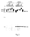
- chimaeric envelopes Lysates of TELCeB6 cells were analysed for envelope expression using antibodies against MLV SU (Fig. 2). For both chimeric envelopes, both a precursor and a processed SU product were detected at ratios similar to wild-type envelopes, suggesting that the mutants were correctly expressed and processed. Cell surface expression of mutant envelopes was examined by FACS analysis of producer cells, using antibodies against the SU or a monoclonal anti-hEGF antibody. All transfected cells were stained with the anti-SU antibodies, and cells expressing the EGF-fusion envelopes were stained with anti-EGF monoclonals (data not shown).
- EMO envelopes MoMLV-derived EGF-fusion envelopes
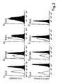
- A431 cells Fig. 3 top
- EGF.R Fig. 3 bottom
- Less binding was found on TE671 and HT1080 target cells which express less EGF.R (Fig. 3).
- No binding could be detected on K422 lymphoma cells with no detectable expression of EGF receptor (Fig. 3).
- the EA envelopes bound to A431 cells as well as EMO (data not shown).
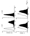
- EGF receptors on A431 cells were down-regulated by pre-incubation with EGF. This treatment did not affect the binding of amphotropic envelopes (Fig. 4 bottom) but abolished binding of EMO envelopes (Fig. 4 top).
- SU envelope glycoproteins of MLVs are known to be weakly associated with their TM protein counterparts (Gliniak et al , 1991 J Biol Chem 266 p22991-22997) and a very low proportion of SU is retained on virions. Therefore it is likely that binding assays in Fig. 3 are due in part to soluble envelope glycoproteins shed from virions.
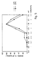
- the supernatant of producer cells was separated by gel-filtration and fractions were analysed for binding activity on A431 cells (Fig. 5). As expected, very little binding activity was found in the early fractions containing the viral particles, with most of the binding activity in the late fractions containing soluble envelopes. However when viral particles were produced at 32°C (in order to reduce the dissociation between SU and TM) a significant binding activity was also found in the fractions containing the virions (Fig. 5), demonstrating that viral particles could bind EGF.R.
- Table 1A shows that viruses incorporating EMO envelopes can infect NIH3T3 mouse fibroblasts. Infection is through the ecotropic MLV receptor because the EMO virus cannot infect NIH3T3 cells expressing the Moloney envelope glycoprotein but can infect those expressing the 4070A envelope glycoprotein. Viruses incorporating EMO envelopes can not only bind to EGF receptors but can also bind and infect cells through ecotropic MLV receptors.
- Table 1B shows that viruses incorporating EMO envelopes could not infect human cells expressing various densities of EGF receptors, despite their ability to bind to the EGF receptors on these cells (Fig 3A).
- the cell line EJ.A1 which stably expresses ecotropic MLV receptors from a transfected plasmid, could not be infected by the EMO virus, but was readily infected by viruses incorporating unmodified MO envelopes.
- the EMO virus could be competitively sequestered by EGF receptors on EJ.A1 cells, preventing it from binding to the ecotropic viral receptors. Infection by virions expressing targeting envelopes A.
- NR6 cells NR6 . NR6-wt hEGF.R . - rEGF + rEGF enV titre titre titre MO 1x10 6 5x10 5 5x10 5 EMO 5x10 4 1x10 3 10 5 A 7x10 4 2x10 5 2x10 5 EA 2x10 5 3x10 3 5x10 5 B. NIH3T3 cells EGFR No . 10,000 80 , 000 400,000 env titre titre titre MO 10 6 10 6 10 6 EMO 10 5 10 4 10 3
- Table 1A shows that viruses incorporating EA envelopes can infect NIH3T3 mouse fibroblasts through the amphotropic MLV receptor.
- the EA virus When titrated on a panel of human cell lines expressing varying densities of EGF receptors, the EA virus showed a selective inability to infect all of the EGF receptor-positive human cells in the panel (Table 1B). However, they could easily infect human B and T cell lines (K422 and Jurkat) which are devoid of EGF.R and are presumably infected through the amphotropic receptor (Table 1).
- EA viruses were efficiently sequestered by EGF.R expressed on human cells and to confirm their competitive sequestration, they were tested on parental and EGF receptor-expressing NR6 mouse fibroblasts (Table 2A). EGF receptor expression on NR6 cells led to a competitive inhibition (100-fold) of viral infection which was reversible when the NR6 transfectants were pre-treated with EGF to block/downregulate their EGF receptors.
- EGF EGF-EGF receptor complexes
- ligand-receptor internalisation ligand-receptor internalisation and routing to lysosomes, where EGF-EGF receptor complexes are degraded.
- EMO-carrying viral particles were used to infect A431 cells treated with the inhibitor of lysosomal degradation, chloroquine, a significant increase of infectivity (by approximately 2 logs) was obtained (Table 3).
- EGF.receptors As EGF.receptor negative cells, such as K422 cells, did not respond similarly (Table 3). Effect of chloroquine on infection NIH3T3 . A431 . TE671 . K422 . enV - + - + - + - + MO 10 6 5x10 5 ⁇ 1 6 ⁇ 1 1 ⁇ 1 ⁇ 1 EMO 10 5 5x10 4 1 225 ⁇ 1 46 ⁇ 1 ⁇ 1
- TELCeB6 cell line was derived from the TELac2 line (Takeuchi et al , 1994 J. Virol. 68 p8001-8007) after transfection and clonal selection of cells containing a plasmid expressing MoMLV (Moloney murine leukemia virus) gag and pol proteins.
- MoMLV Moloney murine leukemia virus
- TELCeB6 cells produce non-infectious viral core particles, carrying the MGFnlsLacZ reporter retroviral vector (Ferry et al , 1991 Proc. Natl. Acad. Sci. USA 88 p8377-8381).
- EJ.A1 an EJ clone that expresses ecotropic MLV receptors, Albritton et al , 1989 Cell 57 p659-666 were grown in DMEM (Gibco-BRL) supplemented with 10% fetal bovine serum (Gibco-BRL).
- K422 cells (Dyer et al , 1990 Blood 75 p709-714) and Jurkat T cells were grown in RPMI 1640 (Gibco-BRL) supplemented with 10% fetal bovine serum (Gibco-BRL).
- NR6 murine fibroblasts lacking detectable EGF receptors (Schneider et al , 1986 Proc. Natl. Acad. Sci. USA 83 p333-336) and NR6-EGF.R (an NR6 subclone obtained after transfection of a plasmid expressing the human epidermal growth factor receptor) cells were kindly provided by G. Gill (La Jolla, USA).
- psi2 cells Mann et al , 1983 Cell 33 p153-159
- GP+EAM12 cells Markowitz et al , 1988 Virol. 167 p400-406 were derived from NIH-3T3 cells and express respectively MoMLV (ecotropic) and MLV-amphotropic envelopes which block the corresponding receptors (EcoR-1 and RAM-1) by interference.
- NIH3T3 clones transfected with EGF receptor expression constructs and expressing moderate or high levels of EGF receptors were kindly provided by Prof Thierry Velu (Erasmus Hospital, Brussels).
- NIH-3T3 and NIH-3T3-derived cell lines were grown in DMEM (Gibco-BRL) supplemented with 10% new born bovine serum (Gibco-BRL).
- Envelope expression plasmids were transfected by calcium phosphate precipitation into TELCeB6 cells. Transfected cells were selected with phleomycin (50 mg/ml) and pools of phleomycin-resistant clones were used to harvest viruses from confluent cells after overnight incubation in DMEM and FBS (10%). These supernatants were used for ultracentrifugation to provide Western blot samples, for binding assays and for infection assays. Viruses (in 100 ml of producer cell supernatant) were also purified by gel-filtration on 2 ml columns (Bio-Rad) on a bed of S-1000 Sephacryl (Pharmacia). Fractions were obtained by elution with PBS at 4°C.
- Virus producer cells were lysed in a 20 mM Tris-HCl buffer (pH 7.5) containing 1% Triton-X100, 0.05% SDS, 5 mg/ml sodium deoxycholate, 150 mM NaCl, and 1 mM PMSF. Lysates were incubated for 10 min at 4°C and were centrifuged for 10 min at 10,000xg to pellet the nuclei. Supernatants were then frozen at -70°C until further analysis. Virus samples were obtained by ultracentrifugation of viral supernatants (10 ml) in a SW41 Beckman Rotor (30,000 RPM, 1 hr, 4°C).
- Pellets were suspended in 100 ⁇ l of PBS (phosphate buffered saline), and frozen at -70°C. Samples (30 mg for cell lysates, or 10 ⁇ l for purified viruses) were mixed in a 375 mM Tris-HCl (pH 6.8) buffer containing 6% SDS, 30% b-mercapto-ethanol, 10% glycerol, and 0.06% bromophenol blue, boiled for 3 min, then run on 10% SDS acrylamide gels. After protein transfer onto nitrocellulose filters, immunostaining was performed in TBS (Tris base saline, pH 7.4) with 5% milk powder and 0.1% TWEEN.
- TBS Tris base saline, pH 7.4
- Antibodies were goat antisera raised against either RLV (Rausher leukemia virus) gp70-SU protein or RLV p30-CA protein, and were diluted 1/1,000 and 1/10,000, respectively. Blots were developed using HRPO-conjugated rabbit anti-goat antibodies (DAKO, UK) and an electrochemiluminescence kit (Amersham Life Science).
- Target cells were washed in PBS and detached by a 10 min incubation at 37°C with versene 0.02% in PBS.
- Cells were washed in PBA (PBS with 2% FCS and 0.1% sodium azide). 10 6 cells were incubated with viruses for 30 min at 4°C. Cells were then washed with PBA and incubated in PBA containing 1/200 of RLV gp70 immune serum for 30 min at 4°C. Cells were washed twice with PBA and incubated with rabbit anti-goat FITC-conjugated antibodies (DAKO, U.K.). 5 min before the two final washes in PBA, cells were stained with 20 mg/ml propidium iodide.
- PBA PBS with 2% FCS and 0.1% sodium azide
- Fluorescence of living cells was analysed with a fluorescent-activated cell sorter (FACSCan, Beckton Dickinson).
- FACSCan fluorescent-activated cell sorter
- 10 6 cells in 100 ml of PBA were incubated with 10 ml of anti-EGF.R antibodies (M886, DAKO, U.K.) for 30 min at 4°C.
- Target cells were seeded in 24 multi-well plates at a density of 3x10 4 cells per well or in 6-multi-well plates at a density of 2x10 5 cells per well.
- Viral supernatant dilutions containing 4 mg/ml polybrene were added and cells were incubated for 3-5 hrs at 37°C. Viral supernatant was then removed and cells were incubated in regular medium for 24-48 hrs.
- X-Gal staining was performed as previously described (Takeuchi et al , 1994 J. Virol. 68 p8001-8007).
- target cells were incubated 30 min at 37°C in a medium containing 10 -6 M rEGF (236-EG, R&D Systems, U.K.). Cells were then washed and infections were carried out as previously described.
- 100 mM chloroquine phosphate Sigma, U.K. was added to the medium for 6 hr from the start of the infection protocol after which the cells were washed and incubated in regular medium.
- EGF amphotropic vector (Cosset et al. 1995 J. Virol 69 p6314), with an EGF cDNA inserted at +1 codon of the N terminus of the 4070A murine leukemia virus (MLV) surface protein (SU) gp 70 ( Figure 6) and flanked by Sfi I and Not I restriction sites, was used as a template for PCR random mutagenesis (Hawkins et al. 1992 J. Mol. Biol. 226 p889).
- E.A. chimaeric EGF amphotropic vector
- the 100 ⁇ l reaction mixture contained 20 ng template, 50 pmoles of Bglenvrev primer (5' TCT AGA CTG ACA TGG CGC GT, Seq. ID No. 3) and 4070Aseq3 primer (5' GGT GAC TCT CCA GGT TAC, Seq. ID No. 4), 1mM of each deoxynucleotide triphosphate, 50 mM KCl, 10 mM Tris-HCl (pH 9.0 at 25°C), 6 mM MgCl 2 , 0.5 mM MnCl 2 and 1% (v/v) Triton X-100.
- the MnCl 2 was added fresh just before the start of the PCR. Amplification consisted of 30 cycles of 94°C (1 min), 65°C (1 min) and 70°C (4 min) using Taq DNA polymerase (Promega; 5 units). The resulting PCR-generated cDNA fragments were digested with Sfi I and Not I restriction enzymes and cloned into Sfi I -Not I digested pNCAEGFL.1 Sfi I-backbone ( Figure 7) in which the Sfi I site in the pol gene was deleted by PCR mutagenesis without change to the amino acid sequence.
- PCR cDNA fragments were generated from the DNA of each of the respective clones using Bglenvrev and envseq5 (5' GTA AGG TCA GGC CAC CAG GT, Seq. ID No. 5) primers at final concentration of 25 ⁇ moles each, 5 ⁇ l Taq polymerase buffer (Promega), 0.25 mM dNTP and amplified for 25 cycles of 94°C (30s), 60°C (30s) and 72°C (30s) using Taq polymerase (5 units). The PCR fragments were gel purified and used as template for cycle sequencing with 9.5 ⁇ l of terminator ready reaction mix (ABI PRISMTM) and 3.2 pmole of envseq5 primer.
- the PCR-generated EGF cDNA was cloned into the pNCAEGFL.1 Sfi I-backbone. Ten different ligations were performed. The ligated products were transformed into electrocompetent cells (Stratagene, Epicurian coli XL1-MRF'), yielding 10 5 transformed cells/ml. The transformed cells were selected in 50 mg/ml ampicillin. Five batches of transformants were grown on large (225 cm x 225 cm) 2 x TY plates while the rest were grown respectively in 5 x 500 ml of 2 x TY liquid media overnight at 37°C. The next day, colonies of cells on the plates were scraped off and resuspended in 6 ml 2 x TY/ampicillin.
- the human fibrosarcoma cell line, HT1080 was transfected with an expression vector coding for the Moloney MLV ecotropic receptor, Rec-1.
- Expression vectors were constructed by blunt end ligation of the cDNA fragment coding for Rec-1 into the RC/CMV vector at the Not I site of the polylinker, detailed below.
- the RC/CMV Rec-1 expression vector was stably transfected into HT1080 cells by the calcium phosphate precipitation method and the cells were pooled and expanded in 10% FCS-DMEM selection media containing 1 mg/ml neomycin (G418).
- Rec-1 receptor The expression of the Rec-1 receptor was confirmed by checking if the HT1080-Rec-1 cells could be infected by wild type MoMLV virus transferring the ⁇ -galactosidase gene. About 65% of the transfected cells were stained blue with X-gal.
- the polylinker in RC/CMV Rec-1 expression vector contains the following sites:
- the EGF plasmid library is transiently transfected into HT1080 cells by calcium phosphate precipitation method (Sambrook et al. , Molecular Cloning, Cold Spring Harbour Laboratory Press). After 72 h the viral supernatant from the HT1080 cells, which contains EGF displaying replicating vectors, is harvested overnight in 5 ml 10% FCS-DMEM and filtered (0.45 ⁇ m). It is then used to infect HT1080-Rec-1 cells in the presence of 4 ⁇ g/ml polybrene. After 72 h, the media from infected HT1080-Rec-1 cells are removed and replaced with 5ml 10%FCS-DMEM. The viral supernatant is harvested as before and used to infect a fresh batch of HT1080-Rec-1. A total three rounds of selection are carried out.
- High molecular weight DNA is extracted from the infected HT1080-Rec-1 cells from both the second and third rounds of selection.
- the cells are washed with PBS, incubated in DNA lysis buffer (TE buffer, 1% SDS) containing 100 ⁇ g/ml proteinase K at 37°C for 5 minutes.
- Cell lysates are incubated at 37°C overnight and then extracted once in 2 ml phenol, by mixing it gently on a rotor wheel for 15 minutes, centrifuged for 15 minutes at 1500xg and then the aqueous phase is transferred to a new tube. The process is repeated once with phenol/chloroform (1:1) and then with chloroform only. After the final chloroform extraction, 2.5 volumes salt/ethanol (250 vol.
- Polymerase chain reaction is carried out as before using Bglenvrev and envseq5 primers on a diluted sample of the high molecular weight DNA to amplify the EGF cDNA.
- the PCR product is ligated into TA Cloning vector (Invitrogen) and the vector is transformed into competent cells. Positive colonies containing the insert are picked and DNA from the individual colonies are extracted and the sequences of the EGF insert are checked on an automated sequencer. Mutations identified in this way are presumed to reduce the affinity of binding to EGF receptors but not to interfere with the folding of the EGF domain. This is confirmed by amplifying the mutant EGF cDNA fragment by PCR and cloning it into the EGF chimaeric construct (E.A). The construct is then transfected into the viral producer cells (TELCeB6) and the viral supernatant harvested for infection assays.
- TELCeB6 the viral producer cells
Abstract
Description
| Infection by virions expressing targeting envelopes | |||
| A. On mouse fibroblastsb | |||
| enva | 3T3 | 3T3/E | 3T3/A |
| A 107 | 107 | 102 | |
| MO | 107 | <1 | 107 |
| EMO | 105 | <1 | 105 |
| EA 106 | nd | 101 |
| On human cell lines | |||||||
| env | A431 | HT108 | TE671 | K422 | Jurkat | EJ | EJ.A1 |
| EGFR | ++++ | ++ | + | - | - | ++ | ++ |
| A | 107 | 107 | 107 | 105 | 104 | 106 | 106 |
| MO | <1 | <1 | <1 | <1 | <1 | <1 | 106 |
| EMO | <1 | <1 | <1 | <1 | <1 | <1 | <1 |
| EA | <1 | <1 | 101 | 104 | 103 | <1 | <1 |
| Inhibition of infection by EGF.R | |||
| A. NR6 cells | |||
| NR6 . | NR6-wt hEGF.R . | ||
| - rEGF | + rEGF | ||
| enV | titre | titre | titre |
| MO | 1x10⁶ | 5x10⁵ | 5x10⁵ |
| EMO | 5x10⁴ | 1x10³ | 105 |
| A | 7x10⁴ | 2x10⁵ | 2x10⁵ |
| EA | 2x10⁵ | 3x10³ | 5x10⁵ |
| B. NIH3T3 cells | |||
| EGFR No. | 10,000 | 80,000 | 400,000 |
| env | titre | titre | titre |
| MO | 106 | 106 | 106 |
| EMO | 105 | 104 | 103 |
| Effect of chloroquine on infection | ||||||||
| NIH3T3 . | A431 . | TE671 . | K422 . | |||||
| enV | - | + | - | + | - | + | - | + |
| MO | 106 | 5x10⁵ | <1 | 6 | <1 | 1 | <1 | <1 |
| EMO | 105 | 5x10 4 | 1 | 225 | <1 | 46 | <1 | <1 |


| Number of clones with 1-5 point mutations in the EGF cDNA. | |
| Number of mutations | Number of |
| 0 | 9 |
| 1 | 7 |
| 2 | 4 |
| 3 | 5 |
| 4 | 1 |
| 5 | 1 |
Total number of bases sequenced = 4293
Total number of point mutations = 39
Number of mutations per gene (159 bases) = 39 x 159/4293 = 1.4


Claims (17)
- A method of screening a plurality of altered first members of a specific binding pair ("sbp") for reduced ability to bind to the second member of the sbp compared to an unaltered first member; the method comprising introducing a plurality of nucleic acid sequences, each encoding a respective altered first member of the sbp, into a plurality of viral display packages so as to form a viral display package library in which each viral display package displays on its surface an altered first member of the sbp encoded by a respective nucleic acid sequence comprised within the viral display package and an infection-competent binding moiety capable of mediating infection of a susceptible target cell; contacting the viral display packages with a plurality of target cells which express on their surface a receptor molecule for the infection-competent binding moiety, such that binding to the receptor molecule tends to facilitate infection of a target cell by the viral display package, said target cells also expressing on their surface the second member of the sbp, binding to which, via the displayed altered first member of the sbp, tends to inhibit infection of a target cell by the viral display package, such that those viral display packages displaying altered first members with reduced ability to bind to the second member of the sbp will tend to infect target cells more successfully; and recovering from infected target cells or their progeny nucleic acid comprising those nucleic acid sequences which encode altered first members with reduced ability to bind to the second member of the sbp.
- A method according to claim 1, wherein the viral display package is capable of infecting eukaryotic cells.
- A method according to claim 1 or 2, wherein the viral display package is based on a retrovirus.
- A method according to any one of claims 1, 2 or 3, wherein the viral display package is based on a C type retrovirus.
- A method according to any one of the preceding claims, wherein the first member of the sbp is displayed as a fusion polypeptide.
- A method according to any one of the preceding claims, wherein the first member of the sbp is displayed as a fusion with the infection-competent binding moiety.
- A method according to any one of the preceding claims, wherein the first member of the sbp is displayed as a fusion with a viral envelope protein.
- A method according to any one of the preceding claims, wherein the first member of the sbp is displayed as an N-terminal fusion with a retroviral envelope glycoprotein.
- A method according to any one of the preceding claims, wherein the infection-competent binding moiety comprises a substantially intact retroviral envelope glycoprotein.
- A method according to any one of the preceding claims, wherein the target cell is eukaryotic.
- A method according to any one of the preceding claims, wherein the target cell is mammalian.
- A method according to any one of the preceding claims, wherein the target cell naturally expresses the receptor molecule and the second member of the sbp.
- A method according to any one of the preceding claims, wherein the target cell has been transfected with a sequence directing the expression of the receptor molecule and/or the second member of the sbp.
- A method according to any one of the preceding claims, wherein the plurality of nucleic acid sequences encoding respective altered first members of the sbp are derived by the introduction of mutations into the sequence encoding the unaltered first member of the sbp.
- A method according to claim 14, wherein the mutations are introduced by a process of substantially random mutagenesis.
- A kit for use in performing the method of any one of the preceding claims, comprising a nucleic acid construct suitable for accepting a sequence encoding a first member of an sbp; retroviral packaging cells for producing viral display packages incorporating the nucleic acid construct; target cells expressing on their surface a receptor which allows infection by the viral display packages and also expressing the second member of the sbp; and instructions for performing a method in accordance with any one of the preceding claims.
- A kit according to claim 16, further comprising one or more of the following: means for introducing mutations in the nucleic acid sequence encoding the unaltered first member of the sbp; and detection means for detecting infection of a target cell by a viral display package.
Applications Claiming Priority (3)
| Application Number | Priority Date | Filing Date | Title |
|---|---|---|---|
| GBGB9516138.6A GB9516138D0 (en) | 1995-08-05 | 1995-08-05 | Improvements in or relating to methods of screening substances |
| GB9516138 | 1995-08-05 | ||
| PCT/GB1996/001905 WO1997006435A1 (en) | 1995-08-05 | 1996-08-05 | Improvements in or relating to methods of screening substances |
Publications (2)
| Publication Number | Publication Date |
|---|---|
| EP0842426A1 EP0842426A1 (en) | 1998-05-20 |
| EP0842426B1 true EP0842426B1 (en) | 2002-09-04 |
Family
ID=10778860
Family Applications (1)
| Application Number | Title | Priority Date | Filing Date |
|---|---|---|---|
| EP96926484A Expired - Lifetime EP0842426B1 (en) | 1995-08-05 | 1996-08-05 | Improvements in or relating to methods of screening substances |
Country Status (8)
| Country | Link |
|---|---|
| EP (1) | EP0842426B1 (en) |
| JP (1) | JPH11510696A (en) |
| AT (1) | ATE223574T1 (en) |
| AU (1) | AU710446B2 (en) |
| CA (1) | CA2225680A1 (en) |
| DE (1) | DE69623480D1 (en) |
| GB (1) | GB9516138D0 (en) |
| WO (1) | WO1997006435A1 (en) |
Families Citing this family (9)
| Publication number | Priority date | Publication date | Assignee | Title |
|---|---|---|---|---|
| AU1127097A (en) * | 1995-12-15 | 1997-07-14 | Pharmacia & Upjohn Company | Method for the simultaneous detection of a therapeutic target for a disease state and its neutralizing antibody-like molecule |
| US6054312A (en) | 1997-08-29 | 2000-04-25 | Selective Genetics, Inc. | Receptor-mediated gene delivery using bacteriophage vectors |
| US6723512B2 (en) | 1997-08-29 | 2004-04-20 | Selective Genetics Inc. | Methods using genetic package display for detecting and identifying protein-protein interactions that facilitate internalization and transgene expression and cells or tissues competent for the same and methods for evolving gene delivery vectors |
| US6589730B1 (en) | 1997-08-29 | 2003-07-08 | Selective Genetics, Inc. | Methods for identifying protein-protein interactions by selective transduction |
| AU740541B2 (en) | 1997-08-29 | 2001-11-08 | Selective Genetics, Inc. | Methods using phage display for selecting internalizing ligands for gene delivery |
| US6451527B1 (en) | 1997-08-29 | 2002-09-17 | Selective Genetics, Inc. | Methods using genetic package display for selecting internalizing ligands for gene delivery |
| US8334238B2 (en) | 1998-04-17 | 2012-12-18 | Rigel, Inc. | Multiparameter FACS assays to detect alterations in cellular parameters and to screen small molecule libraries |
| WO1999054494A2 (en) * | 1998-04-17 | 1999-10-28 | Rigel Pharmaceuticals, Inc. | Multiparameter facs assays to detect alterations in cellular parameters |
| CA2341156A1 (en) * | 1998-09-04 | 2000-03-16 | Zk Pharmaceuticals, Inc. | Method for selecting peptides inhibiting viral surface protein binding to cell surface receptor |
Family Cites Families (4)
| Publication number | Priority date | Publication date | Assignee | Title |
|---|---|---|---|---|
| WO1993000103A1 (en) * | 1991-06-21 | 1993-01-07 | The Wistar Institute Of Anatomy And Biology | Chimeric envelope proteins for viral targeting |
| WO1994006920A1 (en) * | 1992-09-22 | 1994-03-31 | Medical Research Council | Recombinant viruses displaying a nonviral polypeptide on their external surface |
| AU7097494A (en) * | 1993-06-01 | 1994-12-20 | Targeted Genetics Corporation | Envelope fusion vectors for use in gene delivery |
| GB9412844D0 (en) * | 1994-06-27 | 1994-08-17 | Medical Res Council | Improvements in or relating to therapeutic methods |
-
1995
- 1995-08-05 GB GBGB9516138.6A patent/GB9516138D0/en active Pending
-
1996
- 1996-08-05 EP EP96926484A patent/EP0842426B1/en not_active Expired - Lifetime
- 1996-08-05 AT AT96926484T patent/ATE223574T1/en not_active IP Right Cessation
- 1996-08-05 AU AU66648/96A patent/AU710446B2/en not_active Ceased
- 1996-08-05 JP JP9508234A patent/JPH11510696A/en active Pending
- 1996-08-05 WO PCT/GB1996/001905 patent/WO1997006435A1/en active IP Right Grant
- 1996-08-05 CA CA002225680A patent/CA2225680A1/en not_active Abandoned
- 1996-08-05 DE DE69623480T patent/DE69623480D1/en not_active Expired - Lifetime
Also Published As
| Publication number | Publication date |
|---|---|
| DE69623480D1 (en) | 2002-10-10 |
| GB9516138D0 (en) | 1995-10-04 |
| AU710446B2 (en) | 1999-09-23 |
| WO1997006435A1 (en) | 1997-02-20 |
| AU6664896A (en) | 1997-03-05 |
| EP0842426A1 (en) | 1998-05-20 |
| JPH11510696A (en) | 1999-09-21 |
| ATE223574T1 (en) | 2002-09-15 |
| CA2225680A1 (en) | 1997-02-20 |
Similar Documents
| Publication | Publication Date | Title |
|---|---|---|
| Wills et al. | Creation and expression of myristylated forms of Rous sarcoma virus Gag protein in mammalian cells | |
| US6297004B1 (en) | Recombinant viruses displaying a nonviral polypeptide on their external surface | |
| Russell et al. | Retroviral vectors displaying functional antibody fragments | |
| EP0842426B1 (en) | Improvements in or relating to methods of screening substances | |
| US5998192A (en) | Delivery of nucleic acids | |
| US20050158712A1 (en) | Methods for purifying viral particles for gene therapy | |
| WO1999060110A2 (en) | Stable envelope proteins for retroviral, viral and liposome vectors and use in gene and drug therapy | |
| Liu et al. | Construction and screening of a lentiviral secretome library | |
| US20020052040A1 (en) | Virus like particles, their preparation and their use preferably in pharmaceutical screening and functional genomics | |
| US6054281A (en) | Binding assays | |
| US6762031B2 (en) | Targeting viral vectors to specific cells | |
| EP1219705A1 (en) | Virus like particles, their preparation and their use preferably in pharmaceutical screening and functional genomics | |
| Bupp et al. | Selection of feline leukemia virus envelope proteins from a library by functional association with a murine leukemia virus envelope | |
| US7476517B2 (en) | Virus like particles, their preparation and their use preferably in pharmaceutical screening and functional genomics | |
| WO2002034929A2 (en) | Expression vectors and uses thereof | |
| Ryu | Modification of a retroviral envelope protein to achieve cell type specific targeted entry | |
| Vasser | Manipulation of the Moloney Murine Leukemia Virus envelope protein in an effort to develop directly and indirectly targeted retroviral vectors for use in human gene therapy | |
| Novakovic | Protein motifs in the cytoplasmic tail of the bovine leukemia virus transmembrane protein govern protein expression on the cell surface | |
| Benedict | The murine leukemia virus envelope protein: engineering for vector targeting and analysis of interactions with viral core proteins |
Legal Events
| Date | Code | Title | Description |
|---|---|---|---|
| PUAI | Public reference made under article 153(3) epc to a published international application that has entered the european phase |
Free format text: ORIGINAL CODE: 0009012 |
|
| 17P | Request for examination filed |
Effective date: 19971128 |
|
| AK | Designated contracting states |
Kind code of ref document: A1 Designated state(s): AT BE CH DE DK ES FI FR GB GR IE IT LI LU MC NL PT SE |
|
| RAP1 | Party data changed (applicant data changed or rights of an application transferred) |
Owner name: CAMBRIDGE GENETICS LIMITED |
|
| 17Q | First examination report despatched |
Effective date: 19991105 |
|
| GRAG | Despatch of communication of intention to grant |
Free format text: ORIGINAL CODE: EPIDOS AGRA |
|
| RAP1 | Party data changed (applicant data changed or rights of an application transferred) |
Owner name: BIOFOCUS DISCOVERY LIMITED |
|
| GRAG | Despatch of communication of intention to grant |
Free format text: ORIGINAL CODE: EPIDOS AGRA |
|
| GRAH | Despatch of communication of intention to grant a patent |
Free format text: ORIGINAL CODE: EPIDOS IGRA |
|
| GRAH | Despatch of communication of intention to grant a patent |
Free format text: ORIGINAL CODE: EPIDOS IGRA |
|
| GRAA | (expected) grant |
Free format text: ORIGINAL CODE: 0009210 |
|
| AK | Designated contracting states |
Kind code of ref document: B1 Designated state(s): AT BE CH DE DK ES FI FR GB GR IE IT LI LU MC NL PT SE |
|
| PG25 | Lapsed in a contracting state [announced via postgrant information from national office to epo] |
Ref country code: NL Free format text: LAPSE BECAUSE OF FAILURE TO SUBMIT A TRANSLATION OF THE DESCRIPTION OR TO PAY THE FEE WITHIN THE PRESCRIBED TIME-LIMIT Effective date: 20020904 Ref country code: LI Free format text: LAPSE BECAUSE OF FAILURE TO SUBMIT A TRANSLATION OF THE DESCRIPTION OR TO PAY THE FEE WITHIN THE PRESCRIBED TIME-LIMIT Effective date: 20020904 Ref country code: IT Free format text: LAPSE BECAUSE OF FAILURE TO SUBMIT A TRANSLATION OF THE DESCRIPTION OR TO PAY THE FEE WITHIN THE PRESCRIBED TIME-LIMIT;WARNING: LAPSES OF ITALIAN PATENTS WITH EFFECTIVE DATE BEFORE 2007 MAY HAVE OCCURRED AT ANY TIME BEFORE 2007. THE CORRECT EFFECTIVE DATE MAY BE DIFFERENT FROM THE ONE RECORDED. Effective date: 20020904 Ref country code: GR Free format text: LAPSE BECAUSE OF FAILURE TO SUBMIT A TRANSLATION OF THE DESCRIPTION OR TO PAY THE FEE WITHIN THE PRESCRIBED TIME-LIMIT Effective date: 20020904 Ref country code: FR Free format text: LAPSE BECAUSE OF NON-PAYMENT OF DUE FEES Effective date: 20020904 Ref country code: FI Free format text: LAPSE BECAUSE OF FAILURE TO SUBMIT A TRANSLATION OF THE DESCRIPTION OR TO PAY THE FEE WITHIN THE PRESCRIBED TIME-LIMIT Effective date: 20020904 Ref country code: CH Free format text: LAPSE BECAUSE OF FAILURE TO SUBMIT A TRANSLATION OF THE DESCRIPTION OR TO PAY THE FEE WITHIN THE PRESCRIBED TIME-LIMIT Effective date: 20020904 Ref country code: BE Free format text: LAPSE BECAUSE OF FAILURE TO SUBMIT A TRANSLATION OF THE DESCRIPTION OR TO PAY THE FEE WITHIN THE PRESCRIBED TIME-LIMIT Effective date: 20020904 Ref country code: AT Free format text: LAPSE BECAUSE OF FAILURE TO SUBMIT A TRANSLATION OF THE DESCRIPTION OR TO PAY THE FEE WITHIN THE PRESCRIBED TIME-LIMIT Effective date: 20020904 |
|
| REF | Corresponds to: |
Ref document number: 223574 Country of ref document: AT Date of ref document: 20020915 Kind code of ref document: T |
|
| REG | Reference to a national code |
Ref country code: GB Ref legal event code: FG4D |
|
| REG | Reference to a national code |
Ref country code: CH Ref legal event code: EP |
|
| REG | Reference to a national code |
Ref country code: IE Ref legal event code: FG4D |
|
| REF | Corresponds to: |
Ref document number: 69623480 Country of ref document: DE Date of ref document: 20021010 |
|
| PG25 | Lapsed in a contracting state [announced via postgrant information from national office to epo] |
Ref country code: SE Free format text: LAPSE BECAUSE OF FAILURE TO SUBMIT A TRANSLATION OF THE DESCRIPTION OR TO PAY THE FEE WITHIN THE PRESCRIBED TIME-LIMIT Effective date: 20021204 Ref country code: DK Free format text: LAPSE BECAUSE OF FAILURE TO SUBMIT A TRANSLATION OF THE DESCRIPTION OR TO PAY THE FEE WITHIN THE PRESCRIBED TIME-LIMIT Effective date: 20021204 |
|
| PG25 | Lapsed in a contracting state [announced via postgrant information from national office to epo] |
Ref country code: DE Free format text: LAPSE BECAUSE OF FAILURE TO SUBMIT A TRANSLATION OF THE DESCRIPTION OR TO PAY THE FEE WITHIN THE PRESCRIBED TIME-LIMIT Effective date: 20021205 |
|
| PG25 | Lapsed in a contracting state [announced via postgrant information from national office to epo] |
Ref country code: PT Free format text: LAPSE BECAUSE OF FAILURE TO SUBMIT A TRANSLATION OF THE DESCRIPTION OR TO PAY THE FEE WITHIN THE PRESCRIBED TIME-LIMIT Effective date: 20021212 |
|
| NLV1 | Nl: lapsed or annulled due to failure to fulfill the requirements of art. 29p and 29m of the patents act | ||
| REG | Reference to a national code |
Ref country code: CH Ref legal event code: PL |
|
| PG25 | Lapsed in a contracting state [announced via postgrant information from national office to epo] |
Ref country code: ES Free format text: LAPSE BECAUSE OF FAILURE TO SUBMIT A TRANSLATION OF THE DESCRIPTION OR TO PAY THE FEE WITHIN THE PRESCRIBED TIME-LIMIT Effective date: 20030328 |
|
| EN | Fr: translation not filed | ||
| PLBE | No opposition filed within time limit |
Free format text: ORIGINAL CODE: 0009261 |
|
| STAA | Information on the status of an ep patent application or granted ep patent |
Free format text: STATUS: NO OPPOSITION FILED WITHIN TIME LIMIT |
|
| PG25 | Lapsed in a contracting state [announced via postgrant information from national office to epo] |
Ref country code: LU Free format text: LAPSE BECAUSE OF NON-PAYMENT OF DUE FEES Effective date: 20030805 Ref country code: IE Free format text: LAPSE BECAUSE OF NON-PAYMENT OF DUE FEES Effective date: 20030805 Ref country code: GB Free format text: LAPSE BECAUSE OF NON-PAYMENT OF DUE FEES Effective date: 20030805 |
|
| 26N | No opposition filed |
Effective date: 20030605 |
|
| PG25 | Lapsed in a contracting state [announced via postgrant information from national office to epo] |
Ref country code: MC Free format text: LAPSE BECAUSE OF NON-PAYMENT OF DUE FEES Effective date: 20030831 |
|
| GBPC | Gb: european patent ceased through non-payment of renewal fee |
Effective date: 20030805 |
|
| REG | Reference to a national code |
Ref country code: IE Ref legal event code: MM4A |