EP0835662A2 - A drug for the treatment of cancer - Google Patents
A drug for the treatment of cancer Download PDFInfo
- Publication number
- EP0835662A2 EP0835662A2 EP96309012A EP96309012A EP0835662A2 EP 0835662 A2 EP0835662 A2 EP 0835662A2 EP 96309012 A EP96309012 A EP 96309012A EP 96309012 A EP96309012 A EP 96309012A EP 0835662 A2 EP0835662 A2 EP 0835662A2
- Authority
- EP
- European Patent Office
- Prior art keywords
- vip
- approximately
- day
- som
- cells
- Prior art date
- Legal status (The legal status is an assumption and is not a legal conclusion. Google has not performed a legal analysis and makes no representation as to the accuracy of the status listed.)
- Granted
Links
Images


Classifications
-
- A—HUMAN NECESSITIES
- A61—MEDICAL OR VETERINARY SCIENCE; HYGIENE
- A61K—PREPARATIONS FOR MEDICAL, DENTAL OR TOILETRY PURPOSES
- A61K38/00—Medicinal preparations containing peptides
- A61K38/04—Peptides having up to 20 amino acids in a fully defined sequence; Derivatives thereof
- A61K38/08—Peptides having 5 to 11 amino acids
-
- A—HUMAN NECESSITIES
- A61—MEDICAL OR VETERINARY SCIENCE; HYGIENE
- A61K—PREPARATIONS FOR MEDICAL, DENTAL OR TOILETRY PURPOSES
- A61K38/00—Medicinal preparations containing peptides
- A61K38/04—Peptides having up to 20 amino acids in a fully defined sequence; Derivatives thereof
- A61K38/10—Peptides having 12 to 20 amino acids
-
- A—HUMAN NECESSITIES
- A61—MEDICAL OR VETERINARY SCIENCE; HYGIENE
- A61K—PREPARATIONS FOR MEDICAL, DENTAL OR TOILETRY PURPOSES
- A61K38/00—Medicinal preparations containing peptides
- A61K38/16—Peptides having more than 20 amino acids; Gastrins; Somatostatins; Melanotropins; Derivatives thereof
- A61K38/17—Peptides having more than 20 amino acids; Gastrins; Somatostatins; Melanotropins; Derivatives thereof from animals; from humans
- A61K38/22—Hormones
- A61K38/31—Somatostatins
-
- A—HUMAN NECESSITIES
- A61—MEDICAL OR VETERINARY SCIENCE; HYGIENE
- A61P—SPECIFIC THERAPEUTIC ACTIVITY OF CHEMICAL COMPOUNDS OR MEDICINAL PREPARATIONS
- A61P35/00—Antineoplastic agents
Definitions
- the present invention relates to a combination of peptide analogs.
- the combination may be used to block the uncontrolled multiplication of cancer cells of the colon, rectum, lung, breast, and kidney.
- the combination may be used to treat cancers of the colon, rectum, lung, breast, and kidney and may be used to treat leukemia and lymphoma.
- the invention also relates to a pharmaceutical composition containing a combination of such analogs.
- the present invention provides a pharmaceutical composition useful for killing or inhibiting multiplication of tumor cells as well as cancer cells.
- the pharmaceutical composition may also be useful in preventing, inhibiting, or modulating the hypersecretion of VIP, somatostatin, bombesin, Substance P, or a combination of VIP, somatostatin, bombesin, or Substance P.
- the composition may suitably comprise, consist of, or consist essentially of a therapeutically effective combination of peptide analogs of somatostatin, VIP, bombesin, and Substance P.
- the peptide analogs are described in more detail below, but constituents functionally interchangeable with those specifically described may also be employed in the claimed pharmaceutical composition.
- the pharmaceutical composition may suitably comprise, consist of, or consist essentially of an analog of somatostatin and at least four peptides selected from the group consisting of a first analog of VIP, a second analog of VIP, a third analog of VIP, another analog of somatostatin, an analog of bombesin, and an analog of Substance P.
- composition may suitably comprise, consist of, or consist essentially of a therapeutically effective combination of peptide SOM 2 (an analog of somatostatin) and at least four of the following peptides: VIP 1 (a VIP antagonist), VIP 2 (a VIP receptor binding inhibitor), VIP 3 (a VIP receptor antagonist), SOM 1 (a somatostatin analog (also abbreviated "CTOP,” which is derived from the first letters of the following four amino acids: Cys 2 , Tyr 3 , Orn 5 , and Pen 5 )), BOM 1 (a bombesin antagonist), and SP 1 (a Substance P antagonist).
- a pharmaceutically acceptable carrier, diluent, or solvent is used.
- the invention provides a method of treatment for humans, mammals, or other animals suffering from cancer or other tumors.
- the method may suitably comprise, consist of, or consist essentially of administering a therapeutically effective dose of the pharmaceutical composition so as to kill or inhibit the multiplication of cancer or tumor cells.
- the method of treatment of the present invention may be particularly useful in the treatment of cancers or tumors of the colon and rectum.
- the invention also provides a method of treatment for humans, mammals, or other animals suffering from hypersecretion of VIP, somatostatin, bombesin, Substance P, or a combination of VIP, somatostatin, bombesin, or Substance P.
- the method may suitably comprise, consist of, or consist essentially of administering a therapeutically effective dose of the pharmaceutical composition so as to prevent, inhibit, or modulate the hypersecretion of VIP, somatostatin, bombesin, Substance P, or a combination of VIP, somatostatin, bombesin, or Substance P.
- Figure 1 which shows the effect on tumor regression of treatment onset time with MuJ-7, summarizes the mean tumor volume (in mm 3 ) for all of the mice in the in vivo protocols described in Examples 6-13 versus the day numbers.
- Figure 2 is a graph of the mean tumor volume (in mm 3 ) of the treated mice ("o") and the untreated control mice ("+") versus the day numbers for the in vivo protocol described in Example 6.
- Figure 3 is a graph of the mean tumor volume (in mm 3 ) of the group 1 treated mice ("o"), the group 2 treated mice (" ⁇ "), and the untreated control mice ("+") versus the day numbers for the in vivo protocol described in Example 7.
- Figure 4 is a graph of the tumor volume (in mm 3 ) of the treated mouse ("o") and the untreated control mouse ("+") versus the day numbers for the in vivo protocol with SW 620 cells described in Example 8.
- Figure 5 is a graph of the tumor volume (in mm 3 ) of the treated mouse ("o") and the untreated control mouse ("+") versus the day numbers for the in vivo protocol with HT 29 cells described in Example 8.
- Figure 6 is a graph of the tumor volume (in mm 3 ) of the treated mouse ("o") and the untreated control mouse ("+") versus the day numbers for the in vivo protocol with CoLo 205 cells described in Example 8.
- Figure 7 is a graph of the tumor volume (in mm 3 ) of the treated mouse ("o") and the untreated control mouse ("+") versus the day numbers for the in vivo protocol with L 132 cells described in Example 8.
- Figure 8 is a graph of the mean tumor volume (in mm 3 ) of the treated mice ("o") and the untreated control mice ("+") versus the day numbers for the in vivo protocol described in Example 13.
- Figure 9 is a graph of the percentage of surviving treated mice (dotted line) and the percentage of untreated control mice (solid line) versus the day numbers for the in vivo protocol described in Example 13.
- VIP vasoactive intestinal peptide
- somatostatin somatostatin
- Substance P somatostatin
- bombesin are secreted by at least some human tumor and cancer cells and that there are binding sites for these peptides on these cells.
- the four peptides i.e., vasoactive intestinal peptide (VIP), somatostatin, Substance P, and bombesin
- VIP vasoactive intestinal peptide
- somatostatin somatostatin
- Substance P somatostatin
- bombesin the four peptides (i.e., vasoactive intestinal peptide (VIP), somatostatin, Substance P, and bombesin) were shown to bind to tumor cells.
- peptide hormones each refer to VIP, somatostatin, Substance P, and bombesin.
- growth factors peptide growth regulators
- peptides each refer to VIP, somatostatin, Substance P, and bombesin.
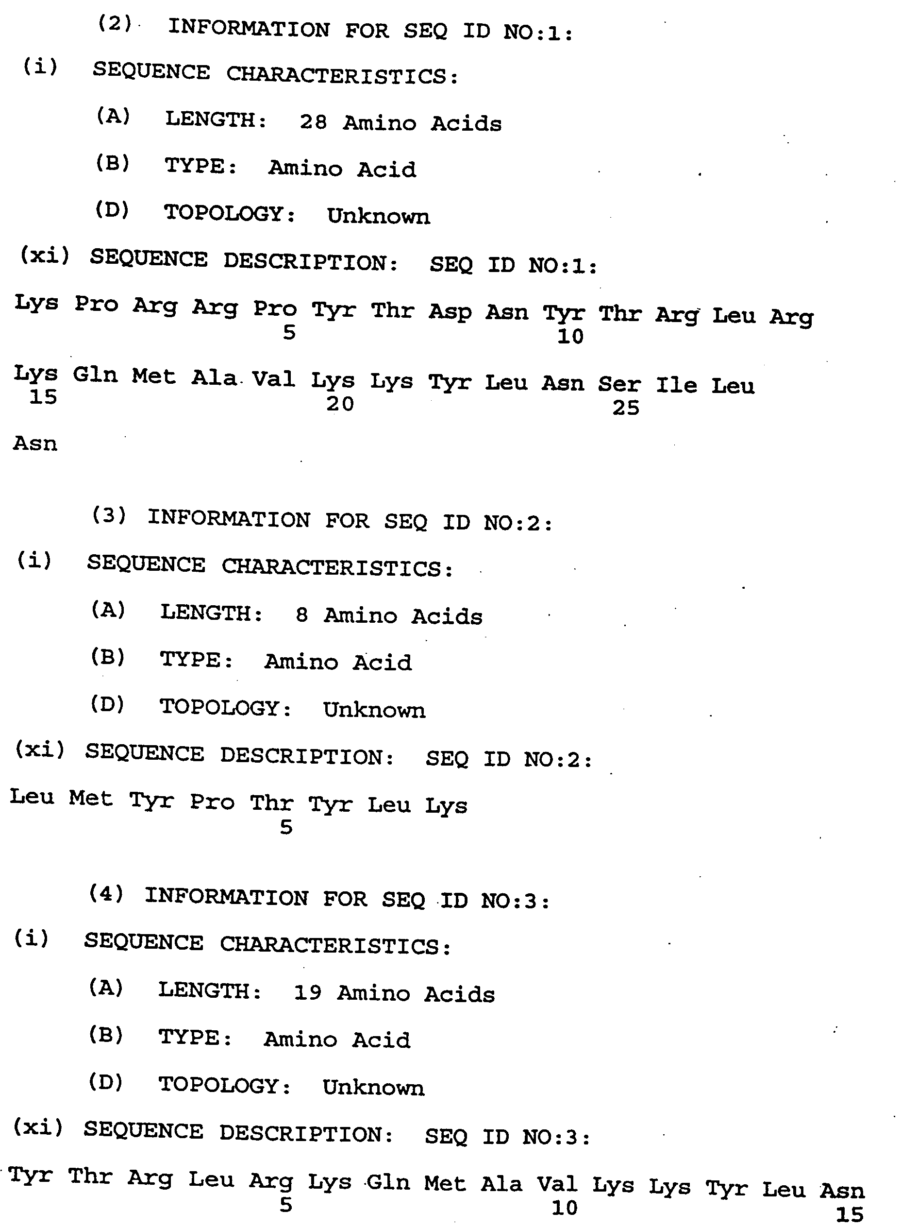
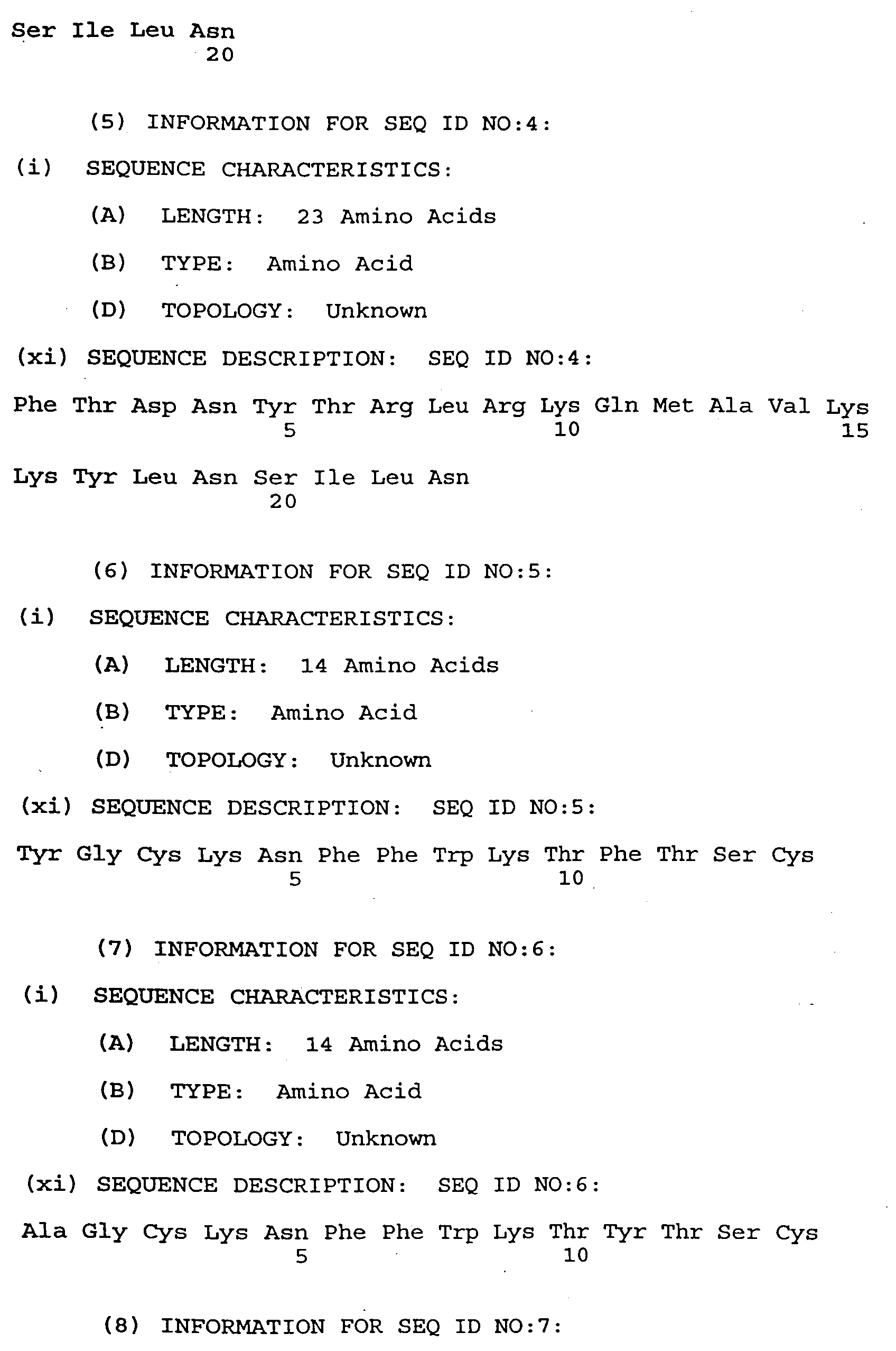
- the amino-acid sequences of the seven analogs (VIP 1 , VIP 2 , VIP 3 , SOM 1 , SOM 2 , BOM 1 , and SP 1 ) are given in Table 2. As will be explained in more detail below, the combination of these seven analogs is known as MuJ-7.
- the amino-acid sequence for VIP 1 (a VIP antagonist) is SEQ ID NO:1; and the amino-acid sequence for VIP 2 (a VIP receptor binding inhibitor) is SEQ ID NO:2.
- the analogs were synthesized manually and using a conventional peptide synthesizer. The purity of the peptides was established by performing high performance liquid chromatography and amino acid analysis, while the analysis was reconfirmed on a sequence analyzer.
- the growth factors synthesized and secreted by tumor cells were identified by different assay systems.
- the peptide hormones involved in uncontrolled proliferation of cancer cells were identified by performing experiments on established cell lines.
- the results obtained were complemented with data obtained from experiments conducted on primary tumor cells of human colon adenocarcinoma as a model tissue, for which we have developed a novel method of establishing cell lines.
- the following article, which describes the novel method of establishing cell lines, is incorporated herein by reference: Jaggi, M., Mukherjee, R., "Establishment of Tumorigenic Cell Lines from Biopsies of Human Colon Adenocarcinomas," Journal of Basic & Applied Biomedicine , 3(4): 27-35 (1995).
- a sandwich ELISA for the peptides was developed and used by the inventors.
- the following article, which describes the sandwich ELISA, is incorporated herein by reference: Jaggi, M., Mukherjee R., "New, Sensitive and Specific ELISA for the Detection of Neuropeptides in Culture Supernatants," Journal of Immunoassay , 15(2): 129-46 (1994).
- the identity of the peptides was established by reverse phase high performance liquid chromatography and sequence analysis. The binding sites for VIP, somatostatin, Substance P, and bombesin on primary human adenocarcinoma tumor cells of the colon were demonstrated by performing receptor-ligand assays.
- Tables 3, 4, 5, and 6 present data on the receptor affinities for VIP, somatostatin, bombesin, and Substance P on eight different primary tumor cultures of human colon adenocarcinoma. These data were obtained by performing receptor-ligand assays using 125 I-VIP, 125 I-somatostatin, 125 I-bombesin, and 125 I-Substance P. See the section below entitled “Description of Protocols" for a detailed description of the receptor-ligand assay.
- K D (M) represents the dissociation constant, the unit of which is moles (M); and R(M/L) stands for the receptor number (i.e., the number of receptors per tumor cell), the unit of which is moles per liter (M/L).
- K D (M) and R(M/L) were computed using LIGAND software, which did Scatchart Analysis using the raw data from the receptor-ligand assays.
- a K D (M) value in the range of about 10 -9 to about 10 -10 M indicates a high-affinity receptor
- a K D (M) value in the range of about 10 -6 to about 10 -8 M indicates a receptor with a moderately high affinity
- Table 3 shows two K D (M) values and two R(M/L) values for each primary tumor culture because the tumor cells have a high-affinity receptor for VIP as well as a receptor with a moderately high affinity for VIP.
- Table 4 shows two K D (M) values and two R(M/L) values for each primary tumor culture because the tumor cells have a high-affinity receptor for somatostatin as well as a receptor with a moderately high affinity for somatostatin.
- Table 5 shows only one K D (M) value and one R(M/L) value for each primary tumor culture because the tumor cells appear to have only a high-affinity receptor for bombesin.
- Table 6 shows only one K D (M) value and one R(M/L) value for each primary tumor culture because the tumor cells appear to have a receptor with a moderately high affinity for Substance P.
- An example of a combination within the scope of the invention comprises SOM 2 , VIP 1 , VIP 2 , VIP 3 , SOM 1 , BOM 1 , and SP 1 .
- a combination, hereinafter referred to as MuJ-7 was prepared using the following seven peptide analogs: (1) VIP 1 (the VIP antagonist) having a molecular weight of approximately 3464.9 and a concentration of approximately 10 -7 M; (2) VIP 2 (the VIP receptor binding inhibitor) having a molecular weight of approximately 1027.55 and a concentration of approximately 10 -8 M; (3) VIP 3 (the VIP receptor antagonist) having a molecular weight of approximately 3342.09 and a concentration of approximately 10 -8 M; (4) SOM 1 (the somatostatin analog (CTOP)) having a molecular weight of approximately 1061.59 and a concentration of approximately 10 -9 M; (5) SOM 2 (the analog of somatostatin) having a molecular weight of approximately 1637.9 and a concentration of approximately 10 -8 M; (6) B
- MuJ-7 may be prepared in the following way.
- a stock solution of each of the seven peptide analogs is prepared with a pH of approximately 7.0 to approximately 7.4.
- sterile phosphate buffered saline was used to prepare the stock solutions for the testing described below, other diluents may be used such as RPMI 1640, buffered saline, isotonic NaCl, Ringer's solution, water (for injection), distilled water, polyethylene glycol (neat or in water), 2% Tween in water, dimethylsulfoxide to 50% in water, propylene glycol (neat or in water), balanced salt solution, glycerol, and other conventional fluids that are suitable for intravenous administration.
- the pH can be adjusted by using 1 N HCl for the lowering the pH or 1 N NaOH for raising the pH, although other conventional agents for adjusting the pH can be used.
- concentration of the peptide analog in each stock solution is approximately 10 -3 M. Aliquots of the seven peptides analogs are mixed together such that the MuJ-7 formulation contains approximately equal weights of each of the seven peptide analogs.
- the concentration of VIP 1 is approximately 10 -7 M; the concentration of VIP 2 is approximately 10 -8 M; the concentration of VIP 3 is approximately 10 -8 M; the concentration of SOM 1 is approximately 10 -9 M; the concentration of SOM 2 is approximately 10 -8 M; the concentration of BOM 1 is approximately 10 -8 M; and the concentration of SP 1 is approximately 10 -8 M.
- the pH of the MuJ-7 solution may range from approximately 7.0 to approximately 7.4. To obtain a pH in this range, the pH can be adjusted by using 1 N HCl for lowering the pH or 1 N NaOH for raising the pH, although other conventional agents for adjusting the pH can be used.
- MuJ-7 was tested against primary tumor cells of human colon adenocarcinoma, and each of the peptide analogs comprising MuJ-7 was tested individually against human colon adenocarcinoma tumor cells and other cancer cell lines.
- the results for primary tumor cells of human colon adenocarcinoma are summarized in Table 7; and the results for other tumor or cancer cell lines are summarized in Table 8.
- Tables 7 and 8 list the maximum cytotoxicity achieved for each peptide analog and MuJ-7.
- cytotoxicity of MuJ-7 and each of the peptide analogs listed in Tables 7 and 8 was tested by performing a one-day MTT cytotoxicity assay, which is based on the principle of uptake of MTT (3-(4,5-dimethylthiazol-2-yl)-2,5-diphenyl tetrazolium bromide), a tetrazolium salt, by the metabolically active cells where it is metabolized by active mitochondria into a blue-colored formazan product, which can be read spectrophotometrically.
- MTT 3-(4,5-dimethylthiazol-2-yl)-2,5-diphenyl tetrazolium bromide
- MTT (3-(4,5-dimethylthiazol-2-yl)-2,5-diphenyl tetrazolium bromide) (Sigma catalogue number M 2128) was dissolved in phosphate buffered saline with a pH of 7.4 to obtain an MTT concentration of 5 mg/ml; the resulting mixture was filtered through a 0.22 ⁇ filter to sterilize and remove a small amount of insoluble residue; the filtered mixture was the MTT stock solution (20 ⁇ l per 200 ⁇ l of medium).
- the assay was terminated after approximately 24 hours by adding approximately 100 ⁇ g (20 ⁇ l) of MTT to each well, then incubating for approximately one additional hour, and finally adding approximately 50 ⁇ l of 10% SDS-0.01 N HCl to each well to lyse the cells and dissolve the formazan.
- the cytotoxicity percentage was calculated according to the above formula and was based on the proliferation of the untreated controls, the value of which was taken as 100%.
- K562 cells are human leukemia cells; MOLT-4 cells are human lymphoma cells; L 132 cells are human lung carcinoma cells; PC3 cells are human pancreas tumor cells; MCF-7 cells are human breast tumor cells; HuTu80 cells are human duodenum tumor cells; Hu 746T cells are human stomach tumor cells; SKO.007 cells are human myeloma cells; HT29 cells are human colon tumor cells; SW 620 cells are human colon tumor cells; G 401 cells are human kidney tumor cells; SK.MEL.28 cells are human melanoma cells; and PTC cells are human colon tumor cells.
- the assay was terminated after approximately 24 hours by adding approximately 100 ⁇ g (20 ⁇ l) of MTT to each well, then incubating for approximately one additional hour, and finally adding approximately 50 ⁇ l of 10% SDS-0.01 N HCl to each well to lyse the cells and dissolve the formazan. After incubating for approximately one hour at 37°C, the plate was read spectrophotometrically at 540 nm; and the cytotoxicity percentage (i.e., the killing percentage) was calculated using the formula presented above. Table 9 lists the maximum cytotoxicity achieved for each subcombination.
- these other peptide analogs were tested: somatostatin analog--RC-160; Substance P analogs--Substance P 1-6 and Spantide I, cholecystokinin analog--CCK-33; and glucagon analog--human glucagon.
- Each of these peptide analogs was tested against primary tumor cells of human colon adenocarcinoma.
- Each peptide analog was tested at concentrations between 10 -6 M and 10 -10 M by performing a one-day MTT cytotoxicity assay. Briefly, approximately 20,000-50,000 primary tumor cells of human colon adenocarcinoma were seeded in a 96-well culture plate and incubated with each peptide analog in a CO 2 incubator for approximately 24 hours.
- Controls which were not treated with the peptide analogs, were similarly incubated.
- the assay was terminated after approximately 24 hours by adding approximately 100 ⁇ g (20 ⁇ l) of MTT to each well, then incubating for approximately one additional hour, and finally adding approximately 50 ⁇ l of 10% SDS-0.01 N HCl to each well to lyse the cells and dissolve the formazan. After incubating for approximately one hour at 37°C, the plate was read spectrophotometrically at 540 nm; and the cytotoxicity percentage (i.e., inhibition percentage) was calculated using the formula presented above.
- the maximum cytotoxicity achieved for Substance P 1-6 was approximately 35.9%; the maximum cytotoxicity achieved for RC-160 was approximately 58.0%; the maximum cytotoxicity achieved for Spantide I was approximately 30.8%; the maximum cytotoxicity achieved for human glucagon was approximately 0%; and the maximum cytotoxicity achieved for CCK-33 was approximately 17.8%.
- Table 10 lists other VIP analogs; Table 11 lists other somatostatin analogs; Table 12 lists other bombesin analogs; and Table 13 lists other Substance P analogs.
- the analogs listed in Tables 10-13 may be able to replace some of the peptide analogs comprising MuJ-7.
- amino-acid sequences disclosed in Tables 2 and 10-13 and claimed herein may include conservatively modified variants of the amino-acid sequences disclosed in Tables 2 and 10-13. It is believed that the claimed invention would still be effective if the amino-acid sequences disclosed in Tables 2 and 10-13 were shortened by removing amino-acid residues (e.g., one, two, or perhaps more amino-acid residues) from the C-terminus and/or from the N-terminus.
- amino-acid residues e.g., one, two, or perhaps more amino-acid residues
- amino-acid residues e.g., one, two, or perhaps more amino-acid residues
- amino-acid residues e.g., one, two, or perhaps more amino-acid residues
- VIP analogs S.No Name Sequence SEQ ID NO: 1 VIP 10-28 Tyr-Thr-Arg-Leu-Arg-Lys-Gln-Met-Ala-Val-Lys-Lys-Tyr-Leu-Asn-Ser-Ile-Leu-Asn-NH 2 SEQ ID NO:3 2 VIP Antagonist ([Ac-Tyr 1 ,D-Phe 2 ]-Growth Hormone Releasing Factor 1-29 Amide Ac-Tyr-D-Phe-Asp-Ala-Ile-Phe-Thr-Asn-Ser-Tyr-Arg-Lys-Val-Leu-Gly-Gln-Leu-Ser-Ala-Arg-Lys-Leu-Leu-Gln-Asp-Ile-Met-Ser-Arg-NH 2 3 VIP (6-28) Phe-Thr-Asp-Asn-Tyr-Thr-Arg-Leu-Arg-Lys-G
- Another aspect of the invention provides a method for treating a mammal (including a human being) afflicted with cancer.
- the types of cancer that may be treated include, but are not necessarily limited to, leukemia and lymphoma; adenocarcinoma of the stomach, pancreas, and prostate; and cancer of the colon, rectum, lung, breast, and kidney.
- the invention will provide a method for treating other diseases and cancers characterized by hypersecretion of one or more of the peptides VIP, somatostatin, bombesin, and Substance P.
- the methods of this invention comprise, consist of, or consist essentially of: administering systematically to the mammal a therapeutically effective combination of peptide SOM 2 and at least four of peptides: VIP 1 , VIP 2 , VIP 3 , SOM 1 , BOM 1 , and SP 1 .
- An effective dose of the combination ranges from 15 to 170 ⁇ g (preferably 25 to 40 ⁇ g) of the peptides per kg of the body weight of the mammal, with the dose dependent on the effects sought, the manner of administration, the peptides selected, and the cancer being treated.
- Systemic administration refers to oral, rectal, nasal, transdermal, and parenteral (i.e., intramuscular, intravenous, and subcutaneous). In accordance with good clinical practice, it is preferred to administer the composition at a dose that will produce anticancer effects without causing undue harmful side effects.
- the composition may be administered either alone or as a mixture with other therapeutic agents.
- the composition may optionally and preferably contain pharmaceutically acceptable diluents, excipients, solvents, binders, stabilizers, and the like.
- diluents may include: RPMI 1640, buffered saline, isotonic NaCl, Ringer's solution, water, distilled water, polyethylene glycol (neat or in water), 2% tween in water, dimethylsulfoxide to 50% in water, propylene glycol (neat or in water), phosphate buffered saline, balanced salt solution, glycerol, and other conventional fluids that are suitable for intravenous administration.
- compositions which provide from about 10 to 2000 ⁇ g of the composition per unit dose are preferred and are conventionally prepared as tablets, lozenges, capsules, powders, aqueous or oily suspensions, syrups, elixirs, and aqueous solutions.
- the nature of the pharmaceutical composition employed will, of course, depend on the desired route of administration.
- a primary tumor cell line of human colon adenocarcinoma was established by using fine needle aspiration cytology (FNAC) and histopathology confirmed tumor tissue of human colon adenocarcinoma. These cells stained positive with a monoclonal antibody specific for a 91 KD surface antigen present on human colon adenocarcinoma cells. The tumorigenicity of these cells was demonstrated by the ability of these cells to form tumors in nude mice on subcutaneous injection. The characteristics of the 12 primary cultures are given in Table 14. Characteristic features of human colon adenocarcinoma biopsies cultured in vitro. Note that cultures established from all the tumor biopsies were then xenografted in nude mice. B.No Age Sex Site FNAC Hist.
- the anti-proliferative effects of MuJ-7 were studied in a 96-well culture plate, wherein the human colon adenocarcinoma tumor cells (approximately 50,000 cells per well) from the each of the twelve human colon adenocarcinoma cell cultures listed in Table 14 were incubated in a CO 2 incubator at approximately 37°C for approximately 72 hours with MuJ-7 (approximately 20 ⁇ l of MuJ-7 per well). Human colon adenocarcinoma cells not treated with MuJ-7 served as controls. Tritiated [ 3 H] thymidine (approximately 1 ⁇ Ci per well) was added to each well, and the plate was incubated for approximately 1 additional hour.
- the cells were harvested on filter mats, and incorporation of [ 3 H] thymidine into the dividing cells was counted on a Beta plate (Pharmacia). For each of the tritiated [ 3 H] thymidine cytotoxicity assays described herein, the proliferation of cells in the untreated controls was taken as 100%. In Example 1, it was observed that MuJ-7 inhibited proliferation of the tumor cells by approximately 95%.
- the cytotoxic effect of MuJ-7 was reconfirmed by a one-day MTT assay.
- Table 15 lists the approximate inhibition percentages for each of the 12 human colon adenocarcinoma cell lines. Biopsy numbers 1 through 12 in Table 14 correspond respectively to sample numbers PTC-1 through PTC-12 in Table 15. Inhibition percentages for each of the 12 human colon adenocarcinoma cell lines Sample No. Percent Inhibition PTC-1 95.2 PTC-2 89.1 PTC-3 94.6 PTC-4 82.2 PTC-5 74.2 PTC-6 81.6 PTC-7 93.8 PTC-8 94.9 PTC-9 81.5 PTC-10 82.7 PTC-11 84.6 PTC-12 80.7
- the cytotoxic effect of MuJ-7 was tested using a three-day MTT cytotoxic assay on three human colon cancer cell lines: CoLo 205, HT 29, and SW 620.
- the method for preparing the MTT stock solution for the three-day MTT cytotoxic assay was the same as the method described above for preparing the MTT stock solution for the one-day MTT cytotoxic assay. Briefly, CoLo 205, HT 29, and SW 620 cells were incubated in a 96-well culture plate (approximately 50,000 cancer cells in each-well) for approximately 72 hours at approximately 37°C in approximately 5% CO 2 .
- CoLo 205, HT 29, and SW 620 cells not treated with MuJ-7 served as controls.
- stock MTT solution (approximately 100 ⁇ g of MTT per well) was added to each well, and incubation continued for approximately 1 additional hour.
- the formazan crystals that formed inside the cells were dissolved with a detergent comprising approximately 10% sodium dodecyl sulfate and approximately 0.01 N HCl; and the optical density of each well was read spectrophotometrically at 540 nm.
- the percentage inhibition caused by MuJ-7 in CoLo 205, HT 29, and SW 620 was approximately 80%, approximately 18%, and approximately 41%, respectively.
- the genomic DNA was extracted from the tumor cells in the first flask after approximately 12 hours; the genomic DNA was extracted from the tumor cells in the second flask after approximately 24 hours; the genomic DNA was extracted from the tumor cells in the third flask after approximately 48 hours; the genomic DNA was extracted from the tumor cells in the fourth flask after approximately 96 hours; and the genomic DNA was extracted from the tumor cells in the control flask after approximately 96 hours.
- the DNA of both untreated and treated cells was run on a 1% agarose gel using ethidium bromide for staining.
- the DNA of tumor cells treated with MuJ-7 for 48 and 96 hours formed a smear on the gel which is indicative of programmed cell death, while the DNA from untreated cells formed a sharp band at 10 kb, thus demonstrating that the DNA from untreated cells was intact.
- the DNA of tumor cells treated with MuJ-7 for 24 hours did not form a smear on the gel. Therefore, the time kinetic experiment in vitro showed that programmed cell death occurs between approximately 24 and 48 hours of treatment with MuJ-7.
- a dose of the MuJ-7 formulation was prepared in the following way.
- a stock solution of each of the seven peptide analogs was first prepared using sterile phosphate buffered saline with an approximate pH of 7.4. The concentration of the peptide analog in each stock solution was approximately 10 -3 M. Aliquots of the seven peptides analogs were mixed together such that the MuJ-7 formulation contained approximately equal weights of each of the seven peptide analogs, with the combined weight of the seven peptide analogs in each dose of MuJ-7 being approximately 8 to 16 ⁇ g, depending upon the size of the dose.
- the concentration of VIP 1 was approximately 10 -7 M; the concentration of VIP 2 was approximately 10 -8 M; the concentration of VIP 3 was approximately 10 -8 M; the concentration of SOM 1 was approximately 10 -9 M; the concentration of SOM 2 was approximately 10 -8 M; the concentration of BOM 1 was approximately 10 -8 M; and the concentration of SP 1 was approximately 10 -8 M.
- the volume of this solution was made up with sterile RPMI 1640 to approximately 150 ⁇ l.
- RPMI 1640 is a cell culture medium that was developed at the Rosewell Park Memorial Institute. The components of RPMI 1640 are listed in Table 16.
- control mice not injected with MuJ-7 were instead injected with RPMI 1640.
- the progressive growth of the tumors in the control mice indicates that RPMI 1640 is not critical to the effectiveness of MuJ-7.
- the tumor volume was calculated with the help of a vernier calliper.
- each tumor weight measurement was taken at the end of the experiment by sacrificing the mouse and resecting out the complete tumor growing superficially on the posterior side immediately below the skin over the muscular layer. The skin and any other tissues attached to the tumor were removed, and the tumor was immediately weighed on an analytical balance.
- Figure 1 which shows the effect on tumor regression of treatment onset time with MuJ-7, summarizes the mean tumor volume (in mm 3 ) for all of the mice in the in vivo protocols described in Examples 6-13 versus the day numbers.
- each data point in Figure 1 represents the mean tumor volume of different numbers of mice from separate experiments.
- the mice are grouped together into four categories: (1) untreated control mice (" ⁇ "); (2) treated mice ("+") that received their first dose of MuJ-7 by day 5; treated mice (" ⁇ ") that received their first dose of MuJ-7 after day 5 and by day 12; and treated mice (" ⁇ ") that received their first dose of MuJ-7 after day 12 and by day 20.
- the arrows indicate days 5, 12, and 20.
- mice On day 1, ten BALB/c nude nu/nu mice were implanted with primary tumor cells of human colon adenocarcinoma (approximately 10 million tumor cells per mouse), and these mice received their first dose of MuJ-7 approximately one hour after they were implanted with the tumor cells.
- the total daily dose of MuJ-7 for each treated mouse comprised approximately 1.143 ⁇ g of VIP 1 , approximately 1.143 ⁇ g of VIP 2 , approximately 1.143 ⁇ g of VIP 3 , approximately 1.143 ⁇ g of SOM 1 , approximately 1.143 ⁇ g of SOM 2 , approximately 1.143 ⁇ g of BOM 1 , and approximately 1.143 ⁇ g of SP 1 .
- the total daily dose of MuJ-7 always contained approximately equal weights of each of the seven peptide analogs (VIP 1 , VIP 2 , VIP 3 , SOM 1 , SOM 2 , BOM 1 , and SP 1 ); and the total weight of these seven peptide analogs was approximately 8 ⁇ g.
- the total daily dose of MuJ-7 was divided into approximately three equal subdoses. Three times a day at approximately eight-hour intervals, a subdose was injected into each treated mouse. The first subdose of the day was injected into the tail vein; the second subdose of the day was injected into one gluteal muscle; and the third subdose of the day was injected into the other gluteal muscle. The treatment lasted for two weeks.
- Controls were randomly selected BALB/c nude nu/nu mice whose weights were similar to the weights of the ten treated mice.
- the control mice were subcutaneously injected with the same type and approximately the same number of human colon adenocarcinoma tumor cells as the treated mice.
- the control mice did not receive MuJ-7.
- FIG. 17 lists the tumor volume in mm 3 for each treated mouse
- Table 18 lists the tumor volume in mm 3 for each control (untreated) mouse.
- Figure 2 is a graph of the mean tumor volume (in mm 3 ) of the treated mice ("o") and the untreated control mice ("+") versus the day numbers.
- the arrow illustrates that the treated mice received their first dose of MuJ-7 approximately one hour after they were treated with tumor cells on day 1.
- mice Twenty BALB/c nude nu/nu mice, which were divided into two groups with ten mice in each group, were implanted on day 1 with primary tumor cells of human colon adenocarcinoma (approximately 10 million tumor cells per mouse).
- the group 1 mice received their first dose of MuJ-7 on day 12 (i.e., 11 days post implantation on the tumor cells on day 1).
- the group 2 mice received their first dose of MuJ-7 on day 20 (i.e., 19 days post implantation of the tumor cells on day 1). These twenty treated mice were injected for 14 days with a daily dose of MuJ-7.
- the total daily dose of MuJ-7 for each treated mouse comprised approximately 1.143 ⁇ g of VIP 1 , approximately 1.143 ⁇ g of VIP 2 , approximately 1.143 ⁇ g of VIP 3 , approximately 1.143 ⁇ g of SOM 1 , approximately 1.143 ⁇ g of SOM 2 , approximately 1.143 ⁇ g of BOM 1 , and approximately 1.143 ⁇ g of SP 1 .
- the total daily dose of MuJ-7 always contained approximately equal weights of each of the seven peptide analogs (VIP 1 , VIP 2 , VIP 3 , SOM 1 , SOM 2 , BOM 1 , and SP 1 ); and the total weight of these seven peptide analogs was approximately 8 ⁇ g.
- the total daily dose of MuJ-7 was divided into approximately three equal subdoses.
- Controls were five randomly selected BALB/c nude nu/nu mice whose weights were similar to the weights of the twenty treated mice in groups 1 and 2. (The same five mice served as controls for both the group 1 and the group 2 experiments.) The control mice were subcutaneously injected with the same type and approximately the same number of human colon adenocarcinoma tumor cells as the treated mice. The control mice did not receive MuJ-7.
- mice In the group 1 mice, the tumor volumes were recorded every five days; and Table 19 lists the mean tumor volumes for the treated mice and the five control (untreated) mice. Three untreated control mice died on days 31, 32, and 34; and these deceased mice were excluded from measurements made after their day of death.
- the mean tumor volume and the mean tumor weight for the treated group 1 mice on day 42 exclude the eight mice showing complete regression.
- the mean tumor volume in group 1 mice treated with MuJ-7 increased from approximately 9.8 mm 3 on day 22 to approximately 44.8 mm 3 on day 42, while the mean tumor volume in the control (untreated) mice increased from approximately 122.1 mm 3 on day 22 to approximately 1978.1 mm 3 on day 42.
- the mean tumor weight on day 42 was approximately 51 mg in the two treated group 1 mice that did not show complete regression of the tumor, while the mean tumor weight on day 42 was approximately 1196 mg in untreated control mice.
- MuJ-7 caused a reduction in the mean tumor volume in the treated mice from approximately 105 mm 3 on day 22 to approximately 3.1 mm 3 on day 34, while the mean tumor volume in the untreated control mice increased from approximately 122.1 mm 3 on day 22 to approximately 1888 mm 3 on day 34.
- the mean tumor weight at the end of the experiment on day 34 was approximately 23 mg in the two treated group 2 mice that did not show complete regression of the tumor, while the mean tumor weight on day 34 was approximately 746 mg in the untreated control mice.
- Figure 3 is a graph of the mean tumor volume (in mm 3 ) of the group 1 treated mice ("o"), the group 2 treated mice (" ⁇ "), and the untreated control mice ("+") versus the day numbers.
- the arrow under day number 12 indicates when the group 1 mice were first treated with MuJ-7; and the arrow under day number 20 indicates when the group 2 mice were first treated with MuJ-7.
- each treated mouse was injected with MuJ-7 for 14 consecutive days, the daily dose of MuJ-7 being administered in approximately three equal subdoses.
- the daily dose of MuJ-7 for each treated mouse comprised approximately 1.143 ⁇ g of VIP 1 , approximately 1.143 ⁇ g of VIP 2 , approximately 1.143 ⁇ g of VIP 3 , approximately 1.143 ⁇ g of SOM 1 , approximately 1.143 ⁇ g of SOM 2 ; approximately 1.143 ⁇ g of BOM 1 , and approximately 1.143 ⁇ g of SP 1 .
- treatment started on day 12 i.e., 11 days post injection with the cancer cells
- treatment started on day 8 i.e., seven days post injection with the cancer cells
- treatment started on day 4 i.e., three days post injection with the cancer cells
- treatment started on day 5 i.e., four days post injection with the cancer cells.
- the total daily dose of MuJ-7 was divided into approximately three equal subdoses. Three times a day at approximately eight-hour intervals, a subdose was injected into each treated mouse.
- the first subdose of the day was injected into the tail vein; the second subdose of the day was injected into one gluteal muscle; and the third subdose of the day was injected into the other gluteal muscle.
- Controls were randomly selected BALB/c nude nu/nu mice, whose weights were similar to the weights of the treated mice.
- the control mice were subcutaneously injected with the same type and approximately the same number of cancer cells as the treated mice.
- the control mice, were not treated with MuJ-7.
- the tumor volume in the untreated control mouse increased from approximately 67.5 mm 3 on day 6 to approximately 508.9 mm 3 on day 26 (i.e., 25 days post injection with the cancer cells on day 1), at which time the mouse died because of the tumor, while the tumor volume in the mouse treated with MuJ-7 decreased from approximately 47.1 mm 3 on day 6 to complete tumor regression on day 16 (after 9 days of treatment).
- Table 20 lists the tumor volume measurements in mm 3 for the untreated control mouse and the treated mouse from day 6 until each mouse died.
- Figure 4 is a graph of the tumor volume (in mm 3 ) of the treated mouse ("o") and the untreated control mouse ("+") versus the day numbers, with the arrow indicating that the treated mouse received its first dose of MuJ-7 on day 8.
- the treated mouse which died on day 87 (i.e., 86 days post injection with SW 620 cells on day 1), had an overall increase in survival time of approximately 244%.
- the tumor volume in the untreated mouse increased from approximately 95.1 mm 3 on day 10 to approximately 4536.3 mm 3 on day 52 (i.e., 51 days post injection with the cells on day 1), at which time the mouse died because of the tumor.
- the tumor volume in the mouse treated with MuJ-7 increased relatively gradually from approximately 159.1 mm 3 on day 10 to approximately 2192.7 mm 3 on day 52, after which the tumor volume decreased to approximately 1556.4 mm 3 on day 86 (i.e., 85 days post injection with the cancer cells).
- Table 21 lists the tumor volume measurements in mm 3 for the untreated control mouse and the treated mouse from day 10 until each mouse died.
- Figure 5 is a graph of the tumor volume (in mm 3 ) of the treated mouse ("o") and the untreated control mouse ("+") versus the day numbers, with the arrow indicating that the treated mouse received its first dose of MuJ-7 on day 12.
- the treated mouse which died on day 104 (i.e., 103 days post the injection with HT29 cells on day 1), had an overall increase in survival time of approximately 102%.
- the tumor volume in the untreated mouse increased from approximately 74.9 mm 3 on day 3 to approximately 7344.9 mm 3 on day 22 (i.e., 21 days post injection with the cancer cells on day 1), at which time the mouse died because of the tumor, while the tumor volume in the mouse treated with MuJ-7 decreased from approximately 78.2 mm 3 on day 3 to complete tumor regression on day 8 (after 4 days of treatment).
- Table 22 lists the tumor volume measurements in mm 3 for the untreated control mouse and the treated mouse from day 3 until each mouse died.
- Figure 6 is a graph of the tumor volume (in mm 3 ) of the treated mouse ("o") and the untreated control mouse ("+") versus the day numbers, with the arrow indicating that the treated mouse received its first dose of MuJ-7 on day 4.
- the treated mouse which died on day 79 (i.e., 78 days post injection with CoLo 205 cells), had an overall increase in survival time of approximately 271%.
- the tumor volume in the untreated mouse increased from approximately 28.5 mm 3 on day 2 to approximately 7174.3 mm 3 on day 34 (i.e., 33 days post injection with the cancer cells on day 1), at which time the mouse died because of the tumor, while the tumor volume in the mouse treated with MuJ-7 decreased from approximately 38.3 mm 3 on day 2 to complete regression on day 27 (after 22 days of treatment).
- Table 23 lists the tumor volume measurements in mm 3 for the untreated control mouse and the treated mouse from day 2 until each mouse died.
- Figure 7 is a graph of the tumor volume (in mm 3 ) of the treated mouse ("o") and the untreated control mouse ("+") versus the day numbers, with the arrow indicating that the treated mouse received its first dose of MuJ-7 on day 5.
- the treated mouse which died on day 70 (i.e., 69 days post injection with the L 132 cells), had an overall increase in survival time at approximately 109%.
- mice Two BALB/c nude nu/nu mice were each implanted with approximately 10 million human colon adenocarcinoma cells on day 1.
- day 22 i.e., 21 days post implantation on day 1
- one mouse began to receive intraperitoneally a daily dose of a combination of VIP 1 , VIP 2 , VIP 3 , SOM 1 , and SOM 2 .
- this treated mouse received approximately 8 ⁇ g of the combination, the combination comprising approximately 1.6 ⁇ g of VIP 1 , approximately 1.6 ⁇ g of VIP 2 , approximately 1.6 ⁇ g of VIP 3 , approximately 1.6 ⁇ g of SOM 1 , and approximately 1.6 ⁇ g of SOM 2 .
- the treated mouse received the last dose of the combination on day 35 (i.e., 34 days post implantation on day 1). At the end of the experiment on day 35, the tumor volume in the treated mouse was approximately 720 mm 3 . The other mouse was untreated and served as a control. On day 35, the tumor volume in the untreated control mouse was approximately 3584 mm 3 .
- mice Three BALB/c nude nu/nu mice were each implanted with approximately 10 million human colon adenocarcinoma cells on day 1.
- day 2 i.e., one day post implantation on day 1
- two mice each began to receive intraperitoneally a daily dose of a combination of VIP 1 , VIP 2 , VIP 3 , SOM 1 , and SOM 2 .
- each treated mouse received approximately 8 ⁇ g of the combination, the combination comprising approximately 1.6 ⁇ g of VIP 1 , approximately 1.6 ⁇ g of VIP 2 , approximately 1.6 ⁇ g of VIP 3 , approximately 1.6 ⁇ g of SOM 1 , and approximately 1.6 ⁇ g of SOM 2 .
- the treated mice received the last dose of the combination on day 15 (i.e., 14 days post implantation on day 1). At the end of the experiment on day 15, the tumor volume in one treated mouse was approximately 80 mm 3 ; and the tumor weight was approximately 0.149 g. On day 15, the tumor volume in the other treated mouse was approximately 0.8 mm 3 ; and the tumor weight was approximately 0.008 g.
- the third mouse was untreated and served as a control. On day 15, the tumor volume in the untreated control mouse was approximately 384 mm 3 ; and the tumor weight in the untreated control mouse was approximately 0.406 g.
- mice Two BALB/c nude nu/nu mice were each implanted with approximately 10 million human colon adenocarcinoma cells on day 1.
- day 2 i.e., one day post implantation on day 1
- one mouse began to receive a daily dose of a combination of VIP 1 , VIP 2 , VIP 3 , SOM 1 , and SOM 2 .
- this treated mouse received approximately 8 ⁇ g of the combination, the combination comprising approximately 1.6 ⁇ g of VIP 1 , approximately 1.6 ⁇ g of VIP 2 , approximately 1.6 ⁇ g of VIP 3 , approximately 1.6 ⁇ g of SOM 1 , and approximately 1.6 ⁇ g of SOM 2 .
- the daily dose of the combination was divided into approximately three equal subdoses.
- a subdose was given to each treated mouse.
- the first subdose each day was given intravenously, and the second and third subdoses each day were given via intramuscular injections.
- the treated mouse received the last dose of the combination on day 15 (i.e., 14 days post implantation on day 1).
- the tumor volume in the treated mouse was approximately 0.8 mm 3 ; and the tumor weight was approximately 0.009 g.
- the other mouse was untreated and served as a control.
- the tumor volume in the untreated control mouse was approximately 1728 mm 3 ; and the tumor weight in the untreated control mouse was approximately 2.18 g.
- mice Three randomly selected BALB/c nude nu/nu mice were each implanted with approximately 16 million primary tumor cells of human colon adenocarcinoma on day 1. On day 21 (i.e., twenty days post implantation on day 1), on day 22, and on day 23, two of these mice were treated with a daily dose of MuJ-7.
- the total daily dose of MuJ-7 for each treated mouse comprised approximately 1.143 ⁇ g of VIP 1 , approximately 1.143 ⁇ g of VIP 2 , approximately 1.143 ⁇ g of VIP 3 , approximately 1.143 ⁇ g of SOM 1 , approximately 1.143 ⁇ g of SOM 2 , approximately 1.143 ⁇ g of BOM 1 , and approximately 1.143 ⁇ g of SP 1 ; and the total weight of the seven peptide analogs was approximately 8 ⁇ g.
- the total daily dose of MuJ-7 for each treated mouse comprised approximately 2.286 ⁇ g of VIP 1 , approximately 2.286 ⁇ g of VIP 2 , approximately 2.286 ⁇ g of VIP 3 , approximately 2.286 ⁇ g of SOM 1 , approximately 2.286 ⁇ g of SOM 2 , approximately 2.286 ⁇ g of BOM 1 , and approximately 2.286 ⁇ g of SP 1 ; and the total weight of the seven peptide analogs was approximately 16 ⁇ g.
- the total daily dose MuJ-7 was divided into approximately three equal subdoses. Three times a day at approximately eight-hour intervals, a subdose was given to each treated mouse.
- the first subdose each day was injected into the tail vein; the second subdose each day was injected into one gluteal muscle; and the third subdose each day was injected into the other gluteal muscle.
- the untreated third mouse whose weight was similar to the weights of the two treated mice, served as a control and did not receive MuJ-7.
- Table 24 lists the tumor volume measurements in mm 3 through day 55 for the first treated mouse and until the second treated mouse and the control mouse died.
- the first treated mouse which survived at least until day 55 (i.e., 54 days post implantation with tumor cells on day 1), had an overall increase in survival of time of at least approximately 108%.
- the second treated mouse which died on day 38 (i.e., 37 days post implantation with tumor cells on day 1), has an overall increase in survival time of at least approximately 42%.
- the daily dose of MuJ-7 for each treated mouse comprised approximately 1.143 ⁇ g of VIP 1 , approximately 1.143 ⁇ g of VIP 2 , approximately 1.143 ⁇ g of VIP 3 , approximately 1.143 ⁇ g of SOM 1 , approximately 1.143 ⁇ g of SOM 2 , approximately 1.143 ⁇ g of BOM 1 , and approximately 1.143 ⁇ g of SP 1 .
- the total daily dose of MuJ-7 always contained approximately equal weights of each of the seven peptide analogs (VIP 1 , VIP 2 , VIP 3 , SOM 1 , SOM 2 , BOM 1 , and SP 1 ); and the total weight of these seven peptide analogs was approximately 8 ⁇ g.
- the total daily dose of MuJ-7 was divided into approximately three equal subdoses. Three times a day at approximately eight-hour intervals, a subdose was injected into each treated mouse. The first subdose of the day was injected into the tail vein; the second subdose of the day was injected into one gluteal muscle; and the third subdose of the day was injected into the other gluteal muscle. The ten untreated mice served as controls.
- the controls were ten randomly selected BALB/c nude nu/nu mice whose weights were similar to the weights of the twelve treated mice.
- the control mice were injected with the same type and approximately the same number of human colon adenocarcinoma tumor cells as the treated mice.
- the control mice did not receive MuJ-7.
- mice The tumor volume in each treated and untreated mouse was recorded generally every five days.
- Table 25 lists the tumor volume in mm 3 for each of the twelve treated mice; and Table 26 lists the tumor volume in mm 3 for each of the ten untreated control mice.
- Figure 8 is a graph of the mean tumor volume (in mm 3 ) of the treated mice ("o") and the untreated control mice ("+") versus the day numbers.
- Figure 9 is a graph of the percentage of surviving treated mice (dotted line) and the percentage of untreated control mice (solid line) versus the day numbers.
- the arrow illustrates that the treated mice received their first dose of MuJ-7 on day 15.
- Treated mouse number 11 responded to therapy between day 15 and day 30, when it was treated with MuJ-7. After the treatment with MuJ-7 ended, the tumor in mouse 11 started to grow; and mouse 11 eventually died on day 50. Nevertheless, the survival time of treated mouse 11 still exceeded the average survival time of the untreated control mice.
- Treated mouse 3 also showed a decrease in the rate of tumor growth compared to the average rate of tumor growth in the untreated control mice. Although treated mouse 3 eventually died on day 50, the survival time of treated mouse 3 still exceeded the average survival time of the untreated control mice.
- Indirect Immunofluorescence About 100 ⁇ l of healthy adherent tumor cell suspension with a density of approximately 10 4 cells/ml from a 3-4 day old culture were plated on a round, sterile cover slip in a 24-day culture plate, and incubated at 37°C in a CO 2 incubator. After 24 hours, when the tumor cells started to adhere to the culture surface, the wells were flooded with growth medium and incubated again at 37°C in a CO 2 incubator.
- the cover slips with adhering tumor cells were washed thoroughly in RPMI 1640 containing approximately 5% fetal calf serum (hereinafter referred to as "FCS”) followed by washing in approximately 0.05 M phosphate buffered saline (hereinafter referred to as "PBS"), which contained approximately 5% FCS and which had an approximate pH of 7.4, followed by washing in plain PBS.
- FCS fetal calf serum
- PBS phosphate buffered saline
- the tumor cells on the cover slips were then incubated at approximately 37°C for approximately one hour with a 1:50 dilution of the antipeptide polyclonal antibody.
- the cover slips were washed again as described above and the tumor cells incubated under the same conditions with a 1:200 dilution of anti-rabbit IgG-FITC conjugate. After washing, the cover slips were mounted in a medium made of carbonate-bicarbonate buffer and glycerol in a 1:1 ratio containing a crystal of para phenyl-diamine, and sealed in an inverted position on a glass slide with a clear varnish solution. The tumor cells were scanned under UV light on a Microphot FX microscope (Nikon).
- Sandwich ELISA Wells of a round-bottomed microtitre highly activated (Maxisorp) plate (Nunc, catalogue number 449824) were coated with l ⁇ g of the purified antibody in 100 ⁇ l of approximately 0.05 M phosphate buffered saline, which contained approximately 0.05% Tween 20 (PBS-T) and which had an approximate pH of 7.4, and were incubated for approximately one hour at approximately 37°C. After incubation, the wells were washed two times with PBS-0.2% Tween in an automatic plate washer (BDSL, UK). To each well 100 ⁇ l of Amicon concentrated culture supernatant of primary tumor cultures were added, followed by incubation for approximately one hour at approximately 37°C.
- PBS-T 0.05% Tween 20
- BDSL automatic plate washer
- Reverse Phase High Performance Liquid Chromatography The supernatant of tumor cell cultures was run on a Waters C-18, 5 micron (46 mm X 15 cm) column.
- the solvent system comprised two solvents that were run as a gradient.
- Solvent A consisted of approximately 0.1% trifluoroacetic acid
- solvent B consisted of approximately 80% acetonitrile in solvent A.
- a flow rate of 1.0 ml/minute was maintained and a gradient of approximately 40% to 100% solvent B in approximately 45 minutes was set up.
- a UV detector at a wavelength of 230 nm was used to detect the peptide.
- Receptor-ligand assay Tumor cells were grown to confluence in a 75 cm 3 flask, and the culture medium decanted. The cells were scraped off with the help of a rubber policeman and suspended in a minimum volume of binding buffer comprising approximately 5% bovine serum albumin (hereinafter referred to as "BSA") in RPMI 1640 to achieve a concentration of approximately 0.5 x 10 6 cells/5O ⁇ l. Increasing counts of I-125 peptides were added to the cells in the assay tube, and the volume was made up to approximately 200 ⁇ l with binding buffer. Radioactive counts were measured in each tube on a gamma counter. All the tubes were then incubated at approximately 37°C for approximately one hour.
- BSA bovine serum albumin
- the optimum cell number and tracer counts per tube were determined from the standard curve. This corresponded to the conditions at which there was no further increase in the number of bound counts on addition of tracer to a fixed cell concentration. Cold competition experiments were performed at these saturation conditions. A fixed cell concentration and tracer counts, as optimized earlier, were added to the assay tubes. This was followed by the addition of increasing concentrations of cold VIP, somatostatin, bombesin, and Substance P in duplicates to the tubes, and making up the volume to 200 ⁇ l with binding buffer. The tubes were then processed in a manner identical to the process described for preparation of the standard curve.
- LIGAND Software version 3.0 is a radioligand binding analysis program, copyrighted by G. A.
- the LIGAND Software performed a Scatchart Analysis by plotting on the Y axis the number of bound counts divided by the total number of counts added to each tube and plotting on the X axis the logarithm of the total number of counts added to each tube.
- the LIGAND Software used the intercept of the slope on the plot to calculate K D (M) and R(M/L).
- TBS Tris buffered saline
- the cells were resuspended in approximately 5-10 volumes of ice-cold TBS and centrifugation repeated. Finally, the cells were suspended in Tris edetate (hereinafter referred to as "TE," the TE having an approximate pH of 8.0) at a concentration of approximately 5 x 10 7 cells/ml.
- TE Tris edetate
- Ten milliliters of extraction buffer were added to 1 ml of cell suspension, and the solution was incubated for approximately 1 hour at approximately 37°C. Proteinase K to a final concentration of 100 ⁇ g/ml was added to this solution, and the solution was incubated in a gently shaking water bath for approximately 12 hours at approximately 50°C. The solution was then cooled to room temperature and transferred to a centrifuge tube.
Abstract
Description
| Peptide | Peptide Secretion And Receptor Positivity | Reference |
| VIP | Neuroblastoma | J. Molecular Neurosciences 5(4):231, 1994 |
| Colorectal and pancreatic adenocarcinoma | N.Eng. J. Med 331(17): 1116, 1994 | |
| Somatostatin | Small Cell Lung Cancer | Eur. J. Cancer 31A(2): 184, 1995 |
| Thyroid carcinoma | Clin. Endocrinol. 42(1): 31, 1995 | |
| Neuroblastoma | Seminars in Oncol. 21: 38, 1994 | |
| Bombesin | Small Cell Lung Cancer | Int. J. Cancer 60: 82, 1995 |
| Glioblastoma | Cancer Res., 54: 5895, 1994 | |
| Pancreatic cancer | Int. J. Pancreatology, 16: 141, 1994 | |
| Gastric cancer | Cancer letters, 85: 111, 1994 | |
| Prostate cancer | Prostate 25(1): 29, 1994 | |
| Substance P | Small cell lung cancer | Cancer Research 54(13): 3602-3610 (July 1, 1994) |
| Dissociation constant (KD(M)) in moles and receptor number (R(M/L)) in moles per liter for VIP on eight primary tumor cultures of human colon adenocarcinoma. | ||
| Sample No | K D (M) | R(M/L) |
| PTC-1 | 1.04 x 10⁻⁹ | 4.83 x 10⁻¹¹ |
| 6.33 x 10⁻⁷ | 1.78 x 10⁻⁸ | |
| PTC-2 | 1.45 x 10⁻⁹ | 6.23 x 10⁻¹¹ |
| 4.23 x 10⁻⁷ | 1.03 x 10⁻⁸ | |
| PTC-3 | 6.35 x 10⁻⁹ | 2.45 x 10⁻¹⁰ |
| 1.51 x 10⁻⁶ | 2.93 x 10⁻⁸ | |
| PTC-4 | 1.10 x 10⁻⁸ | 2.75 x 10⁻¹⁰ |
| 9.45 x 10⁻⁷ | 5.03 x 10⁻⁸ | |
| PTC-5 | 1.95 x 10⁻⁸ | 5.29 x 10⁻¹⁰ |
| 3.51 x 10⁻⁶ | 8.72 x 10⁻⁸ | |
| PTC-6 | 4.41 x 10⁻⁹ | 1.05 x 10⁻¹⁰ |
| 1.88 x 10⁻⁶ | 3.21 x 10⁻⁸ | |
| PTC-7 | 1.49 x 10⁻⁹ | 6.12 x 10⁻¹¹ |
| 5.55 x 10⁻⁷ | 9.37 x 10⁻⁹ | |
| PTC-8 | 1.78 x 10⁻⁹ | 1.50 x 10⁻¹⁰ |
| 8.42 x 10⁻⁶ | 8.49 x 10⁻⁹ |
| Dissociation constant (KD(M)) in moles and receptor number (R(M/L)) in moles per liter for somatostatin on eight primary tumor cultures of human colon adenocarcinoma. | ||
| Sample No | K D (M) | R(M/L) |
| PTC-1 | 3.23 x 10⁻¹⁰ | 6.01 x 10⁻¹¹ |
| 9.37 x 10⁻⁸ | 4.41 x 10⁻⁹ | |
| PTC-2 | 1.70 x 10⁻¹⁰ | 8.99 x 10⁻¹¹ |
| 6.35 x 10⁻⁸ | 2.24 x 10⁻⁹ | |
| PTC-3 | 1.15 x 10⁻⁹ | 1.06 x 10⁻¹⁰ |
| 1.34 x 10⁻⁷ | 5.09 x 10⁻⁹ | |
| PTC-4 | 8.65 x 10⁻¹¹ | 4.66 x 10⁻¹¹ |
| 5.64 x 10⁻⁸ | 2.75 x 10⁻⁹ | |
| PTC-5 | 3.78 x 10⁻¹⁰ | 5.82 x 10⁻¹¹ |
| 1.54 x 10⁻⁸ | 1.24 x 10⁻⁹ | |
| PTC-6 | 5.45 x 10⁻¹⁰ | 6.85 x 10⁻¹¹ |
| 4.30 x 10⁻⁸ | 1.16 x 10⁻⁹ | |
| PTC-7 | 1.11 x 10⁻⁹ | 9.81 x 10⁻¹¹ |
| 1.28 x 10⁻⁷ | 3.89 x 10⁻⁹ | |
| PTC-8 | 9.64 x 10⁻¹⁰ | 1.74 x 10⁻¹⁰ |
| 9.92 x 10⁻⁸ | 6.59 x 10⁻⁹ |
| Dissociation constant (KD(M)) in moles and receptor number (R(M/L)) in moles per liter for bombesin on eight primary tumor cultures of human colon adenocarcinoma. | ||
| Sample No | K D (M) | R(M/L) |
| PTC-1 | 4.39 x 10⁻¹⁰ | 2.24 x 10⁻¹⁰ |
| PTC-2 | 5.93 x 10⁻¹⁰ | 3.22 x 10⁻¹⁰ |
| PTC-3 | 5.69 x 10⁻¹⁰ | 2.97 x 10⁻¹⁰ |
| PTC-4 | 5.68 x 10⁻¹⁰ | 2.89 x 10⁻¹⁰ |
| PTC-5 | 4.62 x 10⁻¹⁰ | 3.35 x 10⁻¹⁰ |
| PTC-6 | 4.85 x 10⁻¹⁰ | 2.24 x 10⁻¹⁰ |
| PTC-7 | 6.70 x 10⁻¹⁰ | 2.55 x 10⁻¹⁰ |
| PTC-8 | 8.83 x 10⁻¹⁰ | 2.85 x 10⁻¹⁰ |
| Dissociation constant (KD(M)) in moles and receptor number (R(M/L)) in moles per liter for Substance P on eight primary tumor cultures of human colon adenocarcinoma. | ||
| Sample No | K D (M) | R(M/L) |
| PTC-1 | 1.54 x 10⁻⁷ | 1.85 x 10⁻⁸ |
| PTC-2 | 1.72 x 10⁻⁷ | 1.71 x 10⁻⁸ |
| PTC-3 | 1.34 x 10⁻⁷ | 1.49 x 10⁻⁸ |
| PTC-4 | 1.54 x 10⁻⁷ | 1.66 x 10⁻⁸ |
| PTC-5 | 2.10 x 10⁻⁸ | 6.2 x 10⁻⁹ |
| PTC-6 | 2.34 x 10⁻⁷ | 2.95 x 10⁻⁸ |
| PTC-7 | 2.62 x 10⁻⁸ | 7.48 x 10⁻⁹ |
| PTC-8 | 1.86 x 10⁻⁷ | 1.32 x 10⁻⁸ |
| Cytotoxic effect of individual peptide analogs and MuJ-7 on primary tumor cells of human colon adenocarcinoma | ||
| Peptide Analog | Concentration | % Killing |
| VIP1 | 10-7 M | 61 |
| VIP2 | 10-8 M | 77 |
| VIP3 | 10-8 M | 76 |
| SOM1 | 10-9 M | 73 |
| SOM2 | 10-8 M | 79 |
| BOM1 | 10-8 M | 64 |
| SP1 | 10-8 M | 54 |
| MuJ-7 | VIP1 (10-7 M) + VIP2 (10-8 M) + VIP3 (10-8 M) + SOM1 (10-9 M) + SOM2 (10-8 M) + BOM1 (10-8 M) + SP1 (10-8 M) | 94 |
| Cytotoxicity of subcombinations of peptide analogs on primary tumor cells of human colon adenocarcinoma | ||
| Subcombination Number | Subcombination | Killing percentage (approximate) |
| 1 | VIP1 (10-7 M) + SOM1 (10-9 M) + BOM1 (10-8 M) | 64.7 |
| 2 | VIP1 (10-7 M) + VIP2 (10-8 M) + SOM1 (10-9 M) + BOM1 (10-8 M) | 75.3 |
| 3 | VIP1 (10-7 M) + VIP2 (10-8 M) + SOM1 (10-9 M) + SOM2 (10-8 M) + SP1 (10-8 M) | 82.9 |
| 4 | VIP1 (10-7 M) + VIP2 (10-8 M) + VIP2 (10-8 M) + SOM1 (10-9 M) + SOM2 (10-8 M) + BOM1 (10-8 M) | 90.2 |
| 5 | VIP1 (10-7 M) + VIP2 (10-8 M) + SOM1 (10-9 M) + SOM2 (10-8 M) + BOM1 (10-8 M) | 94.9 |
| VIP analogs | |||
| S.No | Name | Sequence | SEQ ID NO: |
| 1 | VIP 10-28 | Tyr-Thr-Arg-Leu-Arg-Lys-Gln-Met-Ala-Val-Lys-Lys-Tyr-Leu-Asn-Ser-Ile-Leu-Asn-NH2 | SEQ ID NO:3 |
| 2 | VIP Antagonist ([Ac-Tyr1,D-Phe2]-Growth Hormone Releasing Factor 1-29 Amide | Ac-Tyr-D-Phe-Asp-Ala-Ile-Phe-Thr-Asn-Ser-Tyr-Arg-Lys-Val-Leu-Gly-Gln-Leu-Ser-Ala-Arg-Lys-Leu-Leu-Gln-Asp-Ile-Met-Ser-Arg-NH2 | |
| 3 | VIP (6-28) | Phe-Thr-Asp-Asn-Tyr-Thr-Arg-Leu-Arg-Lys-Gln-Met-Ala-Val-Lys-Lys-Tyr-Leu-Asn-Ser-Ile-Leu-Asn-NH2 | SEQ ID NO:4 |
| Somatostatin analogs | |||
| S.No | Name | Sequence | SEQ ID NO: |
| 1 | [D-Trp8]-Somatostatin | Ala-Gly-Cys-Lys-Asn-Phe-Phe-D-Trp-Lys-Thr-Phe-Thr-Ser-Cys | |
| 2 | CTAP | D-Phe-Cys-Tyr-D-Trp-Arg-Thr-Pen-Thr-NH2 | |
| 3 | Somatostatin agonist | β-(2-Naphthyl)-D-Ala-Cys-Tyr-D-Trp-Lys-Val-Cys-Thr-NH2 | |
| 4 | Somatostatin analog | D-Phe-Cys-Tyr-D-Trp-Lys-Cys-Thr-NH2 | |
| 5 | Leu8,D-Trp22,Tyr25]-Somatostatin 28 | Ser-Ala-Asn-Ser-Asn-Pro-Ala-Leu Ala-Pro-Arg-Glu-Arg-Lys-Ala-Gly-Cys-Lys-Asn-Phe-Phe-D-Trp-Lys-Thr-Tyr-Thr-Ser-Cys | |
| 6 | [D-Trp8,Tyr11]-Somatostatin | Ala-Gly-Cys-Lys-Asn-Phe-Phe-D-Trp-Lys-Thr-Tyr-Thr-Ser-Cys | |
| 7 | [D-Trp11]-Somatostatin | Ala-Gly-Cys-Lys-Asn-Phe-Phe-Trp-Lys-Thr-D-Trp-Thr-Ser-Cys | |
| 8 | [Tyr1]-Somatostatin | Tyr-Gly-Cys-Lys-Asn-Phe-Phe-Trp-Lys-Thr-Phe-Thr-Ser-Cys | SEQ ID NO:5 |
| 9 | [Tyr11]-Somatostatin | Ala-Gly-Cys-Lys-Asn-Phe-Phe-Trp-Lys-Thr-Tyr-Thr-Ser-Cys | SEQ ID NO:6 |
| 10 | Somatostatin analog | β-(2-naphthyl)-D-Ala-Cys-Tyr-D-Trp-Lys-Val-Cys-Thr-NH2 | |
| 11 | [Des-Ala1,Des-Gly2,His4,5,D-Trp8]-Somatostatin | Cys-His-His-Phe-Phe-D-Trp-Lys-Thr-Phe-Thr-Ser-Cys |
| Bombesin analogs | |||
| S.No | Name | Sequence | SEQ ID NO: |
| 1 | [Leu13-Ψ(CH2NH)Leu14]-Bombesin | pGlu-Gln-Arg-Leu-Gly-Asn-Gln-Trp-Ala-Val-Gly-His-Leu-Ψ(CH2NH)Leu-NH2 | SEQ ID NO:7 |
| 2 | [D-Arg1,D-Pro2,D-Trp7,9,Leu11]-Substance P | D-Arg-D-Pro-Lys-Pro-Gln-Gln-D-Trp-Phe-D-Trp-Leu-Leu-NH2 | |
| 3 | (Leu13-®-Leu14)-Bombesin | Pyr-Gln-Arg-Leu-Gly-Asn-Gln-Trp-Ala-Val-Gly-His-Leu(®)-Leu-NH2 | SEQ ID NO:8 |
| 4 | (D-Phe12,Leu14)-Bombesin | Pyr-Gln-Arg-Leu-Gly-Asn-Gln-Trp-Ala-Val-Gly-D-Phe-Leu-Leu-NH2 | |
| 5 | (Tyr4,D-Phe12)-Bombesin | pGlu-Gln-Arg-Tyr-Gly-Asn-Gln-Trp-Ala-Val-Gly-D-Phe-Leu-Met-NH2 | |
| 6 | [D-Phe12]-Bombesin (Bombesin Receptor Antagonist) | pGlu- Gln-Arg-Leu-Gly-Asn-Gln-Trp-Ala-Val-Gly-D-Phe-Leu-Met-NH2 | |
| 7 | [deamino-Phe6,His7,D-Ala11,D-Pro13-Ψ(CH2NH)-Phe14]Bombesin fragment 6-14 | Deamino-Phe-His-Trp-Ala-Val-D-Ala-His-D-Pro-Ψ[CH2NH]-Phe-NH2 | |
| 8 | Bombesin fragment 8-14 | Trp-Ala-Val-Gly-His-Leu-Met-NH2 | SEQ ID NO:9 |
| 9 | (Tyr4)-Bombesin | pGlu-Gln-Arg-Tyr-Gly-Asn-Gln-Trp-Ala-Val-Gly-His-Leu-Met-NH2 | SEQ ID NO:10 |
| Substance P analogs | ||
| S.No | Name | Sequence |
| 1 | Spantide I ([D-Arg1,D-Trp7,9,Leu11]-Substance P) | D-Arg-Pro-Lys-Pro-Gln-Gln-D-Trp-Phe-D-Trp-Leu-Leu-NH2 |
| 2 | Spantide II | D-NicLys-Pro-Thr-Pal-Pro-D-Cl2Phe-Asn-D-Trp-Phe-D-Trp-Leu-Nle-NH2 |
| 3 | [D-Pro2,D-Phe7,D-Trp9]-Substance P | Arg-D-Pro-Lys-Pro-Gln-Gln-D-Phe-Phe-D-Trp-Leu-Met-NH2 |
| 4 | [D-Pro2,D-Trp7,9]-Substance P | Arg-D-Pro-Lys-Pro-Gln-Gln-D-Trp-Phe-D-Trp-Leu-Met-NH2 |
| 5 | [D-Pro4,D-Trp7,9]-Substance P 4-11 | D-Pro-Gln-Gln-D-Trp-Phe-D-Trp-Leu-Met-NH2 |
| 6 | [Arg6, D-Trp7,9,MePhe8]-Substance P 6-11 | Arg-D-Trp-MePhe-D-Trp-Leu-Met-NH2 |
| 7 | (D-Arg1,D-Phe5,D-Trp7,9,Leu11)-Substance P | D-Arg-Pro-Lys-Pro-D-Phe-Gln-D-Trp-Phe-D-Trp-Leu-Leu-NH2 |
| 8 | [D-Pro4,D-Trp7,9,10,Phe11]-Substance P 4-11 | D-Pro-Gln-Gln-D-Trp-Phe-D-Trp-D-Trp-Phe-NH2 |
| 9 | [D-Pro4,D-Trp7,9,10]-Substance P 4-11 | D-Pro-Gln-Gln-D-Trp-Phe-D-Trp-D-Trp-Met-NH2 |
| 10 | (D-Pro4,D-Trp7,9,Nle11)-Substance P (4-11) | D-Pro-Gln-Gln-D-Trp-Phe-D-Trp-Trp-Nle-NH2 |
| 11 | (D-Pro4,D-Trp7,9,10,Val8)-Substance P (4-11) | D-Pro-Gln-Gln-D-Trp-Val-D-Trp-D-Trp-Met-NH2 |
| 12 | (Arg6,D-Trp7,9,N-Me-Phe8)-Substance P (6-11) | Arg-D-Trp-N-Me-Phe-D-Trp-Leu-Met-NH2 |
| 13 | [D-Arg1,D-Pro2,D-Phe7,D-His9]-Substance P | D-Arg-D-Pro-Lys-Pro-Gln-Gln-D-Phe-Phe-D-His-Leu-Met-NH2 |
| 14 | [D-Trp2,7,9]-Substance P | Arg-D-Trp-Lys-Pro-Gln-Gln-D-Trp-Phe-D-Trp-Leu-Met-NH2 |
| 15 | [D-Arg1,D-Pro2,D-Trp7,9,Leu11]-Substance P | D-Arg-D-Pro-Lys-Pro-Gln-Gln-D-Trp-Phe-D-Trp-Leu-Leu-NH2 |
| 16 | [D-Arg1,D-Trp7,9,Leu11]-Substance P | D-Arg- Pro-Lys-Pro-Gln-Gln-D-Trp-Phe-D-Trp-Leu-Leu-NH2 |
| Characteristic features of human colon adenocarcinoma biopsies cultured in vitro. Note that cultures established from all the tumor biopsies were then xenografted in nude mice. | ||||||||||
| B.No | Age | Sex | Site | FNAC | Hist. | MAb | Pass. No | Soft | Tumor Induction | |
| 1 | 52 | M | AC | + | + | + | 36 | + | + | |
| 2 | 47 | F | DC | + | + | + | 36 | + | + | |
| 3 | 74 | F | DC | + | + | + | 34 | + | + | |
| 4 | 68 | M | TC | + | + | + | 32 | + | + | |
| 5 | 60 | M | DC | + | + | + | 32 | + | + | |
| 6 | 58 | M | AC | ND | + | + | 29 | + | + | |
| 7 | 71 | F | DC | + | ND | + | 28 | + | + | |
| 8 | 69 | M | AC | ND | + | + | 18 | + | + | |
| 9 | 57 | M | TC | + | ND | + | 14 | + | + | |
| 10 | 73 | F | DC | + | + | + | 09 | + | + | |
| 11 | 50 | M | AC | ND | + | + | 06 | + | + | |
| 12 | 69 | M | AC | + | + | + | 06 | + | + | |
| B.No: Biopsy number; M: Male; F: Female: AC: Ascending colon; DC: Descending colon; TC: Transverse colon; FNAC: Fine Needle Aspiration Cytology; Hist.: Histopathology; Mab: Monoclonal Antibody Marker; Pass No.: Passage number; ND: Not done |
| Inhibition percentages for each of the 12 human colon adenocarcinoma cell lines | |
| Sample No. | Percent Inhibition |
| PTC-1 | 95.2 |
| PTC-2 | 89.1 |
| PTC-3 | 94.6 |
| PTC-4 | 82.2 |
| PTC-5 | 74.2 |
| PTC-6 | 81.6 |
| PTC-7 | 93.8 |
| PTC-8 | 94.9 |
| PTC-9 | 81.5 |
| PTC-10 | 82.7 |
| PTC-11 | 84.6 |
| PTC-12 | 80.7 |
| The approximate percentage of cytotoxicity (i.e., the percent inhibition) caused by MuJ-7 in each of thirteen cell lines | |
| Cell Lines | Percentage of Cytotoxicity |
| K562 | 45 |
| MOLT-4 | 81 |
| L 132 | 36 |
| MCF-7 | 34 |
| SW 620 | 41 |
| G 401 | 35 |
| CoLo 205 | 80 |
| HuTu80 | 8 |
| | 9 |
| HT29 | 18 |
| | 0 |
| SKO.007 | - |
| SK.MEL.28 | - |

| Tumor volume in mm3 for each treated mouse; "R" stands for "complete regression" and indicates that tumor volume was not measurable | ||||||||
| Mouse | Tumor volume (mm 3 ) on | |||||||
| | Day 8 | | | | Day 24 | Day 28 | Day 32 | |
| 1 | 8 | 12 | 118 | 124 | 64 | R | ||
| 2 | 14 | 26 | 128 | 118 | 56 | 14 | R | |
| 3 | 6 | 42 | 156 | 124 | 32 | | ||
| 4 | 8 | 156 | 124 | 148 | 96 | 24 | | |
| 5 | 6 | 42 | 156 | 178 | 124 | 64 | 26 | R |
| 6 | 8 | 54 | 98 | 136 | 94 | 22 | R | |
| 7 | 3 | 26 | 114 | 92 | 36 | 4 | R | |
| 8 | 2 | 48 | 164 | 132 | 58 | 24 | 8 | |
| 9 | 4 | 28 | 96 | 134 | 78 | 58 | 46 | |
| 10 | 6 | 46 | 116 | 132 | 76 | 64 | 60 | 58 |
| Tumor volume in mm3 for each control (untreated) mouse; the number in parentheses in the column labelled "Day 32" indicates the day of death of each mouse | ||||||||
| Mouse | Tumor Volume (mm 3 ) on | |||||||
| | Day 8 | | | | Day 24 | Day 28 | Day 32 | |
| 1 | 4 | 20 | 92 | 234 | 693 | 890 | 1578 | 2100 (33) |
| 2 | 2 | 41 | 89 | 276 | 537 | 707 | 1678 | 1978 (35) |
| 3 | 3 | 53 | 90 | 234 | 585 | 835 | 1357 | 2144 (34) |
| 4 | 6 | 37 | 59 | 245 | 735 | 845 | 1467 | 2144 (36) |
| 5 | 8 | 37 | 102 | 267 | 714 | 901 | 1786 | 2456 (34) |
| Mean tumor volumes in mm3 for the treated | ||
| Day | Mean tumor volume (mm 3 ) | |
| Treated | Control | |
| 17 | 5.1 | 54.9 |
| 22 | 9.8 | 122.1 |
| 27 | 5.6 | 253.3 |
| 32 | 5.88 | 1043.0 |
| 34 | Not measured | 1888.0 |
| 37 | 11.7 | 1499.6 |
| 42 | 44.8 | 1978.1 |
- where
- NT is the day number of the day of death (or the day number of the last known day of survival) minus the day number of the day of injection with cancer cells for the treated mouse
- and
- NC is the day number of the day of death minus the day number of the day of injection with cancer cells for the untreated control mouse
| Tumor volume in mm3 for the untreated control mouse
and the treated mouse, both of which were injected
with SW 620 cells on | ||
| Day No | Tumor volume (mm 3 ) on | |
| Control | Treated | |
| 6 | 67.5 | 47.1 |
| 10 | 112.9 | 39.8 |
| 16 | 202.4 | R |
| 18 | 314.8 | R |
| 26 | 508.9 (Died) | R |
| 29 | - | R |
| 43 | - | R |
| 87 | - | R (Died) |
| Tumor volume in mm3 for the untreated control mouse
and the treated mouse, both of which were injected
with HT 29 cells on | ||
| Day No | Tumor volume (mm 3 ) on | |
| Control | Treated | |
| 10 | 95.1 | 159.1 |
| 14 | 486.8 | 282.0 |
| 19 | 500.2 | 395.5 |
| 22 | 1161.8 | 655.1 |
| 34 | 2576.9 | 511.4 |
| 48 | 4974.8 | 2129.3 |
| 52 | 4536.3 (Died) | 2192.7 |
| 86 | - | 1556.4 |
| 104 | - | Died |
| Tumor volume in mm3 for the untreated control mouse
and the treated mouse, both of which were injected
with CoLo 205 cells on | ||
| Day No | Tumor volume (mm 3 ) on | |
| Control | Treated | |
| 3 | 74.9 | 78.2 |
| 5 | 497.6 | 29.8 |
| 8 | 668.9 | R |
| 11 | 1127.8 | R |
| 22 | 7344.9 (Died) | R |
| 79 | - | R (Died) |
| Tumor volume in mm3 for the untreated control mouse and
the treated mouse, both of which were injected with L
132 cells on | ||
| Day No | Tumor Volume Measurements (mm3) : L 132 | |
| Control | Treated | |
| 2 | 28.5 | 38.3 |
| 4 | 38.5 | 9.1 |
| 9 | 147.2 | 5.9 |
| 12 | 1264.4 | 0.4 |
| 27 | - | R |
| 34 | 7174.3 (died) | R |
| 37 | - | R |
| 70 | - | R (died) |
| Tumor volume in mm3 for the two treated mice and the
one untreated control mouse; each mouse was implanted
with primary tumor cells of human colon adenocarcinoma
on | |||
| Day No | Tumor volume (mm 3 ) on | ||
| Treated (Mouse 1) | Treated (Mouse 2) | Control | |
| 21 | 2396.2 | 4172.7 | 3297.4 |
| 24 | 2475.7 | 5940.2 | 7381.6 |
| 27 | Not measured | Not measured | 15783.5 (Died) |
| 28 | 1994.3 | 5910.3 | |
| 32 | 2210.0 | 6063.0 | |
| 36 | 2027.1 | 7877.4 | |
| 38 | Not measured | Died | |
| 41 | 1500.7 | ||
| 55 | 6256.7 |
| Tumor volume in mm3 for each treated mouse; "R" stands for "complete regression" and indicates that tumor volume was not measurable | |||||||||||
| Mouse | Tumor volume (mm 3 ) of treated group on | ||||||||||
| Day 5 | Day 10 | Day 15 | Day 20 | Day 25 | Day 30 | Day 35 | Day 40 | Day 45 | Day 50 | Day 55 | |
| 1 | 38 | 949 | 2124 | 2547 | 1988 | 1654 | 1428 | dead | |||
| 2 | 58 | 1011 | 2326 | 3423 | 2846 | 2210 | 1842 | dead | |||
| 3 | 19 | 144 | 3076 | 4264 | 3862 | 2248 | 4264 | 6286 | 6848 | 7239 | dead |
| 4 | 10 | 251 | 626 | 1826 | 982 | 324 | 48 | R | |||
| 5 | - | 404 | 2428 | 5564 | 3284 | 2128 | 1684 | dead | |||
| 6 | 538 | 2317 | 809 | 624 | 102 | R | |||||
| 7 | 360 | 2804 | 1642 | 17 | 8 | 13 | R | ||||
| 8 | 284 | 2247 | 6896 | 3248 | 2246 | 984 | 480 | 54 | 8 | R | |
| 9 | 31 | 2620 | 4470 | 3284 | 4286 | 2278 | 2020 | dead | |||
| 10 | 348 | 1599 | 3437 | 2280 | 982 | 284 | 18 | R | |||
| 11 | 54 | 183 | 218 | 155 | 191 | 265 | - | - | 7424 | dead | |
| 12 | - | 1715 | 3975 | 4307 | 4099 | 6646 | 2475 | - | - | 1976 | dead |
| Tumor volume in mm3 for each untreated control mouse | |||||||
| Mouse | Tumor Volume (mm 3 ) of control group on | ||||||
| Day 5 | Day 10 | Day 15 | Day 20 | Day 25 | Day 30 | Day 35 | |
| 1 | 437 | 1284 | 3297 | 7381 | 15783 | 21744 | dead |
| 2 | 216 | 896 | 2269 | 3409 | dead | ||
| 3 | 284 | 625 | 2290 | 3647 | 5915 | 6152 | dead |
| 4 | 12 | 916 | 3437 | 6142 | 9845 | dead | |
| 5 | 14 | 1715 | 3975 | 4307 | 4099 | 6646 | dead |
| 6 | 18 | 369 | 1299 | 2479 | 4270 | 5125 | dead |
| 7 | - | 316 | 2468 | 3247 | dead | ||
| 8 | 17 | 506 | 1241 | 3024 | 4364 | 5704 | 7805 |
| 9 | 45 | 1013 | 3156 | 6746 | 8702 | 10953 | 11657 |
| 10 | 54 | 183 | 2428 | 4807 | 5786 | dead |


Claims (17)
- A pharmaceutical composition comprising:a therapeutically effective combination of peptide SOM2 and at least four of peptides: VIP1, VIP2, VIP3, SOM1, BOM1, and SP1.
- A pharmaceutical composition as claimed in claim 1, comprising a therapeutically effective combination of VIP1, VIP2, SOM1, SOM2, and BOM1.
- A pharmaceutical composition as claimed in claim 1, comprising a therapeutically effective combination of VIP1, VIP2, VIP3, SOM1, SOM2, BOM1, and SP1.
- A pharmaceutical composition as claimed in claim 1 or 3, wherein the concentration of VIP1 is about 10-7 M, the concentration of VIP2 is about 10-8 M, the concentration of VIP3 is about 10-8 M, the concentration of SOM1 is about 10-9 M, the concentration of SOM2 is about 10-8 M, the concentration of BOM1 is about 10-8 M, and the concentration of SP1 is about 10-8 M.
- A pharmaceutical composition as claimed in claim 1, 3 or 4, wherein the molar ratio of
VIP1:VIP2:VIP3:SOM1:SOM2:BOM1:SP1 is about
1.0:0.1:0.1:0.01:0.1:0.1:0.1. - Use of a therapeutically effective combination comprising peptide SOM2 and at least four of peptides: VIP1, VIP2, VIP3, SOM1, BOM1, and SP1 for preparing a medicament for killing or inhibiting the multiplication of tumor cells or cancer cells in a human or other animal.
- Use of a combination according to claim 6, wherein the combination of peptides comprises a combination of VIP1, VIP2, SOM1, SOM2, and BOM1.
- Use of a combination according to claim 6, wherein the combination of peptides comprises a combination of VIP1, VIP2, VIP3, SOM1, SOM2, BOM1, and SP1.
- Use of a combination according to claim 6 or 8, wherein the concentration of VIP1 is about 10-7 M, the concentration of VIP2 is about 10-8 M, the concentration of VIP3 is about 10-8 M, the concentration of SOM1 is about 10-9 M, the concentration of SOM2 is about 10-8 M, the concentration of BOM1 is about 10-8 M, and the concentration of SP1 is about 10-8 M.
- Use of a combination according to claim 6, 8 or 9, wherein the molar ratio of VIP1:VIP2:VIP3:SOM1:SOM2:BOM1:SP1 is about 1.0:0.1:0.1:0.01:0.1:0.1:0.1.
- Use of a therapeutically effective combination comprising peptide SOM2 and at least four of peptides: VIP1, VIP2, VIP3, SOM1, BOM1, and SP1 for preparing a medicament for preventing, inhibiting, or modulating in a human or other animal the hypersecretion of VIP, somatostatin, bombesin, Substance P, or a combination of VIP, somatostatin, bombesin, or Substance P.
- Use of a combination according to claim 11, wherein the therapeutically effective combination of peptides comprises a combination of VIP1, VIP2, SOM1, SOM2, and BOM1.
- Use of a combination according to claim 11, wherein the therapeutically effective combination of peptides comprises a combination of VIP1, VIP2, VIP3, SOM1, SOM2, BOM1, and SP1.
- Use of a combination according to claim 11 or 13, wherein the concentration of VIP1 is about 10-7 M, the concentration of VIP2 is about 10-8 M, the concentration of VIP3 is about 10-8 M, the concentration of SOM1 is about 10-9 M, the concentration of SOM2 is about 10-8 M, the concentration of BOM1 is about 10-8 M, and the concentration of SP1 is about 10-8 M.
- Use of a combination according to claim 11, 13 or 14, wherein the molar ratio of
VIP1:VIP2:VIP3:SOM1:SOM2:BOM1:SP1 is about
1.0:0.1:0.1:0.01:0.1:0.1:0.1. - A pharmaceutical composition comprising:a therapeutically effective combination comprising peptide analogs of somatostatin, VIP, bombesin, and Substance P.
- A pharmaceutical composition as claimed in claim 16, wherein the pharmaceutical composition comprises an analog of somatostatin and at least four peptides selected from the group consisting of a first analog of VIP, a second analog of VIP, a third analog of VIP, another analog of somatostatin, an analog of bombesin, and an analog of Substance P.
Applications Claiming Priority (2)
| Application Number | Priority Date | Filing Date | Title |
|---|---|---|---|
| US08/727,679 US6156725A (en) | 1996-08-16 | 1996-10-08 | Drug for the treatment of cancer |
| US727679 | 1996-10-08 |
Publications (3)
| Publication Number | Publication Date |
|---|---|
| EP0835662A2 true EP0835662A2 (en) | 1998-04-15 |
| EP0835662A3 EP0835662A3 (en) | 1999-03-03 |
| EP0835662B1 EP0835662B1 (en) | 2001-11-14 |
Family
ID=24923578
Family Applications (1)
| Application Number | Title | Priority Date | Filing Date |
|---|---|---|---|
| EP96309012A Expired - Lifetime EP0835662B1 (en) | 1996-10-08 | 1996-12-11 | A drug for the treatment of cancer |
Country Status (2)
| Country | Link |
|---|---|
| EP (1) | EP0835662B1 (en) |
| IL (2) | IL121329A0 (en) |
Cited By (8)
| Publication number | Priority date | Publication date | Assignee | Title |
|---|---|---|---|---|
| WO2001001922A2 (en) * | 1999-07-05 | 2001-01-11 | Biopolis S.P.A. | Use of substance p antagonists for the treatment of adenocarcinoma |
| EP1150700A1 (en) * | 1999-02-11 | 2001-11-07 | National Institute of Immunology | Antiangiogenic drugs |
| US6489297B1 (en) | 2000-02-18 | 2002-12-03 | Dabur Research Foundation | Vasoactive intestinal peptide analogs |
| WO2003002203A1 (en) * | 2001-06-29 | 2003-01-09 | Dabur Oncology Plc | Peptide combination for the treatment of cancer |
| WO2004056387A1 (en) * | 2002-12-23 | 2004-07-08 | Dabur Research Foundation | Synthetic peptide analogs for the treatment of cancer |
| US6828304B1 (en) | 2000-07-31 | 2004-12-07 | Dabur Research Foundation | Peptides for treatment of cancer |
| US6989371B1 (en) | 1996-08-16 | 2006-01-24 | Dabur Research Foundation | Bombesin analogs for treatment of cancer |
| EP2161037A2 (en) | 2003-04-22 | 2010-03-10 | Ipsen Pharma | Camptothecin-Somatostatin conjugates |
Families Citing this family (1)
| Publication number | Priority date | Publication date | Assignee | Title |
|---|---|---|---|---|
| US5830431A (en) * | 1995-06-07 | 1998-11-03 | Mallinckrodt Medical, Inc. | Radiolabeled peptide compositions for site-specific targeting |
Family Cites Families (3)
| Publication number | Priority date | Publication date | Assignee | Title |
|---|---|---|---|---|
| JP2795449B2 (en) * | 1987-09-24 | 1998-09-10 | ジ・アドミニストレーターズ・オブ・ザ・ツーレイン・エデュケイショナル・ファンド | Therapeutic peptides |
| IE77033B1 (en) * | 1989-08-16 | 1997-11-19 | Univ Tulane | Substance P antagonists |
| US5565424A (en) * | 1994-02-07 | 1996-10-15 | Ramot - University Authority For Applied Research And Industrial Development Ltd. | Superactive VIP antagonists |
-
1996
- 1996-12-11 EP EP96309012A patent/EP0835662B1/en not_active Expired - Lifetime
-
1997
- 1997-07-17 IL IL12132996A patent/IL121329A0/en unknown
- 1997-07-17 IL IL12132997A patent/IL121329A/en not_active IP Right Cessation
Non-Patent Citations (15)
| Title |
|---|
| "National Cancer Institute Monograph 2.", 1959, article WINN H.J., pages: 113 - 138 |
| CANCER LETTERS., vol. 85, 1994, pages 111 |
| CANCER RES., vol. 54, 1994, pages 5895 |
| CANCER RESEARCH., vol. 54, no. 13, 1 July 1994 (1994-07-01), pages 3602 - 3610 |
| CLIN. ENDOCRINOL., vol. 42, no. 1, 1995, pages 31 |
| EUR. J. CANCER., vol. 31A, no. 2, 1995, pages 184 |
| INT. J. CANCER., vol. 60, 1995, pages 82 |
| INT. J. PANCREATOLOGY., vol. 16, 1994, pages 141 |
| J. MOLECULAR NEUROSCIENCES., vol. 5, no. 4, 1994, pages 231 |
| JAGGI M., MUKHERJEE R.: "Establishment of Tumorigenic Cell Lines from Biopsies of Human Colon Adenocarcinomas.", JOURNAL OF BASIC & APPLIED BIOMEDICINE., vol. 3, no. 4, 1995, pages 27 - 35 |
| JAGGI M., MUKHERJEE R.: "New, Sensitive and Specific ELISA for the Detection of Neuropeptides in Culture Supernatants.", JOURNAL OF IMMUNOASSAY., vol. 15, no. 2, 1994, pages 129 - 146 |
| MOSMANN T.: "Rapid Colorimetric Assay for Cellular Growth and Survival: Application to Proliferation and Cytotoxicity Assays.", JOURNAL OF IMMUNOLOGICAL METHODS., vol. 65, 1983, pages 55 - 63, XP023973702, DOI: doi:10.1016/0022-1759(83)90303-4 |
| N. ENG. J. MED., vol. 331, no. 17, 1994, pages 1116 |
| PROSTATE., vol. 25, no. 1, 1994, pages 29 |
| SEMINARS IN ONCOL., vol. 21, 1994, pages 38 |
Cited By (15)
| Publication number | Priority date | Publication date | Assignee | Title |
|---|---|---|---|---|
| US6492330B1 (en) | 1996-08-16 | 2002-12-10 | National Institute Of Immunology | Antiangiogenic drugs |
| US6989371B1 (en) | 1996-08-16 | 2006-01-24 | Dabur Research Foundation | Bombesin analogs for treatment of cancer |
| EP1150700A1 (en) * | 1999-02-11 | 2001-11-07 | National Institute of Immunology | Antiangiogenic drugs |
| EP1150700A4 (en) * | 1999-02-11 | 2002-10-30 | Nat Inst Immunology | Antiangiogenic drugs |
| WO2001001922A2 (en) * | 1999-07-05 | 2001-01-11 | Biopolis S.P.A. | Use of substance p antagonists for the treatment of adenocarcinoma |
| WO2001001922A3 (en) * | 1999-07-05 | 2001-05-10 | Innova Ltd | Use of substance p antagonists for the treatment of adenocarcinoma |
| US6576638B1 (en) | 1999-07-05 | 2003-06-10 | Biopolis S.P.A. | Use of substance P antagonists in the treatment of the adenocarcinomas |
| US6489297B1 (en) | 2000-02-18 | 2002-12-03 | Dabur Research Foundation | Vasoactive intestinal peptide analogs |
| US7094755B2 (en) | 2000-02-18 | 2006-08-22 | Dabur Research Foundation | Vasoactive intestinal peptide analogs |
| US6828304B1 (en) | 2000-07-31 | 2004-12-07 | Dabur Research Foundation | Peptides for treatment of cancer |
| WO2003002203A1 (en) * | 2001-06-29 | 2003-01-09 | Dabur Oncology Plc | Peptide combination for the treatment of cancer |
| WO2004056387A1 (en) * | 2002-12-23 | 2004-07-08 | Dabur Research Foundation | Synthetic peptide analogs for the treatment of cancer |
| AU2003209671B2 (en) * | 2002-12-23 | 2008-08-14 | Dabur Pharma Ltd. | Synthetic peptide analogs for the treatment of cancer |
| EP2161037A2 (en) | 2003-04-22 | 2010-03-10 | Ipsen Pharma | Camptothecin-Somatostatin conjugates |
| EP2662087A1 (en) | 2003-04-22 | 2013-11-13 | Ipsen Pharma | Somatostatin vectors |
Also Published As
| Publication number | Publication date |
|---|---|
| EP0835662A3 (en) | 1999-03-03 |
| EP0835662B1 (en) | 2001-11-14 |
| IL121329A (en) | 2005-08-31 |
| IL121329A0 (en) | 1999-10-28 |
Similar Documents
| Publication | Publication Date | Title |
|---|---|---|
| AU2004240248B2 (en) | Method for diagnosing, imaging, and treating tumors using restrictive receptor for interleukin 13 | |
| CA2182289C (en) | Immuno-stimulatory monoclonal antibodies | |
| CN111936164A (en) | Pharmaceutical composition for treating and/or preventing cancer | |
| AU2005270336A1 (en) | Treatment of hyperproliferative disease with superantigens in combination with another anticancer agent | |
| US5217953A (en) | Vasoactive intestinal peptide antagonist | |
| EP0835662A2 (en) | A drug for the treatment of cancer | |
| JP2011506436A (en) | Method of treatment of melanoma with alpha thymosin peptide combined with antibody against cytotoxic T lymphocyte antigen 4 (CTLA4) | |
| AU2018285523A1 (en) | Antibody drug conjugates that bind LGR5 | |
| EP0578738B1 (en) | Compositions and methods for treating small cell and nonsmall cell lung cancers | |
| CN113597304A (en) | Combination therapy for primary central nervous system lymphoma | |
| JP2003505432A (en) | Combination of anti-EP-CAM antibody and chemotherapeutic agent | |
| US6156725A (en) | Drug for the treatment of cancer | |
| KR101266389B1 (en) | Anti-human tenascin monoclonal antibody | |
| KR20020084107A (en) | Use of CD25 binding molecules in the treatment of inflammatory diseases of the gastro-intestinal tract | |
| EP2222692B1 (en) | Human cd154-binding synthetic peptide and uses thereof | |
| US20010053371A1 (en) | Method for diagnosing, imaging, and treating tumors using restrictive receptor for interleukin 13 | |
| CA2197139C (en) | A drug for the treatment of cancer | |
| WO1988007551A2 (en) | Growth factor receptor | |
| Haase et al. | Neurotensin receptors in adeno-and squamous cell carcinoma | |
| JP5138680B2 (en) | Peptide with growth inhibitory action | |
| US7576180B2 (en) | Opioid growth factor receptors | |
| JP3973360B2 (en) | CD25 binding molecule for use in the treatment of rheumatoid arthritis or skin diseases | |
| CN115869399B (en) | Pharmaceutical composition and pharmaceutical preparation and application thereof in treating non-small cell lung cancer | |
| US5441935A (en) | Growth factor receptors | |
| CN112353947A (en) | Pharmaceutical composition containing metformin and antibody drug conjugate and application thereof |
Legal Events
| Date | Code | Title | Description |
|---|---|---|---|
| PUAI | Public reference made under article 153(3) epc to a published international application that has entered the european phase |
Free format text: ORIGINAL CODE: 0009012 |
|
| 17P | Request for examination filed |
Effective date: 19970411 |
|
| AK | Designated contracting states |
Kind code of ref document: A2 Designated state(s): BE CH DK ES FR GB IE IT LI NL |
|
| PUAL | Search report despatched |
Free format text: ORIGINAL CODE: 0009013 |
|
| AK | Designated contracting states |
Kind code of ref document: A3 Designated state(s): AT BE CH DE DK ES FI FR GB GR IE IT LI LU MC NL PT SE |
|
| AKX | Designation fees paid |
Free format text: BE CH DK ES FR GB IE IT LI NL |
|
| REG | Reference to a national code |
Ref country code: DE Ref legal event code: 8566 |
|
| 17Q | First examination report despatched |
Effective date: 20000620 |
|
| GRAG | Despatch of communication of intention to grant |
Free format text: ORIGINAL CODE: EPIDOS AGRA |
|
| GRAG | Despatch of communication of intention to grant |
Free format text: ORIGINAL CODE: EPIDOS AGRA |
|
| GRAH | Despatch of communication of intention to grant a patent |
Free format text: ORIGINAL CODE: EPIDOS IGRA |
|
| GRAH | Despatch of communication of intention to grant a patent |
Free format text: ORIGINAL CODE: EPIDOS IGRA |
|
| GRAA | (expected) grant |
Free format text: ORIGINAL CODE: 0009210 |
|
| AK | Designated contracting states |
Kind code of ref document: B1 Designated state(s): BE CH DK ES FR GB IE IT LI NL |
|
| PG25 | Lapsed in a contracting state [announced via postgrant information from national office to epo] |
Ref country code: NL Free format text: LAPSE BECAUSE OF FAILURE TO SUBMIT A TRANSLATION OF THE DESCRIPTION OR TO PAY THE FEE WITHIN THE PRESCRIBED TIME-LIMIT Effective date: 20011114 Ref country code: IT Free format text: LAPSE BECAUSE OF FAILURE TO SUBMIT A TRANSLATION OF THE DESCRIPTION OR TO PAY THE FEE WITHIN THE PRESCRIBED TIME-LIMIT;WARNING: LAPSES OF ITALIAN PATENTS WITH EFFECTIVE DATE BEFORE 2007 MAY HAVE OCCURRED AT ANY TIME BEFORE 2007. THE CORRECT EFFECTIVE DATE MAY BE DIFFERENT FROM THE ONE RECORDED. Effective date: 20011114 Ref country code: FR Free format text: LAPSE BECAUSE OF FAILURE TO SUBMIT A TRANSLATION OF THE DESCRIPTION OR TO PAY THE FEE WITHIN THE PRESCRIBED TIME-LIMIT Effective date: 20011114 |
|
| REG | Reference to a national code |
Ref country code: CH Ref legal event code: EP |
|
| REG | Reference to a national code |
Ref country code: CH Ref legal event code: NV Representative=s name: A. BRAUN, BRAUN, HERITIER, ESCHMANN AG PATENTANWAE |
|
| REG | Reference to a national code |
Ref country code: IE Ref legal event code: FG4D |
|
| REG | Reference to a national code |
Ref country code: GB Ref legal event code: IF02 |
|
| PG25 | Lapsed in a contracting state [announced via postgrant information from national office to epo] |
Ref country code: DK Free format text: LAPSE BECAUSE OF FAILURE TO SUBMIT A TRANSLATION OF THE DESCRIPTION OR TO PAY THE FEE WITHIN THE PRESCRIBED TIME-LIMIT Effective date: 20020214 |
|
| NLV1 | Nl: lapsed or annulled due to failure to fulfill the requirements of art. 29p and 29m of the patents act | ||
| PG25 | Lapsed in a contracting state [announced via postgrant information from national office to epo] |
Ref country code: ES Free format text: LAPSE BECAUSE OF FAILURE TO SUBMIT A TRANSLATION OF THE DESCRIPTION OR TO PAY THE FEE WITHIN THE PRESCRIBED TIME-LIMIT Effective date: 20020530 |
|
| PLBE | No opposition filed within time limit |
Free format text: ORIGINAL CODE: 0009261 |
|
| STAA | Information on the status of an ep patent application or granted ep patent |
Free format text: STATUS: NO OPPOSITION FILED WITHIN TIME LIMIT |
|
| EN | Fr: translation not filed | ||
| 26N | No opposition filed | ||
| PGFP | Annual fee paid to national office [announced via postgrant information from national office to epo] |
Ref country code: GB Payment date: 20031105 Year of fee payment: 8 |
|
| PGFP | Annual fee paid to national office [announced via postgrant information from national office to epo] |
Ref country code: IE Payment date: 20031111 Year of fee payment: 8 |
|
| PGFP | Annual fee paid to national office [announced via postgrant information from national office to epo] |
Ref country code: CH Payment date: 20031124 Year of fee payment: 8 |
|
| PGFP | Annual fee paid to national office [announced via postgrant information from national office to epo] |
Ref country code: BE Payment date: 20031217 Year of fee payment: 8 |
|
| PG25 | Lapsed in a contracting state [announced via postgrant information from national office to epo] |
Ref country code: GB Free format text: LAPSE BECAUSE OF NON-PAYMENT OF DUE FEES Effective date: 20041211 |
|
| PG25 | Lapsed in a contracting state [announced via postgrant information from national office to epo] |
Ref country code: IE Free format text: LAPSE BECAUSE OF NON-PAYMENT OF DUE FEES Effective date: 20041213 |
|
| PG25 | Lapsed in a contracting state [announced via postgrant information from national office to epo] |
Ref country code: LI Free format text: LAPSE BECAUSE OF NON-PAYMENT OF DUE FEES Effective date: 20041231 Ref country code: CH Free format text: LAPSE BECAUSE OF NON-PAYMENT OF DUE FEES Effective date: 20041231 Ref country code: BE Free format text: LAPSE BECAUSE OF NON-PAYMENT OF DUE FEES Effective date: 20041231 |
|
| BERE | Be: lapsed |
Owner name: *NATIONAL INSTITUTE OF IMMUNOLOGY Effective date: 20041231 |
|
| GBPC | Gb: european patent ceased through non-payment of renewal fee |
Effective date: 20041211 |
|
| REG | Reference to a national code |
Ref country code: CH Ref legal event code: PL |
|
| REG | Reference to a national code |
Ref country code: IE Ref legal event code: MM4A |
|
| BERE | Be: lapsed |
Owner name: *NATIONAL INSTITUTE OF IMMUNOLOGY Effective date: 20041231 |