EP0832210B1 - Nucleic acid repair enzyme methods for point mutation detection and in vitro mutagenesis - Google Patents
Nucleic acid repair enzyme methods for point mutation detection and in vitro mutagenesis Download PDFInfo
- Publication number
- EP0832210B1 EP0832210B1 EP96921269A EP96921269A EP0832210B1 EP 0832210 B1 EP0832210 B1 EP 0832210B1 EP 96921269 A EP96921269 A EP 96921269A EP 96921269 A EP96921269 A EP 96921269A EP 0832210 B1 EP0832210 B1 EP 0832210B1
- Authority
- EP
- European Patent Office
- Prior art keywords
- probe
- enzyme
- oligonucleotide
- cleaving
- mismatch
- Prior art date
- Legal status (The legal status is an assumption and is not a legal conclusion. Google has not performed a legal analysis and makes no representation as to the accuracy of the status listed.)
- Expired - Lifetime
Links
- 0 *C1CCCCC1 Chemical compound *C1CCCCC1 0.000 description 1
Images


Classifications
-
- C—CHEMISTRY; METALLURGY
- C12—BIOCHEMISTRY; BEER; SPIRITS; WINE; VINEGAR; MICROBIOLOGY; ENZYMOLOGY; MUTATION OR GENETIC ENGINEERING
- C12Q—MEASURING OR TESTING PROCESSES INVOLVING ENZYMES, NUCLEIC ACIDS OR MICROORGANISMS; COMPOSITIONS OR TEST PAPERS THEREFOR; PROCESSES OF PREPARING SUCH COMPOSITIONS; CONDITION-RESPONSIVE CONTROL IN MICROBIOLOGICAL OR ENZYMOLOGICAL PROCESSES
- C12Q1/00—Measuring or testing processes involving enzymes, nucleic acids or microorganisms; Compositions therefor; Processes of preparing such compositions
- C12Q1/68—Measuring or testing processes involving enzymes, nucleic acids or microorganisms; Compositions therefor; Processes of preparing such compositions involving nucleic acids
- C12Q1/6813—Hybridisation assays
- C12Q1/6816—Hybridisation assays characterised by the detection means
- C12Q1/6823—Release of bound markers
-
- C—CHEMISTRY; METALLURGY
- C12—BIOCHEMISTRY; BEER; SPIRITS; WINE; VINEGAR; MICROBIOLOGY; ENZYMOLOGY; MUTATION OR GENETIC ENGINEERING
- C12N—MICROORGANISMS OR ENZYMES; COMPOSITIONS THEREOF; PROPAGATING, PRESERVING, OR MAINTAINING MICROORGANISMS; MUTATION OR GENETIC ENGINEERING; CULTURE MEDIA
- C12N15/00—Mutation or genetic engineering; DNA or RNA concerning genetic engineering, vectors, e.g. plasmids, or their isolation, preparation or purification; Use of hosts therefor
- C12N15/09—Recombinant DNA-technology
- C12N15/10—Processes for the isolation, preparation or purification of DNA or RNA
- C12N15/102—Mutagenizing nucleic acids
-
- C—CHEMISTRY; METALLURGY
- C12—BIOCHEMISTRY; BEER; SPIRITS; WINE; VINEGAR; MICROBIOLOGY; ENZYMOLOGY; MUTATION OR GENETIC ENGINEERING
- C12Q—MEASURING OR TESTING PROCESSES INVOLVING ENZYMES, NUCLEIC ACIDS OR MICROORGANISMS; COMPOSITIONS OR TEST PAPERS THEREFOR; PROCESSES OF PREPARING SUCH COMPOSITIONS; CONDITION-RESPONSIVE CONTROL IN MICROBIOLOGICAL OR ENZYMOLOGICAL PROCESSES
- C12Q1/00—Measuring or testing processes involving enzymes, nucleic acids or microorganisms; Compositions therefor; Processes of preparing such compositions
- C12Q1/68—Measuring or testing processes involving enzymes, nucleic acids or microorganisms; Compositions therefor; Processes of preparing such compositions involving nucleic acids
- C12Q1/6813—Hybridisation assays
- C12Q1/6816—Hybridisation assays characterised by the detection means
- C12Q1/6818—Hybridisation assays characterised by the detection means involving interaction of two or more labels, e.g. resonant energy transfer
-
- C—CHEMISTRY; METALLURGY
- C12—BIOCHEMISTRY; BEER; SPIRITS; WINE; VINEGAR; MICROBIOLOGY; ENZYMOLOGY; MUTATION OR GENETIC ENGINEERING
- C12Q—MEASURING OR TESTING PROCESSES INVOLVING ENZYMES, NUCLEIC ACIDS OR MICROORGANISMS; COMPOSITIONS OR TEST PAPERS THEREFOR; PROCESSES OF PREPARING SUCH COMPOSITIONS; CONDITION-RESPONSIVE CONTROL IN MICROBIOLOGICAL OR ENZYMOLOGICAL PROCESSES
- C12Q1/00—Measuring or testing processes involving enzymes, nucleic acids or microorganisms; Compositions therefor; Processes of preparing such compositions
- C12Q1/68—Measuring or testing processes involving enzymes, nucleic acids or microorganisms; Compositions therefor; Processes of preparing such compositions involving nucleic acids
- C12Q1/6813—Hybridisation assays
- C12Q1/6816—Hybridisation assays characterised by the detection means
- C12Q1/682—Signal amplification
-
- C—CHEMISTRY; METALLURGY
- C12—BIOCHEMISTRY; BEER; SPIRITS; WINE; VINEGAR; MICROBIOLOGY; ENZYMOLOGY; MUTATION OR GENETIC ENGINEERING
- C12Q—MEASURING OR TESTING PROCESSES INVOLVING ENZYMES, NUCLEIC ACIDS OR MICROORGANISMS; COMPOSITIONS OR TEST PAPERS THEREFOR; PROCESSES OF PREPARING SUCH COMPOSITIONS; CONDITION-RESPONSIVE CONTROL IN MICROBIOLOGICAL OR ENZYMOLOGICAL PROCESSES
- C12Q1/00—Measuring or testing processes involving enzymes, nucleic acids or microorganisms; Compositions therefor; Processes of preparing such compositions
- C12Q1/68—Measuring or testing processes involving enzymes, nucleic acids or microorganisms; Compositions therefor; Processes of preparing such compositions involving nucleic acids
- C12Q1/6813—Hybridisation assays
- C12Q1/6827—Hybridisation assays for detection of mutation or polymorphism
-
- C—CHEMISTRY; METALLURGY
- C12—BIOCHEMISTRY; BEER; SPIRITS; WINE; VINEGAR; MICROBIOLOGY; ENZYMOLOGY; MUTATION OR GENETIC ENGINEERING
- C12Q—MEASURING OR TESTING PROCESSES INVOLVING ENZYMES, NUCLEIC ACIDS OR MICROORGANISMS; COMPOSITIONS OR TEST PAPERS THEREFOR; PROCESSES OF PREPARING SUCH COMPOSITIONS; CONDITION-RESPONSIVE CONTROL IN MICROBIOLOGICAL OR ENZYMOLOGICAL PROCESSES
- C12Q1/00—Measuring or testing processes involving enzymes, nucleic acids or microorganisms; Compositions therefor; Processes of preparing such compositions
- C12Q1/68—Measuring or testing processes involving enzymes, nucleic acids or microorganisms; Compositions therefor; Processes of preparing such compositions involving nucleic acids
- C12Q1/6844—Nucleic acid amplification reactions
- C12Q1/6858—Allele-specific amplification
Definitions
- Genomic DNA provides the template for the information that allows the generation of proteins which are expressed and made by an organism. These proteins are generally essential for the survival of any specific cell in an organism. Therefore, the organism requires the template to be correct and free of mistakes in order to generate a protein that is functional in a cell. If a single nucleotide of this DNA sequence is mutated (a "point mutation"), the protein may be nonfunctional. Point mutations which elicit disease states are known for many proteins. Examples include sickle cell anemia hypoxanthine phosphotransferase, and p53, a tumor suppressor gene, and several oncogenes and cancer genes.
- PCR polymerase chain reaction
- LCR ligase chain reaction
- NASBA nucleic acid system-based amplification
- CPT cycling probe technology
- CPT technology was developed, in part to overcome the limitations of PCR. See, for example, U.S. patents No. 4,876,187 and No. 5,011,769.
- the CPT technology entails the use of a synthetic molecule with two non-complementary nucleic acid sequences joined by a scissile linkage.
- CPT technology works by observing a hybridization event with a sample nucleic acid by a single cleavage event.
- This technology utilizes both the enzymatic features of RNAse H and a synthetic DNA-RNA-DNA oligonucleotide.
- RNAse H specifically cleaves the RNA moiety of the DNA-RNA-DNA oligonucleotide only when it is perfectly hybridized to a complementary DNA target molecule.
- a high concentration of the DNA-RNA-DNA molecule is converted to cleaved fragments, which are assayed by gel electrophoresis. The level of cleavage indicates the amount of target molecules present in the
- the CPT system does not amplify the target, alleviating the accumulation of molecules that in turn become amplifiable and generate false-positives, as occurs in PCR.
- the CPT technology is linear, in that increasing amounts of target DNA generate linearly more cleaved DNA-RNA-DNA oligonucleotide. (PCR generates exponentially more signal in response to the presence of more target DNA, making quantitation more problematic.) Additionally, CPT can amplify up to 10 6 cleaved DNA-RNA-DNA probe molecules in about 30 minutes. CPT does not generate more of the target molecule. Therefore, it does not jeopardize the laboratory environment by the possible accumulation of synthesized target DNA molecules, which in turn generate false positive results.
- Still another aspect of this invention concerns helix destabilizing molecule and similar molecules to enhance the hybridization of the probe to the target polynucleotides.
- Another aspect of this invention relates to the attachment of a nucleic acid repair enzyme to a probe in order to enhance the detection of target polynucleotide and point mutations therein.
- a method of detecting a point mutation in a target polynucleotide comprising:
- a method of detecting a point mutation in a target polynucleotide comprising:
- a method of detecting a sequence in a target polynucleotide comprising the steps of:
- a method of detecting a point mutation in a target polynucleotide comprising:
- a method for effecting in vitro mutagenesis of a target polynucleotide comprising transforming a circular double-stranded nucleic acid molecule, wherein said circular double-stranded nucleic acid molecule is prepared by the steps of:
- a method for effecting in vitro mutagenesis of a single-stranded target polynucleotide comprising:
- a method of detecting a point mutation in a target polynucleotide comprising:
- nucleic acid repair enzymes in accordance with the present invention, to detect point mutations.
- nucleotide excision enzymes can be used in the present invention to detect wild-type target polynucleotide sequences and to determine the amount of base repair enzyme activity carried out on a mutated polynucleotide sequence.
- the present invention represents an improvement over the CPT method, because a DNA oligonucleotide suitable for the present invention can be made via more conventional chemistry (and, hence, is less expensive to synthesize) and is more stable.
- nucleic acid repair enzymes can be used to create an oscillating reaction which allows for adequate target detection using a limited amount of unamplified target.
- detection can be enhanced by using nucleic acid repair enzymes, and other enzymes, to tag the oligonucleotide probe which has bound to the target polynucleotide.
- hybridization of probe to polynucleotide target can be enhanced by employing helix destabilizing molecules, as described in greater detail below.
- point mutations in a target polynucleotide of biological sample can be detected, identified or localized.
- This embodiment does not include the use of PCR amplification of target polynucleotide, since PCR amplification introduces spurious point mutations.
- This embodiment entails hybridizing a single-stranded oligonucleotide probe to a target polynucleotide to form a hybrid, double-stranded polynucleotide.
- the hybridization occurs under conditions that are "stringent,” which typically implicates conditions that include a 50-100 mM salt solution at a temperature of (3N - 20°C), where N is the number of nucleotides in the oligonucleotide probe.
- the oligonucleotide probe is designed not to have self complementary regions, palindromic regions and the probe must also have probe specificity.
- the parameters for probe design can be found in Lowe et al ., Nucl . Acids Res . 18:1757-1761 (1990); Rychlik et al ., loc . cit . 17:8543-8551 (1989); Rychlik et al ., loc . cit . 18:6409-6412 (1990), which discusses probe design as applied to PCR reactions.
- the probe is complementary to a non-mutated sequence in the target polynucleotide, there will be a mismatch between non-mutated probe and mutated target polynucleotide.
- the mismatch will occur at the site of point mutation.
- the present invention comprehends the existence of multiple sites of mismatch on the hybrid, double-stranded polynucleotide.
- nucleic acid repair enzyme is an enzyme that will cleave, at a point of mismatch, one strand of a duplex formed by oligonucleotide probe and target polynucleotide.
- nucleic acid repair enzymes are mutY (Wu et al ., Proc . Nat'l Acad . Sci . USA 89: 8779-83 (1992)), T/G mismatch-specific nicking enzyme from HeLa nuclear extracts (Wiebauer & Jiricny, Nature 339: 234-36 (1989); Wiebauer & Jiricny, loc . cit .
- T/G mismatch-specific nicking enzyme from E . coli (Hennecke et al ., Nature 353: 776-78 (1991)), human yeast all-type enzymes (Yeh et al ., J . Biol . Chem . 2667: 6480-84 (1991); Chiang & Lu, Nuc . Acids Res ., 19:4761-4766 (1981)), Deoxyinosine 3'-Endonuclease from E. coli (Yao et al ., J . Biol . Chem . 270: 28609-16 (1995); Yao et al ., J . Biol . Chem . 269: 31390-96 (1994)).
- nucleic acid repair enzyme is an enzyme system comprising a glycosylase combined with an AP cleaving enzyme, such as endonuclease or lyase. Together glycosylase and AP cleaving enzyme, such as endonuclease or lyase cleave oligonucleotide probe/target polynucleotide duplex at a point of mismatch.
- a glycosylase creates an abasic sugar (an AP site) at the point of mismatch, which then is cleaved by an AP cleaving enzyme, such as endonuclease or lyase.
- Illustrative enzymes in these categories are detailed below.
- glycosylases tag-1, alkA, ung, fpy, mutY, nth, xthA, nfo, recJ, uvtA, uvrD, mfd, mutH, mutL, mutS, uracil DNA glycosylase, hydroxymethyluracil glycosylase, 5-mC DNA glycosylase, hypoxanthine DNA glycosylase, thymine mismatch DNA glycosylase, 3-mA DNA glycosylase, hydrated thymine DNA glycosylase (endonuclease III), pyrimidine dimer glycosylase These enzymes can come from any different biological sources.
- AP cleaving enzymes E . coli exonuclease III, E . coli endonuclease IV, Saccharomyes AP endonuclease, Drosphila melanogaster AP endonuclease I and II, human AP endonuclease, human AP lyase, BAP endonuclease, APEX endonuclease, HAP1 and AP endonuclease
- cleavage may also be effected by using a glycosylase enzyme, as described above, in combination with basic conditions and increased temperature.
- increasing pH and temperature effectuates cleavage at the AP site created by the glycosylase enzyme.
- Suitable parameters for cleavage of the AP site are pH levels of approximately 8 to 14, and temperatures ranging from approximately 50 to 95°C.
- the present invention employs a nucleic acid repair enzyme that is thermally stable, in the sense that the enzyme would function at some elevated temperature, such as from 50° to 80°C. Additionally, it is preferable that the thermally stable nucleic acid repair enzyme withstand temperatures up to 100°C for short periods.
- the present invention contemplates the use of a thermally stable glycosylase.
- a thermally stable glycosylase is the ORF10 protein encoded by the DNA sequence of Figure 11. This enzyme has been synthesized by Richard P. Cunningham at the State University of New York at Albany, according to the methods of Example 13.
- the substrate activity of the ORF10 enzyme includes both base cleaving properties and AP endonuclease activities.
- the AP endonuclease activities of this enzyme may be enhanced, however, by changing the amino acid residue in position 126 of Figure 1 from a tyrosine to a lysine. This substitution may be achieved by site directed mutagenesis by the methods discussed in Deng, et al ., J.A. Anal. Biochem. 200: 81 (1992).
- the ORF10 glycosylase is a homologue of the endonuclease III family.
- the skilled artisan may identify and isolate genes of the endonuclease III family from other thermophilic bacteria.
- Suitable probes may be designed as degenerate nucleotide coding sequences for the following amino acid sequences which are highly conserved amongst the members of the endonuclease III family: SEQ1, PYVILITEILLRRTT; SEQ2, AILDLPGVGKYT; SEQ3, MVDANFVRVINR.
- oligonucleotides may be used as PCR primers to amplify portions of the gene from the chromosomal DNA of thermophilic bacteria by PCR. Such amplified PCR products may then be used to screen a library of the thermophilic bacterium. Positive clones would be sequenced and the coding sequence for the mismatch glycosylase cloned into an expression vector for protein production.
- nucleic acid repair enzymes can be used in combination with a AP cleaving enzyme.
- mutY is used in combination with AP cleaving enzymes, such as DNA lyase or DNA AP endonuclease. Such a system of enzymes enhances the speed at which cleavage occurs.
- the nucleic acid repair enzyme can be attached to a probe or a combination of probes.
- the attachment of the enzyme to a probe enhances the speed at which cleavage occurs by keeping the nucleic acid repair enzyme in proximity with the probe.
- the nucleic acid repair enzyme is a single enzyme that exhibits both glycosylase and AP cleaving properties, which can be attached to a single probe which hybridizes to the target polynucleotide at the site of a point mutation.
- This enzyme can be attached either to the 5' or to the 3' end of the probe.
- the attachment of the nucleic acid repair enzyme to the probe can be accomplished by linking methods described, for example, by Corey et al ., Science 238:1401 (1987), and Corey et al ., J . Am . Chem . Soc . 111:8523 (1989).
- the nucleic acid repair enzyme is made up of two separate enzymes, as discussed above; one enzyme is a glycosylase and the other has AP cleaving properties. Such enzymes can be utilized so that the glycosylase is attached to one end of the probe and the AP cleaving enzyme to the opposite end (see FIGURE 8).
- the first probe comports with the description above; that is, it is complementary to a non-mutated sequence in the target polynucleotide, so that a mismatch will occur at the site of point mutation in the target polynucleotide.
- the second probe is designed to hybridize to the target polynucleotide at a location adjacent to the first probe, i.e. , at a location such that the second probe is close enough to the first probe so that enzyme attached to the former can effect cleavage of the latter.
- the second probe will hybridize contiguously with the first probe, but the second probe also can hybridize between 0 and 5 base pairs from the first probe.
- the first probe corresponds to the first probe described above, except that it is not attached to a glycosylase enzyme. Rather, the glycosylase and AP cleaving enzymes are attached to the second and third probes.
- These second and third probes are designed to hybridize to the target polynucleotide at a location adjacent to the first probe, as described above.
- the second and third probe will hybridize contiguously with the first probe, but on opposite ends (see FIGURE 7).
- the second and third probes can hybridize between 0 and 5 base pairs from the first probe.
- the present invention also provides for the detection of specific but unknown point mutations using a combination of different glycosylases.
- a first probe is designed complementary to a non-mutated sequence of the target polynucleotide. The first probe is then attached to a particular glycosylase enzyme.
- mutY mutY
- Thymine mismatch mutY
- All type mismatch enzyme mutY
- mutY, Thymine mismatch and All type mismatch perform to create abasic sugars at A/G and A/C mismatches, G/T mismatches, and all mismatches.
- a second probe with an attached AP cleaving enzyme can be combined with the first probe, as discussed above and shown in FIGURE 9.
- Detection of the cleaved probe can be achieved through the attachment of different flourophores at the 3' end of the probe molecule. That is, a different flourophore is associated with each different glycosylase. The presence of the particular flourophore may be detected by methods known to those skilled in the art.
- three probes are designed so that each probe contains a different base corresponding to the point in the target sequence where the point mutation is known to occur.
- Each of the three probes are specific lengths. Upon cleavage, the length of the cleaved molecule indicates the identity of the specific mismatch present in the target polynucleotide.
- This approach also would allow for the identification of the relative amounts of mutated versus wild type target polynucleotides in a sample.
- the present invention comprehends glycosylases having associated AP cleaving properties.
- a mutY enzyme having both glycosylase and associated AP cleaving properties may be combined with a separate AP cleaving enzyme as contemplated above.
- the amount of cleaved oligonucleotide probe can determined.
- the amount of cleaved oligonucleotide probe can be quantified to indicate the amount in a given sample of target polynucleotide containing a point mutation.
- the size of the cleaved oligonucleotide probe indicates the site of the mismatch in the target sample.
- One method of detecting the amount of cleaved oligonucleotide probe fragments and the size of cleaved oligonucleotide probe fragments is by gel electrophoresis. Radiolabeling, fluorescent labeling or other labeling of the synthetic oligonucleotides can be used, and the processed samples then are electrophoresed on a gel, typically a 20% polyacrylamide/7M urea-1 x TBE gel.
- the gel then can be autoradiographed.
- the autoradiograph can be scanned electronically, along with control lanes containing different amounts of radiolabeled material.
- the density of the uncleaved and cleaved oligonucleotide can then be interpolated from electronically scanned data and controls, and the amount of cleavage quantitated.
- a similar process can be used for florescence using a fluorimeter. Chemiluminescence can be detected by autoradiography.
- Another method for detecting probe fragments involves capillary electrophoresis.
- the processed samples are electrophoresed rapidly, allowing quantitation of the amount of cleaved oligonucleotide probe and size determination.
- Capillary electrophoresis is described in Guttman et al ., J . Chromatography 593:297-303 (1992).
- FRET fluorescence resonance energy transfer
- hybridization of a synthetic, single-stranded oligonucleotide probe to a single-stranded target polynucleotide occurs at a temperature roughly 3°C per nucleotide at 1 M salt conditions.
- the hybridization temperature can be predicted to be about 60°C.
- hybridization can be facilitated by a helix destabilizing molecule.
- a helix destabilizing molecule can allow hybridization of a 20-mer synthetic oligonucleotide to target polynucleotide at 40°C.
- helix destabilizing molecule By reducing the temperature necessary to achieve hybridization of oligonucleotide probe to target polynucleotide, helix destabilizing molecule can eliminate the need for thermostable enzymes and expensive thermocyclers.
- Exemplary helix-destabilizing molecules include *I, herpes simplex virus-type I ICP8, nucleolin, and adenovirus DNA-binding protein. See Topal & Sinha, J . Biol . Chem . 258(20): 12274-79 (1983); Alberts & Frey, Nature 227: 1313-18 (1970); Hosoda & Moise, J . Biol . Chem. 253(20): 7547-55 (1978); Ghisolfi et al ., loc . cit ., 267(5): 2955-59 (1992); Boehmer & Lehman, J . Virol .
- hybridization in accordance with the present invention can be effected with long synthetic oligonucleotides, without the use of thermostable enzymes or expensive thermocyclers.
- a "long" oligonucleotide in this context is greater than 25 nucleotides but preferably not greater than 100 nucleotides. Use of such long oligonucleotides affords the advantage of hybridizing to the target polynucleotide with increased specificity.
- thermostable enzymes because it lowers the temperature necessary for hybridization. In some instances, however, the helix-destabilizing molecule will not lower the temperature sufficiently to allow for the dispensation of thermal stable enzymes. In these instances, the present invention could, in principle employ a helix-destabilizing molecule that is thermally stable, in the sense that the enzyme would function at some elevated temperature, such as from 50° to 80°C. Additionally, the thermally stable enzyme would withstand temperatures up to 100°C for short periods.
- thermostable helix-destabilizing molecule No such thermostable helix-destabilizing molecule has been disclosed in publication to date, however. Accordingly, it is preferable that the destabilizing function of the enzyme employed in the invention should be effected in a temperature in the range of 40° to 70°C.
- Detecting of point mutations is useful in detecting diseases resulting from inherited genetic mutations.
- diseases including sickle cell anemia, and diseases resulting from the mutation of p53 cancer tumor suppressor gene, hypoxanthine phosphotransferase, and oncogenes.
- the gene contains a detectable nucleotide or nucleotides that have been mutated to a different base. These point mutations cause the disease state in the individual.
- an oscillation reaction is created whereby the nucleic acid repair enzyme cleaves the oligonucleotide probe, and the shortened, cleaved oligonucleotide fragments dissociate from the target polynucleotide at a predetermined temperature. That is, The oligonucleotide probe is designed so that, at the predetermined temperature, the oligonucleotide fragments dissociate from the target polynucleotide after cleavage by nucleic acid repair enzyme. A cycle or oscillation reaction then occurs because the target polynucleotide hybridizes to another oligonucleotide probe, and the cleavage process is repeated.
- the oscillation reaction enables the detection of as little as one molecule of target polynucleotide in a sample.
- the oscillation reaction can detect from 10-100 target polynucleotide molecules in a sample. Theoretically, the oscillation reaction may detect as little as one target polynucleotide molecule in a sample.
- oligonucleotide probe a high concentration of oligonucleotide probe is utilized.
- a suitable radiolabeled probe concentration is from 0.01 to 10 pmol. Other concentrations can be used depending on the desired length of autoradiograph exposure times.
- the oscillating reaction is performed at a isothermal temperature of 3N - 20°C, here N is the length of the probe in base pairs. Within this working range the optimal temperature is determined empirically.
- the reaction is performed with 0.01 to 10 pmol of labeled probe, in the presence of either synthetic target sequence or DNA purified from a sample source. This target DNA will ranges from 1 to 10 12 molecules.
- the DNA can be partially degraded with DNAse I to form shorter DNA fragments.
- the reaction can also be performed in the presence of 10 to 100 pmol of a helix destabilizing molecule in the presence of 5 to 10 mM Mg -2 . With the helix destabilizing molecule the operating temperature will need to be empirically determined.
- a typical reaction is performed in a buffer composed of 20 mM Tris-HCL, pH 7.6, 80 mM NaCl, 1 mM dithioerythritol, 1 mM EDTA, pH 8.0, with 5 to 50 units of mutY enzyme.
- the reaction is allowed to proceed for 20 to 60 minutes, a loading dye of 98% formamide, 10 mM EDTA, pH 8.0, 0.025% xylene Cyanol FF, 0.025% Bromophenol blue is added to stop the reaction.
- the sample is then loaded onto a 20% polyacrylamide/7M urea 1X TBE gel and electrophoresed about 10 to 15 cm at 200 to 500 V.
- the gel is then autoradiographed for 1 minute to 5 days, dependent on the amount and specific activity of the probe, which is prepared by standard kinasing reaction conditions for T4 polynucleotide kinase.
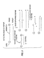
- the detection of cleaved oligonucleotide fragment is enhanced by the addition of a labeled tail.
- an oligonucleotide probe can be synthesized so that it forms a mismatch when hybridized to target polynucleotide.
- the oligonucleotide probe can contain a protected 3' group, preventing a polymerase from extending from the probe sequence. The probe then can be cleaved by a nucleic acid repair enzyme.
- the 5' region of the probe can be designed so that it is long enough to remain bound to the target sequence when the mismatch is cleaved.
- the 3' region of the probe can be designed so that it is short enough so that upon cleavage it dissociate from the target polynucleotide.
- nucleotides used in this embodiment are labeled with a radionucleotide or with some other marker, such as a fluorescent label, then the amount of cleavage can be assayed, using these labeled nucleotides as the signal.
- the 5' region is so designed that it dissociates from the target polynucleotide when the mismatch is cleaved, as shown in side A of FIGURE 2.
- the 5' cleavage fragment then can be extended with polyA polymerase or terminal deoxynucleotidyl transferase (TdT), as shown in side A of FIGURE 2, and detected.
- the tails consist of polymers of adenosines
- the tails will hybridize to polymers of oligonucleotide composed of thymines. If the nucleotides used in this polyA polymerase or TdT reaction are labeled with a radionucleotide or with some other marker, such as a fluorescent label, then the amount of cleavage can be assayed, using these labeled nucleotides as the signal.
- the oligonucleotide probe described in the previous two aspects of the present embodiment can have another feature, a ligand type molecule at the 5' end of the probe oligonucleotide.
- the ligand can serve to label the probe.
- the ligand can be a biotin group, or a group extendable with poly (ADP) ribose polymerase. Additionally, the ligand can be DNP (dinitrophenol) or cholesterol moieties.
- molecules that bind to the ligands can be used to affinity purify the newly formed tails.
- the probes with a biotin ligand can be passed over an avidin or streptavidin column.
- the probes with a poly(ADP)ribose ligand can be passed over a antipoly(ADP)ribose column.
- the DNP and cholesterol moieties can be passed over column with antibody against DNP and cholesterol, respectively.
- Affinity purification techniques, including affinity chromatography are well known to those skilled in the art.
- Yet another embodiment of this invention provides for detecting, identifying, measuring, or localizing, inter alia, known sequences in a target polynucleotide in a biological sample.
- This aspect of the invention includes hybridizing a single-stranded oligonucleotide probe to a target polynucleotide to form a hybrid double-stranded polynucleotide.
- the probe is designed so that it includes a mismatched or repairable base sequence.
- the probe so designed is not complementary to the target polynucleotide, there is a mismatch between the probe containing a synthesized mutation or mismatch site and the wild-type target polynucleotide.
- the mismatch occurs at the site of a mismatched or repairable base sequence.
- the probe is cleaved at the point of mismatch with a nucleic acid repair enzyme as mentioned above.
- the cleaved polynucleotide fragments are then detected as mentioned above.
- the probe oligonucleotide can be synthesized so that it is complementary to the target polynucleotide sequence except at a single nucleotide, which is chosen to be near the middle of the probe.
- the wild-type probe contains a cytosine base in the middle of the probe that correctly hybridizes to a guanosine base on a wild-type target polynucleotide sequence.
- the mismatched probe can be designed by synthesizing an oligonucleotide probe wherein the cytosine base is replaced by an adenine base.
- the adenine base of the probe oligonucleotide mismatches to a guanosine base in the target molecule. But the rest of the strands of the probe and target are complementary.
- the probe When treated with mutY, the probe are cleaved if it has hybridized to a non-mutated wild-type target polynucleotide.
- the probe containing a mutation will not be cleaved. This is true because a mismatch will not have occurred, the probe adenine being complementary to the target thymine.
- Another aspect of the present embodiment includes two probes, used independently, which contain mismatches at slightly different sites. Cleavage occurring for both probes in separate reactions confirms the presence of the target polynucleotide.
- the present embodiment can utilize helix destabilizing molecules, as discussed above. Additionally, this embodiment can be performed using the oscillating and tailing methods already described. Moreover, this embodiment may employ the linking methods and use of a combination of glycosylases and either AP endocuncleases or AP lyases, as described above.
- the present embodiment is useful in DNA diagnostics and DNA profiling.
- repair index in this regard is defined as the ratio of the amount of cleavage in a sample mixed with base-repairing enzyme (shown in the right side of FIGURE 3) to the amount of cleavage in a sample of control, which contains no base-repairing enzymes (control shown in FIGURE 3 on left side).
- the repair index indicates the extent to which base-repairing enzymes have repaired a sample of mismatched or damaged oligonucleotide probes.
- a "base-repairing enzyme” is one that effects repair of a mismatched probe sequence that has hybridized to a target polynucleotide. The base-repairing enzyme replaces the mismatched base with a base that is complementary to the target polynucleotide. Examples of base-repairing enzymes are E . coli DNA purine transferase (E.C. 2.6.99.1), human O 6 -methylguanine-DNA methyltransferase (Koike et al , J . Biol . Chem . 265: 14754) and E . coli DNA photolyase (EMBL Data Library Accession Number S32737).
- the embodiment depicted in FIGURE 3 involves hybridizing a single-stranded oligonucleotide probe to a target polynucleotide to form a hybrid double-stranded polynucleotide.
- the probe is designed so that it includes a mismatched or a repairable base sequence.
- Such a probe can be designed according to the methods described above.
- probe so designed will not be complementary to the target polynucleotide, there will be a mismatch between the mutated probe and the wild-type target polynucleotide. The mismatch will occur at the site of the mismatched or repairable base sequence.
- the hybrid double-stranded polynucleotide will be exposed to a base-repairing enzyme, which is defined above.
- the base-repairing enzyme will repair a certain amount of the mismatched or repairable base sequences, depending on the type of base-repairing enzymes employed.
- the hybrid double-stranded polynucleotide then will be exposed to a nucleic acid repair enzyme, which cleaves non-repaired probe at the point of mismatch.
- the cleaved nucleotide fragments can then be detected by the methods described above.
- the amount of cleavage in a sample mixed with base-repairing enzyme is compared to the amount of cleavage in a sample of unrepaired control.
- Determining the repair index is useful in determining the potency of cancer therapeutic agents on an individual.
- an oligonucleotide is synthesized with a base that has been modified to contain a base adduct.
- Several base adducts are known to be the result of certain chemotherapeutic drugs. See Friedman et al ., supra , table 5-3.
- Base adducts cause a significant distortion of the DNA helix and are both repairable by base repair enzymes and cleaved by nucleic acid repair enzymes.
- the probe is hybridized with target polynucleotide in a medium containing the base-repairing enzymes of a particular individual, and then the duplex is exposed to nucleic acid repair enzyme. Accordingly, the index of repair indicates the extent to which the individual would repair mutations that would be induced by a particular cancer therapeutic agent.
- the present embodiment can employ . helix destabilizing molecules as described above. Additionally, this embodiment of the invention can be performed using the oscillating and tailing methods described above. Moreover, this embodiment may employ the linking methods and use of a combination of glycosylases, as described above.
- the present invention describe, but does not claim, the modification of a single base in a target polynucleotide.
- This includes hybridizing a single-stranded probe to a circular single-stranded target polynucleotide.
- the probe is constructed so that it contains a single base that will form a mismatch with the target polynucleotide at a predetermined site.
- the probe is designed to contain fewer base sequences than the target polynucleotide so that hybridization results in a partially double-stranded circular nucleic acid molecule (See fig. 5).
- a probe which can be either an oligonucleotide molecule or a peptide nucleic acid molecule.
- the partially double-stranded construct, described above is converted into a completely double-stranded construct by filling in the single-stranded region of the probe-target hybrid (see Fig. 5). This conversion can be accomplished using DNA Polymerase and Ligase according to the methods employed by M. Smith and S. Gilliam (1981), in 3 GENETIC ENGINEERING 1 (J.K. Setlow & A. Hollaender, eds.).
- the partially double-stranded construct is treated with T4 or T7 DNA polymerases and T7 or T4 DNA lygases.
- T4 or T7 DNA polymerases and T7 or T4 DNA lygases are superior because they lack 5' to 3' exonuclease activity. Additionally, these enzymes will not effect strand displacement of the oligonucleotide probe. Accordingly a completely double-stranded construct will be formed containing the original probe base sequences.
- This fully double-stranded molecule is then treated with a nucleic acid repair enzyme which cleaves the target polynucleotide strand at the point of mismatch, creating a gap in the target polynucleotide (see Fig. 5).
- a nucleic acid repair enzyme which cleaves the target polynucleotide strand at the point of mismatch, creating a gap in the target polynucleotide (see Fig. 5).
- the probe-target hybrid is transformed into a bacteria according to techniques well known to those skilled in the art. In the process of transformation, the gap created by cleavage is filled with base(s) complementary to exposed probe bases.
- PNA peptide nucleic acid molecule
- PNA's as contemplated in the instant invention are molecules containing neutral, peptide-like backbones and nucleobases. PNAs allow for the hybridization to RNA and DNA with higher affinity and specificity than conventional oligonucleotides. PNAs are commercially available, for example, from PerSpective Biosystems, Inc. (Framingham, MA), and are described in Wittung et al ., Nature 368, 561-563 (1994); Hanvey, et al ., Science 258: 1481-85 (1992).
- a partially double-stranded probe target construct is formed, as discussed above.
- a nucleic repair enzyme effects cleavage of the target polynucleotide at the point of mismatch. Because of its peptide-like backbone, the PNA probe is impervious to cleavage by a nucleic acid repair enzyme. Accordingly, the PNA probe is advantageous in that it assures that cleavage will not occur in the probe molecule at the point of mismatch. Instead, the nucleic repair enzyme will only be able to effect cleavage in the target polynucleotide.
- the gap resulting from this cleavage can then be filled in with DNA polymerase and ligase in combination with the desired dNTP's (see Fig. 10).
- T4 DNA polymerase, T7 DNA polymerase, T4 DNA ligase, and T7 DNA ligase are preferred enzymes for this reaction.
- other enzymes can be used to fill the gap in the target polynucleotide without causing strand displacement.
- Klenow fragment of DNA polymerase I may be used as described by Kunkel, T.A. Nucleic Acids Mol . Biol . 12: 124 (1988); Proc . Natl . Acad . Sci . U . S . A . 82: 488 (1985).
- the target polynucleotide strand can be separated from the PNA probe by heating the PNA-target hybrid to dissociate the two from each other.
- the temperature for disassociation is approximately 3°C per mer in the PNA probe molecule plus approximately 20°C as described in Wittung et al ., Nature 368, 561-563 (1994).
- the target polynucleotide can then be purified by column size exclusion chromatography (e.g. G-SO) to obtain target that is free of PNA probe molecules.
- the target polynucleotide may then be transformed into a bacteria as a single-stranded circular construct.
- the skilled artisan may obtain a double stranded construct by use of a primer, polymerase, ligase enzymes and dNTP's.
- a double-stranded construct may be advantageous because double-stranded circular polynucleotides generally transform with higher efficiency than single-stranded molecules.
- the enzyme mutY was purified by the method of Wu et al ., Proc . Nat'l Acad . Sci . USA 89: 8779-83 (1992), to a concentration of about 50 units• ⁇ l -1 from an overexpressed E. coli clone. (One unit is defined as the conversion of 1 fmol A/G mismatch synthetic oligonucleotide converted to nicked substrate per minute.) Two synthetic oligodeoxyribonucleotides were synthesized by standard phosphoramidite chemistries with the following sequences:
- the oligonucleotides were purified on 20% polyacrylamide/7M Urea 1XTBE gels to about 95% homogeneity. The fragment was detected by autoradiography and a band cut from the gel. It was eluted from the gel slice by electrophoresis onto NA-45 paper (Schleicher & Schuler, Inc) by band interception, followed by elution from the paper by heating at 65°C for 5 minutes in 2M TEAA (triethylammonium acetate). The sample was dried in a vacuum centrifuge until all solvent was removed.
- Oligonucleotide #1 (Oligo 1) was radiolabeled to high specific activity with about 60 pmols (0.1 mCi; 6000 Ci• mMol -1 ) 32P-gamma-ATP and 100 units of T4 polynucleotide kinase at 37°C for 1 hour. The radiolabeled fragment was further gel purified, as described above.
- oligonucleotide #2 (Oligo 2) was diluted from 1 pmol• ⁇ l -1 to 10 -9 pmol• ⁇ l -1 in sterile deionized water.
- a typical reaction mix was set up that contained about 0.2 pmol of radiolabeled oligo 1, mutY buffer (20 mM Tris-HCl, pH 7.6, 80 mM NaCl, 1 mM dithioerythritol, 1 mM EDTA, pH 8.0, 3% glycerol; final concentration), 50 units of mutY enzyme, some reactions contained a dilution of Oligo 2 from 1 to 10 -9 pmol. The final volume was 10 ⁇ l. The reaction proceeded at 37°C for 60 minutes.
- the reaction was stopped by the addition of 1 ⁇ l of loading buffer (98% Formamide, 10 mM EDTA, pH 8.0, 0.025% Xylene Cyanol FF, 0.025% Bromophenol Blue).
- loading buffer 98% Formamide, 10 mM EDTA, pH 8.0, 0.025% Xylene Cyanol FF, 0.025% Bromophenol Blue.
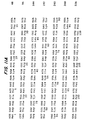
- the reaction was electrophoresed on a 20% polyacrylamide/7M Urea 1X TBE gel, then autoradiographed.
- Lane 1 contains no target molecule.
- Lanes 2 to 11 contain 10°, 10 -1 , 10 -2 , 10 -3 , 10 -4 , 10 -5 , 10 -6 , 10 -7 , 10 -8 , 10 -9 pmol of target.
- the cleavage from the reaction was detected by the generation of a smaller cleaved synthetic oligonucleotide fragment found in lanes 2 to 4. The truncated shorter molecule was due
- the nucleic acid repair enzyme mutY and the helix destabilization enzyme *I were used along with three synthetic oligonucleotides having the sequences: Probes 2 and 3 are complementary. Probes 1 and 3 are complementary with a single base mismatch. The three DNA synthetic oligonucleotides were used as is or were radiolabeled with 32 P- ⁇ -ATP via T4 polynucleotide kinase.
- Reactions were set up as indicated below with combinations of radiolabeled or cold oligonucleotides 1, 2 and 3, with or without mutY and or *I.
- Lanes 1 to 20 contained radiolabeled oligonucleotide 1.
- Lanes 3 and 4 contained radiolabeled oligonucleotide 2.
- Lanes 5 to 7 contained radiolabeled oligonucleotide 3.
- Lane 6 contained 10 pmols of cold oligonucleotide 1.
- Lane 7 contained 10 pmols of cold oligonucleotide 2.
- Lanes 2, 4, 9 and 16 contained 10 pmols of cold oligonucleotide 3.
- Lanes 2, 4,6 to 20 contained mutY enzyme.
- Lanes 15 to 20 contained *I. The reactions were setup with standard mutY reaction buffer and allowed to proceed for 1 hour at 37°C. The reactions were stopped, loading dye was added, and the material was run on a 20% polyacrylamide/7 M Urea gel (1XTBE).
- a synthetic oligonucleotide of length N nucleotides has a temperature of hybridization (melting temperature or Im, in °C) equal to 3N.
- a synthetic oligonucleotide of 20 nucleotides has a temperature of hybridization of about 60°C.
- a shorter synthetic oligonucleotide of 10 nucleotides has a temperature of hybridization of about 30°C.
- the long 20-mer synthetic oligonucleotide hybridizes but the shorter 10-mer synthetic oligonucleotide does not.
- the longer synthetic oligonucleotide hybridizes to a target sequence, then when it is cleaved at a specific place where a mismatch occurs two or more shorter synthetic oligonucleotides are be generated, which have lower thermodynamic stability. These shortened cleaved oligonucleotides dissociate from the target sequence, making it available for another hybridization event with the longer synthetic oligonucleotide.
- the hybridization kinetics favors better, faster hybridization. Further, this allows the detection of a hybridization event by the accumulation of the shortened oligonucleotide fragments.
- a helix destabilizing molecule can also be added to the system of Example 3.
- the operating temperature is 45°C and is kept at that temperature for this example.
- the longer 20-mer synthetic oligonucleotide hybridizes not at 60°C, but rather at a lower temperature, for example, 48°C.
- the hybridization temperature and the operating temperature are closer. This allows in one scenario the reduction of the operating temperature to a lower temperature, perhaps 37°C.
- mesophilic enzymes can be used in the reaction.
- the practice of this example at such a temperature is advantageous in that it does not require thermophilic enzymes (those that function at high temperature extremes, e.g. 60 - 70°C, and can withstand near boiling temperatures).
- in vitro mutagenesis For modifying and generating new genetic sequences, in vitro mutagenesis is used.
- the earliest method of in vitro mutagenesis is that of M. Smith and S. Gilliam (1981), in 3 GENETIC ENGINEERING 1 (J.K. Setlow & A. Hollaender, eds.).
- the nucleic acid repair enzymes can be used for oligonucleotide directed in vitro mutagenesis.
- the mutY is used as the example nucleic acid repair enzyme, however, other enzymes can also be used.
- Single stranded DNA containing the cloned gene sequence to be modified can be generated by the method of Zinder & Boeke, Gene 19: 1-10 (1982), using the M13 bacteriophage system.
- a synthetic oligonucleotide is designed, which is complementary to the region to be modified with a Guanine mismatch at a position where the adenine is to be changed to a Cytosine (figure 5).
- the oligonucleotide is hybridized to the single stranded DNA, treated with T4 DNA polymerase and T4 DNA ligase by methods used by Smith (1981).
- the newly synthesized double stranded molecule with a single mismatch is then treated with 50 units of mutY in mutY buffer (20 mM Tris-HCl, pH 7.6, 80 mM NaCl, 1 mM dithioerythritol, 1 mM EDTA, pH 8.0) for one hour at 37°C.
- the material that is cleaved at the mismatch site is transformed into E . coli using methods well known to those skilled in the art.
- the transformants then are enriched for sequences containing the conversion of the adenine to the cytosine base pair.
- a synthetic oligonucleotide complementary to a region of the p53 gene is synthesized using standard phosphoramidite chemistry.
- Purified mutant E . coli mutY mismatch enzyme (with associated AP endonuclease) is linked to the 5' end of the oligonucleotide with linker using methods already described (Corey et al ., 1987, Science, vol 238: 1401; Corey et al ., 1989, Journal of the American Chemical Society, vol 111:8523). This forms a covalent linkage of an oligonucleotide of specific sequence, with a linker region covalently linked to E . coli mutY mismatch enzyme.
- oligonucleotide is radiolabeled at the 3' end using poly (A) polymerase and 32 P-cordycepin.
- poly (A) polymerase poly (A) polymerase
- 32 P-cordycepin 32 P-cordycepin
- chromosomal DNA prepared from ML-1 myeloid leukemia cells
- dilutions of the target DNA from 10 -1 to 10 -8 pmol are setup.
- 10 pmols of radiolabeled probe 100 ng of purified E . coli mutY mismatch enzyme for the unlinked oligonucleotide and buffer are added. The reaction was allowed to precede for 30 minutes at 37°C.
- the reaction product is then electrophoresed on a 20% polyacrylamide/7M urea gel, then autoradiographed.
- the generation of shortened cleaved oligonucleotides indicates the presence of a DNA mismatch.
- the covalently linked enzyme is expected to show enhanced activity over unlinked enzyme, as expected from previous data (Bruice et al ., 1963, Journal of American Chemical Society, vol 85:1).
- a first synthetic oligonucleotide probe complementary to a region of the p53 gene is synthesized using standard phosphoramidite chemistry.
- Purified E . coli G/T mismatch enzyme is linked to the 5' end of the oligonucleotide with a linker using methods already described (Corey et al ., 1987, Science, vol 238:1401; Corey et al ., 1989, Journal of the American Chemical Society, vol 111:8523). This forms a covalent linkage of an oligonucleotide of specific sequence, with a linker region covalently linked to E . coli G/T mismatch enzyme.
- a second oligonucleotide is synthesized, where the sequence is near to the first oligonucleotide.
- This second oligonucleotide is synthesized as above with a linker which is attached to AP endonuclease (see FIGURE 6a).
- the first oligonucleotide is radiolabeled at the 3' end using poly (A) polymerase and 32 P-cordycepin.
- oligonucleotides without enzymes are prepared and treated alongside with the enzyme linked oligonucleotides.
- chromosomal DNA prepared from ML-1 myeloid leukemia cells
- dilutions of the target DNA from 10 -1 to 10 -8 pmol are setup.
- 10 pmols of radiolabeled probe 100 ng of purified E . coli G/T mismatch enzyme and 100 ng of AP endonuclease for the unlinked oligonucleotide and buffer are added. The reaction are allowed to proceed for 30 minutes at 37°C.
- the reaction product is then electrophoresed on a 20% polyacrylamide/7M urea gel, then autoradiographed.
- the generation of shortened cleaved oligonucleotides indicates the presence of a DNA mismatch.
- the covalently linked enzyme is expected to show enhanced activity over unlinked enzyme, as expected from previous data (Bruice et al ., 1963, Journal of American Chemical Society, vol 85:1).
- a synthetic oligonucleotide (first) complementary to a region of the p53 gene is synthesized using standard phosphoramidite chemistry.
- Purified E. coli G/T mismatch enzyme is linked to the 3' end of the first oligonucleotide with a linker using methods already described (Corey et al ., 1987 Science, vol 238:1401; Corey et al ., 1989, Journal of the American Chemical Society, vol 111:8523). This forms a covalent linkage of an oligonucleotide of specific sequence, with a linker region covalently linked to E . coli G/T mismatch enzyme.
- a second oligonucleotide is synthesized, where the sequence is near to the first oligonucleotide but allows a space at the 3' end where a mutation probe can hybridize.
- This second oligonucleotide is synthesized as above with a linker which is attached to AP endonuclease.
- a third mutation probe is synthesized and radiolabeled at the 5' end using polynucleotide kinase and 32 P- ⁇ -ATP (see FIGURE 7).
- oligonucleotides without enzymes attached are prepared and treated alongside with the enzyme linked oligonucleotides.
- chromosomal DNA prepared from ML-1 myeloid leukemia cells
- dilutions of the target DNA from 10 -1 to 10 -8 pmol are setup.
- 10 pmols of radiolabeled probe 100 ng of purified E . coli G/T mismatch enzyme and 100 ng of AP endonuclease for the unlinked oligonucleotide and buffer are added. The reaction is allowed to proceed for 30 minutes at 37°C.
- the reaction product is then electrophoresed on a 20% polyacrylamide/7M urea gel, then autoradiographed.
- the generation of shortened cleaved oligonucleotides indicates the presence of a DNA mismatch.
- the covalently linked enzyme is expected to show enhanced activity over unlinked enzyme, as expected from previous data (Bruice et al., 1963, Journal of American Chemical Society, vol 85:1).
- a synthetic oligonucleotide complementary to a region of the p53 gene is synthesized using standard phosphoramidite chemistry.
- Purified E . coli G/T mismatch enzyme is linked to the 5' end of the oligonucleotide with a linker using methods already described (Corey et al ., 1987, Science, vol 238:1401; Corey et al ., 1989, Journal of the American Chemical Society, vol 111:8523). This forms a covalent linkage of an oligonucleotide of specific sequence, with a linker region covalently linked to E . coli G/T mismatch enzyme.
- AP endonuclease (see FIGURE 8) is attached to the same oligonucleotide at the 3' end.
- oligonucleotides without enzymes are prepared and treated alongside with the enzyme linked oligonucleotides.
- chromosomal DNA prepared from ML-1 myeloid leukemia cells
- dilutions of the target DNA from 10 -1 to 10 -8 pmol are setup.
- 10 pmols of radiolabeled probe 100 ng of purified E . coli G/T mismatch enzyme and 100 ng of AP endonuclease for the unlinked oligonucleotide and buffer are added. The reaction is allowed to proceed for 30 minutes at 37°C.
- the reaction product is then electrophoresed on a 50 ⁇ m i.d. x 35.5 cm Supelco H-75 methyl coated borosilicate capillary containing TreviSolTM CE B (Trevigen, Inc.). 40 ⁇ l of the reaction product is applied electrokinetically to the capillary, then electrophoresed at -140 V/cm for about 12 minutes.
- the synthetic oligonucleotides are detected by UV detection.
- the generation of shortened cleaved oligonucleotides indicates the presence of a DNA mismatch.
- the covalently linked enzyme is expected to show enhanced activity over unlinked enzyme, as expected from previous data (Bruice et al ., 1963, Journal of American Chemical Society, vol 85:1).
- a synthetic oligonucleotide complementary to a region of the p53 gene is synthesized using standard phosphoramidite chemistry.
- Purified E . coli G/T mismatch enzyme is linked to the 5' end of the oligonucleotide with a linker using methods already described (Corey et al ., 1987, Science, vol 238: 1401; Corey et al ., 1989, Journal of the American Chemical Society, vol 111:8523). This forms a covalent linkage of an oligonucleotide of specific sequence, with a linker region covalently linked to E. coli G/T mismatch enzyme.
- oligonucleotide is radiolabeled at the 3' end using poly (A) polymerase and 32 P-cordycepin.
- poly (A) polymerase poly (A) polymerase
- 32 P-cordycepin 32 P-cordycepin
- chromosomal DNA prepared from ML-1 myeloid leukemia cells
- dilutions of the target DNA from 10 -1 to 10 -8 pmol are setup.
- 10 pmols of radiolabeled probe 100 ng of purified E . coli G/T mismatch enzyme for the unlinked oligonucleotide and buffer are added.
- the reaction is allowed to proceed for 30 with the temperature being cycled from about 30° to 42°C every two minutes to allow the probes with AP sites to fall off of the target molecule and new un-treated probes to hybridize to the target molecule.
- This oscillating process is known as the Mismatch Chain Reaction (MCR).
- MCR Mismatch Chain Reaction
- the AP cleavage is effected by treatment of the samples at high pH (adjusting the pH to a range of 8 to 14) with NaOH and higher temperatures (adjusting the temperature from a temperature range of 50°C to 95°C). This effects AP cleavage.
- the reaction product is then electrophoresed on a 20% polyacrylamide/7M urea gel, then autoradiographed.
- the generation of shortened cleaved oligonucleotides indicates the presence of a DNA mismatch.
- the covalently linked enzyme is expected to show enhanced activity over unlinked enzyme, as expected from previous data (Bruice et al ., 1963, Journal of American Chemical Society, vol 85:1).
- a synthetic oligonucleotide sequence complementary to a region of the p53 gene is synthesized using standard phosphoramidite chemistry. Different specific syntheses of this sequence are made with the attachment of different fluorophores (fluorescein (488 nm), rhodamine (546 nm), coumarin (365 nm)) at the 3' end of the molecule. At different wavelengths, these different fluorescent molecules can be differentiated from each other. With a linker intermediate, three different glycosylases are specifically added to three different oligonucleotide-fluorophore molecules, respectively.
- the three glycosylases are mutY (A/G and A/C mispairs), Thymine mismatch (also called G/T mismatch) and All type enzyme (all mispairs).
- the glycosylase/linker attachment is done using methods already described (Corey et al., 1987, Science, vol 238:1401; Corey et al., 1989, Journal of the American Chemical Society, vol 111:8523).
- chromosomal DNA prepared from HL-60 myeloid leukemia cells
- dilutions of the target DNA from 10 -1 to 10 -8 pmol are setup.
- 10 pmols of radiolabeled probe 100 ng of AP endonuclease are added. The reaction was allowed to proceed for 30 minutes at 37°C.
- reaction products are then electrophoresed on a 50 ⁇ m i.d. x 35.5 cm H-75 methyl coated borosilicate capillary containing TreviSolTM CE B (Trevigen, Inc.) capillary matrix.
- 40 ⁇ l of the reaction product is applied electrokinetically to the capillary, then electrophoresed at -140 V/cm for about 12 minutes.
- the synthetic oligonucleotides are detected by a UV/VIS detector.
- the generation of shortened cleaved oligonucleotides indicates the presence of DNA mismatch.
- the exact form of the mismatch is identified from the specific fluorophore indicating the respective glycosylase used in the reaction.
- both forms of DNA could be present. This technique would allow differentiating between the relative amount of each form of DNA present by measuring the amounts of both forms of the oligonucleotides.
- a synthetic oligonucleotide sequence complementary to a region of the p53 gene is synthesized using standard phosphoramidite chemistry.
- the mismatch or no mismatch
- Different specific syntheses of this sequence are made which have slightly longer lengths at the 3' end of the molecule and where the specific base with the mismatch is synthesized as a different base at that position (e.g. one oligo is A, another oligo is C, and the final oligo is G at one base in the oligonucleotide.
- the oligonucleotides are 30, 32 and 34 nucleotides in length.
- glycosylases are specifically added to the 5' end of three different oligonucleotide molecules of different lengths, respectively.
- the three glycosylases were mutY (A/G and A/C mispairs), Thymine mismatch (also called G/T mismatch) and All type enzyme (all mispairs).
- the glycosylase/linker attachment is done using methods already described (Corey et al., 1987, Science, vol 238:1401; Corey et al., 1989, Journal of the American Chemical Society, vol 111:8523).
- the oligonucleotides are labeled with 32 P-labelled cordycepin and poly (A) polymerase at the 3' end.
- chromosomal DNA prepared from HL-60 myeloid leukemia cells
- dilutions of the target DNA from 10 -1 to 10 -8 pmol are setup.
- 10 pmols of different radiolabeled probe are added to the target DNA, and 100 ng of AP endonuclease is added to each tube. The reaction is allowed to proceed for 30 minutes at 37°C.
- reaction products are then electrophoresed on a 20% polyacrylamide/7 M urea gel.
- the presence of cleaved fragments in the autoradiograph indicates the presence of a mismatch.
- the length of the cleaved molecule indicates which specific mismatch was present.
- both forms of DNA could be present. This technique would allow differentiating between the relative amount of each form of DNA present by measuring the amounts of both forms of the oligonucleotides.
- Escherichia coli JM109 is available from New England Biolabs of Beverly, MA and Escherichia coli BW415 is available form the laboratory of Dr. Richard P. Cunningham at the State University of New York, at Albany, Department of Biological Sciences.
- a similar strain suitable for this protocol is BW434 and it is available from the Coli Genetic Stock Center at Yale University School of Medicine, New Haven, CT.
- BW415 ⁇ DE3 was made with a ⁇ DE3 lysogenization kit from Novagen Inc. of Madison, WI. This integration allowed for the efficient expression of the T/G mismatch specific thymine-DNA glycosylase from a T7 RNA polymerase driven promoter in an endonuclease III deficient strain of Escherichia coli .
- the expression system was contained on plasmid pET14B from Novagen Inc.
- Plasmid pUV2 containing the orf 10 coding sequence is available from Dr. Jork Nolling, Wageningen Agricultrual University of the Netherlands, Department of Microbiology, Hesselink van Suchtelenweg 4, 6703 CT Wageningen, The Netherlands.
- the pUV2 plasmid contains a portion of pFV1, including the orf 10 coding sequence, cloned into pUC19.
- Plasmid pUC19 is available from Sigma Chemical Co., of St. Louis, MO.
- Plasmid DNA was prepared from JM109 by a modified alkaline lysis method.
- the orf 10 coding sequence from pUV2 was cloned into pUC19 via a DraI-EcoRI to HincII-EcoRI ligation using restriction endonucleases and ligase (from New England Bio Labs) according to suppliers' recommendations.
- the gene was PCR mutagenized using Taq Polymerase from Perkin Elmer Corp.
- the cell suspension was sonicated 5 X 3 minutes with a Branson sonifer, and then stirred on ice for one hour. The sonicate was centrifuged at 48,000X g for 20 minutes and the supernatant was retained. Five percent polyethelenimine was added to a final concentration of 0.1% and the suspension was stirred for 1 hour and centrifuged at 48,000X g to give a supernatant with a volume of 500 mls. The supernatant was dialyzed against 4 L of 50 mM KPO 4 pH 7.2 overnight.
- the crude extract was loaded onto an 80 ml SP Sepharose Fast Flow (Pharmacia Biotech Inc. of Piscataway, NJ) column, washed with 100 ml of 50mM KPO 4 pH 7.2 and eluted with a 1 L gradient from 0 to 1M NaCl at a flow rate of 10 ml/min.
- the protein eluted at 0.6M NaCl as detected by 31 kD band visualized by SDS-PAGE, and a characteristic yellow color in the fractions indicative of an Fe-S cluster of the protein.
- the fractions containing the protein were pooled and dialyzed against 50 mM KPO 4 pH 6.6, loaded onto a 5 ml DNA agarose (Pharmacia Biotech Inc.) column, washed with 10 ml of 50 mM KPO 4 and eluted with a 100ml gradient of 0-1 M NaCl at a rate of 1 ml/min. The protein eluted at 0.8M NaCl as determined by the criteria above. Fractions containing the protein were pooled and dialyzed overnight against 50 mM KPO 4 pH 6.6. The protein was loaded onto a 5ml SP high trap column (Pharmacia Biotech Inc.) and eluted with 1M NaCl to concentrate the protein.
- the extract was further concentrated to 1.5 mls with Centriprep 10 concentrators (Amicon Division, W.R. Grace & Co., of Danvers, MA). At this point the protein was a single band on an overloaded Coomassie stained gel and gave an A 410 /A 280 ratio of 0.295. This protein was active in our T-G mismatch assay. The pure protein was stored in 50% glycerol at -20 C.
- Oligonucleotide A was labeled with 32 P ATP from New England Nuclear of Boston, MA using T4 Polynucleotide kinase from New England Bio Labs. The two oligonucleotides were annealed at a three to one excess of B to A on ice for one hour to generate a duplex oligonucleotide with a unique T/G mismatch.
- Glycosylase activity was monitored by measuring DNA strand breakage.
- 32 P labeled substrate was incubated with T/G mismatch specific thymine-DNA glycosylase protein in 50 mM HEPES-KOH pH 7.5, 100 mM KCl and 1mM EDTA at 65°C for 45 minutes.
- the enzyme was inactivated with 5M urea at 100°C for 5 minutes and then the reaction mixture was incubated in 0.15 M NaOH for 15 minutes at 55°C to create strand breaks at the site of AP sites.
- Loading buffer was added to the mixture and then run on a 20% acrylamide gel containing 8M urea for 2 hours at 200 volts. The gels were dried and analyzed on X-ray film.
- the enzyme showed a glycosylase activity against both thymine and uracil mismatched with guanine.
- the activity on each substrate was at roughly the same level of magnitude, with a mared decrease in activity against thymines mismatched with cytosine.
- Temperature assays were run in the same manner using a labeled T-G mismatch, but incubation temperatures were 25,35,45,55,65, and 75°C. Peak activity was identified at 65°C (92.4% cleavage), with 85.9% cleavage seen at 75°C, 90.6% cleavage seen at 55°C, 84.5% cleavage seen at 45°C, 70.2% cleavage seen at 35°C and 36% cleavage seen at 25°C.
- the enzyme was active after being boiled in the absence of 5M urea.
- Oligonucleotide RO563 is complementary to both RO561 and RO562, however the underlined base G in RO562 does not complement the T in RO563, forming a mismatch.
- the length and composition of these synthetic oligonucleotides were chosen to have a hybridization temperature around 70°C The sequences were based on the human p53 gene, which has a mutation at the underlined G position found in the RO562 sequence.
- thermostable mismatch glycosylase made according to Example 14 in a volume of 10 ⁇ l.
- the reaction was allowed to proceed for about 3 hours at 60°C.
- the reaction was loaded onto a 20% acrylamide/7 M Urea/1X TBE gel and electrophoresed at about 300 V for about 3 hours. The gel was then electrophoresed for different lengths of time.
- thermostable enzyme at elevated temperatures resulted in a level of cleavage significantly higher than that found with a mesophilic enzyme. This increased level of cleavage results in a more sensitive assay than one carried out at lower temperatures. For instance, the autoradiograph of the above experiment after a brief 3 hour exposure showed faint cleavage at 0.000001. Longer autoradiographic exposure showed further sensitivity.
- Probe may be present in the assay in concentrations from 1 fmol to 100 pmol.
- the probe need not be radioactively labelled, and may be labelled by other means as are known to those of skill in the art.
- concentration of the thermostable enzyme may be present from approximately 1 to 100 ng, and finally, the buffer composition can have higher concentrations of KCl from 50 mM to 100 mM.
Abstract
Description
- Genomic DNA provides the template for the information that allows the generation of proteins which are expressed and made by an organism. These proteins are generally essential for the survival of any specific cell in an organism. Therefore, the organism requires the template to be correct and free of mistakes in order to generate a protein that is functional in a cell. If a single nucleotide of this DNA sequence is mutated (a "point mutation"), the protein may be nonfunctional. Point mutations which elicit disease states are known for many proteins. Examples include sickle cell anemia hypoxanthine phosphotransferase, and p53, a tumor suppressor gene, and several oncogenes and cancer genes.
- A review by Cotton, Biochem. J. 263: 1 (1989), compared several methodologies for detection of point mutations with respect to the DNA type used, the DNA stage achieved, whether the mutation position was detected, the percentage of mutations detected, the time and cost requirements, and toxicity problems. Each of the methodologies examined by Cotton presents drawbacks. DNA sequencing, for example, is time consuming and expensive. Restriction enzymes do not define the mutation position and detect less than 50% of mutations. Denaturing gradient gels and SSCP, see Murakami et al., Cancer Res. 51: 3356 (1981), do not define the mutation position and are not efficient at detecting mutations. S1 nuclease and RNAse are not efficient at detecting mutations. Finally, Carbodi-imide/ABC nuclease and carbodi-imide are efficient but generate false positives and are toxic.
- Recently, point mutations have been detected with the E. coli repair enzyme mutY. See Hsu et al., Carcinogenesis 15(8): 1657 (1994). In this method a wild type labeled probe is generated using the polymerase chain reaction (PCR) described, for example, by Saiki et al., Nature 324: 163 (1986). The probe then is hybridized to the unknown sample DNA wherein mutY then cleaves mismatches when an adenosine which does not form watson crick base pairing with a guanine nucleotide. The position of mutY cleavage at A/G sites can then be determined by gel electrophoresis. This methodology is limited by the use of PCR, which itself generates mutations in the amplified DNA. See Loeb et al., Nucleic Acids & Molec. Biol. 1: 157 (1987); Tindal et al., Biochemistry 27: 6008 (1988); Kunkel, loc. cit. 29: 8004 (1990).
- Accordingly, there is a need for an accurate and efficient method of detecting point mutations using unamplified DNA source molecules. In addition, such a method would save time, require minimal equipment and is less expensive, as well decreasing the hazard of toxic chemicals. Also, methods of amplifying limiting amounts of the mutated sequences would have advantages. By the same token, there is a need for accurate and inexpensive methods to detect non-mutated target polynucleotides from unamplified DNA source molecules.
- Currently there are several amplification methodologies, well known to those skilled in the art, for the detection of non-mutated DNA. Among these techniques are the polymerase chain reaction (PCR), the ligase chain reaction (LCR), nucleic acid system-based amplification (NASBA), and cycling probe technology (CPT). Other amplification methods are well known to those skilled in the art.
- The polymerase chain reaction described, for example, in U.S. patent No. 4,362,195, is the best known amplification system, but it is limited by the level of amplification (∼2.2x10⁵), is prone to the generation of mutations, and can generate false positives by the generation of amplified molecules contaminating the environment. Despite these limitations, PCR is widely used in the research community. It still is not approved by governmental regulators for clinical and diagnostic applications, however.
- CPT technology was developed, in part to overcome the limitations of PCR. See, for example, U.S. patents No. 4,876,187 and No. 5,011,769. The CPT technology entails the use of a synthetic molecule with two non-complementary nucleic acid sequences joined by a scissile linkage. CPT technology works by observing a hybridization event with a sample nucleic acid by a single cleavage event. This technology utilizes both the enzymatic features of RNAse H and a synthetic DNA-RNA-DNA oligonucleotide. RNAse H specifically cleaves the RNA moiety of the DNA-RNA-DNA oligonucleotide only when it is perfectly hybridized to a complementary DNA target molecule. A high concentration of the DNA-RNA-DNA molecule is converted to cleaved fragments, which are assayed by gel electrophoresis. The level of cleavage indicates the amount of target molecules present in the sample.
- The CPT system does not amplify the target, alleviating the accumulation of molecules that in turn become amplifiable and generate false-positives, as occurs in PCR. The CPT technology is linear, in that increasing amounts of target DNA generate linearly more cleaved DNA-RNA-DNA oligonucleotide. (PCR generates exponentially more signal in response to the presence of more target DNA, making quantitation more problematic.) Additionally, CPT can amplify up to 106 cleaved DNA-RNA-DNA probe molecules in about 30 minutes. CPT does not generate more of the target molecule. Therefore, it does not jeopardize the laboratory environment by the possible accumulation of synthesized target DNA molecules, which in turn generate false positive results. It also is isothermal, i.e., it does not require the use of expensive automated thermocycling equipment. Further, it has been shown to detect a single molecule. The CPT technology is limited, however, because the cleavable portion of the molecule is an RNA moiety.
- It is therefore an object of the present invention to provide a method for detecting point mutations in nucleic acid sequences.
- It is a further aspect of this invention to provide a simple and efficient, and sensitive method to detect point mutations in nucleic acid sequences.
- It is also an object of the invention to provide a means for detecting non-mutated nucleic acid sequences.
- It is yet another object of this invention to enhance target polynucleotide detection using oscillating and tagging techniques.
- It is an additional object of this invention to provide for thermally stable nucleic acid repair enzymes which will facilitate reactions at elevated temperatures.
- Still another aspect of this invention concerns helix destabilizing molecule and similar molecules to enhance the hybridization of the probe to the target polynucleotides.
- It is also an object of this invention to provide for the determination of the amount of base repair enzyme activity carried out on a mutated polynucleotide sequence.
- Another aspect of this invention relates to the attachment of a nucleic acid repair enzyme to a probe in order to enhance the detection of target polynucleotide and point mutations therein.
- It is a further object of this invention to provide for increased specificity in the detection of target nucleotides and point mutations therein, using peptide nucleic acids to construct probes.
- In accomplishing the foregoing objects, there is provided a method of detecting a point mutation in a target polynucleotide, comprising:
- (a) hybridizing a single-stranded oligonucleotide probe, under stringent conditions, to said target polynucleotide to form a hybrid double-stranded polynucleotide such that a mismatch occurs at the site of said point mutation, wherein said probe is complementary to a non-mutated sequence of said target polynucleotide;
- (b) producing probe fragments by cleaving said probe strand of said hybrid polynucleotide at said point of mismatch, wherein said cleaving is effected by a glycosylase attached to said probe and an AP cleaving enzyme; and
- (c) detecting said probe fragments.
-
- A method of detecting a point mutation in a target polynucleotide, comprising:
- (a) hybridizing a single-stranded oligonucleotide probe, under stringent conditions, to said target polynucleotide to form a hybrid double-stranded polynucleotide such that a mismatch occurs at the site of said point mutation, wherein said probe is complementary to a non-mutated sequence of said target polynucleotide;
- (b) hybridizing a second and third single-stranded oligonucleotide probe to said target polynucleotide at a location adjacent to said first probe, wherein a glycosylase is attached to said second single-stranded probe and an AP cleaving enzyme is attached to said third single-stranded probe;
- (c) producing probe fragments by cleaving said probe strand of said hybrid polynucleotide at said point of mismatch, wherein said cleaving is effected by said glycosylase and AP cleaving enzymes; and
- (d) detecting said probe fragments.
-
- A method of detecting a sequence in a target polynucleotide, comprising the steps of:
- (a) hybridizing a single-stranded oligonucleotide probe, under stringent conditions, to said target polynucleotide to form a hybrid double-stranded polynucleotide, wherein said probe contains a mismatched or repairable base sequence, such that a mismatch occurs at the site of said mismatched or repairable base sequence;
- (b) producing probe fragments by cleaving said probe strand of said hybrid polynucleotide at said point of mismatch, wherein said cleaving is effected by a glycosylase attached to said probe and an AP cleaving enzyme; and
- (c) detecting probe fragments produced by said cleavage.
-
- A method of detecting a point mutation in a target polynucleotide, comprising:
- (a) hybridizing a single-stranded oligonucleotide probe, under stringent conditions, to said target polynucleotide to form a hybrid double-stranded polynucleotide such that a mismatch occurs at the site of said point mutation, wherein said probe is complementary to a non-mutated sequence of said target polynucleotide;
- (b) producing probe fragments by cleaving said probe strand of said hybrid polynucleotide at said point of mismatch, wherein said cleaving is effected by a glycosylase enzyme employed in combination with basic conditions and increased temperature; and
- (c) detecting said probe fragments.
-
- The following methods for effecting in vitro mutagenesis are not claimed:
- A method for effecting in vitro mutagenesis of a target polynucleotide, comprising transforming a circular double-stranded nucleic acid molecule, wherein said circular double-stranded nucleic acid molecule is prepared by the steps of:
- (a) hybridizing a single-stranded oligonucleotide probe to said target polynucleotide to form a hybrid partially double-stranded circular polynucleotide, wherein said probe is shorter than said target polynucleotide and contains a single base-pair mismatch at a predetermined site;
- (b) contacting said hybrid polynucleotide with at least one DNA polymerase and one DNA ligase, whereby a completely double stranded hybrid polynucleotide is formed; and
- (c) contacting said completely double-stranded hybrid with a nucleic acid repair enzyme, whereby said target polynucleotide strand is cleaved at the site of said point of mismatch.
-
- A method for effecting in vitro mutagenesis of a single-stranded target polynucleotide, comprising:
- (a) hybridizing a single-stranded peptide nucleic acid probe to said target polynucleotide to form a hybrid partially double-stranded polynucleotide, wherein said probe is shorter than said target polynucleotide and contains a single base-pair mismatch at a predetermined site;
- (b) cleaving said target polynucleotide with a nucleic acid repair enzyme, whereby a gap is introduced into said target polynucleotide;
- (c) filling in said gap by contacting said hybrid polynucleotide with at least one DNA polymerase and one DNA ligase; and
- (d) dissociating said polynucleotide from said PNA probe.
-
- A method of detecting a point mutation in a target polynucleotide, comprising:
- (a) hybridizing at least two single-stranded oligonucleotide probes, under stringent conditions, to at least two target polynucleotides to form at least two hybrid double-stranded polynucleotides such that a mismatch occurs at the site of said point mutation, wherein said probes are complementary to a non-mutated sequence of said target polynucleotides, and attached to each probe is a different glycosylase, only one of which will effect cleavage;
- (b) producing probe fragments by cleaving one of said probe strands at said point of mismatch, wherein said cleavage is effected by one of said glycosylases and an AP cleaving enzyme; and
- (c) detecting said probe fragments.
-
-
- FIGURE 1 is a schematic diagram showing the hybridization of probe to target polynucleotide to form a mismatch. The mismatch can be the consequence of either a wild-type probe hybridizing to a mutant target or a mutant probe hybridizing to a wild-type target. Nucleic acid repair enzyme then cleaves the probe, which dissociates from the target.
- FIGURE 2 is a schematic drawing showing the tail labeling of cleaved probe. Probe is cleaved at the site of mismatch. If the probe is designed to dissociate, as shown in part A, it can, after dissociation, be tagged with a labeled nucleotide tail. If the probe is designed to remain hybridized to target after cleavage, as shown in part B, then it can also be tagged with a labeled nucleotide tail.
- FIGURE 3 is a schematic drawing showing the detection of the index of repair of a mutated or damaged probe. Probe is hybridized to target and then base-repairing enzyme is added. Next, nucleic acid repair enzyme is added and cleaves those probes which have not been repaired. A control group is shown on the left side of the figure.
- FIGURE 4 is a schematic drawing showing the oscillating reaction. An excess of mismatch probe hybridize to available target molecules in the reaction mixture, wherein a Nucleic Acid Repair Enzyme cleaves at the mismatch site. The probe molecule is designed so that the shorter cleaved molecules dissociate from the target molecule. The target then hybridizes to a new intact probe molecule, generating the oscillating reaction. The accumulation of cleaved molecules is linear with the amount of target molecules available.
- FIGURE 5 is a schematic drawing showing an oligonucleotide directed in vitro mutagenesis reaction (not claimed) A probe is hybridized to single stranded DNA (ssDNA) generating a mismatch in the ssDNA. A new strand of DNA is synthesized in vitro with DNA polymerase and ligase, generating a double stranded DNA (dsDNA) molecule. This dsDNA molecule is treated with mutY, cleaving and removing the mismatched base. The cleaved dsDNA molecule is transformed into E. coli, and the replicated dsDNA molecule then has a different base at the site of the mismatch.
- FIGURES 6A and B are schematic drawings showing the linking of nucleic acid repair enzyme to two separate probes designed to hybridize with a target polynucleotide. The nucleic acid repair enzyme comprises two separate individual enzymes, a glycosylase and an AP cleaving enzyme. The glycosylase is linked to a mismatch probe, i.e., a probe that is designed so that it will form a mismatch upon hybridization with the target polynucleotide. The AP cleaving enzyme is linked to a second probe which is complementary to the target polynucleotide and hybridizes to the target polynucleotide at a location adjacent to the mismatch probe. In FIGURE 6A the enzymes are attached to the 5' ends of the probes, while in FIGURE 6B, the enzymes are attached to the 3' ends.
- FIGURE 7 is a schematic drawing depicting a mismatch probe used in combination with a second and third probe which hybridize to the target polynucleotide at a location adjacent to the mismatch probe. The glycosylase is attached to the 3' end of the second probe and the AP cleaving enzyme is attached to the 5' end of the third probe.
- FIGURE 8 is a schematic drawing showing the attachment of glycosylase and AP cleaving enzyme to the 5' and 3' ends of a mismatch probe.
- FIGURE 9 is a schematic representation depicting the use of a combination of glycosylases to detect a point mutation. Three probes are depicted, and a different glycosylase is attached to each one. For a particular point mutation, only a specific probe or probes will be cleaved, thereby indicating the presence of the particular point mutation.
- FIGURE 10 is a schematic illustration of peptide nucleic acid ("PNA") directed in vitro mutagenesis. A PNA probe is hybridized to a single-stranded target polynucleotide, generating a mismatch. A nucleic acid repair enzyme is then contacted with the probe target hybrid cleaving the target strand at the point of mismatch. Next, the gap resulting from this cleavage is filled by contacting the probe target hybrid with appropriate polymerase enzymes.
- FIGURE 11 is a representation of the DNA sequence of orf10 and the amino acid sequence of the corresponding ORF10 polypeptide.
-
- It has been discovered that the deficiencies in conventional techniques in this regard can be overcome by employing nucleic acid repair enzymes. Thus, the problems by Cotton (1989), supra, are avoided by using nucleic acid repair enzymes, in accordance with the present invention, to detect point mutations. Furthermore, nucleotide excision enzymes can be used in the present invention to detect wild-type target polynucleotide sequences and to determine the amount of base repair enzyme activity carried out on a mutated polynucleotide sequence. The present invention represents an improvement over the CPT method, because a DNA oligonucleotide suitable for the present invention can be made via more conventional chemistry (and, hence, is less expensive to synthesize) and is more stable.
- Researchers have used nucleic acid repair enzymes to detect point mutations, see Hsu et al. (1994), supra, but these techniques are limited because they employ PCR to amplify the target polynucleotide. The present invention overcomes this limitation because it does not require PCR amplification. Instead, nucleic acid repair enzymes can be used to create an oscillating reaction which allows for adequate target detection using a limited amount of unamplified target. Alternatively, detection can be enhanced by using nucleic acid repair enzymes, and other enzymes, to tag the oligonucleotide probe which has bound to the target polynucleotide. Moreover, hybridization of probe to polynucleotide target can be enhanced by employing helix destabilizing molecules, as described in greater detail below.
- According to one embodiment of the present invention, point mutations in a target polynucleotide of biological sample can be detected, identified or localized. This embodiment does not include the use of PCR amplification of target polynucleotide, since PCR amplification introduces spurious point mutations.
- This embodiment entails hybridizing a single-stranded oligonucleotide probe to a target polynucleotide to form a hybrid, double-stranded polynucleotide. The hybridization occurs under conditions that are "stringent," which typically implicates conditions that include a 50-100 mM salt solution at a temperature of (3N - 20°C), where N is the number of nucleotides in the oligonucleotide probe.
- As for probe design, preferably, the oligonucleotide probe is designed not to have self complementary regions, palindromic regions and the probe must also have probe specificity. The parameters for probe design can be found in Lowe et al., Nucl. Acids Res. 18:1757-1761 (1990); Rychlik et al., loc. cit. 17:8543-8551 (1989); Rychlik et al., loc. cit. 18:6409-6412 (1990), which discusses probe design as applied to PCR reactions.
- Because the probe is complementary to a non-mutated sequence in the target polynucleotide, there will be a mismatch between non-mutated probe and mutated target polynucleotide. The mismatch will occur at the site of point mutation. The present invention comprehends the existence of multiple sites of mismatch on the hybrid, double-stranded polynucleotide.
- The probe is cleaved at the point of mismatch with a "nucleic acid repair enzyme," which is an enzyme that will cleave, at a point of mismatch, one strand of a duplex formed by oligonucleotide probe and target polynucleotide. Examples of nucleic acid repair enzymes are mutY (Wu et al., Proc. Nat'l Acad. Sci. USA 89: 8779-83 (1992)), T/G mismatch-specific nicking enzyme from HeLa nuclear extracts (Wiebauer & Jiricny, Nature 339: 234-36 (1989); Wiebauer & Jiricny, loc. cit. 87: 5842-45 (1990)), T/G mismatch-specific nicking enzyme from E. coli (Hennecke et al., Nature 353: 776-78 (1991)), human yeast all-type enzymes (Yeh et al., J. Biol. Chem. 2667: 6480-84 (1991); Chiang & Lu, Nuc. Acids Res., 19:4761-4766 (1981)), Deoxyinosine 3'-Endonuclease from E. coli (Yao et al., J. Biol. Chem. 270: 28609-16 (1995); Yao et al., J. Biol. Chem. 269: 31390-96 (1994)).
- Another example of nucleic acid repair enzyme is an enzyme system comprising a glycosylase combined with an AP cleaving enzyme, such as endonuclease or lyase. Together glycosylase and AP cleaving enzyme, such as endonuclease or lyase cleave oligonucleotide probe/target polynucleotide duplex at a point of mismatch. A glycosylase creates an abasic sugar (an AP site) at the point of mismatch, which then is cleaved by an AP cleaving enzyme, such as endonuclease or lyase. Illustrative enzymes in these categories are detailed below.
glycosylases: tag-1, alkA, ung, fpy, mutY, nth, xthA, nfo, recJ, uvtA, uvrD, mfd, mutH, mutL, mutS, uracil DNA glycosylase, hydroxymethyluracil glycosylase, 5-mC DNA glycosylase, hypoxanthine DNA glycosylase, thymine mismatch DNA glycosylase, 3-mA DNA glycosylase, hydrated thymine DNA glycosylase (endonuclease III), pyrimidine dimer glycosylase
These enzymes can come from any different biological sources. For example, Friedberg et al., DNA REPAIR AND MUTAGENESIS (ASM Press 1995), lists uracil DNA glycosylases from herpessimplex virus types
AP cleaving enzymes: E. coli exonuclease III, E. coli endonuclease IV, Saccharomyes AP endonuclease, Drosphila melanogaster AP endonuclease I and II, human AP endonuclease, human AP lyase, BAP endonuclease, APEX endonuclease, HAP1 and AP endonuclease - In addition to the above systems, cleavage may also be effected by using a glycosylase enzyme, as described above, in combination with basic conditions and increased temperature. In this embodiment, increasing pH and temperature effectuates cleavage at the AP site created by the glycosylase enzyme. Suitable parameters for cleavage of the AP site are pH levels of approximately 8 to 14, and temperatures ranging from approximately 50 to 95°C.
- In another embodiment the present invention employs a nucleic acid repair enzyme that is thermally stable, in the sense that the enzyme would function at some elevated temperature, such as from 50° to 80°C. Additionally, it is preferable that the thermally stable nucleic acid repair enzyme withstand temperatures up to 100°C for short periods.
- For instance, the present invention contemplates the use of a thermally stable glycosylase. An example of a thermally stable glycosylase is the ORF10 protein encoded by the DNA sequence of Figure 11. This enzyme has been synthesized by Richard P. Cunningham at the State University of New York at Albany, according to the methods of Example 13.
- The substrate activity of the ORF10 enzyme includes both base cleaving properties and AP endonuclease activities. The AP endonuclease activities of this enzyme may be enhanced, however, by changing the amino acid residue in position 126 of Figure 1 from a tyrosine to a lysine. This substitution may be achieved by site directed mutagenesis by the methods discussed in Deng, et al., J.A. Anal. Biochem. 200: 81 (1992).
- The ORF10 glycosylase is a homologue of the endonuclease III family. As such, the skilled artisan may identify and isolate genes of the endonuclease III family from other thermophilic bacteria. Suitable probes may be designed as degenerate nucleotide coding sequences for the following amino acid sequences which are highly conserved amongst the members of the endonuclease III family: SEQ1, PYVILITEILLRRTT; SEQ2, AILDLPGVGKYT; SEQ3, MVDANFVRVINR.
- These degenerate oligonucleotides may be used as PCR primers to amplify portions of the gene from the chromosomal DNA of thermophilic bacteria by PCR. Such amplified PCR products may then be used to screen a library of the thermophilic bacterium. Positive clones would be sequenced and the coding sequence for the mismatch glycosylase cloned into an expression vector for protein production.
- Additionally, the invention can utilize a combination of nucleic acid repair enzymes. For example, a nucleic acid repair enzyme can be used in combination with a AP cleaving enzyme. Advantageously, mutY is used in combination with AP cleaving enzymes, such as DNA lyase or DNA AP endonuclease. Such a system of enzymes enhances the speed at which cleavage occurs.
- According to the present invention, moreover, the nucleic acid repair enzyme can be attached to a probe or a combination of probes. The attachment of the enzyme to a probe enhances the speed at which cleavage occurs by keeping the nucleic acid repair enzyme in proximity with the probe.
- Pursuant to one aspect of the invention, the nucleic acid repair enzyme is a single enzyme that exhibits both glycosylase and AP cleaving properties, which can be attached to a single probe which hybridizes to the target polynucleotide at the site of a point mutation. This enzyme can be attached either to the 5' or to the 3' end of the probe. The attachment of the nucleic acid repair enzyme to the probe can be accomplished by linking methods described, for example, by Corey et al., Science 238:1401 (1987), and Corey et al., J. Am. Chem. Soc. 111:8523 (1989).
- In another embodiment, the nucleic acid repair enzyme is made up of two separate enzymes, as discussed above; one enzyme is a glycosylase and the other has AP cleaving properties. Such enzymes can be utilized so that the glycosylase is attached to one end of the probe and the AP cleaving enzyme to the opposite end (see FIGURE 8).
- Another embodiment of the present invention employs two probes (see FIGURE 6A and 6B). The first probe comports with the description above; that is, it is complementary to a non-mutated sequence in the target polynucleotide, so that a mismatch will occur at the site of point mutation in the target polynucleotide. The second probe is designed to hybridize to the target polynucleotide at a location adjacent to the first probe, i.e., at a location such that the second probe is close enough to the first probe so that enzyme attached to the former can effect cleavage of the latter. Preferably, the second probe will hybridize contiguously with the first probe, but the second probe also can hybridize between 0 and 5 base pairs from the first probe.
- In another embodiment three probes are used (see FIGURE 7). The first probe corresponds to the first probe described above, except that it is not attached to a glycosylase enzyme. Rather, the glycosylase and AP cleaving enzymes are attached to the second and third probes. These second and third probes are designed to hybridize to the target polynucleotide at a location adjacent to the first probe, as described above. Preferably, the second and third probe will hybridize contiguously with the first probe, but on opposite ends (see FIGURE 7). The second and third probes, however, can hybridize between 0 and 5 base pairs from the first probe.
- The present invention also provides for the detection of specific but unknown point mutations using a combination of different glycosylases. As described above, a first probe is designed complementary to a non-mutated sequence of the target polynucleotide. The first probe is then attached to a particular glycosylase enzyme.
- For example, three different first sets of probes may be attached respectively to mutY, Thymine mismatch and All type mismatch enzyme. Each of these glycosylases will only perform its function, i.e., creating an abasic sugar, at the site of a particular point mutation. Respectively, mutY, Thymine mismatch and All type mismatch perform to create abasic sugars at A/G and A/C mismatches, G/T mismatches, and all mismatches.
- Therefore, only those probes forming mismatches corresponding with the attached glycosylase will be cleaved. A second probe with an attached AP cleaving enzyme can be combined with the first probe, as discussed above and shown in FIGURE 9.
- Detection of the cleaved probe can be achieved through the attachment of different flourophores at the 3' end of the probe molecule. That is, a different flourophore is associated with each different glycosylase. The presence of the particular flourophore may be detected by methods known to those skilled in the art.
- In accordance with another aspect of the present invention, one can detect the existence of a specific but unknown point mutation. To this end, three probes are designed so that each probe contains a different base corresponding to the point in the target sequence where the point mutation is known to occur. Each of the three probes are specific lengths. Upon cleavage, the length of the cleaved molecule indicates the identity of the specific mismatch present in the target polynucleotide.
- This approach also would allow for the identification of the relative amounts of mutated versus wild type target polynucleotides in a sample. One can measure the relative amounts of wild and mutated polynucleotide by assessing the relative amounts of cleaved probe.
- Moreover, the present invention comprehends glycosylases having associated AP cleaving properties. For example, a mutY enzyme having both glycosylase and associated AP cleaving properties may be combined with a separate AP cleaving enzyme as contemplated above.
- After cleavage by a nucleic acid repair enzyme, pursuant to the present invention, the amount of cleaved oligonucleotide probe can determined. In particular, the amount of cleaved oligonucleotide probe can be quantified to indicate the amount in a given sample of target polynucleotide containing a point mutation. The size of the cleaved oligonucleotide probe indicates the site of the mismatch in the target sample.
- One method of detecting the amount of cleaved oligonucleotide probe fragments and the size of cleaved oligonucleotide probe fragments is by gel electrophoresis. Radiolabeling, fluorescent labeling or other labeling of the synthetic oligonucleotides can be used, and the processed samples then are electrophoresed on a gel, typically a 20% polyacrylamide/7M urea-1 x TBE gel.
- The gel then can be autoradiographed. The autoradiograph can be scanned electronically, along with control lanes containing different amounts of radiolabeled material. The density of the uncleaved and cleaved oligonucleotide can then be interpolated from electronically scanned data and controls, and the amount of cleavage quantitated. A similar process can be used for florescence using a fluorimeter. Chemiluminescence can be detected by autoradiography.
- Another method for detecting probe fragments involves capillary electrophoresis. By this approach, the processed samples are electrophoresed rapidly, allowing quantitation of the amount of cleaved oligonucleotide probe and size determination. Capillary electrophoresis is described in Guttman et al., J. Chromatography 593:297-303 (1992).
- Yet another method entails the use of fluorescence resonance energy transfer (FRET), which can be used by placing two fluorescent molecules at either end of the synthetic oligonucleotide. When cleavage occurs, the two fluorescent molecules are physically separated, reducing florescence. Accordingly, reduced fluorescence indicates the amount of cleaved oligonucleotide probe.
- In general, hybridization of a synthetic, single-stranded oligonucleotide probe to a single-stranded target polynucleotide occurs at a temperature roughly 3°C per nucleotide at 1 M salt conditions. For a 20-mer synthetic oligonucleotide, therefore, the hybridization temperature can be predicted to be about 60°C.
- In the present embodiment, hybridization can be facilitated by a helix destabilizing molecule. For instance, a helix destabilizing molecule can allow hybridization of a 20-mer synthetic oligonucleotide to target polynucleotide at 40°C.
- By reducing the temperature necessary to achieve hybridization of oligonucleotide probe to target polynucleotide, helix destabilizing molecule can eliminate the need for thermostable enzymes and expensive thermocyclers.
- Exemplary helix-destabilizing molecules include *I, herpes simplex virus-type I ICP8, nucleolin, and adenovirus DNA-binding protein. See Topal & Sinha, J. Biol. Chem. 258(20): 12274-79 (1983); Alberts & Frey, Nature 227: 1313-18 (1970); Hosoda & Moise, J. Biol. Chem. 253(20): 7547-55 (1978); Ghisolfi et al., loc. cit., 267(5): 2955-59 (1992); Boehmer & Lehman, J. Virol. 67(2): 711-15 (1993); Zijderveld & van der Vleit, J. Virol. 68(2): 1158-64 (1994); Monaghan et al., Nucleic Acids Research 22(5): 742-48 (1994).
- When facilitated by helix-destabilizing molecule, hybridization in accordance with the present invention can be effected with long synthetic oligonucleotides, without the use of thermostable enzymes or expensive thermocyclers. A "long" oligonucleotide in this context is greater than 25 nucleotides but preferably not greater than 100 nucleotides. Use of such long oligonucleotides affords the advantage of hybridizing to the target polynucleotide with increased specificity.
- The presence of a helix-destabilizing molecule thus allows for the use of long synthetic oligonucleotides, without thermostable enzymes or expensive thermocyclers. The helix-destabilizing molecule allows for the dispensation of thermostable enzymes because it lowers the temperature necessary for hybridization. In some instances, however, the helix-destabilizing molecule will not lower the temperature sufficiently to allow for the dispensation of thermal stable enzymes. In these instances, the present invention could, in principle employ a helix-destabilizing molecule that is thermally stable, in the sense that the enzyme would function at some elevated temperature, such as from 50° to 80°C. Additionally, the thermally stable enzyme would withstand temperatures up to 100°C for short periods. No such thermostable helix-destabilizing molecule has been disclosed in publication to date, however. Accordingly, it is preferable that the destabilizing function of the enzyme employed in the invention should be effected in a temperature in the range of 40° to 70°C.
- Detecting of point mutations, in accordance with the present embodiment, is useful in detecting diseases resulting from inherited genetic mutations. There are many well known examples of such diseases, including sickle cell anemia, and diseases resulting from the mutation of p53 cancer tumor suppressor gene, hypoxanthine phosphotransferase, and oncogenes. In each of these cases, the gene contains a detectable nucleotide or nucleotides that have been mutated to a different base. These point mutations cause the disease state in the individual.
- In another embodiment of this invention, an oscillation reaction is created whereby the nucleic acid repair enzyme cleaves the oligonucleotide probe, and the shortened, cleaved oligonucleotide fragments dissociate from the target polynucleotide at a predetermined temperature. That is, The oligonucleotide probe is designed so that, at the predetermined temperature, the oligonucleotide fragments dissociate from the target polynucleotide after cleavage by nucleic acid repair enzyme. A cycle or oscillation reaction then occurs because the target polynucleotide hybridizes to another oligonucleotide probe, and the cleavage process is repeated.
- As a consequence, a small number of target polynucleotides can be detected in a sample, since a single target polynucleotide catalyses the formation of a large number of oligonucleotide probe cleavage fragments. The oscillation reaction enables the detection of as little as one molecule of target polynucleotide in a sample. The oscillation reaction can detect from 10-100 target polynucleotide molecules in a sample. Theoretically, the oscillation reaction may detect as little as one target polynucleotide molecule in a sample.
- To accommodate the oscillation reaction, a high concentration of oligonucleotide probe is utilized. In this regard, a suitable radiolabeled probe concentration is from 0.01 to 10 pmol. Other concentrations can be used depending on the desired length of autoradiograph exposure times.
- One of skill in the art can refer to Duck et al., BioTechniques 9(2): 142 (1990), which refers to CPT a similar but less advantageous technique for amplifying probe.
- Preferably, the oscillating reaction is performed at a isothermal temperature of 3N - 20°C, here N is the length of the probe in base pairs. Within this working range the optimal temperature is determined empirically. Preferably, the reaction is performed with 0.01 to 10 pmol of labeled probe, in the presence of either synthetic target sequence or DNA purified from a sample source. This target DNA will ranges from 1 to 1012 molecules.
- To reduce the double stranded nature of the target DNA the DNA can be partially degraded with DNAse I to form shorter DNA fragments. The reaction can also be performed in the presence of 10 to 100 pmol of a helix destabilizing molecule in the presence of 5 to 10 mM Mg-2. With the helix destabilizing molecule the operating temperature will need to be empirically determined.
- A typical reaction is performed in a buffer composed of 20 mM Tris-HCL, pH 7.6, 80 mM NaCl, 1 mM dithioerythritol, 1 mM EDTA, pH 8.0, with 5 to 50 units of mutY enzyme. The reaction is allowed to proceed for 20 to 60 minutes, a loading dye of 98% formamide, 10 mM EDTA, pH 8.0, 0.025% xylene Cyanol FF, 0.025% Bromophenol blue is added to stop the reaction. The sample is then loaded onto a 20% polyacrylamide/7M urea 1X TBE gel and electrophoresed about 10 to 15 cm at 200 to 500 V. The gel is then autoradiographed for 1 minute to 5 days, dependent on the amount and specific activity of the probe, which is prepared by standard kinasing reaction conditions for T4 polynucleotide kinase.
- In another embodiment of the present invention, the detection of cleaved oligonucleotide fragment is enhanced by the addition of a labeled tail. To add a labeled tail to a cleaved oligonucleotide fragment, an oligonucleotide probe can be synthesized so that it forms a mismatch when hybridized to target polynucleotide. The oligonucleotide probe can contain a protected 3' group, preventing a polymerase from extending from the probe sequence. The probe then can be cleaved by a nucleic acid repair enzyme.
- In one aspect of the present embodiment, represented by side B of FIGURE 2, the 5' region of the probe can be designed so that it is long enough to remain bound to the target sequence when the mismatch is cleaved. The 3' region of the probe can be designed so that it is short enough so that upon cleavage it dissociate from the target polynucleotide.
- The 5' region of the probe that upon cleavage has remained bound to the target polynucleotide now can form a template for DNA polymerase with appropriate dNTPs, as shown in side B of FIGURE 2.
- If the nucleotides used in this embodiment are labeled with a radionucleotide or with some other marker, such as a fluorescent label, then the amount of cleavage can be assayed, using these labeled nucleotides as the signal.
- In another aspect of the present embodiment, the 5' region is so designed that it dissociates from the target polynucleotide when the mismatch is cleaved, as shown in side A of FIGURE 2. The 5' cleavage fragment then can be extended with polyA polymerase or terminal deoxynucleotidyl transferase (TdT), as shown in side A of FIGURE 2, and detected.
- For example, if the tails consist of polymers of adenosines, the tails will hybridize to polymers of oligonucleotide composed of thymines. If the nucleotides used in this polyA polymerase or TdT reaction are labeled with a radionucleotide or with some other marker, such as a fluorescent label, then the amount of cleavage can be assayed, using these labeled nucleotides as the signal.
- Additionally, the oligonucleotide probe described in the previous two aspects of the present embodiment can have another feature, a ligand type molecule at the 5' end of the probe oligonucleotide. The ligand can serve to label the probe. The ligand can be a biotin group, or a group extendable with poly (ADP) ribose polymerase. Additionally, the ligand can be DNP (dinitrophenol) or cholesterol moieties.
- By attaching a ligand molecule to the 5' end of the probe oligonucleotide, molecules that bind to the ligands, such as antibodies can be used to affinity purify the newly formed tails. For example, after cleavage the probes with a biotin ligand can be passed over an avidin or streptavidin column. The probes with a poly(ADP)ribose ligand can be passed over a antipoly(ADP)ribose column. The DNP and cholesterol moieties can be passed over column with antibody against DNP and cholesterol, respectively. Affinity purification techniques, including affinity chromatography are well known to those skilled in the art.
- Yet another embodiment of this invention provides for detecting, identifying, measuring, or localizing, inter alia, known sequences in a target polynucleotide in a biological sample. This aspect of the invention includes hybridizing a single-stranded oligonucleotide probe to a target polynucleotide to form a hybrid double-stranded polynucleotide. In this embodiment, the probe is designed so that it includes a mismatched or repairable base sequence.
- Because the probe so designed is not complementary to the target polynucleotide, there is a mismatch between the probe containing a synthesized mutation or mismatch site and the wild-type target polynucleotide. The mismatch occurs at the site of a mismatched or repairable base sequence.
- The probe is cleaved at the point of mismatch with a nucleic acid repair enzyme as mentioned above. The cleaved polynucleotide fragments are then detected as mentioned above.
- An example of the present embodiment is as follows. The probe oligonucleotide can be synthesized so that it is complementary to the target polynucleotide sequence except at a single nucleotide, which is chosen to be near the middle of the probe. The wild-type probe contains a cytosine base in the middle of the probe that correctly hybridizes to a guanosine base on a wild-type target polynucleotide sequence. The mismatched probe can be designed by synthesizing an oligonucleotide probe wherein the cytosine base is replaced by an adenine base. The adenine base of the probe oligonucleotide mismatches to a guanosine base in the target molecule. But the rest of the strands of the probe and target are complementary. When treated with mutY, the probe are cleaved if it has hybridized to a non-mutated wild-type target polynucleotide.
- If, however, the target polynucleotide contains a point mutation, so that the aforementioned target guanosine is in fact a thymine, the probe containing a mutation will not be cleaved. This is true because a mismatch will not have occurred, the probe adenine being complementary to the target thymine.
- Another aspect of the present embodiment includes two probes, used independently, which contain mismatches at slightly different sites. Cleavage occurring for both probes in separate reactions confirms the presence of the target polynucleotide.
- The present embodiment can utilize helix destabilizing molecules, as discussed above. Additionally, this embodiment can be performed using the oscillating and tailing methods already described. Moreover, this embodiment may employ the linking methods and use of a combination of glycosylases and either AP endocuncleases or AP lyases, as described above.
- The present embodiment is useful in DNA diagnostics and DNA profiling.
- Yet another embodiment of the present invention comprehends detecting the repair index of a mismatched or damaged oligonucleotide probe. The "repair index" in this regard is defined as the ratio of the amount of cleavage in a sample mixed with base-repairing enzyme (shown in the right side of FIGURE 3) to the amount of cleavage in a sample of control, which contains no base-repairing enzymes (control shown in FIGURE 3 on left side).
- The repair index indicates the extent to which base-repairing enzymes have repaired a sample of mismatched or damaged oligonucleotide probes. A "base-repairing enzyme" is one that effects repair of a mismatched probe sequence that has hybridized to a target polynucleotide. The base-repairing enzyme replaces the mismatched base with a base that is complementary to the target polynucleotide. Examples of base-repairing enzymes are E.coli DNA purine transferase (E.C. 2.6.99.1), human O6-methylguanine-DNA methyltransferase (Koike et al, J. Biol. Chem. 265: 14754) and E. coli DNA photolyase (EMBL Data Library Accession Number S32737).
- The embodiment depicted in FIGURE 3 involves hybridizing a single-stranded oligonucleotide probe to a target polynucleotide to form a hybrid double-stranded polynucleotide. The probe is designed so that it includes a mismatched or a repairable base sequence. Such a probe can be designed according to the methods described above.
- Because probe so designed will not be complementary to the target polynucleotide, there will be a mismatch between the mutated probe and the wild-type target polynucleotide. The mismatch will occur at the site of the mismatched or repairable base sequence.
- Next, the hybrid double-stranded polynucleotide will be exposed to a base-repairing enzyme, which is defined above. The base-repairing enzyme will repair a certain amount of the mismatched or repairable base sequences, depending on the type of base-repairing enzymes employed.
- The hybrid double-stranded polynucleotide then will be exposed to a nucleic acid repair enzyme, which cleaves non-repaired probe at the point of mismatch. The cleaved nucleotide fragments can then be detected by the methods described above.
- To obtain the repair index, as defined above, the amount of cleavage in a sample mixed with base-repairing enzyme is compared to the amount of cleavage in a sample of unrepaired control.
- Determining the repair index is useful in determining the potency of cancer therapeutic agents on an individual. For example, an oligonucleotide is synthesized with a base that has been modified to contain a base adduct. Several base adducts are known to be the result of certain chemotherapeutic drugs. See Friedman et al., supra, table 5-3. Base adducts cause a significant distortion of the DNA helix and are both repairable by base repair enzymes and cleaved by nucleic acid repair enzymes.
- Next, the probe is hybridized with target polynucleotide in a medium containing the base-repairing enzymes of a particular individual, and then the duplex is exposed to nucleic acid repair enzyme. Accordingly, the index of repair indicates the extent to which the individual would repair mutations that would be induced by a particular cancer therapeutic agent.
- The present embodiment can employ . helix destabilizing molecules as described above. Additionally, this embodiment of the invention can be performed using the oscillating and tailing methods described above. Moreover, this embodiment may employ the linking methods and use of a combination of glycosylases, as described above.
- The present invention describe, but does not claim, the modification of a single base in a target polynucleotide. This includes hybridizing a single-stranded probe to a circular single-stranded target polynucleotide. The probe is constructed so that it contains a single base that will form a mismatch with the target polynucleotide at a predetermined site. The probe is designed to contain fewer base sequences than the target polynucleotide so that hybridization results in a partially double-stranded circular nucleic acid molecule (See fig. 5).
- This case comprehends a probe which can be either an oligonucleotide molecule or a peptide nucleic acid molecule. Where the probe is an oligonucleotide molecule, the partially double-stranded construct, described above, is converted into a completely double-stranded construct by filling in the single-stranded region of the probe-target hybrid (see Fig. 5). This conversion can be accomplished using DNA Polymerase and Ligase according to the methods employed by M. Smith and S. Gilliam (1981), in 3 GENETIC ENGINEERING 1 (J.K. Setlow & A. Hollaender, eds.). Preferably, the partially double-stranded construct is treated with T4 or T7 DNA polymerases and T7 or T4 DNA lygases. These polymerase enzymes are superior because they lack 5' to 3' exonuclease activity. Additionally, these enzymes will not effect strand displacement of the oligonucleotide probe. Accordingly a completely double-stranded construct will be formed containing the original probe base sequences.
- This fully double-stranded molecule is then treated with a nucleic acid repair enzyme which cleaves the target polynucleotide strand at the point of mismatch, creating a gap in the target polynucleotide (see Fig. 5). Next, the probe-target hybrid is transformed into a bacteria according to techniques well known to those skilled in the art. In the process of transformation, the gap created by cleavage is filled with base(s) complementary to exposed probe bases.
- Where the probe is a peptide nucleic acid molecule ("PNA"), a different methodology is employed. PNA's as contemplated in the instant invention are molecules containing neutral, peptide-like backbones and nucleobases. PNAs allow for the hybridization to RNA and DNA with higher affinity and specificity than conventional oligonucleotides. PNAs are commercially available, for example, from PerSpective Biosystems, Inc. (Framingham, MA), and are described in Wittung et al., Nature 368, 561-563 (1994); Hanvey, et al., Science 258: 1481-85 (1992).
- In the PNA probe method, a partially double-stranded probe target construct is formed, as discussed above. Next, a nucleic repair enzyme effects cleavage of the target polynucleotide at the point of mismatch. Because of its peptide-like backbone, the PNA probe is impervious to cleavage by a nucleic acid repair enzyme. Accordingly, the PNA probe is advantageous in that it assures that cleavage will not occur in the probe molecule at the point of mismatch. Instead, the nucleic repair enzyme will only be able to effect cleavage in the target polynucleotide.
- The gap resulting from this cleavage can then be filled in with DNA polymerase and ligase in combination with the desired dNTP's (see Fig. 10). As discussed above, T4 DNA polymerase, T7 DNA polymerase, T4 DNA ligase, and T7 DNA ligase are preferred enzymes for this reaction. Additionally, other enzymes can be used to fill the gap in the target polynucleotide without causing strand displacement. In particular, Klenow fragment of DNA polymerase I may be used as described by Kunkel, T.A. Nucleic Acids Mol. Biol. 12: 124 (1988); Proc. Natl. Acad. Sci. U.S.A. 82: 488 (1985).
- Next, the target polynucleotide strand can be separated from the PNA probe by heating the PNA-target hybrid to dissociate the two from each other. The temperature for disassociation is approximately 3°C per mer in the PNA probe molecule plus approximately 20°C as described in Wittung et al., Nature 368, 561-563 (1994). The target polynucleotide can then be purified by column size exclusion chromatography (e.g. G-SO) to obtain target that is free of PNA probe molecules.
- The target polynucleotide may then be transformed into a bacteria as a single-stranded circular construct. Alternatively, the skilled artisan may obtain a double stranded construct by use of a primer, polymerase, ligase enzymes and dNTP's. A double-stranded construct may be advantageous because double-stranded circular polynucleotides generally transform with higher efficiency than single-stranded molecules.
- The following examples merely illustrate the invention and, as such, are not to be considered as limiting the invention set forth in the claims.
- The enzyme mutY was purified by the method of Wu et al., Proc. Nat'l Acad. Sci. USA 89: 8779-83 (1992), to a concentration of about 50 units•µl-1 from an overexpressed E. coli clone. (One unit is defined as the conversion of 1 fmol A/G mismatch synthetic oligonucleotide converted to nicked substrate per minute.) Two synthetic oligodeoxyribonucleotides were synthesized by standard phosphoramidite chemistries with the following sequences:


- The oligonucleotides were purified on 20% polyacrylamide/7M Urea 1XTBE gels to about 95% homogeneity. The fragment was detected by autoradiography and a band cut from the gel. It was eluted from the gel slice by electrophoresis onto NA-45 paper (Schleicher & Schuler, Inc) by band interception, followed by elution from the paper by heating at 65°C for 5 minutes in 2M TEAA (triethylammonium acetate). The sample was dried in a vacuum centrifuge until all solvent was removed.
- Oligonucleotide #1 (Oligo 1) was radiolabeled to high specific activity with about 60 pmols (0.1 mCi; 6000 Ci• mMol-1) 32P-gamma-ATP and 100 units of T4 polynucleotide kinase at 37°C for 1 hour. The radiolabeled fragment was further gel purified, as described above.
- oligonucleotide #2 (Oligo 2) was diluted from 1 pmol•µl-1 to 10-9 pmol•µl-1 in sterile deionized water.
- A typical reaction mix was set up that contained about 0.2 pmol of
radiolabeled oligo 1, mutY buffer (20 mM Tris-HCl, pH 7.6, 80 mM NaCl, 1 mM dithioerythritol, 1 mM EDTA, pH 8.0, 3% glycerol; final concentration), 50 units of mutY enzyme, some reactions contained a dilution ofOligo 2 from 1 to 10-9 pmol. The final volume was 10 µl. The reaction proceeded at 37°C for 60 minutes. The reaction was stopped by the addition of 1 µl of loading buffer (98% Formamide, 10 mM EDTA, pH 8.0, 0.025% Xylene Cyanol FF, 0.025% Bromophenol Blue). The reaction was electrophoresed on a 20% polyacrylamide/7M Urea 1X TBE gel, then autoradiographed.Lane 1 contains no target molecule.Lanes 2 to 11 contain 10°, 10-1, 10-2, 10-3, 10-4, 10-5, 10-6, 10-7, 10-8, 10-9 pmol of target. The cleavage from the reaction was detected by the generation of a smaller cleaved synthetic oligonucleotide fragment found inlanes 2 to 4. The truncated shorter molecule was due to cleavage at the A/G mismatch by the mutY enzyme. - The nucleic acid repair enzyme mutY and the helix destabilization enzyme *I were used along with three synthetic oligonucleotides having the sequences:


Probes Probes - Reactions were set up as indicated below with combinations of radiolabeled or
cold oligonucleotides I. Lanes 1 to 20 containedradiolabeled oligonucleotide 1.Lanes 3 and 4 containedradiolabeled oligonucleotide 2.Lanes 5 to 7 containedradiolabeled oligonucleotide 3. Lane 6 contained 10 pmols ofcold oligonucleotide 1. Lane 7 contained 10 pmols ofcold oligonucleotide 2.Lanes 2, 4, 9 and 16 contained 10 pmols ofcold oligonucleotide 3.Lanes 10 and 17, Lanes 11 and 18, Lanes 12 and 19,Lanes 13 and 20, Lane 14 contained 1, 0.1, 0.01, 0.001, 0.0001, respectively ofcold oligonucleotide 3.Lanes 2, 4,6 to 20 contained mutY enzyme.Lanes 15 to 20 contained *I. The reactions were setup with standard mutY reaction buffer and allowed to proceed for 1 hour at 37°C. The reactions were stopped, loading dye was added, and the material was run on a 20% polyacrylamide/7 M Urea gel (1XTBE). - This experiment showed that the wild-type probe showed no cleavage. The probe with a mismatch showed cleavage. The presence of *I enhanced the rate of cleavage.
-
Only lanes 2, 9-14 and 16-20 showed any level of cleaved molecules dependent on target concentration. This showed that only the mismatch probe cleaves when hybridized to its target sequence. The target was not cleaved in this reaction with either the mismatch (lane 6) or wild-type probe (lane 7). Further, the wild-type probe was not cleaved with mutY(lane 4). - In a hybridization reaction, a synthetic oligonucleotide of length N nucleotides has a temperature of hybridization (melting temperature or Im, in °C) equal to 3N. For example, a synthetic oligonucleotide of 20 nucleotides has a temperature of hybridization of about 60°C. A shorter synthetic oligonucleotide of 10 nucleotides has a temperature of hybridization of about 30°C. At a set temperature, such as 45°C, the long 20-mer synthetic oligonucleotide hybridizes but the shorter 10-mer synthetic oligonucleotide does not.
- The longer synthetic oligonucleotide hybridizes to a target sequence, then when it is cleaved at a specific place where a mismatch occurs two or more shorter synthetic oligonucleotides are be generated, which have lower thermodynamic stability. These shortened cleaved oligonucleotides dissociate from the target sequence, making it available for another hybridization event with the longer synthetic oligonucleotide. By using a high concentration of the longer original synthetic oligonucleotide probe sequence (mutation probe) the hybridization kinetics favors better, faster hybridization. Further, this allows the detection of a hybridization event by the accumulation of the shortened oligonucleotide fragments.
- A helix destabilizing molecule can also be added to the system of Example 3. In Example 3 the operating temperature is 45°C and is kept at that temperature for this example. In a mode where a helix destabilizing molecule functions to reduce the hybridization temperature, the longer 20-mer synthetic oligonucleotide hybridizes not at 60°C, but rather at a lower temperature, for example, 48°C. At this temperature the hybridization temperature and the operating temperature are closer. This allows in one scenario the reduction of the operating temperature to a lower temperature, perhaps 37°C. Under this situation, mesophilic enzymes (those that function around 37°C) can be used in the reaction. The practice of this example at such a temperature is advantageous in that it does not require thermophilic enzymes (those that function at high temperature extremes, e.g. 60 - 70°C, and can withstand near boiling temperatures).
- For modifying and generating new genetic sequences, in vitro mutagenesis is used. The earliest method of in vitro mutagenesis is that of M. Smith and S. Gilliam (1981), in 3 GENETIC ENGINEERING 1 (J.K. Setlow & A. Hollaender, eds.).
- The nucleic acid repair enzymes can be used for oligonucleotide directed in vitro mutagenesis. For this example, the mutY is used as the example nucleic acid repair enzyme, however, other enzymes can also be used.
- Single stranded DNA containing the cloned gene sequence to be modified can be generated by the method of Zinder & Boeke, Gene 19: 1-10 (1982), using the M13 bacteriophage system. A synthetic oligonucleotide is designed, which is complementary to the region to be modified with a Guanine mismatch at a position where the adenine is to be changed to a Cytosine (figure 5). The oligonucleotide is hybridized to the single stranded DNA, treated with T4 DNA polymerase and T4 DNA ligase by methods used by Smith (1981). The newly synthesized double stranded molecule with a single mismatch is then treated with 50 units of mutY in mutY buffer (20 mM Tris-HCl, pH 7.6, 80 mM NaCl, 1 mM dithioerythritol, 1 mM EDTA, pH 8.0) for one hour at 37°C. The material that is cleaved at the mismatch site is transformed into E. coli using methods well known to those skilled in the art. The transformants then are enriched for sequences containing the conversion of the adenine to the cytosine base pair.
- A synthetic oligonucleotide complementary to a region of the p53 gene is synthesized using standard phosphoramidite chemistry. Purified mutant E. coli mutY mismatch enzyme (with associated AP endonuclease) is linked to the 5' end of the oligonucleotide with linker using methods already described (Corey et al., 1987, Science, vol 238: 1401; Corey et al., 1989, Journal of the American Chemical Society, vol 111:8523). This forms a covalent linkage of an oligonucleotide of specific sequence, with a linker region covalently linked to E. coli mutY mismatch enzyme. The oligonucleotide is radiolabeled at the 3' end using poly (A) polymerase and 32P-cordycepin. As a comparison, oligonucleotides without enzymes are prepared and treated alongside with the enzyme linked oligonucleotides.
- Using chromosomal DNA prepared from ML-1 myeloid leukemia cells, dilutions of the target DNA from 10-1 to 10-8 pmol are setup. To these
tubes 10 pmols of radiolabeled probe, 100 ng of purified E. coli mutY mismatch enzyme for the unlinked oligonucleotide and buffer are added. The reaction was allowed to precede for 30 minutes at 37°C. - The reaction product is then electrophoresed on a 20% polyacrylamide/7M urea gel, then autoradiographed. The generation of shortened cleaved oligonucleotides indicates the presence of a DNA mismatch. The covalently linked enzyme is expected to show enhanced activity over unlinked enzyme, as expected from previous data (Bruice et al., 1963, Journal of American Chemical Society, vol 85:1).
- A first synthetic oligonucleotide probe complementary to a region of the p53 gene is synthesized using standard phosphoramidite chemistry. Purified E. coli G/T mismatch enzyme is linked to the 5' end of the oligonucleotide with a linker using methods already described (Corey et al., 1987, Science, vol 238:1401; Corey et al., 1989, Journal of the American Chemical Society, vol 111:8523). This forms a covalent linkage of an oligonucleotide of specific sequence, with a linker region covalently linked to E. coli G/T mismatch enzyme. A second oligonucleotide is synthesized, where the sequence is near to the first oligonucleotide. This second oligonucleotide is synthesized as above with a linker which is attached to AP endonuclease (see FIGURE 6a). The first oligonucleotide is radiolabeled at the 3' end using poly (A) polymerase and 32P-cordycepin. As a comparison, oligonucleotides without enzymes are prepared and treated alongside with the enzyme linked oligonucleotides.
- Using chromosomal DNA prepared from ML-1 myeloid leukemia cells, dilutions of the target DNA from 10-1 to 10-8 pmol are setup. To these
tubes 10 pmols of radiolabeled probe, 100 ng of purified E. coli G/T mismatch enzyme and 100 ng of AP endonuclease for the unlinked oligonucleotide and buffer are added. The reaction are allowed to proceed for 30 minutes at 37°C. - The reaction product is then electrophoresed on a 20% polyacrylamide/7M urea gel, then autoradiographed. The generation of shortened cleaved oligonucleotides indicates the presence of a DNA mismatch. The covalently linked enzyme is expected to show enhanced activity over unlinked enzyme, as expected from previous data (Bruice et al., 1963, Journal of American Chemical Society, vol 85:1).
- A synthetic oligonucleotide (first) complementary to a region of the p53 gene is synthesized using standard phosphoramidite chemistry. Purified E. coli G/T mismatch enzyme is linked to the 3' end of the first oligonucleotide with a linker using methods already described (Corey et al., 1987 Science, vol 238:1401; Corey et al., 1989, Journal of the American Chemical Society, vol 111:8523). This forms a covalent linkage of an oligonucleotide of specific sequence, with a linker region covalently linked to E. coli G/T mismatch enzyme. A second oligonucleotide is synthesized, where the sequence is near to the first oligonucleotide but allows a space at the 3' end where a mutation probe can hybridize. This second oligonucleotide is synthesized as above with a linker which is attached to AP endonuclease. A third mutation probe is synthesized and radiolabeled at the 5' end using polynucleotide kinase and 32P-γ-ATP (see FIGURE 7). As a comparison, oligonucleotides without enzymes attached are prepared and treated alongside with the enzyme linked oligonucleotides.
- Using chromosomal DNA prepared from ML-1 myeloid leukemia cells, dilutions of the target DNA from 10-1 to 10-8 pmol are setup. To these
tubes 10 pmols of radiolabeled probe, 100 ng of purified E. coli G/T mismatch enzyme and 100 ng of AP endonuclease for the unlinked oligonucleotide and buffer are added. The reaction is allowed to proceed for 30 minutes at 37°C. - The reaction product is then electrophoresed on a 20% polyacrylamide/7M urea gel, then autoradiographed. The generation of shortened cleaved oligonucleotides indicates the presence of a DNA mismatch. The covalently linked enzyme is expected to show enhanced activity over unlinked enzyme, as expected from previous data (Bruice et al., 1963, Journal of American Chemical Society, vol 85:1).
- A synthetic oligonucleotide complementary to a region of the p53 gene is synthesized using standard phosphoramidite chemistry. Purified E. coli G/T mismatch enzyme is linked to the 5' end of the oligonucleotide with a linker using methods already described (Corey et al., 1987, Science, vol 238:1401; Corey et al., 1989, Journal of the American Chemical Society, vol 111:8523). This forms a covalent linkage of an oligonucleotide of specific sequence, with a linker region covalently linked to E. coli G/T mismatch enzyme. AP endonuclease (see FIGURE 8) is attached to the same oligonucleotide at the 3' end. As a comparison, oligonucleotides without enzymes are prepared and treated alongside with the enzyme linked oligonucleotides.
- Using chromosomal DNA prepared from ML-1 myeloid leukemia cells, dilutions of the target DNA from 10-1 to 10-8 pmol are setup. To these
tubes 10 pmols of radiolabeled probe, 100 ng of purified E. coli G/T mismatch enzyme and 100 ng of AP endonuclease for the unlinked oligonucleotide and buffer are added. The reaction is allowed to proceed for 30 minutes at 37°C. - The reaction product is then electrophoresed on a 50 µm i.d. x 35.5 cm Supelco H-75 methyl coated borosilicate capillary containing TreviSol™ CE B (Trevigen, Inc.). 40 µl of the reaction product is applied electrokinetically to the capillary, then electrophoresed at -140 V/cm for about 12 minutes. The synthetic oligonucleotides are detected by UV detection. The generation of shortened cleaved oligonucleotides indicates the presence of a DNA mismatch. The covalently linked enzyme is expected to show enhanced activity over unlinked enzyme, as expected from previous data (Bruice et al., 1963, Journal of American Chemical Society, vol 85:1).
- A synthetic oligonucleotide complementary to a region of the p53 gene is synthesized using standard phosphoramidite chemistry. Purified E. coli G/T mismatch enzyme is linked to the 5' end of the oligonucleotide with a linker using methods already described (Corey et al., 1987, Science, vol 238: 1401; Corey et al., 1989, Journal of the American Chemical Society, vol 111:8523). This forms a covalent linkage of an oligonucleotide of specific sequence, with a linker region covalently linked to E. coli G/T mismatch enzyme. The oligonucleotide is radiolabeled at the 3' end using poly (A) polymerase and 32P-cordycepin. As a comparison, oligonucleotides without enzymes are prepared and treated alongside with the enzyme linked oligonucleotides.
- Using chromosomal DNA prepared from ML-1 myeloid leukemia cells, dilutions of the target DNA from 10-1 to 10-8 pmol are setup. To these
tubes 10 pmols of radiolabeled probe, 100 ng of purified E. coli G/T mismatch enzyme for the unlinked oligonucleotide and buffer are added. The reaction is allowed to proceed for 30 with the temperature being cycled from about 30° to 42°C every two minutes to allow the probes with AP sites to fall off of the target molecule and new un-treated probes to hybridize to the target molecule. This oscillating process is known as the Mismatch Chain Reaction (MCR). - The AP cleavage is effected by treatment of the samples at high pH (adjusting the pH to a range of 8 to 14) with NaOH and higher temperatures (adjusting the temperature from a temperature range of 50°C to 95°C). This effects AP cleavage.
- The reaction product is then electrophoresed on a 20% polyacrylamide/7M urea gel, then autoradiographed. The generation of shortened cleaved oligonucleotides indicates the presence of a DNA mismatch. The covalently linked enzyme is expected to show enhanced activity over unlinked enzyme, as expected from previous data (Bruice et al., 1963, Journal of American Chemical Society, vol 85:1).
- A synthetic oligonucleotide sequence complementary to a region of the p53 gene is synthesized using standard phosphoramidite chemistry. Different specific syntheses of this sequence are made with the attachment of different fluorophores (fluorescein (488 nm), rhodamine (546 nm), coumarin (365 nm)) at the 3' end of the molecule. At different wavelengths, these different fluorescent molecules can be differentiated from each other. With a linker intermediate, three different glycosylases are specifically added to three different oligonucleotide-fluorophore molecules, respectively. The three glycosylases are mutY (A/G and A/C mispairs), Thymine mismatch (also called G/T mismatch) and All type enzyme (all mispairs). The glycosylase/linker attachment is done using methods already described (Corey et al., 1987, Science, vol 238:1401; Corey et al., 1989, Journal of the American Chemical Society, vol 111:8523).
- Using chromosomal DNA prepared from HL-60 myeloid leukemia cells, dilutions of the target DNA from 10-1 to 10-8 pmol are setup. To these
tubes 10 pmols of radiolabeled probe, 100 ng of AP endonuclease are added. The reaction was allowed to proceed for 30 minutes at 37°C. - The reaction products are then electrophoresed on a 50 µm i.d. x 35.5 cm H-75 methyl coated borosilicate capillary containing TreviSol™ CE B (Trevigen, Inc.) capillary matrix. 40 µl of the reaction product is applied electrokinetically to the capillary, then electrophoresed at -140 V/cm for about 12 minutes. The synthetic oligonucleotides are detected by a UV/VIS detector. The generation of shortened cleaved oligonucleotides indicates the presence of DNA mismatch. The exact form of the mismatch is identified from the specific fluorophore indicating the respective glycosylase used in the reaction.
- For a mixture of both mutant and wild type, which could occur in a biopsy sample containing both tumor sample and normal tissue sample, both forms of DNA could be present. This technique would allow differentiating between the relative amount of each form of DNA present by measuring the amounts of both forms of the oligonucleotides.
- A synthetic oligonucleotide sequence complementary to a region of the p53 gene is synthesized using standard phosphoramidite chemistry. In this example it is known that the mismatch (or no mismatch) occurs at only one position in this sequence. Different specific syntheses of this sequence are made which have slightly longer lengths at the 3' end of the molecule and where the specific base with the mismatch is synthesized as a different base at that position (e.g. one oligo is A, another oligo is C, and the final oligo is G at one base in the oligonucleotide. The oligonucleotides are 30, 32 and 34 nucleotides in length. With a linker intermediate, three different glycosylases are specifically added to the 5' end of three different oligonucleotide molecules of different lengths, respectively. The three glycosylases were mutY (A/G and A/C mispairs), Thymine mismatch (also called G/T mismatch) and All type enzyme (all mispairs). The glycosylase/linker attachment is done using methods already described (Corey et al., 1987, Science, vol 238:1401; Corey et al., 1989, Journal of the American Chemical Society, vol 111:8523). The oligonucleotides are labeled with 32P-labelled cordycepin and poly (A) polymerase at the 3' end.
- Using chromosomal DNA prepared from HL-60 myeloid leukemia cells, dilutions of the target DNA from 10-1 to 10-8 pmol are setup. To these
tubes 10 pmols of different radiolabeled probe are added to the target DNA, and 100 ng of AP endonuclease is added to each tube. The reaction is allowed to proceed for 30 minutes at 37°C. - The reaction products are then electrophoresed on a 20% polyacrylamide/7 M urea gel. The presence of cleaved fragments in the autoradiograph indicates the presence of a mismatch. The length of the cleaved molecule, indicates which specific mismatch was present.
- For a mixture of both mutant and wild type, which could occur in a biopsy sample containing both tumor sample and normal tissue sample, both forms of DNA could be present. This technique would allow differentiating between the relative amount of each form of DNA present by measuring the amounts of both forms of the oligonucleotides.
- It will be apparent to those skilled in the art that various modifications and variations can be made to the processes of this invention. Thus, it is intended that the present invention covers the modifications and variations provided they fall within the scope of the appended claims and their equivalents.
- All of the aforementioned documents are expressly incorporated herein by reference in their entirety.
- Escherichia coli JM109 is available from New England Biolabs of Beverly, MA and Escherichia coli BW415 is available form the laboratory of Dr. Richard P. Cunningham at the State University of New York, at Albany, Department of Biological Sciences. A similar strain suitable for this protocol is BW434 and it is available from the Coli Genetic Stock Center at Yale University School of Medicine, New Haven, CT.
- BW415λDE3 was made with a λDE3 lysogenization kit from Novagen Inc. of Madison, WI. This integration allowed for the efficient expression of the T/G mismatch specific thymine-DNA glycosylase from a T7 RNA polymerase driven promoter in an endonuclease III deficient strain of Escherichia coli. The expression system was contained on plasmid pET14B from Novagen Inc.
- Plasmid pUV2 containing the orf10 coding sequence is available from Dr. Jork Nolling, Wageningen Agricultrual University of the Netherlands, Department of Microbiology, Hesselink van Suchtelenweg 4, 6703 CT Wageningen, The Netherlands. The pUV2 plasmid contains a portion of pFV1, including the orf10 coding sequence, cloned into pUC19.
- The methods for deriving plasmid pFV1 are disclosed in Nolling et al., Nuc. Acids Res. 20(24): 6501 (1992). Plasmid pUC19 is available from Sigma Chemical Co., of St. Louis, MO.
- Plasmid DNA was prepared from JM109 by a modified alkaline lysis method. The orf10 coding sequence from pUV2 was cloned into pUC19 via a DraI-EcoRI to HincII-EcoRI ligation using restriction endonucleases and ligase (from New England Bio Labs) according to suppliers' recommendations. The gene was PCR mutagenized using Taq Polymerase from Perkin Elmer Corp. to simultaneously change a TTG start codon to ATG, create a NcoI restriction site (5' GTG GGG CTG GAT TTC CAT GGA TGA TGC TAC TAA T3' and also a BamHI site (5' CGA CGG CCA GTG GAT CCA AGG GGG CTG ATG 3' outside the gene. These new restriction sites were used to clone orf10 into pET14B.
- This plasmid was transformed into Escherichia coli strain BW415λDE3 and selected from on ampicillin plates. Twenty four liters of cells were grown in Tryptone yeast (TY) broth supplemented with ampicilin at 37 C to an OD595=0.5, and induced with 1 mM IPTG for five hours. The cells were harvested by centrifugation at 17,000X g for 20 minutes to yield 100.82 grams of cell paste which was stored at -80C. The cell pellet was thawed, and suspended in 504 ml of 50mM Tris-HCI pH 8.0, 200 mM NaCl, 2.5mM EDTA 0.1 mM PMSF. The cell suspension was sonicated 5
X 3 minutes with a Branson sonifer, and then stirred on ice for one hour. The sonicate was centrifuged at 48,000X g for 20 minutes and the supernatant was retained. Five percent polyethelenimine was added to a final concentration of 0.1% and the suspension was stirred for 1 hour and centrifuged at 48,000X g to give a supernatant with a volume of 500 mls. The supernatant was dialyzed against 4 L of 50 mM KPO4 pH 7.2 overnight. - The crude extract was loaded onto an 80 ml SP Sepharose Fast Flow (Pharmacia Biotech Inc. of Piscataway, NJ) column, washed with 100 ml of 50mM KPO4 pH 7.2 and eluted with a 1 L gradient from 0 to 1M NaCl at a flow rate of 10 ml/min. The protein eluted at 0.6M NaCl as detected by 31 kD band visualized by SDS-PAGE, and a characteristic yellow color in the fractions indicative of an Fe-S cluster of the protein. The fractions containing the protein were pooled and dialyzed against 50 mM KPO4 pH 6.6, loaded onto a 5 ml DNA agarose (Pharmacia Biotech Inc.) column, washed with 10 ml of 50 mM KPO4 and eluted with a 100ml gradient of 0-1 M NaCl at a rate of 1 ml/min. The protein eluted at 0.8M NaCl as determined by the criteria above. Fractions containing the protein were pooled and dialyzed overnight against 50 mM KPO4pH 6.6. The protein was loaded onto a 5ml SP high trap column (Pharmacia Biotech Inc.) and eluted with 1M NaCl to concentrate the protein. The extract was further concentrated to 1.5 mls with
Centriprep 10 concentrators (Amicon Division, W.R. Grace & Co., of Danvers, MA). At this point the protein was a single band on an overloaded Coomassie stained gel and gave an A410/A280 ratio of 0.295. This protein was active in our T-G mismatch assay. The pure protein was stored in 50% glycerol at -20 C. - Two oligonucleotides were synthesized:and


- Oligonucleotide A was labeled with 32P ATP from New England Nuclear of Boston, MA using T4 Polynucleotide kinase from New England Bio Labs. The two oligonucleotides were annealed at a three to one excess of B to A on ice for one hour to generate a duplex oligonucleotide with a unique T/G mismatch.
- Glycosylase activity was monitored by measuring DNA strand breakage. 32P labeled substrate was incubated with T/G mismatch specific thymine-DNA glycosylase protein in 50 mM HEPES-KOH pH 7.5, 100 mM KCl and 1mM EDTA at 65°C for 45 minutes. The enzyme was inactivated with 5M urea at 100°C for 5 minutes and then the reaction mixture was incubated in 0.15 M NaOH for 15 minutes at 55°C to create strand breaks at the site of AP sites. Loading buffer was added to the mixture and then run on a 20% acrylamide gel containing 8M urea for 2 hours at 200 volts. The gels were dried and analyzed on X-ray film. The enzyme showed a glycosylase activity against both thymine and uracil mismatched with guanine. The activity on each substrate was at roughly the same level of magnitude, with a mared decrease in activity against thymines mismatched with cytosine. Temperature assays were run in the same manner using a labeled T-G mismatch, but incubation temperatures were 25,35,45,55,65, and 75°C. Peak activity was identified at 65°C (92.4% cleavage), with 85.9% cleavage seen at 75°C, 90.6% cleavage seen at 55°C, 84.5% cleavage seen at 45°C, 70.2% cleavage seen at 35°C and 36% cleavage seen at 25°C. We also noted that the enzyme was active after being boiled in the absence of 5M urea.
- The following synthetic oligonucleotides were synthesized using standard phosphoramidite chemistry, well known to those in the art.


- Oligonucleotide RO563 is complementary to both RO561 and RO562, however the underlined base G in RO562 does not complement the T in RO563, forming a mismatch. The length and composition of these synthetic oligonucleotides were chosen to have a hybridization temperature around 70°C The sequences were based on the human p53 gene, which has a mutation at the underlined G position found in the RO562 sequence.
- To confirm the size and quality of all of the above
synthetic oligonucleotides 10 pmols of each were radiolabeled with 32P-γ-ATP and bacteriophage polynucleotide kinase enzyme using standard methods. A portion of each of these oligonucleotides was electrophoresed on a gel. Approximately, 1 pmol of RO561, RO562, RO563 and another unrelated oligonucleotide, RO564 were electrophoresed on a 20% acrylamide/7 M Urea/ 1X TBE gel, inlanes - Next, a set of reactions were setup in the gel with 0.5 pmol radiolabeled oligonucleotide RO563 and unlabelled oligonucleotides as follows:
-
lane 5 no target, - lane 6 0.5 pmol RO561,
- lane 7 1 pmol RO562,
- lane 8 0.1 pmol RO562,
- lane 9 0.01 pmol RO562,
-
lane 10 0.001 pmol RO562, - lane 11 0.0001 pmol RO562,
- lane 12 0.00001 pmol RO562,
- lane 13 0.000001 pmol RO562,
- lane 14 0.0000001 pmol RO562,
-
lane 15 0.00000001 pmol RO562, - lane 16 0.000000001 pmol RO562,
- lane 17 0.0000000001 pmol RO562,
- lane 18 0.00000000001 pmol RO562
-
- Each reaction was setup in 50 mM HEPES,
pH 7, 50 mM KCl and 1 mM EDTA, pH 8 with 3.125 ng of thermostable mismatch glycosylase made according to Example 14 in a volume of 10 µl. The reaction was allowed to proceed for about 3 hours at 60°C. The reaction was loaded onto a 20% acrylamide/7 M Urea/1X TBE gel and electrophoresed at about 300 V for about 3 hours. The gel was then electrophoresed for different lengths of time. - An autoradiograph of the gel electrophoresis showed no cleavage fragment in
lane 5 as was expected because there was no target oligonucleotide in that lane. Lane 6 showed no cleavage fragment because this lane contained no mismatch. Lanes 7 to 18 showed demonstrated cleavage fragments and the level of cleavage fragments decreased in proportion to the decrease in target oligonucleotide. - This experiment revealed that the use of a thermostable enzyme at elevated temperatures resulted in a level of cleavage significantly higher than that found with a mesophilic enzyme. This increased level of cleavage results in a more sensitive assay than one carried out at lower temperatures. For instance, the autoradiograph of the above experiment after a brief 3 hour exposure showed faint cleavage at 0.000001. Longer autoradiographic exposure showed further sensitivity.
- Moreover, the present Example can be varied in the following ways. Probe may be present in the assay in concentrations from 1 fmol to 100 pmol. Furthermore, the probe need not be radioactively labelled, and may be labelled by other means as are known to those of skill in the art. Furthermore, the concentration of the thermostable enzyme may be present from approximately 1 to 100 ng, and finally, the buffer composition can have higher concentrations of KCl from 50 mM to 100 mM.
Claims (16)
- A method of detecting a point mutation in a target polynucleotide, comprising:(a) hybridizing a single-stranded oligonucleotide probe, under stringent conditions, to said target polynucleotide to form a hybrid double-stranded polynucleotide such that a mismatch occurs at the site of said point mutation, wherein said probe is complementary to a non-mutated sequence of said target polynucleotide;(b) cleaving said probe strand of said hybrid polynucleotide at said point of mismatch at a predetermined temperature with a nucleic acid repair enzyme, producing oligonucleotide fragments which spontaneously dissociate from said target polynucleotide at said predetermined temperature;(c) repeating steps (a) and (b);(d) detecting said oligonucleotide fragments; and thereby(e) detecting said point mutation.
- A method of detecting a sequence in a target polynucleotide, comprising the steps of:(a) hybridizing a single-stranded oligonucleotide probe, under stringent conditions, to said target polynucleotide to form a hybrid double-stranded polynucleotide, wherein said probe contains a mismatched or repairable base sequence, such that a mismatch occurs at the site of said mismatched or repairable base sequence;(b) cleaving said probe strand of said hybrid polynucleotide at said point of mismatch at a predetermined temperature with a nucleic acid repair enzyme, producing oligonucleotide fragments which spontaneously dissociate from said target polynucleotide at said predetermined temperature;(c) repeating steps (a) and (b);(d) detecting said oligonucleotide fragments produced by said cleavage; and thereby(e) detecting said sequence in said target polynucleotide.
- A method of detecting a point mutation in a target polynucleotide, comprising:(a) hybridizing at least two single-stranded oligonucleotide probes, under stringent conditions, to at least two target polynucleotides to form at least two hybrid double-stranded polynucleotides such that a mismatch occurs at the site of said point mutation, wherein said probes are complementary to a non-mutated sequence of said target polynucleotides, and attached to each probe is a different glycosylase, only one of which will effect cleavage;(b) producing probe fragments by cleaving one of said probe strands at said point of mismatch, wherein said cleavage is effected by one of said glycosylases and an AP cleaving enzyme; and(c) detecting said probe fragments.
- A method according to claim 3, wherein a different flourophore is attached to each of said probes.
- A method according to claim 3, wherein each of said probes is of a different length.
- A method according to claim 3, wherein in step (a) an oligonucleotide probe with an attached AP cleaving enzyme hybridizes to said target polynucleotides at a location adjacent to said glycosylase-attached probes.
- A method of determining a repair index for a mismatched or damaged oligonucleotide probe, comprising(a) hybridizing a synthetic single-stranded oligonucleotide probe, under stringent conditions, to said target polynucleotide to form a hybrid double-stranded polynucleotide, wherein said probe contains a mismatched or repairable base sequence, such that a mismatch occurs at the site of said mismatched or repairable base sequence; then(b) exposing said double-stranded polynucleotide to a base-repairing enzyme, whereby less than complete repair of said mismatched or repairable base sequence is effected, leaving repaired probe and non-repaired probe;(c) cleaving said non-repaired probe at said site with a nucleic acid repair enzyme;(d) detecting polynucleotide fragments produced by said cleavage: and thereby(e) determining said repair index.
- A method according to any one of claims 1, 2, and 3, wherein said cleaving of step (b) is effected by a thermostable glycosylase.
- A method according to claim 8, wherein said glycosylase is encoded by the DNA sequence of Figure 11.
- A method according to any one of claims 1, 2 and 6, wherein said cleaving of step (b) is effected by a enzyme from the group consisting of mutY, T/G mismatch-specific nicking enzyme, human or yeast all-type enzyme, and deoxyinosine 3'-endonuclease from E. coli.
- A method according to claim 10, wherein said enzyme is combined with DNA lyase or DNA AP endonuclease.
- A method according to claim 1 or 2, wherein said cleaving of step (b) is affected by a glycosylase attached to said probe and an AP cleaving enzyme.
- A method according to claim 12, wherein said AP cleaving enzyme is attached to said probe.
- A method according to claim 1 or 2, wherein in step (a) a second oligonucleotide probe is hybridized to said target polynucleotide at a location adjacent to said first probe, and wherein said cleaving of step (b) is effected by a glycosylase attached to said first probe and an AP cleaving enzyme attached to said second probe.
- A method according to claim 1 or 2, wherein(i) in step (a) a second and third single-stranded oligonucleotide probe are hybridized to said target polynucleotide at a location adjacent to said first probe, and wherein a glycosylase is attached to said second single-stranded probe and an AP cleaving enzyme is attached to said third single-stranded probe; and(ii) wherein said cleaving of step (b) is effected by said glycosylase and AP cleaving enzymes.
- A method according to claims 1 or 2, wherein said cleaving of step (b) is effected by a glycosylase employed in combination with basic conditions and increased temperature.
Applications Claiming Priority (5)
| Application Number | Priority Date | Filing Date | Title |
|---|---|---|---|
| US08/483,089 US5656430A (en) | 1995-06-07 | 1995-06-07 | Oscillating signal amplifier for nucleic acid detection |
| US1295096P | 1996-03-06 | 1996-03-06 | |
| US12950P | 1996-03-06 | ||
| PCT/US1996/008694 WO1996040902A1 (en) | 1995-06-07 | 1996-06-07 | Nucleic acid repair enzyme methods for point mutation detection and in vitro mutagenesis |
| US483089 | 2009-06-11 |
Publications (2)
| Publication Number | Publication Date |
|---|---|
| EP0832210A1 EP0832210A1 (en) | 1998-04-01 |
| EP0832210B1 true EP0832210B1 (en) | 2004-10-27 |
Family
ID=26684228
Family Applications (1)
| Application Number | Title | Priority Date | Filing Date |
|---|---|---|---|
| EP96921269A Expired - Lifetime EP0832210B1 (en) | 1995-06-07 | 1996-06-07 | Nucleic acid repair enzyme methods for point mutation detection and in vitro mutagenesis |
Country Status (5)
| Country | Link |
|---|---|
| EP (1) | EP0832210B1 (en) |
| JP (1) | JPH11506932A (en) |
| AU (1) | AU6252796A (en) |
| DE (1) | DE69633723T2 (en) |
| WO (1) | WO1996040902A1 (en) |
Families Citing this family (14)
| Publication number | Priority date | Publication date | Assignee | Title |
|---|---|---|---|---|
| AU7103098A (en) * | 1997-04-16 | 1998-11-11 | Trevigen, Inc. | Detection and mapping of point mutations using partial digestion |
| US6969584B2 (en) | 1997-06-12 | 2005-11-29 | Rigel Pharmaceuticals, Inc. | Combinatorial enzymatic complexes |
| WO1999042595A1 (en) * | 1998-02-19 | 1999-08-26 | Trevigen, Inc. | Mismatch cleavage enzymes from extreme thermophiles and uses thereof |
| US6403309B1 (en) | 1999-03-19 | 2002-06-11 | Valigen (Us), Inc. | Methods for detection of nucleic acid polymorphisms using peptide-labeled oligonucleotides and antibody arrays |
| AU1601701A (en) * | 1999-11-15 | 2001-05-30 | John G. Hartwell | Methods for generating single stranded cdna fragments |
| US6750014B2 (en) | 2000-02-17 | 2004-06-15 | Primagen Holding B.V. | Reducing background in hybridization reactions |
| EP1130119A1 (en) * | 2000-02-17 | 2001-09-05 | Amsterdam Support Diagnostics B.V. | Reducing background in hybridisation reactions |
| US7582423B2 (en) | 2001-02-02 | 2009-09-01 | Novici Biotech Llc | Population of polynucleotide sequence variants |
| US7838219B2 (en) | 2001-02-02 | 2010-11-23 | Novici Biotech Llc | Method of increasing complementarity in a heteroduplex |
| CA2436214C (en) | 2001-02-02 | 2012-12-04 | Large Scale Biology Corporation | A method of increasing complementarity in a heteroduplex polynucleotide |
| US7078211B2 (en) * | 2002-02-01 | 2006-07-18 | Large Scale Biology Corporation | Nucleic acid molecules encoding endonucleases and methods of use thereof |
| WO2012038831A2 (en) | 2010-09-24 | 2012-03-29 | Genomic Vision | A method for detecting, quantifying and mapping damage and/or repair of dna strands |
| KR102213886B1 (en) * | 2013-03-14 | 2021-02-05 | 다카라 바이오 가부시키가이샤 | Method for using heat-resistant mismatch endonuclease |
| WO2023196499A1 (en) * | 2022-04-07 | 2023-10-12 | Twist Bioscience Corporation | Substrate cleavage for nucleic acid synthesis |
Family Cites Families (4)
| Publication number | Priority date | Publication date | Assignee | Title |
|---|---|---|---|---|
| US5556750A (en) * | 1989-05-12 | 1996-09-17 | Duke University | Methods and kits for fractionating a population of DNA molecules based on the presence or absence of a base-pair mismatch utilizing mismatch repair systems |
| AU3919293A (en) * | 1992-03-27 | 1993-11-08 | University Of Maryland At Baltimore | Detection of gene mutations with mismatch repair enzymes |
| FR2709761B1 (en) * | 1993-09-10 | 1995-11-24 | Pasteur Institut | Method for detecting molecules containing nucleotide mismatches and for locating these mismatches, and application to the detection of base substitutions or deletions. |
| WO1995014106A2 (en) * | 1993-11-17 | 1995-05-26 | Id Biomedical Corporation | Cycling probe cleavage detection of nucleic acid sequences |
-
1996
- 1996-06-07 WO PCT/US1996/008694 patent/WO1996040902A1/en active IP Right Grant
- 1996-06-07 EP EP96921269A patent/EP0832210B1/en not_active Expired - Lifetime
- 1996-06-07 AU AU62527/96A patent/AU6252796A/en not_active Abandoned
- 1996-06-07 DE DE69633723T patent/DE69633723T2/en not_active Expired - Fee Related
- 1996-06-07 JP JP9501202A patent/JPH11506932A/en not_active Ceased
Also Published As
| Publication number | Publication date |
|---|---|
| EP0832210A1 (en) | 1998-04-01 |
| WO1996040902A1 (en) | 1996-12-19 |
| AU6252796A (en) | 1996-12-30 |
| DE69633723D1 (en) | 2004-12-02 |
| DE69633723T2 (en) | 2006-02-02 |
| JPH11506932A (en) | 1999-06-22 |
Similar Documents
| Publication | Publication Date | Title |
|---|---|---|
| US5763178A (en) | Oscillating signal amplifier for nucleic acid detection | |
| US5656430A (en) | Oscillating signal amplifier for nucleic acid detection | |
| CA2320666C (en) | 5' nucleases derived from thermostable dna polymerase | |
| US5614402A (en) | 5' nucleases derived from thermostable DNA polymerase | |
| US5660988A (en) | Cycling probe cleavage detection of nucleic acid sequences | |
| CA2284552C (en) | Detection of nucleic acids by multiple sequential invasive cleavages | |
| US6001567A (en) | Detection of nucleic acid sequences by invader-directed cleavage | |
| EP0850315B1 (en) | Detection of mismatches by resolvase cleavage using a magnetic bead support | |
| US5719028A (en) | Cleavase fragment length polymorphism | |
| CA2273204C (en) | Fen-1 endonucleases, mixtures and cleavage methods | |
| US6312902B1 (en) | Nucleic acid detection | |
| EP0832210B1 (en) | Nucleic acid repair enzyme methods for point mutation detection and in vitro mutagenesis | |
| US6811986B2 (en) | 5′-thio phosphate directed ligation of oligonucleotides and use in detection of single nucleotide polymorphisms | |
| US20040214174A1 (en) | Reactions on a solid surface | |
| AU703951B2 (en) | Detection of mismatches by resolvase cleavage on a solid support | |
| US20040072182A1 (en) | Methods and compositions for detecting target sequences | |
| US6548247B1 (en) | Detection and mapping of point mutations using partial digestion | |
| JP2609238B2 (en) | Methods and nucleic acid constructs for making reagent complexes useful for assaying a target nucleotide sequence | |
| Hou et al. | A Method for HLA Genotyping Using the Specific Cleavage of DNA-rN1-DNA/DNA with RNase HII from Chlamydia pneumoniae | |
| WO2005017140A1 (en) | Prokaryotic dna repair ligases | |
| AU2002239809A1 (en) | 5'-thio phosphate directed ligation of oligonucleotides and use in detection of single nucleotide polymorphisms |
Legal Events
| Date | Code | Title | Description |
|---|---|---|---|
| PUAI | Public reference made under article 153(3) epc to a published international application that has entered the european phase |
Free format text: ORIGINAL CODE: 0009012 |
|
| 17P | Request for examination filed |
Effective date: 19980105 |
|
| AK | Designated contracting states |
Kind code of ref document: A1 Designated state(s): BE CH DE FR GB IT LI NL SE |
|
| 17Q | First examination report despatched |
Effective date: 20030812 |
|
| GRAP | Despatch of communication of intention to grant a patent |
Free format text: ORIGINAL CODE: EPIDOSNIGR1 |
|
| GRAS | Grant fee paid |
Free format text: ORIGINAL CODE: EPIDOSNIGR3 |
|
| GRAA | (expected) grant |
Free format text: ORIGINAL CODE: 0009210 |
|
| AK | Designated contracting states |
Kind code of ref document: B1 Designated state(s): BE CH DE FR GB IT LI NL SE |
|
| REG | Reference to a national code |
Ref country code: GB Ref legal event code: FG4D |
|
| REG | Reference to a national code |
Ref country code: CH Ref legal event code: EP |
|
| REF | Corresponds to: |
Ref document number: 69633723 Country of ref document: DE Date of ref document: 20041202 Kind code of ref document: P |
|
| REG | Reference to a national code |
Ref country code: SE Ref legal event code: TRGR |
|
| REG | Reference to a national code |
Ref country code: CH Ref legal event code: NV Representative=s name: ISLER & PEDRAZZINI AG |
|
| PGFP | Annual fee paid to national office [announced via postgrant information from national office to epo] |
Ref country code: GB Payment date: 20050606 Year of fee payment: 10 |
|
| PGFP | Annual fee paid to national office [announced via postgrant information from national office to epo] |
Ref country code: FR Payment date: 20050610 Year of fee payment: 10 |
|
| PGFP | Annual fee paid to national office [announced via postgrant information from national office to epo] |
Ref country code: DE Payment date: 20050613 Year of fee payment: 10 |
|
| PGFP | Annual fee paid to national office [announced via postgrant information from national office to epo] |
Ref country code: SE Payment date: 20050614 Year of fee payment: 10 Ref country code: CH Payment date: 20050614 Year of fee payment: 10 |
|
| PGFP | Annual fee paid to national office [announced via postgrant information from national office to epo] |
Ref country code: NL Payment date: 20050615 Year of fee payment: 10 |
|
| PGFP | Annual fee paid to national office [announced via postgrant information from national office to epo] |
Ref country code: BE Payment date: 20050704 Year of fee payment: 10 |
|
| PLBE | No opposition filed within time limit |
Free format text: ORIGINAL CODE: 0009261 |
|
| STAA | Information on the status of an ep patent application or granted ep patent |
Free format text: STATUS: NO OPPOSITION FILED WITHIN TIME LIMIT |
|
| ET | Fr: translation filed | ||
| 26N | No opposition filed |
Effective date: 20050728 |
|
| PG25 | Lapsed in a contracting state [announced via postgrant information from national office to epo] |
Ref country code: GB Free format text: LAPSE BECAUSE OF NON-PAYMENT OF DUE FEES Effective date: 20060607 |
|
| PG25 | Lapsed in a contracting state [announced via postgrant information from national office to epo] |
Ref country code: SE Free format text: LAPSE BECAUSE OF NON-PAYMENT OF DUE FEES Effective date: 20060608 |
|
| PG25 | Lapsed in a contracting state [announced via postgrant information from national office to epo] |
Ref country code: LI Free format text: LAPSE BECAUSE OF NON-PAYMENT OF DUE FEES Effective date: 20060630 Ref country code: CH Free format text: LAPSE BECAUSE OF NON-PAYMENT OF DUE FEES Effective date: 20060630 Ref country code: BE Free format text: LAPSE BECAUSE OF NON-PAYMENT OF DUE FEES Effective date: 20060630 |
|
| PGFP | Annual fee paid to national office [announced via postgrant information from national office to epo] |
Ref country code: IT Payment date: 20060630 Year of fee payment: 11 |
|
| PG25 | Lapsed in a contracting state [announced via postgrant information from national office to epo] |
Ref country code: NL Free format text: LAPSE BECAUSE OF NON-PAYMENT OF DUE FEES Effective date: 20070101 |
|
| PG25 | Lapsed in a contracting state [announced via postgrant information from national office to epo] |
Ref country code: DE Free format text: LAPSE BECAUSE OF NON-PAYMENT OF DUE FEES Effective date: 20070103 |
|
| REG | Reference to a national code |
Ref country code: CH Ref legal event code: PL |
|
| EUG | Se: european patent has lapsed | ||
| GBPC | Gb: european patent ceased through non-payment of renewal fee |
Effective date: 20060607 |
|
| NLV4 | Nl: lapsed or anulled due to non-payment of the annual fee |
Effective date: 20070101 |
|
| REG | Reference to a national code |
Ref country code: FR Ref legal event code: ST Effective date: 20070228 |
|
| BERE | Be: lapsed |
Owner name: *TREVIGEN INC. Effective date: 20060630 |
|
| PG25 | Lapsed in a contracting state [announced via postgrant information from national office to epo] |
Ref country code: FR Free format text: LAPSE BECAUSE OF NON-PAYMENT OF DUE FEES Effective date: 20060630 |
|
| PG25 | Lapsed in a contracting state [announced via postgrant information from national office to epo] |
Ref country code: IT Free format text: LAPSE BECAUSE OF NON-PAYMENT OF DUE FEES Effective date: 20070607 |