EP0808907A2 - Compositions and methods for the detection of mycobacterium kansasii - Google Patents
Compositions and methods for the detection of mycobacterium kansasii Download PDFInfo
- Publication number
- EP0808907A2 EP0808907A2 EP97303491A EP97303491A EP0808907A2 EP 0808907 A2 EP0808907 A2 EP 0808907A2 EP 97303491 A EP97303491 A EP 97303491A EP 97303491 A EP97303491 A EP 97303491A EP 0808907 A2 EP0808907 A2 EP 0808907A2
- Authority
- EP
- European Patent Office
- Prior art keywords
- probe
- seq
- nucleic acid
- kansasii
- oligonucleotide
- Prior art date
- Legal status (The legal status is an assumption and is not a legal conclusion. Google has not performed a legal analysis and makes no representation as to the accuracy of the status listed.)
- Granted
Links
Images

Classifications
-
- C—CHEMISTRY; METALLURGY
- C12—BIOCHEMISTRY; BEER; SPIRITS; WINE; VINEGAR; MICROBIOLOGY; ENZYMOLOGY; MUTATION OR GENETIC ENGINEERING
- C12Q—MEASURING OR TESTING PROCESSES INVOLVING ENZYMES, NUCLEIC ACIDS OR MICROORGANISMS; COMPOSITIONS OR TEST PAPERS THEREFOR; PROCESSES OF PREPARING SUCH COMPOSITIONS; CONDITION-RESPONSIVE CONTROL IN MICROBIOLOGICAL OR ENZYMOLOGICAL PROCESSES
- C12Q1/00—Measuring or testing processes involving enzymes, nucleic acids or microorganisms; Compositions therefor; Processes of preparing such compositions
- C12Q1/68—Measuring or testing processes involving enzymes, nucleic acids or microorganisms; Compositions therefor; Processes of preparing such compositions involving nucleic acids
- C12Q1/6876—Nucleic acid products used in the analysis of nucleic acids, e.g. primers or probes
- C12Q1/6888—Nucleic acid products used in the analysis of nucleic acids, e.g. primers or probes for detection or identification of organisms
- C12Q1/689—Nucleic acid products used in the analysis of nucleic acids, e.g. primers or probes for detection or identification of organisms for bacteria
Definitions
- the invention described and claimed herein relates to the design and use of nucleic acid probes and helper oligonucleotides for detecting nucleic acids from the bacterial species Mycobacterium kansasii in test samples, e.g. , from throat swabs, tissue samples, body fluids, and from cultures.
- Mycobacterium kansasii is a slowly growing photochromogenic bacterium that causes chronic pulmonary disease resembling tuberculosis (Wayne. L.G. and G.P. Kubica, 1986, "The Mycobacteria,” pp. 1435-1457, in Sneath et al., eds., BERGEY'S MANUAL OF SYSTEMIC BACTERIOLOGY, Vol. 2, Williams and Wilkins, Baltimore).
- M. kansasii is one of the most frequently isolated species.
- M. kansasii Disseminated infections caused by non-tuberculosis mycobacteria such as M. kansasii have become an increasing public health concern as the number of AIDS infected individuals increases. M. kansasii is currently the second most common nontuberculosis mycobacterium causing disseminated disease in HIV-infected patients (after the M. avium complex).
- M. kansasii cells are moderately long to long rods. Colonies range from flat to raised and smooth to rough colony types. M. kansasii colonies are typically nonpigmented when grown in the dark and turn yellow following exposure to light (photochromogenic). Biochemical tests include positive nitrate reduction, tween and urea hydrolysis, catalase activity and niacin production. It can take several months to seciate a mycobacteria isolate using these identification methods.
- M. kansasii are atypical. See for example Ross et al., J. Clin. Microbiol. 30 :2930-2933 (1992). These atypical subspecies have variations in their 23S rRNA sequence, and therefore are not necessarily detectable with probes directed to 23S rRNA derived from the typical strains of M. kansasii. However, these atypical strains have been implicated as causative agents in infections, and it is therefore causative agents in infections, and it is therefore important to be able to identify the atypical strains as M. kansasii. Therefore, the term M. kansasii as used herein refers to both typical and atypical strains of the organism.
- nucleic acid hybridization probes for the rapid and specific detection of M. kansasii in test samples and particularly in human clinical specimens. Futher, it is an object of the present invention to provide probes capable of detecting formerly undetectable subspecies of M . kansasii.
- test sample is intended to mean any sample suspected of containing the intended target nucleic acid, and includes but is not limited to: biological samples, body fluids or exudate such as urine, blood, milk, cerebrospinal fluid, sputum, saliva, stool, lung aspirates, throat or genital swabs, clinical specimens containing one or more of the foregoing, environmental samples, food samples and laboratory samples.
- Nucleic acid hybridization is the process by which two nucleic acid strands having completely or partially complementary nucleotide sequences come together under predetermined reaction conditions to form a stable, double-stranded hybrid with specific hydrogen bonds.
- Either nucleic acid strand may be a deoxyribonucleic acid (DNA) or a ribonucleic acid (RNA); thus hybridization can involve RNA:RNA hybrids, DNA:DNA hybrids, or RNA:DNA hybrids.
- hybridization refers to the ability of two completely or partly complementary single nucleic acid strands to come together in an antiparallel orientation to form a stable structure having a double-stranded region.
- the two constituent strands of this double-stranded structure sometimes called a hybrid, are held together with hydrogen bonds.
- hydrogen bonds most commonly form between nucleotides containing the bases adenine and thymine or uracil (A and T or U) or cytosine and guanine (C and G)
- base pairing can form between bases which are not members of these "canonical" pairs.
- Non-canonical base pairing is well-known in the art. See e.g., The Biochemistry the Nucleic Acids (Adams et al. , eds., 1992).
- Nucleic acid hybridization is a common method for detecting and quantitating target nucleic acids having specific nucleotide sequences. Such methods are useful for identifying and classifying organisms, diagnosing infectious diseases and genetic abnormalities, testing food and drugs, and identifying criminal suspects, among numerous other goals.
- nucleic acid hybridization assays use a labeled oligonucleotide hybridization assay probe having a nucleic acid sequence complementary to the target sequence.
- labels are well known in the art, and may include radioactive isotopes, enzymes, or fluorescent, luminescent, or chemiluminescent groups; the Applicants prefer the use of chemiluminescent acridinium esters as labels. See Arnold et al. , U.S.
- the probe is mixed with a sample suspected of containing a nucleic acid having the target sequence under hybridization conditions suitable for allowing annealing of the two strands by hydrogen bonding in the region of complementarity.
- the probe then hybridizes to the target nucleic acid present in the sample.
- the resulting hybrid duplex may be detected by various techniques well known in the art, such as hydroxyapatite adsorption. Also included among these techniques are those that involve selectively degrading the label present on unhybridized probe and then measuring the amount of label associated with the remaining hybridized probe, as disclosed in Arnold et al., U.S. Patent No. 5,283,174, which enjoys common ownership with the present application and is incorporated by reference herein.
- This latter technique called the hybridization protection assay (HPA), is presently preferred by the Applicants.
- nucleic acid amplification the method of increasing the amount of the target nucleic acid is referred to as amplifying the target nucleic acid or target nucleotide sequence.
- Amplification methods involve the use of at least one nucleic acid strand containing a target nucleotide sequence as a template in a nucleic acid polymerizing reaction to produce a complementary second strand containing the target nucleotide sequence.
- Amplification can be performed on both the sense and anti-sense strands of a duplex nucleic acid molecule containing the target nucleotide sequence.
- PCR polymerase chain reaction
- RNA synthesis in vitro transcription
- a hybridization assay probe is used to detect, indicate and/or quantify the presence of the intended target nucleic acid; such a probe is usually labeled with a radioactive or luminescent atom or a detectable chemical group, such as a chemiluminescent moiety.
- acridinium ester derivatives as a labeling reagent.
- the presence of the intended target nucleic acid can also be detected without the use of a labeled probe.
- hybrids formed between the probe and the target nucleic acid can be isolated using hydroxyapitite or gel filtration, or can be visualized by using non-denaturing gel electrophoresis.
- the intended target nucleic acid will include any of a population of different nucleic acid molecules with nucleotide sequences usually derived from a biological source.
- the target nucleotide sequence may be shared by the nucleic acids of a genus of organisms (but not by organisms outside the genus), the detection of any of which is desired.
- the target nucleotide sequence may be unique to a species of organism or to a strain of that species.
- helper oligonucleotides are designed to facilitate the ability of a separate assay probe to bind to its target nucleotide sequence.
- helper probes facilitate binding of the assay probe by locally decreasing the amount of intramolecular hydrogen-bonding in the target nucleic acid, thus making the target nucleotide sequence more available for specific hybridization with the labeled probe.
- helper probes may be directed to nucleotide sequence regions proximal to the labeled probe's binding site, or directed to regions distal from the binding site which nevertheless affect probe binding.
- Helper probes are described in Hogan et al., U.S. Patent No. 5,030,557 which enjoys common ownership with the current application, and which is incorporated by reference herein.
- nucleic acid hybridization to detect the presence of particular nucleic acid sequences are given in Kohne, U.S. Patent No. 4,851,330 and in Hogan et al., International Patent Application No. PCT/US87/03009; both of these references enjoy common ownership with the present application, and are incorporated by reference herein.
- Hogan describes methods for determining the presence of a non-viral organism or a group of non-viral organisms in a sample (e.g. , sputum, urine, blood and tissue sections, food, soil and water) using nucleic acid hybridization techniques.
- rRNA ribosomal RNA
- the featured invention discloses and claims oligonucleotide hybridization assay probes and helper oligonucleotides which are designed to be complementary to specific regions of M. kansasii rRNA or the DNA encoding it, or to an oligonucleotide or nucleic acid comprising, consisting essentially of, or consisting of, a M. kansasii rRNA or rDNA nucleotide sequence.
- the hybridization probes of the present invention are designed to hybridize to a target nucleic acid in a region of the molecule having a specific target nucleotide sequence under conditions which allow the selective detection of the target nucleic acid.
- a basic and novel characteristic of the hybridization probes and helper oligonucleotides of the present invention is their ability, under appropriate, defined hybridization reaction conditions, to preferentially hybridize to a predetermined region of a target M. kansasii nucleic acid over non-targeted nucleic acids or nucleic acid regions.
- This specificity is a function of the degree of complementarity between the nucleotide sequences of the regions of the target nucleic acid and hybridization probe involved in the hydrogen-bonded hybridization complex, as well as the hybridization reaction conditions.
- the present invention also discloses and claims double-stranded nucleic acid hybrid molecules formed between the hybridization probes and their specific target nucleic acids.
- Hybrids formed between assay probes and target nucleic acid molecules are useful for the detection and/or quantification of M. kansasii, since these structures may be physically or chemically distinguished from unhybridized assay probe after the hybridization reaction.
- hybrids formed between the assay probes and target nucleic acid molecules can be segregated from unhybridized assay probes through the use of hydroxyapitite, gel filtration, gel electrophoresis, and other related methodologies.
- label present on the assay probes can be detected as part of the hybrids such that the label on the hybrids indicates the presence of the target nucleic acid in the original sample.
- labeled assay probes When unlabeled assay probes are used, the presence of the hybrids can be detected through spectrophotometry, dye binding, and other well known methods.
- hybrids can be detected when labeled assay probes are used without the necessity to physically segregate the hybrids from the unhybridized labeled probe.
- HPA hybridization protection assay
- oligonucleotide hybridization assay probes capable of distinguishing M. kansasii from other microorganisms in a test sample. These probes have a high degree of specificity for M. kansasii nucleic acids, and will hybridize thereto under hybridization conditions which do not favor hybridization of the same probe to nucleic acids from closely related organisms such as M. gastri , M. avium and M. intracellulare.
- hybridization assay probes allows the specific detection or quantification of M. kansasii in a test sample containing these organisms.
- hybridization assay probes may be used alone in a hybridization assay, or may be used in conjunction with other assay probes and/or helper oligonucleotides.
- the hybridization assay probes may be used directly to detect unamplified target nucleic acids, or may be used to detect nucleic acids having M. kansasii nucleotide sequences obtained via nucleic acid amplification.
- the probes of the invention can be either specific or non-specific for strains of M. kansasii. As noted above, atypical variants of M. kansasii exist which have different nucleic acid sequences in their 23S rRNA. Two such atypical subspecies, herein identified as the "BOV” and the “COU” subspecies, are identified below. Probes can be designed so as to be inclusive as to both typical and atypical subspecies of M. kansasii, or to be exclusive for one subspecies. Thus it is an object of the present invention to provide oligonucleotide hybridization assay probes and/or probe mixes capable of distinguishing all M .
- kansasii organisms (typical and atypical) from non- M. kansasii organisms.
- the present invention is directed to hybridization assay probes and helper oligonucleotidesto be used for the specific detection of M . kansasii nucleic acids, including those from atypical strains of M. kansasii. All of the oligonucleotides disclosed and claimed herein share in common the fact that they contain at least one nucleotide sequence region complementary to that of a M . kansasii nucleic acid.
- target nucleic acid is meant a single- or double-stranded nucleic acid having a target nucleotide sequence.
- oligonucleotide is meant a single-stranded nucleotide polymer of greater than 2 nucleotides in length, preferably between 10 and 100 nucleotides, most preferably between 12 and 50 nucleotides in length. Such oligonucleotides may be joined by phosphodiester linkages, by phosphorothioate linkages, or by other rare or non-naturally-occurring linkages.
- an oligonucleotide can include peptide nucleic acids (PNAs) (cite).
- an oligonucleotide may have uncommon nucleotides or non-nucleotide moieties such as 2' methoxy or 2' halide ribopyranosyl moieties.
- An oligonucleotide as defined herein is a nucleic acid, preferably DNA, but may be RNA or have a combination of ribo- and deoxyribonucleotides covalently linked. Substitutions of rare or non-naturally occurring linkages and/or uncommon nucleotides or non-nucleotide moieties must not interfere with the ability of the oligonucleotide to hybridize with target sequences.
- Oligonucleotide probes of a defined sequence may be produced by techniques known to those of ordinary skill in the art, such as by chemical or biochemical synthesis, and by in vitro or in vivo expression from recombinant nucleic acid molecules, e.g. , bacterial or retroviral vectors. As intended by this disclosure, an oligonucleotide does not consist of chromosomal DNA or the in vivo transcription products thereof.
- target nucleic acid sequence By “target nucleic acid sequence,” “target nucleotide sequence” or “target sequence” is meant a specific desired deoxyribonucleotide or ribonucleotide sequence comprising all or a part of the nucleotide sequence of a single-stranded target nucleic acid molecule, and the deoxyribonucleotide or ribonucleotide sequence perfectly complementary thereto.
- a "substantially similar" nucleotide sequence is a nucleotide sequence identical to, or having no more than 20% mismatches, or internal deletions and/or additions (excluding RNA or DNA equivalent nucleotides) as compared to a particular identified nucleic acid sequence.
- An oligonucleotide having a substantially similar nucleotide sequence to an identified sequence in a reference nucleic acid shares the selective hybridization ability of that reference nucleic acid.
- an oligonucleotide having a substantially similar nucleotide sequence can form a stable, detectable hybrid with a nucleic acid having a perfectly complementary nucleotide sequence to the identified sequence under stringent hybridization conditions but will not form a stable detectable hybrid with a non-target nucleic acid sequence.
- These substantially similar sequences can have additional nucleotides at the 3' and/or 5' ends of the identified sequence.
- Stringent hybridization assay conditions refer to conditions wherein a specific hybridization assay probe is able to hybridize with target nucleic acids (preferably rRNA or rDNA of M . kansasii) and not significantly with other nucleic acids present in the test sample derived either from other microorganisms ( e.g. , M. gastri, M. avium and M. intracellulare) or from humans. It will be appreciated that these conditions may vary depending upon factors including the GC content and length of the probe, the hybridization temperature, the composition of the hybridization reagent or solution, and the degree of hybridization specificity sought. Examples of specific stringent hybridization conditions are provided in the disclosure below.
- probe is meant a single-stranded oligonucleotide having a sequence partly or completely complementary to a nucleic acid sequence sought to be detected, so as to stably hybridize thereto under stringent hybridization conditions.
- the probe has the ability to stably hybridize to a target nucleic acid and not to non-target nucleic acids such as those from organisms outside the phylogenetic group or species under stringent hybridization conditions.
- Probes may, but need not, have regions which are not complementary to a target sequence, as long as such sequences do not substantially alter the probe's desired specificity under stringent hybridization conditions.
- non-complementary regions may contain a 5' promoter sequence and/or a binding site for RNA transcription, a restriction endonuclease recognition site, a non-selective sequence permitting immobilization of the probe or hybridization with a specific second target nucleic acid, or may contain sequences which will confer a desired secondary or tertiary structure, such as a catalytic active site or a hairpin structure on the probe, on the target nucleic acid, or both.
- a probe may be labeled with a reporter group moiety such as a radioisotope, a fluorescent or chemiluminescent moiety, with an enzyme or other ligand, which can be used for detection or confirmation that the probe has hybridized to the target sequence.
- probes may also be used as in vivo or in vitro therapeutic oligonucleotides or antisense agents to block or inhibit gene transcription, mRNA splicing, or translation in diseased, infected, or pathogenic cells.
- a probe or oligonucleotide having a nucleic acid sequence consisting essentially of a sequence selected from” a group of specific sequences means that the probe, as a basic and novel characteristic, will form a stable detectable hybrid with a nucleic acid in a nucleotide sequence region having a nucleotide sequence exactly complementary to one of the listed nucleic acid sequences of the group under stringent hybridization conditions.
- An exact complement under this definition includes the corresponding DNA or RNA sequence.
- nucleic acid hybrid or “hybrid” is meant a nucleic acid structure containing a double-stranded, hydrogen-bonded region, preferably of between 10 and 100 nucleotides in length, most preferably of between about 12 and 50 nucleotides in length, wherein each strand is complementary to the other and wherein the region is sufficiently stable under stringent hybridization conditions to be detected by means including but not limited to chemiluminescent or fluorescent light detection, autoradiography, or gel electrophoresis.
- hybrids may comprise RNA:RNA, RNA:DNA, or DNA:DNA duplex molecules.
- nucleotide sequences of similar regions of two single-stranded nucleic acids, or to different regions of the same single-stranded nucleic acid have a nucleotide base composition that allows the single strands to hybridize together in a stable double-stranded hydrogen-bonded region under stringent hybridization conditions.
- a contiguous sequence of nucleotides of one single stranded region is able to form a series of "canonical" hydrogen-bonded base pairs with an analogous sequence of nucleotides of the other single-stranded region such that A is paired with U or T, and C is paired with G, the nucleotides sequences are "perfectly" complementary.
- nucleic acids or oligonucleotides having a nucleotide sequence that is complementary to a first nucleotide sequence region of a first nucleic acid, wherein the first nucleotide sequence region is perfectly complementary to a second nucleotide sequence region contained in a second "reference" nucleic acid.
- Conservatively modified variants have no more than 8 additional nucleotides at the and no more than 8 less nucleotides than the reference nucleic acid. It will be understood that such conservatively modified variants may have 5' and 3' non-complementary nucleotides which render the probe longer than the reference nucleotide sequence.
- Conservatively modified variants will form a stable detectable hybrid with a target nucleic acid region having a M . kansasii nucleotide sequence under stringent hybridization conditions, but will not form a stable detectable hybrid with non-target nucleic acid.
- nucleic acid amplification or “target amplification” is meant increasing the number of nucleic acid molecules having at least one target nucleic acid sequence.
- helper oligonucleotide is meant a normally unlabeled nucleic acid probe designed to hybridize with the target nucleic acid at a different locus than that of a labeled hybridization assay probe, thereby either increasing the rate of hybridization of the labeled probe, increasing the melting temperature(T m ) of the target:labeled probe hybrid, or both.
- Hybridization reaction conditions most importantly the temperature of hybridization and the concentration of salt in the hybridization solution, can be selected to allow the hybridization probes of the present invention to preferentially hybridize to nucleic acids having a target M . kansasii nucleotide sequence over other, untargeted nucleic acids suspected of being present in the test sample.
- increased stringency the extent of nucleic acid hybridization decreases as hydrogen bonding between paired nucleotide bases in the double-stranded hybrid molecule is disrupted; this process is called "melting.”
- the most stable hybrids are those having the largest number of contiguous perfectly matched (i.e., hydrogen-bonded) nucleotide base pairs. Thus, such hybrids would usually be expected to be the last to melt as the stringency of the hybridization conditions increases.
- a double-stranded nucleic acid region containing one or more mismatched, "non-canonical,” or imperfect base pair may still be sufficiently stable under conditions of relatively high stringency to allow the nucleic acid hybrid to be detected in a hybridization assay without cross reacting with other, non-targeted nucleic acids present in the test sample.
- the hybridization assay probes of the present invention were chosen, selected, and/or designed to maximize the difference between the melting temperatures of the probe:target hybrid (T m , defined as the temperature at which half of the potentially double-stranded molecules in a given reaction mixture are in a single-stranded, denatured state) and the T m of a mismatched hybrid formed between the probe and the rRNA or rDNA of the phylogenetically most closely-related organisms expected to be present in the test sample, but not sought to be detected.
- T m melting temperatures of the probe:target hybrid
- T m the temperature at which half of the potentially double-stranded molecules in a given reaction mixture are in a single-stranded, denatured state
- the unlabeled amplification oligonucleotides and helper oligonucleotides need not have such an extremely high degree of specificity as the labeled hybridization assay probe to be useful in the present invention, they are generally designed in a similar manner to preferentially hybridize to target nucleic acids of one or more organism over other nucleic acids.
- Nucleotide sequences of the rRNA of M . kansasii and closely related organisms such as M. gastri, M. avium and M . intracellulare were obtained from published sources, or were independently determined by the Applicants using nucleic acid sequencing techniques well known in the art. See e.g. , Lane et al., Proc. Natl. Acad. Sci. 82:6955 (1985).
- Figure 1 shows the consensus sequences between nucleotides 622 and 680 (as it is numbered for the E. coli 23S rRNA; the "650 region") of 23S rRNA for typical M. kansasii as well as for two atypical variants strains herein labelled "COU” and "BOV.”
- SEQ ID NO: 1 is from the typical strain, while SEQ ID NO: 2 is from strain BOV and SEQ ID NO: 3 is from strain COU.
- the number of contiguous nucleotides within that sequence region which are identical to one or more of the "untargeted" organisms also affect the stability, and thus the T m , of a partially mismatched hybrid between a probe perfectly complementary to M . kansasii rRNA, and a nucleic acid having rRNA nucleotide sequences of the untargeted organism or organisms.
- T m the T m
- a probe may not be species specific despite being targeted to a unique region.
- the desired temperature of hybridization and the hybridization solution composition are two conditions having a major effect on the stability of double-stranded hybrids; these conditions must be taken into account in constructing a group- or species-specific probe.
- the thermal stability of hybrid nucleic acids increases with the ionic strength of the reaction mixture.
- chemical reagents which disrupt hydrogen bonds such as formamide, urea, dimethyl sulfoxide and alcohols, can greatly reduce the thermal stability of the hybrids.
- the subject probes of the present invention were designed to hybridize with their targets under conditions of high stringency. Under such conditions only single nucleic acid strands having a high degree of complementarity will hybridize to each other; single nucleic acid strands without such a high degree of complementarity will tend not to form hybrids. Accordingly, the stringency of the assay conditions (i.e. , the temperature and the ionic strength) can determine the amount of complementarity which should exist between two nucleic acid strands in order to form a hybrid. In conjunction with the present invention, stringency is chosen to maximize the difference in stability between the hybrid formed between the probe and the target nucleic acid and potential hybrids formed between the probe and any single stranded non-target nucleic acids present.
- Proper probe specificity may be designed by minimizing of the length of the probe having a nucleotide sequence perfectly complementary to sequences of non-target organisms, by avoiding G and C rich regions of homology to non-target sequences, and by constructing the probe to contain as many destabilizing mismatches to nontarget sequences as possible.
- the length of the target nucleic acid sequence, and accordingly the total length of the probe sequence, can also be important to specificity.
- a species-specific probe cannot be designed to a particular rRNA variable region, either because the sequence region is not accessable to the probe, or for other reasons. While it is possible for nucleic acids that are not perfectly complementary to hybridize, the longest stretch of perfectly homologous base sequence will generally determine hybrid stability. Oligonucleotide probes of different lengths and base composition may be used.
- Target regions which form strong intramolecular structures inhibitory to hybridization are less preferred target regions.
- probe designs which result in extensive self-complementarity should be avoided.
- hybridization is the association of two single strands of complementary nucleic acids to form a hydrogen-bonded double-stranded hybrid.
- Ribosomal RNA molecules for example, are known to form very stable intramolecular helices and secondary structures by hydrogen bonding.
- the rate and extent of hybridization between probe and target may be greatly increased.
- This may be accomplished is by choosing as a target nucleotide sequence a sequence that is relatively uninvolved in intramolecular hydrogen-bonding.
- the hybridization assay probe may be used in a probe mix with helper oligonucleotides which can make the target site more accessible for hybridization with the hybridization assay probe. Such helper probes are generally described.
- An oligonucleotide is made of nucleotide subunits covalently joined together.
- the sugar groups of the nucleotide subunits may be ribose, deoxyribose, or modified derivatives thereof such as O-methyl ribose or 2' halide ribose.
- the nucleotide subunits may by joined by linkages such as phosphodiester linkages, modified linkages, or by non-nucleotide moieties that do not prevent hybridization of the oligonucleotide.
- Modified linkages include those linkages in which a standard phosphodiester linkage is replaced with a different linkage, such as a phosphorothioate linkage, or methylphosphonate linkage.
- the oligonucleotide when used as a hybridization assay probe the oligonucleotide preferably contains a reporter group such as acridinium ester or a radioisotope to help identify hybrid
- All the oligonucleotides of the present invention may be modified with chemical groups to enhance their performance or to facilitate the characterization of amplification products.
- chemical groups for example, backbone-modified oligonucleotides such as those having phosphorothioate or methylphosphonate groups which render the oligonucleotides resistant to the nucleolytic activity of certain polymerases allow the use of such enzymes in an amplification or other reaction.
- Non-nucleotide linkers e.g., Arnold et al., European Patent Application 88308766-0, hereby incorporated by reference herein
- nucleotides or at an end of the oligonucleotide chain which do not prevent hybridization or the elongation of the primer.
- the 5' end of the oligonucleotides may be modified to be resistant to the 5'-exonuclease activity present in some nucleic acid polymerases.
- modifications can be carried out by adding a non-nucleotide group to the terminal 5' nucleotide of the primer using techniques such as those described by Arnold, et al., supra , entitled “NonNucleotide Linking Reagents for Nucleotide Probes,” previously incorporated by reference herein.
- Oligonucleotide Hybridization Assay Probes to M. kansasii rRNA and rDNA
- the oligonucleotide hybridization assay probes disclosed and claimed herein are able to preferentially hybridize to target nucleic acids containing M. kansasii rRNA or rDNA nucleotide sequences over the nucleic acids of phylogenetically closely related bacterial species, preferably M . gastri, M. avium and M . intracellulare.
- These hybridization assay probes were designed, selected and/or chosen based upon a comparision of the nucleotide sequences of corresponding regions of the ribosomal RNA of M . kansasii, including the rRNA of M . kansasii variants, and said phlogenetically closely-related species.
- hybridization assay probes of the present invention are complementary to the following target rRNA nucleotide sequences: and DNA versions thereof, having thymine substituted for uracil: or the nucleotide sequences perfectly complementary to these sequences.
- the hybridization probes can vary in length from 10, 11, 12, 13, 14 or 15 to 100 nucleotides, and are preferably between 10 and 50 nucleotides in length.
- the probes must be capable of hybridizing to the identified target regions under stringent hybridization conditions, as defined above. As such, they must be at least 75% complementary to a contiguous region of at least 10 nucleotides of one of the target regions. Preferably the complementarity is at least 80%, and probes of 85%, 90%, 95% or more are most preferred, while probes with complementarity anywhere within the range of 75% to complete homology are hereby useful.
- the contiguous region can be greater than 10 nucleotides, for example 11, 12, 13, 14 or 15 nucleotides or more.
- hybridization to the contiguous region must create a detectable hybrid with M . kansasii nucleic acid and must not be capable of forming a detectable hybrid with non-target nucleic acid such as that of M . avium, M . gastri or M . intracellulare.
- oligonucleotide hybridization assay probes have the nucleotide sequence: and RNA versions thereof, having uracil substituted for thymine: and or the nucleotide sequences perfectly complementary thereto.
- Core sequences of these preferred oligonucleotide hybridization assay probes have the nucleotide sequence:
- the oligonucleotide hybridization probes can be used either singly or in combination. Probes corresponding to SEQ ID NO: 7 and their related probes can be used for the detection of typical M . kansasii; probes corresponding to SEQ ID NO: 8 and their related probes can be used for the detection of atypical M . kansasii BOV strains; probes corresponding to SEQ ID NO: 9 and SEQ ID NO: 10 and their related probes can be used for the detection of atypical M. kansasii COU strains. Combinations of these probes can be used for the detection of the corresponding combinations of M . kansasii strains.
- the oligonucleotide hybridization assay probes of the present invention are preferably labeled with a detectable label such as a radioisotope, a fluorescent or chemiluminescent moiety, with an enzyme or other ligand, which can be used for detection or confirmation that the probe has hybridized to the target sequence.
- a detectable label such as a radioisotope, a fluorescent or chemiluminescent moiety, with an enzyme or other ligand, which can be used for detection or confirmation that the probe has hybridized to the target sequence.
- the Applicants prefer the use of chemiluminescent acridinium esters as labels. See Arnold et al., U.S. Patent No. 5,185,439, which enjoys common ownership with the present application and is incorporated by reference herein.
- the assay probe is mixed with a sample suspected of containing a nucleic acid having the target sequence under hybridization conditions suitable for allowing annealing of the two strands by hydrogen bonding in the
- the probe or probes may also be combined with one or more unlabeled helper oligonucleotide to facilitate binding to the nucleic acid having the target M . kansasii nucleotide sequence.
- the probes then hybridize to the target nucleic acid present in the sample; the resulting hybrid duplexes may be separated and detected by various techniques well known in the art, such as hydroxyapatite adsorption and radioactive monitoring. Also included among these techniques are those that involve selectively degrading the label present on unhybridized probe and then measuring the amount of label associated with the remaining hybridized probe, as disclosed in Arnold et al., U.S. Patent No. 5,283,174, which enjoys common ownership with the present application and is incorporated by reference herein. This latter technique, is presently preferred by the Applicants.
- helper oligonucleotides were used to facilitate the hybridization of the hybridization assay probes to the target nucleic acid.
- Helper oligonucleotides are described in Hogan and Milliman, U.S. Patent No. 5,030,557, which enjoys common ownership with the present application and is hereby incorporated by reference herein.
- Specific helper oligonucleotides for facilitating the specific detection of M . kansasii have nucleotide sequences complementary to a M. kansasii RNA nucleotide sequence of: and DNA versions thereof, having thymine substituted for uracil: or the nucleotide sequences perfectly complementary thereto.
- helper oligonucleotides are oligonucleotides having the nucleotide sequence of: and RNA versions thereof, having uracil substituted for thymine, or the nucleotide sequences perfectly complementary thereto.
- Helper oligonucleotides generally may be used under stringent hybridization conditions, but are not necessarily species specific in their selectivity; i.e., the target nucleotide sequences for the helper oligonucleotides are not necessarily unique to the species M . kansasii.
- hybridization assay probes are used in combination with helper oligonucleotides for the detection of M . kansasii.
- the DNA sequences coding for the 23S rRNA of various strains of M. kansasii were obtained using PCR amplification and cycle sequencing.
- the various M . kansasii 23S rRNA sequences were compared to that of some of its closest phlyogentic neighbors, including M . gastri, M. avium and M . intracellulare.
- the region corresponding to an E. coli region near nucleotide 650 was found to have species specific variations which could be used for probe design.
- Probe SEQ ID NO: 7, probe SEQ ID NO: 8 and probe SEQ ID NO: 9 were chosen as providing the best distinction between the rRNA sequences of M . kansasii, including the atypical variants, and the other, closely related organisms.
- a 1 ⁇ l loopful of growth from each culture was added to a bacterial lysing tube containing glass beads and 200 ⁇ l of lysing solution made of 5% sorbitol, 2.85 mM Sodium Azide, 3.7mM Hepes, 0.035% Triton X-100, 50mM succinate, 10mM EDTA, 10mM EGTA, 1% lithium lauryl sulphate (LLS), and 600mM LiCl.
- the tubes were sonicated 15 minutes at room temperature to lyse the organisms, and then inactivated for 10 minutes at 95°C ⁇ 5°C.
- Probes were labeled with acridinium ester. For each probe, approximately 2.5 x 10 6 RLU (Relative Light Units--a measure of the number of photons detected by a luminometer) were used. Hybridizations were performed in a solution containing 0.05 M lithium succinate pH 5, 0.6 M LiCl, 1% (w/v) lithium lauryl sulfate (LLS), 10 mM ethylene diamine tetraacetic acid (EDTA), 10 mM ethylene glycol bis (beta-amino ethyl ether) N,N,N',N' tetraacetic acid (EGTA) at 60°C for 15 minutes.
- RLU Relative Light Units--a measure of the number of photons detected by a luminometer
- Table 1 Detection of M . kansasii nucleic acid using hybridization assay probe having a nucleotide sequence of SEQ ID NO: 2.
- helper probes with the nucleotide sequence of SEQ ID NO: 14 and SEQ ID NO: 15 were used; for probes of the nucleotide sequences of SEQ ID NO: 9, helper probes with the nucleotide sequence of SEQ ID NO: 14 and SEQ ID NO: 13 were used.
- the results demonstrate the specificity of the probes for the two types of variant strains of M . kansasii.
- Table 2 Detection of atypical M . kansasii nucleic acid using hybridization assay probes having a nucleotide sequence of SEQ ID NO: 8 or SEQ ID NO: 9.
- hybridization probe having nucleotide sequence SEQ ID NO: 7 is demonstrated by hybridization with a number of different closely related organisms.
- Cell growth and lysis were as described in Example 2, using RNA released from one colony or >10 8 organisms.
- Hybridization was as described in Example 2, using helper probes with the nucleotide sequence of SEQ ID NO: 13 and SEQ ID NO: 14. Detection was as described in Example 2.
- Table 3 Detection of M. kansasii nucleic acid using hybridization assay probes having a nucleotide sequence of SEQ ID NO: 13 and SEQ ID NO: 14.
- hybridization probe having nucleotide sequence SEQ ID NO: 7 is further demonstrated by hybridization to a wide phylogenetic cross section of organisms.
- Cell growth and lysis was as described in Example 4.
- Hybridization and detection were as described in Example 4, using the same helper probes.
- Table 4 Detection of M. kansasii nucleic acid using hybridization assay probes having a nucleotide sequence of SEQ ID NO: 7.
- Table 5 Specificity of probes having nucleotide sequences SEQ ID NO: 7, SEQ ID NO: 8 and SEQ ID NO: 9 for M. kansasii over other mycobacteria.
- Example 6 the specificity of the mix of probes and helpers used in Example 6 was tested against standard strains of bacteria.
- a total of 68 ATCC (American Type Culture Collection) reference strains were evaluated. These strains represented a phylogenetic cross section of organisms. Standard specificity testing was performed using growth obtained from actively growing cultures of the ATCC strains. Cell growth and lysis were as described in Example 2. Hybridization and detection were also as described in Example 2. All phylogenetic cross sectional organisms produced negative results well below the 30,000 RLU cut-off.
- Table 6 Specificity of probes having nucleotide sequences SEQ ID NO: 7, SEQ ID NO: 8 and SEQ ID NO: 9 for M. kansasii over a phylogenetic cross section of organisms.
- Example 6 The mix of hybridization probes (shown in Example 6) was tested for specificity to M . kansasii against 58 clinical isolates representing 7 species of mycobacteria. Cell culture and growth, and hybridization, were as described in Example 2. No cross reactions were observed with closely related clinical isolates.
- Table 7 Specificity of probe and helper mix for M. kansasii in clinical isolates.
- Example 6 the sensitivity of the probe-helper mix (shown in Example 6) was tested with typical and atypical M . kansasii rRNA.
- the rRNA was used in concentrations of 0, 0.1, 0.25, 0.5 and 1 ng/ ⁇ l. Testing was done in duplicate for each concentration and type of rRNA. 100 ⁇ l of rRNA from either the typical strain of M . kansasii, the BOV atypical strain, or the COU atypical strain was added to tubes containing lyophilized probe and hybridization reagents as described in Example 2. Reactions were vortexed and hybridized at 60°C for 15 minutes. Detection was as described in Example 2. The results show the probes are sensitive and capable of detecting low level amounts of M. kansasii typical and atypical rRNA.
- Table 8 Sensitivity testing of probe and helper mix for typical and atypical rRNAs.
- RLU Rep 1 Mean RLU Net Mean RLU 0 ng typical rRNA 710 10 ng typical rRNA 22,467 20,954 21,711 21,001 25 ng typical rRNA 40,802 43,360 42,081 41,371 50 ng typical rRNA 86,300 92,517 89,409 88,699 100 ng typical rRNA 181,211 165,301 173,256 172,546 0 ng BOV rRNA 710 10 ng BOV rRNA 31,871 30,823 31,347 30,637 25 ng BOV rRNA 76,526 72,370 74,448 73,738 50 ng BOV rRNA 136,198 135,937 136,068 135,358 100 ng BOV rRNA 226,046 181,475 203,761 203,051 0 ng COU rRNA 710 10 ng COU rRNA 54,587 49,980 52,284 51
- Example 6 This experiment tested the sensitivity of the probe/helper mix of Example 6 for detecting M. kansasii rRNA in the presence of non-target cells with their rRNA and rDNA.
- Cells of M. avium, M. gastri, and M. tuberculosis were grown and lysed as described in Example 2.
- Samples were prepared with the appropriate mix of lysed non-target cells, and M. kansasii rRNA at a range from 0 ng to 100 ng, as indicated, and hybridization and detection were conducted as described in Example 2. The results show good signal recovery in the presence of a large number (around 1.5 x 10 7 ) of non-target cells.
- Table 9 Sensitivity of probe/helper mix in the presence of non-target cells. TABLE 9 100 ng rRNA 50 ng rRNA 25 ng rRNA 10 ng rRNA 1 ng rRNA 0 ng rRNA M. kansasii rRNA alone 187,549 109,790 55,499 21,888 3,018 794 M. kansasii rRNA plus M. avium cells 191,671 101,300 51,249 22,830 3,305 985 percent recovery 102 92 92 104 109 M. kansasii rRNA plus M.
Landscapes
- Chemical & Material Sciences (AREA)
- Life Sciences & Earth Sciences (AREA)
- Analytical Chemistry (AREA)
- Proteomics, Peptides & Aminoacids (AREA)
- Organic Chemistry (AREA)
- Zoology (AREA)
- Wood Science & Technology (AREA)
- Health & Medical Sciences (AREA)
- Engineering & Computer Science (AREA)
- Microbiology (AREA)
- Immunology (AREA)
- Molecular Biology (AREA)
- Biotechnology (AREA)
- Biophysics (AREA)
- Physics & Mathematics (AREA)
- Biochemistry (AREA)
- Bioinformatics & Cheminformatics (AREA)
- General Engineering & Computer Science (AREA)
- General Health & Medical Sciences (AREA)
- Genetics & Genomics (AREA)
- Measuring Or Testing Involving Enzymes Or Micro-Organisms (AREA)
Abstract
Description
- The invention described and claimed herein relates to the design and use of nucleic acid probes and helper oligonucleotides for detecting nucleic acids from the bacterial species Mycobacterium kansasii in test samples, e.g., from throat swabs, tissue samples, body fluids, and from cultures.
- Mycobacterium kansasii is a slowly growing photochromogenic bacterium that causes chronic pulmonary disease resembling tuberculosis (Wayne. L.G. and G.P. Kubica, 1986, "The Mycobacteria," pp. 1435-1457, in Sneath et al., eds., BERGEY'S MANUAL OF SYSTEMIC BACTERIOLOGY, Vol. 2, Williams and Wilkins, Baltimore). Among mycobacteria other than M. tuberculosis and M. avium complex strains, M. kansasii is one of the most frequently isolated species.
- Disseminated infections caused by non-tuberculosis mycobacteria such as M. kansasii have become an increasing public health concern as the number of AIDS infected individuals increases. M. kansasii is currently the second most common nontuberculosis mycobacterium causing disseminated disease in HIV-infected patients (after the M. avium complex).
- Classical methods for identification of mycobacteria involve various biochemical techniques, acid fast staining, cell morpholiogy and HPLC analysis. M. kansasii cells are moderately long to long rods. Colonies range from flat to raised and smooth to rough colony types. M. kansasii colonies are typically nonpigmented when grown in the dark and turn yellow following exposure to light (photochromogenic). Biochemical tests include positive nitrate reduction, tween and urea hydrolysis, catalase activity and niacin production. It can take several months to seciate a mycobacteria isolate using these identification methods.
- Certain subspecies of M. kansasii are atypical. See for example Ross et al., J. Clin. Microbiol. 30:2930-2933 (1992). These atypical subspecies have variations in their 23S rRNA sequence, and therefore are not necessarily detectable with probes directed to 23S rRNA derived from the typical strains of M. kansasii. However, these atypical strains have been implicated as causative agents in infections, and it is therefore causative agents in infections, and it is therefore important to be able to identify the atypical strains as M. kansasii. Therefore, the term M. kansasii as used herein refers to both typical and atypical strains of the organism.
- It is therefore an object of the present invention to provide nucleic acid hybridization probes for the rapid and specific detection of M. kansasii in test samples and particularly in human clinical specimens. Futher, it is an object of the present invention to provide probes capable of detecting formerly undetectable subspecies of M. kansasii.
- As used herein, the term "test sample" is intended to mean any sample suspected of containing the intended target nucleic acid, and includes but is not limited to: biological samples, body fluids or exudate such as urine, blood, milk, cerebrospinal fluid, sputum, saliva, stool, lung aspirates, throat or genital swabs, clinical specimens containing one or more of the foregoing, environmental samples, food samples and laboratory samples.
- Nucleic acid hybridization is the process by which two nucleic acid strands having completely or partially complementary nucleotide sequences come together under predetermined reaction conditions to form a stable, double-stranded hybrid with specific hydrogen bonds. Either nucleic acid strand may be a deoxyribonucleic acid (DNA) or a ribonucleic acid (RNA); thus hybridization can involve RNA:RNA hybrids, DNA:DNA hybrids, or RNA:DNA hybrids.
- Thus, as used in this application, the term "hybridization" refers to the ability of two completely or partly complementary single nucleic acid strands to come together in an antiparallel orientation to form a stable structure having a double-stranded region. The two constituent strands of this double-stranded structure, sometimes called a hybrid, are held together with hydrogen bonds. Although these hydrogen bonds most commonly form between nucleotides containing the bases adenine and thymine or uracil (A and T or U) or cytosine and guanine (C and G), base pairing can form between bases which are not members of these "canonical" pairs. Non-canonical base pairing is well-known in the art. See e.g., The Biochemistry the Nucleic Acids (Adams et al., eds., 1992).
- Nucleic acid hybridization is a common method for detecting and quantitating target nucleic acids having specific nucleotide sequences. Such methods are useful for identifying and classifying organisms, diagnosing infectious diseases and genetic abnormalities, testing food and drugs, and identifying criminal suspects, among numerous other goals. Typically, nucleic acid hybridization assays use a labeled oligonucleotide hybridization assay probe having a nucleic acid sequence complementary to the target sequence. Such labels are well known in the art, and may include radioactive isotopes, enzymes, or fluorescent, luminescent, or chemiluminescent groups; the Applicants prefer the use of chemiluminescent acridinium esters as labels. See Arnold et al., U.S. Patent No. 5,185,439, which enjoys common ownership with the present application and is incorporated by reference herein. The probe is mixed with a sample suspected of containing a nucleic acid having the target sequence under hybridization conditions suitable for allowing annealing of the two strands by hydrogen bonding in the region of complementarity. The probe then hybridizes to the target nucleic acid present in the sample. The resulting hybrid duplex may be detected by various techniques well known in the art, such as hydroxyapatite adsorption. Also included among these techniques are those that involve selectively degrading the label present on unhybridized probe and then measuring the amount of label associated with the remaining hybridized probe, as disclosed in Arnold et al., U.S. Patent No. 5,283,174, which enjoys common ownership with the present application and is incorporated by reference herein. This latter technique, called the hybridization protection assay (HPA), is presently preferred by the Applicants.
- Often a test sample will not contain a great enough number of nucleic acid molecules to permit direct detection or quantification by nucleic acid hybridization due to the sensitivity limits of the particular label used. In such a case, the amount of detectable target nucleotide sequence is increased before nucleic acid hybridization is used to identify its presence or amount in the test sample. This procedure is termed nucleic acid amplification, and the method of increasing the amount of the target nucleic acid is referred to as amplifying the target nucleic acid or target nucleotide sequence.
- Amplification methods involve the use of at least one nucleic acid strand containing a target nucleotide sequence as a template in a nucleic acid polymerizing reaction to produce a complementary second strand containing the target nucleotide sequence. Amplification can be performed on both the sense and anti-sense strands of a duplex nucleic acid molecule containing the target nucleotide sequence. By repeating this process, using the product nucleic acids as templates in subsequent cycles, the number of nucleic acid molecules having the target nucleotide sequence increases rapidly.
- A number of amplification methods have been described; among these are various embodiments of the polymerase chain reaction (PCR), (see e.g., Mullis et al., U.S. Patent No. 4,683,195), and methods which utilize in vitro transcription (RNA synthesis) in one or more step of the procedure, (see e.g., Murakawa et al., DNA 7:287-295, Burg et al., PCT Application No. W089/1050, Gingeras et al., PCT Application No. WO88/10315, Kacian & Fultz, European Application No. 89313154, McDonough, et al., PCT Publication No. WO 94/03472, Kacian, et al., PCT Publication No. WO 93/22461, and Dattagupta, et al. (filed in the United States March 16, 1994, U.S. Application Serial Number 08/215,081). The disclosure of these references are incorporated by reference herein; the last two of these references enjoy common ownership with the present application.
- A hybridization assay probe is used to detect, indicate and/or quantify the presence of the intended target nucleic acid; such a probe is usually labeled with a radioactive or luminescent atom or a detectable chemical group, such as a chemiluminescent moiety. Applicants prefer using acridinium ester derivatives as a labeling reagent. However, the presence of the intended target nucleic acid can also be detected without the use of a labeled probe. For example, hybrids formed between the probe and the target nucleic acid can be isolated using hydroxyapitite or gel filtration, or can be visualized by using non-denaturing gel electrophoresis. Sometimes the intended target nucleic acid will include any of a population of different nucleic acid molecules with nucleotide sequences usually derived from a biological source. By way of example only, and not of limitation, the target nucleotide sequence may be shared by the nucleic acids of a genus of organisms (but not by organisms outside the genus), the detection of any of which is desired. Alternatively, the target nucleotide sequence may be unique to a species of organism or to a strain of that species.
- Not all probes are necessarily intended to be detectable. Some hybridization probes, termed "helper oligonucleotides" or "helper probes," are designed to facilitate the ability of a separate assay probe to bind to its target nucleotide sequence. Although not wishing to be bound by theory, Applicants believe helper probes facilitate binding of the assay probe by locally decreasing the amount of intramolecular hydrogen-bonding in the target nucleic acid, thus making the target nucleotide sequence more available for specific hybridization with the labeled probe. Depending on the location of the labeled probe's binding site and the secondary structure of the target nucleic acid, helper probes may be directed to nucleotide sequence regions proximal to the labeled probe's binding site, or directed to regions distal from the binding site which nevertheless affect probe binding. Helper probes are described in Hogan et al., U.S. Patent No. 5,030,557 which enjoys common ownership with the current application, and which is incorporated by reference herein.
- Descriptions of the use of nucleic acid hybridization to detect the presence of particular nucleic acid sequences are given in Kohne, U.S. Patent No. 4,851,330 and in Hogan et al., International Patent Application No. PCT/US87/03009; both of these references enjoy common ownership with the present application, and are incorporated by reference herein. Hogan describes methods for determining the presence of a non-viral organism or a group of non-viral organisms in a sample (e.g., sputum, urine, blood and tissue sections, food, soil and water) using nucleic acid hybridization techniques.
- Hogan, supra, also describes a number of hybridization probes which specifically detect only targeted ribosomal RNA (rRNA) nucleotide sequences belonging to a specific organism or group of organisms.
- The featured invention discloses and claims oligonucleotide hybridization assay probes and helper oligonucleotides which are designed to be complementary to specific regions of M. kansasii rRNA or the DNA encoding it, or to an oligonucleotide or nucleic acid comprising, consisting essentially of, or consisting of, a M. kansasii rRNA or rDNA nucleotide sequence.
- The hybridization probes of the present invention are designed to hybridize to a target nucleic acid in a region of the molecule having a specific target nucleotide sequence under conditions which allow the selective detection of the target nucleic acid.
- Thus, a basic and novel characteristic of the hybridization probes and helper oligonucleotides of the present invention is their ability, under appropriate, defined hybridization reaction conditions, to preferentially hybridize to a predetermined region of a target M. kansasii nucleic acid over non-targeted nucleic acids or nucleic acid regions. This specificity is a function of the degree of complementarity between the nucleotide sequences of the regions of the target nucleic acid and hybridization probe involved in the hydrogen-bonded hybridization complex, as well as the hybridization reaction conditions.
- The present invention also discloses and claims double-stranded nucleic acid hybrid molecules formed between the hybridization probes and their specific target nucleic acids. Hybrids formed between assay probes and target nucleic acid molecules are useful for the detection and/or quantification of M. kansasii, since these structures may be physically or chemically distinguished from unhybridized assay probe after the hybridization reaction. For example, hybrids formed between the assay probes and target nucleic acid molecules can be segregated from unhybridized assay probes through the use of hydroxyapitite, gel filtration, gel electrophoresis, and other related methodologies. When labeled assay probes are used, label present on the assay probes can be detected as part of the hybrids such that the label on the hybrids indicates the presence of the target nucleic acid in the original sample. When unlabeled assay probes are used, the presence of the hybrids can be detected through spectrophotometry, dye binding, and other well known methods.
- Alternatively, the presence of hybrids can be detected when labeled assay probes are used without the necessity to physically segregate the hybrids from the unhybridized labeled probe. As disclosed in Arnold et al, U.S. Patent No. 5,283,174, previously incorporated by reference herein, is selective degradation of the label present on unhybridized probe. This latter technique, called the hybridization protection assay (HPA), is presently preferred by the Applicants.
- Thus, it is an object of the present invention to provide oligonucleotide hybridization assay probes capable of distinguishing M. kansasii from other microorganisms in a test sample. These probes have a high degree of specificity for M. kansasii nucleic acids, and will hybridize thereto under hybridization conditions which do not favor hybridization of the same probe to nucleic acids from closely related organisms such as M. gastri, M. avium and M. intracellulare. Thus, the use of hybridization assay probes allows the specific detection or quantification of M. kansasii in a test sample containing these organisms. These probes may be used alone in a hybridization assay, or may be used in conjunction with other assay probes and/or helper oligonucleotides. The hybridization assay probes may be used directly to detect unamplified target nucleic acids, or may be used to detect nucleic acids having M. kansasii nucleotide sequences obtained via nucleic acid amplification.
- The probes of the invention can be either specific or non-specific for strains of M. kansasii. As noted above, atypical variants of M. kansasii exist which have different nucleic acid sequences in their 23S rRNA. Two such atypical subspecies, herein identified as the "BOV" and the "COU" subspecies, are identified below. Probes can be designed so as to be inclusive as to both typical and atypical subspecies of M. kansasii, or to be exclusive for one subspecies. Thus it is an object of the present invention to provide oligonucleotide hybridization assay probes and/or probe mixes capable of distinguishing all M. kansasii organisms (typical and atypical) from non-M. kansasii organisms. Further it is an object of the present invention to provide oligonucleotide hybridization assay probes which are capable of detecting and identifying one subspecies of M. kansasii organisms. Included in these probes are probes specific for the typical M. kansasii organisms, M. kansasii BOV subspecies, and M. kansasii COU subspecies.
- It is another object of the present invention to provide methods for the detection of all M. kansasii organisms and to distinguish M. kansasii from non-M. kansasii organisms. Further, it is an object of the present invention to provide methods for distinguishing subspecies of M. kansasii, such as typical, BOV and COU, from each other.
- It is another object of the present invention to allow for the rapid, specific, and reproducible identification of M. kansasii in a test sample derived from a throat swab or other sample by the use of hybridization assay probes and helper oligonucleotides directed to M. kansasii nucleic acids.
- It is another object of the present invention to provide a composition to increase the hybridization rate of a M. kansasii-specific hybridization assay probe to its target nucleic acid, as well as to increase the stability of the hybrid thereby formed by using helper oligonucleotides capable of hybridizing to M. kansasii nucleic acids, thereby facilitating the binding of the labeled probe to its target.
- The present invention is directed to hybridization assay probes and helper oligonucleotidesto be used for the specific detection of M. kansasii nucleic acids, including those from atypical strains of M. kansasii. All of the oligonucleotides disclosed and claimed herein share in common the fact that they contain at least one nucleotide sequence region complementary to that of a M. kansasii nucleic acid.
- The following terms have the indicated meanings in the specification unless expressly indicated otherwise.
- By "target nucleic acid" is meant a single- or double-stranded nucleic acid having a target nucleotide sequence.
- By "oligonucleotide" is meant a single-stranded nucleotide polymer of greater than 2 nucleotides in length, preferably between 10 and 100 nucleotides, most preferably between 12 and 50 nucleotides in length. Such oligonucleotides may be joined by phosphodiester linkages, by phosphorothioate linkages, or by other rare or non-naturally-occurring linkages. For example, an oligonucleotide can include peptide nucleic acids (PNAs) (cite). Furthermore, an oligonucleotide may have uncommon nucleotides or non-nucleotide moieties such as 2' methoxy or 2' halide ribopyranosyl moieties. An oligonucleotide as defined herein is a nucleic acid, preferably DNA, but may be RNA or have a combination of ribo- and deoxyribonucleotides covalently linked. Substitutions of rare or non-naturally occurring linkages and/or uncommon nucleotides or non-nucleotide moieties must not interfere with the ability of the oligonucleotide to hybridize with target sequences. Oligonucleotide probes of a defined sequence may be produced by techniques known to those of ordinary skill in the art, such as by chemical or biochemical synthesis, and by in vitro or in vivo expression from recombinant nucleic acid molecules, e.g., bacterial or retroviral vectors. As intended by this disclosure, an oligonucleotide does not consist of chromosomal DNA or the in vivo transcription products thereof.
- By "target nucleic acid sequence," "target nucleotide sequence" or "target sequence" is meant a specific desired deoxyribonucleotide or ribonucleotide sequence comprising all or a part of the nucleotide sequence of a single-stranded target nucleic acid molecule, and the deoxyribonucleotide or ribonucleotide sequence perfectly complementary thereto.
- A "substantially similar" nucleotide sequence is a nucleotide sequence identical to, or having no more than 20% mismatches, or internal deletions and/or additions (excluding RNA or DNA equivalent nucleotides) as compared to a particular identified nucleic acid sequence. An oligonucleotide having a substantially similar nucleotide sequence to an identified sequence in a reference nucleic acid shares the selective hybridization ability of that reference nucleic acid. In addition, an oligonucleotide having a substantially similar nucleotide sequence can form a stable, detectable hybrid with a nucleic acid having a perfectly complementary nucleotide sequence to the identified sequence under stringent hybridization conditions but will not form a stable detectable hybrid with a non-target nucleic acid sequence. These substantially similar sequences can have additional nucleotides at the 3' and/or 5' ends of the identified sequence.
- "Stringent" hybridization assay conditions refer to conditions wherein a specific hybridization assay probe is able to hybridize with target nucleic acids (preferably rRNA or rDNA of M. kansasii) and not significantly with other nucleic acids present in the test sample derived either from other microorganisms (e.g., M. gastri, M. avium and M. intracellulare) or from humans. It will be appreciated that these conditions may vary depending upon factors including the GC content and length of the probe, the hybridization temperature, the composition of the hybridization reagent or solution, and the degree of hybridization specificity sought. Examples of specific stringent hybridization conditions are provided in the disclosure below.
- By "probe" is meant a single-stranded oligonucleotide having a sequence partly or completely complementary to a nucleic acid sequence sought to be detected, so as to stably hybridize thereto under stringent hybridization conditions. In the case of a group or species-specific probe, the probe has the ability to stably hybridize to a target nucleic acid and not to non-target nucleic acids such as those from organisms outside the phylogenetic group or species under stringent hybridization conditions. Probes may, but need not, have regions which are not complementary to a target sequence, as long as such sequences do not substantially alter the probe's desired specificity under stringent hybridization conditions. If such non-complementary regions exist they may contain a 5' promoter sequence and/or a binding site for RNA transcription, a restriction endonuclease recognition site, a non-selective sequence permitting immobilization of the probe or hybridization with a specific second target nucleic acid, or may contain sequences which will confer a desired secondary or tertiary structure, such as a catalytic active site or a hairpin structure on the probe, on the target nucleic acid, or both. A probe may be labeled with a reporter group moiety such as a radioisotope, a fluorescent or chemiluminescent moiety, with an enzyme or other ligand, which can be used for detection or confirmation that the probe has hybridized to the target sequence. One use of a probe is as a hybridization assay probe; probes may also be used as in vivo or in vitro therapeutic oligonucleotides or antisense agents to block or inhibit gene transcription, mRNA splicing, or translation in diseased, infected, or pathogenic cells.
- As used in this disclosure, the phrase "a probe (or oligonucleotide) having a nucleic acid sequence consisting essentially of a sequence selected from" a group of specific sequences means that the probe, as a basic and novel characteristic, will form a stable detectable hybrid with a nucleic acid in a nucleotide sequence region having a nucleotide sequence exactly complementary to one of the listed nucleic acid sequences of the group under stringent hybridization conditions. An exact complement under this definition includes the corresponding DNA or RNA sequence.
- By "nucleic acid hybrid" or "hybrid" is meant a nucleic acid structure containing a double-stranded, hydrogen-bonded region, preferably of between 10 and 100 nucleotides in length, most preferably of between about 12 and 50 nucleotides in length, wherein each strand is complementary to the other and wherein the region is sufficiently stable under stringent hybridization conditions to be detected by means including but not limited to chemiluminescent or fluorescent light detection, autoradiography, or gel electrophoresis. Such hybrids may comprise RNA:RNA, RNA:DNA, or DNA:DNA duplex molecules.
- By "complementary" is meant that the nucleotide sequences of similar regions of two single-stranded nucleic acids, or to different regions of the same single-stranded nucleic acid, have a nucleotide base composition that allows the single strands to hybridize together in a stable double-stranded hydrogen-bonded region under stringent hybridization conditions. When a contiguous sequence of nucleotides of one single stranded region is able to form a series of "canonical" hydrogen-bonded base pairs with an analogous sequence of nucleotides of the other single-stranded region such that A is paired with U or T, and C is paired with G, the nucleotides sequences are "perfectly" complementary.
- By "conservatively modified variants" is meant nucleic acids or oligonucleotides having a nucleotide sequence that is complementary to a first nucleotide sequence region of a first nucleic acid, wherein the first nucleotide sequence region is perfectly complementary to a second nucleotide sequence region contained in a second "reference" nucleic acid. Conservatively modified variants have no more than 8 additional nucleotides at the and no more than 8 less nucleotides than the reference nucleic acid. It will be understood that such conservatively modified variants may have 5' and 3' non-complementary nucleotides which render the probe longer than the reference nucleotide sequence. Conservatively modified variants will form a stable detectable hybrid with a target nucleic acid region having a M. kansasii nucleotide sequence under stringent hybridization conditions, but will not form a stable detectable hybrid with non-target nucleic acid.
- By "nucleic acid amplification" or "target amplification" is meant increasing the number of nucleic acid molecules having at least one target nucleic acid sequence.
- By "helper oligonucleotide" is meant a normally unlabeled nucleic acid probe designed to hybridize with the target nucleic acid at a different locus than that of a labeled hybridization assay probe, thereby either increasing the rate of hybridization of the labeled probe, increasing the melting temperature(Tm) of the target:labeled probe hybrid, or both.
- Hybridization reaction conditions, most importantly the temperature of hybridization and the concentration of salt in the hybridization solution, can be selected to allow the hybridization probes of the present invention to preferentially hybridize to nucleic acids having a target M. kansasii nucleotide sequence over other, untargeted nucleic acids suspected of being present in the test sample. At decreased salt concentrations and/or increased temperatures (called increased stringency) the extent of nucleic acid hybridization decreases as hydrogen bonding between paired nucleotide bases in the double-stranded hybrid molecule is disrupted; this process is called "melting."
- Generally speaking, the most stable hybrids are those having the largest number of contiguous perfectly matched (i.e., hydrogen-bonded) nucleotide base pairs. Thus, such hybrids would usually be expected to be the last to melt as the stringency of the hybridization conditions increases. However, a double-stranded nucleic acid region containing one or more mismatched, "non-canonical," or imperfect base pair (resulting in weaker or non-existent base pairing at that position in the nucleotide sequence of a nucleic acid) may still be sufficiently stable under conditions of relatively high stringency to allow the nucleic acid hybrid to be detected in a hybridization assay without cross reacting with other, non-targeted nucleic acids present in the test sample.
- Hence, depending both upon the degree of sequence variation between nucleic acids of the target organism and those of non-target but closely-related organisms on one hand, and the degree of complementarity between the nucleotide sequence of a particular hybridization probe and that of the target nucleic acid on the other, one or more mismatches between the probe and the target will not necessarily defeat the ability of the oligonucleotide to hybridize to target over non-target nucleic acids.
- The hybridization assay probes of the present invention were chosen, selected, and/or designed to maximize the difference between the melting temperatures of the probe:target hybrid (Tm, defined as the temperature at which half of the potentially double-stranded molecules in a given reaction mixture are in a single-stranded, denatured state) and the Tm of a mismatched hybrid formed between the probe and the rRNA or rDNA of the phylogenetically most closely-related organisms expected to be present in the test sample, but not sought to be detected. While the unlabeled amplification oligonucleotides and helper oligonucleotides need not have such an extremely high degree of specificity as the labeled hybridization assay probe to be useful in the present invention, they are generally designed in a similar manner to preferentially hybridize to target nucleic acids of one or more organism over other nucleic acids.
- Nucleotide sequences of the rRNA of M. kansasii and closely related organisms such as M. gastri, M. avium and M. intracellulare were obtained from published sources, or were independently determined by the Applicants using nucleic acid sequencing techniques well known in the art. See e.g., Lane et al., Proc. Natl. Acad. Sci. 82:6955 (1985).
- By aligning the rRNA sequences of these various organisms, Applicants have discovered specific discrete regions of relative interspecies variability. Those regions which displayed the greatest amount of nucleotide sequence variability between the target organism, M. kansasii, and the "untargeted" organisms, e.g., M. gastri, M. avium and M. intracellulare, were chosen as potential target regions for the design of species-specific hybridization assay probes.
- Figure 1 shows the consensus sequences between nucleotides 622 and 680 (as it is numbered for the E. coli 23S rRNA; the "650 region") of 23S rRNA for typical M. kansasii as well as for two atypical variants strains herein labelled "COU" and "BOV." SEQ ID NO: 1 is from the typical strain, while SEQ ID NO: 2 is from strain BOV and SEQ ID NO: 3 is from strain COU.
- Merely identifying putatively unique potential target nucleotide sequences does not guarantee that a functionally species-specific hybridization assay probe may be made to hybridize to M. kansasii rRNA or rDNA comprising that sequence. Various other factors will determine the suitability of a nucleic acid locus as a target site for species-specific probes. By way of example: increasing the GC content of the potential target nucleotide sequence (and thus of the double-stranded probe:target hybrid) generally increases the stability and thus the Tm of the hybrid. The number of contiguous nucleotides within that sequence region which are identical to one or more of the "untargeted" organisms also affect the stability, and thus the Tm, of a partially mismatched hybrid between a probe perfectly complementary to M. kansasii rRNA, and a nucleic acid having rRNA nucleotide sequences of the untargeted organism or organisms. Thus, if the difference in the melting temperatures of the two hybrids is not sufficiently large, normally at least 2-5°C, a probe may not be species specific despite being targeted to a unique region.
- The desired temperature of hybridization and the hybridization solution composition (such as salt concentration) are two conditions having a major effect on the stability of double-stranded hybrids; these conditions must be taken into account in constructing a group- or species-specific probe. The thermal stability of hybrid nucleic acids increases with the ionic strength of the reaction mixture. On the other hand, chemical reagents which disrupt hydrogen bonds, such as formamide, urea, dimethyl sulfoxide and alcohols, can greatly reduce the thermal stability of the hybrids.
- To maximize the specificity of a probe for its target, the subject probes of the present invention were designed to hybridize with their targets under conditions of high stringency. Under such conditions only single nucleic acid strands having a high degree of complementarity will hybridize to each other; single nucleic acid strands without such a high degree of complementarity will tend not to form hybrids. Accordingly, the stringency of the assay conditions (i.e., the temperature and the ionic strength) can determine the amount of complementarity which should exist between two nucleic acid strands in order to form a hybrid. In conjunction with the present invention, stringency is chosen to maximize the difference in stability between the hybrid formed between the probe and the target nucleic acid and potential hybrids formed between the probe and any single stranded non-target nucleic acids present.
- Proper probe specificity may be designed by minimizing of the length of the probe having a nucleotide sequence perfectly complementary to sequences of non-target organisms, by avoiding G and C rich regions of homology to non-target sequences, and by constructing the probe to contain as many destabilizing mismatches to nontarget sequences as possible.
- The length of the target nucleic acid sequence, and accordingly the total length of the probe sequence, can also be important to specificity. In some cases, there may be several nucleotide sequences in a particular "variable" region, differing in location and length, which may be used as species-specific probe targets. In some cases a species-specific probe cannot be designed to a particular rRNA variable region, either because the sequence region is not accessable to the probe, or for other reasons. While it is possible for nucleic acids that are not perfectly complementary to hybridize, the longest stretch of perfectly homologous base sequence will generally determine hybrid stability. Oligonucleotide probes of different lengths and base composition may be used.
- Target regions which form strong intramolecular structures inhibitory to hybridization are less preferred target regions. Likewise, probe designs which result in extensive self-complementarity should be avoided. As explained above, hybridization is the association of two single strands of complementary nucleic acids to form a hydrogen-bonded double-stranded hybrid. Thus, if one or both of the two strands is wholly or partially involved in intramolecular or intermolecular bonding it will be less able to participate in the formation of a new intermolecular probe:target hybrid. Ribosomal RNA molecules, for example, are known to form very stable intramolecular helices and secondary structures by hydrogen bonding. By designing a hybridization assay so that a substantial portion of the targeted sequence remains in a single-stranded state until hybridization with the probe, the rate and extent of hybridization between probe and target may be greatly increased. One way this may be accomplished is by choosing as a target nucleotide sequence a sequence that is relatively uninvolved in intramolecular hydrogen-bonding. Alternatively or additionally, the hybridization assay probe may be used in a probe mix with helper oligonucleotides which can make the target site more accessible for hybridization with the hybridization assay probe. Such helper probes are generally described.
- A number of formulae are available which provide an estimate of the melting temperature for perfectly matched oligonucleotides to their target nucleic acids. One such formula,
Tm = 81.5 + 16.6 (log10[Na+]) + 0.41 (fraction G + C)-(600/N) (where N = the length of the oligonucleotide in number of nucleotides) provides a good estimate for the Tm for oligonucleotides between about 14 and 70 nucleotides in length. From such calculations, subsequent empirical verification or "fine tuning" of the Tm may be made using screening techniques. (For further information on hybridization and oligonucleotide probes see e.g., Sambrook et al., Molecular Cloning: A Laboratory Manual (Cold Spring Harbor Laboratory Press 1989) hereby incorporated by reference herein (at Chapter 11). This reference, also provides estimates of the effect of mismatches on the Tm of a hybrid. - An oligonucleotide is made of nucleotide subunits covalently joined together. The sugar groups of the nucleotide subunits may be ribose, deoxyribose, or modified derivatives thereof such as O-methyl ribose or 2' halide ribose. The nucleotide subunits may by joined by linkages such as phosphodiester linkages, modified linkages, or by non-nucleotide moieties that do not prevent hybridization of the oligonucleotide. Modified linkages include those linkages in which a standard phosphodiester linkage is replaced with a different linkage, such as a phosphorothioate linkage, or methylphosphonate linkage. As mentioned above, when used as a hybridization assay probe the oligonucleotide preferably contains a reporter group such as acridinium ester or a radioisotope to help identify hybridization of the probe to its target sequence.
- All the oligonucleotides of the present invention, whether hybridization assay probes or helper oligonucleotides, may be modified with chemical groups to enhance their performance or to facilitate the characterization of amplification products. For example, backbone-modified oligonucleotides such as those having phosphorothioate or methylphosphonate groups which render the oligonucleotides resistant to the nucleolytic activity of certain polymerases allow the use of such enzymes in an amplification or other reaction. Another example of modification involves using non-nucleotide linkers (e.g., Arnold et al., European Patent Application 88308766-0, hereby incorporated by reference herein) incorporated between nucleotides or at an end of the oligonucleotide chain which do not prevent hybridization or the elongation of the primer.
- As disclosed above, the 5' end of the oligonucleotides may be modified to be resistant to the 5'-exonuclease activity present in some nucleic acid polymerases. Such modifications can be carried out by adding a non-nucleotide group to the terminal 5' nucleotide of the primer using techniques such as those described by Arnold, et al., supra, entitled "NonNucleotide Linking Reagents for Nucleotide Probes," previously incorporated by reference herein.
- The oligonucleotide hybridization assay probes disclosed and claimed herein are able to preferentially hybridize to target nucleic acids containing M. kansasii rRNA or rDNA nucleotide sequences over the nucleic acids of phylogenetically closely related bacterial species, preferably M. gastri, M. avium and M. intracellulare. These hybridization assay probes were designed, selected and/or chosen based upon a comparision of the nucleotide sequences of corresponding regions of the ribosomal RNA of M. kansasii, including the rRNA of M. kansasii variants, and said phlogenetically closely-related species.
- The hybridization assay probes of the present invention are complementary to the following target rRNA nucleotide sequences:and DNA versions thereof, having thymine substituted for uracil:
 or the nucleotide sequences perfectly complementary to these sequences.
- The hybridization probes can vary in length from 10, 11, 12, 13, 14 or 15 to 100 nucleotides, and are preferably between 10 and 50 nucleotides in length. The probes must be capable of hybridizing to the identified target regions under stringent hybridization conditions, as defined above. As such, they must be at least 75% complementary to a contiguous region of at least 10 nucleotides of one of the target regions. Preferably the complementarity is at least 80%, and probes of 85%, 90%, 95% or more are most preferred, while probes with complementarity anywhere within the range of 75% to complete homology are hereby useful. The contiguous region can be greater than 10 nucleotides, for example 11, 12, 13, 14 or 15 nucleotides or more. Further, hybridization to the contiguous region must create a detectable hybrid with M. kansasii nucleic acid and must not be capable of forming a detectable hybrid with non-target nucleic acid such as that of M. avium, M. gastri or M. intracellulare.
- Preferred embodiments of these oligonucleotide hybridization assay probes have the nucleotide sequence:and RNA versions thereof, having uracil substituted for thymine:
 and or the nucleotide sequences perfectly complementary thereto.
- Core sequences of these preferred oligonucleotide hybridization assay probes have the nucleotide sequence:

- The oligonucleotide hybridization probes can be used either singly or in combination. Probes corresponding to SEQ ID NO: 7 and their related probes can be used for the detection of typical M. kansasii; probes corresponding to SEQ ID NO: 8 and their related probes can be used for the detection of atypical M. kansasii BOV strains; probes corresponding to SEQ ID NO: 9 and SEQ ID NO: 10 and their related probes can be used for the detection of atypical M. kansasii COU strains. Combinations of these probes can be used for the detection of the corresponding combinations of M. kansasii strains.
- The oligonucleotide hybridization assay probes of the present invention are preferably labeled with a detectable label such as a radioisotope, a fluorescent or chemiluminescent moiety, with an enzyme or other ligand, which can be used for detection or confirmation that the probe has hybridized to the target sequence. The Applicants prefer the use of chemiluminescent acridinium esters as labels. See Arnold et al., U.S. Patent No. 5,185,439, which enjoys common ownership with the present application and is incorporated by reference herein. The assay probe is mixed with a sample suspected of containing a nucleic acid having the target sequence under hybridization conditions suitable for allowing annealing of the two strands by hydrogen bonding in the region of complementarity.
- The probe or probes may also be combined with one or more unlabeled helper oligonucleotide to facilitate binding to the nucleic acid having the target M. kansasii nucleotide sequence. The probes then hybridize to the target nucleic acid present in the sample; the resulting hybrid duplexes may be separated and detected by various techniques well known in the art, such as hydroxyapatite adsorption and radioactive monitoring. Also included among these techniques are those that involve selectively degrading the label present on unhybridized probe and then measuring the amount of label associated with the remaining hybridized probe, as disclosed in Arnold et al., U.S. Patent No. 5,283,174, which enjoys common ownership with the present application and is incorporated by reference herein. This latter technique, is presently preferred by the Applicants.
- Specific helper oligonucleotides were used to facilitate the hybridization of the hybridization assay probes to the target nucleic acid. Helper oligonucleotides are described in Hogan and Milliman, U.S. Patent No. 5,030,557, which enjoys common ownership with the present application and is hereby incorporated by reference herein. Specific helper oligonucleotides for facilitating the specific detection of M. kansasii have nucleotide sequences complementary to a M. kansasii RNA nucleotide sequence of:and DNA versions thereof, having thymine substituted for uracil:
 or the nucleotide sequences perfectly complementary thereto.
- Preferred embodiments of these helper oligonucleotides are oligonucleotides having the nucleotide sequence of:and RNA versions thereof, having uracil substituted for thymine,
 or the nucleotide sequences perfectly complementary thereto.
- Helper oligonucleotides generally may be used under stringent hybridization conditions, but are not necessarily species specific in their selectivity; i.e., the target nucleotide sequences for the helper oligonucleotides are not necessarily unique to the species M. kansasii. Preferably, hybridization assay probes are used in combination with helper oligonucleotides for the detection of M. kansasii.
- The following examples of various embodiments of the present invention are for illustration only, and are not intended to limit the scope of the invention.
- The DNA sequences coding for the 23S rRNA of various strains of M. kansasii were obtained using PCR amplification and cycle sequencing.
- The various M. kansasii 23S rRNA sequences were compared to that of some of its closest phlyogentic neighbors, including M. gastri, M. avium and M. intracellulare. The region corresponding to an E. coli region near nucleotide 650 was found to have species specific variations which could be used for probe design. Probe SEQ ID NO: 7, probe SEQ ID NO: 8 and probe SEQ ID NO: 9 were chosen as providing the best distinction between the rRNA sequences of M. kansasii, including the atypical variants, and the other, closely related organisms.
- In this experiment the specificity of the hybridization probe of nucleotide sequence of SEQ ID NO: 8 was tested through hybridization to closely related organisms M. gastri and M. tuberculosis. A helper oligonucleotide with the sequence of SEQ ID NO: 13 was used to facilitate the hybridization of the hybridization assay probes to the target nucleic acid. ATCC type strains of M. kansasii, M. gastri and M. tuberculosis were used. Organisms were inoculated into appropriate solid media and grown to log phase. A 1 µl loopful of growth from each culture was added to a bacterial lysing tube containing glass beads and 200 µl of lysing solution made of 5% sorbitol, 2.85 mM Sodium Azide, 3.7mM Hepes, 0.035% Triton X-100, 50mM succinate, 10mM EDTA, 10mM EGTA, 1% lithium lauryl sulphate (LLS), and 600mM LiCl. The tubes were sonicated 15 minutes at room temperature to lyse the organisms, and then inactivated for 10 minutes at 95°C ± 5°C.
- Probes were labeled with acridinium ester. For each probe, approximately 2.5 x 10⁶ RLU (Relative Light Units--a measure of the number of photons detected by a luminometer) were used. Hybridizations were performed in a solution containing 0.05 M lithium succinate pH 5, 0.6 M LiCl, 1% (w/v) lithium lauryl sulfate (LLS), 10 mM ethylene diamine tetraacetic acid (EDTA), 10 mM ethylene glycol bis (beta-amino ethyl ether) N,N,N',N' tetraacetic acid (EGTA) at 60°C for 15 minutes. Three hundred microliters of a solution containing 0.15 M sodium tetraborate pH 8.5, 1% TRITON® X-100 were added to each tube, and each reaction was incubated at 60°C for 8 minutes, and cooled to room temperature. Detection of hybridization was analyzed in a Gen-Probe LEADER® I luminometer (Gen-Probe Incorporated, San Diego, CA). The luminometer automatically injects two reagents, the first comprising 1 mM nitric acid and 0.1% hydrogen peroxide and the second comprising 1 N sodium hydroxide. Assay results were given in RLU. RLU values greater than 30,000 RLU were considered a positive reaction. For these experiments, each reaction was performed in duplicate and the results are reported below.
-
TABLE 1 M. kansasii 533 M. gastri 529 M. gastri CI 979 M. gastri CI 980 M. gastri CI 981 M. tuberculosis 546A repetition 1 821,729 6,157 4,427 5,770 6,826 3,810 repetition 2 875,094 6,283 4,613 5,998 6,823 3,754 mean 848,412 6,220 4,520 5,884 6,825 3,782 - In this experiment the the specificity of the hybridization probes of the nucleotide sequences of SEQ ID NO: 8 and SEQ ID NO: 9 was tested through hybridization of these probes to atypical strains of M. kansasii. Strains 1-9 were classified as the BOV strain, while strains 10-11 were classified as the COU strain. Cell growth and lysis, hybridization and detection were as described in Example 2. Hybridization was enhanced by the use of helper probes in each hybridization reaction. For probes of the nucleotide sequences of SEQ ID NO: 8, helper probes with the nucleotide sequence of SEQ ID NO: 14 and SEQ ID NO: 15 were used; for probes of the nucleotide sequences of SEQ ID NO: 9, helper probes with the nucleotide sequence of SEQ ID NO: 14 and SEQ ID NO: 13 were used. The results demonstrate the specificity of the probes for the two types of variant strains of M. kansasii.
-
TABLE 2 Organism Strain Number RLU from hybridization with SEQ ID NO: 8 RLU from hybridization with SEQ ID NO: 9 M. kansasii 1 294,999 2,267 M. kansasii 2 462,898 1,751 M. kansasii 3 450,743 1,591 M. kansasii 4 467,158 1,082 M. kansasii 5 424,864 2,556 M. kansasii 6 456,904 1,693 M. kansasii 7 444,551 1,567 M. kansasii 8 382,435 2,593 M. kansasii 9 458,054 2,718 M. kansasii 10 1,686 765,969 M. kansasii 11 1,263 1,076,440 - In this experiment the specificity of the hybridization probe having nucleotide sequence SEQ ID NO: 7 is demonstrated by hybridization with a number of different closely related organisms. Cell growth and lysis were as described in Example 2, using RNA released from one colony or >108 organisms. Hybridization was as described in Example 2, using helper probes with the nucleotide sequence of SEQ ID NO: 13 and SEQ ID NO: 14. Detection was as described in Example 2.
-
TABLE 3 ORGANISM ATCC # RLU Value Mycobacterium avium 25291 3,208 M. bovis 19210 2,739 M. bovis BCG 35734 3,522 M. chelonae 14472 2,576 M. fortuitum 6841 4,019 M. gastri 15754 3,015 M. gordonae 14470 1,885 M. haemophilum 29548 3,165 M. intracellulare 13950 1,373 M. kansasii 12478 123,797 M. kansasii 25414 201,751 M. kansasii 25101 206,062 M. scrofulaceum 19981 2,030 M. simiae 25275 1,764 M. smegmatis 14468 2,378 M. tuberculosis (avir) 25177 3,061 M. tuberculosis (vir) 27294 2,680 M. ulcerans 19423 1,905 M. vaccae 15483 1,905 Nocardia asteroides 19247 3,468 - In this experiment the specificity of the hybridization probe having nucleotide sequence SEQ ID NO: 7 is further demonstrated by hybridization to a wide phylogenetic cross section of organisms. Cell growth and lysis was as described in Example 4. Hybridization and detection were as described in Example 4, using the same helper probes.
-
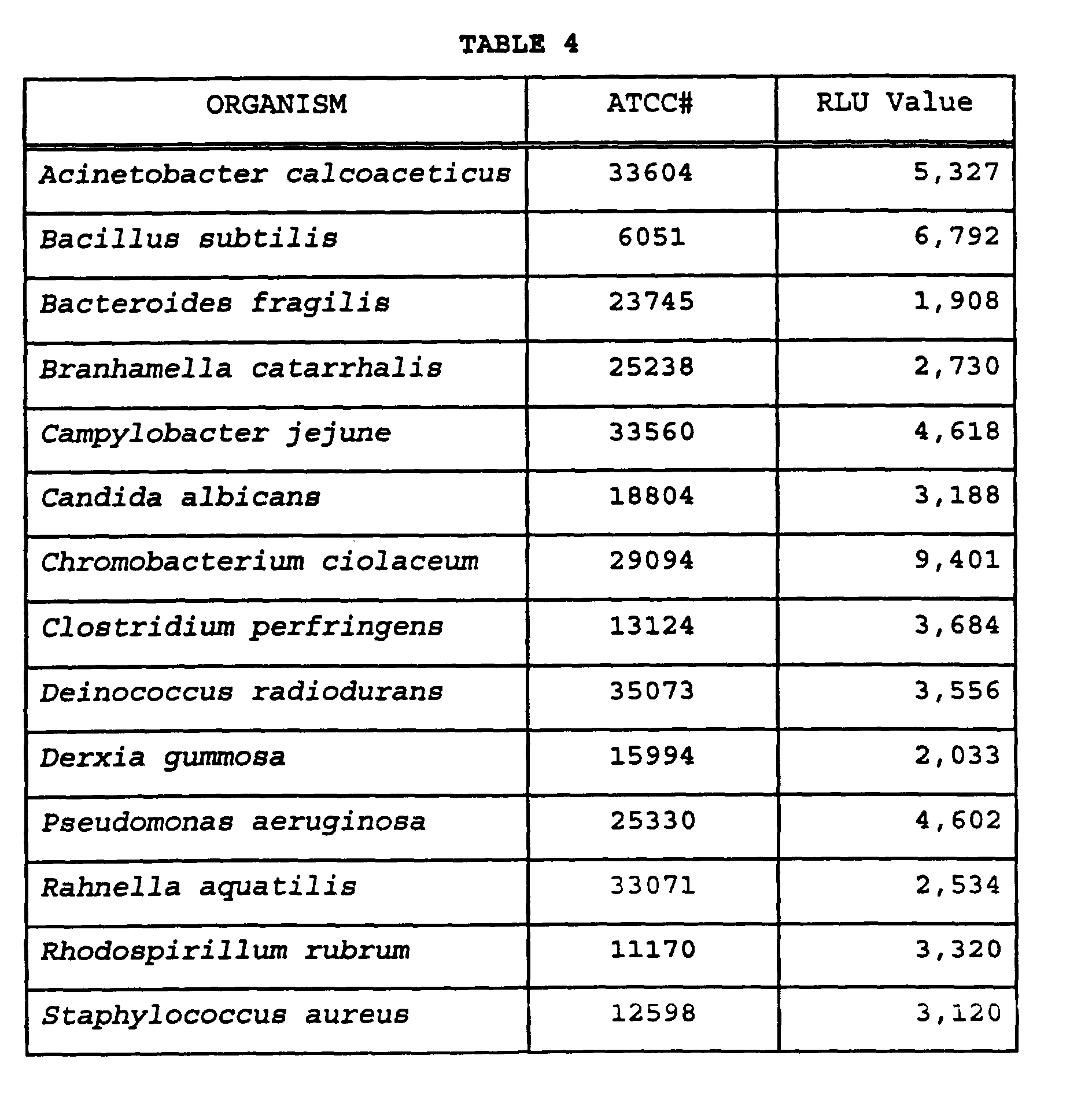

- In this experiment the specificity of a probe mix containing all three designed probes (SEQ ID NO: 7, SEQ ID NO: 8 and SEQ ID NO: 9) and both helper probes (SEQ ID NO: 13 and SEQ ID NO: 14) was tested against standard strains of bacteria. A total of 55 ATCC (American Type Culture Collection) reference strains of mycobacteria were evaluated. These strains represented the most closely related organisms to M. kansasii. Standard specificity testing was performed using growth obtained from actively growing cultures of the ATCC strains, except for Mycobacterium haemophilum, for which cells were not available. Instead, Mycobacterium haemophilum rRNA was used at a concentration equivalent to that available from the growing cell cultures. Cell growth and lysis were as described in Example 2. Hybridization and detection were also as described in Example 2. All closely related mycobacteria produced negative results well below the 30,000 RLU cut-off.
-


- In this experiment the specificity of the mix of probes and helpers used in Example 6 was tested against standard strains of bacteria. A total of 68 ATCC (American Type Culture Collection) reference strains were evaluated. These strains represented a phylogenetic cross section of organisms. Standard specificity testing was performed using growth obtained from actively growing cultures of the ATCC strains. Cell growth and lysis were as described in Example 2. Hybridization and detection were also as described in Example 2. All phylogenetic cross sectional organisms produced negative results well below the 30,000 RLU cut-off.
-


- The mix of hybridization probes (shown in Example 6) was tested for specificity to M. kansasii against 58 clinical isolates representing 7 species of mycobacteria. Cell culture and growth, and hybridization, were as described in Example 2. No cross reactions were observed with closely related clinical isolates.
-

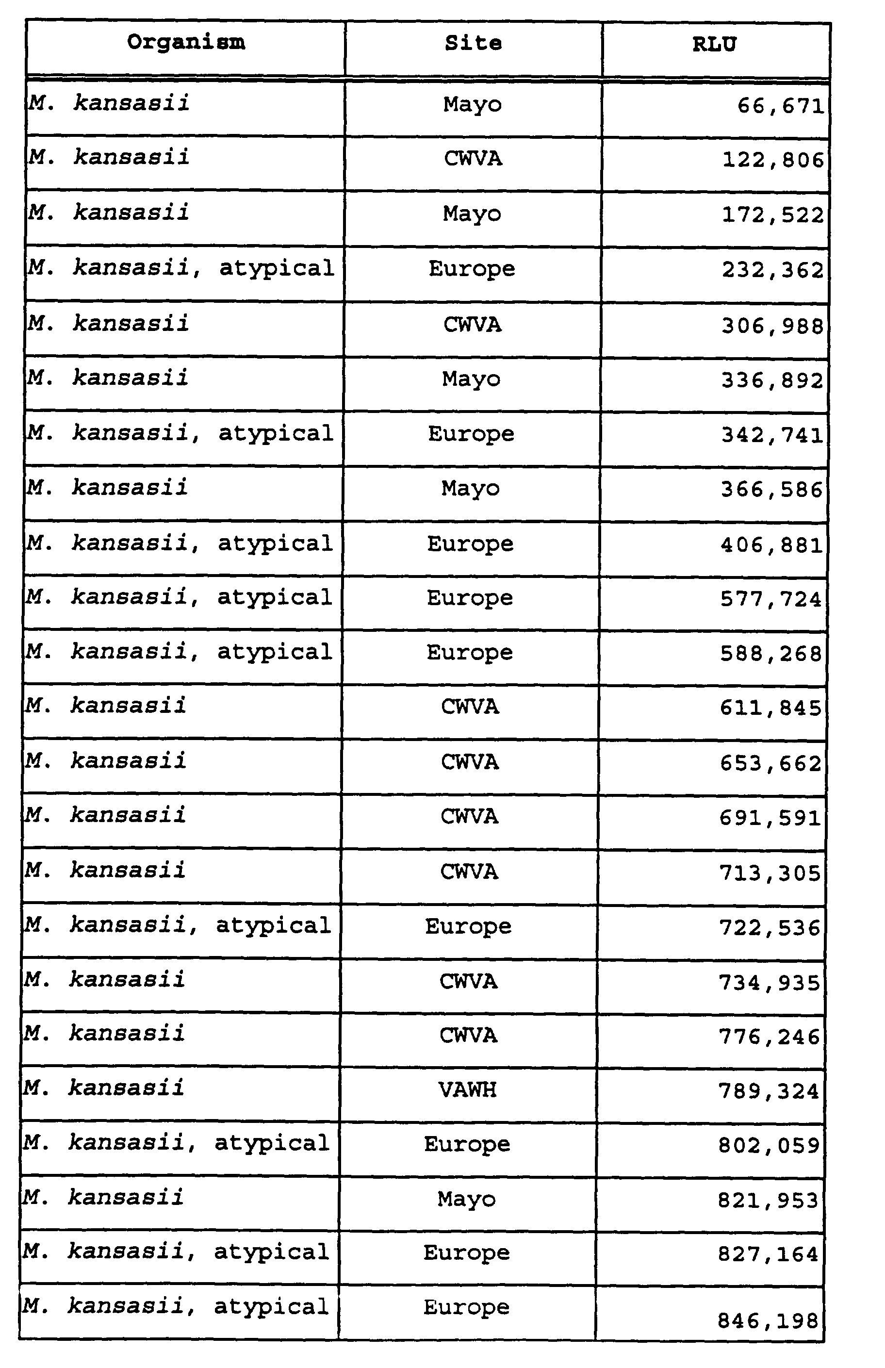

- In this experiment the sensitivity of the probe-helper mix (shown in Example 6) was tested with typical and atypical M. kansasii rRNA. The rRNA was used in concentrations of 0, 0.1, 0.25, 0.5 and 1 ng/µl. Testing was done in duplicate for each concentration and type of rRNA. 100 µl of rRNA from either the typical strain of M. kansasii, the BOV atypical strain, or the COU atypical strain was added to tubes containing lyophilized probe and hybridization reagents as described in Example 2. Reactions were vortexed and hybridized at 60°C for 15 minutes. Detection was as described in Example 2. The results show the probes are sensitive and capable of detecting low level amounts of M. kansasii typical and atypical rRNA.
-
TABLE 8 RLU Rep 1 RLU Rep 2 Mean RLU Net Mean RLU 0 ng typical rRNA 710 10 ng typical rRNA 22,467 20,954 21,711 21,001 25 ng typical rRNA 40,802 43,360 42,081 41,371 50 ng typical rRNA 86,300 92,517 89,409 88,699 100 ng typical rRNA 181,211 165,301 173,256 172,546 0 ng BOV rRNA 710 10 ng BOV rRNA 31,871 30,823 31,347 30,637 25 ng BOV rRNA 76,526 72,370 74,448 73,738 50 ng BOV rRNA 136,198 135,937 136,068 135,358 100 ng BOV rRNA 226,046 181,475 203,761 203,051 0 ng COU rRNA 710 10 ng COU rRNA 54,587 49,980 52,284 51,574 25 ng COU rRNA 115,089 120,045 117,567 116,857 50 ng COU rRNA 237,771 220,013 228,892 228,182 100 ng COU rRNA 395,292 375,581 385,436 384,726 - This experiment tested the sensitivity of the probe/helper mix of Example 6 for detecting M. kansasii rRNA in the presence of non-target cells with their rRNA and rDNA. Cells of M. avium, M. gastri, and M. tuberculosis were grown and lysed as described in Example 2. Samples were prepared with the appropriate mix of lysed non-target cells, and M. kansasii rRNA at a range from 0 ng to 100 ng, as indicated, and hybridization and detection were conducted as described in Example 2. The results show good signal recovery in the presence of a large number (around 1.5 x 10⁷) of non-target cells.
- Table 9: Sensitivity of probe/helper mix in the presence of non-target cells.
TABLE 9 100 ng rRNA 50 ng rRNA 25 ng rRNA 10 ng rRNA 1 ng rRNA 0 ng rRNA M. kansasii rRNA alone 187,549 109,790 55,499 21,888 3,018 794 M. kansasii rRNA plus M. avium cells 191,671 101,300 51,249 22,830 3,305 985 percent recovery 102 92 92 104 109 M. kansasii rRNA plus M. gastri cells 171,647 91,744 50,301 20,702 3,056 1,032 percent recovery 92 84 91 95 101 M. kansasii rRNA plus M. tuberculosis cells 199,513 102,465 50,466 21,196 2,267 1,607 percent recovery 106 93 91 97 75 - The embodiments shown in the various examples described above confirm that the oligonucleotides herein described are capable of detecting M. kansasii nucleic acids, and can be used in an assay to distinguish M. kansasii from its known nearest phylogenetic neighbors. None of the examples described herein are intended to limit the present invention to the embodiments of the preceding disclosure; additional embodiments are within the following claims.


Claims (35)
- A nucleic acid hybridization assay probe for the detection of M. kansasii comprising an oligonucleotide having a nucleic acid sequence selected from the group consisting of CGCTCACGCGTGATACGCC (SEQ ID NO: 8), CGCUCACGCGUGAUACGCC (SEQ ID NO: 20),
GGCGTATCACGCGTGAGCG (SEQ ID NO: 17), GGCGUAUCACGCGUGAGCG (SEQ ID NO: 5), and substantially similar variants thereof. - The probe of claim 1 wherein said oligonucleotide is labeled.
- The probe of claim 2 wherein said label is an acridinium ester.
- The probe of claim 1 wherein M. kansasii is strain BOV.
- A nucleic acid hybridization assay probe for the detection of M. kansasii comprising an oligonucleotide having a nucleic acid sequence selected from the group consisting of CGCTTGCACGTGATACGC (SEQ ID NO: 9), CGCTTGCACGTGATACGCC (SEQ ID NO: 10),
CGCUUGCACGUGAUACGC (SEQ ID NO: 21), CGCUUGCACGUGAUACGCC (SEQ ID NO: 22), GGCGTATCACGTGCAAGCG (SEQ ID NO: 18), GGCGUAUCACGUGCAAGCG (SEQ ID NO: 6), and substantially similar variants thereof. - The probe of claim 5 wherein said oligonucleotide is labeled.
- The probe of claim 6 wherein said label is an acridinium ester.
- The probe of claim 5 wherein M. kansasii is strain COU.
- A nucleic acid hybrid formed between the probe of claim 1 and a nucleotide polymer which hybridizes thereto under high stringency hybridization conditions.
- A nucleic acid hybrid formed between the probe of claim 5 and a nucleotide polymer which hybridizes thereto under high stringency hybridization conditions.
- A nucleic acid hybridization assay probe mix for the detection of M. kansasii wherein said probe mix comprises(a) at least one oligonucleotide hybridization assay probe wherein the probe is selected from the group consisting of(i) an oligonucleotide having a nucleic acid sequence selected from the group consisting of CGCTCACGCGTGATACGCC (SEQ ID NO: 8), CGCUCACGCGUGAUACGCC (SEQ ID NO: 20), GGCGTATCACGCGTGAGCG (SEQ ID NO: 17), GGCGUAUCACGCGUGAGCG(SEQ ID NO: 5), and substantially similar variants thereof, and(ii) an oligonucleotide having a nucleic acid sequence selected from the group consisting of CGCTTGCACGTGATACGC (SEQ ID NO: 9), CGCTTGCACGTGATACGCC (SEQ ID NO: 10), CGCUUGCACGUGAUACGC (SEQ ID NO: 21), CGCUUGCACGUGAUACGCC (SEQ ID NO: 22), GGCGTATCACGTGCAAGCG (SEQ ID NO: 18), GGCGUAUCACGUGCAAGCG (SEQ ID NO: 6), and substantially similar variants thereof; and(b) at least one helper probe, wherein said helper probe comprises an oligonucleotide having a nucleic acid sequence selected from the group consisting
 and substantially similar variants thereof.
- The probe of claim 11 wherein said oligonucleotide is labeled.
- The probe of claim 12 wherein said label is an acridinium ester.
- The probe mix of claim 11 wherein said helper probe comprises an oligonucleotide having a nucleic acid sequence selected from the group consisting ofand substantially similar variants thereof.

- The probe mix of claim 11 further comprising an oligonucleotide having a nucleic acid sequence selected from the group consisting of CGCTCGCGCGCGATACGC (SEQ ID NO: 7), CGCUCGCGCGCGAUACGCC (SEQ ID NO: 19), GCGTATCGCGCGCGAGCG (SEQ ID NO: 16), GCGUAUCGCGCGCGAGCG (SEQ ID NO: 4), and substantially similar variants thereof.
- The probe mix of claim 15 wherein said helper probe comprises an oligonucleotide having a nucleic acid sequence selected from the group consisting of
 and substantially similar variants thereof.
- A nucleic acid hybridization assay probe mix for the detection of M. kansasii wherein said probe mix comprises at least one oligonucleotide hybridization assay probe selected from each of at least two of the groups (a), (b) and (c) wherein(a) group (a) comprises the group of oligonucleotides having a nucleic acid sequence selected from the group consisting of CGCTCACGCGTGATACGCC (SEQ ID NO: 8), CGCUCACGCGUGAUACGCC (SEQ ID NO: 20),
GGCGTATCACGCGTGAGCG (SEQ ID NO: 17), GGCGUAUCACGCGUGAGCG (SEQ ID NO: 5), and substantially similar variants thereof,(b) group (b) comprises the group of oligonucleotides having a nucleic acid sequence selected from the group consisting of CGCTTGCACGTGATACGC (SEQ ID NO: 9), CGCTTGCACGTGATACGCC (SEQ ID NO: 10),
CGCUUGCACGUGAUACGC (SEQ ID NO: 21), CGCUUGCACGUGAUACGCC (SEQ ID NO: 22), GGCGTATCACGTGCAAGCG (SEQ ID NO: 18), GGCGUAUCACGUGCAAGCG (SEQ ID NO: 6), and substantially similar variants thereof; and(c) group (c) comprises the group of oligonucleotides having a nucleic acid sequence selected from the group consisting of CGCTCGCGCGCGATACGC (SEQ ID NO: 7), CGCUCGCGCGCGAUACGCC (SEQ ID NO: 19),
GCGTATCGCGCGCGAGCG (SEQ ID NO: 16), GCGUAUCGCGCGCGAGCG (SEQ ID NO: 4), and substantially similar variants thereof. - The probe of claim 17 wherein said oligonucleotide is labeled.
- The probe of claim 18 wherein said label is an acridinium ester.
- The probe mix of claim 17 further comprising at least one helper probe, wherein said helper probe comprises an oligonucleotide having a nucleic acid sequence selected from the group consisting ofand substantially similar variants thereof.

- The probe mix of claim 20 wherein said helper probe comprises an oligonucleotide having a nucleic acid sequence selected from the group consisting ofand substantially similar variants thereof.

- A nucleic acid hybridization assay probe mix for the detection of M. kansasii wherein said probe mix comprises(a) at least one oligonucleotide having a nucleic acid sequence selected from the group consisting of CGCTCACGCGTGATACGCC (SEQ ID NO: 8),
CGCUCACGCGUGAUACGCC (SEQ ID NO: 20), GGCGTATCACGCGTGAGCG (SEQ ID NO: 17), GGCGUAUCACGCGUGAGCG (SEQ ID NO: 5), and substantially similar variants thereof, and(b) at least one oligonucleotide having a nucleic acid sequence selected from the group consisting of CGCTTGCACGTGATACGC (SEQ ID NO: 9),
CGCTTGCACGTGATACGCC (SEQ ID NO: 10), CGCUUGCACGUGAUACGC (SEQ ID NO: 21), CGCUUGCACGUGAUACGCC (SEQ ID NO: 22), GGCGTATCACGTGCAAGCG (SEQ ID NO: 18), GGCGUAUCACGUGCAAGCG (SEQ ID NO: 6), and substantially similar variants thereof; and(c) at least one oligonucleotide having a nucleic acid sequence selected from the group consisting of CGCTCGCGCGCGATACGC (SEQ ID NO: 7),
CGCUCGCGCGCGAUACGCC (SEQ ID NO: 19), GCGUAUCGCGCGCGAGCG (SEQ ID NO: 4), GCGTATCGCGCGCGAGCG (SEQ ID NO: 16), and substantially similar variants thereof
wherein said probe mix does not form a detectable target-probe duplex under high stringency hybridization conditions with nucleic acids derived from Mycobacterium avium, Mycobacterium chelonae, Mycobacterium fortuitum, Mycobacterium gastri, Mycobacterium gordonae, Mycobacterium haemophilum, Mycobacterium intracellulare, Mycobacterium scrofulacelum, Mycobacterium simiae, and Mycobacterium tuberculosis. - The probe of claim 22 wherein said oligonucleotide is labeled.
- The probe of claim 23 wherein said label is an acridinium ester.
- The probe mix of claim 22 further comprising at least one helper probe, wherein said helper probe comprises an oligonucleotide having a nucleic acid sequence selected from the group consisting of
 and substantially similar variants thereof.
- The probe mix of claim 25 wherein said helper probe comprises an oligonucleotide having a nucleic acid sequence selected from the group consisting ofand substantially similar variants thereof.

- A nucleic acid hybridization assay probe for the detection of M. kansasii comprising an oligonucleotide which is between 10 and about 100 nucleotide bases in length and which is capable of hybridizing to a M. kansasii nucleic acid target region to form a detectable target-probe duplex under high stringency hybridization conditions,
said M. kansasii nucleic acid target region selected from the group consisting ofwherein said probe does not form a detectable target-probe duplex under said high stringency hybridization conditions with nucleic acids derived from Mycobacterium avium, Mycobacterium chelonae, Mycobacterium fortuitum, Mycobacterium gastri, Mycobacterium gordonae, Mycobacterium haemophilum, Mycobacterium intracellulare, Mycobacterium scrofulacelum, Mycobacterium simiae, or Mycobacterium tuberculosis.
- The probe of claim 27 wherein said oligonucleotide is labeled.
- The probe of claim 28 wherein said label is an acridinium ester.
- The probe of claim 27 wherein said oligonucleotide comprises between 10 and about 50 bases.
- The probe of claim 27 wherein said probe comprises a nucleic acid sequence which is at least 80% complementary to a region of 10 contiguous nucleotides in one of said M. kansasii nucleic acid target regions.
- The probe of claim 27 wherein said probe comprises a nucleic acid sequence which is at least 75% complementary to any one of said target regions, and completely complementary to the nucleotide core sequence selected from the group consisting of GCGCGCG (SEQ ID NO: 28), ACGCGUG (SEQ ID NO: 29), ACGUGCG (SEQ ID NO: 30), CGCGCGC (SEQ ID NO: 31), CACGCGU (SEQ ID NO: 32) and CGCACGU (SEQ ID NO: 33).
- A detectable nucleic acid hybrid formed under the stringent hybridization conditions of claim 27 between the probe of claim 27 and a target nucleic acid comprising said M. kansasii nucleic acid target region.
- A method for detecting M. kansasii comprising the steps of(1) providing at least one probe of claim 27, and(2) detecting said target-probe duplex as an indication of the presence of M. kansasii.
- A kit for the detection of M. kansasii comprising at least one probe of claim 27.
Applications Claiming Priority (2)
| Application Number | Priority Date | Filing Date | Title |
|---|---|---|---|
| US1585296P | 1996-05-22 | 1996-05-22 | |
| US15852P | 1996-05-22 |
Publications (3)
| Publication Number | Publication Date |
|---|---|
| EP0808907A2 true EP0808907A2 (en) | 1997-11-26 |
| EP0808907A3 EP0808907A3 (en) | 1998-01-07 |
| EP0808907B1 EP0808907B1 (en) | 2006-01-04 |
Family
ID=21773998
Family Applications (1)
| Application Number | Title | Priority Date | Filing Date |
|---|---|---|---|
| EP97303491A Expired - Lifetime EP0808907B1 (en) | 1996-05-22 | 1997-05-22 | Compositions and methods for the detection of mycobacterium kansasii |
Country Status (10)
| Country | Link |
|---|---|
| US (1) | US6218107B1 (en) |
| EP (1) | EP0808907B1 (en) |
| JP (1) | JP4241929B2 (en) |
| AT (1) | ATE315102T1 (en) |
| AU (1) | AU710996B2 (en) |
| CA (1) | CA2255624C (en) |
| DE (1) | DE69735046T2 (en) |
| DK (1) | DK0808907T3 (en) |
| ES (1) | ES2256871T3 (en) |
| WO (1) | WO1997044488A2 (en) |
Cited By (3)
| Publication number | Priority date | Publication date | Assignee | Title |
|---|---|---|---|---|
| WO2000066788A3 (en) * | 1999-05-03 | 2001-04-05 | Gen Probe Inc | Polynucleotide probes for detection and quantitation of staphylococcus |
| US6235484B1 (en) | 1999-05-03 | 2001-05-22 | Gen-Probe Incorporated | Polynucleotide probes for detection and quantitation of actinomycetes |
| US6821770B1 (en) | 1999-05-03 | 2004-11-23 | Gen-Probe Incorporated | Polynucleotide matrix-based method of identifying microorganisms |
Families Citing this family (7)
| Publication number | Priority date | Publication date | Assignee | Title |
|---|---|---|---|---|
| US7070925B1 (en) | 1996-07-16 | 2006-07-04 | Gen-Probe Incorporated | Method for determining the presence of an RNA analyte in a sample using a modified oligonucleotide probe |
| ATE339514T1 (en) | 1996-07-16 | 2006-10-15 | Gen Probe Inc | METHOD FOR DETECTION AND AMPLIFICATION OF NUCLEIC ACID SEQUENCES USING MODIFIED OLIGONUCLEOTIDES WITH INCREASED TARGET MELTING TEMPERATURE (TM) |
| DE10021947A1 (en) * | 2000-05-05 | 2001-11-08 | Max Planck Gesellschaft | Detecting nucleic acid by in situ hybridization, useful for detecting microorganisms, with addition of helper probes to increase target accessibility |
| DE10237284A1 (en) * | 2002-08-14 | 2004-03-04 | Advalytix Ag | nucleic acid detection |
| GB0526033D0 (en) | 2005-12-21 | 2006-02-01 | Bioeos Ltd | Method |
| JP6650944B2 (en) | 2014-12-23 | 2020-02-19 | スリーエム イノベイティブ プロパティズ カンパニー | Compositions for reducing inhibition of nucleic acid amplification |
| WO2017176469A1 (en) * | 2016-04-08 | 2017-10-12 | 3M Innovative Properties Company | Process for cell lysis and nucleic acid amplification |
Family Cites Families (16)
| Publication number | Priority date | Publication date | Assignee | Title |
|---|---|---|---|---|
| DE3486467T3 (en) | 1983-01-10 | 2004-10-14 | Gen-Probe Inc., San Diego | Methods for the detection, identification and quantification of organisms and viruses |
| US4683195A (en) | 1986-01-30 | 1987-07-28 | Cetus Corporation | Process for amplifying, detecting, and/or-cloning nucleic acid sequences |
| ES2112824T5 (en) | 1986-11-24 | 2008-03-01 | Gen-Probe Incorporated | NUCLEIC ACID PROBES FOR DETECTION AND / OR QUANTIFICATION OF NON-VIRAL ORGANISMS. |
| IL86724A (en) | 1987-06-19 | 1995-01-24 | Siska Diagnostics Inc | Method and kits for the amplification and detection of nucleic acid sequences |
| WO1989001050A1 (en) | 1987-07-31 | 1989-02-09 | The Board Of Trustees Of The Leland Stanford Junior University | Selective amplification of target polynucleotide sequences |
| PT88550A (en) | 1987-09-21 | 1989-07-31 | Ml Tecnology Ventures Lp | PROCESS FOR THE PREPARATION OF NON-NUCLEOTIDIC LIGACATION REAGENTS FOR NUCLEOTIDIAL PROBES |
| US5283174A (en) | 1987-09-21 | 1994-02-01 | Gen-Probe, Incorporated | Homogenous protection assay |
| US5185439A (en) | 1987-10-05 | 1993-02-09 | Gen-Probe Incorporated | Acridinium ester labelling and purification of nucleotide probes |
| US5030557A (en) | 1987-11-24 | 1991-07-09 | Ml Technology Venture | Means and method for enhancing nucleic acid hybridization |
| WO1991001384A1 (en) | 1989-07-11 | 1991-02-07 | Gen-Probe, Incorporated | Nucleic acid sequence amplification methods utilizing a transcription complex |
| US5681698A (en) * | 1991-04-25 | 1997-10-28 | Gen-Probe Incorporated | 23S rRNA nucleic acid probes to mycobacterium kansasii |
| AU681082B2 (en) | 1992-05-06 | 1997-08-21 | Gen-Probe Incorporated | Nucleic acid sequence amplification method, composition and kit |
| AU670116B2 (en) | 1992-08-04 | 1996-07-04 | Gen-Probe Incorporated | Nucleic acid sequence amplification |
| US5518884A (en) * | 1994-02-28 | 1996-05-21 | Becton, Dickinson And Company | Nucleic acid sequences specific for mycbacterium kansasii |
| US5500341A (en) * | 1994-09-19 | 1996-03-19 | Becton, Dickinson And Company | Species-specific detection of Mycobacterium kansasii |
| US5614390A (en) | 1996-06-14 | 1997-03-25 | Becton, Dickinson And Company | Species-specific detection of Mycobacterium kansasii |
-
1997
- 1997-05-15 US US08/856,963 patent/US6218107B1/en not_active Expired - Lifetime
- 1997-05-15 AU AU31329/97A patent/AU710996B2/en not_active Ceased
- 1997-05-15 JP JP54263497A patent/JP4241929B2/en not_active Expired - Fee Related
- 1997-05-15 CA CA002255624A patent/CA2255624C/en not_active Expired - Fee Related
- 1997-05-15 WO PCT/US1997/008486 patent/WO1997044488A2/en not_active Ceased
- 1997-05-22 AT AT97303491T patent/ATE315102T1/en not_active IP Right Cessation
- 1997-05-22 ES ES97303491T patent/ES2256871T3/en not_active Expired - Lifetime
- 1997-05-22 EP EP97303491A patent/EP0808907B1/en not_active Expired - Lifetime
- 1997-05-22 DK DK97303491T patent/DK0808907T3/en active
- 1997-05-22 DE DE69735046T patent/DE69735046T2/en not_active Expired - Lifetime
Cited By (6)
| Publication number | Priority date | Publication date | Assignee | Title |
|---|---|---|---|---|
| WO2000066788A3 (en) * | 1999-05-03 | 2001-04-05 | Gen Probe Inc | Polynucleotide probes for detection and quantitation of staphylococcus |
| US6235484B1 (en) | 1999-05-03 | 2001-05-22 | Gen-Probe Incorporated | Polynucleotide probes for detection and quantitation of actinomycetes |
| WO2000066786A3 (en) * | 1999-05-03 | 2001-07-12 | Gen Probe Inc | Polynucleotide probes for detection and quantitation of actinomycetes |
| US6376186B1 (en) | 1999-05-03 | 2002-04-23 | Gen-Probe Incorporated | Polynucleotide probes for detection and quantitation of staphylococcus |
| US6821770B1 (en) | 1999-05-03 | 2004-11-23 | Gen-Probe Incorporated | Polynucleotide matrix-based method of identifying microorganisms |
| US7449328B2 (en) | 1999-05-03 | 2008-11-11 | Gen-Probe Incorporated | Probe matrix-based device for identifying microorganisms |
Also Published As
| Publication number | Publication date |
|---|---|
| EP0808907A3 (en) | 1998-01-07 |
| AU3132997A (en) | 1997-12-09 |
| CA2255624A1 (en) | 1997-11-27 |
| US6218107B1 (en) | 2001-04-17 |
| WO1997044488A3 (en) | 1997-12-24 |
| DE69735046D1 (en) | 2006-03-30 |
| EP0808907B1 (en) | 2006-01-04 |
| WO1997044488A2 (en) | 1997-11-27 |
| AU710996B2 (en) | 1999-10-07 |
| JP4241929B2 (en) | 2009-03-18 |
| DE69735046T2 (en) | 2006-08-17 |
| JP2000511058A (en) | 2000-08-29 |
| ES2256871T3 (en) | 2006-07-16 |
| ATE315102T1 (en) | 2006-02-15 |
| DK0808907T3 (en) | 2006-05-22 |
| CA2255624C (en) | 2007-09-04 |
Similar Documents
| Publication | Publication Date | Title |
|---|---|---|
| US8628926B2 (en) | Probe and primer for tubercle bacillus detection, and method of detecting human tubercle bacillus therewith | |
| JP4859325B2 (en) | Polynucleotide probes for detection and quantification of Candida species | |
| EP0858514B1 (en) | Improved is6110 based molecular detection of mycobacterium tuberculosis | |
| EP0709467A2 (en) | Compositions and methods for the detection of chlamydia trachomatis | |
| AU2001259264A1 (en) | Polynucleotide probes for detection and quantitation of candida species | |
| EP0808907A2 (en) | Compositions and methods for the detection of mycobacterium kansasii | |
| US20040110129A1 (en) | Multiplex hybridization system for identification of pathogenic mycobacterium and method of use | |
| EP0651057A1 (en) | Nucleic acid probes to mycobacterium avium complex | |
| JPH09501566A (en) | Nucleic acid probe for ureaplasma | |
| KR101425149B1 (en) | Improved method for diagnosing Mycobacterium tuberculosis using one-tube nested real-time PCR | |
| JP4956699B2 (en) | Polynucleotide probes for detection and quantification of Candida albicans and Candida dubliniensis | |
| KR19990022595A (en) | Nucleic Acid Probes and Amplified Oligonucleotides for Neisseria Species | |
| AU2001259329A1 (en) | Polynucleotide probes for detection and quantitation of candida albicans and candida dubliniensis | |
| US6630302B1 (en) | Methods and compositions for determining species of bacteria and fungi | |
| EP4130265A1 (en) | Method for determining single base mutation of erm (41) gene of acid-fast bacillus belonging to mycobacteroides abscessus complex, and primer set and probe used in said method | |
| US7074568B2 (en) | Molecular diagnosis of atypical mycobacterial infections | |
| HK1150864A (en) | Probe and primer for tubercle bacillus detection, and method of detecting human tubercle bacillus therewith |
Legal Events
| Date | Code | Title | Description |
|---|---|---|---|
| PUAI | Public reference made under article 153(3) epc to a published international application that has entered the european phase |
Free format text: ORIGINAL CODE: 0009012 |
|
| PUAL | Search report despatched |
Free format text: ORIGINAL CODE: 0009013 |
|
| AK | Designated contracting states |
Kind code of ref document: A2 Designated state(s): AT BE CH DE DK ES FR GB IT LI LU NL SE |
|
| AK | Designated contracting states |
Kind code of ref document: A3 Designated state(s): AT BE CH DE DK ES FR GB IT LI LU NL SE |
|
| 17P | Request for examination filed |
Effective date: 19980401 |
|
| 17Q | First examination report despatched |
Effective date: 20010328 |
|
| GRAP | Despatch of communication of intention to grant a patent |
Free format text: ORIGINAL CODE: EPIDOSNIGR1 |
|
| GRAS | Grant fee paid |
Free format text: ORIGINAL CODE: EPIDOSNIGR3 |
|
| GRAA | (expected) grant |
Free format text: ORIGINAL CODE: 0009210 |
|
| AK | Designated contracting states |
Kind code of ref document: B1 Designated state(s): AT BE CH DE DK ES FR GB IT LI LU NL SE |
|
| REG | Reference to a national code |
Ref country code: GB Ref legal event code: FG4D |
|
| REG | Reference to a national code |
Ref country code: CH Ref legal event code: EP |
|
| REG | Reference to a national code |
Ref country code: SE Ref legal event code: TRGR |
|
| REF | Corresponds to: |
Ref document number: 69735046 Country of ref document: DE Date of ref document: 20060330 Kind code of ref document: P |
|
| PG25 | Lapsed in a contracting state [announced via postgrant information from national office to epo] |
Ref country code: GB Free format text: LAPSE BECAUSE OF NON-PAYMENT OF DUE FEES Effective date: 20060522 |
|
| REG | Reference to a national code |
Ref country code: DK Ref legal event code: T3 |
|
| REG | Reference to a national code |
Ref country code: ES Ref legal event code: FG2A Ref document number: 2256871 Country of ref document: ES Kind code of ref document: T3 |
|
| ET | Fr: translation filed | ||
| PLBE | No opposition filed within time limit |
Free format text: ORIGINAL CODE: 0009261 |
|
| STAA | Information on the status of an ep patent application or granted ep patent |
Free format text: STATUS: NO OPPOSITION FILED WITHIN TIME LIMIT |
|
| 26N | No opposition filed |
Effective date: 20061005 |
|
| GBPC | Gb: european patent ceased through non-payment of renewal fee |
Effective date: 20060522 |
|
| REG | Reference to a national code |
Ref country code: GB Ref legal event code: 728V |
|
| REG | Reference to a national code |
Ref country code: CH Ref legal event code: PFA Owner name: GEN-PROBE INCORPORATED Free format text: GEN-PROBE INCORPORATED#9880 CAMPUS POINT DRIVE#SAN DIEGO, CALIFORNIA 92121 (US) -TRANSFER TO- GEN-PROBE INCORPORATED#9880 CAMPUS POINT DRIVE#SAN DIEGO, CALIFORNIA 92121 (US) |
|
| REG | Reference to a national code |
Ref country code: GB Ref legal event code: 728Y |
|
| PGFP | Annual fee paid to national office [announced via postgrant information from national office to epo] |
Ref country code: LU Payment date: 20080508 Year of fee payment: 12 Ref country code: DK Payment date: 20080424 Year of fee payment: 12 Ref country code: CH Payment date: 20080425 Year of fee payment: 12 |
|
| PGFP | Annual fee paid to national office [announced via postgrant information from national office to epo] |
Ref country code: AT Payment date: 20080407 Year of fee payment: 12 |
|
| PGFP | Annual fee paid to national office [announced via postgrant information from national office to epo] |
Ref country code: SE Payment date: 20080505 Year of fee payment: 12 |
|
| PGFP | Annual fee paid to national office [announced via postgrant information from national office to epo] |
Ref country code: NL Payment date: 20090527 Year of fee payment: 13 |
|
| PGFP | Annual fee paid to national office [announced via postgrant information from national office to epo] |
Ref country code: BE Payment date: 20090619 Year of fee payment: 13 |
|
| REG | Reference to a national code |
Ref country code: CH Ref legal event code: PL |
|
| REG | Reference to a national code |
Ref country code: DK Ref legal event code: EBP |
|
| PG25 | Lapsed in a contracting state [announced via postgrant information from national office to epo] |
Ref country code: LI Free format text: LAPSE BECAUSE OF NON-PAYMENT OF DUE FEES Effective date: 20090531 Ref country code: CH Free format text: LAPSE BECAUSE OF NON-PAYMENT OF DUE FEES Effective date: 20090531 Ref country code: AT Free format text: LAPSE BECAUSE OF NON-PAYMENT OF DUE FEES Effective date: 20090522 |
|
| PG25 | Lapsed in a contracting state [announced via postgrant information from national office to epo] |
Ref country code: DK Free format text: LAPSE BECAUSE OF NON-PAYMENT OF DUE FEES Effective date: 20090531 |
|
| PGFP | Annual fee paid to national office [announced via postgrant information from national office to epo] |
Ref country code: FR Payment date: 20100525 Year of fee payment: 14 Ref country code: ES Payment date: 20100514 Year of fee payment: 14 |
|
| PGFP | Annual fee paid to national office [announced via postgrant information from national office to epo] |
Ref country code: IT Payment date: 20100517 Year of fee payment: 14 Ref country code: DE Payment date: 20100531 Year of fee payment: 14 |
|
| BERE | Be: lapsed |
Owner name: *GEN-PROBE INC. Effective date: 20100531 |
|
| PGFP | Annual fee paid to national office [announced via postgrant information from national office to epo] |
Ref country code: GB Payment date: 20100401 Year of fee payment: 14 |
|
| REG | Reference to a national code |
Ref country code: NL Ref legal event code: V1 Effective date: 20101201 |
|
| PG25 | Lapsed in a contracting state [announced via postgrant information from national office to epo] |
Ref country code: NL Free format text: LAPSE BECAUSE OF NON-PAYMENT OF DUE FEES Effective date: 20101201 Ref country code: BE Free format text: LAPSE BECAUSE OF NON-PAYMENT OF DUE FEES Effective date: 20100531 |
|
| PG25 | Lapsed in a contracting state [announced via postgrant information from national office to epo] |
Ref country code: LU Free format text: LAPSE BECAUSE OF NON-PAYMENT OF DUE FEES Effective date: 20090522 |
|
| PG25 | Lapsed in a contracting state [announced via postgrant information from national office to epo] |
Ref country code: SE Free format text: LAPSE BECAUSE OF NON-PAYMENT OF DUE FEES Effective date: 20090523 |
|
| REG | Reference to a national code |
Ref country code: DE Ref legal event code: R119 Ref document number: 69735046 Country of ref document: DE |
|
| REG | Reference to a national code |
Ref country code: DE Ref legal event code: R119 Ref document number: 69735046 Country of ref document: DE |
|
| GBPC | Gb: european patent ceased through non-payment of renewal fee |
Effective date: 20110522 |
|
| REG | Reference to a national code |
Ref country code: FR Ref legal event code: ST Effective date: 20120131 |
|
| PG25 | Lapsed in a contracting state [announced via postgrant information from national office to epo] |
Ref country code: IT Free format text: LAPSE BECAUSE OF NON-PAYMENT OF DUE FEES Effective date: 20110522 |
|
| PG25 | Lapsed in a contracting state [announced via postgrant information from national office to epo] |
Ref country code: FR Free format text: LAPSE BECAUSE OF NON-PAYMENT OF DUE FEES Effective date: 20110531 |
|
| PG25 | Lapsed in a contracting state [announced via postgrant information from national office to epo] |
Ref country code: GB Free format text: LAPSE BECAUSE OF NON-PAYMENT OF DUE FEES Effective date: 20110522 |
|
| REG | Reference to a national code |
Ref country code: ES Ref legal event code: FD2A Effective date: 20120717 |
|
| PG25 | Lapsed in a contracting state [announced via postgrant information from national office to epo] |
Ref country code: ES Free format text: LAPSE BECAUSE OF NON-PAYMENT OF DUE FEES Effective date: 20110523 |
|
| PG25 | Lapsed in a contracting state [announced via postgrant information from national office to epo] |
Ref country code: DE Free format text: LAPSE BECAUSE OF NON-PAYMENT OF DUE FEES Effective date: 20111130 |
|
| P01 | Opt-out of the competence of the unified patent court (upc) registered |
Effective date: 20230529 |