EP0808902A2 - Novel genes encoding antiviral proteins of Phytolacca insularis Nakai and recombinant microorganisms expressing the same proteins - Google Patents
Novel genes encoding antiviral proteins of Phytolacca insularis Nakai and recombinant microorganisms expressing the same proteins Download PDFInfo
- Publication number
- EP0808902A2 EP0808902A2 EP96307159A EP96307159A EP0808902A2 EP 0808902 A2 EP0808902 A2 EP 0808902A2 EP 96307159 A EP96307159 A EP 96307159A EP 96307159 A EP96307159 A EP 96307159A EP 0808902 A2 EP0808902 A2 EP 0808902A2
- Authority
- EP
- European Patent Office
- Prior art keywords
- protein
- gene
- gpip50
- gpip2
- proteins
- Prior art date
- Legal status (The legal status is an assumption and is not a legal conclusion. Google has not performed a legal analysis and makes no representation as to the accuracy of the status listed.)
- Withdrawn
Links
- 108090000623 proteins and genes Proteins 0.000 title claims abstract description 203
- 102000004169 proteins and genes Human genes 0.000 title claims abstract description 95
- 230000000840 anti-viral effect Effects 0.000 title claims abstract description 48
- 241000894747 Phytolacca insularis Species 0.000 title abstract description 27
- 244000005700 microbiome Species 0.000 title abstract description 11
- 239000013604 expression vector Substances 0.000 claims abstract description 34
- 238000000034 method Methods 0.000 claims abstract description 10
- 230000008569 process Effects 0.000 claims abstract description 6
- 125000003275 alpha amino acid group Chemical group 0.000 claims abstract 4
- 239000002773 nucleotide Substances 0.000 claims description 26
- 125000003729 nucleotide group Chemical group 0.000 claims description 26
- 241000588724 Escherichia coli Species 0.000 claims description 24
- BPHPUYQFMNQIOC-NXRLNHOXSA-N isopropyl beta-D-thiogalactopyranoside Chemical compound CC(C)S[C@@H]1O[C@H](CO)[C@H](O)[C@H](O)[C@H]1O BPHPUYQFMNQIOC-NXRLNHOXSA-N 0.000 claims description 8
- 210000003000 inclusion body Anatomy 0.000 claims description 6
- 238000004519 manufacturing process Methods 0.000 claims description 6
- FWMNVWWHGCHHJJ-SKKKGAJSSA-N 4-amino-1-[(2r)-6-amino-2-[[(2r)-2-[[(2r)-2-[[(2r)-2-amino-3-phenylpropanoyl]amino]-3-phenylpropanoyl]amino]-4-methylpentanoyl]amino]hexanoyl]piperidine-4-carboxylic acid Chemical compound C([C@H](C(=O)N[C@H](CC(C)C)C(=O)N[C@H](CCCCN)C(=O)N1CCC(N)(CC1)C(O)=O)NC(=O)[C@H](N)CC=1C=CC=CC=1)C1=CC=CC=C1 FWMNVWWHGCHHJJ-SKKKGAJSSA-N 0.000 claims description 4
- 238000012258 culturing Methods 0.000 claims 1
- 238000003306 harvesting Methods 0.000 claims 1
- 230000001939 inductive effect Effects 0.000 claims 1
- 230000002934 lysing effect Effects 0.000 claims 1
- 239000013598 vector Substances 0.000 abstract description 21
- 241000196324 Embryophyta Species 0.000 abstract description 20
- 208000030507 AIDS Diseases 0.000 abstract description 4
- 206010028980 Neoplasm Diseases 0.000 abstract description 4
- 201000011510 cancer Diseases 0.000 abstract description 4
- 229940127121 immunoconjugate Drugs 0.000 abstract description 4
- 238000011282 treatment Methods 0.000 abstract description 4
- 239000004480 active ingredient Substances 0.000 abstract description 3
- 239000003443 antiviral agent Substances 0.000 abstract description 3
- 235000018102 proteins Nutrition 0.000 description 80
- 150000001413 amino acids Chemical group 0.000 description 45
- 239000006228 supernatant Substances 0.000 description 36
- 108020004414 DNA Proteins 0.000 description 33
- 239000012634 fragment Substances 0.000 description 24
- 230000014616 translation Effects 0.000 description 23
- 235000001014 amino acid Nutrition 0.000 description 18
- 108090000829 Ribosome Inactivating Proteins Proteins 0.000 description 15
- 238000013519 translation Methods 0.000 description 13
- 210000004027 cell Anatomy 0.000 description 11
- KFZMGEQAYNKOFK-UHFFFAOYSA-N Isopropanol Chemical compound CC(C)O KFZMGEQAYNKOFK-UHFFFAOYSA-N 0.000 description 10
- 238000001243 protein synthesis Methods 0.000 description 10
- 238000010276 construction Methods 0.000 description 9
- 238000000338 in vitro Methods 0.000 description 9
- HEDRZPFGACZZDS-UHFFFAOYSA-N Chloroform Chemical compound ClC(Cl)Cl HEDRZPFGACZZDS-UHFFFAOYSA-N 0.000 description 8
- ISWSIDIOOBJBQZ-UHFFFAOYSA-N Phenol Chemical compound OC1=CC=CC=C1 ISWSIDIOOBJBQZ-UHFFFAOYSA-N 0.000 description 8
- FAPWRFPIFSIZLT-UHFFFAOYSA-M Sodium chloride Chemical compound [Na+].[Cl-] FAPWRFPIFSIZLT-UHFFFAOYSA-M 0.000 description 8
- 241000700605 Viruses Species 0.000 description 8
- 108091008146 restriction endonucleases Proteins 0.000 description 8
- 238000006467 substitution reaction Methods 0.000 description 8
- 102000007056 Recombinant Fusion Proteins Human genes 0.000 description 7
- 108010008281 Recombinant Fusion Proteins Proteins 0.000 description 7
- 238000003556 assay Methods 0.000 description 7
- 239000000872 buffer Substances 0.000 description 7
- 239000013612 plasmid Substances 0.000 description 7
- KCXVZYZYPLLWCC-UHFFFAOYSA-N EDTA Chemical compound OC(=O)CN(CC(O)=O)CCN(CC(O)=O)CC(O)=O KCXVZYZYPLLWCC-UHFFFAOYSA-N 0.000 description 6
- LFQSCWFLJHTTHZ-UHFFFAOYSA-N Ethanol Chemical compound CCO LFQSCWFLJHTTHZ-UHFFFAOYSA-N 0.000 description 6
- FFEARJCKVFRZRR-BYPYZUCNSA-N L-methionine Chemical compound CSCC[C@H](N)C(O)=O FFEARJCKVFRZRR-BYPYZUCNSA-N 0.000 description 6
- 238000002105 Southern blotting Methods 0.000 description 6
- 238000000246 agarose gel electrophoresis Methods 0.000 description 6
- 230000000694 effects Effects 0.000 description 6
- 238000002955 isolation Methods 0.000 description 6
- 229930182817 methionine Natural products 0.000 description 6
- 108091026890 Coding region Proteins 0.000 description 5
- 238000003780 insertion Methods 0.000 description 5
- 230000037431 insertion Effects 0.000 description 5
- IJGRMHOSHXDMSA-UHFFFAOYSA-N Atomic nitrogen Chemical compound N#N IJGRMHOSHXDMSA-UHFFFAOYSA-N 0.000 description 4
- UXVMQQNJUSDDNG-UHFFFAOYSA-L Calcium chloride Chemical compound [Cl-].[Cl-].[Ca+2] UXVMQQNJUSDDNG-UHFFFAOYSA-L 0.000 description 4
- 108020003215 DNA Probes Proteins 0.000 description 4
- 239000003298 DNA probe Substances 0.000 description 4
- 108700026244 Open Reading Frames Proteins 0.000 description 4
- 108091081024 Start codon Proteins 0.000 description 4
- AIYUHDOJVYHVIT-UHFFFAOYSA-M caesium chloride Chemical compound [Cl-].[Cs+] AIYUHDOJVYHVIT-UHFFFAOYSA-M 0.000 description 4
- 239000001110 calcium chloride Substances 0.000 description 4
- 229910001628 calcium chloride Inorganic materials 0.000 description 4
- 230000000977 initiatory effect Effects 0.000 description 4
- 229960004592 isopropanol Drugs 0.000 description 4
- 239000002244 precipitate Substances 0.000 description 4
- 238000003259 recombinant expression Methods 0.000 description 4
- 239000011780 sodium chloride Substances 0.000 description 4
- 230000009261 transgenic effect Effects 0.000 description 4
- 108010067770 Endopeptidase K Proteins 0.000 description 3
- 239000005089 Luciferase Substances 0.000 description 3
- NIPNSKYNPDTRPC-UHFFFAOYSA-N N-[2-oxo-2-(2,4,6,7-tetrahydrotriazolo[4,5-c]pyridin-5-yl)ethyl]-2-[[3-(trifluoromethoxy)phenyl]methylamino]pyrimidine-5-carboxamide Chemical compound O=C(CNC(=O)C=1C=NC(=NC=1)NCC1=CC(=CC=C1)OC(F)(F)F)N1CC2=C(CC1)NN=N2 NIPNSKYNPDTRPC-UHFFFAOYSA-N 0.000 description 3
- AFCARXCZXQIEQB-UHFFFAOYSA-N N-[3-oxo-3-(2,4,6,7-tetrahydrotriazolo[4,5-c]pyridin-5-yl)propyl]-2-[[3-(trifluoromethoxy)phenyl]methylamino]pyrimidine-5-carboxamide Chemical compound O=C(CCNC(=O)C=1C=NC(=NC=1)NCC1=CC(=CC=C1)OC(F)(F)F)N1CC2=C(CC1)NN=N2 AFCARXCZXQIEQB-UHFFFAOYSA-N 0.000 description 3
- 108091028043 Nucleic acid sequence Proteins 0.000 description 3
- 108010076504 Protein Sorting Signals Proteins 0.000 description 3
- 239000007984 Tris EDTA buffer Substances 0.000 description 3
- 238000003776 cleavage reaction Methods 0.000 description 3
- SUYVUBYJARFZHO-RRKCRQDMSA-N dATP Chemical compound C1=NC=2C(N)=NC=NC=2N1[C@H]1C[C@H](O)[C@@H](COP(O)(=O)OP(O)(=O)OP(O)(O)=O)O1 SUYVUBYJARFZHO-RRKCRQDMSA-N 0.000 description 3
- SUYVUBYJARFZHO-UHFFFAOYSA-N dATP Natural products C1=NC=2C(N)=NC=NC=2N1C1CC(O)C(COP(O)(=O)OP(O)(=O)OP(O)(O)=O)O1 SUYVUBYJARFZHO-UHFFFAOYSA-N 0.000 description 3
- 238000012217 deletion Methods 0.000 description 3
- 230000037430 deletion Effects 0.000 description 3
- 238000000605 extraction Methods 0.000 description 3
- 230000006698 induction Effects 0.000 description 3
- 239000002609 medium Substances 0.000 description 3
- 239000000203 mixture Substances 0.000 description 3
- 239000000523 sample Substances 0.000 description 3
- 230000007017 scission Effects 0.000 description 3
- 238000002415 sodium dodecyl sulfate polyacrylamide gel electrophoresis Methods 0.000 description 3
- XLYOFNOQVPJJNP-UHFFFAOYSA-N water Chemical compound O XLYOFNOQVPJJNP-UHFFFAOYSA-N 0.000 description 3
- VZSRBBMJRBPUNF-UHFFFAOYSA-N 2-(2,3-dihydro-1H-inden-2-ylamino)-N-[3-oxo-3-(2,4,6,7-tetrahydrotriazolo[4,5-c]pyridin-5-yl)propyl]pyrimidine-5-carboxamide Chemical compound C1C(CC2=CC=CC=C12)NC1=NC=C(C=N1)C(=O)NCCC(N1CC2=C(CC1)NN=N2)=O VZSRBBMJRBPUNF-UHFFFAOYSA-N 0.000 description 2
- QKNYBSVHEMOAJP-UHFFFAOYSA-N 2-amino-2-(hydroxymethyl)propane-1,3-diol;hydron;chloride Chemical compound Cl.OCC(N)(CO)CO QKNYBSVHEMOAJP-UHFFFAOYSA-N 0.000 description 2
- USFZMSVCRYTOJT-UHFFFAOYSA-N Ammonium acetate Chemical compound N.CC(O)=O USFZMSVCRYTOJT-UHFFFAOYSA-N 0.000 description 2
- LZZYPRNAOMGNLH-UHFFFAOYSA-M Cetrimonium bromide Chemical compound [Br-].CCCCCCCCCCCCCCCC[N+](C)(C)C LZZYPRNAOMGNLH-UHFFFAOYSA-M 0.000 description 2
- 238000007400 DNA extraction Methods 0.000 description 2
- 108010013369 Enteropeptidase Proteins 0.000 description 2
- 102100029727 Enteropeptidase Human genes 0.000 description 2
- AYFVYJQAPQTCCC-GBXIJSLDSA-N L-threonine Chemical compound C[C@@H](O)[C@H](N)C(O)=O AYFVYJQAPQTCCC-GBXIJSLDSA-N 0.000 description 2
- 108060001084 Luciferase Proteins 0.000 description 2
- 239000006137 Luria-Bertani broth Substances 0.000 description 2
- 101710151833 Movement protein TGBp3 Proteins 0.000 description 2
- 108091034117 Oligonucleotide Proteins 0.000 description 2
- 241000283973 Oryctolagus cuniculus Species 0.000 description 2
- 240000007643 Phytolacca americana Species 0.000 description 2
- 235000009074 Phytolacca americana Nutrition 0.000 description 2
- 101001086866 Sus scrofa Pulmonary surfactant-associated protein B Proteins 0.000 description 2
- XSQUKJJJFZCRTK-UHFFFAOYSA-N Urea Chemical compound NC(N)=O XSQUKJJJFZCRTK-UHFFFAOYSA-N 0.000 description 2
- JLCPHMBAVCMARE-UHFFFAOYSA-N [3-[[3-[[3-[[3-[[3-[[3-[[3-[[3-[[3-[[3-[[3-[[5-(2-amino-6-oxo-1H-purin-9-yl)-3-[[3-[[3-[[3-[[3-[[3-[[5-(2-amino-6-oxo-1H-purin-9-yl)-3-[[5-(2-amino-6-oxo-1H-purin-9-yl)-3-hydroxyoxolan-2-yl]methoxy-hydroxyphosphoryl]oxyoxolan-2-yl]methoxy-hydroxyphosphoryl]oxy-5-(5-methyl-2,4-dioxopyrimidin-1-yl)oxolan-2-yl]methoxy-hydroxyphosphoryl]oxy-5-(6-aminopurin-9-yl)oxolan-2-yl]methoxy-hydroxyphosphoryl]oxy-5-(6-aminopurin-9-yl)oxolan-2-yl]methoxy-hydroxyphosphoryl]oxy-5-(6-aminopurin-9-yl)oxolan-2-yl]methoxy-hydroxyphosphoryl]oxy-5-(6-aminopurin-9-yl)oxolan-2-yl]methoxy-hydroxyphosphoryl]oxyoxolan-2-yl]methoxy-hydroxyphosphoryl]oxy-5-(5-methyl-2,4-dioxopyrimidin-1-yl)oxolan-2-yl]methoxy-hydroxyphosphoryl]oxy-5-(4-amino-2-oxopyrimidin-1-yl)oxolan-2-yl]methoxy-hydroxyphosphoryl]oxy-5-(5-methyl-2,4-dioxopyrimidin-1-yl)oxolan-2-yl]methoxy-hydroxyphosphoryl]oxy-5-(5-methyl-2,4-dioxopyrimidin-1-yl)oxolan-2-yl]methoxy-hydroxyphosphoryl]oxy-5-(6-aminopurin-9-yl)oxolan-2-yl]methoxy-hydroxyphosphoryl]oxy-5-(6-aminopurin-9-yl)oxolan-2-yl]methoxy-hydroxyphosphoryl]oxy-5-(4-amino-2-oxopyrimidin-1-yl)oxolan-2-yl]methoxy-hydroxyphosphoryl]oxy-5-(4-amino-2-oxopyrimidin-1-yl)oxolan-2-yl]methoxy-hydroxyphosphoryl]oxy-5-(4-amino-2-oxopyrimidin-1-yl)oxolan-2-yl]methoxy-hydroxyphosphoryl]oxy-5-(6-aminopurin-9-yl)oxolan-2-yl]methoxy-hydroxyphosphoryl]oxy-5-(4-amino-2-oxopyrimidin-1-yl)oxolan-2-yl]methyl [5-(6-aminopurin-9-yl)-2-(hydroxymethyl)oxolan-3-yl] hydrogen phosphate Polymers Cc1cn(C2CC(OP(O)(=O)OCC3OC(CC3OP(O)(=O)OCC3OC(CC3O)n3cnc4c3nc(N)[nH]c4=O)n3cnc4c3nc(N)[nH]c4=O)C(COP(O)(=O)OC3CC(OC3COP(O)(=O)OC3CC(OC3COP(O)(=O)OC3CC(OC3COP(O)(=O)OC3CC(OC3COP(O)(=O)OC3CC(OC3COP(O)(=O)OC3CC(OC3COP(O)(=O)OC3CC(OC3COP(O)(=O)OC3CC(OC3COP(O)(=O)OC3CC(OC3COP(O)(=O)OC3CC(OC3COP(O)(=O)OC3CC(OC3COP(O)(=O)OC3CC(OC3COP(O)(=O)OC3CC(OC3COP(O)(=O)OC3CC(OC3COP(O)(=O)OC3CC(OC3COP(O)(=O)OC3CC(OC3COP(O)(=O)OC3CC(OC3CO)n3cnc4c(N)ncnc34)n3ccc(N)nc3=O)n3cnc4c(N)ncnc34)n3ccc(N)nc3=O)n3ccc(N)nc3=O)n3ccc(N)nc3=O)n3cnc4c(N)ncnc34)n3cnc4c(N)ncnc34)n3cc(C)c(=O)[nH]c3=O)n3cc(C)c(=O)[nH]c3=O)n3ccc(N)nc3=O)n3cc(C)c(=O)[nH]c3=O)n3cnc4c3nc(N)[nH]c4=O)n3cnc4c(N)ncnc34)n3cnc4c(N)ncnc34)n3cnc4c(N)ncnc34)n3cnc4c(N)ncnc34)O2)c(=O)[nH]c1=O JLCPHMBAVCMARE-UHFFFAOYSA-N 0.000 description 2
- 238000005119 centrifugation Methods 0.000 description 2
- 238000010367 cloning Methods 0.000 description 2
- 230000008021 deposition Effects 0.000 description 2
- 230000029087 digestion Effects 0.000 description 2
- ZMMJGEGLRURXTF-UHFFFAOYSA-N ethidium bromide Chemical compound [Br-].C12=CC(N)=CC=C2C2=CC=C(N)C=C2[N+](CC)=C1C1=CC=CC=C1 ZMMJGEGLRURXTF-UHFFFAOYSA-N 0.000 description 2
- 229960005542 ethidium bromide Drugs 0.000 description 2
- 239000011536 extraction buffer Substances 0.000 description 2
- 230000002068 genetic effect Effects 0.000 description 2
- 238000009396 hybridization Methods 0.000 description 2
- 230000005764 inhibitory process Effects 0.000 description 2
- PHTQWCKDNZKARW-UHFFFAOYSA-N isoamylol Chemical compound CC(C)CCO PHTQWCKDNZKARW-UHFFFAOYSA-N 0.000 description 2
- 239000007788 liquid Substances 0.000 description 2
- 239000006166 lysate Substances 0.000 description 2
- 239000003550 marker Substances 0.000 description 2
- 108020004999 messenger RNA Proteins 0.000 description 2
- 229910052757 nitrogen Inorganic materials 0.000 description 2
- 239000002245 particle Substances 0.000 description 2
- 244000052769 pathogen Species 0.000 description 2
- 230000001717 pathogenic effect Effects 0.000 description 2
- 239000008188 pellet Substances 0.000 description 2
- 239000000047 product Substances 0.000 description 2
- 210000001995 reticulocyte Anatomy 0.000 description 2
- 210000003705 ribosome Anatomy 0.000 description 2
- 238000012163 sequencing technique Methods 0.000 description 2
- 239000007787 solid Substances 0.000 description 2
- MTCFGRXMJLQNBG-REOHCLBHSA-N (2S)-2-Amino-3-hydroxypropansäure Chemical compound OC[C@H](N)C(O)=O MTCFGRXMJLQNBG-REOHCLBHSA-N 0.000 description 1
- YLZOPXRUQYQQID-UHFFFAOYSA-N 3-(2,4,6,7-tetrahydrotriazolo[4,5-c]pyridin-5-yl)-1-[4-[2-[[3-(trifluoromethoxy)phenyl]methylamino]pyrimidin-5-yl]piperazin-1-yl]propan-1-one Chemical compound N1N=NC=2CN(CCC=21)CCC(=O)N1CCN(CC1)C=1C=NC(=NC=1)NCC1=CC(=CC=C1)OC(F)(F)F YLZOPXRUQYQQID-UHFFFAOYSA-N 0.000 description 1
- 108020005029 5' Flanking Region Proteins 0.000 description 1
- FVFVNNKYKYZTJU-UHFFFAOYSA-N 6-chloro-1,3,5-triazine-2,4-diamine Chemical compound NC1=NC(N)=NC(Cl)=N1 FVFVNNKYKYZTJU-UHFFFAOYSA-N 0.000 description 1
- 229930024421 Adenine Natural products 0.000 description 1
- GFFGJBXGBJISGV-UHFFFAOYSA-N Adenine Chemical compound NC1=NC=NC2=C1N=CN2 GFFGJBXGBJISGV-UHFFFAOYSA-N 0.000 description 1
- UIUXXFIKWQVMEX-UFYCRDLUSA-N Arg-Phe-Tyr Chemical compound [H]N[C@@H](CCCNC(N)=N)C(=O)N[C@@H](CC1=CC=CC=C1)C(=O)N[C@@H](CC1=CC=C(O)C=C1)C(O)=O UIUXXFIKWQVMEX-UFYCRDLUSA-N 0.000 description 1
- 241000894006 Bacteria Species 0.000 description 1
- BHPQYMZQTOCNFJ-UHFFFAOYSA-N Calcium cation Chemical compound [Ca+2] BHPQYMZQTOCNFJ-UHFFFAOYSA-N 0.000 description 1
- 240000004244 Cucurbita moschata Species 0.000 description 1
- 235000009854 Cucurbita moschata Nutrition 0.000 description 1
- 102000012410 DNA Ligases Human genes 0.000 description 1
- 108010061982 DNA Ligases Proteins 0.000 description 1
- 102000016928 DNA-directed DNA polymerase Human genes 0.000 description 1
- 108010014303 DNA-directed DNA polymerase Proteins 0.000 description 1
- 241000206602 Eukaryota Species 0.000 description 1
- 241000233866 Fungi Species 0.000 description 1
- MLCPTRRNICEKIS-FXQIFTODSA-N Glu-Asn-Gln Chemical compound [H]N[C@@H](CCC(O)=O)C(=O)N[C@@H](CC(N)=O)C(=O)N[C@@H](CCC(N)=O)C(O)=O MLCPTRRNICEKIS-FXQIFTODSA-N 0.000 description 1
- JRDYDYXZKFNNRQ-XPUUQOCRSA-N Gly-Ala-Val Chemical compound CC(C)[C@@H](C(O)=O)NC(=O)[C@H](C)NC(=O)CN JRDYDYXZKFNNRQ-XPUUQOCRSA-N 0.000 description 1
- 241000238631 Hexapoda Species 0.000 description 1
- OUUCIIJSBIBCHB-ZPFDUUQYSA-N Ile-Leu-Asp Chemical compound CC[C@H](C)[C@H](N)C(=O)N[C@@H](CC(C)C)C(=O)N[C@@H](CC(O)=O)C(O)=O OUUCIIJSBIBCHB-ZPFDUUQYSA-N 0.000 description 1
- ONIBWKKTOPOVIA-BYPYZUCNSA-N L-Proline Chemical compound OC(=O)[C@@H]1CCCN1 ONIBWKKTOPOVIA-BYPYZUCNSA-N 0.000 description 1
- QNAYBMKLOCPYGJ-REOHCLBHSA-N L-alanine Chemical compound C[C@H](N)C(O)=O QNAYBMKLOCPYGJ-REOHCLBHSA-N 0.000 description 1
- 241000219470 Mirabilis Species 0.000 description 1
- 244000022198 Mirabilis jalapa Species 0.000 description 1
- 235000009053 Mirabilis jalapa Nutrition 0.000 description 1
- 108020002230 Pancreatic Ribonuclease Proteins 0.000 description 1
- 102000005891 Pancreatic ribonuclease Human genes 0.000 description 1
- 239000001888 Peptone Substances 0.000 description 1
- 108010080698 Peptones Proteins 0.000 description 1
- ONIBWKKTOPOVIA-UHFFFAOYSA-N Proline Natural products OC(=O)C1CCCN1 ONIBWKKTOPOVIA-UHFFFAOYSA-N 0.000 description 1
- 108020004412 RNA 3' Polyadenylation Signals Proteins 0.000 description 1
- 102100022419 RPA-interacting protein Human genes 0.000 description 1
- 102000006382 Ribonucleases Human genes 0.000 description 1
- 108010083644 Ribonucleases Proteins 0.000 description 1
- 108010039491 Ricin Proteins 0.000 description 1
- 235000004443 Ricinus communis Nutrition 0.000 description 1
- 240000000528 Ricinus communis Species 0.000 description 1
- 240000004808 Saccharomyces cerevisiae Species 0.000 description 1
- PCJLFYBAQZQOFE-KATARQTJSA-N Ser-Thr-Lys Chemical compound C[C@H]([C@@H](C(=O)N[C@@H](CCCCN)C(=O)O)NC(=O)[C@H](CO)N)O PCJLFYBAQZQOFE-KATARQTJSA-N 0.000 description 1
- MTCFGRXMJLQNBG-UHFFFAOYSA-N Serine Natural products OCC(N)C(O)=O MTCFGRXMJLQNBG-UHFFFAOYSA-N 0.000 description 1
- 108020005038 Terminator Codon Proteins 0.000 description 1
- AYFVYJQAPQTCCC-UHFFFAOYSA-N Threonine Natural products CC(O)C(N)C(O)=O AYFVYJQAPQTCCC-UHFFFAOYSA-N 0.000 description 1
- 239000004473 Threonine Substances 0.000 description 1
- 240000006023 Trichosanthes kirilowii Species 0.000 description 1
- 235000009818 Trichosanthes kirilowii Nutrition 0.000 description 1
- 108010021119 Trichosanthin Proteins 0.000 description 1
- 208000036142 Viral infection Diseases 0.000 description 1
- 230000009471 action Effects 0.000 description 1
- 229960000643 adenine Drugs 0.000 description 1
- 235000004279 alanine Nutrition 0.000 description 1
- 239000003513 alkali Substances 0.000 description 1
- AVKUERGKIZMTKX-NJBDSQKTSA-N ampicillin Chemical compound C1([C@@H](N)C(=O)N[C@H]2[C@H]3SC([C@@H](N3C2=O)C(O)=O)(C)C)=CC=CC=C1 AVKUERGKIZMTKX-NJBDSQKTSA-N 0.000 description 1
- 229960000723 ampicillin Drugs 0.000 description 1
- 238000004458 analytical method Methods 0.000 description 1
- 238000000137 annealing Methods 0.000 description 1
- 108010038633 aspartylglutamate Proteins 0.000 description 1
- 238000000211 autoradiogram Methods 0.000 description 1
- 238000000376 autoradiography Methods 0.000 description 1
- 230000001580 bacterial effect Effects 0.000 description 1
- 239000002585 base Substances 0.000 description 1
- 230000015572 biosynthetic process Effects 0.000 description 1
- 210000004899 c-terminal region Anatomy 0.000 description 1
- 239000011575 calcium Substances 0.000 description 1
- 229910001424 calcium ion Inorganic materials 0.000 description 1
- 229940041514 candida albicans extract Drugs 0.000 description 1
- 239000004202 carbamide Substances 0.000 description 1
- 239000013522 chelant Substances 0.000 description 1
- 238000007796 conventional method Methods 0.000 description 1
- 239000000287 crude extract Substances 0.000 description 1
- 210000004748 cultured cell Anatomy 0.000 description 1
- 230000009089 cytolysis Effects 0.000 description 1
- 230000008260 defense mechanism Effects 0.000 description 1
- 238000004925 denaturation Methods 0.000 description 1
- 230000036425 denaturation Effects 0.000 description 1
- 239000005546 dideoxynucleotide Substances 0.000 description 1
- 239000012153 distilled water Substances 0.000 description 1
- 238000001962 electrophoresis Methods 0.000 description 1
- 238000010828 elution Methods 0.000 description 1
- 239000012149 elution buffer Substances 0.000 description 1
- 238000005516 engineering process Methods 0.000 description 1
- 238000002474 experimental method Methods 0.000 description 1
- 239000000499 gel Substances 0.000 description 1
- 238000005227 gel permeation chromatography Methods 0.000 description 1
- 239000007973 glycine-HCl buffer Substances 0.000 description 1
- 108010027668 glycyl-alanyl-valine Proteins 0.000 description 1
- 230000002209 hydrophobic effect Effects 0.000 description 1
- 208000015181 infectious disease Diseases 0.000 description 1
- 230000001524 infective effect Effects 0.000 description 1
- 230000002401 inhibitory effect Effects 0.000 description 1
- 238000002372 labelling Methods 0.000 description 1
- 210000004962 mammalian cell Anatomy 0.000 description 1
- 230000035800 maturation Effects 0.000 description 1
- 230000007246 mechanism Effects 0.000 description 1
- 230000000813 microbial effect Effects 0.000 description 1
- 238000010369 molecular cloning Methods 0.000 description 1
- 108020004707 nucleic acids Proteins 0.000 description 1
- 102000039446 nucleic acids Human genes 0.000 description 1
- 150000007523 nucleic acids Chemical class 0.000 description 1
- 238000004806 packaging method and process Methods 0.000 description 1
- 235000019319 peptone Nutrition 0.000 description 1
- 108700028325 pokeweed antiviral Proteins 0.000 description 1
- 238000003752 polymerase chain reaction Methods 0.000 description 1
- 239000011541 reaction mixture Substances 0.000 description 1
- 230000010076 replication Effects 0.000 description 1
- 230000004044 response Effects 0.000 description 1
- 238000002473 ribonucleic acid immunoprecipitation Methods 0.000 description 1
- 229920006395 saturated elastomer Polymers 0.000 description 1
- 238000012216 screening Methods 0.000 description 1
- AJPJDKMHJJGVTQ-UHFFFAOYSA-M sodium dihydrogen phosphate Chemical compound [Na+].OP(O)([O-])=O AJPJDKMHJJGVTQ-UHFFFAOYSA-M 0.000 description 1
- 229910000162 sodium phosphate Inorganic materials 0.000 description 1
- 239000000243 solution Substances 0.000 description 1
- 230000004083 survival effect Effects 0.000 description 1
- 238000003786 synthesis reaction Methods 0.000 description 1
- 230000002103 transcriptional effect Effects 0.000 description 1
- 238000005199 ultracentrifugation Methods 0.000 description 1
- 244000045561 useful plants Species 0.000 description 1
- 230000009385 viral infection Effects 0.000 description 1
- 239000012138 yeast extract Substances 0.000 description 1
Images


Classifications
-
- C—CHEMISTRY; METALLURGY
- C07—ORGANIC CHEMISTRY
- C07K—PEPTIDES
- C07K14/00—Peptides having more than 20 amino acids; Gastrins; Somatostatins; Melanotropins; Derivatives thereof
- C07K14/415—Peptides having more than 20 amino acids; Gastrins; Somatostatins; Melanotropins; Derivatives thereof from plants
-
- A—HUMAN NECESSITIES
- A01—AGRICULTURE; FORESTRY; ANIMAL HUSBANDRY; HUNTING; TRAPPING; FISHING
- A01N—PRESERVATION OF BODIES OF HUMANS OR ANIMALS OR PLANTS OR PARTS THEREOF; BIOCIDES, e.g. AS DISINFECTANTS, AS PESTICIDES OR AS HERBICIDES; PEST REPELLANTS OR ATTRACTANTS; PLANT GROWTH REGULATORS
- A01N65/00—Biocides, pest repellants or attractants, or plant growth regulators containing material from algae, lichens, bryophyta, multi-cellular fungi or plants, or extracts thereof
- A01N65/08—Magnoliopsida [dicotyledons]
-
- C—CHEMISTRY; METALLURGY
- C12—BIOCHEMISTRY; BEER; SPIRITS; WINE; VINEGAR; MICROBIOLOGY; ENZYMOLOGY; MUTATION OR GENETIC ENGINEERING
- C12N—MICROORGANISMS OR ENZYMES; COMPOSITIONS THEREOF; PROPAGATING, PRESERVING, OR MAINTAINING MICROORGANISMS; MUTATION OR GENETIC ENGINEERING; CULTURE MEDIA
- C12N15/00—Mutation or genetic engineering; DNA or RNA concerning genetic engineering, vectors, e.g. plasmids, or their isolation, preparation or purification; Use of hosts therefor
- C12N15/09—Recombinant DNA-technology
- C12N15/63—Introduction of foreign genetic material using vectors; Vectors; Use of hosts therefor; Regulation of expression
- C12N15/70—Vectors or expression systems specially adapted for E. coli
-
- C—CHEMISTRY; METALLURGY
- C12—BIOCHEMISTRY; BEER; SPIRITS; WINE; VINEGAR; MICROBIOLOGY; ENZYMOLOGY; MUTATION OR GENETIC ENGINEERING
- C12N—MICROORGANISMS OR ENZYMES; COMPOSITIONS THEREOF; PROPAGATING, PRESERVING, OR MAINTAINING MICROORGANISMS; MUTATION OR GENETIC ENGINEERING; CULTURE MEDIA
- C12N15/00—Mutation or genetic engineering; DNA or RNA concerning genetic engineering, vectors, e.g. plasmids, or their isolation, preparation or purification; Use of hosts therefor
- C12N15/09—Recombinant DNA-technology
- C12N15/63—Introduction of foreign genetic material using vectors; Vectors; Use of hosts therefor; Regulation of expression
- C12N15/79—Vectors or expression systems specially adapted for eukaryotic hosts
- C12N15/82—Vectors or expression systems specially adapted for eukaryotic hosts for plant cells, e.g. plant artificial chromosomes (PACs)
- C12N15/8241—Phenotypically and genetically modified plants via recombinant DNA technology
- C12N15/8261—Phenotypically and genetically modified plants via recombinant DNA technology with agronomic (input) traits, e.g. crop yield
- C12N15/8271—Phenotypically and genetically modified plants via recombinant DNA technology with agronomic (input) traits, e.g. crop yield for stress resistance, e.g. heavy metal resistance
- C12N15/8279—Phenotypically and genetically modified plants via recombinant DNA technology with agronomic (input) traits, e.g. crop yield for stress resistance, e.g. heavy metal resistance for biotic stress resistance, pathogen resistance, disease resistance
- C12N15/8283—Phenotypically and genetically modified plants via recombinant DNA technology with agronomic (input) traits, e.g. crop yield for stress resistance, e.g. heavy metal resistance for biotic stress resistance, pathogen resistance, disease resistance for virus resistance
-
- A—HUMAN NECESSITIES
- A61—MEDICAL OR VETERINARY SCIENCE; HYGIENE
- A61K—PREPARATIONS FOR MEDICAL, DENTAL OR TOILETRY PURPOSES
- A61K38/00—Medicinal preparations containing peptides
-
- C—CHEMISTRY; METALLURGY
- C07—ORGANIC CHEMISTRY
- C07K—PEPTIDES
- C07K2319/00—Fusion polypeptide
- C07K2319/40—Fusion polypeptide containing a tag for immunodetection, or an epitope for immunisation
- C07K2319/43—Fusion polypeptide containing a tag for immunodetection, or an epitope for immunisation containing a FLAG-tag
Definitions
- the present invention relates to novel genes coding for antiviral proteins of Phytolacca insularis Nakai and recombinant microorganisms expressing the proteins, more specifically, novel genes coding for antiviral proteins isolated from genomic library of Phytolacca insularis Nakai, antiviral proteins having the amino acid sequences deduced from the said genes, expression vectors containing the said genes, and processes for preparing the antiviral proteins from recombinant microorganisms transformed with the said vectors.
- plants can not escape from foreign pathogenic organisms due to their immobile nature, they must be able to defend themselves by making direct or indirect response to the pathogenic challenge; and, most plants appear to undertake some general defense mechanism to protect themselves against infective pathogens, e.g., fungi, bacteria and viruses.
- antiviral proteins are fallen within a group of ribosome inactivating protein (RIP) and have RNA N-glycosidase activities, by which impair ribosomes by removing adenine in the specific region of rRNA and inhibit the synthesis of proteins( see : Stirpe, F. et al., Bio/Technology, 10:405-412(1992)).
- RIP ribosome inactivating protein
- RIP has been known to give plant an antiviral resistance
- its mode of action has not been clearly understood yet, except for a fact that RIP located out of a plant cell enters the cell together with infecting virus, kills the infected cell, which, in turn, inhibits the infection and spread of the virus( see : Fernandez-Puentes, C. et al., Cell, 20:766-775(1980)).
- the present inventors have isolated novel genes coding for antiviral protein from a genomic library of Phytolacca insularis Nakai, and discovered that all of the recombinant proteins produced from the expression vectors containing said genes are novel antiviral proteins.
- a primary object of the present invention is, therefore, to provide novel genes coding for antiviral proteins of Phytolacca insularis Nakai, and novel antiviral proteins having amino acid sequences translated therefrom.
- the other object of the invention is to provide expression vectors comprising the said genes and recombinant microorganisms transformed with the vectors.
- Another object of the invention is to provide processes for preparing recombinant antiviral proteins from the said microorganisms.
- the present inventors first isolated a genomic DNA from leaves of Phytolacca insularis Nakai by the method conventionally used in the art( see : J. Sambrook et al., Molecular Cloning, A Laboratory Manual, Cold Spring Harbor Laboratory, 1989) and performed polymerase chain reaction(PCR) by employing the genomic DNA as a template, 5'-CCAAGCTTGTGAATACCATCATCTAC-3'(SEQ ID No:5) as NH 2 -terminal primer, and 5'-GGAAGCTTTGATCAGAAACCCTCAAA-3'(SEQ ID No:6) as COOH-terminal primer, respectively.
- 1kb-DNA fragment thus amplified was digested with HindIII, and cloned on pUC19 vector.
- the amplified DNA fragment was found to comprise a coding region of mature protein which consists of 882bp including initiation codon of translation, and it was designated 'gPIP2 gene'.
- open reading frame of gPIP2 gene was revealed to code for a 32.8kDa-protein which consists of 292 amino acids including initiation methionine, and the protein was designated 'gPIP2 protein'.
- the gPIP2 gene is a novel antiviral protein whose activity is very similar to the antiviral proteins translated from the cPAP and cPIP genes but whose amino acid sequence is somewhat different from them. Further, comparison of the nucleotide sequence of the gPIP2 gene with ones of various antiviral protein genes revealed that the gPIP2 gene is novel one.
- the inventors have prepared genomic library based on the genomic DNA from leaves of Phytolacca insularis Nakai, and selected nine positive plaques using the cPIP gene labelled with [ ⁇ - 32 P]dATP as a DNA probe. Then, phage DNAs were isolated from those plaques, digested with SacI or XhoI, and analyzed by Southern hybridization using the same DNA probe. As a result, clone #50 in which clear hybridization occurred at the bands of 5.0kb XhoI fragment, and two 4.8kb- and 3.5kb- SacI fragments, was finally selected.
- 5.0kb-XhoI fragment from the selected clone #50 was digested with various restriction enzymes, and analyzed by Southern hybridization. As a result, one positive signal appeared at the band of 3.4kb-BamHI DNA fragment.
- the said 3.4kb-BamHI fragment was subcloned on pUC19 vector, and its restriction map was prepared and its nucleotide sequence was determined.
- the gene was found to comprise a coding region of mature protein which consists of 951bp including initiation codon of translation, and designated 'gPIP50 gene'.
- open reading frame of gPIP50 gene was revealed to code for a 35.7kDa-protein which consists of 315 amino acids including initiation methionine, and the protein was designated 'gPIP50 protein'.
- the inventors have manufactured a recombinant expression vector by inserting the isolated gPIP2 or gPIP50 gene into a vector which comprises FLAG sequence of octapeptide participating in binding with an anti-FLAG monoclonal antibody and cleavage with enterokinase, and multiple cloning sites participating in insertion of coding sequence, and then, transformed a competent E . coli with the said vector, cultured the transformant, and induced the expression of the recombinant gPIP2 or gPIP50 protein. Cultured transformants were ultrasonicated, and the expression of the recombinant gPIP2 or gPIP50 protein was investigated. As a result, it was found that the said protein was successfully produced from the transformants in a form of inclusion body.
- the recombinant gPIP2 and gPIP50 proteins inhibit the protein synthesis like known antiviral proteins
- the recombinant gPIP2 and gPIP50 proteins were purified from total proteins obtained from the inclusion body of the said transformant and in vitro translation assay was carried out, which finally confirms that they successfully inhibit the protein synthesis.
- the gPIP2 and gPIP50 genes isolated from Phytolacca insularis Nakai can be employed in the manufacture of transgenic plants having antiviral resistance.
- the recombinant gPIP2 and gPIP50 proteins prepared from the transformed microorganisms harboring expression vectors containing gPIP2 and gPIP50 genes can be used as an active ingredient of antiviral agents of plant viruses, and employed in the manufacture of immunoconjugates for the treatment of AIDS and cancer.
- the term 'proteins consisting of functionally equivalent amino acids' is employed to mean all proteins substituted by the combinations such as Gly, Ala; Val, Ile, Leu; Asp, Glu; Asn, Gln; Ser, Thr; Lys, Arg; and, Phe, Tyr among the amino acid sequences of gPIP2 or gPIP50 protein, and in describing the nucleotide sequence of the invention, the term 'functional equivalents' means all genes comprising nucleotide sequences coding for all the said combinations.
- Example 1 Isolation of gPIP2 gene from genomic DNA of Phytolacca insularis Nakai and construction of an expression vector containing the gene
- Genomic DNA was isolated from leaves of Phytolacca insularis Nakai, and gPIP2 gene was amplified by PCR using the gene as a template. Then, the amplified gPIP2 gene was inserted into pFLAG-1TM vector to construct an expression vector for the gPIP2 protein.
- Example 1-1 Isolation of genomic DNA of Phytolacca insularis Nakai
- Genomic DNA was isolated from leaves of Phytolacca insularis Nakai in accordance with Shure et al's method( see : Shure, M., et al., Cell, 35:225-233(1983)): 1g of leaves of Phytolacca insularis Nakai was grinded under a stream of liquid nitrogen, mixed well with the addition of 5ml of DNA extraction buffer, and digested with 250 ⁇ l of proteinase K solution(5mg/ml) for 15 minutes at 65°C. Then, phenol/chloroform extraction and centrifugation were carried out to obtain the supernatant.
- the precipitated genomic DNA was isolated, dissolved in 0.75ml of TE buffer(10mM Tris-HCl buffer(pH 8.0) containing 1mM EDTA), incubated for 30 minutes at 60°C with the addition of RNase A to a final concentration of 10 ⁇ g/ml, and extracted with phenol/chloroform as described aboves. After the addition of NH 4 OAc(pH 5.2) to a final concentration of 0.6M and the same volume of isopropylalcohol as the supernatant, the genomic DNA was isolated.
- Example 1-2 Isolation of a gPIP2 gene and determination of the nucleotide sequence of the gPIP2 gene
- PCR was performed using the genomic DNA obtained in Example 1-1 as the template, and oligonucleotides as followings as primers: NH 2 -terminal primer 5'-CCAAGCTTGTGAATACCATCATCTAC-3'(SEQ ID No:5) COOH-terminal primer 5'-GGAAGCTTTGATCAGAAACCCTCAAA-3'(SEQ ID No:6) That is, 1 ⁇ g of the genomic DNA of Phytolacca insularis Nakai and each 200ng of the primers were mixed and denaturated for 5 minutes at 95°C.
- Amplified 1kb-DNA fragment containing the gPIP2 gene was cleaved with HindIII and cloned on pUC19 vector, and restriction enzyme map was prepared.
- the gPIP2 gene was cleaved with EcoRI or XhoI, and deletion series were made by Erase-a-Base system(Promega, USA). Then, the nucleotide sequences of the deletion series were determined by the dideoxy nucleotide chain termination method ( see : Sanger, F. et al., Proc. Natl. Acad.
- the gPIP2 gene coding for the mature protein consists of 882bp including the start codon of translation, and its open reading frame encodes a 32.8kDa-protein which consists of 292 amino acids including initiation methionine( see : Figure 1).
- Figure 1 shows the nucleotide sequence of the gPIP2 gene and amino acid sequence deduced therefrom(SEQ ID NO:1 and SEQ ID NO:2).
- amino acid sequence in bold letter represents one involved in putative active site, and asterisk(*) represents a termination site of translation, respectively.
- the gPIP2 gene Based on the comparison of the amino acid sequence deduced from the said gPIP2 gene with one from cPAP or cPIP gene coding for a known antiviral protein, one from the gPIP2 gene was found to show high level of homology to them, i.e., 93.1% homology to one from cPAP gene, and 82.5% homology to one from cPIP gene. Therefore, it was suggested that the gPIP2 protein may possess an antiviral activity very similar to the antiviral proteins translated from the cPAP and cPIP genes.
- Example 1-3 Construction of an expression vector for the gPIP2 gene
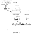
- Example 1-2 1kb-DNA fragment amplified in Example 1-2 was digested with HindIII, and inserted into a pFLAG-1TM vector (International Biotechnologies Inc., USA) which comprises FLAG sequence of octapeptide participating in binding with an anti-FLAG monoclonal antibody and cleavage of enterokinase, and multiple cloning sites participating in insertion of coding sequence ( see : Figure 2).
- Figure 2 shows a construction strategy of an expression vector comprising a gPIP2 gene isolated from a genomic DNA of Phytolacca insularis Nakai.
- the recombinant expression vector thus prepared and competent E . coli JM101 were mixed, stood in ice for 60 minutes, incubated for 2 minutes at 42°C, and cultured for 1 hour at 37°C with the addition of LB medium(10g/L peptone, 5g/L yeast extract, 10g/L NaCl). Then, cells were spread on the solid LB media containing 100 ⁇ g/ml ampicillin and 0.1mM IPTG, and cultured overnight, and colonies whose growth was completely inhibited were selected. Plasmid was isolated from the selected colonies by the modified alkali lysis method( see : Brush, D. et al., Mol.
- Expression vector thus constructed was transformed into Epicurian coli XL1-Blue.
- Transformant thus prepared was designated Epicurian coli XL1-Blue MRF' gPIP2, and deposited under the Budapest Treaty with the Korean Culture Center of Microorganism(KCCM), an international depositary authority as deposition No. KCCM-10080 on April 10, 1996.
- KCCM Korean Culture Center of Microorganism
- Example 2 Isolation of gPIP50 gene from genomic library of Phytolacca insularis Nakai and construction of an expression vector therefor
- DNA fragment containing the gPIP50 gene was isolated from genomic library derived from leaves of Phytolacca insularis Nakai using the cPIP gene labelled with [ ⁇ - 32 p]dATP as a probe. PCR was carried out by employing the prepared DNA fragment as a template to amplify specific region of the gPIP50 gene coding for mature protein, and the amplified DNA was inserted into a pFLAG-1TM vector to construct an expression vector of the gPIP50 protein.
- Example 2-1 Isolation of genomic DNA of Phytolacca insularis Nakai and construction of genomic library
- Precipitate thus obtained was dissolved in 9ml of TE buffer, incubated for 30 minutes in ice with the addition of 9.7g of CsCl, and centrifuged at 7,500 x g for 10 minutes to obtain supernatant.
- 0.5ml of 1%(w/v) ethidium bromide was added to the supernatant, centrifuged analogously as the above to obtain supernatant, and ultracentrifugation of the supernatant was followed at 60,000rpm for 16 hours to precipitate DNA.
- Precipitated DNA was washed repeatedly with isopropanol saturated with CsCl to remove ethidium bromide, incubated for 1 hour at -20°C with the addition of distilled water and ethanol, and centrifuged at 7,500 x g for 10 minutes at 4°C to obtain precipitate.
- the precipitate was resuspended in TE buffer, and centrifuged as mentioned above with the addition of ethanol equivalent to two volume of the said resuspension to isolate genomic DNA from Phytolacca insularis Nakai.
- genomic library of Phytolacca insularis Nakai from the genomic DNA thus isolated, partial digestion of the genomic DNA with Sau3AI was carried out to obtain about 10kb-DNA fragments. 0.9 ⁇ g of 10kb-DNA fragment thus obtained was ligated with 1.0 ⁇ g of ⁇ GEM-11 BamHI arm(Promega, USA) by T4 DNA ligase, and in vitro packaging employing Packagene(Promega, USA) was followed.
- Example 2-2 Screening of the genomic library of Phytolacca insularis Nakai and isolation of gPIP50 gene
- plaques in total were screened over three times of experiments where PCR product comprising the full coding region of cPIP labelled with ( ⁇ - 32 P]dATP by random primer labelling was used as a DNA probe, finally to give nine positive plaques.
- Each of nine plaques selected was cultured in LB medium employing E . coli KW251 as a host cell, and centrifugation was carried out to obtain supernatant. Supernatant thus obtained was ultracentrifuged at 25,000rpm for 2 hours to obtain phage particles. The phage particles thus obtained were treated with SDS, RNase and proteinase K, and phenol extraction were followed to purify phage DNA.
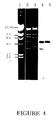
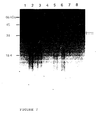
- phage DNA was digested with SacI and XhoI, and analyzed by Southern hybridization using the DNA probe as mentioned above, which confirmed that there were clones which show positive signal of hybridization in XhoI-cleaved 5.0kb-DNA fragment and SacI-cleaved 4.8kb- and 3.5kb-DNA fragments. Among them, clone #50 showing strong positive signal was selected( see : Figure 4).
- lane 1 shows ⁇ DNA digested with HindIII as molecular size marker
- lanes 2 and 3 show 1% agarose gel electrophoresis pattern of phage DNA digested with SacI and XhoI, respectively
- lanes 4 and 5 show corresponding Southern blot analysis of electrophoresis as shown in lanes 2 and 3, respectively.
- pPIP50 5.0kb-DNA fragment of the selected clone #50 was subcloned on SalI-cleaved pUC19 vector, and the resulting plasmid was designated pPIP50.
- digestion of pPIP50 with BamHI, EcoRI, KpnI or SacI, and double-digestion with BamHI+HindIII or EcoRI+KpnI were carried out, and analyzed by Southern blot as mentioned above.
- the restriction enzyme map thus prepared revealed that only one band of 3.4kb-DNA fragment showed positive signal among BamHI fragments, and two bands did among EcoRI, KpnI, or SacI fragments, respectively. Accordingly, it was clearly demonstrated that 3.4kb-BamHI fragment contains the full gPIP50 gene hybridized with the said probe, and there are EcoRI, KpnI, and SacI restriction enzyme sites within the gene.
- Example 2-3 Determination of the nucleotide sequence of the gPIP50 gene
- amino acid sequence deduced from the said gPIP50 gene was found to show 69.4% homology to one from cPIP gene, and 84% homology to one from ⁇ -PAP gene.
- amino acid sequence translated from the gPIP50 gene differs only in two residues of conserved site from known RIP proteins: that is, 200th and 203th amino acids of gPIP50 protein are proline and threonine, while their corresponding amino acids of cPAP or cPIP gene are mostly serine or alanine.
- the difference has little effect on the activity of RIP protein, since it is caused by the substitution of amino acid which is similar in terms of electrical property. Therefore, it was clearly demonstrated that the gPIP50 protein may have an antiviral activity very similar to one of the RIP proteins from the cPIP, ⁇ -PAP, and cPAP genes.
- Example 2-4 Construction of an expression vector containing the gPIP50 gene
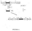
- PCR was performed as described in Example 1-2 employing 3.4kb-BamHI fragment obtained in Example 2 as a template, and oligonucleotides with HindIII restriction enzyme sites at their both ends as primers as followings: NH 2 -terminal primer 5'-CCAAGCTTGCAAGTCCAAATCCAATC-3'(SEQ ID No:9) COOH-terminal primer 5'-GGAAGCTTTGATCAGAAACCCTCAAA-3'(SEQ ID No:6)
- Figure 6 shows a construction strategy of an expression vector comprising a gPIP50 gene which is amplified by PCR using a 3.4kb-BamHI fragment derived from plasmid pPIP50.
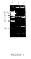
- E . coli was transformed with the recombinant expression vector thus prepared, and the resulting transformant was spread on a solid LB media containing 1.5mM IPTG as in Example 1-3, to select colonies whose growth was inhibited. Plasmids were isolated from the colonies thus selected, digested with HindIII, and analyzed by agarose gel electrophoresis to confirm 1kb-DNA band( see : Figure 3). Also, the nucleotide sequence of DNA insert cloned on pFLAG-1TM vector was determined using N-terminal primer(26-mer) and C-terminal primer(24-mer) in Example 1-3, which finally confirmed proper insertion of desired pPIP50 gene.
- Expression vector thus constructed was transformed into Epicurian coli XL1-Blue.
- Transformant thus prepared was designated E . coli XL1-Blue MRF' gPIP50, and deposited under the Budapest Treaty with the Korean Culture Center of Microorganism(KCCM), an international depositary authority as deposition No. KCCM-10081 on April 10, 1996.
- KCCM Korean Culture Center of Microorganism
- Example 3 Expression of recombinant gPIP2 and gPIP50 proteins
- 1M CaCl 2 was added to the supernatant thus obtained(supernatant I) to a final concentration of 1.0mM, and to the pellet, was added 5ml of 8M urea, and centrifuged at 8,000rpm for 10 minutes to obtain supernatant.
- Supernatant thus obtained(supernatant II) was dialyzed against PBS buffer at 4°C overnight, and 1M CaCl 2 was added to supernatant II to a final concentration of 1.0mM.
- lane 1 shows control supernatant I, i.e., supernatant I obtained from E . coli which was transformed with pFLAG-1TM vector and cultured under IPTG induction
- lane 2 shows supernatant I obtained from E . coli XL1-Blue MRF' gPIP50
- lane 3 shows supernatant I obtained from E . coli XL1-Blue MRF' gPIP2
- lane 4 shows control supernatant II, i.e., supernatant II obtained from E . coli which was transformed with pFLAG-1TM vector and cultured under IPTG induction
- lane 5 shows supernatant II obtained from E .
- coli XL1-Blue MRF' gPIP50 lane 6 shows supernatant II obtained from E . coli XL1-Blue MRF' gPIP2; lane 7 shows the recombinant gPIP50 protein purified from E . coli XL1-Blue MRF' gPIP50; lane 8 shows the recombinant gPIP2 protein purified from E . coli XL1-Blue MRF' gPIP2; and, arrows indicate the bands of the recombinant proteins.
- Example 4 Inhibition of protein synthesis by the recombinant gPIP2 or gPIP50 protein
- Example 3 To study whether the recombinant gPIP2 or gPIP50 protein purified in Example 3 inhibits protein synthesis or not, in vitro translation assay was performed employing rabbit reticulocyte lysate system(Promega, USA): The recombinant protein was added to the reaction mixture containing luciferase mRNA(Promega, USA), rabbit reticulocyte lysate, amino acid mixture free of methionine, and 35 S-labelled methionine, and incubated for 90 minutes at 30°C. Proteins thus synthesized were separated on 15% SDS-PAGE, and the level of protein synthesis was assayed by autoradiography( see : Figure 8).
- lane 1 shows in vitro translation assay using 2.5 ⁇ l of PBS/EDTA elution buffer in anti-FLAG M1 affinity gel chromatography as a substitute for the recombinant protein
- lanes 2 and 3 show in vitro translation assay with the addition of the recombinant gPIP50 protein to a final concentration of 100pM and 10pM, respectively
- lane 4 shows in vitro translation assay with the addition of the recombinant gPIP2 protein to a final concentration of 10pM
- lanes 5 and 6 show in vitro translation assay using 400ng of supernatant II obtained from E .
- the present invention provides novel gPIP2 and gPIP50 genes coding for antiviral proteins isolated from genomic DNA of Phytolacca insularis Nakai, gPIP2 and gPIP50 proteins having the amino acid sequences deduced from the said genes, respectively, expression vectors containing the said genes, and processes for preparing recombinant gPIP2 and gPIP50 proteins from recombinant microorganisms transformed with the said vectors.
- gPIP2 or gPIP50 gene encoding an antiviral protein can be employed in the manufacture of useful plants having antiviral resistance.
- the recombinant gPIP2 or gPIP50 protein prepared in a microorganism transformed with an expression vector containing gPIP2 or gPIP50 gene can be used as an active ingredient of antiviral agents of plant viruses, and employed in the manufacture of immunoconjugates used for the treatment of AIDS and cancer.
- the principal antiviral proteins of the invention have the amino acid sequences shows in SEQ ID NO: 2 (gPIP2) and SEQ ID NO: 4 (gPIP50).
- Other antiviral proteins may have functionally equivalent amino acid sequences.
- the amino acid sequence shown in SEQ ID NO: 2 or 4 modified to contain one or more conservative amino acid substitutions constitutes a functionally equivalent amino acid sequence.
- a functionally equivalent amino acid sequence with respect to gPIP2 is an amino acid sequence which has at least 95% amino acid homology (identity) with the amino acid sequence of gPIP2.
- the homology may be at least 97%, at least 98% or at least 99%.
- a functionally equivalent amino acid sequence with respect to gPIP50 is generally an amino acid sequence which has at least 90% amino acid homology (identity) with the amino acid sequence of gPIP50.
- the homology may be at least 95%, at least 97%, at least 98% or at least 99%.
- An antiviral protein according to the invention which is functionally equivalent to gPIP2 or gPIP50 is capable of inhibiting protein synthesis in the in vitro translation system of Example 4.
- the functionally equivalent protein may contain amino acid deletions and/or insertions as well as or as an alternative to amino acid substitutions such as the conservative amino acid substitutions mentioned above.
- the genes of the invention are typically DNA sequences.
- the gPIP2 gene can have the nucleotide sequence shown in SEQ ID NO: 1 or a functionally equivalent nucleotide sequence.
- a functionally equivalent nucleotide sequence is a nucleotide sequence which also encodes gPIP2, taking into account the degeneracy of the genetic code.
- a functionally equivalent nucleotide sequence as regards the gPIP50 gene is a nucleotide sequence which differs from the nucleotide sequence shown in SEQ ID NO: 3 but which also encodes gPIP50 thanks to the degeneracy of the genetic code.
- a gene according to the invention is typically obtained is isolated form. It may be purified. It can be inserted into a suitable expression vector.
- Expression vectors of the invention thus consist essentially of a gene of the invention operably linked to a promoter.
- the expression vector is typically a plasmid, virus or phage vector provided with an origin of replication. Transcriptional and translational control elements are provided.
- a host is transformed or transfected with the expression vector.
- the vector is generally chosen on the basis that it is compatible with the host into which it is to be transformed or transfected.
- the host may be a microbial host. Suitable host cells are bacterial, yeast, insect or mammalian cells. Plant cells may be transfected.
- the antiviral protein Once expression of the antiviral protein has been achieved, it can be purified. It can be obtained in substantially isolated form.
Landscapes
- Health & Medical Sciences (AREA)
- Life Sciences & Earth Sciences (AREA)
- Genetics & Genomics (AREA)
- Chemical & Material Sciences (AREA)
- Engineering & Computer Science (AREA)
- Organic Chemistry (AREA)
- General Health & Medical Sciences (AREA)
- Molecular Biology (AREA)
- Wood Science & Technology (AREA)
- Zoology (AREA)
- Biotechnology (AREA)
- Biophysics (AREA)
- Bioinformatics & Cheminformatics (AREA)
- General Engineering & Computer Science (AREA)
- Biochemistry (AREA)
- Biomedical Technology (AREA)
- Microbiology (AREA)
- Plant Pathology (AREA)
- Medicinal Chemistry (AREA)
- Gastroenterology & Hepatology (AREA)
- Physics & Mathematics (AREA)
- Botany (AREA)
- Proteomics, Peptides & Aminoacids (AREA)
- Mycology (AREA)
- Virology (AREA)
- Natural Medicines & Medicinal Plants (AREA)
- Agronomy & Crop Science (AREA)
- Cell Biology (AREA)
- Dentistry (AREA)
- Environmental Sciences (AREA)
- Preparation Of Compounds By Using Micro-Organisms (AREA)
- Peptides Or Proteins (AREA)
- Micro-Organisms Or Cultivation Processes Thereof (AREA)
- Medicines That Contain Protein Lipid Enzymes And Other Medicines (AREA)
- Saccharide Compounds (AREA)
- Agricultural Chemicals And Associated Chemicals (AREA)
Abstract
Genes coding for antiviral proteins isolated from genomic DNA and a genomic library of Phytolacca insularis Nakai, and antiviral proteins having the amino acid sequences deduced from the said genes, expression vectors containing the said genes, and processes for preparing the antiviral proteins from recombinant microorganisms transformed with the said vectors. The antiviral proteins can be used as an active ingredient in plant antiviral agents and can be employed in immunoconjugates for the treatments of AIDS and cancer.
Description
- The present invention relates to novel genes coding for antiviral proteins of Phytolacca insularis Nakai and recombinant microorganisms expressing the proteins, more specifically, novel genes coding for antiviral proteins isolated from genomic library of Phytolacca insularis Nakai, antiviral proteins having the amino acid sequences deduced from the said genes, expression vectors containing the said genes, and processes for preparing the antiviral proteins from recombinant microorganisms transformed with the said vectors.
- Since plants can not escape from foreign pathogenic organisms due to their immobile nature, they must be able to defend themselves by making direct or indirect response to the pathogenic challenge; and, most plants appear to undertake some general defense mechanism to protect themselves against infective pathogens, e.g., fungi, bacteria and viruses.
- In this connection, studies on the antiviral proteins from many different plant species, have been carried out, starting from the discovery of pokeweed antiviral protein(PAP) isolated from crude extract of Phytolacca americana L. (see: Irvin, J.D., Arch. Biochem. Biophys., 169:522-528(1975)). In addition to the PAP, antiviral proteins have been successively isolated from several plants, e.g., Ricin(from Ricinus communis)(see: Halling, K.C. et al., Nucleic Acid Res., 13:8019-8033(1985)), Mirabilis antiviral protein(from Mirabilis jalapa L.)(see: Kataoka, J. et al., J. Biol. Chem., 266:8426-8430(1991)), and α-trichosanthin(from Trichosanthes kirilowii)(see: Zhang, X. et al., Nature, 321:477-478(1986)).
- It has been also reported that said antiviral proteins are fallen within a group of ribosome inactivating protein (RIP) and have RNA N-glycosidase activities, by which impair ribosomes by removing adenine in the specific region of rRNA and inhibit the synthesis of proteins(see: Stirpe, F. et al., Bio/Technology, 10:405-412(1992)). As for the survival mechanism of the plants producing RIP without being influenced by their own RIP having those activities, it has been suggested that the plants synthesize ribosomes resistant to their own RIP, or immature RIPs are moved out from the plant cell, before the maturation of RIP, through endoplasmic reticula and Golgi bodies by the aid of signal peptide .
- While RIP has been known to give plant an antiviral resistance, its mode of action has not been clearly understood yet, except for a fact that RIP located out of a plant cell enters the cell together with infecting virus, kills the infected cell, which, in turn, inhibits the infection and spread of the virus(see: Fernandez-Puentes, C. et al., Cell, 20:766-775(1980)).
- Anyway, antiviral property of RIP has prompted the use of RIP in the manufacture of transgenic plants resistant to virus and immunoconjugates for the treatment of AIDS or cancer(see: Lodge, J.K. et al., Proc. Natl. Acad. Sci., USA, 90:7089-7093(1993); Ramarkrishnan, S.D. et al., Ann. Rev. Pharmacol. Toxicol., 32:579-621(1992); Taylor, S.A. et al., Plant J., 5:827-835(1994)). Specifically, considering a fact that there is no remarkable specific remedy for great damage of crops by viral infection, there has been strong reasons for exploring and developing transgenic plants having resistance to pathogenic viruses.
- In line with these activities, the present inventors have deeply studied antiviral proteins of various plants including Phytolacca americana L., Phytolacca insularis Nakai, Cucurbita moschata, and so on, and isolated novel genes encoding their antiviral proteins finally to give the desired antiviral transgenic plants transformed with the recombinant expression vectors containing the said genes (see: USP 5,348,865; Japanese patent 2522151; and, Australian patent 663031).
- In accordance with the present invention, the present inventors have isolated novel genes coding for antiviral protein from a genomic library of Phytolacca insularis Nakai, and discovered that all of the recombinant proteins produced from the expression vectors containing said genes are novel antiviral proteins.
- A primary object of the present invention is, therefore, to provide novel genes coding for antiviral proteins of Phytolacca insularis Nakai, and novel antiviral proteins having amino acid sequences translated therefrom.
- The other object of the invention is to provide expression vectors comprising the said genes and recombinant microorganisms transformed with the vectors.
- Another object of the invention is to provide processes for preparing recombinant antiviral proteins from the said microorganisms.
- The above and the other objects and features of the present invention will become apparent from the following description given in conjunction with the accompanying drawings, in which:
- Figure 1
- is the nucleotide sequence of gPIP2 gene and amino acid sequence deduced therefrom (SEQ ID NO:1 and SEQ ID NO:2);
- Figure 2
- shows a construction strategy of an expression vector comprising the gPIP2 gene isolated from a genomic library of Phytolacca insularis Nakai;
- Figure 3
- is a photograph showing agarose gel electrophoresis pattern of expression vectors comprising gPIP2 or gPIP50 gene digested with restriction enzyme;
- Figure 4
- is a photograph showing agarose gel electrophoresis pattern and Southern hybridization of phage DNA which is isolated from clone #50 in a genomic library of Phytolacca insularis Nakai and digested with SacI or XhoI;
- Figure 5
- is the nucleotide sequence of gPIP50 gene and amino acid sequence translated therefrom(SEQ ID NO:3 and SEQ ID NO:4);
- Figure 6
- shows a stepwise construction strategy of an expression vector comprising the gPIP50 gene derived from plasmid pPIP50;
- Figure 7
- is a photograph showing SDS-PAGE pattern of total protein of inclusion body, pure recombinant gPIP2 and gPIP50 proteins, and supernatant obtained by homogenizing E. coli transformed with the expression vectors containing gPIP2 or gPIP50 genes; and,
- Figure 8
- is an autoradiogram showing the inhibition of protein synthesis occurring when in vitro translation assay of gPIP2 or gPIP50 purified from the recombinant E. coli is carried out.
- The present inventors first isolated a genomic DNA from leaves of Phytolacca insularis Nakai by the method conventionally used in the art(see: J. Sambrook et al., Molecular Cloning, A Laboratory Manual, Cold Spring Harbor Laboratory, 1989) and performed polymerase chain reaction(PCR) by employing the genomic DNA as a template, 5'-CCAAGCTTGTGAATACCATCATCTAC-3'(SEQ ID No:5) as NH2-terminal primer, and 5'-GGAAGCTTTGATCAGAAACCCTCAAA-3'(SEQ ID No:6) as COOH-terminal primer, respectively. 1kb-DNA fragment thus amplified was digested with HindIII, and cloned on pUC19 vector. And then, its restriction map was prepared and nucleotide sequencing was followed. As a result, the amplified DNA fragment was found to comprise a coding region of mature protein which consists of 882bp including initiation codon of translation, and it was designated 'gPIP2 gene'. Also, open reading frame of gPIP2 gene was revealed to code for a 32.8kDa-protein which consists of 292 amino acids including initiation methionine, and the protein was designated 'gPIP2 protein'.
- Based on the comparison study on the amino acid sequence deduced from the said gPIP2 gene with the ones from cPAP and cPIP genes coding for antiviral proteins (see: Lin et al., Plant Molecular Biology, 17(4):609-614(1991); USP 5,348,865), one from the gPIP2 gene was found to show high level of homology to them, i.e., 93.1% and 82.5% homology to ones from cPAP and cPIP genes, respectively. Therefore, it was suggested that the gPIP2 protein is a novel antiviral protein whose activity is very similar to the antiviral proteins translated from the cPAP and cPIP genes but whose amino acid sequence is somewhat different from them. Further, comparison of the nucleotide sequence of the gPIP2 gene with ones of various antiviral protein genes revealed that the gPIP2 gene is novel one.
- On the other hand, the inventors have prepared genomic library based on the genomic DNA from leaves of Phytolacca insularis Nakai, and selected nine positive plaques using the cPIP gene labelled with [α-32P]dATP as a DNA probe. Then, phage DNAs were isolated from those plaques, digested with SacI or XhoI, and analyzed by Southern hybridization using the same DNA probe. As a result, clone #50 in which clear hybridization occurred at the bands of 5.0kb XhoI fragment, and two 4.8kb- and 3.5kb- SacI fragments, was finally selected.
- 5.0kb-XhoI fragment from the selected clone #50 was digested with various restriction enzymes, and analyzed by Southern hybridization. As a result, one positive signal appeared at the band of 3.4kb-BamHI DNA fragment. Thus, the said 3.4kb-BamHI fragment was subcloned on pUC19 vector, and its restriction map was prepared and its nucleotide sequence was determined. The gene was found to comprise a coding region of mature protein which consists of 951bp including initiation codon of translation, and designated 'gPIP50 gene'. Also, open reading frame of gPIP50 gene was revealed to code for a 35.7kDa-protein which consists of 315 amino acids including initiation methionine, and the protein was designated 'gPIP50 protein'.
- Based on the comparison of the amino acid sequence deduced from the said gPIP50 gene with the known antiviral proteins translated from cPIP or α-PAP gene(see: USP 5,348,865; J. Kataoka et al., Plant Mol. Biol., 20:879-886(1992)), one from the gPIP50 gene was found to show high level of homology to them, i.e., 69.4% and 83% homology to ones from cPIP and α-PAP genes, respectively. Therefore, it was suggested that the gPIP50 protein is a novel protein which may have an antiviral activity. Further, comparison of the nucleotide sequence of the gPIP50 gene with ones of various antiviral protein genes, revealed that the gPIP50 gene is novel one.
- The inventors have manufactured a recombinant expression vector by inserting the isolated gPIP2 or gPIP50 gene into a vector which comprises FLAG sequence of octapeptide participating in binding with an anti-FLAG monoclonal antibody and cleavage with enterokinase, and multiple cloning sites participating in insertion of coding sequence, and then, transformed a competent E. coli with the said vector, cultured the transformant, and induced the expression of the recombinant gPIP2 or gPIP50 protein. Cultured transformants were ultrasonicated, and the expression of the recombinant gPIP2 or gPIP50 protein was investigated. As a result, it was found that the said protein was successfully produced from the transformants in a form of inclusion body.
- In order to confirm whether the recombinant gPIP2 and gPIP50 proteins inhibit the protein synthesis like known antiviral proteins, the recombinant gPIP2 and gPIP50 proteins were purified from total proteins obtained from the inclusion body of the said transformant and in vitro translation assay was carried out, which finally confirms that they successfully inhibit the protein synthesis.
- Accordingly, it was clearly determined that the gPIP2 and gPIP50 genes isolated from Phytolacca insularis Nakai, can be employed in the manufacture of transgenic plants having antiviral resistance. Also, the recombinant gPIP2 and gPIP50 proteins prepared from the transformed microorganisms harboring expression vectors containing gPIP2 and gPIP50 genes can be used as an active ingredient of antiviral agents of plant viruses, and employed in the manufacture of immunoconjugates for the treatment of AIDS and cancer.
- In describing the amino acid sequence of the present invention, the term 'proteins consisting of functionally equivalent amino acids' is employed to mean all proteins substituted by the combinations such as Gly, Ala; Val, Ile, Leu; Asp, Glu; Asn, Gln; Ser, Thr; Lys, Arg; and, Phe, Tyr among the amino acid sequences of gPIP2 or gPIP50 protein, and in describing the nucleotide sequence of the invention, the term 'functional equivalents' means all genes comprising nucleotide sequences coding for all the said combinations.
- The present invention is further illustrated by the following examples, which should not be taken to limit the scope of the invention.
- Genomic DNA was isolated from leaves of Phytolacca insularis Nakai, and gPIP2 gene was amplified by PCR using the gene as a template. Then, the amplified gPIP2 gene was inserted into pFLAG-1™ vector to construct an expression vector for the gPIP2 protein.
- Genomic DNA was isolated from leaves of Phytolacca insularis Nakai in accordance with Shure et al's method(see: Shure, M., et al., Cell, 35:225-233(1983)): 1g of leaves of Phytolacca insularis Nakai was grinded under a stream of liquid nitrogen, mixed well with the addition of 5ml of DNA extraction buffer, and digested with 250µl of proteinase K solution(5mg/ml) for 15 minutes at 65°C. Then, phenol/chloroform extraction and centrifugation were carried out to obtain the supernatant.
- The same volume of phenol/chloroform/isoamylalcohol mixture(25:24:1(v/v/v)) was added to the said supernatant and centrifuged to obtain the supernatant. Then, NaCl and 10% CTAB(cetyltrimethylammonium bromide) were added to the supernatant thus obtained to a final concentration of 1.5M and to reach 1/10 of total volume of the mixture, respectively, and incubated for 5 minutes at 65°C. Two cycles of chloroform extraction were followed to obtain supernatant, and 0.5ml of 4.4M NH4OAc(pH 5.2) per 1g of sample and the same volume of isopropylalcohol as the supernatant were added and mixed slowly.
- Then, the precipitated genomic DNA was isolated, dissolved in 0.75ml of TE buffer(10mM Tris-HCl buffer(pH 8.0) containing 1mM EDTA), incubated for 30 minutes at 60°C with the addition of RNase A to a final concentration of 10µg/ml, and extracted with phenol/chloroform as described aboves. After the addition of NH4OAc(pH 5.2) to a final concentration of 0.6M and the same volume of isopropylalcohol as the supernatant, the genomic DNA was isolated.
- PCR was performed using the genomic DNA obtained in Example 1-1 as the template, and oligonucleotides as followings as primers:
NH2-terminal primer
5'-CCAAGCTTGTGAATACCATCATCTAC-3'(SEQ ID No:5)
COOH-terminal primer
5'-GGAAGCTTTGATCAGAAACCCTCAAA-3'(SEQ ID No:6)
That is, 1µg of the genomic DNA of Phytolacca insularis Nakai and each 200ng of the primers were mixed and denaturated for 5 minutes at 95°C. Then, denaturation(95°C, 1 minute), annealing(55°C, 1 minute), and extension(72°C, 1 minute) were carried out for 30 cycles, respectively, employing DNA Thermal Cycler(Cetus/Perkin-Elmer, USA) where the gene was amplified using Vent™ DNA polymerase(NEB, USA). - Amplified 1kb-DNA fragment containing the gPIP2 gene was cleaved with HindIII and cloned on pUC19 vector, and restriction enzyme map was prepared. To determine the full nucleotide sequence of the gPIP2 gene, the gPIP2 gene was cleaved with EcoRI or XhoI, and deletion series were made by Erase-a-Base system(Promega, USA). Then, the nucleotide sequences of the deletion series were determined by the dideoxy nucleotide chain termination method (see: Sanger, F. et al., Proc. Natl. Acad. Sci., USA, 74:5463-5467(1977)) employing Sequenase™(United States Biochemical Co., USA). As a result, it was revealed that the gPIP2 gene coding for the mature protein consists of 882bp including the start codon of translation, and its open reading frame encodes a 32.8kDa-protein which consists of 292 amino acids including initiation methionine(see: Figure 1). Figure 1 shows the nucleotide sequence of the gPIP2 gene and amino acid sequence deduced therefrom(SEQ ID NO:1 and SEQ ID NO:2). In Figure 1, amino acid sequence in bold letter represents one involved in putative active site, and asterisk(*) represents a termination site of translation, respectively.
- Based on the comparison of the amino acid sequence deduced from the said gPIP2 gene with one from cPAP or cPIP gene coding for a known antiviral protein, one from the gPIP2 gene was found to show high level of homology to them, i.e., 93.1% homology to one from cPAP gene, and 82.5% homology to one from cPIP gene. Therefore, it was suggested that the gPIP2 protein may possess an antiviral activity very similar to the antiviral proteins translated from the cPAP and cPIP genes.
- 1kb-DNA fragment amplified in Example 1-2 was digested with HindIII, and inserted into a pFLAG-1™ vector (International Biotechnologies Inc., USA) which comprises FLAG sequence of octapeptide participating in binding with an anti-FLAG monoclonal antibody and cleavage of enterokinase, and multiple cloning sites participating in insertion of coding sequence (see: Figure 2). Figure 2 shows a construction strategy of an expression vector comprising a gPIP2 gene isolated from a genomic DNA of Phytolacca insularis Nakai.
- The recombinant expression vector thus prepared and competent E. coli JM101 were mixed, stood in ice for 60 minutes, incubated for 2 minutes at 42°C, and cultured for 1 hour at 37°C with the addition of LB medium(10g/L peptone, 5g/L yeast extract, 10g/L NaCl). Then, cells were spread on the solid LB media containing 100µg/ml ampicillin and 0.1mM IPTG, and cultured overnight, and colonies whose growth was completely inhibited were selected. Plasmid was isolated from the selected colonies by the modified alkali lysis method(see: Brush, D. et al., Mol. Cell Biol., 5:1307-1317(1985)), cleaved with HindIII, and confirmed to contain 1kb-DNA by agarose gel electrophoresis (see: Figure 3). Also, proper insertion of desired gPIP2 gene into a pFLAG-1™ vector was also investigated, based on the determination of nucleotide sequence of both ends of inserted DNA using NH2-terminal primer(26-mer) and COOH-terminal primer(24-mer) as followings:
NH2-terminal primer
5'-GTAGTATTGCCAAGACCGTTTATAAG-3'(SEQ ID No:7)
COOH-terminal primer
5'-GACATAGTCCGACTTTTAGAAGAG-3'(SEQ ID No:8)
In Figure 3,lane 1 shows λDNA digested with HindIII as molecular size marker;lane 2 shows Hind III-cleaved expression vector containing the gPIP2 gene; and,lane 3 shows Hind III-cleaved expression vector containing the gPIP50 gene which was constructed in Example 2-4. - Expression vector thus constructed was transformed into Epicurian coli XL1-Blue. Transformant thus prepared was designated Epicurian coli XL1-Blue MRF' gPIP2, and deposited under the Budapest Treaty with the Korean Culture Center of Microorganism(KCCM), an international depositary authority as deposition No. KCCM-10080 on April 10, 1996.
- DNA fragment containing the gPIP50 gene was isolated from genomic library derived from leaves of Phytolacca insularis Nakai using the cPIP gene labelled with [α-32p]dATP as a probe. PCR was carried out by employing the prepared DNA fragment as a template to amplify specific region of the gPIP50 gene coding for mature protein, and the amplified DNA was inserted into a pFLAG-1™ vector to construct an expression vector of the gPIP50 protein.
- To isolate genomic DNA from leaves of Phytolacca insularis Nakai, 10g of leaves was grinded under liquid nitrogen, incubated for 2 hours at 55°C with the addition of 30ml of DNA extraction buffer(100mM Tris-HCl(pH 8.0) containing 100mM EDTA, 250mM NaCl, and 100µg/ml proteinase K) and 10%(w/v) sarcosyl to a final concentration of 1%, and centrifuged at 5,500 x g for 10 minutes at 4°C to obtain supernatant. Isopropanol equivalent to 0.6 volume of the supernatant was added to the supernatant, and centrifuged at 7,500 x g for 10 minutes to obtain precipitate. Precipitate thus obtained was dissolved in 9ml of TE buffer, incubated for 30 minutes in ice with the addition of 9.7g of CsCl, and centrifuged at 7,500 x g for 10 minutes to obtain supernatant. 0.5ml of 1%(w/v) ethidium bromide was added to the supernatant, centrifuged analogously as the above to obtain supernatant, and ultracentrifugation of the supernatant was followed at 60,000rpm for 16 hours to precipitate DNA. Precipitated DNA was washed repeatedly with isopropanol saturated with CsCl to remove ethidium bromide, incubated for 1 hour at -20°C with the addition of distilled water and ethanol, and centrifuged at 7,500 x g for 10 minutes at 4°C to obtain precipitate. The precipitate was resuspended in TE buffer, and centrifuged as mentioned above with the addition of ethanol equivalent to two volume of the said resuspension to isolate genomic DNA from Phytolacca insularis Nakai.
- To construct genomic library of Phytolacca insularis Nakai from the genomic DNA thus isolated, partial digestion of the genomic DNA with Sau3AI was carried out to obtain about 10kb-DNA fragments. 0.9µg of 10kb-DNA fragment thus obtained was ligated with 1.0µg of λGEM-11 BamHI arm(Promega, USA) by T4 DNA ligase, and in vitro packaging employing Packagene(Promega, USA) was followed.
- To select gPIP50 gene from the genomic library prepared in Example 2-1, 600,000 plaques in total were screened over three times of experiments where PCR product comprising the full coding region of cPIP labelled with (α-32P]dATP by random primer labelling was used as a DNA probe, finally to give nine positive plaques. Each of nine plaques selected was cultured in LB medium employing E. coli KW251 as a host cell, and centrifugation was carried out to obtain supernatant. Supernatant thus obtained was ultracentrifuged at 25,000rpm for 2 hours to obtain phage particles. The phage particles thus obtained were treated with SDS, RNase and proteinase K, and phenol extraction were followed to purify phage DNA. Purified phage DNA was digested with SacI and XhoI, and analyzed by Southern hybridization using the DNA probe as mentioned above, which confirmed that there were clones which show positive signal of hybridization in XhoI-cleaved 5.0kb-DNA fragment and SacI-cleaved 4.8kb- and 3.5kb-DNA fragments. Among them, clone #50 showing strong positive signal was selected(see: Figure 4). In Figure 4,
lane 1 shows λDNA digested with HindIII as molecular size marker;lanes lanes lanes - 5.0kb-DNA fragment of the selected clone #50 was subcloned on SalI-cleaved pUC19 vector, and the resulting plasmid was designated pPIP50. To prepare the restriction enzyme map of pPIP50, digestion of pPIP50 with BamHI, EcoRI, KpnI or SacI, and double-digestion with BamHI+HindIII or EcoRI+KpnI were carried out, and analyzed by Southern blot as mentioned above. The restriction enzyme map thus prepared revealed that only one band of 3.4kb-DNA fragment showed positive signal among BamHI fragments, and two bands did among EcoRI, KpnI, or SacI fragments, respectively. Accordingly, it was clearly demonstrated that 3.4kb-BamHI fragment contains the full gPIP50 gene hybridized with the said probe, and there are EcoRI, KpnI, and SacI restriction enzyme sites within the gene.
- The nucleotide sequence of the gPIP50 gene located in 3.4kb-BamHI fragment which has confirmed in the restriction enzyme map of plasmid pPIP50, was determined by the conventional method in the art. From the nucleotide sequencing studies, it was revealed that the gPIP50 gene coding for the mature protein consists of 951bp including the start codon of translation, and its open reading frame encodes a 35.7kDa-protein which consists of 315 amino acids including initiation methionine(see: Figure 5). Further, it was also confirmed that 24 hydrophobic amino acids of amino-terminus play as a signal peptide. In Figure 5, a putative CAAT box and two TATA boxes commonly present in eukaryotes in 5'flanking region, and 3'-polyadenylation signal were underlined, (▲) indicates a putative cleavage site of signal peptide, shadow does amino acid sequence of a putative active site, and (*) does termination codon.
- Based on the comparison of the amino acid sequence deduced from the said gPIP50 gene with ones from cPIP and α-PAP genes coding for known antiviral proteins, one from the gPIP50 gene was found to show 69.4% homology to one from cPIP gene, and 84% homology to one from α-PAP gene. In particular, it was determined that the amino acid sequence translated from the gPIP50 gene differs only in two residues of conserved site from known RIP proteins: that is, 200th and 203th amino acids of gPIP50 protein are proline and threonine, while their corresponding amino acids of cPAP or cPIP gene are mostly serine or alanine. However, it was suggested that the difference has little effect on the activity of RIP protein, since it is caused by the substitution of amino acid which is similar in terms of electrical property. Therefore, it was clearly demonstrated that the gPIP50 protein may have an antiviral activity very similar to one of the RIP proteins from the cPIP, α-PAP, and cPAP genes.
- To clone gPIP50 gene coding for the mature protein into an expression vector, pFLAG™, PCR was performed as described in Example 1-2 employing 3.4kb-BamHI fragment obtained in Example 2 as a template, and oligonucleotides with HindIII restriction enzyme sites at their both ends as primers as followings:
NH2-terminal primer
5'-CCAAGCTTGCAAGTCCAAATCCAATC-3'(SEQ ID No:9)
COOH-terminal primer
5'-GGAAGCTTTGATCAGAAACCCTCAAA-3'(SEQ ID No:6) - After 0.8% agarose gel electrophoresis of amplified PCR product, 1kb-DNA fragment containing the gPIP50 gene was electroeluted, digested with HindIII, and cloned on a HindIII-digested pFLAG-1™ vector(see: Figure 6). Figure 6 shows a construction strategy of an expression vector comprising a gPIP50 gene which is amplified by PCR using a 3.4kb-BamHI fragment derived from plasmid pPIP50.
- E. coli was transformed with the recombinant expression vector thus prepared, and the resulting transformant was spread on a solid LB media containing 1.5mM IPTG as in Example 1-3, to select colonies whose growth was inhibited. Plasmids were isolated from the colonies thus selected, digested with HindIII, and analyzed by agarose gel electrophoresis to confirm 1kb-DNA band(see: Figure 3). Also, the nucleotide sequence of DNA insert cloned on pFLAG-1™ vector was determined using N-terminal primer(26-mer) and C-terminal primer(24-mer) in Example 1-3, which finally confirmed proper insertion of desired pPIP50 gene.
- Expression vector thus constructed was transformed into Epicurian coli XL1-Blue. Transformant thus prepared was designated E. coli XL1-Blue MRF' gPIP50, and deposited under the Budapest Treaty with the Korean Culture Center of Microorganism(KCCM), an international depositary authority as deposition No. KCCM-10081 on April 10, 1996.
- E. coli transformed with the expression vector constructed in Examples 1-3 and 2-4, i.e., E. coli XL1-Blue MRF' gPIP2 and E. coli XL1-Blue MRF' gPIP50 were cultured in 500ml of LB medium at a shaking speed of 200rpm for 37°C to reach OD600=0.7, and further cultured for 3 hours with the addition of IPTG(isopropyl-β-D-thiogalactoside) to a final concentration of 0.75mM in order to induce the desired recombinant protein. Cultured cells here harvested by centrifuging at 8,000rpm for 8 minutes at 4°C, resuspended in 35ml of PBS buffer(0.01M NaH2PO4, and 0.15M NaCl, pH 8.4), ultrasonicated, centrifuged at 15,000rpm for 20 minutes at 4°C, and collected supernatant and pellet separately. 1M CaCl2 was added to the supernatant thus obtained(supernatant I) to a final concentration of 1.0mM, and to the pellet, was added 5ml of 8M urea, and centrifuged at 8,000rpm for 10 minutes to obtain supernatant. Supernatant thus obtained(supernatant II) was dialyzed against PBS buffer at 4°C overnight, and 1M CaCl2 was added to supernatant II to a final concentration of 1.0mM.
- Supernatant II containing CaCl2 was loaded on anti-FLAG M1 affinity gel(International Biotechnologies Inc., USA) column which was washed three times with 5ml of glycine-HCl buffer(pH 3.0), and three times with 5ml of PBS buffer. Then, the column was washed three times with 12ml of PBS/Ca buffer(PBS buffer containing 1.0mM CaCl2), and 0.5ml of PBS/EDTA(PBS buffer containing 2.0mM EDTA) was loaded onto the column, and stood for 30 minutes to chelate calcium ion, finally to give fractions of the pure recombinant protein by the intermittent elution with 0.5ml of PBS/EDTA at an interval of 10 minutes.
- Based on 15% SDS-PAGE analysis of the said supernatants and the active fractions thus eluted, it was found that: about 33kDa-recombinant gPIP50 or gPIP2 protein is expressed successfully in the transformant, molecular weight of gPIP50 protein is somewhat larger than one of gPIP2 protein, and the expressed recombinant proteins form inclusion body, in light of a fact that most of them exist in supernatant II(see: Figure 7).
- In Figure 7,
lane 1 shows control supernatant I, i.e., supernatant I obtained from E. coli which was transformed with pFLAG-1™ vector and cultured under IPTG induction;lane 2 shows supernatant I obtained from E. coli XL1-Blue MRF' gPIP50;lane 3 shows supernatant I obtained from E. coli XL1-Blue MRF' gPIP2;lane 4 shows control supernatant II, i.e., supernatant II obtained from E. coli which was transformed with pFLAG-1™ vector and cultured under IPTG induction;lane 5 shows supernatant II obtained from E. coli XL1-Blue MRF' gPIP50;lane 6 shows supernatant II obtained from E. coli XL1-Blue MRF' gPIP2;lane 7 shows the recombinant gPIP50 protein purified from E. coli XL1-Blue MRF' gPIP50;lane 8 shows the recombinant gPIP2 protein purified from E. coli XL1-Blue MRF' gPIP2; and, arrows indicate the bands of the recombinant proteins. - To study whether the recombinant gPIP2 or gPIP50 protein purified in Example 3 inhibits protein synthesis or not, in vitro translation assay was performed employing rabbit reticulocyte lysate system(Promega, USA): The recombinant protein was added to the reaction mixture containing luciferase mRNA(Promega, USA), rabbit reticulocyte lysate, amino acid mixture free of methionine, and 35S-labelled methionine, and incubated for 90 minutes at 30°C. Proteins thus synthesized were separated on 15% SDS-PAGE, and the level of protein synthesis was assayed by autoradiography(see: Figure 8).
- In Figure 8,
lane 1 shows in vitro translation assay using 2.5µl of PBS/EDTA elution buffer in anti-FLAG M1 affinity gel chromatography as a substitute for the recombinant protein;lanes lane 4 shows in vitro translation assay with the addition of the recombinant gPIP2 protein to a final concentration of 10pM; and,lanes lane lane 2, and protein synthesis was markedly inhibited with the addition of 10pM recombinant gPIP50 or gPIP2 protein as shown inlane - As clearly illustrated and demonstrated above, the present invention provides novel gPIP2 and gPIP50 genes coding for antiviral proteins isolated from genomic DNA of Phytolacca insularis Nakai, gPIP2 and gPIP50 proteins having the amino acid sequences deduced from the said genes, respectively, expression vectors containing the said genes, and processes for preparing recombinant gPIP2 and gPIP50 proteins from recombinant microorganisms transformed with the said vectors. gPIP2 or gPIP50 gene encoding an antiviral protein can be employed in the manufacture of useful plants having antiviral resistance. Also, the recombinant gPIP2 or gPIP50 protein prepared in a microorganism transformed with an expression vector containing gPIP2 or gPIP50 gene can be used as an active ingredient of antiviral agents of plant viruses, and employed in the manufacture of immunoconjugates used for the treatment of AIDS and cancer.
- The principal antiviral proteins of the invention have the amino acid sequences shows in SEQ ID NO: 2 (gPIP2) and SEQ ID NO: 4 (gPIP50). Other antiviral proteins may have functionally equivalent amino acid sequences. For example, the amino acid sequence shown in SEQ ID NO: 2 or 4 modified to contain one or more conservative amino acid substitutions constitutes a functionally equivalent amino acid sequence.
- There may be from 1 to 20, for example from 1 to 15 or from 1 to 10, such conservative substitutions in the functionally equivalent amino acid sequence. Thus, there may be 1, 2, 3, 4 or 5 conservative substitutions. The following Table sets out conservative substitutions which may be made. Amino acids in the same line may be substituted for each other:
Table of conservative amino acid substitutions Gly Ala Val Ile Leu Asp Glu Asn Gln Ser Thr Lys Arg Phe Tyr - Typically, a functionally equivalent amino acid sequence with respect to gPIP2 is an amino acid sequence which has at least 95% amino acid homology (identity) with the amino acid sequence of gPIP2. The homology may be at least 97%, at least 98% or at least 99%.
- A functionally equivalent amino acid sequence with respect to gPIP50 is generally an amino acid sequence which has at least 90% amino acid homology (identity) with the amino acid sequence of gPIP50. The homology may be at least 95%, at least 97%, at least 98% or at least 99%.
- An antiviral protein according to the invention which is functionally equivalent to gPIP2 or gPIP50 is capable of inhibiting protein synthesis in the in vitro translation system of Example 4. The functionally equivalent protein may contain amino acid deletions and/or insertions as well as or as an alternative to amino acid substitutions such as the conservative amino acid substitutions mentioned above.
- The genes of the invention are typically DNA sequences. The gPIP2 gene can have the nucleotide sequence shown in SEQ ID NO: 1 or a functionally equivalent nucleotide sequence. Such a functionally equivalent nucleotide sequence is a nucleotide sequence which also encodes gPIP2, taking into account the degeneracy of the genetic code. A functionally equivalent nucleotide sequence as regards the gPIP50 gene is a nucleotide sequence which differs from the nucleotide sequence shown in SEQ ID NO: 3 but which also encodes gPIP50 thanks to the degeneracy of the genetic code.
- A gene according to the invention is typically obtained is isolated form. It may be purified. It can be inserted into a suitable expression vector. Expression vectors of the invention thus consist essentially of a gene of the invention operably linked to a promoter. The expression vector is typically a plasmid, virus or phage vector provided with an origin of replication. Transcriptional and translational control elements are provided.
- A host is transformed or transfected with the expression vector. The vector is generally chosen on the basis that it is compatible with the host into which it is to be transformed or transfected. The host may be a microbial host. Suitable host cells are bacterial, yeast, insect or mammalian cells. Plant cells may be transfected.
- Once expression of the antiviral protein has been achieved, it can be purified. It can be obtained in substantially isolated form.
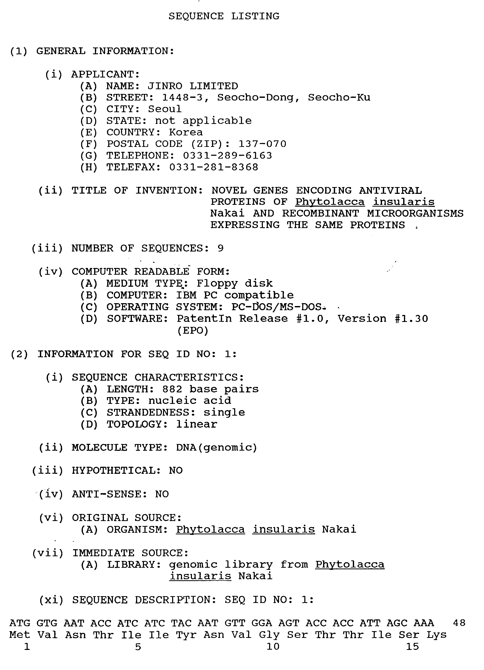


Claims (10)
- A gene coding for (a) an antiviral protein which has the amino acid sequence shown in SEQ ID NO: 2 or 4 or (b) a functional equivalent of the said protein.
- A gene according to claim 1 which has the nucleotide sequence shown in SEQ ID NO: 1 or a functionally equivalent nucleotide sequence.
- A gene according to claim 1 which has the nucleotide sequence shown in SEQ ID NO: 3 or a functionally equivalent nucleotide sequence.
- An expression vector which comprises a gene as claimed in any one of the preceding claims operably linked to a promoter.
- A host transformed or transfected with a gene as claimed in any one of claims 1 to 3.
- A host according to claim 5 which is selected from E. coli XL1-Blue MRF' gPIP2 (KCCM-10080) and E. coli XL1-Blue MRF' gPIP50 (KCCM-10081).
- An antiviral protein which has the amino acid sequence shown in SEQ ID NO: 2 or 4 or a functionally equivalent amino acid sequence.
- A process for preparing an antiviral protein as claimed in claim 7, which process comprises maintaining a host as claimed in claim 5 under such conditions that the said protein is expressed and purifying the resulting said protein.
- A process according to claim 8 which comprises:(i) culturing E. coli XL1-Blue MRF' gPIP2 (KCCM-10080) or E. coli XL1-Blue MRF' gPIP50 (KCCM-10081) and inducing the expression of the said protein by the aid of isopropyl-β-D-thiogalactoside;(ii) harvesting and lysing the E. coli thus induced and obtaining inclusion bodies thus released; and(iii) purifying the said protein from the inclusion bodies.
- A purified recombinant antiviral protein according to claim 7.
Applications Claiming Priority (2)
| Application Number | Priority Date | Filing Date | Title |
|---|---|---|---|
| KR9617404 | 1996-05-22 | ||
| KR1019960017404A KR970074934A (en) | 1996-05-22 | 1996-05-22 | A novel gene of an islet anti-viral protein and a recombinant microorganism expressing it |
Publications (2)
| Publication Number | Publication Date |
|---|---|
| EP0808902A2 true EP0808902A2 (en) | 1997-11-26 |
| EP0808902A3 EP0808902A3 (en) | 1998-02-04 |
Family
ID=19459451
Family Applications (1)
| Application Number | Title | Priority Date | Filing Date |
|---|---|---|---|
| EP96307159A Withdrawn EP0808902A3 (en) | 1996-05-22 | 1996-09-30 | Novel genes encoding antiviral proteins of Phytolacca insularis Nakai and recombinant microorganisms expressing the same proteins |
Country Status (5)
| Country | Link |
|---|---|
| EP (1) | EP0808902A3 (en) |
| JP (1) | JPH09308489A (en) |
| KR (1) | KR970074934A (en) |
| AU (1) | AU6570696A (en) |
| CA (1) | CA2186303A1 (en) |
Family Cites Families (6)
| Publication number | Priority date | Publication date | Assignee | Title |
|---|---|---|---|---|
| JPH05137580A (en) * | 1991-11-20 | 1993-06-01 | Japan Tobacco Inc | New protein and gene capable of coding the same |
| KR950012900B1 (en) * | 1992-08-19 | 1995-10-23 | 주식회사진로 | Antiviral protein expression vector and method for producing plant transformed with it |
| FR2699553B1 (en) * | 1992-12-09 | 2002-10-11 | Innotherapie Lab Sa | Process for obtaining mutants of proteins inactivating ribosomes, mutant genes thus obtained and their applications. |
| KR960015747B1 (en) * | 1993-07-02 | 1996-11-20 | 주식회사 진로 | Microorganisms expressing antiviral protein (PAP) in Aries |
| KR970001565B1 (en) * | 1993-08-28 | 1997-02-11 | 장기하 | Encoding dna for phytolacca insularis antiviral protein and its expression microorganism |
| KR970007864B1 (en) * | 1994-07-21 | 1997-05-17 | 진로 주식회사 | Process of plant which expressing pip of phytolacca insularis nakai |
-
1996
- 1996-05-22 KR KR1019960017404A patent/KR970074934A/en not_active Ceased
- 1996-09-19 AU AU65706/96A patent/AU6570696A/en not_active Abandoned
- 1996-09-24 CA CA002186303A patent/CA2186303A1/en not_active Abandoned
- 1996-09-30 EP EP96307159A patent/EP0808902A3/en not_active Withdrawn
- 1996-11-29 JP JP8320145A patent/JPH09308489A/en active Pending
Also Published As
| Publication number | Publication date |
|---|---|
| EP0808902A3 (en) | 1998-02-04 |
| CA2186303A1 (en) | 1997-11-23 |
| AU6570696A (en) | 1997-11-27 |
| KR970074934A (en) | 1997-12-10 |
| JPH09308489A (en) | 1997-12-02 |
Similar Documents
| Publication | Publication Date | Title |
|---|---|---|
| Grimm et al. | Transiently expressed early light-inducible thylakoid proteins share transmembrane domains with light-harvesting chlorophyll binding proteins | |
| Joosten et al. | Host resistance to a fungal tomato pathogen lost by a single base-pair change in an avirulence gene | |
| Fortin et al. | Nodulin-26, a peribacteroid membrane nodulin is expressed independently of the development of the peribacteroid compartment | |
| EP0242246B1 (en) | Plant cells resistant to glutamine synthetase inhibitors, made by genetic engineering | |
| Wischmann et al. | Transfer of rps10 from the mitochondrion to the nucleus in Arabidopsis thaliana: evidence for RNA-mediated transfer and exon shuffling at the integration site | |
| US20010020300A1 (en) | Transgenic pathogen-resistant organism | |
| WO1996003519A1 (en) | Ubiquitin-lytic peptide fusion gene constructs, protein products deriving therefrom, and methods of making and using same | |
| Last et al. | Plastocyanin is encoded by a single-copy gene in the pea haploid genome | |
| Tetsuo et al. | Sequence of hrdB, an essential gene encoding sigma-like transcription factor of Streptomyces coelicolor A3 (2): homology to principal sigma factors | |
| Pelzer-Reith et al. | Plant aldolase: cDNA and deduced amino-acid sequences of the chloroplast and cytosol enzyme from spinach | |
| Umesono et al. | Nucleotide sequence of Marchantia polymorpha chloroplast DNA: a region possibly encoding three tRNAs and three proteins including a homologue of E. coli ribosomal protein S14 | |
| EP0206613A2 (en) | Activated bacillus thuringienses delta-endotoxin produced by an engineered hybrid gene | |
| AU597142B2 (en) | High level expression in e. coli of soluble mature hil-beta and derivatives with altered biological activity | |
| US6291743B2 (en) | Transgenic plants expressing mutant geminivirus AC1 or C1 genes | |
| KR100346928B1 (en) | Antimicrobial protein | |
| EP0808902A2 (en) | Novel genes encoding antiviral proteins of Phytolacca insularis Nakai and recombinant microorganisms expressing the same proteins | |
| CA2110403A1 (en) | Biocidal proteins | |
| Marshall et al. | Structure and expression of the genes encoding proteins resident in large peripheral vesicles of Phytophthora cinnamomi zoospores | |
| WO1994008010A1 (en) | Method of controlling plant pathogenic fungi | |
| WO1992020800A1 (en) | Plant pathogenesis-related proteins | |
| KR970001565B1 (en) | Encoding dna for phytolacca insularis antiviral protein and its expression microorganism | |
| KR0134377B1 (en) | Mutants of Eglin B and Eglin C, DNA sequences encoding them, recombinant vectors comprising such DNA, hosts transformed with such vectors, methods of making mutants, and pharmaceutical compositions containing the mutants | |
| Halford et al. | Molecular cloning of the barley seed protein CMd: a variant member of the α-amylase/trypsin inhibitor family of cereals | |
| US5648234A (en) | Expression vector for Phytolacca antiviral protein | |
| US6297360B1 (en) | Amphipathic protein-1 |
Legal Events
| Date | Code | Title | Description |
|---|---|---|---|
| PUAI | Public reference made under article 153(3) epc to a published international application that has entered the european phase |
Free format text: ORIGINAL CODE: 0009012 |
|
| 17P | Request for examination filed |
Effective date: 19961011 |
|
| AK | Designated contracting states |
Kind code of ref document: A2 Designated state(s): BE CH DE FR GB LI NL |
|
| STAA | Information on the status of an ep patent application or granted ep patent |
Free format text: STATUS: THE APPLICATION HAS BEEN WITHDRAWN |
|
| PUAL | Search report despatched |
Free format text: ORIGINAL CODE: 0009013 |
|
| 18W | Application withdrawn |
Withdrawal date: 19971203 |
|
| AK | Designated contracting states |
Kind code of ref document: A3 Designated state(s): BE CH DE FR GB LI NL |