EP0807166B1 - A method of preparing an undifferentiated cell - Google Patents
A method of preparing an undifferentiated cell Download PDFInfo
- Publication number
- EP0807166B1 EP0807166B1 EP96901432A EP96901432A EP0807166B1 EP 0807166 B1 EP0807166 B1 EP 0807166B1 EP 96901432 A EP96901432 A EP 96901432A EP 96901432 A EP96901432 A EP 96901432A EP 0807166 B1 EP0807166 B1 EP 0807166B1
- Authority
- EP
- European Patent Office
- Prior art keywords
- cell
- cells
- antigen
- undifferentiated
- chain
- Prior art date
- Legal status (The legal status is an assumption and is not a legal conclusion. Google has not performed a legal analysis and makes no representation as to the accuracy of the status listed.)
- Expired - Lifetime
Links
- 238000000034 method Methods 0.000 title claims abstract description 80
- 210000004027 cell Anatomy 0.000 claims description 491
- 210000004369 blood Anatomy 0.000 claims description 137
- 239000008280 blood Substances 0.000 claims description 137
- 239000000427 antigen Substances 0.000 claims description 128
- 102000036639 antigens Human genes 0.000 claims description 128
- 108091007433 antigens Proteins 0.000 claims description 128
- 210000003719 b-lymphocyte Anatomy 0.000 claims description 68
- 102000006354 HLA-DR Antigens Human genes 0.000 claims description 63
- 108010058597 HLA-DR Antigens Proteins 0.000 claims description 63
- 210000001744 T-lymphocyte Anatomy 0.000 claims description 50
- 101000581981 Homo sapiens Neural cell adhesion molecule 1 Proteins 0.000 claims description 39
- 102100027347 Neural cell adhesion molecule 1 Human genes 0.000 claims description 39
- 102100031573 Hematopoietic progenitor cell antigen CD34 Human genes 0.000 claims description 38
- 101000777663 Homo sapiens Hematopoietic progenitor cell antigen CD34 Proteins 0.000 claims description 38
- 239000003795 chemical substances by application Substances 0.000 claims description 36
- 102000005962 receptors Human genes 0.000 claims description 27
- 210000004698 lymphocyte Anatomy 0.000 claims description 26
- 210000000130 stem cell Anatomy 0.000 claims description 22
- 210000001616 monocyte Anatomy 0.000 claims description 17
- 230000014509 gene expression Effects 0.000 claims description 13
- 230000004069 differentiation Effects 0.000 claims description 12
- 108010001857 Cell Surface Receptors Proteins 0.000 claims description 11
- 210000003643 myeloid progenitor cell Anatomy 0.000 claims description 11
- 210000003738 lymphoid progenitor cell Anatomy 0.000 claims description 10
- 102000043131 MHC class II family Human genes 0.000 claims description 9
- 108091054438 MHC class II family Proteins 0.000 claims description 9
- 206010028980 Neoplasm Diseases 0.000 claims description 8
- 201000011510 cancer Diseases 0.000 claims description 7
- 229940100198 alkylating agent Drugs 0.000 claims description 6
- 239000002168 alkylating agent Substances 0.000 claims description 6
- 230000008512 biological response Effects 0.000 claims description 6
- 239000003607 modifier Substances 0.000 claims description 6
- 210000003651 basophil Anatomy 0.000 claims description 5
- 210000003979 eosinophil Anatomy 0.000 claims description 5
- 238000000338 in vitro Methods 0.000 claims description 5
- 210000002540 macrophage Anatomy 0.000 claims description 5
- 210000000822 natural killer cell Anatomy 0.000 claims description 5
- 210000003593 megakaryocyte Anatomy 0.000 claims description 4
- 210000000440 neutrophil Anatomy 0.000 claims description 4
- 239000005556 hormone Substances 0.000 claims description 3
- 229940088597 hormone Drugs 0.000 claims description 3
- 108090000695 Cytokines Proteins 0.000 claims description 2
- 102000004127 Cytokines Human genes 0.000 claims description 2
- 101150076359 Mhc gene Proteins 0.000 claims description 2
- 239000003102 growth factor Substances 0.000 claims description 2
- 239000002955 immunomodulating agent Substances 0.000 claims description 2
- 230000002584 immunomodulator Effects 0.000 claims description 2
- 229940121354 immunomodulator Drugs 0.000 claims description 2
- 108020004707 nucleic acids Proteins 0.000 claims description 2
- 102000039446 nucleic acids Human genes 0.000 claims description 2
- 150000007523 nucleic acids Chemical class 0.000 claims description 2
- 210000004287 null lymphocyte Anatomy 0.000 claims description 2
- 108090000765 processed proteins & peptides Proteins 0.000 claims description 2
- 102000006240 membrane receptors Human genes 0.000 claims 2
- 102000017420 CD3 protein, epsilon/gamma/delta subunit Human genes 0.000 description 152
- 108050005493 CD3 protein, epsilon/gamma/delta subunit Proteins 0.000 description 152
- 238000011282 treatment Methods 0.000 description 119
- 101000946843 Homo sapiens T-cell surface glycoprotein CD8 alpha chain Proteins 0.000 description 97
- 102100034922 T-cell surface glycoprotein CD8 alpha chain Human genes 0.000 description 97
- 102100036011 T-cell surface glycoprotein CD4 Human genes 0.000 description 68
- 102100024222 B-lymphocyte antigen CD19 Human genes 0.000 description 59
- 101000980825 Homo sapiens B-lymphocyte antigen CD19 Proteins 0.000 description 59
- 101000738771 Homo sapiens Receptor-type tyrosine-protein phosphatase C Proteins 0.000 description 53
- 102100037422 Receptor-type tyrosine-protein phosphatase C Human genes 0.000 description 53
- 101000946889 Homo sapiens Monocyte differentiation antigen CD14 Proteins 0.000 description 29
- 102100035877 Monocyte differentiation antigen CD14 Human genes 0.000 description 29
- 108010088652 Histocompatibility Antigens Class I Proteins 0.000 description 28
- 102000008949 Histocompatibility Antigens Class I Human genes 0.000 description 28
- 230000008569 process Effects 0.000 description 26
- 108020003175 receptors Proteins 0.000 description 25
- 230000007423 decrease Effects 0.000 description 23
- 101000917858 Homo sapiens Low affinity immunoglobulin gamma Fc region receptor III-A Proteins 0.000 description 19
- 101000917839 Homo sapiens Low affinity immunoglobulin gamma Fc region receptor III-B Proteins 0.000 description 19
- 101000914514 Homo sapiens T-cell-specific surface glycoprotein CD28 Proteins 0.000 description 18
- 102100029185 Low affinity immunoglobulin gamma Fc region receptor III-B Human genes 0.000 description 18
- 102100027213 T-cell-specific surface glycoprotein CD28 Human genes 0.000 description 18
- 238000004458 analytical method Methods 0.000 description 17
- 101000934346 Homo sapiens T-cell surface antigen CD2 Proteins 0.000 description 16
- 102100025237 T-cell surface antigen CD2 Human genes 0.000 description 16
- 102000018713 Histocompatibility Antigens Class II Human genes 0.000 description 15
- 108010027412 Histocompatibility Antigens Class II Proteins 0.000 description 15
- 238000013394 immunophenotyping Methods 0.000 description 14
- 102100027208 T-cell antigen CD7 Human genes 0.000 description 12
- 230000003247 decreasing effect Effects 0.000 description 12
- 108090000623 proteins and genes Proteins 0.000 description 11
- 101000934341 Homo sapiens T-cell surface glycoprotein CD5 Proteins 0.000 description 10
- 108010013709 Leukocyte Common Antigens Proteins 0.000 description 10
- 102000017095 Leukocyte Common Antigens Human genes 0.000 description 10
- 102100025244 T-cell surface glycoprotein CD5 Human genes 0.000 description 10
- 238000011534 incubation Methods 0.000 description 10
- 102000000844 Cell Surface Receptors Human genes 0.000 description 9
- 230000000694 effects Effects 0.000 description 9
- 210000003714 granulocyte Anatomy 0.000 description 9
- 102100022749 Aminopeptidase N Human genes 0.000 description 8
- 101000757160 Homo sapiens Aminopeptidase N Proteins 0.000 description 8
- 101001057504 Homo sapiens Interferon-stimulated gene 20 kDa protein Proteins 0.000 description 8
- 101001055144 Homo sapiens Interleukin-2 receptor subunit alpha Proteins 0.000 description 8
- 102100027268 Interferon-stimulated gene 20 kDa protein Human genes 0.000 description 8
- 230000002950 deficient Effects 0.000 description 8
- 239000003814 drug Substances 0.000 description 8
- 210000000265 leukocyte Anatomy 0.000 description 8
- 210000001778 pluripotent stem cell Anatomy 0.000 description 8
- 101000934338 Homo sapiens Myeloid cell surface antigen CD33 Proteins 0.000 description 7
- 102100025243 Myeloid cell surface antigen CD33 Human genes 0.000 description 7
- 208000037265 diseases, disorders, signs and symptoms Diseases 0.000 description 7
- 210000004185 liver Anatomy 0.000 description 7
- 239000003550 marker Substances 0.000 description 7
- 102000013925 CD34 antigen Human genes 0.000 description 6
- 108050003733 CD34 antigen Proteins 0.000 description 6
- 238000011161 development Methods 0.000 description 6
- 230000018109 developmental process Effects 0.000 description 6
- 210000003743 erythrocyte Anatomy 0.000 description 6
- 210000005259 peripheral blood Anatomy 0.000 description 6
- 239000011886 peripheral blood Substances 0.000 description 6
- 238000002360 preparation method Methods 0.000 description 6
- 108010041397 CD4 Antigens Proteins 0.000 description 5
- 102100032768 Complement receptor type 2 Human genes 0.000 description 5
- 102100036242 HLA class II histocompatibility antigen, DQ alpha 2 chain Human genes 0.000 description 5
- 101000941929 Homo sapiens Complement receptor type 2 Proteins 0.000 description 5
- 101000930801 Homo sapiens HLA class II histocompatibility antigen, DQ alpha 2 chain Proteins 0.000 description 5
- 102000003729 Neprilysin Human genes 0.000 description 5
- 108090000028 Neprilysin Proteins 0.000 description 5
- 108091008874 T cell receptors Proteins 0.000 description 5
- 102000016266 T-Cell Antigen Receptors Human genes 0.000 description 5
- 208000032852 chronic lymphocytic leukemia Diseases 0.000 description 5
- 239000000047 product Substances 0.000 description 5
- 208000030507 AIDS Diseases 0.000 description 4
- 102000010910 CD28 Antigens Human genes 0.000 description 4
- 108010062433 CD28 Antigens Proteins 0.000 description 4
- CMSMOCZEIVJLDB-UHFFFAOYSA-N Cyclophosphamide Chemical compound ClCCN(CCCl)P1(=O)NCCCO1 CMSMOCZEIVJLDB-UHFFFAOYSA-N 0.000 description 4
- 102100033215 DNA nucleotidylexotransferase Human genes 0.000 description 4
- 108010008286 DNA nucleotidylexotransferase Proteins 0.000 description 4
- 102100021260 Galactosylgalactosylxylosylprotein 3-beta-glucuronosyltransferase 1 Human genes 0.000 description 4
- 101000894906 Homo sapiens Galactosylgalactosylxylosylprotein 3-beta-glucuronosyltransferase 1 Proteins 0.000 description 4
- 101000878605 Homo sapiens Low affinity immunoglobulin epsilon Fc receptor Proteins 0.000 description 4
- 102100038007 Low affinity immunoglobulin epsilon Fc receptor Human genes 0.000 description 4
- 102000043129 MHC class I family Human genes 0.000 description 4
- 108091054437 MHC class I family Proteins 0.000 description 4
- 210000001185 bone marrow Anatomy 0.000 description 4
- 229960004397 cyclophosphamide Drugs 0.000 description 4
- 201000010099 disease Diseases 0.000 description 4
- 238000000684 flow cytometry Methods 0.000 description 4
- 230000003394 haemopoietic effect Effects 0.000 description 4
- 230000008929 regeneration Effects 0.000 description 4
- 238000011069 regeneration method Methods 0.000 description 4
- 238000011160 research Methods 0.000 description 4
- 230000000284 resting effect Effects 0.000 description 4
- 208000024891 symptom Diseases 0.000 description 4
- 102100022005 B-lymphocyte antigen CD20 Human genes 0.000 description 3
- 101000897405 Homo sapiens B-lymphocyte antigen CD20 Proteins 0.000 description 3
- 108700018351 Major Histocompatibility Complex Proteins 0.000 description 3
- 230000008859 change Effects 0.000 description 3
- 239000003085 diluting agent Substances 0.000 description 3
- 208000035475 disorder Diseases 0.000 description 3
- 230000006870 function Effects 0.000 description 3
- 210000003494 hepatocyte Anatomy 0.000 description 3
- 208000032839 leukemia Diseases 0.000 description 3
- 238000004519 manufacturing process Methods 0.000 description 3
- 239000000203 mixture Substances 0.000 description 3
- 239000000546 pharmaceutical excipient Substances 0.000 description 3
- PHEDXBVPIONUQT-RGYGYFBISA-N phorbol 13-acetate 12-myristate Chemical compound C([C@]1(O)C(=O)C(C)=C[C@H]1[C@@]1(O)[C@H](C)[C@H]2OC(=O)CCCCCCCCCCCCC)C(CO)=C[C@H]1[C@H]1[C@]2(OC(C)=O)C1(C)C PHEDXBVPIONUQT-RGYGYFBISA-N 0.000 description 3
- 239000002243 precursor Substances 0.000 description 3
- 230000020382 suppression by virus of host antigen processing and presentation of peptide antigen via MHC class I Effects 0.000 description 3
- 230000036962 time dependent Effects 0.000 description 3
- 208000005623 Carcinogenesis Diseases 0.000 description 2
- 208000031886 HIV Infections Diseases 0.000 description 2
- 208000017604 Hodgkin disease Diseases 0.000 description 2
- 208000021519 Hodgkin lymphoma Diseases 0.000 description 2
- 208000010747 Hodgkins lymphoma Diseases 0.000 description 2
- 101000914496 Homo sapiens T-cell antigen CD7 Proteins 0.000 description 2
- 206010020983 Hypogammaglobulinaemia Diseases 0.000 description 2
- 206010025323 Lymphomas Diseases 0.000 description 2
- 208000029052 T-cell acute lymphoblastic leukemia Diseases 0.000 description 2
- 230000004913 activation Effects 0.000 description 2
- 230000030741 antigen processing and presentation Effects 0.000 description 2
- 230000015572 biosynthetic process Effects 0.000 description 2
- 210000001772 blood platelet Anatomy 0.000 description 2
- 230000036952 cancer formation Effects 0.000 description 2
- 231100000504 carcinogenesis Toxicity 0.000 description 2
- 231100000135 cytotoxicity Toxicity 0.000 description 2
- 230000003013 cytotoxicity Effects 0.000 description 2
- 239000002158 endotoxin Substances 0.000 description 2
- 210000004700 fetal blood Anatomy 0.000 description 2
- 210000002443 helper t lymphocyte Anatomy 0.000 description 2
- 230000004730 hepatocarcinogenesis Effects 0.000 description 2
- 210000003630 histaminocyte Anatomy 0.000 description 2
- 238000001727 in vivo Methods 0.000 description 2
- 208000015181 infectious disease Diseases 0.000 description 2
- 229920006008 lipopolysaccharide Polymers 0.000 description 2
- 210000002751 lymph Anatomy 0.000 description 2
- 230000036210 malignancy Effects 0.000 description 2
- 230000035800 maturation Effects 0.000 description 2
- 238000000386 microscopy Methods 0.000 description 2
- 230000035772 mutation Effects 0.000 description 2
- 239000002644 phorbol ester Substances 0.000 description 2
- 210000004180 plasmocyte Anatomy 0.000 description 2
- 230000002035 prolonged effect Effects 0.000 description 2
- 230000004043 responsiveness Effects 0.000 description 2
- 230000002441 reversible effect Effects 0.000 description 2
- 210000001541 thymus gland Anatomy 0.000 description 2
- 210000001519 tissue Anatomy 0.000 description 2
- 230000017423 tissue regeneration Effects 0.000 description 2
- 231100000167 toxic agent Toxicity 0.000 description 2
- 239000003440 toxic substance Substances 0.000 description 2
- 108010027122 ADP-ribosyl Cyclase 1 Proteins 0.000 description 1
- 102000018667 ADP-ribosyl Cyclase 1 Human genes 0.000 description 1
- 208000031261 Acute myeloid leukaemia Diseases 0.000 description 1
- 208000008190 Agammaglobulinemia Diseases 0.000 description 1
- 208000023275 Autoimmune disease Diseases 0.000 description 1
- 201000008162 B cell deficiency Diseases 0.000 description 1
- 102100038080 B-cell receptor CD22 Human genes 0.000 description 1
- 208000019838 Blood disease Diseases 0.000 description 1
- 108010003639 CD56 Antigen Proteins 0.000 description 1
- 102000004652 CD56 Antigen Human genes 0.000 description 1
- 108010001017 CD71 antigen Proteins 0.000 description 1
- 108010067225 Cell Adhesion Molecules Proteins 0.000 description 1
- 102000016289 Cell Adhesion Molecules Human genes 0.000 description 1
- 239000004381 Choline salt Substances 0.000 description 1
- 206010011831 Cytomegalovirus infection Diseases 0.000 description 1
- KCXVZYZYPLLWCC-UHFFFAOYSA-N EDTA Chemical compound OC(=O)CN(CC(O)=O)CCN(CC(O)=O)CC(O)=O KCXVZYZYPLLWCC-UHFFFAOYSA-N 0.000 description 1
- 102000004190 Enzymes Human genes 0.000 description 1
- 108090000790 Enzymes Proteins 0.000 description 1
- 108010087819 Fc receptors Proteins 0.000 description 1
- 102000009109 Fc receptors Human genes 0.000 description 1
- 208000037357 HIV infectious disease Diseases 0.000 description 1
- 102100028972 HLA class I histocompatibility antigen, A alpha chain Human genes 0.000 description 1
- 102100028976 HLA class I histocompatibility antigen, B alpha chain Human genes 0.000 description 1
- 102100028971 HLA class I histocompatibility antigen, C alpha chain Human genes 0.000 description 1
- 102100028970 HLA class I histocompatibility antigen, alpha chain E Human genes 0.000 description 1
- 102100028966 HLA class I histocompatibility antigen, alpha chain F Human genes 0.000 description 1
- 102100028967 HLA class I histocompatibility antigen, alpha chain G Human genes 0.000 description 1
- 108010075704 HLA-A Antigens Proteins 0.000 description 1
- 108010058607 HLA-B Antigens Proteins 0.000 description 1
- 108010052199 HLA-C Antigens Proteins 0.000 description 1
- 108010024164 HLA-G Antigens Proteins 0.000 description 1
- 101000884305 Homo sapiens B-cell receptor CD22 Proteins 0.000 description 1
- 101000986085 Homo sapiens HLA class I histocompatibility antigen, alpha chain E Proteins 0.000 description 1
- 101000986080 Homo sapiens HLA class I histocompatibility antigen, alpha chain F Proteins 0.000 description 1
- 101000835093 Homo sapiens Transferrin receptor protein 1 Proteins 0.000 description 1
- 208000007924 IgA Deficiency Diseases 0.000 description 1
- 108010073807 IgG Receptors Proteins 0.000 description 1
- 102000009490 IgG Receptors Human genes 0.000 description 1
- 108060003951 Immunoglobulin Proteins 0.000 description 1
- 238000012404 In vitro experiment Methods 0.000 description 1
- 244000178870 Lavandula angustifolia Species 0.000 description 1
- 235000010663 Lavandula angustifolia Nutrition 0.000 description 1
- 108010031801 Lipopolysaccharide Receptors Proteins 0.000 description 1
- 102000005482 Lipopolysaccharide Receptors Human genes 0.000 description 1
- 101710177649 Low affinity immunoglobulin gamma Fc region receptor III Proteins 0.000 description 1
- 208000028018 Lymphocytic leukaemia Diseases 0.000 description 1
- 102000012750 Membrane Glycoproteins Human genes 0.000 description 1
- 108010090054 Membrane Glycoproteins Proteins 0.000 description 1
- WBNQDOYYEUMPFS-UHFFFAOYSA-N N-nitrosodiethylamine Chemical compound CCN(CC)N=O WBNQDOYYEUMPFS-UHFFFAOYSA-N 0.000 description 1
- 102000016979 Other receptors Human genes 0.000 description 1
- 206010035226 Plasma cell myeloma Diseases 0.000 description 1
- 102000002727 Protein Tyrosine Phosphatase Human genes 0.000 description 1
- 108090000412 Protein-Tyrosine Kinases Proteins 0.000 description 1
- 102000004022 Protein-Tyrosine Kinases Human genes 0.000 description 1
- 206010039915 Selective IgA immunodeficiency Diseases 0.000 description 1
- 108010029180 Sialic Acid Binding Ig-like Lectin 3 Proteins 0.000 description 1
- 102000001555 Sialic Acid Binding Ig-like Lectin 3 Human genes 0.000 description 1
- 102100026144 Transferrin receptor protein 1 Human genes 0.000 description 1
- 241000700605 Viruses Species 0.000 description 1
- 241000269370 Xenopus <genus> Species 0.000 description 1
- 230000003213 activating effect Effects 0.000 description 1
- 230000001154 acute effect Effects 0.000 description 1
- 230000003044 adaptive effect Effects 0.000 description 1
- 230000000961 alloantigen Effects 0.000 description 1
- 230000019552 anatomical structure morphogenesis Effects 0.000 description 1
- 230000000890 antigenic effect Effects 0.000 description 1
- 230000006907 apoptotic process Effects 0.000 description 1
- 210000003567 ascitic fluid Anatomy 0.000 description 1
- 239000011324 bead Substances 0.000 description 1
- 230000009286 beneficial effect Effects 0.000 description 1
- 210000002960 bfu-e Anatomy 0.000 description 1
- 210000000988 bone and bone Anatomy 0.000 description 1
- 230000005907 cancer growth Effects 0.000 description 1
- 230000000711 cancerogenic effect Effects 0.000 description 1
- 210000001043 capillary endothelial cell Anatomy 0.000 description 1
- 231100000315 carcinogenic Toxicity 0.000 description 1
- 210000004970 cd4 cell Anatomy 0.000 description 1
- 230000020411 cell activation Effects 0.000 description 1
- 230000030833 cell death Effects 0.000 description 1
- 230000011712 cell development Effects 0.000 description 1
- 230000010261 cell growth Effects 0.000 description 1
- 210000000170 cell membrane Anatomy 0.000 description 1
- 230000004663 cell proliferation Effects 0.000 description 1
- 230000001413 cellular effect Effects 0.000 description 1
- 238000003759 clinical diagnosis Methods 0.000 description 1
- 230000000112 colonic effect Effects 0.000 description 1
- 230000001332 colony forming effect Effects 0.000 description 1
- 150000001875 compounds Chemical class 0.000 description 1
- 230000001276 controlling effect Effects 0.000 description 1
- 210000000805 cytoplasm Anatomy 0.000 description 1
- 210000003104 cytoplasmic structure Anatomy 0.000 description 1
- 231100000433 cytotoxic Toxicity 0.000 description 1
- 210000001151 cytotoxic T lymphocyte Anatomy 0.000 description 1
- 230000001472 cytotoxic effect Effects 0.000 description 1
- 230000034994 death Effects 0.000 description 1
- 230000007812 deficiency Effects 0.000 description 1
- 230000002939 deleterious effect Effects 0.000 description 1
- 238000012217 deletion Methods 0.000 description 1
- 210000004443 dendritic cell Anatomy 0.000 description 1
- 230000001419 dependent effect Effects 0.000 description 1
- 238000003745 diagnosis Methods 0.000 description 1
- 230000008034 disappearance Effects 0.000 description 1
- 210000003162 effector t lymphocyte Anatomy 0.000 description 1
- 230000002255 enzymatic effect Effects 0.000 description 1
- 210000002919 epithelial cell Anatomy 0.000 description 1
- 210000000267 erythroid cell Anatomy 0.000 description 1
- 230000000925 erythroid effect Effects 0.000 description 1
- 230000001605 fetal effect Effects 0.000 description 1
- MHMNJMPURVTYEJ-UHFFFAOYSA-N fluorescein-5-isothiocyanate Chemical compound O1C(=O)C2=CC(N=C=S)=CC=C2C21C1=CC=C(O)C=C1OC1=CC(O)=CC=C21 MHMNJMPURVTYEJ-UHFFFAOYSA-N 0.000 description 1
- 125000000524 functional group Chemical group 0.000 description 1
- 239000011521 glass Substances 0.000 description 1
- 239000008187 granular material Substances 0.000 description 1
- 210000004524 haematopoietic cell Anatomy 0.000 description 1
- 208000014951 hematologic disease Diseases 0.000 description 1
- 210000003958 hematopoietic stem cell Anatomy 0.000 description 1
- 208000018706 hematopoietic system disease Diseases 0.000 description 1
- 206010073071 hepatocellular carcinoma Diseases 0.000 description 1
- 208000033519 human immunodeficiency virus infectious disease Diseases 0.000 description 1
- 230000001900 immune effect Effects 0.000 description 1
- 210000000987 immune system Anatomy 0.000 description 1
- 238000000760 immunoelectrophoresis Methods 0.000 description 1
- 102000018358 immunoglobulin Human genes 0.000 description 1
- 201000007156 immunoglobulin alpha deficiency Diseases 0.000 description 1
- 208000016245 inborn errors of metabolism Diseases 0.000 description 1
- 230000003993 interaction Effects 0.000 description 1
- 238000011835 investigation Methods 0.000 description 1
- 230000000366 juvenile effect Effects 0.000 description 1
- 239000001102 lavandula vera Substances 0.000 description 1
- 235000018219 lavender Nutrition 0.000 description 1
- 239000003446 ligand Substances 0.000 description 1
- 230000007774 longterm Effects 0.000 description 1
- 238000012423 maintenance Methods 0.000 description 1
- 239000000463 material Substances 0.000 description 1
- 238000005259 measurement Methods 0.000 description 1
- 230000001404 mediated effect Effects 0.000 description 1
- 239000003226 mitogen Substances 0.000 description 1
- 230000000394 mitotic effect Effects 0.000 description 1
- 238000012986 modification Methods 0.000 description 1
- 230000004048 modification Effects 0.000 description 1
- 230000000877 morphologic effect Effects 0.000 description 1
- 210000000066 myeloid cell Anatomy 0.000 description 1
- 201000000050 myeloid neoplasm Diseases 0.000 description 1
- 239000013642 negative control Substances 0.000 description 1
- 230000006911 nucleation Effects 0.000 description 1
- 238000010899 nucleation Methods 0.000 description 1
- 239000002773 nucleotide Substances 0.000 description 1
- 125000003729 nucleotide group Chemical group 0.000 description 1
- 230000005868 ontogenesis Effects 0.000 description 1
- 210000000056 organ Anatomy 0.000 description 1
- 230000033667 organ regeneration Effects 0.000 description 1
- 239000003973 paint Substances 0.000 description 1
- 210000002741 palatine tonsil Anatomy 0.000 description 1
- 210000004738 parenchymal cell Anatomy 0.000 description 1
- 230000036961 partial effect Effects 0.000 description 1
- 238000012753 partial hepatectomy Methods 0.000 description 1
- 210000005105 peripheral blood lymphocyte Anatomy 0.000 description 1
- 210000001539 phagocyte Anatomy 0.000 description 1
- 210000004910 pleural fluid Anatomy 0.000 description 1
- 230000001855 preneoplastic effect Effects 0.000 description 1
- 230000000750 progressive effect Effects 0.000 description 1
- 230000035755 proliferation Effects 0.000 description 1
- 230000001737 promoting effect Effects 0.000 description 1
- 108020000494 protein-tyrosine phosphatase Proteins 0.000 description 1
- 238000012797 qualification Methods 0.000 description 1
- 230000008707 rearrangement Effects 0.000 description 1
- 230000002829 reductive effect Effects 0.000 description 1
- 230000001172 regenerating effect Effects 0.000 description 1
- 230000009703 regulation of cell differentiation Effects 0.000 description 1
- 230000021014 regulation of cell growth Effects 0.000 description 1
- 230000001105 regulatory effect Effects 0.000 description 1
- 238000012552 review Methods 0.000 description 1
- 208000029138 selective IgA deficiency disease Diseases 0.000 description 1
- 238000012882 sequential analysis Methods 0.000 description 1
- 230000019491 signal transduction Effects 0.000 description 1
- 210000000952 spleen Anatomy 0.000 description 1
- 230000002269 spontaneous effect Effects 0.000 description 1
- 238000010186 staining Methods 0.000 description 1
- 230000000638 stimulation Effects 0.000 description 1
- 210000002536 stromal cell Anatomy 0.000 description 1
- 239000000126 substance Substances 0.000 description 1
- 239000000758 substrate Substances 0.000 description 1
- 239000006228 supernatant Substances 0.000 description 1
- 230000001629 suppression Effects 0.000 description 1
- 208000011580 syndromic disease Diseases 0.000 description 1
- 238000012360 testing method Methods 0.000 description 1
- 230000002992 thymic effect Effects 0.000 description 1
- 230000002463 transducing effect Effects 0.000 description 1
- 230000001052 transient effect Effects 0.000 description 1
- 230000007704 transition Effects 0.000 description 1
- 230000001960 triggered effect Effects 0.000 description 1
- 230000003612 virological effect Effects 0.000 description 1
Images


Classifications
-
- C—CHEMISTRY; METALLURGY
- C12—BIOCHEMISTRY; BEER; SPIRITS; WINE; VINEGAR; MICROBIOLOGY; ENZYMOLOGY; MUTATION OR GENETIC ENGINEERING
- C12N—MICROORGANISMS OR ENZYMES; COMPOSITIONS THEREOF; PROPAGATING, PRESERVING, OR MAINTAINING MICROORGANISMS; MUTATION OR GENETIC ENGINEERING; CULTURE MEDIA
- C12N5/00—Undifferentiated human, animal or plant cells, e.g. cell lines; Tissues; Cultivation or maintenance thereof; Culture media therefor
- C12N5/06—Animal cells or tissues; Human cells or tissues
- C12N5/0602—Vertebrate cells
- C12N5/0634—Cells from the blood or the immune system
-
- C—CHEMISTRY; METALLURGY
- C12—BIOCHEMISTRY; BEER; SPIRITS; WINE; VINEGAR; MICROBIOLOGY; ENZYMOLOGY; MUTATION OR GENETIC ENGINEERING
- C12N—MICROORGANISMS OR ENZYMES; COMPOSITIONS THEREOF; PROPAGATING, PRESERVING, OR MAINTAINING MICROORGANISMS; MUTATION OR GENETIC ENGINEERING; CULTURE MEDIA
- C12N5/00—Undifferentiated human, animal or plant cells, e.g. cell lines; Tissues; Cultivation or maintenance thereof; Culture media therefor
- C12N5/06—Animal cells or tissues; Human cells or tissues
- C12N5/0602—Vertebrate cells
-
- A—HUMAN NECESSITIES
- A61—MEDICAL OR VETERINARY SCIENCE; HYGIENE
- A61P—SPECIFIC THERAPEUTIC ACTIVITY OF CHEMICAL COMPOUNDS OR MEDICINAL PREPARATIONS
- A61P37/00—Drugs for immunological or allergic disorders
-
- A—HUMAN NECESSITIES
- A61—MEDICAL OR VETERINARY SCIENCE; HYGIENE
- A61P—SPECIFIC THERAPEUTIC ACTIVITY OF CHEMICAL COMPOUNDS OR MEDICINAL PREPARATIONS
- A61P37/00—Drugs for immunological or allergic disorders
- A61P37/02—Immunomodulators
- A61P37/04—Immunostimulants
-
- C—CHEMISTRY; METALLURGY
- C12—BIOCHEMISTRY; BEER; SPIRITS; WINE; VINEGAR; MICROBIOLOGY; ENZYMOLOGY; MUTATION OR GENETIC ENGINEERING
- C12N—MICROORGANISMS OR ENZYMES; COMPOSITIONS THEREOF; PROPAGATING, PRESERVING, OR MAINTAINING MICROORGANISMS; MUTATION OR GENETIC ENGINEERING; CULTURE MEDIA
- C12N2501/00—Active agents used in cell culture processes, e.g. differentation
- C12N2501/06—Anti-neoplasic drugs, anti-retroviral drugs, e.g. azacytidine, cyclophosphamide
-
- C—CHEMISTRY; METALLURGY
- C12—BIOCHEMISTRY; BEER; SPIRITS; WINE; VINEGAR; MICROBIOLOGY; ENZYMOLOGY; MUTATION OR GENETIC ENGINEERING
- C12N—MICROORGANISMS OR ENZYMES; COMPOSITIONS THEREOF; PROPAGATING, PRESERVING, OR MAINTAINING MICROORGANISMS; MUTATION OR GENETIC ENGINEERING; CULTURE MEDIA
- C12N2501/00—Active agents used in cell culture processes, e.g. differentation
- C12N2501/50—Cell markers; Cell surface determinants
-
- C—CHEMISTRY; METALLURGY
- C12—BIOCHEMISTRY; BEER; SPIRITS; WINE; VINEGAR; MICROBIOLOGY; ENZYMOLOGY; MUTATION OR GENETIC ENGINEERING
- C12N—MICROORGANISMS OR ENZYMES; COMPOSITIONS THEREOF; PROPAGATING, PRESERVING, OR MAINTAINING MICROORGANISMS; MUTATION OR GENETIC ENGINEERING; CULTURE MEDIA
- C12N2506/00—Differentiation of animal cells from one lineage to another; Differentiation of pluripotent cells
- C12N2506/11—Differentiation of animal cells from one lineage to another; Differentiation of pluripotent cells from blood or immune system cells
Definitions
- Described herein is a method of preparing an undifferentiated cell.
- the present invention relates to a method of preparing an undifferentiated cell from a more committed cell.
- Described herein is also the use of the undifferentiated cell of the present invention for the preparation of a new more committed cell - i.e. a recommitted cell.
- the use of the undifferentiated cell of the present invention or the recommitted cell of the present invention to have an effect (directly or indirectly via the use of products obtained therefrom) on the immune system, such as the alleviation of symptoms associated with, or the partial or complete cure from, an immunological condition or disease.
- differentiation is a process whereby structures and functions of cells are progressively committed to give rise to more specialised cells, such as the formation of T cells or B cells. Therefore, as the cells become more committed, they become more specialised.
- retro-differentiation is a process whereby structures and functions of cells are progressively changed to give rise to less specialised cells.
- Undifferentiated cells are capable of multilineage differentiation - i.e. they are capable of differentiating into two or more types of specialised cells.
- a typical example of an undifferentiated cell is a stem cell.
- differentiated cells are incapable of multilineage differentiation.
- a typical example of a differentiated cell is a T cell.
- LPCs lymphohaematopoietic progenitor cells
- LPCs include pluripotent stem cells (PSCs), lymphoid stem cells (LSCs) and myeloid stem cells (MSCs).
- PSCs pluripotent stem cells
- LSCs lymphoid stem cells
- MSCs myeloid stem cells
- LSCs and MSCs are each formed by the differentiation of PSCs. Hence. LSCs and MSCs are more committed than PSCs.
- differentiated cells include T cells, B cells, eosinophils, basophils, neutrophils, megakaryocytes, monocytes, erythrocytes, granulocytes, mast cells, and lymphocytes.
- T cells and B cells are formed by the differentiation of LSCs. Hence. T cells and B cells are more committed than LSCs.
- Eosinophils basophils, neutrophils, megakaryocytes, monocytes, erythrocytes, granulocytes, mast cells, NKs, and lymphocytes are formed by the differentiation of MSCs. Hence, each of these cells are more committed than MSCs.
- Antigens are associated with undifferentiated and differentiated cells.
- the term "associated” here means the cells expressing or capable of expressing, or presenting or capable of being induced to present, or comprising, the respective antigen(s).
- MHC Major Histocompatability Complex
- Each specific antigen associated with an undifferentiated cell or a differentiated cell can act as a marker.
- different types of cells can be distinguished from each other on the basis of their associated particular antigen(s) or on the basis of a particular combination of associated antigens.
- markers antigens include the antigens CD34, CD19 and CD3. If these antigens are present then these particular cells are called CD34 + , CD19 + and CD3 + cells respectively. If these antigens are not present then these cells are called CD34 - , CD19 - and CD3 - cells respectively.
- PSCs are CD34 + cells.
- LSCs are DR + , CD34 + and TdT + cells.
- MSCs are CD34 + , DR + , CD13 + , CD33 + , CD7 + and TdT + cells.
- B cells are CD19 + , CD21 + , CD22 + and DR + cells.
- T cells are CD2 + , CD3 + , and either CD4 + or CD8 + cells.
- Immature lymphocytes are CD4 + and CD8 + cells.
- Activated T cells are DR + cells.
- Natural killer cells (NKs) are CD56 + and CD16 + cells.
- T lymphocytes are CD7 + cells.
- Leukocytes are CD45 + cells.
- Granulocytes are CD13 + and CD33 + cells.
- Monocyte macrophage cells are CD14 + and DR + cells
- Uriel ( ibid ) also reported on work relating to isolated liver parenchymal cells for in vitro cultures. According to Uriel:
- Hass et al Cell Growth & Differentiation [1991] vol 2, pp 541-548 ) reported that long term culture of TPA-differentiated U-937 leukaemia cells in the absence of phorbol ester for 32-36 days resulted in a process of retrodifferentiation and that the retrodifferentiated cells detached from the substrate and reinitiated proliferation.
- WO 93/25216 discloses a population of cells comprising primitive human stem cells.
- the population of cells is self renewing and is capable of differentiating into both stroma stem cells and hematopoietic stem cells.
- This population of cells has the phenotype CD34 + /CD38 - /HLA-DR -
- a stern cell such as a PSC, has the following four characteristics:
- the invention provides an in vitro method for preparing an undifferentiated cell, the method comprising contacting a more committed cell that comprises a HLA-DR receptor with an agent that operably enagages the receptor thereby causing the more committed cell to retrodifferentiate into a CD34 + undifferentiated cell wherein the agent is a monoclonal antibody against the homologous region of the beta chain of HLA-DR.
- Also described is a method comprising contacting a more committed cell with an agent that causes the more committed cell to retrodifferentiate into an undifferentiated cell: and then committing the undifferentiated cell to a recommitted cell.
- the present invention is based on the highly surprising finding that it is possible to form an undifferentiated cell from a more committed cell.
- the present invention is highly advantageous as it is now possible to prepare undifferentiated cells from more committed cells and then use those undifferentiated cells as, or to prepare, medicaments either in vitro or in vivo or combinations thereof for the treatments of disorders.
- the present invention is also advantageous as it is possible to commit the undifferentiated cell prepared by retrodifferentiation to a recommitted cell, such as a new differentiated cell, with a view to correcting or removing the original more committed cell or for correcting or removing a product thereof.
- the term "recommitted cell” means a cell derived from the undifferentiated cell - i.e. a new more committed cell.
- the more committed cell is capable of retrodifferentiating into an MHC Class I + and/or an MHC Class II + undifferentiated cell.
- the more committed cell is capable of retrodifferentiating into an undifferentiated cell comprising a stem cell antigen.
- the more committed cell is capable of retrodifferentiating into a lymphohaematopoietic progenitor cell.
- the more committed cell is capable of retrodifferentiating into a pluripotent stem cell.
- the undifferentiated cell may comprise any components that are concerned with antigen presentation, capture or recognition.
- the undifferentiated cell is an MHC Class II + cell.
- MHc Class I + cells are also disclosed herein.
- the undifferentiated cell comprises a stem cell antigen.
- the more committed cell may comprise any components that are concerned with antigen presentation, capture or recognition.
- the more committed cell is an MHC Class II + cell. MHC Class I + cells are also described herein.
- the agent acts extracelluarly of the more committed cell.
- the more committed cell comprises a HLA-DR receptor that is operably engageable by the agent and wherein the agent operably engages the receptor.
- the HLA-DR receptor comprises at least the homologous regions of the ⁇ -chain of HLA-DR.
- the receptor comprises at least the homologous regions of the ⁇ -chain of HLA-DR.
- the agent is a monoclonal antibody, to the homologous regions of the ⁇ -chain of HLA-DR.
- the agent is used in conjunction with a biological response modifier.
- the biological response modifier is an alkylating agent.
- the alkylating agent is or comprises cyclophosphoamide.
- the more committed cell is a differentiated cell.
- the more committed cell is any one of a B cell or a T cell.
- the more committed cell is a more mature undifferentiated cell.
- the recommitted cell when the undifferentiated cell is committed to a recommitted cell the recommitted cell is of the same lineage as the more committed cell prior to retrodifferentiation.
- the recommitted cell when the undifferentiated cell is committed to a recommitted cell the recommitted cell is of a different lineage as the more committed cell prior to retrodifferentiation.
- the recommitted cell is any one of a B cell, a T cell or a granulocyte.
- the agent modulates MHC gene expression, preferably wherein the agent modulates MHC Class I + and/or MHC Class II + expression.
- the agent operably engages the more committed cell in order to retrodifferentiate that cell into an undifferentiated cell.
- the agent for the retrodifferentiation of the more committed cell into the undifferentiated cell may act in direct engagement or in indirect engagement with the more committed cell.
- An example of direct engagement is when the more committed cell has at least one cell surface receptor on its cell surface, such as a ⁇ -chain having homologous regions (regions that are commonly found having the same or a similar sequence) such as those that may be found on B cells, and wherein the agent directly engages the cell surface receptor.
- Another example is when the more committed cell has a cell surface receptor on its cell surface such as an ⁇ -chain having homologous regions such as those that may be found on T cells, and wherein the agent directly engages the cell surface receptor.
- An example of indirect engagement is when the more committed cell has at least two cell surface receptors on its cell surface and engagement of the agent with one of the receptors affects the other receptor which then induces retrodifferentiation of the more committed cell.
- the agent for the retrodifferentiation of the more committed cell into an undifferentiated cell may be a chemical compound or composition.
- the agent is capable of engaging the HLA-DR receptor on the surface of the more committed cell.
- the agent according to the invention is a monoclonal antibody against the homologous region of the ⁇ chain of HLA-DR.
- An example of a sutiable antibody is CR3/43 (supplied by DAKO).
- the more committed cell is any cell derived or derivable from an undifferentiated cell.
- the more committed cell is also an undifferentiated cell.
- the undifferentiated cell can be a lymphoid stem cell or a myeloid stem cell, and the undifferentiated cell is a pluripotent stem cell.
- Described herein are more committed cells that are differentiated cells, such as a CFC-T cell, a CFC-B cell, a CFC-Eosin cell, a CFC-Bas cell, a CFC-GM cell, a CFC-MEG cell, a BFC-E cell, a CFC-E cell, a T cell, a B cell, an eosinophil, a basophil, a neutrophil, a monocyte, a megakaryocyte or an erythrocyte; and the undifferentiated cell is a myeloid stem cell, a lymphoid stem cell, or a pluripotent stem cell.
- differentiated cells such as a CFC-T cell, a CFC-B cell, a CFC-Eosin cell, a CFC-Bas cell, a CFC-GM cell, a CFC-MEG cell, a BFC-E cell, a CFC-E cell, a T cell, a B cell, an
- Described herein is a committed cell that is a differentiated cell wherein the differentiated cell is B lymphoma (activated or non-activated), a T lymphocyte (activated or non-activated), a cell from the macrophage monocyte lineage, a nucleated cell capable of expressing class I or class II antigens, a cell that can be included to express class I or class II antigens or an enucleated cell (i.e. a cell that does not contain a nucleus - such as a red blood cell).
- B lymphoma activated or non-activated
- T lymphocyte activated or non-activated
- a cell from the macrophage monocyte lineage a nucleated cell capable of expressing class I or class II antigens
- a cell that can be included to express class I or class II antigens or an enucleated cell (i.e. a cell that does not contain a nucleus - such as a red blood cell).
- differentiated cells wherein the differentiated cell that is selected from any one of a group of cells comprising large granular lymphocytes, null lymphocytes and natural killer cells, each expressing the CD56 and/or CD16 cell surface receptors.
- the differentiated cell may even be formed by the nucleation of an enucleated cell.
- the agent may act intracellularly within the more committed cell. However, preferably, the agent acts extracelluarly of the more committed cell.
- agent operably engages the HLA-DR receptor present on the surface of the more committed cell - which receptor may be expressed by the more committed cell, such as a receptor that is capable fo being expressed by the more committed cell.
- receptors wherein the receptor is a Class I or a Class II antigen of the major histocompatibility complex (MHC) and cell surface receptor such as an HLA-DR receptor, a DM receptor, a DP receptor, a DQ receptor, an HLA-A receptor, an HLA-B receptor, an HLA-C receptor, an HLA-E receptor, an HLA-F receptor, or an HLA-G receptor.
- MHC major histocompatibility complex
- a contacting step comprising the agent engaging with any one or more of the following: homologous regions of the ⁇ -chain of class I antigens, homologous regions of the ⁇ -chain of class II antigens, a CD4 cell surface receptor, a CD8 cell surface receptor, homologous regions of the ⁇ -chain of class II antigens in the presence of lymphocytes, homologous regions of the ⁇ -chain of class I antigens in the presence of lymphocytes, or homologous regions of the ⁇ -chain of class II antigens in the presence of lymphocytes.
- the contacting step occurs in the presence of the biological response modifier.
- the biological response modifier is any one or more of a modulator, such as an immunomodulator, a growth factor, a cytokine, a cell surface receptor, a hormone, a nucleic acid, a nucleotide sequence, an antigen or a peptide.
- a modulator such as an immunomodulator, a growth factor, a cytokine, a cell surface receptor, a hormone, a nucleic acid, a nucleotide sequence, an antigen or a peptide.
- the undifferentiated cell is then committed into a recommitted cell, such as a differentiated cell.
- the recommitted cell may be of the same lineage to the more committed cell from which the undifferentiated cell was derived.
- the recommitted cell may be of a different lineage to the more committed cell from which the undifferentiated cell was derived.
- Also described herein is a method for preparing an undifferentiated cell, wherein the method includes committing the undifferentiated cell into a recommitted cell and then fusing the recommitted cell to a myeloma. This allows the expression in vitro of large amounts of the desired product, such as an antibody or an antigen or a hormone etc.
- an undifferentiated cell produced according to the method of the present invention for producing any one of a monoclonal or a polyclonal or a specific antibody from a B-lymphocyte or a T-lymphocyte; a cell from the macrophage monocyte lineage; a nucleated cell capable of expressing class I or class II antigens; a cell capable of being induced to express class I or class II antigens: an enucleated cell; a fragmented cell; or an apoptic cell.
- an undifferentiated cell produced according to the method of the present invention for producing any one or more of: a medicament, such as a medicament comprising or made from a B-lymphocyte, a T-lymphocyte, a cell from the macrophage monocyte lineage, a nucleated cell capable of expressing a class I or a class II antigen, a cell capable of being induced to express a class I or a class II antigen, or an enucleated cell.
- a medicament such as a medicament comprising or made from a B-lymphocyte, a T-lymphocyte, a cell from the macrophage monocyte lineage, a nucleated cell capable of expressing a class I or a class II antigen, a cell capable of being induced to express a class I or a class II antigen, or an enucleated cell.
- a medicament comprising an undifferentiated cell or a product obtained therefrom admixed with a suitable diluent, carrier or excipient.
- the medicament may comprise an antibody or antigen obtained from an undifferentiated cell admixed with a suitable diluent, carrier or excipient.
- the medicament may be for the treatment of any one of: cancer, autoimmune diseases, blood disorders, cellular or tissue regeneration, organ regeneration, the treatment of organ or tissue transplants, or congenital metabolic disorders.
- a process of introducing a gene into the genome of an undifferentiated cell wherein the process comprises introducing the gene into a more committed cell, and then preparing an undifferentiated cell by the method according to the present invention, whereby the gene is present in the undifferentiated cell.
- a process of introducing a gene into the genome of an undifferentiated cell wherein the process comprises inserting the gene into the genome of a more committed cell, and then preparing an undifferentiated cell by the method according to the present invention, whereby the gene is present in the undifferentiated cell is also described.
- a process of introducing the genome of a gene into an undifferentiated cell wherein the process comprises inserting the gene into the genome of a more committed cell, and then preparing an undifferentiated cell by the method according to the present invention, whereby the gene is present in the genome of the undifferentiated cell is described herein
- a medicament comprising an undifferentiated cell prepared by any one of these processes admixed with a suitable diluent, carrier or excipient.
- the undifferentiated cell could be used to produce a beneficial more committed cell, such as one having a correct genomic structure, in order to alleviate any symptoms or conditions brought on by or associated with a more committed cell having an incorrect genomic structure.
- Described herein is a process of removing an acquired mutation from a more committed cell wherein the method comprises forming an undifferentiated cell by the method according to the present invention, committing the undifferentiated cell into a recommitted cell, whereby arrangement or rearrangement of the genome and/or nucleus of the cell causes the mutation to be removed.
- the gene may be inserted into the immunoglobulin region or TCR region of the genome.
- the undifferentiated cell could be used to produce a more committed cell that produces an entity that cures any symptoms or conditions brought on by or associated with a more committed cell having an incorrect genomic structure.
- the present invention may be used to prepare antibodies or T cell receptors to an antigen that is expressed by the more committed cell which has retrodifferentiated into the undifferentiated cell.
- the antigen may be a fetospecific antigen or a cross-reactive fetospecific antigen.
- a method comprising forming an undifferentiated cell by the method according to the present invention and then activating an apoptosis gene to affect the undifferentiated cell, such as bring about the death thereof is described.
- the more committed cell is not a cancer cell.
- the agent is neither carcinogenic nor capable of promoting cancer growth.
- Also described herein is a method of treating a patient suffering from a disease or a disorder resulting from a defective cell or an unwanted cell, the method comprising preparing an undifferentiated cell by contacting a more committed cell with an agent that causes the more committed cell to retrodifferentiate into the undifferentiated cell, and then optionally committing the undifferentiated cell into a recommitted cell; wherein the undifferentiated cell, or the recommitted cell, affects the defective cell or the unwanted cell to alleviate the symptoms of the disease or disorder or to cure the patient of the disease or condition.
- the present invention relates to the preparation of an undifferentiated cell from a more committed cell.
- Blood samples were obtained in lavender top tubes containing EDTA from patients with B-cell chronic lymphocytic leukaemia's, patients with antibody deficiency (including IgA deficiency and X-linked infantile hypogammaglobulinaemias), patients with HIV infections and AIDS syndrome, a patient with CMV infection, a patient with Hodgkin's lymphomas, a patient with acute T-cell leukaemia, a 6-days old baby with Hodgkin's lymphomas, a patient with acute T-cell leukaemia, a 6-days old baby with blastcytosis, various patients with various infections and clinical conditions, cord blood, bone marrow's, and enriched B-lymphocyte preparations of healthy blood donors.
- B-cell chronic lymphocytic leukaemia's patients with antibody deficiency (including IgA deficiency and X-linked infantile hypogammaglobulinaemias)
- HIV infections and AIDS syndrome a patient with CMV infection
- Blood samples once obtained, were treated with pure monoclonal antibody to the homologous region of the ⁇ -chain of the HLA-DR antigen (DAKO) and left to mix on a head to head roller at room temperature for a maximum of 24 hours. Some samples were mixed first on a head to head roller for 15 minutes after which they were left to incubate in an incubator at 22°C. The concentration of monoclonal antibody added to blood samples varied from 10-50 ⁇ l/ml of blood.
- alkylating agents such as cyclophosphoamide were added to blood samples in combination with pure monoclonal antibody to the homologous region of the ⁇ -chain of the HLA-DR antigen.
- Incubation periods with monoclonal antibodies ranged from 2 hour. 4 hour, 6 hour. 12 hour to 24 hour intervals.
- the following monoclonal antibodies were used to detect the following markers on cells by flow cytometry: CD19 and CD3, CD4 and CD8, DR and CD3, CD56 & 16 and CD3.
- CD23 and CD57 and CD25 and CD45 RA Becton & dickenson and DAKO).
- Morphology was analyzed using microscopy and Wright's stain.
- CD3 + cells were retrodifferentiating into undifferentiated cells and then being committed to new differentiated cells, namely T cells.
- patient numbers 2, 3 and 4 are actually numbers representing the same patient and their delineation was merely to show the effect of treatment on blood with time (See Table 1 for experimental and clinical condition of this patient).
- CD19 + CD3 + clones in treated samples seem to decrease with time, reaching original levels to those determined in untreated sample at 2hrs, 6hrs and 24hrs time.
- patient numbers 5 and 6 represent the same patient but analysis of treated and untreated blood samples were monitored with time and at the same time (see Table 1).
- Patient 91 in Table 2 shows a decrease in the relative number of CD3 + cells following treatment of blood which was accompanied by an increase in the relative number of CD3 - CD19 - cells.
- CD4 and CD8 see Table 3
- the patient was observed to have a high relative number of CD4 + CD8 + cells in his blood and this was noted prior to treatment of blood samples with monoclonal antibody to the ⁇ -chain of the DR antigen and these double positive cells decreased appreciably following treatment of blood.
- the relative number of CD3 + cells were seen to have elevated (See Table 4).
- An enriched preparation of B-lymphocytes obtained from healthy blood donors when treated with monoclonal antibody to the ⁇ -chain of DR antigens showed a dramatic increase in the relative number of CD3 + cells which were always accompanied by a decrease in the relative number of CD19 + cells and by an increase in the relative number of CD19 - CD3 - cells. Further analysis using markers such as CD4 and CD8 show a concomitant increase in the relative number of these markers. However, an enriched preparation of T lymphocytes of the same blood donors when treated with the same monoclonal antibody did not show the same changes.
- the CD4 antigen is the receptor for the human immunodificiency virus.
- the CD4 molecule binds MHC class II antigen in the B2 domain, a region which is similar to the CD8 binding sites on class I antigens. Binding of CD4 to class II antigen enhances T cell responsiveness to antigens and so does the binding of CD8 to class I antigens.
- the CD8 antigens are present on the human supressor/cytotoxic T-lymphocytes subset as well as on a subset of natural killer (NK) lymphocytes and a majority of normal thymocytes. The CD4 and CD8 antigens are coexpressed on thymocytes and these cells lose either markers as they mature into T-lymphocytes.
- CD4 + CD8 - cells which are double positive cells, always appeared following treatment of blood samples with monoclonal antibody to the homologous region of the ⁇ -chain and these types of cells were markedly increased in the blood of treated samples of patients with B-CLL and which were absent altogether in untreated samples (See Table 3 and Charts 1, 2 3 & 4).
- the relative number of single positive cells such as CD8 + and CD4 + cells was also noted to increase simultaneously.
- a decrease in the relative number of CD4 - CD8 - cells which, at least in the case of B-CLL correspond to B cells was noted to fall dramatically in treated samples when compared to untreated specimens which remained at the same level when measured with time.
- CD4 + CD8 + cells have a concomitant increase in the number of single positive cells with a decrease in the relative number of double positive cells.
- This type of immunophenotypic change is characteristic of thymic development of progenitor cells of the T-lymphocyte lineage in the thymus (Patient number 2,3 and 4).
- the CD4 antigen is present on the helper/inducer T- lymphocyte subsets (CD4 + CD3 + ) and a majority of normal thymocytes. However, this antigen is present in low density on the cell surface of monocytes and in the cytoplasm of monocytes and macrophages (CD3 - CD4 - ).
- the relative number of CD4 + low cells was affected differently in different blood samples following treatment. The relative number of this type of cells seems unaffected in blood samples of patients with B-CLL following treatment when compared to untreated samples. Such low levels of CD4 expression is found on monocytes and very early thymocytes.
- Patient HIV + 25 on treatment showed a substantial increase in the number of double positive cells expressing CD4 and CD8 simultaneously.
- patient 91 on treatment showed a decrease in this subtype of cells and the observation of such phenomenon is time dependent.
- the relative number of CD8 + cells was observed to increase in untreated blood samples of patients with B-CLL when measured with time whereas the relative number of CD4 + and CD4 + low cells was observed to decrease at the same times (Table 3 patient 2,3 and 4).
- the DR markers are present on monocytes, dendritic cells, B-cells and activated T-lymphocytes.
- Treated and untreated samples analysed with this panel showed similar immunophenotypic changes to those obtained when blood samples were analysed with the CD19 and CD3 markers (see Table 2) and these antigens as mentioned earlier are pan B and T-cell markers respectively.
- the relative number of activated T cells increased in the majority of treated blood samples of patients with B-CLL and these types of cells were affected variably in treated samples of patients with other conditions. Furthermore, the relative number of DR high positive cells appeared in significant numbers in treated samples of patients with B-CLL and a 6 day old baby with increased DR + CD34 + blasts in his blood. However, it should be noted that the blasts which were present in this patient's blood were negative for T and B-cell markers before and after treatment but became more positive for myeloid lineage antigens following treatment.
- the relative number of CD3 - DR - cells increased in the majority of treated blood samples and was proportional to increases in the relative number of CD3 + cells (T-cells) and was inversely proportional to decreases in the relative number of DR+ cells (B-cells).
- the CD56&CD16 markers are found on a heterogeneous group of cells, a subset of lymphocytes known generally as large granular lymphocytes and natural killer (NK) lymphocytes.
- the CD16 antigen is expressed on virtually all resting NK lymphocytes and is weakly expressed on some CD3 + T lymphocytes from certain individuals. This antigen is found on granulocytes in lower amount and is associated with lymphocytes containing large azurophilic granules.
- the CD 16 antigen is the IgG FC receptor III.
- CD16 + lymphocytes coexpress either the CD57 antigen or low-density CD8 antigen or both. In most individuals, there is virtually no overlap with other T-lymphocyte antigens such as the CD5, CD4. or CD3 antigens.
- the CD56 antigen is present on essentially all resting and activated CD16 + NK lymphocytes and these subsets of cells carry out non-major histocompatibility complex restricted cytotoxicity.
- patient numbers 2, 3, and 4 represent the same blood sample but being analysed at 2 hours, 6 hours and 24 hours respectively (before and after treatment).
- This sample shows that treatment of blood with monoclonal antibody to the homologous region of the ⁇ -chain of DR antigen seems to cause spontaneous production of CD56 + and CD16 - cells, CD3 + cells and CD56 + and CD16 + CD3 - cells and these observations were always accompanied by the disappearance of B-cell markers (CD19, DR. CD56, CD16 - CD3 - ).
- the CD45 antigen is present on all human leukocytes, including lymphocytes, monocytes, polymorphonuclear cells, eosinophils, and basophils in peripheral blood, thymus, spleen, and tonsil, and leukocyte progenitors in bone marrow.
- the CD14 is present on 70% to 93% of normal peripheral blood monocytes, 77% to 90% of pleural or peritoneal fluid phagocytes. This antigen is weakly expressed on granulocytes and does not exist on unstimulated lymphocytes, mitogen-activated T lymphocytes, erythrocytes, or platelets.
- the CD45 antigen represents a family of protein tyrosine phosphatases and this molecule interacts with external stimuli (antigens) and effects signal transduction via the Scr-family members leading to the regulation of cell growth and differentiation.
- Samples 5 and 7 reveal opposite immunophenotypic changes to those obtained with other samples obtained from other B-CLL patients and this is because the samples were analysed at a much earlier incubation time with the monoclonal antibody.
- the sequential analysis of blood samples after treatment seems to suggest that the immunophenotypic changes undertaken by B lymphocytes is time dependent because it represents a stage of development and the immunophenotypic changes measured at time X is not going to be the same at time X plus (its not fixed once induced).
- these types of changes must be occurring in a more stringent manner in the body otherwise immunopathology would ensue.
- the CD8 antigenic determinant interacts with class I MHC molecules, resulting in increased adhesion between the CD8+ T lymphocytes and the target cells. This type of interaction enhances the activation of resting lymphocytes.
- the CD8 antigen is coupled to a protein tyrosine kinase (p56ick) and in turn the CD8/p56ick complex may play a role in T-lymphocyte activation.
- the relative number of CD8 + cells increased with time in treated and untreated samples but to a higher extent in untreated samples.
- the relative number of CD8 + CD3 + cells decreased with time in untreated samples.
- the relative number of CD3 + cells increased in treated blood samples when measured with time and these types of cells highly correspond to CD4 + CD3 + single positive cells: a maturer form of thymocytes.
- these samples were also immunophenotyped with other panels (mentioned above in Tables 3, 4, 5 and 6) the overall changes extremely incriminate B cells in the generation of T lymphocyte progenitors and progenies.
- Treatment of blood samples with monoclonal antibody to the homologous region of the ⁇ -chain of the DR antigen and the homologous region of MHC Class I antigens decreased the number of CD3 + cells and increased the number of CD19 - cells.
- Treatment of the same blood with monoclonal antibody to the homologous region of the ⁇ -chain of the DR antigen decreased the number of CD19 + cells and increased the number of CD3 - cells.
- Treatment with the latter monoclonal antibody with cyclophosphoamide revealed the same effect (Table 14 patient 5/6 with B-CLL at 2hr treatment).
- treatment of blood samples of the same patient with monoclonal antibody to the homologous region of the ⁇ chain of the DR antigen or monoclonal antibody to the homologous of the ⁇ -chain of the class I antigen shows an increase in the relative number of CD19 - cells (pan B marker) when compared to untreated sample.
- the relative number of CD19-CD3- cells decreased slightly in blood samples treated with monoclonal antibody to the ⁇ -chain of DR antigen or treated with monoclonal antibody to class I antigens (see Table 14 & Charts 2, 3 & 4).
- Treatment of HIV + and IgA deficient patients with monoclonal antibody to the ⁇ -chain of the DR antigen increased the relative number of CD3- cells and decreased the relative number of CD19 - cells.
- treatment of the same blood sample with monoclonal antibody to the homologous region of class I antigen did not produce the same effect.
- Treatment of blood samples obtained from patients (34/BD and 04/BD) with B-cell deficiency showed variable immunophenotypic changes when treated with monoclonal antibodies to the ⁇ -chain of the DR antigen, class I antigens and CD4 antigen.
- treatments such as those involving engagement of the ⁇ -chain of DR antigens or engagement of the ⁇ -side of the molecule in conjunction with cyclophosphoamide (prolonged incubation time) promoted increases in the relative number of CD19 + cells or DR + cells.
- CD3 + and CD56 + and CD16 + CD3 - cells also increased following treatment of blood samples with monoclonal antibody to the ⁇ -chain, confirming earlier observations noted with the same treatment when the same blood samples were analysed with CD3 and CD19 and DR and CD3 panels.
- blood samples of these patients showed a decrease in the relative number of CD45 + medium cells on treatment with monoclonal antibody to the homologous regions of the ⁇ -chain of the DR antigen.
- CD45 + cells increased in blood samples of IgA/D patient following monoclonal antibody to class I antigen treatment. Cells that were extremely large, heavily granular and expressing intense levels of CD45 antigen were noted in treated blood samples with monoclonal antibody to the homologous region of the ⁇ -chain of DR antigen of MHC class II antigens (see Charts 1, 2, 3, 4 & 5).
- the CD28 antigen is present on approximately 60% to 80% of peripheral blood T (CD3 + ) lymphocytes, 50% of CD8 + T lymphocytes and 5% of immature CD3-thymocytes.
- CD28 antigen expression increases from low density on most CD4 + CD8 + immature thymocytes to a higher density on virtually all mature CD3 + , CD4 + or CD8 - thymocytes.
- Cell activation further augments CD28 antigen density.
- Expression of the CD28 also divides the CD8 + lymphocytes into two functional groups.
- CD8 + CD28 + lymphocytes mediate alloantigen-specific cytotoxicity, that is major histocompatibility complex (MHC) class I-restricted. Suppression of cell proliferation is mediated by the CD8 + CD28 + subset.
- the CD28 antigen is a cell adhesion molecule and functions as a ligand for the B7/BB-1 antigen which is present on activated B lymphocytes.
- the CD34 antigen is present on immature haematopoietic precursor cells and all haematopoietic colony-forming cells in bone marrow, including unipotent (CFU-GM, BFU-E) and pluripotent progenitors (CFU-GEMM. CFU-Mix and CFU-blast).
- CFU-GM unipotent
- CFU-GEMM pluripotent progenitors
- CFU-GEMM pluripotent progenitors
- CFU-Mix and CFU-blast pluripotent progenitors
- Terminal deoxynucleotidyl transferase (TdT) + B- and T-lymphoid precursors in normal bone are CD34 - .
- the CD34 antigen is present on early myeloid cells that express the CD33 antigen but lack the CD 14 and CD 15 antigens and on early erythroid cells that express the CD71 antigen and dimly express the CD45 antigen.
- the CD34 antigen is also found on capillary endothelial cells and approximately 1 % of human thymocytes. Normal peripheral blood lymphocytes, monocytes, granulocytes and platelets do not express the CD34 antigen.
- CD34 antigen density is highest on early haematopoietic progenitor cells and decreases as the cells mature. The antigen is absent on fully differentiated haematopoietic cells.
- Uncommitted CD34 + progenitor cells are CD38 - , DR - and lack lineage-specific antigens, such as CD71, CD33, CD10, and CD5, while CD34+ cells that are lineage-committed express the CD38 antigen in high density.
- CD34 - cells reciprocally express either the CD45RO or CD45RA antigens. Approximately 60% of acute B-lymphoid leukaemia's and acute myeloid leukaemia express the CD34 antigen. The antigen is not expressed on chronic lymphoid leukaemia (B or T lineage) or lymphomas. The CD2 antigen is present on T lymphocytes and a subset of natural killer lymphocytes (NK).
- B or T lineage chronic lymphoid leukaemia
- NK natural killer lymphocytes
- B-lymphocytes were observed colonising glass slides in untreated blood smears were substituted by granulocytes, monocytes, large numbers of primitive looking cells and nucleated red blood cells. No mitotic figures or significant cell death were observed in treated or untreated blood smears.
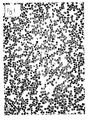
- the method of the present invention was followed visually using a microscope.
- Figure 1 is a microscope picture of differentiated B cells before the method of the present invention.
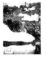
- Figure 2 is a microscope picture of undifferentiated cells formed by the retrodifferentiation of the B cells in accordance with the present invention wherein the agent was a monoclonal antibody to the homologous regions of the ⁇ -chain of HLA-DR antigen. The undifferentiated cells are the dark stained clumps of cells.
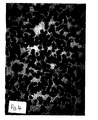
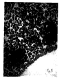
- Figure 3 is a microscope picture of the same undifferentiated cells but at a lower magnification.
- the examples describe in vitro experiments that reveal extremely interesting findings regarding the ontogeny and development of T and B lymphocytes which can be utilised in the generation of stem cells to affect lymphohaematopoiesis in peripheral blood samples in a matter of hours.
- B-CLL B-cell chronic lymphocytic leukaemia's
- B-lymphocytes were constantly observed to lose markers such as CD19.
- CD21, CD23, IgM and DR coincided with the appearance of CD34 + and CD34 + CD2 - cells, increases in CD7 + cells, increases in CD8 + CD28 - and CD28 + cells, increases in CD25 + cells, the appearance of CD10 - and CD34 + cells and CD34 - and CD19 + cells increases in CD5 + cells, and cells expressing low levels of CD45 antigen.
- the immunophenotypic changes associated with such treatment is consistent with retrodifferentiation and subsequent commitment (i.e. recommitment) of B lymphocytes, because the majority of white blood cells in blood of patients with B-CLL before treatment were B lymphocytes. Furthermore, B-lymphocytes of patients with B-CLL which were induced to become T-lymphocytes following treatment with cyclophosphamide and monoclonal antibody to the ⁇ -chain of HLA-DR antigen, were able to revert back to B lymphocytes following prolonged incubation with this treatment.
- CD28 + CD8 - and CD28 + cells appeared after treatment of whole blood of patients with B-CLL with monoclonal antibody to the homologous region of the B chain of the DR antigen. These findings were due to treatment of blood with monoclonal antibody to the homologous region of the ⁇ -chain of HLA-DR antigen.
- T-lymphopoiesis generated in this manner was also observed in peripheral blood of healthy blood donors, cord blood, bone marrow, patients with various infections including HIV + individuals and AIDS patients, enriched fractions for B lymphocytes obtained from blood samples of healthy blood donors, IgA deficient patients and other patients with various other conditions.
- analysis of myeloid markers in treated samples of two patients with B-CLL with monoclonal antibody to the homologous region of the ⁇ -chain of the HLA-DR antigen showed a significant increase in the relative number of cells expressing the myeloid markers such as CD13 and CD33. These markers were coexpressed with the CD56 & 16 or the CD7 antigens.
- stem cells that are produced by the method of the present invention may be stem cells of any tissue and are not necessarily limited to lymphohaematopoietic progenitor cells.
Abstract
Description
- Described herein is a method of preparing an undifferentiated cell.
- The present invention relates to a method of preparing an undifferentiated cell from a more committed cell.
- Described herein is also the use of the undifferentiated cell of the present invention for the preparation of a new more committed cell - i.e. a recommitted cell.
- Also described is the use of the undifferentiated cell of the present invention or the recommitted cell of the present invention to have an effect (directly or indirectly via the use of products obtained therefrom) on the immune system, such as the alleviation of symptoms associated with, or the partial or complete cure from, an immunological condition or disease.
- By way of introduction, differentiation is a process whereby structures and functions of cells are progressively committed to give rise to more specialised cells, such as the formation of T cells or B cells. Therefore, as the cells become more committed, they become more specialised.
- In contrast, retro-differentiation is a process whereby structures and functions of cells are progressively changed to give rise to less specialised cells.
- Undifferentiated cells are capable of multilineage differentiation - i.e. they are capable of differentiating into two or more types of specialised cells. A typical example of an undifferentiated cell is a stem cell.
- In contrast, differentiated cells are incapable of multilineage differentiation. A typical example of a differentiated cell is a T cell.
- There are many undifferentiated cells and differentiated cells found in vivo and the general art is replete with general teachings on them.
- By way of example, reference may be made to inter alia Levitt and Mertelsman 1995 (Haematopoietic Stem Cells, published by Marcel Dekker Inc - especially pages 45-59) and Roitt et al (Immunology, 4th Edition, Eds. Roitt, Brostoff and Male 1996, Publ. Mosby - especially Chapter 10).
- In short, however, examples of undifferentiated cells include lymphohaematopoietic progenitor cells (LPCs). LPCs include pluripotent stem cells (PSCs), lymphoid stem cells (LSCs) and myeloid stem cells (MSCs). LSCs and MSCs are each formed by the differentiation of PSCs. Hence. LSCs and MSCs are more committed than PSCs.
- Examples of differentiated cells include T cells, B cells, eosinophils, basophils, neutrophils, megakaryocytes, monocytes, erythrocytes, granulocytes, mast cells, and lymphocytes.
- T cells and B cells are formed by the differentiation of LSCs. Hence. T cells and B cells are more committed than LSCs.
- Eosinophils, basophils, neutrophils, megakaryocytes, monocytes, erythrocytes, granulocytes, mast cells, NKs, and lymphocytes are formed by the differentiation of MSCs. Hence, each of these cells are more committed than MSCs.
- Antigens are associated with undifferentiated and differentiated cells. The term "associated" here means the cells expressing or capable of expressing, or presenting or capable of being induced to present, or comprising, the respective antigen(s).
- Most undifferentiated cells and differentiated cells comprise Major Histocompatability Complex (MHC) Class I antigens and/or Class II antigens. If these antigens are associated with those cells then they are called Class I- and/or Class II- cells.
- Each specific antigen associated with an undifferentiated cell or a differentiated cell can act as a marker. Hence, different types of cells can be distinguished from each other on the basis of their associated particular antigen(s) or on the basis of a particular combination of associated antigens.
- Examples of these marker antigens include the antigens CD34, CD19 and CD3. If these antigens are present then these particular cells are called CD34+, CD19+ and CD3+ cells respectively. If these antigens are not present then these cells are called CD34-, CD19- and CD3- cells respectively.
- In more detail, PSCs are CD34+ cells. LSCs are DR+, CD34+ and TdT+ cells. MSCs are CD34+, DR+, CD13+, CD33+, CD7+ and TdT+ cells. B cells are CD19+, CD21+, CD22+ and DR+ cells. T cells are CD2+, CD3+, and either CD4+ or CD8+ cells. Immature lymphocytes are CD4+ and CD8+ cells. Activated T cells are DR+ cells. Natural killer cells (NKs) are CD56+ and CD16+ cells. T lymphocytes are CD7+ cells. Leukocytes are CD45+ cells. Granulocytes are CD13+ and CD33+ cells. Monocyte macrophage cells are CD14+ and DR+ cells
- Hence, by looking for the presence of the above-listed antigen markers it is possible to identify certain cell types (e.g. whether or not a cell is an undifferentiated cell or a differentiated cell) and the specialisation of that cell type (e.g. whether that cell is a T cell or a B cell).
- The general concept of retrodifferentiation is not new. In fact, in 1976 Jose Uriel (Cancer Research 36, 4269-4275. November 1976) presented a review on this topic, in which he said:
- "retrodifferentiation appears as a common adaptive process for the maintenance of cell integrity against deleterious agents of varied etiology (physical, chemical, and viral). While preserving the entire information encoded on its genome, cells undergoing retrodifferentiation lose morphological and functional complexity by virtue of a process of self-deletion of cytoplasmic structures and the transition to a more juvenile pattern of gene expression. This results in a progressive uniformization of originally distinct cell phenotypes and to a decrease of responsiveness to regulatory signals operational in adult cells. Retrodifferentiation is normally counterbalanced by a process of reontogeny that tends to restore the terminal phenotypes where the reversion started. This explains why retrodifferentiation remains invariably associated to cell regeneration and tissue repair. "
- Uriel (ibid) then went on to discuss cases of reported retrodifferentiation - such as the work of Gurdon relating to nuclei from gut epithelial cells of Xenopus tadpoles (Advances in Morphogenesis [1966] vol 4, pp 1-43. New York Academic Press, Eds Abercrombie and Bracher), and the work of Bresnick relating to regeneration of liver (Methods in Cancer Research [1971] vol 6, pp 347-391).
- Uriel (ibid) also reported on work relating to isolated liver parenchymal cells for in vitro cultures. According to Uriel:
- "Contrary to the results with fetal or neonatal hepatocytes, with hepatocytes from regenerating liver, or from established hepatomas, it has been difficult to obtain permanent class lines from resting adult hepatocytes. "
- Uriel (ibid) also reported on apparent retrodifferentiation in cancer, wherein he stated:
- "the biochemical phenotypes of many tumours show analogous changes of reversion toward immaturity ... during the preneoplastic phase of liver carcinogenesis, cells also retrodifferentiate. "
- More recent findings on retrodifferentiation include the work of Minoru Fukunda (Cancer Research [1981] vol 41, pp 4621-4628). Fukunda induced specific changes in the cell surface glycoprotein profile of K562 human leukaemic cells by use of the rumour-promoting phorbol ester. 12-O-tetradecanoyl-phorbol-13-acetate (TPA). According to Fukunda TPA appeared to induce the K562 human leukaemic cells into a retrodifferentiated stage.
- Also. Hass et al (Cell Growth & Differentiation [1991] vol 2, pp 541-548) reported that long term culture of TPA-differentiated U-937 leukaemia cells in the absence of phorbol ester for 32-36 days resulted in a process of retrodifferentiation and that the retrodifferentiated cells detached from the substrate and reinitiated proliferation.
-
WO 93/25216 - Another case of retrodifferentiation is the work of Curtin and Snell (Br. J. Cancer [1983] vol 48, pp 495-505]. These workers compared enzymatic changes occurring during diethylnitrosamine-induced hepatocarcinogenesis and liver regeneration after partial hepatectomy to normal liver differentiation. Theses workers found changes in enzyme activities during carcinogenesis that were similar to a step-wise reversal of differentiation. According to these workers, their results suggest that an underlying retrodifferentiation process is common to both the process of hepatocarcinogenesis and liver regeneration.
- More recently. Chastre et al (FEBS Letters [1985] vol 188, number 2, pp 2810-2811] reported on the retrodifferentiation of the human colonic cancerous subclone HT29-18.
- Even more recently, Kobayashi et al (Leukaemia Research [1994] vol 18, no. 12, pp 929-933) have reported on the establishment of a retrodifferentiated cell line (RD-1) from a single rat myelomonocyticleukernia cell which differentiated into a macrophage-like cell by treatment with lipopolysaccharide (LPS).
- According to the current understanding, as borne out by the teachings found on page 911 of Molecular Biology of the Cell (pub. Garland Publishers Inc. 1983) and more recently Levitt and Mertelsman (ibid), a stern cell, such as a PSC, has the following four characteristics:
- i. it is an undifferentiated cell - i.e. it is not terminally differentiated;
- ii. it has the ability to divide without limit;
- iii. it has the ability to give rise to differentiated progeny, such as the differentiated cells mentioned earlier; and
- iv. when it divides each daughter has a choice: it can either remain as PSC like its parent or it can embark on a course leading irreversibly to terminal differentiation.
- Note should be made of the last qualification, namely that according to the general teachings in the art once an undifferentiated cell has differentiated to a more committed cell it can not then retrodifferentiate. This understanding was even supported by the teachings of Uriel (ibid), Fukunda (ibid), Hass et al (ibid), Curtin and Snell (ibid), Chastre et al (ibid), and Kobayashi et al (ibid) as these workers retrodifferentiated certain types of differentiated cells but wherein those cells remained committed to the same lineage and they did not retrodifferentiate into undifferentiated cells.
- Therefore, according to the state of the art before the present invention, it was believed that it was not possible to form undifferentiated cells, such as stem cells, from more committed cells. However, the present invention shows that this belief is inaccurate and that it is possible to form undifferentiated cells from more committed cells.
- The invention provides an in vitro method for preparing an undifferentiated cell, the method comprising contacting a more committed cell that comprises a HLA-DR receptor with an agent that operably enagages the receptor thereby causing the more committed cell to retrodifferentiate into a CD34+ undifferentiated cell wherein the agent is a monoclonal antibody against the homologous region of the beta chain of HLA-DR.
- Also described is a method comprising contacting a more committed cell with an agent that causes the more committed cell to retrodifferentiate into an undifferentiated cell: and then committing the undifferentiated cell to a recommitted cell.
- Thus, in its broadest sense, the present invention is based on the highly surprising finding that it is possible to form an undifferentiated cell from a more committed cell.
- The present invention is highly advantageous as it is now possible to prepare undifferentiated cells from more committed cells and then use those undifferentiated cells as, or to prepare, medicaments either in vitro or in vivo or combinations thereof for the treatments of disorders.
- The present invention is also advantageous as it is possible to commit the undifferentiated cell prepared by retrodifferentiation to a recommitted cell, such as a new differentiated cell, with a view to correcting or removing the original more committed cell or for correcting or removing a product thereof.
- The term "recommitted cell" means a cell derived from the undifferentiated cell - i.e. a new more committed cell.
- Preferably the more committed cell is capable of retrodifferentiating into an MHC Class I+ and/or an MHC Class II+ undifferentiated cell.
- Preferably, the more committed cell is capable of retrodifferentiating into an undifferentiated cell comprising a stem cell antigen.
- Preferably, the more committed cell is capable of retrodifferentiating into a lymphohaematopoietic progenitor cell.
- Preferably, the more committed cell is capable of retrodifferentiating into a pluripotent stem cell.
- The undifferentiated cell may comprise any components that are concerned with antigen presentation, capture or recognition. The undifferentiated cell is an MHC Class II+ cell. MHc Class I+ cells are also disclosed herein.
- Preferably, the undifferentiated cell comprises a stem cell antigen.
- The more committed cell may comprise any components that are concerned with antigen presentation, capture or recognition. The more committed cell is an MHC Class II+ cell. MHC Class I+ cells are also described herein.
- The agent acts extracelluarly of the more committed cell.
- Preferably, the more committed cell comprises a HLA-DR receptor that is operably engageable by the agent and wherein the agent operably engages the receptor.
- Preferably, the HLA-DR receptor comprises at least the homologous regions of the β-chain of HLA-DR.
- Preferably, the receptor comprises at least the homologous regions of the α-chain of HLA-DR.
- The agent is a monoclonal antibody, to the homologous regions of the β-chain of HLA-DR.
- Preferably, the agent is used in conjunction with a biological response modifier.
- Preferably, the biological response modifier is an alkylating agent.
- Preferably, the alkylating agent is or comprises cyclophosphoamide.
- In one preferred embodiment, the more committed cell is a differentiated cell.
- Preferably, the more committed cell is any one of a B cell or a T cell.
- In an alternative preferred embodiment, the more committed cell is a more mature undifferentiated cell.
- In one preferred embodiment, when the undifferentiated cell is committed to a recommitted cell the recommitted cell is of the same lineage as the more committed cell prior to retrodifferentiation.
- In another preferred embodiment, when the undifferentiated cell is committed to a recommitted cell the recommitted cell is of a different lineage as the more committed cell prior to retrodifferentiation.
- Preferably, the recommitted cell is any one of a B cell, a T cell or a granulocyte.
- Preferably, the agent modulates MHC gene expression, preferably wherein the agent modulates MHC Class I+ and/or MHC Class II+ expression.
- The agent operably engages the more committed cell in order to retrodifferentiate that cell into an undifferentiated cell. In this regard, the agent for the retrodifferentiation of the more committed cell into the undifferentiated cell may act in direct engagement or in indirect engagement with the more committed cell.
- An example of direct engagement is when the more committed cell has at least one cell surface receptor on its cell surface, such as a β-chain having homologous regions (regions that are commonly found having the same or a similar sequence) such as those that may be found on B cells, and wherein the agent directly engages the cell surface receptor. Another example, is when the more committed cell has a cell surface receptor on its cell surface such as an α-chain having homologous regions such as those that may be found on T cells, and wherein the agent directly engages the cell surface receptor.
- An example of indirect engagement is when the more committed cell has at least two cell surface receptors on its cell surface and engagement of the agent with one of the receptors affects the other receptor which then induces retrodifferentiation of the more committed cell.
- The agent for the retrodifferentiation of the more committed cell into an undifferentiated cell may be a chemical compound or composition. The agent is capable of engaging the HLA-DR receptor on the surface of the more committed cell.
- The agent according to the invention is a monoclonal antibody against the homologous region of the β chain of HLA-DR. Also described herein are antibodies to one or more of: the β chain of MHC class II antigen, the α chain of MHC class I or class II antigen, the α chain of HLA-DR antigen, the α and the β chain of MHC class II antigen or of a MHC class I antigen. An example of a sutiable antibody is CR3/43 (supplied by DAKO).
- The more committed cell is any cell derived or derivable from an undifferentiated cell.
- Thus, in one preferred embodiment, the more committed cell is also an undifferentiated cell. By way of an example therefore the undifferentiated cell can be a lymphoid stem cell or a myeloid stem cell, and the undifferentiated cell is a pluripotent stem cell.
- Described herein are more committed cells that are differentiated cells, such as a CFC-T cell, a CFC-B cell, a CFC-Eosin cell, a CFC-Bas cell, a CFC-GM cell, a CFC-MEG cell, a BFC-E cell, a CFC-E cell, a T cell, a B cell, an eosinophil, a basophil, a neutrophil, a monocyte, a megakaryocyte or an erythrocyte; and the undifferentiated cell is a myeloid stem cell, a lymphoid stem cell, or a pluripotent stem cell.
- Described herein is a committed cell that is a differentiated cell wherein the differentiated cell is B lymphoma (activated or non-activated), a T lymphocyte (activated or non-activated), a cell from the macrophage monocyte lineage, a nucleated cell capable of expressing class I or class II antigens, a cell that can be included to express class I or class II antigens or an enucleated cell (i.e. a cell that does not contain a nucleus - such as a red blood cell).
- Described herein are differentiated cells wherein the differentiated cell that is selected from any one of a group of cells comprising large granular lymphocytes, null lymphocytes and natural killer cells, each expressing the CD56 and/or CD16 cell surface receptors.
- The differentiated cell may even be formed by the nucleation of an enucleated cell.
- The agent may act intracellularly within the more committed cell. However, preferably, the agent acts extracelluarly of the more committed cell.
- According to the invention, agent operably engages the HLA-DR receptor present on the surface of the more committed cell - which receptor may be expressed by the more committed cell, such as a receptor that is capable fo being expressed by the more committed cell.
- Also described herein are receptors wherein the receptor is a Class I or a Class II antigen of the major histocompatibility complex (MHC) and cell surface receptor such as an HLA-DR receptor, a DM receptor, a DP receptor, a DQ receptor, an HLA-A receptor, an HLA-B receptor, an HLA-C receptor, an HLA-E receptor, an HLA-F receptor, or an HLA-G receptor.
- Described herein is a contacting step comprising the agent engaging with any one or more of the following: homologous regions of the α-chain of class I antigens, homologous regions of the α-chain of class II antigens, a CD4 cell surface receptor, a CD8 cell surface receptor, homologous regions of the β-chain of class II antigens in the presence of lymphocytes, homologous regions of the α-chain of class I antigens in the presence of lymphocytes, or homologous regions of the α-chain of class II antigens in the presence of lymphocytes.
- Preferably the contacting step occurs in the presence of the biological response modifier.
- Preferably the biological response modifier is any one or more of a modulator, such as an immunomodulator, a growth factor, a cytokine, a cell surface receptor, a hormone, a nucleic acid, a nucleotide sequence, an antigen or a peptide.
- In a preferred embodiment of the present invention the undifferentiated cell is then committed into a recommitted cell, such as a differentiated cell.
- The recommitted cell may be of the same lineage to the more committed cell from which the undifferentiated cell was derived.
- Alternatively, the recommitted cell may be of a different lineage to the more committed cell from which the undifferentiated cell was derived.
- Also described herein is a method for preparing an undifferentiated cell, wherein the method includes committing the undifferentiated cell into a recommitted cell and then fusing the recommitted cell to a myeloma. This allows the expression in vitro of large amounts of the desired product, such as an antibody or an antigen or a hormone etc.
- Also described herein are the following:
- The use of any one of the agents of the present invention for preparing an undifferentiated cell from a more committed cell.
- The use of an undifferentiated cell produced according to the method of the present invention for producing any one of a monoclonal or a polyclonal or a specific antibody from a B-lymphocyte or a T-lymphocyte; a cell from the macrophage monocyte lineage; a nucleated cell capable of expressing class I or class II antigens; a cell capable of being induced to express class I or class II antigens: an enucleated cell; a fragmented cell; or an apoptic cell.
- The use of an undifferentiated cell produced according to the method of the present invention for producing effector T-lymphocytes from B-lymphocytes and/or vice versa.
- The use of an undifferentiated cell produced according to the method of the present invention for producing any one or more of: a medicament, such as a medicament comprising or made from a B-lymphocyte, a T-lymphocyte, a cell from the macrophage monocyte lineage, a nucleated cell capable of expressing a class I or a class II antigen, a cell capable of being induced to express a class I or a class II antigen, or an enucleated cell.
- Processes utilising the afore-mentioned uses and products or compositions prepared from such processes.
- A medicament comprising an undifferentiated cell or a product obtained therefrom admixed with a suitable diluent, carrier or excipient.
- The medicament may comprise an antibody or antigen obtained from an undifferentiated cell admixed with a suitable diluent, carrier or excipient.
- The medicament may be for the treatment of any one of: cancer, autoimmune diseases, blood disorders, cellular or tissue regeneration, organ regeneration, the treatment of organ or tissue transplants, or congenital metabolic disorders.
- A process of introducing a gene into the genome of an undifferentiated cell, wherein the process comprises introducing the gene into a more committed cell, and then preparing an undifferentiated cell by the method according to the present invention, whereby the gene is present in the undifferentiated cell.
- A process of introducing a gene into the genome of an undifferentiated cell, wherein the process comprises inserting the gene into the genome of a more committed cell, and then preparing an undifferentiated cell by the method according to the present invention, whereby the gene is present in the undifferentiated cell is also described.
- A process of introducing the genome of a gene into an undifferentiated cell, wherein the process comprises inserting the gene into the genome of a more committed cell, and then preparing an undifferentiated cell by the method according to the present invention, whereby the gene is present in the genome of the undifferentiated cell is described herein
- An undifferentiated cell prepared by any one of these processes of the present invention is described herein
- As already mentioned, described herein is a medicament comprising an undifferentiated cell prepared by any one of these processes admixed with a suitable diluent, carrier or excipient. With such a medicament the undifferentiated cell could be used to produce a beneficial more committed cell, such as one having a correct genomic structure, in order to alleviate any symptoms or conditions brought on by or associated with a more committed cell having an incorrect genomic structure.
- Described herein is a process of removing an acquired mutation from a more committed cell wherein the method comprises forming an undifferentiated cell by the method according to the present invention, committing the undifferentiated cell into a recommitted cell, whereby arrangement or rearrangement of the genome and/or nucleus of the cell causes the mutation to be removed.
- The gene may be inserted into the immunoglobulin region or TCR region of the genome.
- Alternatively, the undifferentiated cell could be used to produce a more committed cell that produces an entity that cures any symptoms or conditions brought on by or associated with a more committed cell having an incorrect genomic structure.
- For example, the present invention may be used to prepare antibodies or T cell receptors to an antigen that is expressed by the more committed cell which has retrodifferentiated into the undifferentiated cell. In this regard, the antigen may be a fetospecific antigen or a cross-reactive fetospecific antigen.
- Also described is a process of controlling the levels of undifferentiated cells and more committed cells. For example, a method comprising forming an undifferentiated cell by the method according to the present invention and then activating an apoptosis gene to affect the undifferentiated cell, such as bring about the death thereof is described.
- In one preferred embodiment of the present invention, the more committed cell is not a cancer cell. In another preferred embodiment of the present invention, the agent is neither carcinogenic nor capable of promoting cancer growth.
- Also described herein is a method of treating a patient suffering from a disease or a disorder resulting from a defective cell or an unwanted cell, the method comprising preparing an undifferentiated cell by contacting a more committed cell with an agent that causes the more committed cell to retrodifferentiate into the undifferentiated cell, and then optionally committing the undifferentiated cell into a recommitted cell; wherein the undifferentiated cell, or the recommitted cell, affects the defective cell or the unwanted cell to alleviate the symptoms of the disease or disorder or to cure the patient of the disease or condition.
- In summation, the present invention relates to the preparation of an undifferentiated cell from a more committed cell.
- The following Figures and examples are useful in understanding the invention.
-
Figure 1 which is a microscope picture of cells before the method of the present invention; -
Figure 2 which is a microscope picture of cells prepared by the method of the present invention; -
Figure 3 which is a microscope picture of cells prepared by the method of the present invention but at a lower magnification; -
Figure 4 which is a microscope picture of cells before the method of the present invention; -
Figure 5 which is a microscope picture of cells prepared by the method of the present invention; and -
Figure 6 which is a microscope picture of cells prepared by the method of the present invention. - Blood samples were obtained in lavender top tubes containing EDTA from patients with B-cell chronic lymphocytic leukaemia's, patients with antibody deficiency (including IgA deficiency and X-linked infantile hypogammaglobulinaemias), patients with HIV infections and AIDS syndrome, a patient with CMV infection, a patient with Hodgkin's lymphomas, a patient with acute T-cell leukaemia, a 6-days old baby with Hodgkin's lymphomas, a patient with acute T-cell leukaemia, a 6-days old baby with blastcytosis, various patients with various infections and clinical conditions, cord blood, bone marrow's, and enriched B-lymphocyte preparations of healthy blood donors.
- The clinical and experimental treatment conditions of patients, including various types of treatment applied to their blood samples, are described in Table 1. Differential white blood cell (WBC) counts were obtained using a Coulter Counter and these are included in the same Table.
- Blood samples, once obtained, were treated with pure monoclonal antibody to the homologous region of the β-chain of the HLA-DR antigen (DAKO) and left to mix on a head to head roller at room temperature for a maximum of 24 hours. Some samples were mixed first on a head to head roller for 15 minutes after which they were left to incubate in an incubator at 22°C. The concentration of monoclonal antibody added to blood samples varied from 10-50µl/ml of blood.
- In addition, other treatments treatments were applied at the same concentrations and these included addition of a monoclonal antibody to the homologous of the α-chain of the HLA-DR antigen, a monoclonal antibody to the homologous region of class I antigens, a monoclonal antibody to CD4. a monoclonal antibody to CD8, and a PE conjugated monoclonal antibody to the homologous region of the β-chain of the HLA-DR antigen.
- Other treatments included the simultaneous addition of monoclonal antibodies to the homologous regions of the α and β-chains of the HLA-DR antigen to blood samples.
- Furthermore, alkylating agents such as cyclophosphoamide were added to blood samples in combination with pure monoclonal antibody to the homologous region of the β-chain of the HLA-DR antigen.
- Following these treatments blood samples were stained with panels of labelled monoclonal antibodies as instructed by the manufacturer's instructions and then analyzed using flow cytometry.
- Incubation periods with monoclonal antibodies ranged from 2 hour. 4 hour, 6 hour. 12 hour to 24 hour intervals.
- The following monoclonal antibodies were used to detect the following markers on cells by flow cytometry: CD19 and CD3, CD4 and CD8, DR and CD3, CD56 & 16 and CD3. CD45 and CD14, CD8 and CD3, CD8 and CD28, simultest control (IgG1 FITC + IgG2a PE), CD34 and CD2, CD7 and CD13 & 33, CD10 and CD25, CD5 and CD10, CD5 and CD21, CD7 and CD5, CD13 and CD20. CD23 and CD57 and CD25 and CD45 RA (Becton & dickenson and DAKO).
- Each patient's blood sample, both treated and untreated, was analyzed using the majority of the above panel in order to account for the immunophenotypic changes that accompanied different types of treatments and these were carried out separately on different aliquots of the same blood sample. Untreated samples and other control treatments were stained and analyzed simultaneously.
- Whole blood sample was stained and lysed according to the manufacturer instructions. Flow cytomery analysis was performed on a FACScan@ with either simultest or PAINT A GATE software (BDIS) which included negative controls back tracking. 10,000 to 20.000 events were acquired and stored in list mode files.
- Morphology was analyzed using microscopy and Wright's stain.
- Treatment of blood samples with monoclonal antibody to the homologous region of the β-chain of the HLA-DR antigen always decreased the relative number of CD19+ cells. This marker is a pan B-cell antigen (see Table). This antigen is present on all human B lymphocytes at all stages of maturation but is lost on terminally differentiated plasma cells. Hence, this is an indication that B cells were retrodifferentiating into undifferentiated cells.
- The same treatment caused the relative number of CD3+ cells to increase dramatically especially in blood of patients with B-CLL, which was always accompanied by an increase in the relative number in CD3-CD19- cells. CD3 is present on all mature T-lymphocytes and on 65%-85% of thymocytes. This marker is always found in association with α-/β- or gamma/delta T-cell receptors (TCR) and together these complexes are important in transducing signals to the cell interior. Hence, this is an indication that B cells were retrodifferentiating into undifferentiated cells and then being committed to new differentiated cells, namely T cells.
- A novel clone of cells appeared in treated blood of B-CLL patients co-expressing the CD19 and CD3 markers - i.e. CD19+ and CD3+ cells (see Charts 1, patient 2, 3 & 4 at 2hr. 6hr & 24hr of starting treatment). Other patients with different conditions showed an increase in the relative number of these clones of cells. These cells were exceptionally large and heavily granulated and extremely high levels of CD 19 were expressed on their cell membrane. The CD3 marker seems to be expressed on these cells at similar levels to those expressed on normal mature lymphocytes.
- In Table 2, patient numbers 2, 3 and 4 are actually numbers representing the same patient and their delineation was merely to show the effect of treatment on blood with time (See Table 1 for experimental and clinical condition of this patient).
- The CD19+CD3+ clones in treated samples seem to decrease with time, reaching original levels to those determined in untreated sample at 2hrs, 6hrs and 24hrs time.
- Another type of cell of the same size and granulity was detected in treated samples and these cells had high levels of CD 19 expressed on their surface but were negative for the CD3 marker and rich in FC receptors. However, the relative number of these cells appeared to decrease in time. Of interest, at 24 hours treatment of blood sample (2, 3 and 4) there was a decrease in the relative number of CD19-CD3- cells in a group of cells that were initially observed to increase after 2 and 6 hr's treatment of blood samples. However, Coulter counts of WBC populations were reduced on treatment of blood with monoclonal antibody to the homologous region of the β-chain of the HLA-DR antigen. This finding suggests that this type of treatment gives rise to atypical cells that cannot be detected by Coulter (Table 1) but can be accounted for when measured by flow cytometry which counts cells on the basis of surface markers, size and granulity. Furthermore, these atypical cells were accounted for by analysing morphology using Wright's stain under a microscope. Flow cytometric charts of these phenomena are represented in Charts (1, 2, 3 & 4) and the immunophenotypic changes obtained on treatment of blood samples seems to suggest that CD19+ and CD3+ lymphocytes are an interconnected group of cells but remain distinct on the basis of CD19 and CD3 relative expression compared to stem cells.
- In Table 2, patient numbers 5 and 6 represent the same patient but analysis of treated and untreated blood samples were monitored with time and at the same time (see Table 1).
- Patients blood with no B-cell malignancy showed similar trends of immunophenotypic changes when compared to blood of B-CLL patients but the changes were not to the same extent. However, the relative and absolute number of B-lymphocytes and MHC class II positive cells in the blood of these patients are extremely low compared to those found in the blood of B-CLL patients.
- Two brothers both with X-linked infantile hypogammaglobulinemia who were B cell deficient showed different immunophenotypic changes in the relative number of CD3+ cells on treatment of their blood. The younger brother who was 2 months old and not ill, on treatment of his blood, showed a slight increase in the relative number of CD3+ cells which was accompanied by a decrease in the relative number of CD3- CD 19- cells. On the other hand, the other brother who was 2 years old and was extremely sick and with a relatively high number of activated T cells expressing the DR antigens showed a decrease in the number of CD3+ cells on treatment of his blood. No other markers were used to measure other immunophentypic changes that might have occurred because the blood samples obtained from these two patients were extremely small (Table 2. ID 43/BD and 04/BD).
- Patient 91 in Table 2 shows a decrease in the relative number of CD3+ cells following treatment of blood which was accompanied by an increase in the relative number of CD3-CD19- cells. However, on analysis of other surface markers such as CD4 and CD8 (see Table 3) the patient was observed to have a high relative number of CD4+CD8+ cells in his blood and this was noted prior to treatment of blood samples with monoclonal antibody to the β-chain of the DR antigen and these double positive cells decreased appreciably following treatment of blood. Furthermore, when further markers were analyzed the relative number of CD3+ cells were seen to have elevated (See Table 4).
- An enriched preparation of B-lymphocytes obtained from healthy blood donors when treated with monoclonal antibody to the β-chain of DR antigens showed a dramatic increase in the relative number of CD3+ cells which were always accompanied by a decrease in the relative number of CD19+ cells and by an increase in the relative number of CD19-CD3- cells. Further analysis using markers such as CD4 and CD8 show a concomitant increase in the relative number of these markers. However, an enriched preparation of T lymphocytes of the same blood donors when treated with the same monoclonal antibody did not show the same changes.
- The CD4 antigen is the receptor for the human immunodificiency virus. The CD4 molecule binds MHC class II antigen in the B2 domain, a region which is similar to the CD8 binding sites on class I antigens. Binding of CD4 to class II antigen enhances T cell responsiveness to antigens and so does the binding of CD8 to class I antigens. The CD8 antigens are present on the human supressor/cytotoxic T-lymphocytes subset as well as on a subset of natural killer (NK) lymphocytes and a majority of normal thymocytes. The CD4 and CD8 antigens are coexpressed on thymocytes and these cells lose either markers as they mature into T-lymphocytes.
- On analysis of the CD4 and CD8 markers - see below - and from a majority of blood samples presented in Table 2, a pattern of staining emerges which supports the presence of a retrodifferentiation process of B-lymphocytes into undifferentiated cells and the subsequent differentiation into T -lymphocytes.
- CD4+CD8- cells, which are double positive cells, always appeared following treatment of blood samples with monoclonal antibody to the homologous region of the β-chain and these types of cells were markedly increased in the blood of treated samples of patients with B-CLL and which were absent altogether in untreated samples (See Table 3 and Charts 1, 2 3 & 4). In the same specimens the relative number of single positive cells such as CD8+ and CD4+ cells was also noted to increase simultaneously. Furthermore, a decrease in the relative number of CD4- CD8- cells which, at least in the case of B-CLL correspond to B cells was noted to fall dramatically in treated samples when compared to untreated specimens which remained at the same level when measured with time. However, measurement of the relative number of CD4+CD8+ cells with time in treated samples showed that there was a concomitant increase in the number of single positive cells with a decrease in the relative number of double positive cells. This type of immunophenotypic change is characteristic of thymic development of progenitor cells of the T-lymphocyte lineage in the thymus (Patient number 2,3 and 4). The CD4 antigen is present on the helper/inducer T- lymphocyte subsets (CD4+CD3+) and a majority of normal thymocytes. However, this antigen is present in low density on the cell surface of monocytes and in the cytoplasm of monocytes and macrophages (CD3-CD4-).
- The relative number of CD4+ low cells was affected differently in different blood samples following treatment. The relative number of this type of cells seems unaffected in blood samples of patients with B-CLL following treatment when compared to untreated samples. Such low levels of CD4 expression is found on monocytes and very early thymocytes.
- Patient HIV+25 on treatment showed a substantial increase in the number of double positive cells expressing CD4 and CD8 simultaneously. On the other hand, patient 91 on treatment showed a decrease in this subtype of cells and the observation of such phenomenon is time dependent. The relative number of CD8+ cells was observed to increase in untreated blood samples of patients with B-CLL when measured with time whereas the relative number of CD4+ and CD4+ low cells was observed to decrease at the same times (Table 3 patient 2,3 and 4).
- The DR markers are present on monocytes, dendritic cells, B-cells and activated T-lymphocytes.
- Treated and untreated samples analysed with this panel showed similar immunophenotypic changes to those obtained when blood samples were analysed with the CD19 and CD3 markers (see Table 2) and these antigens as mentioned earlier are pan B and T-cell markers respectively.
- Treatment of blood with monoclonal antibodies seems to affect the relative number of DR+ B-lymphocytes so that the level of DR+ cells decrease. In contrast, the relative number of CD3+ (T-cells) cells increase significantly (see Table 4 and Chart).
- Furthermore, the relative number of activated T cells increased in the majority of treated blood samples of patients with B-CLL and these types of cells were affected variably in treated samples of patients with other conditions. Furthermore, the relative number of DR high positive cells appeared in significant numbers in treated samples of patients with B-CLL and a 6 day old baby with increased DR+ CD34+ blasts in his blood. However, it should be noted that the blasts which were present in this patient's blood were negative for T and B-cell markers before and after treatment but became more positive for myeloid lineage antigens following treatment. The relative number of CD3-DR- cells increased in the majority of treated blood samples and was proportional to increases in the relative number of CD3+ cells (T-cells) and was inversely proportional to decreases in the relative number of DR+ cells (B-cells).
- The CD56&CD16 markers are found on a heterogeneous group of cells, a subset of lymphocytes known generally as large granular lymphocytes and natural killer (NK) lymphocytes. The CD16 antigen is expressed on virtually all resting NK lymphocytes and is weakly expressed on some CD3+ T lymphocytes from certain individuals. This antigen is found on granulocytes in lower amount and is associated with lymphocytes containing large azurophilic granules. The CD 16 antigen is the IgG FC receptor III.
- A variable number of CD16+ lymphocytes coexpress either the CD57 antigen or low-density CD8 antigen or both. In most individuals, there is virtually no overlap with other T-lymphocyte antigens such as the CD5, CD4. or CD3 antigens. The CD56 antigen is present on essentially all resting and activated CD16+ NK lymphocytes and these subsets of cells carry out non-major histocompatibility complex restricted cytotoxicity.
- Immunophenotyping of treated and untreated blood samples of B-CLL and some other patients with other conditions showed an increase in the relative number of cells coexpressing the CD56&CD16 antigens which were heavily granulated and of medium size (see Table 5 and Charts 1, 2, 3 & 4). These observations were also accompanied by a marked increase in the relative number of cells expressing the CD3 antigen only (without the expression of CD56 and CD16 markers) and cells coexpressing the CD56&CD16 and CD3 markers together.
- In Table 5, patient numbers 2, 3, and 4 represent the same blood sample but being analysed at 2 hours, 6 hours and 24 hours respectively (before and after treatment). This sample shows that treatment of blood with monoclonal antibody to the homologous region of the β-chain of DR antigen seems to cause spontaneous production of CD56+ and CD16- cells, CD3+ cells and CD56+ and CD16+ CD3- cells and these observations were always accompanied by the disappearance of B-cell markers (CD19, DR. CD56, CD16-CD3-).
- Onward analysis of this blood sample before and after treatment showed the levels of CD56+ and CD16+ cells to decrease with time and the level of CD3- cells to increase with time.
- Blood samples of patient 7 with B-CLL, did not show any changes in the number of cells expressing the CD56. CD16 and CD3 antigens when compared to immunophenotypic changes observed in treated and untreated samples and this is because the amount of monoclonal antibody added was extremely low relative to the number of B lymphocytes. However, treatment of this patient's blood sample on a separate occasion with an appropriate amount of monoclonal antibody showed significant increases in the relative number of CD3+, CD56+ & CD16+ and CD56+ and CD16+ CD3+ cells.
- Blood samples of other patients with other conditions showed variable changes in the level of these cells and this seems to be dependent on the number of B-lymphocytes present in blood before treatment, duration of treatment and probably the clinical condition of patients.
- The CD45 antigen is present on all human leukocytes, including lymphocytes, monocytes, polymorphonuclear cells, eosinophils, and basophils in peripheral blood, thymus, spleen, and tonsil, and leukocyte progenitors in bone marrow.
- The CD14 is present on 70% to 93% of normal peripheral blood monocytes, 77% to 90% of pleural or peritoneal fluid phagocytes. This antigen is weakly expressed on granulocytes and does not exist on unstimulated lymphocytes, mitogen-activated T lymphocytes, erythrocytes, or platelets.
- The CD45 antigen represents a family of protein tyrosine phosphatases and this molecule interacts with external stimuli (antigens) and effects signal transduction via the Scr-family members leading to the regulation of cell growth and differentiation.
- Engagement of the β-chain of the DR antigens in treated blood samples especially those obtained from patients with B-CLL suggests that such a treatment affects the level of CD45 antigens on B-lymphocytes. The overall immunophenotypic changes that took place on stimulation of the β-chain of the DR antigen seem to give rise to different types of cells that can be segregated on the basis of the level of CD45 and CD14 expression as well as morphology as determined by forward scatter and side scatter (size and granulity respectively) and these results are presented in Table 6 and Charts (1. 2, 3. 4 & 5).
- On treatment the relative number of CD45 low cells (when compared to untreated samples) increased significantly and so did the relative number of cells co-expressing the CD45 and CD14 antigens. This type of immunophenotypic changes coincided with a decrease in the relative number of CD45 high cells (compared to untreated samples). However, this latter population of cells can be further divided on the basis of morphology and the degree of CD45 expression. One type was extremely large and had extremely high levels of CD45 antigen when compared to the rest of cells present in the charts (see charts 1, 2, 3 and 4). On analysis of this panel following treatment with time (see Table patient 2,3 and 4 and charts) the relative number of CD45+ cells initially fell drastically with time to give rise to CD45 low cells. However, analysis of blood 24 hours later showed the opposite situation.
- Samples 5 and 7 reveal opposite immunophenotypic changes to those obtained with other samples obtained from other B-CLL patients and this is because the samples were analysed at a much earlier incubation time with the monoclonal antibody. In fact the sequential analysis of blood samples after treatment seems to suggest that the immunophenotypic changes undertaken by B lymphocytes is time dependent because it represents a stage of development and the immunophenotypic changes measured at time X is not going to be the same at time X plus (its not fixed once induced). However, these types of changes must be occurring in a more stringent manner in the body otherwise immunopathology would ensue. The effect of treatment of blood samples from other patients with no B-cell malignancy show variable changes in immunophenotypes of cells and this because B-lymphocytes are present in lower amount. However, treatment of enriched fractions of B-lymphocytes obtained from healthy blood donors show similar immunophenotypic changes to those obtained with B-CLL with high B lymphocyte counts.
- The CD8 antigenic determinant interacts with class I MHC molecules, resulting in increased adhesion between the CD8+ T lymphocytes and the target cells. This type of interaction enhances the activation of resting lymphocytes. The CD8 antigen is coupled to a protein tyrosine kinase (p56ick) and in turn the CD8/p56ick complex may play a role in T-lymphocyte activation.
- Treatment of blood samples obtained from patients with B-CLL with monoclonal antibody to the B chain causes a significant increase in the relative number of CD3CD8 and CD3 (highly likely to be CD4CD3) positive cells thus indicating more clearly that double positive cells generated initially are undergoing development into mature T-lymphocytes. This is a process that can be measured directly by CD19 and by DR and indirectly by CD8-CD3- antigens. Serial assessment of treated blood samples of the same patient with time seems to agree with a process which is identical to thymocyte development (Table 7, patient 2, 3 and 4 and Chart 1).
- The relative number of CD8+ cells increased with time in treated and untreated samples but to a higher extent in untreated samples. On the other hand, the relative number of CD8+CD3+ cells decreased with time in untreated samples. However, the relative number of CD3+ cells increased in treated blood samples when measured with time and these types of cells highly correspond to CD4+CD3+ single positive cells: a maturer form of thymocytes. In addition, since these samples were also immunophenotyped with other panels (mentioned above in Tables 3, 4, 5 and 6) the overall changes extremely incriminate B cells in the generation of T lymphocyte progenitors and progenies.
- Blood samples from a patient with B-CLL (number 2, 3 and 4 Tables 1, 2, 3, 4, 5, 6, 7) in separate aliquots were treated with nothing, PE conjugated monoclonal antibody to the homologous region of the β-chain of DR antigen and unconjugated form of the same monoclonal antibody. On comparison of PE conjugated treatment clearly indicates no change in the relative number of CD3 positive cells and associated markers such as CD4 which have been observed in significant levels when the same blood sample was treated with unconjugated form of the antibody. However, an increase in the number of CD45 positive cells with no DR antigen being expressed on their surface was noted when measured with time (see Table 8). A finding that was similar to that noted in untreated samples when immunophenotyped with time (Table 6). Furthermore, the relative number of cells expressing CD45 low decreased in time, a phenomenon which was also noted in the untreated samples (when measured with time) of the same patient (see chart 1A).
- Treatment of blood samples with monoclonal antibody to the homologous region of the α-chain of the DR antigen and the homologous region of MHC Class I antigens decreased the number of CD3+ cells and increased the number of CD19- cells. Treatment of the same blood with monoclonal antibody to the homologous region of the β-chain of the DR antigen decreased the number of CD19+ cells and increased the number of CD3- cells. Treatment with the latter monoclonal antibody with cyclophosphoamide revealed the same effect (Table 14 patient 5/6 with B-CLL at 2hr treatment).
- Onward analysis of CD19+ and CD3+ cells in the same samples revealed further increases in the relative number of CD3+ cells only in blood treated with monoclonal antibody to the homologous region of the β-chain of DR antigen (Table 14 patient 5/6 at 24 hours following treatment). However, onward analysis (24 hours later patient 5/6 Table 14) of blood samples treated with cyclophosphamide plus monoclonal antibody to the β-chain of DR antigen show reversal in the relative number of CD19+ and CD3- cells when compared to that observed at 2 hour incubation time under exactly the same condition.
- In general, treatment of blood samples of the same patient with monoclonal antibody to the homologous region of the α chain of the DR antigen or monoclonal antibody to the homologous of the α-chain of the class I antigen shows an increase in the relative number of CD19- cells (pan B marker) when compared to untreated sample. The relative number of CD19-CD3- cells decreased slightly in blood samples treated with monoclonal antibody to the α-chain of DR antigen or treated with monoclonal antibody to class I antigens (see Table 14 & Charts 2, 3 & 4). Treatment of blood samples of patient 09 with monoclonal antibody to class I antigens increased the relative number of CD3+ cells and decreased slightly the relative number of CD19+ and CD19-CD3- cells. However, treatment of an enriched preparation of B-lymphocytes obtained from healthy blood donors with monoclonal antibody to the β-chain or α-chain of DR antigen showed similar immunophenotypic changes to those obtained with patient with B-CLL.
- Treatment of HIV+ and IgA deficient patients with monoclonal antibody to the β-chain of the DR antigen increased the relative number of CD3- cells and decreased the relative number of CD19- cells. However, treatment of the same blood sample with monoclonal antibody to the homologous region of class I antigen did not produce the same effect. Treatment of blood samples obtained from patients (34/BD and 04/BD) with B-cell deficiency showed variable immunophenotypic changes when treated with monoclonal antibodies to the β-chain of the DR antigen, class I antigens and CD4 antigen.
- Blood samples analysed using the CD19 and CD3 panel (Table 14) were also immunophenotyped with the CD4 and CD8 panel (Table 15). Both panels seem to agree and confirm each other. Incubation for 2 hours of blood samples of patients with B-CLL (Table 15, patients 5/6 and 10. Charts 2. 3 & 4) with monoclonal antibody to the homologous region of the β-chain of the DR antigen or with this monoclonal antibody plus cyclophosphoamide increased the relative number of CD8 and CD4- cells and cells coexpressing both markers. On the other hand, treatment of the same samples with monoclonal antibodies to the homologous region of the α-chain of the DR antigen or the homologous region of the α-chain of class I antigen did not produce the same effects.
- Comparison of immunophenotypic trends obtained at 2 hours and 24 hours incubation periods with monoclonal antibody to the β-chain of the DR antigen plus cyclophosphoamide revealed reverse changes in the relative number of CD4 and CD8 positive cells (Table 15, patient 5/6 with B-CLL at 2 hours and 24 hours) and such changes were in accordance with those obtained when the same blood sample was analysed with the CD19 and CD3 panel (Table 14 the same patient). The later findings indicate that the subsequent differentiation is reversible as the undifferentiated cells can differentiate into T-lymphocytes or B-lymphocytes.
- The immunophenotypic changes obtained with DR and CD3 (Table 16) panel confirm the findings obtained with CD19 and CD3 panel and CD4 and CD8 panel (Tables 14 & 15 & Charts 2. 3 & 4) which followed treatment of the same blood samples with monoclonal antibodies to the homologous region of the beta- or alpha- side of the DR antigen or monoclonal antibody to class I antigens or monoclonal antibody to the β-chain of the DR antigen plus cyclophosphoamide at 2 hour analysis.
- From the results, it would appear that the monoclonal antibody to the homologous region of the β-chain of the DR antigen is extremely capable of driving the production of CD3 positive cells from DR+ cells.
- Furthermore, treatments such as those involving engagement of the α-chain of DR antigens or engagement of the β-side of the molecule in conjunction with cyclophosphoamide (prolonged incubation time) promoted increases in the relative number of CD19+ cells or DR+ cells.
- Treatment of blood samples, especially of those of patients with B-CLL with high B-lymphocyte counts with monoclonal antibody to the homologous region of the β-chain of the DR antigen increased the relative number of CD56&16 positive cells.
- In these patients the relative number of CD3+ and CD56+ and CD16+CD3- cells also increased following treatment of blood samples with monoclonal antibody to the β-chain, confirming earlier observations noted with the same treatment when the same blood samples were analysed with CD3 and CD19 and DR and CD3 panels.
- Blood samples treated with monoclonal antibodies to the β- or alpha- chains of the DR antigen or to the β-chain plus cyclophosphoamide or class I antigens were also analysed with the CD45 and CD14 panel (Table 18). The delineation of CD45 low, CD45 high and CD45 medium is arbitrary. Treatment of blood sample 5/6 (at 2 hours) with monoclonal antibodies to the β-chain of the DR antigen or with this monoclonal antibody plus cyclophosphoamide generated CD45+ low cells and increased the relative number of CD45- medium cells. However, the former treatment increased the relative number of CD45+ high cells and the latter treatment decreased the relative number of CD45+ medium cells and these changes appeared to be time dependent.
- Blood samples of patient 5/6 and 10 (B-CLL) on treatment with monoclonal antibody to class I antigens showed a decrease in the relative number of CD45+ medium cells and similar observations were noted in blood samples 09 and HIV+ following the same treatment when compared to untreated samples. Treatment of blood samples of HIV + and IgA/D patients with monoclonal antibody to class I antigen increased the relative number of CD45+ low cells when compared to untreated samples or samples treated with monoclonal antibody to the β-chain of the DR antigen. However, blood samples of these patients showed a decrease in the relative number of CD45+ medium cells on treatment with monoclonal antibody to the homologous regions of the β-chain of the DR antigen. Medium CD45+ cells increased in blood samples of IgA/D patient following monoclonal antibody to class I antigen treatment. Cells that were extremely large, heavily granular and expressing intense levels of CD45 antigen were noted in treated blood samples with monoclonal antibody to the homologous region of the β-chain of DR antigen of MHC class II antigens (see Charts 1, 2, 3, 4 & 5).
- The CD28 antigen is present on approximately 60% to 80% of peripheral blood T (CD3+) lymphocytes, 50% of CD8+ T lymphocytes and 5% of immature CD3-thymocytes. During thymocyte maturation, CD28 antigen expression increases from low density on most CD4+CD8+ immature thymocytes to a higher density on virtually all mature CD3+, CD4+ or CD8- thymocytes. Cell activation further augments CD28 antigen density. Expression of the CD28 also divides the CD8+ lymphocytes into two functional groups. CD8+CD28+ lymphocytes mediate alloantigen-specific cytotoxicity, that is major histocompatibility complex (MHC) class I-restricted. Suppression of cell proliferation is mediated by the CD8+CD28+ subset. The CD28 antigen is a cell adhesion molecule and functions as a ligand for the B7/BB-1 antigen which is present on activated B lymphocytes.
- Treatment of blood samples of patients (Table 19, patients 5/6 and 8) with B-CLL with monoclonal antibody to the homologous region of β-chain of the DR antigen increased the relative number of CD8+, CD28+ and CD8+CD28+ cells and all other types of treatments did not.
- The CD34 antigen is present on immature haematopoietic precursor cells and all haematopoietic colony-forming cells in bone marrow, including unipotent (CFU-GM, BFU-E) and pluripotent progenitors (CFU-GEMM. CFU-Mix and CFU-blast). The CD34 is also expressed on stromal cell precursors. Terminal deoxynucleotidyl transferase (TdT)+ B- and T-lymphoid precursors in normal bone are CD34-, The CD34 antigen is present on early myeloid cells that express the CD33 antigen but lack the CD 14 and CD 15 antigens and on early erythroid cells that express the CD71 antigen and dimly express the CD45 antigen. The CD34 antigen is also found on capillary endothelial cells and approximately 1 % of human thymocytes. Normal peripheral blood lymphocytes, monocytes, granulocytes and platelets do not express the CD34 antigen. CD34 antigen density is highest on early haematopoietic progenitor cells and decreases as the cells mature. The antigen is absent on fully differentiated haematopoietic cells.
- Uncommitted CD34+ progenitor cells are CD38-, DR- and lack lineage-specific antigens, such as CD71, CD33, CD10, and CD5, while CD34+ cells that are lineage-committed express the CD38 antigen in high density.
- Most CD34- cells reciprocally express either the CD45RO or CD45RA antigens. Approximately 60% of acute B-lymphoid leukaemia's and acute myeloid leukaemia express the CD34 antigen. The antigen is not expressed on chronic lymphoid leukaemia (B or T lineage) or lymphomas. The CD2 antigen is present on T lymphocytes and a subset of natural killer lymphocytes (NK).
- The results are shown in Charts 2, 3 and 5.
- Analysis of blood samples of a patient with B-CLL (Table 20, patient 5/6 at 2hours) after treatment with monoclonal antibodies to the β-chain of the DR antigen or the α-chain of the same antigen revealed marked increases in the relative number of CD34+ and CD34-CD2+ cells after treatment with the former antibody. Since the same blood samples were immunophenotyped with the above mentioned panels (see Tables 14 to 19) for other markers the increase in the relative number of CD34+ and CD34+CD2- cells observed here seems to coincide with increases in the relative number of CD4+CD8+, CD8+CD3+ and CD4+CD3+ single positive (SP) cells. Furthermore, these findings which seem exclusive to engagement of the β-chain of the HLA-DR antigen, are in direct support that the process is giving rise to T-lymphopolesis via B lymphocyte regression.
- On analysing the same treatment 24 hours later the CD34+ cells seemed to decrease in levels to give rise to further increase in the relative number of T lymphocytes. The process of retrodifferentiation that initially gave rise to T-lymphopoiesis can be reversed to give rise to B-lymphopoiesis. The former phenomenon was observed at 2 hours incubation time with monoclonal antibody to the β-chain of the HLA-DR antigen plus cylophosphoamide, whereas the latter process was noted at 24 hours incubation time with the same treatment in the same sample (Chart 2).
- Treatment of blood samples of HIV+ patient (Table 20 patient HIV+) with monoclonal antibody to the β-chain of the HLA-DR antigen markedly increased the relative number of CD34+ and CD2+CD34+ cells and so did treatment of the same blood sample with monoclonal antibody to the β-chain of the HLA-DR antigen and monoclonal antibody to the α-chain of the same antigen when added together. However, treatment of this blood sample with monoclonal antibody to the α-chain of the HLA-DR antigen did not affect the level of CD34+ cells. Treatment of blood samples obtained from a 6-day old baby (BB/ST Table 20) who was investigated at that time for leukaemia and who had very high number of atypical cells (blasts) in his blood with monoclonal antibody to the β-chain of the HLA-DR antigen, or monoclonal antibody to the α-chain of the same antigen or both monoclonal antibodies added together resulted in the following immunophenotypic changes.
- On analysis of untreated blood samples the relative number of CD34+ and DR- cells were markedly increased and on treatment with monoclonal antibody to the β-chain the relative number of CD34- cells further increased but were noted to decrease on treatment with monoclonal antibody to the α-chain of the HLA-DR antigen or treatment with monoclonal antibodies to the α and β-chains of the molecule when added together. However, the latter treatment increased the relative number of CD34+CD2+ cells and the opposite occurred when the same blood sample was treated with monoclonal antibody to the β-chain of the HLA-DR antigen alone. On analysis of treated and untreated blood aliquots of the same patient 24 hours later the relative number of CD34+ decreased with all above mentioned treatments except it was maintained at a much higher level with monoclonal antibody to the β-chain of the HLA-DR antigen treatment. The latter treatment continued to decrease the relative number of CD34+CD2+ cells 24 hours later.
- These results indicate that engagement of the HLA-DR antigen via the β-chain promotes the production of more CD34- cells from CD2+CD34- pool or from more mature types of cells such as B-lymphocytes of patients with B-CLL and these results indicate that this type of treatment promotes retrodifferentiation. However, immunophenotyping of blood samples 24 hours later suggests that these types of cells seem to exist in another lineage altogether and in this case cells seem to exist or rather commit themselves to the myeloid lineage which was observed on analysis of treated blood sample with the CD7 and CD13&33 panel.
- Morphology changes immunophenotypic characteristics of B-lymphocytes of B-CLL and enriched fractions of healthy individuals (using CD19 beads) on treatment with monoclonal antibodies to homologous regions of the β-chain of MHC class II antigens. These were accompanied by a change in the morphology of B-lymphocytes. B-lymphocytes were observed colonising glass slides in untreated blood smears were substituted by granulocytes, monocytes, large numbers of primitive looking cells and nucleated red blood cells. No mitotic figures or significant cell death were observed in treated or untreated blood smears.
- The results of Table 20 also demonstrate a further important finding in that according to the method of the present invention it is possible to prepare an undifferentiated cell by the retrodifferentiation of a more mature undifferentiated cell.
- In addition to the antigen testing as mentioned above, the method of the present invention was followed visually using a microscope.
- In this regard,
Figure 1 is a microscope picture of differentiated B cells before the method of the present invention.Figure 2 is a microscope picture of undifferentiated cells formed by the retrodifferentiation of the B cells in accordance with the present invention wherein the agent was a monoclonal antibody to the homologous regions of the β-chain of HLA-DR antigen. The undifferentiated cells are the dark stained clumps of cells.Figure 3 is a microscope picture of the same undifferentiated cells but at a lower magnification. -
Figures 1 to 3 therefore visually demonstrate the retrodifferentiation of B cells to undifferentiated stem cells by the method of the present invention. -
Figure 4 is a microscope picture of differentiated B cells before the method of the present invention.Figure 5 is a microscope picture of undifferentiated cells formed by the retrodifferentiation of the B cells in accordance with the present invention wherein the agent used was a monoclonal antibody to the homologous regions of the β-chain of HLA-DR antigen. Again, the undifferentiated cells are the dark stained clumps of cells.Figure 6 is a microscope picture of the formation of differentiated granulocyte cells from the same undifferentiated cells ofFigure 5 . -
Figures 4 to 6 therefore visually demonstrate the retrodifferentiation of B cells to undifferentiated stem cells by the method of the present invention followed by commitment of the undifferentiated cells to new differentiated cells being of a different lineage as the original differentiated cells. - The retrodifferentiation of T cells to undifferentiated stem cells by the method of the present invention followed by commitment of the undifferentiated cells to new differentiated cells being of a different lineage as the original differentiated cells was also followed by microscopy.
- In short, the examples describe in vitro experiments that reveal extremely interesting findings regarding the ontogeny and development of T and B lymphocytes which can be utilised in the generation of stem cells to affect lymphohaematopoiesis in peripheral blood samples in a matter of hours.
- Treatment of peripheral blood samples obtained from patients with B-cell chronic lymphocytic leukaemia's (B-CLL) with high B lymphocyte counts, with monoclonal antibody to the homologous region of the β-chain of class-II antigens gave rise to a marked increase in the relative number of single positive (SP) T-lymphocytes and their progenitors which were double positive for the thymocyte markers CD4 and CD8 antigens and these were coexpressed simultaneously. However, these phenomena were always accompanied by a significant decrease in the relative number of B-lymphocytes. These observations were not noted when the same blood samples were treated with monoclonal antibodies to the homologous region of the α-chain of class-II antigens or to the homologous region of class-I antigens.
- Treatment of whole blood obtained from patients with B-cell chronic lymphocytic leukaemia (CLL) with monoclonal antibody to the homologous region of the B chain of the HLA-DR antigen appeared to give rise to T-lymphopoiesis. This event was marked by the appearance of double positive cells coexpressing the CD4 and CD8 markers, the appearance of cells expressing CD34 and the concomitant increase in the number of single positive CD4+ CD3+ and CD8+ CD3+ lymphocytes. Furthermore, the immunophenotypic changes that took place in the generation of such cells were identical to those cited for thymocyte development, especially when measured with time.
- The percentages of double positive cells (DP) generated at 2 hour incubation time of whole blood with monoclonal antibody to the homologous region of the β-chain of the DR antigen, decreased with time and these events were accompanied by increase in the percentages of single positive CD4+ CD3+ and CD8- CD3 cells simultaneously and at later times too. TCR α and β chains were also expressed on these types of cells.
- B-lymphocytes were constantly observed to lose markers such as CD19. CD21, CD23, IgM and DR and this coincided with the appearance of CD34+ and CD34+ CD2- cells, increases in CD7+ cells, increases in CD8+ CD28- and CD28+ cells, increases in CD25+ cells, the appearance of CD10- and CD34+ cells and CD34- and CD19+ cells increases in CD5+ cells, and cells expressing low levels of CD45 antigen. These changes were due to treatment of blood with monoclonal antibody to the homologous region of the β-chain of HLA-DR antigen.
- The immunophenotypic changes associated with such treatment is consistent with retrodifferentiation and subsequent commitment (i.e. recommitment) of B lymphocytes, because the majority of white blood cells in blood of patients with B-CLL before treatment were B lymphocytes. Furthermore, B-lymphocytes of patients with B-CLL which were induced to become T-lymphocytes following treatment with cyclophosphamide and monoclonal antibody to the β-chain of HLA-DR antigen, were able to revert back to B lymphocytes following prolonged incubation with this treatment.
- On analysis of treated samples with monoclonal antibody to the β-chain of HLA-DR antigen, with CD16&56 and CD3 and CD8 and CD3 panels, the relative number of cells expressing these markers steadily increases in increments consistent with those determined with panels such as CD19 and CD3 and DR and CD3. Investigation of the supernatant of treated and untreated samples of patients with HIV infection using nephlometry and immunoelectrophoresis reveals increased levels of IgG indicating that the B-cells must have passed through the plasma cell stage. The increase in the relative number of all above-mentioned cells was also accompanied by the appearance of medium size heavily granulated cells expressing the CD56&16 antigens in extremely high amounts. Other cells which were extremely large and heavily granulated were observed transiently and these were positive for CD34 and double positive for CD4 CD8 markers. Other transient cells were also observed and these were large and granular and positive for the CD3 and CD19 receptors. CD25 which was present on the majority of B-lymphocytes was lost and became expressed by newly formed T-lymphocytes which were always observed to increase in number.
- CD28+CD8- and CD28+ cells appeared after treatment of whole blood of patients with B-CLL with monoclonal antibody to the homologous region of the B chain of the DR antigen. These findings were due to treatment of blood with monoclonal antibody to the homologous region of the β-chain of HLA-DR antigen.
- T-lymphopoiesis generated in this manner was also observed in peripheral blood of healthy blood donors, cord blood, bone marrow, patients with various infections including HIV+ individuals and AIDS patients, enriched fractions for B lymphocytes obtained from blood samples of healthy blood donors, IgA deficient patients and other patients with various other conditions. Furthermore, analysis of myeloid markers in treated samples of two patients with B-CLL with monoclonal antibody to the homologous region of the β-chain of the HLA-DR antigen showed a significant increase in the relative number of cells expressing the myeloid markers such as CD13 and CD33. These markers were coexpressed with the CD56 & 16 or the CD7 antigens. However, the relative number of CD7- cells with T-lymphocyte markers and without myeloid antigens was observed on a separate population of cells. These particular observations were not seen in untreated samples or in samples treated with monoclonal antibodies to class I antigens or the homologous region of the α-chain of HLA-DR antigen (see Charts 2 & 3). These final results suggest that B-lymphocytes once triggered via the β-chain of the HLA-DR antigen are not only able to regress into T-lymphocyte progenitor cells but are also capable of existing into the myeloid and erythroid lineages.
- It should be noted that the stem cells that are produced by the method of the present invention may be stem cells of any tissue and are not necessarily limited to lymphohaematopoietic progenitor cells.
- Other modifications of the present invention will be apparent to those skilled in the art.
TABLE 1 CLINICAL DIAGNOSIS OF PATIENTS AND EXPERIMENTAL CONDITIONS OF BLOOD SAMPLES INCLUDING COULTER COUNTS (WBC) FOLLOWING AND PRIOR TREATMENT OF BLOOD SPECIMENS WITH VARIOUS MONOCLONAL ANTIBODIES AND OTHER AGENTS PATIENT ID DIAGNOSIS EXPT COND WBC/L X10-9 %LYMPH #LYMPH/L 10X-9 AGENT ML/mL B A B A B A 1 B-CLL 12HR AT 22C 100 ND 86.1 ND 86.1 ND ANTI-3 50 2 B-CLL 2HR
AT 22C39.1 9.6 74.4 63.3 29.9
6.1ANTI-B 50 2HR AT 22C 39.1 37.7 74.4 75.1 29.9 28.3 ANTI-B PE 50 3 B-CLL 6HR
AT 22C39.5 9.3 71.9 67.2 28.3
6.2ANTI-B 50 6 HR AT 22C 39.5 37.7 71.9 72.5 28.3 27.4 ANTI-8 PE 50 4 B-CLL 24HR
AT 22C39 9.3 73 66.5 28.4
6.2ANTI-B 50 24HR AT 22C 39 36.2 73 70.4 28.4 25.5 ANTI-B PE 50 5 B-CLL 2HR AT 22C ANTI-B 50 ANTI-A 50 ANTI-I 50 ANTI-B &TOXIC AGENT 25+25 6 B-CLL 24HR AT 22C ANTI-B 50 7 B-CLL 24HR AT 22C 170 128 95.4 91.1 16.9 11.6 ANTI-8 10 178 94.2 16.8 ANTI-I 10 130 90.4 11.9 ANTI-B & TOXIC AGENT 10+20 8 B-CLL 24HR AT 22C 16 7 81.9 51.2 14 3.0 ANTI-B 20 9 B-CLL 12HR AT 22C +++ 89.5 87 85.1 +++ 76.2 ANTI-B 30 +++ 85.4 +++ ANTI-I 30 89.4 ANTI-4 30 +++ 84.9 +++ ANTI -I+II+4 95.4 10+10+10 10 B-CLL 2HR AT 22C 19.3 ND 86 ND 16.7 ND ANTI -B 30 ANTI-I 30 92 OUT PATIENT 2HR AT 22C 5.4 ND 74.5 ND ND ANTI-B 20 87 OUT PATIENT 2HR AT 22C 4.8 ND 59.3 ND ND ANTI-B 20 91 OUT PATIENT 2HR AT 22C 4.2 ND 54.0 NO ND ANTI-B 20 21 OUT PATIENT 2HR AT 22C 3.9 ND 47. 4 NO ND ANTI-B 20 34 OUT PATIENT 2HR AT 22C 7.2 ND 20.0 ND ND ANTI-B 20 36 CMV INFANT 4HR AT 22C 13.4 ND 7.3 ND ND ANTI-B 20 93 HIV+ INFANT 4HR AT 22C 5.6 ND 43.4 ND ND ANTI-B 20 BB/ST 40% BLAST IN BLOOD 2HR AT 22C 60.5 ND 20.2 ND 12.2 ND ANTI-B 50 6 DAYS OLD 24HR AT 22C ANTI-A 50 ANTI-AB 25+25 HIV25 AIDS 2HR AT 22C 7.5 ND 34.8 ND 2.6 ND ANTI-B 50 ANTI-A 50 ANTI-AB 25-25 43/BD B CELL DEFICIENT 4HR AT 22C ANTI-B 20 ANTI-I 20 ANTI-4 20 0B/BD B CELL DEFICIENT 4HR AT 22C ANTI-B 20 ANTI-I 20 ANTI-4 20 HIV+ AIDS 6HR AT 22C ANTI-B 20 ANTI-I IgA-D IgA DEFICIENT 6HR AT 22C ANTI-8 20 ANTI-I 20 EXPT COND EXPERIMENTAL CONDITIONS
B BEFORE
A AFTER
ANTI-B monoclonal antibody to the homologous region of the β-chain of HLA-DR antigen
ANTI-A monoclonal antibody to the homologous region of the α-chain of HLA-DR antigen
ANTI-I : monoclonal antibody to the nomologous region of Class I antigens
ANTI-AB : both ANTI-B and ANTI-A added togather
ANTI-4 : monoclonal antibody to the CD4 antigen
ANTI-I-II+4 : ANTI-I and ANTI-B and ANTI-4 added togather
Cytoxic agent Cyclophophamide
ML/ml micro litre per ml
L litreTABLE 2 IMMUNOPHENOTYPING OF PATIENTS WITH B-CLL AND OTHER CONDITIONS BEFORE AND AFTER TREATMENT OF BLOOD SAMPLES WITH MONOCLONAL ANTIBODY TO THE HOMOLOGOUS REGION OF THE B CHAIN OF THE HLA-DR WITH CD19 AND CD3 MONOCLONAL ANTIBODIES. PATIENT % CD19+ % CD3+ %CD19+CD3+ % C D 3 - CD19- % CD19+HGCD 3. FC+ B A B A B A B A B A 1 88 40 5 19 1 2 6 26 0 12 2 73 15 10 33 2 7 15 41 0 5 3 73 11 11 33 2 2 14 52 0 2 4 71 13 11 37 2 2 16 47 0 2 5 85 40 5 16 1 1 6 26 3 18 6 85 43 5 18 1 1 6 27 3 10 7 90 72 2 4 0 2 7 8 0 14 8 62 25 7 13 0 1 29 55 2 6 9 90 85 2 3 0 0 2 1 1 4 10 78 50 7 14 0 0 14 26 0 8 92 12 10 38 49 0 1 49 40 0 0 91 7 3 35 29 0 1 59 67 0 0 87 5 3 32 38 1 1 63 58 0 0 21 1 1 27 29 1 0 71 70 0 0 34 1 1 13 13 0 2 86 84 0 0 39 10 6 23 25 0 0 67 69 0 0 93 6 3 26 27 1 1 68 70 0 0 BB/ST 1 1 12 13 0 0 87 86 0 0 HIV25 7 2 26 27 0 0 68 67 0 0 43/BD 0 0 40 42 0 1 58 54 0 0 04/BD 0 0 49 41 0 3 43 41 0 0 HIV+ 1 1 10 14 0 0 89 87 0 0 IgA/D 10 1 21 25 2 3 67 71 0 0 B: before treatment A after treatment TABLE 3 IMMUNOPHENOTYPING OF PATIENTS WITH B-CLL AND OTHER CONDITIONS BEFORE AND AFTER TREATMENT OF BLOOD SAMPLES WITH MONOCLONAL ANTIBODY TO THE B CHAIN OF THE HOMOLOGOUS REGION OF THE HLA-DR WITH MONOCLONAL ANTIBODIES TO CD4 AND CD8. PATIENT %CD8+ %CD4+ %CD4+CD8+ %CD4-CD8- CD4+LOW B A B A B A B A B A 1 2.8 16 2.9 11.4 0 3.2 93.1 67.6 0 0 2 6.2 13.2 9.1 24.3 0 9.4 78.7 46 5.8 6.3 3 7.2 13.1 7.4 23.9 0 8.2 78.8 48.1 6.3 6.6 4 10.1 24.2 7.6 24.9 0.3 2.8 77.5 42 4.6 5 5 2.9 16.2 1.8 7.6 0 2 95 62.3 0 0 6 ND 12 ND 8.1 ND 1.7 ND 75.7 ND 0 7 1.9 2.6 1.9 2.8 0 0 95.8 94.3 0 0 8 3.2 7 3.9 6.9 0.1 2 87.3 79.8 4.3 6 9 2.8 2.9 3 3 0 0 94 94.1 0 0 10 5.7 9.4 4.7 9.1 0.6 0.8 88.7 79.2 0 0 92 21 19 21.6 21 0.8 1.9 50.5 52.5 5.3 4.8 91 15.4 18.1 13.6 17.9 6.2 2.6 57 57.3 7.3 3.5 87 16.8 21.8 13.4 20.4 2.9 2.6 59.5 48.9 7 5.6 21 16 24.1 9.1 15.2 1 2.6 69.6 53.2 3.7 4.2 34 9.4 11.9 5.7 4.9 2 3.3 67.6 65.3 14.4 14.5 39 12.1 12.6 13.1 14.6 0.4 1.3 62.3 66.7 11.9 4.3 93 18.9 20.3 9.7 10.3 1.8 1.4 65.5 65.9 3.4 1.8 BB/ST 6.3 13 5.7 7.3 2.2 1.1 34.7 70.3 50.3 7.6 HIV25 24.1 24.9 0.8 1.1 1.3 5 70.2 69.3 2.9 3.8 TABLE 4 IMMUNOPHENOTYPING OF PATIENTS WITH B-CLL AND OTHER CONDITIONS BEFORE AND AFTER TREATMENT OF SAMPLES WITH MONOCLONAL ANTIBODY TO THE B CHAIN OF THE HLA-DR WITH MONOCLONAL ANTIBODIES TO CD3 AND DR PATIENT DR+ CD+ CD+DR+ DR-CD3- DR+HCD3- B A B A B A B A B A 1 87 45.5 3.5 20.8 2.5 4.2 6.9 21.6 0 7.6 2 76.2 19.4 9.6 29.2 3.9 8.7 10.3 36.8 0 5.5 3 77.7 18.3 8.4 29.4 4.1 8.8 9.6 38.1 0 4.7 4 76.8 19.2 7.6 29.5 6.2 10.5 9.1 37.2 0 3.3 5 ND 47.1 ND 11.5 ND 9.9 ND 22.4 ND 7.3 6 ND 7 91.4 85.8 2.4 2.5 0.7 0.7 5.1 4.2 0 6.3 8 61.8 28.9 6.5 11.2 2 3.3 28.6 54.6 0 1.5 9 ND 10 82.6 44.7 4.3 9.8 3.3 5 9.8 22.2 0 17.9 92 23.8 14.1 39.3 41.9 4.5 3.5 32.4 40.5 0 0 91 13.3 7.9 29.6 32.5 3.4 2.9 53.4 56.5 0 0 87 14.8 12.2 28.4 34.1 5.5 6.6 51.1 45.5 0 21 ND 34 11.9 12.9 10.4 13.7 0.8 0.6 76.7 72.8 0 0 39 25.6 13.7 24.6 25.2 3 2.8 46.5 25.2 0 0 93 13.3 8.9 18.4 18.9 9.9 10.1 58.2 61.7 0 0 BB/ST 44.2 32.5 11.7 12.2 0.8 0.8 43 49.4 0 4.6 TABLE 5 IMMUNOPHENOTYPING OF PATIENTS WITH B-CLL AND OTHER CONDITIONS BEFORE AND AFTER TREATMENT OF BLOOD SAMPLES WITH MONOCLONAL ANTIBODY TO THE HOMOLOGOUS REGION OF THE B CHAIN OF THE HLA-DR WITH MONOCLONAL ANTIBODIES TO CD16+56 AND CD3. PATIENTS CD56+&16 CD3+ CD56+&16+CD3+ CD56+&16-CD3- B A B A B A B A 1 2 4.3 5.7 19.7 0.7 1.7 91.3 73 2 11.5 38.9 12.4 32.6 1 6.6 74.5 21 3 12 36.2 12.1 34.5 0.7 6 75.5 23 4 12.2 32.6 12.4 39.6 0.5 5 74.7 22.2 5 ND 13.1 ND 9.4 ND 2.6 ND 73.5 6 ND 7 0.8 0.8 2.8 2.4 0.3 0.2 96.2 96.4 8 24.8 52 5.4 12.4 0.9 4.1 68.3 31.1 9 ND 10 1.1 1.3 6.1 13.7 2.1 2.5 90.5 82.4 92 23.8 34.5 44.3 44.8 2 1.5 29.2 18.6 91 4.6 3.9 28.8 29.4 3 3.2 63.3 63.3 87 47.9 46.4 28.8 36.5 5.8 3.7 16.9 13 21 9.4 9.4 19.7 23.6 4.2 6.7 66 59.5 34 21.5 12.8 11.4 13.7 1.8 0.6 64.6 72.8 39 7 2.7 23.4 26.1 1.1 0.1 68.2 71 93 55.8 54.9 26.2 26.3 1.7 2 16.1 16.8 BB/ST 28.8 29.9 12 14.3 0 8 1.8 49.4 53.6 TABLE 6 IMMUNOPHENTYPING OF PATIENTS WITH B-CLL AND OTHER CONDITIONS BEFORE AND AFTER OF TREATMENT OF BLOOD WITH MONOCLONAL ANTIBODY TO THE HOMOLOGOUS REGION OF THE B CHAIN OF THE HLA-DR WITH MONOCLONAL ANTIBODIES TO CD45 AND CD14. PATIENTS CD45+H CD45+L CD45+CD14+ 3 A B A B A 1 90.5 70.1 75 21.9 0.8 3.3 2 35.8 52.2 8.8 38.3 5.3 9.5 3 84.3 52.2 9.9 33.8 5.1 13.2 4 91.5 79.2 2.1 7 5.7 10.8 5 63.1 84.6 34.9 9.4 0.5 3.6 6 ND 7 52.8 85.2 45.6 13.9 0.5 0.6 8 71.1 55 71.1 34.5 5.3 8.7 9 SEE 10 79.7 47.3 16.3 48 2.1 1.9 92 61.7 64.7 27.4 26.6 5.9 3.6 91 49.4 49.2 40.4 44.3 6.5 3.2 87 52.4 61.5 36.1 28.7 7 6.5 21 45.8 43.3 44.3 47.6 6.2 3.3 34 24.4 24.6 54.8 59.6 13.3 9.7 39 48.7 46.3 30.5 42.1 14.5 8.8 93 SEE HIV+ 22.6 26.9 66.8 63.5 5.8 6.7 IgA/D 47.4 59.8 41.9 33.3 5.9 4.1 TABLE 7 IMMUNOPHENOTYPING OF PATIENT WITH B-CLL AND OTHER CONDITIONS BEFORE AND AFTER TREATMENT OF BLOOD WITH MONOCLONAL ANTIBODIES TO THE HOMOLOGOUS REGION OF THE B-CHAIN OF THE HLA-DR WITH MONOCLONAL ANTIBODIES TO CD8 AND CD3. PATIENTS CD8+ CD3+ CD8+CD3+ CD8-CD3- B A B A B A B A 2 0.6 1.3 7.5 19.3 4.2 19.3 87.7 63.8 3 1.1 1.4 8.3 20.3 5.6 18.4 84.8 59.8 4 3.5 2.9 8.3 27 3.9 16.6 84.2 53.1 92 3.5 1.9 27.6 25.2 18.4 19 50.3 52.8 91 4 3.1 18.2 19 14.1 12.6 63.6 65.3 87 5.7 3.9 19.9 23.6 15.4 17.4 58.8 55 21 4.8 7.4 16.3 17.3 13.7 13 65.2 62 34 3 3.6 5.2 6.7 7.6 7.5 84.1 82.3 TABLE 8 IMMUNOPHENOTYPING OF A PATIENT WITH B-CLL WITH TIME AFTER TREATMENT OF BLOOD WITH PE CONJUGATED MONOCLONAL ANTIBODY TO THE HOMOLOGOUS REGION OF THE B-CHAIN OF THE HLA-DR MEASURE WITH MONOCLONAL ANTIBODIES TO CD45 AND CD14. TIME DR+CD45+CD14+r CD45+L CD45+H 2HR 81.7 8.2 8.2 6HR 80.7 8.1 10.6 24HR 79 1.1 18.4 TABLE 9 IMMUNOPHENOTYPING OF A PATINENT WITH B-CLL WITH TIME AFTER TREATMENT OF BLOOD WITH PE CONJUGATED MONOCLONAL ANTIBODY TO THE HOMOLOGOUS REGION OF THE B-CHAIN OF THE HLA-DR MEASURED WITH MONOCLONAL ANTIBODIES TO CD19 AND CD3. TIME CD19+DR+r CD3+ CD3+DR+ CD19-CD3-DR- 2HR 87.4 10.1 1.8 10.7 6HR 75.5 10.4 3.1 10.7 24HR 74 11.7 2.9 11 TABLE 10 IMMUNOPHENOTYPING OF A PATIENT WITH B-CLL WITH TIME AFTER TREATMENT OF BLOOD WITH PE CONJUGATED MONOCLONAL ANTIBODY TO THE HOMOLOGOUS REGION OF THE B-CHAIN OF THE HLA-DR MEASURED WITH MONOCLONAL ANTIBODIES TO CD4 AND CD8. TIME CD8+& DR+r CD4+ CD4+&CD8+&DR+-r CD4+DR+ CD4-CD8-DR- 2HR 77.6 6.8 5.4 1.3 8.8 6HR 75.8 6.7 6.4 1.8 9.3 24HR 77 6.4 4.8 1.9 11 TABLE 11 IMMUNOPHENOTYPING OF A PATIENT WITH B-CLL WITH TIME AFTER TREATMENT OF BLOOD WITH PE CONJUGATED MONOCLONAL ANTIBODY TO THE HOMOLOGOUS REGION OF THE B-CHAIN OF THE HLA-DR MEASURED WITH MONOCLONAL ANTIBODIES TO CD3 AND DR. TIME DR+ CD3+ CD3+DR+ CD3+DR- 2HR 75 95 4.2 10 9 6HR 74.8 8.8 4.8 10.9 24HR ND ND ND ND TABLE 12 IMMUNOPHENOTYPING OF A PATIENT WITH B-CLL WITH TIME AFTER TREATMENT OF BLOOD WITH PE CONJUGATED MONOCLONAL ANTIBODY TO THE HOMOLOGOUS REGION OF THE B-CHAIN OF THE HLA-DR MEASURED WITH MONOCLONAL ANTIBODIES TO CD16&56 AND CD3. TIME CD56+&16+DR+r CD3+ CD56+CD16+&CD3+DR+r CD56-CD16-&CD16-DR- 2HR 82.5 9.5 4.1 3.5 6HR 84.3 7.5 4.1 3.3 24HR ND ND ND ND TABLE 13 IMMUNOPHENOTYPING OF A PATIENT WITH B-CLL WITH TIME AFTER TREATMENT OF BLOOD WITH PE CONJUGATED MONOCLONAL ANTIBODY TO THE HOMOLOGOUS REGION OF THE B-CHAIN OF THE HLA-DR MEASURED WITH MONOCLONAL ANTIBODIES TO CD8 AND CD3. TIME CD8+DR+ CD3+ CD8+CD+3&DR+r CD8-CD3-DR- 2HR 76.2 6.6 6.7 10.6 6HR 76.5 6.2 6.2 10 3 TABLE 14 IMMUNOPHENOTYPING OF PATIENTS WITH B-CLL BEFORE AND AFTER TREATMENT OF BLOOD WITH MONOCLONAL ANTIBODIES TO THE HOMOLOGOUS REGION OF THE A-CHAIN OF THE HLA-DR. THE HOMOLOGOUS REGION OF THE B-CHAIN OF THE HLA-DR THE TWO MONOCLONAL TOGETHER. MONOCLONAL TO THE HOMOLOGOUS REGION OF THE B-CHAIN PLUS CYCLOPHOSPHOAMIDE AND THE HOMOLOGOUS REGION OF CLASS I ANTIGENS MEASURED WITH TIME. CD19+ CD3+ CD19+CD3+ CD19-CD3- 10 B A AB A AI B A AB A AI B A AB A AI B A AB A AI A BC A BC A BC A BC 5/ 6 2H 86 91 54 40 89 5 4 16 23 5 1 1 3 2 1 6 4 27 33 5 24 N 88 51 60 86 N 4 18 10 4 N 2 1 2 3 N 4 29 28 7 10 2H 77 N 59 N 80 7 N 13 N 7 1 N 1 N 0 14 N 26 N 12 09 24 8 N N N 6 32 N N N 38 1 N N N 1 59 N N N 56 43 /B D 6H 0 N 0 0 0 40 N 42 43 49 0 N 1 0 1 58 N 54 54 47 04 /B D 6H 0 N 0 0 0 49 N 41 45 46 0 N 3 1 3 43 N 42 44 41 HI V+ 6H 1 N 0 N 1 10 N 14 N 12 0 N 0 N 0 89 N 86 N 87 Ig A/ D 6H 10 N 1 N 12 21 N 25 N 20 2 N 1 N 3 67 N 71 N 68 B = Before: A = After: AB = after addition to antibody to beta chain. AA = after addition of antibody to alpha chain: ABC = after addition of antibody to either alpha or beta chain and cycloposphoamide: A1 = after addition of antibody to Class TABLE 15 CD8 AND CD4 CD8+ CD4+ CD4+CD8+ CD4-CD8- ID B A AB A AI B A AB A AI B A AB A AI B A AB A AI A BC A BC A BC A BC 5/ 6 2H 3 2 14 10 4 2 2 8 8 3 0 0 3 2 1 95 94 74 79 93 24 N 3 9 4 4 N 3 8 4 3 N 0 2 2 0 N 94 81 90 93 10 2H 3 N 7 N 4 4 N 7 N 3 1 N 2 N 1 91 N 93 N 92 09 24 10 N N N 15 21 N N N 38 2 N N N 2 61 N N N 53 TABLE 16 CD3 AND DR DR+ CD3+ CD3+DR+ CD3-DR- ID B A AB A AI B A AB A AI B A AB A AI B A AB A AI A BC A BC A BC A BC 5/ 6 2H N 90 54 N 87 N 4 12 N 4 N 2 10 N 3 N 5 22 N 5 10 2H 83 N 63 N 81 4 N 8 N 4 4 N 7 N 4 9 N 23 N 12 09 24 14 N N N 13 30 N N N 36 3 N N N 3 51 N N N 47 TABLE 17 CD16&56 AND CD3 C056+&16+ CD3+ CD56+&16+CD3+ C056-&16-CD3- ID B A AB A AI B A AB A AI B A AB A AI B A AB A AI A BC A BC A BC A BC 5/ 6 2H N 0 13 N 4 N 5 9 N 5 N 1 3 N 1 N 94 74 N 90 10 2H 0 N 1 N 1 6 N 14 N 6 1 N 2 N 1 92 N 65 N 92 09 24 42 N N N 41 36 N N N 38 2 N N N 2 20 N N N 19 TABLE 18 CD45 AND CD14 CD45+L CD45+M CD45+H CD45+CD14+ ID B A AB A AI B A AB A AI B A AB A AI B A AB A AI A BC A BC A BC A BC 5l 6 2H 0 0 5 10 0 44 43 50 50 32 55 43 50 31 67 1 1 1 2 0 10 2H 0 N 0 N 0 43 N 54 N 35 54 N 42 N 62 1 N 1 N 0 09 24 2 N N N 1 18 N N N 16 71 N N N 76 7 N N N 5 HI V- 6H 4 N 3 N 6 63 N 61 N 41 23 N 27 N 40 7 N 7 N 7 Ig A/ D 6H 2 N 2 N 4 40 N 31 N 44 47 N 60 N 44 6 N 4 N 6 TABLE 19 CD8 AND CD28 CD8+ CD28+ CD8+CD28+ CD8-CD28- ID B A AB A AI B A AB A AI B A AB A AI B A AB A AI A BC A BC A BC A BC 5/ 6 2H N 3 6 N 3 N 1 4 N 2 N 1 4 N 1 N 95 86 N 94 8 2H 4 N 6 N N 3 N 5 N N 1 N 3 N N 9 N 86 N N 2 TABLE 20 CD34 AND CD2 CD34+ CD2+ CD34+CD2+ CD34-CD2- ID B A AB A A B A AB A AI B A AB A AI B A AB A AI A BC I A BC A BC A BC 5/ 6 2H N 1 34 N N N 6 13 N N N 3 30 N N N 90 21 N N 24 N 1 6 9 N N 7 23 4 N N 3 33 43 N N 87 34 34 N HI V+ 2H 2 1 12 13 N 20 21 21 12 N 4 5 9 14 N 7 3 73 64 60 N BB /S T 2H 26 23 33 14 N 15 14 15 15 N 3 1 30 23 36 N 2 7 32 28 35 N 24 N 11 29 11 N N 13 12 9 N N 27 9 18 N N 48 49 61 N CHART 1 IMMUNOPHENOTYPIC CHANGES OF UNTREATED AND TREATED BLOOD SAMPLE OF PATIENT (2, 3 & 4) WITH MONOCLONAL ANTIBODY TO THE HOMOLOGOUS REGION OF THE β-CHAIN OF HLA-DR ANTIGEN MEASURED WITH TIME. WITHOUT WITH FL1 FL2 TIME NOTHING001 WITH002 CD45 CD14 2HR N0001 WE002 CD45 CD14 6HR 001001 002002 CD45 CD14 24HR NOTHING003 WITH004 CD3 CD19 2HR N0003 WE004 CD3 CD19 6HR 001003 002004 CD3 CD19 24HR NOTHING004 WITH005 CD4 CD8 2HR NO004 WE005 CD4 CD8 6HR 001004 002005 CD4 CD8 24HR NOTHING005 WITH006 CD3 DR 2HR NO005 WE006 CD3 DR 6HR 00I005 002006 CD3 DR 24HR NOTHING006 WITH007 CD3 CD56&16 2HR NO006 WE007 CD3 CD56&16 6HR 001006 002007 CD3 CD56&16 24HR N003 W004 CD3 CD8 2HR NO007 WE008 CD3 CD8 6HR 001007 002008 CD3 CD8 24HR CHART 1A IMMUNOPHENOTYPIC CHANGES OF UNTREATED AND TREATED BLOOD SAMPLE OF PATIENT (2. 3. 4) WITH MONOCLONAL ANTIBODY TO THE HOMOLOGOUS REGION OF THE β-CHAIN OF HLA-DR ANTIGEN CONJUGATED TO PE MEASURED WITH TIME. ID FL1 FL2 TIME WL003 CD45 CD14 2HR WEL003 CD45 CD14 6HR 003003 CD45 CD14 24HR WL005 CD3 CD19 2HR WEL005 CD3 CD19 6HR 003005 CD3 CD19 24HR WL006 CD4 CD8 2HR WEL006 CD4 CD8 6HR 003006 CD4 CD8 24HR WL007 CD3 DR 2HR WEL 007 CD3 DR 6HR WL008 CD3 CD65&16 2HR WEL 008 CD3 CD56&16 6HR WL005 CD3 CD8 2HR WEL009 CD3 CD8 6HR CHART 2 IMMUNOPHENOTYPIC CHANGES OF UNTREATED AND TREATED BLOOD OF PATIENT (1) WITH MONOCLONAL ANTIBODY TO THE HOMOLOGOUS REGION OF THE β-CHAIN OF HLA-DR ANTIGEN. THIS ANTIBODY AND CYCLOPHOSPHAMIDE. MONOCLONAL ANTIBODY TO THE HOMOLOGOUS REGION OF THE α-CHAIN OF HLA-DR ANTIGEN AND MONOCLONAL ANTIBODY TO THE HOMOLOGOUS REGION OF CLASS I ANTIGEN MEASURED WITH TIME. WITH WITHOUT FL1 FL2 TIME NA001 CD45 CD14 2HR A2B001. AB CD45 CD14 2HR A2A AA CD45 CD14 2HR DNAA001:ABC CD45 CD14 2HR A1001. Al CD45 CD14 2HR NC001 CD3 CD19 2HR C2B001 AB CD3 CD19 2HR C2AG01:AA CD3 CD19 2HR DNAC001.ABC CD3 CD19 2HR C1001: AI CD3 CD19 2HR A124H001.AI CD3 CD19 24HR A2B24H001 AB CD3 CD19 24HR A2A24H001. AA CD3 CD19 24HR A2BX24H001:AB CD3 CD19 24HR C ND001 CD4 CD8 2HR D2B001: AB CD4 CD8 2HR D2A001. AA CD4 CD8 2HR DNAD001:ABC CD4 CD8 2HR D1001: AI CD4 CD8 2HR D124H001:AI CD4 CD8 24HR D2BX24H001:AB CD4 CD8 24HR C 02B001: AB CD4 CD8 24HR D2A001: AA CD4 CD8 24HR E1001: AI CD3 DR 2HR E2B001: AB CD3 DR 2HR E2A001. AA CD3 DR 2HR F1001: AI CD3 CD56&16 2HR F2B001. AB CD3 CD56&16 2HR F2A001: AA CD3 CD56&16 2HR G1001. AI CD28 CD8 2HR G2A001 AA CD28 CD8 2HR G2B001: AB CD28 CD8 2HR H1001 AI CD7 CD33&13 2HR H2A001: AA CD7 CD33&13 2HR WITH WITHOUT FL1 FL2 TIME H2B001: AB CD7 CD33&13 2HR I2A001: AA CD21 CD5 2HR I2B001:AB CD21 CD5 2HR J2A001: AA CD34 CD2 2HR J2B001: AB CD34 CD2 2HR B2A24H001.AA CD34 CD2 24HR B2B24H001 AB CD34 CD2 24HR B2BX24H001 CD34 CD2 24HR ABC K2B001. AB CD10 CD25 2HR K2A001 AA CD10 CD25 2HR CHART 3 IMMUNOPHENOTYPIC CHANGES OF UNTREATED AND TREATED BLOOD OF PATIENT (8) WITH MONOCLONAL ANTIBODY TO THE HOMOLOGOUS REGION OF THE β-CHAIN OF HLA-DR ANTIGEN. WITH WITHOUT FL1 FL2 TIME AN001 CD45 CD14 2HR A2001 CD45 CD14 2HR CN001 CD3 CD19 2HR C2001 CD3 CD19 2HR DN001 CD4 CD8 2HR D2001 CD4 CD8 2HR EN001 C03 DR 2HR E2001 CD3 DR 2HR FN001 CD3 CD56&16 2HR F2001 CD3 CD56&16 2HR GN001 CD28 CD8 2HR G2001 CD28 CD8 2HR HN001 CD7 CD5 2HR H2001 CD7 CD5 2HR IN001 CD13 CD20 2HR I2001 CD13 CD20 2HR JN001 CD45RA CD25 2HR C2001 CD45RA CD25 2HR KN001 CD57 CD23 2HR K2001 CD57 CD23 2HR CHART 4 IIMMUNOPHENOTYPIC CHANGES OF UNTREATED AND TREATED BLOOD SAMPLE OF PATIENT (10) WITH MONOCLONAL ANTIBODY TO THE HOMOLOGOUS REGION OF THE β-CHAIN OF HLA-DR ANTIGEN AND MONOCLONAL ANTIBODY TO THE HOMOLOGOUS REGION OF CLASS I ANTIGENS. WITH WITHOUT FL1 FL2 TIME CLL0001 CD45 CD14 2HR CLL1001 CD45 CD14 2HR CLL2001 CD45 CD14 2HR CLL0003 CD3 CD19 2HR CD3 CD19 2HR CLL1003 CD3 CD19 2HR CLL2003 CD3 CD19 2HR CLL0004 CD4 CD8 2HR CLL1004 CD4 CD8 2HR CLL2004 CD4 CD8 2HR CLL005 CD3 DR 2HR CLL1005 CD3 DR 2HR CLL2005 CD3 DR 2HR CLL0006 CD3 CD56&16 2HR CLL1006 CD3 CD56&16 2HR CLL2006 CD3 CD56&16 2HR
Claims (14)
- An in vitro method for preparing an undifferentiated cell, the method comprising contacting a more committed cell that comprises a HLA-DR receptor with an agent that operably engages the receptor thereby causing the more committed cell to retrodifferentiate into a CD34+ undifferentiated cell wherein the agent is a monoclonal antibody against the homologous region of the beta chain of HLA-DR.
- A method according to claim 1, wherein the more committed cell is(a) a CFC-T cell, a CFC-B cell, a CFC-Eosin cell; a CFC-Bas cell, a CFC-GM cell, a CFC-MEG cell, a BFC-E cell, a CFC-E cell, a T cell, B cell, an eosinophil, a basophil, a neutrophil, a monocyte, a megakaryocyte; or(b) a B lymphocyte, a T lymphocyte, or a macrophage monocyte lineage cell; or(c) a large granular lymphocyte, a null lymphocyte, or a natural killer cell, each expressing the CD56 and/or CD 16 cell surface receptors; and/or(d) is from a blood sample.
- A method according to claim 1 or claim 2 wherein the more committed cell is a non-cancer cell.
- A method according to any one of the preceding claims, wherein the undifferentiated cell is selected from a lymphohaematopoietic progenitor cell, a lymphoid stem cell and a myeloid stem cell.
- A method according to any one of the preceding claims, wherein the undifferentiated cell and/or the more committed cell is an MHC Class II+ cell.
- A method according to any one of the preceding claims, wherein the agent modulates MHC gene expression.
- A method according to claim 6, wherein the agent modulates MHC Class II+ expression.
- A method according to any one of the preceding claims, wherein the agent is used in conjunction with a biological response modifier selected from an immunomodulator, a growth factor, a cytokine, a cell surface receptor, a hormone, a nucleic acid, an antigen, a peptide and an alkylating agent.
- A method according to claim 8 wherein the biological response modifier is an alkylating agent.
- A method according to claim 9 wherein the alkylating agent is or comprises cyclophosphoamide.
- A method according to any one of the preceding claims, which method further comprises recommitting the undifferentiated cell into a more differentiated cell.
- A method according to claim 11, wherein the more differentiated cell is of the same lineage as the cell that has been retrodifferentiated to an undifferentiated cell.
- A method according to claim 11 wherein the more differentiated cell is of a different lineage to the cell that has been retrodifferentiated to an undifferentiated cell.
- A method according to any one of the preceding claims wherein said committed cell is a differentiated cell and wherein the differentiated cell is not capable of multilineage differentiation.
Priority Applications (1)
| Application Number | Priority Date | Filing Date | Title |
|---|---|---|---|
| SI9630761T SI0807166T1 (en) | 1995-02-02 | 1996-01-31 | A method of preparing an undifferentiated cell |
Applications Claiming Priority (3)
| Application Number | Priority Date | Filing Date | Title |
|---|---|---|---|
| GB9502022 | 1995-02-02 | ||
| GBGB9502022.8A GB9502022D0 (en) | 1995-02-02 | 1995-02-02 | A method for preparing lymphohaematopoietic progenitor cells |
| PCT/GB1996/000208 WO1996023870A1 (en) | 1995-02-02 | 1996-01-31 | A method of preparing an undifferentiated cell |
Publications (2)
| Publication Number | Publication Date |
|---|---|
| EP0807166A1 EP0807166A1 (en) | 1997-11-19 |
| EP0807166B1 true EP0807166B1 (en) | 2008-05-21 |
Family
ID=10768971
Family Applications (1)
| Application Number | Title | Priority Date | Filing Date |
|---|---|---|---|
| EP96901432A Expired - Lifetime EP0807166B1 (en) | 1995-02-02 | 1996-01-31 | A method of preparing an undifferentiated cell |
Country Status (23)
| Country | Link |
|---|---|
| US (3) | US6090625A (en) |
| EP (1) | EP0807166B1 (en) |
| JP (4) | JPH10513053A (en) |
| KR (1) | KR100630591B1 (en) |
| CN (1) | CN1289660C (en) |
| AP (1) | AP693A (en) |
| AT (1) | ATE396253T1 (en) |
| AU (1) | AU4545196A (en) |
| BR (1) | BR9606866A (en) |
| CA (1) | CA2211147C (en) |
| CY (1) | CY2206B1 (en) |
| DE (1) | DE69637534D1 (en) |
| DK (1) | DK0807166T3 (en) |
| ES (1) | ES2308774T3 (en) |
| GB (1) | GB9502022D0 (en) |
| HK (1) | HK1017710A1 (en) |
| IL (2) | IL139244A (en) |
| NZ (1) | NZ300650A (en) |
| PT (1) | PT807166E (en) |
| RU (2) | RU2177996C2 (en) |
| SI (1) | SI0807166T1 (en) |
| WO (1) | WO1996023870A1 (en) |
| ZA (1) | ZA96727B (en) |
Families Citing this family (45)
| Publication number | Priority date | Publication date | Assignee | Title |
|---|---|---|---|---|
| GB9502022D0 (en) * | 1995-02-02 | 1995-03-22 | Abuljadayel Ilham M S | A method for preparing lymphohaematopoietic progenitor cells |
| US9068164B1 (en) | 1995-02-02 | 2015-06-30 | Tristem Trading (Cyprus) Limited | Method of preparing an undifferentiated cell |
| US8075881B2 (en) | 1999-08-05 | 2011-12-13 | Regents Of The University Of Minnesota | Use of multipotent adult stem cells in treatment of myocardial infarction and congestive heart failure |
| EP2348104A1 (en) | 1999-08-05 | 2011-07-27 | Mcl Llc | Multipotent adult stem cells and methods for isolation |
| US8609412B2 (en) * | 1999-08-05 | 2013-12-17 | Regents Of The University Of Minnesota | Mapc generation of lung tissue |
| US10638734B2 (en) | 2004-01-05 | 2020-05-05 | Abt Holding Company | Multipotent adult stem cells, sources thereof, methods of obtaining and maintaining same, methods of differentiation thereof, methods of use thereof and cells derived thereof |
| US8252280B1 (en) | 1999-08-05 | 2012-08-28 | Regents Of The University Of Minnesota | MAPC generation of muscle |
| US7015037B1 (en) * | 1999-08-05 | 2006-03-21 | Regents Of The University Of Minnesota | Multiponent adult stem cells and methods for isolation |
| US7927587B2 (en) | 1999-08-05 | 2011-04-19 | Regents Of The University Of Minnesota | MAPC administration for the treatment of lysosomal storage disorders |
| NL1017973C2 (en) * | 2000-05-10 | 2002-11-08 | Tristem Trading Cyprus Ltd | Design. |
| CN100390264C (en) * | 2000-05-10 | 2008-05-28 | 特里施泰姆贸易(塞浦路斯)有限公司 | A device. |
| CA2438501C (en) * | 2001-02-14 | 2014-09-16 | Leo T. Furcht | Multipotent adult stem cells, sources thereof, methods of obtaining and maintaining same, methods of differentiation thereof, methods of use thereof and cells derived thereof |
| DE10144326B4 (en) * | 2001-09-10 | 2005-09-22 | Siemens Ag | Method and system for monitoring a tire air pressure |
| TWI288779B (en) * | 2002-03-28 | 2007-10-21 | Blasticon Biotech Forschung | Dedifferentiated, programmable stem cells of monocytic origin, and their production and use |
| DE10231655A1 (en) * | 2002-07-12 | 2004-02-26 | Blasticon Biotechnologische Forschung Gmbh | Graft acceptance-inducing cells of monocytic origin, as well as their production and use |
| AU2003298016A1 (en) | 2002-11-27 | 2004-06-23 | Regents Of The University Of Minnesota | Homologous recombination in multipotent adult progenitor cells |
| DE602004016664D1 (en) * | 2003-07-11 | 2008-10-30 | Blasticon Biotech Forschung | AUTOLOGOUS SELF-TOLERANCE INDUCING CELLS MONOCYTARY ORIGIN AND ITS USE IN MEDICINAL PRODUCTS |
| JP4920412B2 (en) | 2003-08-12 | 2012-04-18 | ティゲニクス・ナムローゼ・フエンノートシャップ | Use of CXCL6 chemokine in the prevention or recovery of cartilage tissue defects |
| AU2004296848A1 (en) | 2003-12-04 | 2005-06-23 | Regents Of The University Of Minnesota | Compositions and methods for the treatment of lysosomal storage disorders |
| WO2006047743A2 (en) | 2004-10-26 | 2006-05-04 | Regents Of The University Of Minnesota | Swine multipotent adult progenitor cells |
| EP1859028B1 (en) * | 2005-02-10 | 2015-12-02 | Regents Of The University Of Minnesota | Vascular endothelial cells |
| CN101218341A (en) * | 2005-05-05 | 2008-07-09 | 明尼苏达大学董事会 | Use of NK cell inhibition to facilitate persistence of engrafted MHC-I negative cells |
| CA2607218C (en) * | 2005-05-05 | 2016-08-23 | Regents Of The University Of Minnesota | Use of mapc or progeny therefrom to populate lymphohematopoietic tissues |
| WO2007117262A2 (en) * | 2005-07-29 | 2007-10-18 | Athersys, Inc. | Culture of non-embryonic cells at high cell density |
| NZ567082A (en) * | 2005-10-14 | 2012-08-31 | Univ Minnesota | Differentiation of non-embryonic stem cells to cells having a pancreatic phenotype |
| EP2087101A2 (en) | 2006-11-24 | 2009-08-12 | Regents of the University of Minnesota | Endodermal progenitor cells |
| WO2009092092A1 (en) | 2008-01-18 | 2009-07-23 | Regents Of The University Of Minnesota | Stem cell aggregates and methods for making and using |
| WO2010049752A1 (en) | 2008-10-31 | 2010-05-06 | Katholieke Universiteit Leuven | Optimized methods for differentiation of cells into cells with hepatocyte and hepatocyte progenitor phenotypes, cells produced by the methods, and methods for using the cells |
| CN102459575A (en) | 2009-06-05 | 2012-05-16 | 细胞动力国际有限公司 | Reprogramming t cells and hematophietic cells |
| DK2456853T3 (en) * | 2009-07-21 | 2021-02-01 | Abt Holding Co | USE OF STEM CELLS TO REDUCE LEUCOCYT EXTRAVASATION |
| SG10201913618WA (en) * | 2009-07-21 | 2020-03-30 | Abt Holding Co | Use of stem cells to reduce leukocyte extravasation |
| EP3159001B1 (en) * | 2009-09-15 | 2019-03-06 | The University of Tokyo | Novel method for producing differentiated cells |
| EP2490702A1 (en) * | 2009-10-19 | 2012-08-29 | Tristem Trading (Cyprus) Limited | Treatment using reprogrammed mature adult cells |
| WO2011106476A1 (en) * | 2010-02-25 | 2011-09-01 | Abt Holding Company | Modulation of microglia activation |
| US20110206647A1 (en) * | 2010-02-25 | 2011-08-25 | Abt Holding Company | Modulation of Angiogenesis |
| US8774488B2 (en) | 2010-03-11 | 2014-07-08 | Cellscape Corporation | Method and device for identification of nucleated red blood cells from a maternal blood sample |
| WO2011143415A1 (en) | 2010-05-12 | 2011-11-17 | Abt Holding Company | Modulation of splenocytes in cell therapy |
| WO2011158125A2 (en) | 2010-06-17 | 2011-12-22 | Katholieke Universiteit Leuven | Methods for differentiating cells into hepatic stellate cells and hepatic sinusoidal endothelial cells, cells produced by the methods, and methods for using the cells |
| AU2011293440B2 (en) | 2010-08-24 | 2016-05-05 | Katholieke Universiteit Leuven | Non-static suspension culture of cell aggregates |
| WO2013163296A1 (en) * | 2012-04-24 | 2013-10-31 | The Brigham And Women's Hospital, Inc. | Generating pluripotent cells de novo |
| RU2497947C1 (en) * | 2012-08-15 | 2013-11-10 | Государственное бюджетное образовательное учреждение высшего профессионального образования "Сибирский государственный медицинский университет" Министерства зравоохранения и социального развития Российской Федерации (ГБОУ ВПО СибГМУ Минздравсоцразвития России) | Method of modification of peripheral blood monocytes to increase their paracrine activity during autologous transplantation |
| CA2909267C (en) | 2013-04-12 | 2022-10-25 | Saverio LA FRANCESCA | Improving organs for transplantation |
| JP6918003B2 (en) | 2016-01-21 | 2021-08-11 | エイビーティー ホールディング カンパニー | Stem cells for wound healing |
| JP2017008049A (en) * | 2016-07-04 | 2017-01-12 | トライステム・トレイディング・(キプロス)・リミテッドTriStem Trading (Cyprus) Limited | Treatment using reprogrammed mature adult cells |
| US20220125852A1 (en) * | 2020-10-27 | 2022-04-28 | Therapeutic Solutions International, Inc. | Protection and regeneration of neurological function by using stem cells |
Family Cites Families (15)
| Publication number | Priority date | Publication date | Assignee | Title |
|---|---|---|---|---|
| US5004681B1 (en) * | 1987-11-12 | 2000-04-11 | Biocyte Corp | Preservation of fetal and neonatal hematopoietic stem and progenitor cells of the blood |
| US5061620A (en) * | 1990-03-30 | 1991-10-29 | Systemix, Inc. | Human hematopoietic stem cell |
| US6194376B1 (en) * | 1991-03-11 | 2001-02-27 | Creative Biomolecules, Inc. | Method for modulating inflammatory response comprising administering morphogen |
| US5199942A (en) * | 1991-06-07 | 1993-04-06 | Immunex Corporation | Method for improving autologous transplantation |
| JPH07505045A (en) * | 1991-11-22 | 1995-06-08 | ザ ジェネラル ホスピタル コーポレイション | Specific tolerance in transplantation |
| AU4402193A (en) * | 1992-06-08 | 1994-01-04 | Becton Dickinson & Company | Human primitive stem cells |
| DE4240635C2 (en) * | 1992-12-03 | 1997-07-10 | Lothar Prof Dr Kanz | Multiplication of hematopoietic progenitor cells ex vivo and compositions of hematopoietic growth factors |
| US5654186A (en) * | 1993-02-26 | 1997-08-05 | The Picower Institute For Medical Research | Blood-borne mesenchymal cells |
| US5843780A (en) * | 1995-01-20 | 1998-12-01 | Wisconsin Alumni Research Foundation | Primate embryonic stem cells |
| GB9502022D0 (en) * | 1995-02-02 | 1995-03-22 | Abuljadayel Ilham M S | A method for preparing lymphohaematopoietic progenitor cells |
| US5877299A (en) * | 1995-06-16 | 1999-03-02 | Stemcell Technologies Inc. | Methods for preparing enriched human hematopoietic cell preparations |
| US5843633A (en) * | 1996-04-26 | 1998-12-01 | Amcell Corporation | Characterization of a human hematopoietic progenitor cell antigen |
| US6227202B1 (en) * | 1996-09-03 | 2001-05-08 | Maulana Azad Medical College | Method of organogenesis and tissue regeneration/repair using surgical techniques |
| US6468794B1 (en) * | 1999-02-12 | 2002-10-22 | Stemcells, Inc. | Enriched central nervous system stem cell and progenitor cell populations, and methods for identifying, isolating and enriching for such populations |
| TWI288779B (en) * | 2002-03-28 | 2007-10-21 | Blasticon Biotech Forschung | Dedifferentiated, programmable stem cells of monocytic origin, and their production and use |
-
1995
- 1995-02-02 GB GBGB9502022.8A patent/GB9502022D0/en active Pending
-
1996
- 1996-01-31 DK DK96901432T patent/DK0807166T3/en active
- 1996-01-31 KR KR1019970705346A patent/KR100630591B1/en active IP Right Grant
- 1996-01-31 WO PCT/GB1996/000208 patent/WO1996023870A1/en active IP Right Grant
- 1996-01-31 CA CA2211147A patent/CA2211147C/en not_active Expired - Lifetime
- 1996-01-31 JP JP8523345A patent/JPH10513053A/en active Pending
- 1996-01-31 IL IL139244A patent/IL139244A/en not_active IP Right Cessation
- 1996-01-31 RU RU97114802/14A patent/RU2177996C2/en active
- 1996-01-31 RU RU2000130595/14A patent/RU2215539C2/en active IP Right Revival
- 1996-01-31 AU AU45451/96A patent/AU4545196A/en not_active Abandoned
- 1996-01-31 AP APAP/P/1997/001031A patent/AP693A/en active
- 1996-01-31 ES ES96901432T patent/ES2308774T3/en not_active Expired - Lifetime
- 1996-01-31 PT PT96901432T patent/PT807166E/en unknown
- 1996-01-31 CN CNB961930217A patent/CN1289660C/en not_active Expired - Lifetime
- 1996-01-31 ZA ZA96727A patent/ZA96727B/en unknown
- 1996-01-31 DE DE69637534T patent/DE69637534D1/en not_active Expired - Lifetime
- 1996-01-31 NZ NZ300650A patent/NZ300650A/en not_active IP Right Cessation
- 1996-01-31 BR BR9606866A patent/BR9606866A/en not_active Application Discontinuation
- 1996-01-31 IL IL11699496A patent/IL116994A/en not_active IP Right Cessation
- 1996-01-31 SI SI9630761T patent/SI0807166T1/en unknown
- 1996-01-31 AT AT96901432T patent/ATE396253T1/en active
- 1996-01-31 EP EP96901432A patent/EP0807166B1/en not_active Expired - Lifetime
- 1996-01-31 US US08/594,164 patent/US6090625A/en not_active Expired - Lifetime
-
1998
- 1998-10-13 HK HK98111223A patent/HK1017710A1/en not_active IP Right Cessation
-
2000
- 2000-03-09 US US09/521,700 patent/US20020076812A1/en not_active Abandoned
- 2000-12-21 CY CY0000061A patent/CY2206B1/en unknown
-
2007
- 2007-07-12 JP JP2007183193A patent/JP2007259868A/en active Pending
-
2008
- 2008-12-25 JP JP2008330347A patent/JP2009148266A/en active Pending
-
2011
- 2011-02-23 US US13/033,093 patent/US20110143431A1/en not_active Abandoned
- 2011-10-05 JP JP2011220926A patent/JP2012040011A/en active Pending
Non-Patent Citations (1)
| Title |
|---|
| ZHAO ET AL: "A human peripheral blood monocyte-derived subset acts as pluripotent stem cells", PROCEEDINGS OF THE NATIONAL ACADEMY OF SCIENCES OF USA, vol. 100, no. 5, 4 March 2003 (2003-03-04), WASHINGTON, DC, US, pages 2426 - 2431, XP002410596 * |
Also Published As
Similar Documents
| Publication | Publication Date | Title |
|---|---|---|
| EP0807166B1 (en) | A method of preparing an undifferentiated cell | |
| US7410773B2 (en) | Method of preparing an undifferentiated cell | |
| KR100875089B1 (en) | Apparatus for increasing the relative number of undifferentiated cells in a cell population including committed cells | |
| US7112440B2 (en) | Method of increasing the relative number of CD45 low cells in a cell population | |
| US7220412B2 (en) | Method of preparing an undifferentiated cell | |
| GB2297558A (en) | Conversion of a more committed cell into an undifferentiated cell | |
| AU744745B2 (en) | Medicament comprising undifferentiated cells | |
| AU751928B2 (en) | A method of preparing an undifferentiated cell | |
| CA2395452A1 (en) | A method of preparing an undifferentiated cell | |
| IL133155A (en) | Method for producing a medicament comprising retrodifferentiated cells | |
| AU7243100A (en) | A method of preparing an undifferentiated cell | |
| MXPA97005919A (en) | A method for preparing an indiferencial cell | |
| AU2005201200B2 (en) | A Method of Preparing an Undifferentiated Cell |
Legal Events
| Date | Code | Title | Description |
|---|---|---|---|
| PUAI | Public reference made under article 153(3) epc to a published international application that has entered the european phase |
Free format text: ORIGINAL CODE: 0009012 |
|
| 17P | Request for examination filed |
Effective date: 19970808 |
|
| AK | Designated contracting states |
Kind code of ref document: A1 Designated state(s): AT BE CH DE DK ES FR GB GR IE IT LI LU MC NL PT SE |
|
| AX | Request for extension of the european patent |
Free format text: LT PAYMENT 970808;LV PAYMENT 970808;SI PAYMENT 970808 |
|
| 17Q | First examination report despatched |
Effective date: 19991229 |
|
| RAP1 | Party data changed (applicant data changed or rights of an application transferred) |
Owner name: TRISTEM TRADING (CYPRUS) LIMITED |
|
| 17Q | First examination report despatched |
Effective date: 19991229 |
|
| 17Q | First examination report despatched |
Effective date: 19991229 |
|
| GRAP | Despatch of communication of intention to grant a patent |
Free format text: ORIGINAL CODE: EPIDOSNIGR1 |
|
| GRAS | Grant fee paid |
Free format text: ORIGINAL CODE: EPIDOSNIGR3 |
|
| GRAA | (expected) grant |
Free format text: ORIGINAL CODE: 0009210 |
|
| RBV | Designated contracting states (corrected) |
Designated state(s): AT BE CH DE DK ES FR GR IE IT LI LU MC NL PT SE |
|
| AK | Designated contracting states |
Kind code of ref document: B1 Designated state(s): AT BE CH DE DK ES FR GR IE IT LI LU MC NL PT SE |
|
| AX | Request for extension of the european patent |
Extension state: LT LV SI |
|
| REG | Reference to a national code |
Ref country code: CH Ref legal event code: EP |
|
| REF | Corresponds to: |
Ref document number: 69637534 Country of ref document: DE Date of ref document: 20080703 Kind code of ref document: P |
|
| REG | Reference to a national code |
Ref country code: CH Ref legal event code: NV Representative=s name: RITSCHER & PARTNER AG |
|
| REG | Reference to a national code |
Ref country code: IE Ref legal event code: FG4D |
|
| REG | Reference to a national code |
Ref country code: SE Ref legal event code: TRGR |
|
| REG | Reference to a national code |
Ref country code: PT Ref legal event code: SC4A Free format text: AVAILABILITY OF NATIONAL TRANSLATION Effective date: 20080814 |
|
| REG | Reference to a national code |
Ref country code: GR Ref legal event code: EP Ref document number: 20080402175 Country of ref document: GR |
|
| REG | Reference to a national code |
Ref country code: DK Ref legal event code: T3 |
|
| REG | Reference to a national code |
Ref country code: ES Ref legal event code: FG2A Ref document number: 2308774 Country of ref document: ES Kind code of ref document: T3 |
|
| PLBE | No opposition filed within time limit |
Free format text: ORIGINAL CODE: 0009261 |
|
| STAA | Information on the status of an ep patent application or granted ep patent |
Free format text: STATUS: NO OPPOSITION FILED WITHIN TIME LIMIT |
|
| 26N | No opposition filed |
Effective date: 20090224 |
|
| REG | Reference to a national code |
Ref country code: CH Ref legal event code: PFA Owner name: TRISTEM TRADING (CYPRUS) LIMITED, CY Free format text: FORMER OWNER: TRISTEM TRADING (CYPRUS) LIMITED, CY |
|
| PGFP | Annual fee paid to national office [announced via postgrant information from national office to epo] |
Ref country code: DK Payment date: 20141230 Year of fee payment: 20 Ref country code: MC Payment date: 20141229 Year of fee payment: 20 Ref country code: IE Payment date: 20141230 Year of fee payment: 20 Ref country code: GR Payment date: 20141229 Year of fee payment: 20 |
|
| PGFP | Annual fee paid to national office [announced via postgrant information from national office to epo] |
Ref country code: PT Payment date: 20141229 Year of fee payment: 20 Ref country code: FR Payment date: 20141226 Year of fee payment: 20 Ref country code: LU Payment date: 20150121 Year of fee payment: 20 |
|
| PGFP | Annual fee paid to national office [announced via postgrant information from national office to epo] |
Ref country code: NL Payment date: 20150115 Year of fee payment: 20 |
|
| PGFP | Annual fee paid to national office [announced via postgrant information from national office to epo] |
Ref country code: DE Payment date: 20150126 Year of fee payment: 20 Ref country code: CH Payment date: 20150126 Year of fee payment: 20 Ref country code: ES Payment date: 20150114 Year of fee payment: 20 Ref country code: IT Payment date: 20150120 Year of fee payment: 20 |
|
| PGFP | Annual fee paid to national office [announced via postgrant information from national office to epo] |
Ref country code: AT Payment date: 20141229 Year of fee payment: 20 Ref country code: SE Payment date: 20150108 Year of fee payment: 20 |
|
| PGFP | Annual fee paid to national office [announced via postgrant information from national office to epo] |
Ref country code: BE Payment date: 20150112 Year of fee payment: 20 |
|
| REG | Reference to a national code |
Ref country code: DE Ref legal event code: R071 Ref document number: 69637534 Country of ref document: DE |
|
| REG | Reference to a national code |
Ref country code: DK Ref legal event code: EUP Effective date: 20160131 |
|
| REG | Reference to a national code |
Ref country code: NL Ref legal event code: MK Effective date: 20160130 |
|
| REG | Reference to a national code |
Ref country code: PT Ref legal event code: MM4A Free format text: MAXIMUM VALIDITY LIMIT REACHED Effective date: 20160131 |
|
| REG | Reference to a national code |
Ref country code: CH Ref legal event code: PL |
|
| REG | Reference to a national code |
Ref country code: IE Ref legal event code: MK9A |
|
| REG | Reference to a national code |
Ref country code: AT Ref legal event code: MK07 Ref document number: 396253 Country of ref document: AT Kind code of ref document: T Effective date: 20160131 |
|
| REG | Reference to a national code |
Ref country code: SE Ref legal event code: EUG |
|
| PG25 | Lapsed in a contracting state [announced via postgrant information from national office to epo] |
Ref country code: IE Free format text: LAPSE BECAUSE OF EXPIRATION OF PROTECTION Effective date: 20160131 |
|
| REG | Reference to a national code |
Ref country code: ES Ref legal event code: FD2A Effective date: 20160526 |
|
| PG25 | Lapsed in a contracting state [announced via postgrant information from national office to epo] |
Ref country code: PT Free format text: LAPSE BECAUSE OF EXPIRATION OF PROTECTION Effective date: 20160208 |
|
| REG | Reference to a national code |
Ref country code: GR Ref legal event code: MA Ref document number: 20080402175 Country of ref document: GR Effective date: 20160201 |
|
| PG25 | Lapsed in a contracting state [announced via postgrant information from national office to epo] |
Ref country code: ES Free format text: LAPSE BECAUSE OF EXPIRATION OF PROTECTION Effective date: 20160201 |