EP0805859B1 - A chemokine receptor able to bind to mcp-1, mip-1 alpha and/or rantes. its uses - Google Patents
A chemokine receptor able to bind to mcp-1, mip-1 alpha and/or rantes. its uses Download PDFInfo
- Publication number
- EP0805859B1 EP0805859B1 EP96900656A EP96900656A EP0805859B1 EP 0805859 B1 EP0805859 B1 EP 0805859B1 EP 96900656 A EP96900656 A EP 96900656A EP 96900656 A EP96900656 A EP 96900656A EP 0805859 B1 EP0805859 B1 EP 0805859B1
- Authority
- EP
- European Patent Office
- Prior art keywords
- seq
- amino acid
- mip
- polypeptide
- rantes
- Prior art date
- Legal status (The legal status is an assumption and is not a legal conclusion. Google has not performed a legal analysis and makes no representation as to the accuracy of the status listed.)
- Expired - Lifetime
Links
- 108010055166 Chemokine CCL5 Proteins 0.000 title claims abstract 4
- 102000009410 Chemokine receptor Human genes 0.000 title abstract description 8
- 108050000299 Chemokine receptor Proteins 0.000 title abstract description 8
- 101100504320 Caenorhabditis elegans mcp-1 gene Proteins 0.000 title 1
- 102000001327 Chemokine CCL5 Human genes 0.000 title 1
- 101100117488 Neurospora crassa (strain ATCC 24698 / 74-OR23-1A / CBS 708.71 / DSM 1257 / FGSC 987) mip-1 gene Proteins 0.000 title 1
- 239000003795 chemical substances by application Substances 0.000 claims abstract description 24
- 238000012216 screening Methods 0.000 claims abstract description 11
- 208000037260 Atherosclerotic Plaque Diseases 0.000 claims abstract description 3
- 206010020751 Hypersensitivity Diseases 0.000 claims abstract description 3
- 230000007815 allergy Effects 0.000 claims abstract description 3
- 102100021943 C-C motif chemokine 2 Human genes 0.000 claims abstract 3
- 101710155857 C-C motif chemokine 2 Proteins 0.000 claims abstract 3
- 102100032367 C-C motif chemokine 5 Human genes 0.000 claims abstract 3
- 108700012434 CCL3 Proteins 0.000 claims abstract 3
- 102000000013 Chemokine CCL3 Human genes 0.000 claims abstract 3
- 150000001413 amino acids Chemical class 0.000 claims description 41
- 150000007523 nucleic acids Chemical class 0.000 claims description 32
- 108020004707 nucleic acids Proteins 0.000 claims description 29
- 102000039446 nucleic acids Human genes 0.000 claims description 29
- 238000000034 method Methods 0.000 claims description 28
- 108090000765 processed proteins & peptides Proteins 0.000 claims description 17
- 102000004196 processed proteins & peptides Human genes 0.000 claims description 13
- 230000014509 gene expression Effects 0.000 claims description 9
- 239000013598 vector Substances 0.000 claims description 8
- 210000001744 T-lymphocyte Anatomy 0.000 claims description 7
- 239000002773 nucleotide Substances 0.000 claims description 5
- 125000003729 nucleotide group Chemical group 0.000 claims description 5
- 230000004913 activation Effects 0.000 claims description 4
- 238000012217 deletion Methods 0.000 claims description 4
- 230000037430 deletion Effects 0.000 claims description 4
- 239000013543 active substance Substances 0.000 claims description 3
- 238000006467 substitution reaction Methods 0.000 claims description 3
- 238000003780 insertion Methods 0.000 claims description 2
- 230000037431 insertion Effects 0.000 claims description 2
- 229920001184 polypeptide Polymers 0.000 claims 9
- 125000003275 alpha amino acid group Chemical group 0.000 claims 7
- 208000026278 immune system disease Diseases 0.000 claims 1
- 208000027866 inflammatory disease Diseases 0.000 claims 1
- 210000000130 stem cell Anatomy 0.000 abstract description 7
- 208000037265 diseases, disorders, signs and symptoms Diseases 0.000 abstract description 4
- 241000700605 Viruses Species 0.000 abstract description 3
- 238000002512 chemotherapy Methods 0.000 abstract description 3
- 201000010099 disease Diseases 0.000 abstract description 3
- 230000001404 mediated effect Effects 0.000 abstract description 3
- 206010052779 Transplant rejections Diseases 0.000 abstract description 2
- 230000000254 damaging effect Effects 0.000 abstract description 2
- 239000005557 antagonist Substances 0.000 abstract 1
- 208000035475 disorder Diseases 0.000 abstract 1
- 102000005962 receptors Human genes 0.000 description 40
- 108020003175 receptors Proteins 0.000 description 40
- 108020004414 DNA Proteins 0.000 description 28
- 235000001014 amino acid Nutrition 0.000 description 28
- 229940024606 amino acid Drugs 0.000 description 28
- LFQSCWFLJHTTHZ-UHFFFAOYSA-N Ethanol Chemical compound CCO LFQSCWFLJHTTHZ-UHFFFAOYSA-N 0.000 description 27
- 210000004027 cell Anatomy 0.000 description 27
- 239000002299 complementary DNA Substances 0.000 description 24
- 210000000287 oocyte Anatomy 0.000 description 16
- 108090000623 proteins and genes Proteins 0.000 description 15
- 239000000126 substance Substances 0.000 description 15
- HEDRZPFGACZZDS-UHFFFAOYSA-N Chloroform Chemical compound ClC(Cl)Cl HEDRZPFGACZZDS-UHFFFAOYSA-N 0.000 description 12
- 235000018102 proteins Nutrition 0.000 description 12
- 102000004169 proteins and genes Human genes 0.000 description 12
- 239000000523 sample Substances 0.000 description 12
- 102000019034 Chemokines Human genes 0.000 description 11
- 108010012236 Chemokines Proteins 0.000 description 11
- 239000000047 product Substances 0.000 description 10
- FAPWRFPIFSIZLT-UHFFFAOYSA-M Sodium chloride Chemical compound [Na+].[Cl-] FAPWRFPIFSIZLT-UHFFFAOYSA-M 0.000 description 9
- 239000011543 agarose gel Substances 0.000 description 9
- 238000006243 chemical reaction Methods 0.000 description 9
- 108091032973 (ribonucleotides)n+m Proteins 0.000 description 8
- TWRXJAOTZQYOKJ-UHFFFAOYSA-L Magnesium chloride Chemical compound [Mg+2].[Cl-].[Cl-] TWRXJAOTZQYOKJ-UHFFFAOYSA-L 0.000 description 8
- 238000004458 analytical method Methods 0.000 description 8
- 230000000692 anti-sense effect Effects 0.000 description 8
- 239000003446 ligand Substances 0.000 description 8
- 239000013612 plasmid Substances 0.000 description 8
- 101710149862 C-C chemokine receptor type 3 Proteins 0.000 description 7
- 102100024167 C-C chemokine receptor type 3 Human genes 0.000 description 7
- 108010054576 Deoxyribonuclease EcoRI Proteins 0.000 description 7
- 238000009396 hybridization Methods 0.000 description 7
- 210000000265 leukocyte Anatomy 0.000 description 7
- QKNYBSVHEMOAJP-UHFFFAOYSA-N 2-amino-2-(hydroxymethyl)propane-1,3-diol;hydron;chloride Chemical compound Cl.OCC(N)(CO)CO QKNYBSVHEMOAJP-UHFFFAOYSA-N 0.000 description 6
- AIYUHDOJVYHVIT-UHFFFAOYSA-M caesium chloride Chemical compound [Cl-].[Cs+] AIYUHDOJVYHVIT-UHFFFAOYSA-M 0.000 description 6
- 239000007795 chemical reaction product Substances 0.000 description 6
- 210000004072 lung Anatomy 0.000 description 6
- 239000002609 medium Substances 0.000 description 6
- 239000011541 reaction mixture Substances 0.000 description 6
- XLYOFNOQVPJJNP-UHFFFAOYSA-N water Chemical compound O XLYOFNOQVPJJNP-UHFFFAOYSA-N 0.000 description 6
- 238000000636 Northern blotting Methods 0.000 description 5
- VMHLLURERBWHNL-UHFFFAOYSA-M Sodium acetate Chemical compound [Na+].CC([O-])=O VMHLLURERBWHNL-UHFFFAOYSA-M 0.000 description 5
- 239000000872 buffer Substances 0.000 description 5
- 230000004663 cell proliferation Effects 0.000 description 5
- 238000005119 centrifugation Methods 0.000 description 5
- ZMMJGEGLRURXTF-UHFFFAOYSA-N ethidium bromide Chemical compound [Br-].C12=CC(N)=CC=C2C2=CC=C(N)C=C2[N+](CC)=C1C1=CC=CC=C1 ZMMJGEGLRURXTF-UHFFFAOYSA-N 0.000 description 5
- 229960005542 ethidium bromide Drugs 0.000 description 5
- 108020004999 messenger RNA Proteins 0.000 description 5
- 238000000746 purification Methods 0.000 description 5
- 239000001632 sodium acetate Substances 0.000 description 5
- 235000017281 sodium acetate Nutrition 0.000 description 5
- 239000011780 sodium chloride Substances 0.000 description 5
- JKMHFZQWWAIEOD-UHFFFAOYSA-N 2-[4-(2-hydroxyethyl)piperazin-1-yl]ethanesulfonic acid Chemical compound OCC[NH+]1CCN(CCS([O-])(=O)=O)CC1 JKMHFZQWWAIEOD-UHFFFAOYSA-N 0.000 description 4
- 102000001902 CC Chemokines Human genes 0.000 description 4
- 108010040471 CC Chemokines Proteins 0.000 description 4
- 229920002527 Glycogen Polymers 0.000 description 4
- 239000007995 HEPES buffer Substances 0.000 description 4
- NTYJJOPFIAHURM-UHFFFAOYSA-N Histamine Chemical compound NCCC1=CN=CN1 NTYJJOPFIAHURM-UHFFFAOYSA-N 0.000 description 4
- 241001529936 Murinae Species 0.000 description 4
- 108091034117 Oligonucleotide Proteins 0.000 description 4
- ISWSIDIOOBJBQZ-UHFFFAOYSA-N Phenol Chemical compound OC1=CC=CC=C1 ISWSIDIOOBJBQZ-UHFFFAOYSA-N 0.000 description 4
- AVKUERGKIZMTKX-NJBDSQKTSA-N ampicillin Chemical compound C1([C@@H](N)C(=O)N[C@H]2[C@H]3SC([C@@H](N3C2=O)C(O)=O)(C)C)=CC=CC=C1 AVKUERGKIZMTKX-NJBDSQKTSA-N 0.000 description 4
- 229960000723 ampicillin Drugs 0.000 description 4
- 239000000499 gel Substances 0.000 description 4
- 229940096919 glycogen Drugs 0.000 description 4
- 229910001629 magnesium chloride Inorganic materials 0.000 description 4
- 210000001616 monocyte Anatomy 0.000 description 4
- 108091008146 restriction endonucleases Proteins 0.000 description 4
- 238000012163 sequencing technique Methods 0.000 description 4
- 238000010561 standard procedure Methods 0.000 description 4
- 239000008223 sterile water Substances 0.000 description 4
- 208000030507 AIDS Diseases 0.000 description 3
- UXVMQQNJUSDDNG-UHFFFAOYSA-L Calcium chloride Chemical compound [Cl-].[Cl-].[Ca+2] UXVMQQNJUSDDNG-UHFFFAOYSA-L 0.000 description 3
- 241000588724 Escherichia coli Species 0.000 description 3
- WSFSSNUMVMOOMR-UHFFFAOYSA-N Formaldehyde Chemical compound O=C WSFSSNUMVMOOMR-UHFFFAOYSA-N 0.000 description 3
- 102000008394 Immunoglobulin Fragments Human genes 0.000 description 3
- 108010021625 Immunoglobulin Fragments Proteins 0.000 description 3
- 102000004890 Interleukin-8 Human genes 0.000 description 3
- 108090001007 Interleukin-8 Proteins 0.000 description 3
- 241000269370 Xenopus <genus> Species 0.000 description 3
- JLCPHMBAVCMARE-UHFFFAOYSA-N [3-[[3-[[3-[[3-[[3-[[3-[[3-[[3-[[3-[[3-[[3-[[5-(2-amino-6-oxo-1H-purin-9-yl)-3-[[3-[[3-[[3-[[3-[[3-[[5-(2-amino-6-oxo-1H-purin-9-yl)-3-[[5-(2-amino-6-oxo-1H-purin-9-yl)-3-hydroxyoxolan-2-yl]methoxy-hydroxyphosphoryl]oxyoxolan-2-yl]methoxy-hydroxyphosphoryl]oxy-5-(5-methyl-2,4-dioxopyrimidin-1-yl)oxolan-2-yl]methoxy-hydroxyphosphoryl]oxy-5-(6-aminopurin-9-yl)oxolan-2-yl]methoxy-hydroxyphosphoryl]oxy-5-(6-aminopurin-9-yl)oxolan-2-yl]methoxy-hydroxyphosphoryl]oxy-5-(6-aminopurin-9-yl)oxolan-2-yl]methoxy-hydroxyphosphoryl]oxy-5-(6-aminopurin-9-yl)oxolan-2-yl]methoxy-hydroxyphosphoryl]oxyoxolan-2-yl]methoxy-hydroxyphosphoryl]oxy-5-(5-methyl-2,4-dioxopyrimidin-1-yl)oxolan-2-yl]methoxy-hydroxyphosphoryl]oxy-5-(4-amino-2-oxopyrimidin-1-yl)oxolan-2-yl]methoxy-hydroxyphosphoryl]oxy-5-(5-methyl-2,4-dioxopyrimidin-1-yl)oxolan-2-yl]methoxy-hydroxyphosphoryl]oxy-5-(5-methyl-2,4-dioxopyrimidin-1-yl)oxolan-2-yl]methoxy-hydroxyphosphoryl]oxy-5-(6-aminopurin-9-yl)oxolan-2-yl]methoxy-hydroxyphosphoryl]oxy-5-(6-aminopurin-9-yl)oxolan-2-yl]methoxy-hydroxyphosphoryl]oxy-5-(4-amino-2-oxopyrimidin-1-yl)oxolan-2-yl]methoxy-hydroxyphosphoryl]oxy-5-(4-amino-2-oxopyrimidin-1-yl)oxolan-2-yl]methoxy-hydroxyphosphoryl]oxy-5-(4-amino-2-oxopyrimidin-1-yl)oxolan-2-yl]methoxy-hydroxyphosphoryl]oxy-5-(6-aminopurin-9-yl)oxolan-2-yl]methoxy-hydroxyphosphoryl]oxy-5-(4-amino-2-oxopyrimidin-1-yl)oxolan-2-yl]methyl [5-(6-aminopurin-9-yl)-2-(hydroxymethyl)oxolan-3-yl] hydrogen phosphate Polymers Cc1cn(C2CC(OP(O)(=O)OCC3OC(CC3OP(O)(=O)OCC3OC(CC3O)n3cnc4c3nc(N)[nH]c4=O)n3cnc4c3nc(N)[nH]c4=O)C(COP(O)(=O)OC3CC(OC3COP(O)(=O)OC3CC(OC3COP(O)(=O)OC3CC(OC3COP(O)(=O)OC3CC(OC3COP(O)(=O)OC3CC(OC3COP(O)(=O)OC3CC(OC3COP(O)(=O)OC3CC(OC3COP(O)(=O)OC3CC(OC3COP(O)(=O)OC3CC(OC3COP(O)(=O)OC3CC(OC3COP(O)(=O)OC3CC(OC3COP(O)(=O)OC3CC(OC3COP(O)(=O)OC3CC(OC3COP(O)(=O)OC3CC(OC3COP(O)(=O)OC3CC(OC3COP(O)(=O)OC3CC(OC3COP(O)(=O)OC3CC(OC3CO)n3cnc4c(N)ncnc34)n3ccc(N)nc3=O)n3cnc4c(N)ncnc34)n3ccc(N)nc3=O)n3ccc(N)nc3=O)n3ccc(N)nc3=O)n3cnc4c(N)ncnc34)n3cnc4c(N)ncnc34)n3cc(C)c(=O)[nH]c3=O)n3cc(C)c(=O)[nH]c3=O)n3ccc(N)nc3=O)n3cc(C)c(=O)[nH]c3=O)n3cnc4c3nc(N)[nH]c4=O)n3cnc4c(N)ncnc34)n3cnc4c(N)ncnc34)n3cnc4c(N)ncnc34)n3cnc4c(N)ncnc34)O2)c(=O)[nH]c1=O JLCPHMBAVCMARE-UHFFFAOYSA-N 0.000 description 3
- 238000003556 assay Methods 0.000 description 3
- 210000003719 b-lymphocyte Anatomy 0.000 description 3
- 210000003651 basophil Anatomy 0.000 description 3
- 239000001110 calcium chloride Substances 0.000 description 3
- 229910001628 calcium chloride Inorganic materials 0.000 description 3
- 238000011534 incubation Methods 0.000 description 3
- 239000012528 membrane Substances 0.000 description 3
- 235000006109 methionine Nutrition 0.000 description 3
- 238000010369 molecular cloning Methods 0.000 description 3
- 239000008188 pellet Substances 0.000 description 3
- 239000008194 pharmaceutical composition Substances 0.000 description 3
- 150000002989 phenols Chemical class 0.000 description 3
- 238000010839 reverse transcription Methods 0.000 description 3
- 238000001890 transfection Methods 0.000 description 3
- FWMNVWWHGCHHJJ-SKKKGAJSSA-N 4-amino-1-[(2r)-6-amino-2-[[(2r)-2-[[(2r)-2-[[(2r)-2-amino-3-phenylpropanoyl]amino]-3-phenylpropanoyl]amino]-4-methylpentanoyl]amino]hexanoyl]piperidine-4-carboxylic acid Chemical compound C([C@H](C(=O)N[C@H](CC(C)C)C(=O)N[C@H](CCCCN)C(=O)N1CCC(N)(CC1)C(O)=O)NC(=O)[C@H](N)CC=1C=CC=CC=1)C1=CC=CC=C1 FWMNVWWHGCHHJJ-SKKKGAJSSA-N 0.000 description 2
- 108091008927 CC chemokine receptors Proteins 0.000 description 2
- 102000005674 CCR Receptors Human genes 0.000 description 2
- 108050006947 CXC Chemokine Proteins 0.000 description 2
- 102000019388 CXC chemokine Human genes 0.000 description 2
- BHPQYMZQTOCNFJ-UHFFFAOYSA-N Calcium cation Chemical compound [Ca+2] BHPQYMZQTOCNFJ-UHFFFAOYSA-N 0.000 description 2
- 108091026890 Coding region Proteins 0.000 description 2
- DHMQDGOQFOQNFH-UHFFFAOYSA-N Glycine Chemical compound NCC(O)=O DHMQDGOQFOQNFH-UHFFFAOYSA-N 0.000 description 2
- 101000980744 Homo sapiens C-C chemokine receptor type 3 Proteins 0.000 description 2
- 241000701024 Human betaherpesvirus 5 Species 0.000 description 2
- 102000017727 Immunoglobulin Variable Region Human genes 0.000 description 2
- 108010067060 Immunoglobulin Variable Region Proteins 0.000 description 2
- CSNNHWWHGAXBCP-UHFFFAOYSA-L Magnesium sulfate Chemical compound [Mg+2].[O-][S+2]([O-])([O-])[O-] CSNNHWWHGAXBCP-UHFFFAOYSA-L 0.000 description 2
- 241001465754 Metazoa Species 0.000 description 2
- 101100219997 Mus musculus Ccr1 gene Proteins 0.000 description 2
- 101100301239 Myxococcus xanthus recA1 gene Proteins 0.000 description 2
- 108091028043 Nucleic acid sequence Proteins 0.000 description 2
- 102100024952 Protein CBFA2T1 Human genes 0.000 description 2
- UIIMBOGNXHQVGW-UHFFFAOYSA-M Sodium bicarbonate Chemical compound [Na+].OC([O-])=O UIIMBOGNXHQVGW-UHFFFAOYSA-M 0.000 description 2
- 210000001132 alveolar macrophage Anatomy 0.000 description 2
- 239000000427 antigen Substances 0.000 description 2
- 102000036639 antigens Human genes 0.000 description 2
- 108091007433 antigens Proteins 0.000 description 2
- 239000008346 aqueous phase Substances 0.000 description 2
- 230000001580 bacterial effect Effects 0.000 description 2
- 239000012148 binding buffer Substances 0.000 description 2
- 229910001424 calcium ion Inorganic materials 0.000 description 2
- ZCCIPPOKBCJFDN-UHFFFAOYSA-N calcium nitrate Chemical compound [Ca+2].[O-][N+]([O-])=O.[O-][N+]([O-])=O ZCCIPPOKBCJFDN-UHFFFAOYSA-N 0.000 description 2
- 210000000170 cell membrane Anatomy 0.000 description 2
- 239000003153 chemical reaction reagent Substances 0.000 description 2
- 239000013599 cloning vector Substances 0.000 description 2
- 230000000295 complement effect Effects 0.000 description 2
- 238000003745 diagnosis Methods 0.000 description 2
- 230000029087 digestion Effects 0.000 description 2
- 238000004520 electroporation Methods 0.000 description 2
- 210000002950 fibroblast Anatomy 0.000 description 2
- 238000001943 fluorescence-activated cell sorting Methods 0.000 description 2
- 239000012634 fragment Substances 0.000 description 2
- 238000012215 gene cloning Methods 0.000 description 2
- 239000001963 growth medium Substances 0.000 description 2
- 229960001340 histamine Drugs 0.000 description 2
- 210000005260 human cell Anatomy 0.000 description 2
- 239000003112 inhibitor Substances 0.000 description 2
- 238000002347 injection Methods 0.000 description 2
- 239000007924 injection Substances 0.000 description 2
- 238000002372 labelling Methods 0.000 description 2
- 108010017286 macrophage inflammatory protein 1alpha receptor Proteins 0.000 description 2
- 229930182817 methionine Natural products 0.000 description 2
- 239000000203 mixture Substances 0.000 description 2
- 238000000159 protein binding assay Methods 0.000 description 2
- 239000002287 radioligand Substances 0.000 description 2
- 230000007115 recruitment Effects 0.000 description 2
- 230000004044 response Effects 0.000 description 2
- 238000012340 reverse transcriptase PCR Methods 0.000 description 2
- 150000003839 salts Chemical class 0.000 description 2
- 239000000243 solution Substances 0.000 description 2
- 210000000952 spleen Anatomy 0.000 description 2
- UCSJYZPVAKXKNQ-HZYVHMACSA-N streptomycin Chemical compound CN[C@H]1[C@H](O)[C@@H](O)[C@H](CO)O[C@H]1O[C@@H]1[C@](C=O)(O)[C@H](C)O[C@H]1O[C@@H]1[C@@H](NC(N)=N)[C@H](O)[C@@H](NC(N)=N)[C@H](O)[C@H]1O UCSJYZPVAKXKNQ-HZYVHMACSA-N 0.000 description 2
- 238000003786 synthesis reaction Methods 0.000 description 2
- 238000013518 transcription Methods 0.000 description 2
- 230000035897 transcription Effects 0.000 description 2
- VPAWTQTXRCTNFB-WIIGIEKFSA-N (2s)-2-amino-4-methylpentanoic acid;(2s)-2-amino-3-phenylpropanoic acid;(2s)-2-formamido-4-methylsulfanylbutanoic acid Chemical compound CC(C)C[C@H](N)C(O)=O.CSCC[C@@H](C(O)=O)NC=O.OC(=O)[C@@H](N)CC1=CC=CC=C1 VPAWTQTXRCTNFB-WIIGIEKFSA-N 0.000 description 1
- 238000012232 AGPC extraction Methods 0.000 description 1
- 102000002260 Alkaline Phosphatase Human genes 0.000 description 1
- 108020004774 Alkaline Phosphatase Proteins 0.000 description 1
- 208000035285 Allergic Seasonal Rhinitis Diseases 0.000 description 1
- 239000004475 Arginine Substances 0.000 description 1
- DCXYFEDJOCDNAF-UHFFFAOYSA-N Asparagine Natural products OC(=O)C(N)CC(N)=O DCXYFEDJOCDNAF-UHFFFAOYSA-N 0.000 description 1
- 102100022005 B-lymphocyte antigen CD20 Human genes 0.000 description 1
- 101710149814 C-C chemokine receptor type 1 Proteins 0.000 description 1
- 102100031172 C-C chemokine receptor type 1 Human genes 0.000 description 1
- 102100037853 C-C chemokine receptor type 4 Human genes 0.000 description 1
- 108010039171 CC cytokine receptor-4 Proteins 0.000 description 1
- 108010017316 CCR3 Receptors Proteins 0.000 description 1
- 102000004499 CCR3 Receptors Human genes 0.000 description 1
- 241000283707 Capra Species 0.000 description 1
- 241000700199 Cavia porcellus Species 0.000 description 1
- 241000282693 Cercopithecidae Species 0.000 description 1
- VEXZGXHMUGYJMC-UHFFFAOYSA-M Chloride anion Chemical compound [Cl-] VEXZGXHMUGYJMC-UHFFFAOYSA-M 0.000 description 1
- 108020004705 Codon Proteins 0.000 description 1
- 102000029816 Collagenase Human genes 0.000 description 1
- 108060005980 Collagenase Proteins 0.000 description 1
- 206010010741 Conjunctivitis Diseases 0.000 description 1
- 102000004127 Cytokines Human genes 0.000 description 1
- 108090000695 Cytokines Proteins 0.000 description 1
- 102000012410 DNA Ligases Human genes 0.000 description 1
- 108010061982 DNA Ligases Proteins 0.000 description 1
- 108010017826 DNA Polymerase I Proteins 0.000 description 1
- 102000004594 DNA Polymerase I Human genes 0.000 description 1
- 108010053770 Deoxyribonucleases Proteins 0.000 description 1
- 102000016911 Deoxyribonucleases Human genes 0.000 description 1
- 201000004624 Dermatitis Diseases 0.000 description 1
- 206010012438 Dermatitis atopic Diseases 0.000 description 1
- KCXVZYZYPLLWCC-UHFFFAOYSA-N EDTA Chemical compound OC(=O)CN(CC(O)=O)CCN(CC(O)=O)CC(O)=O KCXVZYZYPLLWCC-UHFFFAOYSA-N 0.000 description 1
- 108010067770 Endopeptidase K Proteins 0.000 description 1
- 102000004190 Enzymes Human genes 0.000 description 1
- 108090000790 Enzymes Proteins 0.000 description 1
- 229920001917 Ficoll Polymers 0.000 description 1
- 208000004262 Food Hypersensitivity Diseases 0.000 description 1
- 102000011652 Formyl peptide receptors Human genes 0.000 description 1
- 108010076288 Formyl peptide receptors Proteins 0.000 description 1
- 229930182566 Gentamicin Natural products 0.000 description 1
- CEAZRRDELHUEMR-URQXQFDESA-N Gentamicin Chemical compound O1[C@H](C(C)NC)CC[C@@H](N)[C@H]1O[C@H]1[C@H](O)[C@@H](O[C@@H]2[C@@H]([C@@H](NC)[C@@](C)(O)CO2)O)[C@H](N)C[C@@H]1N CEAZRRDELHUEMR-URQXQFDESA-N 0.000 description 1
- 239000004471 Glycine Substances 0.000 description 1
- 101100297421 Homarus americanus phc-2 gene Proteins 0.000 description 1
- 101000897405 Homo sapiens B-lymphocyte antigen CD20 Proteins 0.000 description 1
- 101000777564 Homo sapiens C-C chemokine receptor type 1 Proteins 0.000 description 1
- 101000738584 Homo sapiens C-C chemokine receptor type 4 Proteins 0.000 description 1
- 101000668058 Infectious salmon anemia virus (isolate Atlantic salmon/Norway/810/9/99) RNA-directed RNA polymerase catalytic subunit Proteins 0.000 description 1
- 229930010555 Inosine Natural products 0.000 description 1
- UGQMRVRMYYASKQ-KQYNXXCUSA-N Inosine Chemical compound O[C@@H]1[C@H](O)[C@@H](CO)O[C@H]1N1C2=NC=NC(O)=C2N=C1 UGQMRVRMYYASKQ-KQYNXXCUSA-N 0.000 description 1
- 102000000588 Interleukin-2 Human genes 0.000 description 1
- 108010002350 Interleukin-2 Proteins 0.000 description 1
- 102100024319 Intestinal-type alkaline phosphatase Human genes 0.000 description 1
- 101710184243 Intestinal-type alkaline phosphatase Proteins 0.000 description 1
- XUJNEKJLAYXESH-REOHCLBHSA-N L-Cysteine Chemical compound SC[C@H](N)C(O)=O XUJNEKJLAYXESH-REOHCLBHSA-N 0.000 description 1
- ODKSFYDXXFIFQN-BYPYZUCNSA-P L-argininium(2+) Chemical compound NC(=[NH2+])NCCC[C@H]([NH3+])C(O)=O ODKSFYDXXFIFQN-BYPYZUCNSA-P 0.000 description 1
- DCXYFEDJOCDNAF-REOHCLBHSA-N L-asparagine Chemical compound OC(=O)[C@@H](N)CC(N)=O DCXYFEDJOCDNAF-REOHCLBHSA-N 0.000 description 1
- CKLJMWTZIZZHCS-REOHCLBHSA-N L-aspartic acid Chemical compound OC(=O)[C@@H](N)CC(O)=O CKLJMWTZIZZHCS-REOHCLBHSA-N 0.000 description 1
- WHUUTDBJXJRKMK-VKHMYHEASA-N L-glutamic acid Chemical compound OC(=O)[C@@H](N)CCC(O)=O WHUUTDBJXJRKMK-VKHMYHEASA-N 0.000 description 1
- ZDXPYRJPNDTMRX-VKHMYHEASA-N L-glutamine Chemical compound OC(=O)[C@@H](N)CCC(N)=O ZDXPYRJPNDTMRX-VKHMYHEASA-N 0.000 description 1
- HNDVDQJCIGZPNO-YFKPBYRVSA-N L-histidine Chemical compound OC(=O)[C@@H](N)CC1=CN=CN1 HNDVDQJCIGZPNO-YFKPBYRVSA-N 0.000 description 1
- AGPKZVBTJJNPAG-WHFBIAKZSA-N L-isoleucine Chemical compound CC[C@H](C)[C@H](N)C(O)=O AGPKZVBTJJNPAG-WHFBIAKZSA-N 0.000 description 1
- ROHFNLRQFUQHCH-YFKPBYRVSA-N L-leucine Chemical compound CC(C)C[C@H](N)C(O)=O ROHFNLRQFUQHCH-YFKPBYRVSA-N 0.000 description 1
- KDXKERNSBIXSRK-YFKPBYRVSA-N L-lysine Chemical compound NCCCC[C@H](N)C(O)=O KDXKERNSBIXSRK-YFKPBYRVSA-N 0.000 description 1
- FFEARJCKVFRZRR-BYPYZUCNSA-N L-methionine Chemical compound CSCC[C@H](N)C(O)=O FFEARJCKVFRZRR-BYPYZUCNSA-N 0.000 description 1
- COLNVLDHVKWLRT-QMMMGPOBSA-N L-phenylalanine Chemical compound OC(=O)[C@@H](N)CC1=CC=CC=C1 COLNVLDHVKWLRT-QMMMGPOBSA-N 0.000 description 1
- QIVBCDIJIAJPQS-VIFPVBQESA-N L-tryptophane Chemical compound C1=CC=C2C(C[C@H](N)C(O)=O)=CNC2=C1 QIVBCDIJIAJPQS-VIFPVBQESA-N 0.000 description 1
- OUYCCCASQSFEME-QMMMGPOBSA-N L-tyrosine Chemical compound OC(=O)[C@@H](N)CC1=CC=C(O)C=C1 OUYCCCASQSFEME-QMMMGPOBSA-N 0.000 description 1
- KZSNJWFQEVHDMF-BYPYZUCNSA-N L-valine Chemical compound CC(C)[C@H](N)C(O)=O KZSNJWFQEVHDMF-BYPYZUCNSA-N 0.000 description 1
- ROHFNLRQFUQHCH-UHFFFAOYSA-N Leucine Natural products CC(C)CC(N)C(O)=O ROHFNLRQFUQHCH-UHFFFAOYSA-N 0.000 description 1
- KDXKERNSBIXSRK-UHFFFAOYSA-N Lysine Natural products NCCCCC(N)C(O)=O KDXKERNSBIXSRK-UHFFFAOYSA-N 0.000 description 1
- 239000004472 Lysine Substances 0.000 description 1
- 102000009571 Macrophage Inflammatory Proteins Human genes 0.000 description 1
- 108010009474 Macrophage Inflammatory Proteins Proteins 0.000 description 1
- 241000699666 Mus <mouse, genus> Species 0.000 description 1
- 239000000020 Nitrocellulose Substances 0.000 description 1
- 241000283973 Oryctolagus cuniculus Species 0.000 description 1
- 241001494479 Pecora Species 0.000 description 1
- 229930182555 Penicillin Natural products 0.000 description 1
- JGSARLDLIJGVTE-MBNYWOFBSA-N Penicillin G Chemical compound N([C@H]1[C@H]2SC([C@@H](N2C1=O)C(O)=O)(C)C)C(=O)CC1=CC=CC=C1 JGSARLDLIJGVTE-MBNYWOFBSA-N 0.000 description 1
- 206010035148 Plague Diseases 0.000 description 1
- 206010035226 Plasma cell myeloma Diseases 0.000 description 1
- 241000276498 Pollachius virens Species 0.000 description 1
- 108010021757 Polynucleotide 5'-Hydroxyl-Kinase Proteins 0.000 description 1
- 102000008422 Polynucleotide 5'-hydroxyl-kinase Human genes 0.000 description 1
- 238000002123 RNA extraction Methods 0.000 description 1
- 239000012980 RPMI-1640 medium Substances 0.000 description 1
- 241000700159 Rattus Species 0.000 description 1
- 102000006382 Ribonucleases Human genes 0.000 description 1
- 108010083644 Ribonucleases Proteins 0.000 description 1
- 241000283984 Rodentia Species 0.000 description 1
- 238000012300 Sequence Analysis Methods 0.000 description 1
- NINIDFKCEFEMDL-UHFFFAOYSA-N Sulfur Chemical compound [S] NINIDFKCEFEMDL-UHFFFAOYSA-N 0.000 description 1
- 239000005864 Sulphur Substances 0.000 description 1
- 101710137500 T7 RNA polymerase Proteins 0.000 description 1
- QIVBCDIJIAJPQS-UHFFFAOYSA-N Tryptophan Natural products C1=CC=C2C(CC(N)C(O)=O)=CNC2=C1 QIVBCDIJIAJPQS-UHFFFAOYSA-N 0.000 description 1
- 101150044134 US28 gene Proteins 0.000 description 1
- KZSNJWFQEVHDMF-UHFFFAOYSA-N Valine Natural products CC(C)C(N)C(O)=O KZSNJWFQEVHDMF-UHFFFAOYSA-N 0.000 description 1
- 241000269368 Xenopus laevis Species 0.000 description 1
- 241000607479 Yersinia pestis Species 0.000 description 1
- FHHZHGZBHYYWTG-INFSMZHSSA-N [(2r,3s,4r,5r)-5-(2-amino-7-methyl-6-oxo-3h-purin-9-ium-9-yl)-3,4-dihydroxyoxolan-2-yl]methyl [[[(2r,3s,4r,5r)-5-(2-amino-6-oxo-3h-purin-9-yl)-3,4-dihydroxyoxolan-2-yl]methoxy-hydroxyphosphoryl]oxy-hydroxyphosphoryl] phosphate Chemical compound N1C(N)=NC(=O)C2=C1[N+]([C@H]1[C@@H]([C@H](O)[C@@H](COP([O-])(=O)OP(O)(=O)OP(O)(=O)OC[C@@H]3[C@H]([C@@H](O)[C@@H](O3)N3C4=C(C(N=C(N)N4)=O)N=C3)O)O1)O)=CN2C FHHZHGZBHYYWTG-INFSMZHSSA-N 0.000 description 1
- 230000002378 acidificating effect Effects 0.000 description 1
- 239000002671 adjuvant Substances 0.000 description 1
- 230000002411 adverse Effects 0.000 description 1
- 238000013019 agitation Methods 0.000 description 1
- 239000000556 agonist Substances 0.000 description 1
- 150000001408 amides Chemical group 0.000 description 1
- 125000000539 amino acid group Chemical group 0.000 description 1
- 230000003321 amplification Effects 0.000 description 1
- 239000003443 antiviral agent Substances 0.000 description 1
- 210000003433 aortic smooth muscle cell Anatomy 0.000 description 1
- ODKSFYDXXFIFQN-UHFFFAOYSA-N arginine Natural products OC(=O)C(N)CCCNC(N)=N ODKSFYDXXFIFQN-UHFFFAOYSA-N 0.000 description 1
- 125000003118 aryl group Chemical group 0.000 description 1
- 229960001230 asparagine Drugs 0.000 description 1
- 235000009582 asparagine Nutrition 0.000 description 1
- 229940009098 aspartate Drugs 0.000 description 1
- 208000006673 asthma Diseases 0.000 description 1
- 230000003143 atherosclerotic effect Effects 0.000 description 1
- 201000008937 atopic dermatitis Diseases 0.000 description 1
- 208000010668 atopic eczema Diseases 0.000 description 1
- 210000003050 axon Anatomy 0.000 description 1
- 108010028263 bacteriophage T3 RNA polymerase Proteins 0.000 description 1
- 108010058966 bacteriophage T7 induced DNA polymerase Proteins 0.000 description 1
- 230000033228 biological regulation Effects 0.000 description 1
- 230000008512 biological response Effects 0.000 description 1
- 210000004899 c-terminal region Anatomy 0.000 description 1
- 230000003185 calcium uptake Effects 0.000 description 1
- 244000309466 calf Species 0.000 description 1
- 230000020411 cell activation Effects 0.000 description 1
- 230000006041 cell recruitment Effects 0.000 description 1
- 230000001413 cellular effect Effects 0.000 description 1
- 229920002678 cellulose Polymers 0.000 description 1
- 239000001913 cellulose Substances 0.000 description 1
- 230000008859 change Effects 0.000 description 1
- 238000004587 chromatography analysis Methods 0.000 description 1
- 238000010367 cloning Methods 0.000 description 1
- 229960002424 collagenase Drugs 0.000 description 1
- 235000018417 cysteine Nutrition 0.000 description 1
- XUJNEKJLAYXESH-UHFFFAOYSA-N cysteine Natural products SCC(N)C(O)=O XUJNEKJLAYXESH-UHFFFAOYSA-N 0.000 description 1
- 125000000151 cysteine group Chemical group N[C@@H](CS)C(=O)* 0.000 description 1
- 210000000805 cytoplasm Anatomy 0.000 description 1
- 238000000432 density-gradient centrifugation Methods 0.000 description 1
- BNIILDVGGAEEIG-UHFFFAOYSA-L disodium hydrogen phosphate Chemical compound [Na+].[Na+].OP([O-])([O-])=O BNIILDVGGAEEIG-UHFFFAOYSA-L 0.000 description 1
- 229910000397 disodium phosphate Inorganic materials 0.000 description 1
- 239000002552 dosage form Substances 0.000 description 1
- 239000003937 drug carrier Substances 0.000 description 1
- 230000000694 effects Effects 0.000 description 1
- 229940088598 enzyme Drugs 0.000 description 1
- 210000002919 epithelial cell Anatomy 0.000 description 1
- 238000002474 experimental method Methods 0.000 description 1
- 235000020932 food allergy Nutrition 0.000 description 1
- 102000037865 fusion proteins Human genes 0.000 description 1
- 108020001507 fusion proteins Proteins 0.000 description 1
- 229930195712 glutamate Natural products 0.000 description 1
- ZDXPYRJPNDTMRX-UHFFFAOYSA-N glutamine Natural products OC(=O)C(N)CCC(N)=O ZDXPYRJPNDTMRX-UHFFFAOYSA-N 0.000 description 1
- 238000010438 heat treatment Methods 0.000 description 1
- HNDVDQJCIGZPNO-UHFFFAOYSA-N histidine Natural products OC(=O)C(N)CC1=CN=CN1 HNDVDQJCIGZPNO-UHFFFAOYSA-N 0.000 description 1
- 102000043444 human CCR4 Human genes 0.000 description 1
- 210000004408 hybridoma Anatomy 0.000 description 1
- XLYOFNOQVPJJNP-UHFFFAOYSA-M hydroxide Chemical compound [OH-] XLYOFNOQVPJJNP-UHFFFAOYSA-M 0.000 description 1
- 238000000338 in vitro Methods 0.000 description 1
- 230000002757 inflammatory effect Effects 0.000 description 1
- 230000005764 inhibitory process Effects 0.000 description 1
- 229960003786 inosine Drugs 0.000 description 1
- 102000010681 interleukin-8 receptors Human genes 0.000 description 1
- 108010038415 interleukin-8 receptors Proteins 0.000 description 1
- 230000003834 intracellular effect Effects 0.000 description 1
- 238000007918 intramuscular administration Methods 0.000 description 1
- 238000001990 intravenous administration Methods 0.000 description 1
- 229960000310 isoleucine Drugs 0.000 description 1
- AGPKZVBTJJNPAG-UHFFFAOYSA-N isoleucine Natural products CCC(C)C(N)C(O)=O AGPKZVBTJJNPAG-UHFFFAOYSA-N 0.000 description 1
- 238000005304 joining Methods 0.000 description 1
- 238000000670 ligand binding assay Methods 0.000 description 1
- 229910052943 magnesium sulfate Inorganic materials 0.000 description 1
- 238000004519 manufacturing process Methods 0.000 description 1
- 239000000463 material Substances 0.000 description 1
- 239000011159 matrix material Substances 0.000 description 1
- WSFSSNUMVMOOMR-NJFSPNSNSA-N methanone Chemical compound O=[14CH2] WSFSSNUMVMOOMR-NJFSPNSNSA-N 0.000 description 1
- 125000001360 methionine group Chemical group N[C@@H](CCSC)C(=O)* 0.000 description 1
- 150000002742 methionines Chemical class 0.000 description 1
- 125000002496 methyl group Chemical group [H]C([H])([H])* 0.000 description 1
- 238000000520 microinjection Methods 0.000 description 1
- 230000003278 mimic effect Effects 0.000 description 1
- 238000012544 monitoring process Methods 0.000 description 1
- 238000002703 mutagenesis Methods 0.000 description 1
- 231100000350 mutagenesis Toxicity 0.000 description 1
- 201000000050 myeloid neoplasm Diseases 0.000 description 1
- 229920001220 nitrocellulos Polymers 0.000 description 1
- 238000003199 nucleic acid amplification method Methods 0.000 description 1
- 229940049954 penicillin Drugs 0.000 description 1
- 210000005259 peripheral blood Anatomy 0.000 description 1
- 239000011886 peripheral blood Substances 0.000 description 1
- 210000003819 peripheral blood mononuclear cell Anatomy 0.000 description 1
- COLNVLDHVKWLRT-UHFFFAOYSA-N phenylalanine Natural products OC(=O)C(N)CC1=CC=CC=C1 COLNVLDHVKWLRT-UHFFFAOYSA-N 0.000 description 1
- 230000003389 potentiating effect Effects 0.000 description 1
- 238000002360 preparation method Methods 0.000 description 1
- 125000002924 primary amino group Chemical group [H]N([H])* 0.000 description 1
- 230000035755 proliferation Effects 0.000 description 1
- 125000006239 protecting group Chemical group 0.000 description 1
- 230000002285 radioactive effect Effects 0.000 description 1
- 239000000018 receptor agonist Substances 0.000 description 1
- 229940044601 receptor agonist Drugs 0.000 description 1
- 238000011084 recovery Methods 0.000 description 1
- 238000011160 research Methods 0.000 description 1
- 206010039083 rhinitis Diseases 0.000 description 1
- 229920006395 saturated elastomer Polymers 0.000 description 1
- 238000000926 separation method Methods 0.000 description 1
- 239000004017 serum-free culture medium Substances 0.000 description 1
- 238000007086 side reaction Methods 0.000 description 1
- 230000011664 signaling Effects 0.000 description 1
- 238000002741 site-directed mutagenesis Methods 0.000 description 1
- 229910000030 sodium bicarbonate Inorganic materials 0.000 description 1
- 210000004989 spleen cell Anatomy 0.000 description 1
- 230000004936 stimulating effect Effects 0.000 description 1
- 230000000638 stimulation Effects 0.000 description 1
- 239000011550 stock solution Substances 0.000 description 1
- 238000003860 storage Methods 0.000 description 1
- 229960005322 streptomycin Drugs 0.000 description 1
- 238000007920 subcutaneous administration Methods 0.000 description 1
- 238000012360 testing method Methods 0.000 description 1
- 210000001519 tissue Anatomy 0.000 description 1
- 230000000699 topical effect Effects 0.000 description 1
- 238000013519 translation Methods 0.000 description 1
- 238000002054 transplantation Methods 0.000 description 1
- OUYCCCASQSFEME-UHFFFAOYSA-N tyrosine Natural products OC(=O)C(N)CC1=CC=C(O)C=C1 OUYCCCASQSFEME-UHFFFAOYSA-N 0.000 description 1
- 241001529453 unidentified herpesvirus Species 0.000 description 1
- 238000003828 vacuum filtration Methods 0.000 description 1
- 239000004474 valine Substances 0.000 description 1
Images


Classifications
-
- C—CHEMISTRY; METALLURGY
- C07—ORGANIC CHEMISTRY
- C07K—PEPTIDES
- C07K14/00—Peptides having more than 20 amino acids; Gastrins; Somatostatins; Melanotropins; Derivatives thereof
- C07K14/435—Peptides having more than 20 amino acids; Gastrins; Somatostatins; Melanotropins; Derivatives thereof from animals; from humans
- C07K14/705—Receptors; Cell surface antigens; Cell surface determinants
- C07K14/715—Receptors; Cell surface antigens; Cell surface determinants for cytokines; for lymphokines; for interferons
- C07K14/7158—Receptors; Cell surface antigens; Cell surface determinants for cytokines; for lymphokines; for interferons for chemokines
-
- A—HUMAN NECESSITIES
- A61—MEDICAL OR VETERINARY SCIENCE; HYGIENE
- A61P—SPECIFIC THERAPEUTIC ACTIVITY OF CHEMICAL COMPOUNDS OR MEDICINAL PREPARATIONS
- A61P11/00—Drugs for disorders of the respiratory system
- A61P11/06—Antiasthmatics
-
- A—HUMAN NECESSITIES
- A61—MEDICAL OR VETERINARY SCIENCE; HYGIENE
- A61P—SPECIFIC THERAPEUTIC ACTIVITY OF CHEMICAL COMPOUNDS OR MEDICINAL PREPARATIONS
- A61P17/00—Drugs for dermatological disorders
-
- A—HUMAN NECESSITIES
- A61—MEDICAL OR VETERINARY SCIENCE; HYGIENE
- A61P—SPECIFIC THERAPEUTIC ACTIVITY OF CHEMICAL COMPOUNDS OR MEDICINAL PREPARATIONS
- A61P27/00—Drugs for disorders of the senses
- A61P27/02—Ophthalmic agents
-
- A—HUMAN NECESSITIES
- A61—MEDICAL OR VETERINARY SCIENCE; HYGIENE
- A61P—SPECIFIC THERAPEUTIC ACTIVITY OF CHEMICAL COMPOUNDS OR MEDICINAL PREPARATIONS
- A61P27/00—Drugs for disorders of the senses
- A61P27/02—Ophthalmic agents
- A61P27/14—Decongestants or antiallergics
-
- A—HUMAN NECESSITIES
- A61—MEDICAL OR VETERINARY SCIENCE; HYGIENE
- A61P—SPECIFIC THERAPEUTIC ACTIVITY OF CHEMICAL COMPOUNDS OR MEDICINAL PREPARATIONS
- A61P27/00—Drugs for disorders of the senses
- A61P27/16—Otologicals
-
- A—HUMAN NECESSITIES
- A61—MEDICAL OR VETERINARY SCIENCE; HYGIENE
- A61P—SPECIFIC THERAPEUTIC ACTIVITY OF CHEMICAL COMPOUNDS OR MEDICINAL PREPARATIONS
- A61P31/00—Antiinfectives, i.e. antibiotics, antiseptics, chemotherapeutics
- A61P31/12—Antivirals
-
- A—HUMAN NECESSITIES
- A61—MEDICAL OR VETERINARY SCIENCE; HYGIENE
- A61P—SPECIFIC THERAPEUTIC ACTIVITY OF CHEMICAL COMPOUNDS OR MEDICINAL PREPARATIONS
- A61P31/00—Antiinfectives, i.e. antibiotics, antiseptics, chemotherapeutics
- A61P31/12—Antivirals
- A61P31/14—Antivirals for RNA viruses
- A61P31/18—Antivirals for RNA viruses for HIV
-
- A—HUMAN NECESSITIES
- A61—MEDICAL OR VETERINARY SCIENCE; HYGIENE
- A61P—SPECIFIC THERAPEUTIC ACTIVITY OF CHEMICAL COMPOUNDS OR MEDICINAL PREPARATIONS
- A61P37/00—Drugs for immunological or allergic disorders
-
- A—HUMAN NECESSITIES
- A61—MEDICAL OR VETERINARY SCIENCE; HYGIENE
- A61P—SPECIFIC THERAPEUTIC ACTIVITY OF CHEMICAL COMPOUNDS OR MEDICINAL PREPARATIONS
- A61P37/00—Drugs for immunological or allergic disorders
- A61P37/08—Antiallergic agents
-
- A—HUMAN NECESSITIES
- A61—MEDICAL OR VETERINARY SCIENCE; HYGIENE
- A61P—SPECIFIC THERAPEUTIC ACTIVITY OF CHEMICAL COMPOUNDS OR MEDICINAL PREPARATIONS
- A61P43/00—Drugs for specific purposes, not provided for in groups A61P1/00-A61P41/00
-
- A—HUMAN NECESSITIES
- A61—MEDICAL OR VETERINARY SCIENCE; HYGIENE
- A61P—SPECIFIC THERAPEUTIC ACTIVITY OF CHEMICAL COMPOUNDS OR MEDICINAL PREPARATIONS
- A61P7/00—Drugs for disorders of the blood or the extracellular fluid
-
- A—HUMAN NECESSITIES
- A61—MEDICAL OR VETERINARY SCIENCE; HYGIENE
- A61P—SPECIFIC THERAPEUTIC ACTIVITY OF CHEMICAL COMPOUNDS OR MEDICINAL PREPARATIONS
- A61P9/00—Drugs for disorders of the cardiovascular system
- A61P9/10—Drugs for disorders of the cardiovascular system for treating ischaemic or atherosclerotic diseases, e.g. antianginal drugs, coronary vasodilators, drugs for myocardial infarction, retinopathy, cerebrovascula insufficiency, renal arteriosclerosis
Definitions
- the present invention relates to chemokine receptors.
- Chemokines are a growing family of chemotactic cytokines, which have been implicated to play a role in the recruitment and activation of cells (Oppenheim, J.J. et al., Ann Rev Immunol., 9 617-48, (1991), Schall, T.J., Cytokine, 3 165-183, (1991)). They are primarily responsible for the activation and recruitment of leukocytes, but not exclusively so. Further analysis of this superfamily of proteins has shown that it can be divided up into two further subfamilies of proteins. These have been termed CXC or ⁇ -chemokines, and the CC or ⁇ -chemokines based on the spacings of two conserved cysteine residues near to the amino terminus of the proteins.
- the first which is receptor primarily for MIP-1 ⁇ (Macrophage inflammatory polypeptide-1 ⁇ ) and RANTES (Raised on Activation, Normal T-cell derived and Secreted) has been described previously (Gao, J.L. et al ., J. Exp. Med., 177 1421-7 (1993), Neote, K. et al., Cell 72 415-25 (1993)).
- MCP-1 monocyte chemotractant protein-1
- Charo I. et al . Proc. Natl. Acad. Sci. USA 91 2752-2756 (1994).
- Another receptor US28 expressed by the human cytomegalovirus, has been shown to be a receptor for RANTES, MIP-1 ⁇ , and MCP-1 (Gao, J.L. and Murphy P.M., J. Biol. Chem. 269, 28539-28542 (1994)). All three receptors are of the seven transmembrane alpha helical segment type, and are expressed into the membranes of cells.
- chemokine receptor having the amino acid sequence shown in Fig 3.
- This receptor is preferably capable of binding MCP-1, MIP-1 ⁇ and RANTES. It may be important in basophil and T-cell function.
- the present invention therefore includes within its scope such agents (which may or may not be proteins) . They may be provided in a pharmaceutical composition together with a pharmaceutically acceptable carrier.
- composition is within the scope of the present invention. It may be prepared by admixing the carrier with the pharmaceutically active agent under sterile conditions.
- the pharmaceutical composition may be provided in unit dosage form. It may be present as part of a kit including instructions for use.
- the pharmaceutical composition may be adapted for administration by any appropriate route, for example by the oral (including buccal or sublingual), rectal, nasal, topical (including buccal, sublingual or transdermal), vaginal or parenteral (including subcutaneous, intramuscular, intravenous or intradermal) route.
- a receptor of the present invention can be used to screen for agents useful in treating allergies e.g. asthma, atopic dermatitis, rhinitis, hay fever, eczema or food allergies. It may also be useful in screening for agents useful in treating conjunctivitis.
- MCP-1, MIP-1 ⁇ and RANTES all bind to the receptor of the present invention and are capable of causing histamine release from basophils. An agent which blocks this binding may thereby prevent or reduce the release of histamine from basophils (i.e. act antagonistically to MCP-1, MIP-1 ⁇ or RANTES).
- Such agents may be variants of MCP-1, MIP-1 ⁇ or RANTES (in which one or more amino acids are deleted, substituted or inserted relative to MCP-1, MIP-1 ⁇ or RANTES), although this is not essential.
- T-lymphocytes a common characteristic of immune and other inflammatory states.
- binding of agents to the receptor of the present invention can be assayed by suitable techniques.
- electrophysiological techniques may be used.
- a Xenopus oocyte for example, can be used to express a receptor of the present invention.
- the receptor can be expressed on the oocyte membrane following micro-injection into the oocyte of RNA coding for said receptor.
- a ligand When a ligand binds to the receptor, it can cause a release of calcium ions either from intracellular stores, or from extracellular sources. These calcium fluxes then cause a chloride current across the cell membrane which can be measured electrophysiologically.
- ligands could be labelled with a detectable label, allowed to bind to a receptor, and the label could then be detected.
- Suitable labels which could be used include radiolabels, fluorescent labels, enzymes which can cause a detectable change, etc.
- the receptor of the present invention may also be used to screen for agents suitable for treating atheromas.
- MCP-1 is a key recruiter of monocytes to atherosclerotic plagues.
- the receptor can be used to screen for agents which prevent or reduce such recruitment (act antagonistically to MCP-1 ⁇ ).
- agents may be variants of MCP-1 itself (wherein one or more amino acids are deleted, substituted or inserted relative to MCP-1), although this is not essential.
- a further use of the receptor of the present invention is to screen for agents which cause inhibition of stem cell proliferation, in other words to screen for agonists of MIP-1 ⁇ .
- MIP-1 ⁇ has been shown (Graham, G.J. et al., Nature 344 442- (1990)) to inhibit proliferation of hemopoetic stem cell proliferation.
- receptor agonists could be used to prevent stem cell proliferation during chemotherapy, which would therefore protect the stem cells from the potentially damaging effects of such chemotherapy.
- MIP-1 ⁇ is known to be a stem cell proliferation inhibitor and agents which are also stem cell proliferation inhibitors can be screened using the receptor of the present invention.
- agents may be variants of MIP-1 ⁇ itself (wherein one or more amino acids are deleted, substituted or inserted relative to MIP-1 ⁇ ), although this is not essential.
- Another use of the receptor of the present invention is in screening for agents useful in reducing the likelihood of transplant rejection or in increasing the length of time before rejection occurs.
- High levels of RANTES are sometimes found in renal grafts and may be associated with the rejection of such grafts.
- Agents which prevent or reduce the binding of RANTES to the receptor of the present invention may therefore be useful in transplantation by acting antagonistically to RANTES.
- Such agents may be variants of RANTES itself (wherein one or more amino acids are deleted, substituted or inserted relative to RANTES), although this is not essential.
- a further use of the present invention is in screening for substances useful in treating diseases mediated by viruses. Thus it may be used as a screen for antiviral agents.
- MIP-1 ⁇ and RANTES levels have been suggested as being at least partially responsible for certain AIDS patients surviving longer than others. Since a receptor of the present invention may bind to MIP-1 ⁇ and/or RANTES, it can be used for screening for other agents which could be useful in treating AIDS.
- the present invention is not limited to the receptor having the amino acids sequence shown in Fig 3 but that it covers variants (allelic and non-allelic variants) having one or more amino acid deletions, insertions or substitutions relative to said sequence, provided that said variants have at least 90% identity with the amino acid sequence shown in Figure 3 and are capable of binding to at least one of the chemokines: RANTES, MIP-1 ⁇ and MCP-1. (Desirably, however, the receptors are capable of binding to all of these chemokines). Binding may be determined by monitoring the response of cells in electrophysiological assay using oocytes, as already described.
- amino acids may often be substituted for other amino acids which have similar properties without substantially altering or adversely affecting certain properties of a protein.
- amino acids glycine, valine, leucine or isoleucine can often be substituted for one another (amino acids having aliphatic hydroxyl side chains).
- amino acids which can often be substituted for one another include: phenylalanine, tyrosine and tryptophan (amino acids having aromatic side chains); lysine, arginine and histidine (amino acids having basic side chains); aspartate and glutamate (amino acids having acidic side chains); asparagine and glutamine (amino acids having amide side chains) and cysteine and methionine (amino acids having sulphur containing side chains).
- phenylalanine, tyrosine and tryptophan amino acids having aromatic side chains
- lysine, arginine and histidine amino acids having basic side chains
- aspartate and glutamate amino acids having acidic side chains
- cysteine and methionine amino acids having sulphur containing side chains
- variants of the receptor having the amino acid sequence shown in Fig 3 have substantial amino acid identity with said amino acid sequence.
- the degree of amino acid identity can be calculated using a program such as "bestfit” (Smith and Waterman, Advances in Applied Mathematics, 482-489 (1981)) to find the best segment of similarity between the two sequences.
- the alignment is based on maximising the score achieved using a matrix of amino acid similarities, such as that described by Schwarz and Dayhof (1979) Atlas of Protein Sequence and Structure, Dayhof, M.O., Ed pp 353-358.
- the degree of sequence identity is at least 90% or at least 95%.
- the receptor or variant thereof may include an N-terminal methionine.
- methionines are sometimes incorporated during translation and not subsequently removed.
- the receptor or variant may be covalently linked to another moiety (e.g. a protein).
- fusion proteins may be formed.
- proteins are well known in the art and may be used to assist in identification or purification or to otherwise alter the properties of the receptor of a variant thereof (e.g. to alter its stability and/or is binding properties).
- Truncated variants of the receptor having the amino acid sequence shown in Figure 3 may also be provided since one or more amino acids may be deleted from said sequence, whilst retaining binding to MIP-1 ⁇ , RANTES and/or MCP-1. These may be N-terminal deletions, C-terminal deletions or may occur within said sequence.
- the receptor or variant (of whatever nature) may be provided in substantially pure form. It may be isolated from other proteins and may be isolated from a cell membrane. It may be in glycosylated or unglycosylated form (depending upon the expression system used). A receptor or variant thereof of the present invention may be provided by any appropriate technique.
- chemical synthesis may be used (although this is less preferred).
- short synthetic peptides may be prepared and then linked together to provide a substance of the present invention.
- Such peptides can be prepared by techniques known to those skilled in the art.
- one end of a molecule can be immobilised and desired amino acid residue can be added sequentially.
- Protective groups can be used to avoid undesired side-reactions and may then be removed.
- Such substances can be used in raising or selecting antibodies.
- These include antibodies which bind to a substance of the present invention.
- Preferred antibodies bind specifically to substances of the present invention so that they can be used to purify such substances.
- the antibodies may be monoclonal or polyclonal.
- Polyclonal antibodies can be raised by stimulating their production in a suitable animal host (e.g. a mouse, rat, guinea pig, rabbit, sheep, goat or monkey) when the substance of the present invention is injected into the animal. If necessary an adjuvant may be administered together with the substance of the present invention. The antibodies can then be purified by virtue of their binding to a substance of the present invention.
- a suitable animal host e.g. a mouse, rat, guinea pig, rabbit, sheep, goat or monkey
- an adjuvant may be administered together with the substance of the present invention.
- the antibodies can then be purified by virtue of their binding to a substance of the present invention.
- Monoclonal antibodies can be produced from hybridomas. These can be formed by fusing myeloma cells and spleen cells which produce the desired antibody in order to form an immortal cell line. This is the well known Kohler & Milstein technique (Nature 256 52-55 (1975)).
- derivatives thereof are included which are capable of binding to substances of the present invention.
- antibody fragments and synthetic constructs are disclosed. Examples of antibody fragments and synthetic constructs are given by Dougall et al in Tibtech 12 372-379 (September 1994).
- Antibody fragments include, for example, Fab, F(ab') 2 and Fv fragments (see Roitt et al [ supra ]).
- Fv fragments can be modified to produce a synthetic construct known as a single chain Fv (scFv) molecule.
- This includes a peptide linker covalently joining V h and V l regions which contribute to the stability of the molecule.
- CDR peptides include CDR peptides. These are synthetic peptides comprising antigen binding determinants. Peptide mimetics may also be used. These molecules are usually conformationally restricted organic rings which mimic the structure of a CDR loop and which include antigen-interactive side chains.
- Synthetic constructs include chimaeric molecules.
- humanised (or primatised) antibodies or derivatives thereof are within the scope of the present invention.
- An example of a humanised antibody is an antibody having human framework regions, but rodent hypervariable regions.
- Synthetic constructs also include molecules comprising a covalently linked moiety which provides the molecule with some desirable property in addition to antigen binding.
- the moiety may be a label (e.g. a fluorescent or radioactive label) or a pharmaceutically active agent.
- the antibodies or derivatives thereof have a wide variety of uses. They can be used in purification and/or identification of the substances of the present invention. Thus they may be used in diagnosis.
- kits for screening for the substances of the present invention can be provided in the form of a kit for screening for the substances of the present invention.
- the present invention also includes within its scope nucleic acid molecules coding for substances of the present invention (i.e. for the aforesaid receptor or variants thereof).
- the nucleic acid molecules may be RNA or DNA and may be provided in isolated or recombinant form.
- Nucleic acid molecules of the present invention may be provided by any appropriate technique.
- Variants of a given nucleic acid sequence can be prepared by mutagenesis techniques (e.g. site directed mutagenesis).
- Vectors may be used to incorporate the nucleic acid molecules of the present invention.
- the vectors may be eukaryotic or prokaryotic vectors and may be incorporated into appropriate host cells or into non-cellular expression systems.
- Nucleic acid molecules which are complementary to the aforesaid nucleic acid molecules are also within the scope of the present invention. These are sometimes referred to as "antisense molecules". They can hybridise to complementary nucleic acid molecules and may thereby prevent or reduce the expression of a gene product. Thus they can be used to alter gene expression.
- nucleic acids which can be used as probes for chemokine receptors.
- Preferred probes can hybridise specifically to a nucleic acid coding for the protein having the amino acid sequences given in Fig 3, or for variants thereof, as described above.
- Such probes can be of any suitable length, but would typically be above 15 nucleotides long and may be at least 100 nucleotides long
- probes will hybridise to a target sequence under stringent hybridisation conditions.
- stringent hybridisation conditions is a temperature of 35°-55°C and a salt concentration of about 0.9 molar.
- Other stringent hybridisation conditions may be used and the salt concentration need not be as high as 0.9 molar.
- nucleic acid sequences given in Figs 1 and 3 herein or fragments thereof can be used as probes or primers or to prepare probes or primers.
- the primers may be used to amplify nucleic acid sequences e.g. by using PCR or other amplification techniques.
- the probes may be used in diagnosis or in purification.
- Probes were used at a specific activity of 10 6 cpm/ml hybridisation solution.
- Fig 7 shows on agarose gels an analysis of K5.5 receptor mRNA expression products from leukocytes and some human cell lines, RNA having been amplified using reverse transcriptase PCR.
- Fig 8A shows an analysis of the current induced in voltage clamped Xenopus oocytes, into which K5.5 cRNA had been micro-injected, on stimulation with various chemokine ligands.
- Fig 8B shows an analysis similar to that performed in respect of Fig. 8A but using different chemokines (apart from MIP-1 ⁇ , which is shown in both figures for comparison).
- the present inventors were unable to obtain any data showing that IL-8 binds to CC-CKR3 molecules.
- Preferred receptors within the scope of the present invention do not bind to IL-8.
- Fig 9 shows the results of a binding assay using [ 125 I] MIP-1 ⁇ and [ 125 I] RANTES to bind to human and murine CC-CKR-3 molecules.
- a second conserved amino acid sequence occurs in the proposed 7th transmembrane domain in these three receptors as well as in two non-chemokine chemotactic peptide receptors for fMLP (formyl-methionine-leucine-phenylalanine) and C5a as follows:
- oligonucleotides were prepared containing the majority of possible codons which could be translated to give the above-mentioned amino acid sequences.
- oligonucleotides had the sequences:
- oligonucleotides were then used to clone a human CC chemokine receptor using the procedure set out below.
- CC-CKR-3 (* The designation CC-CKR-3 is used here for consistency with the designation used in the priority document. However it is noted that other research groups are now using the designation CC-CKR-3 for a different molecule and that the molecule referred to herein as CC-CKR-3 may now be referred to in the literature as CC-CKR-4.)
- PolyA+ mRNA was subsequently isolated by oligodT cellulose chromatography using a polyA quik mRNA purification kit (Stratagene). Single-stranded cDNA was prepared from 1 ⁇ g of polyA+ mRNA in a 50 ⁇ l reaction containing 1 ⁇ g oligodT 12-18 , 4 mM methyl mercuric hydroxide, 1 mM dNTPs, 50mM Tris-HCl pH 8.3 buffer, 50 mM KCl, 8 mM MgCl 2 , 10 units RNAsin and 100 units of AMV reverse transcriptase-XL (Life Sciences Inc.) for 60 min at 42°C.
- PCR reaction products were visualized on 1% agarose gels containing 0.5 ⁇ g/ml ethidium bromide. Reaction products migrating at the predicted size (500-550bp) were gel purified by standard methods (Sambrook J. et al. , 1989 in Molecular Cloning: A Laboratory Manual. Cold Spring Harbor Laboratory Press. Cold Spring Harbor, NY). Gel purified DNA was then rendered blunt-ended by sequential treatment with T4 polynucleotide kinase (New England Biolabs) according to the manufacturer's instructions, in a total volume of 50 ⁇ l for 1 h at 37°C. After this time, 2.5 ⁇ l of 2.5 mM dNTPs and 1 ⁇ l of E.
- DNA polymerase I Klenow fragment (New England Biolabs) were added and the incubation continued for a further 30 min at 37°C. The reaction mixture was then heat inactivated at 70°C for 30 min and then extracted once with Tris-HCl pH 8.0 saturated phenol/chloroform (1:1 v/v). DNA was precipitated by addition of 10 ⁇ l 3M sodium acetate pH 5.5, 1 ⁇ l glycogen (20 mg/ml) (Boehringer) and 250 ⁇ l ethanol at -20°C. The DNA was recovered by centrifugation at 10 000 x g for 20 min at 4°C and washed with 70 % ethanol. The final pellet was resuspended in sterile water at a concentration of 10 ng/ ⁇ l.
- a pBluescript II SK- cloning vector (Stratagene) was prepared as follows: 20 ⁇ g of CsCl gradient purified plasmid was digested in a reaction volume of 100 ⁇ l for 2 h at 37°C with 200 units of Eco RV or Eco RI (New England Biolabs) according to the manufacturer's instructions. After 2 h, the digested vector was treated with 10 ⁇ l of calf intestinal alkaline phosphatase (20 units/ml) (Boehringer) for a further 30 min at 37°C.
- the reaction mixture was inactivated by heating at 68°C for 15 min and then extracted once with Tris-HCl pH 8.0 saturated phenol/chloroform (1:1 v/v). Plasmid DNA was precipitated by addition of 10 ⁇ l 3M sodium acetate pH 5.5 and 250 ⁇ l ethanol at -20°C. The DNA was recovered by centrifugation at 10 000 x g for 20 min at 4°C, washed with 70 % ethanol. The final pellet was resuspended in sterile water at a concentration of 50 ng/ml.
- Blunt-ended PCR product (10 ng) was ligated to 50ng of Eco RV digested, alkaline phosphatase treated pBluescript II SK- plasmid cloning vector in a 20 ⁇ l volume using 2 ⁇ l of T4 DNA ligase (400 000 units/ml) (New England Biolabs) for at least 16 h at 15°C. Ligation products were diluted to 100 ⁇ l with 1 x TE (10 mM Tris-HCl pH 8.0/1 mM EDTA) and phenol/chloroform extracted as described previously.
- 1 x TE (10 mM Tris-HCl pH 8.0/1 mM EDTA
- Ligation products were precipitated by the addition of 10 ⁇ l 3M sodium acetate pH 5.5, 1 ⁇ l glycogen (20 mg/ml) and 250 ⁇ l ethanol for 15 min at -70°C. DNA was recovered by centrifugation as described above and resuspended in 10 ⁇ l of sterile water. Five ⁇ l of resuspended ligation products were then electroporated into electrocompetent E.
- coli strain XL-1 blue (recA1, endA1, gyrA96, thi-1, hsdR17, supE44, relA1, lac, ⁇ F' proAB, lacIqZDM15, Tn10 (tet r )] (40 ⁇ l) using a Bio Rad Gene pulser according to the manufacturer's instructions. Following electroporation, 1 ml of LB medium was added and cells were grown at 37°C for 1 h. After this time, 100 ⁇ l aliquots of the culture medium were plated on LB plates containing 100 ⁇ g/ml of ampicillin and grown up for 16 h at 37°C.
- mini-preps Small scale plasmid DNA preparations (mini-preps) were then made from 3 ml of each culture using a WizardTM mini-prep DNA purification system (Promega) according to the manufacturer's instructions. Three ⁇ l aliquots of mini-prep DNA was then digested with restriction enzymes Hind III and Eco RI (both from New England Biolabs) according to the manufacturer's instructions in a reaction volume of 15 ⁇ l. Reaction products were analysed on 1% agarose gels containing 0.5 ⁇ g/ml ethidium bromide. Mini-prep DNAs which yielded an insert size of approximately 500-550 bp were then subjected to DNA sequence analysis using T3 and T7 primers and Sequenase (USE) according to the manufacturer's instructions.
- T3 and T7 primers and Sequenase USE
- CsCl gradient-purified plasmid DNA was prepared for clone K5.5 by standard methods. 20 ⁇ g of plasmid DNA was digested at 37°C with restriction enzymes Hind III and Eco RI according to the manufacturer's instructions (New England Biolabs). Digestion products were analysed on 1% agarose gels containing 0.5 ⁇ g/ml ethidium bromide. The 514 bp insert DNA corresponding to the sequenced PCR product was gel purified as described previously.
- One hundred ng of the 514 bp insert was labelled with 32 P-dCTP (Amersham International) using a random-primed DNA-labelling kit (Boehringer) according to the manufacturer's instructions, and used to screen 5 x 10 5 clones of a human spleen ⁇ GT11 cDNA library (Clontech) according to the manufacturer's instructions. Following hybridization, duplicating positives were rescreened with the same probe until a pure positive phage plaque was obtained. Phage DNA was recovered from positive plaques using standard methods (Sambrook J. et al (1989)).
- Purified phage DNA (100 ⁇ g) was digested with 200 units of Eco RI (New England Biolabs) in buffer 2 (New England Biolabs) for 16 h at 37°C. Digestion products were fractionated on 1% agarose gels containing ethidium bromide (0.5 ⁇ g/ml) and cDNA inserts were gel purified and ligated into the Eco RI site of pBluescript II SK-vector as described above. Ligation products were transformed into E.
- coli strain XL-1 blue (recA1, endA1, gyrA96, thi-1, hsdR17, supE44, relA1, lac, ⁇ F' proAB, lacIqZDM15, Tn10 (tet r )] by electroporation as previously.
- Individual, ampicillin resistant bacterial colonies were inoculated into L-Broth containing 100 ⁇ g/ml ampicillin and grown up for 16 h at 37°C.
- Mini-prep DNA was prepared from 3 ml of overnight culture medium as described above. Three ⁇ l aliquots of mini-prep DNA was then digested with restriction enzyme Eco RI according to the manufacturers' instructions in a reaction volume of 15 ⁇ l.
- Reaction products were analysed on 1% agarose gels containing 0.5 ⁇ g/ml ethidium bromide.
- Mini-preps which contained cDNA inserts were subsequently sequenced using SequenaseTM and T3 and T7 primers on an Applied Biosystems DNA sequencer.
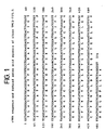
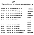
- E1-C19 One clone designated E1-C19, was shown by sequencing with the T7 primer to contain the putative 5' end of K5.5. CsCl gradient-purified DNA of clone E1-C19 was subsequently resequenced with T3 and T7 primers and several internal sequencing primers based on the previous sequencing results (primer sequences are shown in figure 2). The sequence of E1-C19 insert cDNA is shown in figure 3.
- Leukocytes were sorted by FACS using the appropriately labelled antibody on a FACS star (Becton Dickinson) to obtain pure populations (>90%) of B cells (CD20), T cells (CD4, CD8, CD45R0, CD45RA) and monocytes (CD14).
- Pulmonary macrophages and mixed lung leukocytes were prepared from resected human lung samples using the method of Nicod et al (1989) (Separation of potent and poorly functional human lung accessory cells based on autofluorescence. J. Leukocyte. Biol. 45 458 ).
- RNA was electrophoresed in 1% agarose gels containing 2.2% (v/v) formaldehyde, transferred to nitrocellulose and probed with the 32 P-dCTP labelled 514 bp insert from TM(2-7)5.5 using standard Northern blot procedure (Sambrook et al (1989)). The results are shown in Figs 4 to 6.
- PCR reaction products (10 ⁇ l) were analysed on 1% agarose gels as described above, for the presence of a 1085 bp reaction product corresponding to the full coding sequence of K5.5.
- Fig 7 The results are shown in Fig 7, wherein the samples in the lanes indicated in Fig 7 are as follows: Lane Sample 1 Molecular weight markers (1 kb ladder) 2 PB T cells (IL-2 stimulated) 3 PB T cells 4 Jurkat 5 MOLT-4 6 PB B cells 7 PB B cells 8 pulmonary macrophages 9 PB monocytes 10 KU812 11 EOL-3 12 SW900 (lung epithelial cell line) 13 CCLu32 (lung fibroblast cell line) 14 LL24 (lung fibroblast cell line) 15 AALT.16 (aortic smooth muscle cell line)
- CsCl-gradient purified pE1-C19 plasmid DNA (5 ⁇ g) was linearized using restriction enzyme Bam HI (New England Biolabs) in a 100 ⁇ l reaction volume overnight at 37°C. Linearized plasmids were treated with 2 ⁇ l of proteinase K (16.7 mg/ml Boehringer) for 30 min at 37°C. DNA was extracted twice with phenol (0.1 M Tris-saturated pH 8.0) and once with chloroform. Glycogen (1 ⁇ l of 20 mg/ml stock solution) was added to the aqueous phase and linearized DNA was precipitated following addition of 0.1 volume of 3 M sodium acetate pH 5.5 and 2.5 volumes of ethanol for 1 h at -80°C. The DNA was recovered by centrifugation (14 000 rpm, 4°C in a microfuge), washed with 70% ethanol and dissolved in RNase free water at 250 ng/ml.
- Bam HI New England Biolabs
- Capped cRNA transcripts were generated from 1 ⁇ g of Bam HI (New England Biolabs) linearized DNA in a 100 ⁇ l reaction volume containing 20 ⁇ l transcription buffer (5X), 4 ⁇ l NTP mix (10 mM ATP, UTP and CTP, 3 mM GTP), 4 ⁇ l 0.75M DTT, 2.5 ⁇ l RNAsin, 0.5 ⁇ l GTP (10 mM), 4 ⁇ l CAP analog (10 mM m7G(5')ppp(5')G) and 2.5 ⁇ l of T7 or T3 RNA polymerase respectively. All reagents used for the in vitro transcription reaction were from Promega except CAP analog (Pharmacia).
- cRNA was recovered by centrifugation (14 000 rpm, 4°C, 20 min in a microfuge), the pellet washed in 70 % ethanol and resuspended in sterile water at 1 ⁇ g/ ⁇ l.
- An approximate estimate of the cRNA concentration was obtained by running an aliquot of the resuspended material on a 1% agarose gel containing 2.2% (v/v) formaldehyde against RNA markers of known concentration. Samples were stored at -80°C before use.
- Oocytes were harvested from adult female Xenopus laevis, by standard methods (Bertrand et al ., 1991). Oocytes were defollicullated by incubation in 0.2% (w/v) collagenase (Sigma) in 50 ml OR2 medium without Ca2+ and without Mg2+ in a spinner flask under slow agitation for 2 h at room temperature (OR2 medium is 82.5 mM NaCl, 2.5 mM KCl, 1 mM Na 2 HPO 4 , 15 mM HEPES, 2 mM CaCl 2 , 1 mM MgCl 2 pH 7.6).
- Oocytes were rinsed carefully with OR2 followed by MBS (modified Barth's saline: 88 mM NaCl, 1 mM KCl, 0.33 mM Ca(NO 3 ) 2 , 0.41 mM CaCl 2 , 0.82 mM MgSO 4 , 2.4 mM NaHCO 3 , 10 mM HEPES, pH 7.6) and allowed to recover for at least 1-2 h in MBS before selecting stage V-VI oocytes. Selected oocytes were incubated in MBS supplemented penicillin/streptomycin (100 units/ml) (Gibco-BRL) overnight at 18°C before injection.
- MBS modified Barth's saline: 88 mM NaCl, 1 mM KCl, 0.33 mM Ca(NO 3 ) 2 , 0.41 mM CaCl 2 , 0.82 mM MgSO 4 , 2.4 mM NaHCO 3 , 10
- Oocytes were microinjected using an Inject + Matic air pump (Gabay) using needles made from Drummond calibrated 6 ml capillaries. cRNA (25 ng in 50 nl) was injected into the cytoplasm. Oocytes were individually transferred to wells of a 96 well flat bottom culture dish and incubated in MBS for 24-72 h.
- Electrophysiological recordings were made 1-3 days after injection in oocytes superfused with OR2 medium at room temperature under voltage clamped conditions using two microelectrodes (1-2 M ⁇ , both filled with 3 M KCl), the membrane potential being routinely clamped at -100 mV using a Gene Clamp 500 instrument (Axon).
- Test chemokines were purchased from PeptroTech or produced in-house at the Glaxo Institute for Molecular Biology and resuspended at a concentration of 1 ⁇ M in PBS. Fifty ⁇ l of each chemokine was applied directly onto voltage clamped oocytes and the current induced was monitored on a Tektronix 5113 dual-beam storage oscilloscope linked to an IBM-PC. Where multiple chemokines were tested on a single oocyte, a recovery time of 2 min was allowed between each application. The results are shown in Fig 8A.
- Fig 8B shows the results of a similar analysis to that illustrated in Fig 8A, but using different chemokines (apart from MIP-1 ⁇ ).
- human CC-CKR-3-pcDNA1neo, murine CC-CKR-3-pcDNA1neo, or pcDNA1neo were electroporated into 500 ⁇ l HL-60 cells (2 x 10 7 cells/ml in 0.15 M NaCl, 20mM .HEPES, pH 7.3) using a Bio Rad Geno Pulster (260 volts, 960 ⁇ F, 0.4 cm gap cuvette). Cells were seeded into T-175 flasks containing 25 ml AIM-V serum-free media (GIBCO).
- the cells were diluted in a total volume of 45 ml AIM-V media containing 600 ⁇ g/ml G418, and on day 6, cells were further diluted to 180 ml AIM-V media containing 600 ⁇ g/ml G418.
- On days 7-15 post-transfection cells were maintained in AIM-V media (+G418) at a density of 0.4 - 1.2x 10 6 cells/ml, and binding assays were performed during this time.
- Equilibrium competition binding was carried out by incubating 5 x 10 5 cells in 100 ⁇ l binding buffer (1 mM CaCl 2 , 5 mM MgCl 2 , 0.5% BSA, 50 mM HEPES, pH 7.2), 0.34 nM [ 125 I]radioligand, and 0.5 - 2000 nM cold ligand in Millipore®-DV96-well filter plates. After 1.5h incubation at room temperature, cells were washed four times by vacuum filtration with binding buffer containing 0.5 M NaCl. Fifty ⁇ l Optiphase scintillant (Wallac) were added to each well, and the radioactivity was measured with a Wallac Microbeta Plate Reader.
- FIG. 9 illustrates high affinity binding of [ 125 I]MIP-1 ⁇ and [ 125 I]RANTES to human and murine CC-CKR-3.
- HL-60 cells were transfected with human CC-CKR-3 ( ⁇ ), murine CC-CKR-3 ( ⁇ ), or an empty vector ( ⁇ ) and maintained in AIM-V media containing G418 for 7-15 days.
- Equilibrium competition assays were performed as described above with [ 125 I]MIP-1 ⁇ (A) and [ 125 I]RANTES (B). Each point represents the mean ⁇ S.D. of duplicate points from four (A) or three (B) separate experiments.
Landscapes
- Health & Medical Sciences (AREA)
- Life Sciences & Earth Sciences (AREA)
- Chemical & Material Sciences (AREA)
- Organic Chemistry (AREA)
- General Health & Medical Sciences (AREA)
- Medicinal Chemistry (AREA)
- Nuclear Medicine, Radiotherapy & Molecular Imaging (AREA)
- Chemical Kinetics & Catalysis (AREA)
- General Chemical & Material Sciences (AREA)
- Pharmacology & Pharmacy (AREA)
- Animal Behavior & Ethology (AREA)
- Public Health (AREA)
- Veterinary Medicine (AREA)
- Bioinformatics & Cheminformatics (AREA)
- Engineering & Computer Science (AREA)
- Immunology (AREA)
- Pulmonology (AREA)
- Virology (AREA)
- Molecular Biology (AREA)
- Gastroenterology & Hepatology (AREA)
- Cell Biology (AREA)
- Proteomics, Peptides & Aminoacids (AREA)
- Genetics & Genomics (AREA)
- Biophysics (AREA)
- Biochemistry (AREA)
- Communicable Diseases (AREA)
- Oncology (AREA)
- Zoology (AREA)
- Toxicology (AREA)
- Ophthalmology & Optometry (AREA)
- Tropical Medicine & Parasitology (AREA)
- Urology & Nephrology (AREA)
- Diabetes (AREA)
- AIDS & HIV (AREA)
- Heart & Thoracic Surgery (AREA)
- Cardiology (AREA)
- Vascular Medicine (AREA)
- Hematology (AREA)
- Dermatology (AREA)
- Peptides Or Proteins (AREA)
Abstract
Description
phenylalanine, tyrosine and tryptophan (amino acids having aromatic side chains); lysine, arginine and histidine (amino acids having basic side chains); aspartate and glutamate (amino acids having acidic side chains); asparagine and glutamine (amino acids having amide side chains) and cysteine and methionine (amino acids having sulphur containing side chains). Thus the present invention includes within its scope variants of the receptor shown in Fig 3 which includes one or more such substitutions.
- +++
- Very strong positive signal visible after four hours' exposure of the autoradiograph.
- ++
- Clear positive signal visible after four hours' exposure of the autoradiograph.
- +
- Signal not visible after four hours' exposure of the autoradiograph, but clear after 24 hours.
- +/-
- Weak positive signal only visible after 24 hours' exposure.
- -
- No signal.
or
- I = inosine which can substitute for A, T, G, or C
- Y = C or T
- H = A, C or T
- M = A or C
- R = A or G
- D = A, G or T
| | Sample | |
| 1 | Molecular weight markers (1 kb ladder) | |
| 2 | PB T cells (IL-2 stimulated) | |
| 3 | | |
| 4 | | |
| 5 | MOLT-4 | |
| 6 | | |
| 7 | | |
| 8 | | |
| 9 | | |
| 10 | | |
| 11 | EOL-3 | |
| 12 | SW900 (lung epithelial cell line) | |
| 13 | CCLu32 (lung fibroblast cell line) | |
| 14 | LL24 (lung fibroblast cell line) | |
| 15 | AALT.16 (aortic smooth muscle cell line) |
- (A) NAME: WELLS, Timothy Nigel Carl
- (B) STREET: c/o GLAXO INSTITUTE FOR MOLECULAR BIOLOGY, 14 CHEMIN DE AULX
- (C) CITY: PLAN-LES-OUATES
- (D) STATE: GENEVA
- (E) COUNTRY: SWITZERLAND
- (F) POSTAL CODE (ZIP): 1228
- (A) NAME: POWER. Christine Anna
- (B) STREET: c/o GLAXO INSTITUTE FOR MOLECULAR BIOLOGY, 14 CHEMIN DE AULX
- (C) CITY: PLAN-LES-OUATES
- (D) STATE: GENEVA
- (E) COUNTRY: SWITZERLAND
- (F) POSTAL CODE (ZIP): 1228


Claims (14)
- A polypeptide which:a) has the amino acid sequence shown in Figure 3 (SEQ ID No. 20) orb) has one or more amino acid deletions, insertions or substitutions relative to the amino acid sequence shown in Figure 3 (SEQ ID No. 20) and which has at least 90% amino acid sequence identity with the amino acid sequence shown in Figure 3 (SEQ ID No. 20).
- Polypeptide as claimed in claim 1 which has at least 95% amino acid sequence identity with the amino acid sequence shown in Figure 3 (SEQ ID No. 20).
- Polypeptide according to claim 1 which has the amino acid sequence shown in Figure 3 (SEQ ID No. 20).
- Polypeptide as claimed in any of claims 1 to 3 which is capable of binding to RANTES, MIP-1α and/or MCP-1.
- A method of screening for a pharmaceutically active agent using the polypeptide as claimed in any of claims 1 to 4.
- Method as claimed in claim 5 for screening for an agent useful in treating allergies.
- Method as claimed in claim 5 for screening for an agent useful in treating immune and/or inflammatory disorders associated with T-lymphocyte activation.
- Method as claimed in claim 5 for screening for an agent useful in treating atheromas.
- A nucleic acid which codes for a polypeptide as claimed in any of claims 1 to 4.
- Nucleic acid as claimed in claim 9 which has the nucleotide sequence shown in SEQ ID No. 19 (Figure 3).
- Nucleic acid according to claim 9 which comprises nucleotides 183 - 1262 of the nucleotide sequence shown in SEQ ID No. 19 (Figure 3).
- A vector comprising a nucleic acid as claimed in any of claims 9 to 11.
- A host comprising a vector as claimed in claim 12.
- A method for obtaining a polypeptide according to any of claims 1 to 4, comprising incubating a host according to claim 12 under conditions causing expression of said polypeptide and then purifying said polypeptide.
Applications Claiming Priority (3)
| Application Number | Priority Date | Filing Date | Title |
|---|---|---|---|
| GB9501683 | 1995-01-27 | ||
| GBGB9501683.8A GB9501683D0 (en) | 1995-01-27 | 1995-01-27 | Substances and their uses |
| PCT/GB1996/000143 WO1996023068A1 (en) | 1995-01-27 | 1996-01-24 | A chemokine receptor able to bind to mcp-1, mip-1 alpha and/or rantes. its uses |
Publications (3)
| Publication Number | Publication Date |
|---|---|
| EP0805859A1 EP0805859A1 (en) | 1997-11-12 |
| EP0805859B1 true EP0805859B1 (en) | 2005-04-20 |
| EP0805859B2 EP0805859B2 (en) | 2008-06-25 |
Family
ID=10768728
Family Applications (1)
| Application Number | Title | Priority Date | Filing Date |
|---|---|---|---|
| EP96900656A Expired - Lifetime EP0805859B2 (en) | 1995-01-27 | 1996-01-24 | A chemokine receptor able to bind to mcp-1, mip-1 alpha and/or rantes. |
Country Status (9)
| Country | Link |
|---|---|
| US (4) | US6150132A (en) |
| EP (1) | EP0805859B2 (en) |
| JP (2) | JP3771585B2 (en) |
| AT (1) | ATE293689T1 (en) |
| AU (1) | AU4455896A (en) |
| DE (1) | DE69634627T3 (en) |
| ES (1) | ES2240989T5 (en) |
| GB (1) | GB9501683D0 (en) |
| WO (1) | WO1996023068A1 (en) |
Cited By (1)
| Publication number | Priority date | Publication date | Assignee | Title |
|---|---|---|---|---|
| US7183063B2 (en) | 1995-01-27 | 2007-02-27 | Glaxo Group Limited | Chemokine receptor able to bind to MCP-1, MIP-1 α and/or rantes and its uses |
Families Citing this family (36)
| Publication number | Priority date | Publication date | Assignee | Title |
|---|---|---|---|---|
| US6025154A (en) | 1995-06-06 | 2000-02-15 | Human Genome Sciences, Inc. | Polynucleotides encoding human G-protein chemokine receptor HDGNR10 |
| US6743594B1 (en) | 1995-06-06 | 2004-06-01 | Human Genome Sciences, Inc. | Methods of screening using human G-protein chemokine receptor HDGNR10 (CCR5) |
| US6737513B1 (en) | 1996-06-07 | 2004-05-18 | Icos Corporation | Macrophage derived chemokine (MDC) and chemokine analogs and assay to identify modulators of MDC activity, and therapeutic uses for same |
| US6498015B1 (en) | 1995-06-07 | 2002-12-24 | Icos Corporation | Methods of identifying agents that modulate the binding between MDC and an MDC receptor |
| US7018627B1 (en) | 1995-06-07 | 2006-03-28 | Icos Corporation | Macrophage derived chemokine (MDC), MDC analogs, MDC inhibitor substances, and uses thereof |
| EP0883687B1 (en) * | 1996-03-01 | 2004-10-27 | Euroscreen S.A. | C-c ckr-5, cc-chemikines receptor, derivatives thereof and their uses |
| US6258527B1 (en) | 1996-05-20 | 2001-07-10 | The Aaron Diamond Aids Research Center | Methods of identifying g-coupled receptors associated with macrophage-trophic HIV, and diagnostic and therapeutic uses thereof |
| US5939320A (en) * | 1996-05-20 | 1999-08-17 | New York University | G-coupled receptors associated with macrophage-trophic HIV, and diagnostic and therapeutic uses thereof |
| FR2748938B1 (en) * | 1996-05-22 | 1998-07-31 | Pasteur Institut | USE OF ANTAGONIST MOLECULES OF CHEMOKINES FOR THEIR ANTIVIRAL ACTIVITY IN PARTICULAR AGAINST HIV-TYPE RETROVIRUSES |
| US6057102A (en) * | 1996-08-08 | 2000-05-02 | The Aaron Diamond Aids Research Center | HIV coreceptor mutants |
| US5955303A (en) * | 1997-03-06 | 1999-09-21 | Incyte Pharmaceuticals, Inc. | Human chemokine receptor-like protein |
| US6204024B1 (en) | 1997-09-12 | 2001-03-20 | Akzo Nobel N.V. | CCR5 RNA transcription based amplification assay |
| US6121023A (en) | 1998-01-22 | 2000-09-19 | Akzo Nobel N.V. | Isothermal transcription based assay for the detection and quantification of the chemokine rantes |
| US6287805B1 (en) | 1998-03-20 | 2001-09-11 | Millennium Pharmaceuticals, Inc. | Nucleic acid molecules of the protein-coupled heptahelical receptor superfamily and uses therefor |
| US6420121B1 (en) * | 1998-08-31 | 2002-07-16 | Oregon Health Sciences University | Prevention of cell migration initiation with CMV US28 receptor antagonists |
| US6245332B1 (en) | 1999-01-15 | 2001-06-12 | The Board Of Trustees Of The Leland Stanford Junior University | Modulation of systemic memory T cell trafficking |
| US6488930B1 (en) | 1999-01-15 | 2002-12-03 | Millennium Pharmaceuticals, Inc. | Anti-CCR4 antibodies and methods of use therefor |
| ES2156526B1 (en) * | 1999-01-20 | 2002-02-01 | Smithkline Beecham Sa | METABOLITES OF STREPTOMYCES CACAOI AS INHIBITORS OF THE CHEMIOATRAYING PROTEIN OF MONOCITS 1. |
| EP1176195B1 (en) | 1999-04-09 | 2013-05-22 | Kyowa Hakko Kirin Co., Ltd. | Method for controlling the activity of immunologically functional molecule |
| JP3707303B2 (en) * | 1999-06-30 | 2005-10-19 | 株式会社日立製作所 | Histamine measuring method and histamine measuring device |
| CA2401491C (en) | 2000-03-03 | 2011-07-05 | Kyowa Kirin Co., Ltd. | Ccr4-binding antibody and diagnostic and therapeutic uses thereof |
| AU2001288682A1 (en) | 2000-08-30 | 2002-03-13 | Chemocentryx, Inc. | Inhibition of cmv infection and dissemination |
| US6946292B2 (en) | 2000-10-06 | 2005-09-20 | Kyowa Hakko Kogyo Co., Ltd. | Cells producing antibody compositions with increased antibody dependent cytotoxic activity |
| WO2002030464A1 (en) * | 2000-10-11 | 2002-04-18 | Daiichi Pharmaceutical Co., Ltd. | Novel drugs for liver diseases |
| KR100360410B1 (en) * | 2000-11-14 | 2002-11-13 | 삼성전자 주식회사 | Method for MDL semiconductor device including DRAM device having self-aligned contact structure and logic device having dual gate structure |
| EP1399184A2 (en) * | 2000-12-01 | 2004-03-24 | Schering Corporation | Uses of mammalian genes and related reagents |
| CA2437201C (en) * | 2001-02-02 | 2008-11-18 | Chemocentryx, Inc. | Methods and compositions useful for stimulating an immune response |
| US7175988B2 (en) | 2001-02-09 | 2007-02-13 | Human Genome Sciences, Inc. | Human G-protein Chemokine Receptor (CCR5) HDGNR10 |
| US20020165161A1 (en) * | 2001-05-04 | 2002-11-07 | Surromed, Inc. | Chemokine-binding proteins for treating congestive heart failure |
| WO2003018549A2 (en) * | 2001-08-30 | 2003-03-06 | Chemocentryx, Inc. | Bicyclic compounds as inhibitors of chemokine binding to us28 |
| CA2458627C (en) | 2001-08-31 | 2013-09-10 | Kyowa Kirin Co., Ltd. | Human cdr-grafted antibody and antibody fragment thereof |
| US7393934B2 (en) | 2001-12-21 | 2008-07-01 | Human Genome Sciences, Inc. | Human G-protein chemokine receptor (CCR5) HDGNR10 |
| WO2003084569A1 (en) | 2002-04-09 | 2003-10-16 | Kyowa Hakko Kogyo Co., Ltd. | Drug containing antibody composition |
| AU2003260072A1 (en) | 2002-08-27 | 2004-03-19 | Bristol-Myers Squibb Company | Antagonists of chemokine receptors |
| CA2559809A1 (en) | 2004-03-12 | 2005-11-10 | Human Genome Sciences, Inc. | Antibodies against human g-protein chemokine receptor (ccr5) hdgnr10 |
| GB0718167D0 (en) | 2007-09-18 | 2007-10-31 | Cancer Rec Tech Ltd | Cancer marker and therapeutic target |
Family Cites Families (8)
| Publication number | Priority date | Publication date | Assignee | Title |
|---|---|---|---|---|
| US5011912A (en) * | 1986-12-19 | 1991-04-30 | Immunex Corporation | Hybridoma and monoclonal antibody for use in an immunoaffinity purification system |
| US5179078A (en) * | 1989-05-12 | 1993-01-12 | Dana Farber Cancer Institute | Method of suppressing tumor formation in vivo |
| AU5293293A (en) * | 1992-09-28 | 1994-04-26 | Board Of Regents, The University Of Texas System | Methods and compositions for treatment of allergic disease |
| DK0669979T3 (en) * | 1992-11-10 | 2006-03-06 | Genentech Inc | C-C CKR-1, chemokine receptor |
| US5474983A (en) * | 1993-03-15 | 1995-12-12 | The Research Foundation Of State University Of New York | Method of inhibiting pro-inflammatory mediator release from basophils and mast cells |
| ATE183513T1 (en) | 1993-06-03 | 1999-09-15 | Therapeutic Antibodies Inc | PREPARATION OF ANTIBODY FRAGMENTS |
| EP0740701B1 (en) * | 1994-01-13 | 2006-12-06 | The Regents of the University of California | Mammalian monocyte chemoattractant protein receptors |
| GB9501683D0 (en) | 1995-01-27 | 1995-03-15 | Glaxo Group Ltd | Substances and their uses |
-
1995
- 1995-01-27 GB GBGB9501683.8A patent/GB9501683D0/en active Pending
-
1996
- 1996-01-24 DE DE69634627T patent/DE69634627T3/en not_active Expired - Lifetime
- 1996-01-24 EP EP96900656A patent/EP0805859B2/en not_active Expired - Lifetime
- 1996-01-24 JP JP52271996A patent/JP3771585B2/en not_active Expired - Fee Related
- 1996-01-24 US US08/875,573 patent/US6150132A/en not_active Expired - Lifetime
- 1996-01-24 AU AU44558/96A patent/AU4455896A/en not_active Abandoned
- 1996-01-24 WO PCT/GB1996/000143 patent/WO1996023068A1/en not_active Ceased
- 1996-01-24 AT AT96900656T patent/ATE293689T1/en not_active IP Right Cessation
- 1996-01-24 ES ES96900656T patent/ES2240989T5/en not_active Expired - Lifetime
-
2001
- 2001-01-19 US US09/764,413 patent/US6919432B2/en not_active Expired - Fee Related
-
2002
- 2002-04-12 US US10/120,394 patent/US7183063B2/en not_active Expired - Fee Related
- 2002-05-16 JP JP2002141253A patent/JP2003038191A/en not_active Withdrawn
-
2004
- 2004-09-03 US US10/933,356 patent/US7655425B2/en not_active Expired - Fee Related
Cited By (2)
| Publication number | Priority date | Publication date | Assignee | Title |
|---|---|---|---|---|
| US7183063B2 (en) | 1995-01-27 | 2007-02-27 | Glaxo Group Limited | Chemokine receptor able to bind to MCP-1, MIP-1 α and/or rantes and its uses |
| US7655425B2 (en) | 1995-01-27 | 2010-02-02 | Glaxo Group Limited | Uses of a chemokine receptor able to bind to MCP-1, MIP-1α and/or RANTES |
Also Published As
| Publication number | Publication date |
|---|---|
| DE69634627T2 (en) | 2006-03-02 |
| ES2240989T3 (en) | 2005-10-16 |
| JP2003038191A (en) | 2003-02-12 |
| WO1996023068A1 (en) | 1996-08-01 |
| JPH10513046A (en) | 1998-12-15 |
| US20020187930A1 (en) | 2002-12-12 |
| AU4455896A (en) | 1996-08-14 |
| ES2240989T5 (en) | 2008-12-16 |
| US7183063B2 (en) | 2007-02-27 |
| US20020160015A1 (en) | 2002-10-31 |
| US7655425B2 (en) | 2010-02-02 |
| EP0805859A1 (en) | 1997-11-12 |
| US6919432B2 (en) | 2005-07-19 |
| DE69634627D1 (en) | 2005-05-25 |
| JP3771585B2 (en) | 2006-04-26 |
| ATE293689T1 (en) | 2005-05-15 |
| DE69634627T3 (en) | 2008-12-11 |
| US20050014695A1 (en) | 2005-01-20 |
| EP0805859B2 (en) | 2008-06-25 |
| GB9501683D0 (en) | 1995-03-15 |
| US6150132A (en) | 2000-11-21 |
Similar Documents
| Publication | Publication Date | Title |
|---|---|---|
| EP0805859B1 (en) | A chemokine receptor able to bind to mcp-1, mip-1 alpha and/or rantes. its uses | |
| Ponath et al. | Molecular cloning and characterization of a human eotaxin receptor expressed selectively on eosinophils. | |
| JP4117904B2 (en) | CC-KRKR-5, CC-chemokine receptor, derivatives thereof and use thereof | |
| US6730301B1 (en) | MCP-1 receptor antibodies | |
| Lu et al. | Structure and function of the murine chemokine receptor CXCR3 | |
| Nomura et al. | Molecular cloning of cDNAs encoding a LD78 receptor and putative leukocyte chemotactic peptide receptors | |
| Proudfoot et al. | Amino-terminally modified RANTES analogues demonstrate differential effects on RANTES receptors | |
| Nibbs et al. | Cloning and characterization of a novel murine β chemokine receptor, D6: comparison to three other related macrophage inflammatory protein-1α receptors, CCR-1, CCR-3, and CCR-5 | |
| Power et al. | Molecular Cloning and Functional Expression of a Novel CC Chemokine Receptor cDNA from a Human Basophilic Cell Line (∗) | |
| JP3415162B2 (en) | IL-13 receptor polypeptide | |
| US6498015B1 (en) | Methods of identifying agents that modulate the binding between MDC and an MDC receptor | |
| JP2001524068A (en) | G protein-coupled receptor antagonist | |
| CA2399593A1 (en) | Antibodies to ccr5 | |
| JP2001516203A (en) | Exodus chemokine substances and methods | |
| AU669889B2 (en) | Nucleic acid sequences coding for or complementary to nucleic acid sequences coding for interleukin 9 receptor | |
| Youn et al. | Molecular cloning and characterization of a cDNA, CHEMR1, encoding a chemokine receptor with a homology to the human CC chemokine receptor, CCR-4 | |
| JP4116087B2 (en) | Novel mouse CXC chemokine receptor | |
| Daugherty | Identification and characterization of beta-chemokine receptors on eosinophils | |
| US6790947B1 (en) | Polynucleotides encoding macrophage derived chemokine | |
| JPWO1999033876A1 (en) | A novel seven-transmembrane receptor protein |
Legal Events
| Date | Code | Title | Description |
|---|---|---|---|
| PUAI | Public reference made under article 153(3) epc to a published international application that has entered the european phase |
Free format text: ORIGINAL CODE: 0009012 |
|
| 17P | Request for examination filed |
Effective date: 19970702 |
|
| AK | Designated contracting states |
Kind code of ref document: A1 Designated state(s): AT BE CH DE DK ES FR GB GR IE IT LI LU MC NL PT SE |
|
| AX | Request for extension of the european patent |
Free format text: LT PAYMENT 970702;LV PAYMENT 970702;SI PAYMENT 970702 |
|
| 17Q | First examination report despatched |
Effective date: 20030605 |
|
| GRAP | Despatch of communication of intention to grant a patent |
Free format text: ORIGINAL CODE: EPIDOSNIGR1 |
|
| GRAS | Grant fee paid |
Free format text: ORIGINAL CODE: EPIDOSNIGR3 |
|
| GRAA | (expected) grant |
Free format text: ORIGINAL CODE: 0009210 |
|
| RIN1 | Information on inventor provided before grant (corrected) |
Inventor name: POWER, CHRISTINE ANNA CORPORATE INTELLECTUAL Inventor name: WELLS, TIMOTHY NIGEL CARL CORPORATE INTELLECTUAL |
|
| AK | Designated contracting states |
Kind code of ref document: B1 Designated state(s): AT BE CH DE DK ES FR GB GR IE IT LI LU MC NL PT SE |
|
| AX | Request for extension of the european patent |
Extension state: LT LV SI |
|
| PG25 | Lapsed in a contracting state [announced via postgrant information from national office to epo] |
Ref country code: NL Free format text: LAPSE BECAUSE OF FAILURE TO SUBMIT A TRANSLATION OF THE DESCRIPTION OR TO PAY THE FEE WITHIN THE PRESCRIBED TIME-LIMIT Effective date: 20050420 Ref country code: LI Free format text: LAPSE BECAUSE OF FAILURE TO SUBMIT A TRANSLATION OF THE DESCRIPTION OR TO PAY THE FEE WITHIN THE PRESCRIBED TIME-LIMIT Effective date: 20050420 Ref country code: CH Free format text: LAPSE BECAUSE OF FAILURE TO SUBMIT A TRANSLATION OF THE DESCRIPTION OR TO PAY THE FEE WITHIN THE PRESCRIBED TIME-LIMIT Effective date: 20050420 Ref country code: AT Free format text: LAPSE BECAUSE OF FAILURE TO SUBMIT A TRANSLATION OF THE DESCRIPTION OR TO PAY THE FEE WITHIN THE PRESCRIBED TIME-LIMIT Effective date: 20050420 |
|
| REG | Reference to a national code |
Ref country code: GB Ref legal event code: FG4D |
|
| REG | Reference to a national code |
Ref country code: CH Ref legal event code: EP |
|
| REG | Reference to a national code |
Ref country code: IE Ref legal event code: FG4D |
|
| REF | Corresponds to: |
Ref document number: 69634627 Country of ref document: DE Date of ref document: 20050525 Kind code of ref document: P |
|
| PG25 | Lapsed in a contracting state [announced via postgrant information from national office to epo] |
Ref country code: SE Free format text: LAPSE BECAUSE OF FAILURE TO SUBMIT A TRANSLATION OF THE DESCRIPTION OR TO PAY THE FEE WITHIN THE PRESCRIBED TIME-LIMIT Effective date: 20050720 Ref country code: GR Free format text: LAPSE BECAUSE OF FAILURE TO SUBMIT A TRANSLATION OF THE DESCRIPTION OR TO PAY THE FEE WITHIN THE PRESCRIBED TIME-LIMIT Effective date: 20050720 Ref country code: DK Free format text: LAPSE BECAUSE OF FAILURE TO SUBMIT A TRANSLATION OF THE DESCRIPTION OR TO PAY THE FEE WITHIN THE PRESCRIBED TIME-LIMIT Effective date: 20050720 |
|
| PG25 | Lapsed in a contracting state [announced via postgrant information from national office to epo] |
Ref country code: PT Free format text: LAPSE BECAUSE OF FAILURE TO SUBMIT A TRANSLATION OF THE DESCRIPTION OR TO PAY THE FEE WITHIN THE PRESCRIBED TIME-LIMIT Effective date: 20050920 |
|
| LTIE | Lt: invalidation of european patent or patent extension |
Effective date: 20050420 |
|
| REG | Reference to a national code |
Ref country code: ES Ref legal event code: FG2A Ref document number: 2240989 Country of ref document: ES Kind code of ref document: T3 |
|
| REG | Reference to a national code |
Ref country code: CH Ref legal event code: PL |
|
| NLV1 | Nl: lapsed or annulled due to failure to fulfill the requirements of art. 29p and 29m of the patents act | ||
| PLBI | Opposition filed |
Free format text: ORIGINAL CODE: 0009260 |
|
| PG25 | Lapsed in a contracting state [announced via postgrant information from national office to epo] |
Ref country code: IE Free format text: LAPSE BECAUSE OF NON-PAYMENT OF DUE FEES Effective date: 20060124 |
|
| PG25 | Lapsed in a contracting state [announced via postgrant information from national office to epo] |
Ref country code: MC Free format text: LAPSE BECAUSE OF NON-PAYMENT OF DUE FEES Effective date: 20060131 Ref country code: LU Free format text: LAPSE BECAUSE OF NON-PAYMENT OF DUE FEES Effective date: 20060131 |
|
| PLAX | Notice of opposition and request to file observation + time limit sent |
Free format text: ORIGINAL CODE: EPIDOSNOBS2 |
|
| 26 | Opposition filed |
Opponent name: STRAWMAN LIMITED Effective date: 20060118 |
|
| ET | Fr: translation filed | ||
| PLAF | Information modified related to communication of a notice of opposition and request to file observations + time limit |
Free format text: ORIGINAL CODE: EPIDOSCOBS2 |
|
| PLAF | Information modified related to communication of a notice of opposition and request to file observations + time limit |
Free format text: ORIGINAL CODE: EPIDOSCOBS2 |
|
| REG | Reference to a national code |
Ref country code: IE Ref legal event code: MM4A |
|
| PLBB | Reply of patent proprietor to notice(s) of opposition received |
Free format text: ORIGINAL CODE: EPIDOSNOBS3 |
|
| RTI2 | Title (correction) |
Free format text: A CHEMOKINE RECEPTOR ABLE TO BIND TO MCP-1, MIP-1 ALPHA AND/OR RANTES. |
|
| PUAH | Patent maintained in amended form |
Free format text: ORIGINAL CODE: 0009272 |
|
| STAA | Information on the status of an ep patent application or granted ep patent |
Free format text: STATUS: PATENT MAINTAINED AS AMENDED |
|
| 27A | Patent maintained in amended form |
Effective date: 20080625 |
|
| AK | Designated contracting states |
Kind code of ref document: B2 Designated state(s): AT BE CH DE DK ES FR GB GR IE IT LI LU MC NL PT SE |
|
| AX | Request for extension of the european patent |
Extension state: LT LV SI |
|
| REG | Reference to a national code |
Ref country code: ES Ref legal event code: DC2A Date of ref document: 20080923 Kind code of ref document: T5 |
|
| PGFP | Annual fee paid to national office [announced via postgrant information from national office to epo] |
Ref country code: IT Payment date: 20120123 Year of fee payment: 17 |
|
| PGFP | Annual fee paid to national office [announced via postgrant information from national office to epo] |
Ref country code: GB Payment date: 20121228 Year of fee payment: 18 |
|
| PGFP | Annual fee paid to national office [announced via postgrant information from national office to epo] |
Ref country code: DE Payment date: 20130131 Year of fee payment: 18 Ref country code: FR Payment date: 20130128 Year of fee payment: 18 Ref country code: ES Payment date: 20130121 Year of fee payment: 18 |
|
| PGFP | Annual fee paid to national office [announced via postgrant information from national office to epo] |
Ref country code: BE Payment date: 20130205 Year of fee payment: 18 |
|
| BERE | Be: lapsed |
Owner name: *GLAXO GROUP LTD Effective date: 20140131 |
|
| REG | Reference to a national code |
Ref country code: DE Ref legal event code: R119 Ref document number: 69634627 Country of ref document: DE |
|
| GBPC | Gb: european patent ceased through non-payment of renewal fee |
Effective date: 20140124 |
|
| REG | Reference to a national code |
Ref country code: DE Ref legal event code: R119 Ref document number: 69634627 Country of ref document: DE Effective date: 20140801 |
|
| PG25 | Lapsed in a contracting state [announced via postgrant information from national office to epo] |
Ref country code: DE Free format text: LAPSE BECAUSE OF NON-PAYMENT OF DUE FEES Effective date: 20140801 |
|
| REG | Reference to a national code |
Ref country code: FR Ref legal event code: ST Effective date: 20140930 |
|
| PG25 | Lapsed in a contracting state [announced via postgrant information from national office to epo] |
Ref country code: FR Free format text: LAPSE BECAUSE OF NON-PAYMENT OF DUE FEES Effective date: 20140131 Ref country code: GB Free format text: LAPSE BECAUSE OF NON-PAYMENT OF DUE FEES Effective date: 20140124 |
|
| PG25 | Lapsed in a contracting state [announced via postgrant information from national office to epo] |
Ref country code: BE Free format text: LAPSE BECAUSE OF NON-PAYMENT OF DUE FEES Effective date: 20140131 |
|
| REG | Reference to a national code |
Ref country code: ES Ref legal event code: FD2A Effective date: 20150327 |
|
| PG25 | Lapsed in a contracting state [announced via postgrant information from national office to epo] |
Ref country code: ES Free format text: LAPSE BECAUSE OF NON-PAYMENT OF DUE FEES Effective date: 20140125 |
|
| PG25 | Lapsed in a contracting state [announced via postgrant information from national office to epo] |
Ref country code: IT Free format text: LAPSE BECAUSE OF NON-PAYMENT OF DUE FEES Effective date: 20140124 |