EP0804580B1 - Calcium binding recombinant antibody against protein c - Google Patents
Calcium binding recombinant antibody against protein c Download PDFInfo
- Publication number
- EP0804580B1 EP0804580B1 EP95923791A EP95923791A EP0804580B1 EP 0804580 B1 EP0804580 B1 EP 0804580B1 EP 95923791 A EP95923791 A EP 95923791A EP 95923791 A EP95923791 A EP 95923791A EP 0804580 B1 EP0804580 B1 EP 0804580B1
- Authority
- EP
- European Patent Office
- Prior art keywords
- antibody
- protein
- sequence
- hpc
- recombinant
- Prior art date
- Legal status (The legal status is an assumption and is not a legal conclusion. Google has not performed a legal analysis and makes no representation as to the accuracy of the status listed.)
- Expired - Lifetime
Links
Classifications
-
- C—CHEMISTRY; METALLURGY
- C07—ORGANIC CHEMISTRY
- C07K—PEPTIDES
- C07K16/00—Immunoglobulins [IG], e.g. monoclonal or polyclonal antibodies
- C07K16/40—Immunoglobulins [IG], e.g. monoclonal or polyclonal antibodies against enzymes
-
- A—HUMAN NECESSITIES
- A61—MEDICAL OR VETERINARY SCIENCE; HYGIENE
- A61P—SPECIFIC THERAPEUTIC ACTIVITY OF CHEMICAL COMPOUNDS OR MEDICINAL PREPARATIONS
- A61P35/00—Antineoplastic agents
-
- A—HUMAN NECESSITIES
- A61—MEDICAL OR VETERINARY SCIENCE; HYGIENE
- A61P—SPECIFIC THERAPEUTIC ACTIVITY OF CHEMICAL COMPOUNDS OR MEDICINAL PREPARATIONS
- A61P43/00—Drugs for specific purposes, not provided for in groups A61P1/00-A61P41/00
-
- A—HUMAN NECESSITIES
- A61—MEDICAL OR VETERINARY SCIENCE; HYGIENE
- A61P—SPECIFIC THERAPEUTIC ACTIVITY OF CHEMICAL COMPOUNDS OR MEDICINAL PREPARATIONS
- A61P7/00—Drugs for disorders of the blood or the extracellular fluid
- A61P7/04—Antihaemorrhagics; Procoagulants; Haemostatic agents; Antifibrinolytic agents
-
- A—HUMAN NECESSITIES
- A61—MEDICAL OR VETERINARY SCIENCE; HYGIENE
- A61K—PREPARATIONS FOR MEDICAL, DENTAL OR TOILETRY PURPOSES
- A61K38/00—Medicinal preparations containing peptides
-
- C—CHEMISTRY; METALLURGY
- C07—ORGANIC CHEMISTRY
- C07K—PEPTIDES
- C07K2319/00—Fusion polypeptide
Definitions
- This invention is generally in the area of antibodies to plasma proteins, specifically Protein C, and methods for use thereof.
- Protein C is a vitamin K-dependent plasma protein zymogen to a serine protease. Upon activation it becomes a potent anticoagulant. Activated protein C acts through the specific proteolysis of the procoagulant cofactors, factor VIIIa and factor Va. This activity requires the presence of another vitamin K-dependent protein, protein S, calcium and a phospholipid (presumably cellular) surface. As described in Hemostasis and Thrombosis: Basic Principles and Clinical Practice 2nd Ed., Colman, R.W., et al.,p.
- protein C circulates in a two-chain form, with the larger, heavy chain bound to the smaller light chain through a single disulfide link. A small proportion of the protein also circulates in a single chain form, where a Lys-Arg dipeptide in the molecule connects the light chain directly to the heavy chain.
- Protein C is activated to activated protein C (APC).
- APC activated protein C
- Thrombin is capable of activating protein C by the specific cleavage of the Arg 12 -Leu 13 bond in the heavy chain.
- the rate of this activation is enhanced dramatically when thrombin is bound to the endothelial cell cofactor, thrombomodulin.
- Matschiner, et al. Current Advances in Vitamin K Research, pp. 135-140, John W. Suttie, ed. (Elsevier Science Publishing Co., Inc. 1988 ) have further reviewed the role of the Vitamin K dependent proteins in coagulation.
- Protein C has been shown to have major importance in vivo. Patients deficient in protein C, or its cofactor, protein S, show pronounced thrombotic tendencies. Babies born totally deficient in protein C exhibit massive disseminated intravascular coagulation (DIC) and a necrotic syndrome which leads to death within the first few weeks of life if untreated. Activated protein C has also been shown to protect animals against the coagulopathic and lethal effects of endotoxin shock, as described by Taylor, et al., in J. Clin. Invest. 79, 918-925 (1987 ).
- Protein C was originally isolated in semi-pure form from plasma using classic protein purification techniques, including barium citrate adsorption and elution, ammonium sulfate fractionation, DEAE-Sephadex chromatography, dextran sulfate agarose chromatography, and preparative polyacrylamide gel electrophoresis. This procedure was vastly improved and facilitated by the discovery of a unique antibody to Protein C, designated HPC-4, described by Stearns, et al., in J. Biol. Chem. 263(2), 826-832 (1988 ).
- Protein C can be isolated from human plasma by batch adsorption of diluted heparinized plasma on QAE Sephadex, washing with buffered 0.15 M NaCl and eluting with 0.5 M NaCl, recalcifying and batch adsorbing with HPC-4, then washing with a Ca 2+ containing buffer and eluting the Protein C with an EDTA containing buffer.
- HPC-4 is a calcium-dependent monoclonal antibody to human protein C.
- HPC-4 is disclosed and claimed in U.S. Patent No. 5,202,253 to Esmon, et al.
- WO 90/07524 describes a Ca 2+ dependent monoclonal antibody that specifically binds to a specific twelve peptide sequence (E D Q V D P R L I D G K) in the activation region of protein C.
- Blockage of the natural anticoagulant pathways uses the natural procoagulant properties of the tumor to target the tumor capillaries for microvascular thrombosis, leading to hemorrhagic necrosis of the tumor, as described in U.S. Patent No. 5,147,638 to Esmon, et al .
- HPC-4 is a preferred antibody for use in this method for the treatment of solid tumors, either alone or in conjunction with biological response modifiers, chemotherapy or radiation treatments.
- Tumors contain proteins which predispose to the formation of blood clots in the vessels in the tumor bed. Tumors also contain other proteins and cellular elements which prevent thrombosis of tumor blood vessels. Tumor necrosis results from altering the hemostatic balance between procoagulant and anticoagulant mechanisms to favor thrombosis of the tumor microvasculature. The hemostatic balance of the tumor can be altered by blocking the conversion of protein C to its active form (activated protein C). The procoagulant mechanisms present in the tumor bed will then function without opposition and cause thrombosis of the tumor vessels.
- the epitope for the HPC-4 antibody spans the activation site in protein C and as a result blocks protein C activation.
- the antibody cross-reacts with protein C from canine, porcine and at least two primate plasmas, baboon and marmoset. It does not cross-react with bovine or mouse protein C.
- the inhibitory effect can be reversed instantly by administration of activated protein C to which the antibody does not bind.
- the antibody therefore provides a means to selectively inhibit the protein C pathway in vivo and to reverse the process if thrombotic complications ensue at sites other than the tumor.
- the Protein C blocking agent is preferably administered in combination with a cytokine that stimulates natural killer and lymphokine-activated killer cell-mediated cytotoxicity, activates macrophages, stimulates Fc receptor expression on mononuclear cells and antibody-dependent cellular cytotoxicity, enhances HLA class II antigen expression, and/or stimulates procoagulant activity, such as tumor necrosis factor (TNF), interleukin-1 (IL-1), interleukin-2 (IL-2), gamma interferon (gamma-IFN), or granulocyte-macrophage colony stimulating factor (GMCSF).
- TNF tumor necrosis factor
- IL-1 interleukin-1
- IL-2 interleukin-2
- gamma-IFN gamma interferon
- GMCSF granulocyte-macrophage colony stimulating factor
- an agent such as endotoxin, or the purified liposaccharide (LPS) from a gram negative bacteria such as E .
- HPC-4 despite its wonderful properties, is a murine antibody. It would be advantageous to be able to provide a humanized form of the antibody which is non-immunogenic or less immunogenic. In order to construct a humanized form of HPC-4 it is essential to know the sequence of the hypervariable regions of this antibody. Then using conventional mutagenesis methods developed in molecular biology it is possible to replace the sequence of hypervariable regions of an unrelated human antibody with the sequences of HPC-4 hypervariable regions. Such an approach has been successfully used in the humanization of other antibodies.
- the amino acid and nucleic acid sequences of the hypervariable regions of the HPC-4 antibody have been determined and used in the construction of "humanized antibodies”. Peptides derived from the hypervariable regions are also disclosed which are useful in mimicking HPC-4 - protein C binding. These materials are useful in isolation of protein C, treatment of tumor patients, and as inhibitors of coagulation, as well as in diagnostic assays.
- the present invention relates to a recombinant Ca 2+ dependent monoclonal antibody as Fab as defined in claim 1.
- the antibody does not bind to activated protein C (“APC”) and can be used to inhibit activation of Protein C by thrombin-thrombomodulin.
- APC activated protein C
- the Fab (fragment antigen binding) sequence of the HPC-4 antibody was constructed in a bacterial periplasmic expression vector and the recombinant antibody was isolated from bacterial cell culture supernatants in large quantities by affinity chromatography using the peptide sequence described above bound to an immobilized substrate.
- the antibody has a number of specific uses in isolation and characterization of Protein C, as a diagnostic, and as a therapeutic to prevent activation of Protein C. In vivo, a humanized recombinant antibody has been demonstrated to inhibit tumor growth. Further, the antibody is effective in promoting clotting in patients having high levels of Factor VIII inhibitors, hemophilia, platelet deficiencies (thrombocytopenia), and other clotting disorders where it is desirable to increase clotting.
- Antibodies are large proteins (approximately 150,000 daltons in the case of an immunoglobulin G), that consist of four polypeptide chains: two identical heavy chains and two identical light chains.
- the antigen-binding site consists of roughly the first 110 amino acids of the heavy and light chains, and is termed the variable region.
- Antibodies bind molecules with association constants that range from 10 4 to 10 14 M -1 . Small molecules, typically from 100 to 2500 Daltons, are typically bound in the cleft of the antibody molecule, but for large molecules, for example, from 10 KDa to 500 KDa, the binding site can be an extended surface that can cover 600 to 800 ⁇ . The specificity of antibodies for their ligands can exceed that of enzymes for substrates.
- Recombinant antibodies are constructed that typically consist of the hypervariable regions of the heavy and light chains of the antibody from which the sequence is derived, in this case HPC-4, which may be crosslinked or coupled to other antibody domains or fusion proteins as discussed in more detail below.
- the antibody can be modified by site directed mutagenesis of the coding sequence, commonly used in molecular biology to alter affinity or specificity, as well as humanized to improve in vivo utility.
- the antibody binds protein C, not activated protein C (APC), and only in the presence of calcium.
- protein C can be isolated from either plasma-derived sources or from tissue culture expression systems under extremely mild conditions. This is important in maintaining the biological activity of the product and the stability of the solid support resin. Since activated protein C is not bound under any conditions, the resulting product is completely free of APC.
- the antibody binds to the activation site on protein C and can therefore be used to block the formation of the anticoagulant protein APC in vivo. Since it does not bind to or inhibit APC, the in vivo inhibitory effects can be reversed by administration of APC.
- RNA from approximately 1 x 10 8 HPC-4 hybridoma cells grown in 75 ml T-flasks was prepared and mRNA (PolyA + RNA) was isolated on oligo(dT)-cellulose according to the manufacturer's instruction (Stratagene, CA). Approximately 10 ⁇ g of PolyA + RNA was used to synthesize first and then second-strand cDNA according to established procedures. Using standard molecular biology techniques, EcoRI linkers were ligated to double stranded cDNA (ds cDNA) and the ds cDNA ligated to phage lambda (lambda gt10) vector DNA which has been digested with EcoRI.
- ds cDNA double stranded cDNA
- phage lambda phage lambda
- HPC-4 cDNA and lambda gt10 phage vector ligation mixture was packaged in vitro and transformed into C600hflA strain of E . coli, and plated onto agar plates at high density. The bacteriophage plaques were then transferred to Gene Screen Plus TM filters (New England Nuclear) and probed with 32 P labeled cDNA fragments derived from constant regions of an unrelated immunoglobulin heavy chain ( Tasuku Honjo et al, Cell 18:559-568, 1979 ) and light chain genes ( Edward Max et al, J. Biol. Chem. 256:5116-5120, 1981 ).
- variable regions of heavy (VH) and light chain (VL) of HPC-4 monoclonal antibody were cloned by the PCR method as well.
- poly(dG) tail was added to the 3' end of first strand with terminal deoxynucleotidyl transferase (TdT).
- TdT terminal deoxynucleotidyl transferase
- the antisense primer derived from the 3' end of the heavy chain constant region 5'-AAGCGGCCGCTGGATAGACAGATGGGGGTGTCGTTTTGCC-3' (Sequence ID No.
- HPC-4 Fab fragment antigen binding sequence of HPC-4 was amplified from the heavy and light chain cDNA by the PCR methods for expression as outlined briefly below:
- the Fab region of an antibody is made of VH and the constant heavy chain domain 1 (CH1) held together with VL and the constant light chain (CL).
- the heavy chain forward primer was 5'-AGGTTACTCTGCTCGAGTCTGGCCCTGG-3' (Sequence ID No. 5) which was designed to have a XhoI restriction enzyme site for construction purposes.
- the heavy chain reverse primer (complementary to the 3' end of CH1 region) 5'-AGGCCTACTAGTTTACTAACAATCCCTGGGCACAAT-3' (Sequence ID No. 6) was synthesized with two stop codons and an SpeI site after the stop codons.
- a light chain forward primer 5'-TGTCCAGAGGAGAGCTCATTCTCACCCAGTCTCCGGC-3' (Sequence ID No. 7) was synthesized which contained a SacI restriction enzyme site and the reverse primer 5'-TCCTTCTAGATTACTAACACTCTCCCCTGTTGAA-3' (Sequence ID No. 8) contained two stop codons and an XbaI site for construction purposes.
- the heavy and light chain HPC-4 cDNA were amplified by these primers and the resulting DNA fragments were subcloned into Immuno ZAP H TM and Immuno ZAP L TM vectors, respectively, according to the manufacturer's instruction (Stratagene, CA).
- HPC-4 Fab was expressed in the periplasmic space of bacteria (XL1-B strain of E.Coli) and purified on its own 12 residue epitope from human protein C activation peptide region (Glu-Asp-Gln-Val-Asp-Pro-Arg-Leu-Ila-Asp-Gly-Lys (Sequence ID No. 1), linked to Affigel TM .
- the HPC-4 Fab was eluted with TBS (20 mM Tris HCl, pH 7.5, 0.1 M NaCl) containing 5 mM EDTA, indicating that the binding of Fab fragment of HPC-4 to its epitope, like the full length native HPC-4 antibody, is Ca 2+ dependent.
- the mature peptide starts at amino acid 23 which is a Q.
- DNA sequences would code for the polypeptide antibody fragments described above. This is due to existence of the degeneracy of the genetic code, which means that different codons (sets of three bases) can code for the same amino acid residue. These are known to those skilled in the art. It is also possible to synthesize DNA sequence having different additional substitution than those described above but which would still code for a protein having the same binding specifications, for example, which has conservative amino acid substitutions, i.e., substitutions of one amino acid with another of similar size and charge.
- recombinant antibodies can be constructed using known methodology. Methods for constructing chimeric genes have been described by, for example, Kobilka, B.K., et al, "Chimeric ⁇ 2-, ⁇ 2-Adrenergic Receptors: Delineation of Domains Involved in Effector Coupling and Ligand Binding Specificity" Science 240:1310-1316, 1988 ; Verhoeyen, M., C. Milstein, G. Winter, "Reshaping Human Antibodies: Grafting an Antilysozyme Activity," Science, 239:1534-1536, 1988 ; Riechmann,L., M. Clark, H Waldmann, G.
- the target DNA, containing the gene for the monoclonal antibody of interest can be constructed into appropriate expression vectors, such as baculovirus expression vectors, according to the procedures described in Summers, M.D. and G.E. Smith, "A manual of methods for baculovirus vectors and insect cell culture procedures", Texas Agricultural Experimental Station (1987). Expression of the recombinant gene can be achieved by the methods described therein, the teachings of which are incorporated herein. Alternatively, recombinant antibodies can be produced in bacterial periplasmic expression vectors such as those described above. Screening for the desired product can be achieved by ELISA assay wherein released protein is tested for its ability to recognize the antigen for which the target immunoglobulin was specific in a metal dependent manner.
- a humanized antibody is one in which only the antigen-recognizing sites, or complementarity-determining hypervariable regions (CDRs) are of non-human origin, whereas all other regions including the framework regions (FRs) of variable domains are products of human genes.
- CDRs complementarity-determining hypervariable regions
- FRs framework regions
- animal CDRs are distinguished from animal framework regions (FRs) based on locations of the CDRs in known sequences of animal variable genes, ( Kabat, H.A., et al., Sequences of Proteins of Immunological Interest, 4th Ed. U.S. Dept. Health and Human Services, Bethesda, MD, 1987 ).
- FRs animal framework regions
- the animal CDRs and FRs are grafted onto the sequence of an unrelated human heavy and light chain variable region frameworks by the standard molecular biology techniques including the use of synthetic oligonucleotides and polymerase chain reaction (PCR) methods.
- the entire sequences of a known human variable heavy and light chain gene in which all the codons encoding for the CDRs are replaced with the desired CDRs of animal antibody are synthesized in the laboratory by a DNA synthesizer (Applied Biosystems Division of Perkin-Elmer Cetus, CA).
- the resulting synthetic DNA sequences encoding for the human heavy and light chain variable regions with grafted CDRs from animal antibody are subcloned into expression vectors and recombinant fusion antibodies are prepared in baculovirus or periplasmic space of bacteria as described above. Recombinant antibodies can be produced in mammalian expression systems as well.
- the immunogenic stimulus presented by the monoclonal antibodies so produced may be also decreased by the use of Pharmacia's (Pharmacia LKB Biotechnology, Sweden) "Recombinant Phage Antibody System” (RPAS), which generates a single-chain Fv fragment (ScFv) which incorporates the complete antigen-binding domain of the antibody.
- RPAS Recombinant Phage Antibody System
- the variable heavy and light chain genes are separately amplified from the hybridoma mRNA and cloned into an expression vector.
- the heavy and light chain domains are co-expressed on the same polypeptide chain after joining with a short linker DNA which codes for a flexible peptide.
- ScFv single-chain Fv fragment
- HPC-4 from ascites and the recombinant HPC-4 bind to a defined region of the protein C molecule that is contained within residues 6 and 17 of the heavy chain, specifically E D Q V D P R L I D G K (Sequence ID No. 1).
- This peptide can be immobilized directly on a solid support resin and can be used to isolate the antibody in high concentrations from ascites fluid or as recombinant form from cell culture supernatants. This approach allows the isolation of the antibody in extremely pure form in high yield, even from very dilute solutions.
- the antibody can be removed from the solid support peptide either by the removal of calcium ions, if desired, or by 1.5 M guanidine, which does not affect the function of the purified monoclonal antibody. This may be significant, as guanidine is recognized as a viral deactivation agent by regulatory agencies. After elution or treatment with this agent, the antibody will not contain any live virus which may be present either in the ascites fluid derived from the mice used to produce the monoclonal antibody or culture supernatants, if tissue culture for production of recombinant antibody was used. Accordingly, virus will not be introduced into the protein C product from the antibody used to prepare it.
- the peptide is coupled to Affi-Gel TM 15 to give a final concentration of approximately 1.0 mg/ml.
- Coupling of the epitope peptide is performed in 0.1 M NaCl, 0.1 M MOPS, pH 7.5, at 4°C as described by the manufacturer (Bio-Rad, Richmond, CA).
- the Affi-Gel TM is washed with ice cold water immediately before use to remove the organic solvent.
- the epitope peptide is prepared at a concentration of between 1 and 2 mg/ml in 0.1 M NaCl, 0.1 M MOPS, pH 7.5, and mixed with sufficient Affi-Gel TM 15 to give a final ratio of peptide to gel of 1 mg/ml.
- the peptide and the gel are mixed overnight (between approximately 12 and 18 h) on a gentle rocker to couple the peptide to the gel.
- the resin is poured into a glass column, and washed with 0.1 M NaCl 0.01 M MOPS, pH 7.5. 100 ml of resin has a capacity of at least 1.5 grams of HPC-4.
- Human protein C can be coupled to the Affi-Gel TM by the same method. Three to five mg protein C/ml of the buffer described above is mixed with sufficient Affi-Gel TM 15 to give a final ratio of human protein C to gel of 3-5 mg protein/ml of gel.
- the desalted ammonium sulfate fraction from the ascites is loaded onto the epitope affinity column, and the column is washed with at least 4 column volumes of 0.4 M NaCl, 0.02 M Tris HCl, 1 mM CaCl 2 , pH 7.5.
- the HPC-4 or recombinant antibody is then eluted from the column in one of the following ways: (1) 2 M NaCl, 0.02 M Tris HCl, 2 mM EDTA; (2) 2 M NaCl, 1.5 M guanidine HCl, 0.02 M Tris HCl, 2 mM EDTA.
- the advantage of the latter is that the protein elutes as a much sharper peak, with concentrations of greater than 25 mg/ml when 200 ml of ascites is applied to a 100 ml column of resin.
- the antibody retains greater than 95% of the capacity to bind to the epitope after elution under these conditions.
- Antibody is then either dialyzed or desalted into the appropriate buffer for further applications. No contaminants of the antibody are detectable by SDS gel electrophoresis. Additional antibody can be obtained by applying the breakthrough material back to the column if the column is overloaded above its capacity.
- HPC-4 The recombinant antibody can be utilized in the same way as HPC-4 for purification and therapeutic purposes.
- HPC-4 includes both the deposited murine monoclonal antibody and recombinant forms thereof.
- Antibody and the gel are mixed overnight (12-18 h) on a gentle rocker to allow the coupling reaction. Usually greater than 90% of the antibody is bound.
- the resin is poured into a glass column, and washed with 0.1 M NaCl 0.01 M MOPS, pH 7.5. The resin is stable at 4°C under these conditions for at least one year. 100 ml of resin has a capacity of at least 20 milligrams of protein C.
- the peptide can be used in the isolation and purification of HPC-4 by affinity chromatography.
- the peptide can be used to temporarily "protect" the binding site during the process in which the antibody is bound to the chromatography substrate, to insure that the maximum amount of bound antibody is available for binding to the protein to be isolated.
- the reactive groups of the peptide which are capable of reacting with the chromatography substrate (amino terminal, lysine side chain), which are not required for recognition by HPC-4, are first blocked by reaction of the peptide with acetic anhydride using standard methods known to those skilled in the art. After the HPC-4 is coupled to the resin, the peptide bound in the antigen binding site of the antibody is removed by washing the resin with 1.5 M Guanidine HCl, 2 mM EDTA, 0.02 M Tris HCl, pH 7.5.
- the antibody and peptide can be bound to a variety of substrates, for use in purification and isolation of Protein C and the antibody, respectively, including agarose, acrylamide and other types of conventional chromatographic resins, filters, etc. These materials are known to those skilled in the art, as are the methods for attaching the protein to them. The selection of the material will depend in large part on the scale of the purification or the sample to be analyzed, as well as biocompatibility and government agency approval where the end-product is for pharmaceutical use.
- Methods and means for labeling the antibody for use as a diagnostic are known to those skilled in the art, including labelling with a radioactive, fluorescent, luminescent, or enzymatic molecule.
- the antibodies are then used in diagnostic assays to measure the amount of Protein C rather than Activated Protein C or total Protein C, since the antibody does not bind Activated Protein C, unlike other antibodies to Protein C.
- a fusion protein readily isolated by affinity chromatography using HPC-4 antibody is prepared by insertion of a DNA sequence encoding the twelve amino acid HPC-4 epitope into a vector, followed by the gene encoding the protein to be isolated as described in U.S. Patent No. 5,298,599 issued March 29, 1994 .
- a specific protease cleavage site is inserted into the vector between the epitope and protein coding sequence, so that the resulting fusion protein can be easily cleaved to yield the epitope peptide and the desired protein.
- the fusion protein includes a protease cleavage site between the epitope and the protein to be isolated.
- Suitable sites include sequences cleaved by Factor Xa: Ile Glu Gly Arg (IEGR), enterokinase: Asp Asp Asp Asp Lys (DDDDK), and thrombin: Phe/Gly Pro Arg (F/GPR).
- IEGR Ile Glu Gly Arg
- DDDDK enterokinase
- F/GPR thrombin
- the coagulant and anticoagulant systems in mammals provide a delicate check and balance system which maintains blood in its proper fluid state. Alteration of any single element in this system can have an enormous impact on the ability of the mammal to maintain hemostasis.
- the protein C system is an anticoagulant, regulatory system which inhibits blood coagulation and stimulates fibrinolysis.
- This system is activated by thrombin, an enzyme which converts fibrinogen to fibrin in the coagulative process. Free or excess thrombin binds with thrombomodulin, a protein on endothelial cells.
- the thrombin-thrombomodulin complex abolishes the ability of thrombin to catalyze clot formation and converts thrombin into a potent protein C activator.
- Activated Protein C in turn acts in combination with Protein S and a membrane surface to inactivate factor Va and factor VIIIa by limited proteolysis.
- the inactivated factor Va loses the ability to interact effectively with the enzyme factor Xa or the substrate prothrombin.
- Addition of an antibody to Protein C, an antibody to Protein S, or addition of C4b binding protein (C4bBP), which binds Protein S to thereby inactivate it as a cofactor, in an appropriate form, can be used to promote clotting in individuals where it is desirable to do so.
- Patients having factor VIII inhibitors are representative of this group of patients. By preventing the factor Va from being inactivated, coagulation proceeds even in the relative absence of factor VIII.
- This method can also be applied in the treatment of other clotting factor deficiency states, including thrombocytopenia, for example, as induced by heparin or radiation therapy, liver disease and hemorrhagic stroke, both acutely and to minimize re-bleeding after the acute incident.
- thrombocytopenia for example, as induced by heparin or radiation therapy, liver disease and hemorrhagic stroke, both acutely and to minimize re-bleeding after the acute incident.
- HPC-4 can also be used to induce microvascular clotting in a solid tumor bed, as described in U.S. Patent No. 5,147,638 issued September 5, 1992 . In animal tumor models, this has been found to greatly impede growth of the tumor.
- the combination of this antibody and/or the other agents indicated above which are capable of blocking the function of the protein C anticoagulant pathway with other treatments presently in use, such as tumor necrosis factor or radiation, can also be used for treatment of solid tumors.
- Pharmaceutically acceptable carriers for administration of the antibodies include sterile normal saline at physiological pH.
- the agent is injected into the subject, most preferably, intravenously.
- Preferred dosages are between about 30 and about 150 ⁇ g antibody/ml patient plasma, which is sufficient to block greater than 90% of the endogenous protein C.
Landscapes
- Health & Medical Sciences (AREA)
- Chemical & Material Sciences (AREA)
- Organic Chemistry (AREA)
- General Health & Medical Sciences (AREA)
- Medicinal Chemistry (AREA)
- Life Sciences & Earth Sciences (AREA)
- Veterinary Medicine (AREA)
- Nuclear Medicine, Radiotherapy & Molecular Imaging (AREA)
- Pharmacology & Pharmacy (AREA)
- General Chemical & Material Sciences (AREA)
- Animal Behavior & Ethology (AREA)
- Public Health (AREA)
- Chemical Kinetics & Catalysis (AREA)
- Bioinformatics & Cheminformatics (AREA)
- Engineering & Computer Science (AREA)
- Biophysics (AREA)
- Biochemistry (AREA)
- Immunology (AREA)
- Genetics & Genomics (AREA)
- Molecular Biology (AREA)
- Proteomics, Peptides & Aminoacids (AREA)
- Diabetes (AREA)
- Hematology (AREA)
- Peptides Or Proteins (AREA)
- Medicines Containing Antibodies Or Antigens For Use As Internal Diagnostic Agents (AREA)
- Preparation Of Compounds By Using Micro-Organisms (AREA)
- Medicines That Contain Protein Lipid Enzymes And Other Medicines (AREA)
Abstract
Description
- This invention is generally in the area of antibodies to plasma proteins, specifically Protein C, and methods for use thereof.
- Protein C is a vitamin K-dependent plasma protein zymogen to a serine protease. Upon activation it becomes a potent anticoagulant. Activated protein C acts through the specific proteolysis of the procoagulant cofactors, factor VIIIa and factor Va. This activity requires the presence of another vitamin K-dependent protein, protein S, calcium and a phospholipid (presumably cellular) surface. As described in Hemostasis and Thrombosis: Basic Principles and Clinical Practice 2nd Ed., Colman, R.W., et al.,p. 263 (J.B.Lippincott, Philadelphia, PA 1987), protein C circulates in a two-chain form, with the larger, heavy chain bound to the smaller light chain through a single disulfide link. A small proportion of the protein also circulates in a single chain form, where a Lys-Arg dipeptide in the molecule connects the light chain directly to the heavy chain.
- Protein C is activated to activated protein C (APC). Thrombin is capable of activating protein C by the specific cleavage of the Arg12-Leu13 bond in the heavy chain. In vivo, in the presence of physiological concentrations of calcium, the rate of this activation is enhanced dramatically when thrombin is bound to the endothelial cell cofactor, thrombomodulin. Matschiner, et al., Current Advances in Vitamin K Research, pp. 135-140, John W. Suttie, ed. (Elsevier Science Publishing Co., Inc. 1988) have further reviewed the role of the Vitamin K dependent proteins in coagulation.
- Protein C has been shown to have major importance in vivo. Patients deficient in protein C, or its cofactor, protein S, show pronounced thrombotic tendencies. Babies born totally deficient in protein C exhibit massive disseminated intravascular coagulation (DIC) and a necrotic syndrome which leads to death within the first few weeks of life if untreated. Activated protein C has also been shown to protect animals against the coagulopathic and lethal effects of endotoxin shock, as described by Taylor, et al., in J. Clin. Invest. 79, 918-925 (1987).
- As first reported by Kisiel, in J. Clin. Invest. 64, 761-769 (1979), Protein C was originally isolated in semi-pure form from plasma using classic protein purification techniques, including barium citrate adsorption and elution, ammonium sulfate fractionation, DEAE-Sephadex chromatography, dextran sulfate agarose chromatography, and preparative polyacrylamide gel electrophoresis. This procedure was vastly improved and facilitated by the discovery of a unique antibody to Protein C, designated HPC-4, described by Stearns, et al., in J. Biol. Chem. 263(2), 826-832 (1988). As detailed by Esmon, et al., at the Joint IABS/CSL Symposium on Standardization in Blood Fractionation including Coagulation Factors, Melbourne, Australia 1986 (reported in Develop. Biol. Standard., 67, 51-57 (S. Karger, Basel, 1987), Protein C can be isolated from human plasma by batch adsorption of diluted heparinized plasma on QAE Sephadex, washing with buffered 0.15 M NaCl and eluting with 0.5 M NaCl, recalcifying and batch adsorbing with HPC-4, then washing with a Ca2+ containing buffer and eluting the Protein C with an EDTA containing buffer. HPC-4 is a calcium-dependent monoclonal antibody to human protein C. The epitope recognized by the antibody has been identified and corresponds to the stretch of amino acids in the zymogen of protein C which spans the thrombin cleavage site. Activated protein C is not recognized by HPC-4. HPC-4 is disclosed and claimed in
U.S. Patent No. 5,202,253 to Esmon, et al. - Several antibodies to human protein C have been reported, for example, by Laurell, et. al., FEBS Letts. 191(1), 75-81 (1985); Wakabayashi, et al., J. Biol. Chem. 261, 11097-11105 (1986); Sugo, et al., Thromb. Hemost. Abstrs., Brussells, 229 (1987); and Ohlin, et al., J. Biol. Chem. 262, 13798-13804 (1988). Some of these are calcium dependent, for example, one of the antibodies reported by Laurell, et al. However, as far as can be determined in the published reports, this dependence is due to the requirement for calcium binding to the light chain of protein C and the antibodies recognize epitopes on the light chain. Other antibodies recognize the region around the thrombin cleavage site on the heavy chain, but these are not calcium dependent. The HPC-4 antibody of Ohlin, et al., is Ca2+ dependent but is not directed against the activation region, and is therefore different from the antibody described in Stearns, et al., and in
U.S. Patent No. 5,202,253 to Esmon, et al . - All of the other antibodies that bind to the Ca2+ stabilized regions of Protein C recognize both Protein C and the activated form of Protein C. Situations may arise in which the protein uncontaminated by its active form is desirable. This is particularly the case with reference to therapeutic uses of the antibody to inhibit Protein C activation.
-
WO 90/07524 - Blockage of the natural anticoagulant pathways, in particular the protein C pathway, uses the natural procoagulant properties of the tumor to target the tumor capillaries for microvascular thrombosis, leading to hemorrhagic necrosis of the tumor, as described in
U.S. Patent No. 5,147,638 to Esmon, et al . HPC-4 is a preferred antibody for use in this method for the treatment of solid tumors, either alone or in conjunction with biological response modifiers, chemotherapy or radiation treatments. - Tumors contain proteins which predispose to the formation of blood clots in the vessels in the tumor bed. Tumors also contain other proteins and cellular elements which prevent thrombosis of tumor blood vessels. Tumor necrosis results from altering the hemostatic balance between procoagulant and anticoagulant mechanisms to favor thrombosis of the tumor microvasculature. The hemostatic balance of the tumor can be altered by blocking the conversion of protein C to its active form (activated protein C). The procoagulant mechanisms present in the tumor bed will then function without opposition and cause thrombosis of the tumor vessels. The epitope for the HPC-4 antibody spans the activation site in protein C and as a result blocks protein C activation. As an experimental tool it is important to note that the antibody cross-reacts with protein C from canine, porcine and at least two primate plasmas, baboon and marmoset. It does not cross-react with bovine or mouse protein C. The inhibitory effect can be reversed instantly by administration of activated protein C to which the antibody does not bind. The antibody therefore provides a means to selectively inhibit the protein C pathway in vivo and to reverse the process if thrombotic complications ensue at sites other than the tumor. The Protein C blocking agent is preferably administered in combination with a cytokine that stimulates natural killer and lymphokine-activated killer cell-mediated cytotoxicity, activates macrophages, stimulates Fc receptor expression on mononuclear cells and antibody-dependent cellular cytotoxicity, enhances HLA class II antigen expression, and/or stimulates procoagulant activity, such as tumor necrosis factor (TNF), interleukin-1 (IL-1), interleukin-2 (IL-2), gamma interferon (gamma-IFN), or granulocyte-macrophage colony stimulating factor (GMCSF). Alternatively, an agent such as endotoxin, or the purified liposaccharide (LPS) from a gram negative bacteria such as E. coli, can be used to elicit production of cytokines such as TNF.
- HPC-4, despite its wonderful properties, is a murine antibody. It would be advantageous to be able to provide a humanized form of the antibody which is non-immunogenic or less immunogenic. In order to construct a humanized form of HPC-4 it is essential to know the sequence of the hypervariable regions of this antibody. Then using conventional mutagenesis methods developed in molecular biology it is possible to replace the sequence of hypervariable regions of an unrelated human antibody with the sequences of HPC-4 hypervariable regions. Such an approach has been successfully used in the humanization of other antibodies. Furthermore by knowing the sequence of the hypervariable region it may be possible to synthesize short peptides corresponding to the hypervariable regions of the HPC-4 antibody which could mimic HPC-4 and bind to the same region on protein C and prevent activation of protein C by thrombin-thrombomodulin complex. Such peptides could be very effective in disease states where promoting of the clotting is desired.
- It is therefore an object of the present invention to provide a recombinant Ca2+ dependent antibody which binds to the activation region of Protein C like HPC-4.
- It is a further object of the present invention to provide a DNA sequence encoding the hypervariable region of an antibody like HPC-4.
- It is a still further object of the present invention to provide a method and means for using this Ca2+ dependent antibody for therapeutic purposes.
- It is yet another object of the present invention to provide this Ca2+ dependent antibody, antibodies, peptide derivatives and conjugates thereof, for diagnostic purposes.
- The amino acid and nucleic acid sequences of the hypervariable regions of the HPC-4 antibody have been determined and used in the construction of "humanized antibodies". Peptides derived from the hypervariable regions are also disclosed which are useful in mimicking HPC-4 - protein C binding. These materials are useful in isolation of protein C, treatment of tumor patients, and as inhibitors of coagulation, as well as in diagnostic assays.
- The present invention relates to a recombinant Ca2+ dependent monoclonal antibody as Fab as defined in claim 1.
- The variable heavy (VH) and the variable light (VL) chains of a Ca2+ dependent monoclonal antibody that specifically binds to a specific twelve peptide sequence E D Q V D P R L I D G K (Sequence ID No. 1), in the activation region of the Protein C of non-bovine origin, including human, pig, baboon, and canine Protein C, in combination with calcium, has been cloned and sequenced. The antibody does not bind to activated protein C ("APC") and can be used to inhibit activation of Protein C by thrombin-thrombomodulin. As described below, the Fab (fragment antigen binding) sequence of the HPC-4 antibody was constructed in a bacterial periplasmic expression vector and the recombinant antibody was isolated from bacterial cell culture supernatants in large quantities by affinity chromatography using the peptide sequence described above bound to an immobilized substrate.
- The antibody has a number of specific uses in isolation and characterization of Protein C, as a diagnostic, and as a therapeutic to prevent activation of Protein C. In vivo, a humanized recombinant antibody has been demonstrated to inhibit tumor growth. Further, the antibody is effective in promoting clotting in patients having high levels of Factor VIII inhibitors, hemophilia, platelet deficiencies (thrombocytopenia), and other clotting disorders where it is desirable to increase clotting.
- X-ray crystallographic studies have provided structures of antibody molecules and have revealed the nature of antigen-antibody recognition. Antibodies are large proteins (approximately 150,000 daltons in the case of an immunoglobulin G), that consist of four polypeptide chains: two identical heavy chains and two identical light chains. The antigen-binding site consists of roughly the first 110 amino acids of the heavy and light chains, and is termed the variable region. Antibodies bind molecules with association constants that range from 104 to 1014 M-1. Small molecules, typically from 100 to 2500 Daltons, are typically bound in the cleft of the antibody molecule, but for large molecules, for example, from 10 KDa to 500 KDa, the binding site can be an extended surface that can cover 600 to 800 Å. The specificity of antibodies for their ligands can exceed that of enzymes for substrates.
- Recombinant antibodies are constructed that typically consist of the hypervariable regions of the heavy and light chains of the antibody from which the sequence is derived, in this case HPC-4, which may be crosslinked or coupled to other antibody domains or fusion proteins as discussed in more detail below. The antibody can be modified by site directed mutagenesis of the coding sequence, commonly used in molecular biology to alter affinity or specificity, as well as humanized to improve in vivo utility.
- The properties of the monoclonal antibody, HPC-4, deposited with the American Type Culture Collection, Rockville, MD, on November 2, 1988, and assigned ATCC No. HB 9892, which make it uniquely useful are as follows:
- The antibody binds protein C, not activated protein C (APC), and only in the presence of calcium. Thus, when the antibody is immobilized on an affinity support, protein C can be isolated from either plasma-derived sources or from tissue culture expression systems under extremely mild conditions. This is important in maintaining the biological activity of the product and the stability of the solid support resin. Since activated protein C is not bound under any conditions, the resulting product is completely free of APC.
- The antibody binds to the activation site on protein C and can therefore be used to block the formation of the anticoagulant protein APC in vivo. Since it does not bind to or inhibit APC, the in vivo inhibitory effects can be reversed by administration of APC.
- RNA from approximately 1 x 10⁸ HPC-4 hybridoma cells grown in 75 ml T-flasks was prepared and mRNA (PolyA + RNA) was isolated on oligo(dT)-cellulose according to the manufacturer's instruction (Stratagene, CA). Approximately 10 µg of PolyA + RNA was used to synthesize first and then second-strand cDNA according to established procedures. Using standard molecular biology techniques, EcoRI linkers were ligated to double stranded cDNA (ds cDNA) and the ds cDNA ligated to phage lambda (lambda gt10) vector DNA which has been digested with EcoRI. The HPC-4 cDNA and lambda gt10 phage vector ligation mixture was packaged in vitro and transformed into C600hflA strain of E. coli, and plated onto agar plates at high density. The bacteriophage plaques were then transferred to Gene Screen Plus™ filters (New England Nuclear) and probed with 32P labeled cDNA fragments derived from constant regions of an unrelated immunoglobulin heavy chain (Tasuku Honjo et al, Cell 18:559-568, 1979) and light chain genes (Edward Max et al, J. Biol. Chem. 256:5116-5120, 1981).
- Several positive clones from heavy chain and light chain plates were identified. Phage DNA were prepared and the inserts were cleaved by the EcoRI restriction enzyme. The clones identified by heavy chain or light chain probes gave an insert of approximately 1600 or 800 bp, respectively. The heavy and light chain cDNA fragments were subcloned into EcoRI site of pUC19 plasmid and sequenced by the universal pUC forward and reverse sequencing primers.
- Cloning by PCR: The variable regions of heavy (VH) and light chain (VL) of HPC-4 monoclonal antibody were cloned by the PCR method as well. After first strand cDNA synthesis, poly(dG) tail was added to the 3' end of first strand with terminal deoxynucleotidyl transferase (TdT). For cloning of the VH region the product then was amplified with the antisense primer derived from the 3' end of the heavy chain constant region 5'-AAGCGGCCGCTGGATAGACAGATGGGGGTGTCGTTTTGCC-3' (Sequence ID No. 2) and another oligonucleotide primer consisting of a poly(dC) tail AAGCGGCCGCCCCCCCCCCCCCCCCCCCCC-3' (Sequence ID No. 3). Similarly, for cloning of the VL region the poly(dG) tailed first strand DNA was amplified with the antisense primer derived from the 3' end of the light chain constant region 5'-AAGCGGCCGCGAAGATGGATACAGTTGGTGCAGCATCAGC-3' (Sequence ID No. 4) and the other oligonucleotide containing the poly(dC) tail (Sequence ID No. 3). The PCR amplified products which were approximately 400 bp each were separately subcloned into the SmaI site of pUC19 plasmid and sequenced by the universal forward and reverse sequencing primers.
- The sequences of the heavy and light chain variable region by both methods of cloning (PCR or lambda gt10 library) were found to be identical.
- Expression of HPC-4 Fab in bacteria: The Fab (fragment antigen binding) sequence of HPC-4 was amplified from the heavy and light chain cDNA by the PCR methods for expression as outlined briefly below: The Fab region of an antibody is made of VH and the constant heavy chain domain 1 (CH1) held together with VL and the constant light chain (CL). To express HPC-4 Fab in bacteria, four PCR primers were synthesized: The heavy chain forward primer was 5'-AGGTTACTCTGCTCGAGTCTGGCCCTGG-3' (Sequence ID No. 5) which was designed to have a XhoI restriction enzyme site for construction purposes. The heavy chain reverse primer (complementary to the 3' end of CH1 region) 5'-AGGCCTACTAGTTTACTAACAATCCCTGGGCACAAT-3' (Sequence ID No. 6) was synthesized with two stop codons and an SpeI site after the stop codons. Similarly, a light chain forward primer 5'-TGTCCAGAGGAGAGCTCATTCTCACCCAGTCTCCGGC-3' (Sequence ID No. 7) was synthesized which contained a SacI restriction enzyme site and the reverse primer 5'-TCCTTCTAGATTACTAACACTCTCCCCTGTTGAA-3' (Sequence ID No. 8) contained two stop codons and an XbaI site for construction purposes. The heavy and light chain HPC-4 cDNA were amplified by these primers and the resulting DNA fragments were subcloned into Immuno ZAP H™ and Immuno ZAP L™ vectors, respectively, according to the manufacturer's instruction (Stratagene, CA).
- The HPC-4 Fab was expressed in the periplasmic space of bacteria (XL1-B strain of E.Coli) and purified on its own 12 residue epitope from human protein C activation peptide region (Glu-Asp-Gln-Val-Asp-Pro-Arg-Leu-Ila-Asp-Gly-Lys (Sequence ID No. 1), linked to Affigel™. The HPC-4 Fab was eluted with TBS (20 mM Tris HCl, pH 7.5, 0.1 M NaCl) containing 5 mM EDTA, indicating that the binding of Fab fragment of HPC-4 to its epitope, like the full length native HPC-4 antibody, is Ca2+ dependent. SDS-PAGE of purified Fab indicated that the purified Fab is essentially pure and as expected it migrated with an apparent molecular mass of 48 KDa. All indications are that recombinant HPC-4 Fab contains all the properties of wild type HPC-4 monoclonal purified from ascites. It should be noted that the cloning strategies used in Immuno ZAP™ expression system changes the native threonine (amino acid at position 3) to Lysine and Lysine at position 5 to Leucine, in the heavy chain. In the light chain the native HPC-4 contains Glutamine and Isoleucine at the position 1 and 2 of the mature peptide and the cloning strategy changes them to Glutamic acid and Leucine, respectively. These minor changes at the N-terminus of the heavy and light chain which are outside the regions where the epitope binds during expression in bacteria do not effect the properties of HPC-4 Fab as evidenced by its similar Ca2+ - dependent affinity binding to the 12 amino acid residue peptide epitope determined by intrinsic fluorescence spectroscopy.
- Using these techniques, the following nucleic acid and amino acid sequences were obtained:
- 1. Nucleotide sequence encoding HPC-4 Heavy chain variable region (VH Gamma) (Sequence ID No. 9):

The signal peptide is encoded by nucleotides 1 to 57. The mature peptide (form that is expressed) is encoded by nucleotides 58 to 417. - 2. The HPC-4 heavy chain variable region amino acid sequence including the signal sequence (Sequence ID No. 10) is as follows:

The mature peptide starts at amino acid No. 20 which is a Q. Standard one-letter abbreviations for amino acids are used. - 3. Nucleotide sequence encoding HPC-4 light chain variable region (VL Kappa) (Sequence ID No. 11) is as follows:

The signal peptide is encoded by nucleotides 1 to 66. The mature peptide is encoded by nucleotides 67 to 387 (starts at CAAATTA.....). - 4. The HPC-4 light chain variable region amino acid sequence (Kappa chain) (Sequence ID No. 12) is as follows.
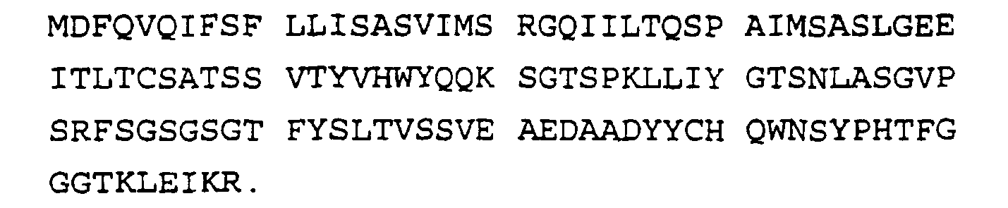
- The mature peptide starts at amino acid 23 which is a Q.
- Those skilled in the art will realize that a variety of DNA sequences would code for the polypeptide antibody fragments described above. This is due to existence of the degeneracy of the genetic code, which means that different codons (sets of three bases) can code for the same amino acid residue. These are known to those skilled in the art. It is also possible to synthesize DNA sequence having different additional substitution than those described above but which would still code for a protein having the same binding specifications, for example, which has conservative amino acid substitutions, i.e., substitutions of one amino acid with another of similar size and charge.
- Using the sequences disclosed above, recombinant antibodies can be constructed using known methodology. Methods for constructing chimeric genes have been described by, for example, Kobilka, B.K., et al, "Chimeric α2-,β2-Adrenergic Receptors: Delineation of Domains Involved in Effector Coupling and Ligand Binding Specificity" Science 240:1310-1316, 1988; Verhoeyen, M., C. Milstein, G. Winter, "Reshaping Human Antibodies: Grafting an Antilysozyme Activity," Science, 239:1534-1536, 1988; Riechmann,L., M. Clark, H Waldmann, G. Winter, "Reshaping human antibodies for therapy," Nature, 332:323-327, 1988). Using standard molecular biology techniques, the target DNA, containing the gene for the monoclonal antibody of interest can be constructed into appropriate expression vectors, such as baculovirus expression vectors, according to the procedures described in Summers, M.D. and G.E. Smith, "A manual of methods for baculovirus vectors and insect cell culture procedures", Texas Agricultural Experimental Station (1987). Expression of the recombinant gene can be achieved by the methods described therein, the teachings of which are incorporated herein. Alternatively, recombinant antibodies can be produced in bacterial periplasmic expression vectors such as those described above. Screening for the desired product can be achieved by ELISA assay wherein released protein is tested for its ability to recognize the antigen for which the target immunoglobulin was specific in a metal dependent manner.
- Methods for "humanizing" antibodies, or generating less immunogenic fragments of non-human antibodies, are well known. A humanized antibody is one in which only the antigen-recognizing sites, or complementarity-determining hypervariable regions (CDRs) are of non-human origin, whereas all other regions including the framework regions (FRs) of variable domains are products of human genes. These "humanized" antibodies are less immunogenic when introduced into a human recipient yet they retain their antigen binding specificity. To accomplish humanization of a selected mouse monoclonal antibody, the CDR grafting method described by Daugherty, et al., Nucl. Acids Res., 19:2471-2476 (1991), incorporated herein by reference, can be used. Briefly, animal CDRs are distinguished from animal framework regions (FRs) based on locations of the CDRs in known sequences of animal variable genes, (Kabat, H.A., et al., Sequences of Proteins of Immunological Interest, 4th Ed. U.S. Dept. Health and Human Services, Bethesda, MD, 1987). Once the animal CDRs and FRs are identified, the animal CDRs are grafted onto the sequence of an unrelated human heavy and light chain variable region frameworks by the standard molecular biology techniques including the use of synthetic oligonucleotides and polymerase chain reaction (PCR) methods. Alternatively, the entire sequences of a known human variable heavy and light chain gene in which all the codons encoding for the CDRs are replaced with the desired CDRs of animal antibody, are synthesized in the laboratory by a DNA synthesizer (Applied Biosystems Division of Perkin-Elmer Cetus, CA). The resulting synthetic DNA sequences encoding for the human heavy and light chain variable regions with grafted CDRs from animal antibody are subcloned into expression vectors and recombinant fusion antibodies are prepared in baculovirus or periplasmic space of bacteria as described above. Recombinant antibodies can be produced in mammalian expression systems as well.
- The immunogenic stimulus presented by the monoclonal antibodies so produced may be also decreased by the use of Pharmacia's (Pharmacia LKB Biotechnology, Sweden) "Recombinant Phage Antibody System" (RPAS), which generates a single-chain Fv fragment (ScFv) which incorporates the complete antigen-binding domain of the antibody. In the RPAS, the variable heavy and light chain genes are separately amplified from the hybridoma mRNA and cloned into an expression vector. The heavy and light chain domains are co-expressed on the same polypeptide chain after joining with a short linker DNA which codes for a flexible peptide. This assembly generates a single-chain Fv fragment (ScFv) which incorporates the complete antigen-binding domain of the antibody. Compared to the intact monoclonal antibody, the recombinant ScFv includes a considerably lower number of antigenic epitopes, and thereby presents a much weaker immunogenic stimulus when injected into humans.
- Both HPC-4 from ascites and the recombinant HPC-4 bind to a defined region of the protein C molecule that is contained within residues 6 and 17 of the heavy chain, specifically E D Q V D P R L I D G K (Sequence ID No. 1). This peptide can be immobilized directly on a solid support resin and can be used to isolate the antibody in high concentrations from ascites fluid or as recombinant form from cell culture supernatants. This approach allows the isolation of the antibody in extremely pure form in high yield, even from very dilute solutions.
- The antibody can be removed from the solid support peptide either by the removal of calcium ions, if desired, or by 1.5 M guanidine, which does not affect the function of the purified monoclonal antibody. This may be significant, as guanidine is recognized as a viral deactivation agent by regulatory agencies. After elution or treatment with this agent, the antibody will not contain any live virus which may be present either in the ascites fluid derived from the mice used to produce the monoclonal antibody or culture supernatants, if tissue culture for production of recombinant antibody was used. Accordingly, virus will not be introduced into the protein C product from the antibody used to prepare it.
- In a preferred embodiment, the peptide is coupled to Affi-Gel™ 15 to give a final concentration of approximately 1.0 mg/ml. Coupling of the epitope peptide is performed in 0.1 M NaCl, 0.1 M MOPS, pH 7.5, at 4°C as described by the manufacturer (Bio-Rad, Richmond, CA). The Affi-Gel™ is washed with ice cold water immediately before use to remove the organic solvent. The epitope peptide is prepared at a concentration of between 1 and 2 mg/ml in 0.1 M NaCl, 0.1 M MOPS, pH 7.5, and mixed with sufficient Affi-Gel™ 15 to give a final ratio of peptide to gel of 1 mg/ml. The peptide and the gel are mixed overnight (between approximately 12 and 18 h) on a gentle rocker to couple the peptide to the gel. After the coupling reaction is completed, the resin is poured into a glass column, and washed with 0.1 M NaCl 0.01 M MOPS, pH 7.5. 100 ml of resin has a capacity of at least 1.5 grams of HPC-4.
- Human protein C can be coupled to the Affi-Gel™ by the same method. Three to five mg protein C/ml of the buffer described above is mixed with sufficient Affi-Gel™ 15 to give a final ratio of human protein C to gel of 3-5 mg protein/ml of gel.
- The desalted ammonium sulfate fraction from the ascites is loaded onto the epitope affinity column, and the column is washed with at least 4 column volumes of 0.4 M NaCl, 0.02 M Tris HCl, 1 mM CaCl2, pH 7.5. The HPC-4 or recombinant antibody is then eluted from the column in one of the following ways: (1) 2 M NaCl, 0.02 M Tris HCl, 2 mM EDTA; (2) 2 M NaCl, 1.5 M guanidine HCl, 0.02 M Tris HCl, 2 mM EDTA. The advantage of the latter is that the protein elutes as a much sharper peak, with concentrations of greater than 25 mg/ml when 200 ml of ascites is applied to a 100 ml column of resin. The antibody retains greater than 95% of the capacity to bind to the epitope after elution under these conditions. Antibody is then either dialyzed or desalted into the appropriate buffer for further applications. No contaminants of the antibody are detectable by SDS gel electrophoresis. Additional antibody can be obtained by applying the breakthrough material back to the column if the column is overloaded above its capacity.
- The recombinant antibody can be utilized in the same way as HPC-4 for purification and therapeutic purposes. As discussed below, "HPC-4" includes both the deposited murine monoclonal antibody and recombinant forms thereof.
- For purification of protein C by affinity chromatography, coupling of the antibody to an immobilized substrate such as Affi-Gel™ resin is performed in 0.1 M NaCl, 0.1 M MOPS, pH 7.5, at 4°C as described by the manufacturer (Bio-Rad, Richmond, CA). The Affi-Gel™ is washed with ice cold water immediately before use to remove the organic solvent. HPC-4 is prepared at a concentration of 3-5 mg/ml in 0.1 M NaCl, 0.1 M MOPS, pH 7.5, and mixed with sufficient Affi-Gel™ 10 to give a final ratio of HPC-4 to gel of 5 mg/ml. Antibody and the gel are mixed overnight (12-18 h) on a gentle rocker to allow the coupling reaction. Usually greater than 90% of the antibody is bound. After the coupling reaction is completed, the resin is poured into a glass column, and washed with 0.1 M NaCl 0.01 M MOPS, pH 7.5. The resin is stable at 4°C under these conditions for at least one year. 100 ml of resin has a capacity of at least 20 milligrams of protein C.
- As described above, the peptide can be used in the isolation and purification of HPC-4 by affinity chromatography. In a similar manner, the peptide can be used to temporarily "protect" the binding site during the process in which the antibody is bound to the chromatography substrate, to insure that the maximum amount of bound antibody is available for binding to the protein to be isolated. The reactive groups of the peptide which are capable of reacting with the chromatography substrate (amino terminal, lysine side chain), which are not required for recognition by HPC-4, are first blocked by reaction of the peptide with acetic anhydride using standard methods known to those skilled in the art. After the HPC-4 is coupled to the resin, the peptide bound in the antigen binding site of the antibody is removed by washing the resin with 1.5 M Guanidine HCl, 2 mM EDTA, 0.02 M Tris HCl, pH 7.5.
- The antibody and peptide can be bound to a variety of substrates, for use in purification and isolation of Protein C and the antibody, respectively, including agarose, acrylamide and other types of conventional chromatographic resins, filters, etc. These materials are known to those skilled in the art, as are the methods for attaching the protein to them. The selection of the material will depend in large part on the scale of the purification or the sample to be analyzed, as well as biocompatibility and government agency approval where the end-product is for pharmaceutical use.
- Methods and means for labeling the antibody for use as a diagnostic are known to those skilled in the art, including labelling with a radioactive, fluorescent, luminescent, or enzymatic molecule. The antibodies are then used in diagnostic assays to measure the amount of Protein C rather than Activated Protein C or total Protein C, since the antibody does not bind Activated Protein C, unlike other antibodies to Protein C.
- Isolation of fusion proteins with antibody
- A fusion protein readily isolated by affinity chromatography using HPC-4 antibody is prepared by insertion of a DNA sequence encoding the twelve amino acid HPC-4 epitope into a vector, followed by the gene encoding the protein to be isolated as described in
U.S. Patent No. 5,298,599 issued March 29, 1994 . In the preferred embodiment, a specific protease cleavage site is inserted into the vector between the epitope and protein coding sequence, so that the resulting fusion protein can be easily cleaved to yield the epitope peptide and the desired protein. In the most preferred embodiment, the fusion protein includes a protease cleavage site between the epitope and the protein to be isolated. Suitable sites include sequences cleaved by Factor Xa: Ile Glu Gly Arg (IEGR), enterokinase: Asp Asp Asp Asp Lys (DDDDK), and thrombin: Phe/Gly Pro Arg (F/GPR). Following purification with the HPC-4, the fusion protein is treated with the appropriate enzyme to cleave the binding peptide from the desired protein. - The coagulant and anticoagulant systems in mammals provide a delicate check and balance system which maintains blood in its proper fluid state. Alteration of any single element in this system can have an enormous impact on the ability of the mammal to maintain hemostasis.
- The protein C system is an anticoagulant, regulatory system which inhibits blood coagulation and stimulates fibrinolysis. This system is activated by thrombin, an enzyme which converts fibrinogen to fibrin in the coagulative process. Free or excess thrombin binds with thrombomodulin, a protein on endothelial cells. The thrombin-thrombomodulin complex abolishes the ability of thrombin to catalyze clot formation and converts thrombin into a potent protein C activator. Activated Protein C in turn acts in combination with Protein S and a membrane surface to inactivate factor Va and factor VIIIa by limited proteolysis. The inactivated factor Va loses the ability to interact effectively with the enzyme factor Xa or the substrate prothrombin.
- Addition of an antibody to Protein C, an antibody to Protein S, or addition of C4b binding protein (C4bBP), which binds Protein S to thereby inactivate it as a cofactor, in an appropriate form, can be used to promote clotting in individuals where it is desirable to do so. Patients having factor VIII inhibitors are representative of this group of patients. By preventing the factor Va from being inactivated, coagulation proceeds even in the relative absence of factor VIII.
- The effect of administering these inhibitors of the Protein C anticoagulation system can be reversed by administration of excess amounts of activated Protein C or Protein S, depending on the agent used to block the pathway. The appropriate amount is based on calculations relating to the relative molar amounts of the proteins present in the blood. The feasibility of this approach to produce a hypercoaguable state has been demonstrated by the administration of HPC-4 to baboons (Taylor, et al, J. Clin. Invest., 79, 918-925 (1987). When HPC-4 was present, the animals developed a massive coagulation response, characterized by total fibrinogen consumption, as the result of the infusion of low levels of bacteria. They did not develop this response in the absence of the antibody. Virtually identical results are obtained when C4bBP levels are elevated to approximately 1 mg/ml plasma. While these responses are detrimental to the animals, they illustrate that either method will enhance the coagulation system. This is beneficial in situations where normal hemostasis is impaired.
- This method can also be applied in the treatment of other clotting factor deficiency states, including thrombocytopenia, for example, as induced by heparin or radiation therapy, liver disease and hemorrhagic stroke, both acutely and to minimize re-bleeding after the acute incident.
- HPC-4 can also be used to induce microvascular clotting in a solid tumor bed, as described in
U.S. Patent No. 5,147,638 issued September 5, 1992 . In animal tumor models, this has been found to greatly impede growth of the tumor. The combination of this antibody and/or the other agents indicated above which are capable of blocking the function of the protein C anticoagulant pathway with other treatments presently in use, such as tumor necrosis factor or radiation, can also be used for treatment of solid tumors. - Pharmaceutically acceptable carriers for administration of the antibodies include sterile normal saline at physiological pH. In the preferred method of administration, the agent is injected into the subject, most preferably, intravenously. Preferred dosages are between about 30 and about 150 µg antibody/ml patient plasma, which is sufficient to block greater than 90% of the endogenous protein C.
- The teachings of the references and patents cited above are representative of methods and reagents known to those skilled in the art.
-
- (1) GENERAL INFORMATION:
- (i) APPLICANT: Oklahoma Medical Research Foundation
- (ii) TITLE OF INVENTION: Calcium Binding Recombinant Antibody Against Protein C
- (iii) NUMBER OF SEQUENCES: 12
- (iv) CORRESPONDENCE ADDRESS:
- (A) ADDRESSEE: Patrea L. Pabst
- (B) STREET: 2800 One Atlantic Center 1201 West Peachtree Street
- (C) CITY: Atlanta
- (D) STATE: Georgia
- (E) COUNTRY: USA
- (F) ZIP: 30309-3450
- (v) COMPUTER READABLE FORM:
- (A) MEDIUM TYPE: Floppy disk
- (B) COMPUTER: IBM PC compatible
- (C) OPERATING SYSTEM: PC-DOS/MS-DOS
- (D) SOFTWARE: PatentIn Release #1.0, Version #1.25
- (vi) CURRENT APPLICATION DATA:
- (A) APPLICATION NUMBER:
- (B) FILING DATE:
- (C) CLASSIFICATION:
- (viii) ATTORNEY/AGENT INFORMATION:
- (A) NAME: Pabst, Patrea L.
- (B) REGISTRATION NUMBER: 31,284
- (C) REFERENCE/DOCKET NUMBER: OMRF106CIP
- (ix) TELECOMMUNICATION INFORMATION:
- (A) TELEPHONE: (404) 873-8794
- (B) TELEFAX: (404) 873-8795
- (2) INFORMATION FOR SEQ ID NO:1:
- (i) SEQUENCE CHARACTERISTICS:
- (A) LENGTH: 12 amino acids
- (B) TYPE: amino acid
- (C) STRANDEDNESS: single
- (D) TOPOLOGY: linear
- (ii) MOLECULE TYPE: peptide
- (iii) HYPOTHETICAL: NO
- (iv) ANTI-SENSE: NO
- (v) FRAGMENT TYPE: Internal
- (xi) SEQUENCE DESCRIPTION: SEQ ID NO:1:

- (i) SEQUENCE CHARACTERISTICS:
- (2) INFORMATION FOR SEQ ID NO:2:
- (i) SEQUENCE CHARACTERISTICS:
- (A) LENGTH: 40 base pairs
- (B) TYPE: nucleic acid
- (C) STRANDEDNESS: single
- (D) TOPOLOGY: linear
- (ii) MOLECULE TYPE: DNA
- (iii) HYPOTHETICAL: NO
- (iv) ANTI-SENSE: YES
- (xi) SEQUENCE DESCRIPTION: SEQ ID NO:2:
AAGCGGCCGC TGGATAGACA GATGGGGGTG TCGTTTTGCC 40
- (i) SEQUENCE CHARACTERISTICS:
- (2) INFORMATION FOR SEQ ID NO:3:
- (i) SEQUENCE CHARACTERISTICS:
- (A) LENGTH: 30 base pairs
- (B) TYPE: nucleic acid
- (C) STRANDEDNESS: single
- (D) TOPOLOGY: linear
- (ii) MOLECULE TYPE: DNA
- (iii) HYPOTHETICAL: NO
- (iv) ANTI-SENSE: NO
- (xi) SEQUENCE DESCRIPTION: SEQ ID NO:3:
AAGCGGCCGC CCCCCCCCCC CCCCCCCCCC 30
- (i) SEQUENCE CHARACTERISTICS:
- (2) INFORMATION FOR SEQ ID NO:4:
- (i) SEQUENCE CHARACTERISTICS:
- (A) LENGTH: 40 base pairs
- (B) TYPE: nucleic acid
- (C) STRANDEDNESS: single
- (D) TOPOLOGY: linear
- (ii) MOLECULE TYPE: DNA
- (iii) HYPOTHETICAL: NO
- (iv) ANTI-SENSE: YES
- (xi) SEQUENCE DESCRIPTION: SEQ ID NO:4:
AAGCGGCCGC GAAGATGGAT ACAGTTGGTG CAGCATCAGC 40
- (i) SEQUENCE CHARACTERISTICS:
- (2) INFORMATION FOR SEQ ID NO:5:
- (i) SEQUENCE CHARACTERISTICS:
- (A) LENGTH: 28 base pairs
- (B) TYPE: nucleic acid
- (C) STRANDEDNESS: single
- (D) TOPOLOGY: linear
- (ii) MOLECULE TYPE: DNA
- (iii) HYPOTHETICAL: NO
- (iv) ANTI-SENSE: NO
- (xi) SEQUENCE DESCRIPTION: SEQ ID NO:5:
AGGTTACTCT GCTCGAGTCT GGCCCTGG 28
- (i) SEQUENCE CHARACTERISTICS:
- (2) INFORMATION FOR SEQ ID NO:6:
- (i) SEQUENCE CHARACTERISTICS:
- (A) LENGTH: 36 base pairs
- (B) TYPE: nucleic acid
- (C) STRANDEDNESS: single
- (D) TOPOLOGY: linear
- (ii) MOLECULE TYPE: DNA
- (iii) HYPOTHETICAL: NO
- (iv) ANTI-SENSE: NO
- (xi) SEQUENCE DESCRIPTION: SEQ ID NO:6:
AGGCCTACTA GTTTACTAAC AATCCCTGGG CACAAT 36
- (i) SEQUENCE CHARACTERISTICS:
- (2) INFORMATION FOR SEQ ID NO:7:
- (i) SEQUENCE CHARACTERISTICS:
- (A) LENGTH: 37 base pairs
- (B) TYPE: nucleic acid
- (C) STRANDEDNESS: single
- (D) TOPOLOGY: linear
- (ii) MOLECULE TYPE: DNA
- (iii) HYPOTHETICAL: NO
- (iv) ANTI-SENSE: NO
- (xi) SEQUENCE DESCRIPTION: SEQ ID NO:7:
TGTCCAGAGG AGAGCTCATT CTCACCCAGT CTCCGGC 37
- (i) SEQUENCE CHARACTERISTICS:
- (2) INFORMATION FOR SEQ ID NO:8:
- (i) SEQUENCE CHARACTERISTICS:
- (A) LENGTH: 34 base pairs
- (B) TYPE: nucleic acid
- (C) STRANDEDNESS: single
- (D) TOPOLOGY: linear
- (ii) MOLECULE TYPE: DNA
- (iii) HYPOTHETICAL: NO
- (iv) ANTI-SENSE: YES
- (xi) SEQUENCE DESCRIPTION: SEQ ID NO:8:
TCCTTCTAGA TTACTAACAC TCTCCCCTGT TGAA 34
- (i) SEQUENCE CHARACTERISTICS:
- (2) INFORMATION FOR SEQ ID NO:9:
- (i) SEQUENCE CHARACTERISTICS:
- (A) LENGTH: 417 base pairs
- (B) TYPE: nucleic acid
- (C) STRANDEDNESS: single
- (D) TOPOLOGY: linear
- (ii) MOLECULE TYPE: DNA
- (iii) HYPOTHETICAL: NO
- (iv) ANTI-SENSE: NO
- (vi) ORIGINAL SOURCE:
- (A) ORGANISM: HPC-4 Heavy Chain Variable Region (VH Gamma)
- (ix) FEATURE:
- (A) NAME/KEY: misc_feature
- (B) LOCATION: 1..57
- (D) OTHER INFORMATION: /note= "Signal peptide encoded by nucleotides 1 through 57."
- (ix) FEATURE:
- (A) NAME/KEY: misc_feature
- (B) LOCATION: 58..417
- (D) OTHER INFORMATION: /note= "Mature peptide encoded by nucleotides 58 through 417."
- (xi) SEQUENCE DESCRIPTION: SEQ ID NO:9:

- (i) SEQUENCE CHARACTERISTICS:
- (2) INFORMATION FOR SEQ ID NO:10:
- (i) SEQUENCE CHARACTERISTICS:
- (A) LENGTH: 139 amino acids
- (B) TYPE: amino acid
- (C) STRANDEDNESS: single
- (D) TOPOLOGY: linear
- (ii) MOLECULE TYPE: peptide
- (iii) HYPOTHETICAL: NO
- (iv) ANTI-SENSE: NO
- (v) FRAGMENT TYPE: Internal
- (vi) ORIGINAL SOURCE:
- (A) ORGANISM: HPC-4 Heavy Chain Variable Region (VH Gamma)
- (ix) FEATURE:
- (A) NAME/KEY: misc feature
- (B) LOCATION: 20..139
- (D) OTHER INFORMATION: /note= "Gln at position 20 starts mature peptide."
- (xi) SEQUENCE DESCRIPTION: SEQ ID NO:10:


- (i) SEQUENCE CHARACTERISTICS:
- (2) INFORMATION FOR SEQ ID NO:11:
- (i) SEQUENCE CHARACTERISTICS:
- (A) LENGTH: 387 base pairs
- (B) TYPE: nucleic acid
- (C) STRANDEDNESS: single
- (D) TOPOLOGY: linear
- (ii) MOLECULE TYPE: DNA
- (iii) HYPOTHETICAL: NO
- (iv) ANTI-SENSE: NO
- (vi) ORIGINAL SOURCE:
- (A) ORGANISM: HPC-4 Light Chain Variable Region (VL Kappa)
- (ix) FEATURE:
- (A) NAME/KEY: misc_feature
- (B) LOCATION: 1..66
- (D) OTHER INFORMATION: /note= "Signal peptide encoded by nucleotides 1 through 66."
- (ix) FEATURE:
- (A) NAME/KEY: misc_feature
- (B) LOCATION: 67..387
- (D) OTHER INFORMATION: /note= "Mature peptide encoded by nucleotides 67 through 387."
- (xi) SEQUENCE DESCRIPTION: SEQ ID NO:11:

- (i) SEQUENCE CHARACTERISTICS:
- (2) INFORMATION FOR SEQ ID NO:12:
- (i) SEQUENCE CHARACTERISTICS:
- (A) LENGTH: 129 amino acids
- (B) TYPE: amino acid
- (C) STRANDEDNESS: single
- (D) TOPOLOGY: linear
- (ii) MOLECULE TYPE: peptide
- (iii) HYPOTHETICAL: NO
- (iv) ANTI-SENSE: NO
- (v) FRAGMENT TYPE: N-terminal
- (vi) ORIGINAL SOURCE:
- (A) ORGANISM: HPC-4 Light Chain Variable Region (VL Kappa)
- (ix) FEATURE:
- (A) NAME/KEY: misc_feature
- (B) LOCATION: 23...129
- (D) OTHER INFORMATION: /note= "Gln at position 23 starts mature peptide."
- (xi) SEQUENCE DESCRIPTION: SEQ ID NO:12:

- (i) SEQUENCE CHARACTERISTICS:
Claims (13)
- A recombinant Ca2+ dependent monoclonal antibody or Fab immunoreactive with an epitope in the activation peptide region of the heavy chain of Protein C defined by E D Q V D P R L I D G K (Sequence ID No. 1) in combination with calcium, where the antibody inhibits Protein C activation by thrombin-thrombomodulin, wherein the antibody or Fab contains human amino acid sequence, the antibody or Fab comprising amino acid sequence selected from the group consisting of:GGTKLEIKR (amino acids 23 -129 of Sequence ID No. 12) or having one or more conservative substitutions of the given amino acid sequence, wherein the binding of the antibody or Fab to the epitope is calcium dependent.

- The antibody of Claim 1 encoded in part by a nucleotide sequence selected from the group consisting of ATGGGCAGGC TTTCTTCTTC

 (nucleotides 67 to 387 of Sequence ID No. 11); and degenerate sequences thereof.
- The antibody of Claim 1 further comprising a pharmaceutically acceptable carrier for administration to a patient.
- The antibody of Claim 3 further comprising a cytokine or an inducer of cytokine expression in a dosage effective in combination with the antibody to coagulate microvasculature in tumors but not in the absence of the antibody.
- The antibody of Claim 1 having a detectable label bound to the antibody.
- The antibody of Claim 1 immobilized to a substrate, wherein the immobilized antibody is suitable for purification of protein C from a biological fluid.
- Use of a recombinant Ca2+ dependent monoclonal antibody as defined in any one of Claims 1 to 3 in the manufacture of a medicament for treating a patient in need of inhibition of protein C anticoagulant.
- The use of Claim 7 wherein the medicament is for treating a patient administered a cytokine or other chemotherapeutic agent in an amount effective to coagulate the microvasculature of a tumor.
- An in vitro method of making a recombinant Ca2+ dependent monoclonal as defined in relation to Claim 1, by expressing nucleotide sequence encoding the antibody.
- The method of Claim 9 wherein the antibody is encoded in part by a nucleotide sequence selected from the group consisting of
 (nucleotides 67-387 of Sequence ID No. 11); and degenerate sequences thereof.
- The method of Claim 9 further comprising binding detectable label to the antibody.
- The method of Claim 9 further comprising immobilizing the antibody to a substrate, wherein the immobilized antibody is suitable for purification of protein C from a biological fluid.
- An antibody according to any one of Claims 1 to 4 for use in medicine.
Applications Claiming Priority (3)
| Application Number | Priority Date | Filing Date | Title |
|---|---|---|---|
| US259321 | 1994-06-10 | ||
| US08/259,321 US7247453B1 (en) | 1988-12-30 | 1994-06-10 | Calcium binding recombinant antibody against protein C |
| PCT/US1995/007372 WO1995034652A1 (en) | 1994-06-10 | 1995-06-09 | Calcium binding recombinant antibody against protein c |
Publications (2)
| Publication Number | Publication Date |
|---|---|
| EP0804580A1 EP0804580A1 (en) | 1997-11-05 |
| EP0804580B1 true EP0804580B1 (en) | 2008-10-08 |
Family
ID=22984456
Family Applications (1)
| Application Number | Title | Priority Date | Filing Date |
|---|---|---|---|
| EP95923791A Expired - Lifetime EP0804580B1 (en) | 1994-06-10 | 1995-06-09 | Calcium binding recombinant antibody against protein c |
Country Status (7)
| Country | Link |
|---|---|
| EP (1) | EP0804580B1 (en) |
| JP (1) | JPH10504186A (en) |
| AT (1) | ATE410514T1 (en) |
| AU (1) | AU710106B2 (en) |
| CA (1) | CA2192467C (en) |
| DE (1) | DE69535855D1 (en) |
| WO (1) | WO1995034652A1 (en) |
Cited By (1)
| Publication number | Priority date | Publication date | Assignee | Title |
|---|---|---|---|---|
| US7750126B2 (en) | 1998-04-14 | 2010-07-06 | Chugai Seiyaku Kabushiki Kaisha | Antibodies that bind to a member of the IL-6/G-CSF/MGF family |
Families Citing this family (16)
| Publication number | Priority date | Publication date | Assignee | Title |
|---|---|---|---|---|
| TW200407335A (en) * | 2002-07-22 | 2004-05-16 | Chugai Pharmaceutical Co Ltd | Non-neutralizing antibody to inhibit the inactivation of activated protein C |
| JP6271251B2 (en) * | 2011-10-05 | 2018-01-31 | 中外製薬株式会社 | An antigen-binding molecule that promotes elimination of an antigen containing a sugar chain receptor-binding domain from plasma |
| CA2882562C (en) | 2012-08-21 | 2019-08-27 | Eric Hryhorenko | Antibodies to aripiprazole and use thereof |
| JP2015527365A (en) | 2012-08-21 | 2015-09-17 | オルソ−クリニカル ダイアグノスティクス,インコーポレイティド | Antibodies against paliperidone and use thereof |
| PL2888592T3 (en) | 2012-08-21 | 2018-03-30 | Janssen Pharmaceutica Nv | Antibodies to quetiapine and use thereof |
| WO2014031656A1 (en) | 2012-08-21 | 2014-02-27 | Ortho-Clinical Diagnostics, Inc | Antibodies to olanzapine haptens and use thereof |
| CA2882596C (en) | 2012-08-21 | 2019-05-14 | Ortho-Clinical Diagnostics, Inc. | Antibodies to olanzapine and use thereof |
| JP6389176B2 (en) | 2012-08-21 | 2018-09-12 | ヤンセン ファーマシューティカ エヌ.ベー. | Antibodies against aripiprazole hapten and use thereof |
| ES2691092T3 (en) | 2012-08-21 | 2018-11-23 | Janssen Pharmaceutica Nv | Risperidone antibodies and their use |
| JP2015529199A (en) | 2012-08-21 | 2015-10-05 | オルソ−クリニカル ダイアグノスティクス,インコーポレイティド | Antibodies against paliperidone hapten and use thereof |
| EP2888284B1 (en) | 2012-08-21 | 2022-07-06 | Janssen Pharmaceutica NV | Antibodies to risperidone haptens and use thereof |
| JP6450314B2 (en) | 2012-08-21 | 2019-01-09 | ヤンセン ファーマシューティカ エヌ.ベー. | Antibodies against quetiapine haptens and uses thereof |
| JP6131414B2 (en) | 2012-08-21 | 2017-05-24 | ヤンセン ファーマシューティカ エヌ.ベー. | Aripiprazole haptens and their use in immunoassays |
| TWI673063B (en) * | 2013-09-20 | 2019-10-01 | 日商中外製藥股份有限公司 | Treatment of bleeding disorders caused by anti-protein C antibodies |
| US10435478B2 (en) | 2015-12-17 | 2019-10-08 | Janssen Pharmaceutica Nv | Antibodies to quetiapine and use thereof |
| EP3390449A1 (en) | 2015-12-17 | 2018-10-24 | Janssen Pharmaceutica N.V. | Antibodies to risperidone and use thereof |
Family Cites Families (3)
| Publication number | Priority date | Publication date | Assignee | Title |
|---|---|---|---|---|
| US5147638A (en) * | 1988-12-30 | 1992-09-15 | Oklahoma Medical Research Foundation | Inhibition of tumor growth by blockade of the protein C system |
| CA2006684C (en) * | 1988-12-30 | 1996-12-17 | Charles T. Esmon | Monoclonal antibody against protein c |
| EP0651655A4 (en) * | 1992-07-24 | 1996-05-29 | Oklahoma Med Res Found | Blockade of protein c activation reduces microvascular surgical blood loss. |
-
1995
- 1995-06-09 AU AU28229/95A patent/AU710106B2/en not_active Expired
- 1995-06-09 CA CA002192467A patent/CA2192467C/en not_active Expired - Lifetime
- 1995-06-09 EP EP95923791A patent/EP0804580B1/en not_active Expired - Lifetime
- 1995-06-09 DE DE69535855T patent/DE69535855D1/en not_active Expired - Fee Related
- 1995-06-09 AT AT95923791T patent/ATE410514T1/en not_active IP Right Cessation
- 1995-06-09 JP JP8502358A patent/JPH10504186A/en not_active Ceased
- 1995-06-09 WO PCT/US1995/007372 patent/WO1995034652A1/en not_active Ceased
Non-Patent Citations (1)
| Title |
|---|
| STEARNEST D.J. ET AL, THE JOURNAL OF BIOLOGICAL CHEMISTRY, vol. 263, no. 2, 1988, pages 826 - 832 * |
Cited By (1)
| Publication number | Priority date | Publication date | Assignee | Title |
|---|---|---|---|---|
| US7750126B2 (en) | 1998-04-14 | 2010-07-06 | Chugai Seiyaku Kabushiki Kaisha | Antibodies that bind to a member of the IL-6/G-CSF/MGF family |
Also Published As
| Publication number | Publication date |
|---|---|
| WO1995034652A1 (en) | 1995-12-21 |
| AU710106B2 (en) | 1999-09-16 |
| CA2192467C (en) | 2009-03-31 |
| CA2192467A1 (en) | 1995-12-21 |
| AU2822995A (en) | 1996-01-05 |
| JPH10504186A (en) | 1998-04-28 |
| ATE410514T1 (en) | 2008-10-15 |
| EP0804580A1 (en) | 1997-11-05 |
| DE69535855D1 (en) | 2008-11-20 |
Similar Documents
| Publication | Publication Date | Title |
|---|---|---|
| EP0804580B1 (en) | Calcium binding recombinant antibody against protein c | |
| CA2045869C (en) | Fusion proteins with immunoglobulin portions, the preparation and use thereof | |
| Gillies et al. | Biological activity and in vivo clearance of anti-tumor antibody/cytokine fusion proteins | |
| JPH07504812A (en) | Novel polypeptides to promote cell attachment | |
| EP1396542B1 (en) | P-Selectin Ligand Protein | |
| EP0753065A1 (en) | Antibodies against e-selectin | |
| US5260274A (en) | Factor VIII binding domain of von Willebrand Factor | |
| WO2001016321A1 (en) | Platelet membrane glycoprotein vi (gpvi) dna and protein sequences, and uses thereof | |
| CZ305602B6 (en) | Von Willebrand factor (vWF) cleaving protease polypeptide, nucleic acid encoding the polypeptide and use of such polypeptide | |
| AU641634B2 (en) | Monoclonal antibody against protein C | |
| IL141158A (en) | Humanized immunoglobulin which binds to human von willebrand factor | |
| AU781618C (en) | FVIIa antagonists | |
| KR100240159B1 (en) | Humanized Immunoglobulins Reactive with Glycoprotein GPIIB / IIIA | |
| US7247453B1 (en) | Calcium binding recombinant antibody against protein C | |
| US5773228A (en) | Activation-dependent protein expressed on the surface of activated platelets and antibodies thereto | |
| AU755574B2 (en) | Synthetic peptide immunogens and antibodies thereto | |
| EP0713496A1 (en) | Fibrin-targeted inhibitors of thrombin | |
| WO1993011778A1 (en) | Bifunctional antithrombotic molecules and antithrombotic polypeptides | |
| WO1992017192A1 (en) | Therapeutic fragments of von willebrand factor | |
| WO1999060405A1 (en) | Method for diagnosis and treatment of cancer | |
| WO2004064709A2 (en) | Thrombolytic agent | |
| JPH05219957A (en) | Tissue factor derivative | |
| IE84574B1 (en) | Fusion proteins with parts of immunoglobulins, their production and use | |
| HK1012015B (en) | Fusionproteins with parts of immunoglobulins, their production and use |
Legal Events
| Date | Code | Title | Description |
|---|---|---|---|
| PUAI | Public reference made under article 153(3) epc to a published international application that has entered the european phase |
Free format text: ORIGINAL CODE: 0009012 |
|
| 17P | Request for examination filed |
Effective date: 19970110 |
|
| AK | Designated contracting states |
Kind code of ref document: A1 Designated state(s): AT BE CH DE DK ES FR GB GR IE IT LI LU MC NL PT SE |
|
| 17Q | First examination report despatched |
Effective date: 20030110 |
|
| 17Q | First examination report despatched |
Effective date: 20030110 |
|
| 17Q | First examination report despatched |
Effective date: 20030110 |
|
| GRAP | Despatch of communication of intention to grant a patent |
Free format text: ORIGINAL CODE: EPIDOSNIGR1 |
|
| RIC1 | Information provided on ipc code assigned before grant |
Ipc: A61K 38/19 20060101ALI20080331BHEP Ipc: A61K 33/06 20060101ALI20080331BHEP Ipc: A61K 39/395 20060101ALI20080331BHEP Ipc: C07K 17/00 20060101ALI20080331BHEP Ipc: C07K 16/46 20060101ALI20080331BHEP Ipc: C07K 16/40 20060101ALI20080331BHEP Ipc: C12N 15/13 20060101AFI20080331BHEP |
|
| GRAS | Grant fee paid |
Free format text: ORIGINAL CODE: EPIDOSNIGR3 |
|
| GRAA | (expected) grant |
Free format text: ORIGINAL CODE: 0009210 |
|
| AK | Designated contracting states |
Kind code of ref document: B1 Designated state(s): AT BE CH DE DK ES FR GB GR IE IT LI LU MC NL PT SE |
|
| REG | Reference to a national code |
Ref country code: GB Ref legal event code: FG4D |
|
| REG | Reference to a national code |
Ref country code: CH Ref legal event code: EP |
|
| REG | Reference to a national code |
Ref country code: IE Ref legal event code: FG4D |
|
| REF | Corresponds to: |
Ref document number: 69535855 Country of ref document: DE Date of ref document: 20081120 Kind code of ref document: P |
|
| NLV1 | Nl: lapsed or annulled due to failure to fulfill the requirements of art. 29p and 29m of the patents act | ||
| PG25 | Lapsed in a contracting state [announced via postgrant information from national office to epo] |
Ref country code: ES Free format text: LAPSE BECAUSE OF FAILURE TO SUBMIT A TRANSLATION OF THE DESCRIPTION OR TO PAY THE FEE WITHIN THE PRESCRIBED TIME-LIMIT Effective date: 20090119 Ref country code: AT Free format text: LAPSE BECAUSE OF FAILURE TO SUBMIT A TRANSLATION OF THE DESCRIPTION OR TO PAY THE FEE WITHIN THE PRESCRIBED TIME-LIMIT Effective date: 20081008 |
|
| PG25 | Lapsed in a contracting state [announced via postgrant information from national office to epo] |
Ref country code: PT Free format text: LAPSE BECAUSE OF FAILURE TO SUBMIT A TRANSLATION OF THE DESCRIPTION OR TO PAY THE FEE WITHIN THE PRESCRIBED TIME-LIMIT Effective date: 20090217 Ref country code: NL Free format text: LAPSE BECAUSE OF FAILURE TO SUBMIT A TRANSLATION OF THE DESCRIPTION OR TO PAY THE FEE WITHIN THE PRESCRIBED TIME-LIMIT Effective date: 20081008 |
|
| PG25 | Lapsed in a contracting state [announced via postgrant information from national office to epo] |
Ref country code: DK Free format text: LAPSE BECAUSE OF FAILURE TO SUBMIT A TRANSLATION OF THE DESCRIPTION OR TO PAY THE FEE WITHIN THE PRESCRIBED TIME-LIMIT Effective date: 20081008 Ref country code: BE Free format text: LAPSE BECAUSE OF FAILURE TO SUBMIT A TRANSLATION OF THE DESCRIPTION OR TO PAY THE FEE WITHIN THE PRESCRIBED TIME-LIMIT Effective date: 20081008 |
|
| PLBE | No opposition filed within time limit |
Free format text: ORIGINAL CODE: 0009261 |
|
| STAA | Information on the status of an ep patent application or granted ep patent |
Free format text: STATUS: NO OPPOSITION FILED WITHIN TIME LIMIT |
|
| PG25 | Lapsed in a contracting state [announced via postgrant information from national office to epo] |
Ref country code: SE Free format text: LAPSE BECAUSE OF FAILURE TO SUBMIT A TRANSLATION OF THE DESCRIPTION OR TO PAY THE FEE WITHIN THE PRESCRIBED TIME-LIMIT Effective date: 20090108 Ref country code: IT Free format text: LAPSE BECAUSE OF FAILURE TO SUBMIT A TRANSLATION OF THE DESCRIPTION OR TO PAY THE FEE WITHIN THE PRESCRIBED TIME-LIMIT Effective date: 20081008 |
|
| 26N | No opposition filed |
Effective date: 20090709 |
|
| PG25 | Lapsed in a contracting state [announced via postgrant information from national office to epo] |
Ref country code: MC Free format text: LAPSE BECAUSE OF NON-PAYMENT OF DUE FEES Effective date: 20090630 |
|
| REG | Reference to a national code |
Ref country code: CH Ref legal event code: PL |
|
| GBPC | Gb: european patent ceased through non-payment of renewal fee |
Effective date: 20090609 |
|
| REG | Reference to a national code |
Ref country code: FR Ref legal event code: ST Effective date: 20100226 |
|
| PG25 | Lapsed in a contracting state [announced via postgrant information from national office to epo] |
Ref country code: LI Free format text: LAPSE BECAUSE OF NON-PAYMENT OF DUE FEES Effective date: 20090630 Ref country code: IE Free format text: LAPSE BECAUSE OF NON-PAYMENT OF DUE FEES Effective date: 20090609 Ref country code: FR Free format text: LAPSE BECAUSE OF NON-PAYMENT OF DUE FEES Effective date: 20090630 Ref country code: CH Free format text: LAPSE BECAUSE OF NON-PAYMENT OF DUE FEES Effective date: 20090630 |
|
| PG25 | Lapsed in a contracting state [announced via postgrant information from national office to epo] |
Ref country code: GB Free format text: LAPSE BECAUSE OF NON-PAYMENT OF DUE FEES Effective date: 20090609 |
|
| PG25 | Lapsed in a contracting state [announced via postgrant information from national office to epo] |
Ref country code: DE Free format text: LAPSE BECAUSE OF NON-PAYMENT OF DUE FEES Effective date: 20100101 |
|
| PG25 | Lapsed in a contracting state [announced via postgrant information from national office to epo] |
Ref country code: GR Free format text: LAPSE BECAUSE OF FAILURE TO SUBMIT A TRANSLATION OF THE DESCRIPTION OR TO PAY THE FEE WITHIN THE PRESCRIBED TIME-LIMIT Effective date: 20090109 |
|
| PG25 | Lapsed in a contracting state [announced via postgrant information from national office to epo] |
Ref country code: LU Free format text: LAPSE BECAUSE OF NON-PAYMENT OF DUE FEES Effective date: 20090609 |