EP0804577B1 - cDNA ENCODING A BMP TYPE II RECEPTOR - Google Patents
cDNA ENCODING A BMP TYPE II RECEPTOR Download PDFInfo
- Publication number
- EP0804577B1 EP0804577B1 EP95939661A EP95939661A EP0804577B1 EP 0804577 B1 EP0804577 B1 EP 0804577B1 EP 95939661 A EP95939661 A EP 95939661A EP 95939661 A EP95939661 A EP 95939661A EP 0804577 B1 EP0804577 B1 EP 0804577B1
- Authority
- EP
- European Patent Office
- Prior art keywords
- brk
- seq
- bmp
- receptor
- protein
- Prior art date
- Legal status (The legal status is an assumption and is not a legal conclusion. Google has not performed a legal analysis and makes no representation as to the accuracy of the status listed.)
- Expired - Lifetime
Links
Images


Classifications
-
- C—CHEMISTRY; METALLURGY
- C07—ORGANIC CHEMISTRY
- C07K—PEPTIDES
- C07K14/00—Peptides having more than 20 amino acids; Gastrins; Somatostatins; Melanotropins; Derivatives thereof
- C07K14/435—Peptides having more than 20 amino acids; Gastrins; Somatostatins; Melanotropins; Derivatives thereof from animals; from humans
- C07K14/705—Receptors; Cell surface antigens; Cell surface determinants
- C07K14/71—Receptors; Cell surface antigens; Cell surface determinants for growth factors; for growth regulators
-
- A—HUMAN NECESSITIES
- A61—MEDICAL OR VETERINARY SCIENCE; HYGIENE
- A61P—SPECIFIC THERAPEUTIC ACTIVITY OF CHEMICAL COMPOUNDS OR MEDICINAL PREPARATIONS
- A61P3/00—Drugs for disorders of the metabolism
- A61P3/02—Nutrients, e.g. vitamins, minerals
-
- A—HUMAN NECESSITIES
- A61—MEDICAL OR VETERINARY SCIENCE; HYGIENE
- A61P—SPECIFIC THERAPEUTIC ACTIVITY OF CHEMICAL COMPOUNDS OR MEDICINAL PREPARATIONS
- A61P43/00—Drugs for specific purposes, not provided for in groups A61P1/00-A61P41/00
Definitions
- the present invention relates to the field of bone formation and development. Specifically, the present invention relates to a bone morphogenetic protein receptor, a DNA sequence coding for said receptor, and cells transfected with a DNA sequence coding for said receptor.
- Bone-related disorders range from bone fractures, to debilitating diseases such as osteoporosis. While in healthy individuals bone growth generally proceeds normally and fractures heal without the need for pharmacological intervention, in certain instances bones may become weakened or may fail to heal properly. For example, healing may proceed slowly in the elderly and in patients undergoing treatment with corticosteroids (e.g., transplant patients). Osteoporosis is a condition in which bone hard tissue is lost disproportionately to the development of new hard tissue.
- Osteoporosis can generally be defined as the reduction in the quantity of bone, or the atrophy of skeletal tissue; marrow and bone spaces become larger, fibrous binding decreases, and compact bone becomes fragile.
- Another bone related disorder is osteoarthritis, which is a disorder of the movable joints characterized by deterioration and abrasion of articular cartilage, as well as by formation of new bone at the joint surface.
- Bone morphogenetic proteins have been demonstrated to play a role in bone formation and development (J. M. Wozney, Molec. Reproduct. and Develop., 32: 160-167 (1992)).
- BMPs may not be limited to their role in bone.
- the finding that the BMPs are found at significant concentrations in other tissues such as brain, kidney, stratified squamous epithelia, and hair follicle (N.A. Wall, M. Blessing, C.V.E. Wright, and B.L.M. Hogan, J. Cell Biol ., 120: 493-502 (1993); E. ⁇ zkaynak, P.N.J. Schnegelsberg, D.F. Jin, G.M. Clifford, F.D. Warren, E.A. Drier, and H. Oppermann, J. Biol. Chem. , 267: 25220-25227 (1992); K.M.
- a BMP initiates its biological effect on cells by binding to a specific BMP receptor expressed on the plasma membrane of a BMP-responsive cell.
- a receptor is a protein, usually spanning the cell membrane, which binds to a ligand from outside the cell, and as a result of that binding sends a signal to the inside of the cell which alters cellular function.
- the ligand is the protein BMP, and the signal induces the cellular differentiation.
- purified BMP receptor compositions are useful in diagnostic assays for BMPs, as well as in raising antibodies to the BMP receptor for use in diagnosis and therapy.
- purified BMP receptor compositions may be used directly in therapy to bind or scavenge BMPs, thereby providing a means for regulating the activities of BMPs in bone and other tissues.
- purified compositions of BMP receptor are needed.
- compositions are obtainable in practical yields only by cloning and expressing genes encoding the receptors using recombinant DNA technology.
- Efforts to purify BMP receptors for use in biochemical analysis or to clone and express mammalian genes encoding BMP receptors have been impeded by lack of a suitable source of receptor protein or mRNA.
- Prior to the present invention few cell lines were known to express high levels of high affinity BMP receptors which precluded purification of the receptor for protein sequencing or construction of genetic libraries for direct expression cloning. Availability of the BMP receptor sequence will make it possible to generate cell lines with high levels of recombinant BMP receptor for biochemical analysis and use in screening experiments.
- the BMPs are members of the TGF- ⁇ superfamily.
- Other members of the TGF- ⁇ superfamily include TGF- ⁇ , activins, inhibins, Müllerian Inhibiting Substance, and the Growth and Differentiation Factors (GDFs).
- GDFs Growth and Differentiation Factors
- the receptors for various members of the TGF- ⁇ superfamily share similar structural features.
- Receptors of the TGF- ⁇ ligand superfamily are typically classified into one of two sub-groups, designated as type I and type II. The type I and type II receptors are classified as such based on amino acid sequence characteristics.
- Both the type I and type II receptors possess a relatively small extracellular ligand binding domain, a transmembrane region, and an intracellular protein kinase domain that is predicted to have serine/threonine kinase activity (Lin and Moustakas, Cellular and Molecular Biology, 40: 337-349 (1994); L.S. Mathews, Endocrine Reviews , 15: 310-325 (1994); L. Attisano, J.L. Wrana, F. Lopez-Casillas, and J. Massagué, Biochimica et Biophysica Acta, 1222: 71-80 (1994)).
- the type I receptors cloned to date belong to a distinct family whose kinase domains are highly related and share > 85% sequence similarity (B.B. Koenig et al., Molecular and Cellular Biology , 14: 5961-5974 (1994)).
- the intracellular juxtamembrane region of the type I receptors is characterized by an SGSGSG motif 35-40 amino acids from the transmembrane region, and the carboxy terminus of these receptors is extremely short (B.B. Koenig et al., Molecular and Cellular Biology , 14: 5961-5974 (1994); L. Attisano, J.L. Wrana, F.
- the extracellular domain of the type I receptors contains a characteristic cluster of cysteine residues, termed the "cysteine box”, located within 25-30 amino acids of the transmembrane region, and another cluster of cysteine residues, termed the "upstream cysteine box", located after the putative signal sequence (B. B. Koenig, et al., Molecular and Cellular Biology , 14: 5961-5974 (1994); L. Attisano, et al., Biochimica et Biophysica Acta , 1222: 71-80 (1994)).
- the kinase domains of the type II receptors are only distantly related to one another.
- the SGSGSG motif found in type I receptors is not found in type II receptors.
- the "upstream cysteine box" of type I receptors is not present in type II receptors.
- all of the activin type II receptors contain a proline-rich sequence motif in the intracellular juxtamembrane region, there is no characteristic sequence motif that is common to all type II receptors (L.S. Mathews, Endocrine Reviews , 15: 310-325 (1994)).
- the length of the carboxy terminus of the type II receptors is considerably variable, with the longest known carboxy terminus being found in the BMP type II receptor, DAF-4 (M. Estevez, L. Attisano, J.L. Wrana, P.S. Albert, J. Massagué, and D.L. Riddle, Nature , 365: 644-49 (1993)), that was cloned from the nematode C . elegans .
- the extracellular domain of the type II receptors contains a single cysteine box located near the transmembrane region. Aside from the presence of the cysteine box, there is little sequence similarity amongst the extracellular domains of the type II receptors for TGF- ⁇ , activin, and BMPs.
- TGF- ⁇ ligand superfamily Signaling by members of the TGF- ⁇ ligand superfamily requires the presence of both type I and type II receptors on the surface of the same cell (L.S. Mathews, Endocrine Reviews , 15: 310-325 (1994); L. Attisano, J.L. Wrana, F. Lopez-Casillas, and J. Massagué, Biochimica et Biophysica Acta , 1222: 71-80 (1994)).
- the BMPs are members of the TGF- ⁇ ligand superfamily; given the high degree of structural similarity among these family members, it is expected that their receptors will be structurally and functionally related to the TGF- ⁇ and activin receptors.
- both a BMP type I receptor and a BMP type II receptor will be needed in order to transduce a BMP signal within a cell or tissue.
- a mammalian type II BMP receptor kinase protein in addition to the type I receptors that have already been cloned.
- BRK-1 Three distinct mammalian type I receptors have been reported for the BMPs: BRK-1 (see U.S.S.N. 08/158,735, filed November 24, 1993 by J. S. Cook, et al., and B.B. Koenig et al., Molecular and Cellular Biology , 14: 5961-5974 (1994)), ALK-2, and ALK-6.
- BRK-1 is the mouse homologue of ALK-3, which has also been demonstrated to bind BMP-4, as does ALK-6;
- ALK-2 binds BMP-7 (see P. ten Dijke, H. Yamashita, T.K. Sampath, A.H. Reddi, M. Estevez, D.L. Riddle, H.
- ALK-6 is the mouse homologue of the chicken receptor BRK-2 (also referred to as RPK-1) (S. Sumitomo, T. Saito, and T. Nohno, DNA Sequence , 3: 297-302 (1993)).
- DAF-4 The only type II receptor for BMP-2 and BMP-4, named DAF-4, has been cloned from the nematode C . elegans (M. Estevez, L. Attisano, J.L. Wrana, P.S. Albert, J. Massagué, and D.L. Riddle, Nature , 365: 644-9 (1993)). Because of the large evolutionary distance between the nematode and mammals, it has not been possible to use the DAF-4 cDNA as a probe with which to clone the mammalian DAF-4 homologue.
- the BMP receptor kinase protein of the present invention provides a mammalian type II receptor which will enable the formation of a high affinity complex that is competent for signaling a response to BMPs in concert with the mammalian type I receptor(s) for BMPs.
- the mammalian BMP receptor complex is therefore more relevant for the identification of novel compounds which interact with the BMP receptor, and which will be useful as therapeutic agents in humans and other mammals, than is a receptor complex that is composed of the nematode type II receptor and the mammalian type I receptor.
- the present invention relates to an isolated BMP type II receptor kinase protein or soluble fragment thereof, a DNA sequence coding for said BMP receptor kinase protein or said soluble fragment thereof, a recombinant expression vector comprising said DNA sequence, a host cell comprising said recombinant expression vector, a method of expressing said BMP receptor kinase protein or soluble fragment thereof, an antibody directed to a BMP type II receptor kinase protein of the present invention, a method for evaluating whether a test compound is capable of acting as an indirect agonist or antagonist to the BMP type II receptor protein kinase of the present invention, and a method for determining whether a compound is capable of binding to a BMP receptor kinase protein of the present invention.
- the present invention answers the need for a mammalian BMP type II receptor by providing an isolated BMP receptor kinase protein; a DNA sequence coding for said protein; a recombinant expression vector comprising said DNA sequence; a host cell comprising said recombinant expression vector; and a method of expressing said BMP receptor kinase protein.
- the BMP type II receptor of the present invention will also reconstitute the high affinity BMP receptor complex thought to be necessary for signaling in concert with the BMP type I receptors.
- human BMP receptor kinase protein-3 or "h-BRK-3” means a protein having the amino acid sequence SEQ ID NO:2, as well as proteins having amino acid sequences substantially similar to SEQ ID NO:2, and which are biologically active in that they are capable of binding a BMP molecule (including, but not limited to BMP-2, DR-BMP-2, BMP-4, and/or BMP-7), or transducing a biological signal initiated by a BMP molecule binding to a cell, or crossreacting with antibodies raised against h-BRK-3 protein, or peptides derived from the protein sequence of h-BRK-3 or m-BRK-3 (see below), or forming a complex with a BMP type I receptor, or co-immunoprecipitating with a BMP type I receptor when antibodies specific for either h-BRK-3 or a BMP type I receptor are used.
- BMP molecule including, but not limited to BMP-2, DR-BMP-2, BMP-4, and/or
- truncated human BMP receptor kinase protein or "t-BRK-3” means a protein having amino acid sequence SEQ ID NO:4, or a sequence having the properties described above for BRK-3.
- mouse BMP receptor kinase protein or “m-BRK-3” means a protein having amino acid sequence SEQ ID NO:8, or a sequence having the properties described above for BRK-3.
- BRK-3 refers generally to h-BRK-3, t-BRK-3 and m-BRK-3, or a substantially similar BMP receptor kinase protein.
- nucleic acid sequences and analogs are “substantially similar” to the specific DNA sequence disclosed herein if the DNA sequences, as a result of degeneracy in the genetic code, encode an amino acid sequence substantially similar to the reference amino acid sequence.
- substantially similar means a receptor protein that will react with antibodies generated against the BRK-3 protein or peptides derived from the protein sequence of BRK-3.
- biologically active means that a particular molecule shares sufficient amino acid sequence similarity with the embodiments of the present invention disclosed herein to be capable of binding detectable quantities of BMP-2 or BMP-4, or transmitting a BMP-2 or BMP-4 stimulus to a cell, for example, as a component of a hybrid receptor construct.
- biologically active BRK-3 within the scope of the present invention means the receptor protein is capable of binding [ 125 I]-BMP-4 with nanomolar or subnanomolar affinity (K d approximately equal to 10 -9 M).
- the affinity is from about 1x10 -10 M to 1x10 -9 M, with a proportion of binding sites exhibiting a K d less than 10 -10 M.
- soluble fragment refers to an amino acid sequence corresponding to the extracellular region of BRK-3 which is capable of binding BMPs. Soluble fragments include truncated proteins wherein regions of the receptor molecule not required for BMP binding have been deleted.
- soluble fragments of the present invention include, but are not limited to, polypeptides having the amino acid sequences substantially similar to SEQ ID NO:6, SEQ ID NO:10; amino acid residues 1-150 depicted in SEQ ID NO:2; amino acid residues 1-150 depicted in SEQ ID NO:8; or polypeptides encoded by nucleic acid residues substantially similar to SEQ ID NO:5; SEQ ID NO:9; nucleic acid residues 409-858 depicted in SEQ ID NO:1, or nucleic acid residues 17-466 depicted in SEQ ID NO:7.
- digital-removed BMP-2 and “DR-BMP-2” refer to a fragment of BMP-2 protein wherein the amino terminus of mature BMP-2 has been removed by mild trypsin digestion (B.B. Koenig et al., Molecular and Cellular Biology , 14: 5961-5974 (1994)).
- isolated in reference to the receptor protein of the present invention or DNA sequences encoding said protein, means that the protein or DNA sequence is removed from the complex cellular milieu in which it naturally occurs, and said protein is expressible from said DNA sequence in a cell that does not naturally express it when operably linked to the appropriate regulatory sequences.
- operably linked refers to a condition in which portions of a linear DNA sequence are capable of influencing the activity of other portions of the same linear DNA sequence.
- DNA for a signal peptide secretory leader
- a promoter is operably linked to a coding sequence if it controls the transcription of the sequence
- a ribosome binding site is operably linked to a coding sequence if it is positioned so as to permit translation.
- operably linked means contiguous and, in the case of secretory leaders, contiguous in reading frame.
- ATCC American Type Culture Collection, Rockville, Maryland.
- bone morphogenetic protein 2 or "BMP-2” means a peptide encoded by a DNA sequence contained in ATCC No. 40345 (see ATCC/NIH REPOSITORY CATALOGUE OF HUMAN AND MOUSE DNA PROBES AND LIBRARIES, sixth Edition, 1992, p. 57, hereinafter "ATCC/NIH REPOSITORY CATALOGUE”). Isolation of BMP-2 is disclosed in U.S. Patent No. 5,013,649, Wang, Wozney and Rosen, issued May 7, 1991; U.S. Patent No. 5,166,058, Wang, Wozney and Rosen, issued November 24, 1992; and U.S. Patent No. 5,168,050, Hammonds and Mason, issued December 1, 1992; each of which is incorporated herein by reference.
- bone morphogenetic protein 4" or "BMP-4" means a peptide encoded by a DNA sequence contained in ATCC No. 40342 (see ATCC/NIH REPOSITORY CATALOGUE). Isolation of BMP-4 is disclosed in U.S. Patent No. 5,013,649, Wang, Wozney and Rosen, issued May 7, 1991, incorporated herein by reference.
- bone morphogenetic protein 7 or "BMP-7” means a peptide encoded by a DNA sequence contained in ATCC No. 68020 and ATT 68182 (see ATCC/NIH Repository Catalogue), where the cDNA in ATCC 68182 is claimed to contain all of the nucleotide sequences necessary to encode BMP-7 proteins. Isolation of BMP-7 is disclosed in U.S. Patent 5,141,905, issued August 25, 1992, to Rosen, et al., which is incorporated herein by reference.
- a "BMP Type I Receptor Kinase” is a protein capable of binding BMP-2, BMP-4 and/or other known BMPs, and bears sequence characteristics of a type I receptor including, but not limited to, an extracellular ligand binding domain containing a cysteine box and an upstream cysteine box, an SGSGSG motif, designated the GS domain, in the intracellular juxtamembrane region, an intracellular kinase domain that is > about 85% similar to other type I receptors for other ligands in the TGF- ⁇ superfamily, and/or a relatively short carboxy terminus.
- BMP Type I Receptor Kinase also includes receptor proteins having the characteristics of a BMP type I receptor as described in the literature, such as in: B.B. Koenig et al., Molecular and Cellular Biology , 14: 5961-5974 (1994); L. Attisano, et al., Biochimica et Biophysica Acta , 1222: 71-80 (1994); J. Massagué, L. Attisano, and J. L. Wrana, Trends in Cell Biology , 4: 172-178 (1994); and ten Dijke, et al., J . Biological Chemistry , 269: 16985-16988 (1994).
- BMP type I receptors include, but are not limited to: BRK-1 (B.B. Koenig et al., Molecular and Cellular Biology , 14: 5961-5974 (1994)); BRK-2, also referred to as RPK-1 (S. Sumitomo, T. Saito, and T. Nohno, DNA Sequence , 3: 297-302 (1993); ALK-2, which has been shown to be a receptor for BMP-7 (ten Dijke et al., J . Biological Chemistry , 269: 16985-16988 (1994)); the Xenopus BMP type I receptor that binds BMP-2 and BMP-4 and which is involved in mesoderm induction (J.M.
- Drosophila receptors from Drosophila that bind the decapentaplegic peptide, which is the Drosophila homologue of BMP-2 and BMP-4.
- Drosophila receptors are designated 25D1, 25D2, and 43E (T. Xie, A.L. Finelli, and R.W. Padgett, Science , 263: 1756-1759 (1994); A. Penton, Y. Chen, K. Staehling-Hampton, J.L. Wrana, L. Attisano, J. Szidonya, A.
- DNA sequence refers to a DNA polymer, in the form of a separate fragment or as a component of a larger DNA construct, which has been derived from DNA isolated at least once in substantially pure form, i.e., free of contaminating endogenous materials and in a quantity or concentration enabling identification, manipulation, and recovery of the sequence and its component nucleotide sequences by standard biochemical methods, for example, using a cloning vector.
- sequences are preferably provided in the form of an open reading frame uninterrupted by internal nontranslated sequences (introns) which are typically present in eukaryotic genes. Genomic DNA containing the relevant sequences could also be used.
- Sequences of non-translated DNA may be present 5' or 3' from the open reading frame, where the same do not interfere with manipulation or expression of the coding regions.
- DNA sequences encoding the proteins provided by this invention can be assembled from cDNA fragments and short oligonucleotide linkers, or from a series of oligonucleotides, to provide a synthetic gene which is capable of being expressed in a recombinant transcriptional unit.
- recombinant means that a protein is derived from a DNA sequence which has been manipulated in vitro and introduced into a host organism.
- microbial refers to recombinant proteins made in bacterial, fungal (e.g., yeast), or insect expression systems.
- recombinant expression vector refers to a DNA construct used to express DNA which encodes a desired protein (for example, BRK-3) and which includes a transcriptional subunit comprising an assembly of 1) genetic elements having a regulatory role in gene expression, for example, promoters and enhancers, 2) a structural or coding sequence which is transcribed into mRNA and translated into protein, and 3) appropriate transcription and translation initiation and termination sequences.
- a desired protein for example, BRK-3
- transcriptional subunit comprising an assembly of 1) genetic elements having a regulatory role in gene expression, for example, promoters and enhancers, 2) a structural or coding sequence which is transcribed into mRNA and translated into protein, and 3) appropriate transcription and translation initiation and termination sequences.
- Possible vectors for use in the present invention include, but are not limited to: for mammalian cells, pJT4 (discussed further below), pcDNA-1 (Invitrogen, San Diego, Ca) and pSV-SPORT I (Gibco-BRL, Gaithersburg, MD); for insect cells, pBlueBac III or pBlueBacHis baculovirus vectors (Invitrogen, San Diego, CA); and for bacterial cells, pET-3 (Novagen, Madison, WI).
- the DNA sequence coding for a BRK-3 protein receptor kinase of the present invention can be present in the vector operably linked to regulatory elements.
- mammalian host cells are preferably transfected with the plasmid construct pJT6-mBRK-3L, thereby resulting in expression of m-BRK-3.
- mammalian host cells are preferably transfected with the plasmid construct, pJT4-hBRK3T, thereby resulting in expression of t-BRK-3. Transfection with the recombinant molecules can be effected using methods well known in the art.
- host cell means a cell comprising a recombinant expression vector of the present invention.
- Host cells may be stably transfected or transiently transfected within a recombinant expression plasmid or infected by a recombinant virus vector.
- the host cells include prokaryotic cells, such as Escherichia coli , fungal systems such as Saccharomyces cerevisiae , permanent cell lines derived from insects such as Sf-9 and Sf-21, and permanent mammalian cell lines such as Chinese hamster ovary (CHO) and SV40-transformed African green monkey kidney cells (COS).
- prokaryotic cells such as Escherichia coli
- fungal systems such as Saccharomyces cerevisiae
- permanent cell lines derived from insects such as Sf-9 and Sf-21
- permanent mammalian cell lines such as Chinese hamster ovary (CHO) and SV40-transformed African green monkey kidney cells (COS).
- the present invention relates to a type II BMP receptor kinase protein, or soluble fragment thereof.
- the BMP receptor kinase protein is h-BRK-3, having an amino acid sequence SEQ ID NO: 2, or the soluble fragment thereof having an amino acid sequence SEQ ID NO: 6.
- the BMP receptor kinase protein m-BRK-3 having an amino acid sequence SEQ ID NO: 8, or the soluble fragment thereof having an amino acid sequence SEQ ID NO: 10.
- the BMP receptor kinase protein t-BRK-3 having an amino acid sequence SEQ ID NO: 4.
- the present invention relates to a DNA sequence coding for the h-BRK-3 receptor protein, or a soluble fragment thereof.
- the DNA can be genomic or cDNA.
- the h-BRK-3 protein is coded for by the nucleic acid sequence SEQ ID NO: 1; the soluble fragment thereof is preferably coded for by the nucleic acid sequence SEQ ID NO: 5.
- the present invention relates to a DNA sequence coding for the t-BRK-3 protein.
- the DNA sequence can be genomic DNA or cDNA.
- Preferably the DNA sequence is SEQ ID NO:3.
- the present invention relates to a DNA sequence coding for the m-BRK-3 protein, or a soluble fragment thereof (The DNA sequence can be genomic DNA or cDNA.)
- the m-BRK-3 protein is coded for by the DNA sequence SEQ ID NO:7; the soluble fragment is preferably coded for by the DNA sequence SEQ ID NO:9.
- the present invention relates to a recombinant expression vector comprising a DNA sequence coding for the m-BRK-3 protein.
- the recombinant expression vector is a plasmid having all of the identifying characteristics of the pJT6-mBRK-3S or pJT6-mBRK-3L plasmid constructs contained in ATCC No. 69694 and ATCC No. 69695, respectively.
- the present invention relates to a host cell comprising the above described recombinant expression vector.
- the host cell is a mammalian cell; more preferably a CHO cell or COS cell, or a mink lung epithelial cell.
- the present invention relates to a recombinant expression vector comprising a DNA sequence coding for t-BRK-3.
- the recombinant expression vector is a plasmid having all of the identifying characteristics of the pJT4-hBRK3T plasmid construct contained in ATCC No. 69676.
- the present invention relates to a host cell comprising the recombinant expression vector comprising a DNA sequence that codes for t-BRK-3.
- the host cell is a mammalian cell; more preferably a CHO cell or COS cell.
- the present invention relates to a recombinant expression vector comprising a DNA sequence coding for h-BRK-3.
- the present invention relates to a host cell comprising the recombinant expression vector comprising a DNA sequence that codes for h-BRK-3.
- the host ceU is a mammalian cell; more preferably a CHO cell or COS cell.
- the present invention relates to a method for producing BRK-3, t-BRK-3, or m-BRK-3 comprising isolating BRK-3, t-BRK-3, or m-BRK-3 from the host cell described above.
- the BMP type II receptor of the present invention is useful for identifying compounds (e.g., BMP (preferably BMP-2, BMP-4, or BMP-7), or other as yet to be discovered compounds) capable of binding to a BMP receptor kinase protein, the method comprising introducing a sample comprising the compound to the BMP type II receptor kinase protein of the present invention that is expressed in a cell, and allowing the compound to bind to the receptor kinase protein.
- compounds e.g., BMP (preferably BMP-2, BMP-4, or BMP-7), or other as yet to be discovered compounds
- the type II receptor kinase protein has amino acid sequence SEQ ID NO:2 (h-BRK-3) or a soluble fragment thereof, or SEQ ID NO:8 (m-BRK-3) or SEQ ID NO:4 (t-BRK-3) or soluble fragment thereof
- SEQ ID NO:2 h-BRK-3 or a soluble fragment thereof
- SEQ ID NO:8 m-BRK-3 or SEQ ID NO:4 (t-BRK-3) or soluble fragment thereof
- Such a method is also useful for determining the amount of BMP or other receptor binding compound present in the sample.
- BMP concentration in a sample can be determined by radioreceptor assay, in which unlabeled BMP in the sample competes with labeled tracer BMP for binding to the BRK-3 receptor.
- the BRK-3 receptor of the present invention may be complexed to a BMP type I receptor. As the amount of BMP in the sample increases, it reduces the amount of labeled BMP which is able to bind to BRK-3 or a receptor protein complex comprising BRK-3. Comparison with a standard curve prepared with known concentrations of unlabeled BMP allows accurate quantitation of BMP concentration in the sample.
- Labeling of tracer BMP is preferably done by iodination with [ 125 I]Nal.
- BRK-3 can be expressed in the outer membrane of a stable cell line, or supplied as a soluble fragment, or as a soluble fragment covalently attached to a solid support.
- unlabeled BMP from the sample and labeled tracer BMP compete for binding to the receptor until equilibrium is reached.
- the receptor-BMP complex is then isolated from free ligand, for example by washing (in the case of an adherent cell line), rapid filtration or centrifugation (in the case of a nonadherent cell line or receptor bound to a solid support), or precipitation of the receptor-ligand complex with antibodies, polyethylene glycol, or other precipitating agent followed by filtration or centrifugation (in the case of a soluble receptor).
- the amount of labeled BMP in the complex is then quantitated, typically by gamma counting, and compared to known standards.
- the BMP type II receptor protein of the present invention is also useful in high-throughput screens to identify compounds capable of binding to BRK-3, or a homologous receptor protein.
- the higher the affinity of the compound for BRK-3 the more efficiently it will compete with the tracer for binding to the receptor, and the lower the counts in the receptor-ligand complex.
- This invention is useful for determining whether a ligand, such as a known or putative drug, is capable of binding to and/or activating the receptors encoded by the DNA molecules of the present invention.
- Transfection of said DNA sequence into the cell systems described herein provides an assay system for the ability of ligands to bind to and/or activate the receptor encoded by the isolated DNA molecule.
- Recombinant cell lines, such as those described herein are useful as living cell cultures for competitive binding assays between known or candidate drugs and ligands which bind to the receptor and which are labeled by radioactive, spectroscopic or other reagents.
- Membrane preparations containing the receptor isolated from transfected cells are also useful for competitive binding assays.
- Soluble receptors derived from the ligand binding domain of the receptor can also be employed in high throughput screening of drug candidates. Functional assays of intracellular signaling can act as assays for binding affinity and efficacy in the activation of receptor function.
- the recombinant cell lines may be modified to include a reporter gene operably linked to a response element such that a signal sent by the receptor turns on the reporter gene. Such a system is especially useful in high throughput screens directed at identification of receptor agonists.
- These recombinant cell lines constitute "drug discovery systems", useful for the identification of natural or synthetic compounds with potential for drug development. Such identified compounds could be further modified or used directly as therapeutic compounds to activate or inhibit the natural functions of the receptor encoded by the isolated DNA molecule.
- the present invention relates to a receptor-reporter system to identify compounds which will alter transcription of the gene for the BMP type II receptor BRK-3, thereby acting as indirect BRK-3 receptor agonists or antagonists.
- the reporter system for evaluating whether test compounds are capable of acting as agonists of the BMP type II receptor protein kinase BRK-3 comprises:
- the reporter system for evaluating whether test compounds are capable of acting as antagonists of the BMP type II receptor protein kinase BRK-3 comprises:
- Cell lines expressing a high number of the BMP type II receptor proteins, or a soluble form thereof, of the present invention are also useful as a source of protein for receptor purification.
- the purified receptor or its soluble form car. then be used for high-throughput screening assays for the purposes described above.
- the purified receptor or its soluble form can also be used for determination of the structure of the BMP:BRK-3 complex, using X-ray crystallography or NMR techniques, which can then be used in rational design of BMP agonists or antagonists.
- the purified receptor or its soluble form can be used in combination with a type I receptor or its soluble form for determination of the structure of a BMP:BRK-3:type I receptor complex.
- the soluble receptor proteins can also be used therapeutically as an agonist or antagonist of BMP function in vivo .
- the present invention also relates to antibodies generated against the BMP type II receptor kinase proteins of the present invention.
- Such antibodies can be prepared by employing standard techniques as are well known to those skilled in the art, using the BMP type II receptor kinase protein of the present invention as antigens for antibody production.
- These antibodies can be employed for diagnostic applications, therapeutic applications, and the like.
- the antibodies will be monoclonal antibodies.
- the soluble receptor proteins of the present invention and the antibodies of the invention can be administered in a clinical setting using methods such as by intraperitoneal, intramuscular, intravenous, or subcutaneous injection, implant or transdermal modes of administration, and the like. Such administration can be expected to provide therapeutic alteration of the activity of the BMPs.
- SEQ ID NO:3 and SEQ ID NO:1 represent the sequence of the DNA that codes for t-BRK-3 and h-BRK-3, respectively, isolated from human skin fibroblasts.
- SEQ ID NO:7 represents the DNA sequence coding for m-BRK-3 receptor protein from mouse NIH3T3 cells. These sequences could be readily used to obtain the cDNA for BRK-3 from other species, including, but not limited to, rat, rabbit, Drosophila , and Xenopus . These cDNA sequences can also be readily used to isolate the genomic DNA for BRK-3. This would permit analysis of the regulatory elements controlling receptor gene expression, which may offer new opportunities for therapeutic intervention and disease diagnosis.
- the nucleotide sequences are also useful to determine the distribution of the BRK-3 receptor in normal tissues and in disease states, which allows an assessment of its physiological role in vivo .
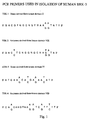
- primers shown in Figure 1 are designed from the kinase domains of the TGF- ⁇ type II receptor.
- the primers are TSK-1, derived from kinase domain II, and TSK-2, derived from kinase domain VIII.
- the template DNA consists of cDNA prepared from mRNA isolated from human skin fibroblasts from a 9 month old male.
- the PCR reaction carried out in a total volume of 50 ⁇ l, contains approximately 0.2 ⁇ g of this cDNA, primers TSK-1 and TSK-2 at a concentration of 15 ⁇ M, stocks of all four deoxynucleotides at a concentration of 0.2 mM each, 1.5 unit of DNA polymerase from Thermus thermophilus (hereafter, Tth polymerase) (Toyobo, Osaka, Japan) and reaction buffer for the Tth polymerase (Toyobo, Osaka, Japan).
- Tth polymerase Thermus thermophilus
- reaction buffer for the Tth polymerase Toyobo, Osaka, Japan
- the temperature cycle is carried out as follows for 35 cycles: melting, 92°C for 40 sec; annealing, 48°C for 40 sec; extension, 75°C for 90 sec. After the 35th cycle, the reaction is held at 75°C for an additional 5 min to complete the extension.
- fragments in this size range are recovered from an agarose gel using QIAEX (Qiagen, Chatsworth, CA; a kit for gel purification of DNA fragments, including activated silica spheres and buffers) according to the manufacturer's instructions, then resuspended in 10 mM Tris, pH 8.0, 1 mM EDTA (TE) in a volume of 20 ⁇ l.
- QIAEX Qiagen, Chatsworth, CA; a kit for gel purification of DNA fragments, including activated silica spheres and buffers
- a second round of PCR is carried out using "nested" primers based on conserved regions of the TGF- ⁇ type II receptor located within the 470 bp region amplified in the first round.
- the nested primers are AVR-5, derived from kinase domain IV of the TGF- ⁇ type II receptor, and TSK-4, derived from kinase domain VIB ( Figure 1).
- the template consists of an aliquot (0.5 ⁇ l) of the PCR fragments isolated from the first round of PCR. To this is added the primers AVR-5 (5 ⁇ M) and TSK-4 (15 ⁇ M).
- the purified fragment is phosphorylated using polynucleotide kinase and ligated to the cloning vector pGEM7Zf (+) (Promega, Madison, WI) which has previously been cut with Sma I and dephosphorylated.
- the ligation mix is used to transform E . coli XLI-Blue (Stratagene, La Jolla, CA).
- a cDNA library is constructed from the same mRNA used to isolate the PCR fragment. This is accomplished using the SUPERSCRIPT Choice System (Life Technologies, Gaithersburg, MD; a kit for cDNA synthesis, including primers, adapters, SUPERSCRIPT II RNAse H - Reverse Transcriptase (Life Technologies, Gaithersburg MD; a modified form of reverse transcriptase from Moloney murine leukemia virus), enzymes, nucleotides, buffers, and gel filtration columns) according to the manufacturer's instructions, except that 180 units of RNase inhibitor (Takara, Kyoto, Japan) is added to the first strand synthesis.
- RNase inhibitor Takara, Kyoto, Japan
- the template is mRNA (4 ⁇ g) from human skin fibroblasts from a 9 month old male. A total of 4 ⁇ g of cDNA is obtained after first and second strand synthesis. This is followed by the addition of Eco RI adapters (supplied with the kit) which contain internal Not I and Sal I sites. The Eco RI-adapted cDNA is then phosphorylated and subjected to size fractionation according to the manufacturer's instructions, using gel filtration columns provided with the kit.
- the size fractionated cDNA is ligated into the Eco RI site of the phage ⁇ gt10, and packaged in vitro with GIGAPACK II Gold Packaging Extract (Stratagene, La Jolla, CA; a restriction-minus in vitro packaging extract for high-efficiency construction of cDNA libraries in ⁇ phage) according to the manufacturer's instructions. A total of 8.1 x 10 5 phages are obtained.
- the library is screened on ten HYBOND Nylon membranes (Amersham, Arlington Heights, IL; nylon membranes optimized for immobilization of nucleic acids), at a density of 1 x 10 5 plaques/filter.
- the insert from HSK7-2 is labeled with the MULTIPRIME DNA Labeling System (Amersham, Arlington Heights, IL; a kit for random primer labeling of DNA, including Klenow DNA polymerase, primers, and buffers) according to the manufacturer's instructions.
- HSK723 One of the clones, designated HSK723, is found to encode the same sequence as the HSK7-2 insert. Complete DNA sequence is obtained for this clone.
- the cDNA from this clone is designated t-BRK-3.
- the DNA sequence of this t- BRK-3 clone is shown in SEQ ID NO: 3, and the deduced protein sequence of t-BRK-3 in SEQ ID NO: 4.
- the t-BRK-3 open reading frame derived from clone HSK723 encodes a protein of at least 583 amino acids. No stop codon is observed to be located in-frame in the 3' region of the HSK723 cDNA, indicating that this clone is incomplete at the 3' end. It is thus designated t-BRK-3.
- the deduced protein sequence of t-BRK-3 shown in SEQ ID NO: 4 is searched against all translated protein sequences in GenBank Release 84.0, dated August 15, 1994, using a standard Needleman-Wunsch algorithm (S.B. Needleman and C.D. Wunsch, J. Mol. Biol . 48: 443-453 (1970)), and is found to represent a novel sequence.
- the cysteine box of t-BRK-3 is identical in 6 of 8 amino acid residues to the cysteine box of the DAF-4 type II receptor for BMP-2 and BMP-4.
- the overall sequence identity of t-BRK-3 to DAF-4 in the extracellular domain is only 7.1%.
- Amino acids 200-504 (in SEQ ID NO: 4) in the predicted cytoplasmic region of t-BRK-3 contains all of the consensus sequences that characterize a protein kinase domain with predicted specificity for serine/threonine residues (S. K. Hanks, A.M. Quinn, and T. Hunter, Science , 241: 42-52 (1988)).
- the pJT4 vector optimized for transient expression in COS cells, includes the cytomegalovirus early promoter and enhancer, which gives very efficient transcription of message; an "R" element from the long terminal repeat of the human T-cell leukemia virus-1, which has been shown to increase expression levels further; an intron splice site from SV40, which is believed to enhance message stability; a multiple cloning site; a polyadenylation signal derived from SV40, which directs the addition of a poly A tail to the message, as is required for most eukaryotic mRNA; and the SV40 origin of replication, which permits the replication of the plasmid to extremely high copy number in cells which contain the SV40 large T antigen, such as COS cells.
- the vector contains an E . coli origin of replication
- PCR is performed to insert a stop codon to permit translation of the protein.
- a PCR primer is designed to insert two stop codons after nucleotide 2028 in SEQ ID NO: 3, thus terminating the kinase after Ile 540 in SEQ ID NO: 4. This is chosen to correspond to the length of the activin type II receptor (L.S. Mathews and W.V. Vale, Cell, 65: 973-982 (1991)), so that it should be sufficient for proper folding of the kinase domain.
- the stop codons are followed by a Kpn I site.
- the complete sequence of the primer (which includes the reverse complement of nucleotides 2013-2028 in SEQ ID NO:3) is 5' ACG CGG TAC CTC ACT AAA TTT TTG GCA CAC GC 3'.
- a second primer is designed as an exact match to the t-BRK-3 sequence in the area of the Afl III site (nucleotides 1618-1637 in SEQ ID NO:3), having the sequence 5' GTA GAC ATG TAT GCT CTT GG 3'.
- the template for the reaction is clone HSK723, described in example 2, which contains the cDNA for t-BRK-3 in BLUESCRIPT II SK (+) (Stratagene, La Jolla, CA; a 2.96 kb colony-producing phagemid derived from pUC 19).
- PCR is carried out using the GENE AMP PCR Kit with AMPLITAQ DNA Polymerase (Perkin Elmer, Norwalk, CT; a kit containing components necessary for amplification of DNA using the polymerase chain reaction, including AMPLTTAQ, a recombinant modified form of the DNA polymerase from Thermus aquaticus (Perkin-Elmer, Norwalk CT), nucleotides, and buffers), according to the manufacturer's instructions, using a GENE AMP PCR System 9600 Thermocycler (Perkin Elmer, Norwalk, CT).
- An initial melting at 95°C for 5 min is followed by 20 cycles of the following program: melting at 95°C for 1 min, annealing at 50°C for 1 min, and extension at 72°C for 1 min. After the last cycle, the temperature is held at 72°C for an additional 2 min to complete extension.
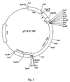
- the resulting amplified band is isolated from an agarose gel and digested with Afl III and Kpn I. Meanwhile, the cDNA for t-BRK-3 is digested with Eco RV and Afl III, and the vector pJT4 is digested with Eco RV and Kpn I. These three isolated fragments are ligated in a single step to give the construct pJT4-hBRK3T, shown in Figure 2.
- the region from the Afl III site to the KpnI site at the 3' end is sequenced using the TAQ DYE DEOXY Terminator Cycle Sequencing Kit (Applied Biosystems, Foster, CA; kit containing components for automated DNA sequencing using the dideoxy terminator method, including AMPLITAQ, nucleotide mix, dye-labeled dideoxy nucleotide terminators, and buffers) and an Applied Biosystems Model 373A Automated DNA Sequencer. No errors are found.
- t-BRK-3 To determine the effects of co-expression of t-BRK-3 with type I BMP receptors, it is necessary to co-express the cDNA for t-BRK-3 with the cDNA for BRK-1 or the cDNA for BRK-2.
- the DNA sequence for mouse BRK-1 is shown in SEQ ID NO: 11, and the deduced amino acid sequence for mouse BRK-1 is shown in SEQ ID NO: 12.
- the DNA sequence for chicken BRK-2 is shown in SEQ ID NO: 13
- the deduced protein sequence shown for chicken BRK-2 is shown in SEQ ID NO: 14.
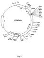
- the plasmid pJT4-JI59F is used for mammalian expression of BRK-1. Construction of this plasmid is described in U.S.S.N. 08/158,735, filed November 24, 1993 by Cook, et al. and B.B. Koenig et al., Molecular and Cellular Biology 14: 5961-5974 (1994); ATCC 69457. Briefly, the construct containing the BRK-1 cDNA subcloned in BLUESCRIPT SK (-) is linearized with the restriction endonuclease Alf III, and the overhanging end filled in using DNA Polymerase I Klenow fragment.
- the linearized plasmid is then digested with Not I, liberating the insert from the plasmid.
- the insert is then subcloned into the pJT4 expression vector at the Not I and EcoRV sites.
- the blunt end generated by the Klenow reaction is compatible with the EcoRV site, which is also a blunt end; ligation eliminates the Eco RV site.
- the construct pJT4-J159F is shown in Figure 3.
- BRK-2 For mammalian expression of BRK-2, its cDNA is subcloned into the vector pJT3. This vector is identical to pJT4, described in this example, except that the multiple cloning site is in the opposite orientation, and an additional Not I site is present at the 5' end of the multiple cloning site.
- the cDNA for BRK-2 (see S. Sumitomo, et al., DNA Sequence, 3 : 297-302 (1993)), originally obtained in the vector pRc/CMV (Invitrogen, San Diego, CA; a mammalian expression vector), is excised by digestion with Kpn I and Xho I.
- DAF-4 the type II BMP receptor from Caenorhabditis elegans
- the cDNA is obtained in BLUESCRIPT II and subcloned into pJT4 as follows.
- a 2.4 kb fragment containing the daf-4 cDNA is excised by digestion with Dra I and Apa I.
- Dra I and Apa I This fragment is subcloned into pJT4 at the Sma I and Apa I site.
- the Apa I site is regenerated, while the Dra I and Sma I sites are destroyed.
- This construct is designated pJT4-Daf4, and is shown in Figure 5.
- Transient expression of BRK-3 in mammalian cells using pJT4-hBRK3T is carried out in COS-7 cells (ATCC CRL 1651) using electroporation or COS-1 cells (ATCC CRL 1650) using DEAE Dextran (Pharmacia Biotech, Piscataway, NJ).
- COS-7 cells are grown to confluence in Dulbecco's Modified Eagle (DME) high glucose media supplemented with 10% fetal bovine serum (Hyclone, Logan, Utah), nonessential amino acids (GIBCO, Gaithersburg, MD), and glutamine, then trypsinized to release cells from the plate.
- DME Dulbecco's Modified Eagle
- the detached COS-7 cells are pelleted in a tabletop centrifuge, then resuspended in fresh media at a concentration of 6.25 x 10 6 cells/ml.
- the cell suspension (5 x 10 6 cells, 0.8 ml) is transferred to the cuvette of a BioRad GENE PULSER electroporation system (BioRad, Hercules, CA).
- the purified plasmid containing the receptor DNA of interest (10 ⁇ g for pJT4-J159F and pJT3-BRK2 and/or 20 ⁇ g for pJT4-hBRK3T) is added to the cuvette, and the cells subjected to electoporation at 4.0 kV/cm, with a capacitance of 25 ⁇ Fd. Cells are then plated (400,000 cells per well for 12 well plates and 5 x 10 6 cells for 100 mm plates) and allowed to recover. Fresh media is supplied after 24 hr. At 48 hr, cells are ready to be tested for binding of BMP-4.
- the cells are grown to approximately 50%-80% confluence in DME high glucose media supplemented with 10% fetal bovine serum (HyClone, Logan, Utah), nonessential amino acids, and glutamine in 100 mm plates.
- the cells are washed twice with 37°C serum-free DME media, after which 4 ml of DNA mixture is added to each 100 mm plate.
- the DNA mixture contains DME, 10% Nu-Serum (Collaborative Biomedical Products, Bedford, MA), 400 ⁇ g/ml DEAE-Dextran (Pharmacia, Piscataway, NJ), 0.1 mM chloroquine (Sigma, St.
- cDNAs of interest for t-BRK-3, 16 ⁇ g pJT4-hBRK3T; for BRK-1, 8 ⁇ g pJT4-J159F; for BRK-2, 8 ⁇ g pJT3-BRK2; for DAF-4, 16 ⁇ g pJT4-Daf4.
- the cells are then incubated at 37°C with the DNA mixture for 3 hr.
- the solution is aspirated and the cells are incubated with 4 ml of a solution containing 10% dimethylsulfoxide (DMSO) in Dulbecco's phosphate buffered saline without calcium or magnesium (PBS; Life Technologies, Gaithersburg, MD).
- DMSO dimethylsulfoxide
- the DMSO solution is aspirated, the cells are washed with the growth media described above, and fresh media is returned to the plates.
- the transfected cells are split into 12 well plates 24 hr post transfection for whole cell binding or cross linking. After 48 to 68 hr the cells are suitable for binding analysis.
- [ 125 I]-BMP-4 is prepared using IODOBEADS (Pierce, Rockford, IL; immobilized chloramine-T on nonporous polystyrene beads). Lyophilized BMP-4 (2 ⁇ g) is taken up in 50 ⁇ l of 10 mM acetic acid and added to 450 ⁇ l of phosphate-buffered saline (PBS) (Sigma, St. Louis, MO) on ice. To the tube is added 500 ⁇ Curie of 125 I (Amersham, Arlington Heights, IL) (2200Ci/mmol) in 5 ⁇ l, and one IODOBEAD. The reaction is incubated on ice for 10 min with occasional shaking.
- the reaction is then terminated by removal of the reaction from the IODOBEAD.
- the mixture is applied to a PD-10 gel filtration column (Pharmacia, Piscataway, NJ) previously equilibrated in 10 mM acetic acid, 0.1 M NaCl, 0.25% gelatin.
- the resulting labeled protein is >95% precipitable by trichloroacetic acid, indicating that all 125 I is protein bound, and has a typical specific activity of 4000 to 9000 Ci/mmol.
- BMP-4 is labeled with 125 I by the chloramine-T method (C.A. Frolik, L.M. Wakefield, D.M. Smith, and M.B. Sporn, J . Biol . Chem ., 259: 10995-11000 (1984)).
- BMP-4 (2 ⁇ g) is taken up in 5 ⁇ l of 30% acetonitrile, 0.1% trifluoracetic acid (TFA) plus an additional 5 ⁇ l of 1.5 M sodium phosphate, pH 7.4.
- Carrier free 125 I (1 mCi, 9 ⁇ l) is added, together with 2 ⁇ l of a chloramine T solution (100 ⁇ g/ml).
- Binding of BMP-4 to t-BRK-3 can be demonstrated by whole cell binding of radiolabeled BMP-4, and by covalent crosslinking of radiolabeled BMP-4 to the receptor. These two methods are described in detail below.
- COS-7 or COS-1 cells are transfected with pJT4-hBRK3T as described in example 5. After transfection, cells are seeded into 12 well plates and the binding experiments are carried out at 48 to 68 hr. At that time, cells are washed once with binding buffer (50 mM HEPES, pH 7.4, 128 mM NaCl, 5 mM KCl, 5 mM MgSO 4 , 1.2 mM CaCl 2 , 2 mg/ml BSA), then equilibrated in the same buffer at 4°C for 30 - 60 min with gentle shaking.
- binding buffer 50 mM HEPES, pH 7.4, 128 mM NaCl, 5 mM KCl, 5 mM MgSO 4 , 1.2 mM CaCl 2 , 2 mg/ml BSA
- binding buffer 4°C
- [ 125 I]-BMP-4 tracer 100 - 400 pM
- concentrations of unlabeled BMP-2, BMP-4, or other unlabeled ligand depending on the assay.
- BMP-4 is added to the binding buffer at a final concentration of 10 to 50 nM.
- a protease inhibitor cocktail is also added, to give a final concentration of 10 ⁇ g/ml leupeptin, 10 ⁇ g/ml antipain, 50 ⁇ g/ml aprotinin, 100 ⁇ g/ml benzamidine, 100 ⁇ g/ml soybean trypsin inhibitor, 10 ⁇ g/ml bestatin, 10 ⁇ g/ml pepstatin, and 300 ⁇ M phenylmethylsulfonyl fluoride (PMSF).
- the cells are incubated for 4 hr at 4°C with gentle shaking.
- the buffer is aspirated, and the cells are rinsed 4 times with 1 ml washing buffer (50 mM HEPES, pH 7.4, 128 mM NaCl, 5 mM KCI, 5 mM MgSO 4 , 1.2 mM CaCl 2 , 0.5 mg/ml BSA).
- 1 ml washing buffer 50 mM HEPES, pH 7.4, 128 mM NaCl, 5 mM KCI, 5 mM MgSO 4 , 1.2 mM CaCl 2 , 0.5 mg/ml BSA.
- solubilization buffer 10 mM Tris Cl, pH 7.4, 1 mM EDTA, 1% (v/v) Triton X-100
- the solubilized cells are then transferred to fresh tubes and counted in a Packard Model 5005 COBRA Gamma Counter (Packard Instruments, Meriden, CT).
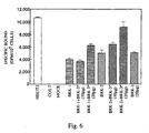
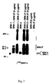
- Results are shown in Figure 6, which shows specific binding of [ 125 I]-BMP-4 to NIH3T3 cells (ATCC CRL 1658), which display significant endogenous binding of BMP-4, and COS 7 cells transfected with the cDNA for t-BRK-3 in the presence and absence of BRK-1 and BRK-2.
- t-BRK-3 is capable of binding [ 125 I]-BMP-4 when expressed alone (bar on far right), at a level similar to that seen for BRK-1 and BRK-2 expressed alone. Binding of [ 125 I]-BMP-4 is increased by co-expression of t-BRK-3 with BRK-1, and to a greater extent by co-expression of t-BRK-3 with BRK-2.
- Bifunctional crosslinking reagent disuccinimidyl glutarate (Pierce, Rockford, IL) is used to covalently crosslink bound radiolabeled ligand to its receptor by reaction with free amino groups on lysine residues in the two proteins.
- DSG disuccinimidyl glutarate
- cellular proteins are separated by gel electrophoresis, and radioactive bands visualized.
- the labeled bands represent the receptor selectively "tagged" with the radiolabeled ligand.
- cells are transfected with the cDNA for BRK-3, and/or BRK-1 or BRK-2, as described in example 5, then seeded into 12 well plates.
- the cells are washed, equilibrated, and incubated with [ 125 I]-BMP-4 and competing unlabeled ligands as described in this example for whole cell binding studies.
- the cells are washed two to three times at 4°C with 2 ml of binding buffer having the same composition as described above, except that no BSA is added.
- To each well is then added 1 ml of fresh BSA-free binding buffer, followed by freshly prepared DSG to a final concentration of 135 ⁇ M. After swirling gently to mix the DSG, the plates are incubated for exactly 15 minutes at 4°C with gentle shaking.
- the media is aspirated and the cells washed with 3 ml detachment buffer (10 mM Tris base, 0.25 M sucrose, 1 mM EDTA, 0.3 mM PMSF) or PBS.
- Solubilization buffer 50 ⁇ l is then added to each well and the cells are allowed to solubilize for 30 - 45 minutes at 4°C with shaking.
- An aliquot of the sample (20 ⁇ l) is transferred to a fresh tube and 5 ⁇ l of 5X sample loading buffer (0.25 M TrisCl, pH 6.8, 10% SDS, 0.5 M DTT, 0.5% bromophenol blue, 50% glycerol; purchased from Five Prime Three Prime, Boulder, CO) is added.
- the samples are boiled for 5 min and centrifuged (13,0000 x g, 5 min).
- the supernatants are loaded onto 7.5% SDS-polyacrylamide gels (Integrated Separation Systems, Natick, MA) and subjected to electrophoresis.
- the gels are stained in 0.12% Coomassie Blue R250, 5% methanol, 7.5% acetic acid; destained in 5% methanol, 7.5% acetic acid; then dried. Radioactivity on the dried gel is visualized and quantitated on a PHOSPHORIMAGER (Molecular Devices, Sunnyvale, CA. a device for quantitation of radioactivity using stable phosphor screens), or subjected to autoradiography using Kodak X-OMAT AR autoradiography film (Kodak, Rochester, NY).
- PHOSPHORIMAGER Molecular Devices, Sunnyvale, CA. a device for quantitation of radioactivity using stable phosphor screens
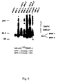
- Results are shown in Figure 7.
- t-BRK-3 When t-BRK-3 is expressed alone in COS-1 cells, no crosslinked band is seen. Expression of BRK-1 alone results in a crosslinked band at a molecular weight of 78 kD, corresponding to the predicted molecular weight of BRK-1 plus the monomer molecular weight of BMP-4. Co-expression of t-BRK-3 and BRK-1 results in the appearance of a band of similar size to that for BRK-1, as well as a new crosslinked band at 94 kD, corresponding to the predicted molecular weight of t-BRK-3 plus the monomer molecular weight of crosslinked BMP-4.
- Receptors of the TGF-B receptor family have been shown to form complexes involving a type I and a type II receptor (L. Attisano, J.L. Wrana, F. Lopez-Casillas, and J. Massagué, J. Biochim Biophys. Acta, 1222: 71-80 (1994)).
- type II BMP receptor t-BRK-3 can form a complex with the type I BMP receptors BRK-1 and BRK-2
- COS-1 cells are co-transfected with the cDNA for t-BRK-3 and BRK-1, or t-BRK-3 and BRK-2, as described in Example 5.
- the receptors are crosslinked to [ 125 I]-BMP-4, then subjected to immunoprecipitation with antibodies specific for the type I receptors BRK-1 and BRK-2. If antibodies specific for a type I receptor precipitate not only the type I receptor crosslinked to [ 125 I]-BMP-4, but also BRK-3 crosslinked to [ 125 I]-BMP-4, this indicates that the two receptors must be forming a complex, as expected for type I and type II receptors having the same ligand-binding specificity.
- Antibodies specific for the type I receptors BRK-1 and BRK-2 are generated using as antigen the peptide LNTRVGTKRYMAPEVLDESLNKNC (B.B. Koenig, et al., Molec. Cell. Biol ., 14: 5961-5974 (1994)).
- This peptide is based on the amino acid sequence of BRK-1 in the intracellular kinase domain, amino acids 398-420 in SEQ ID NO: 12, with the addition of a cysteine at the C terminus to permit conjugation of the peptide.
- the resulting antisera are evaluated for their ability to recognize the original peptide coated on plastic, using an antibody capture ELISA.
- the antisera are designated 1378, 1379, and 1380.
- These antibodies are shown to immunoprecipitate BRK-1 from COS-7 cells transfected with the cDNA for BRK-1, using the procedure detailed in this example (B.B. Koenig, et al., Mol . Cell . Biol ., 14: 5961-5974 (1994)). Because the sequence of BRK-2 is nearly identical to that of BRK-1 in this region, these antibodies are subsequently tested for their ability to immunoprecipitate BRK-2 as well, and are found to be effective for this purpose.
- Antibody 1379 gives superior results for immunoprecipitation of BRK-1, and antibody 1380 is preferred for immunoprecipitation of BRK-2.
- COS-7 or COS-1 cells are transfected with the cDNA for t-BRK-3 and/or BRK-1, BRK-2, or DAF-4 as described in Example 5, and plated into 100 mm dishes. They are then crosslinked to [ 125 I]-BMP-4 as described in example 7, except that the incubation with [ 125 I]-BMP-4 and unlabeled ligand is carried out in a total of 4 ml, instead of 500 ⁇ l, and all other volumes are increased accordingly.
- cells are washed three times with ice-cold PBS, then lysed with 1 ml of RIP buffer (20 mM TrisCl, pH 8.0, 100 mM NaCl, 1 mM Na 2 EDTA, 0.5% Nonidet P-40, 0.5% sodium deoxycholate, 10 mM sodium iodide, and 1% bovine serum albumin) for 10 min.
- the lysate is centrifuged in a microcentrifuge at 13,000 rpm for 10 min at 4°C. The supernatant is transferred to a fresh tube and made 0.1% in SDS.
- PANSORBIN Calbiochem, La Jolla, CA; a 10% solution of Staphylococcus aureus
- the primary antibody ⁇ 1379 when cells are transfected with t-BRK-3 and BRK-1; 1380 when cells are transfected with t-BRK-3 and BRK-2 ⁇ is then added to the tube at a final dilution of 1:100, and incubated for 2 hr on ice or overnight at 4°C.
- To precipitate the complex of antigen:primary antibody 25-50 ⁇ l of PANSORBIN is then added and incubated 30 min on ice. The complex is pelleted at 13,000 rpm for 10 min in a microcentrifuge and the supernatant discarded.
- the pellet is washed twice in RIP buffer containing 0.1% SDS, and once in TNEN buffer (20 mM Tris, pH 8.0, 100 mM NaCl, 1 mM EDTA, 0.5% NP-40).
- the pellet is resuspended in 25 ⁇ l of 1X sample loading buffer. (Alternatively, the pellet may be washed twice with TNEN buffer, with similar results.)
- the sample is boiled for 5 min, centrifuged for 5 min, and subjected to gel electrophoresis after loading of the samples onto a 7.5% SDS-polyacrylamide gel.
- FIG. 8 shows the results of immunoprecipitations on COS-1 cells transfected with t-BRK-3 in the presence or absence of BRK-1 or BRK-2.
- Cells transfected with t-BRK-3 alone, crosslinked to [ 125 I]-BMP-4, and immunoprecipitated with antibody 1380 show no radiolabel in the immunoprecipitate, as expected since t-BRK-3 does not crossreact with this antibody.
- Cells transfected with BRK-1, crosslinked, and immunoprecipitated with antibody 1379 show a single labeled band at 78 kD, consistent with the predicted molecular weight of BRK-1 plus the cross-linked monomer of BMP-4.
- Immunoprecipitation of cells co-transfected with BRK-1 and t-BRK-3 yields the same band seen with BRK-1 alone, plus an additional labeled band at 94 kD, consistent with the predicted molecular weight of t-BRK-3 plus the crosslinked BMP-4 monomer. (A less intense band at 120 kD is also observed.)
- the fact that antibodies to BRK-1 precipitate not only BRK-1, but t-BRK-3 as well in these cells indicates complex formation between BRK-1 and t-BRK-3.
- cells transfected with BRK-2, crosslinked to [ 125 I]-BMP-4, and subjected to immunoprecipitation with antibody 1380 show a labeled band at 75 kD, consistent with the predicted molecular weight of BRK-2 plus the crosslinked monomer of BMP-4.
- Immunoprecipitation of cells co-transfected with BRK-2 and t-BRK-3 yields the same band seen with BRK-2 alone, plus a strongly labeled band at 94 kD, consistent with the predicted molecular weight of t-BRK-3 plus the crosslinked monomer of BMP-4.
- this band co -migrates with the larger labeled band in cells co-transfected with BRK-1 and t-BRK-3. (A less intense band at 120 kD is also observed.) Again, the fact that an antibody to BRK-2 precipitates not only BRK-2 but t-BRK-3 as well in these cells strongly indicates that BRK-2 and t-BRK-3 form a complex. Thus, t-BRK-3 forms a complex with two different type I BMP receptors, as expected for a type II BMP receptor.
- a second immunoprecipitation experiment is carried out to test the ligand specificity of the t-BRK-3 receptor complex for BMP-2, BMP-4, and TGF- ⁇ 1 .
- a derivative of BMP-2 designated "digit -removed" BMP-2 (DR-BMP-2) is also tested; DR-BMP-2 is prepared by mild trypsin digestion of BMP-2 to remove the amino terminus, and shows significantly reduced nonspecific binding to whole cells (B.B. Koenig, et al., Molec. Cell. Biol ., 14: 5961-5974 (1994)).
- COS-1 cells are co-transfected with the cDNA for BRK-2 and t-BRK-3 as described in Example 5, crosslinked to [ 125 I]-BMP-4, and subjected to immunoprecipitation with antibody 1380 as described in this example, except that an excess of unlabeled ligand (10 nM BMP-4, 10 nM BMP-2, 10 nM DR-BMP-2, or 50 nM TGF- ⁇ 1 ) is added to the incubation at the same time as the [ 125 I]-BMP-4.
- an excess of unlabeled ligand 10 nM BMP-4, 10 nM BMP-2, 10 nM DR-BMP-2, or 50 nM TGF- ⁇ 1
- a cDNA library is constructed from NIH3T3 mouse embryonic fibroblasts (ATCC CRL 1658).
- Total RNA (1.26 mg) is isolated from the cells using a Total RNA Separator Kit (Clontech, Palo Alto, CA).
- Messenger RNA (81 ⁇ g) is isolated from this total RNA (1 mg) using the mRNA Separator Kit (Clontech, Palo Alto, CA).
- An aliquot of the mRNA (4 ⁇ g) is used to make cDNA library using the SUPER SCRIPT Plasmid System for cDNA Synthesis and Plasmid Cloning (Life Technologies, Gaithersburg, MD) according to the manufacturer's instructions.
- the resulting library contained approximately 4.9 x 10 5 primary colonies, and is divided into 98 pools, each containing 5000 colonies.
- Human t-BRK-3 is cut with EcoRV and Afl III to give a 1.5 kb fragment.
- the fragment is randomly labeled with alpha[ 32 P]-dCTP having a specific activity of 3000 Ci/mmol (NEN Research Products, Boston, MA), using a PRIME-IT II Random Primer Labeling Kit (Stratagene, La Jolla, CA; a kit for random primer labeling of DNA, including Klenow DNA polymerase, primers, and buffers).
- the labeled probe is allowed to hybridize to the Southern blot for 18 hr at 42°C in hybridization buffer (Sigma, St.
- the blots are then shaken with 50 ⁇ g/ml of proteinase K (Boehringer Mannheim, Indianapolis; IN) in 0.1 M Tris, pH 7.6, 10 mM EDTA, 0.15 M NaCl, 0.02% SDS at 53°C for 1 hr.
- the human BRK-3 fragment (Eco RV-Afl III) is labeled and the blots hybridized, washed, and subjected to autoradiography exactly as described above for the primary screening.
- Colonies which corresponded to labeled spots on the autoradiograph are streaked on plates for tertiary screening, which is performed exactly as described above for secondary screening.
- Four positive clones are isolated.
- One clone, pSPORT1/N89-5, is found to have the largest insert size, 2.9 kb.
- the inserts from the four positive clones are sequenced using the TAQ DYE DEOXY Terminator Cycle Sequencing Kit and an Applied Biosystems Model 373A Automated DNA Sequencer. Comparison of the four sequences shows that three of the four are identical at the 3' end, and all four align with the coding region of human BRK-3 at the 5' end. The longest clone, pSPORT1/N89-5, aligns with the human BRK-3 sequence approximately 600 pairs from the beginning of the coding region.
- the insert from pSPORT1/N89-5 is digested with EcoRI and Sca I, and the resulting 1.4 kb fragment is subcloned into BLUESCRIPT II SK(-) at the Eco RI and Hinc II sites.
- pSPORT1/N89-5 is also digested with Eco RI and Eco RV and the resulting 2.1 kb insert subcloned into the same vector at the same sites.
- the plasmid is digested with Sca I and Not I, and subcloned into the same vector at the Hinc II and Not I sites. Sequencing of these three constructs yields the complete sequence of the insert from pSPORT1/N89-5.
- the missing 600 base pairs at the 5' end of the coding region is cloned using the 5' RACE System for Rapid Amplification of cDNA Ends (Life Technologies, Gaithersburg, MD).
- An antisense primer is designed corresponding to the known sequence of pSPORT1/N89-5, having the sequence 5'CTG TGT GAA GAT AAG CCA GTC 3' (the reverse complement of nucleotides 968-948 in SEQ ID NO:7).
- a poly C tail is added to the newly synthesized cDNA using terminal deoxynucleotidyl transferase, according to the manufacturer's instructions.
- PCR was performed using the GENE-AMP PCR Kit with AMPLITAQ DNA Polymerase. An initial melting period at 95°C for 5 min was followed by 35 cycles of the following program: melting at 95°C for 1 min, annealing at 55°C for 1 min, and extension at 72°C for 2 min. After the last cycle, the reaction was held at 72°C for 5 min to complete extension.
- a second round of PCR is performed using the nested primer 5' CAA GAG CTT ACC CAA TCA CTT G 3', again derived from the known sequence of the insert from pSPORT1/N89-5 (the reverse complement of nucleotides 921-900 in SEQ ID NO: 7), together with same 5' anchor primer used in the first round of PCR.
- the amplified products of the second PCR reaction in the size range of 600-1000 bp are digested with Ecl XI and Sal I and subcloned into BLUESCRIPT II SK(-) at the Ecl XI and Sal I sites.
- the inserts are then sequenced, yielding an additional 600 bp of sequence which align with the coding region of human t-BRK-3.
- Three separate clones, designated R6-8B2, R6-11-1, and R6-11-2, are sequenced with identical results.
- a Sal I site is first placed at the 5' end of clone R6-11-1 as follows.
- a primer is synthesized which contains a Sal I site followed by nucleotides 1-20 of the sequence of R6-11-1; the sequence of the primer is 5' CAC ACG CGT CGA CCA TGA CTT CCT CGC TGC ATC G 3'. This is used together with the M13 reverse primer, 5' AAC AGC TAT GAC CAT G 3', in order to amplify a DNA fragment using plasmid DNA from clone R6-11-1 as the template. PCR was performed using the GENE-AMP PCR Kit with AMPLITAQ DNA Polymerase.
- the vector BLUESCRIPT II SK(-) is digested with Sal I and Pst I.
- the three fragments are combined in a three-way ligation using T4 DNA ligase (3 hr, 25°C) and used to transform electrocompetent E. coli, strain DH5-a, using a BIO-RAD Gene PULSER (BIO-RAD, Hercules, CA) according to the manufacturer's instructions.
- a positive colony is selected and is designated pBLUESCRIPT-mBRK3. Sequencing of the 5' portion of the insert that was amplified by PCR shows a sequence identical to that of clone R6-11-1, indicating that no mutations are introduced during the amplification.
- m-BRK-3 is subcloned into the mammalian expression vector pJT6.
- This vector is a derivative of pJT3, described in example 4 above, in which the Not I site at the 5' end of the multiple cloning site has been deleted, and a spacer inserted between the Pst I and BamHI restriction sites in the multiple cloning site.
- m-BRK-3 is excised from pBLUESCRIPT-mBRK3 using Not I and Sal I, then subcloned into pJT6 at the Not I and Sal I sites to generate pJT6-mBRK3.
- pJT6-mBRK3 is digested with SpeI (site at position 2306 in SEQ ID NO: 7) and Not I (in the multiple cloning site of pJT6), removing the 3' end of the insert.
- the longest clone isolated during the screening of the NIH-3T3 library, pSPORT1/N89-5, is also digested with Spe I and Not I.
- the 1.2 kb fragment liberated from pSPORT1/N89-5 is subcloned into the Spe I/Not I digested pJT6-mBRK3, regenerating both sites.
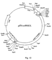
- This construct is designated pJT6-mBRK-3L, and contains the entire 3' end of the pSPORT1/N89-5 clone.
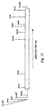
- a map of the construct is shown in Figure 10.
- the 3' end of the clone contains 403 nucleotides in the untranslated region 3' to the stop codon. This region is very A-T rich, which might possibly lead to decreased expression levels.
- a second construct is prepared.
- the pSPORT1/N89-5 plasmid is digested with Hind III (site at nucleotide 3168 in SEQ ID NO: 7, 21 bases 3' to the stop codon).
- the linearized plasmid is treated with Klenow fragment of DNA polymerase (Boehringer Mannheim, Indianapolis, IN) to fill in overhangs, then cut with Spe I to liberate an 863 bp fragment at the 3' end of the insert.
- pJT6-mBRK3 is digested with Not I.
- the linearized plasmid is treated with Klenow fragment, then cut with Spe I, releasing the 3' end of the insert.
- the Not I/Spe I digested pJT6-mBRK3 is then ligated to the fragment liberated from pSPORT1/N89-5 by Hind III/Spe I. This regenerates the Spe I site; the Hind III and Not I sites are destroyed.
- the resulting construct is designated pJT6-mBRK3S, and is shown in Figure 11.
- the construct pJT6-mBRK-3S is also constructed directly from the partial cDNA clone of m-BRK-3, pSPORT1/N89-5, and the construct containing the 5' end of the cDNA, clone R6-11-1. This is accomplished by digestion of clone R6-11-1 with Sal I and Ecl XI, digestion of pSPORT1/N89-5 with Ecl XI and Hind III, and digestion of BLUESCRIPT II SK (-) with Sal I and Hind III. These fragments are then subjected to a three-way ligation to generate the full length m-BRK-3 cDNA in the BLUESCRIPT II vector.
- the full length cDNA is then excised from this construct using Sal I and Not I, then subcloned into the Sal I and Not I sites of the pJT6 vector.
- the resulting plasmid has exactly the same cDNA for BRK-3 as does pJT6-mBRK3S described in the above example. However, it carries additional vector sequence at the 3' end of the cDNA, comprising the region between the Hind III and Not I sites in the multiple cloning site of BLUESCRIPT II SK(-).
- the DNA sequence of the full length mouse BRK-3 insert from pJT6-mBRK3L is shown in SEQ ID NO: 7, and the deduced protein sequence is shown in SEQ ID NO: 8.
- the deduced amino acid sequence of mouse BRK-3 is searched against all translated protein sequences in GenBank release 84.0, dated Aug. 15, 1994, using a standard Needleman-Wunsch algorithm (S.B. Needleman and C.D. Wunsch, J . Mol . Biol ., 48: 443-453 (1970)). It is found to be a unique sequence. It encodes a protein of 1038 amino acids.
- m-BRK-3 contains a predicted transmembrane region encompassing amino acids 151-172.
- the intracellular domain contains all of the consensus sequences that characterize a protein kinase domain with predicted specificity for serine/threonine residues (S.K. Hanks, A.M. Quinn, and T. Hunter, Science , 241: 42-52 (1988)).
- the kinase domain is followed by an extremely long carboxy terminus (534 amino acids). Indeed, due to the presence of this carboxy terminus, the intracellular domain in BRK-3 (866 amino acids) is much larger than that of any other receptor in the TGF- ⁇ receptor family. It is nearly twice as long as the intracellular domain of DAF-4 (490 amino acids), which has the longest intracellular domain known in the TGF- ⁇ family until the present invention.
- COS-1 cells are transfected as described in Example 5 using the constructs pJT6-mBRK-3S and pJT6-mBRK-3L.
- the cells are also co-transfected with cDNA for the type I receptor BRK-2, using the construct pJT3-BRK-2, to determine whether the presence of a type I BMP receptor affects binding of [ 125 I]-BMP-4.
- Whole cell binding with [ 125 I]-BMP-4 is carried out as described in Example 7.
- Figure 12 shows specific binding of [ 125 I]-BMP-4 normalized to cell number.
- specific binding of [ 125 I]-BMP-4 is increased to 4-7 times the level seen with mock transfected cells.
- Transfection of BRK-2 alone shows increased binding at a similar level to that seen with mouse BRK-3 alone.
- the binding is further increased to 9-11 times that of mock-transfected cells, consistent with the results obtained with BRK-2 in combination with t-BRK-3 ( Figure 6 in Example 7 above).
- COS-1 cells are transfected with the cDNA for m-BRK-3, using the construct pJT6-mBRK-3S, and/or with cDNAs for BRK-1 (using pJT4-J159F) or BRK-2 (using pJT3-BRK-2) as described in Example 5.
- the transfected cells are incubated with [ 125 I]-BMP-4 and crosslinked as described in Example 7, except that disuccinimidyl suberate (DSS) is used as the crosslinking agent rather than disuccinimidyl glutarate.
- DSS disuccinimidyl suberate
- transfection of cells with the cDNA for BRK-2 alone yields a crosslinked band migrating at an apparent molecular weight of 78 kD, consistent with the predicted molecular weight of BRK-2 plus the crosslinked BMP-4 monomer.
- the 78 kD species identified with BRK-2 is observed, as well as crosslinked bands at 159 kD and 128 kD, comigrating with the higher molecular weight bands seen in cells transfected with the cDNAs for BRK-1 and m-BRK-3.
- the intensity of crosslinking to the band identified with BRK-2 is considerably increased compared to that seen with BRK-2 alone.
- no labeled bands are observed in cells transfected with vector alone.
- COS-1 cells are transfected with the cDNA for m-BRK-3, using the construct pJT6-mBRK-3S, and/or with cDNAs for BRK-1 (using pJT4-J159F) or BRK-2 (using pJT3-BRK-2) as described in Example 5.
- the transfected cells are incubated with [ 125 I]-BMP-4, crosslinked , and subjected to immunoprecipitation with antibodies to the appropriate type I receptor or preimmune serum as described in example 8, except that DSS is used as the crosslinking agent rather than disuccinimidyl glutarate.
- DSS is used as the crosslinking agent rather than disuccinimidyl glutarate.
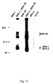
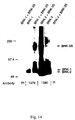
- FIG 14. In cells transfected with cDNA for BRK-1 alone, a single band is precipitated by antibodies to BRK-1, migrating at an apparent molecular weight of 81 kD. In cells transfected with cDNAs for BRK-1 and m-BRK-3, antibodies to BRK-1 precipitate the 81 kD band, which is now increased in intensity.
- a labeled species is seen at 159 kD, consistent with m-BRK-3 and comigrating with the higher molecular weight band seen in cells transfected with cDNAs for BRK-1 and m-BRK-3.
- an additional labeled band is observed at 94 kD.
- cells are transfected with the cDNAs for BRK-1 and m-BRK-3, or BRK-2 and m-BRK-3, then subjected to immunoprecipitation with preimmune sera (lanes far left and far right); no labeled bands are observed.
- Example 2 Since clone HSK723, described in Example 2, does not contain an in-frame stop codon, it is desired to obtain additional sequence 3' to the end of this cDNA. Accordingly, the human foreskin fibroblast library prepared in Example 1 is rescreened with the HSK7-2 PCR fragment, using labeling and screening conditions exactly as described in Example 2. This results in isolation of a longer clone, designated pHSK1030, which contains additional human BRK-3 sequence (total of 3355 base pairs) subcloned in BLUESCRIPT SK(-). Sequencing of the insert from pHSK1030 discloses a coding region of 982 amino acids, but the insert still does not contain an in-frame stop codon.
- the remainder of the coding region is cloned by PCR as follows.
- Two forward primers are derived from the plus strand of clone pHSK1030. The sequences of these primers are as follows: primer RPK3-1, 5' CCTGTCACATAATAGGCGTGTGCC-3' (identical to nucleotides 1998-2021 in SEQ ID NO:1), primer RPK3-2, 5' CGCGGATCCATCATACTGACAGCATCG 3' (which incorporates a BamHI site followed by nucleotides 2078-2095 in SEQ ID NO:1).
- Two additional primers are derived from the minus strand of ⁇ gt10. These primers are: G10F1, 5' GCTGGGTAGTCCCCACCTTT 3' and G10F2, 5' GAGCAAGTTCAGCCTGGT 3'.
- the human fibroblast cDNA library prepared in Example 1 is used as the template for PCR.
- the library (0.3 ⁇ g) is incubated with the RPK3-1 and G10F1 primers (1 ⁇ M each), Tth polymerase ( 1.2 units), all four deoxynucleotides (200 ⁇ M each) , buffer for the Tth polymerase, and water in a total of 50 ⁇ l.
- Conditions for the PCR cycle are as follows: initial melting at 94°C for 2 min, followed by 20 cycles of melting, 94°C for 1.5 min; annealing, 52°C for 2 min; and extension, 72°C for 3 min. After cycle 20, the sample is held at 72°C for an additional 8 min to insure complete extension.
- a second round of nested PCR is carried out.
- the incubation mixture is the same as described in this example for the first round, except that (1) an aliquot of the first PCR reaction (0.5 ⁇ l) is used as the template; and (2) RPK3-2 and G10F2 primers are used, instead of RPK3-1 and G10F1.
- Conditions for the PCR run are identical to those described in this example for the first round of PCR.
- the second round of PCR results in the amplification of a 1.6 kb fragment, which is isolated from an agarose gel by QIAEX. This fragment is digested with EcoRI and BamHI, and subcloned into BLUESCRIPT SK(-) at the EcoRI and Bam HI sites. The resulting construct, pHSK723-3U, is sequenced and found to encode the remaining coding region of BRK-3 with an in-frame stop codon.
- the amino acid sequence of human BRK-3 (SEQ ID NO:2) is compared to the amino acid sequence for m-BRK-3 (SEQ ID NO:8) and found to be 96.7% identical.
- Identification of ligands that interact with BRK-3 can be achieved through the use of assays that are designed to measure the interaction of ligands with BRK-3.
- An example of a receptor binding assay that is adapted to handle large numbers of samples is carried out as follows.
- COS-1 cells are transfected with the cDNA for m-BRK-3 using the construct pJT6-mBRK-3L as described in example 11 above, except that cells are grown in a 12 well culture dish.
- the cells are washed once with 1.0 ml binding buffer (50 mM HEPES, pH 7.4, 128 mM NaCl, 5 mM KCL, 5 mM MgSO 4 , 1.2 mM CaCl 2 , 2 mg/ml BSA), then equilibrated in the same buffer at 4°C for 60 min. with gentle shaking.
- 1.0 ml binding buffer 50 mM HEPES, pH 7.4, 128 mM NaCl, 5 mM KCL, 5 mM MgSO 4 , 1.2 mM CaCl 2 , 2 mg/ml BSA
- the buffer is aspirated, and to each well is added 500 ⁇ l of 4°C binding buffer containing [ 125 I]BMP-4 tracer (100-400 pM) in the presence or absence of varying concentrations of unlabeled test compounds (i.e., putative ligands), for a period of 4 hours at 4°C with gentle shaking.
- BMP-2 is added at a final concentration of 10 nM.
- a protease inhibitor cocktail is also added, to give a final concentration of 10 ⁇ g/ml leupeptin, 10 ⁇ g/ml antipain, 50 ⁇ g/ml aprotinin, 100 ⁇ g/ml benzamidine, 100 ⁇ g/ml soybean trypsin inhibitor, 10 ⁇ g/ml bestatin, 10 ⁇ g/ml pepstatin, and 300 ⁇ M phenylmethylsulfonyl fluoride (PMSF).
- PMSF phenylmethylsulfonyl fluoride
- the buffer is aspirated, and the cells are rinsed 4 times with 1 ml washing buffer (50 mM HEPES, pH 7.4, 128 mM NaCl, 5 mM KCl, 5 mM MgSO 4 , 1.2 mM CaCl 2 , 0.5 mg/ml BSA).
- 1 ml washing buffer 50 mM HEPES, pH 7.4, 128 mM NaCl, 5 mM KCl, 5 mM MgSO 4 , 1.2 mM CaCl 2 , 0.5 mg/ml BSA.
- solubilization buffer 10 mM Tris Cl, pH 7.4, 1 mM EDTA, 1% (v/v) Triton X-100
- the solubilized cells are then transferred to fresh tubes and counted in a Packard Model 5005 COBRA Gamma Counter (Packard Instruments, Meriden, CT).
- Test compounds which interact with the m-BRK-3 receptor are observed to compete with binding to the receptor with the [ 125 I]BMP-4 tracer in the cells expressing m-BRK-3, such that less [ 125 I]BMP-4 tracer is bound in the presence of the test compound in comparison to the binding observed when the tracer is incubated in the absence of the novel compound.
- a decrease in binding of the [ 125 I]BMP-4 tracer by ⁇ 30% at the highest concentration of the test compound that is studied demonstrates that the test compound binds to m-BRK-3.
- Identification of ligands that interact with BRK-3 complexed to a type I BMP receptor can be achieved through the use of assays that are designed to measure the interaction of the ligands with this BMP receptor complex.
- a receptor binding assay that uses the m-BRK-3/BRK-2 complex and is adapted to handle large numbers of samples is carried out as follows.
- COS-1 cells are transfected with the cDNAs for m-BRK-3, using the construct pJT6-mBRK-3L, and BRK-2, using the construct pJT3-BRK-2, as described in example 11 above, except that the cells are grown in a 12 well culture dish.
- the DNA mixture used to transfect the cells contains 2 ⁇ g/ml of pJT3-BRK-2 and 4 ⁇ g/ml of pJT6-mBRK-3L.
- the cells are washed once with 1 ml binding buffer (50 mM HEPES, pH 7.4, 128 mM NaCl, 5 mM KCL, 5 mM MgSO 4 , 1.2 mM CaCl 2 , 2 mg/ml BSA), then equilibrated in the same buffer at 4°C for 60 min with gentle shaking. After equilibration, the buffer is aspirated, and to each well is added 500 ⁇ l of 4°C binding buffer containing [ 125 I]BMP-4 tracer (100-400 pM) in the presence or absence of varying concentrations of test compounds (i.e., putative ligands), for a period of 4 hours at 4°C with gentle shaking.
- 1 ml binding buffer 50 mM HEPES, pH 7.4, 128 mM NaCl, 5 mM KCL, 5 mM MgSO 4 , 1.2 mM CaCl 2 , 2 mg/ml BSA
- BMP-2 is added at a final concentration of 10 nM.
- a protease inhibitor cocktail is also added, to give a final concentration of 10 ⁇ g/ml leupeptin, 10 ⁇ g/ml antipain, 50 ⁇ g/ml aprotinin, 100 ⁇ g/ml benzamidine, 100 ⁇ g/ml soybean trypsin inhibitor, 10 ⁇ g/ml bestatin, 10 ⁇ g/ml pepstatin, and 300 ⁇ M phenylmethylsulfonyl fluoride (PMSF).
- PMSF phenylmethylsulfonyl fluoride
- the buffer is aspirated, and the cells are rinsed 4 times with 1 ml washing buffer (50 mM HEPES, pH 7.4, 128 mM NaCl, 5 mM KCI, 5 mM MgSO 4 , 1.2 mM CaCl 2 , 0.5 mg/ml BSA).
- 1 ml washing buffer 50 mM HEPES, pH 7.4, 128 mM NaCl, 5 mM KCI, 5 mM MgSO 4 , 1.2 mM CaCl 2 , 0.5 mg/ml BSA.
- solubilization buffer 10 mM Tris Cl, pH 7.4, 1 mM EDTA, 1% (v/v) Triton X-100
- Test compounds which interact with the m-BRK-3/BRK-2 receptor complex are observed to compete for binding to the receptor complex with the [ 125 I]BMP-4 tracer, such that less [ 125 I]BMP-4 tracer is bound in the presence of the test compound in comparison to the binding observed when the tracer is incubated in the absence of the novel compound.
- a decrease in binding of the [ 125 I]BMP-4 tracer by ⁇ 30% at the highest concentration of the test compound that is studied demonstrates that the test compound binds to the m-BRK-3/BRK-2 receptor complex.
- E. coli transformed with pJT4-J159F (SEQ ID NO:11 subcloned into expression vector pJT4) was deposited with the ATCC on October 7, 1993, and assigned ATCC Designation No. 69457.
- E. coli transformed with pJT4-hBRK3T (SEQ ID NO:3 subcloned into expression vector pJT4) was deposited with the ATCC on August 16, 1994 and assigned ATCC designation No. 69676.
- E. coli transformed with pJT6-mBRK-3S (SEQ ID NO: 7 subcloned into expression vector pJT6) was deposited with the ATCC on September 28, 1994 and assigned ATCC designation No. 69694.
- E. coli transformed with pJT6-mBRK-3L (SEQ ID NO:7 subcloned into expression vector pJT6) was deposited with the ATCC on September 28, 1994 and assigned ATCC designation No. 69695.
- E . coli transformed with pHSK1040 (SEQ ID NO:1 subcloned into BLUESCRIPT II SK(-) was deposited with the ATCC on October 12, 1994, and assigned ATCC designation No. 69703.
Landscapes
- Health & Medical Sciences (AREA)
- Chemical & Material Sciences (AREA)
- Life Sciences & Earth Sciences (AREA)
- Organic Chemistry (AREA)
- Medicinal Chemistry (AREA)
- General Health & Medical Sciences (AREA)
- Molecular Biology (AREA)
- Toxicology (AREA)
- General Chemical & Material Sciences (AREA)
- Pharmacology & Pharmacy (AREA)
- Chemical Kinetics & Catalysis (AREA)
- Animal Behavior & Ethology (AREA)
- Bioinformatics & Cheminformatics (AREA)
- Public Health (AREA)
- Veterinary Medicine (AREA)
- Proteomics, Peptides & Aminoacids (AREA)
- Engineering & Computer Science (AREA)
- Genetics & Genomics (AREA)
- Biophysics (AREA)
- Cell Biology (AREA)
- Immunology (AREA)
- Nuclear Medicine, Radiotherapy & Molecular Imaging (AREA)
- Zoology (AREA)
- Gastroenterology & Hepatology (AREA)
- Biochemistry (AREA)
- Obesity (AREA)
- Hematology (AREA)
- Diabetes (AREA)
- Nutrition Science (AREA)
- Micro-Organisms Or Cultivation Processes Thereof (AREA)
- Peptides Or Proteins (AREA)
- Preparation Of Compounds By Using Micro-Organisms (AREA)
- Compression Or Coding Systems Of Tv Signals (AREA)
- Dc Digital Transmission (AREA)
- Medicines That Contain Protein Lipid Enzymes And Other Medicines (AREA)
- Enzymes And Modification Thereof (AREA)
- Medicines Containing Material From Animals Or Micro-Organisms (AREA)
Abstract
Description
- a fixed concentration of at least one agonist for transcription of BRK-3 or, and increasing concentrations of at least one test compound whose ability inhibit transcriptional activation of the BRK-3 receptor protein is sought to be determined; and thereafter
NOHNO, TSUTOMU


Claims (14)
- An isolated BMP receptor kinase protein having amino acid sequence SEQ ID NO:2, or soluble fragment thereof which is capable of binding BMPS, amino acid sequence SEQ ID NO:4, or a soluble fragment thereof which is capable of binding BMPS, or amino acid sequence SEQ ID NO:8, or a soluble fragment thereof which is capable of binding BMPS.
- An isolated DNA sequence coding for the BMP receptor kinase protein of Claim 1.
- The DNA sequence of Claim 2, wherein the DNA sequence is SEQ ID NO:1, SEQ ID NO:3 or SEQ ID NO:7.
- The soluble fragment of Claim 1, wherein the soluble fragment has amino acid sequence SEQ ID NO:6 or SEQ ID NO:10.
- A DNA sequence coding for the soluble fragment of Claim 4.
- The DNA sequence of Claim 5, wherein the DNA sequence is SEQ ID NO:5 or SEQ ID NO:9.
- A recombinant expression vector comprising the DNA sequence of Claim 2, Claim 3, Claim 5 or Claim 6.
- The recombinant expression vector of Claim 7, wherein the vector is a plasmid having all of the identifying characteristics of pJT4-hBRK3T contained in ATCC No. 69676 or a plasmid having all of the identifying characteristics of pJT6-mBRK-3L contained in ATCC No. 69695.
- A host cell comprising the recombinant expression vector of Claim 7 or Claim 8, wherein the host cell is preferably a mammalian cell, more preferably a Chinese hamster ovary cell or a COS cell.
- A method for producing truncated BMP receptor kinase protein comprising culturing the host cell of Claim 9 in a manner allowing expression of the truncated BMP receptor kinase protein and isolation of the BMP receptor kinase protein.
- A method for determining whether a compound is capable of binding to a BMP receptor kinase protein, the method comprising introducing a sample comprising the compound to the BMP receptor kinase protein and allowing the compound to bind to the BMP receptor kinase protein, wherein the BMP receptor kinase protein has amino acid sequence:(a) SEQ ID NO:2 or soluble fragment thereof;(b) SEQ ID NO:4 or soluble fragment thereof; or(c) SEQ ID NO:8 or soluble fragment thereof
- An antibody directed to a BMP type II receptor kinase protein selected from the group consisting of SEQ ID NO: 2, SEQ ID NO:6, SEQ ID NO:8 and SEQ ID NO:10.
- A method for evaluating whether a test compound is capable of acting as an agonist of the transcription of the BMP type II receptor protein kinase of SEQ ID NO:2 or SEQ ID NO:8, the method comprising:(a) culturing cells containing:wherein the culturing is carried out in the presence of at least one test compound whose ability to induce the transcriptional activity of the DNA coding for SEQ ID NO:2 or SEQ ID NO:8 is sought to be determined, and thereafter(i) DNA encoding SEQ ID NO:2 or SEQ ID NO:8, and(ii) DNA encoding a hormone response element operatively linked to a reporter gene,(b) monitoring the cells for expression of the reporter gene.
- A method for evaluating whether a test compound is capable of acting as an antagonist of the transcription of the BMP type II receptor protein kinase of SEQ ID NO:2 or SEQ ID NO:8, the method comprising:(a) culturing cells containing:wherein the culturing is carried out in the presence of:(i) DNA encoding the BMP type II receptor protein kinase of SEQ ID NO:2 or SEQ ID NO:8, and(ii) DNA encoding a hormone response element operatively linked to a reporter gene,a fixed concentration of at least one agonist for transcription of the BMP type II receptor protein kinase of SEQ ID NO:2 or SEQ ID NO:8, and increasing concentrations of at least one test compound whose ability to inhibit transcriptional activation of the BMP type II receptor protein kinase of SEQ ID NO:2 or SEQ ID NO:8, is sought to be determined; and thereafter(b) monitoring in the cells the level of expression of the product of the reporter gene as a function of the concentration of the test compound, thereby indicating the ability of the test compound to inhibit activation of transcription.
Applications Claiming Priority (3)
| Application Number | Priority Date | Filing Date | Title |
|---|---|---|---|
| US334179 | 1994-11-04 | ||
| US08/334,179 US6306622B1 (en) | 1994-11-04 | 1994-11-04 | cDNA encoding a BMP type II receptor |
| PCT/US1995/014085 WO1996014412A2 (en) | 1994-11-04 | 1995-10-30 | cDNA ENCODING A BMP TYPE II RECEPTOR |
Publications (2)
| Publication Number | Publication Date |
|---|---|
| EP0804577A2 EP0804577A2 (en) | 1997-11-05 |
| EP0804577B1 true EP0804577B1 (en) | 2004-05-12 |
Family
ID=23305967
Family Applications (1)
| Application Number | Title | Priority Date | Filing Date |
|---|---|---|---|
| EP95939661A Expired - Lifetime EP0804577B1 (en) | 1994-11-04 | 1995-10-30 | cDNA ENCODING A BMP TYPE II RECEPTOR |
Country Status (12)
| Country | Link |
|---|---|
| US (1) | US6306622B1 (en) |
| EP (1) | EP0804577B1 (en) |
| JP (1) | JPH10509591A (en) |
| CN (1) | CN1171816A (en) |
| AT (1) | ATE266725T1 (en) |
| AU (1) | AU710569B2 (en) |
| CA (1) | CA2204151C (en) |
| DE (1) | DE69533044T2 (en) |
| DK (1) | DK0804577T3 (en) |
| FI (1) | FI971896A7 (en) |
| NO (1) | NO972063L (en) |
| WO (1) | WO1996014412A2 (en) |
Families Citing this family (13)
| Publication number | Priority date | Publication date | Assignee | Title |
|---|---|---|---|---|
| US6306622B1 (en) * | 1994-11-04 | 2001-10-23 | The Procter & Gamble Co. | cDNA encoding a BMP type II receptor |
| WO1996018735A2 (en) * | 1994-12-14 | 1996-06-20 | Amgen Boulder Inc. | Tgf-beta superfamily type ii receptor having binding affinity for bone morphogenic protein (bmp) |
| AU709991B2 (en) * | 1995-08-14 | 1999-09-09 | Creative Biomolecules, Inc. | Binding of osteogenic protein-1 (OP-1) and analogs thereof to the cell surface receptor ALK-1 and analogs thereof |
| WO1998052038A1 (en) * | 1997-05-16 | 1998-11-19 | The Procter & Gamble Company | The use of a bone morphogenetic protein (bmp) receptor complex for screening |
| AU2002218774A1 (en) * | 2000-07-12 | 2002-01-21 | The Trustees Of Columbia University In The City Of New York | Role of pph1 gene in pulmonary hypertension |
| EP1487965A4 (en) | 2002-02-25 | 2006-11-15 | Mpex Pharmaceuticals Inc | Minicell compositions and methods |
| US20070036769A9 (en) * | 2004-06-03 | 2007-02-15 | Linheng Li | BMP pathway methods and compositions |
| CA2571218C (en) | 2004-06-17 | 2015-11-03 | William D. Carlson | Tdf-related compounds and analogs thereof |
| CN100410384C (en) * | 2004-12-08 | 2008-08-13 | 上海第二医科大学附属第九人民医院 | Retroviral Vector of Bone Morphogenetic Protein 4 Gene and Its Application |
| WO2006130022A1 (en) * | 2005-05-30 | 2006-12-07 | Agresearch Limited | Modulation of ovulation by agonists and antagonists of bmprii |
| EP2497780B1 (en) | 2005-09-20 | 2015-04-15 | Thrasos Innovation, Inc. | TDF-related compounds and analogs thereof |
| CA3117323A1 (en) | 2018-10-22 | 2020-04-30 | William D. Carlson | Therapeutic combinations of tdfrps and additional agents and methods of use |
| CN112630439A (en) * | 2020-12-11 | 2021-04-09 | 郑州轻工业大学 | Splitting aptamer sensor based on nanogold and preparation method and application thereof |
Family Cites Families (11)
| Publication number | Priority date | Publication date | Assignee | Title |
|---|---|---|---|---|
| ZA874681B (en) | 1986-07-01 | 1988-04-27 | Genetics Inst | Novel osteoinductive factors |
| CA2002011A1 (en) | 1988-11-14 | 1990-05-14 | Anthony F. Purchio | Normal human growth regulatory receptor for tgf-beta |
| AU654724B2 (en) | 1991-05-10 | 1994-11-17 | Salk Institute For Biological Studies, The | Cloning and recombinant production of receptor(s) of the activin/TGF-beta superfamily |
| US5216126A (en) | 1991-06-19 | 1993-06-01 | Genentech, Inc. | Receptor polypeptides and their production and uses |
| CA2122491A1 (en) | 1991-10-31 | 1993-05-13 | Herbert Y. Lin | Tgf-.beta. type receptor cdnas and uses therefor |
| AU3920693A (en) * | 1992-03-18 | 1993-10-21 | General Hospital Corporation, The | Four novel receptors of the TGF-beta receptor family |
| CA2149441C (en) | 1992-11-17 | 2004-03-02 | Kohei Miyazono | Activin receptor-like kinases, proteins having serine threonine kinase domains and their use |
| US6291206B1 (en) | 1993-09-17 | 2001-09-18 | Genetics Institute, Inc. | BMP receptor proteins |
| US6248554B1 (en) | 1993-11-24 | 2001-06-19 | The Procter & Gamble Company | DNA sequence coding for a BMP receptor |
| US6306622B1 (en) * | 1994-11-04 | 2001-10-23 | The Procter & Gamble Co. | cDNA encoding a BMP type II receptor |
| US6210899B1 (en) * | 1994-11-04 | 2001-04-03 | The Procter & Gamble Company | Use of a BMP protein receptor complex for screening bone metabolism actives and cells co-transfected with a type II BMP receptor and type I BMP receptor |
-
1994
- 1994-11-04 US US08/334,179 patent/US6306622B1/en not_active Expired - Fee Related
-
1995
- 1995-10-30 WO PCT/US1995/014085 patent/WO1996014412A2/en not_active Ceased
- 1995-10-30 JP JP8515381A patent/JPH10509591A/en not_active Ceased
- 1995-10-30 AU AU41392/96A patent/AU710569B2/en not_active Ceased
- 1995-10-30 CN CN95197237A patent/CN1171816A/en active Pending
- 1995-10-30 FI FI971896A patent/FI971896A7/en not_active Application Discontinuation
- 1995-10-30 CA CA002204151A patent/CA2204151C/en not_active Expired - Fee Related
- 1995-10-30 EP EP95939661A patent/EP0804577B1/en not_active Expired - Lifetime
- 1995-10-30 DK DK95939661T patent/DK0804577T3/en active
- 1995-10-30 AT AT95939661T patent/ATE266725T1/en not_active IP Right Cessation
- 1995-10-30 DE DE69533044T patent/DE69533044T2/en not_active Expired - Fee Related
-
1997
- 1997-05-05 NO NO19972063A patent/NO972063L/en not_active Application Discontinuation
Also Published As
| Publication number | Publication date |
|---|---|
| EP0804577A2 (en) | 1997-11-05 |
| WO1996014412A2 (en) | 1996-05-17 |
| FI971896L (en) | 1997-07-02 |
| CA2204151A1 (en) | 1996-05-17 |
| DE69533044D1 (en) | 2004-06-17 |
| AU4139296A (en) | 1996-05-31 |
| NO972063D0 (en) | 1997-05-05 |
| JPH10509591A (en) | 1998-09-22 |
| FI971896A0 (en) | 1997-05-02 |
| WO1996014412A3 (en) | 1996-08-29 |
| CN1171816A (en) | 1998-01-28 |
| US6306622B1 (en) | 2001-10-23 |
| DK0804577T3 (en) | 2004-09-20 |
| FI971896A7 (en) | 1997-07-02 |
| NO972063L (en) | 1997-05-05 |
| DE69533044T2 (en) | 2006-02-09 |
| AU710569B2 (en) | 1999-09-23 |
| ATE266725T1 (en) | 2004-05-15 |
| CA2204151C (en) | 2002-09-17 |
Similar Documents
| Publication | Publication Date | Title |
|---|---|---|
| EP0610427B1 (en) | Tgf-beta type iii receptor, cdna encoding it and uses therefor | |
| US5861484A (en) | Inhibitor of vascular endothelial cell growth factor | |
| EP0804577B1 (en) | cDNA ENCODING A BMP TYPE II RECEPTOR | |
| US6248554B1 (en) | DNA sequence coding for a BMP receptor | |
| EP0789844B1 (en) | The use of a bmp protein receptor complex for screening bone metabolism actives and cells co-transfected with a type ii bmp receptor and a type i bmp receptor | |
| AU7478298A (en) | The use of a bone morphogenetic protein (bmp) receptor complex for screening | |
| AU699824C (en) | DNA sequence coding for a BMP receptor | |
| JP2002531112A (en) | Tumor necrosis factor receptor homolog-1 | |
| JP2002300881A (en) | New g protein-conjugated receptor protein and gene encoding the same |
Legal Events
| Date | Code | Title | Description |
|---|---|---|---|
| PUAI | Public reference made under article 153(3) epc to a published international application that has entered the european phase |
Free format text: ORIGINAL CODE: 0009012 |
|
| 17P | Request for examination filed |
Effective date: 19970512 |
|
| AK | Designated contracting states |
Kind code of ref document: A2 Designated state(s): AT BE CH DE DK ES FR GB GR IE IT LI LU NL PT SE |
|
| GRAP | Despatch of communication of intention to grant a patent |
Free format text: ORIGINAL CODE: EPIDOSNIGR1 |
|
| GRAS | Grant fee paid |
Free format text: ORIGINAL CODE: EPIDOSNIGR3 |
|
| GRAA | (expected) grant |
Free format text: ORIGINAL CODE: 0009210 |
|
| AK | Designated contracting states |
Kind code of ref document: B1 Designated state(s): AT BE CH DE DK ES FR GB GR IE IT LI LU NL PT SE |
|
| PG25 | Lapsed in a contracting state [announced via postgrant information from national office to epo] |
Ref country code: AT Free format text: LAPSE BECAUSE OF FAILURE TO SUBMIT A TRANSLATION OF THE DESCRIPTION OR TO PAY THE FEE WITHIN THE PRESCRIBED TIME-LIMIT Effective date: 20040512 |
|
| REG | Reference to a national code |
Ref country code: GB Ref legal event code: FG4D |
|
| REG | Reference to a national code |
Ref country code: CH Ref legal event code: NV Representative=s name: RITSCHER & PARTNER AG PATENTANWAELTE Ref country code: CH Ref legal event code: EP |
|
| REG | Reference to a national code |
Ref country code: IE Ref legal event code: FG4D |
|
| REF | Corresponds to: |
Ref document number: 69533044 Country of ref document: DE Date of ref document: 20040617 Kind code of ref document: P |
|
| REG | Reference to a national code |
Ref country code: SE Ref legal event code: TRGR |
|
| PG25 | Lapsed in a contracting state [announced via postgrant information from national office to epo] |
Ref country code: GR Free format text: LAPSE BECAUSE OF FAILURE TO SUBMIT A TRANSLATION OF THE DESCRIPTION OR TO PAY THE FEE WITHIN THE PRESCRIBED TIME-LIMIT Effective date: 20040812 |
|
| PG25 | Lapsed in a contracting state [announced via postgrant information from national office to epo] |
Ref country code: ES Free format text: LAPSE BECAUSE OF FAILURE TO SUBMIT A TRANSLATION OF THE DESCRIPTION OR TO PAY THE FEE WITHIN THE PRESCRIBED TIME-LIMIT Effective date: 20040823 |
|
| REG | Reference to a national code |
Ref country code: DK Ref legal event code: T3 |
|
| PG25 | Lapsed in a contracting state [announced via postgrant information from national office to epo] |
Ref country code: LU Free format text: LAPSE BECAUSE OF NON-PAYMENT OF DUE FEES Effective date: 20041030 |
|
| ET | Fr: translation filed | ||
| PLBE | No opposition filed within time limit |
Free format text: ORIGINAL CODE: 0009261 |
|
| STAA | Information on the status of an ep patent application or granted ep patent |
Free format text: STATUS: NO OPPOSITION FILED WITHIN TIME LIMIT |
|
| 26N | No opposition filed |
Effective date: 20050215 |
|
| REG | Reference to a national code |
Ref country code: HK Ref legal event code: WD Ref document number: 1004149 Country of ref document: HK |
|
| PGFP | Annual fee paid to national office [announced via postgrant information from national office to epo] |
Ref country code: GB Payment date: 20060915 Year of fee payment: 12 |
|
| PGFP | Annual fee paid to national office [announced via postgrant information from national office to epo] |
Ref country code: DK Payment date: 20060919 Year of fee payment: 12 |
|
| PGFP | Annual fee paid to national office [announced via postgrant information from national office to epo] |
Ref country code: NL Payment date: 20060921 Year of fee payment: 12 |
|
| PGFP | Annual fee paid to national office [announced via postgrant information from national office to epo] |
Ref country code: SE Payment date: 20061005 Year of fee payment: 12 |
|
| PGFP | Annual fee paid to national office [announced via postgrant information from national office to epo] |
Ref country code: IE Payment date: 20061011 Year of fee payment: 12 |
|
| PGFP | Annual fee paid to national office [announced via postgrant information from national office to epo] |
Ref country code: CH Payment date: 20061026 Year of fee payment: 12 |
|
| PGFP | Annual fee paid to national office [announced via postgrant information from national office to epo] |
Ref country code: IT Payment date: 20061031 Year of fee payment: 12 Ref country code: DE Payment date: 20061031 Year of fee payment: 12 |
|
| PGFP | Annual fee paid to national office [announced via postgrant information from national office to epo] |
Ref country code: BE Payment date: 20061103 Year of fee payment: 12 |
|
| PG25 | Lapsed in a contracting state [announced via postgrant information from national office to epo] |
Ref country code: PT Free format text: LAPSE BECAUSE OF NON-PAYMENT OF DUE FEES Effective date: 20041012 |
|
| REG | Reference to a national code |
Ref country code: CH Ref legal event code: PCAR Free format text: RITSCHER & PARTNER AG;RESIRAIN 1;8125 ZOLLIKERBERG (CH) |
|
| BERE | Be: lapsed |
Owner name: THE *PROCTER & GAMBLE CY Effective date: 20071031 |
|
| REG | Reference to a national code |
Ref country code: DK Ref legal event code: EBP |
|
| EUG | Se: european patent has lapsed | ||
| GBPC | Gb: european patent ceased through non-payment of renewal fee |
Effective date: 20071030 |
|
| REG | Reference to a national code |
Ref country code: CH Ref legal event code: PL |
|
| NLV4 | Nl: lapsed or anulled due to non-payment of the annual fee |
Effective date: 20080501 |
|
| PG25 | Lapsed in a contracting state [announced via postgrant information from national office to epo] |
Ref country code: LI Free format text: LAPSE BECAUSE OF NON-PAYMENT OF DUE FEES Effective date: 20071031 Ref country code: DE Free format text: LAPSE BECAUSE OF NON-PAYMENT OF DUE FEES Effective date: 20080501 Ref country code: CH Free format text: LAPSE BECAUSE OF NON-PAYMENT OF DUE FEES Effective date: 20071031 |
|
| PG25 | Lapsed in a contracting state [announced via postgrant information from national office to epo] |
Ref country code: BE Free format text: LAPSE BECAUSE OF NON-PAYMENT OF DUE FEES Effective date: 20071031 |
|
| REG | Reference to a national code |
Ref country code: FR Ref legal event code: ST Effective date: 20080630 |
|
| PG25 | Lapsed in a contracting state [announced via postgrant information from national office to epo] |
Ref country code: SE Free format text: LAPSE BECAUSE OF NON-PAYMENT OF DUE FEES Effective date: 20071031 Ref country code: NL Free format text: LAPSE BECAUSE OF NON-PAYMENT OF DUE FEES Effective date: 20080501 Ref country code: IE Free format text: LAPSE BECAUSE OF NON-PAYMENT OF DUE FEES Effective date: 20071030 Ref country code: DK Free format text: LAPSE BECAUSE OF NON-PAYMENT OF DUE FEES Effective date: 20071031 |
|
| PGFP | Annual fee paid to national office [announced via postgrant information from national office to epo] |
Ref country code: FR Payment date: 20061003 Year of fee payment: 12 |
|
| PG25 | Lapsed in a contracting state [announced via postgrant information from national office to epo] |
Ref country code: GB Free format text: LAPSE BECAUSE OF NON-PAYMENT OF DUE FEES Effective date: 20071030 |
|
| PG25 | Lapsed in a contracting state [announced via postgrant information from national office to epo] |
Ref country code: FR Free format text: LAPSE BECAUSE OF NON-PAYMENT OF DUE FEES Effective date: 20071031 |
|
| PG25 | Lapsed in a contracting state [announced via postgrant information from national office to epo] |
Ref country code: IT Free format text: LAPSE BECAUSE OF NON-PAYMENT OF DUE FEES Effective date: 20071030 |