EP0804469B1 - T-cell affecting peptides - Google Patents
T-cell affecting peptides Download PDFInfo
- Publication number
- EP0804469B1 EP0804469B1 EP96900472A EP96900472A EP0804469B1 EP 0804469 B1 EP0804469 B1 EP 0804469B1 EP 96900472 A EP96900472 A EP 96900472A EP 96900472 A EP96900472 A EP 96900472A EP 0804469 B1 EP0804469 B1 EP 0804469B1
- Authority
- EP
- European Patent Office
- Prior art keywords
- peptide
- leu
- seq
- amino acids
- cells
- Prior art date
- Legal status (The legal status is an assumption and is not a legal conclusion. Google has not performed a legal analysis and makes no representation as to the accuracy of the status listed.)
- Expired - Lifetime
Links
- 108090000765 processed proteins & peptides Proteins 0.000 title claims description 183
- 210000001744 T-lymphocyte Anatomy 0.000 title claims description 50
- 102000004196 processed proteins & peptides Human genes 0.000 title description 35
- 150000001413 amino acids Chemical class 0.000 claims description 58
- 210000004027 cell Anatomy 0.000 claims description 58
- 230000002209 hydrophobic effect Effects 0.000 claims description 28
- 238000000034 method Methods 0.000 claims description 21
- 230000001225 therapeutic effect Effects 0.000 claims description 13
- DHMQDGOQFOQNFH-UHFFFAOYSA-N Glycine Chemical compound NCC(O)=O DHMQDGOQFOQNFH-UHFFFAOYSA-N 0.000 claims description 12
- 239000000203 mixture Substances 0.000 claims description 11
- 239000004471 Glycine Substances 0.000 claims description 7
- KDXKERNSBIXSRK-UHFFFAOYSA-N Lysine Natural products NCCCCC(N)C(O)=O KDXKERNSBIXSRK-UHFFFAOYSA-N 0.000 claims description 6
- 239000000126 substance Substances 0.000 claims description 6
- 238000011282 treatment Methods 0.000 claims description 6
- 239000004475 Arginine Substances 0.000 claims description 5
- 239000004472 Lysine Substances 0.000 claims description 5
- ODKSFYDXXFIFQN-UHFFFAOYSA-N arginine Natural products OC(=O)C(N)CCCNC(N)=N ODKSFYDXXFIFQN-UHFFFAOYSA-N 0.000 claims description 5
- 102000016266 T-Cell Antigen Receptors Human genes 0.000 claims description 4
- 238000000338 in vitro Methods 0.000 claims description 4
- 239000003814 drug Substances 0.000 claims description 3
- 239000002253 acid Substances 0.000 claims description 2
- 125000000637 arginyl group Chemical group N[C@@H](CCCNC(N)=N)C(=O)* 0.000 claims description 2
- 239000003937 drug carrier Substances 0.000 claims description 2
- 125000003588 lysine group Chemical group [H]N([H])C([H])([H])C([H])([H])C([H])([H])C([H])([H])C([H])(N([H])[H])C(*)=O 0.000 claims description 2
- 238000004519 manufacturing process Methods 0.000 claims description 2
- 108091008874 T cell receptors Proteins 0.000 claims 2
- 230000002401 inhibitory effect Effects 0.000 claims 2
- 150000007513 acids Chemical class 0.000 claims 1
- 230000000694 effects Effects 0.000 description 41
- 108010028921 Lipopeptides Proteins 0.000 description 27
- 241000700159 Rattus Species 0.000 description 25
- MHMNJMPURVTYEJ-UHFFFAOYSA-N fluorescein-5-isothiocyanate Chemical compound O1C(=O)C2=CC(N=C=S)=CC=C2C21C1=CC=C(O)C=C1OC1=CC(O)=CC=C21 MHMNJMPURVTYEJ-UHFFFAOYSA-N 0.000 description 25
- 238000002474 experimental method Methods 0.000 description 24
- 239000000427 antigen Substances 0.000 description 19
- 108091007433 antigens Proteins 0.000 description 19
- 102000036639 antigens Human genes 0.000 description 19
- ZMXDDKWLCZADIW-UHFFFAOYSA-N N,N-Dimethylformamide Chemical compound CN(C)C=O ZMXDDKWLCZADIW-UHFFFAOYSA-N 0.000 description 18
- IAZDPXIOMUYVGZ-UHFFFAOYSA-N Dimethylsulphoxide Chemical compound CS(C)=O IAZDPXIOMUYVGZ-UHFFFAOYSA-N 0.000 description 17
- 241001465754 Metazoa Species 0.000 description 17
- 229960001760 dimethyl sulfoxide Drugs 0.000 description 17
- 206010003246 arthritis Diseases 0.000 description 16
- 208000037265 diseases, disorders, signs and symptoms Diseases 0.000 description 16
- 239000007983 Tris buffer Substances 0.000 description 15
- 239000002671 adjuvant Substances 0.000 description 15
- LENZDBCJOHFCAS-UHFFFAOYSA-N tris Chemical compound OCC(N)(CO)CO LENZDBCJOHFCAS-UHFFFAOYSA-N 0.000 description 15
- 206010061218 Inflammation Diseases 0.000 description 14
- LOKCTEFSRHRXRJ-UHFFFAOYSA-I dipotassium trisodium dihydrogen phosphate hydrogen phosphate dichloride Chemical compound P(=O)(O)(O)[O-].[K+].P(=O)(O)([O-])[O-].[Na+].[Na+].[Cl-].[K+].[Cl-].[Na+] LOKCTEFSRHRXRJ-UHFFFAOYSA-I 0.000 description 14
- 230000004054 inflammatory process Effects 0.000 description 14
- 239000002953 phosphate buffered saline Substances 0.000 description 14
- PRAKJMSDJKAYCZ-UHFFFAOYSA-N squalane Chemical compound CC(C)CCCC(C)CCCC(C)CCCCC(C)CCCC(C)CCCC(C)C PRAKJMSDJKAYCZ-UHFFFAOYSA-N 0.000 description 14
- 238000003556 assay Methods 0.000 description 13
- 241000187479 Mycobacterium tuberculosis Species 0.000 description 12
- 102000000588 Interleukin-2 Human genes 0.000 description 11
- 108010002350 Interleukin-2 Proteins 0.000 description 11
- 230000001681 protective effect Effects 0.000 description 11
- 241000699670 Mus sp. Species 0.000 description 10
- 125000003178 carboxy group Chemical group [H]OC(*)=O 0.000 description 10
- 150000001875 compounds Chemical class 0.000 description 10
- 201000010099 disease Diseases 0.000 description 10
- 230000006044 T cell activation Effects 0.000 description 9
- 206010012601 diabetes mellitus Diseases 0.000 description 9
- 241000699666 Mus <mouse, genus> Species 0.000 description 8
- 230000006698 induction Effects 0.000 description 8
- 230000004073 interleukin-2 production Effects 0.000 description 8
- 239000000463 material Substances 0.000 description 8
- 208000009386 Experimental Arthritis Diseases 0.000 description 7
- 208000023275 Autoimmune disease Diseases 0.000 description 6
- PEDCQBHIVMGVHV-UHFFFAOYSA-N Glycerine Chemical compound OCC(O)CO PEDCQBHIVMGVHV-UHFFFAOYSA-N 0.000 description 6
- ZMANZCXQSJIPKH-UHFFFAOYSA-N Triethylamine Chemical compound CCN(CC)CC ZMANZCXQSJIPKH-UHFFFAOYSA-N 0.000 description 6
- 230000001413 cellular effect Effects 0.000 description 6
- 230000021615 conjugation Effects 0.000 description 6
- 238000011161 development Methods 0.000 description 6
- 208000035475 disorder Diseases 0.000 description 6
- 150000002632 lipids Chemical class 0.000 description 6
- 238000005259 measurement Methods 0.000 description 6
- 230000001404 mediated effect Effects 0.000 description 6
- JXTPJDDICSTXJX-UHFFFAOYSA-N n-Triacontane Natural products CCCCCCCCCCCCCCCCCCCCCCCCCCCCCC JXTPJDDICSTXJX-UHFFFAOYSA-N 0.000 description 6
- 108090000623 proteins and genes Proteins 0.000 description 6
- 108020003175 receptors Proteins 0.000 description 6
- 102000005962 receptors Human genes 0.000 description 6
- 239000000243 solution Substances 0.000 description 6
- 229940032094 squalane Drugs 0.000 description 6
- 208000037187 Autoimmune Experimental Neuritis Diseases 0.000 description 5
- 102100030497 Cytochrome c Human genes 0.000 description 5
- 108010075031 Cytochromes c Proteins 0.000 description 5
- 108010058846 Ovalbumin Proteins 0.000 description 5
- FAPWRFPIFSIZLT-UHFFFAOYSA-M Sodium chloride Chemical compound [Na+].[Cl-] FAPWRFPIFSIZLT-UHFFFAOYSA-M 0.000 description 5
- 230000007423 decrease Effects 0.000 description 5
- 210000004408 hybridoma Anatomy 0.000 description 5
- 229940092253 ovalbumin Drugs 0.000 description 5
- 108010005636 polypeptide C Proteins 0.000 description 5
- 102000004169 proteins and genes Human genes 0.000 description 5
- 239000011780 sodium chloride Substances 0.000 description 5
- 241000894007 species Species 0.000 description 5
- PVNIQBQSYATKKL-UHFFFAOYSA-N tripalmitin Chemical compound CCCCCCCCCCCCCCCC(=O)OCC(OC(=O)CCCCCCCCCCCCCCC)COC(=O)CCCCCCCCCCCCCCC PVNIQBQSYATKKL-UHFFFAOYSA-N 0.000 description 5
- 241000283690 Bos taurus Species 0.000 description 4
- DTQVDTLACAAQTR-UHFFFAOYSA-N Trifluoroacetic acid Chemical compound OC(=O)C(F)(F)F DTQVDTLACAAQTR-UHFFFAOYSA-N 0.000 description 4
- 230000009471 action Effects 0.000 description 4
- 125000003277 amino group Chemical group 0.000 description 4
- 238000010171 animal model Methods 0.000 description 4
- 230000030741 antigen processing and presentation Effects 0.000 description 4
- 238000004624 confocal microscopy Methods 0.000 description 4
- 210000002472 endoplasmic reticulum Anatomy 0.000 description 4
- IPCSVZSSVZVIGE-UHFFFAOYSA-N hexadecanoic acid Chemical compound CCCCCCCCCCCCCCCC(O)=O IPCSVZSSVZVIGE-UHFFFAOYSA-N 0.000 description 4
- 238000002649 immunization Methods 0.000 description 4
- 230000003993 interaction Effects 0.000 description 4
- 230000004807 localization Effects 0.000 description 4
- 230000008569 process Effects 0.000 description 4
- 239000006228 supernatant Substances 0.000 description 4
- WEVYAHXRMPXWCK-UHFFFAOYSA-N Acetonitrile Chemical compound CC#N WEVYAHXRMPXWCK-UHFFFAOYSA-N 0.000 description 3
- 102000006306 Antigen Receptors Human genes 0.000 description 3
- 108010083359 Antigen Receptors Proteins 0.000 description 3
- PMATZTZNYRCHOR-CGLBZJNRSA-N Cyclosporin A Chemical compound CC[C@@H]1NC(=O)[C@H]([C@H](O)[C@H](C)C\C=C\C)N(C)C(=O)[C@H](C(C)C)N(C)C(=O)[C@H](CC(C)C)N(C)C(=O)[C@H](CC(C)C)N(C)C(=O)[C@@H](C)NC(=O)[C@H](C)NC(=O)[C@H](CC(C)C)N(C)C(=O)[C@H](C(C)C)NC(=O)[C@H](CC(C)C)N(C)C(=O)CN(C)C1=O PMATZTZNYRCHOR-CGLBZJNRSA-N 0.000 description 3
- 229930105110 Cyclosporin A Natural products 0.000 description 3
- 108010036949 Cyclosporine Proteins 0.000 description 3
- LFQSCWFLJHTTHZ-UHFFFAOYSA-N Ethanol Chemical compound CCO LFQSCWFLJHTTHZ-UHFFFAOYSA-N 0.000 description 3
- OKKJLVBELUTLKV-UHFFFAOYSA-N Methanol Chemical compound OC OKKJLVBELUTLKV-UHFFFAOYSA-N 0.000 description 3
- 206010067584 Type 1 diabetes mellitus Diseases 0.000 description 3
- 210000000612 antigen-presenting cell Anatomy 0.000 description 3
- 230000001363 autoimmune Effects 0.000 description 3
- 239000000872 buffer Substances 0.000 description 3
- 229960001265 ciclosporin Drugs 0.000 description 3
- 238000009826 distribution Methods 0.000 description 3
- 239000000839 emulsion Substances 0.000 description 3
- 239000002158 endotoxin Substances 0.000 description 3
- GNBHRKFJIUUOQI-UHFFFAOYSA-N fluorescein Chemical compound O1C(=O)C2=CC=CC=C2C21C1=CC=C(O)C=C1OC1=CC(O)=CC=C21 GNBHRKFJIUUOQI-UHFFFAOYSA-N 0.000 description 3
- 125000002887 hydroxy group Chemical group [H]O* 0.000 description 3
- 230000028993 immune response Effects 0.000 description 3
- 230000001965 increasing effect Effects 0.000 description 3
- 238000011534 incubation Methods 0.000 description 3
- 238000002347 injection Methods 0.000 description 3
- 239000007924 injection Substances 0.000 description 3
- 230000003834 intracellular effect Effects 0.000 description 3
- 230000009467 reduction Effects 0.000 description 3
- XOAAWQZATWQOTB-UHFFFAOYSA-N taurine Chemical compound NCCS(O)(=O)=O XOAAWQZATWQOTB-UHFFFAOYSA-N 0.000 description 3
- 208000035408 type 1 diabetes mellitus 1 Diseases 0.000 description 3
- XLYOFNOQVPJJNP-UHFFFAOYSA-N water Substances O XLYOFNOQVPJJNP-UHFFFAOYSA-N 0.000 description 3
- 238000010600 3H thymidine incorporation assay Methods 0.000 description 2
- RGHHSNMVTDWUBI-UHFFFAOYSA-N 4-hydroxybenzaldehyde Chemical compound OC1=CC=C(C=O)C=C1 RGHHSNMVTDWUBI-UHFFFAOYSA-N 0.000 description 2
- CMSMOCZEIVJLDB-UHFFFAOYSA-N Cyclophosphamide Chemical compound ClCCN(CCCl)P1(=O)NCCCO1 CMSMOCZEIVJLDB-UHFFFAOYSA-N 0.000 description 2
- 102000004127 Cytokines Human genes 0.000 description 2
- 108090000695 Cytokines Proteins 0.000 description 2
- AEMRFAOFKBGASW-UHFFFAOYSA-N Glycolic acid Chemical compound OCC(O)=O AEMRFAOFKBGASW-UHFFFAOYSA-N 0.000 description 2
- 241000282412 Homo Species 0.000 description 2
- 101000946860 Homo sapiens T-cell surface glycoprotein CD3 epsilon chain Proteins 0.000 description 2
- ROHFNLRQFUQHCH-YFKPBYRVSA-N L-leucine Chemical compound CC(C)C[C@H](N)C(O)=O ROHFNLRQFUQHCH-YFKPBYRVSA-N 0.000 description 2
- 102000006386 Myelin Proteins Human genes 0.000 description 2
- 108010083674 Myelin Proteins Proteins 0.000 description 2
- GQPLMRYTRLFLPF-UHFFFAOYSA-N Nitrous Oxide Chemical compound [O-][N+]#N GQPLMRYTRLFLPF-UHFFFAOYSA-N 0.000 description 2
- 235000021314 Palmitic acid Nutrition 0.000 description 2
- 206010033885 Paraparesis Diseases 0.000 description 2
- 206010033892 Paraplegia Diseases 0.000 description 2
- 108010092262 T-Cell Antigen Receptors Proteins 0.000 description 2
- 102100035794 T-cell surface glycoprotein CD3 epsilon chain Human genes 0.000 description 2
- 230000000172 allergic effect Effects 0.000 description 2
- 239000007864 aqueous solution Substances 0.000 description 2
- 208000010668 atopic eczema Diseases 0.000 description 2
- 210000003719 b-lymphocyte Anatomy 0.000 description 2
- 210000000227 basophil cell of anterior lobe of hypophysis Anatomy 0.000 description 2
- 230000015572 biosynthetic process Effects 0.000 description 2
- 210000004369 blood Anatomy 0.000 description 2
- 239000008280 blood Substances 0.000 description 2
- 230000015556 catabolic process Effects 0.000 description 2
- 238000006243 chemical reaction Methods 0.000 description 2
- 230000002860 competitive effect Effects 0.000 description 2
- 230000008878 coupling Effects 0.000 description 2
- 238000010168 coupling process Methods 0.000 description 2
- 238000005859 coupling reaction Methods 0.000 description 2
- 229960004397 cyclophosphamide Drugs 0.000 description 2
- 229930182912 cyclosporin Natural products 0.000 description 2
- 230000002950 deficient Effects 0.000 description 2
- 238000012217 deletion Methods 0.000 description 2
- 230000037430 deletion Effects 0.000 description 2
- 239000003925 fat Substances 0.000 description 2
- 210000002950 fibroblast Anatomy 0.000 description 2
- 238000000684 flow cytometry Methods 0.000 description 2
- 238000001943 fluorescence-activated cell sorting Methods 0.000 description 2
- BCQZXOMGPXTTIC-UHFFFAOYSA-N halothane Chemical compound FC(F)(F)C(Cl)Br BCQZXOMGPXTTIC-UHFFFAOYSA-N 0.000 description 2
- 229960003132 halothane Drugs 0.000 description 2
- 210000000987 immune system Anatomy 0.000 description 2
- 238000001114 immunoprecipitation Methods 0.000 description 2
- 239000003018 immunosuppressive agent Substances 0.000 description 2
- SUMDYPCJJOFFON-UHFFFAOYSA-N isethionic acid Chemical compound OCCS(O)(=O)=O SUMDYPCJJOFFON-UHFFFAOYSA-N 0.000 description 2
- JVTAAEKCZFNVCJ-UHFFFAOYSA-N lactic acid Chemical compound CC(O)C(O)=O JVTAAEKCZFNVCJ-UHFFFAOYSA-N 0.000 description 2
- 238000011694 lewis rat Methods 0.000 description 2
- 229920006008 lipopolysaccharide Polymers 0.000 description 2
- 230000007246 mechanism Effects 0.000 description 2
- 239000002609 medium Substances 0.000 description 2
- 239000012528 membrane Substances 0.000 description 2
- 230000004048 modification Effects 0.000 description 2
- 238000012986 modification Methods 0.000 description 2
- 210000005012 myelin Anatomy 0.000 description 2
- WQEPLUUGTLDZJY-UHFFFAOYSA-N n-Pentadecanoic acid Natural products CCCCCCCCCCCCCCC(O)=O WQEPLUUGTLDZJY-UHFFFAOYSA-N 0.000 description 2
- 239000013642 negative control Substances 0.000 description 2
- 210000000056 organ Anatomy 0.000 description 2
- 108010091742 peptide F Proteins 0.000 description 2
- 230000002093 peripheral effect Effects 0.000 description 2
- 210000000578 peripheral nerve Anatomy 0.000 description 2
- 239000013641 positive control Substances 0.000 description 2
- 230000004850 protein–protein interaction Effects 0.000 description 2
- 238000011894 semi-preparative HPLC Methods 0.000 description 2
- 239000002904 solvent Substances 0.000 description 2
- 238000010186 staining Methods 0.000 description 2
- 230000000638 stimulation Effects 0.000 description 2
- 238000003756 stirring Methods 0.000 description 2
- BDHFUVZGWQCTTF-UHFFFAOYSA-N sulfonic acid Chemical group OS(=O)=O BDHFUVZGWQCTTF-UHFFFAOYSA-N 0.000 description 2
- 230000008961 swelling Effects 0.000 description 2
- 238000003786 synthesis reaction Methods 0.000 description 2
- 238000012360 testing method Methods 0.000 description 2
- 210000001519 tissue Anatomy 0.000 description 2
- 102000035160 transmembrane proteins Human genes 0.000 description 2
- 108091005703 transmembrane proteins Proteins 0.000 description 2
- 229940086542 triethylamine Drugs 0.000 description 2
- 238000005303 weighing Methods 0.000 description 2
- PAAZPARNPHGIKF-UHFFFAOYSA-N 1,2-dibromoethane Chemical compound BrCCBr PAAZPARNPHGIKF-UHFFFAOYSA-N 0.000 description 1
- SZIFAVKTNFCBPC-UHFFFAOYSA-N 2-chloroethanol Chemical compound OCCCl SZIFAVKTNFCBPC-UHFFFAOYSA-N 0.000 description 1
- PCFGFYKGPMQDBX-FEKONODYSA-N 78355-50-7 Chemical compound C([C@@H](C(=O)N[C@@H](CCSC)C(=O)N[C@@H](CCCNC(N)=N)C(=O)N[C@@H](CCCNC(N)=N)C(=O)N[C@@H](C(C)C)C(=O)NCC(=O)N[C@@H](CCCNC(N)=N)C(=O)N1[C@@H](CCC1)C(=O)N[C@@H](CCC(O)=O)C(=O)N[C@@H](CC=1C2=CC=CC=C2NC=1)C(=O)N[C@@H](CC=1C2=CC=CC=C2NC=1)C(=O)N[C@@H](CCSC)C(=O)N[C@@H](CC(O)=O)C(=O)N[C@@H](CC=1C=CC(O)=CC=1)C(=O)N[C@@H](CCC(N)=O)C(=O)N[C@@H](CCCCN)C(=O)N[C@@H](CCCNC(N)=N)C(=O)N[C@@H](CC=1C=CC(O)=CC=1)C(=O)NCC(=O)NCC(=O)N[C@@H](CC=1C=CC=CC=1)C(=O)N[C@@H](CC(C)C)C(O)=O)NC(=O)CNC(=O)CNC(=O)[C@@H](N)CC=1C=CC(O)=CC=1)C1=CC=CC=C1 PCFGFYKGPMQDBX-FEKONODYSA-N 0.000 description 1
- 206010001889 Alveolitis Diseases 0.000 description 1
- 206010002091 Anaesthesia Diseases 0.000 description 1
- 206010002556 Ankylosing Spondylitis Diseases 0.000 description 1
- 206010003591 Ataxia Diseases 0.000 description 1
- 208000008439 Biliary Liver Cirrhosis Diseases 0.000 description 1
- 208000033222 Biliary cirrhosis primary Diseases 0.000 description 1
- 102000017420 CD3 protein, epsilon/gamma/delta subunit Human genes 0.000 description 1
- 108050005493 CD3 protein, epsilon/gamma/delta subunit Proteins 0.000 description 1
- OYPRJOBELJOOCE-UHFFFAOYSA-N Calcium Chemical compound [Ca] OYPRJOBELJOOCE-UHFFFAOYSA-N 0.000 description 1
- 241000283707 Capra Species 0.000 description 1
- 206010008909 Chronic Hepatitis Diseases 0.000 description 1
- 241000272201 Columbiformes Species 0.000 description 1
- 241000699800 Cricetinae Species 0.000 description 1
- 208000011231 Crohn disease Diseases 0.000 description 1
- 206010011968 Decreased immune responsiveness Diseases 0.000 description 1
- 206010012442 Dermatitis contact Diseases 0.000 description 1
- BWGNESOTFCXPMA-UHFFFAOYSA-N Dihydrogen disulfide Chemical compound SS BWGNESOTFCXPMA-UHFFFAOYSA-N 0.000 description 1
- MYMOFIZGZYHOMD-UHFFFAOYSA-N Dioxygen Chemical compound O=O MYMOFIZGZYHOMD-UHFFFAOYSA-N 0.000 description 1
- 206010061818 Disease progression Diseases 0.000 description 1
- IAYPIBMASNFSPL-UHFFFAOYSA-N Ethylene oxide Chemical compound C1CO1 IAYPIBMASNFSPL-UHFFFAOYSA-N 0.000 description 1
- 238000012413 Fluorescence activated cell sorting analysis Methods 0.000 description 1
- WQZGKKKJIJFFOK-GASJEMHNSA-N Glucose Natural products OC[C@H]1OC(O)[C@H](O)[C@@H](O)[C@@H]1O WQZGKKKJIJFFOK-GASJEMHNSA-N 0.000 description 1
- 208000035895 Guillain-Barré syndrome Diseases 0.000 description 1
- 208000001204 Hashimoto Disease Diseases 0.000 description 1
- 208000030836 Hashimoto thyroiditis Diseases 0.000 description 1
- 206010019755 Hepatitis chronic active Diseases 0.000 description 1
- 208000009889 Herpes Simplex Diseases 0.000 description 1
- 208000007514 Herpes zoster Diseases 0.000 description 1
- 241000725303 Human immunodeficiency virus Species 0.000 description 1
- 208000022559 Inflammatory bowel disease Diseases 0.000 description 1
- QNTJIDXQHWUBKC-BZSNNMDCSA-N Leu-Lys-Phe Chemical compound [H]N[C@@H](CC(C)C)C(=O)N[C@@H](CCCCN)C(=O)N[C@@H](CC1=CC=CC=C1)C(O)=O QNTJIDXQHWUBKC-BZSNNMDCSA-N 0.000 description 1
- ROHFNLRQFUQHCH-UHFFFAOYSA-N Leucine Natural products CC(C)CC(N)C(O)=O ROHFNLRQFUQHCH-UHFFFAOYSA-N 0.000 description 1
- FYYHWMGAXLPEAU-UHFFFAOYSA-N Magnesium Chemical compound [Mg] FYYHWMGAXLPEAU-UHFFFAOYSA-N 0.000 description 1
- 108700018351 Major Histocompatibility Complex Proteins 0.000 description 1
- 206010049567 Miller Fisher syndrome Diseases 0.000 description 1
- 241001529936 Murinae Species 0.000 description 1
- 201000002481 Myositis Diseases 0.000 description 1
- 102000007999 Nuclear Proteins Human genes 0.000 description 1
- 108010089610 Nuclear Proteins Proteins 0.000 description 1
- 206010030113 Oedema Diseases 0.000 description 1
- 241000283973 Oryctolagus cuniculus Species 0.000 description 1
- 208000007542 Paresis Diseases 0.000 description 1
- 241001494479 Pecora Species 0.000 description 1
- 201000011152 Pemphigus Diseases 0.000 description 1
- 101800005149 Peptide B Proteins 0.000 description 1
- 208000031845 Pernicious anaemia Diseases 0.000 description 1
- 239000004743 Polypropylene Substances 0.000 description 1
- 208000012654 Primary biliary cholangitis Diseases 0.000 description 1
- 201000004681 Psoriasis Diseases 0.000 description 1
- 239000012980 RPMI-1640 medium Substances 0.000 description 1
- 241000219061 Rheum Species 0.000 description 1
- 230000024932 T cell mediated immunity Effects 0.000 description 1
- 230000006052 T cell proliferation Effects 0.000 description 1
- IQFYYKKMVGJFEH-XLPZGREQSA-N Thymidine Chemical compound O=C1NC(=O)C(C)=CN1[C@@H]1O[C@H](CO)[C@@H](O)C1 IQFYYKKMVGJFEH-XLPZGREQSA-N 0.000 description 1
- GLNADSQYFUSGOU-GPTZEZBUSA-J Trypan blue Chemical compound [Na+].[Na+].[Na+].[Na+].C1=C(S([O-])(=O)=O)C=C2C=C(S([O-])(=O)=O)C(/N=N/C3=CC=C(C=C3C)C=3C=C(C(=CC=3)\N=N\C=3C(=CC4=CC(=CC(N)=C4C=3O)S([O-])(=O)=O)S([O-])(=O)=O)C)=C(O)C2=C1N GLNADSQYFUSGOU-GPTZEZBUSA-J 0.000 description 1
- 108060008683 Tumor Necrosis Factor Receptor Proteins 0.000 description 1
- 206010053613 Type IV hypersensitivity reaction Diseases 0.000 description 1
- 208000025865 Ulcer Diseases 0.000 description 1
- 230000001464 adherent effect Effects 0.000 description 1
- 125000000539 amino acid group Chemical group 0.000 description 1
- 238000001949 anaesthesia Methods 0.000 description 1
- 230000037005 anaesthesia Effects 0.000 description 1
- 238000004458 analytical method Methods 0.000 description 1
- 229940124599 anti-inflammatory drug Drugs 0.000 description 1
- 238000013459 approach Methods 0.000 description 1
- 230000002917 arthritic effect Effects 0.000 description 1
- 230000005784 autoimmunity Effects 0.000 description 1
- 210000003651 basophil Anatomy 0.000 description 1
- 230000003115 biocidal effect Effects 0.000 description 1
- 230000004071 biological effect Effects 0.000 description 1
- 230000000903 blocking effect Effects 0.000 description 1
- 210000004899 c-terminal region Anatomy 0.000 description 1
- 229910052791 calcium Inorganic materials 0.000 description 1
- 239000011575 calcium Substances 0.000 description 1
- 244000309466 calf Species 0.000 description 1
- 230000005800 cardiovascular problem Effects 0.000 description 1
- 239000000969 carrier Substances 0.000 description 1
- 238000004113 cell culture Methods 0.000 description 1
- 210000000170 cell membrane Anatomy 0.000 description 1
- 230000008859 change Effects 0.000 description 1
- 239000003795 chemical substances by application Substances 0.000 description 1
- FOCAUTSVDIKZOP-UHFFFAOYSA-N chloroacetic acid Chemical compound OC(=O)CCl FOCAUTSVDIKZOP-UHFFFAOYSA-N 0.000 description 1
- 238000011260 co-administration Methods 0.000 description 1
- 238000004891 communication Methods 0.000 description 1
- 230000000295 complement effect Effects 0.000 description 1
- 230000001268 conjugating effect Effects 0.000 description 1
- 239000000470 constituent Substances 0.000 description 1
- 208000010247 contact dermatitis Diseases 0.000 description 1
- 239000013078 crystal Substances 0.000 description 1
- 210000000805 cytoplasm Anatomy 0.000 description 1
- 230000006378 damage Effects 0.000 description 1
- 230000003247 decreasing effect Effects 0.000 description 1
- 230000003111 delayed effect Effects 0.000 description 1
- 230000001419 dependent effect Effects 0.000 description 1
- 230000004069 differentiation Effects 0.000 description 1
- UAOMVDZJSHZZME-UHFFFAOYSA-N diisopropylamine Chemical compound CC(C)NC(C)C UAOMVDZJSHZZME-UHFFFAOYSA-N 0.000 description 1
- 238000010790 dilution Methods 0.000 description 1
- 239000012895 dilution Substances 0.000 description 1
- 230000005750 disease progression Effects 0.000 description 1
- 230000003828 downregulation Effects 0.000 description 1
- 229940079593 drug Drugs 0.000 description 1
- 238000012377 drug delivery Methods 0.000 description 1
- 230000002884 effect on inflammation Effects 0.000 description 1
- 230000008030 elimination Effects 0.000 description 1
- 238000003379 elimination reaction Methods 0.000 description 1
- 238000005516 engineering process Methods 0.000 description 1
- 210000003979 eosinophil Anatomy 0.000 description 1
- DNJIEGIFACGWOD-UHFFFAOYSA-N ethyl mercaptane Natural products CCS DNJIEGIFACGWOD-UHFFFAOYSA-N 0.000 description 1
- 229940117927 ethylene oxide Drugs 0.000 description 1
- 201000001155 extrinsic allergic alveolitis Diseases 0.000 description 1
- 235000013305 food Nutrition 0.000 description 1
- 210000003194 forelimb Anatomy 0.000 description 1
- 239000012634 fragment Substances 0.000 description 1
- 108020001507 fusion proteins Proteins 0.000 description 1
- 102000037865 fusion proteins Human genes 0.000 description 1
- 238000001502 gel electrophoresis Methods 0.000 description 1
- 102000034356 gene-regulatory proteins Human genes 0.000 description 1
- 108091006104 gene-regulatory proteins Proteins 0.000 description 1
- 239000008103 glucose Substances 0.000 description 1
- 150000004820 halides Chemical group 0.000 description 1
- 230000036541 health Effects 0.000 description 1
- 239000000833 heterodimer Substances 0.000 description 1
- 238000004128 high performance liquid chromatography Methods 0.000 description 1
- 210000003630 histaminocyte Anatomy 0.000 description 1
- 208000022098 hypersensitivity pneumonitis Diseases 0.000 description 1
- 210000002865 immune cell Anatomy 0.000 description 1
- 230000001900 immune effect Effects 0.000 description 1
- 230000037451 immune surveillance Effects 0.000 description 1
- 230000001506 immunosuppresive effect Effects 0.000 description 1
- 229960003444 immunosuppressant agent Drugs 0.000 description 1
- 229940125721 immunosuppressive agent Drugs 0.000 description 1
- 238000001727 in vivo Methods 0.000 description 1
- 238000010348 incorporation Methods 0.000 description 1
- 230000001939 inductive effect Effects 0.000 description 1
- 230000001524 infective effect Effects 0.000 description 1
- 230000004968 inflammatory condition Effects 0.000 description 1
- 208000027866 inflammatory disease Diseases 0.000 description 1
- 230000002757 inflammatory effect Effects 0.000 description 1
- 239000003112 inhibitor Substances 0.000 description 1
- 230000005764 inhibitory process Effects 0.000 description 1
- 230000002452 interceptive effect Effects 0.000 description 1
- 238000010212 intracellular staining Methods 0.000 description 1
- 238000007912 intraperitoneal administration Methods 0.000 description 1
- 210000004153 islets of langerhan Anatomy 0.000 description 1
- 238000002955 isolation Methods 0.000 description 1
- 210000003734 kidney Anatomy 0.000 description 1
- 238000002372 labelling Methods 0.000 description 1
- 235000014655 lactic acid Nutrition 0.000 description 1
- 239000004310 lactic acid Substances 0.000 description 1
- 238000002386 leaching Methods 0.000 description 1
- 239000003446 ligand Substances 0.000 description 1
- 210000004185 liver Anatomy 0.000 description 1
- 208000020442 loss of weight Diseases 0.000 description 1
- 210000001165 lymph node Anatomy 0.000 description 1
- 210000004698 lymphocyte Anatomy 0.000 description 1
- 210000002540 macrophage Anatomy 0.000 description 1
- 229910052749 magnesium Inorganic materials 0.000 description 1
- 239000011777 magnesium Substances 0.000 description 1
- 230000002503 metabolic effect Effects 0.000 description 1
- 238000000386 microscopy Methods 0.000 description 1
- 108091005601 modified peptides Proteins 0.000 description 1
- 210000005087 mononuclear cell Anatomy 0.000 description 1
- 201000006417 multiple sclerosis Diseases 0.000 description 1
- 210000000822 natural killer cell Anatomy 0.000 description 1
- FEMOMIGRRWSMCU-UHFFFAOYSA-N ninhydrin Chemical compound C1=CC=C2C(=O)C(O)(O)C(=O)C2=C1 FEMOMIGRRWSMCU-UHFFFAOYSA-N 0.000 description 1
- 239000001272 nitrous oxide Substances 0.000 description 1
- 229910052760 oxygen Inorganic materials 0.000 description 1
- 239000001301 oxygen Substances 0.000 description 1
- 230000001575 pathological effect Effects 0.000 description 1
- 239000008188 pellet Substances 0.000 description 1
- 201000001976 pemphigus vulgaris Diseases 0.000 description 1
- 230000035515 penetration Effects 0.000 description 1
- 108010069653 peptide E (adrenal medulla) Proteins 0.000 description 1
- LQRJAEQXMSMEDP-XCHBZYMASA-N peptide a Chemical compound N([C@@H](CC=1C2=CC=CC=C2NC=1)C(=O)N[C@@H](C)C(=O)NCCCC[C@@H](NC(=O)[C@H](C)NC(=O)[C@H](CC=1C2=CC=CC=C2NC=1)NC(=O)C(\NC(=O)[C@@H](CCCCN)NC(=O)CNC(C)=O)=C/C=1C=CC=CC=1)C(N)=O)C(=O)C(\NC(=O)[C@@H](CCCCN)NC(=O)CNC(C)=O)=C\C1=CC=CC=C1 LQRJAEQXMSMEDP-XCHBZYMASA-N 0.000 description 1
- RJSZPKZQGIKVAU-UXBJKDEOSA-N peptide f Chemical compound C([C@@H](C(=O)N[C@@H](CCSC)C(O)=O)NC(=O)CNC(=O)CNC(=O)[C@H](CC=1C=CC(O)=CC=1)NC(=O)[C@H](CCCNC(N)=N)NC(=O)[C@H](CCCCN)NC(=O)CNC(=O)[C@H](CC(C)C)NC(=O)[C@@H](NC(=O)[C@H](CCC(O)=O)NC(=O)CNC(=O)CNC(=O)[C@H](CC(N)=O)NC(=O)[C@H](C)NC(=O)[C@H](CCC(O)=O)NC(=O)[C@H](CCC(O)=O)NC(=O)[C@H](CCC(O)=O)NC(=O)[C@@H](NC(=O)[C@H](CCC(O)=O)NC(=O)[C@H](CC(C)C)NC(=O)[C@H]1N(CCC1)C(=O)[C@H](CC=1C=CC(O)=CC=1)NC(=O)[C@H](CC(C)C)NC(=O)[C@H](CCC(O)=O)NC(=O)[C@H](CC(O)=O)NC(=O)[C@H](CCSC)NC(=O)[C@H](CCCCN)NC(=O)[C@H](CCCCN)NC(=O)[C@H](CCSC)NC(=O)[C@H](CC=1C=CC=CC=1)NC(=O)CNC(=O)CNC(=O)[C@@H](N)CC=1C=CC(O)=CC=1)C(C)C)C(C)C)C1=CC=CC=C1 RJSZPKZQGIKVAU-UXBJKDEOSA-N 0.000 description 1
- 238000010647 peptide synthesis reaction Methods 0.000 description 1
- 208000008494 pericarditis Diseases 0.000 description 1
- 238000002135 phase contrast microscopy Methods 0.000 description 1
- COLNVLDHVKWLRT-QMMMGPOBSA-N phenylalanine group Chemical group N[C@@H](CC1=CC=CC=C1)C(=O)O COLNVLDHVKWLRT-QMMMGPOBSA-N 0.000 description 1
- 230000004962 physiological condition Effects 0.000 description 1
- -1 polypropylene Polymers 0.000 description 1
- 229920001155 polypropylene Polymers 0.000 description 1
- 238000011533 pre-incubation Methods 0.000 description 1
- 238000002360 preparation method Methods 0.000 description 1
- 230000002265 prevention Effects 0.000 description 1
- 125000002924 primary amino group Chemical group [H]N([H])* 0.000 description 1
- 239000000047 product Substances 0.000 description 1
- 230000035755 proliferation Effects 0.000 description 1
- 230000002035 prolonged effect Effects 0.000 description 1
- 238000001243 protein synthesis Methods 0.000 description 1
- 230000007115 recruitment Effects 0.000 description 1
- 230000002829 reductive effect Effects 0.000 description 1
- 230000003252 repetitive effect Effects 0.000 description 1
- 238000011160 research Methods 0.000 description 1
- 230000000241 respiratory effect Effects 0.000 description 1
- 230000000717 retained effect Effects 0.000 description 1
- 206010039073 rheumatoid arthritis Diseases 0.000 description 1
- 210000002966 serum Anatomy 0.000 description 1
- 230000019491 signal transduction Effects 0.000 description 1
- 230000005808 skin problem Effects 0.000 description 1
- 239000007790 solid phase Substances 0.000 description 1
- 210000000952 spleen Anatomy 0.000 description 1
- 230000002269 spontaneous effect Effects 0.000 description 1
- 239000011550 stock solution Substances 0.000 description 1
- 238000006467 substitution reaction Methods 0.000 description 1
- 230000001629 suppression Effects 0.000 description 1
- 230000020382 suppression by virus of host antigen processing and presentation of peptide antigen via MHC class I Effects 0.000 description 1
- 201000000596 systemic lupus erythematosus Diseases 0.000 description 1
- 229960003080 taurine Drugs 0.000 description 1
- 229940124597 therapeutic agent Drugs 0.000 description 1
- 238000002560 therapeutic procedure Methods 0.000 description 1
- 230000002992 thymic effect Effects 0.000 description 1
- 210000001541 thymus gland Anatomy 0.000 description 1
- 231100000331 toxic Toxicity 0.000 description 1
- 230000002588 toxic effect Effects 0.000 description 1
- 230000014616 translation Effects 0.000 description 1
- 238000002054 transplantation Methods 0.000 description 1
- GPRLSGONYQIRFK-MNYXATJNSA-N triton Chemical compound [3H+] GPRLSGONYQIRFK-MNYXATJNSA-N 0.000 description 1
- 102000003298 tumor necrosis factor receptor Human genes 0.000 description 1
- 230000005951 type IV hypersensitivity Effects 0.000 description 1
- 208000027930 type IV hypersensitivity disease Diseases 0.000 description 1
- 230000036269 ulceration Effects 0.000 description 1
- 238000002255 vaccination Methods 0.000 description 1
- 239000003981 vehicle Substances 0.000 description 1
- 230000035899 viability Effects 0.000 description 1
- 238000012800 visualization Methods 0.000 description 1
- 238000003260 vortexing Methods 0.000 description 1
- DGVVWUTYPXICAM-UHFFFAOYSA-N β‐Mercaptoethanol Chemical compound OCCS DGVVWUTYPXICAM-UHFFFAOYSA-N 0.000 description 1
Images
Classifications
-
- C—CHEMISTRY; METALLURGY
- C07—ORGANIC CHEMISTRY
- C07K—PEPTIDES
- C07K14/00—Peptides having more than 20 amino acids; Gastrins; Somatostatins; Melanotropins; Derivatives thereof
- C07K14/435—Peptides having more than 20 amino acids; Gastrins; Somatostatins; Melanotropins; Derivatives thereof from animals; from humans
- C07K14/705—Receptors; Cell surface antigens; Cell surface determinants
- C07K14/70503—Immunoglobulin superfamily
- C07K14/7051—T-cell receptor (TcR)-CD3 complex
-
- A—HUMAN NECESSITIES
- A61—MEDICAL OR VETERINARY SCIENCE; HYGIENE
- A61P—SPECIFIC THERAPEUTIC ACTIVITY OF CHEMICAL COMPOUNDS OR MEDICINAL PREPARATIONS
- A61P1/00—Drugs for disorders of the alimentary tract or the digestive system
-
- A—HUMAN NECESSITIES
- A61—MEDICAL OR VETERINARY SCIENCE; HYGIENE
- A61P—SPECIFIC THERAPEUTIC ACTIVITY OF CHEMICAL COMPOUNDS OR MEDICINAL PREPARATIONS
- A61P1/00—Drugs for disorders of the alimentary tract or the digestive system
- A61P1/04—Drugs for disorders of the alimentary tract or the digestive system for ulcers, gastritis or reflux esophagitis, e.g. antacids, inhibitors of acid secretion, mucosal protectants
-
- A—HUMAN NECESSITIES
- A61—MEDICAL OR VETERINARY SCIENCE; HYGIENE
- A61P—SPECIFIC THERAPEUTIC ACTIVITY OF CHEMICAL COMPOUNDS OR MEDICINAL PREPARATIONS
- A61P1/00—Drugs for disorders of the alimentary tract or the digestive system
- A61P1/16—Drugs for disorders of the alimentary tract or the digestive system for liver or gallbladder disorders, e.g. hepatoprotective agents, cholagogues, litholytics
-
- A—HUMAN NECESSITIES
- A61—MEDICAL OR VETERINARY SCIENCE; HYGIENE
- A61P—SPECIFIC THERAPEUTIC ACTIVITY OF CHEMICAL COMPOUNDS OR MEDICINAL PREPARATIONS
- A61P11/00—Drugs for disorders of the respiratory system
-
- A—HUMAN NECESSITIES
- A61—MEDICAL OR VETERINARY SCIENCE; HYGIENE
- A61P—SPECIFIC THERAPEUTIC ACTIVITY OF CHEMICAL COMPOUNDS OR MEDICINAL PREPARATIONS
- A61P17/00—Drugs for dermatological disorders
-
- A—HUMAN NECESSITIES
- A61—MEDICAL OR VETERINARY SCIENCE; HYGIENE
- A61P—SPECIFIC THERAPEUTIC ACTIVITY OF CHEMICAL COMPOUNDS OR MEDICINAL PREPARATIONS
- A61P17/00—Drugs for dermatological disorders
- A61P17/02—Drugs for dermatological disorders for treating wounds, ulcers, burns, scars, keloids, or the like
-
- A—HUMAN NECESSITIES
- A61—MEDICAL OR VETERINARY SCIENCE; HYGIENE
- A61P—SPECIFIC THERAPEUTIC ACTIVITY OF CHEMICAL COMPOUNDS OR MEDICINAL PREPARATIONS
- A61P17/00—Drugs for dermatological disorders
- A61P17/04—Antipruritics
-
- A—HUMAN NECESSITIES
- A61—MEDICAL OR VETERINARY SCIENCE; HYGIENE
- A61P—SPECIFIC THERAPEUTIC ACTIVITY OF CHEMICAL COMPOUNDS OR MEDICINAL PREPARATIONS
- A61P17/00—Drugs for dermatological disorders
- A61P17/06—Antipsoriatics
-
- A—HUMAN NECESSITIES
- A61—MEDICAL OR VETERINARY SCIENCE; HYGIENE
- A61P—SPECIFIC THERAPEUTIC ACTIVITY OF CHEMICAL COMPOUNDS OR MEDICINAL PREPARATIONS
- A61P19/00—Drugs for skeletal disorders
-
- A—HUMAN NECESSITIES
- A61—MEDICAL OR VETERINARY SCIENCE; HYGIENE
- A61P—SPECIFIC THERAPEUTIC ACTIVITY OF CHEMICAL COMPOUNDS OR MEDICINAL PREPARATIONS
- A61P19/00—Drugs for skeletal disorders
- A61P19/02—Drugs for skeletal disorders for joint disorders, e.g. arthritis, arthrosis
-
- A—HUMAN NECESSITIES
- A61—MEDICAL OR VETERINARY SCIENCE; HYGIENE
- A61P—SPECIFIC THERAPEUTIC ACTIVITY OF CHEMICAL COMPOUNDS OR MEDICINAL PREPARATIONS
- A61P21/00—Drugs for disorders of the muscular or neuromuscular system
-
- A—HUMAN NECESSITIES
- A61—MEDICAL OR VETERINARY SCIENCE; HYGIENE
- A61P—SPECIFIC THERAPEUTIC ACTIVITY OF CHEMICAL COMPOUNDS OR MEDICINAL PREPARATIONS
- A61P25/00—Drugs for disorders of the nervous system
-
- A—HUMAN NECESSITIES
- A61—MEDICAL OR VETERINARY SCIENCE; HYGIENE
- A61P—SPECIFIC THERAPEUTIC ACTIVITY OF CHEMICAL COMPOUNDS OR MEDICINAL PREPARATIONS
- A61P29/00—Non-central analgesic, antipyretic or antiinflammatory agents, e.g. antirheumatic agents; Non-steroidal antiinflammatory drugs [NSAID]
-
- A—HUMAN NECESSITIES
- A61—MEDICAL OR VETERINARY SCIENCE; HYGIENE
- A61P—SPECIFIC THERAPEUTIC ACTIVITY OF CHEMICAL COMPOUNDS OR MEDICINAL PREPARATIONS
- A61P3/00—Drugs for disorders of the metabolism
- A61P3/08—Drugs for disorders of the metabolism for glucose homeostasis
- A61P3/10—Drugs for disorders of the metabolism for glucose homeostasis for hyperglycaemia, e.g. antidiabetics
-
- A—HUMAN NECESSITIES
- A61—MEDICAL OR VETERINARY SCIENCE; HYGIENE
- A61P—SPECIFIC THERAPEUTIC ACTIVITY OF CHEMICAL COMPOUNDS OR MEDICINAL PREPARATIONS
- A61P31/00—Antiinfectives, i.e. antibiotics, antiseptics, chemotherapeutics
-
- A—HUMAN NECESSITIES
- A61—MEDICAL OR VETERINARY SCIENCE; HYGIENE
- A61P—SPECIFIC THERAPEUTIC ACTIVITY OF CHEMICAL COMPOUNDS OR MEDICINAL PREPARATIONS
- A61P31/00—Antiinfectives, i.e. antibiotics, antiseptics, chemotherapeutics
- A61P31/12—Antivirals
-
- A—HUMAN NECESSITIES
- A61—MEDICAL OR VETERINARY SCIENCE; HYGIENE
- A61P—SPECIFIC THERAPEUTIC ACTIVITY OF CHEMICAL COMPOUNDS OR MEDICINAL PREPARATIONS
- A61P31/00—Antiinfectives, i.e. antibiotics, antiseptics, chemotherapeutics
- A61P31/12—Antivirals
- A61P31/14—Antivirals for RNA viruses
- A61P31/18—Antivirals for RNA viruses for HIV
-
- A—HUMAN NECESSITIES
- A61—MEDICAL OR VETERINARY SCIENCE; HYGIENE
- A61P—SPECIFIC THERAPEUTIC ACTIVITY OF CHEMICAL COMPOUNDS OR MEDICINAL PREPARATIONS
- A61P31/00—Antiinfectives, i.e. antibiotics, antiseptics, chemotherapeutics
- A61P31/12—Antivirals
- A61P31/20—Antivirals for DNA viruses
- A61P31/22—Antivirals for DNA viruses for herpes viruses
-
- A—HUMAN NECESSITIES
- A61—MEDICAL OR VETERINARY SCIENCE; HYGIENE
- A61P—SPECIFIC THERAPEUTIC ACTIVITY OF CHEMICAL COMPOUNDS OR MEDICINAL PREPARATIONS
- A61P37/00—Drugs for immunological or allergic disorders
-
- A—HUMAN NECESSITIES
- A61—MEDICAL OR VETERINARY SCIENCE; HYGIENE
- A61P—SPECIFIC THERAPEUTIC ACTIVITY OF CHEMICAL COMPOUNDS OR MEDICINAL PREPARATIONS
- A61P37/00—Drugs for immunological or allergic disorders
- A61P37/02—Immunomodulators
-
- A—HUMAN NECESSITIES
- A61—MEDICAL OR VETERINARY SCIENCE; HYGIENE
- A61P—SPECIFIC THERAPEUTIC ACTIVITY OF CHEMICAL COMPOUNDS OR MEDICINAL PREPARATIONS
- A61P37/00—Drugs for immunological or allergic disorders
- A61P37/02—Immunomodulators
- A61P37/06—Immunosuppressants, e.g. drugs for graft rejection
-
- A—HUMAN NECESSITIES
- A61—MEDICAL OR VETERINARY SCIENCE; HYGIENE
- A61P—SPECIFIC THERAPEUTIC ACTIVITY OF CHEMICAL COMPOUNDS OR MEDICINAL PREPARATIONS
- A61P37/00—Drugs for immunological or allergic disorders
- A61P37/08—Antiallergic agents
-
- A—HUMAN NECESSITIES
- A61—MEDICAL OR VETERINARY SCIENCE; HYGIENE
- A61P—SPECIFIC THERAPEUTIC ACTIVITY OF CHEMICAL COMPOUNDS OR MEDICINAL PREPARATIONS
- A61P43/00—Drugs for specific purposes, not provided for in groups A61P1/00-A61P41/00
-
- A—HUMAN NECESSITIES
- A61—MEDICAL OR VETERINARY SCIENCE; HYGIENE
- A61P—SPECIFIC THERAPEUTIC ACTIVITY OF CHEMICAL COMPOUNDS OR MEDICINAL PREPARATIONS
- A61P7/00—Drugs for disorders of the blood or the extracellular fluid
- A61P7/02—Antithrombotic agents; Anticoagulants; Platelet aggregation inhibitors
-
- A—HUMAN NECESSITIES
- A61—MEDICAL OR VETERINARY SCIENCE; HYGIENE
- A61P—SPECIFIC THERAPEUTIC ACTIVITY OF CHEMICAL COMPOUNDS OR MEDICINAL PREPARATIONS
- A61P7/00—Drugs for disorders of the blood or the extracellular fluid
- A61P7/06—Antianaemics
-
- A—HUMAN NECESSITIES
- A61—MEDICAL OR VETERINARY SCIENCE; HYGIENE
- A61P—SPECIFIC THERAPEUTIC ACTIVITY OF CHEMICAL COMPOUNDS OR MEDICINAL PREPARATIONS
- A61P9/00—Drugs for disorders of the cardiovascular system
- A61P9/10—Drugs for disorders of the cardiovascular system for treating ischaemic or atherosclerotic diseases, e.g. antianginal drugs, coronary vasodilators, drugs for myocardial infarction, retinopathy, cerebrovascula insufficiency, renal arteriosclerosis
-
- A—HUMAN NECESSITIES
- A61—MEDICAL OR VETERINARY SCIENCE; HYGIENE
- A61K—PREPARATIONS FOR MEDICAL, DENTAL OR TOILETRY PURPOSES
- A61K38/00—Medicinal preparations containing peptides
Definitions
- the present invention relates to peptides which affect T-cells, presumably by action on the T-cell antigen receptor.
- the present invention further relates to the therapy of various inflammatory and autoimmune disease states involving the use of these peptides.
- the peptides are useful in the treatment of disorders where T-cells are involved or recruited.
- T-cells are a subgroup of cells which together with other immune cell types (polymorphonuclear, eosinophils, basophils, mast cells, B-, NK cells), constitute the cellular component of the immune system. Under physiological conditions T-cells function in immune surveillance and in the elimination of foreign antigen(s). However, under pathological conditions there is compelling evidence that T-cells play a major role in the causation and propagation of disease. In these disorders, breakdown of T-cell immunological tolerance, either central or peripheral, is a fundamental process in the causation of autoimmune disease.
- Central tolerance involves thymic deletion of self reactive cells (negative selection) and positive selection of T-cells with low affinity for self major histocompatibility complex antigens (MHC).
- MHC self major histocompatibility complex antigens
- peripheral T-cell tolerance which are involved in the prevention of tissue specific autoimmune disease. These include: anergy (loss of co-stimulatory signals, down regulation of receptors critical for T-cell activation), deletion of reactive T- cells, ignorance of the antigen by the immune system and suppression of autoreactive T-cells. Tolerance once induced does not necessarily persist indefinitely. A breakdown in any of these mechanisms may lead to autoimmune disease.
- T-cells Autoimmune disease and other T-cell mediated disorders are characterised by the recruitment of T-cells to sites of inflammation. T-cells at these sites, coupled with their ability to produce and regulate cytokines and influence B-cell function, orchestrate the immune response and shape the final clinical outcome. An understanding of the process of antigen recognition and subsequent T-cell activation, leading to T-cell proliferation and differentiation, is therefore pivotal to both health and disease.
- the critical component of antigen recognition on the surface of T-cells is the complex antigen receptor (TCR) which is a multisubunit structure that recognises antigen in the context of MHC-encoded proteins on the surface of antigen-presenting cells.
- TCR complex antigen receptor
- WO 93 117 699 discloses fragments from the polymorphic domains of Class I HLA antigen domains used to modulate T-cell activity.
- the TCR is composed of at least seven transmembrane proteins.
- the disulfide-linked ( ⁇ -Ti) heterodimer forms the clonotypic antigen recognition unit, while the invariant chains of CD3, consisting of ⁇ , ⁇ , ⁇ , and ⁇ and ⁇ chains, are responsible for coupling the ligand binding to signalling pathways that result in T-cell activation and the elaboration of the cellular immune responses.
- CD3 disulfide-linked ( ⁇ -Ti) heterodimer forms the clonotypic antigen recognition unit
- CD3 the invariant chains of CD3, consisting of ⁇ , ⁇ , ⁇ , and ⁇ and ⁇ chains, are responsible for coupling the ligand binding to signalling pathways that result in T-cell activation and the elaboration of the cellular immune responses.
- two structural features are common to all known subunits. Firstly, they are transmembrane proteins with a single transmembrane spanning domain - presumably alpha-heli
- TCR chains have the unusual feature of possessing a charged amino acid within the predicted transmembrane domain.
- the invariant chains have a single negative charge, conserved between the mouse and human, and the variant chains possess one (TCR- ⁇ ) or two (TCR- ⁇ ) positive charges.
- TABLE 1 Listed below in TABLE 1 is the transmembrane sequence of TCR- ⁇ in a number of species showing that phylogenetically this region is highly conserved indicating an important functional role. The substitutions between species are very conservative.
- cDNA complementary strand DNA
- COS fibroblast line
- Transmembrane domains are small in size and proteins transversing this region are usually constrained to an alpha-helical configuration.
- the present inventor has developed a series of peptides that are inhibitors of function of this crucial receptor, presumably by interfering with assembly.
- the present inventor has also found that these peptides have an effect on T-cell mediated inflammation and that carboxyl terminal conjugation did not alter the function of the peptides. This is exemplified by coupling peptide to a lipid carrier system with increased effect and no loss of function.
- the present inventor has also found that the peptide alone had the ability to translocate intracellularly making it a potentially effective drug delivery system.
- the efficacious clinical manifestations of the administered peptide would be a decrease in inflammation, e.g. as demonstrated by a decrease of arthritis in an adjuvant model of arthritis.
- the present invention consists in a peptide of the following formula:- A-B-C-D-E in which:
- C is 3 or 4 hydrophobic amino acids.
- A is 2 hydrophobic amino acids and E is 1 to 3, and preferably 1, hydrophobic amino acids.
- B is arginine and D is lysine or B is lysine and D is arginine.
- the peptide is Gly-Leu-Arg-Ile-Leu-Leu-Leu-Lys-Val, Leu-Lys-Ile-Leu-Leu-Leu-Arg-Val, Gly-Phe-Arg-Ile-Leu-Leu-Leu-Lys-Val or Phe-Lys-Ile-Leu-Leu-Leu-Arg-Val.
- the present invention consists in a therapeutic composition comprising the peptide of the first aspect of the present invention and a pharmaceutically acceptable carrier.
- the present invention consists in a method of treating a subject suffering from a disorder in which T-cells are involved or recruited, the method comprising administering to the subject a therapeutically effective amount of the composition of the second aspect of the present invention.
- the therapeutic composition may be administered by any appropriate route as will be recognised by those skilled in the art.
- routes include oral, transdermal, intranasal, parenteral, intraarticular and intraocular.
- the present invention consists in a method of delivering a chemical moiety to a cell comprising exposing the cell to the chemical moiety conjugated to the peptide, preferably to the carboxy terminal, as claimed in any one of claims 1 to 10.
- T-cells A non-exhaustive list of disorders in which T-cells are involved/recruited include:
- subject is intended to cover both human and non-human animals.
- the peptide of the present invention is based on a portion of transmembrane domain of TCR- ⁇ .
- the complete murine sequence of this portion is NLSVMGL RILLLK VAGFNLLMTLRLWSS, whereas the corresponding human sequence is NLSVIGF RILLLKV AGFNLLMTL.
- Peptides including these additional 15 residues may have activity similar to the peptide of the present invention.
- the essential feature is that the peptide includes two positively charged amino acids separated by 3 to 5 hydrophobic amino acids.
- the peptide of the present invention may be modified at the carboxy terminal without loss of activity. Accordingly, it is intended that the present invention includes within its scope peptides which include additional amino acids to the "core" sequence of the peptide of the present invention and which affect the T-cell antigen receptor.
- the peptide of the present invention is able to enter cells. Accordingly it is envisaged that, apart from its other uses, the peptide of the present invention could be used as a "carrier" to deliver other therapeutic agents to cells. This could be achieved, for example, by conjugating the therapeutic to be delivered into the cell to the peptide of the present invention.
- hydrophobic amino acids are Ala, Val, Leu, Ile, Pro, Phe, Tyr and Met, whilst positively charged amino acids are Lys, Arg and His.
- the first step was to synthesise a short hydrophobic peptide corresponding to the predetermined assembly sequence.
- the amino acid sequence of the competitive peptide is NH 2 -Gly-Leu-Arg-Ile-Leu-Leu-Leu-Lys-Val-OH hereafter referred to as "core peptide”.
- core peptide NH 2 -Gly-Leu-Arg-Ile-Leu-Leu-Leu-Lys-Val-OH
- PEPTIDE SEQUENCE Core peptide Gly-Leu-Arg-Ile-Leu-Leu-Leu-Lys-Val-OH A Met-Gly-Leu-Arg-Ile-Leu- Leu-Leu-OH B Leu-Gly-Ile-Leu-Leu-Leu-Gly-Val-OH C Leu-Lys-Ile-Leu-Leu-Leu-Arg-Val-OH D Leu-Asp-Ile-Leu-Leu-Leu-Glu-Val-OH E Leu-Arg-Ile-Leu-Leu-Leu-Ile-Lys-Val-OH F Leu-Arg-Leu-Leu-Leu-Lys-Val-OH
- the core peptide and other peptides listed above were noted to be hydrophobic and insoluble in aqueous solutions.
- solvents and carriers were tested. These included ethanol, dimethylsulphoxide (DMSO), dimethyl formamide (DMF), trifluoracetic acid (TFA), squalane oil (2,6,10,15,19,23-hexamethyltetracosane), and lipid conjugation by addition of palmitic acid to the core peptide via TRIS-conjugation (Whittaker R.G., Bender V.J. 1991) to increase solubility.
- the preferred solvent was DMSO and the final concentration used in cell cultures ranged from 0.1% - 0.2%. Concentrations of DMSO greater than 1% was toxic to cells.
- Stock solutions of peptide and lipopeptide conjugates were dissolved in DMSO and used in a 1/1000 dilution.
- Core peptide containing C 14 -glycine was synthesised by Auspep Australia and used to study solubility.
- C 14 -peptide dissolved/suspended in DMSO was added to a final concentration of 100 ⁇ M to T-cell media (RPMI 1640 supplemented with 10% foetal calf serum and 0.3% mercaptoethanol: TCM) and shaken.
- the media was centrifuged and supernatant filtered through 0.2 ⁇ M filter or left unseparated.
- the total radioactivity in unseparated medium was 20,000 cpm, 1000 cpm after the medium was centrifuged and 500 cpm after the media was filtered.
- C 14 -peptide was added to a flask of 5x10 6 2B4.11 cells (T-cell hybridoma specific for cytochrome c) in a final concentration of 100 ⁇ M and 0.2% DMSO and incubated overnight.
- the adherent cells were washed four times with phosphate buffered saline (PBS) in the flask, solubilised with triton-containing buffer and radioactivity counted.
- the amount of radioactivity in the supernatant was 70,000 cpm and 5000 cpm in the solubilised cells.
- Fluoresceinated labelled core peptide was prepared as follows: 10.25 mg of core peptide was dissolved in 0.5 ml dimethylformamide (DMF) and 2 ⁇ M of FITC in 0.5 ml of DMF was added dropwise with stirring, at room temperature. The pH was adjusted to 9 with N-methyl, N,N-diisopropylamine, and the reaction allowed to proceed for 1 hr. Semi-preparative HPLC was then used to separate FITC-peptide from free FITC, using a C-4 column (6ml/min; buffer A, 0.1% TFA; buffer B, 80% acetonitrile, 20% water; 0.1% TFA; linear gradient of 40%-100% B). Fractions were monitored by analytical HPLC and the fractions containing pure fluoresceinated core peptide (FITC-peptide) pooled.
- DMF dimethylformamide
- the purified compounds were evaporated to dryness and lyophilised from tert. butyalcohol to give the fluorescein labelled peptide monopalmitate (R t B , 7.83) and tripalmitate (R t B 9.85) which were tested as described below.
- TLC of the fluorescein-labelled lipopeptides showed the absence of free FITC and free Gly-Tris-monopalmitate and Gly-Tris-tripalmitate (used in lipopeptide synthesis) (by ninhydrin staining).
- linkers that can be used to join compounds (such as peptides) with a carboxyl group to an amino group. These include:-
- Linkers could also contain disulphide groups that would reduce to liberate modified peptide intracellularly.
- COS cells were grown on coverslips until 80% confluent, washed twice with PBS and incubated with FITC conjugated lipopeptides for 15 min or 2 hr. The final concentration of lipopeptides was 10 ⁇ M, and 6.4 ⁇ M for FITC, for each time point respectively. Cells were washed twice with PBS, mounted with PBS/glycerol and examined with confocal microscopy.
- fluorescein-conjugated lipopeptides can transmigrate across cell membranes and localise to within the cellular cytoplasm, reaching as far as the endoplasmic reticulum (ER), where protein synthesis takes place.
- ER endoplasmic reticulum
- the monopalmitate had the greater ability to infiltrate within the fibroblasts and T-cells so far examined (see below).
- the ER is the best site to try and effect assembly. Once all the chains have assembled and transported to the cell surface it may be much harder to disrupt the receptor at the cell surface membrane. Targetting peptides to the ER is an ideal site to disrupt the TCR complex.
- T-cells were grown in TCM and resuspended in a concentration of 8x10 5 cells/ml. Viability >95% using trypan blue.
- One ml of cells was added to polypropylene tubes and washed twice with PBS.
- Cells were resuspended in PBS and one microlitre of stock FITC-conjugated lipopeptides added for 30 min. Cells were washed with PBS, mounted with PBS/glycerol, and viewed using confocal microscopy.
- the T-cell hybridoma 2B4.11 which expresses a complete TCR on the cell surface was used as a positive control to assess the effects of peptides on TCR expression.
- the cells were grown in TCM.
- the ⁇ -deficient variant 21.2.2 and the ⁇ - and ⁇ -deficient cell line 3.12.29, derived by repetitive subcloning of 2B4.11 cells (Sussman et al., 1988) and lacking TCR expression were used as negative controls.
- Peptides tested included core peptide, lipopeptides and a peptide from tumor necrosis factor receptor termed 558 (used as a negative control). The final concentration of each substance used in incubation was 10 ⁇ M.
- Mouse IgG2a monoclonal antibody (MAb) against TCR- ⁇ chain of the T-cell hybridoma 2B4 (A2B4-2, Samelson et al., 1983), MAb against 2B4.11 TCR- ⁇ chain (KJ25), hamster IgG anti-CD3- ⁇ MAb (145-2C11 [2C11], Leo et al., 1987), rabbit anti-CD3- ⁇ polyclonal antiserum raised against purified mouse CD3- ⁇ (127, Minami et al., 1987), anti-CD3- ⁇ polyclonal antibody (R9) raised in goat immunized with a COOH-terminal peptide of the mouse CD3- ⁇ chain (Samelson et al ., 1986).
- FACS analysis 1 x 10 6 (2B4.11, 21.2.2) cells were incubated with a number of separate peptides and lipopeptides in a final concentration of 10 ⁇ M overnight. The cells were then washed with PBS and incubated with 50 ⁇ l primary antibody (A2B4 or 2C11) for 30 mins at 4°C. Cells were washed twice in PBS and 0.1% BSA and incubated for an additional 30 min at 4°C with FITC-labelled second antibody. Cells were washed two additional times with PBS and 0.1% BSA prior to analysis on a Becton-Dickson FACS Analyser or Becton-Dickson FACS Scan.
- An antigen presentation assay (described below) examined the ability of a number of peptides to inhibit T-cell activation following antigen recognition, by measuring the product of T-cell activation, Interleukin-2 (IL-2).
- IL-2 Interleukin-2
- the following cell lines were used: 2B4.11, a T-cell hybridoma that expresses a complete antigen receptor on the cell surface (Samelson et al., 1983) and produces IL-2 following antigen recognition (cytochrome c).
- Interleukin-2 dependent T- cell line (CTLL) for conventional biological IL-2 assays
- B-cell hybridoma cell line LK 35.2 (LK, I-E k bearing; Kappler et al., 1982) which acts as the antigen presenting cell.
- the hybridomas were grown in TCM. Cytochrome c (Sigma, USA) was added in the media to give a final concentration of 50 ⁇ M in the antigen presenting assay.
- C 14 -core peptide (5mg/mouse) was dissolved in 150 microlitres of squalane oil and injected at the base of the tail of Balb/c mice. After 24 hr, counts were measured from pulped organs. Distribution of the peptide was noted in thymus (5%), spleen (7%), blood (3%) and a large proportion in lymph nodes (28%), kidney (30%) and liver (28%).
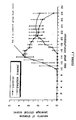
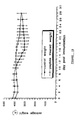
- the rat adjuvant arthritis model is a classic model of inflammation which has been used extensively by a number of laboratories to study disease progression and effects of potential new anti-inflammatory drugs thereon over the last 30 years ( Pearson et al., 1961; Cremer et al., 1990; Holmdahl and Kvick., 1992; Cannon et al.,1993).
- This model has also been widely used by researchers at the Royal North Shore Hospital over the last 10 years and procedures have been established for the study of this model of inflammation. All procedures on the animals were carried out under halothane/oxygen/nitrous oxide anaesthesia [2%v/v halothane in 1 litre/min O 2 and 2 litres/minN 2 O).
- Rats were injected intradermally at the base of the tail with a minimal adjuvant dose (1 mg heat killed Mycobacterium tuberculosis [MTB] in 100 ⁇ l squalane) once and only once.
- MTB heat killed Mycobacterium tuberculosis
- the first experiment consisted of 12 rats weighing approximately 190-210 grams that were purchased from the Perth Animal Resource Centre (ARC) and maintained in the Gore Hill Animal House facility. Used were core peptide (30mg) suspended in adjuvant (0.6 ml squalane containing 7 mg MTB), core peptide Tris-monopalmitate (15mg) suspended in 0.6 ml adjuvant, core peptide Tris-tripalmitate 20 mg/0.6 ml of adjuvant.
- adjuvant 0.6 ml squalane containing 7 mg MTB
- core peptide Tris-monopalmitate 15mg
- core peptide Tris-tripalmitate 20 mg/0.6 ml of adjuvant.
- Rats were divided into four groups, each group containing three rats. First group received adjuvant only (positive control), second group adjuvant with core peptide, third group core peptide.Tris. monopalmitate suspended in adjuvant, and last group core peptide.Tris. tripalmitate in adjuvant. Rats were injected with the above compounds in a 0.1 ml volume at the base of the tail. Baseline measurements of rat weight, paw width, and tail diameter were made on Day 0, and subsequently on day 4, 7, 9, 14, 16, 18, 21, 25 and 28. Arthritis was graded and animals sacrificed if there was marked swelling, redness and obvious discomfort. Not all rats given MTB developed arthritis. In general more than 80% of control rats developed arthritis.
- a feature of adjuvant induced arthritis is the development of inflammation in the tail.
- MTB alone given to rats caused ulceration and inflammation at the site of injection that was not present with rats given core peptide or saline.
- Bovine PNM was prepared essentially as described by Norton and Podulso (1973).
- FIG. 1 A representative example is shown in Figure 1.
- core peptide delayed induction and clinical severity of disease Similar data were observed for peptide C.
- These data confirm the efficacy of core peptide and peptide C as general immunosuppressants.
- IDDM insulin-dependent diabetes mellitus
- NOD mouse spontaneous animal models including the NOD mouse
- a common histopathological feature associated with the development of IDDM is insulitis, the presence within and around the islets of mononuclear cells consisting predominantly of T lymphocytes and to a lesser extent macrophages (Foulis et al., 1986).
- Experimental strategies aimed at suppressing cellular autoimmunity such as neonatal thymectomy, administration of cyclosporin A or administration of anti-T lymphocyte antibodies prevent the development of diabetes (Campbell et al., 1991). Animals
Landscapes
- Health & Medical Sciences (AREA)
- Life Sciences & Earth Sciences (AREA)
- Chemical & Material Sciences (AREA)
- Organic Chemistry (AREA)
- Medicinal Chemistry (AREA)
- General Health & Medical Sciences (AREA)
- Chemical Kinetics & Catalysis (AREA)
- Public Health (AREA)
- Veterinary Medicine (AREA)
- Nuclear Medicine, Radiotherapy & Molecular Imaging (AREA)
- General Chemical & Material Sciences (AREA)
- Pharmacology & Pharmacy (AREA)
- Animal Behavior & Ethology (AREA)
- Engineering & Computer Science (AREA)
- Bioinformatics & Cheminformatics (AREA)
- Immunology (AREA)
- Virology (AREA)
- Diabetes (AREA)
- Molecular Biology (AREA)
- Dermatology (AREA)
- Oncology (AREA)
- Communicable Diseases (AREA)
- Hematology (AREA)
- Physical Education & Sports Medicine (AREA)
- Gastroenterology & Hepatology (AREA)
- Cell Biology (AREA)
- Neurology (AREA)
- Orthopedic Medicine & Surgery (AREA)
- Rheumatology (AREA)
- Pulmonology (AREA)
- Proteomics, Peptides & Aminoacids (AREA)
- Genetics & Genomics (AREA)
- Biophysics (AREA)
- Biochemistry (AREA)
- Zoology (AREA)
- Toxicology (AREA)
- Emergency Medicine (AREA)
- Obesity (AREA)
- Neurosurgery (AREA)
- Vascular Medicine (AREA)
Description
| Sequence comparison of TCR-α trarismembrane region | |
| SPECIES | SEQUENCE |
| MOUSE | NLSVMGLRILLLKVAGFNLLMTL |
| RAT | NLSVMGLRILLLKVAGFNLLMTL |
| SHEEP | NLSVTVFRILLLKVVGFNLLMTL |
| COW | NLSVI VFRILLLKVVGFNLLMTL |
| HUMAN | NLSVI GFRILLLKVAGFNLLMTL |
A-B-C-D-E in which:
| Peptides and their sequence | |
| PEPTIDE | SEQUENCE |
| Core peptide | Gly-Leu-Arg-Ile-Leu-Leu-Leu-Lys-Val-OH |
| A | Met-Gly-Leu-Arg-Ile-Leu- Leu-Leu-OH |
| B | Leu-Gly-Ile-Leu-Leu-Leu-Gly-Val-OH |
| C | Leu-Lys-Ile-Leu-Leu-Leu-Arg-Val-OH |
| D | Leu-Asp-Ile-Leu-Leu-Leu-Glu-Val-OH |
| E | Leu-Arg-Ile-Leu-Leu-Leu-Ile-Lys-Val-OH |
| F | Leu-Arg-Leu-Leu-Leu-Lys-Val-OH |
| Effects of different peptides on adjuvant induced arthritis in rats. | |||
| PEPTIDE | INDUCTION OF ARTHRITIS | EFFECT | |
| MTB ALONE | WITH | ||
| CORE | |||
| 3/3 (100%) | 1/3 (33%) | Protective | |
| 3/5 (60%) | 1/5 (20%) | Protective | |
| 5/5 (100%) | 1/4 (25%) | | |
| A | |||
| 2/4 (50%) | 4/6 (67%) | No | |
| B | |||
| 2/4 (50%) | 2/4 (50%) | No | |
| C | |||
| 4/5 (80%) | 0/4 (0%) | | |
| D | |||
| 4/5 (80%) | 4/5 (80%) | No | |
| E | |||
| 5/5 (100%) | 3/5 (60%) | | |
| F | |||
| 5/5 (100%) | 0/5 (0%) | Protective | |
| 5/5 (100%) | 1/5 (20%) | Protective | |
| DXM* | 5/5 (100%) | 4/4 (100%) | No effect+ |
| Effect of core peptide dosage on adjuvant induced arthritis. | |||
| PEPTIDE | INDUCTION OF ARTHRITIS | EFFECT OF PEPTIDE | |
| MTB ALONE | WITH PEPTIDE | ||
| 7 mg | 11/13 (85%) | 3/12 (25%) | Protective |
| 3.5 | 4/5 (80%) | 2/5 (40%) | Protective |
| 1.7 | 6/7 86%) | 7/8 (88%) | No effect |
| Effect of core peptide on tail inflammation as assessed by tail thickness (mm) | ||||||
| | DAY | |||||
| 0 | 5 | 10 | 15 | 20 | 25 | |
| SALINE (n=4) | 7.13 | 7.75 | 7.95 | 8.48 | 8.78 | 8.80 |
| MTB only (n=5) | 6.86 | 8.80 | 8.96 | 9.46 | 9.60 | 9.73 |
| MTB+PEPTIDE (n=5) | 7.23 | 7.62 | 8.24 | 8.76 | 8.96 | 9.10 |
| Effects of core peptide on the induction of diabetes in NOD/Lt (F) mice | ||||
| TREATMENT | PERCENTAGE OF MICE DEVELOPING | |||
| DAY | ||||
| 0 | DAY10 | DAY14 | DAY 21 | |
| Ovalbumin (n=16) | 0% | 0% | 42% | 65% |
| Peptide (n=16) | 0% | 0% | 5% | 12% |
Claims (19)
- A therapeutic composition comprising a pharmaceutically acceptable carrier and a peptide of the following formula:A-B-C-D-E in which:A is absent, or glycine and 1 hydrophobic amino acid, or 1 or 2 hydrophobic amino acidsB is a positively charged amino acidC is a peptide consisting of 4 hydrophobic amino acidsD is a positively charged amino acid, andE is absent or 1 to 8 hydrophobic amino acids.
- A therapeutic composition as claimed in claim 1 in which A is 2 hydrophobic amino acids.
- A therapeutic composition as claimed in claim 1 in which E is 1 to 3 hydrophobic amino acids.
- A therapeutic composition as claimed in claim 1 in which B is arginine and D is lysine or B is lysine and D is arginine.
- A therapeutic composition as claimed in claim 1 in which the peptide has the sequence Gly-Leu-Arg-Ile-Leu-Leu-Leu-Lys-Val (SEQ ID NO:5).
- A therapeutic composition as claimed in claim 1 in which the peptide has the sequence Gly-Phe-Arg-Ile-Leu-Leu-Leu-Lys-Val (SEQ ID NO:7).
- A therapeutic composition as claimed in claim 1 in which the peptide has the sequence Leu-Lys-Ile-Leu-Leu-Leu-Arg-Val (SEQ ID NO:6).
- A therapeutic composition as claimed in claim 1 in which the peptide has the sequence Phe-Lys-Ile-Leu-Leu-Leu-Arg-Val (SEQ ID NO:8).
- A peptide having the sequence Gly-Leu-Arg-Ile-Leu-Leu-Leu-Lys-Val (SEQ ID NO: 5).
- A peptide having the sequence Gly-Phe-Arg-Ile-Leu-Leu-Leu-Lys-Val (SEQ ID NO:7).
- A peptide having the sequence Leu-Lys-Ile-Leu-Leu-Leu-Arg-Val (SEQ ID NO:6).
- A peptide having the sequence Phe-Lys-Ile-Leu-Leu-Leu-Arg-Val (SEQ ID NO:8).
- A peptide of the formula:for use in inhibiting T-cell receptor function.A-B-C-D-E in which:A is absent, or glycine and 1 hydrophobic amino acid, or 1 or 2 hydrophobic amino acidsB is a positively charged amino acidC is a peptide consisting of 3 to 5 hydrophobic amino acidsD is a positively charged amino acid, andE is absent or up to 8 hydrophobic amino acids,
- A peptide according to claim 13, wherein the peptide is selected from SEQ ID NO:5, SEQ ID NO:6, SEQ ID NO:7 and SEQ ID NO:8, for use in inhibiting T-cell receptor function.
- Use of a peptide of the formula:for the manufacture of a medicament for the treatment of a disorder in which T-cells are recruited or involved.A-B-C-D-E in which:A is absent, or glycine and 1 hydrophobic amino acid, or 1 or 2 hydrophobic acidsB is a positively charged amino acidC is a peptide consisting of 3 to 5 hydrophobic amino acidsD is a positively charged amino acid, andE is absent or up to 8 hydrophobic amino acids
- Use as claimed in claim 13 or 14 in which C is a peptide consisting of 4 amino acids.
- Use according to claim 16 wherein the peptide is selected from SEQ ID NO:5, SEQ ID NO:6, SEQ ID NO:7 and SEQ ID NO:8.
- A method of delivering a chemical moiety to a cell comprising exposing the cell in vitro to the chemical moiety conjugated to a peptide of the formula:A-B-C-D-E in which:A is absent, or glycine and 1 hydrophobic amino acid, or 1 or 2 hydrophobic amino acidsB is a positively charged amino acidC is a peptide consisting of 3 to 5 hydrophobic amino acidsD is a positively charged amino acid, andE is absent or up to 8 hydrophobic amino acids.
- A method according to claim 18 wherein the peptide is selected from SEQ ID NO:5, SEQ ID NO:6, SEQ ID NO:7 and SEQ ID NO: 8.
Applications Claiming Priority (7)
| Application Number | Priority Date | Filing Date | Title |
|---|---|---|---|
| AUPN059095 | 1995-01-16 | ||
| AUPN058995 | 1995-01-16 | ||
| AUPN0590/95 | 1995-01-16 | ||
| AUPN0589/95 | 1995-01-16 | ||
| AUPN0589A AUPN058995A0 (en) | 1995-01-16 | 1995-01-16 | Novel peptide |
| AUPN0590A AUPN059095A0 (en) | 1995-01-16 | 1995-01-16 | Novel lipopeptide |
| PCT/AU1996/000018 WO1996022306A1 (en) | 1995-01-16 | 1996-01-16 | Novel peptide |
Publications (3)
| Publication Number | Publication Date |
|---|---|
| EP0804469A1 EP0804469A1 (en) | 1997-11-05 |
| EP0804469A4 EP0804469A4 (en) | 1998-05-20 |
| EP0804469B1 true EP0804469B1 (en) | 2001-11-28 |
Family
ID=25644844
Family Applications (1)
| Application Number | Title | Priority Date | Filing Date |
|---|---|---|---|
| EP96900472A Expired - Lifetime EP0804469B1 (en) | 1995-01-16 | 1996-01-16 | T-cell affecting peptides |
Country Status (8)
| Country | Link |
|---|---|
| US (1) | US6057294A (en) |
| EP (1) | EP0804469B1 (en) |
| JP (3) | JP3665342B2 (en) |
| AU (1) | AU694602B2 (en) |
| CA (1) | CA2210812C (en) |
| DE (1) | DE69617396T2 (en) |
| ES (1) | ES2168456T3 (en) |
| WO (1) | WO1996022306A1 (en) |
Families Citing this family (16)
| Publication number | Priority date | Publication date | Assignee | Title |
|---|---|---|---|---|
| US6057294A (en) * | 1995-01-16 | 2000-05-02 | Northern Sydney Area Health Service Of Pacific Highway | Peptide |
| AU2020497A (en) * | 1996-03-27 | 1997-10-17 | Susan R. George | Receptor and transporter antagonists |
| US20050070478A1 (en) * | 1996-06-11 | 2005-03-31 | Northern Sydney Area Health Services | T cell antigen receptor peptides |
| US7192928B1 (en) | 1996-06-11 | 2007-03-20 | Northern Sydney & Central Coast Area Health Services | T cell antigen receptor peptides |
| JP4550171B2 (en) | 1997-04-11 | 2010-09-22 | サングスタット メディカル コーポレイション | Cell-regulating lipophilic peptides for modulating immune system activity and inhibiting inflammation |
| US6696545B1 (en) | 1997-04-11 | 2004-02-24 | Sangstat Medical Corporation | Cytomodulating lipophilic peptides for modulating immune system activity and inhibiting inflammation |
| CA2409771C (en) * | 2000-05-19 | 2008-12-02 | University Of Maryland, Baltimore | Method of use of peptide antagonists of zonulin to prevent or delay the onset of diabetes |
| AU2003210659A1 (en) | 2002-01-24 | 2003-09-02 | Sangstat Medical Corporation | Combination therapy for treatment of hiv infection |
| JP2008500289A (en) * | 2004-05-27 | 2008-01-10 | ノヴォザイムズ グロペップ リミテッド | Treatment of inflammatory airway diseases |
| EP1945658A4 (en) * | 2005-09-22 | 2012-05-30 | Yeda Res & Dev | Diastereomeric peptides for modulating t cell immunity |
| EP1870420A1 (en) * | 2006-06-23 | 2007-12-26 | Max-Delbrück-Centrum Für Molekulare Medizin | Peptides regulating the surface expression of the T cell receptor |
| JP2012509848A (en) * | 2008-11-24 | 2012-04-26 | シドニー ウェスト エリア ヘルス サービス | Cyclic peptides and uses thereof |
| US10138276B2 (en) | 2009-09-30 | 2018-11-27 | Signablok, Inc. | Inhibition of TCR signaling with peptide variants |
| US8513185B2 (en) * | 2009-10-13 | 2013-08-20 | Alexander B. Sigalov | Inhibition of TREM receptor signaling with peptide variants |
| WO2018183888A2 (en) | 2017-03-31 | 2018-10-04 | The Board Of Trustees Of The Leland Stanford Junior University | Methods of treating t cell exhaustion by inhibiting or modulating t cell receptor signaling |
| WO2020036987A1 (en) | 2018-08-13 | 2020-02-20 | Signablok, Inc. | Peptides and compositions for targeted treatment and imaging |
Family Cites Families (12)
| Publication number | Priority date | Publication date | Assignee | Title |
|---|---|---|---|---|
| US5888512A (en) * | 1987-01-30 | 1999-03-30 | Board Of Trustees Of The Leland Stanford Junior University | Lymphocyte activity regulation by HLA peptides |
| US5223485A (en) * | 1989-01-31 | 1993-06-29 | Abbott Laboratories | Anaphylatoxin-receptor ligands |
| CA2045578A1 (en) * | 1989-01-31 | 1990-08-01 | Megumi Kawai | Anaphylatoxin-receptor ligands |
| KR0185373B1 (en) * | 1989-03-17 | 1999-05-01 | 로버트 피. 블랙버언 | Polypeptides comprising HCV amino acid sequence portions derived from HCV polyproteins and use thereof |
| AU8228791A (en) * | 1990-06-15 | 1992-01-07 | New York University | Heterohybridomas producing human monoclonal antibodies to hiv-1 |
| US5387671A (en) * | 1990-12-27 | 1995-02-07 | Abbott Laboratories | Hexa- and heptapeptide anaphylatoxin-receptor ligands |
| JP3908271B2 (en) * | 1993-03-05 | 2007-04-25 | エピミューン,インコーポレイティド | HLA-A2.1 binding peptides and their use |
| ES2344643T3 (en) * | 1994-01-14 | 2010-09-02 | Matthias Dr. Med. Rath | HYDROPHILIC SIGNAL OLIGOPEPTIDES AND THERAPEUTIC USE METHODS. |
| US5709995A (en) * | 1994-03-17 | 1998-01-20 | The Scripps Research Institute | Hepatitis C virus-derived peptides capable of inducing cytotoxic T lymphocyte responses |
| AU2238395A (en) * | 1994-04-01 | 1995-10-23 | Immulogic Pharmaceutical Corporation | Haptenated peptides and uses thereof |
| US6057294A (en) * | 1995-01-16 | 2000-05-02 | Northern Sydney Area Health Service Of Pacific Highway | Peptide |
| US5753625A (en) * | 1995-05-12 | 1998-05-19 | Sangstat Medical Corporation | Treatment for inhibiting the progression of autoimmune disease |
-
1996
- 1996-01-16 US US08/875,013 patent/US6057294A/en not_active Expired - Fee Related
- 1996-01-16 EP EP96900472A patent/EP0804469B1/en not_active Expired - Lifetime
- 1996-01-16 WO PCT/AU1996/000018 patent/WO1996022306A1/en not_active Ceased
- 1996-01-16 JP JP52191796A patent/JP3665342B2/en not_active Expired - Fee Related
- 1996-01-16 AU AU44272/96A patent/AU694602B2/en not_active Ceased
- 1996-01-16 DE DE69617396T patent/DE69617396T2/en not_active Expired - Lifetime
- 1996-01-16 ES ES96900472T patent/ES2168456T3/en not_active Expired - Lifetime
- 1996-01-16 CA CA002210812A patent/CA2210812C/en not_active Expired - Fee Related
-
2005
- 2005-02-10 JP JP2005034053A patent/JP2005145985A/en active Pending
-
2006
- 2006-01-10 JP JP2006002633A patent/JP4522367B2/en not_active Expired - Fee Related
Also Published As
| Publication number | Publication date |
|---|---|
| WO1996022306A1 (en) | 1996-07-25 |
| JP4522367B2 (en) | 2010-08-11 |
| ES2168456T3 (en) | 2002-06-16 |
| EP0804469A4 (en) | 1998-05-20 |
| JPH10512267A (en) | 1998-11-24 |
| CA2210812C (en) | 2009-09-01 |
| DE69617396T2 (en) | 2002-09-05 |
| JP2005145985A (en) | 2005-06-09 |
| JP2006117697A (en) | 2006-05-11 |
| US6057294A (en) | 2000-05-02 |
| EP0804469A1 (en) | 1997-11-05 |
| CA2210812A1 (en) | 1996-07-25 |
| DE69617396D1 (en) | 2002-01-10 |
| AU694602B2 (en) | 1998-07-23 |
| AU4427296A (en) | 1996-08-07 |
| JP3665342B2 (en) | 2005-06-29 |
Similar Documents
| Publication | Publication Date | Title |
|---|---|---|
| Manolios et al. | T-cell antigen receptor transmembrane peptides modulate T-cell function and T cell-mediated disease | |
| EP0804469B1 (en) | T-cell affecting peptides | |
| US6458360B1 (en) | Soluble complement regulatory molecules | |
| CA2308765C (en) | The use of an ox-2 protein or nucleic acid in immunomodulation | |
| JP2009029833A (en) | CD28 / CTLA-4 inhibitory peptide mimetics, pharmaceutical compositions thereof, and methods of using them | |
| WO1998023639A2 (en) | ShK TOXIN COMPOSITIONS AND METHODS OF USE | |
| HU211476A9 (en) | Cd2-binding domain of lymphocyte function associated antigen 3 | |
| BRPI0111191B1 (en) | soluble ctla4 mutant molecules, method for their production and uses | |
| Ostrov et al. | T cell receptor antagonism mediated by interaction between T cell receptor junctional residues and peptide antigen analogues | |
| Davidson et al. | Distribution and immunoregulatory properties of antisecretory factor | |
| DE69529515T2 (en) | THE REGULATION OF THE ACTIVITY OF CYTOTOXIC T CELL LYMPHOCYTES WITH MHC CLASS I PEPTIDES | |
| JP4452312B2 (en) | T cell antigen receptor peptide | |
| WO1992006117A1 (en) | Inhibiting unwanted immune responses | |
| KR100333148B1 (en) | Soluble T-cell receptor α chains and derivatives containing Vα 14Jα 281 used as a preventive and therapeutic agent for diabetes diseases | |
| US20100267651A1 (en) | T cell antigen receptor peptides | |
| Rosloniec et al. | Second-generation peptidomimetic inhibitors of antigen presentation effectively treat autoimmune diseases in HLA-DR-transgenic mouse models | |
| AU778551B2 (en) | Novel peptide and screening method by using the same | |
| WO1996031617A1 (en) | Method of production of antigen-specific glycosylation inhibiting factor | |
| WO1991018018A1 (en) | Improved pseudomonas chimeric protein cytotoxic to human cells bearing il2 receptors | |
| JP2003519162A (en) | PP14 fusion protein and methods for producing and using the protein |
Legal Events
| Date | Code | Title | Description |
|---|---|---|---|
| PUAI | Public reference made under article 153(3) epc to a published international application that has entered the european phase |
Free format text: ORIGINAL CODE: 0009012 |
|
| 17P | Request for examination filed |
Effective date: 19970805 |
|
| AK | Designated contracting states |
Kind code of ref document: A1 Designated state(s): DE ES FR GB IT SE |
|
| A4 | Supplementary search report drawn up and despatched |
Effective date: 19980406 |
|
| AK | Designated contracting states |
Kind code of ref document: A4 Designated state(s): AT BE CH DE DK ES FR GB GR IE IT LI LU MC NL PT SE |
|
| 17Q | First examination report despatched |
Effective date: 19990310 |
|
| RTI1 | Title (correction) |
Free format text: T-CELL AFFECTING PEPTIDES |
|
| RTI1 | Title (correction) |
Free format text: T-CELL AFFECTING PEPTIDES |
|
| GRAG | Despatch of communication of intention to grant |
Free format text: ORIGINAL CODE: EPIDOS AGRA |
|
| GRAG | Despatch of communication of intention to grant |
Free format text: ORIGINAL CODE: EPIDOS AGRA |
|
| GRAH | Despatch of communication of intention to grant a patent |
Free format text: ORIGINAL CODE: EPIDOS IGRA |
|
| GRAH | Despatch of communication of intention to grant a patent |
Free format text: ORIGINAL CODE: EPIDOS IGRA |
|
| GRAA | (expected) grant |
Free format text: ORIGINAL CODE: 0009210 |
|
| RBV | Designated contracting states (corrected) |
Designated state(s): DE ES FR GB IT SE |
|
| AK | Designated contracting states |
Kind code of ref document: B1 Designated state(s): DE ES FR GB IT SE |
|
| REG | Reference to a national code |
Ref country code: GB Ref legal event code: IF02 |
|
| REF | Corresponds to: |
Ref document number: 69617396 Country of ref document: DE Date of ref document: 20020110 |
|
| ET | Fr: translation filed | ||
| REG | Reference to a national code |
Ref country code: ES Ref legal event code: FG2A Ref document number: 2168456 Country of ref document: ES Kind code of ref document: T3 |
|
| PLBE | No opposition filed within time limit |
Free format text: ORIGINAL CODE: 0009261 |
|
| STAA | Information on the status of an ep patent application or granted ep patent |
Free format text: STATUS: NO OPPOSITION FILED WITHIN TIME LIMIT |
|
| 26N | No opposition filed | ||
| REG | Reference to a national code |
Ref country code: GB Ref legal event code: 732E |
|
| REG | Reference to a national code |
Ref country code: FR Ref legal event code: TP |
|
| REG | Reference to a national code |
Ref country code: ES Ref legal event code: PC2A |
|
| PGFP | Annual fee paid to national office [announced via postgrant information from national office to epo] |
Ref country code: SE Payment date: 20091218 Year of fee payment: 15 |
|
| PGFP | Annual fee paid to national office [announced via postgrant information from national office to epo] |
Ref country code: ES Payment date: 20100128 Year of fee payment: 15 |
|
| PGFP | Annual fee paid to national office [announced via postgrant information from national office to epo] |
Ref country code: IT Payment date: 20100116 Year of fee payment: 15 Ref country code: FR Payment date: 20100208 Year of fee payment: 15 |
|
| PGFP | Annual fee paid to national office [announced via postgrant information from national office to epo] |
Ref country code: GB Payment date: 20100113 Year of fee payment: 15 Ref country code: DE Payment date: 20100114 Year of fee payment: 15 |
|
| REG | Reference to a national code |
Ref country code: SE Ref legal event code: EUG |
|
| GBPC | Gb: european patent ceased through non-payment of renewal fee |
Effective date: 20110116 |
|
| REG | Reference to a national code |
Ref country code: FR Ref legal event code: ST Effective date: 20110930 |
|
| PG25 | Lapsed in a contracting state [announced via postgrant information from national office to epo] |
Ref country code: FR Free format text: LAPSE BECAUSE OF NON-PAYMENT OF DUE FEES Effective date: 20110131 |
|
| PG25 | Lapsed in a contracting state [announced via postgrant information from national office to epo] |
Ref country code: GB Free format text: LAPSE BECAUSE OF NON-PAYMENT OF DUE FEES Effective date: 20110116 |
|
| PG25 | Lapsed in a contracting state [announced via postgrant information from national office to epo] |
Ref country code: IT Free format text: LAPSE BECAUSE OF NON-PAYMENT OF DUE FEES Effective date: 20110116 |
|
| REG | Reference to a national code |
Ref country code: DE Ref legal event code: R119 Ref document number: 69617396 Country of ref document: DE Effective date: 20110802 |
|
| REG | Reference to a national code |
Ref country code: ES Ref legal event code: FD2A Effective date: 20120220 |
|
| PG25 | Lapsed in a contracting state [announced via postgrant information from national office to epo] |
Ref country code: ES Free format text: LAPSE BECAUSE OF NON-PAYMENT OF DUE FEES Effective date: 20110117 |
|
| PG25 | Lapsed in a contracting state [announced via postgrant information from national office to epo] |
Ref country code: SE Free format text: LAPSE BECAUSE OF NON-PAYMENT OF DUE FEES Effective date: 20110117 |
|
| PG25 | Lapsed in a contracting state [announced via postgrant information from national office to epo] |
Ref country code: DE Free format text: LAPSE BECAUSE OF NON-PAYMENT OF DUE FEES Effective date: 20110802 |