EP0804237B1 - Humanized antibodies against leukocyte adhesion molecule vla-4 - Google Patents
Humanized antibodies against leukocyte adhesion molecule vla-4 Download PDFInfo
- Publication number
- EP0804237B1 EP0804237B1 EP95908741A EP95908741A EP0804237B1 EP 0804237 B1 EP0804237 B1 EP 0804237B1 EP 95908741 A EP95908741 A EP 95908741A EP 95908741 A EP95908741 A EP 95908741A EP 0804237 B1 EP0804237 B1 EP 0804237B1
- Authority
- EP
- European Patent Office
- Prior art keywords
- humanized
- mouse
- human
- immunoglobulin
- seq
- Prior art date
- Legal status (The legal status is an assumption and is not a legal conclusion. Google has not performed a legal analysis and makes no representation as to the accuracy of the status listed.)
- Expired - Lifetime
Links
Images


Classifications
-
- A—HUMAN NECESSITIES
- A61—MEDICAL OR VETERINARY SCIENCE; HYGIENE
- A61K—PREPARATIONS FOR MEDICAL, DENTAL OR TOILETRY PURPOSES
- A61K39/00—Medicinal preparations containing antigens or antibodies
- A61K39/395—Antibodies; Immunoglobulins; Immune serum, e.g. antilymphocytic serum
-
- C—CHEMISTRY; METALLURGY
- C07—ORGANIC CHEMISTRY
- C07K—PEPTIDES
- C07K16/00—Immunoglobulins [IG], e.g. monoclonal or polyclonal antibodies
- C07K16/18—Immunoglobulins [IG], e.g. monoclonal or polyclonal antibodies against material from animals or humans
- C07K16/28—Immunoglobulins [IG], e.g. monoclonal or polyclonal antibodies against material from animals or humans against receptors, cell surface antigens or cell surface determinants
- C07K16/2839—Immunoglobulins [IG], e.g. monoclonal or polyclonal antibodies against material from animals or humans against receptors, cell surface antigens or cell surface determinants against the integrin superfamily
-
- A—HUMAN NECESSITIES
- A61—MEDICAL OR VETERINARY SCIENCE; HYGIENE
- A61P—SPECIFIC THERAPEUTIC ACTIVITY OF CHEMICAL COMPOUNDS OR MEDICINAL PREPARATIONS
- A61P25/00—Drugs for disorders of the nervous system
-
- A—HUMAN NECESSITIES
- A61—MEDICAL OR VETERINARY SCIENCE; HYGIENE
- A61P—SPECIFIC THERAPEUTIC ACTIVITY OF CHEMICAL COMPOUNDS OR MEDICINAL PREPARATIONS
- A61P29/00—Non-central analgesic, antipyretic or antiinflammatory agents, e.g. antirheumatic agents; Non-steroidal antiinflammatory drugs [NSAID]
-
- A—HUMAN NECESSITIES
- A61—MEDICAL OR VETERINARY SCIENCE; HYGIENE
- A61P—SPECIFIC THERAPEUTIC ACTIVITY OF CHEMICAL COMPOUNDS OR MEDICINAL PREPARATIONS
- A61P37/00—Drugs for immunological or allergic disorders
-
- A—HUMAN NECESSITIES
- A61—MEDICAL OR VETERINARY SCIENCE; HYGIENE
- A61P—SPECIFIC THERAPEUTIC ACTIVITY OF CHEMICAL COMPOUNDS OR MEDICINAL PREPARATIONS
- A61P7/00—Drugs for disorders of the blood or the extracellular fluid
-
- C—CHEMISTRY; METALLURGY
- C07—ORGANIC CHEMISTRY
- C07K—PEPTIDES
- C07K16/00—Immunoglobulins [IG], e.g. monoclonal or polyclonal antibodies
- C07K16/18—Immunoglobulins [IG], e.g. monoclonal or polyclonal antibodies against material from animals or humans
- C07K16/28—Immunoglobulins [IG], e.g. monoclonal or polyclonal antibodies against material from animals or humans against receptors, cell surface antigens or cell surface determinants
- C07K16/2839—Immunoglobulins [IG], e.g. monoclonal or polyclonal antibodies against material from animals or humans against receptors, cell surface antigens or cell surface determinants against the integrin superfamily
- C07K16/2842—Immunoglobulins [IG], e.g. monoclonal or polyclonal antibodies against material from animals or humans against receptors, cell surface antigens or cell surface determinants against the integrin superfamily against integrin beta1-subunit-containing molecules, e.g. CD29, CD49
-
- A—HUMAN NECESSITIES
- A61—MEDICAL OR VETERINARY SCIENCE; HYGIENE
- A61K—PREPARATIONS FOR MEDICAL, DENTAL OR TOILETRY PURPOSES
- A61K38/00—Medicinal preparations containing peptides
-
- C—CHEMISTRY; METALLURGY
- C07—ORGANIC CHEMISTRY
- C07K—PEPTIDES
- C07K2317/00—Immunoglobulins specific features
- C07K2317/20—Immunoglobulins specific features characterized by taxonomic origin
- C07K2317/24—Immunoglobulins specific features characterized by taxonomic origin containing regions, domains or residues from different species, e.g. chimeric, humanized or veneered
-
- C—CHEMISTRY; METALLURGY
- C07—ORGANIC CHEMISTRY
- C07K—PEPTIDES
- C07K2319/00—Fusion polypeptide
Definitions
- This invention relates generally to humanized antibodies specific for the alpha-4 subunit of leukocyte adhesion molecule VLA-4.
- Inflammation is a response of vascularized tissues to infection or injury and is effected by adhesion of leukocytes to the endothelial cells of blood vessels and their infiltration into the surrounding tissues.
- the infiltrating leukocytes release toxic mediators to kill invading organisms, phagocytize debris and dead cells, and play a role in tissue repair and the immune response.
- infiltrating leukocytes are over-responsive and can cause serious or fatal damage. See, e.g., Hickey, Psychoneuroimmunology II (Academic Press 1990).
- VLA-4 leukocyte cell-surface receptor was first identified by Hemler, EP 330,506 (1989).
- VLA-4 is a member of the ⁇ 1 integrin family of cell surface receptors, each of which comprises ⁇ and ⁇ chains.
- VLA-4 contains an ⁇ 4 chain and a ⁇ 1 chain.
- VLA-4 specifically binds to an endothelial cell ligand termed VCAM-1.
- VCAM-1 endothelial cell ligand
- Adhesion molecules such as VLA-4 are potential targets for therapeutic agents.
- the VLA-4 receptor is a particularly important target because of its interaction with a ligand residing on brain endothelial cells.
- Diseases and conditions resulting from brain inflammation have particularly severe consequences.
- one such disease multiple sclerosis (MS)
- MS multiple sclerosis
- the disease affects an estimated 250,000 to 350,000 people in the United States alone.
- Antibodies against the VLA-4 receptor have been tested for their anti-inflammatory potential both in vitro and in vivo in animal models. See USSN 07/871,223 and Yednock et al., Nature 356:63-66 (1992).
- the in vitro experiments demonstrate that anti-VLA-4 antibodies block attachment of lymphocytes to brain endothelial cells.
- the animal experiments test the effect of anti-VLA-4 antibodies on animals having an artificially induced condition (experimental autoimmune encephalomyelitis), simulating multiple sclerosis.
- the experiments show that administration of anti-VLA-4 antibodies prevents inflammation of the brain and subsequent paralysis in the animals. Collectively, these experiments identify anti-VLA-4 antibodies as potentially useful therapeutic agents for treating multiple sclerosis and other inflammatory diseases and disorders.
- a significant problem with the anti-VLA-4 antibodies available to-date is that they are all of murine origin, and therefore likely to raise a human anti-mouse response (HAMA) in clinical use.
- HAMA human anti-mouse response
- a HAMA response reduces the efficacy of mouse antibodies in patients and prevents continued administration.
- One approach to this problem is to humanize mouse antibodies. In this approach, complementarity determining regions (CDRs) and certain other amino acids from donor mouse variable regions are grafted into human variable acceptor regions and then joined to human constant regions. See, e . g ., Riechmann et al., Nature 332:323-327 (1988); Winter, US 5,225,539 (1993).
- humanized antibodies Although several examples of humanized antibodies have been produced, the transition from a murine to a humanized antibody involves a compromise of competing considerations, the solution of which varies with different antibodies. To minimize immunogenicity, the immunoglobulin should retain as much of the human acceptor sequence as possible. However, to retain authentic binding properties, the immunoglobulin framework should contain sufficient substitutions of the human acceptor sequence to ensure a three-dimensional conformation of CDR regions as close as possible to that in the original mouse donor immunoglobulin. As a result of these competing considerations, many humanized antibodies produced to-date show some loss of binding affinity compared with the corresponding murine antibodies from which they are derived. See, e .
- WO94/16094 discloses recombinant anti-VLA4 antibody molecules, including humanised recombinant anti-VLA4 antibody molecules. These antibodies are useful in the treatment of specific and non-specific inflammation, including asthma and inflammatory bowel disease. In addition, the humanised recombinant anti-VLA4 antibodies disclosed can be useful in methods of diagnosing and, localizing sites of inflammation.
- WO93/1576 discloses the treatment of inflammatory bowel disease with an antibody, polypeptide or other molecule recognising VLA-4.
- the invention provides humanized immunoglobulins that specifically bind to a VLA-4.
- the humanized antibodies comprise a humanized light chain and a humanized heavy chain.
- the humanized light chain comprises three complementarity determining regions (CDR1, CDR2 and CDR3) having amino acid sequences from the corresponding complamentarity determining regions of a mouse 21-6 immunoglobulin light chain SEQ ID NO:2 (as shown in Figure 1), and a variable region framework from a human kappa light chain variable region framework sequence except in at least one position selected from a first group consisting of positions L45, L49, L58 and L69, wherein the amino acid position is occupied by the same amino acid present in the equivalent position of the mouse 21.6 immunoglobulin light chain variable region framework.
- the humanized heavy chain comprises three complementarity determining regions (CDR1, CDR2 and CDR3) having amino acid sequences from the corresponding complementarity determining regions of a mouse 21-6 immunoglobulin heavy chain of SEQ ID NO 4, and a variable region framework from a human heavy chain variable region framework sequence except in at least one position selected from a group consisting of H27, H28, H29, H30, H44, H71, wherein the amino acid position is occupied by the same amino acid present in the equivalent position of the mouse 21-6 immunoglobulin heavy chain variable region framework.
- the immunoglobulins specifically bind to VLA-4 with an affinity having a lower limit of 10 7 M -1 and an upper limit of five times the affinity of the mouse 21-6 immunoglobulin having the light chain of SEQ ID NO:2 and the heavy chain of SEQ ID NO:4
- the humanized light and heavy chain variable region frameworks are from RE1 and 21/28'CL variable region framework sequences respectively.
- the humanized light chain variable region framework is from RE1
- at least two framework amino acids are replaced.
- One amino acid is from the first group of positions described supra .
- the other amino acids is from a third group consisting of positions L104, L105 and L107. This position is occupied by the same amino acid present in the equivalent position of a kappa light chain from a human immunoglobulin other than RE1.
- humanized immunoglobulins have a mature light chain variable region sequence designated La or Lb in Figure 6, or a mature heavy chain variable region sequence designated Ha, Hb or Hc in Figure 7.
- Preferred humanized immunoglobulins include those having an La light chain and an Ha, Hb or Hc heavy chain.
- the invention also provides binding fragments of the humanized immunoglobulins against VLA-4 described supra.
- the invention provides nucleic acids encoding the humanized immunoglobulins against VLA-4 described supra .
- the invention provides pharmaceutical compositions and methods of treatment using the same.
- the pharmaceutical compositions comprise a humanized immunoglobulin or binding fragment as described supra, and a pharmaceutically acceptable carrier.
- a therapeutically effective amount of a pharmaceutical composition is administered to a patient suffering from an inflammatory disease, such as multiple sclerosis.
- a humanized antibody or binding fragment is administered to a patient or a tissue sample therefrom. Complexes formed by specific binding between the antibody or fragment and VLA-4 present in the sample are detected.
- Examples of unconventional amino acids include: 4-hydroxyproline, ⁇ -carboxyglutamate, ⁇ -N,N,N-trimethyllysine, ⁇ -N-acetyllysine, O-phosphoserine, N-acetylserine, N-formylmethionine, 3-methylhistidine, 5-hydroxylysine, ⁇ -N-methylarginine, and other similar amino acids and imino acids (e.g., 4-hydroxyproline).
- amino acids may be modified by glycosylation, phosphorylation and the like.
- the lefthand direction is the amino terminal direction and the righthand direction is the carboxy-terminal direction, in accordance with standard usage and convention.
- the lefthand end of single-stranded polynucleotide sequences is the 5' end; the lefthand direction of double-stranded polynucleotide sequences is referred to as the 5' direction.
- RNA transcripts The direction of 5' to 3' addition of nascent RNA transcripts is referred to as the transcription direction; sequence regions on the DNA strand having the same sequence as the RNA and which are 5' to the 5' end of the RNA transcript are referred to as "upstream sequences"; sequence regions on the DNA strand having the same sequence as the RNA and which are 3' to the 3' end of the RNA transcript are referred to as "downstream sequences".
- polynucleotide sequence refers to a single or double-stranded polymer of deoxyribonucleotide or ribonucleotide bases read from the 5' to the 3' end. It includes self-replicating plasmids, infectious polymers of DNA or RNA and non-functional DNA or RNA.
- reference sequence is a defined sequence used as a basis for a sequence comparison; a reference sequence may be a subset of a larger sequence, for example, as a segment of a full-length CDNA or gene sequence given in a sequence listing, such as a polynucleotide sequence of Figs. 1 or 2, or may comprise a complete DNA or gene sequence.
- a reference sequence is at least 20 nucleotides in length, frequently at least 25 nucleotides in length, and often at least 50 nucleotides in length. Since two polynucleotides may each (1) comprise a sequence (i.e., a portion of the complete polynucleotide sequence) that is similar between the two polynucleotides, and (2) may further comprise a sequence that is divergent between the two polynucleotides, sequence comparisons between two (or more) polynucleotides are typically performed by comparing sequences of the two polynucleotides over a "comparison window" to identify and compare local regions of sequence similarity.
- a “comparison window”, as used herein, refers to a conceptual segment of at least 20 contiguous nucleotide positions wherein a polynucleotide sequence may be compared to a reference sequence of at least 20 contiguous nucleotides and wherein the portion of the polynucleotide sequence in the comparison window may comprise additions or deletions (i.e., gaps) of 20 percent or less as compared to the reference sequence (which does not comprise additions or deletions) for optimal alignment of the two sequences.
- Optimal alignment of sequences for aligning a comparison window may be conducted by the local homology algorithm of Smith & Waterman, Adv. Appl. Math. 2:482 (1981), by the homology alignment algorithm of Needleman & Wunsch, J.
- sequence identity means that two polynucleotide sequences are identical (i.e., on a nucleotide-by-nucleotide basis) over the window of comparison.
- percentage of sequence identity is calculated by comparing two optimally aligned sequences over the window of comparison, determining the number of positions at which the identical nucleic acid base (e.g., A, T, C, G, U, or I) occurs in both sequences to yield the number of matched positions, dividing the number of matched positions by the total number of positions in the window of comparison (i.e., the window size), and multiplying the result by 100 to yield the percentage of sequence identity.
- the identical nucleic acid base e.g., A, T, C, G, U, or I
- substantially identical denotes a characteristic of a polynucleotide sequence, wherein the polynucleotide comprises a sequence that has at least 85 percent sequence identity, preferably at least 90 to 95 percent sequence identity, more usually at least 99 percent sequence identity as compared to a reference sequence over a comparison window of at least 20 nucleotide positions, frequently over a window of at least 25-50 nucleotides, wherein the percentage of sequence identity is calculated by comparing the reference sequence to the polynucleotide sequence which may include deletions or additions which total 20 percent or less of the reference sequence over the window of comparison.
- the reference sequence may be a subset of a larger sequence, for example, the sequence shown in Figs. 1 or 2.
- sequence identity means peptides share identical amino acids at corresponding positions.
- sequence similarity means peptides have identical or similar amino acids (i.e., conservative substitutions) at corresponding positions.
- substantially identity means that two peptide sequences, when optimally aligned, such as by the programs GAP or BESTFIT using default gap weights, share at least 80 percent sequence identity, preferably at least 90 percent sequence identity, more preferably at least 95 percent sequence identity or more (e.g., 99 percent sequence identity). Preferably, residue positions which are not identical differ by conservative amino acid substitutions.
- substantially similarity means that two peptide sequences share corresponding percentages of sequence similarity.
- substantially pure means an object species is the predominant species present (i.e., on a molar basis it is more abundant than any other individual species in the composition), and preferably a substantially purified fraction is a composition wherein the object species comprises at least about 50 percent (on a molar basis) of all macromolecular species present. Generally, a substantially pure composition will comprise more than about 80 to 90 percent of all macromolecular species present in the composition. Most preferably, the object species is purified to essential homogeneity (contaminant species cannot be detected in the composition by conventional detection methods) wherein the composition consists essentially of a single macromolecular species.
- amino acids are grouped as follows: Group I (hydrophobic sidechains): norleucine, met, ala, val, leu, ile; Group II (neutral hydrophilic side chains): cys, ser, thr; Group III (acidic side chains): asp, glu; Group IV (basic side chains): asn, gln, his, lys, arg; Group V (residues influencing chain orientation): gly, pro; and Group VI (aromatic side chains): trp, tyr, phe. Conservative substitutions involve substitutions between amino acids in the same class. Non-conservative substitutions constitute exchanging a member of one of these classes for another.
- Amino acids from the variable regions of the mature heavy and light chains of immunoglobulins are designated Hx and Lxx respectively, where x is a number designating the position of an amino acids according to the scheme of Kabat et al., Sequences of Proteins of Immunological Interest (National Institutes of Health, Bethesda, MD (1987) and (1991)) (hereinafter collectively referred to as "Kabat et al.,” incorporated by reference in their entirety for all purposes). Kabat et al. list many amino acid sequences for antibodies for each subclass, and list the most commonly occurring amino acid for each residue position in that subclass. Kabat et al.
- Kabat et al.'s scheme is extendible to other antibodies not included in the compendium by aligning the antibody in question with one of the consensus sequences in Kabat et al.
- the use of the Kabat et al. numbering system readily identifies amino acids at equivalent positions in different antibodies. For example, an amino acid at the L50 position of a human antibody occupies the equivalence position to an amino acid position L50 of a mouse antibody.
- humanized immunoglobulins (or antibodies) specific for the alpha-4 subunit of VLA-4 are provided.
- the humanized immunoglobulins have variable framework regions substantially from a human immunoglobulin (termed an acceptor immunoglobulin) and complementarity determining regions substantially from a mouse immunoglobulin termed mu MAb 21.6 (referred to as the donor immunoglobulin).
- the constant region(s), if present, are also substantially from a human immunoglobulin.
- the humanized antibodies exhibit a specific binding affinity for VLA-4 of at least 10 7 , 10 8 , 10 9 , or 10 10 M -1 .
- the upper limit of binding affinity of the humanized antibodies for VLA-4 is within a factor of three or five of that of mu MAb 21.6 (about 10 9 M -1 ).
- the lower limit of binding affinity is also within a factor of three or five of that of mu MAb 21.6.
- the basic antibody structural unit is known to comprise a tetramer.
- Each tetramer is composed of two identical pairs of polypeptide chains, each pair having one "light” (about 25 kDa) and one "heavy" chain (about 50-70 kDa).
- the amino-terminal portion of each chain includes a variable region of about 100 to 110 or more amino acids primarily responsible for antigen recognition.
- the carboxy-terminal portion of each chain defines a constant region primarily responsible for effector function.
- Light chains are classified as either kappa or lambda.
- Heavy chains are classified as gamma, mu, alpha, delta, or epsilon, and define the antibody's isotype as IgG, IgM, IgA, IgD and IgE, respectively.
- the variable and constant regions are joined by a "J" region of about 12 or more amino acids, with the heavy chain also including a "D” region of about 10 more amino acids.
- variable regions of each light/heavy chain pair form the antibody binding site.
- the chains all exhibit the same general structure of relatively conserved framework regions (FR) joined by three hypervariable regions, also called complementarity determining regions or CDRs.
- the CDRs from the two chains of each pair are aligned by the framework regions, enabling binding to a specific epitope.
- CDR and FR residues are delineated according to the standard sequence definition of Kabat et al., supra.
- An alternative structural definition has been proposed by Chothia et al., J. Mol. Biol . 196:901-917 (1987); Nature 342:878-883 (1989); and J. Mol. Biol.
- mu MAb 21.6 The starting material for production of humanized antibodies is mu MAb 21.6.
- the isolation and properties of this antibody are described in USSN 07/871,223.
- mu MAb 21.6 is specific for the alpha-4 subunit of VLA-4 and has been shown to inhibit human lymphocyte binding to tissue cultures of rat brain cells stimulated with tumor necrosis factor.
- the cloning and sequencing of CDNA encoding the mu MAb 21.6 antibody heavy and light chain variable regions is described in Example 1, and the nucleotide and predicted amino acids sequences are shown in Figures 1 and 2. These figures also illustrate the subdivision of the amino acid coding sequencing into framework and complementarity determining domains.
- both light and heavy chains comprise the domains FR1, CDR1, FR2, CDR2, FR3, CDR3 and FR4.
- the assignment of amino acids to each domain is in accordance with the numbering convention of Kabat et al., supra.
- the substitution of mouse CDRs into a human variable domain framework is most likely to result in retention of their correct spatial orientation if the human variable domain framework adopts the same or similar conformation to the mouse variable framework from which the CDRs originated. This is achieved by obtaining the human variable domains from human antibodies whose framework sequences exhibit a high degree of sequence identity with the murine variable framework domains from which the CDRs were derived.
- the heavy and light chain variable framework regions can be derived from the same or different human antibody sequences.
- the human antibody sequences can be the sequences of naturally occurring human antibodies or can be consensus sequences of several human antibodies. See Kettleborough et al., Protein Engineering 4:773 (1991); Kolbinger et al., Protein Engineering 6:971 (1993).
- Suitable human antibody sequences are identified by computer comparisons of the amino acid sequences of the mouse variable regions with the sequences of known human antibodies. The comparison is performed separately for heavy and light chains but the principles are similar for each. This comparison reveals that the mu 21.6 light chain shows greatest sequence identity to human light chains of subtype kappa 1, and that the mu 21.6 heavy chain shows greatest sequence identity to human heavy chains of subtype one, as defined by Kabat et al., supra. Thus, light and heavy human framework regions are usually derived from human antibodies of these subtypes, or from consensus sequences of such subtypes.
- the preferred light and heavy chain human variable regions showing greatest sequence identity to the corresponding regions from mu MAb 21.6 are from antibodies RE1 and 21/28'CL respectively.
- the unnatural juxtaposition of murine CDR regions with human variable framework region can result in unnatural conformational restraints, which, unless corrected by substitution of certain amino acid residues, lead to loss of binding affinity.
- the selection of amino acid residues for substitution is determined, in part, by computer modelling.
- Computer hardware and software for producing three-dimensional images of immunoglobulin molecules are widely available.
- molecular models are produced starting from solved structures for immunoglobulin chains or domains thereof.
- the chains to be modelled are compared for amino acid sequence similarity with chains or domains of solved three dimensional structures, and the chains or domains showing the greatest sequence similarity is/are selected as starting points for construction of the molecular model.
- the starting point for modelling the framework regions, CDR1 and CDR2 regions was the human light chain RE1.
- the starting point was the CDR3 region from the light chain of a different human antibody HyHEL-5.
- the solved starting structures are modified to allow for differences between the actual amino acids in the immunoglobulin chains or domains being modelled, and those in the starting structure.
- the modified structures are then assembled into a composite immunoglobulin.
- the model is refined by energy minimization and by verifying that all atoms are within appropriate distances from one another and that bond lengths and angles are within chemically acceptable limits.
- Example 4 discusses in more detail the steps taken to produce a three dimensional computer model for the variable regions of the mu MAb 21.6, and the model is shown in Figure 5.
- This model can in turn serve as a starting point for predicting the three-dimensional structure of an antibody containing the mu MAb 21.6 complementarity determining regions substituted in human framework structures. Additional models can be constructed representing the structure when further amino acid substitutions to be discussed infra, are introduced.
- the humanized antibodies of the invention comprise variable framework regions substantially from a human immunoglobulin and complementarity determining regions substantially from a mouse immunoglobulin termed mu MAb 21.6. Having identified the complementarity determining regions of mu MAb 21.6 and appropriate human acceptor immunoglobulins, the next step is to determine which, if any, residues from these components should be substituted to optimize the properties of the resulting humanized antibody. In general, substitution of human amino acid residues with murine should be minimized, because introduction of murine residues increases the risk of the antibody eliciting a HAMA response in humans. Amino acids are selected for substitution based on their possible influence on CDR conformation and/or binding to antigen. Investigation of such possible influences is by modelling, examination of the characteristics of the amino acids at particular locations, or empirical observation of the effects of substitution or mutagenesis of particular amino acids.
- the human framework amino acid should usually be substituted by the equivalent mouse amino acid if it is reasonably expected that the amino acid:
- Other candidates for substitution are acceptor human framework amino acids that are unusual for a human immunoglobulin at that position (e.g., amino acids at positions L104, L105 and L107 of mu MAb 21.6). These amino acids can be substituted with amino acids from the equivalent position of more typical human immunoglobulins. Alternatively, amino acids from equivalent positions in the mouse MAb 21.6 can be introduced into the human framework regions when such amino acids are typical of human immunoglobulin at the equivalent positions.
- the humanized antibodies of the present invention will usually contain a substitution of a human light chain framework residue with a corresponding mu MAb 21.6 residue in at least 1, 2 or 3, and more usually 4, of the following positions: L45, L49, L58 and L69.
- the humanized antibodies also usually contain a substitution of a human heavy chain framework residue in at least 1, 2, 3, 4, or 5, and sometimes 6, of the following positions: H27, H28, H29, H30, H44 and H71.
- H36 may also be substituted.
- the human light chain acceptor immunoglobulin is RE1
- the light chain also contains substitutions in at least 1 or 2, and more usually 3, of the following positions: L104, L105 and L107. These positions are substituted with the amino acid from the equivalent position of a human immunoglobulin having a more typical amino acid residues. Appropriate amino acids to substitute are shown in Figures 6 and 7.
- the CDR regions in humanized antibodies are substantially identical, and more usually, identical to the corresponding CDR regions in the mu MAb 21.6 antibody. Occasionally, however, it is desirable to change one of the residues in a CDR region.
- Example 5 identifies an amino acid similarity between the mu MAb 21.6 CDR3 and the VCAM-1 ligand. This observation suggests that the binding affinity of humanized antibodies might be improved by redesigning the heavy chain CDR3 region to resemble VCAM-1 even more closely. Accordingly, one or more amino acids from the CDR3 domain can be substituted with amino acids from the VCAM-1 binding domain. Although not usually desirable, it is sometimes possible to make one or more conservative amino acid substitutions of CDR residues without appreciably affecting the binding affinity of the resulting humanized immunoglobulin.
- the framework regions of humanized immunoglobulins are usually substantially identical, and more usually, identical to the framework regions of the human antibodies from which they were derived.
- many of the amino acids in the framework region make little or no direct contribution to the specificity or affinity of an antibody.
- many individual conservative substitutions of framework residues can be tolerated without appreciable change of the specificity or affinity of the resulting humanized immunoglobulin.
- such substitutions are undesirable.
- nucleic acid sequences will encode each immunoglobulin amino acid sequence.
- the desired nucleic acid sequences can be produced by de novo solid-phase DNA synthesis or by PCR mutagenesis of an earlier prepared variant of the desired polynucleotide. Oligonucleotide-mediated mutagenesis is a preferred method for preparing substitution, deletion and insertion variants of target polypeptide DNA. See Adelman et al., DNA 2:183 (1983).
- the target polypeptide DNA is altered by hybridizing an oligonucleotide encoding the desired mutation to a single-stranded DNA template. After hybridization, a DNA polymerase is used to synthesize an entire second complementary strand of the template that incorporates the oligonucleotide primer, and encodes the selected alteration in the target polypeptide DNA.
- variable segments of humanized antibodies produced as described supra are typically linked to at least a portion of an immunoglobulin constant region (Fc), typically that of a human immunoglobulin.
- Fc immunoglobulin constant region
- Human constant region DNA sequences can be isolated in accordance with well-known procedures from a variety of human cells, but preferably immortalized B-cells (see Kabat et al., supra, and WO87/02671) (each of which is incorporated by reference in its entirety for all purposes).
- the antibody will contain both light chain and heavy chain constant regions.
- the heavy chain constant region usually includes CH1, hinge, CH2, CH3, and CH4 regions.
- the humanized antibodies include antibodies having all types of constant regions, including IgM, IgG, IgD, IgA and IgE, and any isotype, including IgG1, IgG2, IgG3 and IgG4.
- the constant domain is usually a complement-fixing constant domain and the class is typically IgG 1 .
- the constant domain may be of the IgG 2 class.
- the humanized antibody may comprise sequences from more than one class or isotype.
- Nucleic acids encoding humanized light and heavy chain variable regions, optionally linked to constant regions, are inserted into expression vectors.
- the light and heavy chains can be cloned in the same or different expression vectors.
- the DNA segments encoding immunoglobulin chains are operably linked to control sequences in the expression vector(s) that ensure the expression of immunoglobulin polypeptides.
- control sequences include a signal sequence, a promoter, an enhancer, and a transcription termination sequence.
- Expression vectors are typically replicable in the host organisms either as episomes or as an integral part of the host chromosomal DNA. Commonly, expression vectors will contain selection markers, e . g ., tetracycline or neomycin, to permit detection of those cells transformed with the desired DNA sequences ( see, e.g., U.S. Patent 4,704,362.)
- E . coli is one prokaryotic host useful particularly for cloning the polynucleotides of the present invention.
- Other microbial hosts suitable for use include bacilli, such as Bacillus subtilus, and other enterobacteriaceae, such as Salmonella, Serratia , and various Pseudomonas species.
- bacilli such as Bacillus subtilus
- enterobacteriaceae such as Salmonella, Serratia
- various Pseudomonas species include Salmonella, Serratia , and various Pseudomonas species.
- expression vectors which will typically contain expression control sequences compatible with the host cell (e.g., an origin of replication).
- any number of a variety of well-known promoters will be present, such as the lactose promoter system, a tryptophan (trp) promoter system, a beta-lactamase promoter system, or a promoter system from phage lambda.
- the promoters will typically control expression, optionally with an operator sequence, and have ribosome binding site sequences and the like, for initiating and completing transcription and translation.
- Saccharomyces is a preferred host, with suitable vectors having expression control sequences, such as promoters, including 3-phosphoglycerate kinase or other glycolytic enzymes, and an origin of replication, termination sequences and the like as desired.
- mammalian tissue cell culture may also be used to express and produce the polypeptides of the present invention (see Winnacker, From Genes to Clones (VCH Publishers, N.Y., N.Y., 1987).
- Eukaryotic cells are actually preferred, because a number of suitable host cell lines capable of secreting intact immunoglobulins have been developed in the art, and include the CHO cell lines, various Cos cell lines, HeLa cells, preferably myeloma cell lines, or transformed B-cells or hybridomas.
- Expression vectors for these cells can include expression control sequences, such as an origin of replication, a promoter, and an enhancer (Queen et al., Immunol. Rev.
- Preferred expression control sequences are promoters derived from immunoglobulin genes, SV40, adenovirus, bovine papilloma virus, cytomegalovirus and the like.
- the vectors containing the polynucleotide sequences of interest can be transferred into the host cell by well-known methods, which vary depending on the type of cellular host. For example, calcium chloride transfection is commonly utilized for prokaryotic cells, whereas calcium phosphate treatment or electroporation may be used for other cellular hosts. (See generally Sambrook et al., Molecular Cloning: A Laboratory Manual (Cold Spring Harbor Press, 2nd ed., 1989). When heavy and light chains are cloned on separate expression vectors, the vectors are cotransfected to obtain expression and assembly of intact immunoglobulins.
- the whole antibodies, their dimers, individual light and heavy chains, or other immunoglobulin forms of the present invention can be purified according to standard procedures of the art, including ammonium sulfate precipitation, affinity columns, column chromatography, gel electrophoresis and the like (see generally Scopes, Protein Purification (Springer-Verlag, N.Y., 1982).
- Substantially pure immunoglobulins of at least about 90 to 95% homogeneity are preferred, and 98 to 99% or more homogeneity most preferred, for pharmaceutical uses.
- fragments of humanized antibodies are provided. Typically, these fragments exhibit specific binding to the VLA-4 antigen with an affinity of at least 10 7 M -1 , and more typically 10 8 or 10 9 M -1 .
- Humanized antibody fragments include separate heavy chains, light chains Fab, Fab' F(ab') 2 , Fabc, and Fv. Fragments are produced by recombinant DNA techniques, or by enzymic or chemical separation of intact immunoglobulins.
- the humanized antibodies and fragments thereof are usually produced by expression of nucleic acids. All nucleic acids encoding a humanized antibody or a fragment thereof described in this application are expressly included in the invention.
- computers programmed to display three dimensional images of antibodies on a monitor are provided.
- a Silicon Graphics IRIS 4D workstation running under the UNIX operating system and using the molecular modelling package QUANTA (Polygen Corp. USA) is suitable.
- Computers are useful for visualizing models of variants of humanized antibodies.
- the antibodies of the invention already provide satisfactory binding affinity. However, it is likely that antibodies with even stronger binding affinity could be identified by further variation of certain amino acid residues.
- the three dimensional image will also identify many noncritical amino acids, which could be the subject of conservative substitutions without appreciable affecting the binding affinity of the antibody. Collectively even conservative substitutions can have a significant effect on the properties of an immunoglobulin. However, it is likely many individual conservative substitutions will not significantly impair the properties of the immunoglobulins.
- the humanized antibodies of the invention are tested by a variety of assays. These include a simple binding assay for detecting the existence or strength of binding of an antibody to cells bearing the VLA-receptor. The antibodies are also tested for their capacity to block the interaction of cells bearing the VLA-4 receptor with endothelial cells expressing a VCAM-1 ligand. The endothelial cells may be grown and stimulated in culture or may be a component of naturally occurring brain tissue sections. See Yednock et al., supra, and USSN 07/871,223. The humanized antibodies are also tested for their capacity to prevent or reduce inflammation and subsequent paralysis in laboratory animals having experimental autoimmune encephalomyelitis (EAE).
- EAE experimental autoimmune encephalomyelitis
- EAE is induced by injection of a laboratory animal with CD4 + T-cells specific for myelin basic protein or by directly immunizing animals with myelin basic protein. This protein is localized in the central nervous system, and the reactive T-cells initiate destruction of sheaths containing this protein in a manner that simulates the autoimmune response in multiple sclerosis. See Yednock et al., supra , and copending USSN 07/871,223.
- compositions to be used for prophylactic or therapeutic treatment comprising an active therapeutic agent, i.e., a humanized 21.6 antibody or a binding fragment thereof, and a variety of other components.
- active therapeutic agent i.e., a humanized 21.6 antibody or a binding fragment thereof
- the preferred form depends on the intended mode of administration and therapeutic application.
- the compositions may also include, depending on the formulation desired, pharmaceutically-acceptable, non-toxic carriers or diluents, which are defined as vehicles commonly used to formulate pharmaceutical compositions for animal or human administration.
- the diluent is selected so as not to affect the biological activity of the combination. Examples of such diluents are distilled water, physiological phosphate-buffered saline, Ringer's solutions, dextrose solution, and Hank's solution.
- the pharmaceutical composition or formulation may also include other carriers, adjuvants, or nontoxic, nontherapeutic, nonimmunogenic stabilizers and the like.
- the humanized antibodies and their binding fragments are useful for detecting the presence of cells bearing the VLA-4 receptor.
- the presence of such cells in the brain is diagnostic of an inflammatory response and may signal the need for commencement of a therapeutic method discussed infra. Diagnosis can be accomplished by removing a cellular sample from a patient. The amount of expressed VLA-4 antigen in individual cells of the sample is then determined, e.g. , by immunohistochemical staining of fixed cells or by Western blotting of a cell extract with a humanized MAb 21.6 antibody or a binding fragment thereof.
- Diagnosis can also be achieved by in vivo administration of a labelled humanized MAb 21.6 (or binding fragment) and detection by in vivo imaging.
- concentration of humanized MAb 21.6 administered should be sufficient that the binding to cells having the target antigen is detectable compared to the background signal.
- the diagnostic reagent can be labelled with a radioisotope for camera imaging, or a paramagnetic isotope for magnetic resonance or electron spin resonance imaging.
- a change (typically an increase) in the level of VLA-4 protein in a cellular sample or imaged from an individual, which is outside the range of clinically established normal levels, may indicate the presence of an undesirable inflammatory response reaction in the individual from whom the sample was obtained, and/or indicate a predisposition of the individual for developing (or progressing through) such a reaction.
- VLA-4 protein can also be employed as a differentiation marker to identify and type cells of certain lineages and developmental origins. Such cell-type specific detection can be used for histopathological diagnosis of undesired immune responses.
- the invention also provides methods of treatment that exploit the capacity of humanized MAb 21.6 to block ⁇ 4-dependent interactions of the VLA-4 receptor.
- the ⁇ 4-dependent interaction of the VLA-4 receptor with the VCAM-1 ligand on endothelial cells is an early event in many inflammatory responses, particularly those of the central nervous system.
- Undesired diseases and conditions resulting from inflammation of the central nervous system include acute diseases, such as stroke and other cerebral traumas, and chronic diseases, such as multiple sclerosis, meningitis and encephalitis.
- Multiple sclerosis is a progressive neurological autoimmune disease that affects an estimated 250,000 to 350,000 people in the United States.
- Multiple sclerosis is thought to be a the result of a specific autoimmune reaction in which certain leukocytes attack and initiate the destruction of myelin, the insulating sheath covering nerve fibers.
- murine monoclonal antibodies directed against alpha-4-beta-1 integrin have been shown to block the adhesion of leukocytes to the endothelium, and thus prevent inflammation of the central nervous system and subsequent paralysis in the animals.
- the humanized MAb 21.6 antibodies of the present invention offer several advantages over the mouse antibodies already shown to be effective in animals models:
- compositions discussed supra can be administered for prophylactic and/or therapeutic treatments of multiple sclerosis or other inflammatory disorders, particularly those of the central nervous system.
- compositions are administered to a patient suspected of, or already suffering from a disease such as multiple sclerosis, in an amount sufficient to cure, or at least partially arrest, the symptoms of the disease and its complications.
- An amount adequate to accomplish this is defined as a therapeutically- or pharmaceutically-effective dose.
- compositions are administered to a patient susceptible to, or otherwise at risk of, a particular disease in an amount sufficient to eliminate or reduce the risk or delay the outset of the disease. Such an amount is defined to be a prophylactically effective dose.
- risk may be assessed by NMR imaging or, in some cases, by presymptomatic indications observed by the patient.
- compositions will be administered by parenteral, topical, intravenous, oral, or subcutaneous, intramuscular local administration, such as by aerosol or transdermally, for prophylactic and/or therapeutic treatment.
- the pharmaceutical compositions can be administered in a variety of unit dosage forms depending upon the method of administration.
- unit dosage forms suitable for oral administration include powder, tablets, pills, capsules, and lozenges.
- compositions of the present invention for the treatment of the above described conditions will vary depending upon many different factors, including means of administration, target site, physiological state of the patient, and other medicants administered. Thus, treatment dosages will need to be titrated to optimize safety and efficacy.
- These compositions may be administered to mammals for veterinary use and for clinical use in humans in a manner similar to other therapeutic agents, i.e., in a physiologically acceptable carrier. In general, the administration dosage will range from about 0.0001 to 100 mg/kg, and more usually 0.01 to 0.5 mg/kg of the host body weight.
- the humanized antibodies are also useful for affinity purification of the VLA-4 receptor.
- the antibodies are immobilized to a solid support and a solution of dispersed proteins is passed over the support.
- VLA-4 binds to the support and is thereby separated from other proteins.
- the purified VLA-4 or a fragment thereof, made available by this method, can be used as a vaccine or as an immunogen for producing further antibodies.
- the humanized antibodies of the invention are also useful for generating idiotypic antibodies by, for example, immunization of an animal with a humanized antibody.
- An anti-idiotype antibody whose binding to the human antibody is inhibited by VLA-4 or fragments thereof is selected. Because both the anti-idiotypic antibody and the VLA-4 or fragments thereof bind to the humanized immunoglobulin, the anti-idiotypic antibody may represent the "internal image" of an epitope and thus may substitute the ligand of the VLA-4 receptor, i.e., VCAM-1.
- the mouse anti-VLA antibody 21.6 has been described in copending application USSN 07/871,223.
- Total RNA was isolated from hybridoma cells producing mouse 21.6 antibody.
- First-strand cDNA was synthesized using a kit (Pharmacia Biosystems Limited).
- Heavy and light chain variable regions were obtained by using PCR primers designed to hybridize to sequences flanking and external to the sequences coding for the variable regions, thereby allowing cloning of the entire coding sequences for the mouse 21.6 antibody variable regions.
- Sense PCR primers hybridizing to the 5'-ends of mouse kappa light-chain leader sequences and of mouse heavy-chain leader sequences were designed based on databases of 42 mouse kappa light-chain leader sequences and of 55 mouse heavy-chain leader sequences (Jones & Bendig, Bio/Technology 9:88-89 (1991) (incorporated by reference in its entirety for all purposes)). These primers were used in conjunction with anti-sense PCR primers hybridizing to the 3'-ends of the mouse constant regions (kappa or gamma).
- Mouse 21.6 kappa V L regions were PCR-amplified in a 50 ⁇ l reaction typically containing 10 mM Tris-HCl (pH 8.3), 50 mM KCI, 200 ⁇ M dNTPs, 1.5 mM MgCl 2 , 1 unit of AmpliTaq (Perkin Elmer Cetus) DNA polymerase, 1 ⁇ l of cDNA template, 0.25 ⁇ M of MKV primer and 0.25 ⁇ M of mouse kappa light chain anti-sense PCR primer ( Figure 1).
- Mouse 21.6 V H regions were PCR-amplified as described above except that MHVH primer and an anti-sense PCR primer specific for the mouse IgG1 heavy chain constant region were used ( Figure 2).
- PCR reaction was cycled, after an initial melt at 94°C for 5 min, at 94°C for 1 min, 55°C for 1 min, and 72°C for 2 min over 25 cycles. The completion of the last cycle was followed by a final extension at 72°C for 10 min. The ramp time between the primer-annealing and extension steps was 2.5 min. Following PCR amplification, 10 ⁇ l aliquots from each reaction were analyzed on ethidium-bromide-stained 1.5% agarose gels.
- PCR products were cloned using the "TA Cloning System” (Invitrogen Corporation). Vectors containing inserts of the correct size were sequenced using double-stranded plasmid DNA and Sequenase (United States Biochemical Corporation). To avoid any errors that might have been introduced during the PCR amplification steps, at least two independently PCR-amplified and cloned DNA fragments were sequenced for each variable region.
- Chimeric light and heavy chains were constructed by linking the PCR-cloned cDNAs of mouse 21.6 V L and V H regions to human constant regions.
- the 5'- and 3'-ends of the mouse cDNA sequences were modified using specially designed PCR primers.
- the 5'-end PCR-primers (Table 3), which hybridize to the DNA sequences coding for the beginnings of the leader sequences, were designed to create the DNA sequences essential for efficient translation (Kozak, J. Mol. Biol. 196:947-950 (1987)), and to create a HindIII restriction sites for cloning into an expression vector.
- the 3'-end primers (Table 3), which hybridize to the DNA sequences coding for the ends of J regions, were designed to create the DNA sequences essential for splicing to the constant regions, and to create a BamHI site for cloning into an expression vector.
- the products of PCR amplification were digested with HindIII and BamHI, cloned into a pUC19 vector, and sequenced to confirm that no errors had occurred during PCR amplification.
- the adapted mouse 21.6 variable regions were then subcloned into mammalian cells expression vectors containing either the human kappa or gamma-1 constant regions ( Figure 3).
- the two plasmid DNAs coding for the chimeric 21.6 light and heavy chains were cotransfected into Cos cells. After two or three days, media from the Cos cells was analyzed by ELISA (1) for the production of a human IgG-like antibody and (2) for the ability of this human-like antibody to bind to L cells expressing human ⁇ 4 ⁇ 1 integrin on their surface.
- Figures 4 and 12 show analyses of unpurified and protein-A purified samples of chimeric 21.6 antibody for binding to human ⁇ 4 ⁇ 1 integrin, in comparison with purified mouse 21.6 antibody control. These figures show that the chimeric 21.6 antibody bound well to antigen and confirm that the correct mouse 21.6 V L and V H regions had been cloned.
- a molecular model of the V L and V H regions of mouse 21.6 antibody was built.
- the model was built on a Silicon Graphics IRIS 4D workstation running under the UNIX operating system and using the molecular modelling package QUANTA (Polygen Corp., USA).
- the structure of the FRs of mouse 21.6 V L region was based on the solved structure of human Bence-Jones immunoglobulin REI (Epp et al., Biochemistry 14:4943-4952 (1975)).
- the structure of the FRs of mouse 21.6 V H region was based on the solved structure of mouse antibody Gloop2. Identical residues in the FRs were retained; non-identical residues were substituted using the facilities within QUANTA.
- CDR1 and CDR2 of mouse 21.6 V L region were identified as belonging to canonical structure groups 2 and 1, respectively (Chothia et al., supra) . Since CDR1 and CDR2 of REI belong to the same canonical groups, CDR1 and CDR2 of mouse 21.6, V L region were modelled on the structures of CDR1 and CDR2 of REI. CDR3 of mouse 21.6 V L region did not appear to correspond to any of the canonical structure groups for CDR3s of V L regions. A database search revealed, however, that CDR3 in mouse 21.6 V L region was similar to CDR3 in mouse HyHEL-5 V L region (Sheriff et al., Proc. Natl. Acad. Sci.
- CDR3 of mouse 21.6 V L region was modelled on the structure of CDR3 in mouse HyHEL-5 V L region.
- CDR1 and CDR2 of mouse 21.6 V H region were identified as belonging to canonical structure groups 1 and 2, respectively.
- CDR1 of mouse 21.6 V H region was modelled on CDR1 of Gloop2 V H region which closely resembles members of canonical group 1 for CDR1s of V H regions.
- CDR2 of mouse 21.6 V H region was modelled on CDR2 of mouse HyHEL-5 (Sheriff et al., supra), which is also a member of canonical group 2 for CDR2 for V H regions.
- CDR3s of V H regions there are no canonical structures.
- CDR3 in mouse 21.6 V H region was similar to CDR3 in mouse R19.9 V H region (Lascombe et al., Proc. Natl. Acad. Sci. USA 86:607-611 (1989)) and was modelled on this CDR3 by by removing an extra serine residue present at the apex of the CDR3 loop of mouse R19.9 V H region and annealing and refining the gap.
- the model was finally subjected to steepest descents and conjugate gradients energy minimization using the CHARMM potential (Brooks et al., J . Comp. Chem. 4:187-217 (1983)) as implemented in QUANTA in order to relieve unfavorable atomic contacts and to optimize van der Waals and electrostatic interactions.
- FIG. 5 A view of the structural model of the mouse 21.6 variable regions is presented in Figure 5. The model was used to assist in refining the design of the humanized 21.6 antibody variable regions.
- mice 21.6 V L region Human variable regions whose FRs showed a high percent identity to those of mouse 21.6 were identified by comparison of amino acid sequences. Tables 4 and 5 compare the mouse 21.6 variable regions to all known mouse variable regions and then to all known human variable regions.
- the mouse 21.6 V L region was identified as belonging to mouse kappa V L region subgroup 5 as defined by Kabat et al., supra. Individual mouse kappa V L regions were identified that had as much as 93.4% identity to the mouse 21.6 kappa V L region (38C13V'CL and PC613'CL).
- Mouse 21.6 V L region was most similar to human kappa V L regions of subgroup 1 as defined by Kabat et al., supra.
- the next step in the design process for the reshaped human 21.6 V L region was to join the CDRs from mouse 21.6 V L region to the FRs from human REI (Palm et al., supra).
- the first version of reshaped human 21.6 V L region (La) seven changes were made in the human FRs (Table 4, Figure 6).
- amino acids from RE1 were substituted with more typical human J region amino acids from another human kappa light chain (Riechmann et al., Nature 332:323-327 (1988)).
- the tyrosine normally present in REI was changed to an histidine as found at that position in mouse 21.6 V L region.
- the histidine at this position in mouse 21.6 V L region was observed in the model to be located in the middle of the binding site and could possibly make direct contact with antigen during antibody-antigen binding.
- valine normally present in REI was changed to an isoleucine as found at that position in mouse 21.6 V L region.
- the amino acid residue at this position was thought to be important in the supporting the CDR2 loop of the mouse 21.6 V L region.
- the threonine normally present in REI was changed to an arginine as found at that position in mouse 21.6 V L region.
- the arginine at this position in mouse 21.6 V L region was observed in the model to be located adjacent to the CDR1 loop of mouse 21.6 V L region and could possibly make direct contact with the antigen during antibody-antigen binding.
- the next step in the design process for the reshaped human 21.6 V H region was to join the CDRs from mouse 21.6 V H region to the FRs from 21/28'CL (Dersimonian et al., J . Immunol. 139:2496-2501 (1987)).
- Ha first version of reshaped human 21.6 V. region
- five changes were made in the human framework regions (Table 5, Figure 7).
- the five changes in the human FRs were at positions 27, 28, 29, 30, and 71.
- positions 27, 28, 29, and 30 in FR1 the amino acids present in human 21/28'CL were changed to the amino acids found at those positions in mouse 21.6 V H region. Although these positions are designated as being within FR1 (Kabat et al., supra ), positions 26 to 30 are part of the structural loop that forms the CDR1 loop of the V H region. It is likely, therefore, that the amino acids at these positions are directly involved in binding to antigen. Indeed, positions 27 to 30 are part of the canonical structure for CDR1 of the V H region as defined by Chothia et al., supra.
- Position 71 is part of the canonical structure for CDR2 of the V H region as defined by Chothia et al., supra. From the model of the mouse 21.6 variable regions, it appears that the alanine at position 71 is important in supporting the CDR2 loop of the V H region. A substitution of an arginine for an alanine at this position would very probably disrupt the placing of the CDR2 loop.
- a second version (Hb) of reshaped human 21.6 V H region contains the five changes described above for version Ha were made plus one additional change in FR2.
- Reshaped human 21.6 V. region version Hc was designed to make the CDR3 loop look more similar to human VCAM-1. Both mouse 21.6 antibody and human VCAM-1 bind to the ⁇ 4 ⁇ 1 integrin.
- the CDR3 loop of the V H region of antibodies is the most diverse of the six CDR loops and is generally the most important single component of the antibody in antibody-antigen interactions (Chothia et al., supra ; Hoogenboom & Winter, J. Mol. Biol. 227:381-388 (1992); Barbas et al., Proc. Natl. Acad. Sci. USA 89:4457-4461 (1992)).
- the mouse 21.6 VH chain contains an unusual cysteine residue at position 36 in FR2. This position in FR2 is usually a tryptophan in related mouse and human sequences (Table 5). Although cysteine residues are often important for conformation of an antibody, the model of the mouse 21.6 variable regions did not indicate that this cysteine residue was involved either directly or indirectly with antigen binding so the tryptophan present in FR2 of human 21/28'CL V H region was left unsubstituted in all three versions of humanized 21.6 antibody.
- the first version of reshaped human 21.6 V L region was constructed from overlapping PCR fragments essentially as described by Daugherty et al., Nucleic Acids Res. 19:2471-2476 (1991). ( See Figure 8).
- the mouse 21.6 V L region adapted as described in Example 2 and inserted into pUC19, was used as a template.
- Four pairs of primers, APCR1-vlal, vla2-vla3, vla4-vla5, and vla6-vla7 were synthesized (Table 6 and Figure 8). Adjacent pairs overlapped by at least 21 bases.
- the APCR1 primer is complementary to the pUC19 vector.
- the appropriate primer pairs (0.2 ⁇ moles) were combined with 10 ng of template DNA, and 1 unit of AmpliTaq DNA polymerase (Perkin Elmer Cetus) in 50 ⁇ l of PCR buffer containing 10 mM Tris-HCl (pH 8.3), 50 mM KCl, 200 ⁇ M dNTPs, and 1.5 mM MgCl 2 . Each reaction was carried out for 25 cycles. After an initial melt at 94° for 5 min, the reactions were cycled at 94°C for 1 min, 55°C for 1 min, and 72°C for 2 min, and finally incubated at 72°C for a further 10 min. The ramp time between the primer-annealing and extension steps was 2.5 min.
- PCR products A and B, and C and D were joined in a second round of PCR reactions.
- PCR products A and B, and C and D, (50 ng of each) were added to 50 ⁇ l PCR reactions (as described above) and amplified through 20 cycles as described above, except that the annealing temperature was raised to 60°C.
- the products of these reactions were termed E and F.
- the pairs of PCR primers used were APCR1-vla3 and vla4-vla7, respectively.
- PCR products E and F were phenol-extracted and ethanol-precipitated and then assembled in a third round of PCR reactions by their own complementarity in a two step-PCR reaction similar to that described above using APCR1 and vla7 as the terminal primers.
- the fully assembled fragment representing the entire reshaped human 21.6 V L region including a leader sequence was digested with HindIII and BamHI and cloned into pUC19 for sequencing.
- a clone having the correct sequence was designated resh21.6VLa.
- the second version of a reshaped human 21.6 V L region (Lb) was constructed using PCR primers to make minor modifications in the first version of reshaped human 21.6 V L region (La) by the method of Kamman et al., Nucl. Acids Res. 17:5404 (1989). Two sets of primers were synthesized (Table 6). Each PCR reaction was essentially carried out under the same conditions as described above. In a first PCR reaction, mutagenic primer 21.6VLb2 was used to destroy a StyI site (Thr-ACC-97 to Thr-ACA-97) to yield resh21.6VLa2.
- mutagenic primer 21.6VLb1 His-49 to Tyr-49
- pUC-resh21.6VLa2 pUC-resh21.6VLa2 as template DNA.
- the PCR product was cut with StyI and BamHI and subcloned into pUC-resh21.6VLa2, cleaved with the same restriction enzymes.
- a clone with the correct sequence was designated pUC-resh21.6VLb.
- Version "a” of a reshaped human 21.6 V H region was constructed using the same PCR methods as described for the construction of version "a” of reshaped human 21.6 V L region (Table 6 and Figure 9).
- the HindIII-BamHI DNA fragments coding for version "g” of reshaped human 425 V H region (Kettleborough et al., supra) and version "b” of reshaped human AUK12-20 V H region were subcloned into pUC19 vectors yielding pUC-resh425g and pUC-reshAUK12-20b, respectively.
- PCR product A ( Figure 9) was obtained using pUC-reshAUK12-20b as DNA template and APCR1-vha1 as the PCR primer pair.
- PCR products B and D were obtained using pUC-chim21.6VH as DNA template and vha2-vha3 and vha6-APCR4 as PCR primer pairs, respectively.
- PCR product C was obtained using pUC-resh425g as DNA template and vla4-vla5 as the PCR primer pair.
- the final PCR product was subcloned into pUC19 as an HindIII-BamHI fragment for DNA sequencing. A clone with the correct DNA sequence was designated pUC-resh21.6VHa.
- the DNA and amino acid sequences of the first version of the reshaped 21.6 variable region are shown in Figure 10.
- PCR products VHb and VHc were cut with restriction enzymes and subcloned into pUC vector pUC-resh21.6VHa as MscI-BamHI and PstI-BamHI fragments, respectively, to yield pUC-resh21.6VHb and pUC-resh21.6VHc.
- the first version of a reshaped human 21.6 V H region (Ha) was constructed in a similar manner to that used for the construction of the first version of reshaped human 21.6 V L region (La).
- PCR primers were used with three different template DNAs, mouse 21.6 V H region as already adapted for expression of chimeric 21.6 heavy chain, humanized 425 V H region version "g" (Kettleborough et al., supra ), and humanized AUK12-20 version "b” V H region (Table 6, Figure 9).
- the DNA and amino acid sequences of the first version of the humanized 21.6 heavy chain variable region are shown in Figure 11.
- the second and third versions of a humanized 21.6 V H region (Hb and Hc) were constructed using PCR primers to make minor modifications in the first version of humanized 21.6 V H region (Ha) (Table 6).
- HCMV vectors designed to express either human kappa light chains or human gamma-1 heavy chains in mammalian cells (see Figure 3) and Maeda et al., Hum. Antibod. Hybridomas 2:124-134 (1991). Both vectors contain the human cytomegalovirus (HCMV) promoter and enhancer for high level transcription of the immunoglobulin light and heavy chains.
- HCMV human cytomegalovirus
- the light chain expression vector is exactly as described in Maeda et al., supra, and contains genomic DNA coding for the human kappa constant region (Rabbitts et al., Curr. Top. Microbiol.
- the heavy chain expression vector is essentially as described in Maeda et al., supra , with the exception that the genomic DNA coding for the human gamma-1 constant region was replaced with a cDNA.
- cDNA coding for human gamma-1 constant region was cloned from a human cell line that secreted a human gamma-1 antibody by PCR.
- BamHI sites were created at each end of the cDNA.
- a splice acceptor site and a 65 bp intron sequence were created at the 5'-end of the cDNA sequence.
- the BamHI fragment (1176 bp) containing the human gamma-1 cDNA splice acceptor site and intron sequence was substituted for the BamHI fragment (approximately 2.0 kb) in the existing heavy chain vector (Maeda et al., supra).
- the BamHI site to the 3'-side of the human gamma-1 constant region was then removed with Klenow polymerase.
- Expression vectors were introduced into Cos cells by electroporation using the Gene Pulsar apparatus (BioRad). DNA (10 ⁇ g of each vector) was added to a 0.8 ml aliquot of 1 x 10 7 cells/ml in PBS. A pulse was delivered at 1,900 volts, 25 ⁇ F capacitance. After a 10 min recovery period at ambient temperature, the electroporated cells were added to 8 ml of DMEM (GIBCO) containing 5% heat-inactivated gamma globulin-free fetal calf serum. After 72 h incubation, the medium was collected, centrifuged to remove cellular debris, and stored under sterile conditions at 4°C for short periods of time, or at -20°C for longer periods.
- DMEM fetal calf serum
- the pH of the third fraction was adjusted to physiological pH by the addition of 100 ⁇ l of ImmunoPure binding buffer.
- the five 1 ml fractions containing the Protein A-purified antibody were then assayed by ELISA to determine the amount of human IgG antibody present in each fraction.
- Antibody was detected using goat alkaline phosphate-conjugated anti-human IgG (whole molecule, Sigma).
- the substrate o-phenylenediamine dihydrochloride, Sigma
- the reaction was stopped by adding 1 M H 2 SO 4 , and the A 490 was measured.
- Figure 12 compares binding of humanized 21.6 antibody (La + Ha) with chimeric 21.6 antibody.
- the data indicate that the reshaped human 21.6 antibody (La + Ha) bound to antigen as well as, and perhaps slightly better than, the chimeric 21.6 antibody.
- the chimeric 21.6 antibody is expected to be equivalent to mouse 21.6 antibody in its antigen binding characteristics because it contains the intact mouse 21.6 variable regions.
- the reshaped human 21.6 antibody (La + Ha) has also been shown to block binding to human ⁇ 4 ⁇ 1 integrin with an efficiency comparable to the original mouse 21.6 antibody and to the chimeric antibody. It is therefore concluded that reshaped human 21.6 antibody (La + Ha) has a specific binding affinity essentially equal to that of mouse 21.6 antibody.
- the reshaped human 21.6 antibody is predicted to behave like an authentic human antibody.
- Reshaped human 21.6 antibody containing version La of the reshaped human 21.6 VL region and version Hc of the reshaped human 21.6 VH region was also tested for binding to L cells expressing human ⁇ 4 ⁇ 1 integrin on their surface in parallel with chimeric 21.6 antibody.
- the results indicate that reshaped human 21.6 antibody (La + Hc) binds well to antigen.
- the alteration in the CDR3 of the V H region did not impair binding to antigen. Indeed, there is some indication that the alteration in the CDR3 may have slightly improved binding to antigen (Figure 12). Conceivably, the improvement may be more pronounced in a functional blocking assay.
- Mu 21.6 was compared with another antibody against ⁇ 4 integrin called L25.
- L25 is commercially available from Becton Dickinson, and has been reported in the literature to be a good inhibitor of ⁇ 4 ⁇ 1 integrin adhesive function.
- Figure 13 Panel A
- both Mu 21.6 and L25 completely inhibited ⁇ 4 ⁇ 1 integrin-dependent adhesion of human monocytic cells to purified VCAM-1 in the absence of Mn +2 .
- Mn +2 (1 mM) one of several activators of ⁇ 4 ⁇ 1 integrin
- L25 was no longer an effective inhibitor. Similar results were observed when ⁇ 4 ⁇ 1 integrin was activated by other stimuli.
- the capacity to block activated ⁇ 4 ⁇ 1 integrin is likely to be of value in treating inflammatory diseases such as multiple sclerosis.
- This example establishes the efficacy of humanized 21.6 antibody in prophylactic and therapeutic treatment of EAE in an animal model simulating multiple sclerosis in humans.
- FCA Freund's complete adjuvant
- the mixture was emulsified into the consistency of mayonnaise by passing the solution between two syringes connected by a two way stopcock. Each guinea pig was immunized with 600 ⁇ l emulsion divided between three sites of administration.
- the disease symptoms were assessed by prompting each animal to walk and assigning the animal a score by the following commonly accepted criteria:
- Samples were collected by cardiac puncture from methoxyflurane-anesthetized guinea pigs. About 300-400 ⁇ l of blood were collected and placed in microtainer serum separator and allowed to clot for between 20-30 min at room temperature. The tube was then spun for 5 min at room temperature. The serum was drawn off into Eppendorf tubes and stored at -20°C for subsequent analysis of antibody titers by fluorescence activated cell sorting (FACS).
- FACS fluorescence activated cell sorting
- hematocrit tube was placed into a precalibrated Idexx QBC Vet Autoreader designed for quantitative buffey coat analysis. Values were read under the horse calibration system and adjusted to guinea pig equivalents using a predetermined conversion factor.
- the guinea pigs were killed by CO 2 narcosis and the brains and spinal cords removed. Half of the brain and spinal cord from every guinea pig was snap frozen in 2-methyl butane on dry ice (-20 to -40°C). This tissue was cut and immunostained with a pan macrophage marker (Serotec MCA-518) and a T-lymphocyte marker (Serotec MCA-751) using the avidin-biotin linking peroxidase assay (Vector Laboratories, Inc., Burlingame, CA) and diaminobenzidine as a chromagen. The tissue was scored for cellular infiltration according to the following scoring system:
- Serum titers of antibody were measured in three randomly selected animals from each group by cardiac puncture on day 11, roughly 24 hr after the second treatment. Efficacy of the antibodies to delay disease correlated tightly with serum levels. About 20 ⁇ g/ml serum antibody was present in the circulation of all animals injected with the highest dose of both humanized and mouse antibodies. This concentration is of the same order of magnitude as the concentration of 21.6 antibody required to saturate VLA-4 sites in vitro. In contrast, animals from all other groups had little to no detectable serum antibody.
- Antibody serum titers were measured by FACS analysis on samples taken 24 hrs after the first injection (Day 14) and at sacrifice (Day 19). Treatment with mouse 21.6 antibody resulted in slightly lower serum antibody titers than treatment with humanized 21.6 antibody (9.1 vs. 12.6 ⁇ g/ml). This difference became more profound on Day 19, three days after the second administration, when there was very little detectable serum mouse antibody, while the levels of humanized antibody on Day 19 had dropped below saturating but were still measurable (6.1 ⁇ g/ml). These data demonstrate a correlation between plasma levels of antibody and physiologic efficacy and suggest that the effective circulating antibody level is in the range of 10-20 ⁇ g/ml in the guinea pig.
- both humanized and mouse 21.6 antibodies are effective in delaying and reversing clinical symptoms in an animal model simulating multiple sclerosis in humans.
- the humanized antibody is more effective than the same dosage of mouse antibody in reversing symptoms.
Landscapes
- Health & Medical Sciences (AREA)
- Chemical & Material Sciences (AREA)
- Life Sciences & Earth Sciences (AREA)
- Medicinal Chemistry (AREA)
- Organic Chemistry (AREA)
- General Health & Medical Sciences (AREA)
- Immunology (AREA)
- Pharmacology & Pharmacy (AREA)
- Public Health (AREA)
- Animal Behavior & Ethology (AREA)
- Veterinary Medicine (AREA)
- Chemical Kinetics & Catalysis (AREA)
- Nuclear Medicine, Radiotherapy & Molecular Imaging (AREA)
- General Chemical & Material Sciences (AREA)
- Bioinformatics & Cheminformatics (AREA)
- Engineering & Computer Science (AREA)
- Proteomics, Peptides & Aminoacids (AREA)
- Biochemistry (AREA)
- Molecular Biology (AREA)
- Genetics & Genomics (AREA)
- Biophysics (AREA)
- Neurology (AREA)
- Microbiology (AREA)
- Hematology (AREA)
- Biomedical Technology (AREA)
- Rheumatology (AREA)
- Pain & Pain Management (AREA)
- Diabetes (AREA)
- Neurosurgery (AREA)
- Mycology (AREA)
- Epidemiology (AREA)
- Peptides Or Proteins (AREA)
- Medicines Containing Antibodies Or Antigens For Use As Internal Diagnostic Agents (AREA)
- Preparation Of Compounds By Using Micro-Organisms (AREA)
- Organic Low-Molecular-Weight Compounds And Preparation Thereof (AREA)
- Acyclic And Carbocyclic Compounds In Medicinal Compositions (AREA)
Abstract
Description
- This invention relates generally to humanized antibodies specific for the alpha-4 subunit of leukocyte adhesion molecule VLA-4.
- Inflammation is a response of vascularized tissues to infection or injury and is effected by adhesion of leukocytes to the endothelial cells of blood vessels and their infiltration into the surrounding tissues. In normal inflammation, the infiltrating leukocytes release toxic mediators to kill invading organisms, phagocytize debris and dead cells, and play a role in tissue repair and the immune response. However, in pathologic inflammation, infiltrating leukocytes are over-responsive and can cause serious or fatal damage. See, e.g., Hickey, Psychoneuroimmunology II (Academic Press 1990).
- The attachment of leukocytes to endothelial cells is effected via specific interaction of cell-surface ligands and receptors on endothelial cells and leukocytes. See generally Springer, Nature 346:425-433 (1990). The identity of the ligands and receptors varies for different cell subtypes, anatomical locations and inflammatory stimuli. The VLA-4 leukocyte cell-surface receptor was first identified by Hemler, EP 330,506 (1989). VLA-4 is a member of the β1 integrin family of cell surface receptors, each of which comprises α and β chains. VLA-4 contains an α4 chain and a β1 chain. VLA-4 specifically binds to an endothelial cell ligand termed VCAM-1. See Elices et al., Cell 60:577-584 (1990). Although VCAM-1 was first detected on activated human umbilical vein cells, this ligand has also been detected on brain endothelial cells. See commonly owned, co-pending application US Serial No. 07/871,223.
- Adhesion molecules such as VLA-4, are potential targets for therapeutic agents. The VLA-4 receptor is a particularly important target because of its interaction with a ligand residing on brain endothelial cells. Diseases and conditions resulting from brain inflammation have particularly severe consequences. For example, one such disease, multiple sclerosis (MS), has a chronic course (with or without exacerbations and remissions) leading to severe disability and death. The disease affects an estimated 250,000 to 350,000 people in the United States alone.
- Antibodies against the VLA-4 receptor have been tested for their anti-inflammatory potential both in vitro and in vivo in animal models. See USSN 07/871,223 and Yednock et al., Nature 356:63-66 (1992). The in vitro experiments demonstrate that anti-VLA-4 antibodies block attachment of lymphocytes to brain endothelial cells. The animal experiments test the effect of anti-VLA-4 antibodies on animals having an artificially induced condition (experimental autoimmune encephalomyelitis), simulating multiple sclerosis. The experiments show that administration of anti-VLA-4 antibodies prevents inflammation of the brain and subsequent paralysis in the animals. Collectively, these experiments identify anti-VLA-4 antibodies as potentially useful therapeutic agents for treating multiple sclerosis and other inflammatory diseases and disorders.
- A significant problem with the anti-VLA-4 antibodies available to-date is that they are all of murine origin, and therefore likely to raise a human anti-mouse response (HAMA) in clinical use. A HAMA response reduces the efficacy of mouse antibodies in patients and prevents continued administration. One approach to this problem is to humanize mouse antibodies. In this approach, complementarity determining regions (CDRs) and certain other amino acids from donor mouse variable regions are grafted into human variable acceptor regions and then joined to human constant regions. See, e.g., Riechmann et al., Nature 332:323-327 (1988); Winter, US 5,225,539 (1993).
- Although several examples of humanized antibodies have been produced, the transition from a murine to a humanized antibody involves a compromise of competing considerations, the solution of which varies with different antibodies. To minimize immunogenicity, the immunoglobulin should retain as much of the human acceptor sequence as possible. However, to retain authentic binding properties, the immunoglobulin framework should contain sufficient substitutions of the human acceptor sequence to ensure a three-dimensional conformation of CDR regions as close as possible to that in the original mouse donor immunoglobulin. As a result of these competing considerations, many humanized antibodies produced to-date show some loss of binding affinity compared with the corresponding murine antibodies from which they are derived. See, e.g., Jones et al., Nature 321:522-525 (1986); Shearman et al., J. Immunol. 147:4366-4373 (1991); Kettleborough et al., Protein Engineering 4:773-783 (1991); Gorman et al., Proc. Natl. Acad. Sci. USA 88:4181-4185 (1991); Tempest et al., Biotechnology 9:266-271 (1991).
- Based on the foregoing it is apparent that a need exists for humanized anti-VLA-4 antibodies demonstrating a strong affinity for the VLA-4 receptor, while exhibiting little, if any, human-antimouse response. The present invention fulfill this and other needs.
- WO94/16094 discloses recombinant anti-VLA4 antibody molecules, including humanised recombinant anti-VLA4 antibody molecules. These antibodies are useful in the treatment of specific and non-specific inflammation, including asthma and inflammatory bowel disease. In addition, the humanised recombinant anti-VLA4 antibodies disclosed can be useful in methods of diagnosing and, localizing sites of inflammation.
- WO93/1576 discloses the treatment of inflammatory bowel disease with an antibody, polypeptide or other molecule recognising VLA-4.
- The invention provides humanized immunoglobulins that specifically bind to a VLA-4. The humanized antibodies comprise a humanized light chain and a humanized heavy chain. The humanized light chain comprises three complementarity determining regions (CDR1, CDR2 and CDR3) having amino acid sequences from the corresponding complamentarity determining regions of a mouse 21-6 immunoglobulin light chain SEQ ID NO:2 (as shown in Figure 1), and a variable region framework from a human kappa light chain variable region framework sequence except in at least one position selected from a first group consisting of positions L45, L49, L58 and L69, wherein the amino acid position is occupied by the same amino acid present in the equivalent position of the mouse 21.6 immunoglobulin light chain variable region framework. The humanized heavy chain comprises three complementarity determining regions (CDR1, CDR2 and CDR3) having amino acid sequences from the corresponding complementarity determining regions of a mouse 21-6 immunoglobulin heavy chain of
SEQ ID NO 4, and a variable region framework from a human heavy chain variable region framework sequence except in at least one position selected from a group consisting of H27, H28, H29, H30, H44, H71, wherein the amino acid position is occupied by the same amino acid present in the equivalent position of the mouse 21-6 immunoglobulin heavy chain variable region framework. The immunoglobulins specifically bind to VLA-4 with an affinity having a lower limit of 107 M-1 and an upper limit of five times the affinity of the mouse 21-6 immunoglobulin having the light chain of SEQ ID NO:2 and the heavy chain of SEQ ID NO:4 - Usually, the humanized light and heavy chain variable region frameworks are from RE1 and 21/28'CL variable region framework sequences respectively. When the humanized light chain variable region framework is from RE1, at least two framework amino acids are replaced. One amino acid is from the first group of positions described supra. The other amino acids is from a third group consisting of positions L104, L105 and L107. This position is occupied by the same amino acid present in the equivalent position of a kappa light chain from a human immunoglobulin other than RE1.
- Some humanized immunoglobulins have a mature light chain variable region sequence designated La or Lb in Figure 6, or a mature heavy chain variable region sequence designated Ha, Hb or Hc in Figure 7. Preferred humanized immunoglobulins include those having an La light chain and an Ha, Hb or Hc heavy chain.
- The invention also provides binding fragments of the humanized immunoglobulins against VLA-4 described supra.
- In another aspect, the invention provides nucleic acids encoding the humanized immunoglobulins against VLA-4 described supra.
- Also disclosed are computers programmed to display three dimensional images of the mouse 21.6 antibody or the humanized immunoglobulins described supra.
- In another aspect the invention provides pharmaceutical compositions and methods of treatment using the same. The pharmaceutical compositions comprise a humanized immunoglobulin or binding fragment as described supra, and a pharmaceutically acceptable carrier. In some methods of treatment a therapeutically effective amount of a pharmaceutical composition is administered to a patient suffering from an inflammatory disease, such as multiple sclerosis.
- Also provided are methods of detecting VLA-4 antigen using the humanized immunoglobulins and binding fragments described supra. In these methods, a humanized antibody or binding fragment is administered to a patient or a tissue sample therefrom. Complexes formed by specific binding between the antibody or fragment and VLA-4 present in the sample are detected.
-
- Figure 1: DNA (SEQ. ID NO:1) and amino acid (SEQ. ID NO:2) sequences of the mouse 21.6 light chain variable region.
- Figure 2: DNA (SEQ. ID NO:3) and amino acid (SEQ. ID NO:4) sequences of the mouse 21.6 heavy chain variable region.
- Figure 3: Light (A) and heavy (B) chain expression vectors used to produce chimeric and reshaped human antibodies with human kappa light chains and human gamma-1 heavy chains in mammalian cells.
- Figure 4: ELISA comparison of chimeric and mouse 21.6 antibody binding to L cells expressing human α4β1 integrin on their surface.
- Figure 5: Molecular model of the variable regions of mouse 21.6 antibody. Residues of special interest are labelled.
- Figure 6. comparisons of the amino acid sequences of mouse and reshaped human 21.6 (SEQ. ID NO:5) light chain variable regions. The amino acid residues that are part of the Chothia canonical sequences for the CDR loop structures are marked with an asterisk. REI (SEQ. ID NO:6) shows the FRs and CDRs from the VL region of human REI light chain. La (SEQ. ID NO:7) and Lb (SEQ. ID NO:8) are the two versions of reshaped human 21.6 VL region. The residues in the FRs of La that differ from those in the REI sequence are underlined. In Lb, only the residues in the framework regions that differ from those of REI are shown.
- Figure 7. comparisons of the amino acid sequences of the mouse and reshaped human 21.6 (SEQ. ID NO:9) heavy chain variable regions. The amino acid residues that are part of the canonical sequences for the Chothia CDR loop structures are marked with an asterisk. 2*CL (SEQ. ID NO:10) shows the FRs and CDRs from the VH region of human 21/28'CL antibody. Ha (SEQ. ID NO:11), Hb (SEQ. ID NO:12), and Hc (SEQ. ID NO:13) are the three versions of reshaped human 21.6 VH region. The residues in the FRs of Ha that differ from those in the 21/28'CL sequence are underlined. In Hb and Hc, only the residues in the framework regions that differ from those of 21/28'CL are shown.
- Figure 8: PCR-based construction of version "a" of reshaped human 21.6 light chain variable region. The dotted lines indicate a complementary sequence of at least 21 bases between the primers.
- Figure 9: PCR-based construction of version "a" of reshaped human 21.6 heavy chain variable region.
- Figure 10: cDNA and amino acid sequences (SEQ. ID NOS: 14 and 15) of the first version ("a") of reshaped human 21.6 light chain variable region.
- Figure 11: DNA and amino acid sequences (SEQ. ID NOS: 16 and 17) of the first version ("a") of reshaped human 21.6 heavy chain variable region.
- Figure 12: ELISA comparison of chimeric and reshaped human 21.6 antibodies to bind to L cells expressing human α4β1 integrin on their surface.
- Figure 13: Comparison of mouse 21.6 antibody with a different anti-VLA-4 antibody, L25. Panel A compares the ability of the antibodies to block binding of U937 monocytic cells to purified VCA-1 in the presence and absence of Mn2+. Panel B compares the ability of the antibodies to block binding of Jurkat cells to increasing concentrations of VCAM-1.
- Figure 14: Delay of weight loss in animals treated with mouse or human 21.6 antibody.
- Figure 15: Reversal of clinical symptoms in animals treated with mouse or human 21.6 antibody.
- Figure 16: Reversal of weight loss in animals treated with mouse or human 21.6 antibody.
- Abbreviations for the twenty naturally occurring amino acids follow conventional usage (Immunology - A Synthesis (2nd ed., E.S. Golub & D.R. Gren, eds., Sinauer Associates, Sunderland, MA, 1991)). Stereoisomers (e.g., D-amino acids) of the twenty conventional amino acids, unnatural amino acids such as α,α-disubstituted amino acids, N-alkyl amino acids, lactic acid, and other unconventional amino acids may also be suitable components for polypeptides of the present invention. Examples of unconventional amino acids include: 4-hydroxyproline, γ-carboxyglutamate, ∈-N,N,N-trimethyllysine, ∈-N-acetyllysine, O-phosphoserine, N-acetylserine, N-formylmethionine, 3-methylhistidine, 5-hydroxylysine, ω-N-methylarginine, and other similar amino acids and imino acids (e.g., 4-hydroxyproline). Moreover, amino acids may be modified by glycosylation, phosphorylation and the like.
- In the polypeptide notation used herein, the lefthand direction is the amino terminal direction and the righthand direction is the carboxy-terminal direction, in accordance with standard usage and convention. Similarly, unless specified otherwise, the lefthand end of single-stranded polynucleotide sequences is the 5' end; the lefthand direction of double-stranded polynucleotide sequences is referred to as the 5' direction. The direction of 5' to 3' addition of nascent RNA transcripts is referred to as the transcription direction; sequence regions on the DNA strand having the same sequence as the RNA and which are 5' to the 5' end of the RNA transcript are referred to as "upstream sequences"; sequence regions on the DNA strand having the same sequence as the RNA and which are 3' to the 3' end of the RNA transcript are referred to as "downstream sequences".
- The phrase "polynucleotide sequence" refers to a single or double-stranded polymer of deoxyribonucleotide or ribonucleotide bases read from the 5' to the 3' end. It includes self-replicating plasmids, infectious polymers of DNA or RNA and non-functional DNA or RNA.
- The following terms are used to describe the sequence relationships between two or more polynucleotides: "reference sequence", "comparison window", "sequence identity", "percentage of sequence identity", and "substantial identity". A "reference sequence" is a defined sequence used as a basis for a sequence comparison; a reference sequence may be a subset of a larger sequence, for example, as a segment of a full-length CDNA or gene sequence given in a sequence listing, such as a polynucleotide sequence of Figs. 1 or 2, or may comprise a complete DNA or gene sequence. Generally, a reference sequence is at least 20 nucleotides in length, frequently at least 25 nucleotides in length, and often at least 50 nucleotides in length. Since two polynucleotides may each (1) comprise a sequence (i.e., a portion of the complete polynucleotide sequence) that is similar between the two polynucleotides, and (2) may further comprise a sequence that is divergent between the two polynucleotides, sequence comparisons between two (or more) polynucleotides are typically performed by comparing sequences of the two polynucleotides over a "comparison window" to identify and compare local regions of sequence similarity. A "comparison window", as used herein, refers to a conceptual segment of at least 20 contiguous nucleotide positions wherein a polynucleotide sequence may be compared to a reference sequence of at least 20 contiguous nucleotides and wherein the portion of the polynucleotide sequence in the comparison window may comprise additions or deletions (i.e., gaps) of 20 percent or less as compared to the reference sequence (which does not comprise additions or deletions) for optimal alignment of the two sequences. Optimal alignment of sequences for aligning a comparison window may be conducted by the local homology algorithm of Smith & Waterman, Adv. Appl. Math. 2:482 (1981), by the homology alignment algorithm of Needleman & Wunsch, J. Mol. Biol. 48:443 (1970), by the search for similarity method of Pearson & Lipman, Proc. Natl. Acad. Sci. (USA) 85:2444 (1988), by computerized implementations of these algorithms (GAP, BESTFIT, FASTA, and TFASTA in the Wisconsin Genetics Software Package Release 7.0, Genetics Computer Group, 575 Science Dr., Madison, WI), or by inspection, and the best alignment (i.e., resulting in the highest percentage of sequence similarity over the comparison window) generated by the various methods is selected. The term "sequence identity" means that two polynucleotide sequences are identical (i.e., on a nucleotide-by-nucleotide basis) over the window of comparison. The term "percentage of sequence identity" is calculated by comparing two optimally aligned sequences over the window of comparison, determining the number of positions at which the identical nucleic acid base (e.g., A, T, C, G, U, or I) occurs in both sequences to yield the number of matched positions, dividing the number of matched positions by the total number of positions in the window of comparison (i.e., the window size), and multiplying the result by 100 to yield the percentage of sequence identity. The terms "substantial identity" as used herein denotes a characteristic of a polynucleotide sequence, wherein the polynucleotide comprises a sequence that has at least 85 percent sequence identity, preferably at least 90 to 95 percent sequence identity, more usually at least 99 percent sequence identity as compared to a reference sequence over a comparison window of at least 20 nucleotide positions, frequently over a window of at least 25-50 nucleotides, wherein the percentage of sequence identity is calculated by comparing the reference sequence to the polynucleotide sequence which may include deletions or additions which total 20 percent or less of the reference sequence over the window of comparison. The reference sequence may be a subset of a larger sequence, for example, the sequence shown in Figs. 1 or 2.
- As applied to polypeptides, the term "sequence identity" means peptides share identical amino acids at corresponding positions. The term "sequence similarity" means peptides have identical or similar amino acids (i.e., conservative substitutions) at corresponding positions. The term "substantial identity" means that two peptide sequences, when optimally aligned, such as by the programs GAP or BESTFIT using default gap weights, share at least 80 percent sequence identity, preferably at least 90 percent sequence identity, more preferably at least 95 percent sequence identity or more (e.g., 99 percent sequence identity). Preferably, residue positions which are not identical differ by conservative amino acid substitutions. The term "substantial similarity" means that two peptide sequences share corresponding percentages of sequence similarity.
- The term "substantially pure" means an object species is the predominant species present (i.e., on a molar basis it is more abundant than any other individual species in the composition), and preferably a substantially purified fraction is a composition wherein the object species comprises at least about 50 percent (on a molar basis) of all macromolecular species present. Generally, a substantially pure composition will comprise more than about 80 to 90 percent of all macromolecular species present in the composition. Most preferably, the object species is purified to essential homogeneity (contaminant species cannot be detected in the composition by conventional detection methods) wherein the composition consists essentially of a single macromolecular species.
- For purposes of classifying amino acids substitutions as conservative or nonconservative, amino acids are grouped as follows: Group I (hydrophobic sidechains): norleucine, met, ala, val, leu, ile; Group II (neutral hydrophilic side chains): cys, ser, thr; Group III (acidic side chains): asp, glu; Group IV (basic side chains): asn, gln, his, lys, arg; Group V (residues influencing chain orientation): gly, pro; and Group VI (aromatic side chains): trp, tyr, phe. Conservative substitutions involve substitutions between amino acids in the same class. Non-conservative substitutions constitute exchanging a member of one of these classes for another.
- Amino acids from the variable regions of the mature heavy and light chains of immunoglobulins are designated Hx and Lxx respectively, where x is a number designating the position of an amino acids according to the scheme of Kabat et al., Sequences of Proteins of Immunological Interest (National Institutes of Health, Bethesda, MD (1987) and (1991)) (hereinafter collectively referred to as "Kabat et al.," incorporated by reference in their entirety for all purposes). Kabat et al. list many amino acid sequences for antibodies for each subclass, and list the most commonly occurring amino acid for each residue position in that subclass. Kabat et al. use a method for assigning a residue number to each amino acid in a listed sequence, and this method for assigning residue numbers has become standard in the field. Kabat et al.'s scheme is extendible to other antibodies not included in the compendium by aligning the antibody in question with one of the consensus sequences in Kabat et al. The use of the Kabat et al. numbering system readily identifies amino acids at equivalent positions in different antibodies. For example, an amino acid at the L50 position of a human antibody occupies the equivalence position to an amino acid position L50 of a mouse antibody.
- In one embodiment of the invention, humanized immunoglobulins (or antibodies) specific for the alpha-4 subunit of VLA-4 are provided. The humanized immunoglobulins have variable framework regions substantially from a human immunoglobulin (termed an acceptor immunoglobulin) and complementarity determining regions substantially from a mouse immunoglobulin termed mu MAb 21.6 (referred to as the donor immunoglobulin). The constant region(s), if present, are also substantially from a human immunoglobulin. The humanized antibodies exhibit a specific binding affinity for VLA-4 of at least 107, 108, 109, or 1010 M-1. Usually the upper limit of binding affinity of the humanized antibodies for VLA-4 is within a factor of three or five of that of mu MAb 21.6 (about 109 M-1). Often the lower limit of binding affinity is also within a factor of three or five of that of mu MAb 21.6.
- The basic antibody structural unit is known to comprise a tetramer. Each tetramer is composed of two identical pairs of polypeptide chains, each pair having one "light" (about 25 kDa) and one "heavy" chain (about 50-70 kDa). The amino-terminal portion of each chain includes a variable region of about 100 to 110 or more amino acids primarily responsible for antigen recognition. The carboxy-terminal portion of each chain defines a constant region primarily responsible for effector function.
- Light chains are classified as either kappa or lambda. Heavy chains are classified as gamma, mu, alpha, delta, or epsilon, and define the antibody's isotype as IgG, IgM, IgA, IgD and IgE, respectively. Within light and heavy chains, the variable and constant regions are joined by a "J" region of about 12 or more amino acids, with the heavy chain also including a "D" region of about 10 more amino acids. (See generally, Fundamental Immunology (Paul, W., ed., 2nd ed. Raven Press, N.Y., 1989), Ch. 7.
- The variable regions of each light/heavy chain pair form the antibody binding site. The chains all exhibit the same general structure of relatively conserved framework regions (FR) joined by three hypervariable regions, also called complementarity determining regions or CDRs. The CDRs from the two chains of each pair are aligned by the framework regions, enabling binding to a specific epitope. CDR and FR residues are delineated according to the standard sequence definition of Kabat et al., supra. An alternative structural definition has been proposed by Chothia et al., J. Mol. Biol. 196:901-917 (1987); Nature 342:878-883 (1989); and J. Mol. Biol. 186:651-663 (1989) (hereinafter collectively referred to as "Chothia et al."). When framework positions, as defined by Kabat et al., supra, that constitute structural loop positions as defined by Chothia et al., supra, the amino acids present in the mouse antibody are usually incorporated into the humanized antibody.
- The starting material for production of humanized antibodies is mu MAb 21.6. The isolation and properties of this antibody are described in USSN 07/871,223. Briefly, mu MAb 21.6 is specific for the alpha-4 subunit of VLA-4 and has been shown to inhibit human lymphocyte binding to tissue cultures of rat brain cells stimulated with tumor necrosis factor. The cloning and sequencing of CDNA encoding the mu MAb 21.6 antibody heavy and light chain variable regions is described in Example 1, and the nucleotide and predicted amino acids sequences are shown in Figures 1 and 2. These figures also illustrate the subdivision of the amino acid coding sequencing into framework and complementarity determining domains. From N-terminal to C-terminal, both light and heavy chains comprise the domains FR1, CDR1, FR2, CDR2, FR3, CDR3 and FR4. The assignment of amino acids to each domain is in accordance with the numbering convention of Kabat et al., supra.
- The substitution of mouse CDRs into a human variable domain framework is most likely to result in retention of their correct spatial orientation if the human variable domain framework adopts the same or similar conformation to the mouse variable framework from which the CDRs originated. This is achieved by obtaining the human variable domains from human antibodies whose framework sequences exhibit a high degree of sequence identity with the murine variable framework domains from which the CDRs were derived. The heavy and light chain variable framework regions can be derived from the same or different human antibody sequences. The human antibody sequences can be the sequences of naturally occurring human antibodies or can be consensus sequences of several human antibodies. See Kettleborough et al., Protein Engineering 4:773 (1991); Kolbinger et al., Protein Engineering 6:971 (1993).
- Suitable human antibody sequences are identified by computer comparisons of the amino acid sequences of the mouse variable regions with the sequences of known human antibodies. The comparison is performed separately for heavy and light chains but the principles are similar for each. This comparison reveals that the mu 21.6 light chain shows greatest sequence identity to human light chains of
subtype kappa 1, and that the mu 21.6 heavy chain shows greatest sequence identity to human heavy chains of subtype one, as defined by Kabat et al., supra. Thus, light and heavy human framework regions are usually derived from human antibodies of these subtypes, or from consensus sequences of such subtypes. The preferred light and heavy chain human variable regions showing greatest sequence identity to the corresponding regions from mu MAb 21.6 are from antibodies RE1 and 21/28'CL respectively. - The unnatural juxtaposition of murine CDR regions with human variable framework region can result in unnatural conformational restraints, which, unless corrected by substitution of certain amino acid residues, lead to loss of binding affinity. The selection of amino acid residues for substitution is determined, in part, by computer modelling. Computer hardware and software for producing three-dimensional images of immunoglobulin molecules are widely available. In general, molecular models are produced starting from solved structures for immunoglobulin chains or domains thereof. The chains to be modelled are compared for amino acid sequence similarity with chains or domains of solved three dimensional structures, and the chains or domains showing the greatest sequence similarity is/are selected as starting points for construction of the molecular model. For example, for the light chain of mu MAb 21.6, the starting point for modelling the framework regions, CDR1 and CDR2 regions, was the human light chain RE1. For the CDR3 region, the starting point was the CDR3 region from the light chain of a different human antibody HyHEL-5. The solved starting structures are modified to allow for differences between the actual amino acids in the immunoglobulin chains or domains being modelled, and those in the starting structure. The modified structures are then assembled into a composite immunoglobulin. Finally, the model is refined by energy minimization and by verifying that all atoms are within appropriate distances from one another and that bond lengths and angles are within chemically acceptable limits. Example 4 discusses in more detail the steps taken to produce a three dimensional computer model for the variable regions of the mu MAb 21.6, and the model is shown in Figure 5. This model can in turn serve as a starting point for predicting the three-dimensional structure of an antibody containing the mu MAb 21.6 complementarity determining regions substituted in human framework structures. Additional models can be constructed representing the structure when further amino acid substitutions to be discussed infra, are introduced.
- As noted supra, the humanized antibodies of the invention comprise variable framework regions substantially from a human immunoglobulin and complementarity determining regions substantially from a mouse immunoglobulin termed mu MAb 21.6. Having identified the complementarity determining regions of mu MAb 21.6 and appropriate human acceptor immunoglobulins, the next step is to determine which, if any, residues from these components should be substituted to optimize the properties of the resulting humanized antibody. In general, substitution of human amino acid residues with murine should be minimized, because introduction of murine residues increases the risk of the antibody eliciting a HAMA response in humans. Amino acids are selected for substitution based on their possible influence on CDR conformation and/or binding to antigen. Investigation of such possible influences is by modelling, examination of the characteristics of the amino acids at particular locations, or empirical observation of the effects of substitution or mutagenesis of particular amino acids.
- When an amino acid differs between a mu MAb 21.6 variable framework region and an equivalent human variable framework region, the human framework amino acid should usually be substituted by the equivalent mouse amino acid if it is reasonably expected that the amino acid:
- (1) noncovalently binds antigen directly (e.g., amino acids at positions L49, L69 of mu MAb 21.6),
- (2) is adjacent to a CDR region, is part of a CDR region under the alternative definition proposed by Chothia et al., supra, or otherwise interacts with a CDR region (e.g., is within about 3Å of a CDR region) (e.g., amino acids at positions L45, L58, H27, H28, H29, H30 and H71 of mu MAb 21.6), or
- (3) participates in the VL-VH interface (e.g., amino acids at position H44 of mu MAb 21.6).
- Other candidates for substitution are acceptor human framework amino acids that are unusual for a human immunoglobulin at that position (e.g., amino acids at positions L104, L105 and L107 of mu MAb 21.6). These amino acids can be substituted with amino acids from the equivalent position of more typical human immunoglobulins. Alternatively, amino acids from equivalent positions in the mouse MAb 21.6 can be introduced into the human framework regions when such amino acids are typical of human immunoglobulin at the equivalent positions.
- In general, substitution of all or most of the amino acids fulfilling the above criteria is desirable. Occasionally, however, there is some ambiguity about whether a particular amino acid meets the above criteria, and alternative variant immunoglobulins are produced, one of which has that particular substitution, the other of which does not. The humanized antibodies of the present invention will usually contain a substitution of a human light chain framework residue with a corresponding mu MAb 21.6 residue in at least 1, 2 or 3, and more usually 4, of the following positions: L45, L49, L58 and L69. The humanized antibodies also usually contain a substitution of a human heavy chain framework residue in at least 1, 2, 3, 4, or 5, and sometimes 6, of the following positions: H27, H28, H29, H30, H44 and H71. Optionally, H36 may also be substituted. In preferred embodiments when the human light chain acceptor immunoglobulin is RE1, the light chain also contains substitutions in at least 1 or 2, and more usually 3, of the following positions: L104, L105 and L107. These positions are substituted with the amino acid from the equivalent position of a human immunoglobulin having a more typical amino acid residues. Appropriate amino acids to substitute are shown in Figures 6 and 7.
- Usually the CDR regions in humanized antibodies are substantially identical, and more usually, identical to the corresponding CDR regions in the mu MAb 21.6 antibody. Occasionally, however, it is desirable to change one of the residues in a CDR region. For example, Example 5 identifies an amino acid similarity between the mu MAb 21.6 CDR3 and the VCAM-1 ligand. This observation suggests that the binding affinity of humanized antibodies might be improved by redesigning the heavy chain CDR3 region to resemble VCAM-1 even more closely. Accordingly, one or more amino acids from the CDR3 domain can be substituted with amino acids from the VCAM-1 binding domain. Although not usually desirable, it is sometimes possible to make one or more conservative amino acid substitutions of CDR residues without appreciably affecting the binding affinity of the resulting humanized immunoglobulin.
- Other than for the specific amino acid substitutions discussed above, the framework regions of humanized immunoglobulins are usually substantially identical, and more usually, identical to the framework regions of the human antibodies from which they were derived. Of course, many of the amino acids in the framework region make little or no direct contribution to the specificity or affinity of an antibody. Thus, many individual conservative substitutions of framework residues can be tolerated without appreciable change of the specificity or affinity of the resulting humanized immunoglobulin. However, in general, such substitutions are undesirable.
- Having conceptually selected the CDR and framework components of humanized immunoglobulins, a variety of methods are available for producing such immunoglobulins. Because of the degeneracy of the code, a variety of nucleic acid sequences will encode each immunoglobulin amino acid sequence. The desired nucleic acid sequences can be produced by de novo solid-phase DNA synthesis or by PCR mutagenesis of an earlier prepared variant of the desired polynucleotide. Oligonucleotide-mediated mutagenesis is a preferred method for preparing substitution, deletion and insertion variants of target polypeptide DNA. See Adelman et al., DNA 2:183 (1983). Briefly, the target polypeptide DNA is altered by hybridizing an oligonucleotide encoding the desired mutation to a single-stranded DNA template. After hybridization, a DNA polymerase is used to synthesize an entire second complementary strand of the template that incorporates the oligonucleotide primer, and encodes the selected alteration in the target polypeptide DNA.
- The variable segments of humanized antibodies produced as described supra are typically linked to at least a portion of an immunoglobulin constant region (Fc), typically that of a human immunoglobulin. Human constant region DNA sequences can be isolated in accordance with well-known procedures from a variety of human cells, but preferably immortalized B-cells (see Kabat et al., supra, and WO87/02671) (each of which is incorporated by reference in its entirety for all purposes). Ordinarily, the antibody will contain both light chain and heavy chain constant regions. The heavy chain constant region usually includes CH1, hinge, CH2, CH3, and CH4 regions.
- The humanized antibodies include antibodies having all types of constant regions, including IgM, IgG, IgD, IgA and IgE, and any isotype, including IgG1, IgG2, IgG3 and IgG4. When it is desired that the humanized antibody exhibit cytotoxic activity, the constant domain is usually a complement-fixing constant domain and the class is typically IgG1. When such cytotoxic activity is not desirable, the constant domain may be of the IgG2 class. The humanized antibody may comprise sequences from more than one class or isotype.
- Nucleic acids encoding humanized light and heavy chain variable regions, optionally linked to constant regions, are inserted into expression vectors. The light and heavy chains can be cloned in the same or different expression vectors. The DNA segments encoding immunoglobulin chains are operably linked to control sequences in the expression vector(s) that ensure the expression of immunoglobulin polypeptides. Such control sequences include a signal sequence, a promoter, an enhancer, and a transcription termination sequence. Expression vectors are typically replicable in the host organisms either as episomes or as an integral part of the host chromosomal DNA. Commonly, expression vectors will contain selection markers, e.g., tetracycline or neomycin, to permit detection of those cells transformed with the desired DNA sequences (see, e.g., U.S. Patent 4,704,362.)
- E. coli is one prokaryotic host useful particularly for cloning the polynucleotides of the present invention. Other microbial hosts suitable for use include bacilli, such as Bacillus subtilus, and other enterobacteriaceae, such as Salmonella, Serratia, and various Pseudomonas species. In these prokaryotic hosts, one can also make expression vectors, which will typically contain expression control sequences compatible with the host cell (e.g., an origin of replication). In addition, any number of a variety of well-known promoters will be present, such as the lactose promoter system, a tryptophan (trp) promoter system, a beta-lactamase promoter system, or a promoter system from phage lambda. The promoters will typically control expression, optionally with an operator sequence, and have ribosome binding site sequences and the like, for initiating and completing transcription and translation.
- Other microbes, such as yeast, may also be used for expression. Saccharomyces is a preferred host, with suitable vectors having expression control sequences, such as promoters, including 3-phosphoglycerate kinase or other glycolytic enzymes, and an origin of replication, termination sequences and the like as desired.
- In addition to microorganisms, mammalian tissue cell culture may also be used to express and produce the polypeptides of the present invention (see Winnacker, From Genes to Clones (VCH Publishers, N.Y., N.Y., 1987). Eukaryotic cells are actually preferred, because a number of suitable host cell lines capable of secreting intact immunoglobulins have been developed in the art, and include the CHO cell lines, various Cos cell lines, HeLa cells, preferably myeloma cell lines, or transformed B-cells or hybridomas. Expression vectors for these cells can include expression control sequences, such as an origin of replication, a promoter, and an enhancer (Queen et al., Immunol. Rev. 89:49-68 (1986)), and necessary processing information sites, such as ribosome binding sites, RNA splice sites, polyadenylation sites, and transcriptional terminator sequences. Preferred expression control sequences are promoters derived from immunoglobulin genes, SV40, adenovirus, bovine papilloma virus, cytomegalovirus and the like.
- The vectors containing the polynucleotide sequences of interest (e.g., the heavy and light chain encoding sequences and expression control sequences) can be transferred into the host cell by well-known methods, which vary depending on the type of cellular host. For example, calcium chloride transfection is commonly utilized for prokaryotic cells, whereas calcium phosphate treatment or electroporation may be used for other cellular hosts. (See generally Sambrook et al., Molecular Cloning: A Laboratory Manual (Cold Spring Harbor Press, 2nd ed., 1989). When heavy and light chains are cloned on separate expression vectors, the vectors are cotransfected to obtain expression and assembly of intact immunoglobulins.
- Once expressed, the whole antibodies, their dimers, individual light and heavy chains, or other immunoglobulin forms of the present invention can be purified according to standard procedures of the art, including ammonium sulfate precipitation, affinity columns, column chromatography, gel electrophoresis and the like (see generally Scopes, Protein Purification (Springer-Verlag, N.Y., 1982). Substantially pure immunoglobulins of at least about 90 to 95% homogeneity are preferred, and 98 to 99% or more homogeneity most preferred, for pharmaceutical uses.
- In another embodiment of the invention, fragments of humanized antibodies are provided. Typically, these fragments exhibit specific binding to the VLA-4 antigen with an affinity of at least 107 M-1, and more typically 108 or 109 M-1. Humanized antibody fragments include separate heavy chains, light chains Fab, Fab' F(ab')2, Fabc, and Fv. Fragments are produced by recombinant DNA techniques, or by enzymic or chemical separation of intact immunoglobulins.
- The humanized antibodies and fragments thereof are usually produced by expression of nucleic acids. All nucleic acids encoding a humanized antibody or a fragment thereof described in this application are expressly included in the invention.
- In another aspect of the invention, computers programmed to display three dimensional images of antibodies on a monitor are provided. For example, a Silicon Graphics IRIS 4D workstation running under the UNIX operating system and using the molecular modelling package QUANTA (Polygen Corp. USA) is suitable. Computers are useful for visualizing models of variants of humanized antibodies. In general, the antibodies of the invention already provide satisfactory binding affinity. However, it is likely that antibodies with even stronger binding affinity could be identified by further variation of certain amino acid residues. The three dimensional image will also identify many noncritical amino acids, which could be the subject of conservative substitutions without appreciable affecting the binding affinity of the antibody. Collectively even conservative substitutions can have a significant effect on the properties of an immunoglobulin. However, it is likely many individual conservative substitutions will not significantly impair the properties of the immunoglobulins.
- The humanized antibodies of the invention are tested by a variety of assays. These include a simple binding assay for detecting the existence or strength of binding of an antibody to cells bearing the VLA-receptor. The antibodies are also tested for their capacity to block the interaction of cells bearing the VLA-4 receptor with endothelial cells expressing a VCAM-1 ligand. The endothelial cells may be grown and stimulated in culture or may be a component of naturally occurring brain tissue sections. See Yednock et al., supra, and USSN 07/871,223. The humanized antibodies are also tested for their capacity to prevent or reduce inflammation and subsequent paralysis in laboratory animals having experimental autoimmune encephalomyelitis (EAE). EAE is induced by injection of a laboratory animal with CD4+ T-cells specific for myelin basic protein or by directly immunizing animals with myelin basic protein. This protein is localized in the central nervous system, and the reactive T-cells initiate destruction of sheaths containing this protein in a manner that simulates the autoimmune response in multiple sclerosis. See Yednock et al., supra, and copending USSN 07/871,223.
- The invention provides pharmaceutical compositions to be used for prophylactic or therapeutic treatment comprising an active therapeutic agent, i.e., a humanized 21.6 antibody or a binding fragment thereof, and a variety of other components. The preferred form depends on the intended mode of administration and therapeutic application. The compositions may also include, depending on the formulation desired, pharmaceutically-acceptable, non-toxic carriers or diluents, which are defined as vehicles commonly used to formulate pharmaceutical compositions for animal or human administration. The diluent is selected so as not to affect the biological activity of the combination. Examples of such diluents are distilled water, physiological phosphate-buffered saline, Ringer's solutions, dextrose solution, and Hank's solution. In addition, the pharmaceutical composition or formulation may also include other carriers, adjuvants, or nontoxic, nontherapeutic, nonimmunogenic stabilizers and the like.
- The humanized antibodies and their binding fragments are useful for detecting the presence of cells bearing the VLA-4 receptor. The presence of such cells in the brain is diagnostic of an inflammatory response and may signal the need for commencement of a therapeutic method discussed infra. Diagnosis can be accomplished by removing a cellular sample from a patient. The amount of expressed VLA-4 antigen in individual cells of the sample is then determined, e.g., by immunohistochemical staining of fixed cells or by Western blotting of a cell extract with a humanized MAb 21.6 antibody or a binding fragment thereof.
- Diagnosis can also be achieved by in vivo administration of a labelled humanized MAb 21.6 (or binding fragment) and detection by in vivo imaging. The concentration of humanized MAb 21.6 administered should be sufficient that the binding to cells having the target antigen is detectable compared to the background signal. The diagnostic reagent can be labelled with a radioisotope for camera imaging, or a paramagnetic isotope for magnetic resonance or electron spin resonance imaging.
- A change (typically an increase) in the level of VLA-4 protein in a cellular sample or imaged from an individual, which is outside the range of clinically established normal levels, may indicate the presence of an undesirable inflammatory response reaction in the individual from whom the sample was obtained, and/or indicate a predisposition of the individual for developing (or progressing through) such a reaction. VLA-4 protein can also be employed as a differentiation marker to identify and type cells of certain lineages and developmental origins. Such cell-type specific detection can be used for histopathological diagnosis of undesired immune responses.
- The invention also provides methods of treatment that exploit the capacity of humanized MAb 21.6 to block α4-dependent interactions of the VLA-4 receptor. The α4-dependent interaction of the VLA-4 receptor with the VCAM-1 ligand on endothelial cells is an early event in many inflammatory responses, particularly those of the central nervous system. Undesired diseases and conditions resulting from inflammation of the central nervous system include acute diseases, such as stroke and other cerebral traumas, and chronic diseases, such as multiple sclerosis, meningitis and encephalitis. Multiple sclerosis is a progressive neurological autoimmune disease that affects an estimated 250,000 to 350,000 people in the United States. Multiple sclerosis is thought to be a the result of a specific autoimmune reaction in which certain leukocytes attack and initiate the destruction of myelin, the insulating sheath covering nerve fibers. In an animal model for multiple sclerosis, murine monoclonal antibodies directed against alpha-4-beta-1 integrin have been shown to block the adhesion of leukocytes to the endothelium, and thus prevent inflammation of the central nervous system and subsequent paralysis in the animals.
- The humanized MAb 21.6 antibodies of the present invention offer several advantages over the mouse antibodies already shown to be effective in animals models:
- 1) The human immune system should not recognize the framework or constant region of the humanized antibody as foreign, and therefore the antibody response against such an injected antibody should be less than against a totally foreign mouse antibody or a partially foreign chimeric antibody.
- 2) Because the effector portion of the humanized antibody is human, it may interact better with other parts of the human immune system.
- 3) Injected mouse antibodies have been reported to have a half-life in the human circulation much shorter than the half-life of normal human antibodies (Shaw et al., J. Immunol. 138:4534-4538 (1987)). Injected humanized antibodies have a half-life essentially equivalent to naturally occurring human antibodies, allowing smaller and less frequent doses.
- The pharmaceutical compositions discussed supra can be administered for prophylactic and/or therapeutic treatments of multiple sclerosis or other inflammatory disorders, particularly those of the central nervous system. In therapeutic applications, compositions are administered to a patient suspected of, or already suffering from a disease such as multiple sclerosis, in an amount sufficient to cure, or at least partially arrest, the symptoms of the disease and its complications. An amount adequate to accomplish this is defined as a therapeutically- or pharmaceutically-effective dose.
- In prophylactic applications, pharmaceutical compositions are administered to a patient susceptible to, or otherwise at risk of, a particular disease in an amount sufficient to eliminate or reduce the risk or delay the outset of the disease. Such an amount is defined to be a prophylactically effective dose. In patients with multiple sclerosis in remission, risk may be assessed by NMR imaging or, in some cases, by presymptomatic indications observed by the patient.
- The pharmaceutical compositions will be administered by parenteral, topical, intravenous, oral, or subcutaneous, intramuscular local administration, such as by aerosol or transdermally, for prophylactic and/or therapeutic treatment. The pharmaceutical compositions can be administered in a variety of unit dosage forms depending upon the method of administration. For example, unit dosage forms suitable for oral administration include powder, tablets, pills, capsules, and lozenges.
- Effective doses of the compositions of the present invention, for the treatment of the above described conditions will vary depending upon many different factors, including means of administration, target site, physiological state of the patient, and other medicants administered. Thus, treatment dosages will need to be titrated to optimize safety and efficacy. These compositions may be administered to mammals for veterinary use and for clinical use in humans in a manner similar to other therapeutic agents, i.e., in a physiologically acceptable carrier. In general, the administration dosage will range from about 0.0001 to 100 mg/kg, and more usually 0.01 to 0.5 mg/kg of the host body weight.
- The humanized antibodies are also useful for affinity purification of the VLA-4 receptor. The antibodies are immobilized to a solid support and a solution of dispersed proteins is passed over the support. VLA-4 binds to the support and is thereby separated from other proteins. The purified VLA-4 or a fragment thereof, made available by this method, can be used as a vaccine or as an immunogen for producing further antibodies.
- The humanized antibodies of the invention are also useful for generating idiotypic antibodies by, for example, immunization of an animal with a humanized antibody. An anti-idiotype antibody whose binding to the human antibody is inhibited by VLA-4 or fragments thereof is selected. Because both the anti-idiotypic antibody and the VLA-4 or fragments thereof bind to the humanized immunoglobulin, the anti-idiotypic antibody may represent the "internal image" of an epitope and thus may substitute the ligand of the VLA-4 receptor, i.e., VCAM-1.
- The mouse anti-VLA antibody 21.6 has been described in copending application USSN 07/871,223. Total RNA was isolated from hybridoma cells producing mouse 21.6 antibody. First-strand cDNA was synthesized using a kit (Pharmacia Biosystems Limited). Heavy and light chain variable regions were obtained by using PCR primers designed to hybridize to sequences flanking and external to the sequences coding for the variable regions, thereby allowing cloning of the entire coding sequences for the mouse 21.6 antibody variable regions. Sense PCR primers hybridizing to the 5'-ends of mouse kappa light-chain leader sequences and of mouse heavy-chain leader sequences were designed based on databases of 42 mouse kappa light-chain leader sequences and of 55 mouse heavy-chain leader sequences (Jones & Bendig, Bio/Technology 9:88-89 (1991) (incorporated by reference in its entirety for all purposes)). These primers were used in conjunction with anti-sense PCR primers hybridizing to the 3'-ends of the mouse constant regions (kappa or gamma).
- Mouse 21.6 kappa VL regions were PCR-amplified in a 50 µl reaction typically containing 10 mM Tris-HCl (pH 8.3), 50 mM KCI, 200 µM dNTPs, 1.5 mM MgCl2, 1 unit of AmpliTaq (Perkin Elmer Cetus) DNA polymerase, 1 µl of cDNA template, 0.25 µM of MKV primer and 0.25 µM of mouse kappa light chain anti-sense PCR primer (Figure 1). Mouse 21.6 VH regions were PCR-amplified as described above except that MHVH primer and an anti-sense PCR primer specific for the mouse IgG1 heavy chain constant region were used (Figure 2). Each PCR reaction was cycled, after an initial melt at 94°C for 5 min, at 94°C for 1 min, 55°C for 1 min, and 72°C for 2 min over 25 cycles. The completion of the last cycle was followed by a final extension at 72°C for 10 min. The ramp time between the primer-annealing and extension steps was 2.5 min. Following PCR amplification, 10 µl aliquots from each reaction were analyzed on ethidium-bromide-stained 1.5% agarose gels.
- The PCR products were cloned using the "TA Cloning System" (Invitrogen Corporation). Vectors containing inserts of the correct size were sequenced using double-stranded plasmid DNA and Sequenase (United States Biochemical Corporation). To avoid any errors that might have been introduced during the PCR amplification steps, at least two independently PCR-amplified and cloned DNA fragments were sequenced for each variable region.
- The sequences of PCR products were compared with other mouse light chain and heavy chain variable regions (see Tables 1 and 2). This comparison indicated that the PCR products from MKV2 and MKV4 primers represent authentic mouse 21.6 kappa variable regions, and those from MHV1 and MHV2 primers represent authentic mouse VH regions, and it was concluded that the sequences of these product are those of the mouse 21.6 antibody variable regions. The DNA and amino acid sequences of the cDNA coding for the mouse 21.6 VL and VH regions are shown in Figures 1 and 2.
Table 1 Comparison of the mouse 21.6 light chain variable region to other light chain variable regions. Mouse 21.6 VI versus: Percent Similarity1 Percent Identity Consensus sequence for mouse kappa VL subgroup 52 84.0 72.6 Consensus sequence for human kappa VL subgroup 12 84.0 69.8 Consensus sequence for human kappa VL subgroup 22 65.1 52.8 Consensus sequence for human kappa VL subgroup 32 72.6 57.5 Consensus sequence for human kappa VL subgroup 42 72.6 58.5 Sequence of VL from human REI3 (Member of human kappa VL subgroup 1) 81.0 72.4 1Percent similarity was determined using the "GAP" program of the University of Wisconsin Genetics Computer Group. 2Consensus sequences were taken from Kabat et al., supra. 3REI as sequenced by Palm et al., Hoppe-Seyler's Z. Physiol. Chem. 356:167-191 (1975). Table 2 Comparison of the mouse 21.6 heavy chain variable region to other heavy chain variable regions. Mouse 21.6 VH versus: Percent Similarity1 Percent Identity Consensus sequence for mouse VH subgroup 2c2 94.3 91.1 Consensus sequence for human VH subgroup 12 78.0 65.0 Consensus sequence for human VH subgroup 22 70.5 53.3 Consensus sequence for human VH subgroup 32 67.5 52.8 Sequence of VH from human 21/28'CL3 (Member of human VH subgroup 1) 76.5 64.7 1Percent similarity was determined using the "GAP" program of the University of Wisconsin Genetics Computer Group. 2Consensus sequences were taken from Kabat et al., supra. 321/28'CL as sequenced by Dersimonian et al., J. Immunol. 139:2496-2501 (1987). - Chimeric light and heavy chains were constructed by linking the PCR-cloned cDNAs of mouse 21.6 VL and VH regions to human constant regions. The 5'- and 3'-ends of the mouse cDNA sequences were modified using specially designed PCR primers. The 5'-end PCR-primers (Table 3), which hybridize to the DNA sequences coding for the beginnings of the leader sequences, were designed to create the DNA sequences essential for efficient translation (Kozak, J. Mol. Biol. 196:947-950 (1987)), and to create a HindIII restriction sites for cloning into an expression vector. The 3'-end primers (Table 3), which hybridize to the DNA sequences coding for the ends of J regions, were designed to create the DNA sequences essential for splicing to the constant regions, and to create a BamHI site for cloning into an expression vector. The products of PCR amplification were digested with HindIII and BamHI, cloned into a pUC19 vector, and sequenced to confirm that no errors had occurred during PCR amplification. The adapted mouse 21.6 variable regions were then subcloned into mammalian cells expression vectors containing either the human kappa or gamma-1 constant regions (Figure 3).

- The two plasmid DNAs coding for the chimeric 21.6 light and heavy chains were cotransfected into Cos cells. After two or three days, media from the Cos cells was analyzed by ELISA (1) for the production of a human IgG-like antibody and (2) for the ability of this human-like antibody to bind to L cells expressing human α4β1 integrin on their surface. Figures 4 and 12 show analyses of unpurified and protein-A purified samples of chimeric 21.6 antibody for binding to human α4β1 integrin, in comparison with purified mouse 21.6 antibody control. These figures show that the chimeric 21.6 antibody bound well to antigen and confirm that the correct mouse 21.6 VL and VH regions had been cloned.
- A molecular model of the VL and VH regions of mouse 21.6 antibody was built. The model was built on a Silicon Graphics IRIS 4D workstation running under the UNIX operating system and using the molecular modelling package QUANTA (Polygen Corp., USA). The structure of the FRs of mouse 21.6 VL region was based on the solved structure of human Bence-Jones immunoglobulin REI (Epp et al., Biochemistry 14:4943-4952 (1975)). The structure of the FRs of mouse 21.6 VH region was based on the solved structure of mouse antibody Gloop2. Identical residues in the FRs were retained; non-identical residues were substituted using the facilities within QUANTA. CDR1 and CDR2 of mouse 21.6 VL region were identified as belonging to
canonical structure groups canonical structure groups canonical group 1 for CDR1s of VH regions. CDR2 of mouse 21.6 VH region was modelled on CDR2 of mouse HyHEL-5 (Sheriff et al., supra), which is also a member ofcanonical group 2 for CDR2 for VH regions. For CDR3s of VH regions, there are no canonical structures. However, CDR3 in mouse 21.6 VH region was similar to CDR3 in mouse R19.9 VH region (Lascombe et al., Proc. Natl. Acad. Sci. USA 86:607-611 (1989)) and was modelled on this CDR3 by by removing an extra serine residue present at the apex of the CDR3 loop of mouse R19.9 VH region and annealing and refining the gap. The model was finally subjected to steepest descents and conjugate gradients energy minimization using the CHARMM potential (Brooks et al., J. Comp. Chem. 4:187-217 (1983)) as implemented in QUANTA in order to relieve unfavorable atomic contacts and to optimize van der Waals and electrostatic interactions. - A view of the structural model of the mouse 21.6 variable regions is presented in Figure 5. The model was used to assist in refining the design of the humanized 21.6 antibody variable regions.
- Human variable regions whose FRs showed a high percent identity to those of mouse 21.6 were identified by comparison of amino acid sequences. Tables 4 and 5 compare the mouse 21.6 variable regions to all known mouse variable regions and then to all known human variable regions. The mouse 21.6 VL region was identified as belonging to mouse kappa VL region subgroup 5 as defined by Kabat et al., supra. Individual mouse kappa VL regions were identified that had as much as 93.4% identity to the mouse 21.6 kappa VL region (38C13V'CL and PC613'CL). Mouse 21.6 VL region was most similar to human kappa VL regions of
subgroup 1 as defined by Kabat et al., supra. Individual human kappa VL regions were identified that had as much as 72.4% identity to the mouse 21.6 kappa VL region. The framework regions (FRs) from one of the most similar human variable regions, REI, were used in the design of reshaped human 21.6 VL region. Mouse 21.6 VH region was identified as belonging to mouse VH region subgroup 2c as defined by Kabat et al., supra. Individual mouse heavy chain variable regions were identified that have as much as 93.3 % identity to the mouse 21.6 VH region (17.2.25'CL and 87.92.6'CL). Mouse 21.6 VH region was most similar to human VH regions ofsubgroup 1 as defined by Kabat et al., supra. Individual human VH regions were identified that had as much as 64.7% identity to the mouse 21.6 VH region. The FRs from one of the most similar human variable regions, 21/28'CL, was used in the design of reshaped human 21.6 VH region. - The next step in the design process for the reshaped human 21.6 VL region was to join the CDRs from mouse 21.6 VL region to the FRs from human REI (Palm et al., supra). In the first version of reshaped human 21.6 VL region (La), seven changes were made in the human FRs (Table 4, Figure 6).
- At positions 104, 105, and 107 in FR4, amino acids from RE1 were substituted with more typical human J region amino acids from another human kappa light chain (Riechmann et al., Nature 332:323-327 (1988)).
- At position 45 in FR2, the lysine normally present in REI was changed to an arginine as found at that position in mouse 21.6 VL region. The amino acid residue at this position was thought to be important in the supporting the CDR2 loop of the mouse 21.6 VL region.
- At
position 49 in FR2, the tyrosine normally present in REI was changed to an histidine as found at that position in mouse 21.6 VL region. The histidine at this position in mouse 21.6 VL region was observed in the model to be located in the middle of the binding site and could possibly make direct contact with antigen during antibody-antigen binding. - At position 58 in FR3, the valine normally present in REI was changed to an isoleucine as found at that position in mouse 21.6 VL region. The amino acid residue at this position was thought to be important in the supporting the CDR2 loop of the mouse 21.6 VL region.
- At position 69 in FR3, the threonine normally present in REI was changed to an arginine as found at that position in mouse 21.6 VL region. The arginine at this position in mouse 21.6 VL region was observed in the model to be located adjacent to the CDR1 loop of mouse 21.6 VL region and could possibly make direct contact with the antigen during antibody-antigen binding.
- A second version of reshaped human 21.6 VL region (termed Lb) was designed containing the same substitutions as above except that no change was made at
position 49 in FR2 of REI. (Figure 6). - The next step in the design process for the reshaped human 21.6 VH region was to join the CDRs from mouse 21.6 VH region to the FRs from 21/28'CL (Dersimonian et al., J. Immunol. 139:2496-2501 (1987)). In the first version of reshaped human 21.6 V. region (Ha), five changes were made in the human framework regions (Table 5, Figure 7). The five changes in the human FRs were at
positions 27, 28, 29, 30, and 71. - At
positions 27, 28, 29, and 30 in FR1, the amino acids present in human 21/28'CL were changed to the amino acids found at those positions in mouse 21.6 VH region. Although these positions are designated as being within FR1 (Kabat et al., supra), positions 26 to 30 are part of the structural loop that forms the CDR1 loop of the VH region. It is likely, therefore, that the amino acids at these positions are directly involved in binding to antigen. Indeed, positions 27 to 30 are part of the canonical structure for CDR1 of the VH region as defined by Chothia et al., supra. - At position 71 in FR3, the arginine present in human 21/28'CL was changed to a alanine as found at that position in mouse 21.6 VH region. Position 71 is part of the canonical structure for CDR2 of the VH region as defined by Chothia et al., supra. From the model of the mouse 21.6 variable regions, it appears that the alanine at position 71 is important in supporting the CDR2 loop of the VH region. A substitution of an arginine for an alanine at this position would very probably disrupt the placing of the CDR2 loop.
- A second version (Hb) of reshaped human 21.6 VH region contains the five changes described above for version Ha were made plus one additional change in FR2.
- At position 44 in FR2, the arginine present in human 21/28'CL was changed to a glycine as found at that position in mouse 21.6 VH region. Based on published information on the packing Of VL-VH regions and on the model of the mouse 21.6 variable regions, it was thought that the amino acid residue at position 44 might be important in the packing of the VL-VH regions (Chothia et al., supra) (Figure 5).
- Reshaped human 21.6 V. region version Hc was designed to make the CDR3 loop look more similar to human VCAM-1. Both mouse 21.6 antibody and human VCAM-1 bind to the α4β1 integrin. The CDR3 loop of the VH region of antibodies is the most diverse of the six CDR loops and is generally the most important single component of the antibody in antibody-antigen interactions (Chothia et al., supra; Hoogenboom & Winter, J. Mol. Biol. 227:381-388 (1992); Barbas et al., Proc. Natl. Acad. Sci. USA 89:4457-4461 (1992)). Some sequence similarity was identified between the CDR3 of mouse 21.6 VH region and amino acids 86 to 94 of human VCAM-1, particularly, between the YGN (Tyrosine-Glycine-Asparagine) sequence in the CDR3 loop and the FGN (Phenylalanine-Glycine-Asparagine) sequence in VCAM-1. These sequences are thought to be related to the RGD (Arginine-Glycine-Aspartic acid) sequences important in various cell adhesion events (Main et al., Cell 71:671-678 (1992)). Therefore, at position 98 in CDR3, the tyrosine present in mouse 21.6 VH region was changed to a phenylalanine as found in the sequence of human VCAM-1.
- Possible substitution at position 36 in FR2 was also considered. The mouse 21.6 VH chain contains an unusual cysteine residue at position 36 in FR2. This position in FR2 is usually a tryptophan in related mouse and human sequences (Table 5). Although cysteine residues are often important for conformation of an antibody, the model of the mouse 21.6 variable regions did not indicate that this cysteine residue was involved either directly or indirectly with antigen binding so the tryptophan present in FR2 of human 21/28'CL VH region was left unsubstituted in all three versions of humanized 21.6 antibody.
- The first version of reshaped human 21.6 VL region (resh21.6VLa) was constructed from overlapping PCR fragments essentially as described by Daugherty et al., Nucleic Acids Res. 19:2471-2476 (1991). (See Figure 8). The mouse 21.6 VL region, adapted as described in Example 2 and inserted into pUC19, was used as a template. Four pairs of primers, APCR1-vlal, vla2-vla3, vla4-vla5, and vla6-vla7 were synthesized (Table 6 and Figure 8). Adjacent pairs overlapped by at least 21 bases. The APCR1 primer is complementary to the pUC19 vector. The appropriate primer pairs (0.2 µmoles) were combined with 10 ng of template DNA, and 1 unit of AmpliTaq DNA polymerase (Perkin Elmer Cetus) in 50 µl of PCR buffer containing 10 mM Tris-HCl (pH 8.3), 50 mM KCl, 200 µM dNTPs, and 1.5 mM MgCl2. Each reaction was carried out for 25 cycles. After an initial melt at 94° for 5 min, the reactions were cycled at 94°C for 1 min, 55°C for 1 min, and 72°C for 2 min, and finally incubated at 72°C for a further 10 min. The ramp time between the primer-annealing and extension steps was 2.5 min. The products of the four reactions (A, B, C, and D) from the first round of PCR reactions were phenol-extracted and ethanol-precipitated.
Table 6 PCR primers for the construction of reshaped human 21.6 variable regions. A. Light chain variable region 1. Primers for the synthesis of version "a" 


2. Primers for the synthesis of version "b" 

B. Heavy chain variable region 1. Primers for the synthesis of version "a" 


2. Primer for the synthesis of version "b" 
3. Primer for the synthesis of version "c" 
C. Both light and heavy chain variable regions Primers hybridizing to the flanking pUC19 vector DNA 

- PCR products A and B, and C and D were joined in a second round of PCR reactions. PCR products A and B, and C and D, (50 ng of each) were added to 50 µl PCR reactions (as described above) and amplified through 20 cycles as described above, except that the annealing temperature was raised to 60°C. The products of these reactions were termed E and F. The pairs of PCR primers used were APCR1-vla3 and vla4-vla7, respectively. PCR products E and F were phenol-extracted and ethanol-precipitated and then assembled in a third round of PCR reactions by their own complementarity in a two step-PCR reaction similar to that described above using APCR1 and vla7 as the terminal primers. The fully assembled fragment representing the entire reshaped human 21.6 VL region including a leader sequence was digested with HindIII and BamHI and cloned into pUC19 for sequencing. A clone having the correct sequence was designated resh21.6VLa.
- The second version of a reshaped human 21.6 VL region (Lb) was constructed using PCR primers to make minor modifications in the first version of reshaped human 21.6 VL region (La) by the method of Kamman et al., Nucl. Acids Res. 17:5404 (1989). Two sets of primers were synthesized (Table 6). Each PCR reaction was essentially carried out under the same conditions as described above. In a first PCR reaction, mutagenic primer 21.6VLb2 was used to destroy a StyI site (Thr-ACC-97 to Thr-ACA-97) to yield resh21.6VLa2. Then, in a second PCR reaction, mutagenic primer 21.6VLb1 (His-49 to Tyr-49) was used with pUC-resh21.6VLa2 as template DNA. The PCR product was cut with StyI and BamHI and subcloned into pUC-resh21.6VLa2, cleaved with the same restriction enzymes. A clone with the correct sequence was designated pUC-resh21.6VLb.
- Version "a" of a reshaped human 21.6 VH region was constructed using the same PCR methods as described for the construction of version "a" of reshaped human 21.6 VL region (Table 6 and Figure 9). The HindIII-BamHI DNA fragments coding for version "g" of reshaped human 425 VH region (Kettleborough et al., supra) and version "b" of reshaped human AUK12-20 VH region were subcloned into pUC19 vectors yielding pUC-resh425g and pUC-reshAUK12-20b, respectively. (Version "b" of AUK12-20, was derived by PCR mutagenesis of a fragment VHa425 described by Kettleborough et al., supra, and encodes the amino acid sequence (SEQ. ID NO:41):Plasmid pUC-resh425g and pUC-reshAUK12-20b, as well as the pUC vector containing the mouse 21.6 VH region as modified for use in the construction of the chimeric 21.6 heavy chain (pUC-chim21.6VH), were used as template DNAs in the subsequent PCR reactions. PCR primers were designed and synthesized for the construction of version "a" of reshaped human 21.6 VH region (Table 6). PCR product A (Figure 9) was obtained using pUC-reshAUK12-20b as DNA template and APCR1-vha1 as the PCR primer pair. PCR products B and D were obtained using pUC-chim21.6VH as DNA template and vha2-vha3 and vha6-APCR4 as PCR primer pairs, respectively. Finally, PCR product C was obtained using pUC-resh425g as DNA template and vla4-vla5 as the PCR primer pair. The final PCR product was subcloned into pUC19 as an HindIII-BamHI fragment for DNA sequencing. A clone with the correct DNA sequence was designated pUC-resh21.6VHa. The DNA and amino acid sequences of the first version of the reshaped 21.6 variable region are shown in Figure 10.

- The remaining versions of reshaped human 21.6 VH region were constructed essentially as described above for the construction of version "b" of reshaped human 21.6 VL region. Two sets of primers were synthesized (Table 6). For the second (Hb) and third (Hc) versions, mutagenic primers 21.6VHb (Arg-44 to Gly-44) and 21.6VHc (Tyr-98 to Phe-98), respectively, were used in PCR reactions with pUC-resh21.6VHa as the template DNA. The PCR products VHb and VHc were cut with restriction enzymes and subcloned into pUC vector pUC-resh21.6VHa as MscI-BamHI and PstI-BamHI fragments, respectively, to yield pUC-resh21.6VHb and pUC-resh21.6VHc.
- The first version of a reshaped human 21.6 VH region (Ha) was constructed in a similar manner to that used for the construction of the first version of reshaped human 21.6 VL region (La). In this case, however, PCR primers were used with three different template DNAs, mouse 21.6 VH region as already adapted for expression of chimeric 21.6 heavy chain, humanized 425 VH region version "g" (Kettleborough et al., supra), and humanized AUK12-20 version "b" VH region (Table 6, Figure 9). The DNA and amino acid sequences of the first version of the humanized 21.6 heavy chain variable region are shown in Figure 11. The second and third versions of a humanized 21.6 VH region (Hb and Hc) were constructed using PCR primers to make minor modifications in the first version of humanized 21.6 VH region (Ha) (Table 6).
- The DNA fragments coding for the chimeric and reshaped 21.6 VL and VH regions were subcloned into HCMV vectors designed to express either human kappa light chains or human gamma-1 heavy chains in mammalian cells (see Figure 3) and Maeda et al., Hum. Antibod. Hybridomas 2:124-134 (1991). Both vectors contain the human cytomegalovirus (HCMV) promoter and enhancer for high level transcription of the immunoglobulin light and heavy chains. The light chain expression vector is exactly as described in Maeda et al., supra, and contains genomic DNA coding for the human kappa constant region (Rabbitts et al., Curr. Top. Microbiol. Immunol. 113:166-171 (1984)). The heavy chain expression vector is essentially as described in Maeda et al., supra, with the exception that the genomic DNA coding for the human gamma-1 constant region was replaced with a cDNA. cDNA coding for human gamma-1 constant region was cloned from a human cell line that secreted a human gamma-1 antibody by PCR. For convenient subcloning into the expression vector, BamHI sites were created at each end of the cDNA. In addition, a splice acceptor site and a 65 bp intron sequence were created at the 5'-end of the cDNA sequence. The BamHI fragment (1176 bp) containing the human gamma-1 cDNA splice acceptor site and intron sequence was substituted for the BamHI fragment (approximately 2.0 kb) in the existing heavy chain vector (Maeda et al., supra). The BamHI site to the 3'-side of the human gamma-1 constant region was then removed with Klenow polymerase.
- Expression vectors were introduced into Cos cells by electroporation using the Gene Pulsar apparatus (BioRad). DNA (10 µg of each vector) was added to a 0.8 ml aliquot of 1 x 10⁷ cells/ml in PBS. A pulse was delivered at 1,900 volts, 25 µF capacitance. After a 10 min recovery period at ambient temperature, the electroporated cells were added to 8 ml of DMEM (GIBCO) containing 5% heat-inactivated gamma globulin-free fetal calf serum. After 72 h incubation, the medium was collected, centrifuged to remove cellular debris, and stored under sterile conditions at 4°C for short periods of time, or at -20°C for longer periods.
- Supernatants from Cos cell transfections were pooled and purified on immobilized Protein A (ImmunoPure IgG Purification Kit, Pierce). The supernatant was sterilized by filtration through a 0.22 µm filter. After mixing with an equal volume of ImmunoPure IgG binding buffer (pH 8.0), the diluted sample was applied to a 1 ml protein A column and allowed to flow completely into the gel. After washing with 15 ml of ImmunoPure IgG binding buffer, the bound antibody was eluted with 5 ml of ImmunoPure IgG elution buffer (pH 2.8), and 1 ml fractions were collected. The pH of the first and second fractions was approximately 8.0. The pH of the third fraction was adjusted to physiological pH by the addition of 100 µl of ImmunoPure binding buffer. The five 1 ml fractions containing the Protein A-purified antibody were then assayed by ELISA to determine the amount of human IgG antibody present in each fraction. Antibody was detected using goat alkaline phosphate-conjugated anti-human IgG (whole molecule, Sigma).
- The binding of reshaped human 21.6 antibodies to α4β1 integrin was assayed by ELISA in comparison with mouse and chimeric antibodies. Briefly, L cells transformed to express α4β1 integrin on their cell surface were plated out and grown to confluence in 96-well tissue culture plates. The samples to be tested (either crude supernatants or protein-A-purified) were serially diluted and added to each well. After incubation for 1 h on ice and very gentle washing, goat anti-mouse or anti-human (gamma-chain specific) peroxidase conjugates (Sigma) were added. After a further 1 h incubation on ice and very gentle washing, the substrate (o-phenylenediamine dihydrochloride, Sigma) was added. After incubation for 30 min at room temperature, the reaction was stopped by adding 1 M H2SO4, and the A490 was measured.
- Results from analyzing crude supernatants of the two versions of reshaped human 21.6 light chains (La and Lb), in combination with version Ha of reshaped human 21.6 heavy chain, indicated that the La version of reshaped human 21.6 VL region gave slightly better binding to antigen than version Lb. The La version was therefore used in subsequent experiments. Results from analysis of the crude supernatants of humanized 21.6 heavy chains (Ha and Hb), in combination with version La of humanized 21.6 light chain, showed no significant difference between the two versions (Ha and Hb) of reshaped human VH regions. Version Ha was selected for use in further experiments because it contained only five changes in the human FRs compared with six changes in the human Hb.
- Figure 12 compares binding of humanized 21.6 antibody (La + Ha) with chimeric 21.6 antibody. The data indicate that the reshaped human 21.6 antibody (La + Ha) bound to antigen as well as, and perhaps slightly better than, the chimeric 21.6 antibody. The chimeric 21.6 antibody is expected to be equivalent to mouse 21.6 antibody in its antigen binding characteristics because it contains the intact mouse 21.6 variable regions. The reshaped human 21.6 antibody (La + Ha) has also been shown to block binding to human α4β1 integrin with an efficiency comparable to the original mouse 21.6 antibody and to the chimeric antibody. It is therefore concluded that reshaped human 21.6 antibody (La + Ha) has a specific binding affinity essentially equal to that of mouse 21.6 antibody. Moreover, because only minor modifications in the human FRs were necessary to recreate the antigen binding site of mouse 21.6 antibody within human variable regions, the reshaped human 21.6 antibody is predicted to behave like an authentic human antibody.
- Reshaped human 21.6 antibody containing version La of the reshaped human 21.6 VL region and version Hc of the reshaped human 21.6 VH region was also tested for binding to L cells expressing human α4β1 integrin on their surface in parallel with chimeric 21.6 antibody. The results indicate that reshaped human 21.6 antibody (La + Hc) binds well to antigen. The alteration in the CDR3 of the VH region did not impair binding to antigen. Indeed, there is some indication that the alteration in the CDR3 may have slightly improved binding to antigen (Figure 12). Conceivably, the improvement may be more pronounced in a functional blocking assay.
- Mu 21.6 was compared with another antibody against α4 integrin called L25. L25 is commercially available from Becton Dickinson, and has been reported in the literature to be a good inhibitor of α4β1 integrin adhesive function. As shown in Figure 13 (Panel A), both Mu 21.6 and L25 completely inhibited α4β1 integrin-dependent adhesion of human monocytic cells to purified VCAM-1 in the absence of Mn+2. However, in the presence of Mn+2 (1 mM) (one of several activators of α4β1 integrin) L25 was no longer an effective inhibitor. Similar results were observed when α4β1 integrin was activated by other stimuli. The capacity to block activated α4β1 integrin is likely to be of value in treating inflammatory diseases such as multiple sclerosis.
- As a further comparison between mu 21.6 and L25, the capacity of antibody to inhibit human T cell adhesion to increasing amounts of VCAM-1 was determined. In this experiment, increasing amounts of VCAM-1 were coated onto plastic wells of a 96 well assay plate, and the ability of the human T cell line, Jurkat (which expresses high levels of α4β1 integrin), to bind to the coated wells was measured. Values on the Y-axis represent the percentage of Jurkat cells originally added to each well that remained bound after washing the well four times (Figure 13 (Panel B)). This experiment demonstrates that L25 is a good inhibitor of cell adhesion when low levels of VCAM-1 are encountered, but becomes completely ineffective at higher levels of VCAM-1. Mu 21.6, on the other hand, inhibits cell adhesion completely, regardless of the amount of VCAM-1 present. The capacity to block at high concentrations of VCAM-1 is desirable for therapeutic applications because of upregulation of VCAM-1 at sites of inflammation.
- This example establishes the efficacy of humanized 21.6 antibody in prophylactic and therapeutic treatment of EAE in an animal model simulating multiple sclerosis in humans.
- The brain and spinal cord were removed from each of five guinea pigs euthanized by CO2 narcosis. The tissue was kept in PBS on wet ice until it was weighed and homogenized at a concentration of 1 gram of tissue per ml PBS. The tissue was completely homogenized using an electric hand-held homogenizer and subsequently mixed with an equal volume of Freund's complete adjuvant (FCA). FCA was made by adding 100 mg of mycobacterium tuberculosis H37 RA (DIFCO, 3114-33-8) to 10 ml of Freund's incomplete adjuvant (Sigma, F-5506). The mixture was emulsified into the consistency of mayonnaise by passing the solution between two syringes connected by a two way stopcock. Each guinea pig was immunized with 600 µl emulsion divided between three sites of administration.
- The disease symptoms were assessed by prompting each animal to walk and assigning the animal a score by the following commonly accepted criteria:
- 0
- No disease
- 1
- Hind limb weakness
- 2
- Complete hind limb paralysis
- 3
- Complete hind limb and some forelimb paralysis
- 4
- Moribund or dead
- Samples were collected by cardiac puncture from methoxyflurane-anesthetized guinea pigs. About 300-400 µl of blood were collected and placed in microtainer serum separator and allowed to clot for between 20-30 min at room temperature. The tube was then spun for 5 min at room temperature. The serum was drawn off into Eppendorf tubes and stored at -20°C for subsequent analysis of antibody titers by fluorescence activated cell sorting (FACS).
- For hematological analysis, blood was collected into ethylenediaminetetraacetic acid-coated microtainer tubes. A 100 µl aliquot was aspirated into an acridine-coated hematocrit tube. The tube was capped and the blood was mixed with acridine orange by gently inverting the
tube 15 times. A float was put into the hematocrit tube and the sample was centrifuged for 5 minutes. The hematocrit tube was placed into a precalibrated Idexx QBC Vet Autoreader designed for quantitative buffey coat analysis. Values were read under the horse calibration system and adjusted to guinea pig equivalents using a predetermined conversion factor. - At the end of the experiment, the guinea pigs were killed by CO2 narcosis and the brains and spinal cords removed. Half of the brain and spinal cord from every guinea pig was snap frozen in 2-methyl butane on dry ice (-20 to -40°C). This tissue was cut and immunostained with a pan macrophage marker (Serotec MCA-518) and a T-lymphocyte marker (Serotec MCA-751) using the avidin-biotin linking peroxidase assay (Vector Laboratories, Inc., Burlingame, CA) and diaminobenzidine as a chromagen. The tissue was scored for cellular infiltration according to the following scoring system:
- 0
- No infiltrating cells.
- 0.5
- Very little staining; may be artifactual; usually associated with vessels.
- 1
- Staining of a few cells (less than 15) usually near a vessel.
- 2
- Staining of many cells (20-50), usually radiating out from a vessel.
- 3
- Staining of many cells (> 50) scattered throughout the tissue; many cuffed vessels.
- This experiment was designed to evaluate the efficacy of humanized 21.6 antibody in delaying the onset of clinical symptoms. Previous data have demonstrated that leukocyte influx into the brain and spinal cord of EAE guinea pigs typically starts between
day 7 andday 8. Therefore, antibodies were administered onday 7 and onday 10 post-immunization. To compare mouse and humanized 21.6 antibody, equivalent doses of each of the antibodies (3.0, 0.30 and 0.03 mg/kg) were administered. Preliminary pharmacokinetic studies revealed that saturating blood levels of mouse 21.6 antibody were attained within 24 hours after subcutaneous administration, and remained elevated up to 48 hours. - On
day 11, 24 hours after the second dose of antibody, blood samples were drawn from three randomly selected animals in each group. For each treatment group a mean for the number of days for each guinea pig to reach a clinical score of 1 was calculated (Table 7). The mean value for the PBS-treated group in this experiment was 11 days post-immunization (which is typical of previous results). Treatment with the highest dose of humanized and mouse antibody resulted in a significant delay of disease by 4.6 (p=0.000) and 3 (p=0.007) days, respectively. The lower doses of antibody had no effect on the course of disease.Table 7 Effect of mouse or humanized 21.6 antibody on time post immunization to reach a clinical score of 1. GROUP 1 2 3 4 5 6 7 mg/kg 0.03 M#, 3.0 H@ 3.0 H 3.0 M 0.03 H PBS 0.3 M 8 9 13 10 8 9 9 9 10 15 12 10 9 9 9 10 15 14 10 11 11 9 11 16 14 11 11 12 11 11 16 14 12 11 12 12 11 16 15 12 12 13 12 12 17 15 12 12 13 13 17 18 12 13 Mean ± SD 10.0 ± 1.6 10.9± 1.2 **15.6 ± 1.3 *14.0 ± 2.3 10.9± 1.5 11.0 ± 1.4 11.6 ± 1.4 @ H denotes humanized antibody; # M denotes mouse. **p=0.000 and *p=0.007, as compared to PBS. - Daily body weights of the guinea pig reflected a similar effect of the high doses of humanized and mouse antibody. (Figure 14). Animals in these treatment groups steadily gained weight. Guinea pigs in all other treatment groups lost weight starting from just before the day of onset of disease.
- Serum titers of antibody were measured in three randomly selected animals from each group by cardiac puncture on
day 11, roughly 24 hr after the second treatment. Efficacy of the antibodies to delay disease correlated tightly with serum levels. About 20 µg/ml serum antibody was present in the circulation of all animals injected with the highest dose of both humanized and mouse antibodies. This concentration is of the same order of magnitude as the concentration of 21.6 antibody required to saturate VLA-4 sites in vitro. In contrast, animals from all other groups had little to no detectable serum antibody. - About 60 guinea pigs were immunized and allowed to develop clinical symptoms of EAE. On day 13, all guinea pigs that attained a clinical score of 1 were randomly assigned to a treatment group. Figure 15 shows that animals treated with 3 mg/kg humanized antibody began to recover hind limb function within 48 hr of treatment. On Days 17 and 18, one and two days after the second dose, all eight animals were disease free. ANOVA of the area under the curve values for each treatment group revealed that only the 3 mg/kg humanized antibody treated group value was statistically lower than the PBS control group (p=0.042). These animals progressively gained weight within 24 hrs after the first administration until the experiment was terminated on Day 19 (Figure 16).
- Antibody serum titers were measured by FACS analysis on samples taken 24 hrs after the first injection (Day 14) and at sacrifice (Day 19). Treatment with mouse 21.6 antibody resulted in slightly lower serum antibody titers than treatment with humanized 21.6 antibody (9.1 vs. 12.6 µg/ml). This difference became more profound on Day 19, three days after the second administration, when there was very little detectable serum mouse antibody, while the levels of humanized antibody on Day 19 had dropped below saturating but were still measurable (6.1 µg/ml). These data demonstrate a correlation between plasma levels of antibody and physiologic efficacy and suggest that the effective circulating antibody level is in the range of 10-20 µg/ml in the guinea pig.
- Leukocyte infiltration onto brain and spinal cord was evaluated in tissue from animals killed on Day 19. Table 8 shows significant differences in the degree of infiltration as a function of antibody treatment. The reduction in T cell infiltration into brain and spinal cord and macrophage infiltration into spinal cord was significant after treatment with 3 mg/kg. Lower doses tended to reduce infiltration, but did not reach significance. There was no significant difference in cellular infiltrate of macrophages into the spinal cord at any dose. Since the immunohistochemical technique used to evaluate macrophages does not distinguish resident from invading cells, the lack of effect on macrophages likely represents the sustained presence of resident macrophages and microglia.
- The reduction in T-cells and monocytes in brain tissue by administration of the antibody after establishment of the disease suggests that cell trafficking is not a cumulative process, but a dynamic movement of cells into and out of CNS tissue. Importantly, the data suggest that interruption of the entry of leukocytes into parenchymal tissue allows the CNS to rid itself of the invading pathological element.

- Hematology data revealed that treatment with mouse or humanized 21.6 antibody caused no difference in whole white blood cell counts, mononuclear and granulocyte number or in red blood cell count. The high dose of mouse or humanized antibody resulted in a significant increase in platelet counts as compared to PBS treated animals (Table 9). In normal guinea pig platelet counts are 755 ± 103 cells/ml, about double that of PBS-treated EAE animals. Thus, treatment with doses of mouse and humanized antibody that effectively reversed disease, also restored platelet count to normal.
Table 9 Effect of antibody treatment on platelet count in EAE animals. TREATMENT PLATELETS X 10-6 CELLS/ML ++Non EAE guinea pigs 755 ± 103 (9) PBS 373.7 ± 167.5 (7) 3 mg/kg @ H 622.2 ± 97.0 (6) ** 3 mg/kg # M 587.5 ± 57.8 (6) 1 mg/kg H 578.3 ± 123.2 (6) 0.3 mg/kg H 492.5 ± 168.6 (6) ++ Platelet counts in non-EAE guinea pigs were determined in a separate experiment. *p=0.05 vs PBS. - In conclusion, both humanized and mouse 21.6 antibodies are effective in delaying and reversing clinical symptoms in an animal model simulating multiple sclerosis in humans. The humanized antibody is more effective than the same dosage of mouse antibody in reversing symptoms.
- Although the foregoing invention has been described in detail for purposes of clarity of understanding, it will be obvious that certain modifications may be practiced within the scope of the appended claims. All publications and patent documents cited above are hereby incorporated by reference in their entirety for all purposes to the same extent as if each were so individually denoted.
Table 4 Alignment of amino acid sequences leading to the design of reshaped human 21.6 light chain variable regions. Kabat # FR or CDR mouse 21.6 mouse kappa 5 (SEQ. ID NO:42) human kappa 1 (SEQ. ID NO:43) human REI RH V L 21.6 Comment 1 1 FR1 D D D D D 2 2 I I I I I I* 3 3 I Q Q Q Q Q 4 4 I M M M M M 5 5 I T T T T T 6 6 I Q Q Q Q Q 7 7 I S S S S S 8 8 I P P P P P 9 9 I S S S S S 10 10 I S S S S S 11 11 I L L L L L 12 12 I S S S S S 13 13 I A A A A A 14 14 I S S S S S 15 15 I L L V V V 16 16 I G G G G G 17 17 I G D D D D 18 18 I K R R R R 19 19 I V V V V V 20 20 I T T T T T 21 21 I I I I I I 22 22 I T T T T T 23 23 FR1 C C C C C 24 24 CDR1 K R R Q K 25 25 I T A A A T* 26 26 I S S S S S* 27 27 I Q Q Q Q Q* 27A I - D S - - 27B I - - L - - 27C I - - V - - 27D I - - x - - 27E I - - x - - 27F I - - - - - 28 28 I D D S D D* 29 29 I I I I I I* 30 30 I N S S I N* 31 31 I K N N K K* 32 32 I Y Y Y Y Y* 33 33 I M L L L M* 34 34 CDR1 A N A N A 35 35 FR2 W W W W W 36 36 I Y Y Y Y Y 37 37 I Q Q Q Q Q 38 38 I H Q Q Q Q 39 39 I K K K T T K in CAMPATH-1H 40 40 I P P P P P 41 41 I G G G G G 42 42 I K G K K K 43 43 I R S A A A consider R in other versions 44 44 I P P P P P 45 45 I R K K K R supports L2 loop, consider K in other versions 46 46 I L L L L L 47 47 I L L L L L 48 48 I I I I I I* 49 49 FR2 H Y Y Y H in middle of binding site, potential to interact with antigen, consider Y in other versions 50 50 CDR2 Y Y A E Y* 51 51 I T A A A T* 52 52 I S S S S S* 53 53 I A R S N A 54 54 I L L L L L 55 55 I Q H E Q Q 56 56 CDR2 P S S A P 57 57 FR3 G G G G G 58 58 I I V V V I may be supporting L2, consider V in other versions 59 59 I P P P P P 60 60 I S S S S S 61 61 I R R R R R 62 62 I F F F F F 63 63 I S S S S S 64 64 I G G G G G* 65 65 I S S S S S 66 66 I G G G G G 67 67 I S S S S S 68 68 I G G G G G 69 69 I R T T T R adjacent to L1, on the surface near the binding site 70 70 I D D D D D 71 71 I Y Y F Y Y* F in CAMPATH-1H 72 72 I S S T T T 73 73 I F L L F F 74 74 I N T T T T 75 75 I I I I I I 76 76 I S S S S S 77 77 I N N S S S 78 78 I L L L L L 79 79 I E E Q Q Q 80 80 I P Q P P P 81 81 I E E E E E 82 82 I D D D D D 83 83 I I I F I I 84 84 I A A A A A 85 85 I T T T T T 86 86 I Y Y Y Y Y 87 87 I Y F Y Y Y 88 88 FR3 C C C C C 89 89 CDR3 L Q Q Q L 90 90 I Q Q Q Q Q* 91 91 I Y G Y Y Y* 92 92 I D N N Q D* 93 93 I N T S S N* 94 94 I L L L L L* 95 I - P P P - 95A I - P E - - 95B I - - - - - 95C I - - - - - 95D I - - - - - 95E I - - - - - 95F I - - - - - 96 95 I W R W Y W* 97 96 CDR3 T T T T T 98 97 FR4 F F F F F 99 98 I G G G G G 100 99 I G G Q Q Q 101 100 I G G G G G 102 101 I T T T T T 103 102 I K K K K K 104 103 I L L V L V as in CAMPATH-1H 105 104 I E E E Q E as in CAMPATH-1H 106 105 I I I I I I 106A I - - - - - 107 106 FR4 K K K T K as in CAMPATH-1H Legend: (Kabat) numbering according to Kabat et al., supra; (#) sequential numbering as used in the molecular modelling; (mouse 21.6) amino acid sequence of the VL region from mouse 21.6 antibody; (mouse kappa 5) consensus sequence of mouse kappa VL regions from subgroup 5 (Kabat et al., supra); (human kappa 1) consensus sequence of human VL regions from subgroup 1 (Kabat et al., supra); (human REI) amino acid sequence of a human VL region (Palm et al. (1975), supra); (RH VL 21.6) amino acid sequence of version L1 of reshaped human 21.6 VL region; (*) residues that are part of the canonical structures for the CDR loops (Chothia et al., supra); (underlined) residues in the human FRs where the amino acid residue was changed. Table 5 Alignment of amino acid sequences leading to the design of reshaped human 21.6 heavy chain variable regions. Kabat # FR or CDR mouse 21.6 mouse 2c (SEQ. ID NO:44) human 1 (SEQ. ID NO:45) human 21/28'CL RH V H 21.6 Comment 1 1 FR1 E E Q Q Q 2 2 I V V V V V 3 3 I Q Q Q Q Q 4 4 I L L L L L 5 5 I Q Q V V V 6 6 I Q Q Q Q Q 7 7 I S S S S S 8 8 I G G G G G 9 9 I A A A A A 10 10 I E E E E E 11 11 I L L V V V 12 12 I V V K K K 13 13 I K K K K K 14 14 I P P P P P 15 15 I G G G G G 16 16 I A A A A A 17 17 I S S S S S 18 18 I V V V V V 19 19 I K K K K K 20 20 I L L V V V 21 21 I S S S S S 22 22 I C C C C C 23 23 I T T K K K 24 24 I A A A A A 25 25 I S S S S S 26 26 I G G G G G* 27 27 I F F Y Y F* H1 canonical structure, consider Y in other versions 28 28 I N N T T N* H1 canonical structure, on the surface 29 29 I I I F F I* H1 canonical structure, consider F in other versions 30 30 FR1 K K T T K* H1 canonical structure, on the surface 31 31 CDR1 D D S S D* 32 32 I T T Y Y T* 33 33 I Y Y A A Y 34 34 I I M I M I* 35 35 I H H S H H 35A I - - - - - 35B CDR1 - - - - - 36 36 FR2 C W W W W buried residue, no obvious special role for C 37 37 I V V V V V 38 38 I K. K R R R 39 39 I Q Q Q Q Q 40 40 I R R A A A 41 41 I P P P P P 42 42 I E E G G G 43 43 I Q Q Q Q Q 44 44 I G G G R R VL-VH packing, consider G in other versions 45 45 I L L L L L 46 46 I E E E E E 47 47 I W W W W W 48 48 I I I M M M 49 49 FR2 G G G G G 50 50 CDR2 R R W W R 51 51 I I I I I I 52 52 I D D N N D 52A 53 I P P P A P* 52B I - - Y - - 52C I - - - - - 33 54 I A A G G A* 54 55 I N N N N N* 55 56 I G G G G G* 56 57 I Y N D N Y 57 58 I T T T T T 58 59 I K K N K K 59 60 I Y Y Y Y Y 60 61 I D D A S D 61 62 I P P Q Q P 62 63 I K K K K K 63 64 I F F F F F 64 65 I Q Q Q Q Q 65 66 CDR2 G G G G G 66 67 FR3 K K R R R 67 68 I A A V V V 68 69 I T T T T T 69 70 I I I I I I 70 71 I T T T T T 71 72 I A A A R A* H2 canonical structure, supporting H2 72 73 I D D D D D 73 74 I T T T T T 74 75 I S S S S S 75 76 I S S T A A 76 77 I N N S S S 77 78 I T T T T T 78 70 I A A A A A 79 80 I Y Y Y Y Y 80 81 I L L M M M 81 82 I Q Q E E E 82 83 I L L L L L 82A 84 I S S S S S 82B 85 I S S S S S 82C 86 I L L L L L 83 87 I T T R R R 84 88 I S S S S S 85 89 I E E E E E 86 90 I D D D D D 87 91 I T T T T T 88 92 I A A A A A 89 93 I V V V V V 90 94 I Y Y Y Y Y 91 95 I F Y Y Y Y 92 96 I C C C C C 93 97 I A A A A A 94 98 FR3 R R R R R* 95 99 CDR3 E G A G E 96 100 I G Y P G G 97 101 I Y Y G Y Y 98 102 I Y Y Y Y Y 99 103 I G Y G G G 100 104 I N D S S N 100A 105 I Y S G G Y 100B 106 I G X G S G 100C 107 I V V G - V 100D 108 I Y G C - Y 100E 109 I A Y Y - A 100F 110 I M Y R - M 100G I - A G - - 100H I - M D - - 100I I - - Y - - 100J I - - X - - 100K I - - F - - 101 111 I D D D N D 102 112 CDR3 Y Y Y Y Y 103 113 FR4 W W W W W 104 114 I G G G G G 105 115 I Q Q Q Q Q 106 116 I G G G G G 107 117 I T T T T T 108 118 I S X L L L 109 119 I V V V V V 110 120 I T T T T T 111 121 I V V V V V 112 122 I S S S S S 113 123 FR4 S S S S S Legend: (Kabat) numbering according to Kabat et al., supra; (#) sequential numbering as used in the molecular modelling; (mouse 21.6) amino acid sequence of the VH region from mouse 21.6 antibody; (mouse 2c) consensus sequence of mouse VH regions from subgroup 2c (Kabat et al., supra); (human 1) consensus sequence of human VH regions from subgroup 1 (Kabat et al., supra); (human 21/28'CL) amino acid sequence of a human VH region (Dersimonian et al. (1987), supra); (RH VH 21.6) amino acid sequence of version H1 of reshaped human 21.6 VH region; (*) residues that are part of the canonical structures for the CDR loops (Chothia et al., supra); (underlined) residues in the human FRs where the amino acid residue was changed. -
- (1) GENERAL INFORMATION:
- (i) APPLICANT: Bendig, Mary M.
Leger, Olivier J.
Saldanha, Jose
Jones, S. Tarran - (ii) TITLE OF INVENTION: Humanized Antibodies Against Leukocyte Adhesion Molecule VLA-4
- (iii) NUMBER OF SEQUENCES: 45
- (iv) CORRESPONDENCE ADDRESS:
- (A) ADDRESSEE: Townsend and Townsend Khourie and Crew
- (B) STREET: One Market Plaza, Steuart Tower, Suite 2000
- (C) CITY: San Francisco
- (D) STATE: California
- (E) COUNTRY: USA
- (F) ZIP: 94105
- (v) COMPUTER READABLE FORM:
- (A) MEDIUM TYPE: Floppy disk
- (B) COMPUTER: IBM PC compatible
- (C) OPERATING SYSTEM: PC-DOS/MS-DOS
- (D) SOFTWARE: PatentIn Release #1.0, Version #1.25
- (vi) CURRENT APPLICATION DATA:
- (A) APPLICATION NUMBER: US 08/186,269
- (B) FILING DATE: 25-JAN-1994
- (C) CLASSIFICATION:
- (viii) ATTORNEY/AGENT INFORMATION:
- (A) NAME: Smith, William L.
- (B) REGISTRATION NUMBER: 30,223
- (C) REFERENCE/DOCKET NUMBER: 15270-14
- (ix) TELECOMMUNICATION INFORMATION:
- (A) TELEPHONE: 415-543-9600
- (B) TELEFAX: 415-543-5043
- (i) APPLICANT: Bendig, Mary M.
- (2) INFORMATION FOR SEQ ID NO:1:
- (i) SEQUENCE CHARACTERISTICS:
- (A) LENGTH: 483 base pairs
- (B) TYPE: nucleic acid
- (C) STRANDEDNESS: double
- (D) TOPOLOGY: linear
- (ii) MOLECULE TYPE: cDNA
- (ix) FEATURE:
- (A) NAME/KEY: CDS
- (B) LOCATION: 53..430
- (xi) SEQUENCE DESCRIPTION: SEQ ID NO:1:
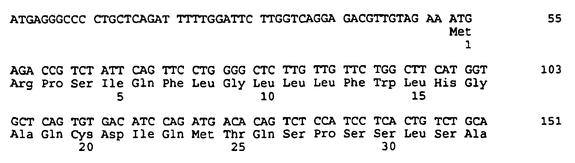
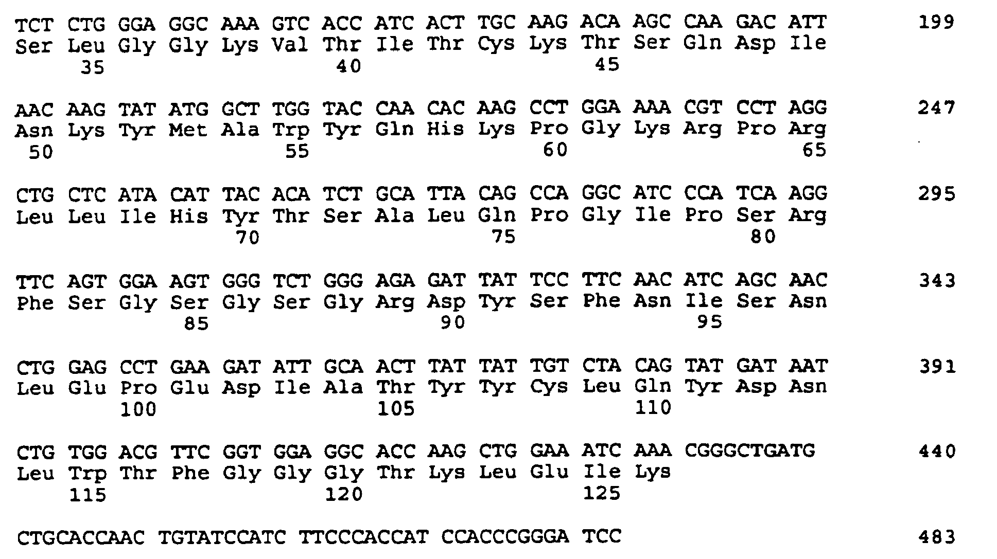
- (i) SEQUENCE CHARACTERISTICS:
- (2) INFORMATION FOR SEQ ID NO:2:
- (i) SEQUENCE CHARACTERISTICS:
- (A) LENGTH: 126 amino acids
- (B) TYPE: amino acid
- (D) TOPOLOGY: linear
- (ii) MOLECULE TYPE: protein
- (xi) SEQUENCE DESCRIPTION: SEQ ID NO:2:

- (i) SEQUENCE CHARACTERISTICS:
- (2) INFORMATION FOR SEQ ID NO:3:
- (i) SEQUENCE CHARACTERISTICS:
- (A) LENGTH: 470 base pairs
- (B) TYPE: nucleic acid
- (C) STRANDEDNESS: double
- (D) TOPOLOGY: linear
- (ii) MOLECULE TYPE: cDNA
- (ix) FEATURE:
- (A) NAME/KEY: CDS
- (B) LOCATION: 1..420
- (xi) SEQUENCE DESCRIPTION: SEQ ID NO:3:

- (i) SEQUENCE CHARACTERISTICS:
- (2) INFORMATION FOR SEQ ID NO:4:
- (i) SEQUENCE CHARACTERISTICS:
- (A) LENGTH: 140 amino acids
- (B) TYPE: amino acid
- (D) TOPOLOGY: linear
- (ii) MOLECULE TYPE: protein
- (xi) SEQUENCE DESCRIPTION: SEQ ID NO:4:

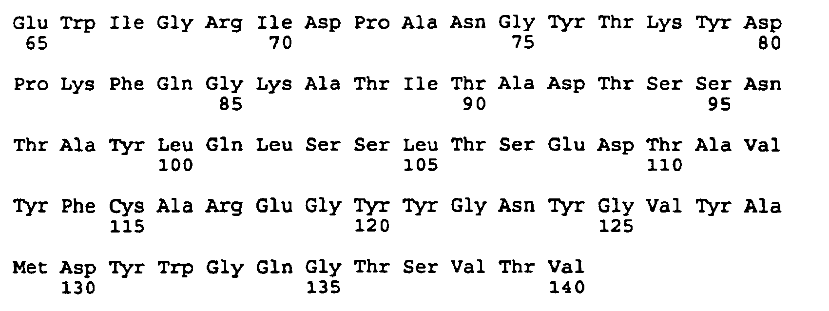
- (i) SEQUENCE CHARACTERISTICS:
- (2) INFORMATION FOR SEQ ID NO:5:
- (i) SEQUENCE CHARACTERISTICS:
- (A) LENGTH: 106 amino acids
- (B) TYPE: amino acid
- (C) STRANDEDNESS: single
- (D) TOPOLOGY: linear
- (ii) MOLECULE TYPE: protein
- (xi) SEQUENCE DESCRIPTION: SEQ ID NO:5:

- (i) SEQUENCE CHARACTERISTICS:
- (2) INFORMATION FOR SEQ ID NO:6:
- (i) SEQUENCE CHARACTERISTICS:
- (A) LENGTH: 107 amino acids
- (B) TYPE: amino acid
- (C) STRANDEDNESS: single
- (D) TOPOLOGY: linear
- (ii) MOLECULE TYPE: protein
- (xi) SEQUENCE DESCRIPTION: SEQ ID NO:6:


- (i) SEQUENCE CHARACTERISTICS:
- (2) INFORMATION FOR SEQ ID NO:7:
- (i) SEQUENCE CHARACTERISTICS:
- (A) LENGTH: 106 amino acids
- (B) TYPE: amino acid
- (C) STRANDEDNESS: single
- (D) TOPOLOGY: linear
- (ii) MOLECULE TYPE: protein
- (xi) SEQUENCE DESCRIPTION: SEQ ID NO:7:

- (i) SEQUENCE CHARACTERISTICS:
- (2) INFORMATION FOR SEQ ID NO:8:
- (i) SEQUENCE CHARACTERISTICS:
- (A) LENGTH: 107 amino acids
- (B) TYPE: amino acid
- (C) STRANDEDNESS: single
- (D) TOPOLOGY: linear
- (ii) MOLECULE TYPE: protein
- (xi) SEQUENCE DESCRIPTION: SEQ ID NO:8:


- (i) SEQUENCE CHARACTERISTICS:
- (2) INFORMATION FOR SEQ ID NO:9:
- (i) SEQUENCE CHARACTERISTICS:
- (A) LENGTH: 123 amino acids
- (B) TYPE: amino acid
- (C) STRANDEDNESS: single
- (D) TOPOLOGY: linear
- (ii) MOLECULE TYPE: protein
- (xi) SEQUENCE DESCRIPTION: SEQ ID NO:9:

- (i) SEQUENCE CHARACTERISTICS:
- (2) INFORMATION FOR SEQ ID NO:10:
- (i) SEQUENCE CHARACTERISTICS:
- (A) LENGTH: 119 amino acids
- (B) TYPE: amino acid
- (C) STRANDEDNESS: single
- (D) TOPOLOGY: linear
- (ii) MOLECULE TYPE: protein
- (xi) SEQUENCE DESCRIPTION: SEQ ID NO:10:


- (i) SEQUENCE CHARACTERISTICS:
- (2) INFORMATION FOR SEQ ID N0:11:
- (i) SEQUENCE CHARACTERISTICS:
- (A) LENGTH: 123 amino acids
- (B) TYPE: amino acid
- (C) STRANDEDNESS: single
- (D) TOPOLOGY: linear
- (ii) MOLECULE TYPE: protein
- (xi) SEQUENCE DESCRIPTION: SEQ ID NO:11:

- (i) SEQUENCE CHARACTERISTICS:
- (2) INFORMATION FOR SEQ ID NO:12:
- (i) SEQUENCE CHARACTERISTICS:
- (A) LENGTH: 119 amino acids
- (B) TYPE: amino acid
- (C) STRANDEDNESS: single
- (D) TOPOLOGY: linear
- (ii) MOLECULE TYPE: protein
- (xi) SEQUENCE DESCRIPTION: SEQ ID NO:12:


- (i) SEQUENCE CHARACTERISTICS:
- (2) INFORMATION FOR SEQ ID NO:13:
- (i) SEQUENCE CHARACTERISTICS:
- (A) LENGTH: 119 amino acids
- (B) TYPE: amino acid
- (C) STRANDEDNESS: single
- (D) TOPOLOGY: linear
- (ii) MOLECULE TYPE: protein
- (xi) SEQUENCE DESCRIPTION: SEQ ID NO:13:
- (i) SEQUENCE CHARACTERISTICS:
- (2) INFORMATION FOR SEQ ID NO:14:
- (i) SEQUENCE CHARACTERISTICS:
- (A) LENGTH: 406 base pairs
- (B) TYPE: nucleic acid
- (C) STRANDEDNESS: double
- (D) TOPOLOGY: linear
- (ii) MOLECULE TYPE: cDNA
- (ix) FEATURE:
- (A) NAME/KEY: CDS
- (B) LOCATION: 16..393
- (xi) SEQUENCE DESCRIPTION: SEQ ID NO:14:

- (i) SEQUENCE CHARACTERISTICS:
- (2) INFORMATION FOR SEQ ID NO:15:
- (i) SEQUENCE CHARACTERISTICS:
- (A) LENGTH: 126 amino acids
- (B) TYPE: amino acid
- (D) TOPOLOGY: linear
- (ii) MOLECULE TYPE: protein
- (xi) SEQUENCE DESCRIPTION: SEQ ID NO:15:


- (i) SEQUENCE CHARACTERISTICS:
- (2) INFORMATION FOR SEQ ID NO:16:
- (i) SEQUENCE CHARACTERISTICS:
- (A) LENGTH: 454 base pairs
- (B) TYPE: nucleic acid
- (C) STRANDEDNESS: single
- (D) TOPOLOGY: linear
- (ii) MOLECULE TYPE: cDNA
- (ix) FEATURE:
- (A) NAME/KEY: CDS
- (B) LOCATION: 16..441
- (xi) SEQUENCE DESCRIPTION: SEQ ID NO:16:

- (i) SEQUENCE CHARACTERISTICS:
- (2) INFORMATION FOR SEQ ID NO:17:
- (i) SEQUENCE CHARACTERISTICS:
- (A) LENGTH: 142 amino acids
- (B) TYPE: amino acid
- (D) TOPOLOGY: linear
- (ii) MOLECULE TYPE: protein
- (xi) SEQUENCE DESCRIPTION: SEQ ID NO:17:
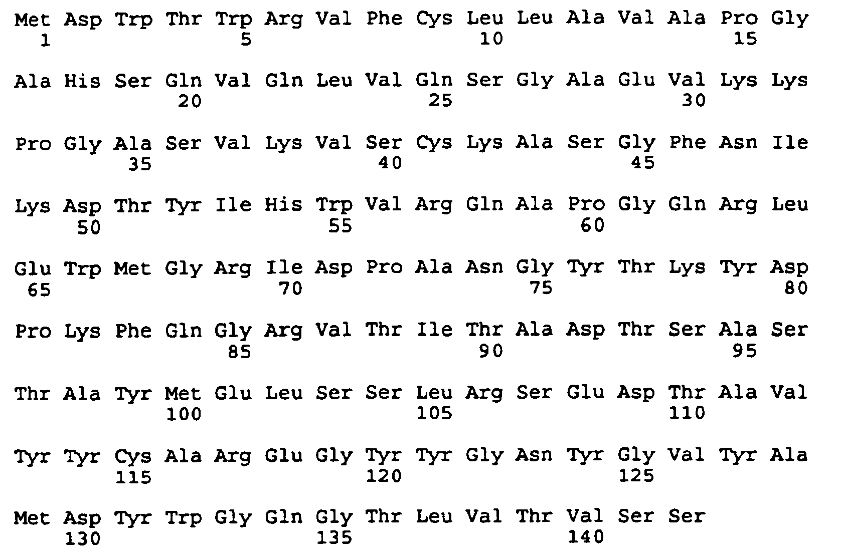
- (i) SEQUENCE CHARACTERISTICS:
- (2) INFORMATION FOR SEQ ID NO:18:
- (i) SEQUENCE CHARACTERISTICS:
- (A) LENGTH: 37 base pairs
- (B) TYPE: nucleic acid
- (C) STRANDEDNESS: single
- (D) TOPOLOGY: linear
- (ii) MOLECULE TYPE: DNA (primer)
- (xi) SEQUENCE DESCRIPTION: SEQ ID NO:18:
CAGAAAGCTT GCCGCCACCA TGAGACCGTC TATTCAG 37
- (i) SEQUENCE CHARACTERISTICS:
- (2) INFORMATION FOR SEQ ID NO:19:
- (i) SEQUENCE CHARACTERISTICS:
- (A) LENGTH: 35 base pairs
- (B) TYPE: nucleic acid
- (C) STRANDEDNESS: single
- (D) TOPOLOGY: linear
- (ii) MOLECULE TYPE: DNA (primer)
- (xi) SEQUENCE DESCRIPTION: SEQ ID NO:19:
CCGAGGATCC ACTCACGTTT GATTTCCAGC TTGGT 35
- (i) SEQUENCE CHARACTERISTICS:
- (2) INFORMATION FOR SEQ ID NO:20:
- (i) SEQUENCE CHARACTERISTICS:
- (A) LENGTH: 37 base pairs
- (B) TYPE: nucleic acid
- (C) STRANDEDNESS: single
- (D) TOPOLOGY: linear
- (ii) MOLECULE TYPE: DNA (primer)
- (xi) SEQUENCE DESCRIPTION: SEQ ID NO:20:
CAGAAAGCTT GCCGCCACCA TGAAATGCAG CTGGGTC 37
- (i) SEQUENCE CHARACTERISTICS:
- (2) INFORMATION FOR SEQ ID NO:21:
- (i) SEQUENCE CHARACTERISTICS:
- (A) LENGTH: 33 base pairs
- (B) TYPE: nucleic acid
- (C) STRANDEDNESS: single
- (D) TOPOLOGY: linear
- (ii) MOLECULE TYPE: DNA (primer)
- (xi) SEQUENCE DESCRIPTION: SEQ ID NO:21:
CCGAGGATCC ACTCACCTGA GGAGACGGTG ACT 33
- (i) SEQUENCE CHARACTERISTICS:
- (2) INFORMATION FOR SEQ ID NO:22:
- (i) SEQUENCE CHARACTERISTICS:
- (A) LENGTH: 39 base pairs
- (B) TYPE: nucleic acid
- (C) STRANDEDNESS: single
- (D) TOPOLOGY: linear
- (ii) MOLECULE TYPE: DNA (primer)
- (xi) SEQUENCE DESCRIPTION: SEQ ID NO:22:
GATGGTGACT CTATCTCCTA CAGATGCAGA CAGTGAGGA 39
- (i) SEQUENCE CHARACTERISTICS:
- (2) INFORMATION FOR SEQ ID NO:23:
- (i) SEQUENCE CHARACTERISTICS:
- (A) LENGTH: 32 base pairs
- (B) TYPE: nucleic acid
- (C) STRANDEDNESS: single
- (D) TOPOLOGY: linear
- (ii) MOLECULE TYPE: DNA (primer)
- (xi) SEQUENCE DESCRIPTION: SEQ ID NO:23:
CTGTAGGAGA TAGAGTCACC ATCACTTGCA AG 32
- (i) SEQUENCE CHARACTERISTICS:
- (2) INFORMATION FOR SEQ ID NO:24:
- (i) SEQUENCE CHARACTERISTICS:
- (A) LENGTH: 39 base pairs
- (B) TYPE: nucleic acid
- (C) STRANDEDNESS: single
- (D) TOPOLOGY: linear
- (ii) MOLECULE TYPE: DNA (primer)
- (xi) SEQUENCE DESCRIPTION: SEQ ID NO:24:
AGGAGCTTTT CCAGGTGTCT GTTGGTACCA AGCCATATA 39
- (i) SEQUENCE CHARACTERISTICS:
- (2) INFORMATION FOR SEQ ID NO:25:
- (i) SEQUENCE CHARACTERISTICS:
- (A) LENGTH: 41 base pairs
- (B) TYPE: nucleic acid
- (C) STRANDEDNESS: single
- (D) TOPOLOGY: linear
- (ii) MOLECULE TYPE: DNA (primer)
- (xi) SEQUENCE DESCRIPTION: SEQ ID NO:25:
ACCAACAGAC ACCTGGAAAA GCTCCTAGGC TGCTCATACA T 41
- (i) SEQUENCE CHARACTERISTICS:
- (2) INFORMATION FOR SEQ ID NO:26:
- (i) SEQUENCE CHARACTERISTICS:
- (A) LENGTH: 40 base pairs
- (B) TYPE: nucleic acid
- (C) STRANDEDNESS: single
- (D) TOPOLOGY: linear
- (ii) MOLECULE TYPE: DNA (primer)
- (xi) SEQUENCE DESCRIPTION: SEQ ID NO:26:
GCAGGCTGCTGATGGTGAAA GTATAATCTC TCCCAGACCC 40
- (i) SEQUENCE CHARACTERISTICS:
- (2) INFORMATION FOR SEQ ID NO:27:
- (i) SEQUENCE CHARACTERISTICS:
- (A) LENGTH: 42 base pairs
- (B) TYPE: nucleic acid
- (C) STRANDEDNESS: single
- (D) TOPOLOGY: linear
- (ii) MOLECULE TYPE: DNA (primer)
- (xi) SEQUENCE DESCRIPTION: SEQ ID NO:27:
ACTTTCACCA TCAGCAGCCT GCAGCCTGAA GATATTGCAA CT 42
- (i) SEQUENCE CHARACTERISTICS:
- (2) INFORMATION FOR SEQ ID NO:28:
- (i) SEQUENCE CHARACTERISTICS:
- (A) LENGTH: 59 base pairs
- (B) TYPE: nucleic acid
- (C) STRANDEDNESS: single
- (D) TOPOLOGY: linear
- (ii) MOLECULE TYPE: DNA (primer)
- (xi) SEQUENCE DESCRIPTION: SEQ ID NO:28:
CCGAGGATCC ACTCACGTTT GATTTCCACC TTGGTGCCTT GACCGAACGT CCACAGATT 59
- (i) SEQUENCE CHARACTERISTICS:
- (2) INFORMATION FOR SEQ ID NO:29:
- (i) SEQUENCE CHARACTERISTICS:
- (A) LENGTH: 33 base pairs
- (B) TYPE: nucleic acid
- (C) STRANDEDNESS: single
- (D) TOPOLOGY: linear
- (ii) MOLECULE TYPE: DNA (primer)
- (xi) SEQUENCE DESCRIPTION: SEQ ID NO:29:
GGAAAAGCTC CTAGGCTGCT CATATATTAC ACA 33
- (i) SEQUENCE CHARACTERISTICS:
- (2) INFORMATION FOR SEQ ID NO:30:
- (i) SEQUENCE CHARACTERISTICS:
- (A) LENGTH: 38 base pairs
- (B) TYPE: nucleic acid
- (C) STRANDEDNESS: single
- (D) TOPOLOGY: linear
- (ii) MOLECULE TYPE: DNA (primer)
- (xi) SEQUENCE DESCRIPTION: SEQ ID NO:30:
CCGAGGATCC ACTCACGTTT GATTTCCACC TTTGTGCC 38
- (i) SEQUENCE CHARACTERISTICS:
- (2) INFORMATION FOR SEQ ID NO:31:
- (i) SEQUENCE CHARACTERISTICS:
- (A) LENGTH: 51 base pairs
- (B) TYPE: nucleic acid
- (C) STRANDEDNESS: single
- (D) TOPOLOGY: linear
- (ii) MOLECULE TYPE: DNA (primer)
- (xi) SEQUENCE DESCRIPTION: SEQ ID NO:31:
AACCCAGTGT ATATAGGTGT CTTTAATGTT GAAACCGCTA GCTTTACAGC T 51
- (i) SEQUENCE CHARACTERISTICS:
- (2) INFORMATION FOR SEQ ID NO:32:
- (i) SEQUENCE CHARACTERISTICS:
- (A) LENGTH: 67 base pairs
- (B) TYPE: nucleic acid
- (C) STRANDEDNESS: single
- (D) TOPOLOGY: linear
- (ii) MOLECULE TYPE: DNA (primer)
- (xi) SEQUENCE DESCRIPTION: SEQ ID NO:32:

- (i) SEQUENCE CHARACTERISTICS:
- (2) INFORMATION FOR SEQ ID NO:33:
- (i) SEQUENCE CHARACTERISTICS:
- (A) LENGTH: 26 base pairs
- (B) TYPE: nucleic acid
- (C) STRANDEDNESS: single
- (D) TOPOLOGY: linear
- (ii) MOLECULE TYPE: DNA (primer)
- (xi) SEQUENCE DESCRIPTION: SEQ ID NO:33:
GACCCGGCCC TGGAACTTCG GGTCAT 26
- (i) SEQUENCE CHARACTERISTICS:
- (2) INFORMATION FOR SEQ ID NO:34:
- (i) SEQUENCE CHARACTERISTICS:
- (A) LENGTH: 66 base pairs
- (B) TYPE: nucleic acid
- (C) STRANDEDNESS: single
- (D) TOPOLOGY: linear
- (ii) MOLECULE TYPE: DNA (primer)
- (xi) SEQUENCE DESCRIPTION: SEQ ID NO:34:

- (i) SEQUENCE CHARACTERISTICS:
- (2) INFORMATION FOR SEQ ID NO:35:
- (i) SEQUENCE CHARACTERISTICS:
- (A) LENGTH: 64 base pairs
- (B) TYPE: nucleic acid
- (C) STRANDEDNESS: single
- (D) TOPOLOGY: linear
- (ii) MOLECULE TYPE: DNA (primer)
- (xi) SEQUENCE DESCRIPTION: SEQ ID NO:35:

- (i) SEQUENCE CHARACTERISTICS:
- (2) INFORMATION FOR SEQ ID NO:36:
- (i) SEQUENCE CHARACTERISTICS:
- (A) LENGTH: 63 base pairs
- (B) TYPE: nucleic acid
- (C) STRANDEDNESS: single
- (D) TOPOLOGY: linear
- (ii) MOLECULE TYPE: DNA (primer)
- (xi) SEQUENCE DESCRIPTION: SEQ ID NO:36:

- (i) SEQUENCE CHARACTERISTICS:
- (2) INFORMATION FOR SEQ ID NO:37:
- (i) SEQUENCE CHARACTERISTICS:
- (A) LENGTH: 37 base pairs
- (B) TYPE: nucleic acid
- (C) STRANDEDNESS: single
- (D) TOPOLOGY: linear
- (ii) MOLECULE TYPE: DNA (primer)
- (xi) SEQUENCE DESCRIPTION: SEQ ID NO:37:
CCAGGGCCGG GTCACCATCA CCAGAGACAC CTCTGCC 37
- (i) SEQUENCE CHARACTERISTICS:
- (2) INFORMATION FOR SEQ ID NO:38:
- (i) SEQUENCE CHARACTERISTICS:
- (A) LENGTH: 27 base pairs
- (B) TYPE: nucleic acid
- (C) STRANDEDNESS: single
- (D) TOPOLOGY: linear
- (ii) MOLECULE TYPE: DNA (primer)
- (xi) SEQUENCE DESCRIPTION: SEQ ID NO:38:
CAGGCCCCTG GCCAAGGGCT GGAGTGG 27
- (i) SEQUENCE CHARACTERISTICS:
- (2) INFORMATION FOR SEQ ID NO:39:
- (i) SEQUENCE CHARACTERISTICS:
- (A) LENGTH: 17 base pairs
- (B) TYPE: nucleic acid
- (C) STRANDEDNESS: single
- (D) TOPOLOGY: linear
- (ii) MOLECULE TYPE: DNA (primer)
- (xi) SEQUENCE DESCRIPTION: SEQ ID NO:39:
TACGCAAACC GCCTCTC 17
- (i) SEQUENCE CHARACTERISTICS:
- (2) INFORMATION FOR SEQ ID NO:40:
- (i) SEQUENCE CHARACTERISTICS:
- (A) LENGTH: 18 base pairs
- (B) TYPE: nucleic acid
- (C) STRANDEDNESS: single
- (D) TOPOLOGY: linear
- (ii) MOLECULE TYPE: DNA (primer)
- (xi) SEQUENCE DESCRIPTION: SEQ ID NO:40:
GAGTGCACCA TATGCGGT 18
- (i) SEQUENCE CHARACTERISTICS:
- (2) INFORMATION FOR SEQ ID NO:41:
- (i) SEQUENCE CHARACTERISTICS:
- (A) LENGTH: 116 amino acids
- (B) TYPE: amino acid
- (C) STRANDEDNESS: single
- (D) TOPOLOGY: linear
- (ii) MOLECULE TYPE: protein
- (xi) SEQUENCE DESCRIPTION: SEQ ID NO:41:

- (i) SEQUENCE CHARACTERISTICS:
- (2) INFORMATION FOR SEQ ID NO:42:
- (i) SEQUENCE CHARACTERISTICS:
- (A) LENGTH: 109 amino acids
- (B) TYPE: amino acid
- (C) STRANDEDNESS: single
- (D) TOPOLOGY: linear
- (ii) MOLECULE TYPE: protein
- (xi) SEQUENCE DESCRIPTION: SEQ ID NO:42:


- (i) SEQUENCE CHARACTERISTICS:
- (2) INFORMATION FOR SEQ ID NO:43:
- (i) SEQUENCE CHARACTERISTICS:
- (A) LENGTH: 114 amino acids
- (B) TYPE: amino acid
- (C) STRANDEDNESS: single
- (D) TOPOLOGY: linear
- (ii) MOLECULE TYPE: protein
- (xi) SEQUENCE DESCRIPTION: SEQ ID NO:43:

- (i) SEQUENCE CHARACTERISTICS:
- (2) INFORMATION FOR SEQ ID NO:44:
- (i) SEQUENCE CHARACTERISTICS:
- (A) LENGTH: 125 amino acids
- (B) TYPE: amino acid
- (C) STRANDEDNESS: single
- (D) TOPOLOGY: linear
- (ii) MOLECULE TYPE: protein
- (xi) SEQUENCE DESCRIPTION: SEQ ID NO:44:


- (i) SEQUENCE CHARACTERISTICS:
- (2) INFORMATION FOR SEQ ID NO:45:
- (i) SEQUENCE CHARACTERISTICS:
- (A) LENGTH: 129 amino acids
- (B) TYPE: amino acid
- (C) STRANDEDNESS: single
- (D) TOPOLOGY: linear
- (ii) MOLECULE TYPE: protein
- (xi) SEQUENCE DESCRIPTION: SEQ ID NO:45:
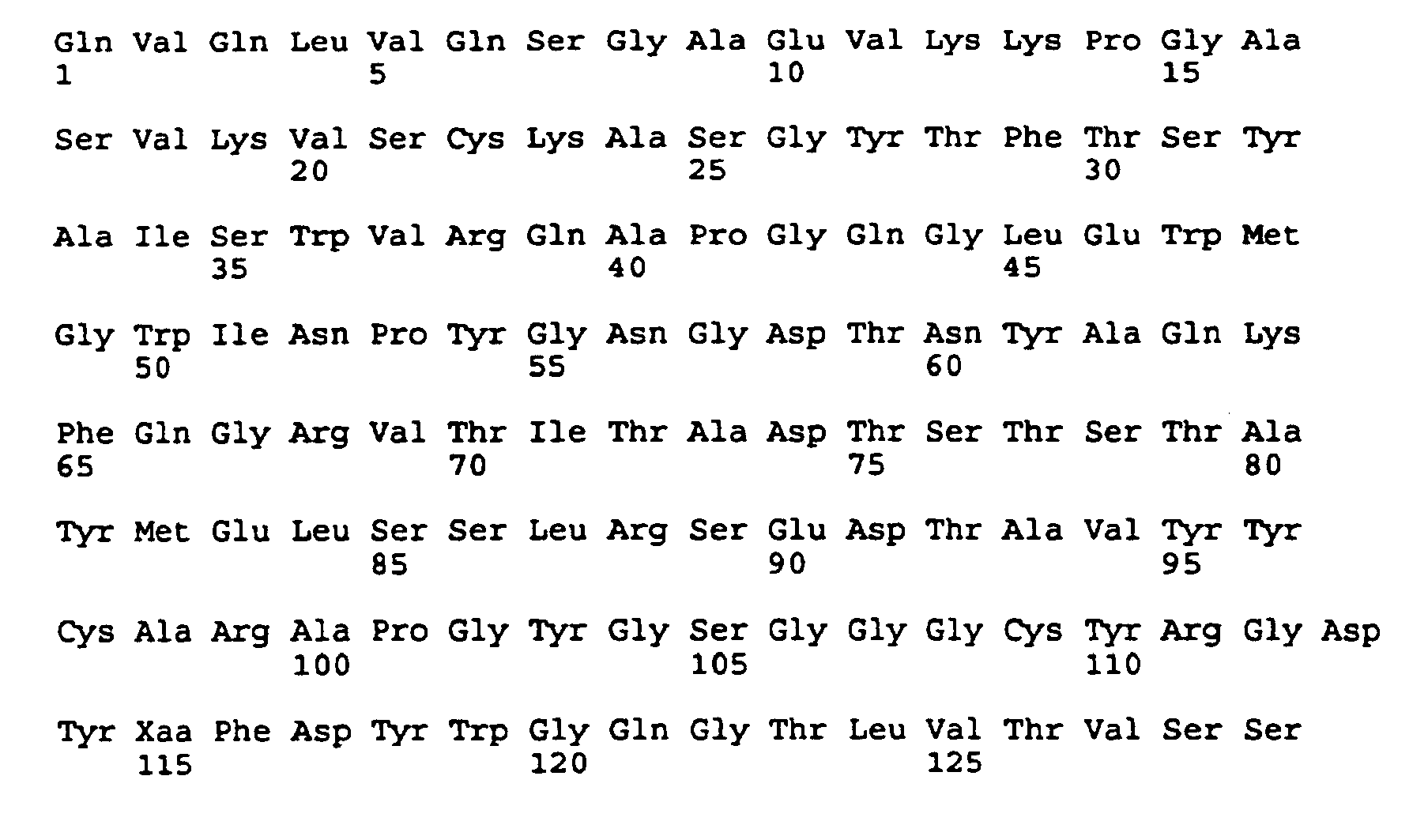
- (i) SEQUENCE CHARACTERISTICS:
Claims (16)
- A humanized immunoglobulin comprising a humanized heavy chain and a humanized light chain:(1) the humanized light chain comprising three complementarity determining regions CDR1, CDR2 and CDR3 having amino acid sequences from the corresponding complementarity determining regions of a mouse 21.6 immunoglobulin light chain SEQ ID NO:2 as shown in Figure 1, and a variable region framework from a human kappa light chain variable region framework sequence except in at least one position selected from a first group consisting of L45, L49, L58 and L69, wherein the amino acid position is occupied by the same amino acid present in the equivalent position of the mouse 21.6 immunoglobulin light chain variable region framework; and(2) the humanized heavy chain comprising three complementarity determining regions CDR1, CDR2 and CDR3 having amino acid sequences from the corresponding complementarity determining regions of a mouse 21.6 immunoglobulin heavy chain SEQ ID NO:4 as shown in Figure 2, and a variable region framework from a human heavy chain variable region framework sequence except in at least one position selected from a group consisting of H27, H28, H29, H30, H44, H71, wherein the amino acid position is occupied by the same amino acid present in the equivalent position of the mouse 21.6 immunoglobulin heavy chain variable region framework;wherein the immunoglobulin specifically binds to VLA-4 with a binding affinity having a lower limit of 107 M-1 and an upper limit of five-times the binding affinity of the mouse 21.6 immunoglobulin having the light chain of SEQ ID NO:2 and the heavy chain of SEQ ID NO:4.
- A humanized immunoglobulin as claimed in claim 1, wherein the humanized light chain variable region framework is from an RE1 variable region framework sequence except in at least one position selected from the first group, and except in at least one position selected from a third group consisting of positions L104, L105 and L107, wherein the amino acid position is occupied by the same amino acid present in the equivalent position of a kappa light chain from a human immunoglobulin other than RE1.
- A humanized immunoglobulin as claimed in claim 1 or claim 2, wherein the humanized heavy chain variable region framework is from a 21/28'CL variable region framework sequence.
- A humanized immunoglobulin as claimed in any one of claims 1 to 3, wherein the humanized light chain variable region framework comprises at least three amino acids from the mouse 21.6 immunoglobulin at positions in the first group and three amino acids from the kappa light chain from the human immunoglobulin other than REI at positions in the third group, and the humanized heavy chain variable region framework comprises at least five amino acids from the mouse 21.6 immunoglobulin at positions in the second group.
- A humanized immunoglobulin as claimed in claim 4, wherein the humanized light chain variable region framework is identical to the REI light chain variable region framework sequence except for the at least three positions from the first group and the three positions from the third group, and the heavy chain variable region framework is identical to the 21/28'CL heavy chain variable region framework sequence except for the at least five positions from the second group.
- A humanized immunoglobulin as claimed in claim 5, wherein the at least three positions from the first group are positions L45, L58 and L69, and at the least five positions from the second group are positions H27, H28, H29, H30 and H71.
- A humanized immunoglobulin as claimed in claim 6, wherein the humanized light chain comprises complementarity determining regions that are identical to the corresponding complementarity determining regions of the mouse 21.6 heavy chain, and the humanized heavy chain comprises complementarity determining regions that are identical to the corresponding complementarity determining regions of the mouse 21.6 heavy chain, except that the CDR3 region of the humanized heavy chain may or may not comprise a phenylalanine residue at position H98.
- A humanized immunoglobulin as claimed in claim 7, wherein the CDR3 of the humanized heavy chain comprises a phenylalanine residue at position H98.
- A humanized immunoglobulin as claimed in claim 1, wherein the amino acid sequence of the mature light chain variable region is the sequence designated La (SEQ ID NO:7) in Fig 6 or the sequence designated Lb (SEQ ID NO:8) in Fig 6.
- A humanized immunoglobulin as claimed in claim 1 or claim 9, wherein the amino acid sequence of the mature heavy chain variable region is selected from: Ha (SEQ ID NO:11), Hb (SEQ ID NO:12) or Hc (SEQ ID NO:13) as shown in Fig 7.
- A humanized immunoglobulin as claimed in claim 10 having a constant region domain, the constant region domain either having an effector function or lacking an effector function, preferably said effector function being capable of complement fixation or antibody dependent cellular toxicity.
- A nucleic acid encodinga) a heavy chain of a humanised antibody of any one of claims 1 to 11 orb) a light chain of a humanized antibody of any one of claims 1 to 11 .
- A pharmaceutical composition comprising a humanized antibody of any one of claims 1 to 11, and a pharmaceutically acceptable carrier therefor.
- A method for detecting VLA-4 antigen, the method comprising contacting a humanized immunoglobulin of any one of claims 1 to 11, with a tissue sample taken from a patient and detecting complexes formed by specific binding between the antibody and VLA-4 present in the target sample.
- The use of a humanised antibody as claimed in any one of claims 1 to 11 for the manufacture of a medicament for the treatment of an inflammatory disease.
- The use as claimed in claim 15, wherein the disease is multiple sclerosis.
Priority Applications (2)
| Application Number | Priority Date | Filing Date | Title |
|---|---|---|---|
| DK06013916.9T DK1759709T3 (en) | 1994-01-25 | 1995-01-25 | Humanized antibodies to leukocyte adhesion molecule VLA-4 |
| EP06013916A EP1759709B1 (en) | 1994-01-25 | 1995-01-25 | Humanized antibodies against leukocyte adhesion molecule VLA-4 |
Applications Claiming Priority (3)
| Application Number | Priority Date | Filing Date | Title |
|---|---|---|---|
| US18626994A | 1994-01-25 | 1994-01-25 | |
| US186269 | 1994-01-25 | ||
| PCT/US1995/001219 WO1995019790A1 (en) | 1994-01-25 | 1995-01-25 | Humanized antibodies against leukocyte adhesion molecule vla-4 |
Related Child Applications (1)
| Application Number | Title | Priority Date | Filing Date |
|---|---|---|---|
| EP06013916A Division EP1759709B1 (en) | 1994-01-25 | 1995-01-25 | Humanized antibodies against leukocyte adhesion molecule VLA-4 |
Publications (4)
| Publication Number | Publication Date |
|---|---|
| EP0804237A1 EP0804237A1 (en) | 1997-11-05 |
| EP0804237A4 EP0804237A4 (en) | 2003-04-16 |
| EP0804237B1 true EP0804237B1 (en) | 2006-07-26 |
| EP0804237B8 EP0804237B8 (en) | 2006-11-08 |
Family
ID=22684288
Family Applications (2)
| Application Number | Title | Priority Date | Filing Date |
|---|---|---|---|
| EP06013916A Expired - Lifetime EP1759709B1 (en) | 1994-01-25 | 1995-01-25 | Humanized antibodies against leukocyte adhesion molecule VLA-4 |
| EP95908741A Expired - Lifetime EP0804237B8 (en) | 1994-01-25 | 1995-01-25 | Humanized antibodies against leukocyte adhesion molecule vla-4 |
Family Applications Before (1)
| Application Number | Title | Priority Date | Filing Date |
|---|---|---|---|
| EP06013916A Expired - Lifetime EP1759709B1 (en) | 1994-01-25 | 1995-01-25 | Humanized antibodies against leukocyte adhesion molecule VLA-4 |
Country Status (21)
| Country | Link |
|---|---|
| EP (2) | EP1759709B1 (en) |
| JP (5) | JP4115517B2 (en) |
| KR (1) | KR100367948B1 (en) |
| CN (1) | CN1211123C (en) |
| AT (1) | ATE333895T1 (en) |
| AU (1) | AU703152B2 (en) |
| CA (1) | CA2182013C (en) |
| DE (2) | DE69535133T2 (en) |
| DK (2) | DK0804237T3 (en) |
| ES (2) | ES2270425T3 (en) |
| FI (1) | FI117509B (en) |
| FR (1) | FR06C0024I2 (en) |
| HU (1) | HU220799B1 (en) |
| LU (1) | LU91271I2 (en) |
| MX (1) | MX9602971A (en) |
| NL (1) | NL300238I2 (en) |
| NO (3) | NO319867B1 (en) |
| NZ (1) | NZ279730A (en) |
| PL (1) | PL181827B1 (en) |
| PT (1) | PT804237E (en) |
| WO (1) | WO1995019790A1 (en) |
Families Citing this family (52)
| Publication number | Priority date | Publication date | Assignee | Title |
|---|---|---|---|---|
| US6307025B1 (en) | 1989-04-28 | 2001-10-23 | Biogen, Inc. | VCAM fusion proteins and DNA coding therefor |
| US5871734A (en) * | 1992-01-13 | 1999-02-16 | Biogen, Inc. | Treatment for asthma with VLA-4 blocking agents |
| US5932214A (en) * | 1994-08-11 | 1999-08-03 | Biogen, Inc. | Treatment for inflammatory bowel disease with VLA-4 blockers |
| CA2153692C (en) | 1993-01-12 | 2011-11-08 | Roy R. Lobb | Recombinant anti-vla4 antibody molecules |
| ES2114183T5 (en) * | 1993-02-09 | 2006-06-16 | Biogen Idec Ma, Inc. | ANTIBODY FOR THE TREATMENT OF INSULIN DEPENDENT DIABETES. |
| US7435802B2 (en) | 1994-01-25 | 2008-10-14 | Elan Pharaceuticals, Inc. | Humanized anti-VLA4 immunoglobulins |
| US5840299A (en) * | 1994-01-25 | 1998-11-24 | Athena Neurosciences, Inc. | Humanized antibodies against leukocyte adhesion molecule VLA-4 |
| US7750137B2 (en) | 1995-09-01 | 2010-07-06 | Millennium Pharmaceuticals, Inc. | Mucosal vascular addressins |
| US6551593B1 (en) | 1995-02-10 | 2003-04-22 | Millennium Pharmaceuticals, Inc. | Treatment of Inflammatory bowel disease by inhibiting binding and/or signalling through α 4 β 7 and its ligands and madcam |
| US7803904B2 (en) | 1995-09-01 | 2010-09-28 | Millennium Pharmaceuticals, Inc. | Mucosal vascular addressing and uses thereof |
| US7147851B1 (en) | 1996-08-15 | 2006-12-12 | Millennium Pharmaceuticals, Inc. | Humanized immunoglobulin reactive with α4β7 integrin |
| AU7348798A (en) * | 1997-05-09 | 1998-12-08 | John P. Robarts Research Institute, The | Method for dislodging infiltrated leukocytes from a tissue |
| DE19741235A1 (en) | 1997-09-18 | 1999-03-25 | Hoechst Marion Roussel De Gmbh | Novel imidazolidine derivatives, their preparation, their use and pharmaceutical compositions containing them |
| DE19741873A1 (en) * | 1997-09-23 | 1999-03-25 | Hoechst Marion Roussel De Gmbh | New 5-ring heterocycles, their preparation, their use and pharmaceutical preparations containing them |
| DE19751251A1 (en) | 1997-11-19 | 1999-05-20 | Hoechst Marion Roussel De Gmbh | Substituted imidazolidine derivatives, their manufacture, their use and pharmaceutical preparations containing them |
| DE19821483A1 (en) | 1998-05-14 | 1999-11-18 | Hoechst Marion Roussel De Gmbh | New imidazolidine derivatives useful as leukocyte adhesion and migration inhibitors and/or VLA-4 receptor antagonists for treating E.G. inflammatory and allergic disorders |
| KR100628818B1 (en) * | 1998-09-14 | 2006-09-27 | 보드 오브 리전츠, 더 유니버시티 오브 텍사스 시스템 | How to treat multiple myeloma and bone resorption by myeloma using integrin antagonists |
| US7618630B2 (en) | 1998-09-14 | 2009-11-17 | Board Of Regents, The University Of Texas System | Methods of treating multiple myeloma and myeloma-induced bone resorption using integrin antagonists |
| PT1173201E (en) * | 1999-04-22 | 2005-08-31 | Biogen Idec Inc | METHOD FOR THE TREATMENT OF FIBROSIS USING AN ALTAGONIST OF THE ALPHA 4 SUBSTANCE OF INTEGRINA |
| DE19922462A1 (en) | 1999-05-17 | 2000-11-23 | Aventis Pharma Gmbh | New phenylureidobenzyl-substituted spiro-imidazolidinedione derivatives, are inhibitors of leukocyte adhesion or migration or VLA-4 receptors, useful e.g. for treating inflammatory or allergic disease |
| DK2314315T3 (en) | 1999-06-01 | 2015-02-02 | Biogen Idec Inc | Blocking monoclonal antibody to the human alpha1-I domain of VLA-1 and their use for the treatment of inflammatory diseases |
| US6949245B1 (en) | 1999-06-25 | 2005-09-27 | Genentech, Inc. | Humanized anti-ErbB2 antibodies and treatment with anti-ErbB2 antibodies |
| AU783110B2 (en) * | 1999-12-16 | 2005-09-22 | Biogen Ma Inc. | Methods of treating central nervous system ischemic or hemorrhagic injury using anti alpha4 integrin antagonists |
| DE10111877A1 (en) | 2001-03-10 | 2002-09-12 | Aventis Pharma Gmbh | Novel imidazolidine derivatives, their preparation, their use and pharmaceutical compositions containing them |
| MXPA03009390A (en) | 2001-04-13 | 2004-01-29 | Biogen Inc | Antibodies to vla-1. |
| DE10137595A1 (en) | 2001-08-01 | 2003-02-13 | Aventis Pharma Gmbh | New 3-alkylaminoalkyl-imdazolidin-4-one derivatives, are VLA-4 receptor and leukocyte adhesion and/or migration inhibitors, useful e.g. for treating inflammatory, allergic, autoimmune or cardiovascular disease |
| AR038605A1 (en) | 2002-02-25 | 2005-01-19 | Elan Pharm Inc | USE OF AGENTS FOR THE MANUFACTURE OF A MEDICINAL PRODUCT FOR THE TREATMENT OF INFLAMMATION AND COMPOSITIONS FOR SUCH TREATMENT |
| EP2270049A3 (en) * | 2002-04-12 | 2011-03-09 | Medimmune, Inc. | Recombinant anti-interleukin-9-antibody |
| GB0210121D0 (en) * | 2002-05-02 | 2002-06-12 | Celltech R&D Ltd | Biological products |
| TWI323265B (en) * | 2002-08-06 | 2010-04-11 | Glaxo Group Ltd | Antibodies |
| JP4494977B2 (en) * | 2002-12-17 | 2010-06-30 | メルク パテント ゲゼルシャフト ミット ベシュレンクテル ハフツング | Humanized antibody (H14.18) of mouse 14.18 antibody that binds to GD2 and its IL-2 fusion protein |
| CU23403A1 (en) * | 2003-04-23 | 2009-08-04 | Centro Inmunologia Molecular | RECOMBINANT ANTIBODIES AND FRAGMENTS RECOGNIZING GANGLIOSIDE N-GLICOLIL GM3 AND ITS USE FOR DIAGNOSIS AND TUMOR TREATMENT |
| KR20110098980A (en) | 2003-12-22 | 2011-09-02 | 아지노모토 가부시키가이샤 | Novel Phenylalanine Derivatives |
| WO2006055871A2 (en) * | 2004-11-19 | 2006-05-26 | Biogen Idec Ma Inc. | Treatment for multiple sclerosis |
| AU2006316629A1 (en) * | 2005-11-17 | 2007-05-31 | Millennium Pharmaceuticals, Inc. | Humanized immunoglobulin reactive with alpha 4 beta 7 integrin |
| EP2839843B1 (en) | 2006-05-25 | 2018-06-20 | Biogen MA Inc. | VLA-1 antagonist for use in treating stroke |
| US20100150915A1 (en) | 2007-02-20 | 2010-06-17 | Stewart Edward J | Methods of treating multiple sclerosis by administration of alpha-fetoprotein in combination with an integrin antagonist |
| ES2707815T3 (en) * | 2007-06-14 | 2019-04-05 | Biogen Ma Inc | Antibody formulations natalizumab |
| KR101449521B1 (en) | 2007-12-13 | 2014-10-13 | 가부시끼가이샤 도꾸야마 | Photochromic curable composition and cured body |
| EA022201B1 (en) | 2008-04-11 | 2015-11-30 | Мерримак Фармасьютикалз, Инк. | Agent capable for binding to tumor cell comprising human serum albumin (hsa) linker and use thereof |
| EP2408816B1 (en) | 2009-03-20 | 2019-09-04 | Amgen Inc. | Alpha-4-beta-7 heterodimer specific antagonist antibody |
| EP2467159A1 (en) | 2009-08-20 | 2012-06-27 | INSERM (Institut National de la Santé et de la Recherche Médicale) | Vla-4 as a biomarker for prognosis and target for therapy in duchenne muscular dystrophy |
| HUE050858T2 (en) | 2010-04-16 | 2021-01-28 | Biogen Ma Inc | Antibodies against VLA-4 |
| US10040855B2 (en) | 2011-05-02 | 2018-08-07 | Millennium Pharmaceuticals, Inc. | Formulation for anti-α4β7 antibody |
| UA116189C2 (en) | 2011-05-02 | 2018-02-26 | Мілленніум Фармасьютікалз, Інк. | COMPOSITION OF ANTI-α4β7 ANTIBODY |
| JP6345123B2 (en) | 2012-02-16 | 2018-06-20 | サンタラス, インコーポレイテッド | Anti-VLA1 (CD49A) antibody pharmaceutical composition |
| US20190225694A1 (en) | 2016-06-28 | 2019-07-25 | Zaklady Farmaceutyczne Polpharma Sa | Recombinant production of monoclonal antibodies |
| US20190374639A1 (en) | 2016-11-21 | 2019-12-12 | Polpharma Biologics S.A. | Aqueous pharmaceutical formulations |
| EP4659808A3 (en) * | 2017-02-17 | 2026-03-11 | Sanofi | Multispecific binding molecules having specificity to dystroglycan and laminin-2 |
| US12043845B2 (en) | 2017-06-08 | 2024-07-23 | Polpharma Biologics S.A. | Methods of cell culture |
| KR102848349B1 (en) | 2018-06-04 | 2025-08-21 | 바이오젠 엠에이 인코포레이티드 | Anti-VLA-4 antibodies with reduced effector function |
| WO2022162164A1 (en) | 2021-01-29 | 2022-08-04 | INSERM (Institut National de la Santé et de la Recherche Médicale) | Methods of assessing the risk of developing progressive multifocal leukoencephalopathy in patients treated with vla-4 antagonists |
Family Cites Families (6)
| Publication number | Priority date | Publication date | Assignee | Title |
|---|---|---|---|---|
| US5225539A (en) | 1986-03-27 | 1993-07-06 | Medical Research Council | Recombinant altered antibodies and methods of making altered antibodies |
| IL162181A (en) * | 1988-12-28 | 2006-04-10 | Pdl Biopharma Inc | A method of producing humanized immunoglubulin, and polynucleotides encoding the same |
| IL95501A (en) * | 1989-09-01 | 1997-04-15 | Hutchinson Fred Cancer Res | Inhibition by antibodies of lymphocyte adherence to vascular endothelium utilizing an extracellular matrix receptor-ligand interaction, and pharmaceutical compositions containing said antibodies |
| ES2103468T3 (en) * | 1992-02-12 | 1997-09-16 | Biogen Inc | TREATMENT OF INTESTINAL INFLAMMATION. |
| CA2153692C (en) * | 1993-01-12 | 2011-11-08 | Roy R. Lobb | Recombinant anti-vla4 antibody molecules |
| AUPP647298A0 (en) | 1998-10-13 | 1998-11-05 | Keystone Retaining Wall Systems, Inc. | Retaining wall block |
-
1995
- 1995-01-25 AU AU16960/95A patent/AU703152B2/en not_active Expired
- 1995-01-25 EP EP06013916A patent/EP1759709B1/en not_active Expired - Lifetime
- 1995-01-25 PT PT95908741T patent/PT804237E/en unknown
- 1995-01-25 CN CNB951913549A patent/CN1211123C/en not_active Expired - Lifetime
- 1995-01-25 DK DK95908741T patent/DK0804237T3/en active
- 1995-01-25 DK DK06013916.9T patent/DK1759709T3/en active
- 1995-01-25 EP EP95908741A patent/EP0804237B8/en not_active Expired - Lifetime
- 1995-01-25 CA CA002182013A patent/CA2182013C/en not_active Expired - Lifetime
- 1995-01-25 KR KR1019960703987A patent/KR100367948B1/en not_active Expired - Lifetime
- 1995-01-25 PL PL95315634A patent/PL181827B1/en unknown
- 1995-01-25 ES ES95908741T patent/ES2270425T3/en not_active Expired - Lifetime
- 1995-01-25 NZ NZ279730A patent/NZ279730A/en not_active IP Right Cessation
- 1995-01-25 WO PCT/US1995/001219 patent/WO1995019790A1/en not_active Ceased
- 1995-01-25 DE DE69535133T patent/DE69535133T2/en not_active Expired - Lifetime
- 1995-01-25 HU HU9602019A patent/HU220799B1/en active Protection Beyond IP Right Term
- 1995-01-25 AT AT95908741T patent/ATE333895T1/en active
- 1995-01-25 ES ES06013916T patent/ES2424292T3/en not_active Expired - Lifetime
- 1995-01-25 DE DE1995635133 patent/DE122006000043I1/en active Pending
- 1995-01-25 JP JP51977095A patent/JP4115517B2/en not_active Expired - Lifetime
-
1996
- 1996-07-24 MX MX9602971A patent/MX9602971A/en unknown
- 1996-07-24 NO NO19963097A patent/NO319867B1/en not_active IP Right Cessation
- 1996-07-24 FI FI962958A patent/FI117509B/en active Protection Beyond IP Right Term
-
2005
- 2005-08-05 JP JP2005228930A patent/JP2006045237A/en not_active Withdrawn
-
2006
- 2006-07-27 FR FR06C0024C patent/FR06C0024I2/en active Active
- 2006-08-02 NL NL300238C patent/NL300238I2/en unknown
- 2006-08-02 LU LU91271C patent/LU91271I2/en unknown
- 2006-08-07 NO NO2006008C patent/NO2006008I2/en unknown
-
2009
- 2009-05-18 JP JP2009120421A patent/JP2009235078A/en not_active Withdrawn
-
2013
- 2013-02-18 JP JP2013028635A patent/JP5487382B2/en not_active Expired - Lifetime
- 2013-03-18 JP JP2013054628A patent/JP2013165718A/en not_active Withdrawn
-
2017
- 2017-08-01 NO NO2017040C patent/NO2017040I2/en unknown
Also Published As
Similar Documents
| Publication | Publication Date | Title |
|---|---|---|
| EP0804237B1 (en) | Humanized antibodies against leukocyte adhesion molecule vla-4 | |
| US5840299A (en) | Humanized antibodies against leukocyte adhesion molecule VLA-4 | |
| US8246958B2 (en) | Methods of inhibiting alpha-4-dependent interactions with VCAM-1 with anti-VLA-4 antibodies | |
| CA2153692C (en) | Recombinant anti-vla4 antibody molecules | |
| US6204007B1 (en) | Antibodies against E-selectin | |
| AU747898B2 (en) | Monoclonal antibodies to human CD6 | |
| HK1011031B (en) | Recombinant anti-vla4 antibody molecules |
Legal Events
| Date | Code | Title | Description |
|---|---|---|---|
| PUAI | Public reference made under article 153(3) epc to a published international application that has entered the european phase |
Free format text: ORIGINAL CODE: 0009012 |
|
| 17P | Request for examination filed |
Effective date: 19960826 |
|
| AK | Designated contracting states |
Kind code of ref document: A1 Designated state(s): AT BE CH DE DK ES FR GB GR IE IT LI LU MC NL PT SE |
|
| A4 | Supplementary search report drawn up and despatched |
Effective date: 20030228 |
|
| 17Q | First examination report despatched |
Effective date: 20030722 |
|
| GRAP | Despatch of communication of intention to grant a patent |
Free format text: ORIGINAL CODE: EPIDOSNIGR1 |
|
| RIN1 | Information on inventor provided before grant (corrected) |
Inventor name: JONES, TARRAN S. Inventor name: SALDANHA, JOSE Inventor name: LEGER, OLIVIER J. Inventor name: BENDIG, MARY M. |
|
| GRAS | Grant fee paid |
Free format text: ORIGINAL CODE: EPIDOSNIGR3 |
|
| RIN1 | Information on inventor provided before grant (corrected) |
Inventor name: JONES, TARRAN S. Inventor name: SALDANHA, JOSE Inventor name: LEGER, OLIVIER J. Inventor name: BENDIG, MARY M. |
|
| GRAA | (expected) grant |
Free format text: ORIGINAL CODE: 0009210 |
|
| AK | Designated contracting states |
Kind code of ref document: B1 Designated state(s): AT BE CH DE DK ES FR GB GR IE IT LI LU MC NL PT SE |
|
| PG25 | Lapsed in a contracting state [announced via postgrant information from national office to epo] |
Ref country code: IT Free format text: LAPSE BECAUSE OF FAILURE TO SUBMIT A TRANSLATION OF THE DESCRIPTION OR TO PAY THE FEE WITHIN THE PRE;WARNING: LAPSES OF ITALIAN PATENTS WITH EFFECTIVE DATE BEFORE 2007 MAY HAVE OCCURRED AT ANY TIME BEFORE 2007. THE CORRECT EFFECTIVE DATE MAY BE DIFFERENT FROM THE ONE RECORDED.SCRIBED TIME-LIMIT Effective date: 20060726 |
|
| REG | Reference to a national code |
Ref country code: GB Ref legal event code: FG4D |
|
| REG | Reference to a national code |
Ref country code: FR Ref legal event code: CP Free format text: PRODUCT NAME: NATALIZUMAB,ANTICORPS MONOCLONAL HUMANISE ANTI-INTEGRINE ALPHA-4; REGISTRATION NO/DATE: EU/1/06/346/001 20060627 Spc suppl protection certif: 06C0024 Filing date: 20060727 Ref country code: FR Ref legal event code: CP Free format text: PRODUCT NAME: NATALIZUMAB; REGISTRATION NO/DATE: EU/1/06/346/001 20060627 Spc suppl protection certif: 06C0024 Filing date: 20060727 |
|
| REG | Reference to a national code |
Ref country code: DE Ref legal event code: V448 Free format text: PRODUCT NAME: NATALIZUMAB; REGISTRATION NO/DATE: EU/1/06/346/001, 20060627 Spc suppl protection certif: 12 2006 000 043 Filing date: 20060810 |
|
| REG | Reference to a national code |
Ref country code: CH Ref legal event code: EP |
|
| RAP2 | Party data changed (patent owner data changed or rights of a patent transferred) |
Owner name: ELAN PHARMACEUTICALS, INC. |
|
| REG | Reference to a national code |
Ref country code: IE Ref legal event code: FG4D |
|
| RIN2 | Information on inventor provided after grant (corrected) |
Inventor name: YEDNOCK, THEODORE A. Inventor name: JONES, TARRAN S. Inventor name: SALDANHA, JOSE Inventor name: LEGER, OLIVIER J. Inventor name: BENDIG, MARY M. |
|
| REG | Reference to a national code |
Ref country code: GB Ref legal event code: CTFF Free format text: SPC/GB06/025: 20060727 Spc suppl protection certif: SPC/GB06/025 Filing date: 20060727 |
|
| REG | Reference to a national code |
Ref country code: IE Ref legal event code: SPCF Free format text: SPC025/2006: 20060731 Spc suppl protection certif: SPC025/2006 Filing date: 20060731 |
|
| REF | Corresponds to: |
Ref document number: 69535133 Country of ref document: DE Date of ref document: 20060907 Kind code of ref document: P |
|
| NLT2 | Nl: modifications (of names), taken from the european patent patent bulletin |
Owner name: ELAN PHARMACEUTICALS, INC. Effective date: 20060816 |
|
| REG | Reference to a national code |
Ref country code: LU Ref legal event code: CCP Free format text: 91271, EXPIRES: 20200125 Spc suppl protection certif: 91271 Expiry date: 20200125 Ref country code: NL Ref legal event code: AC1 Spc suppl protection certif: 300238 Filing date: 20060802 |
|
| REG | Reference to a national code |
Ref country code: DE Ref legal event code: 8327 Ref document number: 69535133 Country of ref document: DE Owner name: ELAN PHARMACEUTICALS, INC., SAN FRANCISCO, CALIF., |
|
| REG | Reference to a national code |
Ref country code: CH Ref legal event code: NV Representative=s name: E. BLUM & CO. PATENTANWAELTE Ref country code: FR Ref legal event code: CR Free format text: PRODUCT NAME: NATALIZUMAB, ANTICORPS MONOCLONAL HUMANISE ANTI-INTEGRINE ALPHA-4; REGISTRATION NO/DATE IN FRANCE: EU/1/06/346/001 DU 20060627; REGISTRATION NO/DATE AT EEC: EU/1/06/346/001 DU 20060627 Spc suppl protection certif: 06C0024 Filing date: 20060727 |
|
| REG | Reference to a national code |
Ref country code: DK Ref legal event code: T3 |
|
| REG | Reference to a national code |
Ref country code: DK Ref legal event code: CTFF Spc suppl protection certif: CA 2006 00025 Filing date: 20060912 Expiry date: 20200125 |
|
| REG | Reference to a national code |
Ref country code: PT Ref legal event code: SC4A Effective date: 20060817 |
|
| REG | Reference to a national code |
Ref country code: SE Ref legal event code: TRGR |
|
| REG | Reference to a national code |
Ref country code: GR Ref legal event code: EP Ref document number: 20060403478 Country of ref document: GR |
|
| REG | Reference to a national code |
Ref country code: IT Ref legal event code: SPCF Ref document number: 502006901455138 Country of ref document: IT Free format text: PRODUCT NAME: NATALIZUMAB(TYSABRI); AUTHORISATION NUMBER(S) AND DATE(S): EU/1/06/346/001, 20060627 Spc suppl protection certif: 132006901471186 |
|
| REG | Reference to a national code |
Ref country code: SE Ref legal event code: SPCF Free format text: PRODUCT NAME: NATALIZUMAB, HUMANISERAD MONOKLONAL ANTIKROPP MOT A4-INTEGRIN; NAT REG. NO/DATE: EG EU/1/06/346/001 20060627; FIRST REG.: EG EU/1/06/346/001 20060627 Spc suppl protection certif: 0690022-9 Filing date: 20060627 |
|
| REG | Reference to a national code |
Ref country code: FR Ref legal event code: CP |
|
| REG | Reference to a national code |
Ref country code: AT Ref legal event code: UEP Ref document number: 333895 Country of ref document: AT |
|
| REG | Reference to a national code |
Ref country code: AT Ref legal event code: ESZA Free format text: PRODUCT NAME: NATALIZUMAB Spc suppl protection certif: SZ 23/2006 Filing date: 20060727 |
|
| REG | Reference to a national code |
Ref country code: ES Ref legal event code: FG2A Ref document number: 2270425 Country of ref document: ES Kind code of ref document: T3 |
|
| NLXE | Nl: other communications concerning ep-patents (part 3 heading xe) |
Free format text: PAT. BUL. 01/2007, PAGE 39: CORR.: EL AN PHARMACEUTICALS, INC. |
|
| REG | Reference to a national code |
Ref country code: NL Ref legal event code: KC1 Free format text: 300238, 20150125, EXPIRES: 20200124 Spc suppl protection certif: 300238 Filing date: 20060802 Expiry date: 20200124 |
|
| REG | Reference to a national code |
Ref country code: IT Ref legal event code: SPCG Ref document number: 502006901455138 Country of ref document: IT Free format text: PRODUCT NAME: NATALIZUMAB(TYSABRI); AUTHORISATION NUMBER(S) AND DATE(S): EU/1/06/346/001, 20060627 Spc suppl protection certif: 132006901471186 Extension date: 20200725 |
|
| REG | Reference to a national code |
Ref country code: CH Ref legal event code: SPCF Free format text: PRODUCT NAME: NATALIZUMABUM; REGISTRATION NUMBER/DATE: SWISSMEDIC 57273 03.05.2007 Filing date: 20070508 |
|
| PLBE | No opposition filed within time limit |
Free format text: ORIGINAL CODE: 0009261 |
|
| STAA | Information on the status of an ep patent application or granted ep patent |
Free format text: STATUS: NO OPPOSITION FILED WITHIN TIME LIMIT |
|
| REG | Reference to a national code |
Ref country code: IE Ref legal event code: SPCG Free format text: SPC 025/2006: 20070523, EXPIRES: 20200124 Spc suppl protection certif: SPC 025/2006 Filing date: 20070523 Expiry date: 20200124 |
|
| 26N | No opposition filed |
Effective date: 20070427 |
|
| REG | Reference to a national code |
Ref country code: GB Ref legal event code: CTFG Free format text: SPC/GB06/025: 20070703, EXPIRES: 20200124 Spc suppl protection certif: SPC/GB06/025 Filing date: 20070703 Expiry date: 20200124 |
|
| REG | Reference to a national code |
Ref country code: DE Ref legal event code: 8364 Ref document number: 69535133 Country of ref document: DE |
|
| REG | Reference to a national code |
Ref country code: CH Ref legal event code: PFA Owner name: ELAN PHARMACEUTICALS, INC. Free format text: ELAN PHARMACEUTICALS, INC.#800 GATEWAY BOULEVARD#SOUTH SAN FRANCISCO, CA 94080 (US) -TRANSFER TO- ELAN PHARMACEUTICALS, INC.#800 GATEWAY BOULEVARD#SOUTH SAN FRANCISCO, CA 94080 (US) |
|
| REG | Reference to a national code |
Ref country code: AT Ref legal event code: EEZF Spc suppl protection certif: SZ 23/2006 Filing date: 20060727 Extension date: 20200125 Effective date: 20071008 |
|
| REG | Reference to a national code |
Ref country code: SE Ref legal event code: SPCG Free format text: 0690022-9 Spc suppl protection certif: 0690022-9 |
|
| REG | Reference to a national code |
Ref country code: DK Ref legal event code: CTFG Free format text: PRODUCT NAME: NATALIZUMAB Spc suppl protection certif: CA 2006 00025 Filing date: 20060912 |
|
| REG | Reference to a national code |
Ref country code: FR Ref legal event code: CY Free format text: PRODUCT NAME: NATALIZUMAB, ANTICORPS MONOCLONAL HUMANISE ANTI-INTEGRINE ALPHA-4; REGISTRATION NO/DATE IN FRANCE: EU/1/06/346/001 DU 20060627; REGISTRATION NO/DATE AT EEC: EU/1/06/346/001 DU 20060627 Spc suppl protection certif: 06C0024 Filing date: 20060727 Extension date: 20200124 |
|
| REG | Reference to a national code |
Ref country code: CH Ref legal event code: SPCG Free format text: PRODUCT NAME: NATALIZUMABUM; REGISTRATION NUMBER/DATE: SWISSMEDIC 57273 03.05.2007 Spc suppl protection certif: C00804237/01 Filing date: 20070508 Extension date: 20200124 |
|
| REG | Reference to a national code |
Ref country code: FR Ref legal event code: AV Spc suppl protection certif: 2006C0024 Ref country code: FR Ref legal event code: AV |
|
| REG | Reference to a national code |
Ref country code: DE Ref legal event code: R082 Ref document number: 69535133 Country of ref document: DE Representative=s name: MEYER & PARTNER GBR, DE |
|
| REG | Reference to a national code |
Ref country code: DE Ref legal event code: R082 Ref document number: 69535133 Country of ref document: DE Representative=s name: MEYER & PARTNER GBR, DE Effective date: 20130910 Ref country code: DE Ref legal event code: R081 Ref document number: 69535133 Country of ref document: DE Owner name: BIOGEN IDEC MA INC., CAMBRIDGE, US Free format text: FORMER OWNER: ELAN PHARMACEUTICALS, INC., SAN FRANCISCO, CALIF., US Effective date: 20130910 Ref country code: DE Ref legal event code: R081 Ref document number: 69535133 Country of ref document: DE Owner name: BIOGEN IDEC MA INC., US Free format text: FORMER OWNER: ELAN PHARMACEUTICALS, INC., SAN FRANCISCO, US Effective date: 20130910 |
|
| REG | Reference to a national code |
Ref country code: GB Ref legal event code: 732E Free format text: REGISTERED BETWEEN 20131010 AND 20131016 |
|
| PGFP | Annual fee paid to national office [announced via postgrant information from national office to epo] |
Ref country code: MC Payment date: 20131211 Year of fee payment: 20 |
|
| PGFP | Annual fee paid to national office [announced via postgrant information from national office to epo] |
Ref country code: LU Payment date: 20131227 Year of fee payment: 20 Ref country code: ES Payment date: 20131211 Year of fee payment: 20 Ref country code: GR Payment date: 20131216 Year of fee payment: 20 |
|
| PGFP | Annual fee paid to national office [announced via postgrant information from national office to epo] |
Ref country code: DK Payment date: 20140110 Year of fee payment: 20 Ref country code: IE Payment date: 20140110 Year of fee payment: 20 Ref country code: NL Payment date: 20140110 Year of fee payment: 20 Ref country code: BE Payment date: 20140114 Year of fee payment: 20 Ref country code: CH Payment date: 20140114 Year of fee payment: 20 Ref country code: SE Payment date: 20140113 Year of fee payment: 20 Ref country code: DE Payment date: 20140122 Year of fee payment: 20 |
|
| PGFP | Annual fee paid to national office [announced via postgrant information from national office to epo] |
Ref country code: FR Payment date: 20140108 Year of fee payment: 20 Ref country code: IT Payment date: 20140115 Year of fee payment: 20 Ref country code: AT Payment date: 20131224 Year of fee payment: 20 |
|
| PGFP | Annual fee paid to national office [announced via postgrant information from national office to epo] |
Ref country code: GB Payment date: 20140122 Year of fee payment: 20 Ref country code: PT Payment date: 20140109 Year of fee payment: 20 |
|
| REG | Reference to a national code |
Ref country code: DE Ref legal event code: R067 Ref document number: 69535133 Country of ref document: DE Free format text: PRODUCT NAME: NATALIZUMAB; NAT. REGISTRATION NO/DATE: EU /1/06/346/001 20060627; FIRST REGISTRATION: EU EU/1/06/346/001 20060627 Spc suppl protection certif: 122006000043 Filing date: 20060810 Expiry date: 20150126 Extension date: 20200125 |
|
| REG | Reference to a national code |
Ref country code: FR Ref legal event code: SPAY Free format text: PRODUCT NAME: NATALIZUMAB; REGISTRATION NO/DATE: EU/1/06/346/001 20060627 Spc suppl protection certif: 06C0024 Filing date: 20060727 Year of fee payment: 1 Extension date: 20200124 |
|
| REG | Reference to a national code |
Ref country code: DK Ref legal event code: EUP Effective date: 20150125 |
|
| REG | Reference to a national code |
Ref country code: DE Ref legal event code: R071 Ref document number: 69535133 Country of ref document: DE |
|
| REG | Reference to a national code |
Ref country code: CH Ref legal event code: PL |
|
| REG | Reference to a national code |
Ref country code: PT Ref legal event code: MM4A Free format text: MAXIMUM VALIDITY LIMIT REACHED Effective date: 20150125 |
|
| REG | Reference to a national code |
Ref country code: NL Ref legal event code: V4 Effective date: 20150125 |
|
| REG | Reference to a national code |
Ref country code: NL Ref legal event code: KC1 Free format text: PRODUCT NAME: NATALIZUMAB; REGISTRATION NO/DATE: EU/1/06/346/001 20060627 Spc suppl protection certif: C300238 Filing date: 20060802 Extension date: 20200124 |
|
| REG | Reference to a national code |
Ref country code: GB Ref legal event code: PE20 Expiry date: 20150124 Ref country code: GB Ref legal event code: CTFE Free format text: PRODUCT NAME: NATALIZUMAB, ANTI -4 INTEGRIN HUMANIZED MONOCLONAL ANTIBODY; REGISTERED: UK EU/1/06/346/001 20060627 Spc suppl protection certif: SPC/GB06/025 Filing date: 20060727 Expiry date: 20150125 Extension date: 20200124 |
|
| REG | Reference to a national code |
Ref country code: IE Ref legal event code: MK9A |
|
| REG | Reference to a national code |
Ref country code: SE Ref legal event code: EUG |
|
| REG | Reference to a national code |
Ref country code: AT Ref legal event code: MK07 Ref document number: 333895 Country of ref document: AT Kind code of ref document: T Effective date: 20150125 |
|
| REG | Reference to a national code |
Ref country code: ES Ref legal event code: FD2A Effective date: 20150401 |
|
| REG | Reference to a national code |
Ref country code: DE Ref legal event code: R069 Ref document number: 69535133 Country of ref document: DE Free format text: PRODUCT NAME: NATALIZUMAB; NAT. REGISTRATION NO/DATE: EU /1/06/346/001 20060627; FIRST REGISTRATION: EU EU/1/06/346/001 20060627 Spc suppl protection certif: 122006000043 Filing date: 20060810 Expiry date: 20150126 Extension date: 20200125 Effective date: 20150120 |
|
| REG | Reference to a national code |
Ref country code: GR Ref legal event code: MA Ref document number: 20060403478 Country of ref document: GR Effective date: 20150126 |
|
| PG25 | Lapsed in a contracting state [announced via postgrant information from national office to epo] |
Ref country code: IE Free format text: LAPSE BECAUSE OF EXPIRATION OF PROTECTION Effective date: 20150125 Ref country code: PT Free format text: LAPSE BECAUSE OF EXPIRATION OF PROTECTION Effective date: 20150203 Ref country code: ES Free format text: LAPSE BECAUSE OF EXPIRATION OF PROTECTION Effective date: 20150126 |
|
| PG25 | Lapsed in a contracting state [announced via postgrant information from national office to epo] |
Ref country code: GB Free format text: LAPSE BECAUSE OF EXPIRATION OF PROTECTION Effective date: 20150124 |
|
| REG | Reference to a national code |
Ref country code: FR Ref legal event code: SPAY Free format text: PRODUCT NAME: NATALIZUMAB; REGISTRATION NO/DATE: EU/1/06/346/001 20060627 Spc suppl protection certif: 06C0024 Filing date: 20060727 Year of fee payment: 2 Extension date: 20200124 |
|
| REG | Reference to a national code |
Ref country code: FR Ref legal event code: SPAY Free format text: PRODUCT NAME: NATALIZUMAB,ANTICORPS MONOCLONAL HUMANISE ANTI-INTEGRINE ALPHA-4; REGISTRATION NO/DATE: EU/1/06/346/001 20060627 Spc suppl protection certif: 06C0024 Filing date: 20060727 Year of fee payment: 3 Extension date: 20200124 Ref country code: FR Ref legal event code: SPAY Free format text: PRODUCT NAME: NATALIZUMAB; REGISTRATION NO/DATE: EU/1/06/346/001 20060627 Spc suppl protection certif: 06C0024 Filing date: 20060727 Year of fee payment: 3 Extension date: 20200124 |
|
| REG | Reference to a national code |
Ref country code: IE Ref legal event code: SPCE Free format text: PRODUCT NAME: NATALIZUMAB (ALSO KNOWN AS TYSABRI); REGISTRATION NO/DATE: EU/1/06/346/001 20060627 Spc suppl protection certif: 2006/025 Effective date: 20170308 |
|
| REG | Reference to a national code |
Ref country code: GB Ref legal event code: SPCY Free format text: PRODUCT NAME: NATALIZUMAB, ANTI -4 INTEGRIN HUMANIZED MONOCLONAL ANTIBODY; REGISTERED: UK EU/1/06/346/001 20060627 Spc suppl protection certif: SPC/GB06/025 Filing date: 20170307 Expiry date: 20150125 |
|
| REG | Reference to a national code |
Ref country code: DE Ref legal event code: R420 Ref document number: 69535133 Country of ref document: DE Free format text: PRODUCT NAME: NATALIZUMAB; NAT. REGISTRATION NO/DATE: EU /1/06/346/001 20060627; FIRST REGISTRATION: EU EU/1/06/346/001 20060627 Spc suppl protection certif: 122006000043 Filing date: 20060810 Expiry date: 20150126 Extension date: 20200725 Ref country code: DE Ref legal event code: R420 Ref document number: 69535133 Country of ref document: DE Free format text: PRODUCT NAME: NATALIZUMAB; NAT. REGISTRATION NO/DATE: EU /1/06/346/001 20060627; FIRST REGISTRATION: EU EU/1/06/346/001 20060627 Spc suppl protection certif: 122006000043 Filing date: 20060810 Expiry date: 20150126 Extension date: 20200125 |
|
| REG | Reference to a national code |
Ref country code: FR Ref legal event code: SPCT Owner name: BIOGEN MA INC., US Free format text: PRODUCT NAME: NATALIZUMAB,ANTICORPS MONOCLONAL HUMANISE ANTI-INTEGRINE ALPHA-4; REGISTRATION NO/DATE: EU/1/06/346/001 20060627 Spc suppl protection certif: 06C0024 Filing date: 20060727 Extension date: 20200724 Ref country code: FR Ref legal event code: SPCT Owner name: BIOGEN MA INC., US Free format text: PRODUCT NAME: NATALIZUMAB,ANTICORPS MONOCLONAL HUMANISE ANTI-INTEGRINE ALPHA-4; REGISTRATION NO/DATE: EU/1/06/346/001 20060627 Spc suppl protection certif: 06C0024 Filing date: 20060727 Extension date: 20200124 Ref country code: FR Ref legal event code: SPCT Owner name: BIOGEN MA INC., US Free format text: PRODUCT NAME: NATALIZUMAB; REGISTRATION NO/DATE: EU/1/06/346/001 20060627 Spc suppl protection certif: 06C0024 Filing date: 20060727 Extension date: 20200124 Ref country code: FR Ref legal event code: SPCT Owner name: ELAN PHARMACEUTICALS INC, US Free format text: PRODUCT NAME: NATALIZUMAB; REGISTRATION NO/DATE: EU/1/06/346/001 20060627 Spc suppl protection certif: 06C0024 Filing date: 20060727 Extension date: 20200124 Ref country code: FR Ref legal event code: SPCC Owner name: BIOGEN MA INC., US Free format text: PRODUCT NAME: NATALIZUMAB,ANTICORPS MONOCLONAL HUMANISE ANTI-INTEGRINE ALPHA-4; REGISTRATION NO/DATE: EU/1/06/346/001 20060627 Spc suppl protection certif: 06C0024 Filing date: 20060727 Extension date: 20200724 Ref country code: FR Ref legal event code: SPCC Owner name: BIOGEN MA INC., US Free format text: PRODUCT NAME: NATALIZUMAB,ANTICORPS MONOCLONAL HUMANISE ANTI-INTEGRINE ALPHA-4; REGISTRATION NO/DATE: EU/1/06/346/001 20060627 Spc suppl protection certif: 06C0024 Filing date: 20060727 Extension date: 20200124 Ref country code: FR Ref legal event code: SPCC Owner name: BIOGEN MA INC., US Free format text: PRODUCT NAME: NATALIZUMAB; REGISTRATION NO/DATE: EU/1/06/346/001 20060627 Spc suppl protection certif: 06C0024 Filing date: 20060727 Extension date: 20200124 Ref country code: FR Ref legal event code: SPCC Owner name: ELAN PHARMACEUTICALS INC, US Free format text: PRODUCT NAME: NATALIZUMAB; REGISTRATION NO/DATE: EU/1/06/346/001 20060627 Spc suppl protection certif: 06C0024 Filing date: 20060727 Extension date: 20200124 |
|
| REG | Reference to a national code |
Ref country code: CH Ref legal event code: SPCP Owner name: BIOGEN MA INC., US Free format text: FORMER OWNER: BIOGEN IDEC MA INC., US Spc suppl protection certif: C00804237/01 Ref country code: CH Ref legal event code: SPCM Owner name: BIOGEN IDEC INTERNATIONAL HOLDING LTD., BM Free format text: FORMER OWNER: ELAN PHARMACEUTICALS, INC., US Spc suppl protection certif: C00804237/01 Ref country code: CH Ref legal event code: SPCM Owner name: BIOGEN IDEC MA INC., US Free format text: FORMER OWNER: BIOGEN IDEC INTERNATIONAL HOLDING LTD., BM Spc suppl protection certif: C00804237/01 |
|
| REG | Reference to a national code |
Ref country code: GB Ref legal event code: SPCZ Free format text: PRODUCT NAME: NATALIZUMAB, ANTI -4 INTEGRIN HUMANIZED MONOCLONAL ANTIBODY; REGISTERED: UK EU/1/06/346/001 20060627 Spc suppl protection certif: SPC/GB06/025 Filing date: 20170307 Expiry date: 20150325 Extension date: 20200724 |
|
| REG | Reference to a national code |
Ref country code: DE Ref legal event code: R082 Ref document number: 69535133 Country of ref document: DE Free format text: PRODUCT NAME: NATALIZUMAB; NAT. REGISTRATION NO/DATE: EU /1/06/346/001 20060627; FIRST REGISTRATION: EU EU/1/06/346/001 20060627 Spc suppl protection certif: 122006000043 Filing date: 20060810 Expiry date: 20150126 Extension date: 20200725 Ref country code: DE Ref legal event code: R082 Ref document number: 69535133 Country of ref document: DE Free format text: PRODUCT NAME: NATALIZUMAB; NAT. REGISTRATION NO/DATE: EU /1/06/346/001 20060627; FIRST REGISTRATION: EU EU/1/06/346/001 20060627 Spc suppl protection certif: 122006000043 Filing date: 20060810 Expiry date: 20150126 Representative=s name: DF-MP DOERRIES FRANK-MOLNIA & POHLMAN PATENTAN, DE Extension date: 20200125 Ref country code: DE Ref legal event code: R082 Ref document number: 69535133 Country of ref document: DE Free format text: PRODUCT NAME: NATALIZUMAB; NAT. REGISTRATION NO/DATE: EU /1/06/346/001 20060627; FIRST REGISTRATION: EU EU/1/06/346/001 20060627 Spc suppl protection certif: 122006000043 Filing date: 20060810 Expiry date: 20150126 Representative=s name: SCHNEIDERS & BEHRENDT PARTMBB, RECHTS- UND PAT, DE Extension date: 20200125 Ref country code: DE Ref legal event code: R082 Ref document number: 69535133 Country of ref document: DE Free format text: PRODUCT NAME: NATALIZUMAB; NAT. REGISTRATION NO/DATE: EU /1/06/346/001 20060627; FIRST REGISTRATION: EU EU/1/06/346/001 20060627 Spc suppl protection certif: 122006000043 Filing date: 20060810 Expiry date: 20150126 Representative=s name: SCHNEIDERS & BEHRENDT RECHTS- UND PATENTANWAEL, DE Extension date: 20200125 Ref country code: DE Ref legal event code: R081 Ref document number: 69535133 Country of ref document: DE Owner name: BIOGEN MA INC., 225 BINNEY STREET, 02142 CAMBR, US Free format text: FORMER OWNER: BIOGEN IDEC MA INC., CAMBRIDGE, MASS., US Spc suppl protection certif: 122006000043 Filing date: 20060810 Expiry date: 20150126 Extension date: 20200725 Ref country code: DE Ref legal event code: R081 Ref document number: 69535133 Country of ref document: DE Owner name: BIOGEN MA INC., 225 BINNEY STREET, 02142 CAMBR, US Free format text: FORMER OWNER: BIOGEN IDEC MA INC., CAMBRIDGE, MASS., US Spc suppl protection certif: 122006000043 Filing date: 20060810 Expiry date: 20150126 Extension date: 20200125 |
|
| REG | Reference to a national code |
Ref country code: FR Ref legal event code: TP Owner name: ELAN PHARMACEUTICALS INC, US Effective date: 20170704 Ref country code: FR Ref legal event code: CD Owner name: ELAN PHARMACEUTICALS INC, US Effective date: 20170704 Ref country code: FR Ref legal event code: CA Effective date: 20170704 |
|
| REG | Reference to a national code |
Ref country code: NL Ref legal event code: SPCE Free format text: PRODUCT NAME: NATALIZUMAB; REGISTRATION NO/DATE: EU/1/06/346/001 20060627 Spc suppl protection certif: 300238 Filing date: 20060802 Expiry date: 20150124 Effective date: 20170821 |
|
| REG | Reference to a national code |
Ref country code: SE Ref legal event code: SPCE Free format text: PRODUCT NAME: NATALIZUMAB, HUMANISERAD MONOKLONAL ANTIKROPP MOT ALFA4-INTEGRIN; PERIOD OF EXTENSION: 20150126 - 20200725 Spc suppl protection certif: 0690022-9 |
|
| REG | Reference to a national code |
Ref country code: SE Ref legal event code: SPCZ Free format text: PRODUCT NAME: NATALIZUMAB, HUMANISERAD MONOKLONAL ANTIKROPP MOT ALFA4-INTEGRIN; PERIOD OF EXTENSION: 20150126 - 20200725 Spc suppl protection certif: 0690022-9 |
|
| REG | Reference to a national code |
Ref country code: FR Ref legal event code: SPCY Free format text: PRODUCT NAME: NATALIZUMAB,ANTICORPS MONOCLONAL HUMANISE ANTI-INTEGRINE ALPHA-4; REGISTRATION NO/DATE: EU/1/06/346/001 20060627 Spc suppl protection certif: 06C0024 Filing date: 20060727 Extension date: 20200724 Ref country code: FR Ref legal event code: SPCY Free format text: PRODUCT NAME: NATALIZUMAB,ANTICORPS MONOCLONAL HUMANISE ANTI-INTEGRINE ALPHA-4; REGISTRATION NO/DATE: EU/1/06/346/001 20060627 Spc suppl protection certif: 06C0024 Filing date: 20060727 Extension date: 20200124 |
|
| REG | Reference to a national code |
Ref country code: FR Ref legal event code: CD Owner name: BIOGEN MA INC., US Effective date: 20170704 Ref country code: FR Ref legal event code: CA Effective date: 20170704 |
|
| REG | Reference to a national code |
Ref country code: FR Ref legal event code: SPCJ Free format text: PRODUCT NAME: NATALIZUMAB,ANTICORPS MONOCLONAL HUMANISE ANTI-INTEGRINE ALPHA-4; REGISTRATION NO/DATE: EU/1/06/346/001 20060627 Spc suppl protection certif: 06C0024 Filing date: 20060727 Extension date: 20200724 Ref country code: FR Ref legal event code: SPCJ Free format text: PRODUCT NAME: NATALIZUMAB,ANTICORPS MONOCLONAL HUMANISE ANTI-INTEGRINE ALPHA-4; REGISTRATION NO/DATE: EU/1/06/346/001 20060627 Spc suppl protection certif: 06C0024 Filing date: 20060727 Extension date: 20200124 |
|
| REG | Reference to a national code |
Ref country code: IE Ref legal event code: SPCZ Free format text: PRODUCT NAME: NATALIZUMAB (ALSO KNOWN AS TYSABRI); REGISTRATION NO/DATE: EU/1/06/346/001 20060627 Spc suppl protection certif: 2006/025 Extension date: 20200724 |
|
| REG | Reference to a national code |
Ref country code: FR Ref legal event code: SPAY Free format text: PRODUCT NAME: NATALIZUMAB,ANTICORPS MONOCLONAL HUMANISE ANTI-INTEGRINE ALPHA-4; REGISTRATION NO/DATE: EU/1/06/346/001 20060627 Spc suppl protection certif: 06C0024 Filing date: 20060727 Year of fee payment: 4 Extension date: 20200724 |
|
| REG | Reference to a national code |
Ref country code: AT Ref legal event code: SPCT Ref document number: 333895 Country of ref document: AT Kind code of ref document: T Owner name: BIOGEN MA INC., US Free format text: PRODUCT NAME: NATALIZUMAB; REGISTRATION NO/DATE: EU/1/06/346/001 20060627 Spc suppl protection certif: 23/2006 Filing date: 20060727 Expiry date: 20150125 Extension date: 20200125 Effective date: 20180117 Ref country code: AT Ref legal event code: SPCE Ref document number: 333895 Country of ref document: AT Kind code of ref document: T Free format text: PRODUCT NAME: NATALIZUMAB; REGISTRATION NO/DATE: EU/1/06/346/001 20060627 Spc suppl protection certif: 23/2006 Filing date: 20060727 Expiry date: 20150125 Extension date: 20200125 Effective date: 20180315 |
|
| REG | Reference to a national code |
Ref country code: FR Ref legal event code: SPCZ Free format text: PRODUCT NAME: NATALIZUMAB,ANTICORPS MONOCLONAL HUMANISE ANTI-INTEGRINE ALPHA-4; REGISTRATION NO/DATE: EU/1/06/346/001 20060627 Spc suppl protection certif: 06C0024 Filing date: 20060727 Extension date: 20200724 |
|
| REG | Reference to a national code |
Ref country code: AT Ref legal event code: SPCZ Ref document number: 333895 Country of ref document: AT Kind code of ref document: T Free format text: PRODUCT NAME: NATALIZUMAB; REGISTRATION NO/DATE: EU/1/06/346/001 20060627 Spc suppl protection certif: 23/2006 Filing date: 20060727 Expiry date: 20150125 Extension date: 20200725 Effective date: 20180415 |
|
| REG | Reference to a national code |
Ref country code: NL Ref legal event code: SPCT Owner name: BIOGEN MA INC.; US Free format text: DETAILS ASSIGNMENT: CHANGE OF OWNER(S), ASSIGNMENT Spc suppl protection certif: 300238 Effective date: 20180313 |
|
| REG | Reference to a national code |
Ref country code: FR Ref legal event code: AV Effective date: 20180413 |
|
| REG | Reference to a national code |
Ref country code: NL Ref legal event code: SPCZ Free format text: PRODUCT NAME: NATALIZUMAB; REGISTRATION NO/DATE: EU/1/06/346/001 20060630 Spc suppl protection certif: 300238 Filing date: 20060802 Expiry date: 20150124 Effective date: 20180717 |
|
| REG | Reference to a national code |
Ref country code: BE Ref legal event code: SPCZ Free format text: PRODUCT NAME: NATALIZUMAB; AUTHORISATION NUMBER AND DATE: EU/1/06/346/001 20060628 Spc suppl protection certif: 2006C/035 Effective date: 20180716 Ref country code: BE Ref legal event code: SPCE Free format text: PRODUCT NAME: NATALIZUMAB; AUTHORISATION NUMBER AND DATE: EU/1/06/346/001 20060628 Spc suppl protection certif: 2006C/035 Effective date: 20170628 |
|
| REG | Reference to a national code |
Ref country code: LU Ref legal event code: SPCZ Free format text: PRODUCT NAME: NATALIZUMAB ET SES DERIVES PHARMACEUTIQUEMENT ACCEPTABLES ( TYSABRI ); AUTHORISATION NUMBER AND DATE: EU/1/06/346/001 Spc suppl protection certif: 91271 Effective date: 20181116 |
|
| REG | Reference to a national code |
Ref country code: FR Ref legal event code: SPAY Free format text: PRODUCT NAME: NATALIZUMAB,ANTICORPS MONOCLONAL HUMANISE ANTI-INTEGRINE ALPHA-4; REGISTRATION NO/DATE: EU/1/06/346/001 20060627 Spc suppl protection certif: 06C0024 Filing date: 20060727 Year of fee payment: 5 Extension date: 20200724 |
|
| REG | Reference to a national code |
Ref country code: DE Ref legal event code: R082 Ref document number: 69535133 Country of ref document: DE Free format text: PRODUCT NAME: NATALIZUMAB; NAT. REGISTRATION NO/DATE: EU /1/06/346/001 20060627; FIRST REGISTRATION: EU EU/1/06/346/001 20060627 Spc suppl protection certif: 122006000043 Filing date: 20060810 Expiry date: 20150126 Extension date: 20200725 Ref country code: DE Ref legal event code: R082 Ref document number: 69535133 Country of ref document: DE Free format text: PRODUCT NAME: NATALIZUMAB; NAT. REGISTRATION NO/DATE: EU /1/06/346/001 20060627; FIRST REGISTRATION: EU EU/1/06/346/001 20060627 Spc suppl protection certif: 122006000043 Filing date: 20060810 Expiry date: 20150126 Representative=s name: DF-MP DOERRIES FRANK-MOLNIA & POHLMAN PATENTAN, DE Extension date: 20200125 |
|
| REG | Reference to a national code |
Ref country code: DE Ref legal event code: R454 Ref document number: 69535133 Country of ref document: DE Free format text: PRODUCT NAME: NATALIZUMAB; NAT. REGISTRATION NO/DATE: EU /1/06/346/001 20060627; FIRST REGISTRATION: EU EU/1/06/346/001 20060627 Spc suppl protection certif: 122006000043 Filing date: 20060810 Expiry date: 20150126 Extension date: 20200725 |
|
| REG | Reference to a national code |
Ref country code: CH Ref legal event code: SPCZ Spc suppl protection certif: C00804237/01 |
|
| REG | Reference to a national code |
Ref country code: DE Ref legal event code: R422 Ref document number: 69535133 Country of ref document: DE Free format text: PRODUCT NAME: NATALIZUMAB; NAT. REGISTRATION NO/DATE: EU /1/06/346/001 20060627; FIRST REGISTRATION: EU EU/1/06/346/001 20060627 Spc suppl protection certif: 122006000043 Filing date: 20060810 Expiry date: 20150126 Extension date: 20200725 |
|
| REG | Reference to a national code |
Ref country code: CH Ref legal event code: SPCZ Spc suppl protection certif: C00804237/01 |
|
| REG | Reference to a national code |
Ref country code: DE Ref legal event code: R071 Ref document number: 69535133 Country of ref document: DE Free format text: PRODUCT NAME: NATALIZUMAB; NAT. REGISTRATION NO/DATE: EU /1/06/346/001 20060627; FIRST REGISTRATION: EU EU/1/06/346/001 20060627 Spc suppl protection certif: 122006000043 Filing date: 20060810 Expiry date: 20150126 Extension date: 20200725 |
|
| REG | Reference to a national code |
Ref country code: NL Ref legal event code: SPCX Free format text: PRODUCT NAME: NATALIZUMAB; REGISTRATION NO/DATE: EU/1/06/346/001 20060630 Spc suppl protection certif: 300238 Filing date: 20060802 Expiry date: 20150124 Extension date: 20200124 Ref country code: CH Ref legal event code: SPCL Spc suppl protection certif: C00804237/01 |
|
| REG | Reference to a national code |
Ref country code: BE Ref legal event code: SPCX Free format text: PRODUCT NAME: NATALIZUMAB; AUTHORISATION NUMBER AND DATE: EU/1/06/346/001 20060628 Spc suppl protection certif: 2006C/035 Extension date: 20200125 |
|
| REG | Reference to a national code |
Ref country code: NL Ref legal event code: SPCL Free format text: PRODUCT NAME: NATALIZUMAB; REGISTRATION NO/DATE: EU/1/06/346/001 20060630 Spc suppl protection certif: 300238 Filing date: 20060802 Expiry date: 20150124 Extension date: 20200124 Effective date: 20200201 |
|
| REG | Reference to a national code |
Ref country code: AT Ref legal event code: SPCX Ref document number: 333895 Country of ref document: AT Kind code of ref document: T Free format text: PRODUCT NAME: NATALIZUMAB; REGISTRATION NO/DATE: EU/1/06/346/001 20060627 Spc suppl protection certif: 23/2006 Filing date: 20060727 Expiry date: 20150125 Extension date: 20200725 Effective date: 20200725 |