EP0803574A2 - Retrovirus vermittelte Sekretion von rekombinanten Produkten - Google Patents
Retrovirus vermittelte Sekretion von rekombinanten Produkten Download PDFInfo
- Publication number
- EP0803574A2 EP0803574A2 EP97106183A EP97106183A EP0803574A2 EP 0803574 A2 EP0803574 A2 EP 0803574A2 EP 97106183 A EP97106183 A EP 97106183A EP 97106183 A EP97106183 A EP 97106183A EP 0803574 A2 EP0803574 A2 EP 0803574A2
- Authority
- EP
- European Patent Office
- Prior art keywords
- gag
- myr
- gene
- protein
- cell
- Prior art date
- Legal status (The legal status is an assumption and is not a legal conclusion. Google has not performed a legal analysis and makes no representation as to the accuracy of the status listed.)
- Withdrawn
Links
Images


Classifications
-
- C—CHEMISTRY; METALLURGY
- C12—BIOCHEMISTRY; BEER; SPIRITS; WINE; VINEGAR; MICROBIOLOGY; ENZYMOLOGY; MUTATION OR GENETIC ENGINEERING
- C12N—MICROORGANISMS OR ENZYMES; COMPOSITIONS THEREOF; PROPAGATING, PRESERVING, OR MAINTAINING MICROORGANISMS; MUTATION OR GENETIC ENGINEERING; CULTURE MEDIA
- C12N15/00—Mutation or genetic engineering; DNA or RNA concerning genetic engineering, vectors, e.g. plasmids, or their isolation, preparation or purification; Use of hosts therefor
- C12N15/09—Recombinant DNA-technology
- C12N15/63—Introduction of foreign genetic material using vectors; Vectors; Use of hosts therefor; Regulation of expression
- C12N15/79—Vectors or expression systems specially adapted for eukaryotic hosts
- C12N15/85—Vectors or expression systems specially adapted for eukaryotic hosts for animal cells
- C12N15/86—Viral vectors
-
- C—CHEMISTRY; METALLURGY
- C07—ORGANIC CHEMISTRY
- C07K—PEPTIDES
- C07K14/00—Peptides having more than 20 amino acids; Gastrins; Somatostatins; Melanotropins; Derivatives thereof
- C07K14/005—Peptides having more than 20 amino acids; Gastrins; Somatostatins; Melanotropins; Derivatives thereof from viruses
-
- C—CHEMISTRY; METALLURGY
- C12—BIOCHEMISTRY; BEER; SPIRITS; WINE; VINEGAR; MICROBIOLOGY; ENZYMOLOGY; MUTATION OR GENETIC ENGINEERING
- C12N—MICROORGANISMS OR ENZYMES; COMPOSITIONS THEREOF; PROPAGATING, PRESERVING, OR MAINTAINING MICROORGANISMS; MUTATION OR GENETIC ENGINEERING; CULTURE MEDIA
- C12N15/00—Mutation or genetic engineering; DNA or RNA concerning genetic engineering, vectors, e.g. plasmids, or their isolation, preparation or purification; Use of hosts therefor
- C12N15/09—Recombinant DNA-technology
- C12N15/11—DNA or RNA fragments; Modified forms thereof; Non-coding nucleic acids having a biological activity
- C12N15/62—DNA sequences coding for fusion proteins
-
- C—CHEMISTRY; METALLURGY
- C07—ORGANIC CHEMISTRY
- C07K—PEPTIDES
- C07K2319/00—Fusion polypeptide
-
- C—CHEMISTRY; METALLURGY
- C07—ORGANIC CHEMISTRY
- C07K—PEPTIDES
- C07K2319/00—Fusion polypeptide
- C07K2319/01—Fusion polypeptide containing a localisation/targetting motif
- C07K2319/033—Fusion polypeptide containing a localisation/targetting motif containing a motif for targeting to the internal surface of the plasma membrane, e.g. containing a myristoylation motif
-
- C—CHEMISTRY; METALLURGY
- C07—ORGANIC CHEMISTRY
- C07K—PEPTIDES
- C07K2319/00—Fusion polypeptide
- C07K2319/40—Fusion polypeptide containing a tag for immunodetection, or an epitope for immunisation
-
- C—CHEMISTRY; METALLURGY
- C07—ORGANIC CHEMISTRY
- C07K—PEPTIDES
- C07K2319/00—Fusion polypeptide
- C07K2319/70—Fusion polypeptide containing domain for protein-protein interaction
- C07K2319/735—Fusion polypeptide containing domain for protein-protein interaction containing a domain for self-assembly, e.g. a viral coat protein (includes phage display)
-
- C—CHEMISTRY; METALLURGY
- C12—BIOCHEMISTRY; BEER; SPIRITS; WINE; VINEGAR; MICROBIOLOGY; ENZYMOLOGY; MUTATION OR GENETIC ENGINEERING
- C12N—MICROORGANISMS OR ENZYMES; COMPOSITIONS THEREOF; PROPAGATING, PRESERVING, OR MAINTAINING MICROORGANISMS; MUTATION OR GENETIC ENGINEERING; CULTURE MEDIA
- C12N2740/00—Reverse transcribing RNA viruses
- C12N2740/00011—Details
- C12N2740/10011—Retroviridae
- C12N2740/13011—Gammaretrovirus, e.g. murine leukeamia virus
- C12N2740/13022—New viral proteins or individual genes, new structural or functional aspects of known viral proteins or genes
-
- C—CHEMISTRY; METALLURGY
- C12—BIOCHEMISTRY; BEER; SPIRITS; WINE; VINEGAR; MICROBIOLOGY; ENZYMOLOGY; MUTATION OR GENETIC ENGINEERING
- C12N—MICROORGANISMS OR ENZYMES; COMPOSITIONS THEREOF; PROPAGATING, PRESERVING, OR MAINTAINING MICROORGANISMS; MUTATION OR GENETIC ENGINEERING; CULTURE MEDIA
- C12N2740/00—Reverse transcribing RNA viruses
- C12N2740/00011—Details
- C12N2740/10011—Retroviridae
- C12N2740/13011—Gammaretrovirus, e.g. murine leukeamia virus
- C12N2740/13041—Use of virus, viral particle or viral elements as a vector
- C12N2740/13043—Use of virus, viral particle or viral elements as a vector viral genome or elements thereof as genetic vector
Definitions
- the retrovirus life cycle involves 1) attachment to a host cell via specific receptors, 2) entry into the host, 3) replication of the genomic RNA via a DNA intermediate which then integrates into the host chromosome, 4) transcription and translation of virion genes, 5) assembly of viral components into virion particles and 6) budding of the particles from the plasma membrane.
- virion particle buds from the cell surface it becomes membrane-enveloped.
- the cell entry step of the retrovirus life cycle only partly determines the host range of a given retrovirus. It has long been established that avian retroviruses can infect and transform mammalian cells, but do not release infectious or non-infectious virus particles [reviewed by Weiss, 1984 in "RNA Tumor Viruses," 2nd ed., Vol. 1 (Weiss, Teich, Varmus and Coffin. eds.) Cold Spring Harbor Laboratory, Cold Spring Harbor, NY, p. 209-260].
- RSV Rous sarcoma virus
- the block to particle release appears to occur during virion assembly and budding and that block involves the gag gene product, Pr76 gag [Vogt et al . 1982, J . Virol . 44 : 725-730].
- retroviruses target their gag products to the plasma membrane, though it is widely believed that the MA protein plays a critical role.
- Mammalian retroviruses almost all encode Gag proteins having a 14-carbon fatty acid, myristate, at the amino-terminus, and this hydrophobic moiety may play a role in membrane interactions during targeting.
- the myristic acid addition appears to occur co-translationally, and results in an amide bond between the acyl group and the ⁇ -amino group of glycine following removal of the initiation methionine (reviewed by Schultz et al ., 1988, App. Rev . Cell Biol . 4 : 611-647).
- Ty-VLPs virus-like particles
- the present invention is directed to retrovirus gag fusions with heterologous genes whose products are then exported or secreted, from the cell by the viral budding process.
- the fusion protein continuously accumulates in the culture medium or extracellular space in membraneous particles.
- these particles are readily purified; they are also useful for rapid purification of the fusion protein, as immunogens and as drug delivery systems.
- the membraneous particles have several advantages for production of fusion proteins. Because of their large size, the particles are easily pelleted upon centrifrugation and thus separated from soluble components in the culture medium. Fusion proteins, or any protein, secreted into the culture via the normal intracellular path cannot be rapidly pelleted. Hence, it is more difficult to separate normally secreted proteins (i.e., not in a particle) from those secreted in a membraneous particle. Further, purification of the fusion protein from the membraneous particle is rapid, since the particles have relatively few components. Additionally, the expression system is highly efficient and allows continual production of the membraneous particles since particle production is not toxic to the cells.
- RSV gag fusions expressed in mammalian cells, they were found to have a half-time of about 30 minutes for passage through the cell and release in membraneous particles. Finally, the particles are safe to work with, since they lack a full retrovirus genome and are, therefore, non-infectious.
- the present invention is directed to replicable expression vectors for producing fusion proteins which are secreted in membraneous particles budded from the cell membrane.
- these vectors express a hybrid gene product composed of a modified retrovirus gag gene fused to a heterologous gene, or any part thereof, wherein the gag gene modification is sufficient to enable a cell to produce the hybrid gene product in a membraneous particle by budding from the cell membrane into the culture medium or extracellular space, a process known as retrovirus-mediated secretion.
- the modified gag gene may comprise the minimal regions of Gag that drive particle formation and budding, preferably at least amino acids 1-8, 84-174 and 417-515.
- an avian retrovirus gag gene is modified to encode a myristic acid addition site and to encode at least the minimal regions (or domains) of the gag gene sufficient to enable a mammalian or an avian cell to produce the gene product, or hybrid gene product when fused to a heterologous gene, in a membraneous particle.
- Another aspect of this invention is directed to the hosts containing the instant expression vectors.
- Still another aspect of the instant invention is directed to the membraneous particles containing the hybrid gene products of the present expression vectors. These particles are useful for purifications of the hybrid gene products or fusion proteins as vaccines, as immunogens and as drug delivery systems.
- a further aspect of the present invention provides a method of producing fusion proteins or membraneous particles by the process of retrovirus-mediated secretion.
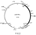
- Fig. 2 depicts a restriction map of the expression vector p SV.Myr 1 .
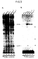
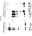
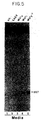
- Fig. 4 is an autoradiograph illustrating that a myristylated RSV Gag protein (Pr76 myr1 ) is secreted into the culture medium and is processed like wild-type RSV Pr76.
- Fig. 8 is an autoradiograph illustrating the Pr76 Gag products of p SV.GAGX, p SV.Myr 0 , p SV.Myr 1 and JD100.
- Fig. 9 is an autoradiograph illustrating that Pr76 myr1 proteins with C-terminal deletions are produced by the corresponding p SV.Myr 1 expression vectors, and further defines the limits of C-terminal deletions that allow truncated Pr76 myr1 derivatives to be produced in membraneous particles.
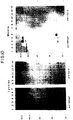
- Fig. 10 is an autoradiograph illustrating that a gene fusion of the RSV gag myr 1 allele and the yeast cycl gene produce fusion proteins released into the culture medium of CV-1 cells.
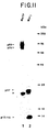
- Fig. 11 is an autoradiograph illustrating that Pr76 Myr1 and the fusion of Pr76 Myr1 -CYCl are produced in COS-1 cells, released in membraneous particles and processed.
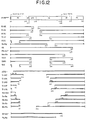
- Fig. 12 is a schematic diagram illustrating the overall structure of Pr76 myr1 (top line) and a series of deletion derivatives of Pr76 myr1 (remainder of figure), which were used to establish which gag sequences are required for membrane particle formation.
- the derivatives were constructed in p SV.Myr 1 , and the designations on the left represent the abbreviated names of the constructs, which are named pSV.Myr1.Y where Y is the abbreviated name, for example, R-3K represents the construct pSV.Myr1.R-3K, etc.
- the solid lines (upper, R-3K to TM) represent deletion derivatives that efficiently formed membrane particles; the open lines (lower, ATG- to Bg-Bs) represent deletion derivatives that did not form membrane particles efficiently.
- the shaded grey areas summarize the minimum gag residues needed to obtain efficient membrane particle formation and budding.
- Each line represents the regions of Pr76 Myr1 present in a given construct; the gaps indicate the deleted regions and the numbers indicate the amino acids of Pr76 Myr1 at the ends of the deletions.
- the (*) indicates that a stop codon was introduced at the indicated site in the polyprotein.
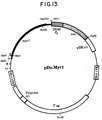
- Fig. 13 is a schematic diagram illustrating the expression vector used for expressing Pr76 Myr1 in murine and avian cells.
- the myr 1 allele is represented by the thick, black line in which the start of the coding sequence is marked with an arrow.
- the unique MluI site was created in altering the 5'-end so that is encodes the first 10 amino acids of p60 v-src .
- the Sac I- Bss HII fragment was cloned from p SV.Myr1 into the SalI site, thereby destroying the sites indicated in parentheses.
- the leftmost LTR serves as the promoter for transcription of myr 1 .
- pDo.Gag is identical to this plasmid except that it contains the wild-type RSV gag gene.
- Fig. 14 demonstrates expression of myristylated and non-myristylated Gag proteins in murine and avian cells.
- Murine (3T3) cells were transfected with pDo.Gag (lane 1) or pDo.Myr1 (lane 2). Positions for Pr76 gag , p27 (CA), p23 (MA), and p15 (PR) are indicated.
- B Turkey embryo fibroblasts were transfectcd with no DNA (lane 1). pDo.Gag (lane 2), or pDo.Myr1 (lane 3).
- the present invention is directed to replicable expression vectors having one or more nucleotide sequences operably linked to a hybrid gene which is a fusion of at least part of a retrovirus gag gene and a heterologous gene, or a part, of the heterologous gene wherein said gag gene enables a cell to produce and release the hybrid gene product in a membraneous particle.
- the hybrid gene contains a nucleotide sequence encoding a proteolytic cleavage site, which links the gag and heterologous genes.
- the present gag gene may be from an avian or mammalian retrovirus, and preferably is from an avian retrovirus.
- Preferred vectors are derived from eukaryotic sources. Expression vectors that function in tissue culture cells are especially useful, but yeast vectors are also contemplated. These vectors include yeast plasmids and minichromosomes, retrovirus vectors, BPV (bovine papilloma virus) vectors, baculovirus vectors, SV40 based vectors and other viral vectors. SV40-based vectors and retrovirus vectors (e.g., murine leukemia viral vectors) are preferred.
- Prokaryotic vectors that may also be suitable for expression of a hybrid gene of the instant invention include bacterial and bacteriophage vectors that can transform such hosts as E . coli , B . subtilis , Streptomyces sps. and other microorganisms. Many of these vectors are based on pBR322 including Bluescript (commercially available from Stratagene) and are well known in the art. Bacteriophage vectors that are used in the invention include lambda and M13.
- Heterologous genes contemplated by the present invention may encode the full amino acid sequence of a protein, or only such part as is desired to be expressed. These parts can be fragments or domains of the heterologous protein.
- Heterologous genes are linked to the gag gene by ligation of compatible restriction sites, by blunt-end ligation or using appropriately designed oligonucleotide linkers.
- Preferred heterologous proteins for expression in this system include yeast cytochrome c ( CYCl gene), cytokines, lymphokines (interferons, interleukins, growth factors), therapeutic proteins, or any protein for which a gene sequence is available and for which production or rapid purification of that protein is desirable.
- any protein useful as an immunogen, useful in a vaccine, or which needs to be targeted to a specific cell for therapeutic purposes can also be expressed in this system. If a gene sequence is not available, then it can be determined from the protein's amino acid sequences and chemically synthesized by standard DNA synthesis techniques. Further, the invention contemplates any modifications or mutation of a protein being expressed by the present expression vectors.
- Sequence elements capable of effecting expression of a gene include promoters, enhancer elements, transcription termination signals and polyadenylation sites. The latter three elements are not always necessary and their use will depend on both the vector and host system used for gene expression. The need for any of these elements can be easily determined by one skilled in the art.
- Promoters are DNA sequence elements for controlling gene expression, in particular, they specify transcription initiation sites.
- Prokaryotic promoters that are useful include the lac promoter, the trp promoter, and P L and P R promoters of lambda and the T7 polymerase promoter.
- Eukaryotic promoters are especially useful in the invention and include promoters of viral origin, such as the SV40 later promoter and the Moloney Leukemia Virus LTR, yeast promoters and any promoters or variations of promoters designed to control gene expression, including genetically-engineered promoters.
- Control of gene expression includes the ability to regulate a gene both positively and negatively (i.e., turning gene expression on or off) to obtain the desired level of expression.
- the replicable expression vectors of the present invention can be made by ligating part or all of a retrovirus gag gene to part or all of a heterologous gene to form a hybrid gene and then ligating the hybrid gene in the proper orientation to the promoter and other sequence elements being used to control gene expression.
- the gag gene region of the present invention is any portion or region of the gag gene sufficient to enable a cell to secrete the fusion product in a membraneous vesicle.
- removal of C-terminal residues or alteration (mutations), thereof in the Gag protein can allow processing and budding. However, it may be that some N-terminal residues can be deleted or changed and still enable the formation of membraneous particles.
- One skilled in the art can determine the maximum extent of a permissible deletion, at either the C-terminus or N-terminus, by constructing those deletions using standard genetic engineering techniques, such as deleting between convenient restriction enzyme sites; using Bal 31 or Exo III digestion for making deletions, or inserting protein termination codons into various sites and then assaying each construct for release of the genetically-engineered gag gene product into the culture medium in membraneous particles.
- mutations in the gag gene for example made by site-directed mutagenesis or natural selection can be assayed for gag -containing particles.
- the culture medium is removed from cells and subjected to high speed contrifugation to collect the particles. The particles are then analyzed for the presence of the Gag products by immunoprecipitating with anti-Gag antibodies.
- deletion derivatives can be made by deleting sequences between convenient restriction sites, inserting termination condons by oligonucleotide-directed mutagenesis or by Bal 31 digestion to further delimit the region of the gag gene sufficient to enable formation of membrane particles and budding.
- These derivatives allow identification of smaller regions of the gag gene encoding only some of the amino acids defined above that are required for particle formation. Therefore, the present invention contemplates any part of a gag gene encoding a region, particularly from among contiguous sequences of amino acids 1-8, 84-174 and 417-514, which enables a cell to produce a hybrid gene product in a membraneous particle.
- Complementation-rescue of the deletion derivatives that are incapable of directing particle formation indicate that the minimum essential region for particle formation may reside in the amino acid domain 417-515 of Pr76 Myr1 .
- avian gag genes and a few mammalian gag genes, it is necessary to introduce a modification into the gag gene to enable the Gag protein to direct formation of membraneous particles in the desired cell types.
- this modification involves altering the gene so that the protein is myristylated and, thus can bud in mammalian cells.
- Such a modification can be made, for example, by site-directed mutagenesis or oligonucleotide splicing of gene segments to form the desired modification.
- the second RSV gag codon is changed from GAA (glutamic acid) to GGA (glycine). This creates the myr 2 allele of the RSV gag gene whose product is Pr76 myr2 .
- This change can be accomplished by site-directed mutagenesis of a nucleic acid encoding the N-terminus of the RSV gag gene and using the mutagenic oligonucleotide: 5'-CAAGCATGGGAGCCGTCATAAAGG-3'.
- the first 10 amino acids of RSV Pr76 gag are replaced by the first 10 amino acids of p60 v-src , a protein known to be myristylated.
- Preferred replicable expression vectors of the present invention include for examples, MGAG.myr o , MGAG.myr 1 , MGAG.myr 2 , p SV.GAGX, p SV.Myr x , p SV.Myr o , p SV,Myr 1 , p SV.Myr 1A , p SV.Myr 1B , p SV.Myr 1C , pSV.Myr 2 , pSV.MyCYE and pSV.MyCYCl.
- the specific vectors described transiently produce fusion proteins by the retrovirus-mediated secretion path.
- the invention further contemplates vectors that lead to stable production of fusion proteins by the retrovirus-mediated secretion path.
- Pr76 fusion proteins can be accomplished using a gene transfer method based upon murine leukemia virus (MLV) expression vectors and other host systems.
- This method involves a helper cell line for packaging the recombinant MLV-RSV gag genome and an MLV-based transfer vector which expresses the recombinant MLV-RSV gag RNA.
- the RSV g ag gene fusion is introduced into the transfer vector by standard recombinant techniques. Many such vectors are available and contain bacterial plasmid sequences and two MLV LTRs (Long Terminal Repeats).
- the MLV packaging sequences ( ), the foreign gene of interest (i.e., the RSV gag gene fusions) and a selectable marker (e.g., a neomycin resistance gene under control of an appropriate promoter).
- the transfer vector is used to transfect a packaging cell line. This cell line releases infectious virion particles which contain RNA transcripts produced from the transfer plasmid. These virion particles are used to infect the target cells which will constitutively express and release RSV-Gag fusion proteins in membraneous particles.
- a packaging cell line for example GP+ env AM12, is a cell line constructed using mouse 3T3 cells, which continuously expresses MLV Gag, Pol, and Env proteins. The cells release particles continuously, but the particles are not infectious because they do not package MLV RNA. There are two reasons for the packaging defect. First, the gag , pol , and env genes are present, but the sequences required for packaging the RNA genome (these are named ) are not present on the gag, pol , and env transcripts. Second, the RNA is not packaged because the helper MLV genome has been "fragmented" and introduced into different sites in the 3T3 genome.
- the env gene in this cell line is derived from an amphotropic strain of MLV having glycoproteins on the surface of its particles which have a very broad host range (mouse, human, canine, simian, etc.).
- Infected target cells are identified via the selectable marker introduced by the transfer vector and then clonally expanded, MDCK (canine) and CV-1 cells are examples of two target cell lines that can be selected to express and release the non-infectious, membraneous particles containing the RSV gag fusion proteins.
- the particles are produced as a result of expression of the RSV gag fusion protein in the target cell, and especially myristylated RSV gag fusion proteins.
- nucleic acid encoding a hybrid gene which can be used in constructing a replicable expression vector of the present invention.
- the nucleic acid is composed of DNA or RNA. Further, the nucleic acid can be recombinant DNA or RNA.
- the hybrid gene encodes a hybrid gene product which is a fusion protein that is secreted by the retrovirus-mediated path into membraneous particles found in the extracellular space or culture medium.
- Transformant microorganisms and cultured cells are made by introducing the replicable expression vector into the desired cell or microorganism by transformation or transfection, or infection of virus or bacteriophage particles.
- Processes for transformation are well known in the art and include but are not limited to CaCl 2 treatment and electroporation for bacterial cells and CaPO 4 co-precipitation. protoplast fusion and electroporation for eukaryotic cells. Direct infection can be used when the vectors are viruses or bacteriophages. The detailed methods for these techniques can be found in standard laboratory manuals on recombinant DNA technology.
- the invention further contemplates any method for incorporating DNA into a host organism.
- a fusion protein having one domain which is a retrovirus Gag protein adapted to enable a cell to produce the fusion protein in a membraneous particle, i.e., by retrovirus-mediated secretion.
- the fusion protein has a second domain which is a heterologous or foreign protein.
- This second domain of the fusion protein consists of the entire foreign protein or any fragment or domain thereof desired to be expressed.
- a domain may be a region of a protein that forms a substructure of that protein.
- a domain may also specify a region having a specific enzymatic activity, a ligand binding site, a proteolytic cleavage site, or any other discrete feature of the protein.
- the second domain can consist of any open reading frame encoded on a nucleic acid.
- Preferred proteins for fusion to the Gag domain include yeast cytochrome c, cytokines, lymphokines (interferons, interleukins), growth factors, therapeutic proteins or any other useful protein desired to be produced, and especially those proteins wherein rapid purification is desired or which can be used as an immunogen in a vaccine or in a drug delivery system. Any fragment or domain of these proteins can be fused to the Gag domain. Further, any modification, substitution, insertion or deletion in these proteins can be fused to the Gag domain.
- the fusion proteins can have a genetically engineered proteolytic cleavage site between the Gag domain and the second domain encoding the heterologous protein.
- the proteolytic cleavage site comprises a specific sequence of amino acid residues that are recognized and cleaved by a protease. Any known proteolytic cleavage site is contemplated by the present invention, including but not limited to the sites recognized by retroviral proteases collagenase, Factor VIII and Factor IX or even a retroviral protease.
- the present invention contemplates any cleavage product or fragment of the above fusion proteins.
- These products may be produced by chemical means, produced by enzymatic means before or after isolation, especially by a protease, which recognizes a proteolytic cleavage site genetically engineered into the fusion protein, or produced during the course of retrovirus-mediated secretion by cellular proteases. In the latter instance, it is postulated that an unknown cellular protease may be responsible for the processing events. In any event, the processing events which occur during retrovirus-mediated secretion may generate fusion protein fragments contemplated by the present invention.
- the membrane of the particles contain specific receptors or ligands which will interact with the target cell.
- the membraneous particles also contain the "drug" being delivered to the target cell.
- the "drug” is the fusion protein, or a domain or fragment of the fusion protein. Any protein domain useful in treating diseases may be fused to the Gag protein and produced as described herein.
- the membraneous particles can be used as immunogens or in vaccine preparations.
- the desired antigen i.e., heterologous protein domain is fused to the Gag protein and expressed by retrovirus-mediated secretion as described herein.
- the membraneous particles are collected from the culture medium and purified by centrifugation.
- the membraneous particles are immunogenic and will elicit an immune response to the fusion protein.
- the particles may be incorporated into a vaccine composition or serve directly as the immunogen.
- Any heterologous protein, or domain thereof, useful in preparing antibodies, such as hepatitis B surface antigen, lymphokines or viral surface antigens may be fused to the Gag protein and thus yield the membraneous particles of the present invention.
- the wild-type RSV gag gene was obtained from PATV-8 (Katz et al . 1982, J . Virol . 42 :346-351), a molecule clone containing an infectious, sequenced copy of the RSV Prague C genome (Schwartz et al . 1983, Cell 32:853-869).
- Plasmid PJD100 carries an infectious but unsequenced copy of the Prague A strain of RSV (Stoltzfus et al . 1987, J . Virol . 61 :3401-3409).
- RSV was grown in turkey cell cultures which were prepared from fertile eggs (Hudson Farms, Muskogee, OK) and propagated in supplemental F10 medium (primary growth medium, PGM) using previously published methods (Hunter, 1979, Meth . Enzymol . 58 :379-393).
- the turkey cell cultures were found to contain no sequences capable of recombining with RSV gag sequences and produced no RSV specific antigens.
- Prague A(JD100) and Prague C(ATV-8) virus was obtained by transfecting secondary turkey cell cultures with pJD100 or pATV-8 DNA, respectively.
- Recombinant plasmids were propagated in E . coli strain DH-1 using solid or liquid LB medium containing 25 ⁇ g of ampicillin per ml.
- Recombinant M13 phages were propagaged in LB medium without ampicillin.
- the SV40 vector used for the expression of wild-type and mutant RSV gag genes in mammalian cells was derived from a previously described construction (Wills et al . 1984, J . Cell Biol . 99 : 2011-2023) as explained in Example 2.
- SV40- gag DNAs Prior to transfection, the SV40- gag DNAs wore digested with Xba I to remove the bacterial plasmid sequence (see Fig. 2) and then ligated at low DNA concentrations to connect the 3'-end of the gag gene with the late SV40 polyadenylation signal.
- CV-1 cells were transfected using a variation of the DEAE-dextran and chloroquine method previously described (Wills et al ., 1984).
- Transfected CV-1 cells were labeled with [9,10(n)- 3 H]-myristic acid (47.5 Ci/mmol, Amersham International) following the general method of Rhee et al .
- 3 H-myristic acid was dried under a gentle stream of nitrogen to evaporate the toluene solvent and dissolved in dimethyl sulfoxide (DMSO) at a concentration of 30 ⁇ Ci/ ⁇ l.
- DMSO dimethyl sulfoxide
- the isotope was then added to complete CV-1 growth medium to give a final concentration of 1 mCi/ml.
- Each 60 mm plate was labeled with 400 ⁇ l (0.4 mCi) of this medium for 1 h at 37°C.
- 5X lysis buffer B 125 mM Tris hydrochloride [pH 8.0], 0.75 M NaCl. 0.5% SDS, 5% Triton X-100, 5% deoxycholate
- 5X protease inhibitors 500 ⁇ g/ml phenylmethylsulfonyl fluoride, 5 ⁇ g/ml pepstatin, 5 ⁇ g/ml leupeptin.
- Monolayers were lysed using 500 ⁇ l of 1X lysis buffer A (25 mM Tris hydrochloride [pH 8.0], 0.15 M NaCl, 1% Triton X-100, 1% deoxycholate) containing 1X concentrations of protease inhibitors.
- 1X lysis buffer A 25 mM Tris hydrochloride [pH 8.0], 0.15 M NaCl, 1% Triton X-100, 1% deoxycholate
- the plates were washed again with 500 ⁇ l of lysis buffer A, and nuclei were removed from the 1 ml lysate by centrifugation at 15,000 g for 1 min. The supernatant was transferred to a clean tube and mixed with 10 ⁇ l of 10% SDS.
- the separated proteins were fixed and stained with Coomassie blue R250 (0.003% Coomassie blue in 10% acetic acid--50% ethanol). Subsequently, the gels were destained in a solution of 5% methanol, 7% acetic acid.
- the radioactive bands were detected by fluorography using Fluoro-Hance (Research Products International, Inc.) and Kodak X-OMAT AR5 film at -70°C. Typically, exposures of 1-16 h were required for the detection of 35 S-methioninc labeled proteins while 3 H-myristic acid labeled proteins required exposures of 1-2 weeks.
- MGAG Oligonucleotide-directed mutagenesis .
- the coding sequence for the RSV gag gene lies between nucleotides (nt) 380 and 2482 in the RSV genome (Schwartz et al .).
- the Sac I- Hind III fragment containing this region (nt 225 to 2740, respectively) was cloned into the polylinker region of M13mp19.
- the resulting clone is named MGAG. Mutagenesis of MGAG was accomplished using the method Kunkel (Kunkel et al . 1987, Meth . Enzymol . 154 : 367-382).
- MGAG phage was sequentially propagated three times in CJ236, a dut ung strain of E .
- oligonucleotides were synthesized using an Applied Biosystems DNA synthesizer, gel purified, phosphorylated using T4 polynucleotide kinase, hybridized with the uracil-containing template, made double-stranded using T4 DNA polymerase, and sealed using T4 DNA ligase. The products of these reactions were transfected into a Dut + Ung + strain and plated to allow selection and segregation of the mutants.
- Clones containing the desired mutations were identified by DNA sequencing use of the method of Sanger (Sanger et al . 1977. Proc. Natl. Acad . Sci . USA . 74 : 5463-5467).
- the clone MGAG.myr 1 was made by substituting the first 10 codons of gag for those of RSV src .
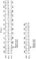
- a 57-mer was used (5'CCCGGTGGATCAAGCATGGGATCCAGCAAAAGCAAGCCTAAGGACGCGTGTAAAACC-3') which was designed to maximize complementarity (Fig. 1).
- presumptive clones were initially identified by the presence of a newly introduced Mlu I site (ACGCGT) contained in the 57-mer.
- the resulting allele confirmed by DNA sequencing, is designated myr 1 .
- the clone MGAG.myr 2 was made by changing the second codon of gag to code for Gly by introducing a single point mutation of A to G (Fig. 1). This was accomplished using a 24-mer (5'-CAAGCATGGGAGCCGTCATAAAGG-3'), and the resulting allele is designated myr 2 . Fragments containing the gag mutations were excised from the RF DNAs by digestion with Sac I and Bgl II (nt 1630) for transfer to the mammalian expression vector.
- transfected CV-1 cells were labeled for 2.5 h with 35 S-methionine, and the culture medium and the cell lysates were analyzed using antibodies against p27 or against RSV gag proteins. Turkey cells infected with Rous sarcoma virus were labeled to obtain authentic gag products for comparison and to show the antibody specificity. Preliminary experiments revealed intracellular half-lives on the order of 30 min for all forms of Pr76; thus, the results obtained using a 2.5 h labeling period approximate steady-state conditions.
- the "p27-doublet" represents mature products, since it is also seen in the medium samples whereas the three larger polypeptides are not.
- the lack of a significant amount of processing intermediates in the medium was confirmed by pulse-chase experiments and indicated the efficiency of cleavage during the budding process.
- Pr76 gag function in mammalian cells may depend (in part) upon the levels of expression, it was of interest to estimate how much protein is produced by the SV40 vector. For this purpose, the efficiency of transfection was measured using an indirect immunofluroescence assay with anti-p27. Typically, 30% of the CV-1 cells expressed Gag antigens. Taking this efficiency into account, it was calculated that the gag products released into the medium during a 2.5 h labeling period (at 48 h post-transfection) was approximately equal to that released from RSV-infected turkey cells during the same period.
- Myristylated RSV Gag Proteins are Budded from Mammalian Cells
- any loose cells present in the medium were removed by centrifugation at 15,000 g for 5 min, and the supernatant was immediately divided into six equal portions and processed as follows: one portion received nothing further, a second received 500 ⁇ g/ml (final concentration) of soybean trypsin inhibitor, a third received Triton X-100 to 1%, a fourth received 200 ⁇ g/ml of trypsin, a fifth received Triton X-100 + trypsin, and the sixth received soybean trypsin inhibitor + tyrpsin.
- the activity of the trypsin was 11,500 units/mg, and a six-fold excess of soybean trypsin inhibitor was used.
- the resulting fluorograph shows that the released products of authentic Pr76 gag (Fig. 6A) and Pr76 myr1 (Fig. 6B) were completely stable during incubations in the presence of nothing (lane 1), trypsin inhibitor alone (lane 2), Triton X-100 alone (lane 3), trypsin (lane 4), and trypsin + inhibitor (lane 6).
- the gag products became susceptible to the protease only when the membrane dissolving agent and trypsin were present together (lane 5).
- the agnoprotein initiation codon is out-of-frame with the gag initiation codon (nt 380), and translation initiated upstream does not terminate until the latter has been passed. If translation resumes at the next internal methionine codon (nt 464), then Pr76 gagX would lack the first 28 amino acids of Pr76.
- Fig. 8 shows the results of transfecting CV-1 cells with nothing (lane 1), p SV.GAGX (lane 2), p SV.Myr 0 (lane 3), or p SV.Myr 1 (lane 4) and JD100-infected turkey cells (lane 5) after pulse-labeling with 35 S-methionine.
- the Gag precursors were collected from cell lysates by immunoprecipitation with anti-p27, electrophoresed in a low concentration (7%) SDS-polyacrylamide gel, and detected by fluorography.
- Pr76 gagX appears to be about 3000 daltons smaller than the other forms of Pr76, consistent with a 28 amino acid truncation. Since another ATG is located only 11 codons downstream (nt 497), the precise site of reinitiation remains unknown in the absence of direct amino acid sequence information; nevertheless, it is clear that Pr76 gagX is truncated.
- Pr76 gagX The behavior of Pr76 gagX is quite distinct from that of Pr76 myr0 and Pr76 myr1 . It is poorly released into the medium and poorly processed to give products that migrate at the position of p27 (Fig. 4, lane 2). It could be that a mutation elsewhere in gag is responsible for this aberrant behavior but that was ruled out because: i) p SV.GAGX is the parent plasmid for all of the other SV40-gag constructions, and ii) its gag sequence has been shown to be fully functional when returned to the RSV genome. Thus, the inability of Pr76 gagX to be released into the medium clearly indicates that the amino-terminus of Pr76 is required for specific events during particle formation, and that the highly expressed proteins are not blebbing into the medium.
- results further indicate that a portion of the Pr76 Myr1 C-terminus can be deleted without impairing targeting or budding. Processing is eliminated because p15 (encoding the protease) has been at least partially deleted by C-terminal truncations.
- the three pSV.MyCYE-derived clones containing an out-of-frame cycl gene produce truncated proteins that reacted only with anti-p27 serum (Fig. 10a, lanes 1, 2, 3) and were released from the cell by budding (Fig. 10B, lanes 1, 2, 3).
- Fig. 10a lanes 10, 11, 12
- Fig. 10b lanes 9, 10, 11
- Fig. 10b lanes 9, 10, 11
- the cells transfected with the in-frame pSV.MyCYC1-derived gene fusions produced proteins that were larger than Pr76 Myr1 and that reacted with both the anti-p27 serum (Fig. 10A, lanes 4, 5) and the anti-cytochrome C serum (Fig. 10A, lanes 13, 14).
- Lane 6 in Fig. 10A is a dilution of the sample in lane 5.
- the larger proteins were released into the culture medium and reacted with both the anti-p27 serum (Fig. 10B, lanes 4, 5) and the anti-cytochrome C serum (Fig. 10B, lanes 12, 13).
- the Pr76 Myr1 -CYC fusion protein was budded into the culture medium.
- the particles released form the transfected cells also contain two small, co-migrating proteins one of which reacted with the anti-p27 serum (Fig. 10B, lanes 4, 5) and the other of which reacted with the anti-cytochrome c serum (Fig. 10B, lanes 12, 13). Because the truncated Pr76 Myr1 protein lacks the viral protease that is implicated in its processing, these cleavage products indicate that a cellular protease may have access to fusion proteins during budding.
- Pr76 Myr1 and Pr76 Myr1 -CYC1 are Packaged
- COS-1 cells were transfected with p SV.Myr 1 and pSV.MyCYC1.
- the transfected cells were metabolically labeled 48 h post-transfection with 35 S-methionine for 12 h.
- the culture medium was harvested, cleared of cellular debris by low speed centrifugation, and the particles were then purified by high speed centrifugation (15,000, 1 h) through a 5% sucrose cushion.
- the pellets were resuspended in lysis buffer and 50,000 CPM/sample were analyzed on a 12.5% SDS-polyacrylamide gel.
- An autoradiograph of the gel indicates that Pr76 Myr1 (lane 2) and Pr76 Myr1 -CYCl (lane 1) were sedimented and processed.
- Pr76 Myr1 was processed to the expected p27, p15 and p12 products.
- Pr76 Myr1 -CYC1 was partially processed releasing a protein slightly smaller than p27 that was immunoprecipitatahle with anti-cytochrome c antibody (compare with Fig. 10B, lanes 12, 13).
- the RSV Gag protein drives budding and membrane particle formation in simian COS-1 and CV-1 cell lines when myristate is added to its amino terminus.
- myristylated RSV Gag protein efficiently forms particles in other cell lines, the myr 1 allele was placed under the control of the murine leukemia virus (MLV) LTR promoter since this promoter functions in a broad range of species, including avian and murine cells.
- MLV murine leukemia virus
- the vector chosen for myr 1 expression pDOL.ATG- provides cloning sites flanked by MLV LTR sequences, a neomycin antibiotic resistance gene ( neo ), a bacterial origin of replication and the gene for the polyoma large T antigen.
- the myr 1 allele was excised from p SV.Myr1 using Sac I and Bss HII, purified by agarose gel electrophoresis, and ligated into pDOL.ATG- at its unique Sal I site after making the DNA ends blunt using the Klenow fragment of DNA polymerase I.
- the plasmids were propagated in E . coli strain DH-1 and selected on LB agar plates containing kanamycin (25 ⁇ g per ml). A recombinant bearing the myr 1 allele in the proper orientation relative to the LTR promoter was obtained and named pDo.Myr1 (Fig. 13).
- pDo.Gag is a control plasmid in which the wild-type gag gene was inserted into the same vector at the Sal I site.
- a Gag precursor of about 76 kDa was detected in the cell lysates (Fig. 14).
- the non-myristylated Pr76 gag produced by pDo.Gag seemed to accumulate to a greater extent within the cell than the myristylated Pr76 Myr1 produced by pDo.Myr1 (lysate lanes 1 and 2, respectively). This accumulation presumably is due to reduced ability of the pDo.Gag product to form particles relative to that of the pDo.Myr1 product (compare media lanes 1 and 2).
- the Gag-related proteins present in the Myr1 particles correspond to the mature cleavage products derived from Pr76, i.e., proteins MA, CA, NC, PR.
- the transient expression assays were repeated as above using turkey embryo fibroblasts (see Fig. 14B). Again, the Pr76 gag produced by pDo.Gag accumulated in the cells relative to Pr76 Myr1 produced by pDo.Myr1 (lysate lanes 2 and 3). However, the level of particle formation obtained with Pr76 myr1 in avian cells was increased relative to that obtained with non-myristylated Pr76 gag (media lanes 2 and 3). To ensure these particles were not the result of an endogenous virus, an uninfected control was included which proved to be negative (lysate and media lanes 1). It was surprising that the Pr76 myr1 produced higher levels of particles relative to the wild-type non-myristylated protein in its native cell type. The pDo.Myr1 construct is thus useful for production of myristylated Gag fusion proteins in a variety of cell types.
Landscapes
- Health & Medical Sciences (AREA)
- Genetics & Genomics (AREA)
- Life Sciences & Earth Sciences (AREA)
- Engineering & Computer Science (AREA)
- Chemical & Material Sciences (AREA)
- Organic Chemistry (AREA)
- Biomedical Technology (AREA)
- Biotechnology (AREA)
- General Engineering & Computer Science (AREA)
- Bioinformatics & Cheminformatics (AREA)
- Wood Science & Technology (AREA)
- Zoology (AREA)
- Molecular Biology (AREA)
- Biochemistry (AREA)
- Biophysics (AREA)
- General Health & Medical Sciences (AREA)
- Physics & Mathematics (AREA)
- Microbiology (AREA)
- Plant Pathology (AREA)
- Virology (AREA)
- Gastroenterology & Hepatology (AREA)
- Medicinal Chemistry (AREA)
- Proteomics, Peptides & Aminoacids (AREA)
- Micro-Organisms Or Cultivation Processes Thereof (AREA)
- Preparation Of Compounds By Using Micro-Organisms (AREA)
- Saccharide Compounds (AREA)
- Peptides Or Proteins (AREA)
Applications Claiming Priority (3)
| Application Number | Priority Date | Filing Date | Title |
|---|---|---|---|
| US35329389A | 1989-05-17 | 1989-05-17 | |
| US353293 | 1989-05-17 | ||
| EP90109343A EP0398332A1 (de) | 1989-05-17 | 1990-05-17 | Retrovirus vermittelte Sekretion von rekombinanten Produkten |
Related Parent Applications (1)
| Application Number | Title | Priority Date | Filing Date |
|---|---|---|---|
| EP90109343.5 Division | 1990-05-17 |
Publications (1)
| Publication Number | Publication Date |
|---|---|
| EP0803574A2 true EP0803574A2 (de) | 1997-10-29 |
Family
ID=23388506
Family Applications (2)
| Application Number | Title | Priority Date | Filing Date |
|---|---|---|---|
| EP97106183A Withdrawn EP0803574A2 (de) | 1989-05-17 | 1990-05-17 | Retrovirus vermittelte Sekretion von rekombinanten Produkten |
| EP90109343A Withdrawn EP0398332A1 (de) | 1989-05-17 | 1990-05-17 | Retrovirus vermittelte Sekretion von rekombinanten Produkten |
Family Applications After (1)
| Application Number | Title | Priority Date | Filing Date |
|---|---|---|---|
| EP90109343A Withdrawn EP0398332A1 (de) | 1989-05-17 | 1990-05-17 | Retrovirus vermittelte Sekretion von rekombinanten Produkten |
Country Status (5)
| Country | Link |
|---|---|
| EP (2) | EP0803574A2 (de) |
| JP (1) | JPH04500308A (de) |
| AU (1) | AU635023B2 (de) |
| CA (1) | CA2016897A1 (de) |
| WO (1) | WO1990014355A1 (de) |
Cited By (4)
| Publication number | Priority date | Publication date | Assignee | Title |
|---|---|---|---|---|
| WO2002026783A3 (en) * | 2000-09-28 | 2003-09-25 | Uab Research Foundation | Chimeric retroviral gag genes and screening assays |
| US7276488B2 (en) | 1997-06-04 | 2007-10-02 | Oxford Biomedica (Uk) Limited | Vector system |
| US7514546B2 (en) | 1999-11-18 | 2009-04-07 | Oxford Biomedica (Uk) Ltd. | Antibodies |
| US7531648B2 (en) | 1997-06-04 | 2009-05-12 | Oxford Biomedica (Uk) Limited | Vector encoding an antibody that binds 5T4 antigen |
Families Citing this family (4)
| Publication number | Priority date | Publication date | Assignee | Title |
|---|---|---|---|---|
| GB9101550D0 (en) * | 1991-01-24 | 1991-03-06 | Mastico Robert A | Antigen-presenting chimaeric protein |
| GB9107631D0 (en) * | 1991-04-10 | 1991-05-29 | British Bio Technology | Proteinaceous particles |
| EP0572737B1 (de) | 1992-06-04 | 2001-02-28 | The Research Foundation For Microbial Diseases Of Osaka University | Gag-Env Fusion-Antigen aus HIV |
| WO2011062155A1 (ja) * | 2009-11-19 | 2011-05-26 | タカラバイオ株式会社 | 変異型Gagタンパク質をコードする核酸 |
Family Cites Families (4)
| Publication number | Priority date | Publication date | Assignee | Title |
|---|---|---|---|---|
| US4828987A (en) * | 1984-05-14 | 1989-05-09 | Merck & Co., Inc. | Avian retrovirus-bovine growth hormone DNA |
| US4861719A (en) * | 1986-04-25 | 1989-08-29 | Fred Hutchinson Cancer Research Center | DNA constructs for retrovirus packaging cell lines |
| DE3788807T2 (de) * | 1986-11-01 | 1994-06-30 | British Bio Technology | Fusionsproteine und -partikel. |
| US4918166A (en) * | 1987-04-10 | 1990-04-17 | Oxford Gene Systems Limited | Particulate hybrid HIV antigens |
-
1990
- 1990-05-16 JP JP2508739A patent/JPH04500308A/ja active Pending
- 1990-05-16 WO PCT/US1990/002750 patent/WO1990014355A1/en not_active Ceased
- 1990-05-16 CA CA002016897A patent/CA2016897A1/en not_active Abandoned
- 1990-05-17 AU AU55078/90A patent/AU635023B2/en not_active Ceased
- 1990-05-17 EP EP97106183A patent/EP0803574A2/de not_active Withdrawn
- 1990-05-17 EP EP90109343A patent/EP0398332A1/de not_active Withdrawn
Cited By (7)
| Publication number | Priority date | Publication date | Assignee | Title |
|---|---|---|---|---|
| US7276488B2 (en) | 1997-06-04 | 2007-10-02 | Oxford Biomedica (Uk) Limited | Vector system |
| US7531648B2 (en) | 1997-06-04 | 2009-05-12 | Oxford Biomedica (Uk) Limited | Vector encoding an antibody that binds 5T4 antigen |
| US7718627B2 (en) | 1997-06-04 | 2010-05-18 | Oxford Biomedica (Uk) Limited | Vector |
| US8084249B2 (en) | 1997-06-04 | 2011-12-27 | Oxford Biomedica (Uk) Limited | Vector |
| US7514546B2 (en) | 1999-11-18 | 2009-04-07 | Oxford Biomedica (Uk) Ltd. | Antibodies |
| WO2002026783A3 (en) * | 2000-09-28 | 2003-09-25 | Uab Research Foundation | Chimeric retroviral gag genes and screening assays |
| US7148325B2 (en) | 2000-09-28 | 2006-12-12 | The Uab Research Foundation | Chimeric retroviral gag genes and screening assays |
Also Published As
| Publication number | Publication date |
|---|---|
| AU5507890A (en) | 1990-11-22 |
| CA2016897A1 (en) | 1990-11-17 |
| AU635023B2 (en) | 1993-03-11 |
| EP0398332A1 (de) | 1990-11-22 |
| WO1990014355A1 (en) | 1990-11-29 |
| JPH04500308A (ja) | 1992-01-23 |
Similar Documents
| Publication | Publication Date | Title |
|---|---|---|
| US5175099A (en) | Retrovirus-mediated secretion of recombinant products | |
| Wills et al. | Creation and expression of myristylated forms of Rous sarcoma virus Gag protein in mammalian cells | |
| US5643756A (en) | Fusion glycoproteins | |
| Reil et al. | Efficient HIV‐1 replication can occur in the absence of the viral matrix protein | |
| EP1036182B1 (de) | Verfahren und mittel zur herstellung von sicheren, rekombinanten lentivirusvektoren mit hohem titer | |
| Weldon Jr et al. | Incorporation of chimeric Gag protein into retroviral particles | |
| Bennett et al. | Functional chimeras of the Rous sarcoma virus and human immunodeficiency virus Gag proteins | |
| Dawson et al. | The role of nucleocapsid of HIV-1 in virus assembly | |
| Perez et al. | Mutants of the Rous sarcoma virus envelope glycoprotein that lack the transmembrane anchor and cytoplasmic domains: analysis of intracellular transport and assembly into virions | |
| Colicelli et al. | Isolation of a recombinant murine leukemia virus utilizing a new primer tRNA | |
| US5880276A (en) | Purified retroviral constitutive transport enhancer elements that enhance nucleocytoplasmic transport of mRNA, and methods of making and using the elements | |
| AU656545B2 (en) | DNA expression systems based on alphaviruses | |
| Ou et al. | Transport of hepatitis B virus precore protein into the nucleus after cleavage of its signal peptide | |
| Ragheb et al. | Uncoupled expression of Moloney murine leukemia virus envelope polypeptides SU and TM: a functional analysis of the role of TM domains in viral entry | |
| Nelle et al. | A large region within the Rous sarcoma virus matrix protein is dispensable for budding and infectivity | |
| JP2004000280A (ja) | Mpmv及びhivに基づく欠損パッケージング非オンコウイルスベクター | |
| EP0803574A2 (de) | Retrovirus vermittelte Sekretion von rekombinanten Produkten | |
| Andrawiss et al. | Murine leukemia virus particle assembly quantitated by fluorescence microscopy: role of Gag-Gag interactions and membrane association | |
| Wheeler et al. | The amino-terminal domain of the v-fms oncogene product includes a functional signal peptide that directs synthesis of a transforming glycoprotein in the absence of feline leukemia virus gag sequences | |
| US4876089A (en) | Feline leukemia virus protein vaccines | |
| Erdie et al. | Myristylation of Rous sarcoma virus Gag protein does not prevent replication in avian cells | |
| Noiman et al. | The Tat protein of equine infectious anemia virus is encoded by at least three types of transcripts | |
| Celma et al. | Domains in the simian immunodeficiency virus gp41 cytoplasmic tail required for envelope incorporation into particles | |
| Thomas et al. | Analysis of mutations within the cytoplasmic domain of the Moloney murine leukemia virus transmembrane protein | |
| Boeke et al. | Yeast Ty elements as retroviruses |
Legal Events
| Date | Code | Title | Description |
|---|---|---|---|
| PUAI | Public reference made under article 153(3) epc to a published international application that has entered the european phase |
Free format text: ORIGINAL CODE: 0009012 |
|
| 17P | Request for examination filed |
Effective date: 19970415 |
|
| AC | Divisional application: reference to earlier application |
Ref document number: 398332 Country of ref document: EP |
|
| AK | Designated contracting states |
Kind code of ref document: A2 Designated state(s): AT BE CH DE DK ES FR GB GR IT LI LU NL SE |
|
| STAA | Information on the status of an ep patent application or granted ep patent |
Free format text: STATUS: THE APPLICATION HAS BEEN WITHDRAWN |
|
| 18W | Application withdrawn |
Withdrawal date: 19990429 |