EP0800829B2 - Use of a pm-1 antibody or of a mh 166 antibody for enhancing the anti-tumor effect of cisplatin or carboplatin - Google Patents
Use of a pm-1 antibody or of a mh 166 antibody for enhancing the anti-tumor effect of cisplatin or carboplatin Download PDFInfo
- Publication number
- EP0800829B2 EP0800829B2 EP95942318A EP95942318A EP0800829B2 EP 0800829 B2 EP0800829 B2 EP 0800829B2 EP 95942318 A EP95942318 A EP 95942318A EP 95942318 A EP95942318 A EP 95942318A EP 0800829 B2 EP0800829 B2 EP 0800829B2
- Authority
- EP
- European Patent Office
- Prior art keywords
- antibody
- cisplatin
- cells
- cell carcinoma
- antitumor
- Prior art date
- Legal status (The legal status is an assumption and is not a legal conclusion. Google has not performed a legal analysis and makes no representation as to the accuracy of the status listed.)
- Expired - Lifetime
Links
Images


Classifications
-
- A—HUMAN NECESSITIES
- A61—MEDICAL OR VETERINARY SCIENCE; HYGIENE
- A61K—PREPARATIONS FOR MEDICAL, DENTAL OR TOILETRY PURPOSES
- A61K45/00—Medicinal preparations containing active ingredients not provided for in groups A61K31/00 - A61K41/00
- A61K45/06—Mixtures of active ingredients without chemical characterisation, e.g. antiphlogistics and cardiaca
-
- A—HUMAN NECESSITIES
- A61—MEDICAL OR VETERINARY SCIENCE; HYGIENE
- A61K—PREPARATIONS FOR MEDICAL, DENTAL OR TOILETRY PURPOSES
- A61K33/00—Medicinal preparations containing inorganic active ingredients
- A61K33/24—Heavy metals; Compounds thereof
- A61K33/243—Platinum; Compounds thereof
-
- A—HUMAN NECESSITIES
- A61—MEDICAL OR VETERINARY SCIENCE; HYGIENE
- A61K—PREPARATIONS FOR MEDICAL, DENTAL OR TOILETRY PURPOSES
- A61K39/00—Medicinal preparations containing antigens or antibodies
- A61K39/395—Antibodies; Immunoglobulins; Immune serum, e.g. antilymphocytic serum
-
- A—HUMAN NECESSITIES
- A61—MEDICAL OR VETERINARY SCIENCE; HYGIENE
- A61P—SPECIFIC THERAPEUTIC ACTIVITY OF CHEMICAL COMPOUNDS OR MEDICINAL PREPARATIONS
- A61P35/00—Antineoplastic agents
-
- A—HUMAN NECESSITIES
- A61—MEDICAL OR VETERINARY SCIENCE; HYGIENE
- A61P—SPECIFIC THERAPEUTIC ACTIVITY OF CHEMICAL COMPOUNDS OR MEDICINAL PREPARATIONS
- A61P43/00—Drugs for specific purposes, not provided for in groups A61P1/00-A61P41/00
Definitions
- the present invention relates to an antitumor agent effect enhancer comprising an interleukin-6 antagonist, which assists and enhances the effects of antitumor agents in the treatment of tumors.
- MDR multi-drug resistance
- Renal cell carcinoma exhibits resistance to therapy to antitumor agents such as cisplatin, adriamycin and vinblastine ( Kakehi, Y. et al., J. Urol., 139, 862-864, 1988 ; Kanamaru, H. et al., J. Natl. Cancer Inst., 81, 844-847, 1989 ; Teicher, B.A. et al., Cancer Res., 47, 388-393, 1987 ).
- Platinum compounds such as cisplatin that possess antitumor effects bond with DNA and inhibit DNA synthesis and cell division ( Pinto, A.L. et al., Biochica et Biophysica Acta, 780, 167-180, 1985 ).
- GST- ⁇ glutathione-S transferase- ⁇
- GST- ⁇ glutathione-S transferase- ⁇
- inhibition of the effects of cisplatin caused by an increase in intracellular levels of substances containing sulfidryl groups, increased DNA repair activities, or activation of oncogenes such as c-myc is considered to be involved in the resistance to therapy of renal cell carcinoma to cisplatin ( Sklar, M.D. et al., Cancer Res., 51, 2118-2123, 1991 ; Mizutani, Y. et al., Cancer in press, 1994 ; Nakagawa, K. et al., Japan J. Cancer Res., 79, 301-305, 1988 ).
- Glutathione is a tripeptide thiol. It plays an important role in the inactivation of DNA-bonding substances such as alkylating agents and cisplatin and in the repair of cell damage caused by these.
- One of the effects of GST- ⁇ is that it promotes inactivation of antitumor agents by causing antitumor agents like those described above to bond to glutathione.
- IL-6 interleukin-6
- IL-6R IL-6 receptor
- IL-6 is a multi-functional cytokine referred to as B cell stimulating factor-2 or Interferon ⁇ 2.
- B cell stimulating factor-2 or Interferon ⁇ 2.
- IL-6 was discovered as a differentiation factor involved in the activation of B lymphoid cells ( Hirano, T. et al., Nature, 324, 73-76, 1986 ). Later, it was clearly shown to be a multi-functional cytokine that affects the functions of various cells ( Akira, S. et al., Adv. in Immunology, 54, 1-78, 1993 ). IL-6 transmits its biological activity by means of two proteins present on cells.
- IL-6R a ligand bonding protein having a molecular weight of approximately 80 KD to which IL-6 bonds.
- sIL-6R soluble IL-6R
- Another protein is gp130 having a molecular weight of approximately 130 KD that is involved in signal transmission of non-ligand bonding.
- IL-6 and IL-6R form an IL-6/IL-6R complex.
- the biological activity of IL-6 is transmitted to the cell (Taga, et al., J. Exp. Med., 196, 967, 1987).
- IL-6 has been reported to inhibit apoptosis induced by antitumor agents ( Kerr, J. et al., Cancer, 73, 2013-2026, 1994 ; Sachs, L. et al., Blood, 82, 15-21, 1993 ).
- antitumor agents like cisplatin and mitomycin C have cytotoxic effects on tumor cells as a result of producing free radicals ( Oyanagi, Y. et al., Biochem. Pharmacol., 26, 473-476,1997 ; Nakano, H. et al., Biochem. Biophys.
- IL-6 is known to promote the expression of manganase superoxide dismutase (MnSOD), which has the effect of decomposing free radicals, and IL-6 antibody inhibits the expression of this promoted MnSOD ( Ono, M. et al., Biochem. Biophys. Res. Commun., 182, 1100-1107, 1992 ; Dougall, W.C. et al., Endocrinology, 129, 2376-2384, 1991 ).
- MnSOD manganase superoxide dismutase
- antitumor agents have been used in the treatment of tumors in the past, since large doses of these drugs produce adverse side effects such as nausea, vomiting, kidney and liver function disorders and inhibition of bone marrow function, there have been cases in which it was dangerous to administer the required dose of antitumor agent for adequately demonstrating antitumor effects.
- an effect enhancer that increases the sensitivity of these tumors to antitumor agents.
- the object of the present invention is to provide a novel antitumor agent effect enhancer that assists and enhances the effects of antitumor agents and increases the sensitivity of therapy-resistant tumor cells to antitumor agents.
- the present invention is thus directed to the use of an IL-6 antagonist for the preparation of a medicament for enhancing the effect of cisplatin or carboplatin in the treatment of a tumor wherein the IL-6 antagonist is the PM-1 or MH166 antibody and said tumor is renal cell carcinoma.
- IL-6 antagonists such as IL-6 antibody or IL-6R antibody increase the sensitivity of tumor cells to antitumor agents, that therapeutic effects are observed with lower doses of antitumor agents, and that therapeutic effects appear by concomitant use of ordinary antitumor agents with an effect enhancer comprising an IL-6 antagonist against tumors exhibiting resistance to therapy.
- the present invention relates to an antitumor agent effect enhancer containing an IL-6 antagonist. More specifically, the present invention refers to the use of an IL-6 antagonist for the preparation of a medicament for enhancing the effect of an antitumor agent.
- PM-1 antibody is humanized PM-1 antibody.
- Antitumor agents used in combination with IL-6 antagonists are cisplatin and carboplatin.
- Figs. 1A and 1B indicate the cytotoxicity against renal cell carcinoma line Caki-1 in the presence of cisplatin at a concentration of 0, 0.1, 1 or 10 ⁇ g/ml and IL-6 antibody ( Fig. 1A ) or IL-6R antibody ( Fig. 1B ).
- Diamonds indicate the cytotoxicity (%) in the presence of cisplatin only, squares that in the presence of cisplatin and 0.1 ⁇ g/ml I L-6 antibody or IL-6R antibody, triangles that in the presence of cisplatin and 1 ⁇ g/ml IL-6 antibody or IL-6R antibody, and circles that in the presence of cisplatin and 10 ⁇ g/ml IL-6 antibody or IL-6R antibody.
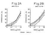
- Figs. 2A and 2B indicate the cytotoxicity against renal cell carcinoma line Caki-1 in the presence of mytomycin C at a concentration of 0,0.1, 1 or 10 ⁇ g/ml and IL-6 antibody ( Fig. 2A ) or IL-6R antibody ( Fig. 2B ).
- Diamonds indicate the cytotoxicity (%) in the presence of mitomycin C only, squares that in the presence of mitomycin C and 0.1 ⁇ g/ml IL-6 antibody or IL-6R antibody, triangles that in the presence of mitomycin C and 1 ⁇ g/ml IL-6 antibody or IL-6R antibody, and circles that in the presence of mitomycin C and 10 ⁇ g/ml IL-6 antibody or IL-6R antibody.
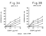
- Figs. 3A and 3B indicate the cytotoxicity against renal cell carcinoma line Caki-1/DDP in the presence of cisplatin at a concentration of 0, 0.1, 1 or 10 ⁇ g/ml and IL-6 antibody ( Fig. 3A ) or IL-6R antibody ( Fig. 3B ).
- Diamonds indicate the cytotoxicity (%) in the presence of cisplatin only, squares that in the presence of cisplatin and 0.1 ⁇ g/ml IL-6 antibody or IL-6R antibody, triangles that in the presence of cisplatin and 1 ⁇ g/ml IL-6 antibody or IL-6R antibody, and circles that in the presence of cisplatin and 10 ⁇ g/ml IL-6 antibody or IL-6R antibody.
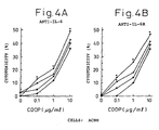
- Figs. 4A and 4B indicate the cytotoxicity against renal cell carcinoma line ACHN in the presence of cisplatin at a concentration of 0, 0.1, 1 or 10 ⁇ g/ml and IL-6 antibody ( Fig. 4A ) or IL-6R antibody ( Fig. 4B ).
- Diamonds indicate the cytotoxicity (%) in the presence of cisplatin only, squares that in the presence of cisplatin and 0.1 ⁇ g/ml IL-6 antibody or IL-6R antibody, triangles that in the presence of cisplatin and 1 ⁇ g/ml IL-6 antibody or IL-6R antibody, and circles that in the presence of cisplatin and 10 ⁇ g/ml IL-6 antibody or IL-6R antibody.
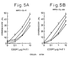
- Figs. 5A and 5B indicate the cytotoxicity against renal cell carcinoma line A704 in the presence of cisplatin at a concentration of 0, 0.1, 1 or 10 ⁇ g/ml and IL-6 antibody ( Fig. 5A ) or IL-6R antibody ( Fig. 5B ).
- Diamonds indicate the cytotoxicity (%) in the presence of cisplatin only, squares that in the presence of cisplatin and 0.1 ⁇ g/ml IL-6 antibody or IL-6R antibody, triangles that in the presence of cisplatin and 1 ⁇ g/ml IL-6 antibody or IL-6R antibody, and circles that in the presence of cisplatin and 10 ⁇ g/ml IL-6 antibody or IL-6R antibody.
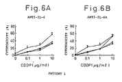
- Figs. 6A and 6B indicate the cytotoxicity against fresh renal cell carcinoma obtained from Patent 1 in the presence of cisplatin at a concentration of 0, 0.1, 1 or 10 ⁇ g/ml and IL-6 antibody ( Fig. 6A ) or IL-6R antibody ( Fig. 6B ).
- Diamonds indicate the cytotoxicity (%) in the presence of cisplatin only, squares that in the presence of cisplatin and 0.1 ⁇ g/ml IL-6 antibody or IL-6R antibody, triangles that in the presence of cisplatin and 1 ⁇ g/ml IL-6 antibody or IL-6R antibody, and circles that in the presence of cisplatin and 10 ⁇ g/ml IL-6 antibody or IL-6R antibody.
- Figs. 7A and 7B indicate the cytotoxicity against fresh renal cell carcinoma obtained from Patent 2 in the presence of cisplatin at a concentration of 0, 0.1, 1 or 10 ⁇ g/ml and IL-6 antibody ( Fig. 7A ) or IL-6R antibody ( Fig. 7B ).
- Diamonds indicate the cytotoxicity (%) in the presence of cisplatin only, squares that in the presence of cisplatin and 0.1 ⁇ g/ml IL-6 antibody or IL-6R antibody, triangles that in the presence of cisplatin and 1 ⁇ g/ml IL-6 antibody or IL-6R antibody, and circles that in the presence of cisplatin and 10 ⁇ g/ml IL-6 antibody or IL-6R antibody.
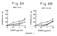
- Figs. 8A and 8B indicate the cytotoxicity against fresh renal cell carcinoma obtained from Patent 3 in the presence of cisplatin at a concentration of 0, 0.1, 1 or 10 ⁇ g/ml and IL-6 antibody ( Fig. 6A ) or IL-6R antibody ( Fig. 6B ).
- Diamonds indicate the cytotoxicity (%) in the presence of cisplatin only, squares that in the presence of cisplatin and 0.1 ⁇ g/ml IL-6 antibody or IL-6R antibody, triangles that in the presence of cisplatin and 1 ⁇ g/ml IL-6 antibody or IL-6R antibody, and circles that in the presence of cisplatin and 10 ⁇ g/ml IL-6 antibody or IL-6R antibody.
- Figs. 9A and 9B indicate the cytotoxicity against renal cell carcinoma line Caki-1 in the presence of carboplatin at a concentration of 0, 1, 10 or 100 ⁇ g/ml and IL-6 antibody ( Fig. 9A ) or IL-6R antibody ( Fig. 9B ).
- Diamonds indicate the cytotoxicity (%) in the presence of carboplatin only, squares that in the presence of carboplatin and 0.1 ⁇ g/ml IL-6 antibody or IL-6R antibody, triangles that in the presence of carboplatin and 1 ⁇ g/ml IL-6 antibody or IL-6R antibody, and circles that in the presence of carboplatin and 10 ⁇ g/ml IL-6 antibody or IL-6R antibody.
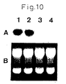
- a in Fig. 10 is a diagram showing the results of Northern blotting of the total RNA of Caki-1 using a GST- ⁇ cDNA probe in order to investigate the expression of GST- ⁇ mRNA in renal cell carcinoma line Caki-1 treated with a culture medium (control), cisplatin at 10 ⁇ g/ml or IL-6 antibody or IL-6R antibody at 10 ⁇ g/ml.
- Lane 1 shows Caki-1 treated with culture medium only, lane 2 that treated with cisplatin, lane 3 that treated with IL-6 antibody, and lane 4 that treated with IL-6R antibody.
- B in Fig. 10 is a diagram of a gel used for Northern blotting stained with ethidium bromide. The diagram shows that RNA is present in each lane.
- the antitumor agent effect enhancer comprising an IL-6 antagonist of the present invention enhances antitumor effects by being used in combination with antitumor agents during treatment of tumors.
- it enables the required dose of antitumor agent to be decreased since it has the effect of increasing the sensitivity to antitumor agents of therapy-resistant tumors for which therapeutic effects are not observed with ordinary chemotherapy.
- Antitumor agents for which antitumor effects are enhanced by the effect enhancer of the present invention are chemotherapy drugs having tumor therapeutic effects that inhibit the development and growth of tumor cells by acting on tumor cells.
- chemotherapy drugs are platinum compounds; selected from cisplatin and carboplatin. Platinum compounds have a platinum atom that forms complexes with other atoms. These compounds exhibit antitumor effects by bonding with DNA, inhibiting DNA synthesis of tumor cells and inhibiting division of tumor cells.
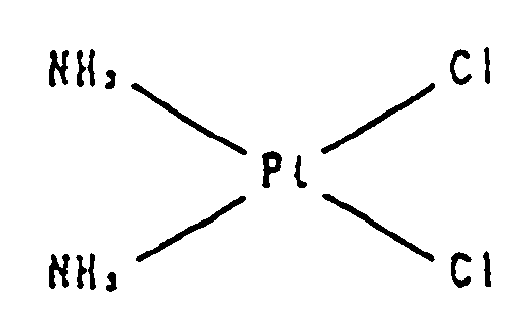
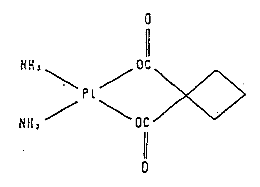
- platinum compounds having antitumor effects that have been used so far include cisplatin (cis-diamine dichloroplatinum (II) having the structural formula shown below): carboplatin (cis-diamine (1,1-cyclobutanedicarboxylato)-platinum (II) having the structural formula shown below):
- antitumor agents can be prepared using routine methods.
- platinum compounds may be used orally or parenterally, and preferably as an injection formulation, by mixing with a pharmaceutical carrier and an excipient, as necessary.
- an injective formulation it may be mixed with distilled water, a salt solution such as sodium chloride or potassium chloride, glucose solution or physiological saline.
- the amount of antitumor agent in these formulations is preferably a unit dose that is convenient for use according to the patient's age, symptoms and so forth. For example, in using for tumor treatment in an adult, it is daily administered at 10 to 2000 mg/m 2 (body surface area), administered continuously for 5 days according to the dose, or a recovery period of 1 to 4 weeks may be provided between administrations.
- Tumor cells in which antitumor effects are observed due to the effect enhancer of the present invention are tumors that have IL-6R and exhibit growth and/or resistance to therapy provided by IL-6 as one of their physiologically active substances.
- the tumor is renal cell carcinoma ( Miki, S. et al., FEBS Letter, 250, 607-610, 1989 ).
- the IL-6 antagonist used in the present invention is an IL-6 antibody or an IL-6R antibody.
- monoclonal antibody-producing cells there are no particular limitation on the animal species of monoclonal antibody-producing cells provided it is mammalian, and they may originate in human antibody or non-human mammalian antibody.
- Monoclonal antibodies of rabbit or rodent origin are preferable as monoclonal antibodies of non-human mammalian origin due to the ease of preparation.
- rodent antibodies preferable examples include mouse, rat and hamster antibodies.
- the IL-6 antibody according to the present invention is the MH166 antibody ( Matsuda, et al., Eur. J. Immunol., 18, 951-956, 1988 ).
- the IL-6R antibody according to the present invention is the PM-1 antibody ( Hirata, et al., J. Immunol., 143, 2900-2906, 1989 ).
- IL-6R antibodies are particularly preferable, that is the above-mentioned PM-1 antibody.
- Monoclonal antibodies can be prepared in the following manner basically using known technology. Namely, using IL-6, or IL-6R for the sensitizing antigen, a host is immunized in accordance with a routine immunization method, the resulting immunized cells are fused with known parent cells by routine cell fusion, and a monoclonal antibody-producing cell is then selected by routine screening methods.
- an antigen of human origin is preferable for the above-mentioned sensitizing antigen, and in the case of human IL-6, antigen is obtained by using the gene sequence of human IL-6 disclosed in Hirano, et al., Nature, 324, 73, 1986.
- the target IL-6 protein is purified from the host cells or culture supernatant, and the purified IL-6 protein may then be used as the sensitizing antigen.
- IL-6R protein can be obtained by following a method similar to that of the above-mentioned human IL-6 by using the gene sequence disclosed in European Patent Publication No. EP325474 .
- IL-6R There are two types of IL-6R, namely that which is expressed on the cell membrane, and a soluble type which is liberated from the cell membrane (sIL-6R) ( Yasukawa, et al., J. Biochem., 108, 673-676, 1990 ).
- sIL-6R is mainly composed of the extracellular region of IL-6R bonded to the cell membrane, and is different from membrane-bound IL-6R in that-it lacks a cell membrane permeating region or cell membrane permeating region and intracellular region.
- mammals that are immunized with sensitizing antigen are preferably selected in consideration of compatibility with the parent cells used in cell fusion, and mice, rats, hamsters or rabbits are commonly used.
- Immunization of an animal with sensitizing antigen is performed in accordance with known methods.
- immunization is performed by intraperitoneally or subcutaneously injecting a sensitizing antigen into an animal. More specifically, after diluting and suspending the sensitizing antigen in a suitable amount of phosphate-buffered saline (PBS) or physiological saline, the resulting suspension is mixed with a suitable amount of an ordinary adjuvant, such as Freund's complete adjuvant, as desired followed by emulsification. The emulsion is then preferably administered to the mammal in several doses every 4 to 21 days.
- a suitable carrier can also be used during immunization with sensitizing antigen.
- the immunized cells are extracted from the mammal and used in cell fusion.
- a particularly preferable example of immunized cells are spleen cells.
- Various known cell lines are preferably used for the mammalian myeloma cells that serve as the parent cells that are fused with the above-mentioned immunized cells, examples of which include P3 (P3x63Ag8.653) (J. Immunol., 123, 1548, 1978), p3-U1 ( Current Topics in Microbiology and Immunology, 81, 1-7, 1978 ), NS-1 ( Eur. J. Immunol., 6, 511-519, 1976 ), MPC-11 ( Cell, 8, 405-415, 1976 ), SP2/0 ( Nature, 276, 269-270, 1978 ), F0 ( J. Immunol. Meth., 35, 1-21, 1980 ), S194 ( J. Exp. Med., 148, 313-323, 1978 ), and R210 ( Nature, 277, 131-133, 1979 ).
- P3 P3x63Ag8.653
- p3-U1 Current Topics in Microbiology and Immunology,
- Cell fusion of the above-mentioned immunized cells and myeloma cells can basically be carried out in accordance with known methods, an example of which is the method of Milstein, et al. ( Milstein, et al., Methods Enzymol., 73, 3-46, 1981 ).
- the above-mentioned cell fusion is, for example, performed in an ordinary nutrient culture liquid in the presence of cell fusion promoter.
- fusion promoters include polyethylene glycol (PEG) and Sendai virus (HVJ).
- a fusion aid such as dimethylsulfoxide can also be added to increase fusion efficiency as desired.
- the ratio of immunized cells and myeloma cells used is preferably, for example, 1 to 10 times more immunized cells than myeloma cells.
- RPMI 1640 culture medium, MEM culture medium or other conventional culture media used in this type of cell culturing can be used for the culture liquid used for the above-mentioned cell fusion.
- these culture media can be used in combination with a serum supplement such as fetal calf serum (FCS).
- PEG solution such as PEG solution having a mean molecular weight of 1000 to 6000, warmed in advance to about 37°C normally at a concentration of 30 to 60% (w/v) and mixing to form the target fused cells (hybridoma).
- PEG solution such as PEG solution having a mean molecular weight of 1000 to 6000
- warmed in advance to about 37°C normally at a concentration of 30 to 60% (w/v) and mixing to form the target fused cells (hybridoma).
- Said hybridoma is selected by culturing in a conventional selective culture medium such as HAT culture medium (culture medium containing hypoxanthine, aminopterin and thymidine). Culturing in said HAT culture medium is normally continued for several days to several weeks to allow adequate time for cells other than the target hybridoma cells (non-fusion cells) to die. Next, a conventional limited dilution method is performed followed by screening and single-cloning of hybridoma that produces the target antibody.
- a conventional selective culture medium such as HAT culture medium (culture medium containing hypoxanthine, aminopterin and thymidine).
- Hybridoma thus prepared that produces monoclonal antibody can be subcultured in an ordinary culture medium, and can be stored for a long time in liquid nitrogen.
- said hybridoma is cultured in accordance with routine methods, and obtained in the form of culture supernatant.
- the hybridoma is grown by administering to a mammal with which it has compatibility and obtaining the monoclonal antibody in its ascites.
- the former method is suited for obtaining highly pure antibody, while the later method is suited for large-volume production of antibody.
- monoclonal antibody can not only be obtained from hybridoma formed by fusing antibody-producing cells obtained by immunizing with antigen, but monoclonal antibody can also be used that is produced using recombinant gene technology by cloning antibody gene, incorporating it in a suitable vector and introducing the vector into a known cell line such as COS or CHO (see, for example, Vandamme, A-M. et al., Eur. J. Biochem., 192, 767-775, 1990 ).
- monoclonal antibody obtained by the above-mentioned method can be purified to high purity using routine purification means such as salting out, gel filtration or affinity chromatography.
- Monoclonal antibody prepared in this manner is able to recognize antigen with high sensitivity and high accuracy by routine immunological means such as radioimmunoassay (RIA), enzyme immunoassay (EIA, ELISA) and immunofluorescence analysis.
- routine immunological means such as radioimmunoassay (RIA), enzyme immunoassay (EIA, ELISA) and immunofluorescence analysis.
- the monoclonal antibody used in the present invention is not limited to monoclonal antibody produced by hybridoma.
- Monoclonal antibody which has been artificially altered for the purpose of lowering heteroantigenicity to humans and so forth is more preferable.
- chimeric antibody composed of the variable region of a mammalian monoclonal antibody other than human monoclonal antibody, for example mouse monoclonal antibody, and the constant region of human antibody can be used.
- This type of chimeric antibody can be produced using known chimeric antibody production methods, and particularly recombinant gene technology.
- reshaped human antibody can also be used in the present invention.
- the complementarity determining region of non-human mammalian antibody such as mouse antibody
- a reshaped human antibody that is useful in the present invention can be obtained using this known technique, a preferable example of which is reshaped PM-1 antibody (see, for example, International Patent Publication No. WO92-19759 ).
- amino acids of the framework (FR) region of the variable region of an antibody may be substituted so as that the complementarity determining region of reshaped human antibody forms a suitable antibody binding site ( Sato, et al., Cancer Res., 53, 1-6, 1993 ).
- a gene that code for antibody fragment such as F(ab') 2 , Fab, Fv, single chain Fv(scFv) linking Fv of H chain and L chain with a suitable linker can be contructed, and the antibody fragment can then be expressed in a suitable host cell and can be used for the above-mentioned purpose provided it binds with antigen and inhibit IL-6 activity (see, for example, Bird, et al., TIBTECH, 9, 132-137, 1991 ).
- An scFv is composed by linking the H chain V region and L chain V.region of an antibody.
- the H chain V region and L chain V region are linked by means of a linker, and preferably by a peptide linker ( Huston, J.S. et al., Proc. Natl. Acad. Sci. U.S.A., 85, 5879-5883, 1988 ).
- the H chain V region and L chain V region in the scFv may be derived from any of the above-mentioned antibodies. These V regions are preferably linked by a peptide linker. Examples of peptide linkers that are used include any single chain peptide comprising 12 to 19 amino acid residues.
- DNA coding for scFv is obtained by using DNA that codes for the H chain or H chain V region of the above-mentioned antibody and DNA coding for the L chain or L chain V region as templates, amplifying the DNA portion of their sequences that codes for the desired amino acid sequence using a primer pair that defines both of its ends, and combining with a primer pair that defines the DNA that codes for the peptide linker portion as well as both ends so as to link with the H chain and the L chain.
- an expression vector that contains it and a host transformed by said expression vector can be obtained in accordance with routine methods.
- scFv can be obtained in accordance with routine methods by using that host. Since scFv has greater ability to migrate into tissue than antibody molecule, it is expected to be used as substance that has similar functions as reshaped human antibody.
- the antitumor agent effect enhancer comprising IL-6 antagonist of the present invention can be effectively used in the treatment of any tumors having IL-6R, growing by using IL-6 as one of its physiological active substances, and/or exhibiting resistance to therapy, provided it blocks IL-6 signal transmission and assists and enhances the effect of the antitumor agent.
- the antitumor agent effect enhancer comprising IL-6 antagonist of the present invention can be administered systemically or locally, and preferably parenterally by, for example, intravenous injection, intramuscular injection, intraperitoneal injection or subcutaneous injection. Moreover, it can be used in the form of a pharmaceutical composition or kit with at least one pharmaceutical carrier or diluent.
- the dose in humans of the antitumor agent effect enhancer comprising IL-6 antagonist of the present invention varies according to the condition and age of the patient or according to the administration method. However, it is necessary to select a suitable dose. For example, in the case of IL-6R antibody, a divided dose of four administrations or less within a range of approximately 1 to 1000 mg/patient can be selected. In addition, it can also be administered at a dose of 1 to 10 mg/kg/week.
- the antitumor agent effect enhancer comprising IL-6 antagonist of the present invention is not limited to these doses.
- the antitumor agent effect enhancer comprising IL-6 antagonist of the present invention can be prepared in accordance with routine methods (Remington's Pharmaceutical Science, latest edition, Mark Publishing Company, Easton, U.S.A.).
- preparations for injection may be prepared by dissolving purified IL-6 antagonist in a solvent such as physiological saline, buffer or glucose solution and adding an adsorption prevention agent such as Tween80, gelatin or human serum albumin (HSA).
- an adsorption prevention agent such as Tween80, gelatin or human serum albumin (HSA).
- HSA human serum albumin
- vehicles that can be used for freeze-drying include sugar alcohols and sugars such as mannitol and glucose.
- IL-6 antibody or IL-6R antibody The effect of IL-6 antibody or IL-6R antibody on the sensitivity of tumor cells to various antitumor agents was investigated.
- Human renal cell carcinoma line Caki-1, cisplatin-resistant strain Caki-1/DDP, a subline of Caki-1, human renal cell carcinoma ACHN and human renal cell carcinoma A704 ( Giard, D.J. et al., J. Natl. Cancer Inst., 51, 1417-1423, 1973 ) were cultured so as to form a single layer in plastic dishes in RPMI 1640 culture medium containing 25 mM HEPES, 2 mM L-glutamine, 1% non-essential amino acids, 100 U/ml penicillin, 100 ⁇ g/ml streptomycin and 10% heat-inactivated fetal bovine serum (FBS) (all manufactured by Gibco) (to be referred to as complete culture medium).
- FBS heat-inactivated fetal bovine serum
- Renal cell carcinoma tumor tissue was obtained during surgical procedures on three renal cell carcinoma patients. After confirming the tissue to be renal cell carcinoma by histological classification, the tumor tissue was finely decomposed by 3 mg/ml of collagenase (Sigma Chemical Co.) to prepare a tumor cell suspension. After washing three times with RPMI 1640 culture medium, the cell suspension was layered over a non-continuous gradient composed of 2 ml each of 100%, 80% and 50% Ficol-Hypaque in 15 ml volumetric plastic tubes followed by centrifugation for 30 minutes at 400 xg. The lymphocyte-rich monocyte layer contained in the above-mentioned 100% layer was removed, and tumor cells and mesothelial cells were obtained from the 80% layer.
- the cell suspension rich in tumor cells was layered over a non-continuous gradient composed of 3 ml each of 25%, 15% and 10% Percoll in complete culture medium contained in 15 ml plastic tubes followed by centrifugation for 7 minutes at 25 xg and room temperature. After washing the resulting tumor cells and suspending in complete culture medium, the presence of tumor cells was confirmed by trypan blue staining.
- the tumor cells prepared in this manner were used in the following experiment.
- IL-6 in the culture supernatant of renal cell carcinoma line Caki-1, Caki-1/DDP, ACHN, A704 and fresh tumor cells derived from renal cell carcinoma patients (Nos. 1-3) was investigated by ELISA (enzyme-linked immunosorbent assay).
- IL-6 antibody 100 ⁇ l was added to 96-well ELISA plates to coat the ELISA plates with IL-6 antibody by allowing to stand at least overnight. These plates were stored for a maximum of 4 weeks at 4°C until the time of use.
- the plates coated with IL-6 antibody were washed three times and blocked for 1 hour with ELISA PBS containing 1% PBS (bovine serum albumin). After washing twice, 100 ⁇ l of tumor cell culture supernatant or E. coli recombinant IL-6 as the control (Yasukawa, et al., Biotechnol. Lett., 12, 419, 1990) were added to each well.
- the plates were incubated for 1 hour and washed three times followed by the addition of 100 ⁇ l of anti-IL-6 polyclonal antibody ( Matsuda, T. et al., Eur. J. Immunol., 18, 951-956, 1988 ) to each well.
- the plates were then incubated for 1 hour followed by the addition of alkaline phosphate-bound goat anti-rabbit IgG to each well and incubating for an additional hour.
- the plates were washed and incubated with alkaline phosphate substrate (Sigma 104, Sigma Chemical Co.). Two hours later, the absorbance at 405 nm was measured with an ELISA READER (Immunoreader, Japan Intermed Co., Ltd.).
- the MTT method ( Mizutani, Y. et al., Cancer, 73, 730-737, 1994 ) was used to investigate the effect of IL-6 antibody or IL-6R antibody on the sensitivity of various renal cell carcinomas, namely Caki-1, Caki-1/DDP, ACHN, A704 and fresh tumor cells derived from renal cell carcinoma patients (Nos. 1 to 3), to various concentrations of antitumor agents, namely cisplatin (cis-diaminedichloroplatinum (II)), mitomycin C (MMC), adriamycin (ADR), vinblastine (VBL) and 5-fluorouracil (5-FU).
- cisplatin cis-diaminedichloroplatinum (II)
- MMC mitomycin C
- ADR adriamycin
- VBL vinblastine
- 5-fluorouracil 5-fluorouracil
- IL-6 antibody or IL-6R antibody present with the cisplatin.
- the combined effects of IL-6 antibody or IL-6R antibody and cisplatin were similarly observed for the other renal cell carcinoma lines ACHN (see Figs. 4A and 4B ) and A704 (see Figs. 5A and 5B ) as well as for the fresh tumor cells obtained from the three renal cell carcinoma patients (see Figs. 6A, 6B , 7A, 7B , 8A and 8B ).
- Caki-1 cells were cultured for 24 hours in the presence of combination of 10 ⁇ g/ml of cisplatin or 100 ⁇ g/ml of 5-FU or culture medium as control, and 10 ⁇ g/ml of control antibody MOPC3/C, or 10 ⁇ g/ml of IL-6 antibody or IL-6R antibody.
- the intracellular accumulation of cisplatin was measured by flameless atomic absorption spectrometry ( Daley-Tates, P.T. et al., Biochem. Pharmacol., 34, 2263-2369 ; Riley, C.M. et al., Analytical Biochem., 124, 167-179, 1982 ).
- the Zeeman Z-8000 Zeeman Z-8000 Spectrophotometer, Hitachi Co., Ltd. was used for the measuring instrument.
- the intracellular accumulation of 5-FU was measured by gas chromatography and mass spectrometry ( Marunaka, T. et al., J. Pharm.
- Caki-1 cells were cultured for 4 hours with control culture medium, 10 ⁇ g/ml of cisplatin or 10 ⁇ g/ml of IL-6 antibody or IL-6R antibody.
- the total RNA of the cells was prepared according to the method of Mizutani, Y. et al. (Cancer, 73, 730-737, 1994 ) followed by electrophoresis in 1.2% agarose-2.2 M HCHO gel in 1 x MOPS buffer containing 200 mM MOPS (3-[N-morpholino]propane sulfonate), 50 mM sodium acetate and 10 mM sodium EDTA so as to result in 10 ⁇ g of RNA per lane.
- RNA was transcribed to a Biodyne A membrane (Poll) in 20 x SSC solution containing 3 M NaCl and 0.3 M sodium citrate (pH 7.0).
- a 50 to 100 ng GST- ⁇ cDNA probe Nakagawa, K. et al., J. Biol. Chem., 265, 4296-4301, 1990 ) was labeled by random oligoprimer elongation using a 32 P-dCTP (NEN).
- the above-mentioned nylon membrane to which the RNA was transcribed was cross-linked with ultraviolet rays and hybridized with the above-mentioned probe. Those results are shown in Fig. 10 .
- IL-6 antagonists such as IL-6 antibody or IL-6R antibody
- antitumor agents IL-6 antagonists and antitumor agents.
- tumor cells exhibiting resistance to therapy to antitumor agents were proven to be treatable as a result of IL-6 antagonists enhancing their sensitivity to antitumor agents.
- the antitumor agent effect enhancer comprising IL-6 antagonist of the present invention is able to reduce the cytotoxic effects of antitumor agents on tissue by allowing the required dose of antitumor agent to be lowered, thus giving it considerable value in use as an enhancer of antitumor agent effects.
Landscapes
- Health & Medical Sciences (AREA)
- Chemical & Material Sciences (AREA)
- Medicinal Chemistry (AREA)
- Life Sciences & Earth Sciences (AREA)
- Veterinary Medicine (AREA)
- Pharmacology & Pharmacy (AREA)
- Animal Behavior & Ethology (AREA)
- General Health & Medical Sciences (AREA)
- Public Health (AREA)
- Epidemiology (AREA)
- Bioinformatics & Cheminformatics (AREA)
- Engineering & Computer Science (AREA)
- Organic Chemistry (AREA)
- Inorganic Chemistry (AREA)
- General Chemical & Material Sciences (AREA)
- Chemical Kinetics & Catalysis (AREA)
- Immunology (AREA)
- Microbiology (AREA)
- Mycology (AREA)
- Nuclear Medicine, Radiotherapy & Molecular Imaging (AREA)
- Medicines Containing Antibodies Or Antigens For Use As Internal Diagnostic Agents (AREA)
- Medicines That Contain Protein Lipid Enzymes And Other Medicines (AREA)
- Pharmaceuticals Containing Other Organic And Inorganic Compounds (AREA)
- Acyclic And Carbocyclic Compounds In Medicinal Compositions (AREA)
- Steroid Compounds (AREA)
- Peptides Or Proteins (AREA)
Abstract
Description
- The present invention relates to an antitumor agent effect enhancer comprising an interleukin-6 antagonist, which assists and enhances the effects of antitumor agents in the treatment of tumors.
- Various drugs such as alkylating agents, metabolism antagonists, antitumor antibiotics and platinum compounds have been used in the past for chemotherapy on human tumors. In the case where remarkable therapeutic effects are not observed when these antitumor agents are used alone, therapeutic methods have been devised in which a plurality of antitumor agents are used concomitantly (Frei, E. III, Cancer Res., 32, 2593-2607, 1992). Tumor cells exhibit various sensitivities to antitumor agents, and certain cells are known to exhibit resistance to therapy by antitumor agents (Magrath, I., New Directions in Cancer Treatment, 1989, Springer-Verlag). Acquisition of resistance to therapy by tumor cells is said to be caused by, for example, the occurrence of multi-drug resistance (MDR) (Tsuruo, T. et al., Cancer Res., 42, 4730-4733, 1982), reduction in cell uptake of antitumor agent (Sherman, S.E. et al., Science, 230, 412, 1985), increased DNA repair activity (Borch, R.F., Metabolism and Action of Anticancer Drugs, Powis, G. & Prough, R. ed., Taylor & Francis, London, 1987, 163-193), or promotion of inactivation of antitumor agent in the cells (Teicher, B.A. et al., Cancer Res., 46, 4379, 1986). In such cases, there are many cases in which the expected therapeutic effects are not observed simply by administering antitumor agent.
- Renal cell carcinoma exhibits resistance to therapy to antitumor agents such as cisplatin, adriamycin and vinblastine (Kakehi, Y. et al., J. Urol., 139, 862-864, 1988; Kanamaru, H. et al., J. Natl. Cancer Inst., 81, 844-847, 1989; Teicher, B.A. et al., Cancer Res., 47, 388-393, 1987). Platinum compounds such as cisplatin that possess antitumor effects bond with DNA and inhibit DNA synthesis and cell division (Pinto, A.L. et al., Biochica et Biophysica Acta, 780, 167-180, 1985).
- The expression of glutathione-S transferase-π (GST-π), inhibition of the effects of cisplatin caused by an increase in intracellular levels of substances containing sulfidryl groups, increased DNA repair activities, or activation of oncogenes such as c-myc is considered to be involved in the resistance to therapy of renal cell carcinoma to cisplatin (Sklar, M.D. et al., Cancer Res., 51, 2118-2123, 1991; Mizutani, Y. et al., Cancer in press, 1994; Nakagawa, K. et al., Japan J. Cancer Res., 79, 301-305, 1988).
- In addition, changes in membrane permeability and transport capabilities in tumor cells are said to cause a decrease in cisplatin uptake within the cells, thus increasing resistance to therapy to cisplatin (Richon, V. et al., Cancer Res., 47, 2056-2061, 1987; Waud, W.R. et al., Cancer Res., 47, 6549-6555, 1987). Glutathione, an example of a substance containing sulfidryl groups that is present in the largest amounts in mammalian animal cells, is reported to inactivate cisplatin in cells, and certain types of tumors have been shown to have higher intracellular glutathione and metallothionein levels (Hromas, R.A. et al., Cancer LETT., 34, 9-13, 1987; Taylor, D.M. et al., Eur. J. Cancer, 12, 249-254, 1976).
- Glutathione is a tripeptide thiol. It plays an important role in the inactivation of DNA-bonding substances such as alkylating agents and cisplatin and in the repair of cell damage caused by these. One of the effects of GST-π is that it promotes inactivation of antitumor agents by causing antitumor agents like those described above to bond to glutathione.
- Since renal cell carcinoma produces interleukin-6 (IL-6) and expresses IL-6 receptor (IL-6R), it has been suggested that IL-6 plays some type of role in the growth activity of renal cell carcinoma (Miki, S. et al. FEBS Lett., 250, 607-610, 1989; Takenawa, J. et al., J. Natl. Cancer. Inst., 83, 1668-1672, 1991). Moreover, it has been reported that the level of IL-6 in the serum increases when the prognosis for treatment of renal cell carcinoma patients is poor (Blay, J. et al., Cancer Res., 52, 3317-3322, 1992; Tsukamoto, T. et al., J. Urol., 148, 1778-1782, 1992). However, a clear correlation between IL-6 and the resistance to therapy of renal cell carcinoma to antitumor agents has not yet been found, and is still unknown.
- IL-6 is a multi-functional cytokine referred to as B cell stimulating factor-2 or Interferon β2. IL-6 was discovered as a differentiation factor involved in the activation of B lymphoid cells (Hirano, T. et al., Nature, 324, 73-76, 1986). Later, it was clearly shown to be a multi-functional cytokine that affects the functions of various cells (Akira, S. et al., Adv. in Immunology, 54, 1-78, 1993). IL-6 transmits its biological activity by means of two proteins present on cells.
- One of them is IL-6R, a ligand bonding protein having a molecular weight of approximately 80 KD to which IL-6 bonds. In addition to the cell bonding form which is expressed on the cell membrane by passing through the cell membrane, it is also present as soluble IL-6R (sIL-6R), which is mainly composed of its extracellular region. Another protein is gp130 having a molecular weight of approximately 130 KD that is involved in signal transmission of non-ligand bonding. IL-6 and IL-6R form an IL-6/IL-6R complex. As a result of then bonding with the other membrane protein gp130, the biological activity of IL-6 is transmitted to the cell (Taga, et al., J. Exp. Med., 196, 967, 1987).
- Although platinum compounds like cisplatin and antitumor agents such as mitomycin C induce apoptosis in tumor cells, IL-6 has been reported to inhibit apoptosis induced by antitumor agents (Kerr, J. et al., Cancer, 73, 2013-2026, 1994; Sachs, L. et al., Blood, 82, 15-21, 1993). In addition, antitumor agents like cisplatin and mitomycin C have cytotoxic effects on tumor cells as a result of producing free radicals (Oyanagi, Y. et al., Biochem. Pharmacol., 26, 473-476,1997; Nakano, H. et al., Biochem. Biophys. Acta., 796, 285-293,1984). IL-6, however, is known to promote the expression of manganase superoxide dismutase (MnSOD), which has the effect of decomposing free radicals, and IL-6 antibody inhibits the expression of this promoted MnSOD (Ono, M. et al., Biochem. Biophys. Res. Commun., 182, 1100-1107, 1992; Dougall, W.C. et al., Endocrinology, 129, 2376-2384, 1991).
- However, none of these reports state that the effects of antitumor agents are enhanced by blocking the biological activity of IL-6. In addition, there are no reports of actually attempting to use an IL-6 antagonist as an effect enhancer of antitumor agents.
- Although antitumor agents have been used in the treatment of tumors in the past, since large doses of these drugs produce adverse side effects such as nausea, vomiting, kidney and liver function disorders and inhibition of bone marrow function, there have been cases in which it was dangerous to administer the required dose of antitumor agent for adequately demonstrating antitumor effects. In addition, since there are tumors having resistance to therapy in which chemotherapy using ordinary antitumor agents is not effective, there is a need for an effect enhancer that increases the sensitivity of these tumors to antitumor agents.
- The object of the present invention is to provide a novel antitumor agent effect enhancer that assists and enhances the effects of antitumor agents and increases the sensitivity of therapy-resistant tumor cells to antitumor agents. The present invention is thus directed to the use of an IL-6 antagonist for the preparation of a medicament for enhancing the effect of cisplatin or carboplatin in the treatment of a tumor wherein the IL-6 antagonist is the PM-1 or MH166 antibody and said tumor is renal cell carcinoma.
- As a result of various studies on the effects of IL-6 antagonists on changes in sensitivity of tumor cells to antitumor agents, the inventors of the present invention found that IL-6 antagonists such as IL-6 antibody or IL-6R antibody increase the sensitivity of tumor cells to antitumor agents, that therapeutic effects are observed with lower doses of antitumor agents, and that therapeutic effects appear by concomitant use of ordinary antitumor agents with an effect enhancer comprising an IL-6 antagonist against tumors exhibiting resistance to therapy.
- Namely, the present invention relates to an antitumor agent effect enhancer containing an IL-6 antagonist. More specifically, the present invention refers to the use of an IL-6 antagonist for the preparation of a medicament for enhancing the effect of an antitumor agent.
- Specific examples PM-1 antibody are humanized PM-1 antibody. Antitumor agents used in combination with IL-6 antagonists are cisplatin and carboplatin.
-
Figs. 1A and 1B indicate the cytotoxicity against renal cell carcinoma line Caki-1 in the presence of cisplatin at a concentration of 0, 0.1, 1 or 10 µg/ml and IL-6 antibody (Fig. 1A ) or IL-6R antibody (Fig. 1B ). Diamonds indicate the cytotoxicity (%) in the presence of cisplatin only, squares that in the presence of cisplatin and 0.1 µg/ml I L-6 antibody or IL-6R antibody, triangles that in the presence of cisplatin and 1 µg/ml IL-6 antibody or IL-6R antibody, and circles that in the presence of cisplatin and 10 µg/ml IL-6 antibody or IL-6R antibody. -
Figs. 2A and 2B indicate the cytotoxicity against renal cell carcinoma line Caki-1 in the presence of mytomycin C at a concentration of 0,0.1, 1 or 10 µg/ml and IL-6 antibody (Fig. 2A ) or IL-6R antibody (Fig. 2B ). Diamonds indicate the cytotoxicity (%) in the presence of mitomycin C only, squares that in the presence of mitomycin C and 0.1 µg/ml IL-6 antibody or IL-6R antibody, triangles that in the presence of mitomycin C and 1 µg/ml IL-6 antibody or IL-6R antibody, and circles that in the presence of mitomycin C and 10 µg/ml IL-6 antibody or IL-6R antibody. -
Figs. 3A and 3B indicate the cytotoxicity against renal cell carcinoma line Caki-1/DDP in the presence of cisplatin at a concentration of 0, 0.1, 1 or 10 µg/ml and IL-6 antibody (Fig. 3A ) or IL-6R antibody (Fig. 3B ). Diamonds indicate the cytotoxicity (%) in the presence of cisplatin only, squares that in the presence of cisplatin and 0.1 µg/ml IL-6 antibody or IL-6R antibody, triangles that in the presence of cisplatin and 1 µg/ml IL-6 antibody or IL-6R antibody, and circles that in the presence of cisplatin and 10 µg/ml IL-6 antibody or IL-6R antibody. -
Figs. 4A and 4B indicate the cytotoxicity against renal cell carcinoma line ACHN in the presence of cisplatin at a concentration of 0, 0.1, 1 or 10 µg/ml and IL-6 antibody (Fig. 4A ) or IL-6R antibody (Fig. 4B ). Diamonds indicate the cytotoxicity (%) in the presence of cisplatin only, squares that in the presence of cisplatin and 0.1 µg/ml IL-6 antibody or IL-6R antibody, triangles that in the presence of cisplatin and 1 µg/ml IL-6 antibody or IL-6R antibody, and circles that in the presence of cisplatin and 10 µg/ml IL-6 antibody or IL-6R antibody. -
Figs. 5A and 5B indicate the cytotoxicity against renal cell carcinoma line A704 in the presence of cisplatin at a concentration of 0, 0.1, 1 or 10 µg/ml and IL-6 antibody (Fig. 5A ) or IL-6R antibody (Fig. 5B ). Diamonds indicate the cytotoxicity (%) in the presence of cisplatin only, squares that in the presence of cisplatin and 0.1 µg/ml IL-6 antibody or IL-6R antibody, triangles that in the presence of cisplatin and 1 µg/ml IL-6 antibody or IL-6R antibody, and circles that in the presence of cisplatin and 10 µg/ml IL-6 antibody or IL-6R antibody. -
Figs. 6A and 6B indicate the cytotoxicity against fresh renal cell carcinoma obtained fromPatent 1 in the presence of cisplatin at a concentration of 0, 0.1, 1 or 10 µg/ml and IL-6 antibody (Fig. 6A ) or IL-6R antibody (Fig. 6B ). Diamonds indicate the cytotoxicity (%) in the presence of cisplatin only, squares that in the presence of cisplatin and 0.1 µg/ml IL-6 antibody or IL-6R antibody, triangles that in the presence of cisplatin and 1 µg/ml IL-6 antibody or IL-6R antibody, and circles that in the presence of cisplatin and 10 µg/ml IL-6 antibody or IL-6R antibody. -
Figs. 7A and 7B indicate the cytotoxicity against fresh renal cell carcinoma obtained fromPatent 2 in the presence of cisplatin at a concentration of 0, 0.1, 1 or 10 µg/ml and IL-6 antibody (Fig. 7A ) or IL-6R antibody (Fig. 7B ). Diamonds indicate the cytotoxicity (%) in the presence of cisplatin only, squares that in the presence of cisplatin and 0.1 µg/ml IL-6 antibody or IL-6R antibody, triangles that in the presence of cisplatin and 1 µg/ml IL-6 antibody or IL-6R antibody, and circles that in the presence of cisplatin and 10 µg/ml IL-6 antibody or IL-6R antibody. -
Figs. 8A and 8B indicate the cytotoxicity against fresh renal cell carcinoma obtained fromPatent 3 in the presence of cisplatin at a concentration of 0, 0.1, 1 or 10 µg/ml and IL-6 antibody (Fig. 6A ) or IL-6R antibody (Fig. 6B ). Diamonds indicate the cytotoxicity (%) in the presence of cisplatin only, squares that in the presence of cisplatin and 0.1 µg/ml IL-6 antibody or IL-6R antibody, triangles that in the presence of cisplatin and 1 µg/ml IL-6 antibody or IL-6R antibody, and circles that in the presence of cisplatin and 10 µg/ml IL-6 antibody or IL-6R antibody. -
Figs. 9A and 9B indicate the cytotoxicity against renal cell carcinoma line Caki-1 in the presence of carboplatin at a concentration of 0, 1, 10 or 100 µg/ml and IL-6 antibody (Fig. 9A ) or IL-6R antibody (Fig. 9B ). Diamonds indicate the cytotoxicity (%) in the presence of carboplatin only, squares that in the presence of carboplatin and 0.1 µg/ml IL-6 antibody or IL-6R antibody, triangles that in the presence of carboplatin and 1 µg/ml IL-6 antibody or IL-6R antibody, and circles that in the presence of carboplatin and 10 µg/ml IL-6 antibody or IL-6R antibody. - A in
Fig. 10 is a diagram showing the results of Northern blotting of the total RNA of Caki-1 using a GST-π cDNA probe in order to investigate the expression of GST-π mRNA in renal cell carcinoma line Caki-1 treated with a culture medium (control), cisplatin at 10 µg/ml or IL-6 antibody or IL-6R antibody at 10 µg/ml.Lane 1 shows Caki-1 treated with culture medium only,lane 2 that treated with cisplatin,lane 3 that treated with IL-6 antibody, andlane 4 that treated with IL-6R antibody. B inFig. 10 is a diagram of a gel used for Northern blotting stained with ethidium bromide. The diagram shows that RNA is present in each lane. - The antitumor agent effect enhancer comprising an IL-6 antagonist of the present invention enhances antitumor effects by being used in combination with antitumor agents during treatment of tumors. In addition, it enables the required dose of antitumor agent to be decreased since it has the effect of increasing the sensitivity to antitumor agents of therapy-resistant tumors for which therapeutic effects are not observed with ordinary chemotherapy.
- Antitumor agents for which antitumor effects are enhanced by the effect enhancer of the present invention are chemotherapy drugs having tumor therapeutic effects that inhibit the development and growth of tumor cells by acting on tumor cells. These chemotherapy drugs are platinum compounds; selected from cisplatin and carboplatin. Platinum compounds have a platinum atom that forms complexes with other atoms. These compounds exhibit antitumor effects by bonding with DNA, inhibiting DNA synthesis of tumor cells and inhibiting division of tumor cells. Known examples of platinum compounds having antitumor effects that have been used so far include cisplatin (cis-diamine dichloroplatinum (II) having the structural formula shown below):
- These antitumor agents can be prepared using routine methods. For example, platinum compounds may be used orally or parenterally, and preferably as an injection formulation, by mixing with a pharmaceutical carrier and an excipient, as necessary. For preparing an injective formulation, it may be mixed with distilled water, a salt solution such as sodium chloride or potassium chloride, glucose solution or physiological saline. The amount of antitumor agent in these formulations is preferably a unit dose that is convenient for use according to the patient's age, symptoms and so forth. For example, in using for tumor treatment in an adult, it is daily administered at 10 to 2000 mg/m2 (body surface area), administered continuously for 5 days according to the dose, or a recovery period of 1 to 4 weeks may be provided between administrations.
- Tumor cells in which antitumor effects are observed due to the effect enhancer of the present invention are tumors that have IL-6R and exhibit growth and/or resistance to therapy provided by IL-6 as one of their physiologically active substances. According to the present invention the tumor is renal cell carcinoma (Miki, S. et al., FEBS Letter, 250, 607-610, 1989).
- The IL-6 antagonist used in the present invention is an IL-6 antibody or an IL-6R antibody.
- There are no particular limitation on the animal species of monoclonal antibody-producing cells provided it is mammalian, and they may originate in human antibody or non-human mammalian antibody. Monoclonal antibodies of rabbit or rodent origin are preferable as monoclonal antibodies of non-human mammalian origin due to the ease of preparation. Although there are no particular limitations on rodent antibodies, preferable examples include mouse, rat and hamster antibodies.
- The IL-6 antibody according to the present invention is the MH166 antibody (Matsuda, et al., Eur. J. Immunol., 18, 951-956, 1988). The IL-6R antibody according to the present invention is the PM-1 antibody (Hirata, et al., J. Immunol., 143, 2900-2906, 1989).
- Of these antibodies, IL-6R antibodies are particularly preferable, that is the above-mentioned PM-1 antibody.
- Monoclonal antibodies can be prepared in the following manner basically using known technology. Namely, using IL-6, or IL-6R for the sensitizing antigen, a host is immunized in accordance with a routine immunization method, the resulting immunized cells are fused with known parent cells by routine cell fusion, and a monoclonal antibody-producing cell is then selected by routine screening methods.
- More specifically, the following procedure may be performed to prepare monoclonal antibody. For example, an antigen of human origin is preferable for the above-mentioned sensitizing antigen, and in the case of human IL-6, antigen is obtained by using the gene sequence of human IL-6 disclosed in Hirano, et al., Nature, 324, 73, 1986. After inserting the gene sequence of human IL-6 into a known expression vector system and transforming suitable host cells, the target IL-6 protein is purified from the host cells or culture supernatant, and the purified IL-6 protein may then be used as the sensitizing antigen.
- In the case of human IL-6R, IL-6R protein can be obtained by following a method similar to that of the above-mentioned human IL-6 by using the gene sequence disclosed in European Patent Publication No.
EP325474 - Although there are no particular limitations, mammals that are immunized with sensitizing antigen are preferably selected in consideration of compatibility with the parent cells used in cell fusion, and mice, rats, hamsters or rabbits are commonly used.
- Immunization of an animal with sensitizing antigen is performed in accordance with known methods. As a typical example, immunization is performed by intraperitoneally or subcutaneously injecting a sensitizing antigen into an animal. More specifically, after diluting and suspending the sensitizing antigen in a suitable amount of phosphate-buffered saline (PBS) or physiological saline, the resulting suspension is mixed with a suitable amount of an ordinary adjuvant, such as Freund's complete adjuvant, as desired followed by emulsification. The emulsion is then preferably administered to the mammal in several doses every 4 to 21 days. In addition, a suitable carrier can also be used during immunization with sensitizing antigen.
- Thus, after immunizing the mammal and confirming that the desired antibody level has risen in the serum, the immunized cells are extracted from the mammal and used in cell fusion. A particularly preferable example of immunized cells are spleen cells.
- Various known cell lines are preferably used for the mammalian myeloma cells that serve as the parent cells that are fused with the above-mentioned immunized cells, examples of which include P3 (P3x63Ag8.653) (J. Immunol., 123, 1548, 1978), p3-U1 (Current Topics in Microbiology and Immunology, 81, 1-7, 1978), NS-1 (Eur. J. Immunol., 6, 511-519, 1976), MPC-11 (Cell, 8, 405-415, 1976), SP2/0 (Nature, 276, 269-270, 1978), F0 (J. Immunol. Meth., 35, 1-21, 1980), S194 (J. Exp. Med., 148, 313-323, 1978), and R210 (Nature, 277, 131-133, 1979).
- Cell fusion of the above-mentioned immunized cells and myeloma cells can basically be carried out in accordance with known methods, an example of which is the method of Milstein, et al. (Milstein, et al., Methods Enzymol., 73, 3-46, 1981).
- More specifically, the above-mentioned cell fusion is, for example, performed in an ordinary nutrient culture liquid in the presence of cell fusion promoter. Examples of fusion promoters that are used include polyethylene glycol (PEG) and Sendai virus (HVJ). Moreover, a fusion aid such as dimethylsulfoxide can also be added to increase fusion efficiency as desired.
- The ratio of immunized cells and myeloma cells used is preferably, for example, 1 to 10 times more immunized cells than myeloma cells. RPMI 1640 culture medium, MEM culture medium or other conventional culture media used in this type of cell culturing can be used for the culture liquid used for the above-mentioned cell fusion. Moreover, these culture media can be used in combination with a serum supplement such as fetal calf serum (FCS).
- For cell fusion, prescribed amounts of the above-mentioned immunized cells and myeloma cells are mixed well in the above-mentioned culture medium followed by the addition of PEG solution, such as PEG solution having a mean molecular weight of 1000 to 6000, warmed in advance to about 37°C normally at a concentration of 30 to 60% (w/v) and mixing to form the target fused cells (hybridoma). Next, by repeating a procedure consisting of sequentially adding a suitable culture medium, centrifuging and removing the supernatant, cell fusion agents and so forth unsuitable for hybridoma growth can be removed.
- Said hybridoma is selected by culturing in a conventional selective culture medium such as HAT culture medium (culture medium containing hypoxanthine, aminopterin and thymidine). Culturing in said HAT culture medium is normally continued for several days to several weeks to allow adequate time for cells other than the target hybridoma cells (non-fusion cells) to die. Next, a conventional limited dilution method is performed followed by screening and single-cloning of hybridoma that produces the target antibody.
- Hybridoma thus prepared that produces monoclonal antibody can be subcultured in an ordinary culture medium, and can be stored for a long time in liquid nitrogen.
- In order to acquire monoclonal antibody from said hybridoma, said hybridoma is cultured in accordance with routine methods, and obtained in the form of culture supernatant. Alternatively, the hybridoma is grown by administering to a mammal with which it has compatibility and obtaining the monoclonal antibody in its ascites. The former method is suited for obtaining highly pure antibody, while the later method is suited for large-volume production of antibody.
- In addition, monoclonal antibody can not only be obtained from hybridoma formed by fusing antibody-producing cells obtained by immunizing with antigen, but monoclonal antibody can also be used that is produced using recombinant gene technology by cloning antibody gene, incorporating it in a suitable vector and introducing the vector into a known cell line such as COS or CHO (see, for example, Vandamme, A-M. et al., Eur. J. Biochem., 192, 767-775, 1990).
- Moreover, monoclonal antibody obtained by the above-mentioned method can be purified to high purity using routine purification means such as salting out, gel filtration or affinity chromatography.
- Monoclonal antibody prepared in this manner is able to recognize antigen with high sensitivity and high accuracy by routine immunological means such as radioimmunoassay (RIA), enzyme immunoassay (EIA, ELISA) and immunofluorescence analysis.
- The monoclonal antibody used in the present invention is not limited to monoclonal antibody produced by hybridoma. Monoclonal antibody which has been artificially altered for the purpose of lowering heteroantigenicity to humans and so forth is more preferable. For example, chimeric antibody composed of the variable region of a mammalian monoclonal antibody other than human monoclonal antibody, for example mouse monoclonal antibody, and the constant region of human antibody can be used. This type of chimeric antibody can be produced using known chimeric antibody production methods, and particularly recombinant gene technology.
- Moreover, reshaped human antibody can also be used in the present invention. In this type of antibody, the complementarity determining region of non-human mammalian antibody, such as mouse antibody, is grafted to the complementarity determining region of human antibody, by known general gene recombination technique. A reshaped human antibody that is useful in the present invention can be obtained using this known technique, a preferable example of which is reshaped PM-1 antibody (see, for example,
International Patent Publication No. WO92-19759 - Furthermore, the amino acids of the framework (FR) region of the variable region of an antibody may be substituted so as that the complementarity determining region of reshaped human antibody forms a suitable antibody binding site (Sato, et al., Cancer Res., 53, 1-6, 1993). Moreover, a gene that code for antibody fragment such as F(ab')2, Fab, Fv, single chain Fv(scFv) linking Fv of H chain and L chain with a suitable linker can be contructed, and the antibody fragment can then be expressed in a suitable host cell and can be used for the above-mentioned purpose provided it binds with antigen and inhibit IL-6 activity (see, for example, Bird, et al., TIBTECH, 9, 132-137, 1991).
- An scFv is composed by linking the H chain V region and L chain V.region of an antibody. In this scFv, the H chain V region and L chain V region are linked by means of a linker, and preferably by a peptide linker (Huston, J.S. et al., Proc. Natl. Acad. Sci. U.S.A., 85, 5879-5883, 1988). The H chain V region and L chain V region in the scFv may be derived from any of the above-mentioned antibodies. These V regions are preferably linked by a peptide linker. Examples of peptide linkers that are used include any single chain peptide comprising 12 to 19 amino acid residues.
- DNA coding for scFv is obtained by using DNA that codes for the H chain or H chain V region of the above-mentioned antibody and DNA coding for the L chain or L chain V region as templates, amplifying the DNA portion of their sequences that codes for the desired amino acid sequence using a primer pair that defines both of its ends, and combining with a primer pair that defines the DNA that codes for the peptide linker portion as well as both ends so as to link with the H chain and the L chain.
- In addition, once a DNA coding for scFv has been prepared, an expression vector that contains it and a host transformed by said expression vector can be obtained in accordance with routine methods. In addition, scFv can be obtained in accordance with routine methods by using that host. Since scFv has greater ability to migrate into tissue than antibody molecule, it is expected to be used as substance that has similar functions as reshaped human antibody.
- The antitumor agent effect enhancer comprising IL-6 antagonist of the present invention can be effectively used in the treatment of any tumors having IL-6R, growing by using IL-6 as one of its physiological active substances, and/or exhibiting resistance to therapy, provided it blocks IL-6 signal transmission and assists and enhances the effect of the antitumor agent.
- The antitumor agent effect enhancer comprising IL-6 antagonist of the present invention can be administered systemically or locally, and preferably parenterally by, for example, intravenous injection, intramuscular injection, intraperitoneal injection or subcutaneous injection. Moreover, it can be used in the form of a pharmaceutical composition or kit with at least one pharmaceutical carrier or diluent.
- The dose in humans of the antitumor agent effect enhancer comprising IL-6 antagonist of the present invention varies according to the condition and age of the patient or according to the administration method. However, it is necessary to select a suitable dose. For example, in the case of IL-6R antibody, a divided dose of four administrations or less within a range of approximately 1 to 1000 mg/patient can be selected. In addition, it can also be administered at a dose of 1 to 10 mg/kg/week. However, the antitumor agent effect enhancer comprising IL-6 antagonist of the present invention is not limited to these doses.
- The antitumor agent effect enhancer comprising IL-6 antagonist of the present invention can be prepared in accordance with routine methods (Remington's Pharmaceutical Science, latest edition, Mark Publishing Company, Easton, U.S.A.). For example, preparations for injection may be prepared by dissolving purified IL-6 antagonist in a solvent such as physiological saline, buffer or glucose solution and adding an adsorption prevention agent such as Tween80, gelatin or human serum albumin (HSA). Alternatively, it can also be prepared by freeze-drying for reconstitution prior to use. Examples of vehicles that can be used for freeze-drying include sugar alcohols and sugars such as mannitol and glucose.
- Although the following provides a detailed explanation of the present invention using examples, reference examples and experiments, the present invention is not limited to them.
- The effect of IL-6 antibody or IL-6R antibody on the sensitivity of tumor cells to various antitumor agents was investigated.
- Human renal cell carcinoma line Caki-1, cisplatin-resistant strain Caki-1/DDP, a subline of Caki-1, human renal cell carcinoma ACHN and human renal cell carcinoma A704 (Giard, D.J. et al., J. Natl. Cancer Inst., 51, 1417-1423, 1973) were cultured so as to form a single layer in plastic dishes in RPMI 1640 culture medium containing 25 mM HEPES, 2 mM L-glutamine, 1% non-essential amino acids, 100 U/ml penicillin, 100 µg/ml streptomycin and 10% heat-inactivated fetal bovine serum (FBS) (all manufactured by Gibco) (to be referred to as complete culture medium).
- On the other hand, fresh tumor cells were obtained from renal cell carcinoma patients in accordance with the method of Mizutani, Y. et al. (Cancer, 69, 537-545, 1992). Renal cell carcinoma tumor tissue was obtained during surgical procedures on three renal cell carcinoma patients. After confirming the tissue to be renal cell carcinoma by histological classification, the tumor tissue was finely decomposed by 3 mg/ml of collagenase (Sigma Chemical Co.) to prepare a tumor cell suspension. After washing three times with RPMI 1640 culture medium, the cell suspension was layered over a non-continuous gradient composed of 2 ml each of 100%, 80% and 50% Ficol-Hypaque in 15 ml volumetric plastic tubes followed by centrifugation for 30 minutes at 400 xg. The lymphocyte-rich monocyte layer contained in the above-mentioned 100% layer was removed, and tumor cells and mesothelial cells were obtained from the 80% layer.
- To prevent contamination by other cells, the cell suspension rich in tumor cells was layered over a non-continuous gradient composed of 3 ml each of 25%, 15% and 10% Percoll in complete culture medium contained in 15 ml plastic tubes followed by centrifugation for 7 minutes at 25 xg and room temperature. After washing the resulting tumor cells and suspending in complete culture medium, the presence of tumor cells was confirmed by trypan blue staining. The tumor cells prepared in this manner were used in the following experiment.
- The presence of IL-6 in the culture supernatant of renal cell carcinoma line Caki-1, Caki-1/DDP, ACHN, A704 and fresh tumor cells derived from renal cell carcinoma patients (Nos. 1-3) was investigated by ELISA (enzyme-linked immunosorbent assay).
- 100 µl of IL-6 antibody was added to 96-well ELISA plates to coat the ELISA plates with IL-6 antibody by allowing to stand at least overnight. These plates were stored for a maximum of 4 weeks at 4°C until the time of use. The plates coated with IL-6 antibody were washed three times and blocked for 1 hour with ELISA PBS containing 1% PBS (bovine serum albumin). After washing twice, 100 µl of tumor cell culture supernatant or E. coli recombinant IL-6 as the control (Yasukawa, et al., Biotechnol. Lett., 12, 419, 1990) were added to each well.
- The plates were incubated for 1 hour and washed three times followed by the addition of 100 µl of anti-IL-6 polyclonal antibody (Matsuda, T. et al., Eur. J. Immunol., 18, 951-956, 1988) to each well. The plates were then incubated for 1 hour followed by the addition of alkaline phosphate-bound goat anti-rabbit IgG to each well and incubating for an additional hour. The plates were washed and incubated with alkaline phosphate substrate (Sigma 104, Sigma Chemical Co.). Two hours later, the absorbance at 405 nm was measured with an ELISA READER (Immunoreader, Japan Intermed Co., Ltd.). According to those results, the renal cell carcinoma were all clearly shown to produce IL-6 (see Table 1).
Table 1 Concentration of IL-6 in Supernatant RCC Cells IL-6 Concentration (pg/ml; mean ± standard deviation Caki-1 1337±35 Caki-1/DDP 3900±325 A704 1290±141 ACHN 1282±106 Fresh RCC cells (Patient No. 1) 1236±71 Fresh RCC cells (Patient No. 2) 42±4 Fresh RCC cells (Patient No. 3) 2579±219 - The MTT method (Mizutani, Y. et al., Cancer, 73, 730-737, 1994) was used to investigate the effect of IL-6 antibody or IL-6R antibody on the sensitivity of various renal cell carcinomas, namely Caki-1, Caki-1/DDP, ACHN, A704 and fresh tumor cells derived from renal cell carcinoma patients (Nos. 1 to 3), to various concentrations of antitumor agents, namely cisplatin (cis-diaminedichloroplatinum (II)), mitomycin C (MMC), adriamycin (ADR), vinblastine (VBL) and 5-fluorouracil (5-FU).
- 100 µl of the above-mentioned renal cell carcinoma tumor cell suspensions (2 x 10⁴ cells) were added to a 96-well bottom-surface microtiter plate (Corning Glass Works, Corning). The plate was incubated in a humid environment at 37°C in the presence of 5% CO2 and the tumor cells were cultured for 24 hours. The cell culture supernatant was aspirated and the tumor cells were washed three times with RPMI 1640 culture medium. 200 µl of a solution containing each antitumor agent or control in the form of complete culture solution were added to each well in the presence of IL-6 antibody (Matsuda, T. et al., Eur. J. Immunol., 18, 951-956, 1988) or IL-6R antibody (Hirata, Y. et al., J. Immunol., 143, 2900-2906, 1989) followed by culturing for 24 hours at 37°C. 20 µl of MTT solution (5 mg/ml; Sigma Chemical Co.) were added to each well after which culturing was continued for 4 hours in a humid environment at 37°C and in the presence of 5% CO2. The culture medium was then removed from each well and replaced with isopropanol containing 0.05 N HCl (Sigma Chemical Co.).
- The absorbance of the solution in each well at 540 nm was measured with a microculture plate reader (Immunoreader, Japan Intermed Co., Ltd.). The rate of cytotoxicity was calculated with the following formula.

- As a result, cytotoxicity caused by cisplatin (see
Figs. 1A and 1B ) or MMC (seeFigs. 2A and 2B ) in Caki-1 cells clearly increased in the presence of antibody. In the experiment group to which control antibody MOPC3 (J. Natl. Cancer Inst. (Bethesda), 41, 1083, 1968) was added, there were no effects on sensitivity to cisplatin or MMC observed. In comparison with the case of adding antitumor agent alone, the amount of antitumor agent required to elicit cytotoxic effects equivalent to those in the presence of antitumor agent and IL-6 antibody or IL-6R antibody was 1/10 to 1/100. On the other hand, there was no change in the sensitivity of tumor cells to antitumor agents in the presence of IL-6 antibody or IL-6R antibody and ADR, VBL or 5-FU. - The resistance of Caki-1/DDP cells to cisplatin was overcome by having IL-6 antibody or IL-6R antibody present with the cisplatin (see
Figs. 3A and 3B ). The combined effects of IL-6 antibody or IL-6R antibody and cisplatin were similarly observed for the other renal cell carcinoma lines ACHN (seeFigs. 4A and 4B ) and A704 (seeFigs. 5A and 5B ) as well as for the fresh tumor cells obtained from the three renal cell carcinoma patients (seeFigs. 6A, 6B ,7A, 7B ,8A and 8B ). - The cytotoxicity when IL-6 antibody or IL-6R antibody was present with carboplatin prepared at various concentrations was examined in the same manner as Example 1 using renal cell carcinoma line Caki-1. As a result, the sensitivity of Caki-1 cells to carboplatin was enhanced (see
Figs. 9A and 9B ). - Caki-1 cells were cultured for 24 hours in the presence of combination of 10 µg/ml of cisplatin or 100 µg/ml of 5-FU or culture medium as control, and 10 µg/ml of control antibody MOPC3/C, or 10 µg/ml of IL-6 antibody or IL-6R antibody.
- Next, the culture medium was removed and the cells were washed three times with RPMI 1640 culture liquid. The intracellular accumulation of cisplatin was measured by flameless atomic absorption spectrometry (Daley-Tates, P.T. et al., Biochem. Pharmacol., 34, 2263-2369; Riley, C.M. et al., Analytical Biochem., 124, 167-179, 1982). The Zeeman Z-8000 (Zeeman Z-8000 Spectrophotometer, Hitachi Co., Ltd.) was used for the measuring instrument. In addition, the intracellular accumulation of 5-FU was measured by gas chromatography and mass spectrometry (Marunaka, T. et al., J. Pharm. Sci., 69, 1296-1300, 1980). The JMS-D300 mass spectrometer equipped with the JGC-20KP gas chromatograph (JOEL) was used for the measuring instrument. Those results are shown in Table 2. The results clearly showed that IL-6 antibody or IL-6R antibody do not have any effect whatsoever on the accumulation of cisplatin and 5-FU within tumor cells.
Table 2 Intracellular Accumulation of CDDP or 5-FU (ng/107 cells) Treatment Drug Control (culture medium) Control Ab Anti-IL-6 mAb Anti-IL-6R mAb CDDP 0.28±0.05 0.27±0.02 0.26±0.05 0.27±0.02 5-FU 1.48±0.32 1.57±0.45 1.50±0.19 1.63±0.31 - Figures in the table were calculated from the data obtained from three experiments (mean ± standard deviation).
- Caki-1 cells were cultured for 4 hours with control culture medium, 10 µg/ml of cisplatin or 10 µg/ml of IL-6 antibody or IL-6R antibody. Next, the total RNA of the cells was prepared according to the method of Mizutani, Y. et al. (Cancer, 73, 730-737, 1994) followed by electrophoresis in 1.2% agarose-2.2 M HCHO gel in 1 x MOPS buffer containing 200 mM MOPS (3-[N-morpholino]propane sulfonate), 50 mM sodium acetate and 10 mM sodium EDTA so as to result in 10 µg of RNA per lane. Next, the RNA was transcribed to a Biodyne A membrane (Poll) in 20 x SSC solution containing 3 M NaCl and 0.3 M sodium citrate (pH 7.0). A 50 to 100 ng GST-π cDNA probe (Nakagawa, K. et al., J. Biol. Chem., 265, 4296-4301, 1990) was labeled by random oligoprimer elongation using a32P-dCTP (NEN). The above-mentioned nylon membrane to which the RNA was transcribed was cross-linked with ultraviolet rays and hybridized with the above-mentioned probe. Those results are shown in
Fig. 10 . - The expression of GST-π mRNA of Caki-1 cells was not affected at all by cisplatin. However, when IL-6 antibody or IL-6R antibody was added, expression of GST-π mRNA decreased. Based on these findings, it was suggested that the decrease in the level of expression of GST-π mRNA is involved in the increase in sensitivity of renal cell carcinoma to cisplatin caused by IL-6 antibody or IL-6R antibody.
- The sensitivity of tumor cells to antitumor agents was observed at lower doses as a result of combining IL-6 antagonists such as IL-6 antibody or IL-6R antibody with antitumor agents, thereby confirming that combined effects are demonstrated by IL-6 antagonists and antitumor agents. Moreover, tumor cells exhibiting resistance to therapy to antitumor agents were proven to be treatable as a result of IL-6 antagonists enhancing their sensitivity to antitumor agents.
- In addition, the antitumor agent effect enhancer comprising IL-6 antagonist of the present invention is able to reduce the cytotoxic effects of antitumor agents on tissue by allowing the required dose of antitumor agent to be lowered, thus giving it considerable value in use as an enhancer of antitumor agent effects.
Claims (4)
- Use of an interleukin-6 (IL-6) antagonist for the preparation of a medicament for enhancing the effect of cisplatin or carboplatin in the treatment of a tumor, wherein said IL-6 antagonist the PM-1 antibody or MH166-antibody, and wherein said tumor is renal cell carcinoma.
- The use as set forth in claim 1, wherein said antibody is a PM-1 antibody.
- The use as set forth in claim 2, wherein said antibody is a humanized PM-1 antibody.
- Use of an IL-6 antagonist and an antitumor agent selected from cisplatin and carboplatin for the preparation of a composition for the treatment of a tumor, wherein the composition has an enhanced antitumor effect with respect to the effect of the antitumor agent alone and wherein said IL-6 antagonist is the PM-1 antibody or the MH166-antibody to the IL-6 receptor, and wherein said tumor is renal cell carcinoma.
Priority Applications (1)
| Application Number | Priority Date | Filing Date | Title |
|---|---|---|---|
| DE69525971T DE69525971T3 (en) | 1994-12-29 | 1995-12-28 | USE OF A PM-1 ANTIBODY OR AN MH 166 ANTIBODY FOR REINFORCING THE ANTI-TUMOR EFFECT OF CISPLATINE OR CARBOPLATIN |
Applications Claiming Priority (4)
| Application Number | Priority Date | Filing Date | Title |
|---|---|---|---|
| JP34042594 | 1994-12-29 | ||
| JP340425/94 | 1994-12-29 | ||
| JP34042594 | 1994-12-29 | ||
| PCT/JP1995/002769 WO1996020728A1 (en) | 1994-12-29 | 1995-12-28 | Antitumor agent potentiator comprising il-6 antagonist |
Publications (4)
| Publication Number | Publication Date |
|---|---|
| EP0800829A1 EP0800829A1 (en) | 1997-10-15 |
| EP0800829A4 EP0800829A4 (en) | 1999-12-22 |
| EP0800829B1 EP0800829B1 (en) | 2002-03-20 |
| EP0800829B2 true EP0800829B2 (en) | 2012-07-25 |
Family
ID=18336838
Family Applications (1)
| Application Number | Title | Priority Date | Filing Date |
|---|---|---|---|
| EP95942318A Expired - Lifetime EP0800829B2 (en) | 1994-12-29 | 1995-12-28 | Use of a pm-1 antibody or of a mh 166 antibody for enhancing the anti-tumor effect of cisplatin or carboplatin |
Country Status (11)
| Country | Link |
|---|---|
| US (1) | US6086874A (en) |
| EP (1) | EP0800829B2 (en) |
| JP (2) | JP4079461B2 (en) |
| KR (1) | KR100252743B1 (en) |
| CN (1) | CN1075387C (en) |
| AT (1) | ATE214602T1 (en) |
| AU (1) | AU704723B2 (en) |
| CA (1) | CA2209124C (en) |
| DE (1) | DE69525971T3 (en) |
| ES (1) | ES2170815T5 (en) |
| WO (1) | WO1996020728A1 (en) |
Families Citing this family (106)
| Publication number | Priority date | Publication date | Assignee | Title |
|---|---|---|---|---|
| US4503939A (en) * | 1983-08-19 | 1985-03-12 | Westinghouse Electric Corp. | Elevator system |
| AU736282B2 (en) | 1997-03-21 | 2001-07-26 | Chugai Seiyaku Kabushiki Kaisha | A preventive or therapeutic agent for sensitized T cell- mediated diseases comprising IL-6 antagonist as an active ingredient |
| IT1291932B1 (en) * | 1997-06-20 | 1999-01-21 | Angeletti P Ist Richerche Bio | ANTI-TUMOR PHARMACEUTICAL COMPOSITIONS ALSO CAPABLE OF REDUCING DRUG RESISTANCE IN CANCER CELLS, PARTICULARLY CARCINOMAS |
| US20020187150A1 (en) * | 1997-08-15 | 2002-12-12 | Chugai Seiyaku Kabushiki Kaisha | Preventive and/or therapeutic agent for systemic lupus erythematosus comprising anti-IL-6 receptor antibody as an active ingredient |
| DE19818044A1 (en) * | 1998-04-22 | 1999-10-28 | Klinge Co Chem Pharm Fab | Reducing side effects or neutralizing action of carcinostatic or immunosuppressive agents, especially pyridine derivatives, using vitamin PP compounds, e.g. nicotinamide |
| WO2000044407A2 (en) * | 1999-02-01 | 2000-08-03 | Amgen Canada | Materials and methods to inhibit hodgkin and reed sternberg cell growth |
| UA80091C2 (en) | 2001-04-02 | 2007-08-27 | Chugai Pharmaceutical Co Ltd | Remedies for infant chronic arthritis-relating diseases and still's disease which contain an interleukin-6 (il-6) antagonist |
| EP1459762B1 (en) * | 2001-11-02 | 2008-07-02 | Yoshiko Yasuda | Use of emp9 for the prevention of proliferative organ diseases |
| US20050079156A1 (en) * | 2001-11-06 | 2005-04-14 | Grace Wong | Method of treating endometreosis |
| BRPI0214168B8 (en) * | 2001-11-14 | 2021-05-25 | Centocor Inc | anti-il-6 antibodies, nucleic acid molecules encoding the same, vectors comprising said molecules, compositions and formulations comprising said antibodies, as well as methods of producing the same |
| AU2003280410B8 (en) * | 2002-01-18 | 2009-04-23 | Zymogenetics, Inc. | Cytokine receptor zcytor17 multimers |
| AU2003211990A1 (en) * | 2002-02-14 | 2003-09-04 | Chugai Seiyaku Kabushiki Kaisha | Antibody-containing solution pharmaceuticals |
| EP1498137A1 (en) * | 2002-04-25 | 2005-01-19 | Chugai Seiyaku Kabushiki Kaisha | Remedy for lung cancer |
| EP1572103A4 (en) * | 2002-11-15 | 2008-02-13 | Centocor Inc | Anti-angiogenic uses of il-6 antagonists |
| GB2401040A (en) * | 2003-04-28 | 2004-11-03 | Chugai Pharmaceutical Co Ltd | Method for treating interleukin-6 related diseases |
| US20050100550A1 (en) * | 2003-11-10 | 2005-05-12 | Mohit Trikha | Anti-angiogenic uses of IL-6 antagonists |
| US8398980B2 (en) | 2004-03-24 | 2013-03-19 | Chugai Seiyaku Kabushiki Kaisha | Subtypes of humanized antibody against interleuken-6 receptor |
| US8017395B2 (en) | 2004-12-17 | 2011-09-13 | Lifescan, Inc. | Seeding cells on porous supports |
| PE20061324A1 (en) | 2005-04-29 | 2007-01-15 | Centocor Inc | ANTI-IL-6 ANTIBODIES, COMPOSITIONS, METHODS AND USES |
| CN102321174B (en) | 2005-05-06 | 2013-10-16 | 津莫吉尼蒂克斯公司 | IL-31 monoclonal antibodies and methods of use |
| CA2613889A1 (en) | 2005-06-08 | 2006-12-14 | Centocor, Inc. | A cellular therapy for ocular degeneration |
| JP2007091710A (en) * | 2005-08-31 | 2007-04-12 | Ishikawajima Harima Heavy Ind Co Ltd | Medicine, medicine guidance device, magnetic detection device, and medicine design method |
| WO2007076927A1 (en) * | 2005-12-30 | 2007-07-12 | Merck Patent Gmbh | Anti-il-6 antibodies preventing the binding of il-6 complexed with il-6ralpha to gp130 |
| US8741643B2 (en) | 2006-04-28 | 2014-06-03 | Lifescan, Inc. | Differentiation of pluripotent stem cells to definitive endoderm lineage |
| US8080248B2 (en) | 2006-06-02 | 2011-12-20 | Regeneron Pharmaceuticals, Inc. | Method of treating rheumatoid arthritis with an IL-6R antibody |
| ES2398076T3 (en) * | 2006-06-02 | 2013-03-13 | Regeneron Pharmaceuticals, Inc. | High affinity antibodies against the human IL-6 receptor |
| EP2345974B1 (en) | 2006-06-28 | 2014-08-06 | IHI Corporation | Magnetically guidable compound |
| MX2009001110A (en) * | 2006-08-03 | 2009-05-11 | Vaccinex Inc | Anti-il-6 monoclonal antibodies and uses thereof. |
| WO2008042611A2 (en) * | 2006-09-29 | 2008-04-10 | Centocor, Inc. | Method of using il6 antagonists with mitoxantrone for prostate cancer |
| US8404235B2 (en) * | 2007-05-21 | 2013-03-26 | Alderbio Holdings Llc | Antagonists of IL-6 to raise albumin and/or lower CRP |
| KR20170036814A (en) * | 2007-05-21 | 2017-04-03 | 앨더바이오 홀딩스 엘엘씨 | Novel rabbit antibody humanization methods and humanized rabbit antibodies |
| US8062864B2 (en) | 2007-05-21 | 2011-11-22 | Alderbio Holdings Llc | Nucleic acids encoding antibodies to IL-6, and recombinant production of anti-IL-6 antibodies |
| HUE032563T2 (en) | 2007-05-21 | 2017-09-28 | Alderbio Holdings Llc | Antibodies to il-6 and use thereof |
| US7906117B2 (en) * | 2007-05-21 | 2011-03-15 | Alderbio Holdings Llc | Antagonists of IL-6 to prevent or treat cachexia, weakness, fatigue, and/or fever |
| US20090238825A1 (en) * | 2007-05-21 | 2009-09-24 | Kovacevich Brian R | Novel rabbit antibody humanization methods and humanized rabbit antibodies |
| US8252286B2 (en) | 2007-05-21 | 2012-08-28 | Alderbio Holdings Llc | Antagonists of IL-6 to prevent or treat thrombosis |
| US9701747B2 (en) | 2007-05-21 | 2017-07-11 | Alderbio Holdings Llc | Method of improving patient survivability and quality of life by anti-IL-6 antibody administration |
| US8178101B2 (en) | 2007-05-21 | 2012-05-15 | Alderbio Holdings Inc. | Use of anti-IL-6 antibodies having specific binding properties to treat cachexia |
| US9080145B2 (en) | 2007-07-01 | 2015-07-14 | Lifescan Corporation | Single pluripotent stem cell culture |
| CA3114827C (en) | 2007-07-31 | 2023-09-05 | Lifescan, Inc. | Differentiation of human embryonic stem cells to pancreatic endocrine |
| CN101246160B (en) * | 2007-09-06 | 2012-11-21 | 三峡大学 | IL-6/sIL-6R combined molecular model established by ELISA method in vitro |
| CA2701155C (en) * | 2007-10-02 | 2016-11-22 | Chugai Seiyaku Kabushiki Kaisha | Therapeutic agents for graft-versus-host disease comprising interleukin 6 receptor inhibitor as active ingredient |
| EP2229434B1 (en) | 2007-11-27 | 2011-09-07 | Lifescan, Inc. | Differentiation of human embryonic stem cells |
| EP2796466B1 (en) | 2007-12-07 | 2017-11-22 | ZymoGenetics, Inc. | Humanized antibody molecules specific for IL-31 |
| PE20091174A1 (en) | 2007-12-27 | 2009-08-03 | Chugai Pharmaceutical Co Ltd | LIQUID FORMULATION WITH HIGH CONCENTRATION OF ANTIBODY CONTENT |
| US20090169484A1 (en) | 2007-12-28 | 2009-07-02 | Ihi Corporation | Iron-salen complex |
| KR101731474B1 (en) | 2008-02-21 | 2017-05-11 | 얀센 바이오테크 인코포레이티드 | Methods, surface modified plates and compositions for cell attachment, cultivation and detachment |
| US8623648B2 (en) | 2008-04-24 | 2014-01-07 | Janssen Biotech, Inc. | Treatment of pluripotent cells |
| US8188235B2 (en) | 2008-06-18 | 2012-05-29 | Pfizer Inc. | Antibodies to IL-6 and their uses |
| PL2310492T3 (en) | 2008-06-30 | 2015-12-31 | Janssen Biotech Inc | Differentiation of pluripotent stem cells |
| MX349178B (en) | 2008-10-31 | 2017-07-17 | Centocor Ortho Biotech Inc | Differentiation of human embryonic stem cells to the pancreatic endocrine lineage. |
| PL2346988T3 (en) | 2008-10-31 | 2017-10-31 | Janssen Biotech Inc | Differentiation of human embryonic stem cells to the pancreatic endocrine lineage |
| CA2742786A1 (en) * | 2008-11-13 | 2010-05-20 | Femta Pharmaceuticals, Inc. | Humanized anti-il-6 antibodies |
| ES2584053T3 (en) | 2008-11-20 | 2016-09-23 | Janssen Biotech, Inc. | Methods and compositions for cell binding and culture in flat substrates |
| JP5513405B2 (en) | 2008-11-20 | 2014-06-04 | 株式会社Ihi | Self-magnetic metal salen complex compound |
| ES2642070T3 (en) | 2008-11-20 | 2017-11-15 | Janssen Biotech, Inc. | Cultivation of pluripotent stem cells in microcarriers |
| US8337847B2 (en) | 2008-11-25 | 2012-12-25 | Alderbio Holdings Llc | Methods of treating anemia using anti-IL-6 antibodies |
| US8420089B2 (en) | 2008-11-25 | 2013-04-16 | Alderbio Holdings Llc | Antagonists of IL-6 to raise albumin and/or lower CRP |
| US9212223B2 (en) | 2008-11-25 | 2015-12-15 | Alderbio Holdings Llc | Antagonists of IL-6 to prevent or treat thrombosis |
| US8992920B2 (en) | 2008-11-25 | 2015-03-31 | Alderbio Holdings Llc | Anti-IL-6 antibodies for the treatment of arthritis |
| US8323649B2 (en) | 2008-11-25 | 2012-12-04 | Alderbio Holdings Llc | Antibodies to IL-6 and use thereof |
| US9452227B2 (en) | 2008-11-25 | 2016-09-27 | Alderbio Holdings Llc | Methods of treating or diagnosing conditions associated with elevated IL-6 using anti-IL-6 antibodies or fragments |
| JO3672B1 (en) | 2008-12-15 | 2020-08-27 | Regeneron Pharma | High Affinity Human Antibodies to PCSK9 |
| JP5317690B2 (en) | 2008-12-27 | 2013-10-16 | 株式会社東芝 | Memory system |
| EP2464666B1 (en) * | 2009-07-15 | 2018-03-21 | Eva Kovacs-Benke | Combination of a human interleukin-6 antagonist and a human interleukin-6 receptor antagonist in therapy of tumour diseases |
| SG177483A1 (en) | 2009-07-20 | 2012-02-28 | Janssen Biotech Inc | Differentiation of human embryonic stem cells |
| EP2456859A4 (en) | 2009-07-20 | 2015-03-18 | Janssen Biotech Inc | Differentiation of human embryonic stem cells |
| KR102058901B1 (en) | 2009-07-20 | 2019-12-24 | 얀센 바이오테크 인코포레이티드 | Differentiation of human embryonic stem cells |
| WO2011051231A1 (en) | 2009-10-26 | 2011-05-05 | F. Hoffmann-La Roche Ag | Method for the production of a glycosylated immunoglobulin |
| WO2011066374A2 (en) | 2009-11-24 | 2011-06-03 | Alder Biopharmaceuticals, Inc. | Antagonists of il-6 to prevent or treat cachexia, weakness, fatigue, and/or fever |
| US9775921B2 (en) | 2009-11-24 | 2017-10-03 | Alderbio Holdings Llc | Subcutaneously administrable composition containing anti-IL-6 antibody |
| CA2784425A1 (en) | 2009-12-23 | 2011-06-30 | Centocor Ortho Biotech Inc. | Differentiation of human embryonic stem cells |
| SG181687A1 (en) | 2009-12-23 | 2012-07-30 | Centocor Ortho Biotech Inc | Differentiation of human embryonic stem cells |
| JO3417B1 (en) | 2010-01-08 | 2019-10-20 | Regeneron Pharma | Stabilized formulations containing anti-interleukin-6 receptor (il-6r) antibodies |
| EP2542667A4 (en) | 2010-03-01 | 2013-07-31 | Janssen Biotech Inc | Methods for purifying cells derived from pluripotent stem cells |
| RU2663339C1 (en) | 2010-05-12 | 2018-08-03 | Янссен Байотек, Инк. | Differentiation of human embryo stem cells |
| JP6051049B2 (en) | 2010-05-28 | 2016-12-21 | 中外製薬株式会社 | Anti-tumor T cell response enhancer |
| EP2611907B1 (en) | 2010-08-31 | 2016-05-04 | Janssen Biotech, Inc. | Differentiation of pluripotent stem cells |
| ES2658146T3 (en) | 2010-08-31 | 2018-03-08 | Janssen Biotech, Inc. | Differentiation of human embryonic stem cells |
| CN103221536B (en) | 2010-08-31 | 2016-08-31 | 詹森生物科技公司 | The differentiation of human embryo stem cell |
| TWI603738B (en) | 2010-11-08 | 2017-11-01 | 建南德克公司 | Subcutaneously administered anti-il-6 receptor antibody |
| US9304134B2 (en) | 2010-11-23 | 2016-04-05 | Alderbio Holdings Llc | Anti-IL-6 antibodies for the treatment of anemia |
| PL3395836T3 (en) | 2011-01-28 | 2021-12-13 | Sanofi Biotechnology | Human antibodies to pcsk9 for use in methods of treating particular groups of subjects |
| AR087305A1 (en) | 2011-07-28 | 2014-03-12 | Regeneron Pharma | STABILIZED FORMULATIONS CONTAINING ANTI-PCSK9 ANTIBODIES, PREPARATION METHOD AND KIT |
| TWI589299B (en) | 2011-10-11 | 2017-07-01 | 再生元醫藥公司 | Composition for treating rheumatoid arthritis and method of use thereof |
| JO3370B1 (en) | 2011-11-10 | 2019-03-13 | Regeneron Pharma | Methods of inhibiting tumor growth by antagonizing il-6 receptor |
| KR102203056B1 (en) | 2011-12-22 | 2021-01-14 | 얀센 바이오테크 인코포레이티드 | Differentiation of human embryonic stem cells into single hormonal insulin positive cells |
| HK1206058A1 (en) | 2012-03-07 | 2015-12-31 | Janssen Biotech, Inc. | Defined media for expansion and maintenance of pluripotent stem cells |
| AU2013271581B2 (en) | 2012-06-08 | 2018-08-09 | Janssen Biotech, Inc. | Differentiation of human embryonic stem cells into pancreatic endocrine cells |
| BR112015015770A2 (en) | 2012-12-31 | 2017-07-11 | Janssen Biotech Inc | Human embryonic stem cell culture at air-liquid interface for differentiation in endocrine pancreatic cells |
| US10370644B2 (en) | 2012-12-31 | 2019-08-06 | Janssen Biotech, Inc. | Method for making human pluripotent suspension cultures and cells derived therefrom |
| JP6557147B2 (en) | 2012-12-31 | 2019-08-07 | ヤンセン バイオテツク,インコーポレーテツド | Differentiation of human embryonic stem cells into pancreatic endocrine cells using HB9 modulators |
| RU2687379C2 (en) | 2012-12-31 | 2019-05-13 | Янссен Байотек, Инк. | Suspending and clustering of human pluripotent cells with the purpose of their differentiation in pancreatic endocrine cells |
| CN105378480B (en) | 2013-07-04 | 2018-06-12 | 豪夫迈·罗氏有限公司 | Interference-suppressed immunoassay for detection of anti-drug antibodies in serum samples |
| MX2016006226A (en) | 2013-11-12 | 2016-09-07 | Sanofi Biotechnology | Dosing regimens for use with pcsk9 inhibitors. |
| US9017678B1 (en) | 2014-07-15 | 2015-04-28 | Kymab Limited | Method of treating rheumatoid arthritis using antibody to IL6R |
| US10006006B2 (en) | 2014-05-16 | 2018-06-26 | Janssen Biotech, Inc. | Use of small molecules to enhance MAFA expression in pancreatic endocrine cells |
| JP2018523684A (en) | 2015-08-18 | 2018-08-23 | リジェネロン・ファーマシューティカルズ・インコーポレイテッドRegeneron Pharmaceuticals, Inc. | Anti-PCSK9 inhibitory antibody for treating hyperlipidemic patients undergoing lipoprotein apheresis |
| US11484591B2 (en) | 2016-02-22 | 2022-11-01 | Ohio State Innovation Foundation | Chemoprevention using controlled-release formulations of anti-interleukin 6 agents, synthetic vitamin A analogues or metabolites, and estradiol metabolites |
| DK3443073T3 (en) | 2016-04-14 | 2024-09-02 | Janssen Biotech Inc | DIFFERENTIATION OF PLURIPOTENT STEM CELLS TO INTESTINAL MEDIUM ENDOWER CELLS |
| WO2018170405A1 (en) | 2017-03-17 | 2018-09-20 | Ohio State Innovation Foundation | Nanoparticles for delivery of chemopreventive agents |
| BR112021014377A2 (en) | 2019-01-31 | 2021-12-28 | Regeneron Pharma | Compositions and methods for treating juvenile idiopathic arthritis |
| JP2022527972A (en) | 2019-04-02 | 2022-06-07 | アンスティチュ ナショナル ドゥ ラ サンテ エ ドゥ ラ ルシェルシュ メディカル | How to predict and prevent cancer in patients with premalignant lesions |
| AU2020261431A1 (en) | 2019-04-24 | 2021-12-09 | Regeneron Pharmaceuticals, Inc. | Methods of diagnosis and treatment of rheumatoid arthritis |
| EP3980459A1 (en) | 2019-06-04 | 2022-04-13 | Sanofi Biotechnology | Compositions and methods for treating pain in subj ects with rheumatoid arthritis |
| AU2020418554A1 (en) * | 2019-12-31 | 2022-08-25 | Peptinov | Pharmaceutical composition for the prevention or treatment of post-surgical pain |
Family Cites Families (3)
| Publication number | Priority date | Publication date | Assignee | Title |
|---|---|---|---|---|
| JP2598666B2 (en) * | 1987-10-19 | 1997-04-09 | 忠三 岸本 | Anti-human BCDF monoclonal antibody |
| DE3939706C1 (en) * | 1989-12-01 | 1991-03-21 | Centre Regional De Transfusion Sanguine, Besancon, Fr | |
| CA2103258A1 (en) * | 1991-05-17 | 1992-11-18 | Cory A. Waters | Cytokine receptor targeted molecules for treatment of neoplastic cell growth |
-
1995
- 1995-12-28 EP EP95942318A patent/EP0800829B2/en not_active Expired - Lifetime
- 1995-12-28 US US08/860,487 patent/US6086874A/en not_active Expired - Lifetime
- 1995-12-28 KR KR1019970704446A patent/KR100252743B1/en not_active Expired - Fee Related
- 1995-12-28 AT AT95942318T patent/ATE214602T1/en not_active IP Right Cessation
- 1995-12-28 CN CN95197477A patent/CN1075387C/en not_active Expired - Fee Related
- 1995-12-28 CA CA2209124A patent/CA2209124C/en not_active Expired - Fee Related
- 1995-12-28 WO PCT/JP1995/002769 patent/WO1996020728A1/en not_active Ceased
- 1995-12-28 DE DE69525971T patent/DE69525971T3/en not_active Expired - Lifetime
- 1995-12-28 ES ES95942318T patent/ES2170815T5/en not_active Expired - Lifetime
- 1995-12-28 AU AU43568/96A patent/AU704723B2/en not_active Ceased
- 1995-12-28 JP JP35226995A patent/JP4079461B2/en not_active Expired - Fee Related
-
2007
- 2007-11-22 JP JP2007303485A patent/JP4812736B2/en not_active Expired - Fee Related
Non-Patent Citations (1)
| Title |
|---|
| MALLARDO ET AL.: "DNA Damaging Agents Increase the Stability of Interleukin-", THE JOURNAL OF BIOLOGICAL CHEMISTRY, vol. 269, no. 21, 27 May 1994 (1994-05-27), pages 14899 - 14904 † |
Also Published As
| Publication number | Publication date |
|---|---|
| CA2209124C (en) | 2010-03-23 |
| JP4079461B2 (en) | 2008-04-23 |
| DE69525971T3 (en) | 2013-01-10 |
| EP0800829A4 (en) | 1999-12-22 |
| ES2170815T3 (en) | 2002-08-16 |
| DE69525971T2 (en) | 2002-11-28 |
| JP4812736B2 (en) | 2011-11-09 |
| AU704723B2 (en) | 1999-04-29 |
| WO1996020728A1 (en) | 1996-07-11 |
| DE69525971D1 (en) | 2002-04-25 |
| EP0800829B1 (en) | 2002-03-20 |
| EP0800829A1 (en) | 1997-10-15 |
| AU4356896A (en) | 1996-07-24 |
| JP2008063344A (en) | 2008-03-21 |
| ES2170815T5 (en) | 2012-11-14 |
| HK1001512A1 (en) | 1998-06-26 |
| CA2209124A1 (en) | 1996-07-11 |
| ATE214602T1 (en) | 2002-04-15 |
| CN1075387C (en) | 2001-11-28 |
| CN1174507A (en) | 1998-02-25 |
| US6086874A (en) | 2000-07-11 |
| JPH08231433A (en) | 1996-09-10 |
| KR100252743B1 (en) | 2000-09-01 |
Similar Documents
| Publication | Publication Date | Title |
|---|---|---|
| EP0800829B2 (en) | Use of a pm-1 antibody or of a mh 166 antibody for enhancing the anti-tumor effect of cisplatin or carboplatin | |
| US10421811B2 (en) | Compositions comprising coformulation of anti-PD-L1 and anti-CTLA-4 antibodies | |
| JP6586087B2 (en) | Cancer treatment with a combination of a PD-1 antagonist and dinacribib | |
| EP3119806B1 (en) | Il-21 antibodies | |
| AU2018250875A1 (en) | An interleukin-2 immunoconjugate, a CD40 agonist, and optionally a PD-1 axis binding antagonist for use in methods of treating cancer | |
| EP3608336B1 (en) | Pan elr+ cxc chemokine antibodies | |
| KR20240064733A (en) | Combination of a pd-1 antagonist and a vegfr/fgfr/ret tyrosine kinase inhibitor for treating cancer | |
| CN110636861B (en) | Synergistic therapy comprising a small molecule CSF-1R inhibitor and an agonistic antibody that specifically binds CD40 for the treatment of cancer | |
| US20230365701A1 (en) | Anti-lymphotoxin beta receptor antibodies and methods of use thereof | |
| GB2550114A (en) | Methods, regimens, combinations & antagonists | |
| JP2026004356A (en) | Methods for treating relapsed/refractory follicular lymphoma with mosunetuzumab and lenalidomide | |
| WO2017176565A1 (en) | Combinations of an anti-b7-h1 antibody and a cxcr4 peptide antagonist for treating a solid tumor | |
| US20200237779A1 (en) | Combination therapy with a bet inhibitor and a bcl-2 inhibitor | |
| HK1001512B (en) | Antitumor agent potentiator comprising il-6 antagonist | |
| CN119255817A (en) | Pharmaceutical composition comprising mixed antibodies of anti-CTLA4 and anti-PD1 and therapeutic use thereof | |
| TW202317185A (en) | Anti-tgf-beta antibody formulations and their use | |
| US20250381268A1 (en) | Pharmaceutical composition comprising anti-ctla4 and anti-pd1 antibody mixture and therapeutic use thereof | |
| TW202525861A (en) | Stable mixed antibody drug combination | |
| CN118946366A (en) | Pharmaceutical composition comprising mixed antibodies of anti-CTLA4 and anti-PD1 and therapeutic use thereof | |
| HK40110052A (en) | Methods and compositions for treating cancer | |
| CN117940452A (en) | Methods and compositions for treating cancer |
Legal Events
| Date | Code | Title | Description |
|---|---|---|---|
| PUAI | Public reference made under article 153(3) epc to a published international application that has entered the european phase |
Free format text: ORIGINAL CODE: 0009012 |
|
| 17P | Request for examination filed |
Effective date: 19970729 |
|
| AK | Designated contracting states |
Kind code of ref document: A1 Designated state(s): AT BE CH DE DK ES FR GB GR IE IT LI LU MC NL PT SE |
|
| RIC1 | Information provided on ipc code assigned before grant |
Free format text: 6A 61K 31/28 A, 6A 61K 33/24 B, 6A 61K 38/00 B, 6A 61K 45/00 B, 6A 61K 39/395 B |
|
| A4 | Supplementary search report drawn up and despatched |
Effective date: 19991105 |
|
| AK | Designated contracting states |
Kind code of ref document: A4 Designated state(s): AT BE CH DE DK ES FR GB GR IE IT LI LU MC NL PT SE |
|
| 17Q | First examination report despatched |
Effective date: 20000718 |
|
| GRAG | Despatch of communication of intention to grant |
Free format text: ORIGINAL CODE: EPIDOS AGRA |
|
| GRAG | Despatch of communication of intention to grant |
Free format text: ORIGINAL CODE: EPIDOS AGRA |
|
| GRAH | Despatch of communication of intention to grant a patent |
Free format text: ORIGINAL CODE: EPIDOS IGRA |
|
| GRAH | Despatch of communication of intention to grant a patent |
Free format text: ORIGINAL CODE: EPIDOS IGRA |
|
| REG | Reference to a national code |
Ref country code: GB Ref legal event code: IF02 |
|
| GRAA | (expected) grant |
Free format text: ORIGINAL CODE: 0009210 |
|
| AK | Designated contracting states |
Kind code of ref document: B1 Designated state(s): AT BE CH DE DK ES FR GB GR IE IT LI LU MC NL PT SE |
|
| PG25 | Lapsed in a contracting state [announced via postgrant information from national office to epo] |
Ref country code: NL Free format text: LAPSE BECAUSE OF FAILURE TO SUBMIT A TRANSLATION OF THE DESCRIPTION OR TO PAY THE FEE WITHIN THE PRESCRIBED TIME-LIMIT Effective date: 20020320 Ref country code: LI Free format text: LAPSE BECAUSE OF FAILURE TO SUBMIT A TRANSLATION OF THE DESCRIPTION OR TO PAY THE FEE WITHIN THE PRESCRIBED TIME-LIMIT Effective date: 20020320 Ref country code: GR Free format text: LAPSE BECAUSE OF FAILURE TO SUBMIT A TRANSLATION OF THE DESCRIPTION OR TO PAY THE FEE WITHIN THE PRESCRIBED TIME-LIMIT Effective date: 20020320 Ref country code: CH Free format text: LAPSE BECAUSE OF FAILURE TO SUBMIT A TRANSLATION OF THE DESCRIPTION OR TO PAY THE FEE WITHIN THE PRESCRIBED TIME-LIMIT Effective date: 20020320 Ref country code: BE Free format text: LAPSE BECAUSE OF FAILURE TO SUBMIT A TRANSLATION OF THE DESCRIPTION OR TO PAY THE FEE WITHIN THE PRESCRIBED TIME-LIMIT Effective date: 20020320 Ref country code: AT Free format text: LAPSE BECAUSE OF FAILURE TO SUBMIT A TRANSLATION OF THE DESCRIPTION OR TO PAY THE FEE WITHIN THE PRESCRIBED TIME-LIMIT Effective date: 20020320 |
|
| REF | Corresponds to: |
Ref document number: 214602 Country of ref document: AT Date of ref document: 20020415 Kind code of ref document: T |
|
| REG | Reference to a national code |
Ref country code: CH Ref legal event code: EP |
|
| REF | Corresponds to: |
Ref document number: 69525971 Country of ref document: DE Date of ref document: 20020425 |
|
| PG25 | Lapsed in a contracting state [announced via postgrant information from national office to epo] |
Ref country code: SE Free format text: LAPSE BECAUSE OF FAILURE TO SUBMIT A TRANSLATION OF THE DESCRIPTION OR TO PAY THE FEE WITHIN THE PRESCRIBED TIME-LIMIT Effective date: 20020620 Ref country code: PT Free format text: LAPSE BECAUSE OF FAILURE TO SUBMIT A TRANSLATION OF THE DESCRIPTION OR TO PAY THE FEE WITHIN THE PRESCRIBED TIME-LIMIT Effective date: 20020620 Ref country code: DK Free format text: LAPSE BECAUSE OF FAILURE TO SUBMIT A TRANSLATION OF THE DESCRIPTION OR TO PAY THE FEE WITHIN THE PRESCRIBED TIME-LIMIT Effective date: 20020620 |
|
| REG | Reference to a national code |
Ref country code: ES Ref legal event code: FG2A Ref document number: 2170815 Country of ref document: ES Kind code of ref document: T3 |
|
| ET | Fr: translation filed | ||
| NLV1 | Nl: lapsed or annulled due to failure to fulfill the requirements of art. 29p and 29m of the patents act | ||
| REG | Reference to a national code |
Ref country code: CH Ref legal event code: PL |
|
| PLBQ | Unpublished change to opponent data |
Free format text: ORIGINAL CODE: EPIDOS OPPO |
|
| PG25 | Lapsed in a contracting state [announced via postgrant information from national office to epo] |
Ref country code: LU Free format text: LAPSE BECAUSE OF NON-PAYMENT OF DUE FEES Effective date: 20021228 |
|
| PG25 | Lapsed in a contracting state [announced via postgrant information from national office to epo] |
Ref country code: IE Free format text: LAPSE BECAUSE OF NON-PAYMENT OF DUE FEES Effective date: 20021230 |
|
| PLBI | Opposition filed |
Free format text: ORIGINAL CODE: 0009260 |
|
| PLBF | Reply of patent proprietor to notice(s) of opposition |
Free format text: ORIGINAL CODE: EPIDOS OBSO |
|
| 26 | Opposition filed |
Opponent name: CENTOCOR, INC. Effective date: 20021220 |
|
| PG25 | Lapsed in a contracting state [announced via postgrant information from national office to epo] |
Ref country code: MC Free format text: LAPSE BECAUSE OF NON-PAYMENT OF DUE FEES Effective date: 20030701 |
|
| PLAX | Notice of opposition and request to file observation + time limit sent |
Free format text: ORIGINAL CODE: EPIDOSNOBS2 |
|
| PLBB | Reply of patent proprietor to notice(s) of opposition received |
Free format text: ORIGINAL CODE: EPIDOSNOBS3 |
|
| REG | Reference to a national code |
Ref country code: IE Ref legal event code: MM4A |
|
| RDAF | Communication despatched that patent is revoked |
Free format text: ORIGINAL CODE: EPIDOSNREV1 |
|
| APBP | Date of receipt of notice of appeal recorded |
Free format text: ORIGINAL CODE: EPIDOSNNOA2O |
|
| APAH | Appeal reference modified |
Free format text: ORIGINAL CODE: EPIDOSCREFNO |
|
| APBQ | Date of receipt of statement of grounds of appeal recorded |
Free format text: ORIGINAL CODE: EPIDOSNNOA3O |
|
| PLAB | Opposition data, opponent's data or that of the opponent's representative modified |
Free format text: ORIGINAL CODE: 0009299OPPO |
|
| APBU | Appeal procedure closed |
Free format text: ORIGINAL CODE: EPIDOSNNOA9O |
|
| R26 | Opposition filed (corrected) |
Opponent name: CENTOCOR ORTHO BIOTECH INC. Effective date: 20021220 |
|
| PLAY | Examination report in opposition despatched + time limit |
Free format text: ORIGINAL CODE: EPIDOSNORE2 |
|
| PLAH | Information related to despatch of examination report in opposition + time limit modified |
Free format text: ORIGINAL CODE: EPIDOSCORE2 |
|
| PLAH | Information related to despatch of examination report in opposition + time limit modified |
Free format text: ORIGINAL CODE: EPIDOSCORE2 |
|
| PLBC | Reply to examination report in opposition received |
Free format text: ORIGINAL CODE: EPIDOSNORE3 |
|
| RTI2 | Title (correction) |
Free format text: USE OF A PM-1 ANTIBODY OR OF A MH 166 ANTIBODY FOR ENHANCING THE ANTI-TUMOR EFFECT OF CISPLATIN OR CARBOPLATIN |
|
| PUAH | Patent maintained in amended form |
Free format text: ORIGINAL CODE: 0009272 |
|
| STAA | Information on the status of an ep patent application or granted ep patent |
Free format text: STATUS: PATENT MAINTAINED AS AMENDED |
|
| 27A | Patent maintained in amended form |
Effective date: 20120725 |
|
| AK | Designated contracting states |
Kind code of ref document: B2 Designated state(s): AT BE CH DE DK ES FR GB GR IE IT LI LU MC NL PT SE |
|
| REG | Reference to a national code |
Ref country code: DE Ref legal event code: R102 Ref document number: 69525971 Country of ref document: DE Effective date: 20120725 |
|
| REG | Reference to a national code |
Ref country code: ES Ref legal event code: DC2A Ref document number: 2170815 Country of ref document: ES Kind code of ref document: T5 Effective date: 20121114 |
|
| PGFP | Annual fee paid to national office [announced via postgrant information from national office to epo] |
Ref country code: GB Payment date: 20121227 Year of fee payment: 18 Ref country code: IT Payment date: 20121218 Year of fee payment: 18 Ref country code: ES Payment date: 20121219 Year of fee payment: 18 |
|
| PGFP | Annual fee paid to national office [announced via postgrant information from national office to epo] |
Ref country code: FR Payment date: 20130107 Year of fee payment: 18 |
|
| PGFP | Annual fee paid to national office [announced via postgrant information from national office to epo] |
Ref country code: DE Payment date: 20121231 Year of fee payment: 18 |
|
| REG | Reference to a national code |
Ref country code: DE Ref legal event code: R119 Ref document number: 69525971 Country of ref document: DE |
|
| GBPC | Gb: european patent ceased through non-payment of renewal fee |
Effective date: 20131228 |
|
| REG | Reference to a national code |
Ref country code: DE Ref legal event code: R119 Ref document number: 69525971 Country of ref document: DE Effective date: 20140701 |
|
| REG | Reference to a national code |
Ref country code: FR Ref legal event code: ST Effective date: 20140829 |
|
| PG25 | Lapsed in a contracting state [announced via postgrant information from national office to epo] |
Ref country code: DE Free format text: LAPSE BECAUSE OF NON-PAYMENT OF DUE FEES Effective date: 20140701 |
|
| PG25 | Lapsed in a contracting state [announced via postgrant information from national office to epo] |
Ref country code: GB Free format text: LAPSE BECAUSE OF NON-PAYMENT OF DUE FEES Effective date: 20131228 Ref country code: FR Free format text: LAPSE BECAUSE OF NON-PAYMENT OF DUE FEES Effective date: 20131231 |
|
| REG | Reference to a national code |
Ref country code: ES Ref legal event code: FD2A Effective date: 20150327 |
|
| PG25 | Lapsed in a contracting state [announced via postgrant information from national office to epo] |
Ref country code: ES Free format text: LAPSE BECAUSE OF NON-PAYMENT OF DUE FEES Effective date: 20131229 |
|
| PG25 | Lapsed in a contracting state [announced via postgrant information from national office to epo] |
Ref country code: IT Free format text: LAPSE BECAUSE OF NON-PAYMENT OF DUE FEES Effective date: 20131228 |