EP0787186B1 - Neuronal apoptosis inhibitor protein, gene sequence and mutations causative of spinal muscular atrophy - Google Patents
Neuronal apoptosis inhibitor protein, gene sequence and mutations causative of spinal muscular atrophy Download PDFInfo
- Publication number
- EP0787186B1 EP0787186B1 EP95934015A EP95934015A EP0787186B1 EP 0787186 B1 EP0787186 B1 EP 0787186B1 EP 95934015 A EP95934015 A EP 95934015A EP 95934015 A EP95934015 A EP 95934015A EP 0787186 B1 EP0787186 B1 EP 0787186B1
- Authority
- EP
- European Patent Office
- Prior art keywords
- exons
- gene
- sma
- sequence
- naip
- Prior art date
- Legal status (The legal status is an assumption and is not a legal conclusion. Google has not performed a legal analysis and makes no representation as to the accuracy of the status listed.)
- Expired - Lifetime
Links
Images







Classifications
-
- C—CHEMISTRY; METALLURGY
- C07—ORGANIC CHEMISTRY
- C07K—PEPTIDES
- C07K14/00—Peptides having more than 20 amino acids; Gastrins; Somatostatins; Melanotropins; Derivatives thereof
- C07K14/435—Peptides having more than 20 amino acids; Gastrins; Somatostatins; Melanotropins; Derivatives thereof from animals; from humans
- C07K14/46—Peptides having more than 20 amino acids; Gastrins; Somatostatins; Melanotropins; Derivatives thereof from animals; from humans from vertebrates
- C07K14/47—Peptides having more than 20 amino acids; Gastrins; Somatostatins; Melanotropins; Derivatives thereof from animals; from humans from vertebrates from mammals
- C07K14/4701—Peptides having more than 20 amino acids; Gastrins; Somatostatins; Melanotropins; Derivatives thereof from animals; from humans from vertebrates from mammals not used
- C07K14/4747—Apoptosis related proteins
-
- G—PHYSICS
- G01—MEASURING; TESTING
- G01N—INVESTIGATING OR ANALYSING MATERIALS BY DETERMINING THEIR CHEMICAL OR PHYSICAL PROPERTIES
- G01N33/00—Investigating or analysing materials by specific methods not covered by groups G01N1/00 - G01N31/00
- G01N33/48—Biological material, e.g. blood, urine; Haemocytometers
- G01N33/50—Chemical analysis of biological material, e.g. blood, urine; Testing involving biospecific ligand binding methods; Immunological testing
- G01N33/68—Chemical analysis of biological material, e.g. blood, urine; Testing involving biospecific ligand binding methods; Immunological testing involving proteins, peptides or amino acids
- G01N33/6893—Chemical analysis of biological material, e.g. blood, urine; Testing involving biospecific ligand binding methods; Immunological testing involving proteins, peptides or amino acids related to diseases not provided for elsewhere
- G01N33/6896—Neurological disorders, e.g. Alzheimer's disease
-
- C—CHEMISTRY; METALLURGY
- C12—BIOCHEMISTRY; BEER; SPIRITS; WINE; VINEGAR; MICROBIOLOGY; ENZYMOLOGY; MUTATION OR GENETIC ENGINEERING
- C12Q—MEASURING OR TESTING PROCESSES INVOLVING ENZYMES, NUCLEIC ACIDS OR MICROORGANISMS; COMPOSITIONS OR TEST PAPERS THEREFOR; PROCESSES OF PREPARING SUCH COMPOSITIONS; CONDITION-RESPONSIVE CONTROL IN MICROBIOLOGICAL OR ENZYMOLOGICAL PROCESSES
- C12Q1/00—Measuring or testing processes involving enzymes, nucleic acids or microorganisms; Compositions therefor; Processes of preparing such compositions
- C12Q1/68—Measuring or testing processes involving enzymes, nucleic acids or microorganisms; Compositions therefor; Processes of preparing such compositions involving nucleic acids
- C12Q1/6876—Nucleic acid products used in the analysis of nucleic acids, e.g. primers or probes
- C12Q1/6883—Nucleic acid products used in the analysis of nucleic acids, e.g. primers or probes for diseases caused by alterations of genetic material
Definitions
- NAIP neuronal apoptosis inhibitor protein
- SMAs spinal muscular atrophies
- SMAs The childhood spinal muscular atrophies (SMAs) are a group of autosomal recessive, neurodegenerative disorders classified into three types based upon the age of onset and clinical progression (Dubowitz et al., 1978; Dubowitz et al., 1991). All three types are characterized by the degeneration of the alpha motor neurons of the spinal cord manifesting as weakness and wasting of the proximal voluntary muscles.
- Type I SMA is the most severe form with onset either in utero or within the first few months of life. Affected children are unable to sit unsupported and are prone to recurrent chest infections due to respiratory insufficiency, thus rarely surviving the first few years of life (Dubowitz et al., 1978; Dubowitz et al., 1991).
- a gene encoding a neuronal apoptosis inhibitor protein was discovered in the q13 region of human chromosome.
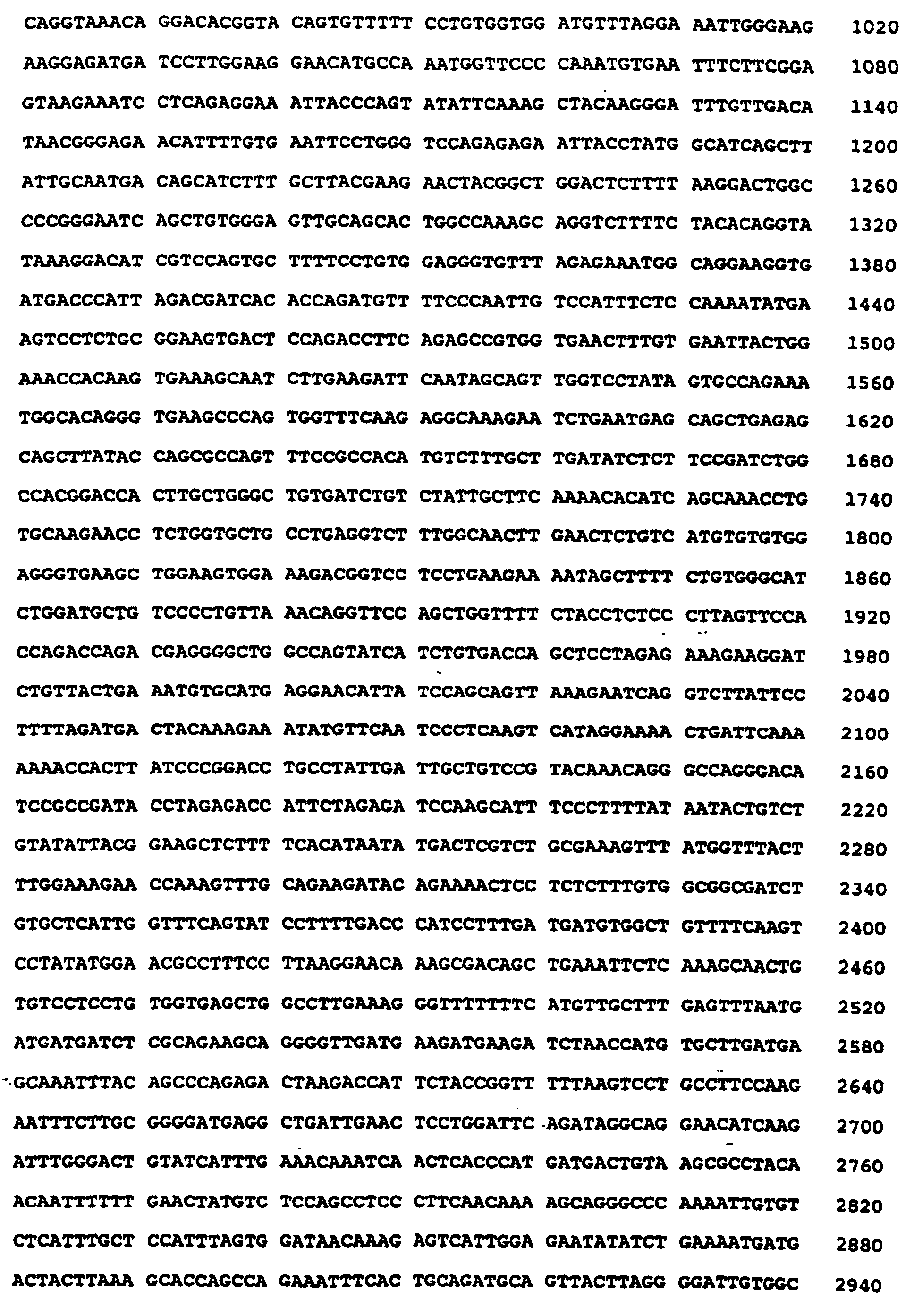
- the cDNA sequence coding of the neuronal apoptosis inhibitor protein is provided and set out in Table 4.
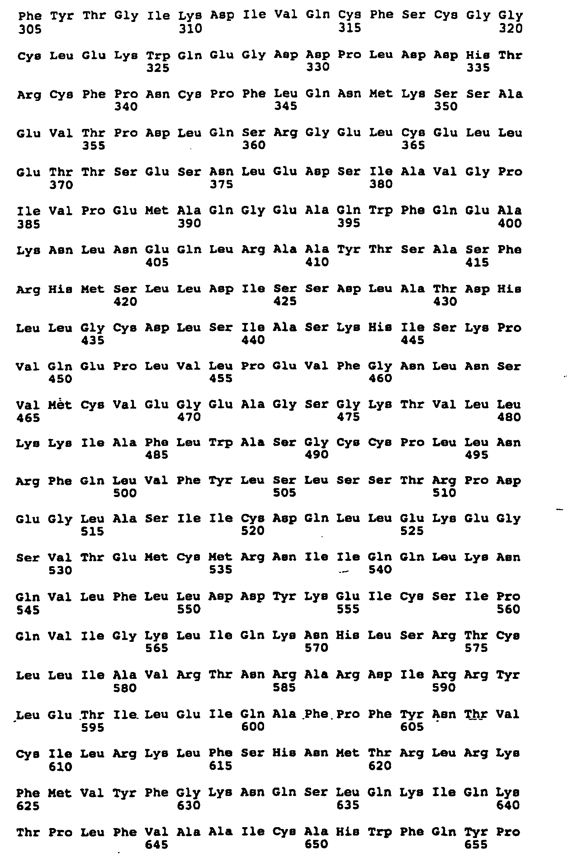
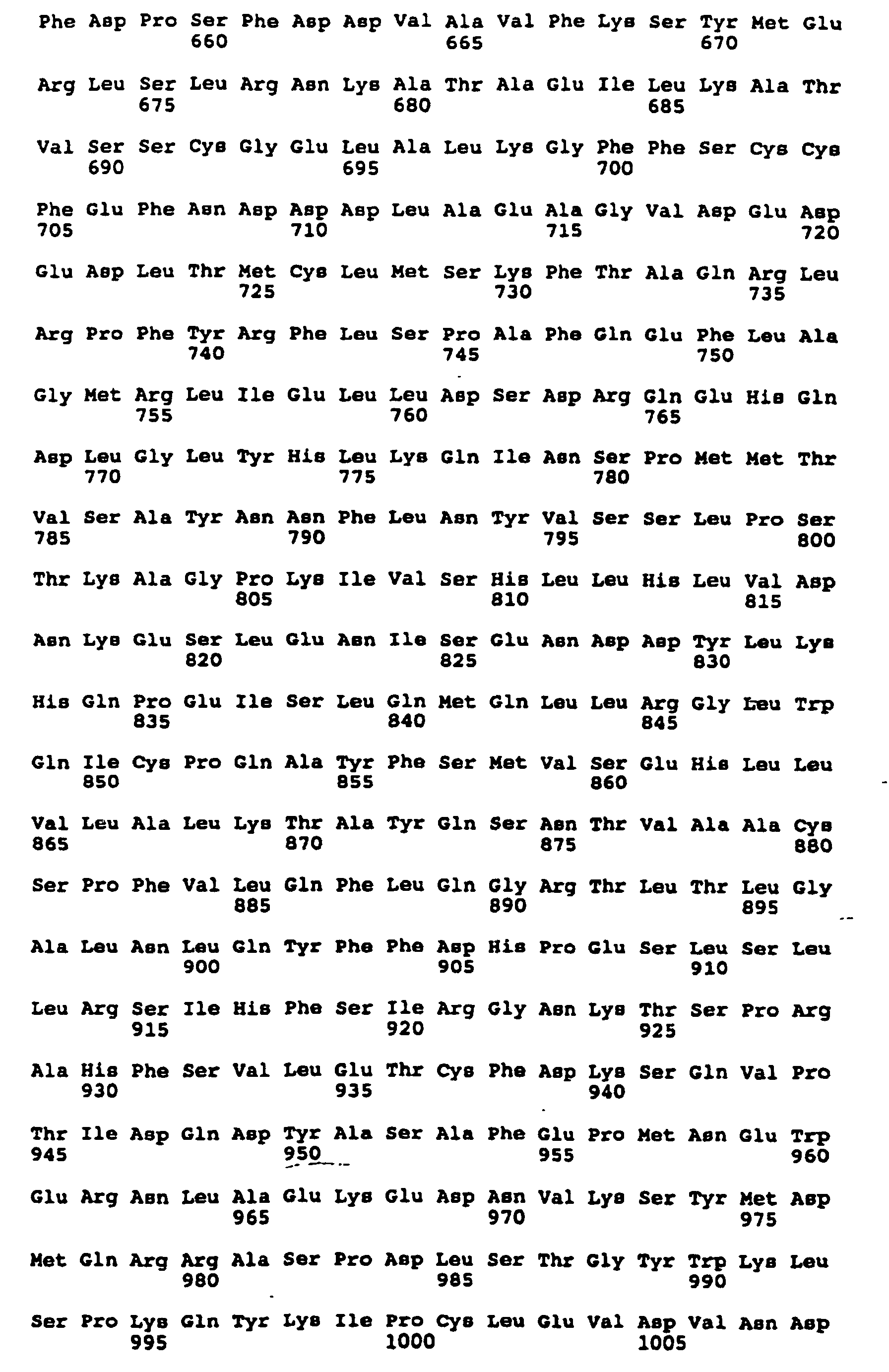
- the predicted amino acid sequence of the neuronal apoptosis inhibitor protein is provided from the cDNA sequence.
- a deletion of the neuronal apoptosis inhibitor protein gene was discovered in persons with Type I, II and III Spinal Muscular Atrophy disease.
- the discovery of the neuronal apoptosis inhibitor protein gene deletion provides a diagnostic indicator for use in the diagnosis of Spinal Muscular Atrophy.
- a human gene which maps to the SMA containing region of chromosome 5q13 said gene spanning approximately 80kb of genomic DNA, the gene characterized by exons 1 to 17 of Sequence ID No. 1 encoding neuronal apoptosis inhibitor protein.
- the gene comprises exons 1 through 17 of approximately 5.5 kb and having a restriction map for exons 2 through 11, as shown in Figure 8.
- exons 1 through 17 have a restriction map for exons 2 through 16, as shown in Figure 9D.
- a human gene of the above aspects wherein exons 5 through 16 code for the NAIP protein having an amino acid sequence biologically functionally equivalent to the amino acid sequence of Sequence ID No. 2.
- the human gene of the above aspects have exons 5 through 16 with a cDNA sequence biologically functionally equivalent to the cDNA sequence of Sequence ID No. 1.
- a purified nucleotide sequence comprises genetic DNA, cDNA, mRNA, anti-sense DNA or homologous DNA corresponding to the cDNA sequence of Sequence ID No. 1.
- a DNA molecule sequence coding for the NAIP protein having sequence ID No. 2.
- a purified DNA sequence consists essentially of DNA Sequence ID No. 1.
- a purified DNA sequence consists essentially of a DNA sequence coding for amino acid Sequence ID No. 2.
- a purified DNA sequence comprises at least 18 sequential base of Sequence ID No. 1.
- DNA probes, PCR primers, DNA hybridization molecules and the like may be provided by using the purified DNA sequence of at least 18 sequential bases.
- NAIP protein encoded by the above DNA sequences.
- NAIP protein comprising an amino acid sequence biologically equivalent to the amino acid sequence of sequence ID No. 2.
- NAIP protein consisting essentially of the amino acid sequence of Sequence ID No. 2.
- NAIP protein fragment comprises at least 15 sequential amino acids of Sequence ID No. 2.
- a method for analyzing a biological sample to determine the presence or absence of a gene encoding NAIP protein comprises:
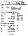
- sequence numbering Scheme #1 The original numbering of exons for the NAIP gene begin with exon 0 and progressed through exon 16. This is identified in drawings as sequence numbering Scheme #1. However, for conventional exon numbering, it is preferable to begin with exon 1 and progress through to exon 17-. This is now identified as sequence numbering Scheme #2.
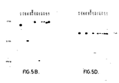
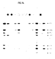
- Primary PCR of products shown in panels A and B was with exon 1 primer 1884 and exon 13 primers 1285 or 1974 and those in panel C with exon 6 primer 1919 and exon 13 primer 1285.
- Secondary PCR reactions for panel A used exon 4 primer 1886 and exon 13 primer 1974; for panel B, exon 5 primer 1864 and exon 11 primer 1979 and for panel C, exon 9 primer 1844 and exon 13 primer 1974.
- Panel A Failure or amplification of reduced products can be seen in panel A for spinal cord and lymphoblast tissue for samples a2, a3, a4, a5, a6 and a7.
- Panel B also shows amplification of reduced size bands in a2 and a3, and in a7 a larger product in keeping with an insertion.
- Panel C shows reduced band size in keeping with deletions of exons 11 and 12 (Scheme #2) in a2, a3, a9 and a11.
- This invention is directed to the identification, location and sequence characteristics of a gene which encodes Neuronal Apoptosis Inhibitor Protein (NAIP).
- NAIP Neuronal Apoptosis Inhibitor Protein
- SMA Spinal Muscular Atrophies
- the gene comprises exons 1 through 17 of approximately 5.5 kb and has a restriction map for exons 2 through 11, as shown in Figure 8.
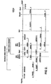
- An updated restriction map for exons 2 through 16 is provided in Figures 9D and 11A.
- the gene is considerably longer than the sequence for exons 1 through 17.
- Considerable intron information exists between the exons which has not yet been sequenced. From the standpoint of diagnosing SMA, the sequence information of exons 1 through 17 is very valuable.
- the normal sequence is provided in Table 4, as well as being listed under Sequence ID No. 1. Any genetic mutation, that is, changes in the DNA sequence, whether they be due to deletion, entire absence of gene substitution or polymorphisms and the like, are or can be causative of the disease. The most common mutations are thought to be:
- Any form of biological assay may be employed to diagnose a person's susceptibility to SMA by virtue of conducting a biological assay to determine the normal sequence or absence or presence of mutations in the normal sequence.
- Such biological assays may include DNA hybridization by use of DNA probes and the like, restriction enzyme analysis, PCR amplification of the relevant portions of the sequence, messenger RNA detection and DNA sequencing of the relevant portions of the sequence, as isolated from chromosome 5 of the human biological sample. It is appreciated that a variety of the above generally identified biological assay procedures may be conducted where the preferred techniques are as follows:
- SMA diagnoses will be conducted in two ways. Initially, the genome of the human at risk will be assayed for the absence of NAIP exons 5 and 6. These exons are found to be absent with a frequency of .05% in the general population and 50% in Type 1 SMA. The second approach will be to assess the number of copies of the NAIP gene in the individuals being tested. We have observed that there is a general depletion of both deleted and intact forms of the NAIP gene, in individuals with SMA. By using a densitometric approach to-assess the number of gene copies, an accurate assessment of the risk having SMA can be established. The best correlation is observed for exons 2 through 4 and exon 13.
- NAIP protein has significant homology with proteins for inhibiting cell apoptosis.
- any neurodegenerative disease which is based on neuronal cell apoptosis can now be predicted by use of the DNA sequence information of the NAIP gene.
- Such neuronal cell apoptosis is most likely linked to mutations in the NAIP gene similar to the mutations associated with SMA or other mutations in the gene which affect the biological activity of the NAIP protein inhibiting neuronal apoptosis.
- RT-PCR is a rapid technique for the analysis of RNA transcripts which is a crucial part of several molecular biology applications. This method is much more sensitive and efficient than traditional Northern blot, RNA dot/slot blots, and in situ hybridization assays. The sensitivity of such a technique allows one to study RNA transcripts of low abundance or RNA isolated from small amounts of cells. In addition, an entire panel of transcripts can be analyzed simultaneously.
- RNA is first isolated from tissues or cells and then is used as a template for reverse transcription to complimentary DNA (cDNA).
- the reverse transcription (RNA-directed synthesis of DNA), is catalyzed by the enzyme reverse transcriptase.
- the cDNA is then used as the template for PCR using primers designed to amplify a selected cDNA region.
- the product is analyzed by agarose gel electrophoresis.
- the amplified cDNA is identified by the size of the PCR product which is predicted from knowledge of the cDNA nucleotide sequence.
- the PCR product can be further validated by restriction digestion, hybridization or nucleotide sequencing.
- RT-PCR Enzymatic Amplification of RNA by PCR
- This method is used to enzymatically amplify RNA using PCR.
- RNA and cDNA primer are coprecipitated by adding together poly(A) + RNA, cDNA primer, and water. Sodium acetate is added and ethanol. This is precipitated overnight over -20°C. The pellet is collected after microcentrifugation. The pellet is washed with ethanol. Then water, Tis-HCl, and KCl are added and the mixture is heated to 90°C and then cooled slowly to 67°C. Microcentrifuge and incubate 3 hours at 52°C. This final annealing temperature may be adjusted according to base composition of primer. Alternatively, the primer can be annealed to the RNA by mixing poly(A) + RNA, cDNA primer, and water. This mixture is heated 3 to 15 minutes at 65°C. To the cooled mixture, add reverse transcriptase buffer.
- the cDNA is now synthesized.
- Tris-Cl/EDTA mix then buffered phenol and vortex.
- Microcentrifuge. Add sodium acetate and ethanol to aqueous phase. Mix and precipitate overnight at -20°C. Microcentrifuge, dry pellet, and resuspend in water.
- the cDNA is then amplified by PCR.
- the mixture contains prepared cDNA, amplification, dNTP mix, amplification buffer, and water. Usually one of the amplification primers is the same as cDNA primer. If a different amplification primer is used, the cDNA primer should be removed from the cDNA reaction.
- the reaction mixture is then heated 2 minutes at 94°C, and microcentrifuged to collect condensate. Add Taq DNA polymerase, mix, centrifuge, overlay with mineral oil. Set up amplification cycles. The number of cycles is varied depending upon the abundance of RNA. Forty cycles are usually sufficient.
- the products are then analyzed by gel electrophoresis in agarose or nondenaturing polyacrylamide gels.
- the cDNA can also be introduced directly into the amplification step.
- any specifically referenced sequence includes any and all biologically functional equivalence thereof.
- listed protein sequences it is understood that such terminology includes any and all biologically functional equivalence thereof insofar as the intended purpose is concerned.
- the full length or partial length sequences of the DNA or protein may be used.
- at least 18 sequential bases of the DNA sequence are useful as hybridization probes, PCR primers and the like.
- at least 15 sequential amino acid sequences may be correspondingly useful in developing protein receptors such as monoclonal antibodies.
- Such monoclonal antibodies may be made in accordance with the standard techniques by developing hybridomas for producing monoclonals specific to certain antigenic determinants of the protein structure.
- exons 1 through 17 has been provided in sequence ID No. 1.
- the encoding portion of the sequence commences at the ATG codon of base 396 of exon 5.
- the encoding portion ends at the stop codon TAA of exon 16 at base position 4092.
- Exons 1 through 4 are at the 5' untranslated region and exon 17 is at the 3'unstranslated region.
- mutations or polymorphism in the untranslated regions may as well be causative of the disease so that sequence portions in the form of probes and the like in regions other than the region of significant IAP homology may be valuable in the diagnosis of SMA.
- sequence information of sequence ID No. 1 may be used in the construction of suitable cloning vectors for purposes of producing multiple copies of the gene or expression vectors for purposes of transfecting a host to produce significant quantities by recombinant techniques of the NAIP protein. Sections or fragments or full-length sequence information may be used in the construction of the cloning vectors or expression vectors depending upon the end use of such vectors.
- a YAC contig of the Spinal Muscular Atrophy (SMA) disease gene region along chromosome 5q13 was produced which incorporated the D5S435-D5S112 interval and encompassed 4 Megabases.
- the CATT-40G1 subloci on the cosmid array showed significant linkage disequilibrium with Spinal Muscular Atrophy indicating close proximity to the gene.
- delineation of the precise region containing the SMA gene was not possible based on this information alone.
- a PAC contiguous array containing the CATT region comprised of 9 clones and extending approximately 400 kb was constructed.
- pYAC yeast artificial chromosome plasmids
- Circular pYAC plasmids can replicate in E. coli.
- In vitro digestion of pYAC, ligation to exogenous DNA, and direct transformation of the subsequent linear molecules (with telomeric sequences at each termini) into yeast generates a library that can be screened by standard techniques.
- YAC constructs are as stable as natural chromosomes. They are good vectors for the construction of libraries from complex genomes such as the human genome. In addition, sequences which are unclonable in E. coli cosmid and lambda vectors are successfully cloned in YAC vectors.
- YAC vectors are normally propagated in bacteria as circular plasmids. Restriction enzyme target sites are arranged to produce two arms upon digestion, each of which contains a different selectable marker and terminates at one end in a telomere, the other in a blunt end. In addition, one of the arms contains an ARS element. The two arms are purified away from a linking fragment and ligated with donor DNA fragmented so as to leave blunt ends. The ligation mixture is used to transform yeast cells, and the selection conditions are such as to require the presence of both arms, the insert interrupts a third selectable marker which allows non-recombinant structures to be recognized.
- YAC clones were isolated from three libraries, constructed at the National Centers of Excellence(NCE, Toronto), the Imperial Cancer Research Fund (ICRF, London) (Larin et al., 1991) and the Centre d'Etude du Polymorphisme Humaine (CEPH, Paris) (Albertson et al., 1990), all of which were prepared from partial EcoRI digests of total DNA ligated into the YAC vector pYAC4. ICRF YAC clones were identified by probing library filters with 5q13.1 probes. YAC DNA from the NCE library was screened by PCR amplification, electrophoresed, immobilized onto Southern blots and hybridized with the radiolabelled STS product to identify positives.
- Yeast strains with YACs positive for 5q13.1 STSs were grown on selective plates and examined for stability in the following manner: 4 colonies of each were grown for preparation in agarose blocks, yeast chromosomal DNA was separated by pulsed field gel electrophoresis and transferred to filters and the size and number of YAC clones contained within each yeast colony was determined by hybridization with radiolabelled total human genomic DNA. Positive clones were confirmed either by hybridization or PCR amplification with the original probe. Only YAC 24D6-2 contained some colonies with more than one YAC.
- YAC end clones and inter- Alu products were isolated by vector- Alu PCR and inter- Alu PCR respectively.
- the location of these products within 5q11-13 was confirmed by hybridization to Southern filters of the somatic cell hybrids HHW105 (Dana et al., 1982), containing the entire chromosome 5, and HHW1064 (Gilliam et al., 1989), a derivative containing chromosome 5 with a deletion at 5q11.2-13.3. Many of these probes demonstrated hybridization profiles indicative of locations both within the 5q11-13 region and elsewhere on chromosome 5.
- primers specific for the ends of each YAC were generated from the sequences of YAC end clones isolated by vector- Alu -PCR.
- each new STS to 5q11 - 13 was determined by PCR amplification of DNA from the somatic cell hybrids HHW105 and HHW1064. In a few cases it was found that a primer pair contained a chromosome 5 repetitive sequence as the PCR amplified products from both HHW1064 and HHW105 were positive. Formulation of new STS primers resulted in the amplification of products specific to the 5q11-13 region. End clone hybridization and STS analysis performed on all YACs confirmed the orientation and location of each YAC.
- the assembly of a contiguous array of YACs covering the SMA interval was initiated from two markers which flank SMA; D5S125 (Mankoo et al., 1991), which lies centromeric to D5S435 and the more telomeric marker D5S112 (Lien et al., 1991) (see Figure 1).
- D5S125 Mankoo et al., 1991
- D5S112 telomeric marker
- D5S112 telomeric marker pJK53
- D06100 was shown to extend the furthest centromerically based on end clone STS analysis.
- the centromeric end of this YAC identified two YACs from the NCE library, 1281 and 1284.
- YACs positive for the D5S125 or D5S435 markers were not found in the ICRF or NCE library thus the CEPH library was screened, from which clones containing D5S435 were isolated.
- a microsatellite polymorphism mapping into the center of the gap, CATT-1 was utilized to detect three YACS, 24D6-2, 27H5 and 33H1O. These YACs were shown to be linked to both the centromeric and the telomeric YACs (1281, 1284) by STS analysis. Internal YAC products generated by Alu PCR were utilized to probe all YACs establishing the degree of overlap.
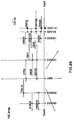
- a restriction map of the critical SMA region was constructed from the STS Y116U (Kleyn et al., 1993), approximately 100 kb proximal to D5S629, to the STS Y107U (Kleyn et al., 1993), which lies approximately 500 kb distal to D5S557 (see Figure 2).
- additional YACs isolated from the CEPH library (Kleyn et al., 1993), mapping within this region were included in the analysis.
- YACs 24D62, 27H5, 33H1O, 155H11, 76C1, 235B7, 184H2, 428C5, and 81B11 were partially digested utilizing the rare cutter restriction endonucleases NotI, BssHII, SfiI, and RsrI .
- Southern blots of the Pulse Field Gel Electrophoresis (PFGE) separated restriction products were hybridized with YAC left arm and right arm specific probes which revealed the positions of cleavage sites from both ends of each YAC. The orientation and overlap of the YACs had been previously determined based on STS analysis, therefore the position of the rare cutter sites among the overlapping YACs were compared.
- PFGE Pulse Field Gel Electrophoresis
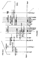
- Bi-directional walking was initiated from these 4 cosmid subloci. Positive hybridization was observed for cosmid 25OB6 with one end of 58G12 and for 192F7 with the other end resulting in the ordering of cen-192F7-58G12-25O86-tel (Figure 4). All cosmids which contained the CATT-192F7 allele were mapped to this location based on the size of their CATT-1 allele and their restriction enzyme profiles.
- the CATT-192F7 sublocus is telomeric to the STR CMS-1, which itself lies telomeric to the CATT-40G1 sublocus.
- Cosmids identified by end clones which hybridized to HHW1064 were eliminated and walking was continued by utilizing a different inter-Alu product from the clone of origin, which was verified in the same manner. Cosmid sizes were calculated by the addition of EcoRI restriction fragments and the extent of overlap was determined by the addition of those fragments in common.
- a 5X cosmid library was produced from YAC 76C1.
- the STS Y98T identified three cosmids including one previously identified by the probe p2281.8, derived from a chromosome 5 library clone, 228C8, also containing the STS Y98T.
- An end product of this cosmid hybridized to ten cosmids.
- an end fragment of a CATT40G1 sublocus was shown to hybridize to four of these ten cosmids thus linking CATT-40G1 and CMS-1 with the more centromeric STS Y98T ( Figure 4).
- Filter hybridization and STS mapping experiments indicated a second more telomeric location of the CATT40G1 sublocus. A duplication of this sublocus would agree with genotype data in our SMA kindreds (McLean et al., in press).
- An EcoRI restriction map was generated utilizing a minimal set of cosmids necessary to span the region. To ensure the reliability of the contig, we sought to integrate it with the contig constructed from the chromosome 5 specific library. Concordance of the contigs was evident by comparison of the restriction maps, the position of probes and STSs on the map and Alu -PCR fingerprinting. In this manner the size of the contig was estimated to be 210 kb. A directed walking strategy has thus resulted in the generation of a single contiguous set of cosmids containing the CATT-1 cluster of subloci with known centromere/telomere orientation.
- YAC 428C5 does not contain the CATT40G1 sublocus upon PCR amplification, this may be explained either by a null allele in the chromosome from which the YAC was derived or a deletion in the YAC. We have previously observed null alleles in individuals at distinct CATT-1 subloci. A second more telomeric location of CATT-40G1 was determined by the hybridization to CATT40G1 cosmids of the probes pGA- 1, pL7, and pZY8 all of which bind the more telomeric YACs 33H1O, 24D62.
- the STRs CMS-1 and D5F150 were present in a variable number of copies per chromosome 5.
- STS analysis localized CMS-1 to YACs 428C5, 76C1, 81B11 and 27H5 with allele sizes of 5, 4, 4 and 3, and 4 respectively.
- PCR amplification of genomic DNA revealed up to four alleles per individual indicating as many as two copies per chromosome.
- D5F150 was present at two locations within the cosmid array yet only one location was detected in the YAC contig.
- D5F151 was not detected within our cosmid array nevertheless it was placed at the centromeric end of YAC 33H10, which encompasses the cosmid array, based on the positive amplification of YAC 428C5.
- One location of D5F149 was detected on both our cosmid and YAC clones.
- Our data suggested, as with CATT-1, the existence of null alleles and/or instability of the CMS-1, D5F150, D5F151, D5F149 sequences in YACS.
- a deletion event was observed in hybridization with an 800 bp EcoRI fragment isolated as a single copy probe from the CATT-40G1 containing cosmid 234A1 from the chromosome 5 specific cosmid library. Probings of YAC DNA failed to detect this fragment in any of our YACs. Hybridization to genomic DNA of several individuals did not identify any deletion events thus this sequence may be susceptible to instability in the YACS. Sequencing of this fragment did not reveal any exons or coding region.
- D5F153 and CATT-1 are related STRs which appear to have diverged from a common ancestor.
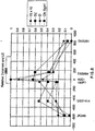
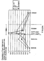
- a linkage disequilibrium analysis employing 5 complex and simple tandem repeats mapping to the SMA region was conducted. Two of the polymorphisms employed in this analysis were the CATT-40G1 and CATT-192F7 subloci which we mapped to our cosmid array. Specific amplification of the two individual subloci was achieved by constructing primers ending on sequence polymorphisms in the region flanking the CA repeat. A clear linkage disequilibrium peak was observed at the CATT-40G1 sublocus as shown in Figure 6.
- Two genomic libraries were constructed by performing complete and partial (average insert size 5 kb) Sau3A1 on PAC 125D9 and cloning the restricted products into BamH1 digested Bluescript plasmids. Genomic sequencing was conducted on both termini of 200 clones from the 5 kb insert partial Sau3A1 library in the manner of (Chen et al., 1993) permitting the construction of contiguous and overlapping genomic clones covering most of the PAC. This proved instrumental in the elucidation of the neuronal apoptosis inhibitor protein gene structure.
- PAC 125D9 is cleaved into 30 kb centromeric and 125 kb telomeric fragments by a NotI site (which was later shown to bisect exon 7 of the PAC 125D9 at the beginning of the apoptosis inhibitor domain.
- the NotI PAC fragments were isolated by preparative PFGE and used separately to probe fetal brain cDNA libraries. Physical mapping and sequencing of the NotI site region was also undertaken to assay for the presence of a CpG island, an approach which rapidly detected coding sequences.
- the PAC 125D9 was also used as a template in an exon trapping system resulting in the identification of the exons contained in the neuronal apoptosis inhibitor protein gene.
- the multipronged approach in addition to the presence of transcripts identified previously by hybridization by clones from the cosmid array (such as, GA1 and L7), resulted in the rapid identification of six cDNA clones contained in neuronal apoptosis inhibitor protein gene.
- the clones were arranged, where possible, into overlapping arrays. Chimerism was excluded on a number of occasions by detection of co-linearity of the cDNA clone termini with sequences from clones derived from the PAC 125D9 partial Sau3A1 genomic library.
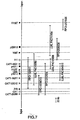
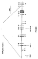
- a human fetal spinal cord cDNA library was probed with the entire genomic DNA insert of cosmid 250B6 containing one of the 5 CATT subloci. This resulted in a detection of a 2.2 kb transcript referred to as GA1 which location is shown in Figure 7. Further probings of fetal brain libraries with the contiguous cosmid inserts (cosmids 40G1) as well as single copy subclones isolated from such cosmids were undertaken. A number of transcripts were obtained including one termed L7. No coding region was detected for L7 probably due to the fact that a substantial portion of the clone contained unprocessed heteronuclear RNA.
- the extended GA1 transcript was compared to other known sequences to reveal that its amino acid sequence had significant homology to the inhibitor apoptosis polypeptides of Orgyia Pseudotsugata and Cydia Pomonella viruses (Table 3). This sequence analysis revealed the presence of inhibitor apoptosis protein homology in exons 5 and 6.
- the cDNA sequence of NAIP shown in Table 4 allows one skilled in the art to develop from this gene, primers, probes and also antibodies against the protein product.
- the cDNA sequence of Table 4 may be used in recombinant DNA technology to express the sequence in an appropriate host in order to-produce the neuronal apoptosis inhibitor protein. In this manner, a source of neuronal apoptosis inhibitor protein is provided.
- deletions in the sequence may also be detected, for instance, in the disorder Spinal Muscular Atrophy.
- the NAIP gene contains 17 exons comprising at least 5.5 kb and spans an estimated 80 kb of genomic DNA.
- the NAIP coding region spans 3698 nucleotides resulting in a predicted gene product of 1233 amino acids.
- NAIP contains two potential transmembrane regions and an intracellular inhibitor of apoptosis domain immediately contiguous with a GTP binding site. Searches of the protein domain programs generated the following results:
- a cDNA20.3 probe was found by using the entire PAC 125D9 as a probe to screen cDNA libraries. Probing of genomic southerns with cDNA probe 20.3 revealed the absence of a 9 kb EcoRI band in a Type III consanguineous family. This information mapped the NAIP gene deletions to exons 5 and 6. Thus the deletion covers the exon containing the rare NotI restriction site and the exon immediately downstream. Primers in and around these exons were constructed revealing the absence of amplification from 3 Type I and 3 Type III SMA individuals.
- NAIP transcript isolation was achieved by probing a human fetal brain cDNA library with the entire 28 kb genomic DNA insert of cosmid 250B6 that contains one of five CATT subloci present in the cosmid library. This resulted in the detection of a 2.2 kb transcript that ultimately proved to be exon 14 of the NAIP gene. Further probing of fetal brain libraries with the contiguous cosmid inserts (cosmid 40G1), as well as single copy subclones isolated from such cosmids identified a number of transcripts including the L7 transcript that ultimately proved to contain exon 13 of the NAIP locus. No coding region was detected for L7, probably due to the fact that a substantial proportion of the clone contained unprocessed heteronuclear RNA, obscuring its true nature.
- PAC 125D9 the completed genetic and linkage disequilibrium analyses and construction of the PAC contiguous array identified PAC 125D9 as having a good probability of containing the SMA locus.
- Four PAC 125D9 genomic libraries were constructed by performing complete and partial (average insert size 5 kb) Sau3AI, BamHI and BamHI/NotI digests on the PAC insert and cloning the restricted products into plasmid vector. High through put genomic sequencing was conducted on both termini of 200 clones from the 5 kb insert partial Sau3AI digestion library in the manner of (Chen et al., 1993), permitting the construction of contiguous and overlapping genomic clones covering most of PAC 125D9 (data not shown). This has proven instrumental in elucidating the gene structure of the NAIP locus.
- PAC 125D9 is divided into 24 kb centromeric and 130 kb telomeric fragments by NotI digestion, bisecting exon 6 of the NAIP gene at the beginning of the first potential transmembrane domain mapping upstream of the inhibitor of apoptosis homologous domains ( Figure 11 and Table 4).
- the NotI PAC fragments were isolated by preparative PFGE and used separately to probe human fetal brain cDNA libraries. Physical mapping and sequencing of the NotI site region was also undertaken to assay for the presence of a CpG island, an approach that rapidly detected coding sequence.
- the PAC was also used as a template in an exon trapping system (Church et al., 1994) resulting in the identification of the NAIP gene exons 5, 12, 16 and 17.
- Both IAPs contain in their amino terminus an 80 amino acid BIR (baculovirus IAP repeat) motif that, after an intervening sequence of approximately 30 residues, is duplicated with 33% identity (Clem and Miller, 1993).
- BIR baculovirus IAP repeat
- the same phenomenon is observed in NAIP; amino acids 185-250 encoded by exons 6, 7 and 8 are 35% homologous to amino acids 300-370 encoded in exon 10, 11 and 12. The greatest stretch of homology is observed over a 53 amino acid region with 29 identical amino acids.
- NAIP contains two potential transmembrane regions that bracket an inhibitor of apoptosis domain and a contiguous GTP binding site. Additional searches of protein domain programs generated the following more specific results than the aforementioned protein domain evaluation.
- exons that comprise 400 bp of 5' untranslated region (5'UTR); it is possible that more exist.
- a striking feature of this region is the presence of a perfect duplication of a 90 bp region in the 5' UTR before exon 2 and in the region bridging exons 2 and 3 (Table 4).
- Occludin homologous sequence has been detected in four different cDNA clones and two isoforms of the gene.
- the possibility of the occludin sequence representing a coding exon of the NAIP gene with the putative 3' UTR actually being heteronuclear RNA is also unlikely given the consistency with which the 3' UTR is observed and the presence of in frame translational stop codons mapping upstream of the region of occludin homology.
- Preliminary RT-PCR analysis indicates that the occludin tract is transcribed.
- RT-PCR reverse transcriptase-PCR
- the clones 238D12 and 30B2 were noted to show significant sequence similarity with 125D9 but not to contain the NotI site in PAC 125D9 that is located in NAIP exon 6. This indicated the possibility of duplicated copies of the NAIP gene and so further analysis by hybridization of Southern blots containing PAC DNA with NAIP exon probes and PCR STS content assessment was undertaken. In this manner, two aberrant versions of the NAIP locus were detected, one with exons 2 to 7 deleted (PAC 238D12), and another with exons 6, 7 and 12 to 15 deleted (PACs 30B2 and 250I7).
- genomic DNA Southern blots hybridized with NAIP exon probes revealed more bands than would be expected with a single intact copy of the NAIP gene.
- probing of blots containing BamHI restricted genomic DNA with NAIP exons 3-11 should lead to a single band comprised of equal sized contiguous 14.5 kb BamHI fragments in the intact NAIP locus ( Figure 11).
- the 9.4 fragment Bam HI has been subcloned from a cosmid and found to contain exons 8-11 with a deletion incorporating exons 2 to 7 occurring just upstream of the 8th exon ( Figure 11).
- the 23 kb band is generated by a 6 kb deletion removing a BamHI site leading to the replacement of the two contiguous 14.5 kb BamHI fragments with a 23 BamHI fragment containing exons 2 to 5 and 8 to 11 and lacking exons 5 and 6 as depicted in Figure 11.
- the 9.4 kb band was seen in all individuals in keeping with the presence of at least one copy of exons 5 and 6 in each of the approximately 900 individuals tested.
- the 3 kb band was observed in every individual reflecting a virtually complete dispersion of some form of the exon 5 through 6 deleted NAIP gene in the general population.
- the variable band dosage observed for the 3 kb band suggested that the number of copies of the exon 5-6 deleted NAIP gene is polymorphic possibly ranging as high as 4 or 5 copies per genome.
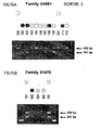
- PCR analysis was then extended to 110 SMA families, employing exon 5 and 6 primers. Seventeen of 38 (45%) Type I SMA individuals and 13 of 72 (18%) Type II and III SMA individuals were homozygously deleted for these exons. Assuming random assortment of chromosomes and therefore taking the square of the observed frequency of homozygous exon 5 through 6 deleted individuals yields estimated frequencies for exon 5 through 6 deleted chromosomes of 67% in Type I SMA and 42% in Type II/III SMA. PCR analysis was next conducted on 168 parents of SMA children revealed failure of amplification suggesting homozygous deletion of exon 5 and 6 in three individuals. This finding was confirmed by Southern analysis in the two cases with sufficient DNA for this assay.
- the protein sequence encoded by the deleted exon 5 and 6 IAP motif is approximately 35% homologous to the IAP motif encoded in exons 10 and 11 possibly accounting for the absence of discernible phenotype in the three exon 5 through 6 deleted individuals.
- One possible model is that a single copy of exon 5 through 6 deleted NAIP on each chromosome results in the mild SMA phenotype, while individuals with greater than 3 or 4 copies of the exon 4-5 deleted NAIP locus are clinically unaffected.
- duplication of the SMA gene underlies the disease has recently been proposed by DiDonato et al. (1994).
- RT-PCR amplification of RNA from SMA and non-SMA tissue The results of RT-PCR amplification using RNA from both non-SMA and SMA individuals as template are shown in Figure 16.
- RNA from non-SMA tissues as template and reverse transcribing from exons 10 or 13 consistently amplified product of the expected size.
- similar RT-PCR experiments on RNA from SMA tissue revealed no amplification in five cases in keeping with the marked down regulation or complete absence of the intact transcript in such individuals ( Figure 16A).
- the RNA obtained from the SMA tissues was no more than 12 hours post-mortem.
- difficulty with amplification was seen for all SMA tissues which suggests against the possibility that NAIP is transcribed solely in the motor neuron with depletion of this cell type in SMA resulting in RT-PCR failure in spinal cord tissue.
- the discovery of a neuronal apoptosis inhibitor protein gene in the SMA region of chromosome 5 demonstrates that the SMA condition is a result of deletions in the apoptosis inhibitor protein domains.
- the long time survival of motorneurons is dependent on the production of complete neuronal apoptosis inhibitor protein.
- the deletion of the apoptosis inhibitor protein domain compromises the protein activity.
- the identified region of 5q13.1 contains a variable number of copies of intact and partially deleted forms of the NAIP gene. While we cannot rule out the presence of additional loci in 5q13.1 that when mutated contribute to the SMA phenotype, we believe that mutations of NAIP gene are necessary and possibly sufficient for the genesis of SMA. In contrast to most autosomal recessive diseases where causal mutations are usually detected in the single copy of a given gene, we propose that an SMA chromosome is characterized by a paucity or, for severe SMA mutations, an absence of both the intact NAIP gene as well as that version which has had exons 3 and 4 deleted. The genesis of such chromosomes may involve unequal crossovers leaving the chromosome depleted for these loci with the resulting absence of the NAIP-gene product leading to SMA.
- the evidence in support of mutations in or the absence of the NAIP gene causing SMA includes the following:
- NAIP region of 5q13.1 has more similarity to the area of chromosome 6 containing CYP21, the gene that encodes steroid 21-hydroxylase (Wedell and Luthman, 1993).
- CYP21 which when mutated causes an autosomal recessive 21-hydroxylase deficiency, has been observed in 0-3 copies in individuals.
- CYP21P There also exists in the region a variable number of inactive pseudogene copies of CYP21 known collectively as CYP21P.
- the majority of the CYP21 mutations that have been observed in 21-hydroxylase deficiency can also be found in some form of CYP21P and it is thought that the pseudogenes act as a source of the mutations observed in CYP21.
- NAIP genes are analogous to CYP21P only instead of the gene conversion postulated for CYP21/CYP21P it is possible that unequal crossing over results in chromosomes deleted for forms of the NAIP gene that encode functional protein.
- the existence of a polymorphic number of mutated NAIP genes on 5q13.1 is a credible mechanism for generation of SMA chromosomes in this fashion.
- NAIP shows significant homology with the two baculoviral gene products, CpIAP and OpIAP, that are capable of inhibiting insect cell apoptosis (Table 4). Insect cell apoptosis following baculoviral infection has been well documented and is postulated to be a defence mechanism. Premature death of infected insect cells result in an attenuation of viral replication (Clem and Miller, 1994a). CpIAP and OpIAP are thought to represent baculoviral responses to this apoptotic mechanism. Both act independently of other viral proteins to inhibit host insect cell apoptosis, thereby permitting increased viral proliferation (Clem and Miller, 1994a, 1994b). They are known to be strongly similar only to each other; until now no sequences similarities with cross phyla proteins have been reported. Their mode of action is unknown, although some interaction with DNA has been postulated.
- NAIP apoptosis inhibitor in the motor neuron.
- Transfection assays employing NAIP both in insect and mammalian neuronal cells will help in this regard.
- ligand binding of the carboxy terminus of the NAIP activates the GTP binding site which in turn activates the IAP domain.
- the survival of a motor neuron might, therefore, be dependent on the presence of the ligand(s): should the concentration drop below a critical threshold, the IAP domains cease to function with ensuing cell death. This represents a possible mechanism for the natural winnowing of motor neurons observed in embryogenesis.
- the source of the ligand might be postulated to be either muscle cells or Schwann cells.
- the embryogenesis of motor neurons might, therefore, be viewed as a competition between the cells with only those that make sufficient contacts to maintain the NAIP occupancy rate surviving.
- NAIP does inhibit apoptosis
- Bcl-2 a constituent of a previously uncharacterized mammalian apoptotic pathway or a (presumably) upstream component of the pathway involving the human inhibitor of apoptosis, Bcl-2 (Vaux et al., 1988; Hockenberry et al., 1990; Garcia et al., 1992).
- Assays employing apoptosis inhibition deficient baculoviral strains have revealed that Bcl-2 does not complement the deficiency in such assays (Clem and Miller, 1994b).
- NAIP is a functional homolog of the baculoviral IAPs
- this observation might suggest a role in a previously uncharacterized eucaryotic apoptotic pathway.
- NAIP represents an intersection of a novel apoptotic mechanism with the neurotrophic cytokine, ciliary neurotrophic factor (CNTF, Raff et al., 1993; Meakin and Shooter, 1993) or one of the downstream components of this pathway (Stahl et al., 1994).
- CNTF null mice show a pathologic picture that is similar to that of SMA with normal development of the neurons initially followed by their progressive apoptotic depletion (Masu et al., 1993).
- the role of the lipid attachment site in NAIP is unknown. Similar sites have been known to serve as procaryotic protein leader sequences usually situated in the protein's amino terminus. We have detected the consensus pattern in 218 human sequences in the Swiss-Protein Database (release 28). These sequences are present in a variety of functional settings; transmembrane regions, signal sequences, extracellular and cytoplasmic domains.
- the lipoprotein attachment site is extracellular and binds a constituent of the Schwann cell proteolipid in a manner that has been postulated for the apoptosis inhibiting interaction of integrin with the extracellular matrix (Meredith et al., 1993; Frisch and Francis, 1994). Furthermore, the site may play a more active role in the hepatic form of the NAIP that we have observed on Northern blot analysis. It is noteworthy that serum fatty acid abnormalities have been detected in children with SMA (Kelley and Sladky, 1986).
- the identified region of 5q13.1 contains, in addition to the NAIP gene, a variable number of copies of internally deleted and truncated forms of the gene.
- a lack or absence of both the intact NAIP gene and the NAIP locus with exons 5 and 6 deleted from a given individual's genome are likely to cause SMA.
- the identification of NAIP has allowed us to develop accurate molecular based diagnoses of SMA as well as directing the formulation of conventional and genetic therapies for these debilitating conditions.
- the identification of genes showing homology with the NAIPlocus and proteins that interact with NAIP may help in the continuing elucidation of apoptotic mechanisms in mammalian cells.
- Genotyping with microsattelite markers was as outlined in MacKenzie et al. (1993) and McLean et al. (1994). The following 5q13.1 loci were used as described: D5S112 (Brzustowitcz et al., 1990), D5S351 (Hudson et al., 1992), D5S435 (Soares et al., 1993), DSS557 (Francis et al., 1993), D5S629 and D5S637 (Clermont et al., 1994), D5S684 (Brahe et al, 1994), Y98T, Y97T, Y116T, Y122T and CMS (Kleyn et al., 1993), CATT (Burghes et al., 1994, McLean et al., 1994) and MAP1B (Lien et al., 1991).
- Cosmid and YAC contig assembly was as outlined in Roy et al. (1994).
- PACs were constructed as outlined in Sicilnou et al. (1994). Using these procedures three PAC libraries have been constructed with a combined total of 175,000 clones and propagated as individual clones in microtiter dishes (Ioannou et al., unpublished results). Pools derived from the three libraries (designated LLNL PAC1, RPCI1 and RPCI2) were screened with 5q13.1 STS's. Positive PACs were arranged into a contiguous and overlapping arrays by further analysis with additional STSs combined with probings of Southern blots containing PAC DNA by single copy genomic DNA and cDNA probes.
- Coding sequences from the PACs were isolated by the exon amplification procedure as described by Church et al. (1994). PACs were digested with BamHI or BamHI and BglII and subcloned into pSPL3. Pooled clones of each PAC were transfected into COS-1 cells. After a 24h transfection total RNA was extracted. Exons were cloned into pAMP10 (Gibco, BRL) and sequenced utilizing primer SD2 (GTG AAC TGC ACT GTG ACA AGC TGC).
- DNA sequencing was conducted on an ABI 373A automated DNA sequencer. Two commercial human fetal brain cDNA libraries in lambda gt (Stratagene) and lambda ZAP (Clontech) were used for candidate transcript isolation. The Northern blot was commercially acquired (Clontech) and probing was performed using standard methodology.
- primers used in the paper for PCR were selected for T m s of 60°C and can be used with the following conditions: 30 cycles of 94°C, 60s; 60°C, 60s; 72°C, 90s.
- PCR primer mappings are as referred to in the figure legends and text. Primer sequences are as follows:
- cDNA was synthesized in a 20 ⁇ l reaction utilizing 7 ⁇ g of total RNA.
- the RNA was denatured for 5 minutes at 95°C and cooled to 37°C.
- Reverse transcription was performed at 42°C for 1 hour after addition of 5 ⁇ l 5X reverse transtriction buffer, 2 ⁇ l 0.1 M DTT, 41 2.5 mM dNTPs, 8 units RNasin, 25 ng cDNA primer (1285) and 400 units of MMLV (Gibco, BRL).
- 1 ⁇ l of cDNA was utilized as template in subsequent 50 ⁇ l PCR reactions. 1 ⁇ l of this primary PCR was utilized as template for secondary PCR amplifications.
- ICRF Imperial Cancer Research Fund, CEPH: Centre d'Etude du Polymorphisme Humaine.
Landscapes
- Health & Medical Sciences (AREA)
- Life Sciences & Earth Sciences (AREA)
- Chemical & Material Sciences (AREA)
- Engineering & Computer Science (AREA)
- Molecular Biology (AREA)
- Biomedical Technology (AREA)
- Immunology (AREA)
- Biochemistry (AREA)
- General Health & Medical Sciences (AREA)
- Hematology (AREA)
- Medicinal Chemistry (AREA)
- Urology & Nephrology (AREA)
- Proteomics, Peptides & Aminoacids (AREA)
- Organic Chemistry (AREA)
- Neurology (AREA)
- Toxicology (AREA)
- Biophysics (AREA)
- Neurosurgery (AREA)
- Biotechnology (AREA)
- Cell Biology (AREA)
- Gastroenterology & Hepatology (AREA)
- Zoology (AREA)
- Microbiology (AREA)
- Genetics & Genomics (AREA)
- Food Science & Technology (AREA)
- Physics & Mathematics (AREA)
- Analytical Chemistry (AREA)
- General Physics & Mathematics (AREA)
- Pathology (AREA)
- Measuring Or Testing Involving Enzymes Or Micro-Organisms (AREA)
- Peptides Or Proteins (AREA)
- Medicines That Contain Protein Lipid Enzymes And Other Medicines (AREA)
- Investigating Or Analysing Biological Materials (AREA)
Abstract
Description
The method comprises:
- CTR -
- complex tandem repeat
- DNA -
- deoxyribonucleic acid
- PCR -
- polymerase chain reaction
- PFGE -
- pulsed field gel electrophoresis
- PAC -
- P1 artificial chromosome
- RNA -
- ribonucleic acid
- RT-PCR -
- reverse transcriptase-polymerase chain reaction
- STR -
- simple tandem repeat
- STS -
- sequence tag site
- YAC -
- yeast artificial chromosome
The long range restriction map of the overlapping YACs derived from different sources was mostly in agreement with the exception of 33H1O and 428C5. 428C5 has previously been documented to contain a deletion (Kleyn et al., 1993), evident by comparison of its STS content and its size of only 300 kb, indicating that it lies further centromeric than its placement in Figure 2. YAC 33H10, based on STS analysis contains an internal deletion and YAC 155H11 is chimeric at its telomeric end therefore rare cutter sites at the telomeric end of the map which could not be confirmed were not included. The results indicate the distance from the centromeric boundary D5S435 to the telomeric boundary D5S557 to be 1.4Mb in marked contrast to 400 kb as previously reported (Francis et al., 1993) but in agreement with one other estimate (Wirth et al., 1993). Furthermore, the D5S629-D5S557 interval can be estimated at 1.1 Mb and the distance of the genetically defined CMS1-SMA-D5S557 interval is approximately 550kb.
Furthermore, the identification of genes showing homology with the NAIPlocus and proteins that interact with NAIP may help in the continuing elucidation of apoptotic mechanisms in mammalian cells.














| The YACs isolated in this study, their size and library of origin are listed. NCE: National Centers of Excellence, Toronto, Ontario, Canada. ICRF: Imperial Cancer Research Fund, CEPH: Centre d'Etude du Polymorphisme Humaine. | ||
| YAC | SIZE | LIBRARY |
| 12H1 | 560kb | NCE |
| 12H4 | 270kb | NCE |
| 24D6 | 750kb | NCE |
| 27H5 | 630kb | NCE |
| 33H10 | 1.3Mb | NCE |
| H0416 | 390kb | ICRF |
| E0320 | 440kb | ICRF |
| G1138 | 850kb | ICRF |
| A0848 | 350kb | ICRF |
| D06100 | 580kb | ICRF |
| D0981 | 450kb | ICRF |
| 919C2 | 800kb | CEPH |
| 755B12 | 1Mb | CEPH |
| 754H5 | 500kb | CEPH |
| PROBE | SOURCE/ REFERENCE | PROBE | SOURCE/ REFERENCE |
| YD33 | STS developed from Alu- 5'-trp PCR product of YAC D06100 | Y13.1 | STS developed from inter Alu-5' PCR product of YAC 12H1(this study) |
| Y14.1 | STS developed from Alu- 3'-ura PCR product of YAC 12H4 (this study) | Y15.1 | STS developed from Alu-5'-ura PCR product of YAC 12H4 (this study) |
| Y9.2 | STS developed from inter-Alu-5' PCR product of YAC 27H5 (this study) | Y5.6 | STS developed fron inter-Alu-3' PCR product of YAC 24D6 (this study) |
| Y11.2 | STS developed from Alu- 3'-trp PCR product of YAC 33H10 (this study) | pZY8 | subcloned 1.3 kb HindIII fragment from cosmid 250B6 (this study) |
| H7T733 | Alu 33-T7 PCR product from cosmid 1H7 (this study) | p151.2 | subcloned 1.2 kb inter-Alu PCR product of cosmid 15F8 (this study) |
| G10T333 | Alu 33-T3 PCR product of cosmid IG10 (this study) | p402.1 | subcloned 2.1 kb Bam HI/HindIII fragment of cosmid 40GI (this study) |
| G3T733 | Alu 33-T7 PCR product of cosmid IG3 (this study) | pL7 | liver transcript isolated with subcloned 1.1 kb BamHI/SalI fragment from 58G12 (this study) |
| p2281.8 | subcloned 1.8 kb HindIII fragment of cosmid 228C8 (this study) | F933 | inter-Alu PCR product of cosmid 1F9 (this study) |
| pGA1 | fetal brain transcript isolated with cosmid 250B6 | β-glucuronidase | (Oshima et al. 1987) |
| MAP1B | (Lien et al. 1991) | Y122T | (Kleyn et al., 1993) |
| D5S351 | (Yaraghi et al., in press) | CMS-1 | (Kleyn et al., 1993) |
| D5S557 | (Francis et al., 1993) | Y98T | (Kleyn et al., 1993) |
| D5S112 | (Brzustowitcz et al., 1990) | Y97T | (Kleyn et al., 1993) |
| Y112U | (Kleyn et al., 1993) | Y88T | (Kleyn et al, 1993) |
| Y119T | (Kleyn et al, 1993) | Y116U | (Kleyn et al., 1993) |
| CATT-1 | (Burghes et al., 1994; McLean et al., in press) | Y55U | (Kleyn et al., 1993) |
| D5S127 | (Sherrington et al., 1991) | Y38T | (Kleyn et al., 1993) |
| D5S435 | (Soares et al., 1993) | D5S125 | (Hudson et al., 1992) |
| Y107U | (Kleyn et al., 1993) | Y97U | (Kleyn et al., 1993) |
| D5F149 (C212) | (Melki et al., 1994) | D5F151 (C171) | (Melki et al., 1994) |
| D5F150 (C272) | (Melki et al., 1994) | D5F153 (C161) | (Melki et al., 1994) |
| D5S637 | (Clermont et al., 1994) | D5S629 | (Clermont et al., 1994) |


- (A) NAME: RESEARCH DEVELOPMENT CORPORATION OFJAPAN
- (B) STREET: 4-1-8, Honcho, Rawaguchi-shi
- (C) CITY: Saitama 332
- (E) COUNTRY: Japan
- (F) POSTAL CODE (ZIP): none
- (A) NAME: MacKENZIE, Alex E.
- (B) STREET: 35 Rockliffe Way
- (C) CITY: Ottawa
- (D) STATE: Ontario
- (E) COUNTRY: Canada
- (F) POSTAL CODE (ZIP): K1N 1A3
- (A) NAME: KORNELUK, Robert G.
- (B) STREET: 1901 Tweed Avenue
- (C) CITY: Ottawa
- (D) STATE: Ontario
- (E) COUNTRY: Canada
- (F) POSTAL CODE (ZIP): K1G 2L8
- (A) NAME: MAHADEVAN, Mani S.
- (B) STREET: 818 South Grammon Road, Apt. 4
- (C) CITY: Madison
- (D) STATE: Wisconsin
- (E) COUNTRY: United States of America
- (F) POSTAL CODE (ZIP): 53719
- (A) NAME: McLEAN, Michael
- (B) STREET: 1 Halesmanor Crt.
- (C) CITY: Guelph
- (D) STATE: Ontario
- (E) COUNTRY: Canada
- (F) POSTAL CODE (ZIP): N1G 4E1
- (A) NAME: ROY, Natalie
- (B) STREET: 6 McLeod Street,
- (C) CITY: Ottawa
- (D) STATE: Ontario
- (E) COUNTRY: Canada
- (F) POSTAL CODE (ZIP): K2P 0Z5
- (A) NAME: IKEDA, Joh-E.
- (B) STREET: 31-1 Kamineguro 5-chome, Meguro-ku,
- (C) CITY: Tokyo
- (E) COUNTRY: Japan
- (F) POSTAL CODE: 153
















Claims (22)
- A human gene isolated from the SMA containing region of human chromosome 5q13, said gene spanning approximately 80kb of genomic DNA, the gene characterized by exons 1 to 17 of Sequence ID No. 1, encoding neuronal apoptosis inhibitor protein.
- A human gene as claimed in claim 1, wherein a deletion of exons 5 or 6 is causative of spinal muscular atrophy.
- A human gene as claimed in claim 1, said gene being characterized by 17 exons of at least 5.5 kb and having a restriction map for exons 2 through 11 of Figure 8.
- A human gene as claimed in claim 1, said gene being characterized by 17 exons of at least 5.5 kb and having a restriction map for exons 2 through 16 of Figure 9D.
- A purified nucleotide sequence consisting of genomic DNA, cDNA, antisense DNA, homologous DNA, or mRNA, corresponding to at least one of exons 1 through 16 of Table 4.
- A nucleotide probe, said probe characterized by a sequence of at least 18 sequential nucleotides of a sequence of claim 5.
- A PCR primer of at least 18 sequential nucleotides said primer being selected from the group of sequences represented by SEQ. ID Nos. 3 to 20.
- The use of a nucleotide sequence of claim 5 in the construction of a cloning vector or an expression vector.
- Neuronal apoptosis inhibitor protein encoded by a human gene of claim 1, characterized by 17 exons which maps to the SMA containing region of chromosome 5q13.
- A fragment of the protein as claimed in claim 9, wherein said fragment has an amino acid sequence encoded by the nucleotide sequence of exons 1 to 16 of Table 4.
- The protein as claimed in claim 10, wherein amino acids 185-250 encoded by exons 6, 7 and 8 are 35% homologous to amino acids 300-370 encoded by exons 10,11 and 12.
- A fragment of neuronal apoptosis inhibitor protein of claim 9, said fragment being characterized by at least 15 sequential amino acids of SEQ. ID NO.2.
- A neuronal apoptosis inhibitor protein fragment of claim 12, said fragment being characterized by an amino acid sequence encoded by exons 1 to 16 of Table 4.
- A neuronal apoptosis inhibitor protein fragment as claimed in claim 13 said protein fragment having a deletion of the amino acids encoded by exons 5 or 6 causative of spinal muscular atrophy.
- The use of a neuronal apoptosis inhibitor protein as claimed in claim 9 or 10 for the production of monoclonal and polyclonal antibodies which recognize neuronal apoptosis inhibitor protein.
- The use of the neuronal apoptosis inhibitor protein fragment as claimed in claim 14 for the production of monoclonal and polyclonal antibodies which recognize the neuronal apoptosis inhibitor protein responsible for spinal muscular atrophy.
- A method for analyzing a biological sample to diagnose the presence or absence of a gene encoding neuronal apoptosis inhibitor protein, said method being characterized by the steps of conducting a biological assay on a biological sample containing the SMA region q13 of human chromosome 5 to determine the presence or absence in said biological sample of at least a member selected from the group consisting of:a) the nucleotide sequence of exons 1 to 16 of Table 4; andb) the amino acid sequence of exons 4 to 16 of Table 4.
- A method for diagnosing the risk for a human of developing spinal muscular atrophy, said method consisting of the method of claim 17, and being further characterized by the step of assaying for mutations in the sequences of claim 17.
- A method of claim 18, wherein the presence or absence of exons 5 or 6 of group (a) of claim 17 are assayed.
- A method of claim 17, said method being further characterized by determining the intact gene copy number of chromosome 5 which encodes for neuronal apoptosis inhibitor protein.
- The method of claim 17, wherein said biological assay is selected from the group of assays consisting of DNA hybridization, restriction enzyme digest, PCR amplification, mRNA detection and DNA sequencing.
- The method of claim 21, wherein said biological assay is PCR amplification of exon 5 and exon 6 using PCR primers selected from the 5 region of exon 5 and the 3' region of exon 6.
Applications Claiming Priority (5)
| Application Number | Priority Date | Filing Date | Title |
|---|---|---|---|
| GB9421019 | 1994-10-18 | ||
| GB9421019A GB9421019D0 (en) | 1994-10-18 | 1994-10-18 | Neuronal apoptosis inhibitor protein gene and deletion mutation in spinal muscular atrophy |
| CA 2138425 CA2138425A1 (en) | 1994-10-18 | 1994-12-19 | Neuronal apoptosis inhibitor protein, gene sequence and mutations causative of spinal muscular atrophy |
| CA2138425 | 1994-12-19 | ||
| PCT/CA1995/000581 WO1996012016A1 (en) | 1994-10-18 | 1995-10-17 | Neuronal apoptosis inhibitor protein, gene sequence and mutations causative of spinal muscular atrophy |
Publications (2)
| Publication Number | Publication Date |
|---|---|
| EP0787186A1 EP0787186A1 (en) | 1997-08-06 |
| EP0787186B1 true EP0787186B1 (en) | 2005-12-28 |
Family
ID=25677680
Family Applications (1)
| Application Number | Title | Priority Date | Filing Date |
|---|---|---|---|
| EP95934015A Expired - Lifetime EP0787186B1 (en) | 1994-10-18 | 1995-10-17 | Neuronal apoptosis inhibitor protein, gene sequence and mutations causative of spinal muscular atrophy |
Country Status (7)
| Country | Link |
|---|---|
| US (2) | US6020127A (en) |
| EP (1) | EP0787186B1 (en) |
| JP (3) | JP3944654B2 (en) |
| AT (1) | ATE314469T1 (en) |
| AU (1) | AU697985B2 (en) |
| DE (1) | DE69534720D1 (en) |
| WO (1) | WO1996012016A1 (en) |
Families Citing this family (27)
| Publication number | Priority date | Publication date | Assignee | Title |
|---|---|---|---|---|
| US6156535A (en) * | 1995-08-04 | 2000-12-05 | University Of Ottawa | Mammalian IAP gene family, primers, probes, and detection methods |
| GB9601108D0 (en) * | 1996-01-19 | 1996-03-20 | Univ Ottawa | Neuronal apoptosis inhibitor protein (NAIP) |
| US6133437A (en) * | 1997-02-13 | 2000-10-17 | Apoptogen, Inc. | Modulation of IAPs for the treatment of proliferative diseases |
| CA2222453C (en) | 1996-04-26 | 2009-06-30 | Universite D'ottawa/ University Of Ottawa | Therapeutic and drug screening methods for the treatment and prevention of neuronal disease |
| US6511828B1 (en) | 1996-05-31 | 2003-01-28 | Arch Development Corporation | Human and drosophila inhibitors of apoptosis proteins (IAPs) |
| JPH11116599A (en) * | 1997-10-14 | 1999-04-27 | Japan Science & Technology Corp | Apoptosis inhibitory protein, gene encoding this protein and its cDNA |
| US20050100997A1 (en) * | 1999-01-29 | 2005-05-12 | Joh-E Ikeda | Apoptosis inhibitory protein, gene encoding the protein and cDNA thereof |
| US7163801B2 (en) | 1999-09-01 | 2007-01-16 | The Burnham Institute | Methods for determining the prognosis for cancer patients using tucan |
| US6818750B2 (en) | 1999-09-01 | 2004-11-16 | The Burnham Institute | Card proteins involved in cell death regulation |
| CA2308994A1 (en) * | 2000-05-19 | 2001-11-19 | Aegera Therapeutics Inc. | Neuroprotective compounds |
| US7129250B2 (en) * | 2000-05-19 | 2006-10-31 | Aegera Therapeutics Inc. | Neuroprotective and anti-proliferative compounds |
| US7196182B2 (en) | 2000-05-24 | 2007-03-27 | The Burnham Institute | Card domain containing polypeptides, encoding nucleic acids, and methods of use |
| AU2001265036A1 (en) * | 2000-05-24 | 2001-12-03 | The Burnham Institute | Card domain containing polypeptides, encoding nucleic acids, and methods of use |
| US6673917B1 (en) | 2000-09-28 | 2004-01-06 | University Of Ottawa | Antisense IAP nucleic acids and uses thereof |
| WO2003080638A2 (en) | 2002-03-27 | 2003-10-02 | Aegera Therapeutics, Inc. | Antisense iap nucleobase oligomers and uses thereof |
| US8012944B2 (en) | 2003-10-30 | 2011-09-06 | Pharmascience Inc. | Method for treating cancer using IAP antisense oligomer and chemotherapeutic agent |
| US10010574B2 (en) | 2004-07-31 | 2018-07-03 | Brainguard Co., Ltd. | Silk peptide for improving neuroprotective and neurofunctional effects and a method of its preparation |
| US12129514B2 (en) | 2009-04-30 | 2024-10-29 | Molecular Loop Biosolutions, Llc | Methods and compositions for evaluating genetic markers |
| EP2425240A4 (en) | 2009-04-30 | 2012-12-12 | Good Start Genetics Inc | Methods and compositions for evaluating genetic markers |
| US9163281B2 (en) | 2010-12-23 | 2015-10-20 | Good Start Genetics, Inc. | Methods for maintaining the integrity and identification of a nucleic acid template in a multiplex sequencing reaction |
| US8209130B1 (en) | 2012-04-04 | 2012-06-26 | Good Start Genetics, Inc. | Sequence assembly |
| US10227635B2 (en) | 2012-04-16 | 2019-03-12 | Molecular Loop Biosolutions, Llc | Capture reactions |
| US10851414B2 (en) | 2013-10-18 | 2020-12-01 | Good Start Genetics, Inc. | Methods for determining carrier status |
| US11408024B2 (en) * | 2014-09-10 | 2022-08-09 | Molecular Loop Biosciences, Inc. | Methods for selectively suppressing non-target sequences |
| US10066259B2 (en) | 2015-01-06 | 2018-09-04 | Good Start Genetics, Inc. | Screening for structural variants |
| KR102157141B1 (en) | 2018-11-27 | 2020-09-17 | 주식회사 파미니티 | Compositions containing and natural clematis mandshurica extracts for improving cognitive function |
| KR102172916B1 (en) | 2018-12-07 | 2020-11-02 | 주식회사 파미니티 | Compositions containing silk peptide and natural extracts for improving cognitive function |
Family Cites Families (2)
| Publication number | Priority date | Publication date | Assignee | Title |
|---|---|---|---|---|
| WO1992000386A1 (en) * | 1990-06-27 | 1992-01-09 | The Trustees Of Columbia University In The City Of New York | Methods for detecting spinal muscular atrophy type i and types ii/iii and marker therefor |
| US5882868A (en) * | 1997-04-14 | 1999-03-16 | The Nemours Foundation | Method of diagnosing spinal muscular atrophy |
-
1995
- 1995-10-17 JP JP51281896A patent/JP3944654B2/en not_active Expired - Fee Related
- 1995-10-17 US US08/836,134 patent/US6020127A/en not_active Expired - Lifetime
- 1995-10-17 WO PCT/CA1995/000581 patent/WO1996012016A1/en not_active Ceased
- 1995-10-17 AT AT95934015T patent/ATE314469T1/en not_active IP Right Cessation
- 1995-10-17 AU AU36476/95A patent/AU697985B2/en not_active Ceased
- 1995-10-17 DE DE69534720T patent/DE69534720D1/en not_active Expired - Lifetime
- 1995-10-17 EP EP95934015A patent/EP0787186B1/en not_active Expired - Lifetime
-
2000
- 2000-01-28 US US09/493,784 patent/US6429011B1/en not_active Expired - Fee Related
-
2005
- 2005-08-24 JP JP2005241922A patent/JP2006025797A/en not_active Withdrawn
-
2007
- 2007-04-03 JP JP2007097099A patent/JP4106391B2/en not_active Expired - Fee Related
Also Published As
| Publication number | Publication date |
|---|---|
| AU697985B2 (en) | 1998-10-22 |
| JP2007289183A (en) | 2007-11-08 |
| EP0787186A1 (en) | 1997-08-06 |
| JPH10509305A (en) | 1998-09-14 |
| JP4106391B2 (en) | 2008-06-25 |
| AU3647695A (en) | 1996-05-06 |
| JP3944654B2 (en) | 2007-07-11 |
| US6020127A (en) | 2000-02-01 |
| JP2006025797A (en) | 2006-02-02 |
| US6429011B1 (en) | 2002-08-06 |
| DE69534720D1 (en) | 2006-02-02 |
| ATE314469T1 (en) | 2006-01-15 |
| WO1996012016A1 (en) | 1996-04-25 |
Similar Documents
| Publication | Publication Date | Title |
|---|---|---|
| AU697985B2 (en) | Neuronal apoptosis inhibitor protein, gene sequence and mutations causative of spinal muscular atrophy | |
| Roy et al. | The gene for neuronal apoptosis inhibitory protein is partially deleted in individuals with spinal muscular atrophy | |
| CA2697031C (en) | Detection and treatment of polycystic kidney disease | |
| EP0820526B1 (en) | Coding sequences of the human brca1 gene | |
| US7122644B2 (en) | Methods and compositions for inhibiting neoplastic cell growth | |
| Angelicheva et al. | Congenital cataracts facial dysmorphism neuropathy (CCFDN) syndrome: a novel developmental disorder in Gypsies maps to 18qter | |
| JP3449419B2 (en) | Long QT syndrome gene encoding KVLQT1 and its association with minK | |
| JP2002519027A (en) | Nucleic acids encoding retinoblastoma binding protein (RBP-7) and polymorphic markers associated with said nucleic acids | |
| WO1992021694A1 (en) | Molecular diagnosis of autosomal dominant charcot-marie-tooth disease | |
| JP4368061B2 (en) | Nephrin gene and protein | |
| EP1609864A2 (en) | Use of neuronal apoptosis inhibitor protein (NAIP) | |
| US5952223A (en) | Compositions for the diagnosis and treatment of Chediak-Higashi syndrome | |
| US7060457B2 (en) | Aberrant glutamate transporters and methods of use | |
| Wang et al. | Extensive DNA deletion associated with severe disease alleles on spinal muscular atrophy homologues | |
| WO2000021985A2 (en) | Genes encoding olfactory receptors and biallelic markers thereof | |
| US20030013087A1 (en) | Novel mutations in the freac3 gene for diagnosis and prognosis of glaucoma and anterior segment dysgenesis | |
| US7189833B2 (en) | Prostate cancer gene | |
| JP2006506988A (en) | Human type II diabetes gene located on chromosome 5q35-SLIT-3 | |
| EP1394268A1 (en) | HNF1alpha as a tumor suppressor gene, and diagnostic and therapeutic applications thereof | |
| CA2138425A1 (en) | Neuronal apoptosis inhibitor protein, gene sequence and mutations causative of spinal muscular atrophy | |
| US7148011B2 (en) | Method of testing for allergic diseases | |
| JP4297209B2 (en) | Use of KIAA0172 gene for disease treatment and diagnosis and drug discovery | |
| US20070128645A1 (en) | Use of KIAA0172 gene in treatment and diagnosis of diseases as well as in pharmaceutical development | |
| US6686163B2 (en) | Coding sequence haplotype of the human BRCA1 gene | |
| US20060019248A1 (en) | Mutant sh-3 binding protein compositions and methods |
Legal Events
| Date | Code | Title | Description |
|---|---|---|---|
| PUAI | Public reference made under article 153(3) epc to a published international application that has entered the european phase |
Free format text: ORIGINAL CODE: 0009012 |
|
| 17P | Request for examination filed |
Effective date: 19970421 |
|
| AK | Designated contracting states |
Kind code of ref document: A1 Designated state(s): AT BE CH DE DK ES FR GB GR IE IT LI LU MC NL PT SE |
|
| 17Q | First examination report despatched |
Effective date: 20020204 |
|
| GRAP | Despatch of communication of intention to grant a patent |
Free format text: ORIGINAL CODE: EPIDOSNIGR1 |
|
| RAP1 | Party data changed (applicant data changed or rights of an application transferred) |
Owner name: RESEARCH DEVELOPMENT CORPORATION OF JAPAN Owner name: UNIVERSITY OF OTTAWA |
|
| GRAS | Grant fee paid |
Free format text: ORIGINAL CODE: EPIDOSNIGR3 |
|
| GRAA | (expected) grant |
Free format text: ORIGINAL CODE: 0009210 |
|
| AK | Designated contracting states |
Kind code of ref document: B1 Designated state(s): AT BE CH DE DK ES FR GB GR IE IT LI LU MC NL PT SE |
|
| PG25 | Lapsed in a contracting state [announced via postgrant information from national office to epo] |
Ref country code: NL Free format text: LAPSE BECAUSE OF FAILURE TO SUBMIT A TRANSLATION OF THE DESCRIPTION OR TO PAY THE FEE WITHIN THE PRESCRIBED TIME-LIMIT Effective date: 20051228 Ref country code: LI Free format text: LAPSE BECAUSE OF FAILURE TO SUBMIT A TRANSLATION OF THE DESCRIPTION OR TO PAY THE FEE WITHIN THE PRESCRIBED TIME-LIMIT Effective date: 20051228 Ref country code: IT Free format text: LAPSE BECAUSE OF FAILURE TO SUBMIT A TRANSLATION OF THE DESCRIPTION OR TO PAY THE FEE WITHIN THE PRESCRIBED TIME-LIMIT;WARNING: LAPSES OF ITALIAN PATENTS WITH EFFECTIVE DATE BEFORE 2007 MAY HAVE OCCURRED AT ANY TIME BEFORE 2007. THE CORRECT EFFECTIVE DATE MAY BE DIFFERENT FROM THE ONE RECORDED. Effective date: 20051228 Ref country code: CH Free format text: LAPSE BECAUSE OF FAILURE TO SUBMIT A TRANSLATION OF THE DESCRIPTION OR TO PAY THE FEE WITHIN THE PRESCRIBED TIME-LIMIT Effective date: 20051228 Ref country code: BE Free format text: LAPSE BECAUSE OF FAILURE TO SUBMIT A TRANSLATION OF THE DESCRIPTION OR TO PAY THE FEE WITHIN THE PRESCRIBED TIME-LIMIT Effective date: 20051228 Ref country code: AT Free format text: LAPSE BECAUSE OF FAILURE TO SUBMIT A TRANSLATION OF THE DESCRIPTION OR TO PAY THE FEE WITHIN THE PRESCRIBED TIME-LIMIT Effective date: 20051228 |
|
| REG | Reference to a national code |
Ref country code: GB Ref legal event code: FG4D |
|
| REG | Reference to a national code |
Ref country code: CH Ref legal event code: EP |
|
| REG | Reference to a national code |
Ref country code: IE Ref legal event code: FG4D |
|
| REF | Corresponds to: |
Ref document number: 69534720 Country of ref document: DE Date of ref document: 20060202 Kind code of ref document: P |
|
| PG25 | Lapsed in a contracting state [announced via postgrant information from national office to epo] |
Ref country code: SE Free format text: LAPSE BECAUSE OF FAILURE TO SUBMIT A TRANSLATION OF THE DESCRIPTION OR TO PAY THE FEE WITHIN THE PRESCRIBED TIME-LIMIT Effective date: 20060328 Ref country code: GR Free format text: LAPSE BECAUSE OF FAILURE TO SUBMIT A TRANSLATION OF THE DESCRIPTION OR TO PAY THE FEE WITHIN THE PRESCRIBED TIME-LIMIT Effective date: 20060328 Ref country code: DK Free format text: LAPSE BECAUSE OF FAILURE TO SUBMIT A TRANSLATION OF THE DESCRIPTION OR TO PAY THE FEE WITHIN THE PRESCRIBED TIME-LIMIT Effective date: 20060328 |
|
| PG25 | Lapsed in a contracting state [announced via postgrant information from national office to epo] |
Ref country code: DE Free format text: LAPSE BECAUSE OF FAILURE TO SUBMIT A TRANSLATION OF THE DESCRIPTION OR TO PAY THE FEE WITHIN THE PRESCRIBED TIME-LIMIT Effective date: 20060329 |
|
| PG25 | Lapsed in a contracting state [announced via postgrant information from national office to epo] |
Ref country code: ES Free format text: LAPSE BECAUSE OF FAILURE TO SUBMIT A TRANSLATION OF THE DESCRIPTION OR TO PAY THE FEE WITHIN THE PRESCRIBED TIME-LIMIT Effective date: 20060408 |
|
| PG25 | Lapsed in a contracting state [announced via postgrant information from national office to epo] |
Ref country code: PT Free format text: LAPSE BECAUSE OF FAILURE TO SUBMIT A TRANSLATION OF THE DESCRIPTION OR TO PAY THE FEE WITHIN THE PRESCRIBED TIME-LIMIT Effective date: 20060529 |
|
| NLV1 | Nl: lapsed or annulled due to failure to fulfill the requirements of art. 29p and 29m of the patents act | ||
| REG | Reference to a national code |
Ref country code: CH Ref legal event code: PL |
|
| PG25 | Lapsed in a contracting state [announced via postgrant information from national office to epo] |
Ref country code: IE Free format text: LAPSE BECAUSE OF NON-PAYMENT OF DUE FEES Effective date: 20061017 |
|
| PG25 | Lapsed in a contracting state [announced via postgrant information from national office to epo] |
Ref country code: MC Free format text: LAPSE BECAUSE OF NON-PAYMENT OF DUE FEES Effective date: 20061031 |
|
| PLBE | No opposition filed within time limit |
Free format text: ORIGINAL CODE: 0009261 |
|
| STAA | Information on the status of an ep patent application or granted ep patent |
Free format text: STATUS: NO OPPOSITION FILED WITHIN TIME LIMIT |
|
| 26N | No opposition filed |
Effective date: 20060929 |
|
| REG | Reference to a national code |
Ref country code: IE Ref legal event code: MM4A |
|
| PG25 | Lapsed in a contracting state [announced via postgrant information from national office to epo] |
Ref country code: FR Free format text: LAPSE BECAUSE OF FAILURE TO SUBMIT A TRANSLATION OF THE DESCRIPTION OR TO PAY THE FEE WITHIN THE PRESCRIBED TIME-LIMIT Effective date: 20070216 |
|
| PG25 | Lapsed in a contracting state [announced via postgrant information from national office to epo] |
Ref country code: LU Free format text: LAPSE BECAUSE OF NON-PAYMENT OF DUE FEES Effective date: 20061017 |
|
| PG25 | Lapsed in a contracting state [announced via postgrant information from national office to epo] |
Ref country code: FR Free format text: LAPSE BECAUSE OF FAILURE TO SUBMIT A TRANSLATION OF THE DESCRIPTION OR TO PAY THE FEE WITHIN THE PRESCRIBED TIME-LIMIT Effective date: 20051228 |
|
| PGFP | Annual fee paid to national office [announced via postgrant information from national office to epo] |
Ref country code: GB Payment date: 20101020 Year of fee payment: 16 |
|
| GBPC | Gb: european patent ceased through non-payment of renewal fee |
Effective date: 20121017 |
|
| PG25 | Lapsed in a contracting state [announced via postgrant information from national office to epo] |
Ref country code: GB Free format text: LAPSE BECAUSE OF NON-PAYMENT OF DUE FEES Effective date: 20121017 |