EP0770681A2 - T cell epitopes of the major allergens from ambrosia artemisiifolia - Google Patents
T cell epitopes of the major allergens from ambrosia artemisiifolia Download PDFInfo
- Publication number
- EP0770681A2 EP0770681A2 EP96202492A EP96202492A EP0770681A2 EP 0770681 A2 EP0770681 A2 EP 0770681A2 EP 96202492 A EP96202492 A EP 96202492A EP 96202492 A EP96202492 A EP 96202492A EP 0770681 A2 EP0770681 A2 EP 0770681A2
- Authority
- EP
- European Patent Office
- Prior art keywords
- seq
- amb
- rae
- peptide
- amino acid
- Prior art date
- Legal status (The legal status is an assumption and is not a legal conclusion. Google has not performed a legal analysis and makes no representation as to the accuracy of the status listed.)
- Withdrawn
Links
- 210000001744 T-lymphocyte Anatomy 0.000 title claims abstract description 200
- 239000013566 allergen Substances 0.000 title claims abstract description 110
- 244000036975 Ambrosia artemisiifolia Species 0.000 title claims abstract description 37
- 235000003133 Ambrosia artemisiifolia Nutrition 0.000 title claims abstract description 23
- 108090000765 processed proteins & peptides Proteins 0.000 claims abstract description 438
- 102000004196 processed proteins & peptides Human genes 0.000 claims abstract description 225
- 108090000623 proteins and genes Proteins 0.000 claims abstract description 153
- 102000004169 proteins and genes Human genes 0.000 claims abstract description 149
- 239000009342 ragweed pollen Substances 0.000 claims abstract description 73
- 238000000034 method Methods 0.000 claims abstract description 28
- 239000000203 mixture Substances 0.000 claims abstract description 28
- 230000001225 therapeutic effect Effects 0.000 claims abstract description 25
- 108091005601 modified peptides Proteins 0.000 claims abstract description 22
- 150000007523 nucleic acids Chemical class 0.000 claims abstract description 14
- 230000035945 sensitivity Effects 0.000 claims abstract description 10
- 108020004707 nucleic acids Proteins 0.000 claims abstract description 7
- 102000039446 nucleic acids Human genes 0.000 claims abstract description 7
- 235000018102 proteins Nutrition 0.000 claims description 147
- 125000000539 amino acid group Chemical group 0.000 claims description 66
- 210000004027 cell Anatomy 0.000 claims description 46
- 125000003275 alpha amino acid group Chemical group 0.000 claims description 34
- 230000027455 binding Effects 0.000 claims description 32
- 230000000638 stimulation Effects 0.000 claims description 30
- 235000001014 amino acid Nutrition 0.000 claims description 20
- 108060003951 Immunoglobulin Proteins 0.000 claims description 12
- 102000018358 immunoglobulin Human genes 0.000 claims description 12
- 210000004369 blood Anatomy 0.000 claims description 11
- 239000008280 blood Substances 0.000 claims description 11
- 108091008874 T cell receptors Proteins 0.000 claims description 10
- 102000016266 T-Cell Antigen Receptors Human genes 0.000 claims description 10
- 208000026935 allergic disease Diseases 0.000 claims description 10
- 210000003651 basophil Anatomy 0.000 claims description 10
- 230000004936 stimulating effect Effects 0.000 claims description 9
- WHUUTDBJXJRKMK-UHFFFAOYSA-N Glutamic acid Natural products OC(=O)C(N)CCC(O)=O WHUUTDBJXJRKMK-UHFFFAOYSA-N 0.000 claims description 8
- 235000004279 alanine Nutrition 0.000 claims description 8
- 239000003085 diluting agent Substances 0.000 claims description 8
- 235000013922 glutamic acid Nutrition 0.000 claims description 8
- 239000004220 glutamic acid Substances 0.000 claims description 8
- QNAYBMKLOCPYGJ-REOHCLBHSA-N L-alanine Chemical compound C[C@H](N)C(O)=O QNAYBMKLOCPYGJ-REOHCLBHSA-N 0.000 claims description 7
- 108091028043 Nucleic acid sequence Proteins 0.000 claims description 7
- 230000006052 T cell proliferation Effects 0.000 claims description 7
- 239000003937 drug carrier Substances 0.000 claims description 7
- 210000003630 histaminocyte Anatomy 0.000 claims description 7
- 238000004519 manufacturing process Methods 0.000 claims description 7
- WHUUTDBJXJRKMK-VKHMYHEASA-N L-glutamic acid Chemical compound OC(=O)[C@@H](N)CCC(O)=O WHUUTDBJXJRKMK-VKHMYHEASA-N 0.000 claims description 4
- 230000010799 Receptor Interactions Effects 0.000 claims description 4
- 239000003814 drug Substances 0.000 claims description 4
- 125000000250 methylamino group Chemical group [H]N(*)C([H])([H])[H] 0.000 claims description 4
- 239000012503 blood component Substances 0.000 claims description 2
- 230000003915 cell function Effects 0.000 claims description 2
- 230000008614 cellular interaction Effects 0.000 claims 2
- 235000003129 Ambrosia artemisiifolia var elatior Nutrition 0.000 abstract description 14
- 230000002829 reductive effect Effects 0.000 abstract description 8
- 230000000694 effects Effects 0.000 abstract description 4
- 238000003745 diagnosis Methods 0.000 abstract description 2
- 230000004044 response Effects 0.000 description 134
- NTYJJOPFIAHURM-UHFFFAOYSA-N Histamine Chemical compound NCCC1=CN=CN1 NTYJJOPFIAHURM-UHFFFAOYSA-N 0.000 description 52
- 230000009257 reactivity Effects 0.000 description 42
- 108091007433 antigens Proteins 0.000 description 27
- 102000036639 antigens Human genes 0.000 description 27
- 239000000427 antigen Substances 0.000 description 25
- 238000003556 assay Methods 0.000 description 25
- 229960001340 histamine Drugs 0.000 description 25
- 238000000338 in vitro Methods 0.000 description 23
- 238000006467 substitution reaction Methods 0.000 description 22
- 208000010668 atopic eczema Diseases 0.000 description 21
- 239000000284 extract Substances 0.000 description 20
- 241000699670 Mus sp. Species 0.000 description 19
- 230000000172 allergic effect Effects 0.000 description 17
- 230000009260 cross reactivity Effects 0.000 description 17
- 239000007983 Tris buffer Substances 0.000 description 16
- 238000004458 analytical method Methods 0.000 description 16
- LENZDBCJOHFCAS-UHFFFAOYSA-N tris Chemical compound OCC(N)(CO)CO LENZDBCJOHFCAS-UHFFFAOYSA-N 0.000 description 16
- 102000007056 Recombinant Fusion Proteins Human genes 0.000 description 15
- 108010008281 Recombinant Fusion Proteins Proteins 0.000 description 15
- 210000001165 lymph node Anatomy 0.000 description 14
- FAPWRFPIFSIZLT-UHFFFAOYSA-M Sodium chloride Chemical compound [Na+].[Cl-] FAPWRFPIFSIZLT-UHFFFAOYSA-M 0.000 description 13
- 238000007792 addition Methods 0.000 description 13
- 229940024606 amino acid Drugs 0.000 description 13
- 150000001413 amino acids Chemical class 0.000 description 12
- 239000000872 buffer Substances 0.000 description 12
- 238000002474 experimental method Methods 0.000 description 12
- 230000005867 T cell response Effects 0.000 description 11
- 235000003484 annual ragweed Nutrition 0.000 description 11
- 235000006263 bur ragweed Nutrition 0.000 description 11
- 235000003488 common ragweed Nutrition 0.000 description 11
- LOKCTEFSRHRXRJ-UHFFFAOYSA-I dipotassium trisodium dihydrogen phosphate hydrogen phosphate dichloride Chemical compound P(=O)(O)(O)[O-].[K+].P(=O)(O)([O-])[O-].[Na+].[Na+].[Cl-].[K+].[Cl-].[Na+] LOKCTEFSRHRXRJ-UHFFFAOYSA-I 0.000 description 11
- 239000002953 phosphate buffered saline Substances 0.000 description 11
- 235000009736 ragweed Nutrition 0.000 description 11
- XSQUKJJJFZCRTK-UHFFFAOYSA-N Urea Chemical compound NC(N)=O XSQUKJJJFZCRTK-UHFFFAOYSA-N 0.000 description 10
- 210000003819 peripheral blood mononuclear cell Anatomy 0.000 description 10
- MTCFGRXMJLQNBG-UHFFFAOYSA-N Serine Natural products OCC(N)C(O)=O MTCFGRXMJLQNBG-UHFFFAOYSA-N 0.000 description 9
- 210000000612 antigen-presenting cell Anatomy 0.000 description 9
- 230000004048 modification Effects 0.000 description 9
- 238000012986 modification Methods 0.000 description 9
- 230000035755 proliferation Effects 0.000 description 9
- 238000001262 western blot Methods 0.000 description 9
- 241000701044 Human gammaherpesvirus 4 Species 0.000 description 8
- 230000001186 cumulative effect Effects 0.000 description 8
- 239000002609 medium Substances 0.000 description 8
- 239000006228 supernatant Substances 0.000 description 8
- 238000005119 centrifugation Methods 0.000 description 7
- 238000003776 cleavage reaction Methods 0.000 description 7
- 238000010790 dilution Methods 0.000 description 7
- 239000012895 dilution Substances 0.000 description 7
- 230000007017 scission Effects 0.000 description 7
- 239000011780 sodium chloride Substances 0.000 description 7
- 239000000243 solution Substances 0.000 description 7
- 239000013598 vector Substances 0.000 description 7
- 239000011248 coating agent Substances 0.000 description 6
- 238000000576 coating method Methods 0.000 description 6
- IQFYYKKMVGJFEH-OFKYTIFKSA-N 1-[(2r,4s,5r)-4-hydroxy-5-(tritiooxymethyl)oxolan-2-yl]-5-methylpyrimidine-2,4-dione Chemical compound C1[C@H](O)[C@@H](CO[3H])O[C@H]1N1C(=O)NC(=O)C(C)=C1 IQFYYKKMVGJFEH-OFKYTIFKSA-N 0.000 description 5
- 238000002965 ELISA Methods 0.000 description 5
- 108010002350 Interleukin-2 Proteins 0.000 description 5
- 102000004388 Interleukin-4 Human genes 0.000 description 5
- KDXKERNSBIXSRK-UHFFFAOYSA-N Lysine Natural products NCCCCC(N)C(O)=O KDXKERNSBIXSRK-UHFFFAOYSA-N 0.000 description 5
- 239000004472 Lysine Substances 0.000 description 5
- 239000002202 Polyethylene glycol Substances 0.000 description 5
- 230000017274 T cell anergy Effects 0.000 description 5
- 239000002253 acid Substances 0.000 description 5
- 238000005917 acylation reaction Methods 0.000 description 5
- 239000002671 adjuvant Substances 0.000 description 5
- 239000004202 carbamide Substances 0.000 description 5
- 239000012634 fragment Substances 0.000 description 5
- 229920001223 polyethylene glycol Polymers 0.000 description 5
- 238000000746 purification Methods 0.000 description 5
- 239000000126 substance Substances 0.000 description 5
- 208000024891 symptom Diseases 0.000 description 5
- 238000012360 testing method Methods 0.000 description 5
- 101001094887 Ambrosia artemisiifolia Pectate lyase 1 Proteins 0.000 description 4
- 101001123576 Ambrosia artemisiifolia Pectate lyase 2 Proteins 0.000 description 4
- 101001123572 Ambrosia artemisiifolia Pectate lyase 3 Proteins 0.000 description 4
- 101000573177 Ambrosia artemisiifolia Pectate lyase 5 Proteins 0.000 description 4
- 238000012286 ELISA Assay Methods 0.000 description 4
- 206010020751 Hypersensitivity Diseases 0.000 description 4
- IQFYYKKMVGJFEH-XLPZGREQSA-N Thymidine Chemical compound O=C1NC(=O)C(C)=CN1[C@@H]1O[C@H](CO)[C@@H](O)C1 IQFYYKKMVGJFEH-XLPZGREQSA-N 0.000 description 4
- 208000003455 anaphylaxis Diseases 0.000 description 4
- 210000003719 b-lymphocyte Anatomy 0.000 description 4
- 230000008859 change Effects 0.000 description 4
- 238000006243 chemical reaction Methods 0.000 description 4
- 125000000151 cysteine group Chemical class N[C@@H](CS)C(=O)* 0.000 description 4
- 238000012217 deletion Methods 0.000 description 4
- 230000037430 deletion Effects 0.000 description 4
- 238000010828 elution Methods 0.000 description 4
- 230000002255 enzymatic effect Effects 0.000 description 4
- 239000013604 expression vector Substances 0.000 description 4
- 230000028993 immune response Effects 0.000 description 4
- 230000002163 immunogen Effects 0.000 description 4
- 238000010348 incorporation Methods 0.000 description 4
- 238000011534 incubation Methods 0.000 description 4
- 230000000670 limiting effect Effects 0.000 description 4
- 230000028327 secretion Effects 0.000 description 4
- 125000003607 serino group Chemical group [H]N([H])[C@]([H])(C(=O)[*])C(O[H])([H])[H] 0.000 description 4
- DGVVWUTYPXICAM-UHFFFAOYSA-N β‐Mercaptoethanol Chemical compound OCCS DGVVWUTYPXICAM-UHFFFAOYSA-N 0.000 description 4
- MTCFGRXMJLQNBG-REOHCLBHSA-N (2S)-2-Amino-3-hydroxypropansäure Chemical compound OC[C@H](N)C(O)=O MTCFGRXMJLQNBG-REOHCLBHSA-N 0.000 description 3
- 238000010600 3H thymidine incorporation assay Methods 0.000 description 3
- 206010002198 Anaphylactic reaction Diseases 0.000 description 3
- 241000894006 Bacteria Species 0.000 description 3
- 108020004635 Complementary DNA Proteins 0.000 description 3
- 108020004414 DNA Proteins 0.000 description 3
- 241000588724 Escherichia coli Species 0.000 description 3
- 101001002657 Homo sapiens Interleukin-2 Proteins 0.000 description 3
- 101001002709 Homo sapiens Interleukin-4 Proteins 0.000 description 3
- 108090000978 Interleukin-4 Proteins 0.000 description 3
- 241000699666 Mus <mouse, genus> Species 0.000 description 3
- 108091034117 Oligonucleotide Proteins 0.000 description 3
- 229920001213 Polysorbate 20 Polymers 0.000 description 3
- 239000012980 RPMI-1640 medium Substances 0.000 description 3
- 240000004808 Saccharomyces cerevisiae Species 0.000 description 3
- HEMHJVSKTPXQMS-UHFFFAOYSA-M Sodium hydroxide Chemical compound [OH-].[Na+] HEMHJVSKTPXQMS-UHFFFAOYSA-M 0.000 description 3
- 230000010933 acylation Effects 0.000 description 3
- 230000007815 allergy Effects 0.000 description 3
- 230000036783 anaphylactic response Effects 0.000 description 3
- 210000004899 c-terminal region Anatomy 0.000 description 3
- 238000010366 cell biology technique Methods 0.000 description 3
- 239000002299 complementary DNA Substances 0.000 description 3
- 235000018417 cysteine Nutrition 0.000 description 3
- XUJNEKJLAYXESH-UHFFFAOYSA-N cysteine Natural products SCC(N)C(O)=O XUJNEKJLAYXESH-UHFFFAOYSA-N 0.000 description 3
- 238000006471 dimerization reaction Methods 0.000 description 3
- 229940042399 direct acting antivirals protease inhibitors Drugs 0.000 description 3
- 239000000499 gel Substances 0.000 description 3
- 125000000291 glutamic acid group Chemical group N[C@@H](CCC(O)=O)C(=O)* 0.000 description 3
- 239000001963 growth medium Substances 0.000 description 3
- 229960004198 guanidine Drugs 0.000 description 3
- PJJJBBJSCAKJQF-UHFFFAOYSA-N guanidinium chloride Chemical compound [Cl-].NC(N)=[NH2+] PJJJBBJSCAKJQF-UHFFFAOYSA-N 0.000 description 3
- 238000009169 immunotherapy Methods 0.000 description 3
- 238000001727 in vivo Methods 0.000 description 3
- 230000001965 increasing effect Effects 0.000 description 3
- 239000003112 inhibitor Substances 0.000 description 3
- 125000003588 lysine group Chemical group [H]N([H])C([H])([H])C([H])([H])C([H])([H])C([H])([H])C([H])(N([H])[H])C(*)=O 0.000 description 3
- 239000000463 material Substances 0.000 description 3
- 239000000137 peptide hydrolase inhibitor Substances 0.000 description 3
- 239000000256 polyoxyethylene sorbitan monolaurate Substances 0.000 description 3
- 239000000047 product Substances 0.000 description 3
- 238000000159 protein binding assay Methods 0.000 description 3
- 210000003491 skin Anatomy 0.000 description 3
- 238000007920 subcutaneous administration Methods 0.000 description 3
- 238000003786 synthesis reaction Methods 0.000 description 3
- 231100000419 toxicity Toxicity 0.000 description 3
- 230000001988 toxicity Effects 0.000 description 3
- 238000012546 transfer Methods 0.000 description 3
- PIGCSKVALLVWKU-UHFFFAOYSA-N 2-Aminoacridone Chemical compound C1=CC=C2C(=O)C3=CC(N)=CC=C3NC2=C1 PIGCSKVALLVWKU-UHFFFAOYSA-N 0.000 description 2
- BFSVOASYOCHEOV-UHFFFAOYSA-N 2-diethylaminoethanol Chemical compound CCN(CC)CCO BFSVOASYOCHEOV-UHFFFAOYSA-N 0.000 description 2
- HRPVXLWXLXDGHG-UHFFFAOYSA-N Acrylamide Chemical compound NC(=O)C=C HRPVXLWXLXDGHG-UHFFFAOYSA-N 0.000 description 2
- 229920000936 Agarose Polymers 0.000 description 2
- 208000035285 Allergic Seasonal Rhinitis Diseases 0.000 description 2
- 108091081738 Amb a family Proteins 0.000 description 2
- ATRRKUHOCOJYRX-UHFFFAOYSA-N Ammonium bicarbonate Chemical compound [NH4+].OC([O-])=O ATRRKUHOCOJYRX-UHFFFAOYSA-N 0.000 description 2
- 229910000013 Ammonium bicarbonate Inorganic materials 0.000 description 2
- DWRXFEITVBNRMK-UHFFFAOYSA-N Beta-D-1-Arabinofuranosylthymine Natural products O=C1NC(=O)C(C)=CN1C1C(O)C(O)C(CO)O1 DWRXFEITVBNRMK-UHFFFAOYSA-N 0.000 description 2
- 102000014914 Carrier Proteins Human genes 0.000 description 2
- 108010062580 Concanavalin A Proteins 0.000 description 2
- GUBGYTABKSRVRQ-WFVLMXAXSA-N DEAE-cellulose Chemical compound OC1C(O)C(O)C(CO)O[C@H]1O[C@@H]1C(CO)OC(O)C(O)C1O GUBGYTABKSRVRQ-WFVLMXAXSA-N 0.000 description 2
- BWGNESOTFCXPMA-UHFFFAOYSA-N Dihydrogen disulfide Chemical compound SS BWGNESOTFCXPMA-UHFFFAOYSA-N 0.000 description 2
- WSFSSNUMVMOOMR-UHFFFAOYSA-N Formaldehyde Chemical compound O=C WSFSSNUMVMOOMR-UHFFFAOYSA-N 0.000 description 2
- DHMQDGOQFOQNFH-UHFFFAOYSA-N Glycine Chemical compound NCC(O)=O DHMQDGOQFOQNFH-UHFFFAOYSA-N 0.000 description 2
- 101000637792 Homo sapiens Solute carrier family 35 member G5 Proteins 0.000 description 2
- ZDXPYRJPNDTMRX-VKHMYHEASA-N L-glutamine Chemical compound OC(=O)[C@@H](N)CCC(N)=O ZDXPYRJPNDTMRX-VKHMYHEASA-N 0.000 description 2
- ROHFNLRQFUQHCH-UHFFFAOYSA-N Leucine Natural products CC(C)CC(N)C(O)=O ROHFNLRQFUQHCH-UHFFFAOYSA-N 0.000 description 2
- 108010074338 Lymphokines Proteins 0.000 description 2
- 102000008072 Lymphokines Human genes 0.000 description 2
- TWRXJAOTZQYOKJ-UHFFFAOYSA-L Magnesium chloride Chemical compound [Mg+2].[Cl-].[Cl-] TWRXJAOTZQYOKJ-UHFFFAOYSA-L 0.000 description 2
- 241001529936 Murinae Species 0.000 description 2
- 241000283973 Oryctolagus cuniculus Species 0.000 description 2
- 240000007594 Oryza sativa Species 0.000 description 2
- 235000007164 Oryza sativa Nutrition 0.000 description 2
- 108091005804 Peptidases Proteins 0.000 description 2
- ONIBWKKTOPOVIA-UHFFFAOYSA-N Proline Natural products OC(=O)C1CCCN1 ONIBWKKTOPOVIA-UHFFFAOYSA-N 0.000 description 2
- 239000004365 Protease Substances 0.000 description 2
- 108020004511 Recombinant DNA Proteins 0.000 description 2
- 102100037486 Reverse transcriptase/ribonuclease H Human genes 0.000 description 2
- VMHLLURERBWHNL-UHFFFAOYSA-M Sodium acetate Chemical compound [Na+].CC([O-])=O VMHLLURERBWHNL-UHFFFAOYSA-M 0.000 description 2
- 102100032019 Solute carrier family 35 member G5 Human genes 0.000 description 2
- 241000307523 Xenostegia media Species 0.000 description 2
- 238000002835 absorbance Methods 0.000 description 2
- 230000002378 acidificating effect Effects 0.000 description 2
- 235000012538 ammonium bicarbonate Nutrition 0.000 description 2
- 239000001099 ammonium carbonate Substances 0.000 description 2
- 238000012870 ammonium sulfate precipitation Methods 0.000 description 2
- 230000001580 bacterial effect Effects 0.000 description 2
- 102000005936 beta-Galactosidase Human genes 0.000 description 2
- 108010005774 beta-Galactosidase Proteins 0.000 description 2
- IQFYYKKMVGJFEH-UHFFFAOYSA-N beta-L-thymidine Natural products O=C1NC(=O)C(C)=CN1C1OC(CO)C(O)C1 IQFYYKKMVGJFEH-UHFFFAOYSA-N 0.000 description 2
- 238000001516 cell proliferation assay Methods 0.000 description 2
- 230000001413 cellular effect Effects 0.000 description 2
- 239000003153 chemical reaction reagent Substances 0.000 description 2
- 239000003795 chemical substances by application Substances 0.000 description 2
- 230000000295 complement effect Effects 0.000 description 2
- 238000004132 cross linking Methods 0.000 description 2
- 239000003797 essential amino acid Substances 0.000 description 2
- 235000020776 essential amino acid Nutrition 0.000 description 2
- 230000004927 fusion Effects 0.000 description 2
- 102000037865 fusion proteins Human genes 0.000 description 2
- 108020001507 fusion proteins Proteins 0.000 description 2
- 210000002443 helper t lymphocyte Anatomy 0.000 description 2
- 230000002209 hydrophobic effect Effects 0.000 description 2
- 230000001939 inductive effect Effects 0.000 description 2
- 230000000977 initiatory effect Effects 0.000 description 2
- 238000004255 ion exchange chromatography Methods 0.000 description 2
- 238000002955 isolation Methods 0.000 description 2
- 229960000310 isoleucine Drugs 0.000 description 2
- AGPKZVBTJJNPAG-UHFFFAOYSA-N isoleucine Natural products CCC(C)C(N)C(O)=O AGPKZVBTJJNPAG-UHFFFAOYSA-N 0.000 description 2
- BPHPUYQFMNQIOC-NXRLNHOXSA-N isopropyl beta-D-thiogalactopyranoside Chemical compound CC(C)S[C@@H]1O[C@H](CO)[C@H](O)[C@H](O)[C@H]1O BPHPUYQFMNQIOC-NXRLNHOXSA-N 0.000 description 2
- 238000000464 low-speed centrifugation Methods 0.000 description 2
- 238000012423 maintenance Methods 0.000 description 2
- 230000014759 maintenance of location Effects 0.000 description 2
- 210000004962 mammalian cell Anatomy 0.000 description 2
- 238000013507 mapping Methods 0.000 description 2
- 230000001404 mediated effect Effects 0.000 description 2
- 229910021645 metal ion Inorganic materials 0.000 description 2
- 229940046166 oligodeoxynucleotide Drugs 0.000 description 2
- 239000008188 pellet Substances 0.000 description 2
- 238000010647 peptide synthesis reaction Methods 0.000 description 2
- 235000020030 perry Nutrition 0.000 description 2
- 102000054765 polymorphisms of proteins Human genes 0.000 description 2
- 235000010486 polyoxyethylene sorbitan monolaurate Nutrition 0.000 description 2
- 238000001556 precipitation Methods 0.000 description 2
- 238000002360 preparation method Methods 0.000 description 2
- 125000002924 primary amino group Chemical group [H]N([H])* 0.000 description 2
- 230000009696 proliferative response Effects 0.000 description 2
- 125000001500 prolyl group Chemical group [H]N1C([H])(C(=O)[*])C([H])([H])C([H])([H])C1([H])[H] 0.000 description 2
- 235000019419 proteases Nutrition 0.000 description 2
- 238000003127 radioimmunoassay Methods 0.000 description 2
- 125000006853 reporter group Chemical group 0.000 description 2
- 235000009566 rice Nutrition 0.000 description 2
- QZAYGJVTTNCVMB-UHFFFAOYSA-N serotonin Chemical compound C1=C(O)C=C2C(CCN)=CNC2=C1 QZAYGJVTTNCVMB-UHFFFAOYSA-N 0.000 description 2
- 210000002966 serum Anatomy 0.000 description 2
- 238000002415 sodium dodecyl sulfate polyacrylamide gel electrophoresis Methods 0.000 description 2
- UCSJYZPVAKXKNQ-HZYVHMACSA-N streptomycin Chemical compound CN[C@H]1[C@H](O)[C@@H](O)[C@H](CO)O[C@H]1O[C@@H]1[C@](C=O)(O)[C@H](C)O[C@H]1O[C@@H]1[C@@H](NC(N)=N)[C@H](O)[C@@H](NC(N)=N)[C@H](O)[C@H]1O UCSJYZPVAKXKNQ-HZYVHMACSA-N 0.000 description 2
- 229940104230 thymidine Drugs 0.000 description 2
- 230000001810 trypsinlike Effects 0.000 description 2
- 125000003088 (fluoren-9-ylmethoxy)carbonyl group Chemical group 0.000 description 1
- OWEGMIWEEQEYGQ-UHFFFAOYSA-N 100676-05-9 Natural products OC1C(O)C(O)C(CO)OC1OCC1C(O)C(O)C(O)C(OC2C(OC(O)C(O)C2O)CO)O1 OWEGMIWEEQEYGQ-UHFFFAOYSA-N 0.000 description 1
- IHPYMWDTONKSCO-UHFFFAOYSA-N 2,2'-piperazine-1,4-diylbisethanesulfonic acid Chemical compound OS(=O)(=O)CCN1CCN(CCS(O)(=O)=O)CC1 IHPYMWDTONKSCO-UHFFFAOYSA-N 0.000 description 1
- QKNYBSVHEMOAJP-UHFFFAOYSA-N 2-amino-2-(hydroxymethyl)propane-1,3-diol;hydron;chloride Chemical compound Cl.OCC(N)(CO)CO QKNYBSVHEMOAJP-UHFFFAOYSA-N 0.000 description 1
- HLJNCCWMJINORB-UHFFFAOYSA-N 2-amino-3-(methylamino)butanoic acid Chemical compound CNC(C)C(N)C(O)=O HLJNCCWMJINORB-UHFFFAOYSA-N 0.000 description 1
- 101150090724 3 gene Proteins 0.000 description 1
- 101800000263 Acidic protein Proteins 0.000 description 1
- DCXYFEDJOCDNAF-UHFFFAOYSA-N Asparagine Natural products OC(=O)C(N)CC(N)=O DCXYFEDJOCDNAF-UHFFFAOYSA-N 0.000 description 1
- UXVMQQNJUSDDNG-UHFFFAOYSA-L Calcium chloride Chemical compound [Cl-].[Cl-].[Ca+2] UXVMQQNJUSDDNG-UHFFFAOYSA-L 0.000 description 1
- 241000283707 Capra Species 0.000 description 1
- 108010078791 Carrier Proteins Proteins 0.000 description 1
- 102000005600 Cathepsins Human genes 0.000 description 1
- 108010084457 Cathepsins Proteins 0.000 description 1
- 108020004705 Codon Proteins 0.000 description 1
- 150000008574 D-amino acids Chemical class 0.000 description 1
- 102000004190 Enzymes Human genes 0.000 description 1
- 108090000790 Enzymes Proteins 0.000 description 1
- 108010014173 Factor X Proteins 0.000 description 1
- 108010010803 Gelatin Proteins 0.000 description 1
- 239000004471 Glycine Substances 0.000 description 1
- 244000068988 Glycine max Species 0.000 description 1
- 235000010469 Glycine max Nutrition 0.000 description 1
- 241000238631 Hexapoda Species 0.000 description 1
- 102000008100 Human Serum Albumin Human genes 0.000 description 1
- 108091006905 Human Serum Albumin Proteins 0.000 description 1
- 102000009438 IgE Receptors Human genes 0.000 description 1
- 108010073816 IgE Receptors Proteins 0.000 description 1
- 206010061218 Inflammation Diseases 0.000 description 1
- DCXYFEDJOCDNAF-REOHCLBHSA-N L-asparagine Chemical compound OC(=O)[C@@H](N)CC(N)=O DCXYFEDJOCDNAF-REOHCLBHSA-N 0.000 description 1
- 229930182816 L-glutamine Natural products 0.000 description 1
- AGPKZVBTJJNPAG-WHFBIAKZSA-N L-isoleucine Chemical compound CC[C@H](C)[C@H](N)C(O)=O AGPKZVBTJJNPAG-WHFBIAKZSA-N 0.000 description 1
- ROHFNLRQFUQHCH-YFKPBYRVSA-N L-leucine Chemical compound CC(C)C[C@H](N)C(O)=O ROHFNLRQFUQHCH-YFKPBYRVSA-N 0.000 description 1
- AYFVYJQAPQTCCC-GBXIJSLDSA-N L-threonine Chemical compound C[C@@H](O)[C@H](N)C(O)=O AYFVYJQAPQTCCC-GBXIJSLDSA-N 0.000 description 1
- GDBQQVLCIARPGH-UHFFFAOYSA-N Leupeptin Natural products CC(C)CC(NC(C)=O)C(=O)NC(CC(C)C)C(=O)NC(C=O)CCCN=C(N)N GDBQQVLCIARPGH-UHFFFAOYSA-N 0.000 description 1
- GUBGYTABKSRVRQ-PICCSMPSSA-N Maltose Natural products O[C@@H]1[C@@H](O)[C@H](O)[C@@H](CO)O[C@@H]1O[C@@H]1[C@@H](CO)OC(O)[C@H](O)[C@H]1O GUBGYTABKSRVRQ-PICCSMPSSA-N 0.000 description 1
- UEZVMMHDMIWARA-UHFFFAOYSA-N Metaphosphoric acid Chemical compound OP(=O)=O UEZVMMHDMIWARA-UHFFFAOYSA-N 0.000 description 1
- 241001465754 Metazoa Species 0.000 description 1
- 108700005084 Multigene Family Proteins 0.000 description 1
- 102000016943 Muramidase Human genes 0.000 description 1
- 108010014251 Muramidase Proteins 0.000 description 1
- 108010062010 N-Acetylmuramoyl-L-alanine Amidase Proteins 0.000 description 1
- 239000000020 Nitrocellulose Substances 0.000 description 1
- 101000831256 Oryza sativa subsp. japonica Cysteine proteinase inhibitor 1 Proteins 0.000 description 1
- 239000007990 PIPES buffer Substances 0.000 description 1
- 241000776474 Patescibacteria group Species 0.000 description 1
- 229930182555 Penicillin Natural products 0.000 description 1
- JGSARLDLIJGVTE-MBNYWOFBSA-N Penicillin G Chemical compound N([C@H]1[C@H]2SC([C@@H](N2C1=O)C(O)=O)(C)C)C(=O)CC1=CC=CC=C1 JGSARLDLIJGVTE-MBNYWOFBSA-N 0.000 description 1
- 239000004743 Polypropylene Substances 0.000 description 1
- 101710118538 Protease Proteins 0.000 description 1
- 208000036284 Rhinitis seasonal Diseases 0.000 description 1
- 206010048908 Seasonal allergy Diseases 0.000 description 1
- 239000012506 Sephacryl® Substances 0.000 description 1
- 229920005654 Sephadex Polymers 0.000 description 1
- 239000012507 Sephadex™ Substances 0.000 description 1
- 241000270295 Serpentes Species 0.000 description 1
- BQCADISMDOOEFD-UHFFFAOYSA-N Silver Chemical compound [Ag] BQCADISMDOOEFD-UHFFFAOYSA-N 0.000 description 1
- 108010090804 Streptavidin Proteins 0.000 description 1
- AYFVYJQAPQTCCC-UHFFFAOYSA-N Threonine Natural products CC(O)C(N)C(O)=O AYFVYJQAPQTCCC-UHFFFAOYSA-N 0.000 description 1
- 239000004473 Threonine Substances 0.000 description 1
- 108090000190 Thrombin Proteins 0.000 description 1
- 229940122618 Trypsin inhibitor Drugs 0.000 description 1
- 101710162629 Trypsin inhibitor Proteins 0.000 description 1
- QIVBCDIJIAJPQS-UHFFFAOYSA-N Tryptophan Natural products C1=CC=C2C(CC(N)C(O)=O)=CNC2=C1 QIVBCDIJIAJPQS-UHFFFAOYSA-N 0.000 description 1
- 108091023045 Untranslated Region Proteins 0.000 description 1
- JLCPHMBAVCMARE-UHFFFAOYSA-N [3-[[3-[[3-[[3-[[3-[[3-[[3-[[3-[[3-[[3-[[3-[[5-(2-amino-6-oxo-1H-purin-9-yl)-3-[[3-[[3-[[3-[[3-[[3-[[5-(2-amino-6-oxo-1H-purin-9-yl)-3-[[5-(2-amino-6-oxo-1H-purin-9-yl)-3-hydroxyoxolan-2-yl]methoxy-hydroxyphosphoryl]oxyoxolan-2-yl]methoxy-hydroxyphosphoryl]oxy-5-(5-methyl-2,4-dioxopyrimidin-1-yl)oxolan-2-yl]methoxy-hydroxyphosphoryl]oxy-5-(6-aminopurin-9-yl)oxolan-2-yl]methoxy-hydroxyphosphoryl]oxy-5-(6-aminopurin-9-yl)oxolan-2-yl]methoxy-hydroxyphosphoryl]oxy-5-(6-aminopurin-9-yl)oxolan-2-yl]methoxy-hydroxyphosphoryl]oxy-5-(6-aminopurin-9-yl)oxolan-2-yl]methoxy-hydroxyphosphoryl]oxyoxolan-2-yl]methoxy-hydroxyphosphoryl]oxy-5-(5-methyl-2,4-dioxopyrimidin-1-yl)oxolan-2-yl]methoxy-hydroxyphosphoryl]oxy-5-(4-amino-2-oxopyrimidin-1-yl)oxolan-2-yl]methoxy-hydroxyphosphoryl]oxy-5-(5-methyl-2,4-dioxopyrimidin-1-yl)oxolan-2-yl]methoxy-hydroxyphosphoryl]oxy-5-(5-methyl-2,4-dioxopyrimidin-1-yl)oxolan-2-yl]methoxy-hydroxyphosphoryl]oxy-5-(6-aminopurin-9-yl)oxolan-2-yl]methoxy-hydroxyphosphoryl]oxy-5-(6-aminopurin-9-yl)oxolan-2-yl]methoxy-hydroxyphosphoryl]oxy-5-(4-amino-2-oxopyrimidin-1-yl)oxolan-2-yl]methoxy-hydroxyphosphoryl]oxy-5-(4-amino-2-oxopyrimidin-1-yl)oxolan-2-yl]methoxy-hydroxyphosphoryl]oxy-5-(4-amino-2-oxopyrimidin-1-yl)oxolan-2-yl]methoxy-hydroxyphosphoryl]oxy-5-(6-aminopurin-9-yl)oxolan-2-yl]methoxy-hydroxyphosphoryl]oxy-5-(4-amino-2-oxopyrimidin-1-yl)oxolan-2-yl]methyl [5-(6-aminopurin-9-yl)-2-(hydroxymethyl)oxolan-3-yl] hydrogen phosphate Polymers Cc1cn(C2CC(OP(O)(=O)OCC3OC(CC3OP(O)(=O)OCC3OC(CC3O)n3cnc4c3nc(N)[nH]c4=O)n3cnc4c3nc(N)[nH]c4=O)C(COP(O)(=O)OC3CC(OC3COP(O)(=O)OC3CC(OC3COP(O)(=O)OC3CC(OC3COP(O)(=O)OC3CC(OC3COP(O)(=O)OC3CC(OC3COP(O)(=O)OC3CC(OC3COP(O)(=O)OC3CC(OC3COP(O)(=O)OC3CC(OC3COP(O)(=O)OC3CC(OC3COP(O)(=O)OC3CC(OC3COP(O)(=O)OC3CC(OC3COP(O)(=O)OC3CC(OC3COP(O)(=O)OC3CC(OC3COP(O)(=O)OC3CC(OC3COP(O)(=O)OC3CC(OC3COP(O)(=O)OC3CC(OC3COP(O)(=O)OC3CC(OC3CO)n3cnc4c(N)ncnc34)n3ccc(N)nc3=O)n3cnc4c(N)ncnc34)n3ccc(N)nc3=O)n3ccc(N)nc3=O)n3ccc(N)nc3=O)n3cnc4c(N)ncnc34)n3cnc4c(N)ncnc34)n3cc(C)c(=O)[nH]c3=O)n3cc(C)c(=O)[nH]c3=O)n3ccc(N)nc3=O)n3cc(C)c(=O)[nH]c3=O)n3cnc4c3nc(N)[nH]c4=O)n3cnc4c(N)ncnc34)n3cnc4c(N)ncnc34)n3cnc4c(N)ncnc34)n3cnc4c(N)ncnc34)O2)c(=O)[nH]c1=O JLCPHMBAVCMARE-UHFFFAOYSA-N 0.000 description 1
- 238000010521 absorption reaction Methods 0.000 description 1
- 230000009471 action Effects 0.000 description 1
- 230000004913 activation Effects 0.000 description 1
- 238000001042 affinity chromatography Methods 0.000 description 1
- 125000003295 alanine group Chemical group N[C@@H](C)C(=O)* 0.000 description 1
- 230000029936 alkylation Effects 0.000 description 1
- 238000005804 alkylation reaction Methods 0.000 description 1
- 230000002009 allergenic effect Effects 0.000 description 1
- 229940037003 alum Drugs 0.000 description 1
- 230000006229 amino acid addition Effects 0.000 description 1
- 230000030741 antigen processing and presentation Effects 0.000 description 1
- 239000012062 aqueous buffer Substances 0.000 description 1
- 235000009582 asparagine Nutrition 0.000 description 1
- 229960001230 asparagine Drugs 0.000 description 1
- 235000003704 aspartic acid Nutrition 0.000 description 1
- CKLJMWTZIZZHCS-REOHCLBHSA-N aspartic acid group Chemical group N[C@@H](CC(=O)O)C(=O)O CKLJMWTZIZZHCS-REOHCLBHSA-N 0.000 description 1
- 238000000376 autoradiography Methods 0.000 description 1
- OQFSQFPPLPISGP-UHFFFAOYSA-N beta-carboxyaspartic acid Natural products OC(=O)C(N)C(C(O)=O)C(O)=O OQFSQFPPLPISGP-UHFFFAOYSA-N 0.000 description 1
- 238000013357 binding ELISA Methods 0.000 description 1
- 108091008324 binding proteins Proteins 0.000 description 1
- 238000002306 biochemical method Methods 0.000 description 1
- 230000015572 biosynthetic process Effects 0.000 description 1
- 239000001110 calcium chloride Substances 0.000 description 1
- 229910001628 calcium chloride Inorganic materials 0.000 description 1
- 239000002775 capsule Substances 0.000 description 1
- 238000004113 cell culture Methods 0.000 description 1
- 239000006143 cell culture medium Substances 0.000 description 1
- 230000005859 cell recognition Effects 0.000 description 1
- 239000006285 cell suspension Substances 0.000 description 1
- 230000004700 cellular uptake Effects 0.000 description 1
- 238000012412 chemical coupling Methods 0.000 description 1
- 239000012707 chemical precursor Substances 0.000 description 1
- 238000004587 chromatography analysis Methods 0.000 description 1
- 238000012875 competitive assay Methods 0.000 description 1
- 230000002860 competitive effect Effects 0.000 description 1
- 239000000306 component Substances 0.000 description 1
- 238000012790 confirmation Methods 0.000 description 1
- 238000010276 construction Methods 0.000 description 1
- 239000012228 culture supernatant Substances 0.000 description 1
- 238000012258 culturing Methods 0.000 description 1
- ATDGTVJJHBUTRL-UHFFFAOYSA-N cyanogen bromide Chemical compound BrC#N ATDGTVJJHBUTRL-UHFFFAOYSA-N 0.000 description 1
- 239000002852 cysteine proteinase inhibitor Substances 0.000 description 1
- 230000001419 dependent effect Effects 0.000 description 1
- 238000013461 design Methods 0.000 description 1
- 238000001514 detection method Methods 0.000 description 1
- 239000003599 detergent Substances 0.000 description 1
- 238000011161 development Methods 0.000 description 1
- 238000000502 dialysis Methods 0.000 description 1
- 230000003292 diminished effect Effects 0.000 description 1
- VHJLVAABSRFDPM-QWWZWVQMSA-N dithiothreitol Chemical compound SC[C@@H](O)[C@H](O)CS VHJLVAABSRFDPM-QWWZWVQMSA-N 0.000 description 1
- 238000001962 electrophoresis Methods 0.000 description 1
- 239000000839 emulsion Substances 0.000 description 1
- 238000005516 engineering process Methods 0.000 description 1
- 230000002708 enhancing effect Effects 0.000 description 1
- 229940088598 enzyme Drugs 0.000 description 1
- 210000003979 eosinophil Anatomy 0.000 description 1
- 238000011067 equilibration Methods 0.000 description 1
- 230000032050 esterification Effects 0.000 description 1
- 238000005886 esterification reaction Methods 0.000 description 1
- 125000000524 functional group Chemical group 0.000 description 1
- 238000001502 gel electrophoresis Methods 0.000 description 1
- 238000001641 gel filtration chromatography Methods 0.000 description 1
- 239000008273 gelatin Substances 0.000 description 1
- 229920000159 gelatin Polymers 0.000 description 1
- 235000019322 gelatine Nutrition 0.000 description 1
- 235000011852 gelatine desserts Nutrition 0.000 description 1
- 239000011521 glass Substances 0.000 description 1
- 239000003365 glass fiber Substances 0.000 description 1
- ZDXPYRJPNDTMRX-UHFFFAOYSA-N glutamine Natural products OC(=O)C(N)CCC(N)=O ZDXPYRJPNDTMRX-UHFFFAOYSA-N 0.000 description 1
- 238000003306 harvesting Methods 0.000 description 1
- 210000000548 hind-foot Anatomy 0.000 description 1
- 235000014304 histidine Nutrition 0.000 description 1
- 125000000487 histidyl group Chemical class [H]N([H])C(C(=O)O*)C([H])([H])C1=C([H])N([H])C([H])=N1 0.000 description 1
- 102000055229 human IL4 Human genes 0.000 description 1
- 210000002865 immune cell Anatomy 0.000 description 1
- 210000000987 immune system Anatomy 0.000 description 1
- 230000005847 immunogenicity Effects 0.000 description 1
- 230000001976 improved effect Effects 0.000 description 1
- 230000004054 inflammatory process Effects 0.000 description 1
- 230000002401 inhibitory effect Effects 0.000 description 1
- 230000005764 inhibitory process Effects 0.000 description 1
- 238000002347 injection Methods 0.000 description 1
- 239000007924 injection Substances 0.000 description 1
- 230000017307 interleukin-4 production Effects 0.000 description 1
- 238000007918 intramuscular administration Methods 0.000 description 1
- 238000001990 intravenous administration Methods 0.000 description 1
- 125000000741 isoleucyl group Chemical group [H]N([H])C(C(C([H])([H])[H])C([H])([H])C([H])([H])[H])C(=O)O* 0.000 description 1
- 238000005304 joining Methods 0.000 description 1
- 125000001909 leucine group Chemical class [H]N(*)C(C(*)=O)C([H])([H])C(C([H])([H])[H])C([H])([H])[H] 0.000 description 1
- GDBQQVLCIARPGH-ULQDDVLXSA-N leupeptin Chemical compound CC(C)C[C@H](NC(C)=O)C(=O)N[C@@H](CC(C)C)C(=O)N[C@H](C=O)CCCN=C(N)N GDBQQVLCIARPGH-ULQDDVLXSA-N 0.000 description 1
- 108010052968 leupeptin Proteins 0.000 description 1
- 239000002502 liposome Substances 0.000 description 1
- 238000005567 liquid scintillation counting Methods 0.000 description 1
- 210000004072 lung Anatomy 0.000 description 1
- 210000004698 lymphocyte Anatomy 0.000 description 1
- 229960000274 lysozyme Drugs 0.000 description 1
- 239000004325 lysozyme Substances 0.000 description 1
- 235000010335 lysozyme Nutrition 0.000 description 1
- 229910001629 magnesium chloride Inorganic materials 0.000 description 1
- 210000004379 membrane Anatomy 0.000 description 1
- 239000012528 membrane Substances 0.000 description 1
- 230000003278 mimic effect Effects 0.000 description 1
- 238000010369 molecular cloning Methods 0.000 description 1
- 210000002850 nasal mucosa Anatomy 0.000 description 1
- 239000013642 negative control Substances 0.000 description 1
- 230000025020 negative regulation of T cell proliferation Effects 0.000 description 1
- 229920001220 nitrocellulos Polymers 0.000 description 1
- 229940049954 penicillin Drugs 0.000 description 1
- 229950000964 pepstatin Drugs 0.000 description 1
- 108010091212 pepstatin Proteins 0.000 description 1
- FAXGPCHRFPCXOO-LXTPJMTPSA-N pepstatin A Chemical compound OC(=O)C[C@H](O)[C@H](CC(C)C)NC(=O)[C@H](C)NC(=O)C[C@H](O)[C@H](CC(C)C)NC(=O)[C@H](C(C)C)NC(=O)[C@H](C(C)C)NC(=O)CC(C)C FAXGPCHRFPCXOO-LXTPJMTPSA-N 0.000 description 1
- 239000008363 phosphate buffer Substances 0.000 description 1
- 239000006069 physical mixture Substances 0.000 description 1
- 238000007747 plating Methods 0.000 description 1
- 201000004338 pollen allergy Diseases 0.000 description 1
- 229920001184 polypeptide Polymers 0.000 description 1
- -1 polypropylene Polymers 0.000 description 1
- 229920001155 polypropylene Polymers 0.000 description 1
- 229920000136 polysorbate Polymers 0.000 description 1
- 239000002243 precursor Substances 0.000 description 1
- 230000003449 preventive effect Effects 0.000 description 1
- 238000012545 processing Methods 0.000 description 1
- 238000000734 protein sequencing Methods 0.000 description 1
- 230000017854 proteolysis Effects 0.000 description 1
- 230000002285 radioactive effect Effects 0.000 description 1
- 239000000700 radioactive tracer Substances 0.000 description 1
- 108020003175 receptors Proteins 0.000 description 1
- 102000005962 receptors Human genes 0.000 description 1
- 238000010188 recombinant method Methods 0.000 description 1
- 230000007115 recruitment Effects 0.000 description 1
- 230000009467 reduction Effects 0.000 description 1
- 230000004043 responsiveness Effects 0.000 description 1
- 238000004007 reversed phase HPLC Methods 0.000 description 1
- 238000002805 secondary assay Methods 0.000 description 1
- 238000000926 separation method Methods 0.000 description 1
- 229940076279 serotonin Drugs 0.000 description 1
- 229910052709 silver Inorganic materials 0.000 description 1
- 239000004332 silver Substances 0.000 description 1
- 238000002741 site-directed mutagenesis Methods 0.000 description 1
- 238000010181 skin prick test Methods 0.000 description 1
- 238000000527 sonication Methods 0.000 description 1
- 241000894007 species Species 0.000 description 1
- 210000000952 spleen Anatomy 0.000 description 1
- 229910001220 stainless steel Inorganic materials 0.000 description 1
- 239000010935 stainless steel Substances 0.000 description 1
- 229960005322 streptomycin Drugs 0.000 description 1
- 239000000758 substrate Substances 0.000 description 1
- 239000000725 suspension Substances 0.000 description 1
- 238000010257 thawing Methods 0.000 description 1
- 229940124597 therapeutic agent Drugs 0.000 description 1
- 229960004072 thrombin Drugs 0.000 description 1
- 238000004448 titration Methods 0.000 description 1
- 238000006257 total synthesis reaction Methods 0.000 description 1
- 231100000331 toxic Toxicity 0.000 description 1
- 230000002588 toxic effect Effects 0.000 description 1
- 239000002753 trypsin inhibitor Substances 0.000 description 1
- 125000000430 tryptophan group Chemical group [H]N([H])C(C(=O)O*)C([H])([H])C1=C([H])N([H])C2=C([H])C([H])=C([H])C([H])=C12 0.000 description 1
- 238000000108 ultra-filtration Methods 0.000 description 1
- 238000003260 vortexing Methods 0.000 description 1
Images


Classifications
-
- C—CHEMISTRY; METALLURGY
- C07—ORGANIC CHEMISTRY
- C07K—PEPTIDES
- C07K14/00—Peptides having more than 20 amino acids; Gastrins; Somatostatins; Melanotropins; Derivatives thereof
- C07K14/415—Peptides having more than 20 amino acids; Gastrins; Somatostatins; Melanotropins; Derivatives thereof from plants
-
- A—HUMAN NECESSITIES
- A61—MEDICAL OR VETERINARY SCIENCE; HYGIENE
- A61P—SPECIFIC THERAPEUTIC ACTIVITY OF CHEMICAL COMPOUNDS OR MEDICINAL PREPARATIONS
- A61P37/00—Drugs for immunological or allergic disorders
- A61P37/08—Antiallergic agents
-
- G—PHYSICS
- G01—MEASURING; TESTING
- G01N—INVESTIGATING OR ANALYSING MATERIALS BY DETERMINING THEIR CHEMICAL OR PHYSICAL PROPERTIES
- G01N33/00—Investigating or analysing materials by specific methods not covered by groups G01N1/00 - G01N31/00
- G01N33/48—Biological material, e.g. blood, urine; Haemocytometers
- G01N33/50—Chemical analysis of biological material, e.g. blood, urine; Testing involving biospecific ligand binding methods; Immunological testing
- G01N33/68—Chemical analysis of biological material, e.g. blood, urine; Testing involving biospecific ligand binding methods; Immunological testing involving proteins, peptides or amino acids
-
- A—HUMAN NECESSITIES
- A61—MEDICAL OR VETERINARY SCIENCE; HYGIENE
- A61K—PREPARATIONS FOR MEDICAL, DENTAL OR TOILETRY PURPOSES
- A61K39/00—Medicinal preparations containing antigens or antibodies
Definitions
- Ambrosia artemisiifolia or short ragweed pollen is the major cause of late summer hayfever in North America and Canada and considerable effort has been expended in trying to identify the major allergens produced by this species.
- Amb a I or Antigen E (AgE) has been reported to be the predominant allergen. (King, T.P., et al. Biochemistry , 3 :458 (1964)). AgE has been characterized and reported to be a nonglycosylated protein of 38kD molecular mass (King, T.P., Adv. Immunol. 23 :77 (1976); King, T.P., et al., Arch. Biochem. Biophys. , 212 :127 (1981)).
- Amb a II An immunochemically related protein, Amb a II (AgK), has been reported to have similar properties (King, T.P., Adv. Immunol. 23 :77 (1976); King, T.P., Biochemistry , 11 :367 (1972)).
- Amb a I or AgE can be purified using conventional chromatographic or biochemical techniques. However, it has been reported that due to cleavage of the 38 kD single-chain precursors by the action of a trypsin-like pollen protease, purification often results in the isolation of two noncovalently associated chains of 26 and 12 kD molecular mass, designated ⁇ and ⁇ , respectively (King, T.P., et al., Arch. Biochem. Biophys.
- Amb a I was reported to be a family of homologous, but distinguishable sequences (Rafnar, T., et al., J. Biol. Chem. , 266 :1229 (1991)). Rafnar and co-workers reported that the individual cloned members of the Amb a I family, designated Amb a I.1, Amb a I.2, and Amb a I.3, share amino acid sequence homology exceeding 80%.
- the fourth family member designated Amb a I.D ( Amb a I.4) is disclosed in U.S.S.N. 07/529,951, filed May 29, 1990.
- the present invention provides isolated peptides of the major protein allergens of Ambrosia artemisiifolia including peptides derived from the family of related proteins, previously designated Amb a IA, Amb a IB, Amb a IC, and Amb a ID. These allergens have been renamed according to the IUIS approved nomenclature as Amb a I.1 ( Amb a IA), Amb a I.2 ( Amb a IB), Amb a I.3 ( Amb a IC) and Amb a I.4 ( Amb a ID).
- Peptides within the scope of the invention comprise at least one T cell epitope, preferably at least two T cell epitopes, of a protein allergen selected from the family of Amb a I allergens and Amb a II.
- the invention further provides peptides comprising at least two regions, each region comprising at least one T cell epitope of a ragweed pollen allergen. The regions are derived from the same or from different ragweed pollen allergens.
- the invention also provides modified peptides having similar or enhanced therapeutic properties as the corresponding, naturally-occurring allergen or portion thereof, but having reduced side effects as well as modified peptides having improved properties such as increased solubility and stability.
- Peptides of the invention are capable of modifying, in a ragweed pollen-sensitive individual to whom they are administered, the allergic response of the individual to a ragweed pollen allergen. Methods of treatment or of diagnosis of sensitivity to a ragweed pollen allergen in an individual and therapeutic compositions comprising one or more peptides of the invention are also provided.
- Fig. 1 shows Western blot analysis of IgE binding to recombinant Amb a I proteins.
- Fig. 2 is a graphic representation of a direct binding assay of IgE from a single ragweed allergic patient to recombinant Amb a I and Amb a II proteins.
- Fig. 3 is a graphic representation of the results of a direct binding assay of IgE from pooled human sera to native Amb a I, Amb a II, recombinant Amb a I.1, recombinant Amb a II and pollen extract.
- Fig. 4A and 4B are graphic representations depicting the responses of lymph node cells isolated from mice tolerized in vivo with either Amb a I.1 or PBS and CFA and challenged in vitro with various antigens.
- Fig. 5A-5F are graphic representations depicting the responses of lymph node cells isolated from mice tolerized in vivo with Amb a I.1 or pollen extract, challenged with Amb a I.1, and tested with various antigens.
- Fig. 6A-6F are graphic representations depicting the responses of lymph node cells isolated from mice tolerized with pollen extract challenged with pollen extract, and tested with various antigens.
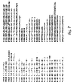
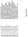
- Fig. 7 shows various peptides of desired length derived from the Amb a I.1, Amb a I.2 and Amb a I.3 protein allergens.
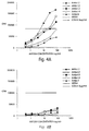
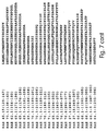
- Fig. 8 is a graphic representation depicting the responses of T cell lines from 39 patients primed in vitro to recombinant Amb a I.1 protein and analyzed for response to various overlapping Amb a I.1 peptides and selected Amb a 1.2 and Amb a I.3 peptides by percent of positive responses within the individuals tested, the mean stimulation index of positive responses for the peptide and the ranked sum of peptide responses.
- Fig. 9 shows selected peptides of desired lengths derived from the Amb a I.1 protein allergen.
- Fig. 10 is a graphic representation depicting the responses of T cell lines from 48 patients primed in vitro to recombinant Amb a I.1 protein and analyzed for response to selected peptides derived from Region 1 of the Amb a I.1 protein, by percent of positive responses within the individuals tested, the mean stimulation index of positive responses for the peptide and the ranked sum of peptide responses.
- Fig. 11 is a graphic representation depicting the responses of T cell lines from 48 patients primed in vitro to recombinant Amb a I.1 protein and analyzed for response to selected peptides derived from Region 2 of the Amb a I.1 protein, by percent of positive responses within the individuals tested, the mean stimulation index of positive responses for the peptide and the ranked sum of peptide responses.
- Fig. 12 is a graphic representation depicting the responses of T cell lines from 48 patients primed in vitro to recombinant Amb a I.1 protein and analyzed for response to selected peptides derived from Region 3 of the Amb a I.1 protein, by percent of positive responses within the individuals tested, the mean stimulation index of positive responses for the peptide and the ranked sum of peptide responses.
- Fig. 13 is a graphic representation depicting the responses of T cell lines from 48 patients primed in vitro to recombinant Amb a I.1 protein and analyzed for response to selected peptides derived from Region 4 of the Amb a I.1 protein, by percent of positive responses within the individuals tested, the mean stimulation index of positive responses for the peptide and the ranked sum of peptide responses.
- Fig. 14 shows selected peptides of desired lengths derived from the Amb a I.1 protein allergen and the Amb a I.3 protein allergen.
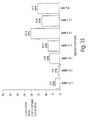
- Fig. 15 is a graphic representation depicting the responses of T cell lines from 23 patients primed in vitro to recombinant Amb a I.1 protein and analyzed for response to selected peptides derived from Region 1 of the Amb a I.1 protein, by percent of positive responses within the individuals tested, the mean stimulation index of positive responses for the peptide and the ranked sum of peptide responses.
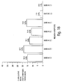
- Fig. 16 is a graphic representation depicting the responses of T cell lines from 23 patients primed in vitro to recombinant Amb a I.1 protein and analyzed for response to selected peptides derived from Region 2 of the Amb a I.1 protein, by percent of positive responses within the individuals tested, the mean stimulation index of positive responses for the peptide and the ranked sum of peptide responses.
- Fig. 17 is a graphic representation depicting the responses of T cell lines from 23 patients primed in vitro to recombinant Amb a I.1 protein and analyzed for response to selected peptides derived from Region 3 of the Amb a I.1 protein, by percent of positive responses within the individuals tested, the mean stimulation index of positive responses for the peptide and the ranked sum of peptide responses.
- Fig. 18 is a graphic representation depicting the responses of T cell lines from 23 patients primed in vitro to recombinant Amb a I.1 protein and analyzed for response to selected peptides derived from Region 4 of the Amb a I.1 protein, by percent of positive responses within the individuals tested, the mean stimulation index of positive responses for the peptide and the ranked sum of peptide responses.
- Fig. 19 is a graphic representation depicting the responses of T cell lines of 9 patients primed in vitro to recombinant Amb a I.1 protein or recombinant Amb a I.3 protein and analyzed for response to selected peptides derived from Amb a I.1, by percent of positive responses within the individuals tested and the mean stimulation index of positive responses for the peptide.
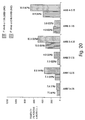
- Fig. 20 is a graphic representation depicting the responses of T cell lines of 9 patients primed in vitro to recombinant Amb a I.1 protein or recombinant Amb a I.3 protein and analyzed for response to selected peptides derived from Amb a I.3, by percent of positive responses within the individuals tested and the mean stimulation index of positive responses for the peptide.
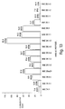
- Fig. 21 is a graphic representation of a direct binding assay of IgE from a single ragweed allergic patient to peptides derived from Amb a I.
- Fig. 22 is a graphic representation depicting the responses of T cell lines of 28 patients primed in vitro to recombinant Amb a I.1 protein and analyzed for response to selected peptides derived from Amb a I.1 by percent of positive responses within the individuals tested, the mean stimulation index of positive responses to the peptide and the ranked sum of peptide responses.
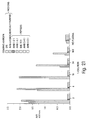
- Fig. 23 is a graphic representation depicting the responses of T cell lines of 28 patients primed in vitro to recombinant Amb a I.1 protein and analyzed for response to selected peptides derived from Region 4 of Amb a I.1 by percent of positive responses within the individuals tested, the mean stimulation index of positive responses to the peptide and the ranked sum of peptide responses.
- Fig. 24 is a graphic representation depicting the responses of T cell lines of 32 patients primed in vitro to recombinant Amb a I.1 protein and analyzed for response to six selected peptides derived from Amb a I.1 by percent of positive responses within the individuals tested, the mean stimulation index of positive responses to the peptide and the ranked sum of peptide responses.
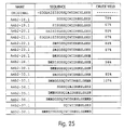
- Fig. 25 shows various peptides derived from peptide RAE 70.1 which include modifications designed to increase the solubility of the peptide.
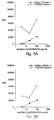
- Fig. 26 is a graphic representation depicting the response of a T cell line from patient 956.2 primed in vitro to Fel d I and analyzed for response to various peptides derived from the Amb a I.1 protein.
- Fig. 27 is a graphic representation depicting the response of a T cell line from patient 119 primed in vitro with recombinant Amb a I.1 and analyzed for response to various modified peptides derived from Region 2 of the Amb a I.1 protein by tritiated thymidine incorporation.
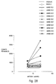
- Fig. 28 is a graphic representation depicting the response of a T cell line from patient 1199 primed in vitro with recombinant Amb a I.1 and analyzed for response to various modified peptides derived from Region 2 of the Amb a I.1 protein by tritiated thymidine incorporation.
- Fig. 29 is a graphic representation depicting the response of a T cell clone generated by limiting dilution from an Amb a I.1 specific T cell line stimulated with the AMB 2-10.1 peptide, primed in vitro w/recombinant Amb a I.1 and analyzed for response to various modified peptides derived from Region 2 of the Amb a I.1 protein by tritiated thymidine incorporation.
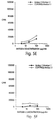
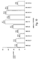
- Fig. 30 is a graphic representation depicting the percent of total histamine release in blood samples from 8 ragweed-allergic patients in response to selected peptides derived from the Amb a I.1 protein.
- the present invention provides isolated peptides derived from the major protein allergens of Ambrosia artemisiifolia .
- a peptide refers to an amino acid sequence having fewer amino acids than the entire amino acid sequence of a protein from which the peptide is derived.
- Peptides of the invention include peptides derived from Amb a I.1, Amb a I.2, Amb a I.3, Amb a I.4 and Amb a II which comprise at least one T cell epitope of the allergen.
- Peptides comprising at least two regions, each region comprising at least one T cell epitope of a protein allergen of Ambrosia artemisiifolia are also within the scope of the invention. Each region of such peptides is derived from the same or from different ragweed pollen allergens. Isolated peptides or regions of isolated peptides, each comprising at least two T cell epitopes of a ragweed pollen allergen are particularly desirable for increased therapeutic effectiveness. Peptides which are immunologically related (e.g., by antibody or T cell cross-reactivity) to peptides of the present invention are also within the scope of the invention.
- Peptides immunologically related by antibody cross-reactivity are bound by antibodies specific for a peptide of a protein allergen of Ambrosia artemisiifolia .
- Peptides immunologically related by T cell cross-reactivity are capable of reacting with the same T cells as a peptide of the invention.
- the present invention also pertains to a ragweed pollen allergen encoded by a nucleic acid sequence of clone IPCl/5.
- the full-length nucleic acid sequence of clone IPCl/5 has been determined and the encoded protein has been produced recombinantly in both the pSEM vector (as a fusion protein with ⁇ -galactosidase) and the pETlld vector.
- the recombinant protein was determined to bind approximately 10-20% of allergic serum IgE on a Western blot.
- the protein encoded by clone IPCl/5 was found to have a high degree of amino acid sequence homology with cysteine proteinase inhibitors in man and rice.
- the protein has 66.6% homology with the rice protein oryzacystatin-I.
- the nucleic acid sequence and deduced amino acid sequence of the allergen encoded by clone IPCl/5 is represented in SEQ ID NO. 11 and 12.
- Isolated proteins and isolated peptides of the invention can be produced by recombinant DNA techniques in a host cell transformed with a nucleic acid having a sequence encoding such protein or peptide.
- the isolated proteins and isolated peptides of the invention can also be produced by chemical synthesis. In certain limited situations, isolated peptides can be produced by chemical cleavage of a protein allergen.
- host cells transformed with a nucleic acid having a sequence encoding the protein or peptide or the functional equivalent of the nucleic acid sequence are cultured in a medium suitable for the cells and protein or peptides can be purified from cell culture medium, host cells, or both using techniques known in the art for purifying proteins and peptides including ion-exchange chromatography, gel filtration chromatography, ultrafiltration, electrophoresis or immunopurification with antibodies specific for the protein or peptide, the protein allergen of Ambrosia artemisiifolia from which the peptide is derived, or a portion thereof.
- protein and peptides of the present invention are substantially free of cellular material or culture medium when produced by recombinant DNA techniques, or substantially free of chemical precursors or other chemicals when synthesized chemically.
- Recombinant ragweed pollen proteins including recombinant Amb a I.1, Amb a I.2, Amb a I.3, Amb a I.4, and Amb a II have been produced.
- Suitable expression vectors for producing recombinant protein and recombinant peptides of the invention include pTRC, pGEX, pMAL, pRIT5, pETlld and pCA.
- the use of pTRC, pETlld and pGEX as expression vectors will result in expression of ragweed pollen protein as an unfused protein.
- the use of pMAL, pRIT5, pCA and pSEM as expression vectors will result in expression of ragweed pollen protein fused to maltose E binding protein (pMAL), protein A (pRIT5), truncated protein A (pCA), or ⁇ -galactosidase (pSEM).
- Suitable expression vectors are commercially available.
- recombinant ragweed pollen protein When produced as a fusion protein, recombinant ragweed pollen protein can be recovered from the fusion protein through enzymatic or chemical (e.g., cyanogen bromide or dilute acid) cleavage and biochemical purification.
- enzymatic cleavage sites for Factor X or thrombin can be introduced at the fusion junction between the carrier protein (e.g., Protein A) and the ragweed pollen protein.
- Suitable host cells for expression of recombinant ragweed pollen protein include bacteria, yeast and insect or mammalian cells. Appropriate vectors for expression in yeast include YepSec, pMF ⁇ and JRY88. These vectors are also commercially available.
- a ragweed pollen allergen is divided into non-overlapping peptides of desired lengths or overlapping peptides of desired lengths as discussed in Example V which may be produced recombinantly, synthetically or in certain limited situations by chemical cleavage of the allergen.
- Peptides comprising at least one T cell epitope are capable of eliciting a T cell response, such as T cell proliferation or lymphokine secretion and/or are capable of inducing T cell anergy (i.e., tolerization).
- isolated peptides are tested by, for example, T cell biology techniques to determine whether the peptides elicit a T cell response or induce T cell anergy. Those peptides found to elicit a T cell response or induce T cell anergy are defined as having T cell stimulating activity.
- human T cell stimulating activity can be tested by culturing T cells obtained from an individual sensitive to a ragweed pollen allergen, (i.e., an individual who has an IgE mediated immune response to a ragweed pollen allergen) with a peptide derived from the allergen and determining whether proliferation of T cells occurs in response to the peptide as measured, e.g., by cellular uptake of tritiated thymidine.
- stimulation indices for responses by T cells to peptides can be calculated as the maximum CPM in response to a peptide divided by the medium control CPM.
- a peptide comprising at least one T cell epitope when determined by T cell stimulation requires a stimulation index of at least 2.0.
- a peptide having a T cell stimulation index of 2.0 is considered useful as a therapeutic agent.
- Preferred peptides have a stimulation index of at least 2.5, more preferably at least 3.5, and most preferably at least 5.0.
- a peptide having T cell stimulating activity and thus comprising at least one T cell epitope as determined by T cell biology techniques is modified by addition or deletion of amino acid residues at either the amino or carboxy terminus of the peptide and tested to determine a change in T cell reactivity to the modified peptide. If two or more peptides which share an area of overlap in the native protein sequence are found to have human T cell stimulating activity, as determined by T cell biology techniques, additional peptides can be produced comprising all or a portion of such peptides and these additional peptides can be tested by a similar procedure. Following this technique, peptides are selected and produced recombinantly or synthetically.
- Peptides are selected based on various factors, including the strength of the T cell response to the peptide (e.g., stimulation index), the frequency of the T cell response to the peptide in a population of individuals sensitive to ragweed pollen, and the potential cross-reactivity of the peptide with Amb a I family members and Amb a II.
- the physical and chemical properties of these selected peptides e.g., solubility, stability
- the ability of the selected peptides or selected modified peptides to stimulate human T cells e.g., induce proliferation, lymphokine secretion is determined.
- preferred peptides of the invention do not bind immunoglobulin E (IgE) or bind IgE to a substantially lesser extent than the protein allergen from which the peptide is derived binds IgE.
- Recombinant ragweed pollen allergens including recombinant Amb a I.1, Amb a I.2, Amb a I.3, Amb a I.4, and Amb a II have been produced and shown to have reduced IgE binding activity as compared to the corresponding native protein allergen (See Fig. 3).
- the major complications of standard immunotherapy are IgE-mediated responses such as anaphylaxis.
- Immunoglobulin E is a mediator of anaphylactic reactions which result from the binding and cross-linking of antigen to IgE on mast cells or basophils and the release of mediators (e.g., histamine, serotonin, eosinophil chemotacic factors).
- mediators e.g., histamine, serotonin, eosinophil chemotacic factors.
- anaphylaxis in a substantial percentage of a population of individuals sensitive to ragweed pollen allergen could be avoided by the use in immunotherapy of a recombinant protein, a peptide or peptides which do not bind IgE in a substantial percentage (e.g., at least about 75%) of a population of individuals sensitive to ragweed pollen allergen, or if the protein or peptide(s) binds IgE, such binding does not result in the release of mediators from mast cells or basophils.
- the risk of anaphylaxis can be reduced by the use in immunotherapy of a recombinant protein, a peptide or peptides which have reduced IgE binding.
- Minimal IgE stimulating activity refers to IgE production that is less than the amount of IgE production and/or IL-4 production stimulated by the native protein allergen (e.g., Amb a I.1).
- a peptide or recombinant protein of the invention when administered to a ragweed pollen-sensitive individual, is capable of modifying the allergic response of the individual to the allergen.
- peptides of the invention comprising at least one T cell epitope of a ragweed pollen allergen or at least two regions derived from a ragweed pollen allergen each comprising at least one T cell epitope, when administered to a ragweed pollen-sensitive individual are capable of modifying the T cell response of the individual to the allergen.
- modification of the allergic response of a ragweed pollen-sensitive individual to a ragweed pollen allergen can be defined as non-responsiveness or diminution in symptoms to a ragweed pollen allergen, as determined by standard clinical procedures (see e.g., Varney et al., British Medical Journal 302 : 265-269 (1990)).
- T cell epitopes are believed to be involved in initiation and perpetuation of the immune response to ragweed pollen allergen(s) which are responsible for the clinical symptoms of ragweed pollen allergy.
- These T cell epitopes are thought to trigger early events at the level of the T helper cell by binding to an appropriate HLA molecule on the surface of an antigen presenting cell and stimulating the relevant T cell subpopulation. These events lead to T cell proliferation, lymphokine secretion, local inflammatory reactions, the recruitment of additional immune cells to the site, and activation of the B cell cascade leading to production of antibodies.
- T cell epitope is the basic element or smallest unit of recognition by a T cell receptor, where the epitope comprises amino acid residues essential to receptor recognition which may be contiguous and/or non-contiguous in the amino acid sequence of the protein.
- Amino acid sequences which mimic those of T cell epitopes and which modify the allergic response to protein allergens of Ambrosia artemisiifolia are within the scope of this invention.
- Exposure of ragweed pollen allergic patients to peptides of the present invention may tolerize or anergize appropriate T cell subpopulations such that they become unresponsive to ragweed pollen allergen(s) and do not participate in mounting an immune response upon such exposure.
- administration of a peptide of the present invention may modify the lymphokine secretion profile as compared with exposure to the naturally-occurring ragweed pollen allergen or portion thereof (e.g., result in a decrease of IL-4 and/or an increase in IL-2).
- T cell subpopulations which normally participate in the response to ragweed pollen allergen(s) such that these T cells are drawn away from the site(s) of normal exposure to the allergen (e.g., nasal mucosa, skin, and lung) towards the site(s) of therapeutic administration of the peptide.
- This redistribution of T cell subpopulations may ameliorate or reduce the ability of an individual's immune system to stimulate the immune response at the site of normal exposure to the ragweed pollen allergen(s), resulting in a diminution in allergic symptoms.
- Isolated peptides of the invention comprise at least one T cell epitope of a protein allergen of Ambrosia artemisiifolia (i.e., the peptide comprises at least approximately seven amino acid residues of the protein allergen).
- therapeutic compositions of the invention preferably comprise at least two T cell epitopes of a ragweed pollen allergen.
- isolated peptides of the invention preferably comprise at least two T cell epitopes (i.e., the peptide comprises at least approximately eight amino acid residues, and preferably fifteen amino acid residues).
- isolated peptides of the invention preferably comprise a sufficient percentage of the T cell epitopes of the entire protein allergen such that upon administration of the peptide to an individual sensitive to ragweed pollen, T cells of individual are tolerized to the protein allergen.
- Isolated peptides of the invention comprising up to approximately 45 amino acid residues in length, and most preferably up to approximately 30 amino acid residues in length are particularly desirable as increases in length may result in difficulty in peptide synthesis as well as retention of an undesirable property (e.g., immunoglobulin binding or enzymatic activity) due to maintenance of conformational similarity between the peptide and the protein allergen from which it is derived. All of the peptides shown in Fig. 8 were found to have human T cell stimulating activity.
- Preferred peptides comprise all or a portion of the areas of major T cell reactivity within the Amb a I.1 protein allergen, i.e., Region 1, Region 2, Region 3 and Region 4. Each area is broadly defined as follows: Region 1 comprises amino acid residues 48-107; Region 2 comprises amino acid residues 171-216; Region 3 comprises amino acid residues 278-322; and Region 4 comprises amino acid residues 331-377. Preferred areas of major T cell reactivity within each Region comprise: amino acid residues 57-101; amino acid residues 182-216; amino acid residues 280-322; and amino acid residues 342-377.
- Amb a I.1 stimulated T cells have been shown to recognize both Amb a I.1 derived peptides and homologous Amb a I.3 derived peptides (See Example IX).
- Amb a I.3 stimulated T cell recognize both Amb a I.1 and Amb a I.3 derived peptides.
- Preferred ragweed pollen peptides comprise all or a portion of the following peptides: RAE 67.1 (SEQ ID NO:13); RAE 57.1 (SEQ ID NO:14); RAE 24.E (SEQ ID NO:15); RAE 24.1 (SEQ ID NO:16); RAE 22.E (SEQ ID NO:17); RAE 22.E-1 (SEQ ID NO:18); RAE 3.D (SEQ ID NO:19); RAE 3.1 (SEQ ID NO:20); RAE 22.E-2 (SEQ ID NO:21); RAE 5.D (SEQ ID NO:22); RAE 6.D (SEQ ID NO:23); RAE 6.1 (SEQ ID NO:24); RAE 7.D (SEQ ID NO:25); RAE 7.D-1 (SEQ ID NO:26); RAE 40.1-6 (SEQ ID NO:27); RAE 40.1-5 (SEQ ID NO:28); RAE 40.1-4 (SEQ ID NO:29); RAE 40.D (SEQ ID NO:30); RAE
- Particularly preferred peptides comprise all or a portion of the following peptides: AMB 1-2.1 (SEQ ID NO:86); AMB 2-6.1 (SEQ ID NO:93); AMB 2-4.1 (SEQ ID NO:90); Amb 2-36.1 (SEQ ID NO:139); Amb 2-38.1 (SEQ ID NO:141); RA-02.1 (SEQ ID NO:150); AMB 2-9.1 (SEQ ID NO:98); AMB 3-5.1 (SEQ ID NO:102); and AMB 4-9.1 (SEQ ID NO:110).
- Another embodiment of the present invention provides peptides comprising at least two regions, each region comprising at least one T cell epitope of a protein allergen of Ambrosia artemisiifolia (e.g., each region comprises at least approximately seven amino acid residues).
- These peptides comprising at least two regions can comprise as many amino acid residues as desired and preferably comprise at least about 7, more preferably at least about 15, even more preferably about 30 and most preferably at least about 40 amino acid residues of a ragweed pollen allergen.
- Each region of such peptide preferably comprises up to 45 amino acid residues in length, more preferably up to 40 residues in length and most preferably up to 30 amino acid residues in length as increases in length of a region may result in difficulty in peptide synthesis as well as retention of an undesirable property (e.g., immunoglobulin binding or enzymatic activity) due to maintenance of conformational similarity between the peptide and the protein allergen from which it is derived.
- the amino acid sequences of the regions can be produced and joined by a linker to increase sensitivity to processing by antigen-presenting cells.
- linker can be any non-epitope amino acid sequence or other appropriate linking or joining agent.
- the regions are arranged in a configuration different from a naturally-occurring configuration of the regions in the allergen.
- the regions containing T cell epitope(s) can be arranged in a noncontiguous configuration and can preferably be derived from the same protein allergen.
- Noncontiguous is defined as an arrangement of regions containing T cell epitope(s) which is different than that of an amino acid sequence present in the protein allergen from which the regions are derived.
- noncontiguous regions containing T cell epitopes can be arranged in a nonsequential order (e.g., in an order different from the order of the amino acids of the native protein allergen from which the region containing T cell epitope(s) are derived in which amino acids are arranged from an amino terminus to a carboxy terminus).
- a peptide can comprise at least 15%, at least 30%, at least 50% or up to 100% of the T cell epitopes of a ragweed pollen allergen.
- the individual peptide regions can be produced and tested to determine which regions bind immunoglobulin E specific for a ragweed pollen allergen and which of such regions would cause the release of mediators (e.g., histamine) from mast cells or basophils.
- mediators e.g., histamine
- Those peptide regions found to bind immunoglobulin E and cause the release of mediators from mast cells or basophils in greater than approximately 10-15% of the allergic sera tested are preferably not included in the peptide regions arranged to form peptides of the invention.
- Preferred peptides of the invention comprise two or more regions derived from the same or from different ragweed pollen allergens (e.g., Amb a I.1, Amb a I.2, Amb a I.3, Amb a I.4 and Amb a II).
- one region can be derived from Amb a I.1 and one region can be derived from Amb a I.2; one region can be derived from Amb a I.1 and one region can be derived from Amb a I.3; one region can be derived from Amb a I.1 and one region can be derived from Amb a I.4; one region can be derived from Amb a I.2 and one region can be derived from Amb a I.3; one region can be derived from Amb a I.2 and one region can be derived from Amb a I.4; one region can be derived from Amb a I.3 and one region can be derived from Amb a I.4; one region can be derived from Amb a I.1 and one region can be derived from Amb a II; one region can be derived from Amb a I.2 and one region can be derived from Amb a II; one region can be derived from Amb a I.3 and one one region
- Regions of a peptide of the invention preferably comprise all or a portion of Region 1, Region 2, Region 3 and Region 4 of Amb a I.1, and the above discussed preferred areas of major T cell reactivity within each Region. If Region 1, 2, 3 or 4 is found to bind IgE and cause the release of mediators from mast cells or basophils, then it is preferred that more than one region of the peptide comprise such Region and that the various regions of the peptide do not bind IgE or cause release of mediators from mast cells or basophils.
- Examples of preferred regions include: AMB 1-1.1 (SEQ ID NO:85); AMB 1-2.1 (SEQ ID NO:86); AMB 1-3.1 (SEQ ID NO:87); AMB 1-4.1 (SEQ ID NO:84); AMB 1-5.1 (SEQ ID NO:83); AMB 1-6.1 (SEQ ID NO:82); AMB 1-4.15 (SEQ ID NO:88); AMB 1-2.15 (SEQ ID NO:89); AMB 2-4.1 (SEQ ID NO:90); AMB 2-3.1 (SEQ ID NO:91); AMB 2-5.1 (SEQ ID NO:92); AMB 2-6.1 (SEQ ID NO:93); AMB 2-2.1 (SEQ ID NO:94); AMB 2-1.1 (SEQ ID NO:95); AMB 2-7.1 (SEQ ID NO:96); AMB 2-8.1 (SEQ ID NO:97); AMB 2-9.1 (SEQ ID NO:98); AMB 2-10.1 (SEQ ID NO:99); AMB 2-11.1 (SEQ ID NO:100); AMB 2-1.15 (SEQ ID NO:101
- Preferred peptides comprise various combinations of two or more regions, each region comprising all or a portion of Region 1, Region 2, Region 3 or Region 4 of Amb a I.1.
- Preferred peptides comprise various combinations of two or more regions, each region having an amino acid sequence as shown in Fig. 14, such combination of regions including the following: AMB 4-6.1 and RAE 70.1 (SEQ ID NO:111 and SEQ ID NO:44); AMB 4-6.1 and AMB 2-5.1 (SEQ ID NO:111 and SEQ ID NO:92); AMB 4-9.1 and AMB 2-5.1 (SEQ ID NO:110 and SEQ ID NO:92); AMB 4-9.1 and RAE 70.1 (SEQ.
- Peptides of protein allergens of Ambrosia artemisiifolia within the scope of the invention can be used in methods of treating and preventing allergic reactions to ragweed pollen allergens.
- one aspect of the present invention provides therapeutic compositions comprising a peptide of Amb a I.1, Amb a I.2, Amb a I.3, Amb a I.4 or Amb a II including at least one T cell epitope, or preferably at least two T cell epitopes, and a pharmaceutically acceptable carrier or diluent.
- the therapeutic composition comprises a pharmaceutically acceptable carrier or diluent and a peptide comprising at least two regions, each region comprising at least one T cell epitope of a ragweed pollen allergen and is derived from the same or from different ragweed pollen allergens.
- a peptide derived from a ragweed pollen allergen comprising at least one T cell epitope can be administered in combination with an appropriate diluent, a carrier, and/or an adjuvant.
- peptides are administered in soluble form.
- Pharmaceutically acceptable diluents include saline and aqueous buffer solutions.
- Pharmaceutically acceptable carriers include polyethylene glycol (Wie et al., International Archives of Allergy and Applied Immunology 64 : 84-99 (1981)) and liposomes (Strejan et al., Journal of Neuroimmunology 7 : 27 (1984)).
- compositions include alum.
- the therapeutic composition is preferably administered in non-immunogenic form, e.g., one that does not include adjuvant.
- Such compositions will generally be administered by injection (e.g., intravenous, subcutaneous, intramuscular), oral administration, (e.g., as in the form of a capsule), inhalation, transdermal application or rectal administration.
- therapeutic compositions are administed subcutaneously.
- the therapeutic compositions of the invention are administered to ragweed pollen-sensitive individuals at dosages and for lengths of time effective to reduce sensitivity (i.e., reduce the allergic response) or tolerize an individual to a ragweed pollen allergen.
- a therapeutically effective amount of one or more of the same or of different therapeutic composition can be administered simultaneously or sequentially to a ragweed pollen-sensitive individual. Effective amounts of the therapeutic compositions will vary according to factors such as the degree of sensitivity of the individual to ragweed pollen allergens, the age, sex, and weight of the individual, and the ability of the peptide to stimulate a T cell response in the individual.
- compositions comprising at least two peptides (e.g., a physical mixture of at least two peptides), each comprising at least one T cell epitope of a protein allergen of Ambrosia artemisiifolia .
- Compositions comprising several peptides or combinations of separate peptides can include as many peptides as desired (e.g., 5, 6, 7...) for therapeutic efficacy.
- the peptides are derived from the same or from different ragweed pollen allergens.
- Such compositions can be administered in the form of a therapeutic composition with a pharmaceutically acceptable carrier or diluent.
- peptides are administered in soluble form.
- a therapeutically effective amount of one or more of such compositions can be administered simultaneously or sequentially, preferably subcutaneously, to a ragweed pollen-sensitive individual to desensitize or tolerize the individual to ragweed pollen.
- compositions and preferred combinations of peptides which can be administered simultaneously or sequentially include the following combinations: AMB 4-6.1 and RAE 70.1 (SEQ ID NO:111 and SEQ ID NO:44); AMB 4-6.1 and AMB 2-5.1 (SEQ ID NO:111 and SEQ ID NO:92); AMB 4-9.1 and AMB 2-5.1 (SEQ ID NO:110 and SEQ ID NO:92); AMB 4-9.1 and RAE 70.1 (SEQ.
- compositions and preferred combinations of peptides for therapeutic administration include the following combinations: AMB 1-2.1 and AMB 4-9.1 (SEQ ID NO:86 and SEQ ID NO:110); AMB 1-2.1, Amb 2-38.1 and AMB 4-9.1 (SEQ ID NO:86, SEQ ID NO:141 and SEQ ID NO:110); AMB 1-2.1, Amb 2-38.1, AMB 4-9.1 and AMB 2-4.1 (SEQ ID NO:86, SEQ ID NO:141, SEQ ID NO:110 and SEQ ID NO:90); AMB 1-2.1, Amb 2-38.1, AMB 4-9.1, AMB 2-4.1 and AMB 3-5.1 (SEQ ID NO:86, SEQ ID NO:141, SEQ ID NO:110, SEQ ID NO:90, and SEQ ID NO:102); AMB 1-2.1, Amb 2-36.1 and AMB 4-9.1 (SEQ ID NO:86, SEQ ID NO:139 and SEQ ID NO:110); AMB 1-2.1, Amb 2-36.1, AMB 4-9.1 and AMB 2-4.1 (SEQ ID NO:86
- the present invention also provides methods of detecting sensitivity in individuals to ragweed pollen allergens comprising combining a blood sample obtained from the individual with a peptide of the present invention, under conditions appropriate for binding of blood components with the peptide and determining the extent to which such binding occurs.
- the extent to which binding occurs is determined by assessing T cell function, T cell proliferation or a combination thereof.
- a modified peptide can be produced in which the amino acid sequence has been altered, such as by amino acid substitution, deletion, or addition, to modify immunogenicity and/or reduce allergenicity, or to which a component has been added for the same purpose.
- a peptide can be modified so that it maintains the ability to induce T cell anergy and bind MHC proteins without the ability to induce a strong proliferative response or possibly, any proliferative response when administered in immunogenic form.
- critical binding residues for the T cell receptor can be determined using known techniques (e.g., substitution of each residue such as, for example, with alanine and determination of the presence or absence of T cell reactivity).
- residues shown to be essential to interact with the T cell receptor can be modified by replacing the essential amino acid with another, preferably similar amino acid residue (a conservative substitution) whose presence is shown to enhance, diminish, but not eliminate, or not affect T cell reactivity.
- amino acid residues which are not essential for T cell receptor interaction can be modified by being replaced by another amino acid whose incorporation may enhance, diminish or not affect T cell reactivity, but not eliminate binding to relevant MHC.
- Preferred amino acid substitutions for non-essential amino acids include, but are not limited to substitutions with alanine, gluatamic acid or a methyl amino acid.
- peptide AMB 4-9.1 which contains an acid-sensitive aspartic acid-proline bond at amino acid residues 360-361, was modified to increase the stability of this peptide.
- the proline at position 361 was substituted with alanine to remove the acid-sensitive bond.
- peptides can also be modified to incorporate one or more polymorphisms in the amino acid sequence of a protein allergen resulting from natural allelic variation.
- D-amino acids, non-natural amino acids or non-amino acid analogues can be substituted or added to produce a modified peptide within the scope of this invention.
- peptides can be modified using the polyethylene glycol (PEG) method of A. Sehon and co-workers (Wie et al., supra ) to produce a peptide conjugated with PEG.
- PEG polyethylene glycol
- Modifications of peptides or portions thereof can also include reduction/alkylation (Tarr in: Methods of Protein Microcharacterization , J.E. Silver ed. Humana Press, Clifton, NJ, pp. 155-194 (1986)); acylation (Tarr, supra ); esterification (Tarr, supra ); chemical coupling to an appropriate carrier (Mishell and Shiigi, eds, Selected Methods in Cellular Immunology , WH Freeman, San Francisco, CA (1980); U.S. Patent 4,939,239); or mild formalin treatment (Marsh International Archives of Allergy and Applied Immunology 41 : 199-215 (1971)).
- peptides within an allergen group can be modified to enhance T cell reactivity.
- an allergen group e.g., Amb a I or Amb a II
- a peptide of one group allergen which may be less reactive than a peptide of another group allergen corresponding in amino acid position can have one or more amino acids substituted with one or more amino acids from the corresponding peptide.
- peptides can be modified to incorporate a polymorphism in the amino acid sequence of a protein allergen resulting from natural allelic variation. Modification of peptides to include one or more of these polymorphisms may result in enhanced stability and/or reactivity.
- reporter group(s) to the peptide backbone.
- poly-histidine can be added to a peptide to purify the peptide on immobilized metal ion affinity chromatography (Hochuli, E. et al., Bio/Technology , 6 :1321-1325 (1988)).
- specific endoprotease cleavage sites can be introduced, if desired, between a reporter group and amino acid sequences of a peptide to facilitate isolation of peptides free of irrelevant sequences.
- a selected peptide AMB 3-4.1 was modified to increase its solubility by the addition of three naturally occurring sequential residues found in the Amb a I.1 protein, "RHG", to the 5' end of the peptide. These residues are not necessary for T cell recognition and are also found in peptide AMB 3-5.1.
- peptide RAE 70.1 was divided into two fragments and modified to increase solubility.
- a fragment of RAE 70.1 (“ORIGINAL" in Figure 25, amino acid residues 194-216, SEQ ID NO:125) was modified by substitution or addition of amino acids to increase the hydrophilicity and decrease the pI of the peptide to thereby increase the solubility.
- isoleucine was substituted with glutamic acid to decrease the pI and avoid precipitation of the peptide from solution at a physiological pH. Similar substitutions and additions are shown in Figure 25.
- canonical protease sensitive sites can be recombinantly or synthetically engineered between regions, each comprising at least one T cell epitope.
- charged amino acid pairs such as KK or RR, can be introduced within a peptide or at the ends of a peptide during recombinant or synthetic construction of the peptide.
- the resulting peptide can be rendered sensitive to cathepsin and/or other trypsin-like enzymes cleavage to generate portions of the peptide containing one or more T cell epitopes.
- such charged amino acid residues can result in an increase in solubility of a peptide.
- Site-directed mutagenesis of DNA encoding a peptide of the invention can be used to modify the structure of the peptide. Such methods may involve PCR with degenerate oligonucleotides (Ho et al., Gene , 77 :51-59 (1989)) or total synthesis of mutated genes (Hostomsky, Z., et al., Biochem. Biophys. Res. Comm ., 161 :1056-1063 (1989)). To enhance bacterial expression, the aforementioned methods can be used in conjunction with other procedures to change the eucaryotic codons in DNA constructs encoding peptides of the invention to ones preferentially used in E. coli , yeast, mammalian cells or other eucaryotic cells.
- the present invention a!so provides nucleic acids having sequences encoding proteins and peptides of the invention.
- Nucleic acid sequences used in any embodiment of this invention can be cDNA as described herein, or alternatively, can be any oligodeoxynucleotide sequence having all or a portion of a sequence represented herein, or their functional equivalents. Such oligodeoxynucleotide sequences can be produced chemically or mechanically, using known techniques.
- a functional equivalent of an oligonucleotide sequence is one which is 1) a sequence capable of hybridizing to a complementary oligonucleotide to which the sequence (or corresponding sequence portions) of SEQ ID NO: 1, 3, 5, 7, 9 and 11 or fragments thereof hybridizes, or 2) the sequence (or corresponding sequence portion) complementary SEQ ID NO: 1, 3, 5, 7, 9 and 11 and/or 3) a sequence which encodes a product (e.g., a polypeptide or peptide) having the same functional characteristics of the product encoded by the sequence (or corresponding sequence portion) of SEQ ID NO: 1, 3, 5, 7, 9 and 11.
- a product e.g., a polypeptide or peptide
- a functional equivalent must meet one or more criteria will depend on its use (e.g., if it is to be used only as an oligoprobe, it need meet only the first or second criteria and if it is to be used to produce a peptide of the present invention, it need only meet the third criterion).
- Amb a I and Amb a II proteins have been recombinantly expressed in E. coli , purified and shown to have reduced binding to human allergic ragweed pollen IgE on Western blots.
- Overlapping peptides derived from the Amb a I.1 protein and various peptides derived from Amb a I.3 and Amb a I.2 were synthesized and used to identify regions of T cell reactivity within the protein. These regions of T cell reactivity were further defined by modifying selected Amb a I.1 peptides and determining T cell reactivity to these peptides.
- defatted short ragweed pollen (Greer Labs) was extracted in 500 ml .05 M Tris pH 7.95 containing protease inhibitors.
- the extract was then depigmented by batch absorption with Whatman DE-52 DEAE cellulose (150 g dry weight) in the presence of 0.2 M NaCl at 4°C. Unbound material was dialysed against .025 M Tris pH 7.95 with protease inhibitors.
- the depigmented sample was next applied to an 80 ml DEAE cellulose column (Whatman DE-52) equilibrated in .025 M Tris pH 7.95 containing protease inhibitors. Acidic proteins were eluted with .025 M Tris, 0.2 M NaCl pH 7.95 at 4°C with inhibitors.
- the acidic DEAE elution sample was fractionated by ammonium sulfate precipitation into 0-45% and 45-59% saturation samples (4°C).
- the Amb a I-enriched 45-59% pellet was applied at 0.5 ml/min (4°C) to a 500 ml Sephacryl S200 (Pharmacia) column in .05 M ammonium bicarbonate containing inhibitors.
- Purified Amb a I was recovered in the 38 kD region and dialysed against .04 M Tris pH 8.0. This sample was next applied to an 8 ml Mono Q HR 10/10 (Pharmacia) column in .04 M Tris pH 8.0 at 25°C. Elution was performed with .04 M Tris pH 8.0 containing .08M NaCl and the major peaks were analyzed as discussed below for confirmation and purity of Amb a I.
- the acidic DEAE elution sample was separated into an Amb a II-enriched fraction by ammonium sulfate precipitation at 0-45% saturation (4°C).
- the pellet was applied at 0.5 ml/min (4°C) to a 200 ml Sephadex G75 (Pharmacia) column in .05 M ammonium bicarbonate containing inhibitors.
- Purified Amb a II was recovered in the 38 kD region and dialysed against .04 M Tris pH 8.0 at 25°C to separate contaminating Amb a I from Amb a II. Elution was performed with .04 M Tris pH 8.0 containing .08M NaCl.
- nucleotide sequences and deduced amino acid sequences of the Amb a I family members are shown in the sequence listing as SEQ ID NO:1 and 2 ( Amb a I.1), SEQ ID NO:3 and 4 ( Amb a I.2), SEQ ID NO:5 and 6 ( Amb a I.3), SEQ ID NO:7 and 8 ( Amb a I.4), and SEQ ID NO:9 and 10 ( Amb a II).
- the cDNAs encoding each allergen were cloned in frame with a polylinker encoding six sequential histidines, (CAC) 6 , that had been inserted into the 5' end of the pTrc99A vector as a NcoI/EcoRI synthetic adapter (Maniatis T., et al. Molecular Cloning: A Laboratory Manual , Cold Spring Harbor Laboratory, Cold Spring Harbor, NY (1982). These cDNA inserts were then cloned in-frame into the appropriate pTrc99. To further enhance expression, a retroregular stem-loop sequence was placed at the 3' end in the untranslated region (Skoglund, C. M., et al.
- the H 6 leader sequence allowed purification using QIAGEN NTA-Agarose (Diagen GmbH, Dusseldorf, FRG), a Ni 2+ chelating support (Hochuli, E. et al., Biotechnology , 6 :1321 (1988)).
- the cells were resuspended in lysozyme containing phosphate buffer (0.4 mg/ml) and incubated for 30 minutes on ice.
- the cell suspension was frozen and quick thawed followed by sonication (Bond, J. G. et al., J. Immunol. , 146 :3380 (1991)).
- Insoluble recombinant protein was recovered by a low speed centrifugation and solubulized in 10 ml (per 1 liter growth) of buffer containing 8 M urea, 50 mM Tris, pH 8.0, 2 ⁇ g/ml leupeptin, 2 ⁇ g/ml pepstatin and 1 ⁇ g/ml soybean trypsin inhibitor.
- the urea solubilized preparation was subjected to a low speed centrifugation and the recombinant proteins in the supernatant isolated by metal ion chromatography (Hochui supra ).
- the pelleted bacteria were resuspended in 6 M guanidine HCl, 100 mM 2-mercaptoethanol, 100 mM NaPO 4 , 10 mM Tris pH 8.0.
- This suspension was subjected to centrifugation at 15,000 X g, and the supernatant removed, adjusted to pH 8.0 with 10 N NaOH, and applied to an NTA agarose column that had been equilibrated in 6 M guanidine HCl, 100 mM NaPO 4 , 10 mM Tris pH 8.0.
- the column was washed in 6 M guanidine HCl, 100 mM NaPO 4 , 10 mM Tris pH 8.0 until the OD 280 of the effluent reached background.
- the column buffer was then switched to 8 M urea, 100 mM NaPO 4 , 10 mM Tris pH 8.0. After equilibration, a more stringent wash was performed in 8 M urea, 100 mM NaOAc, 10 mM Tris pH 6.3 until the OD 280 of the effluent reached background.
- Recombinant protein (as an H 6 fusion) was then eluted in 8 M urea, 100 mM NaOAc, 10 mM Tris pH 4.5 and collected in aliquots whose OD 280 profile was monitored.
- the protein peak was dialyzed 3 times into 500 volumes of PBS for human T-cells analysis. Yield ranged from 1-3 ⁇ g of recombinant protein per litre with purity of approximately 55% (as determined by densitometric scanning).
- the antigens were loaded on the gels as follows: lane 1 SPE (Soluble Pollen Extract), 15 ⁇ g/lane; lane 2 rAmb a I.1 (recombinant Amb a I.1), 3 ⁇ g/lane; lane 3 r Amb a I.2, 4 ⁇ g/lane; lane 4 r Amb a I.3, 3 ⁇ g/lane; lane 5 r Amb a I.4, 3 ⁇ g/lane; and lane 6 r Amb a II.1, 4 ⁇ g/lane.
- SPE Soluble Pollen Extract
- the blots were rinsed in blot solution (25 mM Tris-HCl, pH 7.5, 0.17 M NaCl, and 0.05% Tween 20) and stained for 1 hour with 0.1% India ink. All subsequent incubations with antibodies and washes were performed in blot solution at room temperature. The first antibody incubations were performed overnight then rinsed and incubated with the appropriate biotinylated second antibody (Kirkegaard Perry Laboratories, Gaithersburg, MD). The final incubation was performed using 125 -I streptavidin (1 ⁇ Ci/25 ml blot solution) for 1 hour followed by removal of unbound labeled material and autoradiography at -80 o C with an intensifying screen.
- blot solution 25 mM Tris-HCl, pH 7.5, 0.17 M NaCl, and 0.05% Tween 20
- All subsequent incubations with antibodies and washes were performed in blot solution at room temperature. The first antibody incubations were performed overnight then rinsed
- Fig. 1 shows the western blot IgE binding pattern of two ragweed allergic patient plasma samples (#475 and #143). These patterns are representative of the typical IgE reactivity to these proteins on a Western blot. Both patients show binding to the Amb a I.1 and Amb a I.3 gene products in the soluble pollen extract (lane 1, Fig. 1). The results demonstrate clear IgE binding reactivity in these patients to Amb a I.1, Amb a I.3, Amb a I.4. In contrast, Amb a I.2 and Amb a II.1 showed markedly reduced IgE binding.
- Fig. 2 shows the results of this analysis performed according to the following method.
- Corning assay plates (#25882-96) were coated with each antigen listed in Fig. 2 at the following concentrations: Soluble Pollen Extract (SPE) 15 ⁇ g/ml; r Amb a I.1, 5 ⁇ g/ml; r Amb a I.2, 20 ⁇ g/ml; r Amb a I.3, 5 ⁇ g/ml; r Amb a I.4, 15 ⁇ g/ml; and r Amb a II, 20 ⁇ g/ml. 50 ⁇ ls/well of the above antigens were added and coating was carried out overnight at 4° C.
- SPE Soluble Pollen Extract
- the coating antigens were removed and the wells were blocked with 0.5% gelatin in PBS, 200 ⁇ l/well for 2 hours at room temperature.
- Patient #143 plasma was serially diluted with PBS-Tween 20 (PBS with 0.05% nonionic detergent Tween-20 Sigma, St. Louis MO) and 100 ⁇ l/well was added and incubated overnight at 4°C (plasma dilutions are tested in duplicate).
- the second antibody biotinylated goat anti-Human IgE, 1:1000, Kirkegaard & Perry Laboratories Inc. Gaithersburg, MD), was added at 100 ⁇ l/well for one hour at room temperature.
- FIG. 3 Another ELISA assay was performed using a pool of human allergic sera. This assay demonstrates that biochemically purified (native) Amb a I and Amb a II proteins bind significantly more IgE than the respective recombinant proteins (r Amb a I.1 and r Amb a II). (The control in this experiment was plates coated without antigen and PBS).
- Balb/c mice H-2 d were immunized in the hind foot pads and at the base of the tail with an emulsion of Complete Freunds Adjuvant (CFA) and 50 ⁇ g/mouse Amb a I.1.
- CFA Complete Freunds Adjuvant
- the popliteal lymph nodes, superficial inguinal lymph nodes, and the periaortic lymph nodes were harvested and cultured (1 x 10 5 lymph node cells + 2 x 10 5 irradiated spleen feeders) with various challenge antigens in vitro .
- the in vitro antigens consisted of various doses of Amb a I.1, Amb a I.2, Amb a I.3, Amb a I.4, Amb a II, and a media control. Cultures were incubated for 3 days at 37°C in a CO 2 incubator. On day 3, each culture was pulsed with 1 ⁇ Ci 3 H thymidine and on day 4 the cultures were harvested and proliferation was monitored by incorporation of 3 H
- mice that were immunized with 50 ⁇ g Amb a I.1 + CFA have a good response to Amb a I.1, Amb a I.3, and Amb a I.4.
- the response to Amb a I.2 and Amb a II is also good but less than other antigens. It appears as though Amb a I.1 is very immunogenic in Balb/c mice and results in a response to each Amb a family member.
- mice were immunized with PBS + CFA there was no significant response to any of the Amb a family members (Fig. 4B).
- mice were tolerized with Amb a I.1. and pollen extract and then challenged with Amb a I.1 or pollen extract.
- the outline of this experiment #5 is shown below:
- the animals were sacrificed by cervical dislocation on day 23 and the popliteal lymph nodes, superficial inguinal lymph nodes, and the periaortic lymph nodes were removed and placed in rinsing buffer (cold RPMI 1640 containing 1% FCS).
- the nodes were rinsed with rinsing buffer and forced through a fine stainless steel mesh, using a glass pestal to suspend them in rinsing buffer.
- the suspended cells were rinsed two times by centrifugation at 1000 rpm for 10 minutes and resuspended in rinsing buffer. An aliquot from each sample was taken in order to do a cell count.
- the cells (4 x 10 6 /ml) were incubated in culture media (RPMI 1640 media containing 10% FCS, 2mM L-glutamine, 50U/ml penicillin, 50 ⁇ g/ml streptomycin and 5 x 10 -5 M 2-mercaptoethanol) and test antigens at various concentrations.
- culture media RPMI 1640 media containing 10% FCS, 2mM L-glutamine, 50U/ml penicillin, 50 ⁇ g/ml streptomycin and 5 x 10 -5 M 2-mercaptoethanol
- the triplicate 0.1 ml cultures (U-bottom 96 well plates (Costar)) were incubated at 37 o C and at 5% CO 2 . After 24 hours, 50 ⁇ l of media from each well was placed in separate flat bottom 96 well plates (Costar) and frozen overnight at -20°C to eliminate carryover of live cells.
- CTLL3 in log phase growth were rinsed 3 times by centrifugation at 1000 rpm for 10 minutes.
- CTLL3's were added to the warmed culture supernatants (5 x 10 3 cells/well) and the IL-2 assay was incubated at 37°C and 5% CO 2 .
- 1 ⁇ Ci/well 3 H thymidine was added in 50 ⁇ l/well and the CTLL3 cells were incubated for an additional 4-6 hours.
- the plates were harvested on a Tom Tek 96 well cell harvester. The 3 H incorporation in each well was counted by a Betaplate Model 1205 scintillation counter. Background counts were not subtracted.
- Fig. 5 shows the lymph node responses to Amb a I.1, Amb a I.2, Amb a I.3, Amb a I.4, and Amb a II in the mice that were tolerized with Amb a I.1 or Phosphate Buffered Saline (PBS) and challenged with Amb a I.1.
- Fig. 5A shows that the mice that were tolerized with Amb a I.1 have a lower response to Amb a I.1 than the mice that were tolerized with PBS. The same appears to be true in the response to Amb a I.2, Amb a 1.3, and Amb a I.4 (Figs. 5B, 5C, 5D).
- Figs. 5B, 5C, 5D the lymph node responses to Amb a I.1, Amb a I.2, Amb a I.3, Amb a I.4, and Amb a II in the mice that were tolerized with Amb a I.1 or Phosp
- FIGs. 6A-6F show the lymph node responses to the various proteins in the mice that were exposed to pollen extract or PBS and challenged with pollen extract. Table 1 summarizes the results of these experiments.
- Amb a I.1 overlapping peptides as shown in Fig. 7 were synthesized using standard Fmoc/tBoc synthetic chemistry and purified by dialysis or Reverse Phase HPLC.
- various peptides derived from Amb a I.2 and Amb a I.3 were synthesized.
- the amino acid residues of the synthesized peptides are in brackets by the peptide name and the amino acid sequence (in single letter code) is next to the peptide name.
- the peptide names are consistent throughout the Figures. In the design of the overlapping peptides, the relationship of the Amb a I family members at the level of T cell cross-reactivity as determined in Example VIII was considered.
- the Amb a I protein allergens share a high degree of cross-reactivity.
- Amb a I.1 and Amb a II were found to have 55.2% cross-reactivity.
- the amino acid sequences of the Amb a I family members and Amb a II were examined to identify conserved and variable regions. It was expected that conserved regions within the Amb a I family members and Amb a II would contain "shared" T cell epitopes.
- PBMC Peripheral blood mononuclear cells
- LSM lymphocyte separation medium
- Viable cells were purified by LSM centrifugation and cultured in complete medium supplemented with 5 units recombinant human IL-2/ml and 5 units recombinant human IL-4/ml for up to three weeks until the cells no longer responded to lymphokines and were considered "rested”. The ability of the T cells to proliferate to Amb a I.1, Amb a I.2 and Amb a I.3 sequence-derived synthetic peptides was then assessed.
- 2 x 10 4 rested cells were restimulated in the presence of 2 x 10 4 autologous Epstein-Barr virus (EBV)-transformed B cells (gamma-irradiated with 25,000 RADS) or 5 x 10 4 autologous PBMC (3,500 RADS) with various concentrations of Amb a I.1 synthetic peptides in a volume of 200 ⁇ l complete medium in duplicate or triplicate wells in 96-well round bottom plates for 3 days. Each well then received 1 ⁇ Ci tritiated thymidine for 16-20 hours. The counts incorporated were collected onto glass fiber filter mats and processed for liquid scintillation counting. Table II shows the results of a representative assay.
- EBV Epstein-Barr virus
- the maximum response in a titration of each peptide is expressed as the S.I. or stimulation index.
- the S.I. is the CPM incorporated by cells in response to peptide divided by the CPM incorporated by cells in medium only.
- An S.I. value greater than the background level is considered "positive" and indicates that the peptide contains a T cell epitope.
- only individual S.I. values above 2.0 were used in calculating mean stimulation indices for each peptide for the group of patients tested.
- the results shown in Table II demonstrate that this patient (#466) responds very well to peptides RAE 7.D, RAE 69.1, RAE 64.1, and RAE 29.1.
- the number above each bar is the mean S.I. of the positive responses (S.I. of 2.0 or greater) from the group of patients to that peptide.
- the P.I. for each peptide is determined by multiplying the mean S.I. by the percent of patients who responded to that peptide. The P.I. therefore represents both the strength of the response (S.I.) and the frequency of a response to a peptide in the group of patients tested. For example, peptide RAE 69.1 had the highest cumulative rank so it was the best peptide response in the overall population of 39 even through it did not have the highest mean S.I.
- RAE 70.1 had the highest mean S.I but not the best cumulative rank or P.I.
- P.I the response to RAE 70.1 was strong when it occurred but it did not occur as frequently in the population as the response to other peptides.
- the peptide with the highest P.I., RAE 29.1 also had a strong S.I. and the second highest cumulative rank. The response to this peptide was therefore generally strong and relatively frequent in this population.
- T cell studies similar to those of Example VI were performed using these selected peptides to more precisely define the major areas of T cell reactivity within Regions 1-4 of the Amb a I.1 protein.
- PBMC from a single ragweed-allergic patient were isolated as described in Example VI and were stimulated with 20 ⁇ g/ml of recombinant Amb a I.1 as described above.
- the results of proliferation assays with this one patient to selected peptides using irradiated (24,000 RADS) autologous EBV B cells as antigen presenting cells is shown in Table III. The data indicates that T cells from this patient respond well to RAE 7.D, RAE 70.1, RAE 40.1-4, and RAE 28.1-2.
- Fig. 10 indicates that the major area of T cell reactivity within Region 1 of the Amb a I.1 protein is represented by peptides RAE 6.D, RAE 7.D, RAE 40.1, RAE 40.1-4, and RAE 40.1-5.
- a preferred area of major T cell reactivity within Region 1 comprises amino acid residues 57-101.
- Fig. 11 indicates that there is a broad area of weak T cell reactivity in Region 2 of the Amb a I.1 protein relative to Region 1.
- a preferred area of major T cell reactivity within Region 2 thus comprises amino acid residues 182-216.
- Fig. 12 shows that the most frequent and dominant response within Region 3 of the Amb a I.3 protein is to peptide RAE 64.1.
- T cell reactivity is represented by peptide RAE 66.1.
- a preferred area of major T cell reactivity within Region 3 comprises amino acid residues 280-322.
- Fig. 13 indicates chat the major area of T cell reactivity in Region 4 is represented by peptides RAE 28.1-2, RAE 28.1-1, and RAE 28.1. From this analysis, a preferred area of major T cell reactivity within Region 4 comprises amino acid residues 342-377.
- peptides derived from the Amb a I.1 protein comprising T cell epitopes selected peptides from Regions 1-4 were further modified by addition or deletion of amino acid residues as described above. These selected modified peptides are shown in Fig. 14.
- PBMC were stimulated with recombinant Amb a I.1 and assayed with the selected peptides as described in Example VI, except in some cases autologous PBMC (irradiated 3,500 RADS) were used as antigen presenting cells.
- the assay was followed with a number of patients and resulted in 23 positive experiments. In these assays an individual S.I.
- Figs. 15-18 the bar represents the cumulative rank of each peptide. The best 3 peptide responses for each patient were ranked as described above. The mean S.I. for each peptide and percent positive are found above the bar.
- Amb a I family members i.e., Amb a I.1, I.2, I.3, and I.4
- PBMC peripheral blood mononuclear cells
- 2 x 10 4 rested cells were restimulated in the presence of an equal number of autologous EBV-transformed B cells (irradiated with 25,000 RADS) with various concentrations (0-100 ⁇ g/ml) of recombinant Amb a I.1, I.2, I.3, I.4, Amb a II, or ragweed pollen extract in a volume of 200 ml complete medium in duplicate or triplicate wells in 96-well round bottom plates for 3 days. Each well then received 1 ⁇ Ci tritiated thymidine for 16-20 hours. The counts incorporated were assessed and analyzed as described in Example VI.
- Table IV shows the S.I., P.I., percent of cultures positive for the assay antigen, and number of cultures analyzed. For these assays an individual S.I. greater or equal to 2.0 was used in calculating the mean S.I. Amb a I.1 stimulated cells respond less well and less frequently to the least homologous family members I.3 and I.2. However, this level of cross-reactivity, 68.8% and 60% respectively, is still considered high. The lowest level of Amb a I.1 stimulated T cell cross-reactivity is noted to the least homologous allergen, Amb a II.
- Amb a I.1 reactive T cells are found at a high percentage from ragweed pollen extract stimulated cultures, again demonstrating the importance of the Amb a I.1 allergen.
- TABLE IV Assay Antigen S.I. Recombinant Amb a I.1 Specific Lines % positive n P.I. Pollen Extract 13.5 87.8% 41 1185 r Amb a I.1 27.5 95.1% 41 2616 r Amb a I.2 5.0 60% 5 300 r Amb a I.3 9.3 68.8% 16 639 r Amb a I.4 8.5 100% 11 850 r Amb a II 12.8 55.2% 29 70
- PBMC from ragweed allergic patients were stimulated with 20 ⁇ g/ml recombinant Amb a I.1 protein as described in Example VI with the addition of cultures of PBMC which were stimulated with 20 ⁇ g/ml recombinant Amb a I.3 protein.
- Assays were performed as described in Example VI except the homologous Amb a I.3 peptides were also tested at various doses. In these experiments an individual S.I of 2.0 or greater was used in calculating the mean S.I. Fig.
- FIG. 19 shows the results from a set of 9 matched patients stimulated with either recombinant Amb a I.1 protein or recombinant Amb a I.3 protein and tested on the set of Amb a I.1 peptides described above.
- Each bar represents the P.I.
- the dark bar represents cells stimulated with recombinant Amb a I.1 protein and tested with Amb a I.1 peptides
- the stippled bar represents cells stimulated with recombinant Amb a I.3 protein and tested with Amb a I.1 peptides.
- Example III To analyze IgE reactivity of peptides derived from the Amb a I.1 protein, a direct binding ELISA was performed according to the procedure described in Example III.
- the source of IgE for this analysis was a pool of ragweed allergic patient plasma from 38 ragweed skin test positive patients.
- the ELISA protocol was the same as Example III except that the antigen coating with peptides and proteins was a concentration of 10 ⁇ g/ml at 100 ⁇ ls/well.
- Fig. 21 shows the graphed results of this assay demonstrating strong reactivity to both SPE and biochemically purified Amb a I.1. By this assay there is no detectable binding to any of the peptides. A set of these assays was also run with rabbit and mouse antisera to demonstrate that the coating of the peptides onto these plates was successful.
- Amb a I.1 specific T cell reactivity selected peptides from Figure 14 were analyzed.
- Amb a I.1 specific T cells lines were derived from 28 ragweed allergic patients as described in Example VI. These lines were assessed for their ability to proliferate in response to the peptide in the presence of autologous EBV transformed antigen presenting cells by the uptake of tritiated thymidine as described previously.
- Several lysine substituted peptides derived from the peptide RAE 70.1 sequence were designed to increase the solubility of the peptide from Region 2.
- These peptides shown in Figure 22 were tested against non-substituted controls to determine if the modification resulted in a change in T cell reactivity.
- truncations of peptide AMB 4-6.1 amino acid residues 347-377) were tested to further refine the relative T cell reactivity in that region.
- Figure 22 shows the responses of the 28 T cell lines to these peptides as analyzed by the positivity index, the mean stimulation index and the percent of positive responses.
- the positivity index as defined previously, is the mean stimulation index multiplied by the percent of patients responding to a peptide. Responses to peptides were considered positive if they were greater or equal to 2 fold over background.
- Figure 22 demonstrates that certain lysine substituted peptides in the 182-216 sequence resulted in greater T cell responses, indicating that not all substitutions are recognized equally by a given T cell line.
- the T cell responses to the lysine modified peptides having a substitution of cysteine at position 212 with serine reflect the responses of T cell lines tested with peptides without the lysine substitutions.
- the decrease in response to AMB 2-10.1, with a substitution of leucine for cysteine at 212 reflects the decrease in the percentage of patients responding to this peptide relative to peptides AMB 2-9.1 and AMB 2-11.1 which contain serine and glutamic acid substitutions, respectively, at position 212.
- AMB 4-9.1EP glutamic acid
- AMB 4-9.1NP asparagine
- AMB 4-9.1AP alanine
- AMB 4-9.1SP serine
- AMB 4-9.1QP glutamine
- FIG. 23 shows the ranked sum of the strongest three peptide responses in the 28 T cell lines. The ranked sum was determined as described previously with the strongest response to a peptide given a value of 3, the second strongest a 2, and the third strongest a value of 1. The values from all 28 lines were then added to obtain the ranked sum.
- Figure 23 indicates that the modified peptide AMB 4-9.1DA elicits T cell responses similar to those of the native sequence AMB 4-9.1. In contrast, other substitutions, while eliciting strong responses do not rank as highly in this analysis.
- peptides were selected for further study (AMB 1-2.1, AMB 2-6.1, AMB 2-9.1, AMB 3-5.1 and AMB 4-9.1).
- An additional peptide, RA-02.1 (SEQ ID NO:150) was also synthesized and tested to confirm that the serine substitution at position 212 was recognized by Amb a I.1 specific T cell lines.
- the T cell reactivity of these peptides in 32 different Amb a I.1 specific T cell lines was determined as described previously.
- Figure 24 demonstrates the relative positivity indices, percent of positive responses and mean stimulation indices of these six peptides compared to the overall response to Amb a I.1. This figure shows that peptide AMB 4-9.1 elicits the strongest response in a high percentage of patients.
- RAE 70.1 SEQ ID NO:44
- peptide RAE 70.1 was divided into two fragments and modified to increase solubility and to further define the residues necessary for T cell reactivity.
- a fragment of RAE 70.1 amino acid residues 194-216 was modified by substitution or addition of amino acids which would increase the hydrophilicity and decrease the pI of the peptide to thereby increase solubility.
- Peptides Amb 2-18.1 (SEQ ID NO:126), Amb2-19.1 (SEQ ID NO:127), Amb2-20.1 (SEQ ID NO:128) and Amb2-21.1 (SEQ ID NO:129) correspond to different lengths of the original peptide which were synthesized (including the serine substitution at position 212) and analyzed to determine solubility. It was found that peptide Amb2-19.1 (residues 200-217) was most soluble. Thus, substitutions and/or amino acid additions to this peptide were made in order to further increase the solubility while maintaining T cell reactivity.
- isoleucine at position 201 was substituted with glutamic acid (Amb2-22.1) or lysine (Amb2-23.1) to lower the pI of the resulting peptide and avoid precipitation of the peptide at physiological pH.
- Figures 27 and 28 show representative examples of proliferation of two individual Amb a I.1 specific T cell lines to peptides selected from those shown in Figure 25. Assays were performed as described previously. Both Figure 27 and Figure 28 indicate a hierarchy of responses to these peptides.
- Patient 119 shown in Figure 27, has a hierarchy of response from strongest to weakest as follows: AMB2-23.1>AMB2-22.1>AMB2-30.1>AMB2-26.1>AMB2-33.1>AMB2 -32.1>AMB2-18.1>AMB2-19.1.
- Peptides AMB2-34.1, AMB2-35.1, and AMB 2-9.1 did not elicit significant T cell proliferation in this patient.
- Patient 1199 shown in Figure 28 demonstrated strong responses to AMB 2-26.1>AMB 2-22.1>AMB 2-33.1. T cells from this patient showed less than 2 fold over background responses to all other peptides. These responses are not significantly different from the media control of the T cell line plus autologous EBV antigen presenting cells.
- a T cell clone was generated by limiting dilution from an Amb a I.1 specific T cell line stimulated with AMB 2-10.1. Briefly, an Amb a I.1 specific T cell line from patient 776 was shown to respond to AMB 2-10.1 in a proliferation assay as described previously.
- Amb a I.1 specific T cells were plated at 0.3 cells/well in a V-bottom 96 well plate with 20,000 irradiated autologous EBV transformed antigen presenting cells, AMB 2-10.1 at 40 ⁇ g/ml, leukoagglutin at 1 mg/ml, and recombinant human IL-2 and IL-4 at 10 units/ml in a total volume of 100 ⁇ l/well.
- Plating the T cells at 0.3 cells/well insures not more than 1 T cell/well can potentially proliferate to the peptide with the progeny of that T cell representing a clonal population.
- IL-2 and IL-4 were recombinant human IL-2 and IL-4 to expand peptide-specific T cells.
- This addition of IL-2 and IL-4 was repeated again every three days for the duration of the culture period. Twelve days after the initiation of culture, T cells in the wells were restimulated with 20,000 irradiated EBV antigen presenting cells and 40 ⁇ g/ml AMB 2-10.1. This was repeated again 20 days after the initial stimulation. T cells from wells which showed signs of growth were separated from cell debris by density centrifugation as described previously in the generation of T cell lines. The T cell clone was then expanded with additional IL-2 and IL-4 at 10 units/ml until there were significant numbers to assay for proliferation.
- the objective of the histamine release analysis was to compare the effects of Amb a I.1 or Amb a I.1-derived peptides in an in vitro allergic response system.
- the release of histamine via IgE recognition and IgE receptor crosslinking on viable cells directly assays the allergic potential of a protein antigen.
- the histamine release assay used for these studies is based on the detection of an acylated derivative of histamine using a specific monoclonal antibody (Morel, A.M. and Delaage, M.A. (1988) J. Allergy Clin. Immunol. 82 :646-654).
- the assay was performed in two steps: 1) the release of histamine from basophils present in heparinized whole blood in the presence of different concentrations of protein or peptide; and 2) the assay of histamine present in the supernatants of the release reactions following cell removal by centrifugation.
- the reagents for this second step are available commercially as a competitive radioimmunoassay from AMAC Inc. (Westbrook, ME).
- test protein Amb a I.1 and peptides AMB 1-2.1, AMB 2-9.1, AMB 4-9.1, and AMB 3-5.1 were each diluted to 2X the final release concentration in PACM buffer (PIPES 25mM, NaCl 110mM, KCl 5.0 mM, human serum albumin 0.003% (w/v), CaCl 2 5mM, MgCl 2 2mM, pH7.3) and 0.2 ml of each dilution was added to a 1.5 ml polypropylene tube. A 0.2ml aliquot of blood was then added to each tube and the release reactions started by inversion of the tubes.
- PACM buffer PPES 25mM, NaCl 110mM, KCl 5.0 mM, human serum albumin 0.003% (w/v), CaCl 2 5mM, MgCl 2 2mM, pH7.3
- a negative control of 0.2 ml blood incubated with 0.2 ml PACM buffer was also included. The release reactions were performed for 30 minutes at 37°C. The tubes were then centrifuged in a microfuge at 1500 RPM for 3 minutes and the supernatants were carefully removed and analyzed or stored at -20°C for later analysis. To analyze the total histamine released, 0.1 ml blood was diluted with 0.9 ml PACM buffer and boiled 3 minutes. This tube was then centrifuged 2 minutes at 12,000 RPM and the supernatant was removed and save for analysis.
- a 50 ⁇ l aliquot of each release supernatant was mixed with 150 ⁇ l of histamine release buffer (supplied with the kit from AMAC).
- the diluted supernatant 100 ⁇ l was added to a kit tube containing an acylation reagent.
- a 50 ⁇ l aliquot of acylation buffer was then immediately added and the tube mixed by vortexing.
- the acylation reactions were incubated at least 30 minutes at room temperature.
- a set of histamine standards supplied with the kit were acylated at the same time. 50 ⁇ l of each acylation reaction was then placed in a tube coated with a monoclonal antibody which specifically recognizes the acylated form of histamine.
- a standard curve was generated from the histamine standard counts and graphed on a semi-log plot. Since this is a competitive assay (the 125 -labelled trace competes with acylated histamine in the samples for binding to the antibody-coated tubes), the lower the number of radioactive counts measured, the greater the amount of histamine in the sample. The amount of histamine in each sample data point was extrapolated from the standard curve using a computer statistical program (StatView for the MacIntosh). The assay is sensitive to 0.2 nM histamine.
- FIG. 30 shows the representative results from 1 of the patients (#1273).
- the graph shows the concentration of each antigen in ⁇ g/ml versus the percent of total histamine released. The results are presented as the percent of the total histamine released for that patient, since patients vary greatly in their overall releasable histamine levels. This variability is likely to result from variation in the number of basophils per ml between patients and the histamine content of each basophil.
- Amb a I.1 at all the concentrations tested, yields a high level of histamine release in this patient.
Abstract
Description
- Ambrosia artemisiifolia or short ragweed pollen is the major cause of late summer hayfever in North America and Canada and considerable effort has been expended in trying to identify the major allergens produced by this species. Amb a I or Antigen E (AgE) has been reported to be the predominant allergen. (King, T.P., et al. Biochemistry, 3:458 (1964)). AgE has been characterized and reported to be a nonglycosylated protein of 38kD molecular mass (King, T.P., Adv. Immunol. 23:77 (1976); King, T.P., et al., Arch. Biochem. Biophys., 212:127 (1981)). An immunochemically related protein, Amb a II (AgK), has been reported to have similar properties (King, T.P., Adv. Immunol. 23:77 (1976); King, T.P., Biochemistry, 11:367 (1972)). Amb a I or AgE can be purified using conventional chromatographic or biochemical techniques. However, it has been reported that due to cleavage of the 38 kD single-chain precursors by the action of a trypsin-like pollen protease, purification often results in the isolation of two noncovalently associated chains of 26 and 12 kD molecular mass, designated α and β, respectively (King, T.P., et al., Arch. Biochem. Biophys., 212:127 (1981); King, T.P., et al., Immunochemistry, 11:83 (1974)). It has been reported that biochemically purified Amb a I has been used as an immunogen to produce murine monoclonal antibodies (mAb) as well as rabbit polyclonal antisera reactive with both the native (Olson, J.R., and D.G. Klapper, J. Immunol., 136:2109 (1986)) and denatured (Smith, et al., Molec. Immunol., 25:355 (1988)) protein.
- Early studies using skin tests with Amb a I-depleted pollen extract led to the estimation that at least 90% of the allergenic activity in ragweed pollen can be attributed to Amb a I (King, T.P., et al., Biochemistry, 3:458 (1964); King, T.P., et al., Immunol., 23:77 (1976)). Competition experiments using murine mAb to inhibit the binding of Amb a I by human IgE in ELISA assays have confirmed that Amb a I binds a substantial proportion of human ragweed allergic IgE (Olson, J.R., et al., J. Immunol., 136:2109 (1986)). Recently, three cDNAs encoding proteins with properties of Amb a I were cloned and Amb a I was reported to be a family of homologous, but distinguishable sequences (Rafnar, T., et al., J. Biol. Chem., 266:1229 (1991)). Rafnar and co-workers reported that the individual cloned members of the Amb a I family, designated Amb a I.1, Amb a I.2, and Amb a I.3, share amino acid sequence homology exceeding 80%. The fourth family member designated Amb a I.D (Amb a I.4) is disclosed in U.S.S.N. 07/529,951, filed May 29, 1990. The deduced amino acid sequence of Amb a II has been disclosed and reported to share approximately 65% sequence identity with the Amb a I multigene family of allergens. (Rogers, B. L., et al., Journal of Immunology 147:2547-2552 (1992)).
- The present invention provides isolated peptides of the major protein allergens of Ambrosia artemisiifolia including peptides derived from the family of related proteins, previously designated Amb a IA, Amb a IB, Amb a IC, and Amb a ID. These allergens have been renamed according to the IUIS approved nomenclature as Amb a I.1 (Amb a IA), Amb a I.2 (Amb a IB), Amb a I.3 (Amb a IC) and Amb a I.4 (Amb a ID). Peptides within the scope of the invention comprise at least one T cell epitope, preferably at least two T cell epitopes, of a protein allergen selected from the family of Amb a I allergens and Amb a II. The invention further provides peptides comprising at least two regions, each region comprising at least one T cell epitope of a ragweed pollen allergen. The regions are derived from the same or from different ragweed pollen allergens.
- The invention also provides modified peptides having similar or enhanced therapeutic properties as the corresponding, naturally-occurring allergen or portion thereof, but having reduced side effects as well as modified peptides having improved properties such as increased solubility and stability. Peptides of the invention are capable of modifying, in a ragweed pollen-sensitive individual to whom they are administered, the allergic response of the individual to a ragweed pollen allergen. Methods of treatment or of diagnosis of sensitivity to a ragweed pollen allergen in an individual and therapeutic compositions comprising one or more peptides of the invention are also provided.
- Fig. 1 shows Western blot analysis of IgE binding to recombinant Amb a I proteins.
- Fig. 2 is a graphic representation of a direct binding assay of IgE from a single ragweed allergic patient to recombinant Amb a I and Amb a II proteins.
- Fig. 3 is a graphic representation of the results of a direct binding assay of IgE from pooled human sera to native Amb a I, Amb a II, recombinant Amb a I.1, recombinant Amb a II and pollen extract.
- Fig. 4A and 4B are graphic representations depicting the responses of lymph node cells isolated from mice tolerized in vivo with either Amb a I.1 or PBS and CFA and challenged in vitro with various antigens.
- Fig. 5A-5F are graphic representations depicting the responses of lymph node cells isolated from mice tolerized in vivo with Amb a I.1 or pollen extract, challenged with Amb a I.1, and tested with various antigens.
- Fig. 6A-6F are graphic representations depicting the responses of lymph node cells isolated from mice tolerized with pollen extract challenged with pollen extract, and tested with various antigens.
- Fig. 7 shows various peptides of desired length derived from the Amb a I.1, Amb a I.2 and Amb a I.3 protein allergens.
- Fig. 8 is a graphic representation depicting the responses of T cell lines from 39 patients primed in vitro to recombinant Amb a I.1 protein and analyzed for response to various overlapping Amb a I.1 peptides and selected Amb a 1.2 and Amb a I.3 peptides by percent of positive responses within the individuals tested, the mean stimulation index of positive responses for the peptide and the ranked sum of peptide responses.
- Fig. 9 shows selected peptides of desired lengths derived from the Amb a I.1 protein allergen.
- Fig. 10 is a graphic representation depicting the responses of T cell lines from 48 patients primed in vitro to recombinant Amb a I.1 protein and analyzed for response to selected peptides derived from
Region 1 of the Amb a I.1 protein, by percent of positive responses within the individuals tested, the mean stimulation index of positive responses for the peptide and the ranked sum of peptide responses. - Fig. 11 is a graphic representation depicting the responses of T cell lines from 48 patients primed in vitro to recombinant Amb a I.1 protein and analyzed for response to selected peptides derived from
Region 2 of the Amb a I.1 protein, by percent of positive responses within the individuals tested, the mean stimulation index of positive responses for the peptide and the ranked sum of peptide responses. - Fig. 12 is a graphic representation depicting the responses of T cell lines from 48 patients primed in vitro to recombinant Amb a I.1 protein and analyzed for response to selected peptides derived from
Region 3 of the Amb a I.1 protein, by percent of positive responses within the individuals tested, the mean stimulation index of positive responses for the peptide and the ranked sum of peptide responses. - Fig. 13 is a graphic representation depicting the responses of T cell lines from 48 patients primed in vitro to recombinant Amb a I.1 protein and analyzed for response to selected peptides derived from
Region 4 of the Amb a I.1 protein, by percent of positive responses within the individuals tested, the mean stimulation index of positive responses for the peptide and the ranked sum of peptide responses. - Fig. 14 shows selected peptides of desired lengths derived from the Amb a I.1 protein allergen and the Amb a I.3 protein allergen.
- Fig. 15 is a graphic representation depicting the responses of T cell lines from 23 patients primed in vitro to recombinant Amb a I.1 protein and analyzed for response to selected peptides derived from
Region 1 of the Amb a I.1 protein, by percent of positive responses within the individuals tested, the mean stimulation index of positive responses for the peptide and the ranked sum of peptide responses. - Fig. 16 is a graphic representation depicting the responses of T cell lines from 23 patients primed in vitro to recombinant Amb a I.1 protein and analyzed for response to selected peptides derived from
Region 2 of the Amb a I.1 protein, by percent of positive responses within the individuals tested, the mean stimulation index of positive responses for the peptide and the ranked sum of peptide responses. - Fig. 17 is a graphic representation depicting the responses of T cell lines from 23 patients primed in vitro to recombinant Amb a I.1 protein and analyzed for response to selected peptides derived from
Region 3 of the Amb a I.1 protein, by percent of positive responses within the individuals tested, the mean stimulation index of positive responses for the peptide and the ranked sum of peptide responses. - Fig. 18 is a graphic representation depicting the responses of T cell lines from 23 patients primed in vitro to recombinant Amb a I.1 protein and analyzed for response to selected peptides derived from
Region 4 of the Amb a I.1 protein, by percent of positive responses within the individuals tested, the mean stimulation index of positive responses for the peptide and the ranked sum of peptide responses. - Fig. 19 is a graphic representation depicting the responses of T cell lines of 9 patients primed in vitro to recombinant Amb a I.1 protein or recombinant Amb a I.3 protein and analyzed for response to selected peptides derived from Amb a I.1, by percent of positive responses within the individuals tested and the mean stimulation index of positive responses for the peptide.
- Fig. 20 is a graphic representation depicting the responses of T cell lines of 9 patients primed in vitro to recombinant Amb a I.1 protein or recombinant Amb a I.3 protein and analyzed for response to selected peptides derived from Amb a I.3, by percent of positive responses within the individuals tested and the mean stimulation index of positive responses for the peptide.
- Fig. 21 is a graphic representation of a direct binding assay of IgE from a single ragweed allergic patient to peptides derived from Amb a I.
- Fig. 22 is a graphic representation depicting the responses of T cell lines of 28 patients primed in vitro to recombinant Amb a I.1 protein and analyzed for response to selected peptides derived from Amb a I.1 by percent of positive responses within the individuals tested, the mean stimulation index of positive responses to the peptide and the ranked sum of peptide responses.
- Fig. 23 is a graphic representation depicting the responses of T cell lines of 28 patients primed in vitro to recombinant Amb a I.1 protein and analyzed for response to selected peptides derived from
Region 4 of Amb a I.1 by percent of positive responses within the individuals tested, the mean stimulation index of positive responses to the peptide and the ranked sum of peptide responses. - Fig. 24 is a graphic representation depicting the responses of T cell lines of 32 patients primed in vitro to recombinant Amb a I.1 protein and analyzed for response to six selected peptides derived from Amb a I.1 by percent of positive responses within the individuals tested, the mean stimulation index of positive responses to the peptide and the ranked sum of peptide responses.
- Fig. 25 shows various peptides derived from peptide RAE 70.1 which include modifications designed to increase the solubility of the peptide.
- Fig. 26 is a graphic representation depicting the response of a T cell line from patient 956.2 primed in vitro to Fel d I and analyzed for response to various peptides derived from the Amb a I.1 protein.
- Fig. 27 is a graphic representation depicting the response of a T cell line from patient 119 primed in vitro with recombinant Amb a I.1 and analyzed for response to various modified peptides derived from
Region 2 of the Amb a I.1 protein by tritiated thymidine incorporation. - Fig. 28 is a graphic representation depicting the response of a T cell line from patient 1199 primed in vitro with recombinant Amb a I.1 and analyzed for response to various modified peptides derived from
Region 2 of the Amb a I.1 protein by tritiated thymidine incorporation. - Fig. 29 is a graphic representation depicting the response of a T cell clone generated by limiting dilution from an Amb a I.1 specific T cell line stimulated with the AMB 2-10.1 peptide, primed in vitro w/recombinant Amb a I.1 and analyzed for response to various modified peptides derived from
Region 2 of the Amb a I.1 protein by tritiated thymidine incorporation. - Fig. 30 is a graphic representation depicting the percent of total histamine release in blood samples from 8 ragweed-allergic patients in response to selected peptides derived from the Amb a I.1 protein.
- The present invention provides isolated peptides derived from the major protein allergens of Ambrosia artemisiifolia. As used herein, a peptide refers to an amino acid sequence having fewer amino acids than the entire amino acid sequence of a protein from which the peptide is derived. Peptides of the invention include peptides derived from Amb a I.1, Amb a I.2, Amb a I.3, Amb a I.4 and Amb a II which comprise at least one T cell epitope of the allergen.
- Peptides comprising at least two regions, each region comprising at least one T cell epitope of a protein allergen of Ambrosia artemisiifolia are also within the scope of the invention. Each region of such peptides is derived from the same or from different ragweed pollen allergens. Isolated peptides or regions of isolated peptides, each comprising at least two T cell epitopes of a ragweed pollen allergen are particularly desirable for increased therapeutic effectiveness. Peptides which are immunologically related (e.g., by antibody or T cell cross-reactivity) to peptides of the present invention are also within the scope of the invention. Peptides immunologically related by antibody cross-reactivity are bound by antibodies specific for a peptide of a protein allergen of Ambrosia artemisiifolia. Peptides immunologically related by T cell cross-reactivity are capable of reacting with the same T cells as a peptide of the invention.
- The present invention also pertains to a ragweed pollen allergen encoded by a nucleic acid sequence of clone IPCl/5. The full-length nucleic acid sequence of clone IPCl/5 has been determined and the encoded protein has been produced recombinantly in both the pSEM vector (as a fusion protein with β-galactosidase) and the pETlld vector. The recombinant protein was determined to bind approximately 10-20% of allergic serum IgE on a Western blot. The protein encoded by clone IPCl/5 was found to have a high degree of amino acid sequence homology with cysteine proteinase inhibitors in man and rice. The protein has 66.6% homology with the rice protein oryzacystatin-I. The nucleic acid sequence and deduced amino acid sequence of the allergen encoded by clone IPCl/5 is represented in SEQ ID NO. 11 and 12.
- Isolated proteins and isolated peptides of the invention can be produced by recombinant DNA techniques in a host cell transformed with a nucleic acid having a sequence encoding such protein or peptide. The isolated proteins and isolated peptides of the invention can also be produced by chemical synthesis. In certain limited situations, isolated peptides can be produced by chemical cleavage of a protein allergen. When a protein or peptide is produced by recombinant techniques, host cells transformed with a nucleic acid having a sequence encoding the protein or peptide or the functional equivalent of the nucleic acid sequence are cultured in a medium suitable for the cells and protein or peptides can be purified from cell culture medium, host cells, or both using techniques known in the art for purifying proteins and peptides including ion-exchange chromatography, gel filtration chromatography, ultrafiltration, electrophoresis or immunopurification with antibodies specific for the protein or peptide, the protein allergen of Ambrosia artemisiifolia from which the peptide is derived, or a portion thereof. By isolated is meant that protein and peptides of the present invention are substantially free of cellular material or culture medium when produced by recombinant DNA techniques, or substantially free of chemical precursors or other chemicals when synthesized chemically. Recombinant ragweed pollen proteins including recombinant Amb a I.1, Amb a I.2, Amb a I.3, Amb a I.4, and Amb a II have been produced.
- Suitable expression vectors for producing recombinant protein and recombinant peptides of the invention include pTRC, pGEX, pMAL, pRIT5, pETlld and pCA. The use of pTRC, pETlld and pGEX as expression vectors will result in expression of ragweed pollen protein as an unfused protein. The use of pMAL, pRIT5, pCA and pSEM as expression vectors will result in expression of ragweed pollen protein fused to maltose E binding protein (pMAL), protein A (pRIT5), truncated protein A (pCA), or β-galactosidase (pSEM). Suitable expression vectors are commercially available. When produced as a fusion protein, recombinant ragweed pollen protein can be recovered from the fusion protein through enzymatic or chemical (e.g., cyanogen bromide or dilute acid) cleavage and biochemical purification. For example, enzymatic cleavage sites for Factor X or thrombin can be introduced at the fusion junction between the carrier protein (e.g., Protein A) and the ragweed pollen protein. Suitable host cells for expression of recombinant ragweed pollen protein include bacteria, yeast and insect or mammalian cells. Appropriate vectors for expression in yeast include YepSec, pMFα and JRY88. These vectors are also commercially available.
- To obtain isolated peptides of the present invention, a ragweed pollen allergen is divided into non-overlapping peptides of desired lengths or overlapping peptides of desired lengths as discussed in Example V which may be produced recombinantly, synthetically or in certain limited situations by chemical cleavage of the allergen. Peptides comprising at least one T cell epitope are capable of eliciting a T cell response, such as T cell proliferation or lymphokine secretion and/or are capable of inducing T cell anergy (i.e., tolerization). To determine peptides comprising at least one T cell epitope, isolated peptides are tested by, for example, T cell biology techniques to determine whether the peptides elicit a T cell response or induce T cell anergy. Those peptides found to elicit a T cell response or induce T cell anergy are defined as having T cell stimulating activity. As discussed in the Examples, human T cell stimulating activity can be tested by culturing T cells obtained from an individual sensitive to a ragweed pollen allergen, (i.e., an individual who has an IgE mediated immune response to a ragweed pollen allergen) with a peptide derived from the allergen and determining whether proliferation of T cells occurs in response to the peptide as measured, e.g., by cellular uptake of tritiated thymidine. As described in detail in the Examples, stimulation indices for responses by T cells to peptides can be calculated as the maximum CPM in response to a peptide divided by the medium control CPM. As used throughout this application, a peptide comprising at least one T cell epitope, when determined by T cell stimulation requires a stimulation index of at least 2.0. A peptide having a T cell stimulation index of 2.0 is considered useful as a therapeutic agent. Preferred peptides have a stimulation index of at least 2.5, more preferably at least 3.5, and most preferably at least 5.0.
- In order to determine precise T cell epitopes by, for example, fine mapping techniques, a peptide having T cell stimulating activity and thus comprising at least one T cell epitope as determined by T cell biology techniques is modified by addition or deletion of amino acid residues at either the amino or carboxy terminus of the peptide and tested to determine a change in T cell reactivity to the modified peptide. If two or more peptides which share an area of overlap in the native protein sequence are found to have human T cell stimulating activity, as determined by T cell biology techniques, additional peptides can be produced comprising all or a portion of such peptides and these additional peptides can be tested by a similar procedure. Following this technique, peptides are selected and produced recombinantly or synthetically. Peptides are selected based on various factors, including the strength of the T cell response to the peptide (e.g., stimulation index), the frequency of the T cell response to the peptide in a population of individuals sensitive to ragweed pollen, and the potential cross-reactivity of the peptide with Amb a I family members and Amb a II. The physical and chemical properties of these selected peptides (e.g., solubility, stability) are examined to determine whether the peptides are suitable for use in therapeutic compositions or whether the peptides require modification as described herein. The ability of the selected peptides or selected modified peptides to stimulate human T cells (e.g., induce proliferation, lymphokine secretion) is determined.
- In addition, preferred peptides of the invention do not bind immunoglobulin E (IgE) or bind IgE to a substantially lesser extent than the protein allergen from which the peptide is derived binds IgE. Recombinant ragweed pollen allergens including recombinant Amb a I.1, Amb a I.2, Amb a I.3, Amb a I.4, and Amb a II have been produced and shown to have reduced IgE binding activity as compared to the corresponding native protein allergen (See Fig. 3). The major complications of standard immunotherapy are IgE-mediated responses such as anaphylaxis. Immunoglobulin E is a mediator of anaphylactic reactions which result from the binding and cross-linking of antigen to IgE on mast cells or basophils and the release of mediators (e.g., histamine, serotonin, eosinophil chemotacic factors). Thus, anaphylaxis in a substantial percentage of a population of individuals sensitive to ragweed pollen allergen could be avoided by the use in immunotherapy of a recombinant protein, a peptide or peptides which do not bind IgE in a substantial percentage (e.g., at least about 75%) of a population of individuals sensitive to ragweed pollen allergen, or if the protein or peptide(s) binds IgE, such binding does not result in the release of mediators from mast cells or basophils. Additionally, the risk of anaphylaxis can be reduced by the use in immunotherapy of a recombinant protein, a peptide or peptides which have reduced IgE binding. Moreover, recombinant protein or peptides which have minimal IgE stimulating activity are desirable for therapeutic effectiveness. Minimal IgE stimulating activity refers to IgE production that is less than the amount of IgE production and/or IL-4 production stimulated by the native protein allergen (e.g., Amb a I.1).
- A peptide or recombinant protein of the invention, when administered to a ragweed pollen-sensitive individual, is capable of modifying the allergic response of the individual to the allergen. Particularly, peptides of the invention comprising at least one T cell epitope of a ragweed pollen allergen or at least two regions derived from a ragweed pollen allergen each comprising at least one T cell epitope, when administered to a ragweed pollen-sensitive individual are capable of modifying the T cell response of the individual to the allergen. As used herein, modification of the allergic response of a ragweed pollen-sensitive individual to a ragweed pollen allergen can be defined as non-responsiveness or diminution in symptoms to a ragweed pollen allergen, as determined by standard clinical procedures (see e.g., Varney et al., British Medical Journal 302: 265-269 (1990)).
- As a result of the work described herein, peptides derived from ragweed pollen allergens comprising at least one T cell epitope have been produced. T cell epitopes are believed to be involved in initiation and perpetuation of the immune response to ragweed pollen allergen(s) which are responsible for the clinical symptoms of ragweed pollen allergy. These T cell epitopes are thought to trigger early events at the level of the T helper cell by binding to an appropriate HLA molecule on the surface of an antigen presenting cell and stimulating the relevant T cell subpopulation. These events lead to T cell proliferation, lymphokine secretion, local inflammatory reactions, the recruitment of additional immune cells to the site, and activation of the B cell cascade leading to production of antibodies. One isotype of these antibodies, IgE, is fundamentally important in the development of allergic symptoms and its production is influenced early in the cascade of events, at the level of the T helper cell, by the nature of the lymphokines secreted. A T cell epitope is the basic element or smallest unit of recognition by a T cell receptor, where the epitope comprises amino acid residues essential to receptor recognition which may be contiguous and/or non-contiguous in the amino acid sequence of the protein. Amino acid sequences which mimic those of T cell epitopes and which modify the allergic response to protein allergens of Ambrosia artemisiifolia are within the scope of this invention.
- Exposure of ragweed pollen allergic patients to peptides of the present invention may tolerize or anergize appropriate T cell subpopulations such that they become unresponsive to ragweed pollen allergen(s) and do not participate in mounting an immune response upon such exposure. In addition, administration of a peptide of the present invention may modify the lymphokine secretion profile as compared with exposure to the naturally-occurring ragweed pollen allergen or portion thereof (e.g., result in a decrease of IL-4 and/or an increase in IL-2). Furthermore, exposure to a peptide of the invention may influence T cell subpopulations which normally participate in the response to ragweed pollen allergen(s) such that these T cells are drawn away from the site(s) of normal exposure to the allergen (e.g., nasal mucosa, skin, and lung) towards the site(s) of therapeutic administration of the peptide. This redistribution of T cell subpopulations may ameliorate or reduce the ability of an individual's immune system to stimulate the immune response at the site of normal exposure to the ragweed pollen allergen(s), resulting in a diminution in allergic symptoms.
- Isolated peptides of the invention comprise at least one T cell epitope of a protein allergen of Ambrosia artemisiifolia (i.e., the peptide comprises at least approximately seven amino acid residues of the protein allergen). For purposes of therapeutic effectiveness, therapeutic compositions of the invention preferably comprise at least two T cell epitopes of a ragweed pollen allergen. Accordingly, isolated peptides of the invention preferably comprise at least two T cell epitopes (i.e., the peptide comprises at least approximately eight amino acid residues, and preferably fifteen amino acid residues). Additionally, isolated peptides of the invention preferably comprise a sufficient percentage of the T cell epitopes of the entire protein allergen such that upon administration of the peptide to an individual sensitive to ragweed pollen, T cells of individual are tolerized to the protein allergen. Isolated peptides of the invention comprising up to approximately 45 amino acid residues in length, and most preferably up to approximately 30 amino acid residues in length are particularly desirable as increases in length may result in difficulty in peptide synthesis as well as retention of an undesirable property (e.g., immunoglobulin binding or enzymatic activity) due to maintenance of conformational similarity between the peptide and the protein allergen from which it is derived. All of the peptides shown in Fig. 8 were found to have human T cell stimulating activity.
- Preferred peptides comprise all or a portion of the areas of major T cell reactivity within the Amb a I.1 protein allergen, i.e.,
Region 1,Region 2,Region 3 andRegion 4. Each area is broadly defined as follows:Region 1 comprises amino acid residues 48-107;Region 2 comprises amino acid residues 171-216;Region 3 comprises amino acid residues 278-322; andRegion 4 comprises amino acid residues 331-377. Preferred areas of major T cell reactivity within each Region comprise: amino acid residues 57-101; amino acid residues 182-216; amino acid residues 280-322; and amino acid residues 342-377. Similar areas of major T cell reactivity can be found within the other Amb a I family members (i.e., Amb a I.2, Amb a I.3 and Amb a I.4), and Amb a II. As shown in Example VIII, the Amb a I protein allergens and Amb a II demonstrate a high degree of T cell cross-reactivity. Given this cross-reactivity, shared areas of major T cell reactivity and shared T cell epitopes are likely to be found in conserved regions between Amb a I and the remaining Amb a I family members and Amb a II. For example, Amb a I.1 stimulated T cells have been shown to recognize both Amb a I.1 derived peptides and homologous Amb a I.3 derived peptides (See Example IX). Similarly, Amb a I.3 stimulated T cell recognize both Amb a I.1 and Amb a I.3 derived peptides. - Preferred ragweed pollen peptides comprise all or a portion of the following peptides: RAE 67.1 (SEQ ID NO:13); RAE 57.1 (SEQ ID NO:14); RAE 24.E (SEQ ID NO:15); RAE 24.1 (SEQ ID NO:16); RAE 22.E (SEQ ID NO:17); RAE 22.E-1 (SEQ ID NO:18); RAE 3.D (SEQ ID NO:19); RAE 3.1 (SEQ ID NO:20); RAE 22.E-2 (SEQ ID NO:21); RAE 5.D (SEQ ID NO:22); RAE 6.D (SEQ ID NO:23); RAE 6.1 (SEQ ID NO:24); RAE 7.D (SEQ ID NO:25); RAE 7.D-1 (SEQ ID NO:26); RAE 40.1-6 (SEQ ID NO:27); RAE 40.1-5 (SEQ ID NO:28); RAE 40.1-4 (SEQ ID NO:29); RAE 40.D (SEQ ID NO:30); RAE 40.1 (SEQ ID NO:31); RAE 61.1 (SEQ ID NO:32); RAE 80.1 (SEQ ID NO:33); RAE 45.1 (SEQ ID NO:34); RAE 75.1 (SEQ ID NO:35); RAE 62.1 (SEQ ID NO:36); RAE 69.1 (SEQ ID NO:37); RAE 69.1-1 (SEQ ID NO:38); RAE 69.1-2 (SEQ ID NO:39); RAE 69.1-3 (SEQ ID NO:40); RAE 70.1-3 (SEQ ID NO:41); RAE 70.1-2 (SEQ ID NO:42); RAE 70.1-1 (SEQ ID NO:43); RAE 70.1 (SEQ ID NO:44); RAE 71.1 (SEQ ID NO:45); RAE 65.1 (SEQ ID NO:46); RAE 63.1 (SEQ ID NO:47); RAE 76.1 (SEQ ID NO:48); RAE 27.1 (SEQ ID NO:49); RAE 66.1 (SEQ ID NO:50); RAE 66.1-1 (SEQ ID NO:51); RAE 66.1-2 (SEQ ID NO:52); RAE 66.1-3 (SEQ ID NO:53); RAE 64.1-3 (SEQ ID NO:54); RAE 64.1-2 (SEQ ID NO:55); RAE 64.1-1 (SEQ ID NO:56); RAE 64.1 (SEQ ID NO:57); RAE 73.1 (SEQ ID NO:58); RAE 74.1 (SEQ ID NO:59); RAE 74.1-1 (SEQ ID NO:60); RAE 29.1 (SEQ ID NO:61); RAE 29.1-1 (SEQ ID NO:62); RAE 28+29 (SEQ ID NO:63); RAE 29.1-2 (SEQ ID NO:64); RAE 29.1-3 (SEQ ID NO:65); RAE 29.1-4 (SEQ ID NO:66); RAE 28.1-3 (SEQ ID NO:67); RAE 28.1-2 (SEQ ID NO:68); RAE 28.1-1 (SEQ ID NO:69); RAE 28.1 (SEQ ID NO:70); RAE 20.1 (SEQ ID NO:71); RAE 20.1-3 (SEQ ID NO:72); RAE 20.1-2 (SEQ ID NO:73); RAE 20.1-1 (SEQ ID NO:74); RAE 21.1 (SEQ ID NO:75); RAE 17.1 (SEQ ID NO:76); RAE 55.1 (SEQ ID NO:77); RAE 76.6 (SEQ ID NO:78); RAE 67.15 (SEQ ID NO:79); RAE 45.15 (SEQ ID NO:80); RAE 27.15 (SEQ ID NO:81); AMB 1-1.1 (SEQ ID NO:85); AMB 1-2.1 (SEQ ID NO:86); AMB 1-3.1 (SEQ ID NO:87); AMB 1-4.1 (SEQ ID NO:84); AMB 1-5.1 (SEQ ID NO:83); AMB 1-6.1 (SEQ ID NO:82); AMB 1-4.15 (SEQ ID NO:88); AMB 1-2.15 (SEQ ID NO:89); AMB 2-4.1 (SEQ ID NO:90); AMB 2-3.1 (SEQ ID NO:91); AMB 2-5.1 (SEQ ID NO:92); AMB 2-6.1 (SEQ ID NO:93); AMB 2-2.1 (SEQ ID NO:94); AMB 2-1.1 (SEQ ID NO:95); AMB 2-7.1 (SEQ ID NO:96); AMB 2-8.1 (SEQ ID NO:97); AMB 2-9.1 (SEQ ID NO:98); AMB 2-10.1 (SEQ ID NO:99); AMB 2-11.1 (SEQ ID NO:100); AMB 2-1.15 (SEQ ID NO:101); AMB 3-4.1 (SEQ ID NO:103); AMB 3-5.1 (SEQ ID NO:102); AMB 3-3.1 (SEQ ID NO:104); AMB 3-2.1 (SEQ ID NO:105); AMB 3-1.1 (SEQ ID NO:106); AMB 3-4.15 (SEQ ID NO:107); AMB 3-1.15 (SEQ ID NO:108); AMB 4-8.1 (SEQ ID NO:109); AMB 4-9.1 (SEQ ID NO:110); AMB 4-6.1 (SEQ ID NO:111); AMB 4-5.1 (SEQ ID NO:112); AMB 4-3.1 (SEQ ID NO:113); AMB 4-2.1 (SEQ ID NO:114); AMB 4-1.1 (SEQ ID NO:115); AMB 4-3.15 (SEQ ID NO:116); Amb 2-18.1 (SEQ ID NO:126); Amb 2-19.1 (SEQ ID NO:127); Amb 2-20.1 (SEQ ID NO:128); Amb 2-21.1 (SEQ ID NO:129); Amb 2-22.1 (SEQ ID NO:130); Amb 2-23.1 (SEQ ID NO:131); Amb 2-26.1 (SEQ ID NO:132); Amb 28.1 (SEQ ID NO:133); Amb 2-30.1 (SEQ ID NO:134); Amb 2-32.1 (SEQ ID NO:135); Amb 2-33.1 (SEQ ID NO:136); Amb 2-34.1 (SEQ ID NO:137); Amb 2-35.1 (SEQ ID NO:138); Amb 2-36.1 (SEQ ID NO:139); Amb 2-37.1 (SEQ ID NO:140); Amb 2-38.1 (SEQ ID NO:141); AMB 4-9.1EP (SEQ ID NO:142); AMB 4-9.1NP (SEQ ID NO:143); AMB 4-9.1AP (SEQ ID NO:144); AMB 4-9.1SP (SEQ ID NO:145); AMB 4-9.1QP (SEQ ID NO:146); AMB 4-9.1DA (SEQ ID NO:147); amb 4-9.1DS (SEQ ID NO:148); AMB 4-9.1DG (SEQ ID NO:149); and RA-02.1 (SEQ ID NO:150), the amino acid sequences of such peptides being shown in Figs. 7, 14, 23, 24 and 25. Particularly preferred peptides comprise all or a portion of the following peptides: AMB 1-2.1 (SEQ ID NO:86); AMB 2-6.1 (SEQ ID NO:93); AMB 2-4.1 (SEQ ID NO:90); Amb 2-36.1 (SEQ ID NO:139); Amb 2-38.1 (SEQ ID NO:141); RA-02.1 (SEQ ID NO:150); AMB 2-9.1 (SEQ ID NO:98); AMB 3-5.1 (SEQ ID NO:102); and AMB 4-9.1 (SEQ ID NO:110).
- Another embodiment of the present invention provides peptides comprising at least two regions, each region comprising at least one T cell epitope of a protein allergen of Ambrosia artemisiifolia (e.g., each region comprises at least approximately seven amino acid residues). These peptides comprising at least two regions can comprise as many amino acid residues as desired and preferably comprise at least about 7, more preferably at least about 15, even more preferably about 30 and most preferably at least about 40 amino acid residues of a ragweed pollen allergen. Each region of such peptide preferably comprises up to 45 amino acid residues in length, more preferably up to 40 residues in length and most preferably up to 30 amino acid residues in length as increases in length of a region may result in difficulty in peptide synthesis as well as retention of an undesirable property (e.g., immunoglobulin binding or enzymatic activity) due to maintenance of conformational similarity between the peptide and the protein allergen from which it is derived. If desired, the amino acid sequences of the regions can be produced and joined by a linker to increase sensitivity to processing by antigen-presenting cells. Such linker can be any non-epitope amino acid sequence or other appropriate linking or joining agent. To obtain preferred peptides comprising at least two regions, each comprising at least one T cell epitope, the regions are arranged in a configuration different from a naturally-occurring configuration of the regions in the allergen. For example, the regions containing T cell epitope(s) can be arranged in a noncontiguous configuration and can preferably be derived from the same protein allergen. Noncontiguous is defined as an arrangement of regions containing T cell epitope(s) which is different than that of an amino acid sequence present in the protein allergen from which the regions are derived. Furthermore, the noncontiguous regions containing T cell epitopes can be arranged in a nonsequential order (e.g., in an order different from the order of the amino acids of the native protein allergen from which the region containing T cell epitope(s) are derived in which amino acids are arranged from an amino terminus to a carboxy terminus). A peptide can comprise at least 15%, at least 30%, at least 50% or up to 100% of the T cell epitopes of a ragweed pollen allergen.
- The individual peptide regions can be produced and tested to determine which regions bind immunoglobulin E specific for a ragweed pollen allergen and which of such regions would cause the release of mediators (e.g., histamine) from mast cells or basophils. Those peptide regions found to bind immunoglobulin E and cause the release of mediators from mast cells or basophils in greater than approximately 10-15% of the allergic sera tested are preferably not included in the peptide regions arranged to form peptides of the invention.
- Preferred peptides of the invention comprise two or more regions derived from the same or from different ragweed pollen allergens (e.g., Amb a I.1, Amb a I.2, Amb a I.3, Amb a I.4 and Amb a II). For example, one region can be derived from Amb a I.1 and one region can be derived from Amb a I.2; one region can be derived from Amb a I.1 and one region can be derived from Amb a I.3; one region can be derived from Amb a I.1 and one region can be derived from Amb a I.4; one region can be derived from Amb a I.2 and one region can be derived from Amb a I.3; one region can be derived from Amb a I.2 and one region can be derived from Amb a I.4; one region can be derived from Amb a I.3 and one region can be derived from Amb a I.4; one region can be derived from Amb a I.1 and one region can be derived from Amb a II; one region can be derived from Amb a I.2 and one region can be derived from Amb a II; one region can be derived from Amb a I.3 and one region can be derived from Amb a II; and one region can be derived from Amb a I.4 and one region can be derived from Amb a II. Alternatively, the regions can be derived from the same protein allergen, e.g., Amb a I.1 and Amb a I.1, etc.
- Regions of a peptide of the invention preferably comprise all or a portion of
Region 1,Region 2,Region 3 andRegion 4 of Amb a I.1, and the above discussed preferred areas of major T cell reactivity within each Region. IfRegion - Preferred peptides comprise various combinations of two or more regions, each region comprising all or a portion of
Region 1,Region 2,Region 3 orRegion 4 of Amb a I.1. Preferred peptides comprise various combinations of two or more regions, each region having an amino acid sequence as shown in Fig. 14, such combination of regions including the following: AMB 4-6.1 and RAE 70.1 (SEQ ID NO:111 and SEQ ID NO:44); AMB 4-6.1 and AMB 2-5.1 (SEQ ID NO:111 and SEQ ID NO:92); AMB 4-9.1 and AMB 2-5.1 (SEQ ID NO:110 and SEQ ID NO:92); AMB 4-9.1 and RAE 70.1 (SEQ. ID NO:110 and SEQ ID NO:44); AMB 4-6.1, AMB 2-5.1 and AMB 1-2.1 (SEQ ID NO:111, SEQ ID NO:92 and SEQ ID NO:86); AMB 4-9.1, AMB 2-5.1 and AMB 1-2.1 (SEQ ID NO:110, SEQ ID NO:92 and SEQ ID NO:86); AMB 4-6.1, RAE 70.1 and AMB 1-2.1 (SEQ ID NO:111, SEQ ID NO:44 and SEQ ID NO:86); AMB 4-9.1, RAE 70.1 and AMB 1-2.1 (SEQ ID NO:110, SEQ ID NO:44 and SEQ ID NO:86); AMB 4-6.1, RAE 70.1, AMB 1-2.1 and AMB 3-5.1 (SEQ ID NO:111, SEQ ID NO:44, SEQ ID NO:86 and SEQ ID NO:102); AMB 4-9.1, RAE 70.1, AMB 1-2.1 and AMB 3-5.1 (SEQ ID NO:110, SEQ ID NO:44, SEQ ID NO:86 and SEQ ID NO:102); AMB 4-6.1, AMB 2-5.1, AMB 1-2.1 and AMB 3-5.1 (SEQ ID NO:111, SEQ ID NO:92, SEQ ID NO:86 and SEQ ID NO:102); AMB 4-9.1, AMB 2-5.1, AMB 1-2.1 and AMB 3-5.1 (SEQ ID NO:110, SEQ ID NO:92, SEQ ID NO:86 and SEQ ID NO:102); AMB 4-6.1, RAE 70.1, AMB 1-2.1 and AMB 3-4.1 (SEQ ID NO:111, SEQ ID NO:44, SEQ ID NO:86 and SEQ ID NO:103); AMB 4-9.1, RAE 70.1, AMB 1-2.1 and AMB 3-4.1 (SEQ ID NO:110, SEQ ID NO:44, SEQ ID NO:86 and SEQ ID NO:103); AMB 4-6.1, AMB 2-5.1, AMB 1-2.1 and AMB 3-4.1 (SEQ ID NO:111, SEQ ID NO:92, SEQ ID NO:86 and SEQ ID NO:103); AMB 4-9.1, AMB 2-5.1, AMB 1-2.1 and AMB 3-4.1 (SEQ ID NO:110, SEQ ID NO:92, SEQ ID NO:86 and SEQ ID NO:103); AMB 2-1.15 and AMB 4-3.15 (SEQ ID NO:101, and SEQ ID NO:116); AMB 1-2.15, AMB 2-1.15 and AMB 4-3.15 (SEQ ID NO:89, SEQ ID NO:101, and SEQ ID NO:116); and AMB 1-2.15, AMB 2-1.15, AMB 4-3.15 and AMB 3-4.15 (SEQ ID NO:89, SEQ ID NO:101, SEQ ID NO:116 and SEQ ID NO:107). - Peptides of protein allergens of Ambrosia artemisiifolia within the scope of the invention can be used in methods of treating and preventing allergic reactions to ragweed pollen allergens. Thus, one aspect of the present invention provides therapeutic compositions comprising a peptide of Amb a I.1, Amb a I.2, Amb a I.3, Amb a I.4 or Amb a II including at least one T cell epitope, or preferably at least two T cell epitopes, and a pharmaceutically acceptable carrier or diluent. In another aspect, the therapeutic composition comprises a pharmaceutically acceptable carrier or diluent and a peptide comprising at least two regions, each region comprising at least one T cell epitope of a ragweed pollen allergen and is derived from the same or from different ragweed pollen allergens.
- Administration of the therapeutic compositions of the present invention to desensitize an individual can be carried out using known techniques. For example, a peptide derived from a ragweed pollen allergen comprising at least one T cell epitope can be administered in combination with an appropriate diluent, a carrier, and/or an adjuvant. Preferably, peptides are administered in soluble form. Pharmaceutically acceptable diluents include saline and aqueous buffer solutions. Pharmaceutically acceptable carriers include polyethylene glycol (Wie et al., International Archives of Allergy and Applied Immunology 64: 84-99 (1981)) and liposomes (Strejan et al., Journal of Neuroimmunology 7: 27 (1984)). Pharmaceutically acceptable adjuvants include alum. For purposes of inducing T-cell anergy or tolerance in an individual, the therapeutic composition is preferably administered in non-immunogenic form, e.g., one that does not include adjuvant. Such compositions will generally be administered by injection (e.g., intravenous, subcutaneous, intramuscular), oral administration, (e.g., as in the form of a capsule), inhalation, transdermal application or rectal administration. Preferably, therapeutic compositions are administed subcutaneously.
- The therapeutic compositions of the invention are administered to ragweed pollen-sensitive individuals at dosages and for lengths of time effective to reduce sensitivity (i.e., reduce the allergic response) or tolerize an individual to a ragweed pollen allergen. A therapeutically effective amount of one or more of the same or of different therapeutic composition can be administered simultaneously or sequentially to a ragweed pollen-sensitive individual. Effective amounts of the therapeutic compositions will vary according to factors such as the degree of sensitivity of the individual to ragweed pollen allergens, the age, sex, and weight of the individual, and the ability of the peptide to stimulate a T cell response in the individual.
- In yet another aspect of the present invention, a composition is provided comprising at least two peptides (e.g., a physical mixture of at least two peptides), each comprising at least one T cell epitope of a protein allergen of Ambrosia artemisiifolia. Compositions comprising several peptides or combinations of separate peptides can include as many peptides as desired (e.g., 5, 6, 7...) for therapeutic efficacy. The peptides are derived from the same or from different ragweed pollen allergens. Such compositions can be administered in the form of a therapeutic composition with a pharmaceutically acceptable carrier or diluent. Preferably, peptides are administered in soluble form. A therapeutically effective amount of one or more of such compositions can be administered simultaneously or sequentially, preferably subcutaneously, to a ragweed pollen-sensitive individual to desensitize or tolerize the individual to ragweed pollen.
- Preferred compositions and preferred combinations of peptides which can be administered simultaneously or sequentially (comprising peptides having amino acid sequences shown in Fig. 14) include the following combinations: AMB 4-6.1 and RAE 70.1 (SEQ ID NO:111 and SEQ ID NO:44); AMB 4-6.1 and AMB 2-5.1 (SEQ ID NO:111 and SEQ ID NO:92); AMB 4-9.1 and AMB 2-5.1 (SEQ ID NO:110 and SEQ ID NO:92); AMB 4-9.1 and RAE 70.1 (SEQ. ID NO:110 and SEQ ID NO:44); AMB 4-6.1, AMB 2-5.1 and AMB 1-2.1 (SEQ ID NO:111, SEQ ID NO:92 and SEQ ID NO:86); AMB 4-9.1, AMB 2-5.1 and AMB 1-2.1 (SEQ ID NO:110, SEQ ID NO:92 and SEQ ID NO:86); AMB 4-6.1, RAE 70.1 and AMB 1-2.1 (SEQ ID NO:111, SEQ ID NO:44 and SEQ ID NO:86); AMB 4-9.1, RAE 70.1 and AMB 1-2.1 (SEQ ID NO:110, SEQ ID NO:44 and SEQ ID NO:86); AMB 4-6.1, RAE 70.1, AMB 1-2.1 and AMB 3-5.1 (SEQ ID NO:111, SEQ ID NO:44, SEQ ID NO:86 and SEQ ID NO:102); AMB 4-9.1, RAE 70.1, AMB 1-2.1 and AMB 3-5.1 (SEQ ID NO:110, SEQ ID NO:44, SEQ ID NO:86 and SEQ ID NO:102); AMB 4-6.1, AMB 2-5.1, AMB 1-2.1 and AMB 3-5.1 (SEQ ID NO:111, SEQ ID NO:92, SEQ ID NO:86 and SEQ ID NO:102); AMB 4-9.1, AMB 2-5.1, AMB 1-2.1 and AMB 3-5.1 (SEQ ID NO:110, SEQ ID NO:92, SEQ ID NO:86 and SEQ ID NO:102); AMB 4-6.1, RAE 70.1, AMB 1-2.1 and AMB 3-4.1 (SEQ ID NO:111, SEQ ID NO:44, SEQ ID NO:86 and SEQ ID NO:103); AMB 4-9.1, RAE 70.1, AMB 1-2.1 and AMB 3-4.1 (SEQ ID NO:110, SEQ ID NO:44, SEQ ID NO:86 and SEQ ID NO:103); AMB 4-6.1, AMB 2-5.1, AMB 1-2.1 and AMB 3-4.1 (SEQ ID NO:111, SEQ ID NO:92, SEQ ID NO:86 and SEQ ID NO:103); AMB 4-9.1, AMB 2-5.1, AMB 1-2.1 and AMB 3-4.1 (SEQ ID NO:110, SEQ ID NO:92, SEQ ID NO:86 and SEQ ID NO:103); AMB 2-1.15 and AMB 4-3.15 (SEQ ID NO:101, and SEQ ID NO:116); AMB 1-2.15, AMB 2-1.15 and AMB 4-3.15 (SEQ ID NO:89, SEQ ID NO:101, and SEQ ID NO:116); and AMB 1-2.15, AMB 2-1.15, AMB 4-3.15 and AMB 3-4.15 (SEQ ID NO:89, SEQ ID NO:101, SEQ ID NO:116 and SEQ ID NO:107).
- Particularly preferred compositions and preferred combinations of peptides for therapeutic administration include the following combinations: AMB 1-2.1 and AMB 4-9.1 (SEQ ID NO:86 and SEQ ID NO:110); AMB 1-2.1, Amb 2-38.1 and AMB 4-9.1 (SEQ ID NO:86, SEQ ID NO:141 and SEQ ID NO:110); AMB 1-2.1, Amb 2-38.1, AMB 4-9.1 and AMB 2-4.1 (SEQ ID NO:86, SEQ ID NO:141, SEQ ID NO:110 and SEQ ID NO:90); AMB 1-2.1, Amb 2-38.1, AMB 4-9.1, AMB 2-4.1 and AMB 3-5.1 (SEQ ID NO:86, SEQ ID NO:141, SEQ ID NO:110, SEQ ID NO:90, and SEQ ID NO:102); AMB 1-2.1, Amb 2-36.1 and AMB 4-9.1 (SEQ ID NO:86, SEQ ID NO:139 and SEQ ID NO:110); AMB 1-2.1, Amb 2-36.1, AMB 4-9.1 and AMB 2-4.1 (SEQ ID NO:86, SEQ ID NO:139, SEQ ID NO:110 and SEQ ID NO:90); and AMB 1-2.1, Amb 2-36.1, AMB 4-9.1, AMB 2-4.1 and AMB 3-5.1 (SEQ ID NO:86, SEQ ID NO:139, SEQ ID NO:110, SEQ ID NO:90, and SEQ ID NO:102).
- The present invention also provides methods of detecting sensitivity in individuals to ragweed pollen allergens comprising combining a blood sample obtained from the individual with a peptide of the present invention, under conditions appropriate for binding of blood components with the peptide and determining the extent to which such binding occurs. The extent to which binding occurs is determined by assessing T cell function, T cell proliferation or a combination thereof.
- It is also possible to modify the structure of a peptide of the invention for such purposes as increasing solubility, enhancing therapeutic or preventive efficacy, or stability (e.g., shelf life ex vivo, and resistance to proteolytic degradation in vivo.) A modified peptide can be produced in which the amino acid sequence has been altered, such as by amino acid substitution, deletion, or addition, to modify immunogenicity and/or reduce allergenicity, or to which a component has been added for the same purpose.
- For example, a peptide can be modified so that it maintains the ability to induce T cell anergy and bind MHC proteins without the ability to induce a strong proliferative response or possibly, any proliferative response when administered in immunogenic form. In this instance, critical binding residues for the T cell receptor can be determined using known techniques (e.g., substitution of each residue such as, for example, with alanine and determination of the presence or absence of T cell reactivity). Those residues shown to be essential to interact with the T cell receptor can be modified by replacing the essential amino acid with another, preferably similar amino acid residue (a conservative substitution) whose presence is shown to enhance, diminish, but not eliminate, or not affect T cell reactivity. In addition, those amino acid residues which are not essential for T cell receptor interaction can be modified by being replaced by another amino acid whose incorporation may enhance, diminish or not affect T cell reactivity, but not eliminate binding to relevant MHC. Preferred amino acid substitutions for non-essential amino acids include, but are not limited to substitutions with alanine, gluatamic acid or a methyl amino acid.
- Another example of a modification of peptides is substitution of cysteine residues preferably with serine, threonine, leucine or glutamic acid to minimize dimerization via disulfide linkages. As described in Example XI, a peptide from
Region 2, RAE 70.1 (SEQ ID NO:44) was modified to minimize dimerization by substituting serine for the cysteine residue at amino acid position 212. In addition, as shown in Figure 23, peptide AMB 4-9.1 which contains an acid-sensitive aspartic acid-proline bond at amino acid residues 360-361, was modified to increase the stability of this peptide. For example, in peptide AMB 4-9.1DA, the proline at position 361 was substituted with alanine to remove the acid-sensitive bond. - In order to enhance stability and/or reactivity, peptides can also be modified to incorporate one or more polymorphisms in the amino acid sequence of a protein allergen resulting from natural allelic variation. Additionally, D-amino acids, non-natural amino acids or non-amino acid analogues can be substituted or added to produce a modified peptide within the scope of this invention. Furthermore, peptides can be modified using the polyethylene glycol (PEG) method of A. Sehon and co-workers (Wie et al., supra) to produce a peptide conjugated with PEG. In addition, PEG can be added during chemical synthesis of a peptide of the invention. Modifications of peptides or portions thereof can also include reduction/alkylation (Tarr in: Methods of Protein Microcharacterization, J.E. Silver ed. Humana Press, Clifton, NJ, pp. 155-194 (1986)); acylation (Tarr, supra); esterification (Tarr, supra); chemical coupling to an appropriate carrier (Mishell and Shiigi, eds, Selected Methods in Cellular Immunology, WH Freeman, San Francisco, CA (1980); U.S. Patent 4,939,239); or mild formalin treatment (Marsh International Archives of Allergy and Applied Immunology 41: 199-215 (1971)).
- In another embodiment, peptides within an allergen group (e.g., Amb a I or Amb a II) can be modified to enhance T cell reactivity. Given the cross-reactivity within the Amb a I family and Amb a II, a peptide of one group allergen which may be less reactive than a peptide of another group allergen corresponding in amino acid position can have one or more amino acids substituted with one or more amino acids from the corresponding peptide. Additionally, peptides can be modified to incorporate a polymorphism in the amino acid sequence of a protein allergen resulting from natural allelic variation. Modification of peptides to include one or more of these polymorphisms may result in enhanced stability and/or reactivity.
- To facilitate purification and potentially increase solubility of peptides of the invention, it is possible to add reporter group(s) to the peptide backbone. For example, poly-histidine can be added to a peptide to purify the peptide on immobilized metal ion affinity chromatography (Hochuli, E. et al., Bio/Technology, 6:1321-1325 (1988)). In addition, specific endoprotease cleavage sites can be introduced, if desired, between a reporter group and amino acid sequences of a peptide to facilitate isolation of peptides free of irrelevant sequences. In order to successfully desensitize an individual to a protein antigen, it may be necessary to increase the solubility of a peptide by adding functional groups to the peptide or by not including hydrophobic T cell epitopes or regions containing hydrophobic epitopes in the peptides. For example, in
Region 3 of the Amb a I.1 protein, a selected peptide AMB 3-4.1 was modified to increase its solubility by the addition of three naturally occurring sequential residues found in the Amb a I.1 protein, "RHG", to the 5' end of the peptide. These residues are not necessary for T cell recognition and are also found in peptide AMB 3-5.1. Similarly, as described in Example XI, peptide RAE 70.1 was divided into two fragments and modified to increase solubility. As shown in Figure 25, a fragment of RAE 70.1 ("ORIGINAL" in Figure 25, amino acid residues 194-216, SEQ ID NO:125) was modified by substitution or addition of amino acids to increase the hydrophilicity and decrease the pI of the peptide to thereby increase the solubility. For example, in peptide Amb 2-22.1, isoleucine was substituted with glutamic acid to decrease the pI and avoid precipitation of the peptide from solution at a physiological pH. Similar substitutions and additions are shown in Figure 25. - To potentially aid proper antigen processing of T cell epitopes within a peptide, canonical protease sensitive sites can be recombinantly or synthetically engineered between regions, each comprising at least one T cell epitope. For example, charged amino acid pairs, such as KK or RR, can be introduced within a peptide or at the ends of a peptide during recombinant or synthetic construction of the peptide. (See Fig. 14, AMB 2-8.1, AMB 2-9.1, AMB 2-10.1, AMB 2-7.1 and AMB 2-11.1) The resulting peptide can be rendered sensitive to cathepsin and/or other trypsin-like enzymes cleavage to generate portions of the peptide containing one or more T cell epitopes. In addition, such charged amino acid residues can result in an increase in solubility of a peptide.
- Site-directed mutagenesis of DNA encoding a peptide of the invention can be used to modify the structure of the peptide. Such methods may involve PCR with degenerate oligonucleotides (Ho et al., Gene, 77:51-59 (1989)) or total synthesis of mutated genes (Hostomsky, Z., et al., Biochem. Biophys. Res. Comm., 161:1056-1063 (1989)). To enhance bacterial expression, the aforementioned methods can be used in conjunction with other procedures to change the eucaryotic codons in DNA constructs encoding peptides of the invention to ones preferentially used in E. coli, yeast, mammalian cells or other eucaryotic cells.
- The present invention a!so provides nucleic acids having sequences encoding proteins and peptides of the invention. Nucleic acid sequences used in any embodiment of this invention can be cDNA as described herein, or alternatively, can be any oligodeoxynucleotide sequence having all or a portion of a sequence represented herein, or their functional equivalents. Such oligodeoxynucleotide sequences can be produced chemically or mechanically, using known techniques. A functional equivalent of an oligonucleotide sequence is one which is 1) a sequence capable of hybridizing to a complementary oligonucleotide to which the sequence (or corresponding sequence portions) of SEQ ID NO: 1, 3, 5, 7, 9 and 11 or fragments thereof hybridizes, or 2) the sequence (or corresponding sequence portion) complementary SEQ ID NO: 1, 3, 5, 7, 9 and 11 and/or 3) a sequence which encodes a product (e.g., a polypeptide or peptide) having the same functional characteristics of the product encoded by the sequence (or corresponding sequence portion) of SEQ ID NO: 1, 3, 5, 7, 9 and 11. Whether a functional equivalent must meet one or more criteria will depend on its use (e.g., if it is to be used only as an oligoprobe, it need meet only the first or second criteria and if it is to be used to produce a peptide of the present invention, it need only meet the third criterion).
- As described in the Examples which follow, Amb a I and Amb a II proteins have been recombinantly expressed in E. coli, purified and shown to have reduced binding to human allergic ragweed pollen IgE on Western blots. Overlapping peptides derived from the Amb a I.1 protein and various peptides derived from Amb a I.3 and Amb a I.2 were synthesized and used to identify regions of T cell reactivity within the protein. These regions of T cell reactivity were further defined by modifying selected Amb a I.1 peptides and determining T cell reactivity to these peptides.
- This invention is further illustrated by the following non-limiting examples.
- What follows is a description of the work done to biochemically purify the allergens of Ambrosia artemisiifolia in their native form as primary antigens for human T-cell epitope mapping.
- 50 g of defatted short ragweed pollen (Greer Labs) was extracted in 500 ml .05 M Tris pH 7.95 containing protease inhibitors. The extract was then depigmented by batch absorption with Whatman DE-52 DEAE cellulose (150 g dry weight) in the presence of 0.2 M NaCl at 4°C. Unbound material was dialysed against .025 M Tris pH 7.95 with protease inhibitors. The depigmented sample was next applied to an 80 ml DEAE cellulose column (Whatman DE-52) equilibrated in .025 M Tris pH 7.95 containing protease inhibitors. Acidic proteins were eluted with .025 M Tris, 0.2 M NaCl pH 7.95 at 4°C with inhibitors.
- In order to biochemically purify Amb a I, the acidic DEAE elution sample was fractionated by ammonium sulfate precipitation into 0-45% and 45-59% saturation samples (4°C). The Amb a I-enriched 45-59% pellet was applied at 0.5 ml/min (4°C) to a 500 ml Sephacryl S200 (Pharmacia) column in .05 M ammonium bicarbonate containing inhibitors. Purified Amb a I was recovered in the 38 kD region and dialysed against .04 M Tris pH 8.0. This sample was next applied to an 8 ml
Mono Q HR 10/10 (Pharmacia) column in .04 M Tris pH 8.0 at 25°C. Elution was performed with .04 M Tris pH 8.0 containing .08M NaCl and the major peaks were analyzed as discussed below for confirmation and purity of Amb a I. - To biochemically purify Amb a II, the acidic DEAE elution sample was separated into an Amb a II-enriched fraction by ammonium sulfate precipitation at 0-45% saturation (4°C). The pellet was applied at 0.5 ml/min (4°C) to a 200 ml Sephadex G75 (Pharmacia) column in .05 M ammonium bicarbonate containing inhibitors. Purified Amb a II was recovered in the 38 kD region and dialysed against .04 M Tris pH 8.0 at 25°C to separate contaminating Amb a I from Amb a II. Elution was performed with .04 M Tris pH 8.0 containing .08M NaCl.
- The major peaks were analyzed by IEF SDS-PAGE using a mixture of 4.5-5.3 and 3.5-10.0 ampholytes (Pharmacia) in a 7% acrylamide gel. Protein sequencing was also performed to confirm Amb a II.
- What follows is a description of the work done to produce the allergens of Ambrosia artemisiifolia as recombinant proteins in E. coli.
- Described and provided in USSN 07/529,951, filed May 29, 1990, (incorporated herein by reference) are full length cDNAs encoding Amb a I.1, Amb a I.2, Amb a I.3, Amb a I.4 and Amb a II. cDNA inserts encoding these five protein allergens were constructed in the vector pTrc99A (Amann, E., et al., Gene, 69:301 (1988)) which was kindly provided by Dr. Egon Amann (Behringwerke AG, Marburg, FRG). The nucleotide sequences and deduced amino acid sequences of the Amb a I family members are shown in the sequence listing as SEQ ID NO:1 and 2 (Amb a I.1), SEQ ID NO:3 and 4 (Amb a I.2), SEQ ID NO:5 and 6 (Amb a I.3), SEQ ID NO:7 and 8 (Amb a I.4), and SEQ ID NO:9 and 10 (Amb a II). The cDNAs encoding each allergen were cloned in frame with a polylinker encoding six sequential histidines, (CAC)6, that had been inserted into the 5' end of the pTrc99A vector as a NcoI/EcoRI synthetic adapter (Maniatis T., et al. Molecular Cloning: A Laboratory Manual, Cold Spring Harbor Laboratory, Cold Spring Harbor, NY (1982). These cDNA inserts were then cloned in-frame into the appropriate pTrc99. To further enhance expression, a retroregular stem-loop sequence was placed at the 3' end in the untranslated region (Skoglund, C. M., et al. Gene 88:1 (1990)). The H6 leader sequence allowed purification using QIAGEN NTA-Agarose (Diagen GmbH, Dusseldorf, FRG), a Ni2+ chelating support (Hochuli, E. et al., Biotechnology, 6:1321 (1988)). The vectors were transformed into the XL-1 Blue host bacteria. Expression of individual recombinant proteins was induced when cultures reached OD600=0.6 to 0.7 by the addition of isopropyl-D-thiogalactopyranoside (IPTG) to 1 mM concentration to the culture medium. After 2 hours of further growth at 37°, the cells were pelleted. Recombinant proteins were then obtained by either of the following procedures. In one embodiment, the cells were resuspended in lysozyme containing phosphate buffer (0.4 mg/ml) and incubated for 30 minutes on ice. The cell suspension was frozen and quick thawed followed by sonication (Bond, J. G. et al., J. Immunol., 146:3380 (1991)). Insoluble recombinant protein was recovered by a low speed centrifugation and solubulized in 10 ml (per 1 liter growth) of buffer containing 8 M urea, 50 mM Tris, pH 8.0, 2 µg/ml leupeptin, 2 µg/ml pepstatin and 1 µg/ml soybean trypsin inhibitor. The urea solubilized preparation was subjected to a low speed centrifugation and the recombinant proteins in the supernatant isolated by metal ion chromatography (Hochui supra).
- In another embodiment, the pelleted bacteria were resuspended in 6 M guanidine HCl, 100 mM 2-mercaptoethanol, 100 mM NaPO4, 10 mM Tris pH 8.0. This suspension was subjected to centrifugation at 15,000 X g, and the supernatant removed, adjusted to pH 8.0 with 10 N NaOH, and applied to an NTA agarose column that had been equilibrated in 6 M guanidine HCl, 100 mM NaPO4, 10 mM Tris pH 8.0. The column was washed in 6 M guanidine HCl, 100 mM NaPO4, 10 mM Tris pH 8.0 until the OD280 of the effluent reached background. The column buffer was then switched to 8 M urea, 100 mM NaPO4, 10 mM Tris pH 8.0. After equilibration, a more stringent wash was performed in 8 M urea, 100 mM NaOAc, 10 mM Tris pH 6.3 until the OD280 of the effluent reached background. Recombinant protein (as an H6 fusion) was then eluted in 8 M urea, 100 mM NaOAc, 10 mM Tris pH 4.5 and collected in aliquots whose OD280 profile was monitored. The protein peak was dialyzed 3 times into 500 volumes of PBS for human T-cells analysis. Yield ranged from 1-3 µg of recombinant protein per litre with purity of approximately 55% (as determined by densitometric scanning).
- Both Western blotting and ELISA techniques were used to analyze the binding of IgE to purified recombinant Amb a I and Amb a II proteins. Additionally, ELISA was used for analysis of IgE reactivity to peptides derived from the Amb a I.1 protein.
- The antigens were loaded on the gels as follows:
lane 1 SPE (Soluble Pollen Extract), 15 µg/lane;lane 2 rAmb a I.1 (recombinant Amb a I.1), 3 µg/lane;lane 3 rAmb a I.2, 4 µg/lane;lane 4 rAmb a I.3, 3 µg/lane;lane 5 rAmb a I.4, 3 µg/lane; andlane 6 rAmb a II.1, 4 µg/lane. - The gel electrophoresis and Western blot transfer procedures used were essentially as described elsewhere (Towbin, H., et al., Proc. Natl. Acad. Sci., USA, 76:4350 (1979)). Briefly, SDS-PAGE was performed in 10% acrylamide gels under reducing conditions (10mM dithiothreitol, at constant current). The transfer to nitrocellulose (0.1 µM, Schleicher & Schuell, Keene, NH) was performed in a Hoeffer apparatus according to the protocol of Towbin et al., supra. After transfer, the blots were rinsed in blot solution (25 mM Tris-HCl, pH 7.5, 0.17 M NaCl, and 0.05% Tween 20) and stained for 1 hour with 0.1% India ink. All subsequent incubations with antibodies and washes were performed in blot solution at room temperature. The first antibody incubations were performed overnight then rinsed and incubated with the appropriate biotinylated second antibody (Kirkegaard Perry Laboratories, Gaithersburg, MD). The final incubation was performed using 125-I streptavidin (1 µCi/25 ml blot solution) for 1 hour followed by removal of unbound labeled material and autoradiography at -80oC with an intensifying screen.
- Fig. 1 shows the western blot IgE binding pattern of two ragweed allergic patient plasma samples (#475 and #143). These patterns are representative of the typical IgE reactivity to these proteins on a Western blot. Both patients show binding to the Amb a I.1 and Amb a I.3 gene products in the soluble pollen extract (
lane 1, Fig. 1). The results demonstrate clear IgE binding reactivity in these patients to Amb a I.1, Amb a I.3, Amb a I.4. In contrast, Amb a I.2 and Amb a II.1 showed markedly reduced IgE binding. -
Patient # 143 IgE was also tested for binding to purified recombinant Amb a I and Amb a II proteins by ELISA. Fig. 2 shows the results of this analysis performed according to the following method. - Corning assay plates (#25882-96) were coated with each antigen listed in Fig. 2 at the following concentrations: Soluble Pollen Extract (SPE) 15 µg/ml; rAmb a I.1, 5 µg/ml; rAmb a I.2, 20 µg/ml; rAmb a I.3, 5 µg/ml; rAmb a I.4, 15 µg/ml; and rAmb a II, 20 µg/ml. 50 µls/well of the above antigens were added and coating was carried out overnight at 4° C. The coating antigens were removed and the wells were blocked with 0.5% gelatin in PBS, 200µl/well for 2 hours at room temperature.
Patient # 143 plasma was serially diluted with PBS-Tween 20 (PBS with 0.05% nonionic detergent Tween-20 Sigma, St. Louis MO) and 100µl/well was added and incubated overnight at 4°C (plasma dilutions are tested in duplicate). The second antibody (biotinylated goat anti-Human IgE, 1:1000, Kirkegaard & Perry Laboratories Inc. Gaithersburg, MD), was added at 100 µl/well for one hour at room temperature. This solution was removed and streptavidin-HRPO, 1:10000, (Southern Biotechnology Associates, Inc., Birmingham, AL) was then added at 100µl/well for one hour at room temperature (all wells are washed three times with PBS-Tween between each incubation step). TMB Membrane Peroxidates Substrate system (Kirkegaard & Perry Laboratories) was freshly mixed, and added at 100µl/well. The color was allowed to develop for 2-5 minutes. The reaction was stopped by the addition of 100µl/well of 1 M phosphoric acid. Plates were read on a Microplate EL 310 Autoreader (Biotek Instruments, Winooski, VT) with a 450nm filter. The absorbance levels of duplicate wells was averaged. The graphed results (log of the dilution vs absorbance) of the ELISA assays are shown in Fig. 2. The order of coating antigens listed vertically in these figures corresponds in order from left to right to the coating antigens listed for each histogram. - The results of the ELISA assay, Fig. 2, demonstrate a similar reactivity pattern as seen in the Western blot analysis (Fig. 1). The observation of much stronger binding to SPE is due to the fact that this antigen preparation is non-denatured. However, the recombinant Amb a proteins, because of their non-native bacterial origin behave as denatured antigens. The hierarchy of IgE binding in this patient to rAmb a proteins by ELISA shows that Amb a I.1 binds IgE more strongly than Amb a I.3 and Amb a I.4. The results also show that Amb a I.2 and Amb a II demonstrate markedly reduced binding to IgE.
- As shown in Fig. 3, another ELISA assay was performed using a pool of human allergic sera. This assay demonstrates that biochemically purified (native) Amb a I and Amb a II proteins bind significantly more IgE than the respective recombinant proteins (rAmb a I.1 and rAmb a II). (The control in this experiment was plates coated without antigen and PBS).
- Balb/c mice (H-2 d) were immunized in the hind foot pads and at the base of the tail with an emulsion of Complete Freunds Adjuvant (CFA) and 50µg/mouse Amb a I.1. Seven days later, the popliteal lymph nodes, superficial inguinal lymph nodes, and the periaortic lymph nodes were harvested and cultured (1 x 10⁵ lymph node cells + 2 x 10⁵ irradiated spleen feeders) with various challenge antigens in vitro. The in vitro antigens consisted of various doses of Amb a I.1, Amb a I.2, Amb a I.3, Amb a I.4, Amb a II, and a media control. Cultures were incubated for 3 days at 37°C in a CO2 incubator. On
day 3, each culture was pulsed with 1µCi 3H thymidine and onday 4 the cultures were harvested and proliferation was monitored by incorporation of 3H into DNA. - As seen in Fig. 4A, the mice that were immunized with 50µg Amb a I.1 + CFA have a good response to Amb a I.1, Amb a I.3, and Amb a I.4. The response to Amb a I.2 and Amb a II is also good but less than other antigens. It appears as though Amb a I.1 is very immunogenic in Balb/c mice and results in a response to each Amb a family member. When mice were immunized with PBS + CFA, there was no significant response to any of the Amb a family members (Fig. 4B).
- An experiment was conducted to determine whether mice could be tolerized with Amb a I.1. The mice were tolerized with Amb a I.1 and pollen extract and then challenged with Amb a I.1 or pollen extract. The outline of this
experiment # 5 is shown below: - Day 1:
- Group (1): 6 Balb/c mice tolerized with 300µg Amb a I.1 + Incomplete Freunds Adjuvant (IFA) intraperitoneally (i.p.)
Group (2): 6 Balb/c mice tolerized with 300µg Pollen Extract + IFA i.p.
Group (3): 6 Balb/c mice exposed to PBS + IFA i.p. -
Day 15 -
Challenge 3 mice from each group with 50µg Amb a I.1 + CFA subcutaneously (s.c.)
Challenge the remaining 3 mice from each group with 50µg Pollen extract + CFA s.c. - Day 23
- Harvest Lymph Nodes (Analyze each mouse separately)
Test Lymph Node Response on Amb a I.1, I.2, I.3, I.4, II.1, Pollen Extract, Concanavalin A (Con A), and media. - The animals were sacrificed by cervical dislocation on day 23 and the popliteal lymph nodes, superficial inguinal lymph nodes, and the periaortic lymph nodes were removed and placed in rinsing buffer (cold RPMI 1640 containing 1% FCS). The nodes were rinsed with rinsing buffer and forced through a fine stainless steel mesh, using a glass pestal to suspend them in rinsing buffer. The suspended cells were rinsed two times by centrifugation at 1000 rpm for 10 minutes and resuspended in rinsing buffer. An aliquot from each sample was taken in order to do a cell count. The cells (4 x 10⁶/ml) were incubated in culture media (RPMI 1640 media containing 10% FCS, 2mM L-glutamine, 50U/ml penicillin, 50 µg/ml streptomycin and 5 x 10⁻⁵ M 2-mercaptoethanol) and test antigens at various concentrations. The triplicate 0.1 ml cultures (U-bottom 96 well plates (Costar)) were incubated at 37oC and at 5% CO2. After 24 hours, 50µl of media from each well was placed in separate flat bottom 96 well plates (Costar) and frozen overnight at -20°C to eliminate carryover of live cells. The supernatants were tested after thawing for their ability to support the growth of CTLL3, an IL-2 dependent T cell clone. CTLL3 in log phase growth were rinsed 3 times by centrifugation at 1000 rpm for 10 minutes. CTLL3's were added to the warmed culture supernatants (5 x 10³ cells/well) and the IL-2 assay was incubated at 37°C and 5% CO2. After 24 hours, 1 µCi/well 3H thymidine was added in 50 µl/well and the CTLL3 cells were incubated for an additional 4-6 hours. Following the pulse with 3H, the plates were harvested on a
Tom Tek 96 well cell harvester. The 3H incorporation in each well was counted by a Betaplate Model 1205 scintillation counter. Background counts were not subtracted. - Fig. 5 shows the lymph node responses to Amb a I.1, Amb a I.2, Amb a I.3, Amb a I.4, and Amb a II in the mice that were tolerized with Amb a I.1 or Phosphate Buffered Saline (PBS) and challenged with Amb a I.1. Fig. 5A, shows that the mice that were tolerized with Amb a I.1 have a lower response to Amb a I.1 than the mice that were tolerized with PBS. The same appears to be true in the response to Amb a I.2, Amb a 1.3, and Amb a I.4 (Figs. 5B, 5C, 5D). In Figs. 5E and 5F, it appears as though there is no significant difference in the response to Amb a II or pollen extract between the tolerized group and the non-tolerized group. Figs. 6A-6F show the lymph node responses to the various proteins in the mice that were exposed to pollen extract or PBS and challenged with pollen extract. Table 1 summarizes the results of these experiments.
TABLE 1 Amba I.1 Amba I.2 Amba I.3 Amba I.4 Amba II.1 Pollen Tolerize/Challenge Control/Amba I.1 + + + + - - Amba I.1/Amba I.1 - - - - - - Control/Pollen + + +/- +/- - + Pollen/Pollen - - - - - - (-) indicates a diminished response as compared to control - Amb a I.1 overlapping peptides as shown in Fig. 7 were synthesized using standard Fmoc/tBoc synthetic chemistry and purified by dialysis or Reverse Phase HPLC. In addition, various peptides derived from Amb a I.2 and Amb a I.3 were synthesized. The amino acid residues of the synthesized peptides are in brackets by the peptide name and the amino acid sequence (in single letter code) is next to the peptide name. The peptide names are consistent throughout the Figures. In the design of the overlapping peptides, the relationship of the Amb a I family members at the level of T cell cross-reactivity as determined in Example VIII was considered. As shown in Table IV, the Amb a I protein allergens share a high degree of cross-reactivity. In addition, Amb a I.1 and Amb a II were found to have 55.2% cross-reactivity. Given the high degree of cross-reactivity it was expected that "shared" T cell epitopes exist among Groups I and II. Thus, the amino acid sequences of the Amb a I family members and Amb a II were examined to identify conserved and variable regions. It was expected that conserved regions within the Amb a I family members and Amb a II would contain "shared" T cell epitopes.
- Peripheral blood mononuclear cells (PBMC) were purified by Sepracell-MN or lymphocyte separation medium (LSM) centrifugation of 60 ml of heparinized blood from ragweed-allergic patients who exhibited clinical symptoms of seasonal rhinitis and were skin prick test positive for ragweed. T cell lines were established from these cells by stimulation of 1-2
x 106 PBMC/ml in RPMI-1640 containing 5% human AB serum (complete medium) with recombinant Amb a I.1 at 20-30 µg/ml for 5-6 days at 37°C in a humidified CO2 incubator. Viable cells were purified by LSM centrifugation and cultured in complete medium supplemented with 5 units recombinant human IL-2/ml and 5 units recombinant human IL-4/ml for up to three weeks until the cells no longer responded to lymphokines and were considered "rested". The ability of the T cells to proliferate to Amb a I.1, Amb a I.2 and Amb a I.3 sequence-derived synthetic peptides was then assessed. - For assay, 2 x 10⁴ rested cells were restimulated in the presence of 2 x 10⁴ autologous Epstein-Barr virus (EBV)-transformed B cells (gamma-irradiated with 25,000 RADS) or 5 x 10⁴ autologous PBMC (3,500 RADS) with various concentrations of Amb a I.1 synthetic peptides in a volume of 200 µl complete medium in duplicate or triplicate wells in 96-well round bottom plates for 3 days. Each well then received 1 µCi tritiated thymidine for 16-20 hours. The counts incorporated were collected onto glass fiber filter mats and processed for liquid scintillation counting. Table II shows the results of a representative assay. The maximum response in a titration of each peptide is expressed as the S.I. or stimulation index. The S.I. is the CPM incorporated by cells in response to peptide divided by the CPM incorporated by cells in medium only. An S.I. value greater than the background level is considered "positive" and indicates that the peptide contains a T cell epitope. However, only individual S.I. values above 2.0 (a response two-fold or greater over background) were used in calculating mean stimulation indices for each peptide for the group of patients tested. The results shown in Table II demonstrate that this patient (#466) responds very well to peptides RAE 7.D, RAE 69.1, RAE 64.1, and RAE 29.1. This indicates that these peptides contain Amb a I.1 T cell epitopes recognized by T cells from this particular allergic patient.
TABLE II Antigen Concentration (µg/ml) CPM S.I. Medium -- 1677 -- Amb a I.1 2 12113 10 25047 50 45710 27.3 RAE 22.E 5 3198 50 3581 2.1 RAE 7. D 5 3948 50 8572 5.1 RAE 45.15 5 1160 50 1370 0.8 RAE 69.1 5 6800 50 11464 6.8 RAE 70.1 5 4116 2.5 50 3888 RAE 65.1 5 1236 50 1784 1.1 RAE 64.1 5 12828 7.7 50 2259 RAE 29.1 5 6998 50 13078 7.8 - The above procedure was followed with a number of other patients. Individual patient results were used in calculating the mean S.I. for each peptide if the patient responded to the Amb a I.1 protein at an S.I. of 2.0 or greater and the patient responded to at least one peptide derived from Amb a I.1 at an S.I. of 2.0 or greater. A summary of positive experiments from 39 patients is shown in Fig. 8. The bar represents the cumulative rank of the peptide response in the 39 patients. To determine the cumulative rank, the 5 peptides with the highest S.I. in each patient were determined and assigned a numerical rank in descending order with 5 representing the strongest response. The ranks for each peptide were then summed in the 39 patients to determine the cumulative rank for the peptide. The number above each bar is the mean S.I. of the positive responses (S.I. of 2.0 or greater) from the group of patients to that peptide. In parentheses above each bar is the positivity index (P.I). The P.I. for each peptide is determined by multiplying the mean S.I. by the percent of patients who responded to that peptide. The P.I. therefore represents both the strength of the response (S.I.) and the frequency of a response to a peptide in the group of patients tested. For example, peptide RAE 69.1 had the highest cumulative rank so it was the best peptide response in the overall population of 39 even through it did not have the highest mean S.I. Similarly, RAE 70.1 had the highest mean S.I but not the best cumulative rank or P.I. Thus, the response to RAE 70.1 was strong when it occurred but it did not occur as frequently in the population as the response to other peptides. The peptide with the highest P.I., RAE 29.1, also had a strong S.I. and the second highest cumulative rank. The response to this peptide was therefore generally strong and relatively frequent in this population.
- Based on the
above analysis 4 major areas of T cell reactivity within Amb a I.1 were identified. (Regions Region 1, amino acids residues 48-107;Region 2, amino acid residues 171-216;Region 3, amino acid residues 278-322; andRegion 4, amino acid residues 331-377. Based in part on the T cell reactivity shown in Fig. 8, Amb a I.1 peptides were selected and modified by addition or deletion of amino acid residues at either the 5' or 3' end of the peptide. This set of peptides is shown in Fig. 9. T cell studies similar to those of Example VI were performed using these selected peptides to more precisely define the major areas of T cell reactivity within Regions 1-4 of the Amb a I.1 protein. For example, PBMC from a single ragweed-allergic patient were isolated as described in Example VI and were stimulated with 20 µg/ml of recombinant Amb a I.1 as described above. The results of proliferation assays with this one patient to selected peptides using irradiated (24,000 RADS) autologous EBV B cells as antigen presenting cells is shown in Table III. The data indicates that T cells from this patient respond well to RAE 7.D, RAE 70.1, RAE 40.1-4, and RAE 28.1-2.TABLE III Antigen Concentration (µg/ml) CPM S.I. Medium -- 2762 -- PHA 1 97864 35.4 Amb a I.1 2 28025 10 56172 50 86598 31.4 RAE 22.E 5 3318 1.2 50 784 RAE 7. D 5 53292 50 60943 22.1 RAE 40.1-4 5 4504 50 13549 4.9 RAE 69.1 5 5213 50 5981 2.2 RAE 70.1 5 14223 5.1 50 4729 RAE 64.1 5 3418 1.2 50 2538 RAE 28.1-2 5 7542 50 24208 8.8 - The above procedure was followed with a number of other patients and yielded 47 positive experiments. A summary of the results for
Region 1 peptides of the Amb a I.1 protein is shown in Fig. 10,Region 2 peptides in Fig. 11,Region 3 peptides in Fig. 12, andRegion 4 peptides in Fig. 13. As described in Example VI, the bar shows the cumulative rank. In those experiments the 3 best peptide responses were ranked. The number above each bar represents the mean S.I. for the peptide and the number in parentheses the P.I. for the peptide. In these experiments an individual S.I. of 2.5 or greater was used in calculating the mean S.I. - Fig. 10 indicates that the major area of T cell reactivity within
Region 1 of the Amb a I.1 protein is represented by peptides RAE 6.D, RAE 7.D, RAE 40.1, RAE 40.1-4, and RAE 40.1-5. Thus, a preferred area of major T cell reactivity withinRegion 1 comprises amino acid residues 57-101. Fig. 11 indicates that there is a broad area of weak T cell reactivity inRegion 2 of the Amb a I.1 protein relative toRegion 1. A preferred area of major T cell reactivity withinRegion 2 thus comprises amino acid residues 182-216. Fig. 12 shows that the most frequent and dominant response withinRegion 3 of the Amb a I.3 protein is to peptide RAE 64.1. However, another area of T cell reactivity is represented by peptide RAE 66.1. A preferred area of major T cell reactivity withinRegion 3 comprises amino acid residues 280-322. Fig. 13 indicates chat the major area of T cell reactivity inRegion 4 is represented by peptides RAE 28.1-2, RAE 28.1-1, and RAE 28.1. From this analysis, a preferred area of major T cell reactivity withinRegion 4 comprises amino acid residues 342-377. - To further validate and define peptides derived from the Amb a I.1 protein comprising T cell epitopes, selected peptides from Regions 1-4 were further modified by addition or deletion of amino acid residues as described above. These selected modified peptides are shown in Fig. 14. To determine the T cell reactivity of these peptides, PBMC were stimulated with recombinant Amb a I.1 and assayed with the selected peptides as described in Example VI, except in some cases autologous PBMC (irradiated 3,500 RADS) were used as antigen presenting cells. The assay was followed with a number of patients and resulted in 23 positive experiments. In these assays an individual S.I. of 2.0 was used in calculating the mean stimulation index. In Figs. 15-18, the bar represents the cumulative rank of each peptide. The best 3 peptide responses for each patient were ranked as described above. The mean S.I. for each peptide and percent positive are found above the bar.
- To determine the relationship of the Amb a I family members (i.e., Amb a I.1, I.2, I.3, and I.4) at the level of T cell reactivity, PBMC were stimulated with recombinant Amb a I.1 (r Amb a I.1) as described in Example VI. For assay, 2 x 10⁴ rested cells were restimulated in the presence of an equal number of autologous EBV-transformed B cells (irradiated with 25,000 RADS) with various concentrations (0-100 µg/ml) of recombinant Amb a I.1, I.2, I.3, I.4, Amb a II, or ragweed pollen extract in a volume of 200 ml complete medium in duplicate or triplicate wells in 96-well round bottom plates for 3 days. Each well then received 1 µCi tritiated thymidine for 16-20 hours. The counts incorporated were assessed and analyzed as described in Example VI. Table IV shows the S.I., P.I., percent of cultures positive for the assay antigen, and number of cultures analyzed. For these assays an individual S.I. greater or equal to 2.0 was used in calculating the mean S.I. Amb a I.1 stimulated cells respond less well and less frequently to the least homologous family members I.3 and I.2. However, this level of cross-reactivity, 68.8% and 60% respectively, is still considered high. The lowest level of Amb a I.1 stimulated T cell cross-reactivity is noted to the least homologous allergen, Amb a II. In addition, Amb a I.1 reactive T cells are found at a high percentage from ragweed pollen extract stimulated cultures, again demonstrating the importance of the Amb a I.1 allergen.
TABLE IV Assay Antigen S.I. Recombinant Amb a I.1 Specific Lines % positive n P.I. Pollen Extract 13.5 87.8% 41 1185 r Amb a I.1 27.5 95.1% 41 2616 r Amb a I.2 5.0 60% 5 300 r Amb a I.3 9.3 68.8% 16 639 r Amb a I.4 8.5 100% 11 850 r Amb a II 12.8 55.2% 29 70 - Since a high degree of T cell cross-reactivity could be demonstrated between Amb a I.1 and the most highly sequence divergent family member Amb a I.3, the ability of Amb a I.1 stimulated cells to recognize both Amb a I.1 peptides and homologous Amb a I.3 peptides was assessed. In a similar fashion, the ability of Amb a I.3 stimulated cells to recognize both Amb a I.3 peptides and homologous Amb a I.1 peptides was determined. The peptides used in this study are lasted below. The sequences for the Amb a I.1 homologous Amb a I.3 peptides were based on an alignment of the Amb a I.3 sequence to the Amb a I.1 sequence.
-
- AMB 1-2.1
- KGTVGGKDGDIYTVTSELDDDVAN
- AMB 1-4.1
- AENRKALADCAQGFGKGTVGGKDGD
- AMB 2-1.1
- GPAAPRAGSDGDAISISGSSQ
- AMB 3-1.1
- GSYAIGGSASPTILSQGNRFCAPDERSK
- AMB 3-4.1
- FFQVVNNNYDKWGSYAIGGSASPT
- AMB 4-3.1
- VLENGAIFVASGVDPVLTPEQSAGMIP
-
- AMB 1-2.15
- KGTYGGKWGDVYTVSNLDDDVAN
- AMB 1-4.15
- ENNRQALADCAQGFAKGTYGGKWGD
- AMB 2-1.15
- GPPILRQASDGDTINVAGSSQ
- AMB 3-1.15
- GTYAIGGSSAPTILCQGNRFLAPDDQIK
- AMB 3-4.15
- FFQVVNNNYDRWGTYAIGGSSAPT
- AMB 4-3.15
- LLENGAIFVTSGSDPVLTPVQSAGMIP
- PBMC from ragweed allergic patients were stimulated with 20 µg/ml recombinant Amb a I.1 protein as described in Example VI with the addition of cultures of PBMC which were stimulated with 20 µg/ml recombinant Amb a I.3 protein. Assays were performed as described in Example VI except the homologous Amb a I.3 peptides were also tested at various doses. In these experiments an individual S.I of 2.0 or greater was used in calculating the mean S.I. Fig. 19 shows the results from a set of 9 matched patients stimulated with either recombinant Amb a I.1 protein or recombinant Amb a I.3 protein and tested on the set of Amb a I.1 peptides described above. Each bar represents the P.I. The dark bar represents cells stimulated with recombinant Amb a I.1 protein and tested with Amb a I.1 peptides, whereas, the stippled bar represents cells stimulated with recombinant Amb a I.3 protein and tested with Amb a I.1 peptides. With two exceptions, the results directly parallel each other, indicating that cells stimulated with recombinant Amb a I.3 proteins recognize Amb a I.1 derived peptides comprising at least one T cell epitope. One exception is that cells primed with Amb a I.3 protein recognize the RAE 7.D peptide poorly compared to recombinant Amb a I.1 stimulated cells. In addition, cells stimulated with recombinant Amb a I.3 protein gave stronger responses overall to Amb a I.1 derived peptides from
Region 4 than the recombinant Amb a I.1 stimulated cells, particularly peptide RAE 28.1-2. - Cells were also stimulated with either recombinant Amb a I.1 or recombinant Amb a I.3 and tested for reactivity with Amb a I.3 derived peptides (Fig. 20). The results indicate that cells stimulated with recombinant Amb a I.1 protein recognize the Amb a I.3 derived peptides in a similar pattern to that of cells stimulated with recombinant Amb a I.3 protein. In Fig. 20 the bars represent the P.I. and the number above the bar the mean S.I. in the nine patients. The ".15" designation following the peptide name indicates that the peptide is derived from the Amb a I.3 protein. The data suggests a high degree of cross-reactivity at the T cell level between the Amb a I.1 protein and the Amb a I.3 protein.
- To analyze IgE reactivity of peptides derived from the Amb a I.1 protein, a direct binding ELISA was performed according to the procedure described in Example III. The source of IgE for this analysis was a pool of ragweed allergic patient plasma from 38 ragweed skin test positive patients. The ELISA protocol was the same as Example III except that the antigen coating with peptides and proteins was a concentration of 10 µg/ml at 100 µls/well. Fig. 21 shows the graphed results of this assay demonstrating strong reactivity to both SPE and biochemically purified Amb a I.1. By this assay there is no detectable binding to any of the peptides. A set of these assays was also run with rabbit and mouse antisera to demonstrate that the coating of the peptides onto these plates was successful.
- To further define the regions of Amb a I.1 specific T cell reactivity, selected peptides from Figure 14 were analyzed. Amb a I.1 specific T cells lines were derived from 28 ragweed allergic patients as described in Example VI. These lines were assessed for their ability to proliferate in response to the peptide in the presence of autologous EBV transformed antigen presenting cells by the uptake of tritiated thymidine as described previously. Several lysine substituted peptides derived from the peptide RAE 70.1 sequence were designed to increase the solubility of the peptide from
Region 2. These peptides shown in Figure 22 were tested against non-substituted controls to determine if the modification resulted in a change in T cell reactivity. In addition, truncations of peptide AMB 4-6.1 (amino acid residues 347-377) were tested to further refine the relative T cell reactivity in that region. - Figure 22 shows the responses of the 28 T cell lines to these peptides as analyzed by the positivity index, the mean stimulation index and the percent of positive responses. The positivity index, as defined previously, is the mean stimulation index multiplied by the percent of patients responding to a peptide. Responses to peptides were considered positive if they were greater or equal to 2 fold over background. Figure 22 demonstrates that certain lysine substituted peptides in the 182-216 sequence resulted in greater T cell responses, indicating that not all substitutions are recognized equally by a given T cell line. In addition, the T cell responses to the lysine modified peptides having a substitution of cysteine at position 212 with serine reflect the responses of T cell lines tested with peptides without the lysine substitutions. Thus, the decrease in response to AMB 2-10.1, with a substitution of leucine for cysteine at 212, reflects the decrease in the percentage of patients responding to this peptide relative to peptides AMB 2-9.1 and AMB 2-11.1 which contain serine and glutamic acid substitutions, respectively, at position 212.
- In addition, the truncation peptides derived from peptide AMB 4-6.1 showed indistinguishable responses based on this analysis. Thus, the shortest sequence, 345-370 was chosen for further analysis. Substitutions at positions 360 and 361 were made to increase the stability of the AMB 4-9.1 peptide which contained an acid-sensitive aspartic acid-proline bond. As shown in Figure 23, three peptides were synthesized in which the proline at position 361 was substituted with either alanine, serine or glycine (AMB 4-9.1DA (SEQ ID NO:147), AMB 4-9.1DS (SEQ ID NO:148), and AMB 4-9.1DG (SEQ ID NO:149), respectively). Similarly, five peptides were synthesized in which the aspartic acid at position 360 was substituted with the following: glutamic acid (AMB 4-9.1EP, SEQ ID NO:142); asparagine (AMB 4-9.1NP, SEQ ID NO:143); alanine (AMB 4-9.1AP, SEQ ID NO:144); serine (AMB 4-9.1SP, SEQ ID NO:145); and glutamine (AMB 4-9.1QP, SEQ ID NO:146). This group of modified peptides were tested to determine whether a change in T cell reactivity occurred. 28 Amb a I.1 specific T cells lines were analyzed by the relative strength of the response to the modified peptides in a given T cell line compared to the overall response to all peptides from that line. Figure 23 shows the ranked sum of the strongest three peptide responses in the 28 T cell lines. The ranked sum was determined as described previously with the strongest response to a peptide given a value of 3, the second strongest a 2, and the third strongest a value of 1. The values from all 28 lines were then added to obtain the ranked sum. Figure 23 indicates that the modified peptide AMB 4-9.1DA elicits T cell responses similar to those of the native sequence AMB 4-9.1. In contrast, other substitutions, while eliciting strong responses do not rank as highly in this analysis.
- Based in part on the above described analyses, five peptides were selected for further study (AMB 1-2.1, AMB 2-6.1, AMB 2-9.1, AMB 3-5.1 and AMB 4-9.1). An additional peptide, RA-02.1 (SEQ ID NO:150) was also synthesized and tested to confirm that the serine substitution at position 212 was recognized by Amb a I.1 specific T cell lines. The T cell reactivity of these peptides in 32 different Amb a I.1 specific T cell lines was determined as described previously. Figure 24 demonstrates the relative positivity indices, percent of positive responses and mean stimulation indices of these six peptides compared to the overall response to Amb a I.1. This figure shows that peptide AMB 4-9.1 elicits the strongest response in a high percentage of patients.
- In contrast to previous data, the response to the 182-216 peptide (AMB 2-6.1) and substituted variants within this sequence (RA-02.1) gave weaker stimulation indices. This could reflect a potential toxic effect of the peptides in the T cell assays. To investigate this possibility, an assay was conducted to determine the ability of the peptides to inhibit T cell proliferation. Inhibition was defined as proliferation of a T cell line in response to the peptide which was less than half the proliferation of media control T cell lines plus autologous EBV transformed antigen presenting cells. As seen in Table V, the percent of patients in which inhibition of T cell proliferation is seen is far greater in cultures where the AMB 2-6.1, RA-02.1, and AMB 2-9.1 peptides are present as compared to AMB 1-2.1 or AMB 4-9.1. This data suggests that AMB 2-6.1, RA-02.1 and AMB 2-9.1 may be inhibitory due to toxicity in in vitro T cell line culture. This is further supported by the finding that these peptides also inhibit a T cell line from patient 956.2 which is specific for an irrelevant antigen, native Fel d I, as shown in Figure 26.
TABLE V ASSESSMENT OF PEPTIDE TOXICITY IN Amb a I.1 PRIMED SECONDARY T CELL CULTURE peptide #/(%)patients low dose only high dose only both AMB 1-2.1 3/46 (7) 2/3 1/3 0 AMB 2-6.1 14/44 (32) 0 11/14 3/14 RA-02.1 19/45 (41) 0 15/19 4/19 AMB 2-9.1 23/38 (61) 0 14/23 9/23 AMB 4-9.1 1/46 (2) 0 0 1/1 AMB 3-5.1 19/46(41) 0 17/19 2/19 Legend: Toxicity defined as proliferation, as assessed by incorporation of tritiated thymidine, which is less than half the proliferation of media control of an Amb a I.1 stimulated T cell line plus APC. Low dose = 5 µg/ml peptide in secondary assay, high dose = 50 µg/ml peptide. - A peptide from
Region 2, RAE 70.1 (SEQ ID NO:44) was modified to minimize dimerization via disulfide linkages by substituting serine for cysteine at position 212. In addtion, peptide RAE 70.1 was divided into two fragments and modified to increase solubility and to further define the residues necessary for T cell reactivity. As shown in Figure 25, a fragment of RAE 70.1 (amino acid residues 194-216) was modified by substitution or addition of amino acids which would increase the hydrophilicity and decrease the pI of the peptide to thereby increase solubility. - Peptides Amb 2-18.1 (SEQ ID NO:126), Amb2-19.1 (SEQ ID NO:127), Amb2-20.1 (SEQ ID NO:128) and Amb2-21.1 (SEQ ID NO:129) correspond to different lengths of the original peptide which were synthesized (including the serine substitution at position 212) and analyzed to determine solubility. It was found that peptide Amb2-19.1 (residues 200-217) was most soluble. Thus, substitutions and/or amino acid additions to this peptide were made in order to further increase the solubility while maintaining T cell reactivity. For example, in peptides Amb2-22.1 (SEQ ID NO:130) and Amb2-23.1 (SEQ ID NO:131) isoleucine at position 201 was substituted with glutamic acid (Amb2-22.1) or lysine (Amb2-23.1) to lower the pI of the resulting peptide and avoid precipitation of the peptide at physiological pH. Similarly, the following peptides were also synthesized with various substitutions or additions designed to decrease the pI and increase the hydrophilicity of the peptide, each is shown in Figure 25: Amb2-26.1 (SEQ ID NO:132); Amb2-28.1 (SEQ ID NO:133); Amb2-30.1 (SEQ ID NO:134); Amb2-32.1 (SEQ ID NO:135); Amb2-33.1 (SEQ ID NO:136); Amb2-34.1 (SEQ ID NO:137); Amb2-35.1 (SEQ ID NO:138); Amb2-36.1 (SEQ ID NO:139); Amb2-37.1 (SEQ ID NO:140); and Amb2-38.1 (SEQ ID NO:141).
- Figures 27 and 28 show representative examples of proliferation of two individual Amb a I.1 specific T cell lines to peptides selected from those shown in Figure 25. Assays were performed as described previously. Both Figure 27 and Figure 28 indicate a hierarchy of responses to these peptides. Patient 119 shown in Figure 27, has a hierarchy of response from strongest to weakest as follows: AMB2-23.1>AMB2-22.1>AMB2-30.1>AMB2-26.1>AMB2-33.1>AMB2 -32.1>AMB2-18.1>AMB2-19.1. Peptides AMB2-34.1, AMB2-35.1, and AMB 2-9.1 did not elicit significant T cell proliferation in this patient. Patient 1199 shown in Figure 28 demonstrated strong responses to AMB 2-26.1>AMB 2-22.1>AMB 2-33.1. T cells from this patient showed less than 2 fold over background responses to all other peptides. These responses are not significantly different from the media control of the T cell line plus autologous EBV antigen presenting cells.
- To more closely examine the T cell epitopes within the above set of peptides to which Amb a I.1 specific T cell lines respond, a T cell clone was generated by limiting dilution from an Amb a I.1 specific T cell line stimulated with AMB 2-10.1. Briefly, an Amb a I.1 specific T cell line from patient 776 was shown to respond to AMB 2-10.1 in a proliferation assay as described previously. To generate a AMB 2-10.1 specific T cell clone, Amb a I.1 specific T cells were plated at 0.3 cells/well in a V-bottom 96 well plate with 20,000 irradiated autologous EBV transformed antigen presenting cells, AMB 2-10.1 at 40 µg/ml, leukoagglutin at 1 mg/ml, and recombinant human IL-2 and IL-4 at 10 units/ml in a total volume of 100µl/well. Plating the T cells at 0.3 cells/well insures not more than 1 T cell/well can potentially proliferate to the peptide with the progeny of that T cell representing a clonal population. After five days, wells received an additional 15 units/ml of recombinant human IL-2 and IL-4 to expand peptide-specific T cells. This addition of IL-2 and IL-4 was repeated again every three days for the duration of the culture period. Twelve days after the initiation of culture, T cells in the wells were restimulated with 20,000 irradiated EBV antigen presenting cells and 40 µg/ml AMB 2-10.1. This was repeated again 20 days after the initial stimulation. T cells from wells which showed signs of growth were separated from cell debris by density centrifugation as described previously in the generation of T cell lines. The T cell clone was then expanded with additional IL-2 and IL-4 at 10 units/ml until there were significant numbers to assay for proliferation.
- Assessment for T cell proliferation to selected peptides from those shown in Figure 25 was, performed as described previously for T cell lines. The results of a representative assay using this AMB 2-10.1 specific T cell clone are shown in Figure 29. The data indicates that the T cell epitope recognized by this clone is contained by all the peptides except AMB 2-34.1 and AMB 2-35.1. Thus, a substitution of the tryptophan at position 208 eliminates the ability of the T cell clone to respond to these peptides. This data suggests that substitutions on the N and C terminal ends of the truncated peptides does not effect the recognition of the peptides by the T cell clone and, thus, does not alter the residues which comprise the T cell epitope.
- The objective of the histamine release analysis was to compare the effects of Amb a I.1 or Amb a I.1-derived peptides in an in vitro allergic response system. The release of histamine via IgE recognition and IgE receptor crosslinking on viable cells directly assays the allergic potential of a protein antigen.
- The histamine release assay used for these studies is based on the detection of an acylated derivative of histamine using a specific monoclonal antibody (Morel, A.M. and Delaage, M.A. (1988) J. Allergy Clin. Immunol. 82:646-654). The assay was performed in two steps: 1) the release of histamine from basophils present in heparinized whole blood in the presence of different concentrations of protein or peptide; and 2) the assay of histamine present in the supernatants of the release reactions following cell removal by centrifugation. The reagents for this second step are available commercially as a competitive radioimmunoassay from AMAC Inc. (Westbrook, ME).
- Heparinized whole blood was drawn from ragweed allergic patients. The test protein Amb a I.1 and peptides AMB 1-2.1, AMB 2-9.1, AMB 4-9.1, and AMB 3-5.1 were each diluted to 2X the final release concentration in PACM buffer (PIPES 25mM, NaCl 110mM, KCl 5.0 mM, human serum albumin 0.003% (w/v), CaCl2 5mM, MgCl2 2mM, pH7.3) and 0.2 ml of each dilution was added to a 1.5 ml polypropylene tube. A 0.2ml aliquot of blood was then added to each tube and the release reactions started by inversion of the tubes. A negative control of 0.2 ml blood incubated with 0.2 ml PACM buffer was also included. The release reactions were performed for 30 minutes at 37°C. The tubes were then centrifuged in a microfuge at 1500 RPM for 3 minutes and the supernatants were carefully removed and analyzed or stored at -20°C for later analysis. To analyze the total histamine released, 0.1 ml blood was diluted with 0.9 ml PACM buffer and boiled 3 minutes. This tube was then centrifuged 2 minutes at 12,000 RPM and the supernatant was removed and save for analysis.
- For the competition radioimmunoassay, a 50 µl aliquot of each release supernatant was mixed with 150 µl of histamine release buffer (supplied with the kit from AMAC). The diluted supernatant (100 µl) was added to a kit tube containing an acylation reagent. A 50 µl aliquot of acylation buffer was then immediately added and the tube mixed by vortexing. The acylation reactions were incubated at least 30 minutes at room temperature. A set of histamine standards supplied with the kit were acylated at the same time. 50 µl of each acylation reaction was then placed in a tube coated with a monoclonal antibody which specifically recognizes the acylated form of histamine. A 0.5 ml aliquot of 125I-labelled tracer was added and the tubes were incubated at least 18 hours at 4°C. The solutions were aspirated from the tubes and the radioactivity bound to the tube was measured on a gamma counter (Cobra 5005, Beckman, Inc.) for two minutes per tube.
- A standard curve was generated from the histamine standard counts and graphed on a semi-log plot. Since this is a competitive assay (the 125-labelled trace competes with acylated histamine in the samples for binding to the antibody-coated tubes), the lower the number of radioactive counts measured, the greater the amount of histamine in the sample. The amount of histamine in each sample data point was extrapolated from the standard curve using a computer statistical program (StatView for the MacIntosh). The assay is sensitive to 0.2 nM histamine.
- To study whether peptides derived from the Amb a I.1 sequence could induce histamine release, blood samples from 8 ragweed-allergic patients who exhibited histamine release to native Amb a I.1 were analyzed with AMB 1-2.1, AMB 2-9.1, AMB 4-9.1 and AMB 3-5.1. Six or more 10-fold dilutions of Amb a I.1 starting at 10 µg/ml (approximately 0.66 µM) were analyzed. Five five-fold dilutions of each peptide were also analyzed (ranging from 50 µg/ml to 0.08 µg/ml, this is approximately 15 µM to 24 nM in concentration for each peptide). The peptide concentrations were selected to encompass the higher end of the concentration curve. Figure 30 shows the representative results from 1 of the patients (#1273). The graph shows the concentration of each antigen in µg/ml versus the percent of total histamine released. The results are presented as the percent of the total histamine released for that patient, since patients vary greatly in their overall releasable histamine levels. This variability is likely to result from variation in the number of basophils per ml between patients and the histamine content of each basophil. As Figure 30 shows, Amb a I.1, at all the concentrations tested, yields a high level of histamine release in this patient. Similar experiments using blood samples from other patients have shown that for some patients the Amb a I.1 concentration does not become limiting until 10-5-10-6 µg/ml. By contrast, there is no discernible histamine release to any of the four Amb a I.1-derived peptides which were used. Similar results were obtained for seven other patients similarly tested.
- Although the invention has been described with reference to its preferred embodiments, other embodiments can achieve the same results. Variations and modifications to the present invention will be obvious to those skilled in the art and it is intended to cover in the appended claims all such modifications and equivalents that follow in the true spirit and scope of this invention.


Claims (26)
- All or a portion of an isolated peptide of Amb a I.1, optionally said portion comprising at least fifteen amino acid residues, said peptide or portion thereof comprising at least one T cell epitope of Amb a I.1, optionally at least two such epitopes, said peptide comprising an amino acid sequence selected from any of the following, or said peptide being a modified peptide thereof:a) AMB 2-4.1 (SEQ ID NO:90);b) AMB 2-3.1 (SEQ ID NO:91);c) AMB 2-5.1 (SEQ ID NO:92);d) AMB 2-6.1 (SEQ ID NO:93);e) AMB 2-2.1 (SEQ ID NO:94);f) AMB 2-1.1 (SEQ ID NO:95);g) RAE 70.1-1 (SEQ ID NO:43);h) AMB 2-7.1 (SEQ ID NO:96);i) AMB 2-8.1 (SEQ ID No:97);j) AMB 2-9.1 (SEQ ID NO:98);k) AMB 2-10.1 (SEQ ID NO:99);l) AMB 2-11.1 (SEQ ID NO:100);m) RAE 70.1 (SEQ ID NO:44);n) AMB 3-4.1 (SEQ ID NO:103);o) AMB 3-5.1 (SEQ ID NO:102);p) AMB 3-3.1 (SEQ ID NO:104);q) AMB 3-2.1 (SEQ ID NO:105);r) AMB 3-1.1 (SEQ ID NO:106);s) AMB 4-8.1 (SEQ ID NO:109);t) AMB 4-9.1 (SEQ ID NO:110);u) AMB 4-6.1 (SEQ ID NO:111);v) AMB 4-5.1 (SEQ ID NO:112);w) AMB 4-3.1 (SEQ ID NO:113);x) AMB 4-2.1 (SEQ ID NO:114);y) AMB 4-1.1 (SEQ ID NO:115);z) AMB 2-38.1 (SEQ ID NO:141);a') RA-02.1 (SEQ ID NO:150); andb') AMB 2-36.1 (SEQ ID NO:139).
- All or a portion of an isolated or modified peptide of claim 1 which has a stimulation index of at least 2.0.
- All or a portion of an isolated or modified peptide of claim 1 which has minimal immunoglobulin E stimulating activity.
- All or a portion of an isolated or modified peptide of claim 1 which either:-(a) does not bind immunoglobulin E specific for Amb a I.1 in a substantial percentage of individuals sensitive to Amb a I.1, or if binding of the peptide to said immunoglobulin E occurs, such binding does not result in release of mediators from mast cells or basophils in a substantial percentage of individuals sensitive to Amb a I.1; or(b) binds immunoglobulin E to a substantially lesser extent than Amb a I.1 binds said immunoglobulin E.
- All or a portion of an isolated or modified peptide of any one of claims 1 to 4 which modifies, in a ragweed pollen-sensitive individual to whom it is administered, the allergic response of the individual to ragweed pollen.
- An isolated peptide of Amb a I.1 comprising an amino acid sequence selected from any of the following, or a modified peptide thereof:a) AMB 2-4.1 (SEQ ID NO:90);b) AMB 2-3.1 (SEQ ID NO:91);c) AMB 2-5.1 (SEQ ID NO:92);d) AMB 2-6.1 (SEQ ID NO:93);e) AMB 2-2.1 (SEQ ID NO:94);f) AMB 2-1.1 (SEQ ID NO:95);g) RAE 70.1-1 (SEQ ID NO:43);h) AMB 2-7.1 (SEQ ID NO:96);i) AMB 2-8.1 (SEQ ID NO:97);j) AMB 2-9.1 (SEQ ID NO:98);k) AMB 2-10.1 (SEQ ID NO:99);l) AMB 2-11.1 (SEQ ID NO:100);m) RAE 70.1 (SEQ ID NO:44);n) AMB 3-4.1 (SEQ ID NO:103);o) AMB 3-5.1 (SEQ ID NO:102);p) AMB 3-3.1 (SEQ ID NO:104);q) AMB 3-2.1 (SEQ ID NO:105);r) AMB 3-1.1 (SEQ ID NO:106);s) AMB 4-8.1 (SEQ ID NO:109);t) AMB 4-9.1 (SEQ ID NO:110);u ) AMB 4-6.1 (SEQ ID NO:111);v ) AMB 4-5.1 (SEQ ID NO:112);w ) AMB 4-3.1 (SEQ ID NO:113);x ) AMB 4-2.1 (SEQ ID NO:114);y ) AMB 4-1.1 (SEQ ID NO:115);z ) AMB 2-38.1 (SEQ ID NO:141);a' ) RA-02.1 (SEQ ID NO:150); andb' ) AMB 2-36.1 (SEQ ID NO:139).optionally in which either:-(a) at least one amino acid residue not essential for T cell receptor interaction has been substituted with another amino acid residue, optionally selected from a methyl amino acid, alanine and glutamic acid; or(b) at least one amino acid residue essential for T cell receptor interaction has been substituted with a similar amino acid residue.
- An isolated peptide which is immunologically cross-reactive with antibodies specific for all or a portion of a peptide of any one of claims 1 to 5 or which is immunologically cross-reactive with T cells reactive with all or a portion of a peptide of any one of claims 1 to 5.
- An isolated nucleic acid having a sequence encoding all or a portion of a peptide of any one of claims 1 to 5 or the functional equivalent of said nucleic acid sequence.
- All or a portion of an isolated peptide of Ambrosia artemisiifolia, said peptide or portion thereof comprising at least one T cell epitope of Ambrosia artemisiifolia, said peptide comprising an amino acid sequence selected from:a) RAE 67.1 (SEQ ID NO:13);b) RAE 57.1 (SEQ ID NO:14);c) RAE 24.1 (SEQ ID NO:15);d) RAE 24.1 (SEQ ID NO:16);e ) RAE 61.1 (SEQ ID NO:32);f ) RAE 80.1 (SEQ ID NO:33);g ) RAE 45.1 (SEQ ID No:34);h ) RAE 75.1 (SEQ ID NO:35);i ) RAE 62.1 (SEQ ID NO:36);j ) RAE 69.1 (SEQ ID NO:37);k ) RAE 69.1-1 (SEQ ID NO:38);l ) RAE 69.1-2 (SEQ ID NO:39);m) RAE 69.1-3 (SEQ ID NO:40);n) RAE 70.1-3 (SEQ ID NO:41);o) RAE 70.1-2 (SEQ ID NO:42);p) RAE 71.1 (SEQ ID NO:45);q) RAE 65.1 (SEQ ID NO:46);r) RAE 63.1 (SEQ ID NO:47);s) RAE 76.1 (SEQ ID NO:48);t) RAE 27.1 (SEQ ID NO:49);u) RAE 66.1 (SEQ ID NO:50);v) RAE 66.1-1 (SEQ ID NO:51);w) RAE 66.1-2 (SEQ ID NO:52);x) RAE 66.1-3 (SEQ ID NO:53);y) RAE 64.1-3 (SEQ ID NO:54);z) RAE 64.1-2 (SEQ ID NO:55);a') RAE 64.1-1 (SEQ ID NO:56);b') RAE 64.1 (SEQ ID NO:57);c') RAE 73.1 (SEQ ID NO:58);d') RAE 74.1 (SEQ ID NO:59);e') RAE 74.1-1 (SEQ ID NO:60);f') RAE 29.1 (SEQ ID NO:61);g') RAE 29.1-1 (SEQ ID NO:62);h') RAE 28+29 (SEQ ID NO:63);i') RAE 29.1-2 (SEQ ID NO:64);j') RAE 29.1-3 (SEQ ID NO:65);k') RAE 29.1-4 (SEQ ID NO:66);l') RAE 28.1-3 (SEQ ID NO:67);m') RAE 28.1-2 (SEQ ID NO:68);n') RAE 28.1-1 (SEQ ID NO:69);o') RAE 28.1 (SEQ ID NO:70);p') RAE 20.1 (SEQ ID NO:71);q') RAE 20.1-3 (SEQ ID NO:72);r') RAE 20.1-2 (SEQ ID NO:73);s') RAE 20.1-1 (SEQ ID NO:74);t') RAE 21.1 (SEQ ID NO:75);u') RAE 17.1 (SEQ ID NO:76);v') RAE 55.1 (SEQ ID NO:77);w') RAE 76.6 (SEQ ID NO:78);x') RAE 67.15 (SEQ ID NO:79);y') RAE 45.15 (SEQ ID NO:80); andz') RAE 27.15 (SEQ ID NO:81).
- An isolated peptide comprising at least two regions, each region comprising at least one T cell epitope of a protein allergen of Ambrosia artemisiifolia, said regions derived from the same or from different protein allergens of Ambrosia artemisiifolia, said regions each comprising all or a portion of an amino acid sequence selected from:a) AMB 1-1.1 (SEQ ID NO:85);b) AMB 1-2.1 (SEQ ID NO:86);c) AMB 1-3.1 (SEQ ID NO:87);d) AMB 1-4.1 (SEQ ID NO:84);e) AMB 1-5.1 (SEQ ID NO:83);f) AMB 1-6.1 (SEQ ID NO:82);g) AMB 2-4.1 (SEQ ID NO:90);h) AMB 2-3.1 (SEQ ID NO:91);i) AMB 2-5.1 (SEQ ID NO:92);j) AMB 2-6.1 (SEQ ID NO:93);k) AMB 2-2.1 (SEQ ID NO:94);l) AMB 2-1.1 (SEQ ID NO:95);m) RAE 70.1-1 (SEQ ID NO:43);n) AMB 2-7.1 (SEQ ID NO:96);o) AMB 2-8.1 (SEQ ID NO:97);p) AMB 2-9.1 (SEQ ID NO:98);q) AMB 2-10.1 (SEQ ID NO:99);r) AMB 2-11.1 (SEQ ID NO:100);s) RAE 70.1 (SEQ ID NO:44);t) AMB 3-4.1 (SEQ ID NO:103);u) AMB 3-5.1 (SEQ ID NO:102);v) AMB 3-3.1 (SEQ ID NO:104);w) AMB 3-2.1 (SEQ ID NO:105);x) AMB 3-1.1 (SEQ ID NO:106);y) AMB 4-8.1 (SEQ ID NO:109);z) AMB 4-9.1 (SEQ ID NO:110);a') AMB 4-6.1 (SEQ ID NO:111);b') AMB 4-5.1 (SEQ ID NO:112);c') AMB 4-3.1 (SEQ ID NO:113);d') AMB 4-2.1 (SEQ ID NO:114);e') AMB 4-1.1 (SEQ ID NO:115);f') AMB 2-38.1 (SEQ ID NO:141);g') RA-02.1 (SEQ ID NO:150); andh') AMB 2-36.1 (SEQ ID NO:139).
- An isolated peptide of claim 10 wherein said peptide comprises various combinations of regions, said combinations of regions selected from:a) AMB 4-6.1 and RAE 70.1 (SEQ ID NO:111 and SEQ ID NO:44);b) AMB 4-6.1 and AMB 2-5.1 (SEQ ID NO:111 and SEQ ID NO:92);c) AMB 4-9.1 and AMB 2-5.1 (SEQ ID NO:110 and SEQ ID NO:92);d) AMB 4-9.1 and RAE 70.1 (SEQ. ID NO:110 and SEQ ID NO:44);e) AMB 4-6.1, AMB 2-5.1 and AMB 1-2.1 (SEQ ID NO:111, SEQ ID NO:92 and SEQ ID NO:86);f) AMB 4-9.1, AMB 2-5.1 and AMB 1-2.1 (SEQ ID NO:110, SEQ ID NO:92 and SEC ID NO:86);g) AMB 4-6.1, RAE 70.1 and AMB 1-2.1 (SEQ ID NO:111, SEQ ID NO:44 and SEQ ID NO:86);h) AMB 4-9.1, RAE 70.1 and AMB 1-2.1 (SEQ ID NO:110, SEQ ID NO:44 and SEQ ID NO:86);i) AMB 4-6.1, RAE 70.1, AMB 1-2.1 and AMB 3-5.1 (SEQ ID NO:111, SEQ ID NO:44, SEC ID NO:86 and SEQ ID NO:102);j) AMB 4-9.1, RAE 70.1, AMB 1-2.1 and AMB 3-5.1 (SEQ ID NO:110, SEQ ID NO:44, SEQ ID NO:86 and SEQ ID NO:102);k) AMB 4-6.1, AMB 2-5.1, AMB 1-2.1 and AMB 3-5.1 (SEQ ID NO:111, SEQ ID NO:92, SEQ ID NO:86 and SEQ ID NO:102);l) AMB 4-9.1, AMB 2-5.1, AMB 1-2.1 and AMB 3-5.1 (SEQ ID NO:110, SEQ ID NO:92, SEQ ID NO:86 and SEQ ID NO:102);m) AMB 4-6.1, RAE 70.1, AMB 1-2.1 and AMB 3-4.1 (SEQ ID NO:111, SEQ ID NO:44, SEQ ID NO:86 and SEQ ID NO:103);n) AMB 4-9.1, RAE 70.1, AMB 1-2.1 and AMB 3-4.1 (SEQ ID NO:110, SEQ ID NO:44, SEQ ID NO:86 and SEQ ID NO:103);o) AMB 4-6.1, AMB 2-5.1, AMB 1-2.1 and AMB 3-4.1 (SEQ ID NO:111, SEQ ID NO:92, SEQ ID NO:86 and SEQ ID NO:103);p) AMB 4-9.1, AMB 2-5.1, AMB 1-2.1 and AMB 3-4.1 (SEQ ID NO:110, SEQ ID NO:92, SEQ ID NO:86 and SEQ ID NO:103);q') AMB 2-1.15 and AMB 4-3.15 (SEQ ID NO:101, and SEQ ID NO:116);r') AMB 1-2.15, AMB 2-1.15 and AMB 4-3.15 (SEQ ID NO:89, SEQ ID NO:101, and SEQ ID NO:116); ands') AMB 1-2.15, AMB 2-1.15, AMB 4-3.15 and AMB 3-4.15 (SEQ ID NO:89, SEQ ID NO:101, SEQ ID NO:116 and SEQ ID NO:107).
- An isolated peptide of claim 10 wherein the regions each comprise an amino acid sequence selected from:a) AMB 1-1.1 (SEQ ID NO:85);b) AMB 1-2.1 (SEQ ID NO:86);c) AMB 1-3.1 (SEQ ID NO:87);d) AMB 1-4.1 (SEQ ID NO:84);e) AMB 1-5.1 (SEQ ID NO:83);f) AMB 1-6.1 (SEQ ID NO:82);g) AMB 2-4.1 (SEQ ID NO:90);h) AMB 2-3.1 (SEQ ID NO:91);i) AMB 2-5.1 (SEQ ID NO:92);j) AMB 2-6.1 (SEQ ID NO:93);k) AMB 2-2.1 (SEQ ID NO:94);L) AMB 2-1.1 (SEQ ID NO:95);m) RAE 70.1-1 (SEQ ID NO:43);n) AMB 2-7.1 (SEQ ID NO:96);o) AMB 2-8.1 (SEQ ID NO:97);p) AMB 2-9.1 (SEQ ID NO:98);q) AMB 2-10.1 (SEQ ID NO:99);r) AMB 2-11.1 (SEQ ID NO:100);s) RAE 70.1 (SEQ ID NO:44);t) AMB 3-4.1 (SEQ ID NO:103);u) AMB 3-5.1 (SEQ ID NO:102);v) AMB 3-3.1 (SEQ ID NO:104);w) AMB 3-2.1 (SEQ ID NO:105);x) AMB 3-1.1 (SEQ ID NO:106);y) AMB 4-8.1 (SEQ ID NO:109);z) AMB 4-9.1 (SEQ ID NO:110);a') AMB 4-6.1 (SEQ ID NO:111);b') AMB 4-5.1 (SEQ ID NO:112);c') AMB 4-3.1 (SEQ ID NO:113);d') AMB 4-2.1 (SEQ ID NO:114); ande') AMB 4-1.1 (SEQ ID NO:115).
- An isolated nucleic acid having a sequence encoding all or a portion of a peptide of any one of claims 10 to 12, or the functional equivalent of said nucleic acid sequence.
- An isolated peptide produced in a host cell transformed with the nucleic acid of claim 13.
- All or a portion of an isolated peptide of Amb a I.3, said peptide or portion thereof comprising at least one T cell epitope of Amb a I.3, said peptide comprising an amino acid sequence selected from the group consisting of:a) AMB 1-4.15 (SEQ ID NO:88);b) AMB 1-2.15 (SEQ ID NO:89);c) AMB 2-1.15 (SEQ ID NO:101);d) AMB 3-4.15 (SEQ ID NO:107);e) AMB 3-1.15 (SEQ ID NO:108); andf) AMB 4-3.15 (SEQ ID NO:116).
- A therapeutic composition comprising all or a portion of an isolated peptide of any one of claims 1 to 6 or 9 to 12 or 15 and a pharmaceutically acceptable carrier or diluent.
- A method of detecting sensitivity to ragweed pollen in an individual, comprising combining a blood sample obtained from the individual with all or a portion of peptide of any one of claims 1 to 6 or 9 to 12 or 15, under conditions appropriate for binding of blood components with the peptide, and determining the extent to which such binding occurs as indicative of sensitivity in the individual to ragweed pollen, optionally the extent to which binding occurs being determined by assessing T cell function, T cell proliferation or a combination thereof.
- A composition comprising at least two peptides, said peptides each comprising at least one T cell epitope of a protein allergen of ambrosia artemisiifolia, said peptides derived from the same of from different protein allergens of ambrosia artemisiifolia, said composition selected from:a) AMB 4-6.1 and RAE 70.1 (SEQ ID NO:111 and SEQ ID NO:44);b) AMB 4-6.1 and AMB 2-5.1 (SEQ ID NO:111 and SEQ ID NO:92);c) AMB 4-9.1 and AMB 2-5.1 (SEQ ID NO:110 and SEQ ID NO:92);d) AMB 4-9.1 and RAE 70.1 (SEQ ID NO:110 and SEQ ID NO:44);e) AMB 4-6.1, AMB 2-5.1 and AMB 1-2.1 (SEQ ID NO:111, SEQ ID NO:92 and SEQ ID NO:86);f) AMB 4-9.1, AMB 2-5.1 and AMB 1-2.1 (SEQ ID NO:110, SEQ ID NO:92 and SEQ ID NO:86);g) AMB 4-6.1, RAE 70.1 and AMB 1-2.1 (SEQ ID NO:111, SEQ ID NO:44 and SEQ ID NO:86);h) AMB 4-9.1, RAE 70.1 and AMB 1-2.1 (SEQ ID NO:110, SEQ ID NO:44 and SEQ ID NO:86);i) AMB 4-6.1, RAE 70.1, AMB 1-2.1 and AMB 3-5.1 (SEQ ID NO:111, SEQ ID NO:44, SEQ ID NO:86 and SEQ ID NO:102);j) AMB 4-9.1, RAE 70.1, AMB 1-2.1 and AMB 3-5.1 (SEQ ID NO:110, SEQ ID NO:44, SEQ ID NO:86 and SEQ ID NO:102);k) AMB 4-6.1, AMB 2-5.1, AMB 1-2.1 and AMB 3-5.1 (SEQ ID NO:111, SEQ ID NO:92, SEQ ID NO:86 and SEQ ID NO:102);l) AMB 4-9.1, AMB 2-5.1, AMB 1-2.1 and AMB 3-5.1 (SEQ ID NO:110, SEQ ID NO:92, SEQ ID NO:86 and SEQ ID NO:102);m) AMB 4-6.1, RAE 70.1, AMB 1-2.1 and AMB 3-4.1 (SEQ ID NO:111, SEQ ID NO:44, SEQ ID NO:86 and SEQ ID NO:103);n) AMB 4-9.1, RAE 70.1, AMB 1-2.1 and AMB 3-4.1 (SEQ ID NO:110, SEQ ID NO:44, SEQ ID NO:86 and SEQ ID NO:103);o) AMB 4-6.1, AMB 2-5.1, AMB 1-2.1 and AMB 3-4.1 (SEQ ID NO:111, SEQ ID NO:92, SEQ ID NO:86 and SEQ ID NO:103);p) AMB 4-9.1, AMB 2-5.1, AMB- 1-2.1 and AMB 3-4.1 (SEQ ID NO:110, SEQ ID NO:92, SEQ ID NO:86 and SEQ ID NO:103);q') AMB 2-1.15 and AMB 4-3.15 (SEQ ID NO:101, and SEQ ID NO:116);r') AMB 1-2.15, AMB 2-1.15 and AMB 4-3.15 (SEQ ID NO:89, SEQ ID NO:101, and SEQ ID NO:116); ands') AMB 1-2.15, AMB 2-1.15, AMB 4-3.15 and AMB 3-4.15 (SEQ ID NO:89, SEQ ID NO:101, SEQ ID NO:116 and SEQ ID NO:107).
- A therapeutic composition comprising a pharmaceutically acceptable carrier or diluent and at least two peptides, said peptides each comprising at least one T cell epitope of a protein allergen of Ambrosia artemisiifolia, said peptides derived from the same or from different protein allergens of Ambrosia artemisiifolia; optionally said peptides being selected from those combinations of peptides referred to in claim 18.
- An isolated peptide comprising at least two different regions, each region comprising at least one T cell epitope of Amb a I.1, said regions each comprising all or a portion of an amino acid sequence selected from amino acid residues 48-107 (SEQ ID NO 117); amino acid residues 171-216 (SEQ ID NO 118); amino acid residues 278-322 (SEQ ID NO 119); amino acid residues 331-377 (SEQ ID NO 120); amino acid residues 57-101 (SEQ ID NO 121); amino acid residues 182-216 (SEQ ID NO 122); amino acid residues 280-322 (SEQ ID NO 123); and amino acid residues 342-377 (SEQ ID NO 124); optionally wherein the regions comprise amino acid residues 171-216 (SEQ ID NO 118) of Amb a I.1 and amino acid residues 331-377 (SEQ ID NO 120) of Amb a I.1.
- An isolated peptide of Amb a I.1 comprising at least one T cell epitope of Amb a I.1 said peptide comprising all or a portion of an amino acid sequence selected from the group consisting of: amino acid residues 48-107 (SEQ ID NO 117); amino acid residues 171-216 (SEQ ID NO 118); amino acid residues 278-322 (SEQ ID NO 119); and amino acid residues 331-377 (SEQ ID NO 120).
- A therapeutic composition comprising at least one peptide of Amb a I.1 and a pharmaceutically acceptable carrier or diluent, said at least one peptide comprising a sufficient percentage of the T cell epitopes of Amb a I.1 such that upon administration of the peptide to an individual sensitive to ragweed pollen, T cells of the individual are tolerized to Amb a I.1.
- The use of all or a portion of an isolated peptide of any one of claims 1 to 6, 9 to 12, or 15, or of peptides as defined in claims 18 or 19, or of a peptide as defined in claim 20 or 21, or of a peptide as referred to in claim 22, in the manufacture of a medicament for treating or preventing sensitivity to ragweed pollen optionally said medicament providing for administration of at least two peptides sequentially.
- The use of a protein allergen selected from recombinant Amb a I.1, recombinant Amb a I.2, recombinant Amb a I.3, recombinant Amb a I.4 and recombinant Amb a II, said protein allergen binding immunoglobulin E to a substantially lesser extent that the corresponding native protein allergen, in the manufacture of a medicament for treating sensitivity to ragweed pollen.
- A composition comprising at least two peptides, said peptides each comprising at least one T cell epitope of Amb a I.1, said composition selected from the group consisting of:a) AMB 1-2.1 and AMB 4-9.1 (SEQ ID NO:86 and SEQ ID NO:110);b) AMB 1-2.1, AMB 2-38.1 and AMB 4-9.1 (SEQ ID NO:86, SEQ ID NO:141, and SEQ ID NO:110);c) AMB 1-2.1, AMB 2-38.1, AMB 4-9.1 and AMB 2-4.1 (SEQ ID NO:86, SEQ ID NO:141, SEQ ID NO:110, and SEQ ID NO:90);d) AMB 1-2.1, AMB 2-38.1, AMB 4-9.1, AMB 2-4.1, and AMB 3-5.1 (SEQ ID NO:86, SEQ ID NO:141, SEQ ID NO:110, SEQ ID NO:90, and SEQ ID NO:102);e) AMB 1-2.1, AMB 2-36.1 and AMB 4-9.1 (SEQ ID NO:86, SEQ ID NO:139, and SEQ ID NO:110);f) AMB 1-2.1, AMB 2-36.1, AMB 4-9.1 and AMB 2-4.1 (SEQ ID NO:86, SEQ ID NO:139, SEQ ID NO:110, and SEQ ID NO:90); andg) AMB 1-2.1, AMB 2-36.1, AMB 4-9.1, AMB 2-4.1, and AMB 3-5.1 (SEQ ID NO:86, SEQ ID NO:139, SEQ ID NO:110, SEQ ID NO:90, and SEQ ID NO:102).
- A process for preparing a modified peptide of claim 6, comprising the steps of either:(i)a) determining the amino acid residues of said peptide essential to T cell receptor recognition;b) substituting at least one amino acid residue not essential to T cell receptor recognition with an amino acid residue, selected from the group consisting of a methyl amino acid, alanine and glutamic acid; and optionallyc) substituting at least one amino acid residue essential for T cell interaction with a similar amino acid residue; or(ii)a) determining the amino acid residues of said peptide which are essential to T cell receptor recognition;b) substituting at least one amino acid residue essential for T cell interaction with a similar amino acid residue; and optionallyc) substituting at least one amino acid residue not essential to T cell receptor interaction with an amino acid residue selected from the group consisting of methyl amino acid, alanine and glutamic acid.
Applications Claiming Priority (3)
| Application Number | Priority Date | Filing Date | Title |
|---|---|---|---|
| US86667992A | 1992-04-09 | 1992-04-09 | |
| US866679 | 1992-04-09 | ||
| EP93911584A EP0636180A1 (en) | 1992-04-09 | 1993-04-06 | T CELL EPITOPES OF THE MAJOR ALLERGENS FROM $i(AMBROSIA ARTEMISIIFOLIA) |
Related Parent Applications (2)
| Application Number | Title | Priority Date | Filing Date |
|---|---|---|---|
| EP93911584A Division EP0636180A1 (en) | 1992-04-09 | 1993-04-06 | T CELL EPITOPES OF THE MAJOR ALLERGENS FROM $i(AMBROSIA ARTEMISIIFOLIA) |
| EP93911584.6 Division | 1993-11-11 |
Publications (2)
| Publication Number | Publication Date |
|---|---|
| EP0770681A2 true EP0770681A2 (en) | 1997-05-02 |
| EP0770681A3 EP0770681A3 (en) | 1997-06-04 |
Family
ID=25348157
Family Applications (2)
| Application Number | Title | Priority Date | Filing Date |
|---|---|---|---|
| EP96202492A Withdrawn EP0770681A3 (en) | 1992-04-09 | 1993-04-06 | T cell epitopes of the major allergens from ambrosia artemisiifolia |
| EP93911584A Withdrawn EP0636180A1 (en) | 1992-04-09 | 1993-04-06 | T CELL EPITOPES OF THE MAJOR ALLERGENS FROM $i(AMBROSIA ARTEMISIIFOLIA) |
Family Applications After (1)
| Application Number | Title | Priority Date | Filing Date |
|---|---|---|---|
| EP93911584A Withdrawn EP0636180A1 (en) | 1992-04-09 | 1993-04-06 | T CELL EPITOPES OF THE MAJOR ALLERGENS FROM $i(AMBROSIA ARTEMISIIFOLIA) |
Country Status (12)
| Country | Link |
|---|---|
| EP (2) | EP0770681A3 (en) |
| JP (1) | JPH07507924A (en) |
| KR (1) | KR950700991A (en) |
| AU (1) | AU685392B2 (en) |
| CA (1) | CA2117779A1 (en) |
| FI (1) | FI944697A (en) |
| IL (1) | IL105352A0 (en) |
| MX (1) | MX9302024A (en) |
| NO (1) | NO943811L (en) |
| NZ (1) | NZ252013A (en) |
| WO (1) | WO1993021321A2 (en) |
| ZA (1) | ZA932530B (en) |
Cited By (1)
| Publication number | Priority date | Publication date | Assignee | Title |
|---|---|---|---|---|
| WO2001077353A2 (en) * | 2000-04-05 | 2001-10-18 | Syngenta Participations Ag | Expression of pollen allergens in plastids |
Families Citing this family (12)
| Publication number | Priority date | Publication date | Assignee | Title |
|---|---|---|---|---|
| US6759234B1 (en) | 1994-09-02 | 2004-07-06 | Immulogic Pharmaceutical Corporation | Compositions and methods for administering to humans, peptides capable of down regulating an antigen specific immune response |
| AU748104B2 (en) * | 1994-09-02 | 2002-05-30 | Merck Patent Gmbh | Peptide compositions capable of down regulating an antigen specific immune response |
| EP0783322A1 (en) * | 1994-09-02 | 1997-07-16 | Immunologic Pharmaceutical Corporation | Peptide compositions capable of down regulating an antigen specific immune response |
| WO1996013589A1 (en) * | 1994-10-27 | 1996-05-09 | Immulogic Pharmaceutical Corporation | T cell epitopes of the major allergens from ambrosia artemisiifolia |
| AU730198C (en) | 1996-03-21 | 2002-03-14 | Circassia Limited | Cryptic peptides and method for their identification |
| EP1958645A1 (en) * | 2007-02-13 | 2008-08-20 | Biomay AG | Peptides derived from the major allergen of ragweed (Ambrosia artemisiifolia) and uses thereof |
| GB0710529D0 (en) | 2007-06-01 | 2007-07-11 | Circassia Ltd | Vaccine |
| WO2010018384A1 (en) | 2008-08-15 | 2010-02-18 | Circassia Limited | T-cell antigen peptide from allergen for stimulation of il-10 production |
| DK2153841T4 (en) | 2008-08-15 | 2016-02-15 | Circassia Ltd | Vaccine comprising Amb a 1 peptides for use in the treatment of ambrosieallergi |
| GB0821806D0 (en) | 2008-11-28 | 2009-01-07 | Circassia Ltd | Compositions with reduced dimer formation |
| DK2723373T3 (en) * | 2011-06-27 | 2017-09-18 | Anergis S A | OVERLAPPING ADJUSTING PEPTIDES FOR TREATMENT |
| WO2017179025A1 (en) * | 2016-04-15 | 2017-10-19 | Alk-Abelló A/S | Epitope polypeptides of ragweed pollen allergens |
Citations (1)
| Publication number | Priority date | Publication date | Assignee | Title |
|---|---|---|---|---|
| WO1990011293A1 (en) * | 1989-03-17 | 1990-10-04 | Immulogic Pharmaceutical Corporation | Allergenic proteins from ragweed and uses therefor |
-
1993
- 1993-04-06 CA CA002117779A patent/CA2117779A1/en not_active Abandoned
- 1993-04-06 EP EP96202492A patent/EP0770681A3/en not_active Withdrawn
- 1993-04-06 NZ NZ252013A patent/NZ252013A/en unknown
- 1993-04-06 WO PCT/US1993/003156 patent/WO1993021321A2/en not_active Application Discontinuation
- 1993-04-06 EP EP93911584A patent/EP0636180A1/en not_active Withdrawn
- 1993-04-06 AU AU40462/93A patent/AU685392B2/en not_active Expired - Fee Related
- 1993-04-06 JP JP5518403A patent/JPH07507924A/en active Pending
- 1993-04-07 MX MX9302024A patent/MX9302024A/en unknown
- 1993-04-08 ZA ZA932530A patent/ZA932530B/en unknown
- 1993-04-09 IL IL105352A patent/IL105352A0/en unknown
-
1994
- 1994-10-07 FI FI944697A patent/FI944697A/en not_active Application Discontinuation
- 1994-10-07 NO NO943811A patent/NO943811L/en unknown
- 1994-10-08 KR KR1019940703588A patent/KR950700991A/en not_active Application Discontinuation
Patent Citations (1)
| Publication number | Priority date | Publication date | Assignee | Title |
|---|---|---|---|---|
| WO1990011293A1 (en) * | 1989-03-17 | 1990-10-04 | Immulogic Pharmaceutical Corporation | Allergenic proteins from ragweed and uses therefor |
Non-Patent Citations (3)
| Title |
|---|
| INTERNATIONAL ARCHIVES OF ALLERGY AND APPLIED IMMUNOLOGY, vol. 96, no. 4, 1991, pages 296-304, XP000647685 I.J.GRIFFITH ET AL.: "Sequence polymorphism of Amb a I and Amb a II, the major allergens in Ambrosia artemisiifolia (short ragweed)" * |
| JOURNAL OF BIOLOGICAL CHEMISTRY, vol. 266, no. 2, 15 January 1991, MD US, pages 1229-1236, XP002022505 T. RAFNAR ET AL.: "Cloning of Amb a I (antigen E), the major allergen family of short ragweed pollen" * |
| JOURNAL OF IMMUNOLOGY, vol. 146, no. 10, 15 May 1991, BALTIMORE US, pages 3380-3385, XP002022504 J.F. BOND ET AL.: "Multiple Amb a I allergens demonstrate specific reactivity with IgE and T cells from ragweed-allergic patients" * |
Cited By (2)
| Publication number | Priority date | Publication date | Assignee | Title |
|---|---|---|---|---|
| WO2001077353A2 (en) * | 2000-04-05 | 2001-10-18 | Syngenta Participations Ag | Expression of pollen allergens in plastids |
| WO2001077353A3 (en) * | 2000-04-05 | 2002-04-11 | Syngenta Participations Ag | Expression of pollen allergens in plastids |
Also Published As
| Publication number | Publication date |
|---|---|
| EP0770681A3 (en) | 1997-06-04 |
| CA2117779A1 (en) | 1993-10-28 |
| AU685392B2 (en) | 1998-01-22 |
| MX9302024A (en) | 1994-07-29 |
| NO943811L (en) | 1994-12-06 |
| WO1993021321A2 (en) | 1993-10-28 |
| EP0636180A1 (en) | 1995-02-01 |
| FI944697A (en) | 1994-12-07 |
| ZA932530B (en) | 1993-10-28 |
| JPH07507924A (en) | 1995-09-07 |
| IL105352A0 (en) | 1993-08-18 |
| NO943811D0 (en) | 1994-10-07 |
| WO1993021321A3 (en) | 1994-02-03 |
| KR950700991A (en) | 1995-02-20 |
| NZ252013A (en) | 1996-08-27 |
| AU4046293A (en) | 1993-11-18 |
| FI944697A0 (en) | 1994-10-07 |
Similar Documents
| Publication | Publication Date | Title |
|---|---|---|
| US8003382B2 (en) | Nucleic acid molecules encoding a house dust mite allergen Der f VII, and antigenic peptides thereof | |
| US8287875B2 (en) | Nucleic acids encoding a house dust mite allergen, Der p III, and uses therefor | |
| EP0770681A2 (en) | T cell epitopes of the major allergens from ambrosia artemisiifolia | |
| US5710126A (en) | T cell epitopes of ryegrass pollen allergen | |
| EP0756629A1 (en) | Pharmaceutical formulations for treating japanese cedar pollen allergy | |
| EP0714440A1 (en) | T cell epitopes of ryegrass pollen allergen | |
| WO1996013589A1 (en) | T cell epitopes of the major allergens from ambrosia artemisiifolia | |
| US5698204A (en) | Recombinant allergenic proteins from ragweed pollen | |
| US7112333B1 (en) | T cell epitopes of ryegrass pollen allergen | |
| JPH09501043A (en) | T cell epitopes of major allergens derived from DERMATOPHAGOIDES | |
| KR100733887B1 (en) | Isolated Nucleic Acids Encoding DerpIII Protein Allergens |
Legal Events
| Date | Code | Title | Description |
|---|---|---|---|
| PUAI | Public reference made under article 153(3) epc to a published international application that has entered the european phase |
Free format text: ORIGINAL CODE: 0009012 |
|
| PUAL | Search report despatched |
Free format text: ORIGINAL CODE: 0009013 |
|
| 17P | Request for examination filed |
Effective date: 19960907 |
|
| AC | Divisional application: reference to earlier application |
Ref document number: 636180 Country of ref document: EP |
|
| AK | Designated contracting states |
Kind code of ref document: A2 Designated state(s): AT BE CH DE DK ES FR GB GR IE IT LI LU MC NL PT SE |
|
| AK | Designated contracting states |
Kind code of ref document: A3 Designated state(s): AT BE CH DE DK ES FR GB GR IE IT LI LU MC NL PT SE |
|
| 17Q | First examination report despatched |
Effective date: 19990329 |
|
| STAA | Information on the status of an ep patent application or granted ep patent |
Free format text: STATUS: THE APPLICATION IS DEEMED TO BE WITHDRAWN |
|
| 18D | Application deemed to be withdrawn |
Effective date: 19990808 |