EP0751743B1 - Procede et dispositif d'evaluation et de caracterisation des proprietes des os - Google Patents
Procede et dispositif d'evaluation et de caracterisation des proprietes des os Download PDFInfo
- Publication number
- EP0751743B1 EP0751743B1 EP95914421A EP95914421A EP0751743B1 EP 0751743 B1 EP0751743 B1 EP 0751743B1 EP 95914421 A EP95914421 A EP 95914421A EP 95914421 A EP95914421 A EP 95914421A EP 0751743 B1 EP0751743 B1 EP 0751743B1
- Authority
- EP
- European Patent Office
- Prior art keywords
- bone
- mhz
- ultrasonic
- frequency
- transducers
- Prior art date
- Legal status (The legal status is an assumption and is not a legal conclusion. Google has not performed a legal analysis and makes no representation as to the accuracy of the status listed.)
- Revoked
Links
- 210000000988 bone and bone Anatomy 0.000 title claims description 89
- 238000000034 method Methods 0.000 title claims description 25
- 230000005540 biological transmission Effects 0.000 claims description 48
- 238000005259 measurement Methods 0.000 claims description 36
- 230000006870 function Effects 0.000 claims description 31
- 238000012545 processing Methods 0.000 claims description 28
- 238000002604 ultrasonography Methods 0.000 claims description 27
- 238000002310 reflectometry Methods 0.000 claims description 6
- 230000003993 interaction Effects 0.000 claims description 5
- XLYOFNOQVPJJNP-UHFFFAOYSA-N water Substances O XLYOFNOQVPJJNP-UHFFFAOYSA-N 0.000 claims description 5
- 238000012512 characterization method Methods 0.000 claims description 4
- 230000008878 coupling Effects 0.000 claims description 4
- 238000010168 coupling process Methods 0.000 claims description 4
- 238000005859 coupling reaction Methods 0.000 claims description 4
- 238000001727 in vivo Methods 0.000 claims description 4
- 238000011156 evaluation Methods 0.000 claims description 3
- 238000007654 immersion Methods 0.000 claims description 3
- 239000007788 liquid Substances 0.000 claims description 3
- 238000004519 manufacturing process Methods 0.000 claims 1
- 238000004364 calculation method Methods 0.000 description 17
- 238000010586 diagram Methods 0.000 description 14
- 210000000459 calcaneus Anatomy 0.000 description 10
- 238000001228 spectrum Methods 0.000 description 5
- 208000001132 Osteoporosis Diseases 0.000 description 4
- 238000004458 analytical method Methods 0.000 description 4
- 230000008569 process Effects 0.000 description 4
- 238000012544 monitoring process Methods 0.000 description 3
- 238000001514 detection method Methods 0.000 description 2
- 238000009792 diffusion process Methods 0.000 description 2
- 238000006073 displacement reaction Methods 0.000 description 2
- 238000002592 echocardiography Methods 0.000 description 2
- 235000021183 entrée Nutrition 0.000 description 2
- 230000005284 excitation Effects 0.000 description 2
- 238000000338 in vitro Methods 0.000 description 2
- 238000013507 mapping Methods 0.000 description 2
- 230000035800 maturation Effects 0.000 description 2
- 210000004417 patella Anatomy 0.000 description 2
- 230000005855 radiation Effects 0.000 description 2
- 230000035945 sensitivity Effects 0.000 description 2
- 230000003595 spectral effect Effects 0.000 description 2
- 208000010392 Bone Fractures Diseases 0.000 description 1
- 241000777300 Congiopodidae Species 0.000 description 1
- 206010017076 Fracture Diseases 0.000 description 1
- 230000009471 action Effects 0.000 description 1
- 230000003321 amplification Effects 0.000 description 1
- 230000033558 biomineral tissue development Effects 0.000 description 1
- 230000037118 bone strength Effects 0.000 description 1
- 210000004027 cell Anatomy 0.000 description 1
- 238000006243 chemical reaction Methods 0.000 description 1
- 230000001427 coherent effect Effects 0.000 description 1
- 238000005115 demineralization Methods 0.000 description 1
- 230000002328 demineralizing effect Effects 0.000 description 1
- 238000013461 design Methods 0.000 description 1
- 229940082150 encore Drugs 0.000 description 1
- 230000006872 improvement Effects 0.000 description 1
- 230000004048 modification Effects 0.000 description 1
- 238000012986 modification Methods 0.000 description 1
- 238000003199 nucleic acid amplification method Methods 0.000 description 1
- 208000005368 osteomalacia Diseases 0.000 description 1
- 230000000704 physical effect Effects 0.000 description 1
- 238000000053 physical method Methods 0.000 description 1
- 230000000644 propagated effect Effects 0.000 description 1
- 238000012216 screening Methods 0.000 description 1
- 238000010183 spectrum analysis Methods 0.000 description 1
- 230000000472 traumatic effect Effects 0.000 description 1
- 238000009683 ultrasonic thickness measurement Methods 0.000 description 1
- 238000012285 ultrasound imaging Methods 0.000 description 1
Images



Classifications
-
- A—HUMAN NECESSITIES
- A61—MEDICAL OR VETERINARY SCIENCE; HYGIENE
- A61B—DIAGNOSIS; SURGERY; IDENTIFICATION
- A61B5/00—Measuring for diagnostic purposes; Identification of persons
- A61B5/45—For evaluating or diagnosing the musculoskeletal system or teeth
- A61B5/4504—Bones
-
- A—HUMAN NECESSITIES
- A61—MEDICAL OR VETERINARY SCIENCE; HYGIENE
- A61B—DIAGNOSIS; SURGERY; IDENTIFICATION
- A61B8/00—Diagnosis using ultrasonic, sonic or infrasonic waves
- A61B8/08—Clinical applications
- A61B8/0875—Clinical applications for diagnosis of bone
Definitions
- the present invention relates to a method of evaluation and characterization in vivo mechanical or architectural properties of bones, using the ultrasound technique, with a view to obtaining parametric images.
- WO 90/01903 and WO 87/07494 describe devices for measuring the propagation speed of ultrasound into the bone using two transducers that are placed on either side of the bone.
- US-A-4,774,959 describes a device ensuring the measurement of the coefficient attenuation in transmission using a first pair of transducers, this device comprising a second pair of transducers ensuring measurement the thickness of the bone where the ultrasound beam passed through the bone.
- EP-A-0341 969 and O 480 554 describe devices for measuring the coefficient of attenuation in transmission and speed of ultrasound using transducers placed face to face.
- US-A-4 941 474 describes an apparatus making it possible to analyze both signals transmitted through the bone and signals that are reflected or scattered by the internal architecture of the bone.
- This device includes at least one transducer emitting an ultrasonic beam through the bone, means for receiving and memorize the signals transmitted through the bone or reflected by the faces thereof and means for processing the signals thus stored for the purpose of measurement of the speed of propagation of the ultrasonic beam in the bone, the thickness of the latter, of the beam transmission attenuation coefficient ultrasound and reflection parameters. This device does not allow take parametric images.
- the invention provides a method of the defined type in US-4,941,474, but making it possible to obtain parametric images.
- a pair of focused transducers is used, placed opposite and operating at low frequency, that is to say at a frequency central between 100 kHz and 3 MHz, and preferably between 100 kHz and 1 MHz.
- the method which is the subject of the invention uses a transducer focused or a pair of focused transducers, placed opposite, and capable of operate in a wider frequency range, i.e. at a center frequency between 100 kHz and 10 MHz.
- the present invention for the realization in ultrasound mode of images of reflectivity and for the estimation of the attenuation and / or backscatter coefficients as a function of the frequency of the ultrasonic beam, and also with a view to realization, in transmission, of parametric attenuation images according to the frequency and propagation speed, we implement a pair of transducers placed opposite and operating at low frequency, that is to say at a center frequency between 100 kHz and 3 MHz, preferably between 100 kHz and 1 MHz and a focused transducer or a pair of focused transducers, placed opposite and operating at high frequency, that is to say at a frequency central between 1 MHz and 10 MHz.
- the invention secondly relates to a device as defined in claim 7.
- the measurements can be performed in immersion, the device then comprising a filled enclosure liquid such as water, or these measurements can be made by contact using a coupling medium and using a network of transducers ultrasonic.
- the signal processing module includes a computer, mass memory and a set of software signal processing designed to exploit data from the module acquisition module, this processing module comprising a set of functions allowing in particular the calculation of the attenuation coefficient in transmission, the calculation of the thickness of the bone crossed at the place of measurement, the calculation of the propagation speed, the calculation of the backscattering coefficient in reflection and the calculation of the reflection attenuation coefficient.
- the device implementing the method which is the subject of the invention comprises essentially an enclosure 10 filled with water making it possible to carry out the measurements in immersion, the presence of the liquid medium ensuring good coupling between the radiation source and the bone whose properties we want to assess in particular mechanical.
- This device comprises a source of emission of ultrasound, constituted here pairs of focused piezoelectric ultrasonic transducers, placed opposite screws on either side of the bone to be analyzed, one of the pairs of transducers such as 12 operating at high frequency, i.e. in the frequency range of 1 MHz to 10 MHz, specified above, while the other pair of transducers such as 14, operate at low frequency in a range between 100 kHz and 1 MHz.
- These pairs of transducers are mounted on a movable bridge which in the figure 1 has been shown respectively in the raised position 16 and in the lowered position 16 ' for measurement.
- the principle on which the process object of the invention resides in the transmission of an ultrasonic wave in the bone and in studying the interaction of this wave with bone.
- the device according to the invention illustrated by Figure 1 can operate both in transmission and in reflection.
- it is provided with a conventional beam scanning system ultrasonic, this scanning being obtained by displacement of the focused transducers under the action of two motors ensuring displacements in a plane, according to X and Y orthogonal axes, allowing exploration of the entire volume bony.
- the device also includes an acquisition module responsible for transmitting ultrasonic signals and the reception of these signals after their interaction in the bone.
- a signal storage system is also provided. obtained, and a module for processing these signals for the estimation of certain acoustic parameters known for their relation to mechanical properties or visco-elastic bone.
- the invention in particular thanks to an automatic scanning of the ultrasonic beam provides a solution to the difficulties resulting from the use of current devices, particularly with regard to precise positioning of the bone for measuring and location of the region where the measurement is made.
- the invention further allows to perform ultrasonic measurements in transmission through the bone and in reflection.
- the collection of signals reflected and / or broadcast by the internal architecture of the bone provides additional information to that already contained in the signals that have been transmitted through the bone.
- the device according to the invention allows to obtain a much greater amount of information than that obtained by devices of known types.
- the invention provides a much better precision.
- the process object of the invention makes it possible to obtain, in transmission mode, images parametric attenuation as a function of the frequency and speed of propagation of the ultrasonic beam, using a pair of focused transducers, placed opposite and operating at low frequency, that is to say at a center frequency between 100 kHz and 1 MHz.
- the invention also makes it possible to produce, in ultrasound mode, images of reflectivity and to obtain an estimate of the attenuation and backscatter coefficients, using a single focused transducer or a pair of transducers focused, placed opposite, and operating at high frequency, that is to say at a center frequency between 1 MHz and 10 MHz.
- the invention highlights operates a pair of transducers placed opposite and operating at low frequency, that is to say at a central frequency between 100 kHz and 1 MHz, and a pair of focused transducers, placed facing each other and operating at high frequency, i.e. at a central frequency between 1 MHz and 10 MHz.
- the device implementing the method according to the invention essentially comprises a module ⁇ Signal acquisition ultrasound ⁇ and a module ⁇ Signal processing ⁇ .
- This module ⁇ Acquisition ⁇ is composed as defined above of the sources and ultrasonic receivers (pair of focused transducers). thus radiated is transmitted in the propagation medium with which it interacts. As specified above, the measurement can be made underwater for obtain a good coupling between the radiation source and the bone. The interaction between the medium of propagation and the incident wave gives rise to a wave directly transmitted (coherent wave) and to one or more reflected waves or released by the bone and / or its internal structures.
- This ⁇ Acquisition module module also consists of a detection stage, amplification (e.g. automatic gain control) and conversion analog-digital signals, a computer, and a processor with sound classic environment. This part of the device is of classic design and in consequently it will not be described.
- This module also comprises, in this embodiment, the device mechanical ensuring scanning of the ultrasonic beam in a plane perpendicular to the direction of propagation of the ultrasonic beams.
- the ultrasonic transmitter and receiver perform the same movement, from synchronously, so that their relative position remains the same. This scan is ensured using two programmable motors.
- FIG. 2 is a general diagram of the device according to the invention ensuring a operation only in transmission mode.
- This device includes a transducer T1 for transmission, associated with a transducer T'1 for reception, arranged opposite.
- the transducers are focused, the distance between them being approximately twice the focal length.
- the T'1 transducer detects waves emitted by the transducer T1, and transforms them into an electrical signal which is amplified, digitized, and transferred to the computer.
- FIG. 3 represents a general diagram of a device operating both in transmission and reflection mode.
- An ultrasonic transducer T2 or T3 is excited periodically so as to radiate an ultrasonic wave in the medium of spread. This wave interacts with the propagation medium and it undergoes a partial reflection and / or diffusion. Part of the incident energy is reflected at the entry of the bone, part of the energy transmitted in the bone is then diffused by the internal structures of the latter and in particular backscattered to the transmitting transducer T2 or T3. It is this same T2 or T3 transducer which serves as a receiver for detect the waves reflected by the bone and backscattered by the internal architecture of this last and to transform them into an electrical signal which is then amplified, scanned, and transferred to the computer.
- the T2 or T3 transducer is connected electrically on the reception floor.
- the combined use in reflection of two transducers T2 and T3, placed opposite, allows simultaneous detection of echoes from two opposite sides of the bone. We can thus perform a measurement automatic ultrasound of bone thickness.
- signal processing can be carried out subsequently on the backscattered signals recorded by the T2 or T3 transducers to extract acoustic parameters useful for characterize the bone: attenuation as a function of frequency, cross section of backscatter, texture setting etc ...
- T1 emits the first
- T'1 has detected the wave emitted by T1
- T2 transmits and detects reflected and backscattered signals.
- T3 is excited at his tower.
- FIG. 4 shows the diagram of the acquisition by transmission. The different stages of this acquisition emerge clearly from the examination of this figure.
- the data transmitted and reflected are recorded during a single scan. For each position X, Y, transducers T1, T2, T3 are excited successively.
- the transmitted and reflected data are recorded during three different scans: the first scan is intended data acquisition by the pair of transducers T1 and T'1 and the second and third scans are intended for the acquisition of the data in reflection by the T2 and T3 transducers.
- the device implementing the present invention comprises, as has been specified above, a signal processing module.
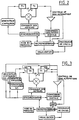
- the general principle of signal processing is illustrated by the diagram in Figure 7.
- the parameters are obtained for each position of the transducers, which allows obtain a mapping of the parameters.
- Image processing is included in the software thus making it possible to select one or more regions of the measurement, of any form, for an estimate of a local average of the parameters.
- the method of estimating the parameters (attenuation and speed of propagation) in transmission is based on the comparison of a reference signal with a signal transmitted through the bone.
- the reference signal is a signal that has propagated in an environment whose acoustic characteristics (attenuation and speed) are well known (the water in this example).
- the reference signal and the signals transmitted in the bone are registered under the same conditions.
- the reference signal can be acquired either when the device is started up or before each exam. You can also record a unique reference and store it in the computer memory and then recall it for each exam.
- Figure 9 illustrates the principle of ultrasound signal processing for measurement of this thickness. Indeed, by ultrasound we can determine the distance separating a transducer from the bone face (face of interest). Just identify the echo of this face and measure its flight time. Each T2 and T3 transducer (or T1 and T'1) is interrogated in turn in ultrasound mode, and the times are measured flight t1 and t2 of the echoes reflected by each of the opposite lateral faces of the bone. We deduce the thickness of the bone.
- the value of the acoustic parameters depends on where the measurement is taken.
- the scanning device the device according to the invention makes it possible to explore the entire bone volume and to obtain a parameter mapping. From the attenuation images as a function of the frequency and speed of the ultrasonic wave. we can select a region of the measurement (region of interest) and estimate an average of the parameters in this region.
- the software includes some simple image processing functions, in particular for the selection of regions of measurement, of variable shape and size. Image support is used to select measurement regions identical in different patients or in repeated measurements in the same patient.
- the invention makes it possible in particular to obtain ultrasound imaging of the calcaneum (attenuation and speed images) through the use of transducers focused, the images thus obtained being comparable to those obtained by to scan.
- Attenuation can be estimated from the radiofrequency ultrasound signal sampled.
- the principle of calculating the attenuation is based on a time-frequency analysis of the ultrasound signal.
- the invention thus makes it possible to measure the effective backscattering section as a function frequency. We know that the physical properties of bone tissue are highlighted by this measurement.
- the invention provides a physical method not traumatic in vivo assessment of bone quality (mass, rigidity, architecture). We can thus quantitatively assess the risk of fracture associated with a decrease in bone strength, this decrease being as is known, a consequence of demineralization and modification of bone architecture encountered in osteoporosis.
Landscapes
- Health & Medical Sciences (AREA)
- Life Sciences & Earth Sciences (AREA)
- Biomedical Technology (AREA)
- Heart & Thoracic Surgery (AREA)
- Rheumatology (AREA)
- Biophysics (AREA)
- Veterinary Medicine (AREA)
- Pathology (AREA)
- Public Health (AREA)
- Engineering & Computer Science (AREA)
- Orthopedic Medicine & Surgery (AREA)
- Physics & Mathematics (AREA)
- Medical Informatics (AREA)
- Molecular Biology (AREA)
- Surgery (AREA)
- Animal Behavior & Ethology (AREA)
- General Health & Medical Sciences (AREA)
- Radiology & Medical Imaging (AREA)
- Nuclear Medicine, Radiotherapy & Molecular Imaging (AREA)
- Dentistry (AREA)
- Oral & Maxillofacial Surgery (AREA)
- Ultra Sonic Daignosis Equipment (AREA)
Description
- on utilise au moins un transducteur focalisé émettant un faisceau ultrasonore au-travers de l'os ;
- on réalise un balayage dudit faisceau dans un plan perpendiculaire à la direction de propagation du faisceau ultrasonore ;
- on recueille les signaux transmis au-travers de l'os et/ou réfléchis par les faces de l'os et/ou diffusés par les structures internes de l'os ;
- on met en mémoire les signaux obtenus et,
- on traite les signaux ainsi stockés pour la mesure de la vitesse de propagation du faisceau ultrasonore dans l'os, de l'épaisseur de l'os, du coefficient d'atténuation en transmission du faisceau ultrasonore et/ou des paramètres de réflexion pour l'estimation des coefficients de rétro-diffusion et d'atténuation.
- la figure 1 est une vue schématique en élévation représentant un exemple de réalisation d'un appareil pour la mise en oeuvre du procédé objet de l'invention ;
- la figure 2 est un schéma par blocs illustrant, d'une façon générale, le module d'acquisition des données mis en oeuvre par l'invention ;
- la figure 3 est également un schéma illustrant d'une façon générale un module d'acquisition fonctionnant à la fois en transmission et en réflexion ;
- la figure 4 est un schéma illustrant le principe de l'acquisition en transmission ;
- la figure 5 est un schéma qui illustre le principe général de l'acquisition en transmission et en réflexion ;
- la figure 6 est un schéma illustrant une variante du mode d'acquisition en transmission et réflexion ;
- la figure 7 est un schéma par blocs illustrant le principe général du traitement des signaux dans le procédé et le dispositif objet de l'invention ;
- la figure 8 est un schéma illustrant le traitement du signal transmis en vue de la mesure de la vitesse de propagation de l'onde ultrasonore dans l'os ;
- la figure 9 est encore un schéma illustrant le traitement du signal, en mode échographique, en vue de la mesure de l'épaisseur de l'os et,
- la figure 10 est un schéma par blocs illustrant le principe du traitement du signal transmis pour la mesure de l'atténuation.
- l'émission des ondes ultrasonores à l'aide des transducteurs définis ci-dessus ;
- la réception d'ondes ultrasonores, dites de référence, la réception d'ondes ultrasonores transmises à travers l'os, la réception d'ondes réfléchies et/ou diffusées par l'os et ses structures ;
- le balayage de tout le volume osseux par le faisceau ultrasonore.
- calcul de l'atténuation (en dB) en fonction de la fréquence en transmission
- calcul du coefficient d'atténuation en fonction (en dB/MHz) de la fréquence en transmission
- calcul des temps de vol des signaux transmis à travers l'os
- calcul de l'épaisseur de l'os traversé à l'endroit de la mesure
- calcul du coefficient d'atténuation en fonction de la fréquence (dB/cm.MHz) en transmission
- calcul de la vitesse de propagation des ultrasons en transmission
- calcul de l'atténuation (en dB) en fonction de la fréquence en réflexion
- calcul du coefficient d'atténuation en fonction de la fréquence (dB/cm.MHz) en réflexion
- calcul de la section efficace de rétrodiffusion (en dB) en fonction de la fréquence en réflexion
- calcul du coefficient de rétrodiffusion (dB/MHz) en réflexion
- calcul du coefficient intégral de rétrodiffusion (dB.MHz) en réflexion.
- Les transducteurs sont à la position X, Y: Le transducteur T1 émet et l'onde ainsi émise, transmise à travers l'os, est détectée par le transducteur-récepteur T'1, puis amplifiée, numérisée, et transférée sur ordinateur. Bien entendu, et comme spécifié ci-dessus, pour des mesures à travers le calcanéum, les transducteurs et l'os sont placés dans un bain dont la température est contrôlée-par thermostat (voir la figure 1). Cette première mesure est destinée à l'estimation du temps de vol de l'onde transmise à travers l'os et au calcul de la différence entre ce temps de vol et celui du signal de référence;
- Le transducteur T2 (ou T1) émet et le transducteur T2 (ou T1) reçoit le signal ainsi émis et réfléchi par l'os. L'onde ultrasonore réfléchie est amplifiée, numérisée, et transférée sur ordinateur.
- On estime la durée du temps de vol de l'écho qui est réfléchi par la face d'entrée de l'os tournée vers le transducteur T2 (ou T1) :
- Le transducteur T3 (ou T'1) émet et le transducteur T3 (ou T'1) assure la réception du signal réfléchi par l'os. L'onde réfléchie est amplifiée, numérisée, et transférée sur ordinateur.
- On estime la durée du temps de vol de l'écho réfléchi par la face d'entrée de l'os, qui est tournée vers T3 (ou T'1).
- Ces deux dernières mesures sont destinées au calcul de l'épaisseur du calcaneum à l'endroit de la mesure. La vitesse de propagation en transmission de l'onde ultrasonore se déduit de manière connue du temps de vol de l'onde entre les deux transducteurs. Pour estimer la vitesse de propagation, il faut connaítre l'épaisseur de l'os et celle-ci, selon l'invention, est mesurée par ultrasons.
- Les transducteurs sont initialement dans la position X,Y
- Le transducteur T1 émet et l'onde transmise à travers l'os est détectée par le transducteur-récepteur T'1, puis elle est amplifiée, numérisée et transférée sur ordinateur. Comme précédemment, pour des mesures à travers le calcaneum, dans cet exemple de réalisation, les transducteurs et l'os sont placés dans un bain dont la température est contrôlée par thermostat (fig. 1);
- On calcule les spectres en fréquence du signal transmis à travers l'os et du signal de référence. L'atténuation en fonction de la fréquence est obtenue par comparaison du spectre d'un signal de référence enregistré dans l'eau et de celui du signal transmis à travers l'os. Le signal ultrasonore est du type impulsionnel et il comporte plusieurs fréquences dans l'intervalle compris entre 0,2 MHz et 1 MHz.
- Les transducteurs sont à la position X et Y ;
- Le transducteur T2 (ou T3, ou T1, ou T'1) émet
- Le transducteur T2 (ou T3, ou T1, ou T'1) assure la réception du signal échographique rétrodiffusé par l'architecture interne de l'os, ce signal étant ensuite amplifié, numérisé, et transféré sur ordinateur .
- On effectue une analyse spectrale glissante du signal échographique, et une estimation des spectres en fonction de la profondeur ;
- On calcule les centroïdes spectraux en fonction de la profondeur ;
- On calcule le coefficient d'atténuation en fonction de la fréquence (dB/cm.MHz) en réflexion ;
- On calcule la différence spectrale entre le spectre du signal et un spectre de référence, puis l'on calcule la section efficace de rétrodiffusion (en dB) en fonction de la fréquence en réflexion :
- On calcule le coefficient de rétrodiffusion (dB/MHz) en réflexion et
- On calcule le coefficient intégral de rétrodiffusion (dB.MHz) en réflexion. Le balayage du faisceau ultrasonore permet de réaliser une exploration de la totalité du volume osseux. Les coefficients d'atténuation et de rétrodiffusion sont calculés pour chaque position des transducteurs. On peut ensuite estimer une moyenne locale des paramètres à l'intérieur d'une région d'intérét sélectionnée par l'opérateur.
- le dispositif de balayage automatique du faisceau ultrasonore permet de résoudre le problème du positionnement précis de l'os et du positionnement de la région de mesure où d'intérêt.
- L'utilisation de transducteurs focalises permet d'obtenir des images de bonne qualité ;
- Elle permet d'obtenir des mesures ultrasonores en transmission à travers l'os et en réflexion ;
- Elle permet de recueillir et de traiter des signaux réfléchis et/ou diffusés par l'architecture interne de l'os ce qui apporte une information complémentaire à celles se trouvant déjà dans les signaux qui ont été transmis à travers l'os ; Ainsi, le procédé et le dispositif objets de l'invention apportent une quantité d'informations bien supérieure à celle fournie par les dispositifs selon l'état antérieur de la technique, tout en offrant une précision une reproductibilité et une sensibilité meilleures.
- La maturation squelettique chez les nouveaux nés ;
- Les ostéoporoses secondaires ostéomalacie etc...
- La surveillance des chevaux de course .
- La caractérisation de pièces osseuses in vitro.
Claims (10)
- Procédé d'évaluation et de caractérisation in vivo des propriétés mécaniques ou architecturales des os, par propagation d'une onde ultra-sonore au-travers de l'os et l'étude de l'interaction de cette onde avec l'os, en vue de l'obtention d'images paramétriques selon lequel :on utilise au moins un transducteur focalisé émettant un faisceau ultra-sonore au travers de l'os ;on réalise un balayage dudit faisceau dans un plan perpendiculaire à la direction de propagation du faisceau ultrasonore ;on recueille les signaux transmis au-travers de l'os et/ou réfléchis par les faces de l'os et/ou diffusés par les structures internes de l'os ;on met en mémoire les signaux obtenus et,on traite les signaux ainsi stockés pour la mesure de la vitesse de propagation du faisceau ultra-sonore dans l'os, de l'épaisseur de l'os, du coefficient d'atténuation en transmission du faisceau ultrasonore et/ou des paramètres de réflexion pour l'estimation des coefficients de rétro-diffusion et d'atténuation.
- Procédé selon la revendication 1 pour la réalisation, en mode transmission, d'images paramétriques d'atténuation en fonction de la fréquence et de la vitesse de propagation du faisceau ultrasonore, dans lequel on utilise une paire de transducteurs focalisés, placés en vis à vis et fonctionnant à basse fréquence, c'est-à-dire à une fréquence centrale comprise entre 100 kHz et 3 MHz, et de préférence entre 100 kHz et 1MHz.
- Procédé selon la revendication 1 pour la réalisation en mode échographique d'images de réflectivité, dans lequel on utilise un transducteur focalisé ou une paire de transducteurs focalisés, placés en vis à vis, et fonctionnant avec une fréquence centrale comprise entre 100 kHz et 10 MHz.
- Procédé selon la revendication 1 appliqué à la réalisation en mode échographique d'images de réflectivité et à l'estimation des coefficients d'atténuation et/ou de rétrodiffusion en fonction de la fréquence du faisceau ultrasonore, et également en vue de la réalisation, en transmission, d'images paramétriques d'atténuation en fonction de la fréquence et de la vitesse de propagation dans lequel on utilise une paire de transducteurs placés en vis-à-vis et fonctionnant à basse fréquence, c'est-à-dire à une fréquence centrale comprise entre 100 kHz et 1 MHz et, un transducteur focalisé ou une paire de transducteurs focalisés placés en vis à vis et fonctionnant à haute fréquence, c'est-à-dire à une fréquence centrale comprise entre 1 MHz et 10 MHz.
- Procédé selon l'une quelconque des revendications 1 à 4 dans lequel les mesures sont effectuées par contact, à l'aide d'un milieu de couplage.
- Procédé selon l'une quelconque des revendications précédentes dans lequel on utilise un réseau de transducteurs ultrasonores avec lesquels la focalisation et le balayage du faisceau ultrasonore peuvent être effectués électroniquement.
- Dispositif pour la mise en oeuvre du procédé selon l'une quelconque des revendications précédentes comportant des moyens d'émission et de réception ultrasonores (12, 14) placés en vis-à-vis de part et d'autre de l'os à analyser, un module d'acquisition incluant les moyens d'émission d'ultrasons et de réception des signaux ultrasonores après leur interaction dans l'os, un système de mise en mémoire des signaux ainsi obtenus et, un module de traitement desdits signaux pour l'estimation des paramètres acoustiques connus pour leur relation avec les propriétés mécaniques ou viscoélastiques de l'os, lesdits moyens d'émission et de réception ultrasonores étant focalisés et le dispositif comprenant des moyens permettant de réaliser un balayage du faisceau ultrasonore ainsi émis.
- Dispositif selon la revendication 7 dans lequel le module de traitement des signaux comporte un ordinateur, une mémoire de masse et un ensemble de logiciels de traitement de signaux conçus pour l'exploitation des données provenant du module d'acquisition, ce module de traitement comportant un ensemble de fonctions permettant notamment d'effectuer le calcul du coefficient d'atténuation en transmission, le calcul de l'épaisseur de l'os traversé à l'endroit de la mesure, le calcul de la vitesse de propagation, le calcul du coefficient de rétrodiffusion en réflexion et le calcul du coefficient d'atténuation en réflexion.
- Dispositif selon l'une des revendications 7 ou 8 dans lequel les moyens d'émission et de réception comportent des transducteurs focalisés (12, 14), comprenant un seul élément, placés en vis-à-vis.
- Dispositif selon l'une quelconque des revendications 7 à 9 comportant une enceinte (10) remplie d'un liquide, tel que de l'eau, afin de réaliser les mesures en immersion.
Priority Applications (1)
| Application Number | Priority Date | Filing Date | Title |
|---|---|---|---|
| DE29522392U DE29522392U1 (de) | 1994-03-25 | 1995-03-24 | Gerät zur Auswertung und Charakterisierung von Knocheneigenschaften |
Applications Claiming Priority (3)
| Application Number | Priority Date | Filing Date | Title |
|---|---|---|---|
| FR9403555A FR2717671B1 (fr) | 1994-03-25 | 1994-03-25 | Procédé et dispositif d'évaluation et de caractérisation des propriétés des os. |
| FR9403555 | 1994-03-25 | ||
| PCT/FR1995/000376 WO1995026160A1 (fr) | 1994-03-25 | 1995-03-24 | Procede et dispositif d'evaluation et de caracterisation des proprietes des os |
Publications (2)
| Publication Number | Publication Date |
|---|---|
| EP0751743A1 EP0751743A1 (fr) | 1997-01-08 |
| EP0751743B1 true EP0751743B1 (fr) | 1999-05-26 |
Family
ID=9461446
Family Applications (1)
| Application Number | Title | Priority Date | Filing Date |
|---|---|---|---|
| EP95914421A Revoked EP0751743B1 (fr) | 1994-03-25 | 1995-03-24 | Procede et dispositif d'evaluation et de caracterisation des proprietes des os |
Country Status (11)
| Country | Link |
|---|---|
| US (1) | US5806520A (fr) |
| EP (1) | EP0751743B1 (fr) |
| JP (1) | JPH09511163A (fr) |
| KR (1) | KR100366472B1 (fr) |
| AU (1) | AU689570B2 (fr) |
| DE (1) | DE69509882T2 (fr) |
| DK (1) | DK0751743T3 (fr) |
| ES (1) | ES2132654T3 (fr) |
| FR (1) | FR2717671B1 (fr) |
| NZ (1) | NZ283395A (fr) |
| WO (1) | WO1995026160A1 (fr) |
Families Citing this family (34)
| Publication number | Priority date | Publication date | Assignee | Title |
|---|---|---|---|---|
| US6277076B1 (en) | 1988-05-11 | 2001-08-21 | Lunar Corporation | Ultrasonic densitometer with pre-inflated fluid coupling membranes |
| US6027449A (en) * | 1988-05-11 | 2000-02-22 | Lunar Corporation | Ultrasonometer employing distensible membranes |
| US6517487B1 (en) | 1995-03-01 | 2003-02-11 | Lunar Corporation | Ultrasonic densitometer with opposed single transducer and transducer array |
| JP2000500998A (ja) * | 1995-11-22 | 2000-02-02 | オステオメータ メディテック エイ/エス | 骨の状態を評価する方法及び装置 |
| EP1014859A4 (fr) | 1997-08-19 | 2007-05-02 | Philipp Lang | Mesure du fluide interstitiel propre aux capillaires utilisant des methodes et des dispositifs echographiques |
| US6077224A (en) * | 1998-03-23 | 2000-06-20 | Lang; Philipp | Methods and device for improving broadband ultrasonic attenuation and speed of sound measurements using anatomical landmarks |
| EP1061854A4 (fr) * | 1998-03-09 | 2005-07-13 | Philipp Lang | Procedes et dispositifs ameliorant l'attenuation ultrasonore en large bande et les mesures de vitesse des sons |
| US6585649B1 (en) * | 1998-05-02 | 2003-07-01 | John D. Mendlein | Methods and devices for improving ultrasonic measurements using multiple angle interrogation |
| US6013031A (en) * | 1998-03-09 | 2000-01-11 | Mendlein; John D. | Methods and devices for improving ultrasonic measurements using anatomic landmarks and soft tissue correction |
| FR2778838A1 (fr) * | 1998-05-19 | 1999-11-26 | Koninkl Philips Electronics Nv | Procede de detection de variations d'elasticite et appareil echographique pour mettre en oeuvre ce procede |
| AU6365099A (en) * | 1998-11-08 | 2000-05-29 | Sunlight Medical, Ltd | Bone velocity determination |
| US6234969B1 (en) * | 1999-02-01 | 2001-05-22 | Hologic, Inc. | Bone sonometry, densitometry and imaging |
| DE10018654A1 (de) * | 2000-04-14 | 2001-12-13 | Jacek Czernicki | Verfahren und Vorrichtung zur Ermittlung des anatomischen Zustands eines Menschen oder eines Tieres, insbesondere zur Diagnose von Osteoporose |
| US6494097B1 (en) | 2000-09-05 | 2002-12-17 | Elias Edmond Shihadeh | Method and apparatus for measuring thickness of a layer in a multi-layered object |
| WO2002071949A2 (fr) * | 2001-02-28 | 2002-09-19 | Research Foundation Of State University Of New York | Procede et appareil permettant d'effectuer un diagnostic acoustique confocal par balayage de la qualite osseuse |
| WO2003009758A1 (fr) * | 2001-07-24 | 2003-02-06 | Sunlight Medical, Ltd. | Determination de l'age osseux au moyen d'ultrasons |
| AU2002321792A1 (en) * | 2001-07-24 | 2003-02-17 | Sunlight Medical, Ltd. | Method and apparatus for bone diagnosis |
| WO2003009762A1 (fr) * | 2001-07-24 | 2003-02-06 | Sunlight Medical, Ltd. | Analyse d'articulations par ultrasons |
| FR2839876A1 (fr) | 2002-05-27 | 2003-11-28 | Centre Nat Rech Scient | Procede, sonde et appareil pour caracteriser par voie ultrasonore un milieu d'etude, en particulier la couche corticale d'un os |
| FR2839877B1 (fr) | 2002-05-27 | 2005-04-08 | Centre Nat Rech Scient | Procede, sonde et appareil pour evaluer de maniere non-invasive une duree de parcours ou une vitesse d'ultra-sons le long d'une interface, notamment osseuse |
| US7840247B2 (en) * | 2002-09-16 | 2010-11-23 | Imatx, Inc. | Methods of predicting musculoskeletal disease |
| US7611465B2 (en) * | 2003-07-15 | 2009-11-03 | Board Of Regents, The University Of Texas System | Rapid and accurate detection of bone quality using ultrasound critical angle reflectometry |
| JP2006014979A (ja) * | 2004-07-02 | 2006-01-19 | Oyo Denki Kk | 骨強度測定装置 |
| EP1557126A1 (fr) * | 2004-01-21 | 2005-07-27 | OYO Electric Co., Ltd. | Dispositif pour mesurer la résistance de l'os |
| US20070238991A1 (en) * | 2006-01-25 | 2007-10-11 | Jaltec Biomedical Inc. | Ultrasound method and apparatus for characterizing and identifying biological tissues |
| US20090112094A1 (en) * | 2006-04-13 | 2009-04-30 | The Research Foundation Of State University Of New York | Phased Apply Ultrasound With Electronically Controlled Focal Point For Assessing Bone Quality Via Acoustic Topology And Wave Transmit Functions |
| US20100063394A1 (en) * | 2006-11-14 | 2010-03-11 | Kagoshima University | Osteoporosis progress stage measuring device and osteoporosis progress stage measuring method |
| US20080125653A1 (en) * | 2006-11-27 | 2008-05-29 | Board Of Regents, The University Of Texas System | Density and porosity measurements by ultrasound |
| FR2946753B1 (fr) * | 2009-06-11 | 2011-07-22 | Centre Nat Rech Scient | Procede et dispositif ultrasonores pour caracteriser un milieu |
| US8585595B2 (en) | 2011-01-27 | 2013-11-19 | Biomet Manufacturing, Llc | Method and apparatus for aligning bone screw holes |
| KR101239525B1 (ko) | 2011-04-15 | 2013-03-05 | 성균관대학교산학협력단 | 초음파 투과 모니터링에 의한 진단 장치 및 방법 |
| WO2015106027A1 (fr) * | 2014-01-08 | 2015-07-16 | Klock John C | Imagerie à ultrasons par transmission quantitative de structures anatomiques denses |
| WO2015113813A1 (fr) * | 2014-01-17 | 2015-08-06 | Koninklijke Philips N.V. | Dispositif à ultrasons et procédé pour évaluer un os d'un sujet |
| CN103860203B (zh) * | 2014-03-21 | 2016-03-23 | 南京科进实业有限公司 | 一体式超声骨密度仪 |
Family Cites Families (6)
| Publication number | Priority date | Publication date | Assignee | Title |
|---|---|---|---|---|
| US3847141A (en) * | 1973-08-08 | 1974-11-12 | Nasa | Ultrasonic bone densitometer |
| US4774959A (en) * | 1986-01-10 | 1988-10-04 | Walker Sonix A/S | Narrow band ultrasonic frequency attentuation bone measurement system |
| US5343863A (en) * | 1988-05-11 | 1994-09-06 | Lunar Corporation | Ultrasonic densitometer device and method |
| US4941474A (en) * | 1988-07-01 | 1990-07-17 | Massachusetts Institute Of Technology | Multivariable analysis of bone condition |
| US5038787A (en) * | 1988-08-10 | 1991-08-13 | The Board Of Regents, The University Of Texas System | Method and apparatus for analyzing material properties using reflected ultrasound |
| DE69329168T2 (de) * | 1992-05-20 | 2001-04-05 | Aloka Co. Ltd., Mitaka | Vorrichtung zur Bestimmung der Eigenschaften von Knochen |
-
1994
- 1994-03-25 FR FR9403555A patent/FR2717671B1/fr not_active Expired - Lifetime
-
1995
- 1995-03-24 JP JP7525006A patent/JPH09511163A/ja active Pending
- 1995-03-24 AU AU21426/95A patent/AU689570B2/en not_active Expired
- 1995-03-24 WO PCT/FR1995/000376 patent/WO1995026160A1/fr not_active Ceased
- 1995-03-24 KR KR1019960705463A patent/KR100366472B1/ko not_active Expired - Lifetime
- 1995-03-24 DE DE69509882T patent/DE69509882T2/de not_active Revoked
- 1995-03-24 DK DK95914421T patent/DK0751743T3/da active
- 1995-03-24 NZ NZ283395A patent/NZ283395A/en not_active IP Right Cessation
- 1995-03-24 EP EP95914421A patent/EP0751743B1/fr not_active Revoked
- 1995-03-24 US US08/702,444 patent/US5806520A/en not_active Expired - Lifetime
- 1995-03-24 ES ES95914421T patent/ES2132654T3/es not_active Expired - Lifetime
Also Published As
| Publication number | Publication date |
|---|---|
| KR100366472B1 (ko) | 2003-08-02 |
| WO1995026160A1 (fr) | 1995-10-05 |
| ES2132654T3 (es) | 1999-08-16 |
| US5806520A (en) | 1998-09-15 |
| EP0751743A1 (fr) | 1997-01-08 |
| AU2142695A (en) | 1995-10-17 |
| DK0751743T3 (da) | 1999-11-29 |
| FR2717671B1 (fr) | 1996-06-21 |
| DE69509882T2 (de) | 1999-12-16 |
| DE69509882D1 (de) | 1999-07-01 |
| FR2717671A1 (fr) | 1995-09-29 |
| JPH09511163A (ja) | 1997-11-11 |
| AU689570B2 (en) | 1998-04-02 |
| NZ283395A (en) | 1998-07-28 |
Similar Documents
| Publication | Publication Date | Title |
|---|---|---|
| FR2717671A1 (fr) | Procédé et dispositif d'évaluation et de caractérisation des propriétés des os. | |
| CA2878545C (fr) | Procede de traitement de signaux issus d'une acquisition par sondage ultrasonore, programme d'ordinateur et dispositif de sondage a ultrasons correspondants | |
| EP1531733B1 (fr) | Dispositif et procede pour la mesure de l'elasticite d'un organe humain ou animal | |
| EP1546757B1 (fr) | Procede et dispositif d'imagerie utilisant des ondes de cisaillement | |
| EP0342757B1 (fr) | Echographe ultrasonore à cohérence de phase contrôlable | |
| FR2883982A1 (fr) | Procede et dispositif d'imagerie utilisant des ondes de cisaillement | |
| WO2000055616A1 (fr) | Procede et dispositif d'imagerie utilisant les ondes de cisaillement | |
| JP7828094B2 (ja) | 混合トランスデューサアレイのための画像複合 | |
| EP0196713B1 (fr) | Procédé et appareil d'exploration de milieux par échographie ultrasonore | |
| WO2018109314A1 (fr) | Procede de traitement de signaux issus d'une acquisition par sondage ultrasonore, programme d'ordinateur et dispositif de sondage a ultrasons correspondants | |
| US4993416A (en) | System for ultrasonic pan focal imaging and axial beam translation | |
| EP0164808A1 (fr) | Appareil d'examen de milieux par échographie ultrasonore | |
| EP0197582B1 (fr) | Procédé et appareil d'exploration de milieux par échographie ultrasonore | |
| CA2184510C (fr) | Procede et dispositif d'evaluation et de caracterisation des proprietes des os | |
| EP0196714B1 (fr) | Procédé et appareil d'exploration de milieux par échographie ultrasonore | |
| JPH0658917A (ja) | 超音波検査方法および装置 |
Legal Events
| Date | Code | Title | Description |
|---|---|---|---|
| PUAI | Public reference made under article 153(3) epc to a published international application that has entered the european phase |
Free format text: ORIGINAL CODE: 0009012 |
|
| 17P | Request for examination filed |
Effective date: 19960916 |
|
| AK | Designated contracting states |
Kind code of ref document: A1 Designated state(s): DE DK ES FR GB IT SE |
|
| GRAG | Despatch of communication of intention to grant |
Free format text: ORIGINAL CODE: EPIDOS AGRA |
|
| 17Q | First examination report despatched |
Effective date: 19980922 |
|
| GRAG | Despatch of communication of intention to grant |
Free format text: ORIGINAL CODE: EPIDOS AGRA |
|
| GRAH | Despatch of communication of intention to grant a patent |
Free format text: ORIGINAL CODE: EPIDOS IGRA |
|
| GRAH | Despatch of communication of intention to grant a patent |
Free format text: ORIGINAL CODE: EPIDOS IGRA |
|
| TPAD | Observations filed by third parties |
Free format text: ORIGINAL CODE: EPIDOS TIPA |
|
| GRAA | (expected) grant |
Free format text: ORIGINAL CODE: 0009210 |
|
| AK | Designated contracting states |
Kind code of ref document: B1 Designated state(s): DE DK ES FR GB IT SE |
|
| REF | Corresponds to: |
Ref document number: 69509882 Country of ref document: DE Date of ref document: 19990701 |
|
| GBT | Gb: translation of ep patent filed (gb section 77(6)(a)/1977) |
Effective date: 19990616 |
|
| REG | Reference to a national code |
Ref country code: ES Ref legal event code: FG2A Ref document number: 2132654 Country of ref document: ES Kind code of ref document: T3 |
|
| REG | Reference to a national code |
Ref country code: DK Ref legal event code: T3 |
|
| PLBQ | Unpublished change to opponent data |
Free format text: ORIGINAL CODE: EPIDOS OPPO |
|
| PLBI | Opposition filed |
Free format text: ORIGINAL CODE: 0009260 |
|
| PLBF | Reply of patent proprietor to notice(s) of opposition |
Free format text: ORIGINAL CODE: EPIDOS OBSO |
|
| 26 | Opposition filed |
Opponent name: OSTEOMETER MEDITECH, INC. Effective date: 20000216 |
|
| PLBF | Reply of patent proprietor to notice(s) of opposition |
Free format text: ORIGINAL CODE: EPIDOS OBSO |
|
| PLBF | Reply of patent proprietor to notice(s) of opposition |
Free format text: ORIGINAL CODE: EPIDOS OBSO |
|
| RDAH | Patent revoked |
Free format text: ORIGINAL CODE: EPIDOS REVO |
|
| APAC | Appeal dossier modified |
Free format text: ORIGINAL CODE: EPIDOS NOAPO |
|
| APAE | Appeal reference modified |
Free format text: ORIGINAL CODE: EPIDOS REFNO |
|
| REG | Reference to a national code |
Ref country code: GB Ref legal event code: IF02 |
|
| PGFP | Annual fee paid to national office [announced via postgrant information from national office to epo] |
Ref country code: DK Payment date: 20020220 Year of fee payment: 8 |
|
| PGFP | Annual fee paid to national office [announced via postgrant information from national office to epo] |
Ref country code: SE Payment date: 20020225 Year of fee payment: 8 Ref country code: GB Payment date: 20020225 Year of fee payment: 8 |
|
| PGFP | Annual fee paid to national office [announced via postgrant information from national office to epo] |
Ref country code: DE Payment date: 20020313 Year of fee payment: 8 |
|
| PGFP | Annual fee paid to national office [announced via postgrant information from national office to epo] |
Ref country code: ES Payment date: 20020320 Year of fee payment: 8 |
|
| PGFP | Annual fee paid to national office [announced via postgrant information from national office to epo] |
Ref country code: FR Payment date: 20020325 Year of fee payment: 8 |
|
| APAC | Appeal dossier modified |
Free format text: ORIGINAL CODE: EPIDOS NOAPO |
|
| RDAG | Patent revoked |
Free format text: ORIGINAL CODE: 0009271 |
|
| STAA | Information on the status of an ep patent application or granted ep patent |
Free format text: STATUS: PATENT REVOKED |
|
| 27W | Patent revoked |
Effective date: 20020508 |
|
| GBPR | Gb: patent revoked under art. 102 of the ep convention designating the uk as contracting state |
Free format text: 20020508 |
|
| APAH | Appeal reference modified |
Free format text: ORIGINAL CODE: EPIDOSCREFNO |