-
This invention relates to a human interleukin-2 protein and a method of producing the same by a genetic engineering technique.
-
Interleukin-2 [also called T cell growth factor (TCGF); hereinafter abbreviated as IL-2] is a lymphokine produced by T cells upon stimulation with a lectin or alloantigen, for instance [Science, 193, 1007 (1976); Immunological Reviews, 51, 257 (1980)]. IL-2 enables long-period culture of T cells by allowing them to grow in vitro while maintaining their functions. Furthermore, IL-2 reportedly promotes the mitogen reaction of thymus cells (costimulator), restores the antibody production by spleen cells against T cell-dependent antigens in nude mice (T cell replacing factor) or promotes the differentiation and proliferation of killer cells (killer helper factor) [The Journal of Immunology, 123, 2928 (1979); Immunological Reviews, 51, 257 (1980)].
-
Killer T cell clones, helper T cell clones and, further, natural killer cell clones have so far been obtained in great numbers through the utilization of IL-2 [e.g., Nature, 268, 154 (1977); The Journal of Immunology, 130, 981 (1983)]. In addition to such direct use in T cell or natural killer cell cloning, IL-2 can be used in selectively growing killer T cells specific to special antigens, for example killer T cells which recognise a tumor antigen and destroy the tumor, in vitro. By injecting the tumor-specific killer T cells grown in this manner into an animal, it is possible to inhibit and prevent the tumor growth [The Journal of Immunology, 125, 1904 (1980)]. It is also known that IL-2 induces the production of IFN-γ [The Journal of Immunology, 130, 1784 (1983)] and that IL-2 activates natural killer cells [The Journal of Immunology, 130, 1970 (1983)].
-
The above experimental facts suggest that IL-2 may possibly be useful as an antitumor agent. IL-2 is known to restore the helper T cell function in nude mice lacking thymic function [European Journal of Immunology, 10, 719 (1980)] or restore the induction of killer T cells against homologous cells [Nature, 284, 278 (1980)] and, therefore IL-2 can be expected to be useful in treating immunocompromised diseases.
-
However, large scale production of IL-2 has been difficult so far, so that clinical applications thereof are impossible for the time being. Accordingly, a technique which renders it possible to produce highly purified IL-2 in large amounts, in an easy and simple manner and at low cost has been awaited.
-
Taniguchi et al. cloned the human IL-2 gene by using IL-2 mRNA isolated from the human T cell leukemia line Jurkat as the starting material, reported the amino acid sequence of human IL-2 protein as deduced therefrom and expressed the gene in COS-7 cells [Nature, 302, 305 (1983)]. Later, Devos et al. reported that the human spleen cell-derived IL-2 gene had been cloned and expressed in Escherichia coli [Nucleic Acids Research, 11, 4307 (1983)]. Nevertheless, so far, the presence of an IL-2-like substance has been only estimated on the basis of there being detected TCGF activity. There is no account reporting that a human IL-2 protein produced by a transformant carrying a DNA coding for human IL-2 has actually been isolated in a purified form.
-
The present inventors conducted intensive researches in an attempt to develop a technique by which the desired IL-2 protein can be obtained through cloning of the human IL-2 gene utilizing the gene manipulation technology, introduction of the recombinant DNA molecule obtained into a host and expression of the human IL-2 gene in said host, and as a result could establish a method of producing a substantially pure, non-glycosylated human IL-2 protein and have completed the present invention.
-
Thus, the present invention provides a substantially pure, non-glycosylated human IL-2 protein and a method of producing said human IL-2 protein which comprises cultivating a transformant carrying a DNA having a base sequence coding for human IL-2 and purifying said protein from the culture broth.
-
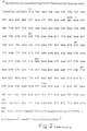
The DNA coding for human IL-2 to be used in the practice of the present invention is, for example, a DNA (I) having the base sequence defined by codons 1-133 in Fig. 2 (SEQ ID NO.1). This DNA (I) may optionally have, at its 5' end, either ATG or a signal sequence represented in Fig. 2 (SEQ ID NO.1) by codons S1-S20 and preferably has TAA, TGA or TAG, most preferably TGA, at the 3' end.
-
The DNA (I) is preferably connected downstream from a promoter, for example the tryptophan (trp) promoter, rec A promoter or λPL promoter, suitably the trp or λPL promoter.
-
In accordance with the invention, the mRNA coding for human IL-2 is isolated from a culture of human peripheral leukocytes stimulated by concanavalin A, for instance, then a single-stranded cDNA is synthesized using reverse transcriptase, and further a double-stranded DNA is synthesized. The DNA is inserted into a plasmid, the recombinant plasmid is used for transformation of a strain of Escherichia coli or Bacillus subtilis, for example, and the cDNA-containing plasmid is cloned. In this way, a double-stranded DNA coding for human IL-2 can be produced.
-
More particularly, the human IL-2-encoding mRNA to be used in the practice of the invention can be obtained by the method of Hinuma et al. [Biochemical and Biophysical Research Communications, 109, 363 (1982)]. With the thus-obtained human IL-2 mRNA as a template, a cDNA is synthesized by the per se known method using reverse transcriptase, and the cDNA is converted to a double-stranded DNA [Maniatis, T. et al. Cell, 8, 163 (1976); Land, H. et al., Nucleic Acids Research, 9, 2251 (1981)]. This DNA is inserted, for example, into the plasmid pBR322 at the PstI restriction endonuclease cleavage site by the dG-dC homopolymer ligation method [Nelson, T. S., Methods in Enzymology, 68, 41 (1979)], for instance. Furthermore, an oligonucleotide having the base sequence corresponding to the amino acid sequence of a part of human IL-2, for instance, is chemically synthesized and then labeled with 32P. Using this as a probe, the desired clone is selected from among tetracycline- or ampicillin-resistant transformants by the per se known colony hybridization method [Grunstein, M. and Hogness, D. S., Proc. Natl. Acad. Sci. USA, 72, 3961 (1975); Alwine, J. C. et al., Methods in Enzymology, 68, 220 (1979)]. The presence of human IL-2 gene is confirmed by determination of the base sequence of DNA derived from the clone reacting positively in the above hybridization method, by the Maxam-Gilbert method [Maxam, A.M. et al., Proc. Natl. Acad. Sci. USA, 74, 560 (1977)] or the dinucleotide synthetic chain termination method using phage M13 [Messing, J. et al., Nucleic Acids Research, 9, 309 (1981)]. Then, the whole, or part, of the human IL-2 gene is excised from the clone obtained, and inserted into a plasmid, downstream from an appropriate promoter and the SD (Shine and Dalgarno) base sequence, for the subsequent introduction into an appropriate host.
-
The use of the trp promoter, among others, as the promoter is advantageous. As the host, a strain of Escherichia coli (e.g. strain 294, strain DHl, strain N4830), among others, is used with advantage.
-
The strains Escherichia coli 294 [Beckman et al., Proc. Natl. Acad. Sci. USA, 73, 4174 (1976)], E. coli DHl [Selson, M. E. et al., Nature, 217, 1110 (1968)] and E. coli N4830 [Cell, 25, 713 (1981)] are known strains.
-
The transformation of such hosts with the DNA can be performed by the known method [Cohen, S. N. et al., Proc. Natl. Acad. Sci. USA, 69, 2110 (1972)]. The host thus obtained is cultivated in a per se known medium, such as glucose- and casamino acid-containing M9 medium [Miller, J. Experiments in Molecular Genetics, pages 431-433 (Cold Spring Harbor Laboratory, New York, 1972)]. In cases where the trp promoter is used, an agent such as 3-β-indolylacrylic acid may be added for increasing the efficiency of said promoter. The cultivation is generally carried out at 15-43°C for 3-24 hours, with aeration and/or stirring as necessary.
-
In case where the λPL promoter is used, the cultivation is preferably carried out at a relatively low temperature of about 30 to 36°C to grow the transformants, and then, of about 37 to 42°C to inactivate the repressor in the transformants and to effectively express the DNA coding for human IL-2.
-
Human IL-2 thus formed can be assayed using an IL-2-dependent cell line. Since human IL-2 is known to promote the growth of rat, mouse or other IL-2-dependent cells as well as human IL-2-dependent cells [Immunological Reviews, 51, 257 (1980)], not only IL-2-dependent human cell lines,but also rat or mouse IL-2-dependent cell lines may be used [Journal of Immunology, 130, 981 and 988 (1983)].
-
In particular, mouse IL-2-dependent cell lines can be maintained stably by subculturing for a long period of time, so that their use can give highly reproducible assay results.
-
In extracting the human IL-2 according to the invention from cultivated cells, the cells, after cultivation, are harvested by the known method, suspended in a solution of a protein denaturing agent, such as a mineral acid salt of guanidine (e.g. hydrochloride, nitrate, sulfate), and the suspension is stirred in the cold and then centrifuged, whereby an IL-2-containing supernatant is obtained, or the cells harvested are suspended in a buffer and disrupted by sonication, lysozyme treatment and/or freezing and thawing, followed by centrifuging to give an IL-2-containing supernatant. Any other appropriate method may also be used.
-
The isolation of IL-2 from the above supernatant and the subsequent purification of the same can be carried out by an appropriate combination of per se known methods of separation and purification. Such known separation and purification methods include, among others, methods utilizing solubility differences, such as salting out methods and solvent precipitation methods, methods utilizing molecular weight differences in the main, such as dialysis, ultrafiltration, gel filtration and SDS-polyacrylamide gel electrophoresis, methods utilizing charge differences, such as ion exchange chromatography, methods utilizing specific affinities, such as affinity chromatography, methods utilizing hydrophobicity differences, such as reversed phase high performance liquid chromatography, and methods utilizing isoelectric point differences, such as isoelectric focusing. Since the human IL-2 protein is highly hydrophobic, hydrophobic column chromatography, in particular high performance liquid chromatography using a reversed phase column, is very effective in purifying said protein.
-
Thus, for instance, the cells obtained by cultivating a strain of Escherichia coli carrying the gene coding for human IL-2 are suspended in a solution, preferably in a buffer of a protein denaturing agent, such as guanidine hydrochlozide (in a concentration of 2 M to 8 M, preferably 6 M to 8M), and the suspension is stirred, preferablly at 0 - 5°C for 0.5 - 3 hours and than centrifuged. The supernatant thus collected is, as it is, or after concentration using an ultrafiltration device or the like, subjected to dialysis. The resulting precipitate is removed by centrifugation, the supernatant is subjected to anion exchange chromatography using diethylaminoethylcellulose [e.g. DE 52 cellulose (Whatman, Gt. Britain) column], for instance, and an active fraction is collected.
-
Then, after concentration using an ultrafiltration device, the active fraction is subjected to gel filtration using N,N'-methylenebisacrylamide-crosslinked allyldextran, such as a Sephacryl S-200 (Pharmacia, Sweden) column, or the like. The active fraction collected is then subjected to the above-mentioned high performance liquid chromatography. In this manner, the non-glycosylated human IL-2 protein according to the invention can be obtained.
-
As the reversed phase system to be used in high performance liquid chromatography, resins coated with alkylated (C1-18) silanes are effectively used among others. As the eluent, a C1-6 lower alkanol (e.g. ethanol, propanol) or acetonitrile can advantageously be used, and the eluent has a pH of 1.5-8, preferably 1.5-4. The rate of elution is preferably 0.1-100 ml/min.
-
The human IL-2 protein thus obtained can be turned into a powder by lyophilization as necessary. On the occasion of lyophilization, a stabilizer such as sorbitol, mannitol, dextrose, maltose, glycerol or human serum albumin (HSA) may be added.
-
When assayed for IL-2 activity using, as the index, the radioactive thymidine uptake by IL-2-dependent mouse cells, the human IL-2 protein obtained in accordance with the present invention shows a specific activity of not less than 1 x 10⁴ U/mg. In accordance with the invention, there can be obtained a highly pure, non-glycosylated human IL-2 protein having a specific activity of not less than 2 x 10⁴ U/mg and up to 4 x 10⁴ U/mg.
-
As mentioned herein, the IL-2 activity in units (U) was calculated in the following manner.
-
Thus, an IL-2-containing sample was added to a medium containing cells of a mouse cell line whose growth depends on the concentration of IL-2, and the whole mixture was incubated at 37°C overnight in the presence of 5% CO2, the growth of said-cell line was determined using tritiated thymidine. For calculating the activity in units (U) in the sample in question, a standard IL-2 (1 U/ml) was always used for comparison and said activity in units (U) was calculated from the activity ratio between the sample and the standard.
-
More particularly, cells of an IL-2-dependent mouse cell line (NKC3) [Hinuma et al., Biochem. Biophys. Res. Commun., 109, 363 (1982)] maintained by subculturing in 20% FCS-added RPMI 1640 medium supplemented with an IL-2-containing conditioned medium at 37°C in the presence of 5% CO2, are washed twice with a serum-free RPMI 1640 medium and resuspended in 20% FCS-added RPMI 1640 medium to 6 x 10⁵ cells/ml.
-
An IL-2-containing sample is distributed, in 50-µl portions, into the wells in the first row of a 96-well flat-bottomed microtiter plate (Nunc, Denmark). Using 50-µl portions of 20% FCS-added RPMI 1640 medium, a two fold serial dilution is produced until the 12th row. Then the above NKC3 cell suspension is distributed, in 50-µl portions, into all the wells, followed by incubation at 37°C in the presence of 5% CO2 for 24 hours. After 20 hours of incubation, 1 µCi of tritiated thymidine (Amersham, Great Britain) is added to each well. After continued incubation for 4 hours, cells were recovered onto a glass filter using a cell harvester (Flow, USA) and measured for tritiated thymidine uptake using a liquid scintillation counter. In carrying out the above assay, a standard IL-2 sample is subjected to the same procedure, as with the sample to be assayed,and the tritiated thymidine uptake is determined.
-
The calculation of the activity in units (U) is carried out by the probit method according to the Journal of Immunology, 120, 2027 (1978). Thus, the maximum uptake in the standard IL-2 sample dilution series is regarded as 100%, and the percentage (%) of the uptake for each dilution stage is calculated. The values thus obtained are plotted on a normal probability paper and the dilution factor at which the uptake is 50% is determined graphically. Also for each IL-2-containing sample, the dilution factor at which the uptake is 50% is determined in the same manner. The amount of IL-2 activity contained in the culture supernatant after 48 hours of incubation, at 37°C in the presence of 5% CO2, of a suspension of human peripheral blood lymphocytes (5 x 10⁶ cells/ml) in 10% FCS-added RPMI 1640 medium supplemented with 40 µg/ml of concanavalin A and 15 ng/ml of 12-O-tetradecanoylphorbol-13-acetate is defined as 1 U/ml.
-
The IL-2 concentration (U/ml) of the sample is calculated by the formula:
-
The natural IL-2 obtained from the human peripheral blood had a specific activity of 20,000-70,000 U/mg as determined by the above assay method. This activity is almost equal to the activity of the non-glycosylated human IL-2 protein according to the present invention.
-
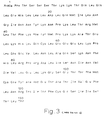
The non-glycosylated human IL-2 protein according to the invention preferably comprises the polypeptide (II) having the amino acid sequence shown in Fig. 3 (SEQ ID NO.2) wherein X is Met or hydrogen.
-
The human IL-2 protein produced in accordance with the invention has the following characteristics:
- 1) It is homogeneous in SDS-polyacrylamide gel electrophoresis and has a molecular weight of 15,000 ± 1,000 daltons as determined by said electrophoresis;
- 2) It contains alanine or methionine as the amino-terminal amino acid;
- 3) It contains threonine as the carboxy-terminal amino acid;
- 4) It has an activity of growing T cells or natural killer cells while maintaining their functions.
-
The human IL-2 protein produced in accordance with the invention reacts negatively in the limulus lysate test [Haemostasis, 7, 183 (1978)] and is very low in impurity proteins and pyrogen contents, so that it can be used safely as a bulk substance for manufacturing injections and so on.
-
The non-glycosylated human IL-2 protein obtained in accordance with the invention has an activity of promoting the growth of normal T cells or natural killer cells while maintaining their functions. Therefore, the IL-2 protein according to the invention can be used in growing and subculturing T cells or natural killer cells in vitro for a long period of time or cloning the same. Moreover, this property can be utilized in human IL-2 activity measurement.
-
Furthermore, the human IL-2 protein of the invention makes it possible to grow selectively antigen-specific killer T cells, which recognize and destruct tumor antigens, or natural killer cells, which are capable of killing tumor cells irrespective of the presence,or absence, of an experience of antigenic sensitization, in vitro. When inoculated into a living organism simultaneously with the introduction of said killer cells into the living organism, the human IL-2 of the present invention increases the anti-tumor effect of killer T cells. Therefore, it can be used for the prevention or treatment of tumors,or the treatment of immunodeficiency diseases,in warm-blooded animals (e.g. mouse, rat, rabbit, dog, cat, pig, horse, sheep, cattle, human).
-
The human IL-2 protein according to the invention is a highly purified product, has little antigenicity for humans and is low in toxicity.
-
As an agent for the prevention and treatment of tumors, the human IL-2 protein of the invention can be administered either parenterally or orally in the form of, for example, injections or capsules prepared by appropriate blending or dilution with a per se known carrier. It can be used either alone or in combination with killer T cells or natural killer cells grown in vitro, as mentioned hereinbefore.
-
The human IL-2 protein according to the invention has substantially the same biological activity as the natural human IL-2 and accordingly can be used in the same manner as said natural one. Its dissociation constant in relation to the IL-2 receptor of the responding cells is very small, so that a very small dose is sufficient in most cases.
-
For the purpose of growing T cells in vitro, the human IL-2 of the invention can be added to the medium in a concentration of about 0.01-1 U/ml, preferably about 0.1-0.5 U/ml.
-
In an example of the use for the purpose of growing T cells in vitro, the IL-2 protein of the invention is added, to a concentration of 0.1-0.5 unit/ml, to a cell suspension containing, for example, alloantigen-sensitized T cells obtained by 3-day mixed lymphocyte culture of human peripheral blood-derived T cells (1 x 10⁶ cells/ml), with B cell transformants (1 x 10⁶ cells/ml) resulting from X ray irradiation (1,500 rads) added, in RPMI 1640 medium containing 20% of fetal bovine serum. Cultivation is continued for about 1 month while repeating medium exchange at about one-week intervals.
-
The transformant disclosed in the following examples, namely Escherichia coli DHl/pTF4, has been deposited at the Institute for Fermentation, Osaka (IFO) under the deposit number IFO-14299, and also deposited at the Fermentation Research Institute, Agency of Industrial Science and Technology, Ministy of International Trade and Industry, Japan (FRI) under the accession number of FERM BP-628 of a deposit under the Budapest Treaty.
-
In the present specification and drawings, the bases and amino acids, when indicated by abbreviations, are abbreviated according to the rules of the IUPAC-IUB Commission on Biochemical Nomenclature or the practice in the field concerned. Examples are shown in Table 1. In case optical isomerism is involved, the amino acids mentioned are in the L form unless otherwise specifically indicated.
Table 1 | DNA: | Deoxyribonucleic acid |
| cDNA: | Complementary deoxyribonucleic acid |
| A: | Adenine |
| T: | Thymine |
| G: | Guanine |
| C: | Cytosine |
| RNA: | Ribonucleic acid |
| mRNA: | Messenger ribonucleic acid |
| dATP: | Deoxyadenosine triphosphate |
| dTTP: | Deoxythymidine triphosphate |
| dGTP: | Deoxyguanosine triphosphate |
| dCTP: | Deoxycytidine triphosphate |
| ATP: | Adenosine triphosphate |
| EDTA: | Ethylenediaminetetraacetic acid |
| SDS: | Sodium dodecyl sulfate |
| Gly: | Glycine |
| Ala: | Alanine |
| Val: | Valine |
| Leu: | Leucine |
| Ile: | Isoleucine |
| Ser: | Serine |
| Thr: | Threonine |
| Cys: | Cysteine |
| 1/2 Cys: | Half cystine |
| Met: | Methionine |
| Glu: | Glutamic acid |
| Asp: | Aspartic acid |
| Lys: | Lysine |
| Arg: | Arginine |
| His: | Histidine |
| Phe: | Phenylalanine |
| Tyr: | Tyrosine |
| Trp: | Tryptophan |
| Pro: | Proline |
| Asn: | Asparagine |
| Gln: | Glutamine |
Brief Description of the Drawings
-
Fig. 1 and Fig. 2 (SEQ ID NO.1) show the restriction enzyme map of the plasmid pILOT135-8 obtained in Reference Example 1 (vii) (
indicating the portion coding for the signal peptide and
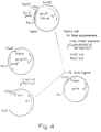
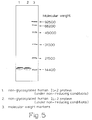
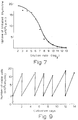
indicating the portion coding for IL-2) and the primary structure (base sequence) of said plasmid, respectively. Fig. 3 (SEQ ID NO.2) shows the amino acid sequence of the non-glycosylated human IL-2 protein according to the invention, therein X is Met or a hydrogen atom . Fig. 4 shows the scheme of constructing the expression plasmid pTF4 as taken in Reference Example 2. Fig. 5 shows the results of the SDS-polyacrylamide slab gel electrophoreses performed in Example 1(V) (1) and (2), Fig. 6 the trypsin digestion peptide map mentioned in Example 1 (v) (6), and Fig. 7 and Fig. 8 the effects of the human IL-2 protein according to the invention on the uptake of tritiated thymidine by the NKC3 cell line and human cell line, respectively, as revealed in Example 1(V)(7).Fig. 9 shows the results of the long-period subculturing of the NKC3 cell line conducted in Example 1(V) (7).
Reference Example 1
(i) Isolation of mRNA coding for human IL-2
-
Lymphocytes prepared from the human peripheral blood were incubated in RPMI 1640 medium supplemented with 10% fetal bovine serum, 12-O-tetradecanoylphorbol-13-acetate (TPA) (15 ng/nl) and concanavalin A (40 µg/ml) at 37°C to thereby induce IL-2. After 24 hours of incubation, 1 x 10¹⁰ human lymphocytes thus induced were disrupted and denatured in a solution containing 5 M guanidine thiocyanate, 5% mercaptoethanol, 50 mM Tris·HCl (pH 7.6) and 10 mM EDTA using a Teflon homogenizer, then sodium N-lauroylsarcosinate was added to a concentration of 4%, and the mixture, after homogenization, was layered onto 6 ml of 5.7 M cesium chloride solution (5.7 M cesium chloride, 1 M EDTA) and centrifuged using a Beckman SW28 rotor at 24,000 rpm and 15°C for 48 hours, to give an RNA precipitate. This RNA precipitate was dissolved in 0.25% sodium N-lauroylsarcosinate and precipitated with ethanol to give 10 mg of RNA. In a high-concentration salt solution (0.5 M NaCl, 10 mM Tris·HCl pH 7.6, 1 mM EDTA, 0.3% SDS), this RNA was adsorbed on an oligo(dT)cellulose column. Elution with a low-concentration salt solution (10 mM Tris·HCl pH 7.6, 1 mM EDTA, 0.3% SDS) gave 300 µg of poly(A)-containing mRNA. This mRNA was further subjected to precipitation with ethanol, then dissolved in 0.2 ml of a solution (10 mM Tris.HCl pH 7.6, 2mM EDTA, 0.3% SDS), treated at 65°C for 2 minutes and fractionated by 10-35% sucrose density gradient centrifugation (at 20°C and 25,000 rpm for 21 hours using a Beckman SW28 rotor) into 22 fractions. An aliquot of each fraction was injected into oocytes of Xenopus laevis and the IL-2 activity in proteins thus synthesized was measured. Fractions Nos. 11-15 (sedimentation coefficient 8S-15S) were found to have IL-2 activity. About 25 µg of IL-2 mRNA was contained in these fractions.
(ii) Synthesis of single-stranded DNA
-
Using the mRNA obtained above and reverse transcriptase, incubation was carried out in 100 µl of reaction solution (5 µg of mRNA, 50 µg of oligo(dT), 100 units of reverse transcriptase, 1 mM dATP, 1 mM dCTP, 1 mM dGTP, 1 mM dTTP, 8 mM MgCl2, 50 mM KCl, 10 mM dithiothreitol, 50 mM Tris·HCl pH 8.3) at 42°C for 1 hour, followed by deproteinization with phenol and treatment with 0.1 N NaOH at 70°C for 20 minutes for removal of RNA by decomposition.
(iii) Synthesis of double-stranded DNA
-
A double-stranded DNA was synthesized by treating the thus-synthesized single-stranded complementary DNA in 50 µl of a reaction solution (the same as the above-mentioned reaction solution except that the mRNA and oligo(dT) were absent) at 42°C for 2 hours.
(iv) Addition of dC tail
-
This double-stranded DNA was treated with nuclease Sl in 50 µl of a reaction solution (double-stranded DNA, 0.1 M sodium acetate pH 4.5, 0.25 M NaCl, 1.5 mM ZnSO4, 60 units of Sl nuclease) at room temperature for 30 minutes, followed by deproteinization with phenol and DNA precipitation with ethanol. The DNA was treated with terminal transferase in 50 µl of a reaction solution (double-stranded DNA, 0.14 M potassium cacodylate, 0.3 M Tris (base) pH 7.6, 2 mM dithiothreitol, 1 mM CoCl2, 0.15 mM dCTP, 30 units of terminal transferase) at 37°C for 3 minutes to thereby cause extension of the double-stranded DNA by about 15 deoxycytidines at both the 3' ends. This series of reactions gave about 300 ng of a deoxycytidine chain-containing double-stranded DNA.
(v) Cleavage of Escherichia coli plasmid and addition of dG tail
-
Separately, 10 µg of Escherichia coli plasmid pBR322 DNA was treated with the restriction enzyme PstI in 50 µl of a reaction solution (10 µg of DNA, 50 mM NaCl, 6 mM Tris·HCl pH 7.4, 6 mM MgCl2, 6 mM 2-mercaptoethanol, 100 µg/ml bovine serum albumin, 20 units of PstI) at 37°C for 3 hours to thereby cleave the one PstI recognition site occurring in the pBR322 DNA, followed by deproteinization with phenol. The cleaved plasmid pBR322 DNA was extended by about 17 deoxyguanines at both the 3' ends by treating with terminal transferase in 50 µl of a reaction solution (10 µg of DNA, 0.14 M potassium cacodylate, 0.3 M Tris base pH 7.6, 2 mM dithiothreitol, 1 mM CoCl2, 0.15 mM GTP, 30 units of terminal transferase) at 37°C for 3 minutes.
(vi) Annealing of cDNA and transformation of Escherichia coli
-
The thus-obtained synthetic double-stranded DNA (0.1 µg) and 0.5 µg of the above-mentioned plasmid pBR322 were annealed,by heating in a solution containing 0.1 M NaCl, 50 mM Tris·HCl pH 7.6 and 1 mM EDTA at 65°C for 2 minutes and then at 45°C for 2 hours followed by gradual cooling, and the product was used for transformation of Escherichia coli MM294 in accordance with the method of Enea et al. [J. Mol. Biol., 96, 495 (1975)].
(vii) Isolation of cDNA-containing plasmid
-
In this way, about 20,000 tetracycline-resistant transformants were isolated. DNAs of each of them were fixed on a nitrocellulose filter. Based on the amino acid sequence of IL-2 reported by Taniguchi et al. [Nature, 302, 305 (1983)], the complementary oligonucleotides of base sequences (5'AAA CAT CTT CAG TGT3' and 5'ACA TTC ATG TGT GAA3') correponding to amino acids Nos. 74-78 (Lys-His-Leu-Gln-Cys) and amino acids Nos, 122-126 (Thr-Phe-Met-Cys-Glu), respectively, were chemically synthesized by the phosphotriester method [Crea, R. et al., Proc. Natl. Acad. Sci. USA, 75, 5765 (1978)].
-
These oligonucleotides were labeled with 32P at the 5' end by treatment with T4 polynucleotide kinase in 50 µl of a reaction solution (0.20 µg of oligonucleotide, 50 mM Tris·HCl pH 8.0, 10 mM MgCl2, 10 mM 2-mercaptoetnanol, 50 µCi of γ-32P ATP, 3 units of T4 polynucleotide kinase) at 37°C for 1 hour. These labeled oligonucleotides, as probes, were annealed with the above-mentioned DNAs fixed on nitrocellulose filter by the method of Lawn et al. [Nucleic Acids Res., 9, 6103 (1981)]. Four transformants reactive to the above two oligonucleotide probes were detected by autoradiography. The plasmid DNA was isolated from cells of each of these transformants by the Birnboim-Doly alkali method [Birnboim, H. C. & Doly, J., Nucleic Acids Res., 7, 1513 (1979)]. Then the insert in the plasmid DNA was cut out using the restriction enzyme PstI. From among the plasmids isolated, the one containing the longest insert fragment was selected and named pILOT 135-8. The restriction enzyme map of this plasmid is shown in Fig. 1.
-
The primary structure (base sequence) of the cDNA inserted in this pILOT 135-8 was determined by the dideoxynucleotide method and the Maxam-Gilbert method. The primary structure thus determined is shown in Fig. 2 (SEQ ID NO.1). The peptide defined by this base sequence consists of 153 amino acids, starting with the synthesis start signal (Nos. 64-66 ATG). Of these, the 20 amino acids from the N-terminal are considered to constitute a signal peptide. The above primary structure has revealed that this plasmid has the whole base sequence coding for the human IL-2 protein. This fact indicates that insertion of the gene inserted in said plasmid into an appropriate expression plasmid can lead to production of an optional polypeptide portion of the IL-2 protein.
Reference Example 2
-
The plasmid pILOT135-8 obtained in Reference Example 1 was cleaved with the restriction enzyme HgiAl. The thus-obtained 1294 bp DNA fragment containing the IL-2 gene was treated with T4 DNA polymerase, joined with the ClaI linker CGATA ATG GCA, which contained the codon GCA for alanine and the codon ATG for methionine, and the product was treated with ClaI and PstI, followed by insertion into ptrp771 at the ClaI-PstI site. The expression plasmid thus obtained was named pTF4 (Fig. 4).
Reference Example 3
-
Escherichia coli DHl was transformed with the plasmid pTF4 obtained in Reference Example 2 in accordance with the method of Cohen et al. [Proc. Natl. Acad. Sci. USA, 69, 2110 (1972)] to obtain a transformant (Escherichia coli DHl/pTF4) carrying said plasmid.
Example 1
-
- (i) E. coli DHl/pTF4 (obtained in Reference Example 3) was inoculated into 50 ml of a liquid medium (pH 7.0) containing 1% Bacto tryptone (Difco Laboratories, USA), 0.5% Bacto yeast extract (Difco Laboratories, USA), 0.5% sodium chloride and 7 µg/ml tetracycline as placed in a 250-ml Erlenmeyer flask. After incubation at 37°C overnight on a swing rotor, the culture medium was transferred to a 5-liter jar fermenter containing 2.5 liters of M9 medium containing 0.5% casamino acids, 0.5% glucose and 7 µg/ml tetracycline. Incubation was then conducted with aeration and stirring at 37°C for 4 hours and, after addition of 3-β-indolylacrylic acid (25 µg/ml), for a further 4 hours. Cells were harvested from the thus-obtained 2.5-liter culture broth by centrifuging, frozen at -80°C and stored.
- (ii) The freeze-stored cells (12.1 g) obtained in Example 1(i) were suspended in 100 ml of an extractant (pH 7.0) containing 7 M guanidine hydrochloride and 0.1 M Tris·HCl, the suspension was stirred at 4°C for 1 hour and the lysate was centrifuged at 28,000 x g for 20 minutes to obtain 93 ml of a supernatant.
- (iii) Separately, various procedures were conducted to extract IL-2 from the transformant E. coli DHl/pTF4 cells obtained by the method of Example 1(i) to compare the respective extraction efficiencies.
-
In a lysozyme-EDTA method, 2 g of E. coli DHl/pTF4 cells obtained in Example 1(i) were mixed with 16 ml of a solution (pH7.0) containing 0.1 M Tris-HCl, 10 mM EDTA and 250 mg/l lysozyme, the mixture was stirred at 4°C for 1 hour, and subseqently at 37°C for 5 minutes, and the lysate was centrifuged at 28,000 x g for 20 minutes.
-
In a sonication method, 2 g of the cells mentioned above were suspended in 16 ml of a solution (pH7.0) containing 0.1 M Tris-HCl, the suspension was subjected to sonication at 0°C for 5 minutes and the lysate was centrifuged at 28,000 x g for 20 minutes.
-
In a guanidine-HCl method, 2 g of the cells mentioned above were mixed with 16 ml of solutions (pH7.0) containing 0.1 M Tris-HCl and 2M, 4M or 7M guanidine-HCl, the mixtures were stirred at 4°C for 1 hour and the lysates were centrifuged at 28,000 x g for 20 minutes. Supernatant fluids thus obtained were used for the measurements of protein concentration and IL-2 activity.
-
The results are summarized in Table 2.
Table 2 | Extraction of IL-2 |
| Extraction procedure | Protein concentration (mg/ml) | IL-2 activity in the extract (U/ml) | Relative ratio (%) |
| Lysozyme-EDTA | 5.40 | 3.3 | 0.02 |
| Sonication | 6.54 | 2.2 | 0.01 |
| 2M Gu·HCl | 2.60 | 46 | 0.2 |
| 4M Gu·HCl | 4.12 | 144 | 0.8 |
| 7M Gu·HCl | 7.22 | 19100 | 100 |
| Gu·HCl: guanidine hydrochloride |
- (iv) The supernatant fluid obtained in Example 1(ii) was dialyzed against 0.01 M Tris·HCl buffer (pH 8.5) and then centrifuged at 19,000 x g for 10 minutes to give 94 ml of a dialyzed supernatant fluid. This was applied to a DE 52 (DEAE-cellulose, Whatman, Great Britain) column (50 ml in volume) equilibrated with 0.01 M Tris·HCl buffer (pH 8.5) for protein adsorption. Proteins were eluted with a linear NaCl concentration gradient (0-0.15 M NaCl, 1 liter). The active fractions (53 ml) obtained were concentrated to 4.8 ml using a YM-5 membrane (Amicon, USA) and subjected to gel filtration using a Sephacryl S-200 (Pharmacia, Sweden) column (500 ml in volume)equilibrated with 0.1 M Tris·HCL (pH 8.0)-1 M NaCl buffer. The active fractions (28 ml) obtained were concentrated to 2.5 ml using a YM-5 membrane. The concentrate was applied to an Ultrapore RPSC (Altex, USA) column for adsoprtion, and high performance liquid chromatography was performed using a tritluoroacetic acidacetonitrile system as the mobile phase.
-
The conditions used: column, Ultrapore RPSC (4.6 x 75 mm): column temperature, 30°C; solvent A, 0.1% trifluoroacetic acid-99.9% water; solvent B, 0.1% trifluoroacetic acid-99.9% acetonitrile; elution program, minute 0 (68% A + 32% B) - minute 25 (55% A + 45% B) - minute 35 (45% A + 55% B) - minute 45 (30% A + 70% B) - minute 48 (100% B); elution rate, 0.8 ml/min.; detection wave length, 230 nm. An active fraction was collected at a retention time of about 39 minutes. Thus was obtained 10 ml of a solution containing 0.53 mg of non-glycosylated human IL-2 protein [specific activity, 30,000 U/mg; activity recovery from the starting material, 30.6% purity of protein, 99% (determined by densitometry on an SDS-polyacrylamide gel electrophorogram)].
-
Lyophilization of the above solution gave a white powder. The powder had a specific activity of 26,000 U/mg.
- (V) The human IL-2 protein obtained in Example 1 (iv) was examined for the following properties:
(1) Homogeneity:
-
Staining with Coomassie Brilliant Blue following SDS-polyacrylamide gel electrophoresis according to Laemmli et al. [Nature, 227, 680 (1970)] revealed only a single band of said human IL-2 protein (cf. Fig. 5). The location of the band remained unchanged under reducing conditions as well as under non-reducing conditions.
(2) Molecular weight:
-
The molecular weight of said human IL-2 protein was calculated to be about 15,000 daltons based on the result of SDS-polyacrylamide gel electrophoresis (cf. Fig. 5).
(3) Amino acid composition:
-
A 20-µg portion of said human IL-2 protein was placed in a glass test tube for hydrolysis, constant boiling hydrochloric acid containing 4% thioglycolic acid was added, the tube was then sealed
in vacuo and hydrolysis was performed at 110°C for 24, 48 or 72 hours. After hydrolysis, the tube was opened, the hydrochloric acid was removed under reduced pressure, and the residue was dissolved in 0.02 N hydrochloric acid and subjected to amino acid analysis using a Hitachi model 835 amino acid analyzer. For determination of cystine and cysteine, said human IL-2 protein was oxidized with performic acid by the method of Hirs [Methods in Enzymol.,
11, 197 (1967)] followed by hydrolysis in constant boiling hydrochloric acid under reduced pressure for 24 hours. The hydrolysate was subjected to cysteic acid determination using an amino acid analyzer. The results obtained by the above amino acid analyses are shown in Table 3. The values are each the mean of three values respectively obtained after 24, 48 and 72 hours of hydrolysis except for the values for serine and threonine which were determined by extrapolation to zero time of hydrolysis.
Table 3 | Amino acid | Mole % |
| Asp/Asn | 8.8 |
| Thr | 9.3 |
| Ser | 5.7 |
| Glu/Gln | 13.7 |
| Pro | 3.4 |
| Gly | 1.7 |
| Ala | 3.8 |
| 1/2 Cys | 2.3 |
| Val | 3.1 |
| Met | 3.7 |
| Ile | 6.3 |
| Leu | 16.3 |
| Tyr | 2.3 |
| Phe | 4.5 |
| Lys | 8.3 |
| His | 2.5 |
| Arg | 3.1 |
| Trp | 1.1 |
(4) N-Terminal amino acid sequence:
-
A 34-µg portion of said human Il-2 protein was analyzed for the N-terminal amino acid sequence by applying thereto an automatic Edman degradation method using a gas-phase protein sequenator (model 470A, Applied Biosystems, USA). Phenylthiohydantoin-amino acids (PTH-amino acids) were identified by high performance liquid chromatography using a Micropak-SP column (Varian, USA). The PTH-amino acid or acids detected in each step are shown in Table 4.
Table 4 | Step | PTH-amino acid detected |
| 1 | Ala |
| | Met |
| |
| 2 | Pro |
| | Ala |
| |
| 3 | Thr |
| | Pro |
| |
| 4 | Ser |
| | Thr |
| |
| 5 | Ser |
| 6 | Ser |
| 7 | Thr |
| 8 | Lys |
| 9 | Lys |
| 10 | Thr |
| 11 | Gln |
| 12 | Leu |
| 13 | Gln |
| 14 | Leu |
| 15 | Glu |
| 16 | Y* |
| 17 | Leu |
| 18 | Leu |
| 19 | Leu |
| 20 | Asp |
(5) C-Terminal amino acid:
-
A 33-µg portion of said human IL-2 protein was placed in a glass test tube for hydrazine degradation, 0.05 ml of anhydrous hydrazine was added and the tube was sealed in vacuo and heated at 100°C for 6 hours. The hydrazine degradation product obtained was lyophilized and dissolved in distilled water. Benzaldehyde was added to the solution, the mixture was stirred at room temperature for 1 hour and then centrifuged. The aqueous layer thus obtained was lyophilized and subjected to amino acid analysis using a Hitachi model 835 amino acid analyzer. Threonine alone was detected.
(6) Tryptic peptide mapping
-
A 15-µg portion of said IL-2 sample was digested with 0.4 µg of trypsin (Washington, USA) in 120 µl of 0.02 M sodium hydrogen carbonate at 37°C for 18 hours. Then, 5 µl of 2-mercaptoethanol was added and the reaction was continued at 37°C for a further 2 hours. Thereafter, the reaction was terminated by addition of 75 µl of 1% trifluoroacetic acid. High performance liquid chromatography of the reaction mixture, which was performed under the conditions given below, gave a map shown in Fig. 6. Column: Ultrasphere-Octyl (5 µm, 4.6 x 250 mm; Altex, USA); Column temperature: 30°C;
- Mobile phase:
-
- Solvent A, 0.02% trifluoroacetic acid-99.98% water;
- Solvent B, 0.02% trifuloroacetic acid-99.98% acetonitrile;
- Minute 0 (95% solvent A + 5% solvent B) - minute
40 (30% solvent A + 70% solvent B);
- Flow rate:
- 1.0 ml/minute;
- Detection:
- Fluorescence method [Analytical Biochem., 67, 438 (1975)] using fluorescamine (Roche, USA).
(7) Activity against IL-2-dependent cell lines
-
Assay of the non-glycosylated human IL-2 protein according to the invention,in accordance with the method described in Biochem. Biophys. Res. Commun., 109, 363 (1982), revealed that said IL-2 protein had an activity to promote tritiated thymidine uptake in an IL-2-dependent mouse cell line (NKC3; cf. Fig. 7), as well as in an IL-2-dependent human cell line (cf. Fig. 8).
-
Furthermore, said IL-2 protein was dissolved in 20% FCS-added RPMI-1640 medium to a concentration of 0.5 U/ml, and 2 x 10⁵ cells/ml of the NKC3 cell line were suspended in the medium. Subculturing was continued on a Linbro Multi dish (Flow, USA) at 37°C in the presence of 5% CO2 while repeating viable cell counting and resuspending the culture in a new fresh medium at 2- or 3-day intervals. As a result, said IL-2 protein was found to have an activity to maintain the growth of the NKC3 cell line for a long period of time, as illustrated in Fig. 9.
Example 2 (Preparation for injection)
-
The non-glycosylated human IL-2 protein-containing solution obtained in Example 1 (iv), is applied to a CM Toyopearl (Toyo Soda, Japan) column equilibrated with 0.025 M ammonium acetate buffer (pH 5.0) under aseptic conditions for adsorption, followed by elution with the above buffer containing 0.15 M NaCl. The eluate is diluted by addition of an appropriate amount of 0.15 M NaCl, then HSA is added to a concentration of 0.5%, and the mixture is filtered through a membrane filter (0.22 µm in pore diameter). The filtrate is distributed aseptically in 1-ml portions into vials, followed by lyophilization. The human IL-2 preparation for injection in each vial is dissolved in 1 ml of distilled water, for injection, prior to use.


 indicating the portion coding for IL-2) and the primary structure (base sequence) of said plasmid, respectively. Fig. 3 (SEQ ID NO.2) shows the amino acid sequence of the non-glycosylated human IL-2 protein according to the invention, therein X is Met or a hydrogen atom . Fig. 4 shows the scheme of constructing the expression plasmid pTF4 as taken in Reference Example 2. Fig. 5 shows the results of the SDS-polyacrylamide slab gel electrophoreses performed in Example 1(V) (1) and (2), Fig. 6 the trypsin digestion peptide map mentioned in Example 1 (v) (6), and Fig. 7 and Fig. 8 the effects of the human IL-2 protein according to the invention on the uptake of tritiated thymidine by the NKC3 cell line and human cell line, respectively, as revealed in Example 1(V)(7).Fig. 9 shows the results of the long-period subculturing of the NKC3 cell line conducted in Example 1(V) (7).
