EP0743523B1 - Dye-based reference material, preparation and use thereof - Google Patents
Dye-based reference material, preparation and use thereof Download PDFInfo
- Publication number
- EP0743523B1 EP0743523B1 EP96303490A EP96303490A EP0743523B1 EP 0743523 B1 EP0743523 B1 EP 0743523B1 EP 96303490 A EP96303490 A EP 96303490A EP 96303490 A EP96303490 A EP 96303490A EP 0743523 B1 EP0743523 B1 EP 0743523B1
- Authority
- EP
- European Patent Office
- Prior art keywords
- dye
- fractions
- cas
- level
- sulforhodamine
- Prior art date
- Legal status (The legal status is an assumption and is not a legal conclusion. Google has not performed a legal analysis and makes no representation as to the accuracy of the status listed.)
- Expired - Lifetime
Links
Images


Classifications
-
- G—PHYSICS
- G01—MEASURING; TESTING
- G01N—INVESTIGATING OR ANALYSING MATERIALS BY DETERMINING THEIR CHEMICAL OR PHYSICAL PROPERTIES
- G01N33/00—Investigating or analysing materials by specific methods not covered by groups G01N1/00 - G01N31/00
-
- G—PHYSICS
- G01—MEASURING; TESTING
- G01N—INVESTIGATING OR ANALYSING MATERIALS BY DETERMINING THEIR CHEMICAL OR PHYSICAL PROPERTIES
- G01N33/00—Investigating or analysing materials by specific methods not covered by groups G01N1/00 - G01N31/00
- G01N33/48—Biological material, e.g. blood, urine; Haemocytometers
- G01N33/50—Chemical analysis of biological material, e.g. blood, urine; Testing involving biospecific ligand binding methods; Immunological testing
- G01N33/96—Chemical analysis of biological material, e.g. blood, urine; Testing involving biospecific ligand binding methods; Immunological testing involving blood or serum control standard
-
- G—PHYSICS
- G01—MEASURING; TESTING
- G01N—INVESTIGATING OR ANALYSING MATERIALS BY DETERMINING THEIR CHEMICAL OR PHYSICAL PROPERTIES
- G01N2496/00—Reference solutions for assays of biological material
- G01N2496/15—Reference solutions for assays of biological material containing dyes to mimic optical absorption of, e.g. hemoglobin
-
- G—PHYSICS
- G01—MEASURING; TESTING
- G01N—INVESTIGATING OR ANALYSING MATERIALS BY DETERMINING THEIR CHEMICAL OR PHYSICAL PROPERTIES
- G01N2496/00—Reference solutions for assays of biological material
- G01N2496/70—Blood gas control solutios containing dissolved oxygen, bicarbonate and the like
-
- G—PHYSICS
- G01—MEASURING; TESTING
- G01N—INVESTIGATING OR ANALYSING MATERIALS BY DETERMINING THEIR CHEMICAL OR PHYSICAL PROPERTIES
- G01N33/00—Investigating or analysing materials by specific methods not covered by groups G01N1/00 - G01N31/00
- G01N33/48—Biological material, e.g. blood, urine; Haemocytometers
- G01N33/50—Chemical analysis of biological material, e.g. blood, urine; Testing involving biospecific ligand binding methods; Immunological testing
- G01N33/58—Chemical analysis of biological material, e.g. blood, urine; Testing involving biospecific ligand binding methods; Immunological testing involving labelled substances
- G01N33/583—Chemical analysis of biological material, e.g. blood, urine; Testing involving biospecific ligand binding methods; Immunological testing involving labelled substances with non-fluorescent dye label
-
- Y—GENERAL TAGGING OF NEW TECHNOLOGICAL DEVELOPMENTS; GENERAL TAGGING OF CROSS-SECTIONAL TECHNOLOGIES SPANNING OVER SEVERAL SECTIONS OF THE IPC; TECHNICAL SUBJECTS COVERED BY FORMER USPC CROSS-REFERENCE ART COLLECTIONS [XRACs] AND DIGESTS
- Y10—TECHNICAL SUBJECTS COVERED BY FORMER USPC
- Y10T—TECHNICAL SUBJECTS COVERED BY FORMER US CLASSIFICATION
- Y10T436/00—Chemistry: analytical and immunological testing
- Y10T436/10—Composition for standardization, calibration, simulation, stabilization, preparation or preservation; processes of use in preparation for chemical testing
- Y10T436/102499—Blood gas standard or control
-
- Y—GENERAL TAGGING OF NEW TECHNOLOGICAL DEVELOPMENTS; GENERAL TAGGING OF CROSS-SECTIONAL TECHNOLOGIES SPANNING OVER SEVERAL SECTIONS OF THE IPC; TECHNICAL SUBJECTS COVERED BY FORMER USPC CROSS-REFERENCE ART COLLECTIONS [XRACs] AND DIGESTS
- Y10—TECHNICAL SUBJECTS COVERED BY FORMER USPC
- Y10T—TECHNICAL SUBJECTS COVERED BY FORMER US CLASSIFICATION
- Y10T436/00—Chemistry: analytical and immunological testing
- Y10T436/10—Composition for standardization, calibration, simulation, stabilization, preparation or preservation; processes of use in preparation for chemical testing
- Y10T436/105831—Protein or peptide standard or control [e.g., hemoglobin, etc.]
-
- Y—GENERAL TAGGING OF NEW TECHNOLOGICAL DEVELOPMENTS; GENERAL TAGGING OF CROSS-SECTIONAL TECHNOLOGIES SPANNING OVER SEVERAL SECTIONS OF THE IPC; TECHNICAL SUBJECTS COVERED BY FORMER USPC CROSS-REFERENCE ART COLLECTIONS [XRACs] AND DIGESTS
- Y10—TECHNICAL SUBJECTS COVERED BY FORMER USPC
- Y10T—TECHNICAL SUBJECTS COVERED BY FORMER US CLASSIFICATION
- Y10T436/00—Chemistry: analytical and immunological testing
- Y10T436/10—Composition for standardization, calibration, simulation, stabilization, preparation or preservation; processes of use in preparation for chemical testing
- Y10T436/109163—Inorganic standards or controls
Definitions
- This invention relates to a method of preparing dye-based reference materials useful for calibrating or qualifying instrument systems that are diagnostic spectroscopically for hemoglobin and CO-ox fractions.
- the reference materials may also be used in instrument systems that have, in addition to capability to measure CO-ox fractions, sensors for the measurement of blood pH, gas and other blood analytes including electrolyte concentrations and metabolite concentrations.
- pH and blood gas instruments measure blood pH, pCO 2 .
- CO-oximeter instruments typically measure the total hemoglobin concentration (THb); the hemoglobin fractions such as oxyhemoglobin (O 2 Hb); methemoglobin (MetHb); carboxyhemoglobin (COHb); and reduced hemoglobin (HHb) (collectively referred to herein as "CO-ox fractions").
- Electrolyte instruments measure any number of blood electrolytes, including sodium, potassium, lithium, calcium, and so on.
- Instrument systems currently available may combine the measurement of blood pH, gases, electrolytes, various metabolites, and CO-ox fractions in one instrument for a comprehensive testing of the properties of blood, as is particularly useful in respiratory and pulmonary ailments. Vigorous therapeutic treatment is often dictated by such test results.
- CO-ox fractions relate to the loading of oxygen onto the hemoglobin of red blood cells circulating through the pulmonary capillaries.
- the actual amount of oxygen loaded onto the hemoglobin is determined not only by the concentration of total hemoglobin (THb), but also by the amount of non-oxygen-binding derivatives of hemoglobin such as carboxyhemoglobin (COHb) and methemoglobin (MetHb).
- THb total hemoglobin
- COHb carboxyhemoglobin
- MetHb methemoglobin
- Reduced hemoglobin (HHb) is an unoxygenated form of normal hemoglobin and elevations in the arterial fractional HHb indicate that lesser amounts of oxygen have been bound as a result of ventilation and/or perfusion defects.
- Blood, hemoglobin and hemoglobin CO-ox fractions absorb visible light.
- a normal blood spectrum has a main absorption peak at 578 nm and decreases rapidly close to zero at wavelengths greater than about 610 nm as shown in Figure 1.
- the second absorption peak of blood is at 542 nm.
- Absorbance maxima of hemoglobin derivatives are oxyhemoglobin, 541, 568 - 572 nm; reduced hemoglobin, 555 nm; carboxyhemoglobin, 537, 568 - 572 nm; and methemoglobin 540, 578, 630 nm.
- CO-oximeters measure the absorbance of the blood sample at multiple wavelengths on the spectrum.
- CO-oximeters analyze blood samples by the collection of absorption data at specific wavelengths. The data are then typically recorded and a process called multicomponent analysis is used to simultaneously calculate the concentrations of the each of hemoglobin CO-ox fraction present in the blood sample.
- CO-oximeter instruments are typically designed to measure CO-ox fractions with values broader than the ranges observed in patient samples.
- instruments may have the capability to measure CO-ox fractions in the defined instrument ranges of: about 4 to about 25 g/dL of THb, about 30 % to about 98 % oxyhemoglobin (O 2 Hb); 0 to about 50 % carboxyhemoglobin (COHb); 0 to about 40 % methemoglobin (MetHb); and 0 to about 50 % reduced hemoglobin (HHb), with all CO-ox fraction percentages herein based on the total amount of hemoglobin.
- the clinically meaningful range is defined herein as: total hemoglobin 8 to 20 g/dL, 60 to 98 % oxyhemoglobin (O 2 Hb); 0 to 20 % carboxyhemoglobin (COHb); 0 to about 20 % methemoglobin (MetHb); and 0 to about 20 % reduced hemoglobin (HHb).
- the normal physiological range is defined herein as: total hemoglobin 14 to 17 g/dL for men and 12 to 15 g/dL for women, oxyhemoglobin 94 to 98%, carboxyhemoglobin 0 to about 1 % for nonsmoker, methemoglobin 0 to about 1.5 %, and reduced hemoglobin from about 1 to about 5%.
- Reference materials generally function to validate the performance of a diagnostic instrument.
- ideal quality control standard materials are formulated to provide pre-determined CO-ox fraction values not only within the broad instrument capability range, but also within the normal physiological range.
- the prior art teaches two general types of CO-oximeter quality control standard materials.
- the first type are aqueous dye-based materials, where dyes are used in an attempt to match the spectrum of blood.
- the second type are blood-based materials, where the presence blood allows for the direct match of the spectrum of blood. Both types of materials have been associated with a variety of problems as discussed below.
- Blood-based reference materials provide clinically meaningful CO-ox fraction values, however, numerous limitations have been associated with using blood in the quality control standard materials. For example, a significant problem encountered by users of the proteinaceous blood-based materials is that blood-based materials are very susceptible to bacterial contamination and have a limited shelf life. Consequently, in most cases, users of blood-based materials must refrigerate the products during storage. Additionally, blood-based reference materials are generally classified as biohazardous materials, thus requiring the user to take additional safety precautions.
- a method to prepare a reference material for CO-oximeter instrument systems comprising (a) selecting a red dye having a main absorption maxima at a wavelength region between about 560 to about 580 nm to provide a basic spectrum; (b) modifying said main absorption maxima of said red dye by using a dye having an absorption maxima at a wavelength region between about 350 to about 450 nm; and (c) establishing a new relative baseline of said spectrum at wavelengths greater than about 640 nm and providing a sufficient absorbance at about 630 nm to provide a positive methemoglobin CO-ox fraction using one or more dyes.
- a reference material prepared according to said method wherein said dyes are used in sufficient amounts to provide a reference material with CO-ox fractions falling within a range covering instrument capability.
- the method may also be used to prepare reference materials having CO-ox fractions with values falling within the defined clinical range or within the defined normal physiological range.
- a reference material comprising (a) a first dye having a main absorption maxima at a wavelength between about 560 to about 580 nm; (b) a second dye having an absorption maxima at a wavelength between about 350 to about 450 nm; and (c) a third dye having an absorption maxima at a wavelength between about 625 and about 640 nm; and (d) a fourth dye having an absorption maxima at a wavelength between about 700 to about 780 nm and an absorption band width of from about 150 to about 200 nm.
- the amounts of the dyes used in the materials may be adjusted to provide quality control materials with desired CO-ox fraction values.
- a method for quality control of CO-oximeter instrument systems comprising: (1) subjecting said instrument to a quality control standard material comprising (a) a first dye having a main absorption maxima at a wavelength between about 560 to about 580 nm; (b) a second dye having an absorption maxima at a wavelength between about 350 to about 450 nm; and (c) a third dye having an absorption maxima at a wavelength between about 625 and about 640 nm; and (d) a fourth dye having an absorption maxima at a wavelength between about 700 to about 780 nm and an absorption band width of from about 150 to about 200 nm; wherein said dyes are used in a sufficient amount to provide said control with a pre-determined level of CO-ox fractions; (2) obtaining an instrument measurement of CO-ox fractions of said control; and (3) comparing said instrument measurement of said CO-ox fractions of said control with said predetermined level of CO-ox fractions to check the accuracy of said instrument.
- the dye-based reference materials are non-proteinaceous and may be interpreted by CO-oximeter instrument systems as providing a spectrum substantially equivalent to the spectrum of blood, particularly with regard to total hemoglobin and hemoglobin CO-ox fractions.
- inventive method of preparing the reference materials dye-based reference materials may be formulated to provide CO-ox fractions approximating those seen in a clinical setting, including normal physiological values.
- These dye-based reference materials may also be combined with other ingredients which make the materials useful as reference materials for pH and blood gas instruments and/or electrolyte instruments in addition to CO-oximeter instruments.
- the present invention provides a method to formulate dye-based reference materials which mimic the spectrum of human blood when the reference materials are utilized in optical types of CO-oximeter instrument systems.
- the formulating method is described herein in distinct steps for purposes of clarity only. It should be understood that the steps set forth may be carried out in any order and may also be combined and carried out simultaneously. Additionally, the designation of first dye, second dye, and so on, is not intended to imply a specific order of dyes.
- the types of dyes selected, the amount of dyes utilized and the concentration of dyes described in these steps may be adjusted until the desired level of hemoglobin CO-ox fractions are obtained, as preferably determined by taking a reading from the specific CO-oximeter instrument that will be used with the reference material.
- a red dye is selected to provide a basic spectrum (also referred to herein as the first dye).
- the general characteristics of the spectrum of normal blood should be considered.
- Normal blood has a spectrum with a main absorption peak at 578 nm which decreases rapidly close to zero at a wavelength greater than about 610 nm as shown in Figure 1.
- the second absorption peak of blood is at approximately 542 nm.
- the absorbance maxima of the CO-ox fraction oxyhemoglobin is at the approximate wavelengths of 541 nm and 568 - 572 nm.
- the dye-based formulation should have a red dye with a spectrum similar to at least the general absorption features of oxyhemoglobin.
- the preferred red dyes in the invention have an absorption peak in the wavelength range from about 560 nm to about 580 nm. Further, particularly preferred red dyes exhibit minimal absorbance at a wavelength of greater than about 610 nm. Red dyes, such as, for example, sulforhodamine B, chlorophenol red, xylenol orange, sulforhodamine 101, etc., are currently preferred.
- red dye Most preferably employed as the red dye is sulforhodamine B, which has an absorption maximum at approximately 566 nm in aqueous solution, a fairly narrow absorption bandwidth, and an absorbance which decreases to zero at about 620 nm, as shown as spectrum 1 in Figure 2.
- the single red dye does not exhibit a spectrum sufficiently similar to that of blood for purposes of obtaining clinically meaningful CO-ox fractions, as exemplified by the CO-ox fractions measured from 1.2 g/L of sulforhodamine B: THb 14 g/dL, O 2 Hb -16%, COHb 105%, MetHb -2.5% and HHb 13.5%. As these values indicate, negative CO-ox fractions are obtained for the oxyhemoglobin and the methemoglobin.
- red dye One approach to alter the absorption peak of the red dye might be to introduce another red dye.
- another red dye with absorption maximum wavelength greater than that of the first red dye could be utilized.
- another red dye with absorption maximum wavelength less than that of the first red dye could be utilized.
- a combination of the two red dyes would be predicted to result in a new overlapped maximum that would complement the required absorption features.
- sulforhodamine B in aqueous solution provides a spectrum that does not have enough strength of absorbance at 570 to 585 nm because the absorption peak of the sulforhodamine B is located at a shorter wavelength than that of blood.
- Sulforhodamine 101 has an absorption peak at 587 nm.
- a combination of sulforhodamine B and sulforhodamine 101 shifts the absorption peak of sulforhodamine B and enhances the absorbance at 570 nm to 585 nm.
- spectrum 1 is sulforhodamine B
- spectrum 2 is sulforhodamine 101
- spectrum 3 is the combination of 1 g/L sulforhodamine B and 0.1 g/L sulforhodamine 101.
- absorbance of the two red dye combination at 570 to 585 nm is enhanced and results in the change of CO-ox fractions from negative values to positive values, e.g., 14 g/dL THb, 16% O 2 Hb, 1% MetHb, 14% COHb and 69% HHb.
- the addition of the second red dye (sulforhodamine 101) introduces an undesired high absorbance near wavelength range of 590 nm to 605 nm, thus not providing an acceptable method of formulating references having clinically meaningful and/or normal physiological CO-ox fraction values.
- step (b) modification and manipulation of the absorption band of the red dye is conducted where the absorption peak of the red dye is shifted to a longer wavelength to more closely approximate that of blood.
- the absorption maxima of the red dye is shifted to fall between the wavelengths of approximately 570 nm to about 585 nm. It is preferred that the shift of the absorption maxima does not inadvertently increase the absorption in the wavelength region of 590 to 605 nm. It has been found that dye(s) having an absorption maxima at wavelengths of about 350 to 450 nm are useful in step (b) because the dye(s) increase the absorbance of the spectrum at the ultraviolet region and force the absorption band of the red dye to shift toward the longer wavelength.
- step (b) is accomplished using a yellow dye (or a combination thereof) including, for example, mordant yellow 7, tartrazine, orange G, hydroxypyrenetrisulfonic acid, mordant yellow 10, combinations thereof, and so on.
- mordant yellow 7 dye especially when sulforhodamine B is selected as the red dye.
- Figure 3 illustrates the changes of the spectrum of sulforhodamine B after addition of mordant yellow 7.
- Spectrum 1 in Figure 3 is the sulforhodamine B aqueous solution
- spectrum 2 is the sulforhodamine B aqueous solution containing mordant yellow 7.
- the original absorption peak of sulforhodamine B is shifted from 566 nm to 572 nm, with the edge of the absorption band at approximately 610 nm, and with little increase of absorbance in the wavelength range of 590 to about 605 nm.
- step (c) a new relative baseline of the spectrum is established at wavelengths greater than about 640 nm and sufficient absorbance at about 630 nm is provided to yield a positive methemoglobin CO-ox fraction.
- Step (c) addresses that the absorbance at about 590 to about 605 nm of the dye or dye combination used in steps (a) and (b) is higher than that of normal blood. This step may be accomplished using one or more dyes.
- the original absorbance can be normalized, with the normalized absorbance reduced and smaller than the original absorbance. Further, the original absorbance at about 590 to about 605 nm, although having a similar absolute absorbance value, is relatively reduced upon the establishment of the new lifted baseline.
- CO-oximeters take the lifted baseline from the CO-oximeters interpret the dye spectrum, and calculate the CO-ox fractions therefrom.
- inventive reference material will read as providing CO-ox fraction values within the instrument capability and clinically meaningful ranges, and optionally, within the normal physiological range, as described in more detail hereinafter.
- the third dye(s) One group of dyes particularly useful in step (c) are designated herein as the third dye(s). These dyes have an absorption maxima at about 625 to about 640 nm (more preferably about 630 nm) and have only minimum impact on the absorbance at wavelengths shorter than about 600 nm.
- the third dye is a blue dye selected from brilliant blue FC (FD&C blue 1), erioglaucine, lissamine, alphazurine A, patent blue violet, hydroxy naphthol blue, patent blue VF, combinations thereof, and equivalents thereof (with patent blue violet, patent blue VF, and alphazurine more preferred).
- Patent blue violet has an absorption maxima at about 639 nm and is most preferred because it provides the absorbance not only at 630 nm, but also absorbance to some extent at a wavelength greater than 640 nm, thus generally accomplishing both functions of step (c).
- Adding the patent blue violet into the sulforhodamine B and yellow mordant 7 combination pushes the CO-ox fractions to the following: O 2 Hb 80%, MetHb 15%, COHb 2% and HHb 3 %, thus placing the reference material within the values set forth for instrument capability and clinically meaningful CO-ox fractions.
- a second group of dyes that may be used in step (c) are designated as the fourth dye(s). These dyes have an main absorbance maxima positioned at a wavelength of from about 700 to about 780 nm and an absorption band width of from about 150 to about 200.nm.
- the tail of the absorption band of the dye (or dyes) of step (c) preferably lifts the baseline of the absorbance spectrum with minimal effect on absorbance at 500 to 605 nm.
- Preferred dyes falling within this description may be selected from any number of dyes including IR 125, naphthol green B, and combinations thereof, and so on. Most preferred is naphthol green B, having an absorption maxima at approximately 716 nm in an aqueous solution, and considered particularly effective when combined with mordant yellow 7, patent violet blue and sulforhodamine B.
- step (c) the combination of the third and fourth dyes allow the reference material containing the first and second dyes to yield CO-ox fraction values falling with the normal, physiological range.
- Figure 3 exemplifies the principles of the present method of formulation.
- the sulforhodamine B (SRB) is the red dye that provides a basic spectrum.
- the addition of mordant yellow 7 (MY7) to the sulforhodamine B (SRB) modifies the spectrum by tilting the absorption band of the sulforhodamine B towards a longer wavelength and shifts the absorption maxima from about 566 nm to about 572 nm (SRB + MY7), to more closely approximate the main absorption spectrum of blood.
- Patent blue violet is added for purposes of providing a spectrum absorption for methemoglobin at approximately 630 nm (SRB + MY7 + PBV) and also slightly lifts the baseline spectrum.
- the addition of the naphthol green B (NGB) further lifts the spectrum baseline to the right of the main absorption maxima.
- the ultimate result is shown by Spectrum "SRB + MY7 + PBV + NGB”.
- dyes were used: about 1.15 g/L sulforhodamine B; about 2.4 g/L mordant yellow 7; about 0.08 g/L patent blue violet; and about 1 g/L naphthol green B.
- any suitable dyes may be used in the formulation of the reference materials with either aqueous or nonaqueous solvents.
- Water soluble dyes are particularly well-suited, including acid dyes, basic dyes, direct dyes, and so on, and equivalents thereof.
- the dye composition may be prepared as a dry material for ease of storage and packaging. If prepared as a dry composition, prior to usage the composition may be prepared as a solution using a suitable liquid, including water and various organic solvents, or mixtures thereof and so on, by techniques well known to those skilled in the art. It is particularly. preferred that compatible dyes are used, with a particularly preferred embodiment utilizing anionic dyes.
- a particularly preferred composition employs a total dye concentration of from about 0.1 to about 10 mM. Further, the stability of the reference materials will be increased when high purity dyes, which are either commercially available or purified, using conventional methods known to those skilled in the art, are used in formulation.
- the reference materials prepared according to the invention may be utilized on various types of optical CO-oximeters (or instruments incorporating a CO-oximeter function). Because various types of CO-oximeters may be designed and constructed to measure the absorbance of blood at different set of wavelengths, the reference materials used for these CO-oximeters will typically need dye concentration adjustment for this variation, where the adjustment may be accomplished by those skilled in the art employing the principles of this invention.
- the reference materials may be prepared as calibration materials to simultaneously calibrate instrument systems that include sensors for blood gas, electrolyte, and various metabolites in addition to the CO-oximeter capability. Further, the reference materials are particularly well-suited for preparation as quality control materials for instrument systems that include a CO-oximeter capability, in addition to various other sensors, as described herein.
- the CO-ox fraction accuracy is generally checked both in the broader instrument capability CO-ox fraction range as well as in the more narrow normal physiological range (as defined herein). More preferably, the CO-oximeter instrument system is checked for performance accuracy on at least three levels.
- These three reference materials are preferably formulated to provide pre-determined CO-ox fraction values for the total hemoglobin as well as various hemoglobin CO-ox fractions outside of and within normal physiological ranges.
- compositions provide a commercially acceptable stability.
- the stability may also extend to high temperature stability such that the composition may be autoclaved for purposes of facilitating sterilization, if desired.
- the dye formulations may be prepared such that they are stable to exposure to light.
- Appropriate ingredients that be included in the dye formulation preferably are those ingredients that do not interfere with the sensors of the instruments such that the reference materials may be used to provide calibration or quality control of different types of sensors simultaneously.
- a preferred reference prepared using the dye formulation of this invention may be prepared by combining in the appropriate relative proportions, buffer(s), dyes, sources of analytes (typically salts), and other standard chemicals commonly used in reference materials including preservatives and surfactants, and so on, as well known in the art. Buffer materials that may be used, particularly when preparing a reference standard for simultaneous usage on pH blood gas and electrolyte sensor system, are preferably selected for having a pKa close to the desired working pH.
- buffer materials are zwitterion buffers that are compatible with the selected dyes, including, for example, N-2-hyroxyethylpiperazine-N'-2-ethanesulfonic acid (HEPES), 3-(N-morpholinio)propanesulfonic acid (MOPS), Tris-(Hydroxymethyl)aminomethane (TRIS), and so on, as are well known to those skilled in the art.
- HEPES N-2-hyroxyethylpiperazine-N'-2-ethanesulfonic acid
- MOPS 3-(N-morpholinio)propanesulfonic acid
- TMS Tris-(Hydroxymethyl)aminomethane
- a combination of the salts such as, for example, NaCl, NaOH, KCl, LiCl, NaHCO 3 , Na 2 SO 4 , CaCl 2 , glucose, lithium lactate, and equivalents thereof, can be used to provide the desired concentration of electrolytes, as is well known to those skilled in the art.
- a stable ionized calcium concentration may be provided by using calcium chelating agents, such as, ethylenediaminetetraacetic acid (EDTA), N-hydroxyethyliminodiacetic acid (HIDA), nitrilotriacetic acid (NTA), and citric acid.
- the desired pCO 2 and pO 2 ranges may be reached by tonometering to an equilibrium at approximately 25° C using gas combination with proper oxygen, carbon dioxide, and nitrogen contents, as well within the skill of those trained in the art.
- a particularly useful reference material includes the above-described dyes providing normal physiological CO-ox fractions while in combination with an aqueous solution of known quantities of pH, pO 2 , pCO 2 , Na, K, Cl, and ionized Ca (iCa), glucose, and lactate (preferably in amounts that typically fall within the range of a blood sample).
- the reference material may be divided into aliquots which are placed in sealed receptacles. Thereafter the receptacles may be flushed with tonometry gas, and the receptacle sealed.
- COMPOUND APPROXIMATE CONCENTRATIONS NaCl 110 to 160 mM KCl 2.5 to 7.5 mM CaCl 2 2 to 4 mM HEPES 35 to 40 mM TRIS.HCl 15 to 40 mM NaOH 20 to 50 mM EDTA 1 to 1.7 mM NaHCO 3 15 to 20 mM LiCl 10 to 25 mM Glucose 0.5 to 10 g/L Li lactate 0.5 to 14 mM Sulforhodamine B 0.55 to 1.3 g/L Mordant Yellow 7 1.4 to 3 g/L Patent Blue Violet 0.07 to 0.08 g/L Naphthol Green B 0.4 to 1.1 g/L
- the first dye is used in an amount ranging from about 0.55 mM to about 4 mM; the second dye is used in an amount ranging from about 1 mM to about 10 mM; the third dye is used in an amount ranging from about 0.01 mM to 0.3 mM; and the fourth dye is used in an amount ranging from 0 to about 2.5 mM.
- a three-level quality control standard material was prepared with varying levels of pH, pCO 2 , pO 2 , sodium (Na), potassium (K), chloride (Cl), ionized calcium (iCa), and three levels of total hemoglobin (THb), oxyhemoglobin (O 2 Hb), carboxyhemoglobin (COHb), methemoglobin (MetHb), and reduced hemoglobin (HHb), as shown in TABLES I and II.
- the three levels prepared represent clinically significant ranges of acidosis (level a), normal or physiological (level b), and alkalosis (level c).
- ANALYZER 1 Ciba Corning M288 blood gas analyzer
- ANALYZER 2 Ciba Corning M270 CO-oximeter. Results are summarized in TABLE IV.
- FIG. 4 provides the absorbance spectra of the three dye-based quality control standard described above, where spectrum a represents level a ; spectrum b represents a level b; and spectrum c represents level c, each of which were measured using a 0.1 mm cuvette.
- Example 1 A three level quality control standard was prepared by the procedure of Example 1. To the bulk solutions described in TABLE I of Example 1 the dye formulations shown in TABLE V below were added. DYE FORMULATIONS Dye Level a Level b Level c Sulforhodamine B 1.27 mM 2.53 mM 3.22 mM Tartrazine 1.34 1.87 1.60 Patent Blue Violet 0.11 0.17 0.25 Naphthol Green B 0.98 1.96 1.64
- Example 1 The formulated solutions were then tonometered at 25° C to equilibrium using the same gas mixtures described in TABLE III of Example 1. The tonometered solutions were then transferred into glass ampoules while purging the same gas mixture used in the solution tonometry. The filled ampoules were flame-sealed. Those three levels of reference were measured in the same manner as described in Example 1. The measured ranges were similar to the data shown in Example 1.
- FIG. 5 provides the absorbance spectra of the three level dye-based quality control standard described above, where spectrum a represents level a ; spectrum b represents a level b ; and spectrum c represents level c, each of which were measured using a 0.1 mm cuvette.
- a three level quality control standard was prepared by the same procedure of Example 1.
- the bulk solutions of TABLE I were combined with the dye formulations of TABLE VII below.
- Dye Formulations Dye Level a Level b Level c Sulforhodamine B 0.93 mM 2.14 mM 2.37 mM Mordant Yellow 10 8.95 9.24 4.91 patent blue VF 0.12 0.13 0.08 Naphthol Green B --- 1.33 0.81
- Example 1 The formulated solutions were tonometered and transferred into ampoules, sealed, and thereafter measured, as described in Example 1. The data obtained using this formulation were similar to those measured in Example 1.
- FIG. 6 provides the absorbance spectra of the three level dye-based quality control standard described above, where spectrum a represents level a; spectrum b represents a level b; and spectrum c represents level c , each of which were measured using a 0.1 mm cuvette.
- a three level quality control standard was prepared by the same procedure of Example 1.
- the bulk solutions of TABLE I were combined with the dye formulations of TABLE VIII below.
- Dye Formulations Dye Level a Level b Level c Sulforhodamine B 1.17 mM 1.97 mM 2.19 mM Mordant Yellow 7 5.33 6.14 2.62 FD&C Blue 1 0.03 0.04 0.03 Naphthol Green B 0.97 2.30 0.14
- Example 1 The formulated solutions were tonometered and transferred into ampoules, sealed, and thereafter measured, as described in Example 1. The data obtained using this formulation were similar to those measured in Example 1.
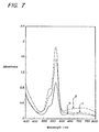
- FIG. 7 provides the absorbance spectra of the three level dye-based quality control standard described above, where spectrum a represents level a ; spectrum b represents a level b; and spectrum c represents level c , each of which were measured using a 0.1 mm cuvette.
Abstract
Description
- This invention relates to a method of preparing dye-based reference materials useful for calibrating or qualifying instrument systems that are diagnostic spectroscopically for hemoglobin and CO-ox fractions. The reference materials may also be used in instrument systems that have, in addition to capability to measure CO-ox fractions, sensors for the measurement of blood pH, gas and other blood analytes including electrolyte concentrations and metabolite concentrations.
- Improvements in instrumentation have made the determination of blood pH, gas, electrolytes and CO-ox fractions relatively routine in clinical laboratories. Typically, pH and blood gas instruments measure blood pH, pCO2. CO-oximeter instruments typically measure the total hemoglobin concentration (THb); the hemoglobin fractions such as oxyhemoglobin (O2Hb); methemoglobin (MetHb); carboxyhemoglobin (COHb); and reduced hemoglobin (HHb) (collectively referred to herein as "CO-ox fractions"). Electrolyte instruments measure any number of blood electrolytes, including sodium, potassium, lithium, calcium, and so on. Instrument systems currently available may combine the measurement of blood pH, gases, electrolytes, various metabolites, and CO-ox fractions in one instrument for a comprehensive testing of the properties of blood, as is particularly useful in respiratory and pulmonary ailments. Vigorous therapeutic treatment is often dictated by such test results.
- Quantitative determination of the various CO-ox fractions in clinical settings is desirable as CO-ox fractions relate to the loading of oxygen onto the hemoglobin of red blood cells circulating through the pulmonary capillaries. The actual amount of oxygen loaded onto the hemoglobin is determined not only by the concentration of total hemoglobin (THb), but also by the amount of non-oxygen-binding derivatives of hemoglobin such as carboxyhemoglobin (COHb) and methemoglobin (MetHb). Reduced hemoglobin (HHb) is an unoxygenated form of normal hemoglobin and elevations in the arterial fractional HHb indicate that lesser amounts of oxygen have been bound as a result of ventilation and/or perfusion defects.
- Blood, hemoglobin and hemoglobin CO-ox fractions absorb visible light. A normal blood spectrum has a main absorption peak at 578 nm and decreases rapidly close to zero at wavelengths greater than about 610 nm as shown in Figure 1. The second absorption peak of blood is at 542 nm. Absorbance maxima of hemoglobin derivatives are oxyhemoglobin, 541, 568 - 572 nm; reduced hemoglobin, 555 nm; carboxyhemoglobin, 537, 568 - 572 nm; and methemoglobin 540, 578, 630 nm.
- Generally, optical type of CO-oximeters measure the absorbance of the blood sample at multiple wavelengths on the spectrum. Ultimately, based on the known CO-ox fraction absorption wavelengths regions, CO-oximeters analyze blood samples by the collection of absorption data at specific wavelengths. The data are then typically recorded and a process called multicomponent analysis is used to simultaneously calculate the concentrations of the each of hemoglobin CO-ox fraction present in the blood sample.
- CO-oximeter instruments are typically designed to measure CO-ox fractions with values broader than the ranges observed in patient samples. For example, instruments may have the capability to measure CO-ox fractions in the defined instrument ranges of: about 4 to about 25 g/dL of THb, about 30 % to about 98 % oxyhemoglobin (O2Hb); 0 to about 50 % carboxyhemoglobin (COHb); 0 to about 40 % methemoglobin (MetHb); and 0 to about 50 % reduced hemoglobin (HHb), with all CO-ox fraction percentages herein based on the total amount of hemoglobin. Within the range of instrument capability is a clinically meaningful CO-ox fraction range and a normal physiological CO-ox fraction range. The clinically meaningful range is defined herein as: total hemoglobin 8 to 20 g/dL, 60 to 98 % oxyhemoglobin (O2Hb); 0 to 20 % carboxyhemoglobin (COHb); 0 to about 20 % methemoglobin (MetHb); and 0 to about 20 % reduced hemoglobin (HHb). The normal physiological range is defined herein as: total hemoglobin 14 to 17 g/dL for men and 12 to 15 g/dL for women, oxyhemoglobin 94 to 98%,
carboxyhemoglobin 0 to about 1 % for nonsmoker,methemoglobin 0 to about 1.5 %, and reduced hemoglobin from about 1 to about 5%. - Reference materials generally function to validate the performance of a diagnostic instrument. For CO-oximeter instrument systems, ideal quality control standard materials are formulated to provide pre-determined CO-ox fraction values not only within the broad instrument capability range, but also within the normal physiological range.
- The prior art teaches two general types of CO-oximeter quality control standard materials. The first type are aqueous dye-based materials, where dyes are used in an attempt to match the spectrum of blood. The second type are blood-based materials, where the presence blood allows for the direct match of the spectrum of blood. Both types of materials have been associated with a variety of problems as discussed below.
- In developing dye-based quality control standard materials for CO-ox instrument systems, to more closely approximate the spectrum of blood, combinations of dyes have been used. The combinations of dyes have been used because no single synthetic dye has a spectrum sufficiently similar to the absorption bands of a blood spectrum. Because the spectrum of blood has multiple distinctive bands of absorption, it is challenging to prepare a quality control standard material to mimic these characteristics. Although one dye might contribute an absorption characteristic that is present in the normal blood spectrum, it may also present other absorption characteristics that are dissimilar to blood at other portions of the spectrum.
- Prior art methods teach dye combinations that only partially simulate blood's visible spectrum. Consequently, the CO-ox fraction values of the prior art quality control standard materials are often not clinically meaningful. For example, U.S. Patent 4,843,013 teaches a combination of dyes for a CO-ox quality control standard, however, as shown in Cols. 6 and 7 of U.S. Patent 4,843,013, the described quality control standard provides negative values for some of the CO-ox fractions. A negative value for a CO-ox fraction would never appear in a blood sample and thus these control standards have limited usefulness in qualifying CO-oximeters in a clinical setting. Reference may also be made to WO 89/12827, which relates to a liquid control standard for use in the quality assurance of blood analysis instrumentation systems. There has been a long-felt commercial need for dye-based quality control materials that may be formulated in a predictable manner to provide pre-determined clinically meaningful CO-ox fractions, particularly in the normal physiological range.
- Blood-based reference materials provide clinically meaningful CO-ox fraction values, however, numerous limitations have been associated with using blood in the quality control standard materials. For example, a significant problem encountered by users of the proteinaceous blood-based materials is that blood-based materials are very susceptible to bacterial contamination and have a limited shelf life. Consequently, in most cases, users of blood-based materials must refrigerate the products during storage. Additionally, blood-based reference materials are generally classified as biohazardous materials, thus requiring the user to take additional safety precautions.
- There is a need to provide non-proteinaceous dye-based reference materials that provide clinically and physiologically meaningful CO-ox fraction ranges.
- According to the invention, a method to prepare a reference material for CO-oximeter instrument systems is provided, said method comprising (a) selecting a red dye having a main absorption maxima at a wavelength region between about 560 to about 580 nm to provide a basic spectrum; (b) modifying said main absorption maxima of said red dye by using a dye having an absorption maxima at a wavelength region between about 350 to about 450 nm; and (c) establishing a new relative baseline of said spectrum at wavelengths greater than about 640 nm and providing a sufficient absorbance at about 630 nm to provide a positive methemoglobin CO-ox fraction using one or more dyes.
- Further provided is a reference material prepared according to said method, wherein said dyes are used in sufficient amounts to provide a reference material with CO-ox fractions falling within a range covering instrument capability. The method may also be used to prepare reference materials having CO-ox fractions with values falling within the defined clinical range or within the defined normal physiological range.
- Also provided in one embodiment is a reference material comprising (a) a first dye having a main absorption maxima at a wavelength between about 560 to about 580 nm; (b) a second dye having an absorption maxima at a wavelength between about 350 to about 450 nm; and (c) a third dye having an absorption maxima at a wavelength between about 625 and about 640 nm; and (d) a fourth dye having an absorption maxima at a wavelength between about 700 to about 780 nm and an absorption band width of from about 150 to about 200 nm. The amounts of the dyes used in the materials may be adjusted to provide quality control materials with desired CO-ox fraction values.
- Further provided is a method for quality control of CO-oximeter instrument systems comprising: (1) subjecting said instrument to a quality control standard material comprising (a) a first dye having a main absorption maxima at a wavelength between about 560 to about 580 nm; (b) a second dye having an absorption maxima at a wavelength between about 350 to about 450 nm; and (c) a third dye having an absorption maxima at a wavelength between about 625 and about 640 nm; and (d) a fourth dye having an absorption maxima at a wavelength between about 700 to about 780 nm and an absorption band width of from about 150 to about 200 nm; wherein said dyes are used in a sufficient amount to provide said control with a pre-determined level of CO-ox fractions; (2) obtaining an instrument measurement of CO-ox fractions of said control; and (3) comparing said instrument measurement of said CO-ox fractions of said control with said predetermined level of CO-ox fractions to check the accuracy of said instrument.
- The dye-based reference materials are non-proteinaceous and may be interpreted by CO-oximeter instrument systems as providing a spectrum substantially equivalent to the spectrum of blood, particularly with regard to total hemoglobin and hemoglobin CO-ox fractions. Using the inventive method of preparing the reference materials, dye-based reference materials may be formulated to provide CO-ox fractions approximating those seen in a clinical setting, including normal physiological values. These dye-based reference materials may also be combined with other ingredients which make the materials useful as reference materials for pH and blood gas instruments and/or electrolyte instruments in addition to CO-oximeter instruments.
Referring to the accompanying illustrative drawings: - FIG. 1 shows the spectrum of blood.
- FIG. 2 shows the spectrums of single dyes and a dye combination that does not provide clinically meaningful CO-ox fractions. Spectrum 1 is taken from a sulforhodamine B solution. Spectrum 2 is taken from a sulforhodamine 101 solution. Spectrum 3 is taken from a solution containing 1 g/L sulforhodamine B and 0.1 g/L sulforhodamine 101.
- FIG. 3 illustrates spectroscopically how the spectrum of sulforhodamine B is modified using the inventive method of formulation. "SRB" is an abbreviation for sulforhodamine B; "MY7" is an abbreviation for mordant yellow 7; "PBV" is an abbreviation for patent blue violet; and "NGB" is an abbreviation for naphthol green B.
- FIGs. 4-7 show the spectra of the three levels of formulations shown in Examples 1-4, respectively.
-
- The present invention provides a method to formulate dye-based reference materials which mimic the spectrum of human blood when the reference materials are utilized in optical types of CO-oximeter instrument systems. The formulating method is described herein in distinct steps for purposes of clarity only. It should be understood that the steps set forth may be carried out in any order and may also be combined and carried out simultaneously. Additionally, the designation of first dye, second dye, and so on, is not intended to imply a specific order of dyes. The types of dyes selected, the amount of dyes utilized and the concentration of dyes described in these steps may be adjusted until the desired level of hemoglobin CO-ox fractions are obtained, as preferably determined by taking a reading from the specific CO-oximeter instrument that will be used with the reference material. All measurements of CO-ox fractions set forth herein were taken on Ciba Corning M270 CO-oximeters (as available from Ciba Corning Diagnostics Corp., Medfield, Mass. USA). All measurements of blood gas and electrolytes set forth herein were taken on Ciba Corning M288 Blood Gas Analyzers.
- According to the invention, in step (a) a red dye is selected to provide a basic spectrum (also referred to herein as the first dye). In selecting the red dye, the general characteristics of the spectrum of normal blood should be considered. Normal blood has a spectrum with a main absorption peak at 578 nm which decreases rapidly close to zero at a wavelength greater than about 610 nm as shown in Figure 1. The second absorption peak of blood is at approximately 542 nm. The absorbance maxima of the CO-ox fraction oxyhemoglobin is at the approximate wavelengths of 541 nm and 568 - 572 nm. As normal arterial blood has an oxyhemoglobin fraction greater than about 95%, the dye-based formulation should have a red dye with a spectrum similar to at least the general absorption features of oxyhemoglobin. The preferred red dyes in the invention have an absorption peak in the wavelength range from about 560 nm to about 580 nm. Further, particularly preferred red dyes exhibit minimal absorbance at a wavelength of greater than about 610 nm. Red dyes, such as, for example, sulforhodamine B, chlorophenol red, xylenol orange, sulforhodamine 101, etc., are currently preferred. Most preferably employed as the red dye is sulforhodamine B, which has an absorption maximum at approximately 566 nm in aqueous solution, a fairly narrow absorption bandwidth, and an absorbance which decreases to zero at about 620 nm, as shown as
spectrum 1 in Figure 2. - The single red dye does not exhibit a spectrum sufficiently similar to that of blood for purposes of obtaining clinically meaningful CO-ox fractions, as exemplified by the CO-ox fractions measured from 1.2 g/L of sulforhodamine B: THb 14 g/dL, O2Hb -16%, COHb 105%, MetHb -2.5% and HHb 13.5%. As these values indicate, negative CO-ox fractions are obtained for the oxyhemoglobin and the methemoglobin.
- One approach to alter the absorption peak of the red dye might be to introduce another red dye. In this approach, to shift the absorption peak to a longer wavelength, another red dye with absorption maximum wavelength greater than that of the first red dye could be utilized. Alternatively, to shift absorption peak to a shorter wavelength, another red dye with absorption maximum wavelength less than that of the first red dye could be utilized. A combination of the two red dyes would be predicted to result in a new overlapped maximum that would complement the required absorption features. However, as shown in Figure 2, sulforhodamine B in aqueous solution provides a spectrum that does not have enough strength of absorbance at 570 to 585 nm because the absorption peak of the sulforhodamine B is located at a shorter wavelength than that of blood. Sulforhodamine 101 has an absorption peak at 587 nm. A combination of sulforhodamine B and sulforhodamine 101 shifts the absorption peak of sulforhodamine B and enhances the absorbance at 570 nm to 585 nm. In Figure 2,
spectrum 1 is sulforhodamine B,spectrum 2 is sulforhodamine 101, and spectrum 3 is the combination of 1 g/L sulforhodamine B and 0.1 g/L sulforhodamine 101. As shown in Figure 2, absorbance of the two red dye combination at 570 to 585 nm is enhanced and results in the change of CO-ox fractions from negative values to positive values, e.g., 14 g/dL THb, 16% O2Hb, 1% MetHb, 14% COHb and 69% HHb. The addition of the second red dye (sulforhodamine 101) introduces an undesired high absorbance near wavelength range of 590 nm to 605 nm, thus not providing an acceptable method of formulating references having clinically meaningful and/or normal physiological CO-ox fraction values. - In step (b), modification and manipulation of the absorption band of the red dye is conducted where the absorption peak of the red dye is shifted to a longer wavelength to more closely approximate that of blood. Upon step (b), the absorption maxima of the red dye is shifted to fall between the wavelengths of approximately 570 nm to about 585 nm. It is preferred that the shift of the absorption maxima does not inadvertently increase the absorption in the wavelength region of 590 to 605 nm. It has been found that dye(s) having an absorption maxima at wavelengths of about 350 to 450 nm are useful in step (b) because the dye(s) increase the absorbance of the spectrum at the ultraviolet region and force the absorption band of the red dye to shift toward the longer wavelength.
- Preferably in step (b) is accomplished using a yellow dye (or a combination thereof) including, for example, mordant yellow 7, tartrazine, orange G, hydroxypyrenetrisulfonic acid, mordant yellow 10, combinations thereof, and so on. Particularly preferred is mordant yellow 7 dye (especially when sulforhodamine B is selected as the red dye).
- Figure 3 illustrates the changes of the spectrum of sulforhodamine B after addition of mordant yellow 7.
Spectrum 1 in Figure 3 is the sulforhodamine B aqueous solution, andspectrum 2 is the sulforhodamine B aqueous solution containing mordant yellow 7. As shown, the original absorption peak of sulforhodamine B is shifted from 566 nm to 572 nm, with the edge of the absorption band at approximately 610 nm, and with little increase of absorbance in the wavelength range of 590 to about 605 nm. - According to the present invention, in step (c) a new relative baseline of the spectrum is established at wavelengths greater than about 640 nm and sufficient absorbance at about 630 nm is provided to yield a positive methemoglobin CO-ox fraction. Step (c) addresses that the absorbance at about 590 to about 605 nm of the dye or dye combination used in steps (a) and (b) is higher than that of normal blood. This step may be accomplished using one or more dyes. With respect to the new lifted baseline, the original absorbance can be normalized, with the normalized absorbance reduced and smaller than the original absorbance. Further, the original absorbance at about 590 to about 605 nm, although having a similar absolute absorbance value, is relatively reduced upon the establishment of the new lifted baseline. CO-oximeters take the lifted baseline from the CO-oximeters interpret the dye spectrum, and calculate the CO-ox fractions therefrom. Upon step (c), the inventive reference material will read as providing CO-ox fraction values within the instrument capability and clinically meaningful ranges, and optionally, within the normal physiological range, as described in more detail hereinafter.
- One group of dyes particularly useful in step (c) are designated herein as the third dye(s). These dyes have an absorption maxima at about 625 to about 640 nm (more preferably about 630 nm) and have only minimum impact on the absorbance at wavelengths shorter than about 600 nm. Preferably, the third dye is a blue dye selected from brilliant blue FC (FD&C blue 1), erioglaucine, lissamine, alphazurine A, patent blue violet, hydroxy naphthol blue, patent blue VF, combinations thereof, and equivalents thereof (with patent blue violet, patent blue VF, and alphazurine more preferred). Patent blue violet has an absorption maxima at about 639 nm and is most preferred because it provides the absorbance not only at 630 nm, but also absorbance to some extent at a wavelength greater than 640 nm, thus generally accomplishing both functions of step (c). Adding the patent blue violet into the sulforhodamine B and yellow mordant 7 combination pushes the CO-ox fractions to the following: O2Hb 80%, MetHb 15%,
COHb 2% and HHb 3 %, thus placing the reference material within the values set forth for instrument capability and clinically meaningful CO-ox fractions. - A second group of dyes that may be used in step (c) are designated as the fourth dye(s). These dyes have an main absorbance maxima positioned at a wavelength of from about 700 to about 780 nm and an absorption band width of from about 150 to about 200.nm. The tail of the absorption band of the dye (or dyes) of step (c) preferably lifts the baseline of the absorbance spectrum with minimal effect on absorbance at 500 to 605 nm. Preferred dyes falling within this description may be selected from any number of dyes including IR 125, naphthol green B, and combinations thereof, and so on. Most preferred is naphthol green B, having an absorption maxima at approximately 716 nm in an aqueous solution, and considered particularly effective when combined with mordant yellow 7, patent violet blue and sulforhodamine B.
- For step (c) the combination of the third and fourth dyes allow the reference material containing the first and second dyes to yield CO-ox fraction values falling with the normal, physiological range. Figure 3 exemplifies the principles of the present method of formulation. The sulforhodamine B (SRB) is the red dye that provides a basic spectrum. The addition of mordant yellow 7 (MY7) to the sulforhodamine B (SRB) modifies the spectrum by tilting the absorption band of the sulforhodamine B towards a longer wavelength and shifts the absorption maxima from about 566 nm to about 572 nm (SRB + MY7), to more closely approximate the main absorption spectrum of blood. Patent blue violet (PBV) is added for purposes of providing a spectrum absorption for methemoglobin at approximately 630 nm (SRB + MY7 + PBV) and also slightly lifts the baseline spectrum. The addition of the naphthol green B (NGB) further lifts the spectrum baseline to the right of the main absorption maxima. The ultimate result is shown by Spectrum "SRB + MY7 + PBV + NGB". In this formulation the following amounts of dyes were used: about 1.15 g/L sulforhodamine B; about 2.4 g/L mordant yellow 7; about 0.08 g/L patent blue violet; and about 1 g/L naphthol green B. When this combination was measured on a Ciba Corning M270 CO-oximeter the following CO-ox fraction ranges (which are within the normal physiological range) were provided: THb 14 g/dL, O2Hb 96 %,
COHb 1%, MetHb 0.8% and HHb 2.2 %. Providing a reference material having an oxyhemoglobin value greater than about 60 % has been met with limited success in the past, however, the present invention provides a method to easily yield this objective. - Any suitable dyes may be used in the formulation of the reference materials with either aqueous or nonaqueous solvents. Water soluble dyes are particularly well-suited, including acid dyes, basic dyes, direct dyes, and so on, and equivalents thereof. The dye composition may be prepared as a dry material for ease of storage and packaging. If prepared as a dry composition, prior to usage the composition may be prepared as a solution using a suitable liquid, including water and various organic solvents, or mixtures thereof and so on, by techniques well known to those skilled in the art. It is particularly. preferred that compatible dyes are used, with a particularly preferred embodiment utilizing anionic dyes. Although the method of formulating may be accomplished using various amounts of dyes, a particularly preferred composition employs a total dye concentration of from about 0.1 to about 10 mM. Further, the stability of the reference materials will be increased when high purity dyes, which are either commercially available or purified, using conventional methods known to those skilled in the art, are used in formulation.
- The reference materials prepared according to the invention may be utilized on various types of optical CO-oximeters (or instruments incorporating a CO-oximeter function). Because various types of CO-oximeters may be designed and constructed to measure the absorbance of blood at different set of wavelengths, the reference materials used for these CO-oximeters will typically need dye concentration adjustment for this variation, where the adjustment may be accomplished by those skilled in the art employing the principles of this invention.
- The reference materials may be prepared as calibration materials to simultaneously calibrate instrument systems that include sensors for blood gas, electrolyte, and various metabolites in addition to the CO-oximeter capability. Further, the reference materials are particularly well-suited for preparation as quality control materials for instrument systems that include a CO-oximeter capability, in addition to various other sensors, as described herein. For CO-oximeter instrument systems, ideally, the CO-ox fraction accuracy is generally checked both in the broader instrument capability CO-ox fraction range as well as in the more narrow normal physiological range (as defined herein). More preferably, the CO-oximeter instrument system is checked for performance accuracy on at least three levels. These three reference materials are preferably formulated to provide pre-determined CO-ox fraction values for the total hemoglobin as well as various hemoglobin CO-ox fractions outside of and within normal physiological ranges.
- Particularly preferred compositions provide a commercially acceptable stability. The stability may also extend to high temperature stability such that the composition may be autoclaved for purposes of facilitating sterilization, if desired. Additionally, the dye formulations may be prepared such that they are stable to exposure to light.
- Appropriate ingredients that be included in the dye formulation preferably are those ingredients that do not interfere with the sensors of the instruments such that the reference materials may be used to provide calibration or quality control of different types of sensors simultaneously. A preferred reference prepared using the dye formulation of this invention may be prepared by combining in the appropriate relative proportions, buffer(s), dyes, sources of analytes (typically salts), and other standard chemicals commonly used in reference materials including preservatives and surfactants, and so on, as well known in the art. Buffer materials that may be used, particularly when preparing a reference standard for simultaneous usage on pH blood gas and electrolyte sensor system, are preferably selected for having a pKa close to the desired working pH. Particularly useful buffer materials are zwitterion buffers that are compatible with the selected dyes, including, for example, N-2-hyroxyethylpiperazine-N'-2-ethanesulfonic acid (HEPES), 3-(N-morpholinio)propanesulfonic acid (MOPS), Tris-(Hydroxymethyl)aminomethane (TRIS), and so on, as are well known to those skilled in the art. A combination of the salts such as, for example, NaCl, NaOH, KCl, LiCl, NaHCO3, Na2SO4, CaCl2, glucose, lithium lactate, and equivalents thereof, can be used to provide the desired concentration of electrolytes, as is well known to those skilled in the art. A stable ionized calcium concentration may be provided by using calcium chelating agents, such as, ethylenediaminetetraacetic acid (EDTA), N-hydroxyethyliminodiacetic acid (HIDA), nitrilotriacetic acid (NTA), and citric acid. Further, when providing a reference for a blood gas measurement, the desired pCO2 and pO2 ranges may be reached by tonometering to an equilibrium at approximately 25° C using gas combination with proper oxygen, carbon dioxide, and nitrogen contents, as well within the skill of those trained in the art.
- A particularly useful reference material includes the above-described dyes providing normal physiological CO-ox fractions while in combination with an aqueous solution of known quantities of pH, pO2, pCO2, Na, K, Cl, and ionized Ca (iCa), glucose, and lactate (preferably in amounts that typically fall within the range of a blood sample). After said combination, the reference material may be divided into aliquots which are placed in sealed receptacles. Thereafter the receptacles may be flushed with tonometry gas, and the receptacle sealed. These references may be used for verifying the accuracy and reliability of instrument systems having multiple sensors for measuring blood pH, gas, CO-ox fractions and various electrolyte and metabolite levels. A particularly preferred embodiment that is described in TABLE A below.
COMPOUND APPROXIMATE CONCENTRATIONS NaCl 110 to 160 mM KCl 2.5 to 7.5 mM CaCl 2 2 to 4 mM HEPES 35 to 40 mM TRIS.HCl 15 to 40 mM NaOH 20 to 50 mM EDTA 1 to 1.7 mM NaHCO3 15 to 20 mM LiCl 10 to 25 mM Glucose 0.5 to 10 g/L Li lactate 0.5 to 14 mM Sulforhodamine B 0.55 to 1.3 g/L Mordant Yellow 7 1.4 to 3 g/L Patent Blue Violet 0.07 to 0.08 g/L Naphthol Green B 0.4 to 1.1 g/L - To prepare clinically meaningful reference materials, preferably the first dye is used in an amount ranging from about 0.55 mM to about 4 mM; the second dye is used in an amount ranging from about 1 mM to about 10 mM; the third dye is used in an amount ranging from about 0.01 mM to 0.3 mM; and the fourth dye is used in an amount ranging from 0 to about 2.5 mM.
- The following Examples illustrate the present invention.
- A three-level quality control standard material was prepared with varying levels of pH, pCO2, pO2, sodium (Na), potassium (K), chloride (Cl), ionized calcium (iCa), and three levels of total hemoglobin (THb), oxyhemoglobin (O2Hb), carboxyhemoglobin (COHb), methemoglobin (MetHb), and reduced hemoglobin (HHb), as shown in TABLES I and II. The three levels prepared represent clinically significant ranges of acidosis (level a), normal or physiological (level b), and alkalosis (level c).
- More particularly, three bulk solutions containing the required salts were prepared in an aqueous solution, as shown below in TABLE I.
BULK SOLUTIONS COMPOUND Level a Level b Level c NaCl 55.5 mM 60 mM 65 mM KCl 3.20 4.85 710 CaCl2 3.81 2.80 2.13 NaOH 29.5 38 34.3 LiCl 21.5 12.9 --- EDTA 1.63 1.20 1.14 HEPES 40 40 40 TRIS.HCl 35.5 15 --- TRIS base 10 --- --- NaHCO3 19.5 23.4 19.5 DYE FORMULATIONS Dye Level a Level b Level c Sulforhodamine B 0.98 mM 1.95 mM 2.27 mM Mordant Yellow 7 6.65 7.63 3.89 Patent Blue Violet 0.14 0.11 0.14 Naphthol Green B --- 1.10 0.48 - After dyes dissolved in the bulk solution, the solutions were then tonometered at 25° C to an equilibrium using the following gas mixtures.
GAS CONCENTRATIONS (%) Gas Level a Level b Level c CO2 6.10 3.15 1.77 O2 5.42 11.60 17.45 N2 88.48 85.25 80.78 - After tonometry equilibrium was established, the pH, pO2, and pCO2 of the solutions remained constant even after a prolonged period of tonometry. Aliquots of the tonometered solutions were then transferred into glass ampoules while purging with the same gas mixture used in the solution tonometry. The filled ampoules were then flame-sealed.
- The three levels of quality control standard having the combinations listed in TABLES I, II, and III were then evaluated by two types of instruments:
ANALYZER 1 = Ciba Corning M288 blood gas analyzer andANALYZER 2 = Ciba Corning M270 CO-oximeter. Results are summarized in TABLE IV.Evaluations of Reference Standards ANALYTE LEVEL a LEVEL b LEVEL c ANALYZER 1 (Blood Gas) pH 7.15 ± 0.02 7.4 ± 0.02 7.6 ± 0.02 pCO2 mm Hg 70 ± 2 40 ± 2 21 ± 2 pO2 mm Hg 63 ± 2 100 ± 3 150 ± 5 Na mM 115 ± 2 135 ± 2 155 ± 3 K mM 2.9 ± 0.3 4.8 ± 0.3 7.1 ± 0.3 Cl mM 120 ± 2 100 ± 2 80 ± 2 iCa mM 1.6 ± 0.05 1.15 ± 0.05 0.65 ± 0.05 ANALYZER 2 (CO-oximeter) THb g/dL 8.0 ± 0.2 14 ± 0.2 18 ± 0.2 O2Hb % 78 ± 2 95 ± 3 60 ± 2 COHb % 3.5 ± 1 2.5 ± 1 6 ± 1 MetHb % 16 ± 2 1 ± 0.3 18 ± 2 HHb % 2.5 ± 1 1.5 ± 0.7 16 ± 2 - FIG. 4 provides the absorbance spectra of the three dye-based quality control standard described above, where spectrum a represents level a; spectrum b represents a level b; and spectrum c represents level c, each of which were measured using a 0.1 mm cuvette.
- A three level quality control standard was prepared by the procedure of Example 1. To the bulk solutions described in TABLE I of Example 1 the dye formulations shown in TABLE V below were added.
DYE FORMULATIONS Dye Level a Level b Level c Sulforhodamine B 1.27 mM 2.53 mM 3.22 mM Tartrazine 1.34 1.87 1.60 Patent Blue Violet 0.11 0.17 0.25 Naphthol Green B 0.98 1.96 1.64 - The formulated solutions were then tonometered at 25° C to equilibrium using the same gas mixtures described in TABLE III of Example 1. The tonometered solutions were then transferred into glass ampoules while purging the same gas mixture used in the solution tonometry. The filled ampoules were flame-sealed. Those three levels of reference were measured in the same manner as described in Example 1. The measured ranges were similar to the data shown in Example 1.
- FIG. 5 provides the absorbance spectra of the three level dye-based quality control standard described above, where spectrum a represents level a; spectrum b represents a level b; and spectrum c represents level c, each of which were measured using a 0.1 mm cuvette.
- A three level quality control standard was prepared by the same procedure of Example 1. The bulk solutions of TABLE I were combined with the dye formulations of TABLE VII below.
Dye Formulations Dye Level a Level b Level c Sulforhodamine B 0.93 mM 2.14 mM 2.37 mM Mordant Yellow 10 8.95 9.24 4.91 patent blue VF 0.12 0.13 0.08 Naphthol Green B --- 1.33 0.81 - The formulated solutions were tonometered and transferred into ampoules, sealed, and thereafter measured, as described in Example 1. The data obtained using this formulation were similar to those measured in Example 1.
- FIG. 6 provides the absorbance spectra of the three level dye-based quality control standard described above, where spectrum a represents level a; spectrum b represents a level b; and spectrum c represents level c, each of which were measured using a 0.1 mm cuvette.
- A three level quality control standard was prepared by the same procedure of Example 1. The bulk solutions of TABLE I were combined with the dye formulations of TABLE VIII below.
Dye Formulations Dye Level a Level b Level c Sulforhodamine B 1.17 mM 1.97 mM 2.19 mM Mordant Yellow 7 5.33 6.14 2.62 FD&C Blue 10.03 0.04 0.03 Naphthol Green B 0.97 2.30 0.14 - The formulated solutions were tonometered and transferred into ampoules, sealed, and thereafter measured, as described in Example 1. The data obtained using this formulation were similar to those measured in Example 1.
- FIG. 7 provides the absorbance spectra of the three level dye-based quality control standard described above, where spectrum a represents level a; spectrum b represents a level b; and spectrum c represents level c, each of which were measured using a 0.1 mm cuvette.
Claims (13)
- A method for the preparation of a CO-oximeter instrument system reference material characterised in that it comprises (a) selecting a red dye having a main absorption maxima in a wavelength region of from 560 to 580 nm to provide a basic spectrum; (b) modifying the said main absorption maxima of the said red dye by using a dye having an absorption maxima in a wavelength region of from 350 to 450 nm; and (c) establishing a new relative baseline of the said spectrum at wavelengths greater than 640 nm and providing a sufficient absorbance at about 630 nm to provide a positive methemoglobin CO-ox fraction value using one or more dyes.
- A method as claimed in claim 1 wherein the said red dye has an absorption band width of less than 50 nm and an absorption of about zero at a wavelength of greater than 610 nm.
- A method as claimed in claim 1 or claim 2 wherein the said dye of (b) is a yellow dye and the said main absorption maxima is shifted to a wavelength in the region of from 570 to 580 nm.
- A method as claimed in any of claims 1 to 3 wherein (c) is accomplished using two dyes, one of which is a blue dye having an absorption peak at from 625 to 640 nm and the other of which is a dye having an absorbance maxima at a wavelength of from 700 to 780 nm and an absorption band width of from 150 to 200 nm.
- A method as claimed in any of claims 1 to 4 wherein the said reference material provides normal physiological CO-ox fractions of: oxyhemoglobin from 94 to 98 %, carboxyhemoglobin from 0 to 1 %, methemoglobin from 0 to 1.5 % and reduced hemoglobin from 1 to 5 %.
- A reference material characterised in that it comprises (a) a first dye having a main absorption maxima at a wavelength of from 560 to 580 nm; (b) a second dye having an absorption maxima at a wavelength of from 350 to 450 nm; (c) a third dye having an absorption maxima at a wavelength of from 625 to 640 nm; and (d) a fourth dye having an absorption maxima at a wavelength of from 700 to 780 nm and an absorption band width of from 150 to 200 nm.
- A material as claimed in claim 6 wherein each of the said dyes is present in a sufficient amount to provide the said material with clinically meaningful CO-ox fractions.
- A material as claimed in claim 6 or claim 7 wherein the said first dye is a red dye selected from sulforhodamine B (Xanthylium, 3,6-bis(diethylamino)-9-(2,4-disulfophenyl)-, inner salt, sodium salt / CAS No. 3520-42-1 / C.I. No. 45100), chlorophenyl red (Phenol, 4,4'-(1,1-dioxido-3H-2,1-benzoxathiol-3-ylidene)bis[2-chloro- / CAS No. 4430-20-0), xylenol orange (Glycine, N-[[3- [[3- [[bis (carboxymethyl) amino] methyl]-4-hydroxy-5-methylphenyl] (2-sulfophenyl) methylene] -5-methyl-6-oxo-1,4-cyclohexadien-1-yl]methyl]-N-(carboxymethyl)-, sodium salt / CAS No. 63721-83-5) and sulforhodamine 101 (1H,5H,11H,15H-Xantheno[2,3,4-ij:5,6,7-i'j']diquinolizin-18-ium, 9- [2 (or 4)-(chlorosulfonyl)-4(or 2)-sulfophenyl]-2,3,6,7,12,13,16,17-octahydro-, inner salt / CAS No. 82354-19-6); the said second dye is a yellow dye selected from mordant yellow 7 (Benzoic acid, 2-hydroxy-3-methyl-5-[(4-sulfophenyl)azo]-, disodium salt / CAS No. 6408-91-9 / C.I. No. 14180), tartrazine (lH-Pyrazole-3-carboxylic acid, 4,5-dihydro-5-oxo-1-(4-sulfophenyl)-4-[(4-sulfophenyl)azo]-, trisodium salt / CAS No. 1934-21-0 / C.I. No. 19140), Orange G (1,3-Naphthalenedisulfonic acid, 7-hydroxy-8-(phenylazo)-, disodium salt / CAS No. 1936-15-8 / C.I. No. 16230), hydroxypyrenetrisulfonic acid (1,3,6-Pyrenetrisulfonic acid, 8-hydroxy-, trisodium salt / CAS No. 6358-69-6 / C.I. No. 59040), mordant yellow 10 (Benzoic acid, 2-hydroxy-5-[(4-sulfophenyl)azo]-, disodium salt / CAS No. 6054-99-5 / C.I. No. 14010) and combinations thereof; the said third dye is selected from brilliant blue FC (FD&C blue 1) (Benzenemethanaminium, N-ethyl-N-[4-[[4-[ethyl[(3-sulfophenyl)methyl]amino]phenyl](2-sulfophenyl)methylene]-2,5-cyclohexadien-1-ylidene]-3-sulfo-, inner salt, diammonium salt / CAS No. 2650-18-2), erioglaucine (Benzenemethanaminium, N-ethyl-N-[4-[[4-[ethyl [(3-sulfophenyl) methyl] amino] phenyl] (2-sulfophenyl)methylene]-2,5-cyclohexadien-1-ylidene]-3-sulfo-, inner salt, disodium salt / CAS No. 3844-45-9 / C.I. No. 42090), lissamine (Methanaminium, N-[4-[[4-(dimethylamino)phenyl](2-hydroxy-3,6-disulfo-1-naphthalenyl)methylene]-2,5-cyclohexadien-1-ylidene]-N-methyl-, inner salt, monosodium salt / CAS No. 3087-16-9 / C.I. No. 44090), alphazurine A (Benzenemethanaminium, N- [4-((2,4-disulfophenyl) [4-[ethyl(phenylmethyl)amino]phenyl]methylene]-2,5-cyclohexadien-1-ylidene]-N-ethyl-, inner salt, sodium salt / CAS No. 3486-30-4 / C.I. No. 42080), patent blue violet (Ethanaminium, N -[4-[[4-(diethylamino)phenyl](2,5-disulfophenyl)methylene]-2,5-cyclohexadien-1-ylidene]-N-ethyl-, inner salt, sodium salt / CAS No. 68238-36-8), patent blue VF (Ethanaminium, N-[4-[[4-(diethylamino) phenyl] (2,4-disulfophenyl) methylene] -2,5-cyclohexadien-1-ylidene]-N-ethyl-, inner salt, sodium salt / CAS No. 129-17-9 / C.I. No. 42045), hydroxynaphthol blue (2,7-Napthalenedisulfonic acid, 3-hydroxy-4-[(2-hydroxy-4-sulfo-1-naphthalenyl)azo]-, disodium salt / CAS No. 165660-27-5) and combinations thereof; and the said fourth dye is selected from IR 125, (1H-Benz[e] indolium, 2-[7-[1,3-dihydro-1,1-dimethyl-3-(4-sulfobutyl)-2H-benz [e]indol-2-ylidene]-1,3,5-heptatrienyl]-1,1-dimethyl-3-(4-sulfobutyl)-, inner salt, sodium salt / CAS No. 3599-32-4), naphthol green B (Ferrate(3-), tris[5,6-dihydro-5-(hydroxyimino-.kappa.N)-6-(oxo-.kappa.O)-2- naphthalenesulfonato(2-)]-, trisodium / CAS No. 19381-50-1 / C.I. No. 10020) and combinations thereof; preferably the said first dye is sulforhodamine B; the said second dye is mordant yellow 7; the said third dye is patent blue violet; and the said fourth dye is naphthol green B; or preferably the said first dye is sulforhodamine B; the said second dye is tartrazine; the said third dye is patent blue violet; and the said fourth dye is naphthol green B; or preferably the said first dye is sulforhodamine B; the said second dye is mordant yellow 10; the said third dye is patent blue VF; and the said fourth dye is naphthol green B; or preferably the said first dye is sulforhodamine B; the said second dye is mordant yellow 7; the said third dye is FD&C blue 1; and the said fourth dye is naphthol green B.
- A material as claimed in any of claims 6 to 8 wherein the said first dye is used in an amount of from 0.55 to 4 mM; the said second dye is used in an amount of from 1 to 10 mM; the said third dye is used in an amount of from 0.01 to 0.3 mM; and the said fourth dye is used in an amount of from 0 to 2.5 mM.
- A material as claimed in any of claims 6 to 9 wherein it comprises:
COMPOUND CONCENTRATIONS NaCl 110 to 160 mM KCl 2.5 to 7.5 mM CaCl2 2 to 4 mM HEPES 35 to 40 mM TRIS.HCl 15 to 40 mM NaOH 20 to 50 mM EDTA 1 to 1.7 mM NaHCO3 15 to 20 mM LiCl 10 to 25 mM Glucose 0.5 to 10 g/L Li lactate 0.5 to 14 mM Sulforhodamine B 0.55 to 1.3 g/L Mordant Yellow 7 1.4 to 3 g/L Patent Blue Violet 0.07 to 0.08 g/L Naphthol Green B 0.4 to 1.1 g/L - A method for the quality control of a CO-oximeter instrument system characterised in that it comprises: (1) subjecting the said instrument to a quality control standard material prepared by a method as claimed in any of claims 1 to 5, the said dyes being used in a sufficient amount to provide the said control with a predetermined level of CO-ox fractions; (2) obtaining an instrument measurement of CO-ox fractions of the said control; and (3) comparing the said instrument measurement of the said CO-ox fractions of the said control with the said predetermined level of CO-ox fractions to check the accuracy of the said instrument.
- A method for the quality control of a CO-oximeter instrument system characterised in that it comprises (1) subjecting the said instrument to a quality control standard material as claimed in any of claims 6 to 10, the said dyes being used in a sufficient amount to provide the said control with a predetermined level of CO-ox fractions; (2) obtaining an instrument measurement of CO-ox fractions of the said control; and (3) comparing the said instrument measurement of the said CO-ox fractions of the said control with the said predetermined level of CO-ox fractions to check the accuracy of the said instrument.
- A method as claimed in claim 12 wherein three levels of quality control standards are provided:
Dye Level a Level b Level c Sulforhodamine B 0.98 mM 1.95 mM 2.27 mM Mordant Yellow 7 6.65 7.63 3.89 Patent Blue Violet 0.14 0.11 0.14 Naphthol Green B 0 1.10 0.48 Dye Level a Level b Level c Sulforhodamine B 1.27 mM 2.53 mM 3.22 mM Mordant Yellow 7 1.34 1.87 1.60 Patent Blue Violet 0.11 0.17 0.25 Naphthol Green B 0.98 1.96 1.64 Dye Level a Level b Level c Sulforhodamine B 0.93 mM 2.14 mM 2.37 mM Mordant Yellow 10 8.95 9.24 4.91 Patent Blue VF 0.12 0.13 0.08 Naphthol Green B 0 1.33 0.81 Dye Level a Level b Level c Sulforhodamine B 1.17 mM 1.97 mM 2.19 mM Mordant Yellow 7 5.33 6.14 2.62 FD&C Blue 1 0.03 0.04 0.03 Naphthol Green B 0.97 2.30 0.14
Applications Claiming Priority (2)
| Application Number | Priority Date | Filing Date | Title |
|---|---|---|---|
| US447168 | 1995-05-19 | ||
| US08/447,168 US5637505A (en) | 1995-05-19 | 1995-05-19 | Method to prepare dye-based reference material |
Publications (3)
| Publication Number | Publication Date |
|---|---|
| EP0743523A2 EP0743523A2 (en) | 1996-11-20 |
| EP0743523A3 EP0743523A3 (en) | 1997-11-19 |
| EP0743523B1 true EP0743523B1 (en) | 2002-08-28 |
Family
ID=23775276
Family Applications (1)
| Application Number | Title | Priority Date | Filing Date |
|---|---|---|---|
| EP96303490A Expired - Lifetime EP0743523B1 (en) | 1995-05-19 | 1996-05-16 | Dye-based reference material, preparation and use thereof |
Country Status (11)
| Country | Link |
|---|---|
| US (2) | US5637505A (en) |
| EP (1) | EP0743523B1 (en) |
| JP (1) | JP2982891B2 (en) |
| KR (1) | KR960042054A (en) |
| AT (1) | ATE223061T1 (en) |
| AU (1) | AU699664B2 (en) |
| CA (1) | CA2169803A1 (en) |
| DE (1) | DE69623175T2 (en) |
| DK (1) | DK0743523T3 (en) |
| ES (1) | ES2181848T3 (en) |
| PL (1) | PL313540A1 (en) |
Families Citing this family (22)
| Publication number | Priority date | Publication date | Assignee | Title |
|---|---|---|---|---|
| US6136607A (en) * | 1995-11-02 | 2000-10-24 | Bayer Corporation | Multi-analyte reference solutions with stable pO2 in zero headspace containers |
| US7157282B2 (en) | 1996-06-12 | 2007-01-02 | Spectromedical Inc. | Quality control material for reagentless measurement of analytes |
| US6828152B2 (en) | 1996-06-12 | 2004-12-07 | Spectromedical Inc. | Quality control material for reagentless measurement of analytes |
| AU734428B2 (en) * | 1997-01-07 | 2001-06-14 | Obayashi Corporation | Method for analyzing formaldehyde in ambient air |
| US6221612B1 (en) | 1997-08-01 | 2001-04-24 | Aurora Biosciences Corporation | Photon reducing agents for use in fluorescence assays |
| US6061176A (en) * | 1998-09-14 | 2000-05-09 | Shih; Song Hsin | Microscope system for observation and display of microcirculation at multiple body areas |
| DZ2947A1 (en) * | 1998-11-25 | 2004-03-15 | Chiesi Farma Spa | Pressure metered dose inhaler. |
| US6949384B2 (en) | 2001-12-21 | 2005-09-27 | Spectromedical Inc. | Method for monitoring degradation of Hb-based blood substitutes |
| US20050037505A1 (en) * | 2000-05-11 | 2005-02-17 | James Samsoondar | Spectroscopic method and apparatus for analyte measurement |
| US6470279B1 (en) * | 1999-11-23 | 2002-10-22 | James Samsoondar | Method for calibrating spectrophotometric apparatus with synthetic fluids to measure plasma and serum analytes |
| US7449339B2 (en) * | 1999-11-23 | 2008-11-11 | Nir Diagnostics Inc. | Spectroscopic method and apparatus for total hemoglobin measurement |
| DK3536344T3 (en) * | 2002-03-01 | 2020-03-16 | Chiesi Farm Spa | SUPERFIN FORMOTHER FORMULATION |
| SE0301897D0 (en) * | 2003-06-27 | 2003-06-27 | Foss Tecator Ab | Referece standard and method of calibration |
| WO2009076302A1 (en) | 2007-12-10 | 2009-06-18 | Bayer Healthcare Llc | Control markers for auto-detection of control solution and methods of use |
| EP2261673B1 (en) * | 2008-03-31 | 2018-01-24 | Terumo Kabushiki Kaisha | Method for preparing calibrating reagent, calibrating reagent, and method for calibrating blood component measuring device using said calibrating reagent |
| US8404158B2 (en) * | 2008-04-07 | 2013-03-26 | Artel, Inc. | System and method for liquid delivery evaluation using solutions with multiple light absorbance spectral features |
| US7791716B2 (en) * | 2008-04-07 | 2010-09-07 | Artel, Inc. | System and method for liquid delivery evaluation using solutions with multiple light absorbance spectral features |
| EP2281191A4 (en) | 2008-04-30 | 2011-10-05 | Instrumentation Lab Co | Hemoglobin based bilirubin reference material |
| US9468403B2 (en) | 2014-01-15 | 2016-10-18 | Hong Kong Applied Science and Technology Research Institute Company Limited | Single-body unit for pulse oximeter calibration |
| CA3002401A1 (en) * | 2015-10-23 | 2017-04-27 | Geosyntec Consultants, Inc. | Use of visibly detectable compounds as performance reference compounds in passive sampling devices |
| US11460476B2 (en) | 2019-03-04 | 2022-10-04 | Siemens Healthcare Diagnostics Inc. | Dye-based liquid reagent volume indicator for use in analyte detection assays |
| KR102317339B1 (en) * | 2020-01-03 | 2021-10-26 | 주식회사 딕스젠 | reagent composition for detecting hemoglobin and detection method of hemoglobin using thereof |
Family Cites Families (14)
| Publication number | Priority date | Publication date | Assignee | Title |
|---|---|---|---|---|
| US3729427A (en) * | 1971-04-08 | 1973-04-24 | Baxter Laboratories Inc | Neutral density solutions containing nigrosin dye and carbon black |
| US3751381A (en) * | 1971-04-27 | 1973-08-07 | Warner Lambert Co | Dyed albumen-cohn fraction iii-lipid mixtures serum lipid assay standard |
| US4146604A (en) * | 1973-05-31 | 1979-03-27 | Block Engineering, Inc. | Differential counting of leukocytes and other cells |
| US3920580A (en) * | 1973-07-12 | 1975-11-18 | Miles Lab | Liquid control solution |
| ZA783915B (en) * | 1977-08-15 | 1979-07-25 | Miles Lab | Device for preparation of liquid control solutions |
| US4279775A (en) * | 1979-12-31 | 1981-07-21 | Louderback Allan Lee | Blood gas control |
| JPS6027861A (en) * | 1983-07-21 | 1985-02-12 | コ−ニング・グラス・ワ−クス | Co-oximetry characteristic control composition |
| DK282085D0 (en) * | 1985-06-21 | 1985-06-21 | Radiometer As | METHOD AND APPARATUS FOR DETERMINING BLOOD COMPONENTS |
| US4843013A (en) * | 1986-04-09 | 1989-06-27 | Bionostics, Incorporated | Multiple control standard for blood analysis |
| US4753888A (en) * | 1986-04-09 | 1988-06-28 | Bionostics, Inc. | Multiple control standard for blood analysis |
| JPS646865A (en) * | 1987-06-30 | 1989-01-11 | Japan Synthetic Rubber Co Ltd | Standard liquid for calibration of blood measuring instrument |
| US4945062A (en) * | 1988-06-15 | 1990-07-31 | Bionostics Incorporated | Control for blood gas/calcium analysis instrumentation |
| US5013666A (en) * | 1988-06-15 | 1991-05-07 | Bionostics, Incorporated | Control for blood gas/calcium analysis instrumentation |
| US4962039A (en) * | 1988-06-30 | 1990-10-09 | 2V Industries, Inc. | Simple and rapid method for detecting the presence of water in various organic fluids |
-
1995
- 1995-05-19 US US08/447,168 patent/US5637505A/en not_active Expired - Lifetime
-
1996
- 1996-01-24 AU AU42162/96A patent/AU699664B2/en not_active Ceased
- 1996-02-19 CA CA002169803A patent/CA2169803A1/en not_active Abandoned
- 1996-03-28 PL PL96313540A patent/PL313540A1/en unknown
- 1996-05-16 JP JP8121681A patent/JP2982891B2/en not_active Expired - Fee Related
- 1996-05-16 AT AT96303490T patent/ATE223061T1/en not_active IP Right Cessation
- 1996-05-16 EP EP96303490A patent/EP0743523B1/en not_active Expired - Lifetime
- 1996-05-16 DK DK96303490T patent/DK0743523T3/en active
- 1996-05-16 DE DE69623175T patent/DE69623175T2/en not_active Expired - Lifetime
- 1996-05-16 ES ES96303490T patent/ES2181848T3/en not_active Expired - Lifetime
- 1996-05-18 KR KR1019960016880A patent/KR960042054A/en not_active Application Discontinuation
-
1997
- 1997-02-28 US US08/810,214 patent/US5891730A/en not_active Expired - Lifetime
Also Published As
| Publication number | Publication date |
|---|---|
| JP2982891B2 (en) | 1999-11-29 |
| CA2169803A1 (en) | 1996-11-20 |
| DE69623175T2 (en) | 2003-01-02 |
| JPH08313532A (en) | 1996-11-29 |
| EP0743523A2 (en) | 1996-11-20 |
| US5637505A (en) | 1997-06-10 |
| ES2181848T3 (en) | 2003-03-01 |
| US5891730A (en) | 1999-04-06 |
| PL313540A1 (en) | 1996-11-25 |
| AU4216296A (en) | 1996-11-28 |
| DE69623175D1 (en) | 2002-10-02 |
| ATE223061T1 (en) | 2002-09-15 |
| EP0743523A3 (en) | 1997-11-19 |
| DK0743523T3 (en) | 2003-01-06 |
| AU699664B2 (en) | 1998-12-10 |
| KR960042054A (en) | 1996-12-19 |
Similar Documents
| Publication | Publication Date | Title |
|---|---|---|
| EP0743523B1 (en) | Dye-based reference material, preparation and use thereof | |
| Van Kampen et al. | Spectrophotometry of hemoglobin and hemoglobin derivatives | |
| US4753888A (en) | Multiple control standard for blood analysis | |
| US3964865A (en) | Lyophilized human hemoglobin standard for colorimetric determination of total hemoglobin | |
| US5320965A (en) | Stable hemoglobin reference solution | |
| Zijlstra et al. | Absorption spectra of human fetal and adult oxyhemoglobin, de-oxyhemoglobin, carboxyhemoglobin, and methemoglobin | |
| Rand et al. | A new diazo method for the determination of bilirubin | |
| US4001142A (en) | Blood gas control | |
| EP1051627B1 (en) | Multi-analyte reference solutions with stable po2 | |
| CA2414316C (en) | Analysis method using a non-hygroscopic hemolysing agent and cuvette therefore | |
| EP0419556B1 (en) | Control for blood gas/calcium analysis instrumentation | |
| US4843013A (en) | Multiple control standard for blood analysis | |
| JP5389906B2 (en) | Hemoglobin-based bilirubin reference material | |
| CA1091558A (en) | Blood control standard | |
| US5397710A (en) | Process for measuring magnesium in biological fluids | |
| JPS6027861A (en) | Co-oximetry characteristic control composition | |
| Zijlstra et al. | Performance of an automated six-wavelength photometer (Radiometer OSM3) for routine measurement of hemoglobin derivatives. | |
| JP2004535568A (en) | Automatic correction of blood analysis parameter results affected by interference by exogenous blood substitutes in whole blood, plasma, and serum | |
| US5013666A (en) | Control for blood gas/calcium analysis instrumentation | |
| EP0573942B1 (en) | Method and reagent for the determination of fructosamine | |
| Speakman et al. | Measurement of methemoglobin in neonatal samples containing fetal hemoglobin | |
| US20030068822A1 (en) | Hemoglobin assay calibration and control | |
| WO1995034819A1 (en) | A stable hemoglobin reference solution | |
| GB2308444A (en) | Liquid controls useful in blood analysis | |
| JP2002527076A (en) | A method for measuring alkaline phosphatase while eliminating hemoglobin interference |
Legal Events
| Date | Code | Title | Description |
|---|---|---|---|
| PUAI | Public reference made under article 153(3) epc to a published international application that has entered the european phase |
Free format text: ORIGINAL CODE: 0009012 |
|
| AK | Designated contracting states |
Kind code of ref document: A2 Designated state(s): AT BE CH DE DK ES FR GB IT LI |
|
| PUAL | Search report despatched |
Free format text: ORIGINAL CODE: 0009013 |
|
| AK | Designated contracting states |
Kind code of ref document: A3 Designated state(s): AT BE CH DE DK ES FR GB IT LI |
|
| 17P | Request for examination filed |
Effective date: 19980420 |
|
| RAP1 | Party data changed (applicant data changed or rights of an application transferred) |
Owner name: BAYER CORPORATION |
|
| 17Q | First examination report despatched |
Effective date: 20010130 |
|
| GRAG | Despatch of communication of intention to grant |
Free format text: ORIGINAL CODE: EPIDOS AGRA |
|
| GRAG | Despatch of communication of intention to grant |
Free format text: ORIGINAL CODE: EPIDOS AGRA |
|
| GRAH | Despatch of communication of intention to grant a patent |
Free format text: ORIGINAL CODE: EPIDOS IGRA |
|
| GRAH | Despatch of communication of intention to grant a patent |
Free format text: ORIGINAL CODE: EPIDOS IGRA |
|
| GRAA | (expected) grant |
Free format text: ORIGINAL CODE: 0009210 |
|
| AK | Designated contracting states |
Kind code of ref document: B1 Designated state(s): AT BE CH DE DK ES FR GB IT LI |
|
| REF | Corresponds to: |
Ref document number: 223061 Country of ref document: AT Date of ref document: 20020915 Kind code of ref document: T |
|
| REG | Reference to a national code |
Ref country code: GB Ref legal event code: FG4D |
|
| REG | Reference to a national code |
Ref country code: CH Ref legal event code: EP |
|
| REF | Corresponds to: |
Ref document number: 69623175 Country of ref document: DE Date of ref document: 20021002 |
|
| REG | Reference to a national code |
Ref country code: CH Ref legal event code: NV Representative=s name: KIRKER & CIE SA |
|
| REG | Reference to a national code |
Ref country code: DK Ref legal event code: T3 |
|
| REG | Reference to a national code |
Ref country code: ES Ref legal event code: FG2A Ref document number: 2181848 Country of ref document: ES Kind code of ref document: T3 |
|
| ET | Fr: translation filed | ||
| PLBE | No opposition filed within time limit |
Free format text: ORIGINAL CODE: 0009261 |
|
| STAA | Information on the status of an ep patent application or granted ep patent |
Free format text: STATUS: NO OPPOSITION FILED WITHIN TIME LIMIT |
|
| 26N | No opposition filed |
Effective date: 20030530 |
|
| PGFP | Annual fee paid to national office [announced via postgrant information from national office to epo] |
Ref country code: AT Payment date: 20040504 Year of fee payment: 9 |
|
| PGFP | Annual fee paid to national office [announced via postgrant information from national office to epo] |
Ref country code: CH Payment date: 20040524 Year of fee payment: 9 |
|
| PGFP | Annual fee paid to national office [announced via postgrant information from national office to epo] |
Ref country code: ES Payment date: 20040609 Year of fee payment: 9 |
|
| PGFP | Annual fee paid to national office [announced via postgrant information from national office to epo] |
Ref country code: BE Payment date: 20040622 Year of fee payment: 9 |
|
| PG25 | Lapsed in a contracting state [announced via postgrant information from national office to epo] |
Ref country code: IT Free format text: LAPSE BECAUSE OF NON-PAYMENT OF DUE FEES;WARNING: LAPSES OF ITALIAN PATENTS WITH EFFECTIVE DATE BEFORE 2007 MAY HAVE OCCURRED AT ANY TIME BEFORE 2007. THE CORRECT EFFECTIVE DATE MAY BE DIFFERENT FROM THE ONE RECORDED. Effective date: 20050516 Ref country code: AT Free format text: LAPSE BECAUSE OF NON-PAYMENT OF DUE FEES Effective date: 20050516 |
|
| PG25 | Lapsed in a contracting state [announced via postgrant information from national office to epo] |
Ref country code: ES Free format text: LAPSE BECAUSE OF NON-PAYMENT OF DUE FEES Effective date: 20050517 |
|
| PG25 | Lapsed in a contracting state [announced via postgrant information from national office to epo] |
Ref country code: LI Free format text: LAPSE BECAUSE OF NON-PAYMENT OF DUE FEES Effective date: 20050531 Ref country code: CH Free format text: LAPSE BECAUSE OF NON-PAYMENT OF DUE FEES Effective date: 20050531 Ref country code: BE Free format text: LAPSE BECAUSE OF NON-PAYMENT OF DUE FEES Effective date: 20050531 |
|
| BERE | Be: lapsed |
Owner name: *BAYER CORP. Effective date: 20050531 |
|
| REG | Reference to a national code |
Ref country code: CH Ref legal event code: PL |
|
| REG | Reference to a national code |
Ref country code: ES Ref legal event code: FD2A Effective date: 20050517 |
|
| BERE | Be: lapsed |
Owner name: *BAYER CORP. Effective date: 20050531 |
|
| PGFP | Annual fee paid to national office [announced via postgrant information from national office to epo] |
Ref country code: FR Payment date: 20100603 Year of fee payment: 15 Ref country code: DK Payment date: 20100506 Year of fee payment: 15 |
|
| PGFP | Annual fee paid to national office [announced via postgrant information from national office to epo] |
Ref country code: GB Payment date: 20100510 Year of fee payment: 15 Ref country code: DE Payment date: 20100719 Year of fee payment: 15 |
|
| REG | Reference to a national code |
Ref country code: GB Ref legal event code: 732E Free format text: REGISTERED BETWEEN 20101104 AND 20101110 |
|
| REG | Reference to a national code |
Ref country code: FR Ref legal event code: TP |
|
| REG | Reference to a national code |
Ref country code: DE Ref legal event code: R119 Ref document number: 69623175 Country of ref document: DE |
|
| REG | Reference to a national code |
Ref country code: DE Ref legal event code: R119 Ref document number: 69623175 Country of ref document: DE |
|
| REG | Reference to a national code |
Ref country code: DK Ref legal event code: EBP |
|
| GBPC | Gb: european patent ceased through non-payment of renewal fee |
Effective date: 20110516 |
|
| REG | Reference to a national code |
Ref country code: FR Ref legal event code: ST Effective date: 20120131 |
|
| PG25 | Lapsed in a contracting state [announced via postgrant information from national office to epo] |
Ref country code: FR Free format text: LAPSE BECAUSE OF NON-PAYMENT OF DUE FEES Effective date: 20110531 |
|
| PG25 | Lapsed in a contracting state [announced via postgrant information from national office to epo] |
Ref country code: GB Free format text: LAPSE BECAUSE OF NON-PAYMENT OF DUE FEES Effective date: 20110516 Ref country code: DK Free format text: LAPSE BECAUSE OF NON-PAYMENT OF DUE FEES Effective date: 20110531 |
|
| PG25 | Lapsed in a contracting state [announced via postgrant information from national office to epo] |
Ref country code: DE Free format text: LAPSE BECAUSE OF NON-PAYMENT OF DUE FEES Effective date: 20111130 |