EP0740708B1 - Design of drugs involving receptor-ligand-dna interactions - Google Patents
Design of drugs involving receptor-ligand-dna interactions Download PDFInfo
- Publication number
- EP0740708B1 EP0740708B1 EP95904188A EP95904188A EP0740708B1 EP 0740708 B1 EP0740708 B1 EP 0740708B1 EP 95904188 A EP95904188 A EP 95904188A EP 95904188 A EP95904188 A EP 95904188A EP 0740708 B1 EP0740708 B1 EP 0740708B1
- Authority
- EP
- European Patent Office
- Prior art keywords
- pharmacophore
- dna
- fit
- molecule
- molecules
- Prior art date
- Legal status (The legal status is an assumption and is not a legal conclusion. Google has not performed a legal analysis and makes no representation as to the accuracy of the status listed.)
- Expired - Lifetime
Links
Images


Classifications
-
- C—CHEMISTRY; METALLURGY
- C12—BIOCHEMISTRY; BEER; SPIRITS; WINE; VINEGAR; MICROBIOLOGY; ENZYMOLOGY; MUTATION OR GENETIC ENGINEERING
- C12Q—MEASURING OR TESTING PROCESSES INVOLVING ENZYMES, NUCLEIC ACIDS OR MICROORGANISMS; COMPOSITIONS OR TEST PAPERS THEREFOR; PROCESSES OF PREPARING SUCH COMPOSITIONS; CONDITION-RESPONSIVE CONTROL IN MICROBIOLOGICAL OR ENZYMOLOGICAL PROCESSES
- C12Q1/00—Measuring or testing processes involving enzymes, nucleic acids or microorganisms; Compositions therefor; Processes of preparing such compositions
- C12Q1/68—Measuring or testing processes involving enzymes, nucleic acids or microorganisms; Compositions therefor; Processes of preparing such compositions involving nucleic acids
- C12Q1/6811—Selection methods for production or design of target specific oligonucleotides or binding molecules
-
- G—PHYSICS
- G16—INFORMATION AND COMMUNICATION TECHNOLOGY [ICT] SPECIALLY ADAPTED FOR SPECIFIC APPLICATION FIELDS
- G16B—BIOINFORMATICS, i.e. INFORMATION AND COMMUNICATION TECHNOLOGY [ICT] SPECIALLY ADAPTED FOR GENETIC OR PROTEIN-RELATED DATA PROCESSING IN COMPUTATIONAL MOLECULAR BIOLOGY
- G16B15/00—ICT specially adapted for analysing two-dimensional or three-dimensional molecular structures, e.g. structural or functional relations or structure alignment
- G16B15/30—Drug targeting using structural data; Docking or binding prediction
-
- G—PHYSICS
- G16—INFORMATION AND COMMUNICATION TECHNOLOGY [ICT] SPECIALLY ADAPTED FOR SPECIFIC APPLICATION FIELDS
- G16C—COMPUTATIONAL CHEMISTRY; CHEMOINFORMATICS; COMPUTATIONAL MATERIALS SCIENCE
- G16C20/00—Chemoinformatics, i.e. ICT specially adapted for the handling of physicochemical or structural data of chemical particles, elements, compounds or mixtures
- G16C20/50—Molecular design, e.g. of drugs
-
- G—PHYSICS
- G16—INFORMATION AND COMMUNICATION TECHNOLOGY [ICT] SPECIALLY ADAPTED FOR SPECIFIC APPLICATION FIELDS
- G16C—COMPUTATIONAL CHEMISTRY; CHEMOINFORMATICS; COMPUTATIONAL MATERIALS SCIENCE
- G16C20/00—Chemoinformatics, i.e. ICT specially adapted for the handling of physicochemical or structural data of chemical particles, elements, compounds or mixtures
- G16C20/60—In silico combinatorial chemistry
- G16C20/64—Screening of libraries
Definitions
- DNA deoxyribonucleic acid
- DNA is a remarkably uncomplicated molecule composed of recurring sugar-phosphate units attached to one of four possible bases: adenine (A), thymine (T), cytosine (C) or guanine (G).
- A adenine
- T thymine
- C cytosine
- G guanine
- A adenine
- T thymine
- C cytosine
- G guanine
- the simplicity of gene structure is further evident in the Watson and Crick base pairing scheme of double-stranded DNA (A with T and C with G), and the helical chirality (handedness) dictated by the absolute configuration of the sugar D-deoxyribose.
- Gene structure could conceivably be composed of many other chemical units, for example, other sugar stereoisomers such as L-deoxyribose or sugar homologs related to D-glucose.
- proteins are also simple, ubiquitous molecules. Nature limits the structure of proteins by constructing them from only twenty basic units, the amino acids; protein chirality is constrained by the absolute L-configuration of the amino acids.
- nucleic acid subunits a wide range of structural alternatives are possible for protein amino acids. Examples include changes in the chirality of a given amino acid side chain (e.g., D-isoleucine), rearrangements in the pattern of atoms (e.g., the t-butyl isomer of isoleucine) or the addition of atoms (e.g., pipecolic acid, a homologue of proline).
- Structural constraints are also evident in the stereochemistry of low molecular weight natural products. Particularly conspicuous are limitations imposed by nature on the number, size, shape, elemental composition, and chirality of biologically active small molecules.
- the pervasive neurotransmitters histamine and serotonin are unique in that alternative structures with changes in the position or composition of heteroatoms and/or ring patterns generally do not exist in nature.
- many small molecular weight hormones are few in number, have recurring structural patterns and possess a single absolute chirality.
- DNA base pairs were analogous to many classes of small molecules, including gibberellic acid, a phytohormone; benzo [a] pyrene oxide, a carcinogen; the prostaglandin PGE2; morphine, a narcotic; estradiol, a hormone; riboflavin, vitamin B 12 ; serotonin, a neurotransmitter; and actinomycin, an antibiotic.
- gibberellic acid a phytohormone
- benzo [a] pyrene oxide a carcinogen
- the prostaglandin PGE2 the prostaglandin PGE2
- morphine a narcotic
- estradiol a hormone
- riboflavin vitamin B 12
- serotonin a neurotransmitter
- actinomycin an antibiotic
- some compounds such as the plant hormone gibberellic acid, the steroid hormone estradiol, and prostaglandins, contained heteroatoms separated by internuclear distances similar to that of phosphate oxygens on adjacent strands of double-stranded DNA. This was particularly evident in functional groups attached at the 3 and 17 ⁇ positions of the steroids.
- estradiol could be inserted between base pairs in DNA, and the hydroxyl groups at 3 and 17 ⁇ of estradiol were positioned such that they could form hydrogen bonds to phosphate oxygens on adjacent strands of DNA.
- steroids including testosterone and progesterone, were also capable of stereochemical insertion between base pairs.
- complementary donor/acceptor linkages could be formed and the steroid conformed well to the topography of the double helix.
- Certain synthetic compounds with hormonal activity can also be accommodated within the DNA; in many cases, the fit of synthetic compounds such as diethylstilbestrol mimicked that of the natural hormone.
- synthetic compounds such as diethylstilbestrol mimicked that of the natural hormone.
- prostaglandins, the insect hormone ecdysone and several phytohormones were also capable of stereochemical insertion and "recognition" by the double helix.
- gibberellic acid four stereospecific hydrogen bonds could be formed to donor/acceptor positions on the DNA.
- the steroids only the naturally occurring enantiomer of gibberellic acid conformed to the topography of the double helix.
- amino acids did not initially show any clear accommodation to the space between base pairs.
- stereochemical logic is defined as those unique features of nucleic acid structure which ultimately dictate constraints on molecular structure, function, metabolism, and biologic activity.
- Modern methods of drug design include studies which focus on the binding of a molecule to a protein such as a polypeptide ligand for a receptor, or a steroid such as an estrogen or progesterone for a receptor.
- drugs can be designed based upon the interaction of substrates with various enzymes. For the most part, however, binding sites in proteins have been difficult to characterize. There are many situations where other mechanisms must be involved to explain the feedback between protein regulation and regulation of gene expression.
- the method should be easy to perform and should be able to predict both agonist and antagonist activity.
- the present invention relates to a computer-based method for creating a pharmacophore, a receptorphore-pharmacophore pair, a metabophore, a solvent pharmacophore, as defined in claims 1, 10, 13 and 14, respectively.
- molecules are screened by determining the degree of "fit" in the pharmacophore.
- the method according to the present invention can be used to identify drugs having increased biological activity or which have usefulness as antagonists or agonists, including, for example, estrogens and anti-estrogens.
- This method can also be used for the following: to predict the fit of compounds into nucleic acids, especially DNA; to predict the bioactivity of compounds, to screen compounds for toxicity; to design chemical groups to add to specific sites on molecules to facilitate metabolism or render the drug an agonist or antagonist; and to create molecules that mimic the activity of the DNA binding regions of receptors.
- the present invention also includes pharmacophores and the method of producing the pharmacophores and the use of the pharmacophores in predicting biological activity of a given compound.
- the present invention also includes the design of biologically active molecules using the pharmacophore.
- Still another object of the present invention is to provide a method to predict the toxicity of compounds.
- Another object of the present invention is to provide a method to predict the toxicity of compounds for specific organs, tissues, and cells.
- Yet another object of the present invention is to provide a method to design compounds that will have particular types of biological activities, including, but not limited to, hormonal, neurotransmitter, metabolic, genetic, immunologic, pathologic, toxic, and anti-mitotic activities.
- Still another object of the present invention is to provide a method to predict the bioactivity of compounds including, but not limited to estrogenic, anti-estrogenic, androgenic, anti-androgenic, progestational, anti-progestational, mineralocorticoid, retinoid, vitamin D like, thyroid, and glucocorticoid bioactivities.
- Yet another object of the present invention is to provide a method to create pharmacophores that can be used to design compounds such as drugs, hormones, neurotransmitters, agonists and antagonists more efficiently and economically.
- Yet another object of the present invention is to provide a receptor pharmacophore that can be used to design molecules that bind to nucleic acids with different affinity than the receptor.
- Yet another object of the present invention is to provide a pharmacophore that represents the three dimensional arrangement of solvent molecules around the ligand pharmacophore that binds to nucleic acids.
- Another object of the present invention is to provide a pharmacophore that represents the three dimensional arrangement of solvent molecules around the receptor pharmacophore that binds to nucleic acids.
- Another object of the present invention is to provide a pharmacophore that represents the three dimensional arrangement of molecules that can be attached to other pharmacophores to modify their biological activity.
- Still another object of the present invention is to provide a pharmacophore that represents the three dimensional arrangement of molecules that can be attached to other pharmacophores in order to design sites for enzymatic cleavage.
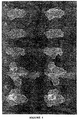
- Figure 1 is a schematic of a cavity in DNA and the numbered sites which accommodate steroid/thyroid/vitamin A and D ligands: testosterone (1,7); progesterone (2,7); aldosterone (2,5,7 and 9); cortisol (2,3,4,5, and 7); estradiol (1 and 6); triiodothyronine (T 3 ) (1 and 6); retinoic acid (6); and 1,25- (OH) 2 vitamin D 3 (1 and 6); these are divided into two groups based on their interaction with either site 6 or 7.
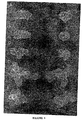
- Figure 2 is a computer generated space filling stereo view of the DNA cavity (A), which fits active estrogens oriented by energy calculations into the DNA cavity (B), whereas (C) shows the combined active surface of estrogens removed from the cavity in DNA that is used to construct the pharmacophore.
- Figure 3 demonstrates a volume contour map (yellow) in stereo with dummy atoms (magenta) surrounding the active molecules which were used in the construction of the pharmacophore (A); the empty pharmacophore (B); fit of the highly active estrogen 3,11 ⁇ ,17 ⁇ -trihydroxy-7 ⁇ -methylestra-1,3,5(10)-triene 11-nitrate ester (hereinafter 7 ⁇ -methylestradiol-11 ⁇ -nitrate ester reported in Peters et al., J. Med . Chem . 32:2306-2310 (1989)) which is accommodated completely within the pharmacophore (C); and poor fit of the inactive estrogen 9 ⁇ -estradiol which extends appreciably beyond the surface of the pharmacophore (D).
- A pharmacophore
- B empty pharmacophore
- fit of the highly active estrogen 3,11 ⁇ ,17 ⁇ -trihydroxy-7 ⁇ -methylestra-1,3,5(10)-triene 11-nitrate ester hereinafter 7
- Figure 4 shows a stereo view of volume maps (green mesh) and dummy atoms (magenta) for pharmacophores for estrogen (A), androgen (B), thyroid (C), anti-estrogen (D), and toxicity (E). Dummy atoms are not presented with the toxicity pharmacophore volume map.
- Figure 5 left column, shows the volume maps (green mesh) and dummy atoms (magenta) for pharmacophores for estrogen (A). androgen (B), thyroid (C), anti-estrogen (D), and toxicity (E).
- the right column presents an orthogonal view (90 degree rotation) of the corresponding images in the left column.
- Figure 6 demonstrates the relationship between the degree of fit of various compounds to the estrogen pharmacophore and the relative uterotropic (estrogenic) activity.
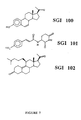
- Figure 7 shows the chemical structures of three separate molecules, SGI 100, SGI 101, and SGI 102 designed with pharmacophore technology.
- Appendix 1 is a QIC computer tape containing data files showing volume maps for each of the pharmacophores (estrogen, androgen, thyroid, antiestrogen, and toxicity) presented in Figures 4 and 5; the molecular database containing the dummy atoms for each pharmacophore designated by name; and a file containing the four color prints ( Figures 2-5) submitted with this application.
- nucleic acids including amino acids, phytohormones, cyclic nucleotides, prostaglandins, insect hormones, steroid hormones, neurotransmitters, sugars, peptide hormones, thyroid hormones, pheromones, and vitamins.
- a striking example is the cyclopentanophenanthrene motif repeated in all classes of mammalian steroid hormones, such as estrogen and progesterone.
- Another example is the kaurene nucleus containing eight chiral centers which is evident in the gibberellin class of plant hormones.
- Silastic polymer models can be constructed based upon computer derived space filling x-ray coordinates to reflect the stereochemistry of partially unwound DNA/RNA complexes.
- the apurinic/apyrimidinic sites accommodate amino acids according to the known genetic code.
- the plant hormone gibberellic acid fits best into the partially unwound site 5'-dTdA-3', 5'-dTdA-3'; members of the mammalian steroid/thyroid hormone superfamily fit best into 5'dTdG-3', 5'-dCdA-3'.
- Each class of mammalian hormone forms unique stereospecific donor-acceptor hydrogen bonds with DNA.
- the capacity to fit within these cavities in the manner of the index biologically active molecule correlates with the degree of biologic activity. It is not possible to fit chiral naturally occurring molecules into ent-DNA. However, ent-DNA accommodates the biologic unknown chiral enantiomers, such as ent-progesterone.
- computer modeling is used to examine the relationships between compounds and their fit in helical DNA. Although described herein with reference to double stranded, helical DNA. many of the same principles are applicable to double stranded RNA, and/or to RNA-DNA hybrids. Unless otherwise specified, double stranded RNA and DNA are to be considered equivalents as used herein.
- Computer modeling can be used to view the interactions of molecules as well as to measure the energy of a given interaction. While a variety of software packages are available for computer modeling of molecules, a preferred software package is Sybyl software (version 6.03; Tripos Associates, St. Louis. MO) for measuring the docking of various small molecular weight ligands into DNA.
- the software is run on a Silicon Graphics Indigo Extreme equipped with hardware stereo, i.e., Crystal Eyes (StereoGraphics, San Rafael. CA). Structures of small molecules are obtained via: the Cambridge Crystallographic Database, Lanfield Rd., Cambridge, England; construction with the Concord program or from fragment libraries and/or modifications of existing x-ray structures followed by energy minimization. All energy calculations are made using the Sybyl force field with a 1.2 ⁇ van der Waals parameter for hydrogen, as described by Hendry, et al., J . Steroid Biochem. Molec . Biol . 42:659-670 (1992) and Hendry, et al., J . Steroid Biochem. Molec.
- Each ligand is inserted into the cavity in DNA using van der Waals dot, mesh, and space filling surfaces in stereo to guide the docking procedure and minimize steric strain.
- the oxygens of the phosphate groups are permitted to act either as proton donors or acceptors and oriented to maximize the fit of any given ligand.
- the docking of the molecules is repeated several times.
- the distances between heteroatoms are monitored interactively to optimize the direction and distances of potential hydrogen bonds. While evaluating fit of compounds in a given hormone class, attempts are made to insert all candidate ligands into the DNA sequence with donor/acceptor linkages common to the hormone. Donor/acceptor relationships are further maximized by appropriate orientation of functional groups on the ligands, e.g., by adjusting the conformation of each structure to best mimic the fit of the hormone.
- Van der Waals interactions of the candidate ligands are optimized with the DNA surface.
- the force field is used to calculate the relative fit of each ligand by assessing the optimum favorable energy change resulting from docking the ligand.
- Each ligand is docked into the DNA and the steric fit is calculated from the change in van der Waals energy; the hydrogen bonding fit is calculated from the change in electrostatic energy using charges on donor hydrogens and acceptor heteroatoms.
- the energy changes are assessed for each ligand. The greater the negative energy change upon insertion of a given ligand into DNA, the more favorable the fit and the more stable the complex. Optimal docking is determined when no further increase in fit is observed. The most favorable change in energy is selected to compare the relative fit of each molecule.
- the overall fit of each ligand is assessed by adding the change in kcal of the van der Waals and electrostatic energies and normalizing the fit to that of the parent hormone.
- binding of the steroid to its receptor serves as a means to recognize the general class of hormone (e.g., estrogen versus androgen) whereas the type and degree of the fit of the steroid in the DNA is largely responsible for governing the magnitude of the biologic response.
- the steroid alone is incapable of proper insertion into DNA either in vivo or in vitro without the receptor and thus would be unable to generate a full hormonal response without the receptor.
- a potentially critical role for the receptor upon binding to DNA is to regulate the physicochemical properties of the site in DNA to permit insertion of the steroid, including the degree of unwinding, the capacity of heteroatoms to act as either donors or acceptors, and the pattern and degree of solvation. This is further supported by the decrease in the surface hydrophobicity of the receptor upon binding estrogens and antiestrogens as well as phosphorylation which enhances binding of the estrogen receptor to specific DNA sequences, reported by Denton, et al.. J. Biol . Chem . 267:7263-7268 (1992).
- the process of a receptor-mediated insertion of steroids into DNA presumably involves several steps.
- the initial contact of the DNA by the steroid-receptor complex could involve a partially exposed D ring of the steroid with the A ring still attached to the receptor.
- Analysis of receptor binding data supports this possibility.
- Stereospecific recognition of the DNA by partial insertion and hydrogen bonding of the 17 ⁇ -hydroxyl of the steroid D ring with the 5'-dTdG-3' strand could be followed by complete insertion and the recognition within the double helix manifest by the linkage of both stereospecific hydrogen bonds.
- the weak binding observed for the steroids with DNA suggests that the steroid/DNA complex might be short lived and rapidly reversible. Certain estrogen antagonists which are accommodated differently in DNA may form longer lived interactions within the site.
- RNA and RNA-DNA hybrids can accommodate various ligands, for example, estradiol in 5'-rUrG-3', 5'-dCdA-3'.
- the ligands were inserted into the cavity in DNA using van der Waals dot surfaces and the stereoviewer to guide the docking procedure and minimize any obvious steric strain.
- the distances between heteroatoms were monitored interactively to optimize the direction and distances of potential hydrogen bonds. Donor/acceptor relationships were further maximized by appropriate orientation of functional groups on the ligands, e.g., by adjusting the conformation of each structure. Attempts were made to optimize van der Waals interactions of the candidate ligands with the DNA surfaces.
- the force field was used to assess the relative fit of each ligand by quantitating the optimum favorable energy change resulting from docking the ligand.
- Steric fit was calculated from the change in van der Waals energy
- the hydrogen bonding fit was calculated from the change in electrostatic energy using charges on donor hydrogens and acceptor oxygens.
- the greater the negative energy change upon insertion of a given ligand into DNA the more favorable the fit and the more stable the complex. Docking was completed when no further increase in fit was observed.
- the most favorable change in energy was selected to compare the relative fit of each molecule.
- the overall fit of each ligand was assessed by adding the change in kcal of the van der Waals and electrostatic energies and normalizing the value to that of the best fitting molecule (100%). It should be noted that while the energies reported here were derived from widely used force field calculations. they were not empirically derived. Thus, the absolute values in kcal do not have independent experimental significance. At the same time, they are valuable indicators of the relative degree of fit into DNA of candidate molecules.
- Increased fit of the ligand was obtained by substituting a para hydroxl group on the phenyl ring; this substitution enabled a second hydrogen bond to be formed between the hydroxyl group and a phosphate oxygen on the adjacent DNA strand.
- the increase in fit measured by energy calculations due to the second hydrogen bond (2.64 ⁇ ) was reflected in an additional -24.6 kcal in electrostatic energy.
- Other substitutions which were made on the 3-phenylacetylamino-2, 6-piperidinedione skeleton did not significantly increase fit demonstrated by the normalized energy calculations for certain halogenated analogs.
- the active esters were reacted with L-glutamine in the presence of sodium bicarbonate to obtain the glutamine derivatives.
- crude products were directly used for the next reaction.
- the glutamine derivatives were again reacted with N-hydroxysuccinimide in the presence of DCC to give the active esters which without purification were heated at 95-100°C to obtain the desired 2,6-piperidinediones in various yields. During the heating process the compounds were racemized.
- Prolactin stimulated growth of rat Nb2 lymphoma cells was inhibited by each of the compounds with p-hydroxy-3-phenylacetylamino-2,6-piperidinedione manifesting the greatest activity.
- Compound p-hydroxy-3-phenylacetylamino-2,6-piperidinedione was more active in Nb2 lymphoma cells than 3-phenylacetylamino-2.6 piperidinedione over the range tested (10 -4 M to 10 -3 M).
- 3-phenylacetylamino-2,6-piperidinedione is capable of inserting between base pairs in partially unwound double stranded DNA and forming an energetically favorable complex.
- a hydroxyl group placed in the para position of the phenyl ring of 3-phenylacetylamino-2,6-piperidinedione enabled formation of a second hydrogen bond thereby linking both DNA strands.
- Molecular modeling facilitates the establishment of the best fit of molecules into nucleic acids such as double-stranded DNA based on steric and electrostatic considerations.
- Individual molecules, such as estradiol fit optimally into specific sites on DNA based on the location of specific nucleotides and the bonding characteristics of individual heteroatoms (see Example 2).
- Molecules that are related to a specific molecule such as estradiol but display chemical differences will fit into the estradiol site with different degrees of precision: some may fit better and give rise to estradiol agonistic responses while those with poor fit display weak estrogenic activity.
- These different molecules may be aligned relative to the docking of heteroatoms with heteroatoms on the DNA to optimize electrostatic interactions.
- molecules with activity equal to or greater than that of the hormone are chosen for alignment. To date, such molecules fit equally well or better than the hormone into DNA using the energy calculation methodology described above. Molecules which do not fit as well into DNA as the parent hormone are excluded from inclusion in the construction of the pharmacophore.
- the alignment of the combined surfaces of the molecules occupies a specific volume of space thereby forming a three dimensional shape.
- Pharmacophores are three dimensional arrangements of chemical groups related to a given biological activity which enables meaningful comparison of molecules exhibiting the same biological function (Naruto et al., Eur . J. Med. Chem . 20:529-532 (1985)). Pharmacophores can be derived by simple overlap of active structures or common functional groups in the molecules. Without a way to orient the molecules e.g., based upon fit with another macromolecule - a receptor, enzyme, or in this case DNA, it is difficult and, in some cases, impossible to construct a reliable pharmacophore. This problem results in part from the fact that even closely related active molecules frequently fit into macromolecules in very different ways.
- a pharmacophore as used herein, is defined as a 3-dimensional shape having a specific volume derived from the combined van der Waals surface of active molecules oriented by fit into DNA, coupled with point charges located adjacent to the surface.
- a pharmacophore represents an aggregate array of positions in space of a series of molecules having the same or similar biological activity.
- the van der Waals surface can be represented in various ways including as a volume map, a dot surface, or a Connolly surface.
- the point charges are represented as dummy atoms whose positions are determined by the average positions of functional groups on active molecules which can form hydrogen bonds. Suitable charges are placed on the dummy atoms consistent with the capacity of the active molecules to form hydrogen bonds.
- the pharmacophores are specific for different compounds, their related molecules and a particular biological activity. According to the present invention, within the general class of molecules called hormones, an estrogen pharmacophore, an anti-estrogen pharmacophore, an androgen pharmacophore, a thyroid hormone pharmacophore, and a toxicity pharmacophore (shown in Figures 2-5 and Appendix 1) have been disclosed. It should be emphasized that these created pharmacophores do not exist as such in nature and are the product of aligning several related molecules to common binding sites in DNA using methods as described herein.
- a pharmacophore once created, stands alone and is subsequently independent from the nucleic acid that was involved in its formation. Thus, after formation of the pharmacophore, one no longer needs to use the DNA as a template for the design of biologically active molecules.
- the pharmacophore itself can be used to generate new molecules that will possess the same or similar structural and charge features that are represented by the pharmacophore. This is a completely different concept from the one of using the DNA as the model for the design of compounds.
- the pharmacophore can be used itself for any number of applications, including but not limited to the following: as a screening tool for drug development; to determine if a particular compound will possess bioactivity of a certain type, for instance estrogenic or androgenic activity; for toxicological evaluation; and to design compounds that possess increased or decreased binding affinity for DNA.
- Each pharmacophore has a characteristic shape, topology, volume, and electrostatic profile.
- a pharmacophore is accurately described by its three dimensional shape which is represented by a coordinate system that is configured in computer memory (see Figures 2-5 for examples of pharmacophores which are also contained in data files in Appendix 1, the magnetic QIC tape submitted with the application).
- Each specific atom within a molecule that fits in a pharmacophore has a specific location relative to the docking heteroatoms.
- the individual atoms also have electrical charges assigned to them. These charges are represented numerically and through many other ways including the use of colors and shading to indicate field strength.
- the term "energy of interaction” as used herein is the total energy in -kcal of a molecule as it is being fitted into a pharmacophore. This has been observed in the case of molecules that fit within the estrogen pharmacophore and bioactivity in a uterotropic assay.
- the volume of a pharmacophore is described in cubic angstroms.
- the pharmacophore can be cross sectioned precisely in any plane and internal distances measured with an angstrom ruler. The circumference of any cross section is easily measured with morphometric analysis.
- specific subregions of the pharmacophore such as the site that binds to the DNA, can be subjected to the same methods of analysis.
- Figure 2A is a computer generated space filling stereo view of the DNA cavity which fits estrogens.
- Figure 2B The fit of active estrogens oriented by energy calculations into the DNA cavity, using the methods described above, is presented in Figure 2B.
- Figure 2C shows the combined active surface of estrogens removed from the cavity in DNA that is used to construct the pharmacophore.
- the atoms are colored in the following manner: carbon/white; hydrogen/cyan; nitrogen/blue; oxygen/red; phosphorus/yellow.
- Figure 3 demonstrates a volume contour map (yellow) in stereo with dummy atoms (magenta) surrounding the active molecules which were used in the construction of the pharmacophore (A); the empty pharmacophore (B); fit of the highly active estrogen 3,11 ⁇ ,17 ⁇ -Trihydroxy-7 ⁇ -methylestra-1,3,5(10)-triene 11-nitrate ester (hereinafter 7 ⁇ -methylestradiol-11 ⁇ -nitrate ester reported in Peters et al., J . Med. Chem. 32:2306-2310 (1989)) which is accommodated completely within the pharmacophore (C); poor fit of the inactive estrogen 9 ⁇ -estradiol which extends appreciably beyond the surface of the pharmacophore (D).
- A pharmacophore
- B empty pharmacophore
- fit of the highly active estrogen 3,11 ⁇ ,17 ⁇ -Trihydroxy-7 ⁇ -methylestra-1,3,5(10)-triene 11-nitrate ester hereinafter 7 ⁇ -
- Figures 4 and 5 present examples of the three dimensional appearance of estrogen, androgen, thyroid, anti-estrogen, and toxicity pharmacophores.
- the data files for the volume maps and dummy atoms for each of these pharmacophores and for Figures 2 - 5 are contained in Appendix 1, the magnetic QIC 120 tape submitted with the application.
- This QIC computer tape has the following format and files generated from Sybyl 6.0 run on a Silicon Graphics Indigo Extreme.
- 7 ⁇ -methylestradiol-11 ⁇ -nitrate ester (labeled 2), which is not part of the data set used to construct the pharmacophore, fits appreciably better than estradiol (labeled 1).
- 7 ⁇ -methylestradiol-11 ⁇ -nitrate ester binds poorly to the estrogen receptor (less than 6% of the binding of estradiol).
- the uterotropic values for 7 ⁇ -methylestradiol-11 ⁇ -nitrate ester (labeled 2) relative to estradiol set to a normalized value of 100% are considerably greater than that of estradiol (Peters et al., J. Med. Chem .
- the fit of compounds into DNA is consistent with, but not the same as, fit into the pharmacophores.
- the distinction between the DNA cavities and the pharmacophores is that the surfaces were derived from different structures, i.e. the DNA cavities from the DNA structure and the pharmacophores from the combined surfaces of active compounds.
- the degree of fit to the pharmacophores is obtained by fit to the three dimensional map which represents the active compounds.
- the pharmacophores enable quantitative determination of the degree of fit to the combined surfaces of the active compounds and this information cannot be obtained from fit into DNA.
- the fit of active compounds to the pharmacophore can be quantitated based upon the portion of the molecule which does not fit within the pharmacophore volume. This enables automatic assessment of predicted inactive structures.
- Pharmacophores can be constructed to represent a three dimensional shape that is predictive of toxic biological activity. Such pharmacophores, called toxicophores, have regions that would potentially damage DNA.
- a typical toxicophore has been constructed using, tetrodotoxin, dioxin, RU 486, dilantin, thalidomide and oroflex, among other compounds.
- An example of this toxicophore is provided in Figures 4E and 5E and on the submitted magnetic data tape as Appendix 1.
- Pharmacophores in their relationship to nucleic acids are usually surrounded by a solvent.
- the predominant solvent in living organisms is water and accordingly, most pharmacophores exist in an aqueous environment. Water is the preferred embodiment of the solvent pharmacophore and is termed an aquaphore.
- Pharmacophores, and their molecules may also be placed in non-aqueous environments for various purposes such as crystallographic studies or other analytical procedures.
- the aqueous environment surrounding the pharmacophore also has an intimate association with the adjacent nucleic acid.
- This aqueous shell assists in the optimal fit of the pharmacophore into the cavity of the double stranded DNA, and has its own three dimensional shape.
- the optimal steric and electrostatic placement of water molecules in the space between the pharmacophore and the DNA is achieved in the present invention.
- This three dimensional shape is called a solvent pharmacophore, and can be described in all the ways listed above for the pharmacophores based on other molecules such as estrogen.
- Solvent pharmacophores assist the designer of compounds by placing limits on the dimensions of a compound designed using a particular pharmacophore as a template.
- solvent pharmacophores assist the creator of pharmacophores because the solvent shell or cage represented by the pharmacophore provides enhanced ability to properly align molecules relative to DNA during the creation of the pharmacophore.
- receptors Many molecules, such as steroid hormones, are shuttled to the nucleus by other molecules known as receptors (Tsai and O'Malley, Ann. Rev . Biochem . 63:451-486 (1994)). These receptors bind the hormones (called ligands), bind to the nucleic acids, for example in their DNA binding domain, and present ligands to nucleic acids such as DNA. Evidence suggests that the binding of the receptor to the DNA causes a conformational change in the DNA to facilitate insertion of the ligand (Nardulli et al., Molec. Endocr. 7:331-340 (1993)).
- the pharmacophore concept is based on the three dimensional shape of the optimal fit of related molecules into nucleic acids such as partially unwound, double-stranded DNA.
- the DNA binding domain of the receptor can be modeled into a three dimensional shape based on the same principles described above for the pharmacophore.
- the resultant shape is termed a receptophore and is the three dimensional representation of the sites of interaction of the receptor and the nucleotides of the DNA.
- the DNA binding region of each receptor likely gives rise to a different receptophore. This receptophore provides a valuable tool to molecular designers interested in developing new receptors, or in modulating receptor binding to DNA.
- the nucleic acid binding region of receptors and their ligands can be modeled as receptophores and pharmacophores, respectively.
- the configuration of the receptophore and its associated pharmacophore in their proper alignment relative to their respective DNA binding regions constitutes a specific pair of shapes that represents the minimal molecular unit for DNA binding and ligand insertion.
- Designers of compounds utilize this information to synthesize and screen molecules to modify the facility of docking and ligand insertion. Such modifications may provide a host of new therapies such as treatments for hormone dependent carcinomas of the prostate or breast.
- sites can be modeled relative to the pharmacophore to produce a three dimensional representation of a preferred site for modification of the molecule.
- This three dimensional representation termed a metabophore, provides constraints for rational design of active and inactive variants of the parent molecules that fit into the pharmacophore. Analysis of which chemicals can effectively be added at the attachment point of the metabophore to the pharmacophore reveals the most favorable molecules to pursue for synthesis, purification and testing.
- the mammalian female hormone estradiol also fits in DNA (Figure 1). Two stereospecific hydrogen bonds of approximately 2.65 ⁇ are formed between each hydroxyl group of the steroid and phosphate oxygens on adjacent strands. The overall fit within the complex is about -59 kcal. Most alterations of the positions of the hydroxyl groups on the estratriene nucleus result in a substantial loss of potential electrostatic interactions with the DNA. Moreover, most alterations of the absolute stereochemistry of the cyclopentanophenanthrene ring pattern also result in a substantial loss of potential electrostatic interactions with the DNA. Moreover, most alterations of the absolute stereochemistry of the cyclopentanophenanthrene ring pattern also result in a poor fitting molecule.
- estradiol is a "lock and key” fit into DNA, although most structural alternatives to estradiol fit poorly provides further support for the premise that DNA stereochemistry contains the master blueprint for natural product structures.
- Ligands that fit into more than one site in DNA have been observed to have multiple biologic actions. Both desirable and undesirable "side effects" should thus be predictable from the specific DNA sequence which a given compound fits into as well as the manner and relative degree of fit. Examples of molecules that have been observed to fit into more than one site in DNA include the psychotropics cocaine, morphine, LSD and tetrahydrocannabinoids, and certain intercalating antibiotics.
- the monoamine oxidase inhibitor selegiline fits into the site in DNA which accommodates glucose and various oral antidiabetic drugs as reported by Rowland et al., J. Clin. Pharmacol . 34:80-85(1994). This observation is consistent with the finding that selegiline causes hypoglycemia in some patients.
- ligands which cause stress, chemical modifications and/or covalent linkages to the DNA when fit into a given site frequently possess toxicity examples include certain carcinogens and teratogens, e.g., thalidomide, dioxin, arene oxides, aflatoxins and some diethylstilbestrol metabolites.
- Another example is the anti-progestin RU486 which stresses base pair hydrogen bonds when inserted fully into the progesterone site in DNA. Similar strain is produced by other anti-progestins having the same side chains (e.g., 11 ⁇ -phenylamines) raising the possibility that such features may correlate with abortifacient activity attributed to RU486 and related analogs.
- Observations with thalidomide enantiomers indicate that teratogenicity associated with this compound may correlate with a stereospecific effect on base pairing.
- An example of such a toxicity pharmacophore, called a toxicophore, is presented in Figures 4E and 5E and the corresponding data file is submitted on magnetic tape.
- 3-Phenylacetylamino-2,6-piperidinedione (A10) is a modified amino-acid derivative, which was originally isolated from freeze-dried human urine. Despite having low toxicity, high concentrations of A10 were required to demonstrate significant growth inhibitory activity on tumor cells. The focus of the following study was to develop more potent analogs. Modeling studies demonstrated that A10 was capable of inserting into partially unwound double stranded DNA and forming a single hydrogen bond between the imino proton of the piperidinedione ring and a phosphate oxygen on a single strand.
- the drug design technology described here can be used in conjunction with quantitative-structure-activity-relationship methods (QSAR), e.g., comparative field molecular analysis (CoMFA).
- QSAR quantitative-structure-activity-relationship methods
- CoMFA comparative field molecular analysis
- One value of the approach is that it facilitates the orientation of various ligands relative to one another in three dimensions.
- the successful structure-activity relationship found for estrogens derived from fit into DNA is described here. If one were to attempt to derive such a relationship a priori without first knowing the detailed three-dimensional structure of an appropriate macromolecule (e.g., the ligand binding site of a receptor or an enzymatic site), chemical intuition would necessitate searching for common features that exist in known active structures.
- Phenylacetate has been shown to reduce levels of the myc oncogene which is involved in the development of several cancers including breast, brain, prostate, blood, lung and colon. Another mechanism by which phenylacetate is thought to be effective is by reducing levels of the amino acid glutamine. Phenylacetate conjugates with circulating glutamine to produce the excreted urinary metabolite phenylacetylglutamine (PAG). Cancer cells require glutamine for growth and are known to be more sensitive to glutamine depletion that normal cells. These findings have led to the initiation of Phase I clinical trials with phenylacetate in brain and prostate cancer at the National Cancer Institute.
- the molecule predicated to be the most active based upon degree of fit in DNA i.e., the p-hydroxy derivative
- MCF-7 E3 human breast cancer cells
- the p-hydroxy derivative possessed antiestrogenic activity in the range of the drug tamoxifen which is currently in clinical use for the treatment of breast cancer.
- SGI 100 Three separate molecules designed with modeling technology are shown in Figure 7. These molecules, termed SGI 100, SGI 101 and SGI 102 bear similarities and differences to each other. They all show structural similarities to components of both estrogen and progesterone. SGI 100 was designed on the basis of its ability to fit into the site in DNA which accommodates both estradiol and progesterone. The manner in which it fits predicted antagonist activity. When fit to the estrogen pharmacophore ( Figure 2 and Appendix 1), the acetyl group at the 17 ⁇ position extended out of the pharmacophore and had electrostatic repulsion with dummy atoms (-131 kcal) compared to estrogen (-51 kcal). Accordingly, this high positive energy of interaction indicates that SGI 100 acts as an antagonist.
- SGI 100 The binding of SGI 100 to the estrogen receptor is dose dependent and approximately 144 times less than estradiol.
- SGI 100 significantly decreased cell growth in MCF-7 human breast cancer cells (134,431 cells) at a dose of 10 -8 M when compared to control cells (252,197 cells).
- the same concentration of tamoxifen citrate decreased the number of MCF-7 cells to 187,759.
- the design of this compound based on the pharmacophore approach of this invention predicted a demonstrable anti-estrogen bioactivity that was greater than tamoxifen.
- SGI 100 binds in a dose dependent manner to the progesterone receptor but with 133 to 200 times less affinity.
- SGI 101 was designed on the basis of fitting into DNA at the site which accommodates estradiol but with opposite hydrogen bonding properties which predict estrogen antagonist activity. SGI 101 extends beyond the estrogen pharmacophore and has electrostatic repulsion between the para-nitro group and dummy atoms. SGI 101 is the most potent analog designed by the technology as measured by growth inhibition of MCF-7 cells. At a dose of 10 -8 M, SGI 101 inhibited cell growth (81,103 cells) relative to control (252,197 cells) and was substantially more active than the same concentration of tamoxifen (187,759 cells).
- SGI 102 was designed on the basis of its fit into DNA at the site which accommodates progesterone.
- SGI 102 possesses an alkyl amino side chain at the 11 ⁇ position which extends out of the site between base pairs into the major groove. It has different hydrogen bonding properties than progesterone and would extend beyond the volume map of the progesterone pharmacophore. As such, it is predicted to be an antagonist.
- SGI 102 was designed prospectively, synthesized, and tested in various biological assays. SGI 102 binds in a dose dependent manner to the progesterone receptor but not as strongly as progesterone or the abortifacient antiprogestin RU486. In animal experiments, SGI 102 showed no abortifacient activity. However, in experiments using MCF-7 human breast cancer cells, SGI 102 had equivalent activity to RU486 in inhibiting growth. These findings are consistent with the predictions made by the modeling technology.
Abstract
Description
This is the volume map for the estrogen pharmacophore.
This is the volume map for the androgen pharmacophore.
This is the volume map for the thyroid pharmacophore.
This is the volume map for the antiestrogen pharmacophore.
This is the volume map for the toxicity pharmacophore.
This is the molecular database file containing the appropriate dummy atoms for each pharmacophore designated by name.
This is a file containing the four color prints (Figures 2 through 5) submitted with the patent application describing the pharmacophores constructed with the Silicon Graphics program Showcase. Copies of the
Claims (16)
- A computer-based method for creating a pharmacophore comprising the computer implemented steps of:determining the optimal fit of a plurality of compounds having the same or similar biological activity into nucleic acid sequences such that the lowest energy of interaction and best steric fit are obtained;aligning the compounds relative to the heteroatoms on the nucleic acids;defining a three dimensional shape as a pharmacophore representing the aggregate average shape resulting from said alignment of the compounds ; andrepresenting the pharmacophore by a coordinate system that is configured in computer memory.
- The method of claim 1, wherein the nucleic acid sequence is selected from the group consisting of deoxyribonucleic acid, double stranded deoxyribonucleic acid, ribonucleic acid, complexes of deoxyribonucleic acid and ribonucleic acid, and apurinic and apyrimidinic sites.
- The method of any of the preceding claims, wherein the biological activity is selected from the group consisting of estrogenic activity, antiestrogenic activity, androgenic activity, thyroid hormone activity, and toxic biological activity and the pharmacophore is selected from the group consisting of an estrogen pharmacophore, an antiestrogen pharmacophore, an androgen pharmacophore, a thyroid hormone pharmacophore, and a toxicity pharmacophore respectively.
- The method of claim 3, further comprising overlaying of a toxicity pharmacophore on another pharmacophore.
- A computer-based method for screening a molecule for a desired biological activity wherein the molecule is examined for the ability to fit into the respective pharmacophore created using the method according to any of the preceding claims comprising the computer implemented steps of:measuring the energy of interaction when the molecule is fitted into the pharmacophore;comparing the energy of interaction of the molecule to a predetermined energy of interaction that correlates to the biological activity.
- The method of claim 5, further comprising:quantitating the fit of the molecule into the pharmacophore by measuring an amount of volume of the molecule which is placed within the pharmacophore; andnormalizing the amount to an amount of volume of a hormone placed within the pharmacophore.
- The method of claim 5, further comprising:quantitating values of electrostatic interactions of the molecule with dummy atoms in the pharmacophore;optimizing the values of the electrostatic interactions of the molecule with dummy atoms in the pharmacophore; andnormalizing the values of the electrostatic interactions of the molecule to the values for the electrostatic interactions of a hormone for the dummy atoms in the pharmacophore.
- The method of claim 7, further comprising:adding the normalized value for the electrostatic interactions of the molecule in the pharmacophore to the normalized value for the volume of the molecule in the pharmacophore.
- A computer-based method for designing a molecule with a desired biological activity comprising the computer implemented steps of:determining a structure of a molecule which will have a minimal predetermined energy of interaction when fitted into the pharmacophore created using the method according to any of claims 1-4 with the desired biological activity.
- A computer-based method for creating a receptophore-pharmacophore pair comprising the computer implemented steps of:modeling the nucleic acid binding region of a receptor as receptophore comprising the computer implemented steps of:determining the optimal fit of the nucleic acid binding domain of a receptor onto nucleic acid sequences such that the lowest energy of interaction and best steric fit are obtained;defining a three dimensional shape as a receptophore representing the shape resulting from said optimal fit; andrepresenting the receptophore by a coordinate system that is configured in computer memory;modeling a pharmacophore of a molecule or a plurality of molecules with the same or similar biological activity according to the method of any of claims 1-4 respectively;configuring the receptophore and the pharmacophore in their proper alignment relative to their respective nucleic acid binding region;defining a receptophore-pharmacophore as a pair of shapes; andrepresenting the receptophore-pharmacophore pair of shapes by a coordinate system that is configured in computer memory.
- The method of claim 10, wherein the nucleic acid sequence is selected from the group consisting of deoxyribonucleic acid, double stranded deoxyribonucleic acid, ribonucleic acid, complexes of deoxyribonucleic acid and ribonucleic acid, and apurinic and apyrimidinic sites.
- Use of the receptophore-pharmacophore pair obtainable by a method according to any of claims 10 and 11 for synthesizing and screening molecules to modify the facility of docking and ligand insertion.
- A computer-based method for creating a metabophore comprising the computer implemented steps of:modeling a preferred site for modification of a molecule with a biological activity relative to the respective pharmacophore obtainable by a method according to any of claims 1-4 to produce a three dimensional representation of a preferred site for modification of the molecule,representing said shape by a coordinate system configured in a computer memory.
- A computer-based method for creating a solvent pharmacophore comprising the computer implemented steps of:determining the optimal steric and electrostatic placement of solvent molecules in the space between the pharmacophore obtainable by the method of any of the claims 1-4 and the nucleic acid sequence;defining a three dimensional shape as a solvent pharmacophore representing the aggregate average shape resulting from said placement ; andrepresenting the solvent pharmacophore by a coordinate system thatis configured in computer memory.
- The method of claim 14, wherein the solvent molecules are water molecules.
- The method of any of claims 14 and 15, wherein the nucleic acid sequence is DNA.
Applications Claiming Priority (3)
| Application Number | Priority Date | Filing Date | Title |
|---|---|---|---|
| US15868993A | 1993-11-26 | 1993-11-26 | |
| US158689 | 1993-11-26 | ||
| PCT/US1994/013765 WO1995014791A1 (en) | 1993-11-26 | 1994-11-28 | Design of drugs involving receptor-ligand-dna interactions |
Publications (3)
| Publication Number | Publication Date |
|---|---|
| EP0740708A1 EP0740708A1 (en) | 1996-11-06 |
| EP0740708A4 EP0740708A4 (en) | 1999-12-01 |
| EP0740708B1 true EP0740708B1 (en) | 2004-08-04 |
Family
ID=22569263
Family Applications (1)
| Application Number | Title | Priority Date | Filing Date |
|---|---|---|---|
| EP95904188A Expired - Lifetime EP0740708B1 (en) | 1993-11-26 | 1994-11-28 | Design of drugs involving receptor-ligand-dna interactions |
Country Status (8)
| Country | Link |
|---|---|
| US (5) | US5705335A (en) |
| EP (1) | EP0740708B1 (en) |
| JP (1) | JPH09505603A (en) |
| AT (1) | ATE272719T1 (en) |
| AU (1) | AU1297995A (en) |
| CA (1) | CA2177388A1 (en) |
| DE (1) | DE69433935D1 (en) |
| WO (1) | WO1995014791A1 (en) |
Families Citing this family (50)
| Publication number | Priority date | Publication date | Assignee | Title |
|---|---|---|---|---|
| US6040137A (en) * | 1995-04-27 | 2000-03-21 | Tripep Ab | Antigen/antibody specification exchanger |
| US6933366B2 (en) * | 1996-12-27 | 2005-08-23 | Tripep Ab | Specificity exchangers that redirect antibodies to bacterial adhesion receptors |
| US6660842B1 (en) * | 1994-04-28 | 2003-12-09 | Tripep Ab | Ligand/receptor specificity exchangers that redirect antibodies to receptors on a pathogen |
| US5978740A (en) * | 1995-08-09 | 1999-11-02 | Vertex Pharmaceuticals Incorporated | Molecules comprising a calcineurin-like binding pocket and encoded data storage medium capable of graphically displaying them |
| ATE364629T1 (en) | 1996-01-22 | 2007-07-15 | Curis Inc | METHODS FOR PRODUCING OP-1 MORPHOGEN ANALOGUES |
| US7541151B2 (en) | 1997-06-05 | 2009-06-02 | Duke University | Single-cell biosensor for the measurement of GPCR ligands in a test sample |
| US5891646A (en) * | 1997-06-05 | 1999-04-06 | Duke University | Methods of assaying receptor activity and constructs useful in such methods |
| US6528271B1 (en) * | 1997-06-05 | 2003-03-04 | Duke University | Inhibition of βarrestin mediated effects prolongs and potentiates opioid receptor-mediated analgesia |
| AU9378098A (en) | 1997-09-05 | 1999-03-22 | Molecular Simulations, Inc. | Modeling interactions with atomic parameters including anisotropic dipole polarizability |
| WO1999028347A1 (en) * | 1997-11-27 | 1999-06-10 | Commonwealth Scientific And Industrial Research Organisation | Method of designing agonists and antagonists to igf receptor |
| WO1999058722A1 (en) * | 1998-05-12 | 1999-11-18 | Isis Pharmaceuticals, Inc. | Characterization of interactions between molecular interaction sites of rna and ligands therefor |
| AUPP380498A0 (en) * | 1998-05-29 | 1998-06-25 | Biomolecular Research Institute Limited | Egf receptor agonists and antagonists |
| US20020035459A1 (en) | 1998-09-14 | 2002-03-21 | George M. Grass | Pharmacokinetic-based drug design tool and method |
| KR100303263B1 (en) * | 1998-10-13 | 2001-12-01 | 조양호 | Visualization Method of RNA Molecular Secondary Structure Using Vectors |
| US6298259B1 (en) | 1998-10-16 | 2001-10-02 | Univ Minnesota | Combined magnetic resonance imaging and magnetic stereotaxis surgical apparatus and processes |
| US7083777B1 (en) * | 1999-04-02 | 2006-08-01 | The Brigham And Women's Hospital, Inc. | Immunomodulating polymers |
| US6343257B1 (en) * | 1999-04-23 | 2002-01-29 | Peptor Ltd. | Identifying pharmacophore containing combinations of scaffold molecules and substituents from a virtual library |
| US6969763B1 (en) | 1999-05-12 | 2005-11-29 | Isis Pharmaceuticals, Inc. | Molecular interaction sites of interleukin-2 RNA and methods of modulating the same |
| US20030083483A1 (en) * | 1999-05-12 | 2003-05-01 | Ecker David J. | Molecular interaction sites of vimentin RNA and methods of modulating the same |
| EP1272839A4 (en) * | 2000-03-23 | 2006-03-01 | California Inst Of Techn | Method and apparatus for predicting ligand binding interactions |
| WO2002041184A1 (en) * | 2000-11-14 | 2002-05-23 | Kyorin Pharmaceutical Co., Ltd. | Method of searching for novel lead compound |
| SV2003000753A (en) | 2000-12-05 | 2003-06-16 | Brigham & Womens Hospital | USE OF ZWITTERIONIC POLYSACARIDS FOR THE SPECIFIC MODULATION OF IMMUNE PROGRESS |
| IL156110A0 (en) | 2000-12-09 | 2003-12-23 | Univ California | X-RAY CRYSTAL STRUCTURES OF FUNCTIONAL RIBOSOME COMPLEXES CONTAINING TRANSFER RNA AND MODEL MESSENGER RNAs AND METHODS OF USE |
| US20030162219A1 (en) * | 2000-12-29 | 2003-08-28 | Sem Daniel S. | Methods for predicting functional and structural properties of polypeptides using sequence models |
| US20030013137A1 (en) * | 2001-03-13 | 2003-01-16 | Barak Larry S. | Automated methods of detecting receptor activity |
| WO2002087505A2 (en) * | 2001-04-27 | 2002-11-07 | The General Hospital Corporation | 3-structure of a whole integrin $g(a)v$g(b)3 extracellular region and uses therefor |
| US20040002052A1 (en) * | 2001-10-23 | 2004-01-01 | Hendry Lawrence B. | Systems and methods for rapid evaluation and design of molecules for predicted biological activity |
| US20040067512A1 (en) * | 2001-11-09 | 2004-04-08 | Neurogenetics, Inc. | Single nucleotide polymorphisms and mutations on Alpha-2-Macroglobulin |
| US20030162202A1 (en) * | 2001-11-09 | 2003-08-28 | Becker Kenneth David | Single nucleotide polymorphisms and mutations on Alpha-2-Macroglobulin |
| US20030182669A1 (en) * | 2002-03-19 | 2003-09-25 | Rockman Howard A. | Phosphoinositide 3-kinase mediated inhibition of GPCRs |
| JP2005529158A (en) * | 2002-05-28 | 2005-09-29 | ザ・トラスティーズ・オブ・ザ・ユニバーシティ・オブ・ペンシルベニア | Method, system and computer program product for computer analysis and design of amphiphilic polymers |
| US7290215B2 (en) * | 2002-06-03 | 2007-10-30 | Microsoft Corporation | Dynamic wizard interface system and method |
| KR20050056250A (en) | 2002-10-18 | 2005-06-14 | 주식회사 엘지생명과학 | Gene families associated with cancers |
| CA2504334A1 (en) | 2002-10-30 | 2004-05-21 | Galileo Pharmaceuticals, Inc. | Identifying therapeutic compounds based on their physical-chemical properties |
| US7335359B2 (en) | 2003-02-06 | 2008-02-26 | Tripep Ab | Glycosylated specificity exchangers |
| WO2004069873A2 (en) | 2003-02-06 | 2004-08-19 | Tripep Ab | Antigen/antibody or ligand/receptor glycosylated specificity exchangers |
| EP1638581A2 (en) | 2003-03-31 | 2006-03-29 | The Brigham And Women's Hospital, Inc. | Zwitterionic immunomodulators for the treatment of asthma and allergy |
| US8345988B2 (en) * | 2004-06-22 | 2013-01-01 | Sri International | Method and apparatus for recognizing 3-D objects |
| US20060052943A1 (en) * | 2004-07-28 | 2006-03-09 | Karthik Ramani | Architectures, queries, data stores, and interfaces for proteins and drug molecules |
| US8206726B2 (en) | 2006-02-06 | 2012-06-26 | The Brigham And Women's Hospital, Inc. | Zwitterionic polysaccharides for promotion of immune system maturation and health |
| KR100954322B1 (en) | 2006-06-14 | 2010-04-21 | 주식회사 엘지생명과학 | Gene familyLBFL313 associated with pancreatic cancer |
| US20090092631A1 (en) * | 2007-03-26 | 2009-04-09 | Tripep Ab | Glycosylated specificity exchangers that induce an antibody dependent cellular cytotoxicity (adcc) response |
| WO2008154456A2 (en) * | 2007-06-06 | 2008-12-18 | Nationwide Children's Hospital, Inc. | Methods and compositions relating to viral fusion proteins |
| US8383589B2 (en) * | 2007-07-09 | 2013-02-26 | The United States Of America, As Represented By The Secretary, Department Of Health And Human Services | Aegyptin and uses thereof |
| US9561274B2 (en) | 2011-06-07 | 2017-02-07 | University Of Hawaii | Treatment and prevention of cancer with HMGB1 antagonists |
| US9244074B2 (en) | 2011-06-07 | 2016-01-26 | University Of Hawaii | Biomarker of asbestos exposure and mesothelioma |
| JP6273200B2 (en) | 2011-07-12 | 2018-01-31 | ザ・ブリガーム・アンド・ウーメンズ・ホスピタル・インコーポレーテッド | Lipid-containing PSA compositions, methods of isolation and methods of use |
| CN102798708A (en) * | 2012-08-23 | 2012-11-28 | 中国科学院长春应用化学研究所 | Method for detecting binding specificity between ligand and target and drug screening method |
| WO2017031431A1 (en) | 2015-08-19 | 2017-02-23 | President And Fellows Of Harvard College | Lipidated psa compositions and methods |
| WO2018014012A1 (en) | 2016-07-15 | 2018-01-18 | President And Fellows Of Harvard College | Glycolipid compositions and methods of use |
Family Cites Families (5)
| Publication number | Priority date | Publication date | Assignee | Title |
|---|---|---|---|---|
| US4461619A (en) * | 1981-12-29 | 1984-07-24 | Medical College Of Georgia Research Inst. | Method of predicting biological activity of compounds by nucleic acid models |
| US4705796A (en) * | 1986-08-25 | 1987-11-10 | Stereochemical Genetics, Inc. | Use of 3-N-phenylacetylamino-2,6-piperidinedione for treatment of neuropsychiatric disorders |
| US5025388A (en) * | 1988-08-26 | 1991-06-18 | Cramer Richard D Iii | Comparative molecular field analysis (CoMFA) |
| AU7742491A (en) * | 1990-04-12 | 1991-11-11 | Stereo-Chemical Genetics, Inc. | Synthetic piperidinediones with cytostatic activity |
| US5238947A (en) * | 1990-04-12 | 1993-08-24 | University Of Georgia Research Foundation, Inc. | Synthetic piperidinediones with cytostatic activity |
-
1994
- 1994-11-28 WO PCT/US1994/013765 patent/WO1995014791A1/en active IP Right Grant
- 1994-11-28 US US08/369,779 patent/US5705335A/en not_active Expired - Fee Related
- 1994-11-28 DE DE69433935T patent/DE69433935D1/en not_active Expired - Lifetime
- 1994-11-28 AU AU12979/95A patent/AU1297995A/en not_active Abandoned
- 1994-11-28 JP JP7515280A patent/JPH09505603A/en active Pending
- 1994-11-28 EP EP95904188A patent/EP0740708B1/en not_active Expired - Lifetime
- 1994-11-28 AT AT95904188T patent/ATE272719T1/en not_active IP Right Cessation
- 1994-11-28 CA CA002177388A patent/CA2177388A1/en not_active Abandoned
-
1997
- 1997-05-28 US US08/864,669 patent/US5888738A/en not_active Expired - Fee Related
- 1997-08-22 US US08/935,219 patent/US5888741A/en not_active Expired - Fee Related
-
1999
- 1999-01-28 US US09/239,491 patent/US6306595B1/en not_active Expired - Fee Related
-
2001
- 2001-08-28 US US09/941,230 patent/US20020064790A1/en not_active Abandoned
Non-Patent Citations (1)
| Title |
|---|
| JACOBO-MOLINA ET AL, PNAS, vol. 90, 1993, pages 6320 - 6324 * |
Also Published As
| Publication number | Publication date |
|---|---|
| EP0740708A4 (en) | 1999-12-01 |
| WO1995014791A1 (en) | 1995-06-01 |
| US20020064790A1 (en) | 2002-05-30 |
| US5888741A (en) | 1999-03-30 |
| AU1297995A (en) | 1995-06-13 |
| US5705335A (en) | 1998-01-06 |
| DE69433935D1 (en) | 2004-09-09 |
| JPH09505603A (en) | 1997-06-03 |
| ATE272719T1 (en) | 2004-08-15 |
| CA2177388A1 (en) | 1995-06-01 |
| US6306595B1 (en) | 2001-10-23 |
| US5888738A (en) | 1999-03-30 |
| EP0740708A1 (en) | 1996-11-06 |
Similar Documents
| Publication | Publication Date | Title |
|---|---|---|
| EP0740708B1 (en) | Design of drugs involving receptor-ligand-dna interactions | |
| Hendry et al. | Small molecule intercalation with double stranded DNA: implications for normal gene regulation and for predicting the biological efficacy and genotoxicity of drugs and other chemicals | |
| Hillisch et al. | Utility of homology models in the drug discovery process | |
| Kristensen et al. | Genetic variants of CYP19 (aromatase) and breast cancer risk | |
| Green et al. | Oestrogen-receptor-mediated transcription and the influence of co-factors and chromatin state | |
| Tibbs et al. | Structural plasticity in the human cytosolic sulfotransferase dimer and its role in substrate selectivity and catalysis | |
| Blumberg et al. | BXR, an embryonic orphan nuclear receptor activated by a novel class of endogenous benzoate metabolites | |
| Paço et al. | Dynamics and mechanism of binding of androstenedione to membrane-associated aromatase | |
| Brewerton et al. | Structural analysis of DNA–PKcs: modelling of the repeat units and insights into the detailed molecular architecture | |
| Williams | Expression genetics and the phenotype revolution | |
| Mooser et al. | ‘Omic’approaches and lipid metabolism: are these new technologies holding their promises? | |
| Singh et al. | Pharmacokinetics and pharmacodynamics analysis of drug candidates | |
| Bowen et al. | Computer‐Assisted Molecular Modeling: Indispensable Tools for Molecular Pharmacology | |
| CA2303327A1 (en) | A method for determining the in vivo function of dna coding sequences | |
| Hendry et al. | The ligand insertion hypothesis in the genomic action of steroid hormones | |
| Valdez et al. | Taenia crassiceps WFU cysticerci synthesize corticosteroids in vitro: metyrapone regulates the production | |
| Hendry et al. | Antiestrogenic piperidinediones designed prospectively using computer graphics and energy calculations of DNA-ligand complexes | |
| CA2294771A1 (en) | Method for inferring protein functions with the use of ligand data base | |
| Sidell et al. | Transcriptional inhibition of the estrogen response element by antiestrogenic piperidinediones correlates with intercalation into DNA measured by energy calculations | |
| Rawłuszko-Wieczorek et al. | Chromatin modifiers–Coordinators of estrogen action | |
| Hendry et al. | Multidimensional screening and design of pharmaceuticals by using endocrine pharmacophores | |
| Hendry et al. | Stereochemical complementarity of progesterone, RU486 and cavities between base pairs in partially unwound double stranded DNA assessed by computer modeling and energy calculations | |
| Mehra et al. | What's in the “fold”? | |
| McPhaul | Factors that mediate and modulate androgen action | |
| Kousi | Transcriptomics in rare diseases |
Legal Events
| Date | Code | Title | Description |
|---|---|---|---|
| PUAI | Public reference made under article 153(3) epc to a published international application that has entered the european phase |
Free format text: ORIGINAL CODE: 0009012 |
|
| 17P | Request for examination filed |
Effective date: 19960618 |
|
| AK | Designated contracting states |
Kind code of ref document: A1 Designated state(s): AT BE CH DE DK ES FR GB GR IE IT LI LU MC NL PT SE |
|
| A4 | Supplementary search report drawn up and despatched |
Effective date: 19991018 |
|
| AK | Designated contracting states |
Kind code of ref document: A4 Designated state(s): AT BE CH DE DK ES FR GB GR IE IT LI LU MC NL PT SE |
|
| 17Q | First examination report despatched |
Effective date: 20001110 |
|
| GRAP | Despatch of communication of intention to grant a patent |
Free format text: ORIGINAL CODE: EPIDOSNIGR1 |
|
| GRAS | Grant fee paid |
Free format text: ORIGINAL CODE: EPIDOSNIGR3 |
|
| GRAA | (expected) grant |
Free format text: ORIGINAL CODE: 0009210 |
|
| AK | Designated contracting states |
Kind code of ref document: B1 Designated state(s): AT BE CH DE DK ES FR GB GR IE IT LI LU MC NL PT SE |
|
| PG25 | Lapsed in a contracting state [announced via postgrant information from national office to epo] |
Ref country code: NL Free format text: LAPSE BECAUSE OF FAILURE TO SUBMIT A TRANSLATION OF THE DESCRIPTION OR TO PAY THE FEE WITHIN THE PRESCRIBED TIME-LIMIT Effective date: 20040804 Ref country code: LI Free format text: LAPSE BECAUSE OF FAILURE TO SUBMIT A TRANSLATION OF THE DESCRIPTION OR TO PAY THE FEE WITHIN THE PRESCRIBED TIME-LIMIT Effective date: 20040804 Ref country code: IT Free format text: LAPSE BECAUSE OF FAILURE TO SUBMIT A TRANSLATION OF THE DESCRIPTION OR TO PAY THE FEE WITHIN THE PRE;WARNING: LAPSES OF ITALIAN PATENTS WITH EFFECTIVE DATE BEFORE 2007 MAY HAVE OCCURRED AT ANY TIME BEFORE 2007. THE CORRECT EFFECTIVE DATE MAY BE DIFFERENT FROM THE ONE RECORDED.SCRIBED TIME-LIMIT Effective date: 20040804 Ref country code: FR Free format text: LAPSE BECAUSE OF FAILURE TO SUBMIT A TRANSLATION OF THE DESCRIPTION OR TO PAY THE FEE WITHIN THE PRESCRIBED TIME-LIMIT Effective date: 20040804 Ref country code: CH Free format text: LAPSE BECAUSE OF FAILURE TO SUBMIT A TRANSLATION OF THE DESCRIPTION OR TO PAY THE FEE WITHIN THE PRESCRIBED TIME-LIMIT Effective date: 20040804 Ref country code: BE Free format text: LAPSE BECAUSE OF FAILURE TO SUBMIT A TRANSLATION OF THE DESCRIPTION OR TO PAY THE FEE WITHIN THE PRESCRIBED TIME-LIMIT Effective date: 20040804 Ref country code: AT Free format text: LAPSE BECAUSE OF FAILURE TO SUBMIT A TRANSLATION OF THE DESCRIPTION OR TO PAY THE FEE WITHIN THE PRESCRIBED TIME-LIMIT Effective date: 20040804 |
|
| REG | Reference to a national code |
Ref country code: GB Ref legal event code: FG4D |
|
| REG | Reference to a national code |
Ref country code: CH Ref legal event code: EP |
|
| REG | Reference to a national code |
Ref country code: IE Ref legal event code: FG4D |
|
| REF | Corresponds to: |
Ref document number: 69433935 Country of ref document: DE Date of ref document: 20040909 Kind code of ref document: P |
|
| PG25 | Lapsed in a contracting state [announced via postgrant information from national office to epo] |
Ref country code: SE Free format text: LAPSE BECAUSE OF FAILURE TO SUBMIT A TRANSLATION OF THE DESCRIPTION OR TO PAY THE FEE WITHIN THE PRESCRIBED TIME-LIMIT Effective date: 20041104 Ref country code: GR Free format text: LAPSE BECAUSE OF FAILURE TO SUBMIT A TRANSLATION OF THE DESCRIPTION OR TO PAY THE FEE WITHIN THE PRESCRIBED TIME-LIMIT Effective date: 20041104 Ref country code: DK Free format text: LAPSE BECAUSE OF FAILURE TO SUBMIT A TRANSLATION OF THE DESCRIPTION OR TO PAY THE FEE WITHIN THE PRESCRIBED TIME-LIMIT Effective date: 20041104 |
|
| PG25 | Lapsed in a contracting state [announced via postgrant information from national office to epo] |
Ref country code: DE Free format text: LAPSE BECAUSE OF FAILURE TO SUBMIT A TRANSLATION OF THE DESCRIPTION OR TO PAY THE FEE WITHIN THE PRESCRIBED TIME-LIMIT Effective date: 20041105 |
|
| PG25 | Lapsed in a contracting state [announced via postgrant information from national office to epo] |
Ref country code: ES Free format text: LAPSE BECAUSE OF FAILURE TO SUBMIT A TRANSLATION OF THE DESCRIPTION OR TO PAY THE FEE WITHIN THE PRESCRIBED TIME-LIMIT Effective date: 20041115 |
|
| PG25 | Lapsed in a contracting state [announced via postgrant information from national office to epo] |
Ref country code: LU Free format text: LAPSE BECAUSE OF NON-PAYMENT OF DUE FEES Effective date: 20041128 Ref country code: GB Free format text: LAPSE BECAUSE OF NON-PAYMENT OF DUE FEES Effective date: 20041128 |
|
| PG25 | Lapsed in a contracting state [announced via postgrant information from national office to epo] |
Ref country code: IE Free format text: LAPSE BECAUSE OF NON-PAYMENT OF DUE FEES Effective date: 20041129 |
|
| PG25 | Lapsed in a contracting state [announced via postgrant information from national office to epo] |
Ref country code: MC Free format text: LAPSE BECAUSE OF NON-PAYMENT OF DUE FEES Effective date: 20041130 |
|
| NLV1 | Nl: lapsed or annulled due to failure to fulfill the requirements of art. 29p and 29m of the patents act | ||
| REG | Reference to a national code |
Ref country code: CH Ref legal event code: PL |
|
| PLBE | No opposition filed within time limit |
Free format text: ORIGINAL CODE: 0009261 |
|
| STAA | Information on the status of an ep patent application or granted ep patent |
Free format text: STATUS: NO OPPOSITION FILED WITHIN TIME LIMIT |
|
| GBPC | Gb: european patent ceased through non-payment of renewal fee |
Effective date: 20041128 |
|
| 26N | No opposition filed |
Effective date: 20050506 |
|
| EN | Fr: translation not filed | ||
| REG | Reference to a national code |
Ref country code: IE Ref legal event code: MM4A |
|
| PG25 | Lapsed in a contracting state [announced via postgrant information from national office to epo] |
Ref country code: PT Free format text: LAPSE BECAUSE OF NON-PAYMENT OF DUE FEES Effective date: 20050104 |