EP0722431B1 - Oxidized carotenoids, retinoids and related conjugated polyenes, and derived fractions and compounds useful as cell-differentiation inducers, cytostatic agents, and anti-tumor agents - Google Patents
Oxidized carotenoids, retinoids and related conjugated polyenes, and derived fractions and compounds useful as cell-differentiation inducers, cytostatic agents, and anti-tumor agents Download PDFInfo
- Publication number
- EP0722431B1 EP0722431B1 EP95927612A EP95927612A EP0722431B1 EP 0722431 B1 EP0722431 B1 EP 0722431B1 EP 95927612 A EP95927612 A EP 95927612A EP 95927612 A EP95927612 A EP 95927612A EP 0722431 B1 EP0722431 B1 EP 0722431B1
- Authority
- EP
- European Patent Office
- Prior art keywords
- carotene
- car
- oxidation
- oxidized
- fractions
- Prior art date
- Legal status (The legal status is an assumption and is not a legal conclusion. Google has not performed a legal analysis and makes no representation as to the accuracy of the status listed.)
- Expired - Lifetime
Links
- 235000021466 carotenoid Nutrition 0.000 title claims abstract description 36
- 150000001875 compounds Chemical class 0.000 title claims description 37
- 239000002246 antineoplastic agent Substances 0.000 title claims description 13
- 150000001747 carotenoids Chemical class 0.000 title abstract description 32
- 230000024245 cell differentiation Effects 0.000 title abstract description 13
- 150000004291 polyenes Chemical class 0.000 title description 9
- 239000000824 cytostatic agent Substances 0.000 title description 8
- 239000000411 inducer Substances 0.000 title description 6
- OENHQHLEOONYIE-UKMVMLAPSA-N all-trans beta-carotene Natural products CC=1CCCC(C)(C)C=1/C=C/C(/C)=C/C=C/C(/C)=C/C=C/C=C(C)C=CC=C(C)C=CC1=C(C)CCCC1(C)C OENHQHLEOONYIE-UKMVMLAPSA-N 0.000 claims abstract description 108
- 235000013734 beta-carotene Nutrition 0.000 claims abstract description 108
- 239000011648 beta-carotene Substances 0.000 claims abstract description 108
- TUPZEYHYWIEDIH-WAIFQNFQSA-N beta-carotene Natural products CC(=C/C=C/C=C(C)/C=C/C=C(C)/C=C/C1=C(C)CCCC1(C)C)C=CC=C(/C)C=CC2=CCCCC2(C)C TUPZEYHYWIEDIH-WAIFQNFQSA-N 0.000 claims abstract description 108
- 229960002747 betacarotene Drugs 0.000 claims abstract description 108
- OENHQHLEOONYIE-JLTXGRSLSA-N β-Carotene Chemical compound CC=1CCCC(C)(C)C=1\C=C\C(\C)=C\C=C\C(\C)=C\C=C\C=C(/C)\C=C\C=C(/C)\C=C\C1=C(C)CCCC1(C)C OENHQHLEOONYIE-JLTXGRSLSA-N 0.000 claims abstract description 108
- 238000007254 oxidation reaction Methods 0.000 claims abstract description 73
- 230000003647 oxidation Effects 0.000 claims abstract description 72
- 206010028980 Neoplasm Diseases 0.000 claims abstract description 51
- 239000000203 mixture Substances 0.000 claims abstract description 51
- FDSDTBUPSURDBL-LOFNIBRQSA-N canthaxanthin Chemical compound CC=1C(=O)CCC(C)(C)C=1/C=C/C(/C)=C/C=C/C(/C)=C/C=C/C=C(C)C=CC=C(C)C=CC1=C(C)C(=O)CCC1(C)C FDSDTBUPSURDBL-LOFNIBRQSA-N 0.000 claims abstract description 50
- OOUTWVMJGMVRQF-DOYZGLONSA-N Phoenicoxanthin Natural products CC(=C/C=C/C=C(C)/C=C/C=C(C)/C=C/C1=C(C)C(=O)C(O)CC1(C)C)C=CC=C(/C)C=CC2=C(C)C(=O)CCC2(C)C OOUTWVMJGMVRQF-DOYZGLONSA-N 0.000 claims abstract description 25
- 235000012682 canthaxanthin Nutrition 0.000 claims abstract description 25
- 239000001659 canthaxanthin Substances 0.000 claims abstract description 25
- 229940008033 canthaxanthin Drugs 0.000 claims abstract description 25
- -1 which cannot form Chemical compound 0.000 claims abstract description 7
- 230000004663 cell proliferation Effects 0.000 claims abstract description 4
- 229910052760 oxygen Inorganic materials 0.000 claims description 26
- 239000001301 oxygen Substances 0.000 claims description 26
- 241001465754 Metazoa Species 0.000 claims description 19
- UPYKUZBSLRQECL-UKMVMLAPSA-N Lycopene Natural products CC(=C/C=C/C=C(C)/C=C/C=C(C)/C=C/C1C(=C)CCCC1(C)C)C=CC=C(/C)C=CC2C(=C)CCCC2(C)C UPYKUZBSLRQECL-UKMVMLAPSA-N 0.000 claims description 14
- 239000002904 solvent Substances 0.000 claims description 14
- JEVVKJMRZMXFBT-XWDZUXABSA-N Lycophyll Natural products OC/C(=C/CC/C(=C\C=C\C(=C/C=C/C(=C\C=C\C=C(/C=C/C=C(\C=C\C=C(/CC/C=C(/CO)\C)\C)/C)\C)/C)\C)/C)/C JEVVKJMRZMXFBT-XWDZUXABSA-N 0.000 claims description 13
- 238000005194 fractionation Methods 0.000 claims description 13
- 235000012661 lycopene Nutrition 0.000 claims description 13
- OAIJSZIZWZSQBC-GYZMGTAESA-N lycopene Chemical compound CC(C)=CCC\C(C)=C\C=C\C(\C)=C\C=C\C(\C)=C\C=C\C=C(/C)\C=C\C=C(/C)\C=C\C=C(/C)CCC=C(C)C OAIJSZIZWZSQBC-GYZMGTAESA-N 0.000 claims description 13
- 239000001751 lycopene Substances 0.000 claims description 13
- 229960004999 lycopene Drugs 0.000 claims description 13
- ZCIHMQAPACOQHT-ZGMPDRQDSA-N trans-isorenieratene Natural products CC(=C/C=C/C=C(C)/C=C/C=C(C)/C=C/c1c(C)ccc(C)c1C)C=CC=C(/C)C=Cc2c(C)ccc(C)c2C ZCIHMQAPACOQHT-ZGMPDRQDSA-N 0.000 claims description 13
- 238000000034 method Methods 0.000 claims description 10
- 238000001556 precipitation Methods 0.000 claims description 9
- 239000007787 solid Substances 0.000 claims description 8
- 239000003814 drug Substances 0.000 claims description 6
- 230000008569 process Effects 0.000 claims description 5
- 238000004519 manufacturing process Methods 0.000 claims description 4
- FDZCRHSDRBSPBW-UHFFFAOYSA-N 2-methyl-6-oxohepta-2,4-dienal Chemical compound CC(=O)C=CC=C(C)C=O FDZCRHSDRBSPBW-UHFFFAOYSA-N 0.000 claims description 3
- 239000003112 inhibitor Substances 0.000 claims description 3
- 239000003960 organic solvent Substances 0.000 claims description 3
- 239000003999 initiator Substances 0.000 claims description 2
- 238000002955 isolation Methods 0.000 claims description 2
- 210000004927 skin cell Anatomy 0.000 claims description 2
- 210000004881 tumor cell Anatomy 0.000 claims description 2
- QVGXLLKOCUKJST-UHFFFAOYSA-N atomic oxygen Chemical compound [O] QVGXLLKOCUKJST-UHFFFAOYSA-N 0.000 claims 2
- 239000003054 catalyst Substances 0.000 claims 1
- 238000001542 size-exclusion chromatography Methods 0.000 claims 1
- 230000000694 effects Effects 0.000 abstract description 70
- 229930002330 retinoic acid Natural products 0.000 abstract description 65
- SHGAZHPCJJPHSC-YCNIQYBTSA-N all-trans-retinoic acid Chemical compound OC(=O)\C=C(/C)\C=C\C=C(/C)\C=C\C1=C(C)CCCC1(C)C SHGAZHPCJJPHSC-YCNIQYBTSA-N 0.000 abstract description 63
- 229960001727 tretinoin Drugs 0.000 abstract description 58
- FPIPGXGPPPQFEQ-OVSJKPMPSA-N all-trans-retinol Chemical compound OC\C=C(/C)\C=C\C=C(/C)\C=C\C1=C(C)CCCC1(C)C FPIPGXGPPPQFEQ-OVSJKPMPSA-N 0.000 abstract description 27
- FPIPGXGPPPQFEQ-UHFFFAOYSA-N 13-cis retinol Natural products OCC=C(C)C=CC=C(C)C=CC1=C(C)CCCC1(C)C FPIPGXGPPPQFEQ-UHFFFAOYSA-N 0.000 abstract description 24
- FPIPGXGPPPQFEQ-BOOMUCAASA-N Vitamin A Natural products OC/C=C(/C)\C=C\C=C(\C)/C=C/C1=C(C)CCCC1(C)C FPIPGXGPPPQFEQ-BOOMUCAASA-N 0.000 abstract description 21
- 235000019155 vitamin A Nutrition 0.000 abstract description 21
- 239000011719 vitamin A Substances 0.000 abstract description 21
- 229940045997 vitamin a Drugs 0.000 abstract description 21
- 239000000126 substance Substances 0.000 abstract description 19
- 238000004458 analytical method Methods 0.000 abstract description 12
- 230000004071 biological effect Effects 0.000 abstract description 10
- 231100000252 nontoxic Toxicity 0.000 abstract description 7
- 230000003000 nontoxic effect Effects 0.000 abstract description 7
- 241000700605 Viruses Species 0.000 abstract description 6
- 231100000588 tumorigenic Toxicity 0.000 abstract description 5
- 230000000381 tumorigenic effect Effects 0.000 abstract description 5
- 150000004492 retinoid derivatives Chemical class 0.000 abstract description 4
- 239000013543 active substance Substances 0.000 abstract description 2
- 230000001028 anti-proliverative effect Effects 0.000 abstract description 2
- 231100000404 nontoxic agent Toxicity 0.000 abstract description 2
- 230000003540 anti-differentiation Effects 0.000 abstract 1
- 210000004027 cell Anatomy 0.000 description 154
- 230000004069 differentiation Effects 0.000 description 54
- 239000000047 product Substances 0.000 description 33
- 230000001085 cytostatic effect Effects 0.000 description 31
- MYMOFIZGZYHOMD-UHFFFAOYSA-N Dioxygen Chemical compound O=O MYMOFIZGZYHOMD-UHFFFAOYSA-N 0.000 description 30
- UHOVQNZJYSORNB-UHFFFAOYSA-N Benzene Chemical compound C1=CC=CC=C1 UHOVQNZJYSORNB-UHFFFAOYSA-N 0.000 description 27
- VLKZOEOYAKHREP-UHFFFAOYSA-N n-Hexane Chemical compound CCCCCC VLKZOEOYAKHREP-UHFFFAOYSA-N 0.000 description 27
- 238000012360 testing method Methods 0.000 description 22
- 238000005227 gel permeation chromatography Methods 0.000 description 20
- 239000000463 material Substances 0.000 description 20
- OYPRJOBELJOOCE-UHFFFAOYSA-N Calcium Chemical compound [Ca] OYPRJOBELJOOCE-UHFFFAOYSA-N 0.000 description 17
- 241000699666 Mus <mouse, genus> Species 0.000 description 17
- 239000011575 calcium Substances 0.000 description 17
- 229910052791 calcium Inorganic materials 0.000 description 17
- WYURNTSHIVDZCO-UHFFFAOYSA-N Tetrahydrofuran Chemical compound C1CCOC1 WYURNTSHIVDZCO-UHFFFAOYSA-N 0.000 description 16
- RWSXRVCMGQZWBV-WDSKDSINSA-N glutathione Chemical compound OC(=O)[C@@H](N)CCC(=O)N[C@@H](CS)C(=O)NCC(O)=O RWSXRVCMGQZWBV-WDSKDSINSA-N 0.000 description 16
- WEVYAHXRMPXWCK-UHFFFAOYSA-N Acetonitrile Chemical compound CC#N WEVYAHXRMPXWCK-UHFFFAOYSA-N 0.000 description 15
- 239000000243 solution Substances 0.000 description 15
- 230000000259 anti-tumor effect Effects 0.000 description 13
- 238000011160 research Methods 0.000 description 13
- 238000000926 separation method Methods 0.000 description 13
- LFQSCWFLJHTTHZ-UHFFFAOYSA-N Ethanol Chemical compound CCO LFQSCWFLJHTTHZ-UHFFFAOYSA-N 0.000 description 12
- 238000006243 chemical reaction Methods 0.000 description 12
- NHBKXEKEPDILRR-UHFFFAOYSA-N 2,3-bis(butanoylsulfanyl)propyl butanoate Chemical compound CCCC(=O)OCC(SC(=O)CCC)CSC(=O)CCC NHBKXEKEPDILRR-UHFFFAOYSA-N 0.000 description 11
- 201000011510 cancer Diseases 0.000 description 11
- 238000000338 in vitro Methods 0.000 description 11
- 230000001965 increasing effect Effects 0.000 description 11
- 230000015572 biosynthetic process Effects 0.000 description 10
- 238000004128 high performance liquid chromatography Methods 0.000 description 10
- 210000002510 keratinocyte Anatomy 0.000 description 10
- 241000341655 Human papillomavirus type 16 Species 0.000 description 9
- 238000001727 in vivo Methods 0.000 description 9
- 230000001939 inductive effect Effects 0.000 description 9
- 241000699670 Mus sp. Species 0.000 description 8
- 229960003180 glutathione Drugs 0.000 description 8
- 230000006698 induction Effects 0.000 description 8
- SGDBTWWWUNNDEQ-LBPRGKRZSA-N melphalan Chemical compound OC(=O)[C@@H](N)CC1=CC=C(N(CCCl)CCCl)C=C1 SGDBTWWWUNNDEQ-LBPRGKRZSA-N 0.000 description 8
- 229960001924 melphalan Drugs 0.000 description 8
- 108090000623 proteins and genes Proteins 0.000 description 8
- 102000004169 proteins and genes Human genes 0.000 description 8
- YLQBMQCUIZJEEH-UHFFFAOYSA-N tetrahydrofuran Natural products C=1C=COC=1 YLQBMQCUIZJEEH-UHFFFAOYSA-N 0.000 description 8
- 238000011282 treatment Methods 0.000 description 8
- 230000004614 tumor growth Effects 0.000 description 8
- 206010006187 Breast cancer Diseases 0.000 description 7
- 208000026310 Breast neoplasm Diseases 0.000 description 7
- 206010029260 Neuroblastoma Diseases 0.000 description 7
- 201000008274 breast adenocarcinoma Diseases 0.000 description 7
- 230000022131 cell cycle Effects 0.000 description 7
- 239000002609 medium Substances 0.000 description 7
- 210000003098 myoblast Anatomy 0.000 description 7
- 229920000642 polymer Polymers 0.000 description 7
- 150000003254 radicals Chemical class 0.000 description 7
- 231100000419 toxicity Toxicity 0.000 description 7
- 230000001988 toxicity Effects 0.000 description 7
- OKKJLVBELUTLKV-UHFFFAOYSA-N Methanol Chemical compound OC OKKJLVBELUTLKV-UHFFFAOYSA-N 0.000 description 6
- 230000009471 action Effects 0.000 description 6
- 239000003795 chemical substances by application Substances 0.000 description 6
- 208000032839 leukemia Diseases 0.000 description 6
- 239000003550 marker Substances 0.000 description 6
- 210000002569 neuron Anatomy 0.000 description 6
- 238000006701 autoxidation reaction Methods 0.000 description 5
- 201000008275 breast carcinoma Diseases 0.000 description 5
- 230000010261 cell growth Effects 0.000 description 5
- 230000000875 corresponding effect Effects 0.000 description 5
- 238000002474 experimental method Methods 0.000 description 5
- 238000000684 flow cytometry Methods 0.000 description 5
- 239000007924 injection Substances 0.000 description 5
- 238000002347 injection Methods 0.000 description 5
- NCYCYZXNIZJOKI-UHFFFAOYSA-N vitamin A aldehyde Natural products O=CC=C(C)C=CC=C(C)C=CC1=C(C)CCCC1(C)C NCYCYZXNIZJOKI-UHFFFAOYSA-N 0.000 description 5
- PSQYTAPXSHCGMF-BQYQJAHWSA-N β-ionone Chemical compound CC(=O)\C=C\C1=C(C)CCCC1(C)C PSQYTAPXSHCGMF-BQYQJAHWSA-N 0.000 description 5
- GVJHHUAWPYXKBD-UHFFFAOYSA-N (±)-α-Tocopherol Chemical compound OC1=C(C)C(C)=C2OC(CCCC(C)CCCC(C)CCCC(C)C)(C)CCC2=C1C GVJHHUAWPYXKBD-UHFFFAOYSA-N 0.000 description 4
- 208000036762 Acute promyelocytic leukaemia Diseases 0.000 description 4
- AOJJSUZBOXZQNB-TZSSRYMLSA-N Doxorubicin Chemical compound O([C@H]1C[C@@](O)(CC=2C(O)=C3C(=O)C=4C=CC=C(C=4C(=O)C3=C(O)C=21)OC)C(=O)CO)[C@H]1C[C@H](N)[C@H](O)[C@H](C)O1 AOJJSUZBOXZQNB-TZSSRYMLSA-N 0.000 description 4
- 238000001157 Fourier transform infrared spectrum Methods 0.000 description 4
- 108010024636 Glutathione Proteins 0.000 description 4
- XEEYBQQBJWHFJM-UHFFFAOYSA-N Iron Chemical compound [Fe] XEEYBQQBJWHFJM-UHFFFAOYSA-N 0.000 description 4
- 241000699660 Mus musculus Species 0.000 description 4
- 238000005481 NMR spectroscopy Methods 0.000 description 4
- 239000003963 antioxidant agent Substances 0.000 description 4
- 230000003078 antioxidant effect Effects 0.000 description 4
- 235000006708 antioxidants Nutrition 0.000 description 4
- 238000007912 intraperitoneal administration Methods 0.000 description 4
- 238000011580 nude mouse model Methods 0.000 description 4
- 230000036961 partial effect Effects 0.000 description 4
- 230000002062 proliferating effect Effects 0.000 description 4
- 230000002207 retinal effect Effects 0.000 description 4
- 238000001228 spectrum Methods 0.000 description 4
- 230000002269 spontaneous effect Effects 0.000 description 4
- 239000006228 supernatant Substances 0.000 description 4
- 210000001519 tissue Anatomy 0.000 description 4
- OPSSCPNCFKJCFR-ANKZSMJWSA-N (2e,4e)-3-methyl-5-(2,6,6-trimethylcyclohexen-1-yl)penta-2,4-dienal Chemical compound O=C\C=C(/C)\C=C\C1=C(C)CCCC1(C)C OPSSCPNCFKJCFR-ANKZSMJWSA-N 0.000 description 3
- UBTNVRPIHJRBCI-LUXGDSJYSA-N 13-apo-beta-carotenone Chemical compound CC(=O)\C=C\C=C(/C)\C=C\C1=C(C)CCCC1(C)C UBTNVRPIHJRBCI-LUXGDSJYSA-N 0.000 description 3
- MOQGCGNUWBPGTQ-UHFFFAOYSA-N 2,6,6-trimethyl-1-cyclohexene-1-carboxaldehyde Chemical compound CC1=C(C=O)C(C)(C)CCC1 MOQGCGNUWBPGTQ-UHFFFAOYSA-N 0.000 description 3
- ARSRBNBHOADGJU-UHFFFAOYSA-N 7,12-dimethyltetraphene Chemical compound C1=CC2=CC=CC=C2C2=C1C(C)=C(C=CC=C1)C1=C2C ARSRBNBHOADGJU-UHFFFAOYSA-N 0.000 description 3
- CSCPPACGZOOCGX-UHFFFAOYSA-N Acetone Chemical compound CC(C)=O CSCPPACGZOOCGX-UHFFFAOYSA-N 0.000 description 3
- 238000011725 BALB/c mouse Methods 0.000 description 3
- 108091006146 Channels Proteins 0.000 description 3
- 108010009685 Cholinergic Receptors Proteins 0.000 description 3
- 206010009944 Colon cancer Diseases 0.000 description 3
- XEKOWRVHYACXOJ-UHFFFAOYSA-N Ethyl acetate Chemical compound CCOC(C)=O XEKOWRVHYACXOJ-UHFFFAOYSA-N 0.000 description 3
- 102100025756 Keratin, type II cytoskeletal 5 Human genes 0.000 description 3
- 108010070553 Keratin-5 Proteins 0.000 description 3
- 241001529936 Murinae Species 0.000 description 3
- 206010033128 Ovarian cancer Diseases 0.000 description 3
- 206010061535 Ovarian neoplasm Diseases 0.000 description 3
- 208000033826 Promyelocytic Acute Leukemia Diseases 0.000 description 3
- 238000010240 RT-PCR analysis Methods 0.000 description 3
- 102000034337 acetylcholine receptors Human genes 0.000 description 3
- 230000003042 antagnostic effect Effects 0.000 description 3
- 230000008485 antagonism Effects 0.000 description 3
- 229940041181 antineoplastic drug Drugs 0.000 description 3
- 238000004166 bioassay Methods 0.000 description 3
- 230000037396 body weight Effects 0.000 description 3
- 150000001728 carbonyl compounds Chemical class 0.000 description 3
- 230000011712 cell development Effects 0.000 description 3
- 230000001413 cellular effect Effects 0.000 description 3
- 238000012512 characterization method Methods 0.000 description 3
- 239000007795 chemical reaction product Substances 0.000 description 3
- 238000004587 chromatography analysis Methods 0.000 description 3
- 229940125904 compound 1 Drugs 0.000 description 3
- 230000003247 decreasing effect Effects 0.000 description 3
- 238000011161 development Methods 0.000 description 3
- 230000018109 developmental process Effects 0.000 description 3
- 238000000921 elemental analysis Methods 0.000 description 3
- 230000004927 fusion Effects 0.000 description 3
- 230000012010 growth Effects 0.000 description 3
- 210000005260 human cell Anatomy 0.000 description 3
- 230000005917 in vivo anti-tumor Effects 0.000 description 3
- 239000004615 ingredient Substances 0.000 description 3
- 210000004498 neuroglial cell Anatomy 0.000 description 3
- 230000001590 oxidative effect Effects 0.000 description 3
- 150000002978 peroxides Chemical class 0.000 description 3
- 230000003389 potentiating effect Effects 0.000 description 3
- 230000035755 proliferation Effects 0.000 description 3
- 229960003471 retinol Drugs 0.000 description 3
- 235000020944 retinol Nutrition 0.000 description 3
- 239000011607 retinol Substances 0.000 description 3
- 238000004007 reversed phase HPLC Methods 0.000 description 3
- 229920006395 saturated elastomer Polymers 0.000 description 3
- 230000035945 sensitivity Effects 0.000 description 3
- 230000002195 synergetic effect Effects 0.000 description 3
- 238000003786 synthesis reaction Methods 0.000 description 3
- 230000001225 therapeutic effect Effects 0.000 description 3
- 206010044412 transitional cell carcinoma Diseases 0.000 description 3
- 208000023747 urothelial carcinoma Diseases 0.000 description 3
- 230000035899 viability Effects 0.000 description 3
- 238000005406 washing Methods 0.000 description 3
- GVJHHUAWPYXKBD-IEOSBIPESA-N α-tocopherol Chemical compound OC1=C(C)C(C)=C2O[C@@](CCC[C@H](C)CCC[C@H](C)CCCC(C)C)(C)CCC2=C1C GVJHHUAWPYXKBD-IEOSBIPESA-N 0.000 description 3
- SFEOKXHPFMOVRM-UHFFFAOYSA-N (+)-(S)-gamma-ionone Natural products CC(=O)C=CC1C(=C)CCCC1(C)C SFEOKXHPFMOVRM-UHFFFAOYSA-N 0.000 description 2
- ZTJZJYUGOJYHCU-RMKNXTFCSA-N (5r,6s)-5,6-epoxy-7-megastigmen-9-one Chemical compound C1CCC(C)(C)C2(/C=C/C(=O)C)C1(C)O2 ZTJZJYUGOJYHCU-RMKNXTFCSA-N 0.000 description 2
- OBHGOXFSRVNKBS-AATRIKPKSA-N 2,4,4-trimethyl-3-[(e)-3-oxobut-1-enyl]cyclohex-2-en-1-one Chemical compound CC(=O)\C=C\C1=C(C)C(=O)CCC1(C)C OBHGOXFSRVNKBS-AATRIKPKSA-N 0.000 description 2
- 208000010507 Adenocarcinoma of Lung Diseases 0.000 description 2
- 102100035248 Alpha-(1,3)-fucosyltransferase 4 Human genes 0.000 description 2
- CURLTUGMZLYLDI-UHFFFAOYSA-N Carbon dioxide Chemical compound O=C=O CURLTUGMZLYLDI-UHFFFAOYSA-N 0.000 description 2
- VFZRZRDOXPRTSC-UHFFFAOYSA-N DMBA Natural products COC1=CC(OC)=CC(C=O)=C1 VFZRZRDOXPRTSC-UHFFFAOYSA-N 0.000 description 2
- WSFSSNUMVMOOMR-UHFFFAOYSA-N Formaldehyde Chemical compound O=C WSFSSNUMVMOOMR-UHFFFAOYSA-N 0.000 description 2
- 102100039289 Glial fibrillary acidic protein Human genes 0.000 description 2
- 101710193519 Glial fibrillary acidic protein Proteins 0.000 description 2
- 101001022185 Homo sapiens Alpha-(1,3)-fucosyltransferase 4 Proteins 0.000 description 2
- 101000934338 Homo sapiens Myeloid cell surface antigen CD33 Proteins 0.000 description 2
- KJQFBVYMGADDTQ-CVSPRKDYSA-N L-buthionine-(S,R)-sulfoximine Chemical compound CCCCS(=N)(=O)CC[C@H](N)C(O)=O KJQFBVYMGADDTQ-CVSPRKDYSA-N 0.000 description 2
- XUMBMVFBXHLACL-UHFFFAOYSA-N Melanin Chemical compound O=C1C(=O)C(C2=CNC3=C(C(C(=O)C4=C32)=O)C)=C2C4=CNC2=C1C XUMBMVFBXHLACL-UHFFFAOYSA-N 0.000 description 2
- 102100025243 Myeloid cell surface antigen CD33 Human genes 0.000 description 2
- 108091093105 Nuclear DNA Proteins 0.000 description 2
- OFBQJSOFQDEBGM-UHFFFAOYSA-N Pentane Chemical compound CCCCC OFBQJSOFQDEBGM-UHFFFAOYSA-N 0.000 description 2
- 230000018199 S phase Effects 0.000 description 2
- 108700005077 Viral Genes Proteins 0.000 description 2
- 108010067390 Viral Proteins Proteins 0.000 description 2
- 229930003427 Vitamin E Natural products 0.000 description 2
- 238000010521 absorption reaction Methods 0.000 description 2
- 239000002253 acid Substances 0.000 description 2
- 229940009456 adriamycin Drugs 0.000 description 2
- 238000010171 animal model Methods 0.000 description 2
- 239000012298 atmosphere Substances 0.000 description 2
- 230000008901 benefit Effects 0.000 description 2
- 208000029742 colonic neoplasm Diseases 0.000 description 2
- 125000000113 cyclohexyl group Chemical group [H]C1([H])C([H])([H])C([H])([H])C([H])(*)C([H])([H])C1([H])[H] 0.000 description 2
- 229940127089 cytotoxic agent Drugs 0.000 description 2
- 229910001882 dioxygen Inorganic materials 0.000 description 2
- 238000009826 distribution Methods 0.000 description 2
- 229940079593 drug Drugs 0.000 description 2
- 239000003480 eluent Substances 0.000 description 2
- 238000010828 elution Methods 0.000 description 2
- 150000002118 epoxides Chemical class 0.000 description 2
- 229940011871 estrogen Drugs 0.000 description 2
- 239000000262 estrogen Substances 0.000 description 2
- 102000015694 estrogen receptors Human genes 0.000 description 2
- 108010038795 estrogen receptors Proteins 0.000 description 2
- 238000011156 evaluation Methods 0.000 description 2
- WIGCFUFOHFEKBI-UHFFFAOYSA-N gamma-tocopherol Natural products CC(C)CCCC(C)CCCC(C)CCCC1CCC2C(C)C(O)C(C)C(C)C2O1 WIGCFUFOHFEKBI-UHFFFAOYSA-N 0.000 description 2
- 239000011521 glass Substances 0.000 description 2
- 230000002518 glial effect Effects 0.000 description 2
- 210000005046 glial fibrillary acidic protein Anatomy 0.000 description 2
- 230000002008 hemorrhagic effect Effects 0.000 description 2
- 238000012606 in vitro cell culture Methods 0.000 description 2
- 230000002401 inhibitory effect Effects 0.000 description 2
- 230000005764 inhibitory process Effects 0.000 description 2
- 230000003993 interaction Effects 0.000 description 2
- 230000003834 intracellular effect Effects 0.000 description 2
- 229910052742 iron Inorganic materials 0.000 description 2
- 201000005249 lung adenocarcinoma Diseases 0.000 description 2
- 210000004962 mammalian cell Anatomy 0.000 description 2
- 230000007246 mechanism Effects 0.000 description 2
- 230000001404 mediated effect Effects 0.000 description 2
- 201000001441 melanoma Diseases 0.000 description 2
- 230000000877 morphologic effect Effects 0.000 description 2
- 230000036457 multidrug resistance Effects 0.000 description 2
- 208000025113 myeloid leukemia Diseases 0.000 description 2
- 210000000107 myocyte Anatomy 0.000 description 2
- 230000017066 negative regulation of growth Effects 0.000 description 2
- 238000010525 oxidative degradation reaction Methods 0.000 description 2
- 239000002245 particle Substances 0.000 description 2
- 239000000049 pigment Substances 0.000 description 2
- 230000019612 pigmentation Effects 0.000 description 2
- 239000011148 porous material Substances 0.000 description 2
- 239000002243 precursor Substances 0.000 description 2
- 230000003449 preventive effect Effects 0.000 description 2
- 102000003998 progesterone receptors Human genes 0.000 description 2
- 108090000468 progesterone receptors Proteins 0.000 description 2
- 102000005962 receptors Human genes 0.000 description 2
- 108020003175 receptors Proteins 0.000 description 2
- 230000002829 reductive effect Effects 0.000 description 2
- 230000004044 response Effects 0.000 description 2
- 102000027483 retinoid hormone receptors Human genes 0.000 description 2
- 108091008679 retinoid hormone receptors Proteins 0.000 description 2
- 230000002441 reversible effect Effects 0.000 description 2
- VZGDMQKNWNREIO-UHFFFAOYSA-N tetrachloromethane Chemical compound ClC(Cl)(Cl)Cl VZGDMQKNWNREIO-UHFFFAOYSA-N 0.000 description 2
- 238000002560 therapeutic procedure Methods 0.000 description 2
- 231100000331 toxic Toxicity 0.000 description 2
- 230000002588 toxic effect Effects 0.000 description 2
- 238000013518 transcription Methods 0.000 description 2
- 230000035897 transcription Effects 0.000 description 2
- 238000002211 ultraviolet spectrum Methods 0.000 description 2
- 235000019165 vitamin E Nutrition 0.000 description 2
- 239000011709 vitamin E Substances 0.000 description 2
- 229940046009 vitamin E Drugs 0.000 description 2
- 239000003643 water by type Substances 0.000 description 2
- ZTJZJYUGOJYHCU-UHFFFAOYSA-N -5,6-epoxy-beta-ionone Natural products C1CCC(C)(C)C2(C=CC(=O)C)C1(C)O2 ZTJZJYUGOJYHCU-UHFFFAOYSA-N 0.000 description 1
- SLUKQUGVTITNSY-UHFFFAOYSA-N 2,6-di-tert-butyl-4-methoxyphenol Chemical compound COC1=CC(C(C)(C)C)=C(O)C(C(C)(C)C)=C1 SLUKQUGVTITNSY-UHFFFAOYSA-N 0.000 description 1
- OZAIFHULBGXAKX-UHFFFAOYSA-N 2-(2-cyanopropan-2-yldiazenyl)-2-methylpropanenitrile Chemical compound N#CC(C)(C)N=NC(C)(C)C#N OZAIFHULBGXAKX-UHFFFAOYSA-N 0.000 description 1
- OBHGOXFSRVNKBS-UHFFFAOYSA-N 4-oxo-beta-ionone Natural products CC(=O)C=CC1=C(C)C(=O)CCC1(C)C OBHGOXFSRVNKBS-UHFFFAOYSA-N 0.000 description 1
- DFMMVLFMMAQXHZ-DOKBYWHISA-N 8'-apo-beta,psi-caroten-8'-al Chemical compound O=CC(/C)=C/C=C/C(/C)=C/C=C/C=C(\C)/C=C/C=C(\C)/C=C/C1=C(C)CCCC1(C)C DFMMVLFMMAQXHZ-DOKBYWHISA-N 0.000 description 1
- 241000722946 Acanthocybium solandri Species 0.000 description 1
- 101800000263 Acidic protein Proteins 0.000 description 1
- 229920001817 Agar Polymers 0.000 description 1
- 208000032791 BCR-ABL1 positive chronic myelogenous leukemia Diseases 0.000 description 1
- 108090000312 Calcium Channels Proteins 0.000 description 1
- 102000003922 Calcium Channels Human genes 0.000 description 1
- 229940127291 Calcium channel antagonist Drugs 0.000 description 1
- 239000004215 Carbon black (E152) Substances 0.000 description 1
- 206010008342 Cervix carcinoma Diseases 0.000 description 1
- 208000010833 Chronic myeloid leukaemia Diseases 0.000 description 1
- 108020004414 DNA Proteins 0.000 description 1
- 230000006820 DNA synthesis Effects 0.000 description 1
- 101710088194 Dehydrogenase Proteins 0.000 description 1
- IMKHDCBNRDRUEB-UHFFFAOYSA-N Dihydroactinidiolide Natural products C1CCC(C)(C)C2=CC(=O)OC21C IMKHDCBNRDRUEB-UHFFFAOYSA-N 0.000 description 1
- 241000196324 Embryophyta Species 0.000 description 1
- 102000016359 Fibronectins Human genes 0.000 description 1
- 108010067306 Fibronectins Proteins 0.000 description 1
- 230000010190 G1 phase Effects 0.000 description 1
- 230000010337 G2 phase Effects 0.000 description 1
- 108010017480 Hemosiderin Proteins 0.000 description 1
- 101001046686 Homo sapiens Integrin alpha-M Proteins 0.000 description 1
- 101000573199 Homo sapiens Protein PML Proteins 0.000 description 1
- 102100022338 Integrin alpha-M Human genes 0.000 description 1
- 206010058467 Lung neoplasm malignant Diseases 0.000 description 1
- 235000007688 Lycopersicon esculentum Nutrition 0.000 description 1
- 201000003793 Myelodysplastic syndrome Diseases 0.000 description 1
- 208000033761 Myelogenous Chronic BCR-ABL Positive Leukemia Diseases 0.000 description 1
- 206010067482 No adverse event Diseases 0.000 description 1
- 108020005497 Nuclear hormone receptor Proteins 0.000 description 1
- 108700020796 Oncogene Proteins 0.000 description 1
- 102000016978 Orphan receptors Human genes 0.000 description 1
- 108070000031 Orphan receptors Proteins 0.000 description 1
- 206010034972 Photosensitivity reaction Diseases 0.000 description 1
- 101150044929 SLSG gene Proteins 0.000 description 1
- VYPSYNLAJGMNEJ-UHFFFAOYSA-N Silicium dioxide Chemical compound O=[Si]=O VYPSYNLAJGMNEJ-UHFFFAOYSA-N 0.000 description 1
- FAPWRFPIFSIZLT-UHFFFAOYSA-M Sodium chloride Chemical compound [Na+].[Cl-] FAPWRFPIFSIZLT-UHFFFAOYSA-M 0.000 description 1
- 240000003768 Solanum lycopersicum Species 0.000 description 1
- AUYYCJSJGJYCDS-LBPRGKRZSA-N Thyrolar Chemical class IC1=CC(C[C@H](N)C(O)=O)=CC(I)=C1OC1=CC=C(O)C(I)=C1 AUYYCJSJGJYCDS-LBPRGKRZSA-N 0.000 description 1
- 208000006105 Uterine Cervical Neoplasms Diseases 0.000 description 1
- 102000013127 Vimentin Human genes 0.000 description 1
- 108010065472 Vimentin Proteins 0.000 description 1
- 208000036142 Viral infection Diseases 0.000 description 1
- 238000000862 absorption spectrum Methods 0.000 description 1
- 238000009825 accumulation Methods 0.000 description 1
- 230000002378 acidificating effect Effects 0.000 description 1
- 150000007513 acids Chemical class 0.000 description 1
- 230000001154 acute effect Effects 0.000 description 1
- 230000001464 adherent effect Effects 0.000 description 1
- 239000008272 agar Substances 0.000 description 1
- 150000001298 alcohols Chemical class 0.000 description 1
- 229940087168 alpha tocopherol Drugs 0.000 description 1
- 230000004075 alteration Effects 0.000 description 1
- 210000004102 animal cell Anatomy 0.000 description 1
- 230000001093 anti-cancer Effects 0.000 description 1
- 238000011319 anticancer therapy Methods 0.000 description 1
- 239000003005 anticarcinogenic agent Substances 0.000 description 1
- 238000003556 assay Methods 0.000 description 1
- 235000013735 beta-apo-8'-carotenal Nutrition 0.000 description 1
- 239000001652 beta-apo-8'-carotenal Substances 0.000 description 1
- 125000001409 beta-carotene group Chemical group 0.000 description 1
- 238000001574 biopsy Methods 0.000 description 1
- 210000004556 brain Anatomy 0.000 description 1
- 208000029028 brain injury Diseases 0.000 description 1
- 239000000480 calcium channel blocker Substances 0.000 description 1
- 229910002092 carbon dioxide Inorganic materials 0.000 description 1
- 239000001569 carbon dioxide Substances 0.000 description 1
- 239000011203 carbon fibre reinforced carbon Substances 0.000 description 1
- 150000001746 carotenes Chemical class 0.000 description 1
- 235000005473 carotenes Nutrition 0.000 description 1
- 125000002091 cationic group Chemical group 0.000 description 1
- 238000004113 cell culture Methods 0.000 description 1
- 210000003855 cell nucleus Anatomy 0.000 description 1
- 108091092356 cellular DNA Proteins 0.000 description 1
- 238000005119 centrifugation Methods 0.000 description 1
- 201000010881 cervical cancer Diseases 0.000 description 1
- 239000002738 chelating agent Substances 0.000 description 1
- SMNPLAKEGAEPJD-UHFFFAOYSA-N chembl34922 Chemical compound Cl.Cl.Cl.C1CN(C)CCN1C1=CC=C(NC(=N2)C=3C=C4N=C(NC4=CC=3)C=3C=CC(O)=CC=3)C2=C1 SMNPLAKEGAEPJD-UHFFFAOYSA-N 0.000 description 1
- 230000002113 chemopreventative effect Effects 0.000 description 1
- 239000012627 chemopreventive agent Substances 0.000 description 1
- 229940124443 chemopreventive agent Drugs 0.000 description 1
- 230000000973 chemotherapeutic effect Effects 0.000 description 1
- 210000000349 chromosome Anatomy 0.000 description 1
- 238000011260 co-administration Methods 0.000 description 1
- 239000003086 colorant Substances 0.000 description 1
- 238000002648 combination therapy Methods 0.000 description 1
- 230000002301 combined effect Effects 0.000 description 1
- 229940125782 compound 2 Drugs 0.000 description 1
- 208000029078 coronary artery disease Diseases 0.000 description 1
- 239000012043 crude product Substances 0.000 description 1
- 210000004748 cultured cell Anatomy 0.000 description 1
- 238000012258 culturing Methods 0.000 description 1
- 230000003111 delayed effect Effects 0.000 description 1
- 235000005911 diet Nutrition 0.000 description 1
- 230000000378 dietary effect Effects 0.000 description 1
- 235000021196 dietary intervention Nutrition 0.000 description 1
- IMKHDCBNRDRUEB-LLVKDONJSA-N dihydroactinidiolide Chemical compound C1CCC(C)(C)C2=CC(=O)O[C@@]21C IMKHDCBNRDRUEB-LLVKDONJSA-N 0.000 description 1
- 125000004852 dihydrofuranyl group Chemical group O1C(CC=C1)* 0.000 description 1
- 231100000673 dose–response relationship Toxicity 0.000 description 1
- 230000002900 effect on cell Effects 0.000 description 1
- 230000007831 electrophysiology Effects 0.000 description 1
- 238000002001 electrophysiology Methods 0.000 description 1
- 210000001671 embryonic stem cell Anatomy 0.000 description 1
- 230000002255 enzymatic effect Effects 0.000 description 1
- 230000000763 evoking effect Effects 0.000 description 1
- 230000007717 exclusion Effects 0.000 description 1
- 238000001506 fluorescence spectroscopy Methods 0.000 description 1
- 235000013305 food Nutrition 0.000 description 1
- 239000012634 fragment Substances 0.000 description 1
- YFHXZQPUBCBNIP-UHFFFAOYSA-N fura-2 Chemical compound CC1=CC=C(N(CC(O)=O)CC(O)=O)C(OCCOC=2C(=CC=3OC(=CC=3C=2)C=2OC(=CN=2)C(O)=O)N(CC(O)=O)CC(O)=O)=C1 YFHXZQPUBCBNIP-UHFFFAOYSA-N 0.000 description 1
- 238000004817 gas chromatography Methods 0.000 description 1
- 239000000499 gel Substances 0.000 description 1
- 230000009036 growth inhibition Effects 0.000 description 1
- 239000001963 growth medium Substances 0.000 description 1
- 210000003128 head Anatomy 0.000 description 1
- 238000010438 heat treatment Methods 0.000 description 1
- 238000010562 histological examination Methods 0.000 description 1
- 239000005556 hormone Substances 0.000 description 1
- 229940088597 hormone Drugs 0.000 description 1
- 102000054896 human PML Human genes 0.000 description 1
- 229930195733 hydrocarbon Natural products 0.000 description 1
- 150000002430 hydrocarbons Chemical class 0.000 description 1
- 238000003384 imaging method Methods 0.000 description 1
- 238000003119 immunoblot Methods 0.000 description 1
- 238000010185 immunofluorescence analysis Methods 0.000 description 1
- 230000002163 immunogen Effects 0.000 description 1
- 230000002055 immunohistochemical effect Effects 0.000 description 1
- 238000011065 in-situ storage Methods 0.000 description 1
- 229910001055 inconels 600 Inorganic materials 0.000 description 1
- 231100000405 induce cancer Toxicity 0.000 description 1
- 230000004941 influx Effects 0.000 description 1
- 238000007689 inspection Methods 0.000 description 1
- 239000007928 intraperitoneal injection Substances 0.000 description 1
- DCYOBGZUOMKFPA-UHFFFAOYSA-N iron(2+);iron(3+);octadecacyanide Chemical compound [Fe+2].[Fe+2].[Fe+2].[Fe+3].[Fe+3].[Fe+3].[Fe+3].N#[C-].N#[C-].N#[C-].N#[C-].N#[C-].N#[C-].N#[C-].N#[C-].N#[C-].N#[C-].N#[C-].N#[C-].N#[C-].N#[C-].N#[C-].N#[C-].N#[C-].N#[C-] DCYOBGZUOMKFPA-UHFFFAOYSA-N 0.000 description 1
- 230000002427 irreversible effect Effects 0.000 description 1
- 238000009533 lab test Methods 0.000 description 1
- 208000002741 leukoplakia Diseases 0.000 description 1
- 150000002632 lipids Chemical class 0.000 description 1
- 239000007788 liquid Substances 0.000 description 1
- 210000004072 lung Anatomy 0.000 description 1
- 201000005202 lung cancer Diseases 0.000 description 1
- 208000020816 lung neoplasm Diseases 0.000 description 1
- 238000012423 maintenance Methods 0.000 description 1
- 230000010534 mechanism of action Effects 0.000 description 1
- 230000002503 metabolic effect Effects 0.000 description 1
- 230000002438 mitochondrial effect Effects 0.000 description 1
- 238000012544 monitoring process Methods 0.000 description 1
- 230000004660 morphological change Effects 0.000 description 1
- 238000013425 morphometry Methods 0.000 description 1
- 108700024542 myc Genes Proteins 0.000 description 1
- 210000003739 neck Anatomy 0.000 description 1
- 230000017074 necrotic cell death Effects 0.000 description 1
- 230000017095 negative regulation of cell growth Effects 0.000 description 1
- 230000009826 neoplastic cell growth Effects 0.000 description 1
- 210000003061 neural cell Anatomy 0.000 description 1
- 230000001537 neural effect Effects 0.000 description 1
- 230000000683 nonmetastatic effect Effects 0.000 description 1
- 102000006255 nuclear receptors Human genes 0.000 description 1
- 108020004017 nuclear receptors Proteins 0.000 description 1
- 210000004940 nucleus Anatomy 0.000 description 1
- 108091008819 oncoproteins Proteins 0.000 description 1
- 102000027450 oncoproteins Human genes 0.000 description 1
- 230000004792 oxidative damage Effects 0.000 description 1
- 230000001766 physiological effect Effects 0.000 description 1
- 238000002264 polyacrylamide gel electrophoresis Methods 0.000 description 1
- 238000006116 polymerization reaction Methods 0.000 description 1
- 239000002244 precipitate Substances 0.000 description 1
- 238000002953 preparative HPLC Methods 0.000 description 1
- 230000002265 prevention Effects 0.000 description 1
- 230000003244 pro-oxidative effect Effects 0.000 description 1
- 238000012545 processing Methods 0.000 description 1
- 229960003351 prussian blue Drugs 0.000 description 1
- 239000013225 prussian blue Substances 0.000 description 1
- 210000000449 purkinje cell Anatomy 0.000 description 1
- 239000011541 reaction mixture Substances 0.000 description 1
- 230000035484 reaction time Effects 0.000 description 1
- 230000008929 regeneration Effects 0.000 description 1
- 238000011069 regeneration method Methods 0.000 description 1
- 230000001105 regulatory effect Effects 0.000 description 1
- 230000000284 resting effect Effects 0.000 description 1
- NCYCYZXNIZJOKI-OVSJKPMPSA-N retinal group Chemical group C\C(=C/C=O)\C=C\C=C(\C=C\C1=C(CCCC1(C)C)C)/C NCYCYZXNIZJOKI-OVSJKPMPSA-N 0.000 description 1
- 238000005070 sampling Methods 0.000 description 1
- 231100000004 severe toxicity Toxicity 0.000 description 1
- 230000019491 signal transduction Effects 0.000 description 1
- 231100000161 signs of toxicity Toxicity 0.000 description 1
- 239000000741 silica gel Substances 0.000 description 1
- 229910002027 silica gel Inorganic materials 0.000 description 1
- 210000003491 skin Anatomy 0.000 description 1
- 208000020431 spinal cord injury Diseases 0.000 description 1
- 238000010186 staining Methods 0.000 description 1
- 239000007858 starting material Substances 0.000 description 1
- 238000003860 storage Methods 0.000 description 1
- 239000000758 substrate Substances 0.000 description 1
- 239000012134 supernatant fraction Substances 0.000 description 1
- 230000000153 supplemental effect Effects 0.000 description 1
- 230000004083 survival effect Effects 0.000 description 1
- 239000000725 suspension Substances 0.000 description 1
- 230000008961 swelling Effects 0.000 description 1
- 208000024891 symptom Diseases 0.000 description 1
- 230000001360 synchronised effect Effects 0.000 description 1
- 238000010189 synthetic method Methods 0.000 description 1
- 230000008685 targeting Effects 0.000 description 1
- 229940124597 therapeutic agent Drugs 0.000 description 1
- 231100001274 therapeutic index Toxicity 0.000 description 1
- 229940036555 thyroid hormone Drugs 0.000 description 1
- 239000005495 thyroid hormone Substances 0.000 description 1
- 102000004217 thyroid hormone receptors Human genes 0.000 description 1
- 108090000721 thyroid hormone receptors Proteins 0.000 description 1
- 230000019432 tissue death Effects 0.000 description 1
- 229960000984 tocofersolan Drugs 0.000 description 1
- 238000002723 toxicity assay Methods 0.000 description 1
- 230000009466 transformation Effects 0.000 description 1
- 238000002054 transplantation Methods 0.000 description 1
- 210000003932 urinary bladder Anatomy 0.000 description 1
- 238000003026 viability measurement method Methods 0.000 description 1
- 210000005048 vimentin Anatomy 0.000 description 1
- 230000009385 viral infection Effects 0.000 description 1
- 229940088594 vitamin Drugs 0.000 description 1
- 229930003231 vitamin Natural products 0.000 description 1
- 235000013343 vitamin Nutrition 0.000 description 1
- 239000011782 vitamin Substances 0.000 description 1
- 102000009310 vitamin D receptors Human genes 0.000 description 1
- 108050000156 vitamin D receptors Proteins 0.000 description 1
- QYSXJUFSXHHAJI-YRZJJWOYSA-N vitamin D3 Chemical compound C1(/[C@@H]2CC[C@@H]([C@]2(CCC1)C)[C@H](C)CCCC(C)C)=C\C=C1\C[C@@H](O)CCC1=C QYSXJUFSXHHAJI-YRZJJWOYSA-N 0.000 description 1
- 239000011647 vitamin D3 Substances 0.000 description 1
- 150000003722 vitamin derivatives Chemical class 0.000 description 1
- XLYOFNOQVPJJNP-UHFFFAOYSA-N water Substances O XLYOFNOQVPJJNP-UHFFFAOYSA-N 0.000 description 1
- 238000001262 western blot Methods 0.000 description 1
- 239000002076 α-tocopherol Substances 0.000 description 1
- 235000004835 α-tocopherol Nutrition 0.000 description 1
Images












Classifications
-
- C—CHEMISTRY; METALLURGY
- C07—ORGANIC CHEMISTRY
- C07C—ACYCLIC OR CARBOCYCLIC COMPOUNDS
- C07C45/00—Preparation of compounds having >C = O groups bound only to carbon or hydrogen atoms; Preparation of chelates of such compounds
- C07C45/27—Preparation of compounds having >C = O groups bound only to carbon or hydrogen atoms; Preparation of chelates of such compounds by oxidation
- C07C45/28—Preparation of compounds having >C = O groups bound only to carbon or hydrogen atoms; Preparation of chelates of such compounds by oxidation of CHx-moieties
-
- C—CHEMISTRY; METALLURGY
- C07—ORGANIC CHEMISTRY
- C07K—PEPTIDES
- C07K5/00—Peptides containing up to four amino acids in a fully defined sequence; Derivatives thereof
- C07K5/04—Peptides containing up to four amino acids in a fully defined sequence; Derivatives thereof containing only normal peptide links
- C07K5/06—Dipeptides
- C07K5/06139—Dipeptides with the first amino acid being heterocyclic
-
- A—HUMAN NECESSITIES
- A61—MEDICAL OR VETERINARY SCIENCE; HYGIENE
- A61P—SPECIFIC THERAPEUTIC ACTIVITY OF CHEMICAL COMPOUNDS OR MEDICINAL PREPARATIONS
- A61P3/00—Drugs for disorders of the metabolism
- A61P3/02—Nutrients, e.g. vitamins, minerals
-
- A—HUMAN NECESSITIES
- A61—MEDICAL OR VETERINARY SCIENCE; HYGIENE
- A61P—SPECIFIC THERAPEUTIC ACTIVITY OF CHEMICAL COMPOUNDS OR MEDICINAL PREPARATIONS
- A61P35/00—Antineoplastic agents
-
- A—HUMAN NECESSITIES
- A61—MEDICAL OR VETERINARY SCIENCE; HYGIENE
- A61P—SPECIFIC THERAPEUTIC ACTIVITY OF CHEMICAL COMPOUNDS OR MEDICINAL PREPARATIONS
- A61P43/00—Drugs for specific purposes, not provided for in groups A61P1/00-A61P41/00
-
- C—CHEMISTRY; METALLURGY
- C07—ORGANIC CHEMISTRY
- C07C—ACYCLIC OR CARBOCYCLIC COMPOUNDS
- C07C403/00—Derivatives of cyclohexane or of a cyclohexene or of cyclohexadiene, having a side-chain containing an acyclic unsaturated part of at least four carbon atoms, this part being directly attached to the cyclohexane or cyclohexene or cyclohexadiene rings, e.g. vitamin A, beta-carotene, beta-ionone
Definitions
- This invention relates to the use of oxidized carotenoids, retinoids and related conjugated polyenes and some derivatives thereof having cell-differentiation-inducing, cytostatic and anti-tumor properties, as chemotherapeutic and chemopreventive agents, and more particularly, to the use of such derivatives obtained by extensive oxidation of carotenoids, retinoids and related conjugated polyenes and subsequent processing.
- Carotenoids and retinoids are naturally occurring substances which contain extensively conjugated polyene chains. Carotenoids have the most extensively conjugated systems of carbon-carbon double bonds which give rise to their many varied and brilliant colors. Many carotenoids and retinoids, which are naturally occurring substances, are biologically active.
- ⁇ -carotene or pro-vitamin A, one of the most abundant carotenoids in food
- retinol one form of vitamin A
- carotenoids protect plants from photosensitized oxidative damage, probably by deactivating singlet oxygen
- epidemiological evidence indicates that carotenoid intake correlates inversely with the incidence of some types of cancer (Peto et al, Nature, 1981, 290, 201-208).
- Carotenoids and retinoids have been shown to retard the development of some experimentally induced animal tumors (N. I. Krinsky. Actions of Carotenoids in Biological Systems, Annu. Rev.
- ⁇ -carotene has antioxidant properties at the low oxygen pressures found in tissues (Burton and Ingold, ⁇ -Carotene: an unusual type of lipid antioxidant, Science, 1984, 224, 569-573).
- Carotenoids, retinoids and related conjugated polyenes are reactive towards molecular oxygen (O 2 ) and may therefore be oxidatively degraded in foodstuffs during storage, even at reduced temperatures. Carotenoids are more reactive than retinoids towards oxygen because of their larger, more extensively conjugated system of double bonds. The products of such oxidative degradation of carotenoids retinoids, and related conjugated polyenes and their potential physiological activities have, nevertheless, received remarkably little attention, with the exception of vitamin A, which is obtained as a product of the biological oxidation of ⁇ -carotene.
- the present invention provides the use of a product for the manufacture of a medicament for use in treating a tumour or skin cell proliferation in a human or animal, wherein the product is a mixture containing more than one oxidized compound, obtainable by the process of oxidation of a carotenoid compound with oxygen, such that 6 to 8 molecules of oxygen react per molecule of the carotenoid compound.
- the oxidation can be carried out with oxygen and a carotenoid, retinoid or conjugated polyene, either in the solid state or dissolved in an organic solvent.
- both the oxidized mixtures (and the oligomeric/polymeric component) obtained upon oxidation of ⁇ -carotene, canthaxanthin, lycopene, retinoic acid and partially oxidized mixtures thereof are believed to be effective as anti-proliferative and anti-tumor agents and differentiation inducers.
- the material can be termed an oligomer as well as a polymer.
- the term polymer, or polymeric will be used to define the material.
- the mixture of the invention obtained by the extensive oxidation as defined above, can be fractionated according to molecular weight.
- the fractionation may be carried out for example by solvent precipitation or by gel permeation chromatography.
- Certain fractions thus obtained can be further fractionated (sub-fractionated) according to their polarity. It has been found that many of the fractions and sub-fractions thus obtained, and certain compounds contained therein, exhibit an anti-tumor, cytostatic and/or cell-differentiation activity comparable or higher than the respective activities of the mixture defined in the first phase of the research work.
- a specific compound, 2-methyl-6-oxo-2,4-heptadienal was isolated and found to exhibit cytostatic and anti-tumor properties.
- the compound can be obtained by fractionation of the extensively oxidized mixture of the invention (oxidized ⁇ -carotene), or by a conventional synthetic method. This compound has been reported previously as a product obtained by the oxidation of trans-retinoic acid, Oyler et al, "Characterization of autoxidation products of retinoic acid" published in Tetrahedron Vol. 45, No. 25, pp. 7679-7694, 1989.
- ⁇ -Carotene, retinoic acid and related compounds have been identified as potential anti-cancer agents or even used as preventive and/or therapeutic agents in the treatment of different forms of cancer e.g. lung cancer and some forms of leukemia.
- the chemopreventive action of ⁇ -carotene has also received some attention in connection with the mode of action of vitamin A (retinoic acid), itself an oxidation product of ⁇ - carotene.
- Vitamin A has been shown to be capable of causing some types of cancer cells to at least partly revert from their proliferative, embryonic-like state to that resembling normal cells.
- the severe toxicity of vitamin A strictly limits its therapeutic applications.
- Isolated ⁇ -carotene and other carotenoids readily undergo spontaneous oxidation by reaction with oxygen in air. Retinoids also are capable of undergoing spontaneous oxidation. However, it is appreciated from the outset that the presence of fewer conjugated olefinic bonds in retinoids diminishes the rate and extent of their spontaneous oxidation.
- Spontaneous oxidation may cause carotenoids and retinoids to behave in vivo as intracellular pro-oxidants, acting as sources of biologically active radicals and/or radical-derived products.
- Free radicals and products of free radical oxidation have been recognized to act as secondary messengers playing a significant role in the signaling pathways of living cells. Indeed, although it has been recognized for a long time that the production of free radicals is an inevitable consequence of life in an aerobic environment and this was generally regarded as harmful to cells, more recently there has been a growing appreciation that free radicals, particularly oxy-radicals, play an important role in the maintenance, control and development of cells.
- ⁇ -car ox a fractionation scheme was developed for the extensively oxidized ⁇ -carotene mixture, termed herein ⁇ -car ox .
- the mixture was split into three fractions based on molecular weight (MW) distribution using a combination of solvent precipitation and gel permeation (size exclusion) chromatography (GPC).
- GPC gel permeation chromatography
- the low MW fraction has been fractionated further using solvent precipitation to begin with but later using reverse phase high performance liquid chromatography (RP-HPLC). The point has been reached at which some fractions contain only one or just a few major compounds. Some of these compounds have been identified.
- One compound, an unsaturated ketoaldehyde has been obtained by an independent route (synthesis) and has been found to have strong cytostatic and antitumor properties. This compound has not previously been identified as being formed from ⁇ -carotene but has been reported as a product of the autoxidation of RA. It appears that the cytostatic and antitumor properties of the unsaturated conjugated ketoaldehyde class of compounds has not been recognized before.
- ⁇ -car ox fractions have been assayed for cytostatic and differentiation-inducing activities. Several of the fractions have stronger cytostatic activity than ⁇ -car ox . Induction of differentiation has been tested in several human cell lines. The strongest effects are seen with the IMR32 neuroblastoma line. The most striking result has been the observation that precursor cells in a single cell line can give rise to two different phenotypes (neurons and glial cells) when treated with different fractions. (Substances with such properties could find application in neural regeneration therapies for brain and spinal cord injuries.)
- ⁇ -Car ox antitumor activity has been demonstrated for a human ovarian cancer line grown as a xenograft in nude mice.
- Nude mice xenografts derived from human cell lines are regarded as closer reflections of human tumors than animal tumors derived from animal cell lines.
- Testing of many of the ⁇ -car ox fractions using the in vivo model described in the first patent has shown several fractions have strong activity. It is noteworthy too that activity is achieved at concentrations which exhibit no overt toxicity in the animals.
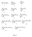
- Lycopene (see Fig. 14), a carotenoid found in tomatoes, also has been found to possess cytostatic activity after extensive oxidation. Lycopene lacks the two cyclohexyl rings present in ⁇ -carotene.
- retinol nor retinoic acid both products of the in vivo, enzymatic oxidation of ⁇ -carotene, were detected in the oxidation mixture.
- retinal which can be oxidized to retinoic acid, has been identified as a product it is present in too small amount to account for the biological activity of the oxidized ⁇ -carotene mixture.
- the biological activity of the oxidized mixture differs substantially from that of vitamin A, as will be described below.
- Fig. 1 presents a reversed phase High Performance Liquid Chromatography (HPLC) characterization of the oxidation mixture.
- HPLC High Performance Liquid Chromatography
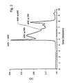
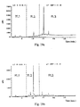
- Fig. 2 presents a GPC characterization of the oxidation mixture showing, in arbitrary units, the composition of the oxidation mixture according to the molecular weight of the components.
- the molecular weight range is broadly distributed from about 600 to about 8000 Dalton with the maximum at about 900 Dalton (the sharp peak at 4.9 min. is an artifact, due to the nature of the GPC column, all the components of the mixture with molecular weight above ca. 1400 elute simultaneously).
- Fig. 3 is a Fourier Transformed Infrared (FTIR) spectrum of the oxidation mixture.
- Fig. 4 is a proton Nuclear Magnetic Resonance (NMR) spectrum of the oxidation mixture.
- fraction 1 The sample was centrifuged (3000 rpm, 3 min.) and the oily residue was separated, washed once with a mixture of THF and hexane (5:15) and dried under vacuum to yield fraction 1. The remaining solution was combined with the liquid from the washing, evaporated and the residue dissolved in THF (3 ml). Precipitation with hexane (15 ml) followed by centrifugation and washing (THF/hexane, 3:15) gave fraction 2. As before, the remaining solution, combined with the wash, was evaporated and dissolved in benzene (3 ml). Precipitation with hexane (15 ml) followed by washing (benzene/hexane, 3:15) gave fraction 3. The supernatant material was labeled as fraction 4.
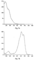
- the ultra violet-visible absorption spectrum of the polymeric substances show maximum absorption at a much shorter wavelength (ca 240 nm with a shoulder at 280 nm) compared to the parent ⁇ -carotene (Fig. 5a and Fig. 5b correspondingly).
- the polymeric material which is soluble in THF, methanol, acetone and acetonitrile, is stable indefinitely below room temperature but partially decomposes under heating forming volatile products as found by way of gas chromatography.
- ⁇ -carotene (20 mM) also was carried out under the conditions described above but in the presence of 0.01 to 0.10 molar equivalents (with respect to ⁇ -carotene) of either alpha-tocopherol or 2,6-di-t-butyl-4-methoxyphenol.
- the reaction was slowed down considerably (1 molar equivalent of oxygen was consumed in ca. 6 days).
- the oxygen uptake plot was linear. The slope of this line was independent of the inhibitor type and its concentration.
- the ⁇ -carotene consumption did not exceed 10 % over a 6 day period.
- Canthaxanthin was oxidized under conditions identical to those used for the oxidation of B-carotene.
- a 20 mM solution of canthaxanthin (Fluka) in benzene saturated with oxygen was incubated in a shaker bath, in the dark, at 30°C under pure oxygen at atmospheric pressure.
- the solvent was evaporated to give a resin-like, yellow residue.
- GPC analysis of the oxidation mixture indicated strong similarities with extensively oxidized ⁇ -carotene, i.e., the reaction products were predominantly polymeric substances.
- Reaction of retinoic acid with oxygen under the conditions used for ⁇ -carotene and canthaxanthin proceeded very slowly.
- the reaction was accelerated by carrying it out at elevated pressure.
- Retinoic acid (Sigma) dissolved in benzene (0.5 ml, 20 mM) in a glass test tube was placed in a high pressure apparatus constructed of INCONEL 600. The apparatus was pressurized to 300 psi oxygen and placed for 3 days in a temperature-controlled bath set at 42 C. HPLC analysis of the reaction mixture showed that the reaction was incomplete. Reaction was continued for a further 2 days under the same conditions, except that the temperature was increased to 50 C, for a total reaction time of 5 days. Very little unoxidized retinoic acid remained (less than 1% of the total product).
- ⁇ -car ox was tested in vitro for cytostatic effects and induction of differentiation.
- Retinoic acid and/or ⁇ -carotene were used as controls in some of the cell lines tested in order to differentiate their effects from that of oxidized ⁇ -carotene.
- the influence of oxidized ⁇ -carotene on the cell cycle was also studied. All concentrations relating to activity of ⁇ -car ox are expressed in micro molar equivalents of ⁇ -carotene.
- a variety of cell lines were used to test the effect of ⁇ -car ox on proliferation and/or differentiation.
- the lines are either established transformed cell lines or are isolated from tumors of patients suffering from cancer.
- two murine cell lines were used in which the cellular differentiation program is well defined with appropriate protein markers.
- HPV16 human papilloma virus type 16
- HPV16 human papilloma virus type 16
- the transformed phenotypes L6-HPV16 (derived from L6 cells) and BALB/c/MK-HPV16 (derived from BALB/c/MK cells) have been characterized by their hormone independence and ability to form colonies on soft agar.
- the pattern of expression of the virus proteins in biopsies obtained from patients with severe neoplasia was established in our laboratory using the reverse transcriptase polymerase chain reaction (RT-PCR). In the biopsied material, the pattern of expression is similar to that of the transformed L6 clone, demonstrating the relevance and validity of the L6 model for testing purposes.
- RT-PCR reverse transcriptase polymerase chain reaction
- the BALB/c/MK cell line is of interest since it differentiates upon exposure to high concentrations of Ca 2+ ions. When cells are exposed to low calcium, they revert to their pre-differentiation state in less than 72 hr.
- mice embryonic stem cells i.e., a quasi normal cell line
- animal and human neuroblastoma cell lines i.e., cancerous cells
- Embryonic stem (ES) cells are totipotent, i.e., they can give rise to any cell lineage of the organism. In vitro, under certain conditions, they differentiate spontaneously into a variety of mixed types. Treatment with substances known to promote differentiation directs ES cells towards a single phenotype. For example, retinoic acid prompts ES cells cultured under conditions established at the National Research Council to differentiate into neurons (unpublished data).
- Mat B MLNr and Mat B-DOXr exhibit multidrug resistance; Mat B-WT is the wild type. They are poorly differentiated cells which do not express estrogen or progesterone receptors.
- the cells are moderately differentiated and positive for estrogen and progesterone receptors.
- Table 2 summarizes the results obtained with the MTT viability/toxicity assay from Promega (a metabolic test based on mitochondrial dehydrogenase activity). Viability was assessed at increasing concentrations (multiples of 2.5) of ⁇ -car ox . The doses which halve the growth of the cell population, as compared to untreated controls, are reported as IC 50 values.
- IC 50 values (expressed in ⁇ -carotene equivalents) were determined graphically from survival curves (plots of cell growth versus concentration of ⁇ -car ox ). Each value in the Table 2 corresponds to an average of at least two independent experiments. Different batches of extensively oxidized ⁇ -carotene were tested and gave consistent results. Note the sensitivity of the NB4 leukemia cell line and the lack of activity on the L6 control cells. Retinoic acid and ⁇ -carotene (each 3 ⁇ M), used as controls for some cell lines, were found to have no effect on the cell growth curves, except for NB4 cells which responded to retinoic acid.
- the cultured rat myoblasts, L6, are capable of differentiation in vitro.
- five steps have been characterized in the differentiation program of these cells using morphometry, in situ immunofluorescence and flow cytometry analysis.
- the known markers (characteristic proteins) of myocyte differentiation used were: fibronectin, a-actin, N-CAM, vimentin and expression of acetylcholine receptors.
- acetylcholine receptors specific to the prefusion stage were not expressed in the L6-HPV16 cells. Five stages were identified, the phenotypes of which in order of increasing differentiation are described below:
- HPV16 transfected cells are transformed and blocked at the pre-fusion stage. That is, HPV16 transfected cells are blocked at stage 4.
- HPV16 transfected cells are blocked at stage 4.
- ⁇ -car ox Upon treatment with ⁇ -car ox , however, better orientation of the cells was observed and some syncitium formation was initiated, corresponding to partial entry into stage 5 of differentiation.
- cytokeratins 1, 5 and 10 were first identified by Western immunoblot. Cytokeratins 1 and 10 are known to be associated with higher levels of differentiation while cytokeratin 5 is associated with less differentiated, still proliferating cells.
- Flow cytometry analysis showed that cytokeratins were generally less expressed in untreated HPV16 transfected cells than in untreated matching control cells.
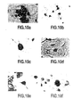
- ⁇ -car ox Under conditions similar to those developed at the National Research Council for the induction of differentiation by retinoic acid of ES cells into neurons, ⁇ -car ox also promotes the differentiation of ES cells into neural cells as shown in Fig. 10 and as assessed by specific markers, using immunohistochemical techniques. However, there are two significant differences between the effect of retinoic acid and ⁇ -car ox on ES cells.
- Neuro2A, IMR32, SK-N-SH and SK-N-MC ⁇ -car ox promoted similarly striking high degrees of differentiation, whereas retinoic acid evoked only partial differentiation.
- ⁇ -car ox may possess activity against the expression of tumorigenic viral genes relies on three independent experiments.
- Cytopathy The symptoms of viral infection, clearly visible as intense vacuolization around the nuclei of the transfected cells L6-HPV16 and BALB/c/MK-HPV16, drastically decreased or disappeared upon exposure to ⁇ -car ox .
- Messenger RNA was extracted from transfected cells that had and had not been exposed to the ⁇ -car ox .
- the kinetics of the expression pattern of the viral gene products has been analyzed by RT-PCR.
- Initial results showed that the pattern of expression of the viral proteins and the myc oncogene (an oncogene related to proliferation) was different for cells treated with ⁇ -car ox , compared to those treated with retinoic acid or ⁇ -carotene.
- a monoclonal antibody raised against a fragment common to the E6 and E7 oncogenic proteins of HPV16 was used to determine the expression of both proteins using polyacrylamide gel electrophoresis, Western blot analysis and flow cytometry. A decrease of the expression of E6 and E7 proteins was observed after 9 to 12 days exposure of the virus transfected cells to ⁇ -car ox .
- Table 5 shows the relative effects towards the MC7-WT cell line of incompletely oxidized ⁇ -carotene containing both the polymeric oxidation product and unreacted ⁇ -carotene, the oxidation products obtained from the oxidation of ⁇ -carotene inhibited by a-tocopherol (5 mole %), fully oxidized canthaxanthin and retinoic acid (syntheses described above). All of the samples contained at least some of the higher MW oxidation materials and all showed considerable cytostatic activity.
- Toxicity was assessed by monitoring body weight of female BALB/c mice and by general examination of the animal. Dosages of 5 mg/kg and 10 mg/kg were injected intraperitoneally on days 1, 3 and 5 (Table 6). The control groups received solvent only (20% aqueous ethanol). A similar study was carried out using 50 and 100 mg/kg injections on days 1,3,5,8 and 11. No overt toxic effects were observed (data not shown).
- ⁇ -Car ox is non-toxic to healthy mice even when applied in six doses of 100 mg/kg. It should be stressed that even with repeated doses (9 times) of up to 150 mg/kg (with tumor bearing mice), no adverse effects have been observed.
- the mouse D1-DMBA-3 (DA-3) mammary adenocarcinoma model was used.
- the cell line was derived from BALB/c mice bearing an immunogenic non-metastatic, murine mammary adenocarcinoma induced by 7,12-dimethylbenzanthracene (DMBA).
- DA-3 cells were injected subcutaneously into each female BALB/c mouse.
- the animals were randomized into groups and injected intraperitoneally with the ⁇ -car ox at dosages ranging from 5 mg/kg up to 150 mg/kg. Tumor growth was measured every 2-3 days. The evaluation was carried out by determining inhibition of tumor growth by measuring tumor volume as a function of time, as described by Alaoui-Jamali et al., in J. Pharmacol. Exp. Ther. 1993 , 264 (3), 1299. Again, the control group received solvent only (20% aqueous ethanol).
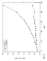
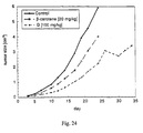
- Fig. 11 and Fig. 12 illustrate the effect of ⁇ -car ox on tumor growth.
- Fig. 11 corresponds to a dose of 10 mg/kg injected on days 0,2,4,7,9,11,14,16 and 18.
- Fig. 12 corresponds to a dose of 150 mg/kg injected in the same way.
- Histological examination a few, randomly chosen animals were sacrificed and tumors were dissected and fixed in 10% formalin in normal saline. Histological sections were prepared from each of the formalin-fixed, paraffin-embedded tumors and stained with hematoxylin-eosin.
- ⁇ -car ox has a growth-retarding effect on a cancer cell-derived tumor implanted in mice.
- Fig. 11 and 12 show that extensively ⁇ -car ox as applied repeatedly at dose as low as 10 mg/kg has the ability to effectively arrest the growth of the tumor and stabilize it for a long period of time.
- Fig. 13a and Fig. 13b show the comparison of tumors in sacrificed animals which had been treated with different doses of ⁇ -car ox and the control (untreated animal). In some animals hemorrhaging occurred around the tumors (Fig. 13b). In these cases the actual tumor size is smaller than when measured with calipers (the discrepancy is attributed to hemorrhagic swelling increasing the apparent tumor size).
- the therapeutic index of ⁇ -car ox appears to be very high, which potentially offers a major advantage over traditional anti-cancer drugs.
- ⁇ -Carotene and canthaxanthin as representative carotenoids, and to a lesser extent, retinoic acid, a representative retinoid
- ⁇ -car ox is a model, which demonstrate properties that make the substances useful as non-toxic agents active against uncontrolled cell proliferation, tumors, and tumorigenic viruses, and useful as promoters of cell differentiation. It is evident from chemical analysis of ⁇ -car ox that none of the various forms of vitamin A are present or are present only in minor amounts. Furthermore, the biological activities of oxidized canthaxanthin and retinoic acid, which cannot form vitamin A, indicate the presence of active substances that are different from vitamin A.
- ⁇ -car ox Although the cytostatic and differentiation-promotion activities of ⁇ -car ox resemble those of vitamin A itself, generally the effects are more powerful for ⁇ -car ox in a wide variety of circumstances. Also, there is the very important difference that ⁇ -car ox is non-toxic.
- Lycopene (see Fig. 14) dissolved in benzene was extensively oxidized under an atmosphere of oxygen in essentially the same manner as ⁇ -carotene in the first phase of work.
- the crude product mixture was tested for cytostatic activity in a manner analogous to ⁇ -car ox .
- ⁇ -Car ox was synthesized using a procedure very similar to that described in the first phase. Briefly, ⁇ -carotene (30 g) dissolved in benzene (3.0 1; 0.02 M) was stirred for 4 days at room temperature under an atmosphere of gaseous oxygen. GPC chromatography confirmed the presence of three major components, i.e., the low ( ⁇ 300 Da), medium (300-1000 Da), and high (> 1000 Da) MW fractions ( Figure 15).
- Level 1 separation (phases 1-3): the solution (3.0 l) was concentrated to ca. 200 ml and then diluted with approximately 2 l of hexane.
- the precipitate, IG1 ca. 65% of the total mixture, contained most of the high MW material (> 1000 Da).
- Level 2 separation (phase 1) : fraction SG1 (1.2 g) was stirred in hexane (120 ml) at room temperature for 30 min.
- the insoluble fraction, IG2 that was filtered off, contained mostly medium MW and some low MW material ( Figure 15).
- the supernatant containing the soluble fraction was evaporated to yield SG2, containing mostly low MW and some medium MW material ( Figure 16).
- phase 2 separation phases 2 and 3 : fraction SG1 (10 g) dissolved in tetrahydrofuran (10 ml) was separated into low and medium MW fractions, LSG (40%) and MSG (55%), respectively, by successive injections of samples (250 ⁇ l) onto a preparative-scale GPC chromatography column (19 x 300 mm, Waters styragel, 15 ⁇ m particle size, 10 nm pore size) and elution with tetrahydrofuran (4 ml/min). A clean separation into two MW fractions was obtained as illustrated in Fig. 17.
- Level 3 separation (phase 1): fraction SG2 (688 mg) was loaded onto a silica gel column and eluted with hexane/ethyl acetate (95:5). The eluted fractions were combined and the solvent removed to give fraction SG3.
- Level 3 separation (phase 2): fraction LSG (600 mg) was stirred with ice-cold pentane (1 ml) for 1 min. and then most of the supernatant was decanted off. This procedure was repeated four times. The insoluble fraction, ILSG, was filtered and the solvent was evaporated from the combined supernatant fractions containing the soluble component, giving fraction SLSG.
- phase 3 Level 3 separation (phase 3): fraction LSG (4.4 g), dissolved in acetonitrile (10 ml), was separated into two fractions by successive injections (450 ⁇ l; total 9.0 ml) onto a preparative -scale HPLC instrument equipped with three Waters NovaPak HR C18 (reverse phase, 6 ⁇ m particle size, 6 nm pore size) PrepPak cartridges (40 x 100 mm) connected in series and eluted with acetonitrile (40 ml/min). Fractions F1 (80%) and F2 (20%) were obtained by collecting eluent from 4.8 to 6.4 min and 6.4 to 12 min, respectively.
- phase 3 fraction F1 (700 mg), dissolved in acetonitrile (1.5 ml), was separated into three fractions, F1.1 (11%), F1.2 (13%) and F1.3 (5%), by successive injections (250 ⁇ l; total 1.25 ml) onto a preparative-scale HPLC instrument equipped with the same column already described and eluted with a 50:45:5 mixture of water, acetonitrile and methanol at a flow rate of 40 ml/min. The fractions were obtained by collecting eluent from 2.0 to 6.6 min, 6.6 to 12.0 min and 12.0 to 23 min, respectively. The dotted vertical lines in Figure 19 show how the cuts were made to obtain the three fractions.
- Fractions F1.1, F1.2 and F1.3 each contain only a few compounds as Figure 19 indicates.
- a major component in F1.2 is 2-methyl-6-oxo-2,4-heptadienal (compound 1 , referred to elsewhere in the text as ketoaldehyde).
- Dihydroactinidiolide (compound 2) is a major component of F1.3.
- Compounds 3-14 have been identified in fraction F2.
- In vitro biological assays were carried out by testing for cytostatic activity, induction of differentiation, and the effect upon the cell cycle. In vivo tests were carried out by testing for inhibition of growth of tumors in mice.
- Fractions were tested for cytostatic properties and ability to induce differentiation. The results were compared with those obtained for ⁇ -car ox , which served as a reference indicating whether or not fractionation was leading to fractions with enhanced activity. (Retinoic acid and/or ⁇ -carotene were used as controls in some of the cell lines tested in order to differentiate their effects from that of ⁇ -car ox ). Given the present indeterminate nature of many of the fractions, all samples were tested in identical 'by weight' concentrations. Comparisons have been done on a pseudo molarity scale by dividing the weight of sample used by the molecular weight of ⁇ -carotene (537 Da).
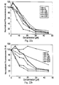
- Cells were treated with six concentrations of each sample (2.5, 7.5, 15, 22.5, 30, 45 ⁇ M for HCT116 and IMR32 and 2.5, 5, 7.5, 10, 15, 22.5 ⁇ M for NB4 and K562 cell lines). After 3 days, the cells were lysed, incubated with Hoechst dye 33258 and the fluorescence of the solution was measured to provide a measure of the amount of cellular DNA present. The fluorescence data for each substance tested were collected and divided by the corresponding values for untreated cells (after correcting for background fluorescence) to determine the cytostatic effect for each sample concentration.
- Figures 22a and 22b illustrate results that were obtained for the HCT116 human colon cancer and the K562 human leukemia cell lines respectively.
- Table 7 summarizes the results that were obtained for the four cell lines. Fractions have been ranked relative to ⁇ -car ox by dividing each growth inhibition value at each sample concentration by the corresponding ⁇ -car ox value and determining qualitatively by inspection the trends across the six sample concentrations. Activities of fractions are expressed as "+”, “o”, and “-”, indicating activity higher, similar to, and smaller than, respectively, the activity of ⁇ -car ox .
- the cell differentiation-inducing effects of the fractions were determined qualitatively by observation of morphological changes in cell lines and quantitatively by measuring the expression of characteristic protein markers using monoclonal antibodies and flow cytometry.
- active fractions are denoted by the "+” symbol and highly active fractions are denoted by a "++”.
- the greater activity in the transformed cell lines, particularly IMR32, compared to the quasi-normal L6 line provides support for the strategy of using selective differentiation of transformed cells to control cancerous cell growth in a non-toxic manner. Differentiation inducing effects of ⁇ -car ox fractions.
- Cytokeratins 1 and 10 are high molecular weight proteins which are increasingly expressed with the progression of the differentiation program. In in vitro cell cultures, the differentiation is classically induced by exposure to culture media containing high levels of calcium. Cells cultured in low calcium media remain proliferative. As illustrated in Table 10, retinoic acid can substitute for Ca ++ in increasing expression of cytokeratins 1 and 10. ⁇ -Car ox also can substitute for calcium in inducing Balb/c/MK keratinocyte differentiation. The apparently smaller effect of ⁇ -car ox compared to retinoic acid is due not to lower potency but to a delayed action. Table 11 shows that similar results have been obtained using a panel of other cytokeratins.
- differentiation markers are expressed transiently during the differentiation program.
- protein CD33 is associated with early stages of differentiation, followed by protein CD11b, an intermediate marker, and eventually by protein CD15, an advanced differentiation marker.
- Enhanced differentiation therefore will be characterized by a decrease in the expression and intensity of the CD33 marker and an increase in expression and intensity of the CD15 marker.
- Table 13 presents quantitative data on the effect of all-trans retinoic acid, ⁇ -car ox and some of its fractions on the level of expression of these specific differentiation markers in NB4 cells, supplemented either with individual fractions or various binary combinations with all-trans retinoic acid.
- the results indicate that ⁇ -car ox is not as effective as retinoic acid ( ⁇ -car ox requires more time to fully express its effect).
- Combined treatment with retinoic acid and ⁇ -car ox , or some of its fractions results in inhibition of differentiation (an antagonistic effect) as has been already noted above for ⁇ -car ox (Table 12), but with fraction IG2 differentiation proceeds further than with either IG2 or retinoic acid alone (a synergistic effect).
- retinoic acid is shortly to become generally available for the treatment of acute promyleocytic leukemia (APL), despite the fact that it provides only 5-6 months of remission before it becomes ineffective in treating the resurgent cancer.
- APL acute promyleocytic leukemia
- Our results support the possibility that a combination of two agents, particularly those indicated above, will push leukemic cells more fully down a terminal differentiation path, increasing the likelihood that their return to a proliferative state is blocked.
- ⁇ -car ox when the concentration of RA is increased to 2.5 ⁇ M and the concentration of ⁇ -car ox is Seduced to 3 ⁇ M, the expression of NF is higher then for either of them alone (synergy).
- GFAP a marker indicating differentiation towards glial cells
- retinoic acid itself is most effective at low concentration of 1 ⁇ M
- the presence of ⁇ -car ox increases this optimal concentration up to 10 ⁇ M depending on the concentration of ⁇ -car ox (see table 13a).
- the distribution of the ⁇ -car ox -treated cell population among the three phases of the cell cycle shows an accumulation of cells in the S phase. There is no G1 block. This effect is due to a lengthening of the duration of the cell cycle. It was found that the ⁇ -car ox -treated cells completed a cell cycle in 36 h whereas the duration of the cell cycle in controls and retinoic acid-treated matching samples was 22 and 18 h, respectively.
- the data were acquired using flow cytometry analysis of the individual intracellular DNA content of cells from synchronized populations. Sampling was done every 5 h over a period of 48 h. Two types of cells were used: growing in suspension (NB4 leukemia cells) and growing as a adherent cells (L6 myoblasts).
- ⁇ -car ox Effect of ⁇ -car ox in decreasing the level of cellular glutathione and on increasing the sensitivity of a cancer cell line towards a conventional chemotherapeutic agent.
- classical anticancer drugs are ineffective because cancerous cells acquire resistance towards the treatment.
- this phenomenon is linked to an increased level of glutathione (GSH), which protects cancerous cells by reacting with the active component of the drug and thereby assisting its removal from the cell.
- GSH glutathione
- ⁇ -car ox with a conventional anticancer agent, such as melphalan, will increase the sensitivity of cancerous cells towards the anticancer drug.
- a conventional anticancer agent such as melphalan
- a lower dose will be required for a therapeutic effect thereby reducing the unwelcome side effects resulting from the high, general toxicity of melphalan.
- the mouse D1-DMBA-3 (DA-3) mammary adenocarcinoma model was used.
- the results displayed in Figs. 25a and 25b show that several fractions were at least as potent (fractions SG, F1, F2) or more potent (fractions F1.1, F1.2, F1.3; note lower concentrations used) than ⁇ -car ox .
- the potency of the ketoaldehyde (compound 1, Figure 8) is striking.
- Table 16 shows the estimated tumor volume expressed relative to the control (i.e., the group receiving the vehicle only) on day 28 of the experiment. In contrast to what was previously observed for ⁇ -car ox , it can be seen that at least in the case of fraction F1 a dose-response effect exists. In vivo antitumor activity of ⁇ -car ox and its fractions in the DA-3 model.
Landscapes
- Chemical & Material Sciences (AREA)
- Organic Chemistry (AREA)
- Health & Medical Sciences (AREA)
- General Health & Medical Sciences (AREA)
- Medicinal Chemistry (AREA)
- Life Sciences & Earth Sciences (AREA)
- General Chemical & Material Sciences (AREA)
- Pharmacology & Pharmacy (AREA)
- Nuclear Medicine, Radiotherapy & Molecular Imaging (AREA)
- Animal Behavior & Ethology (AREA)
- Chemical Kinetics & Catalysis (AREA)
- Public Health (AREA)
- Veterinary Medicine (AREA)
- Bioinformatics & Cheminformatics (AREA)
- Proteomics, Peptides & Aminoacids (AREA)
- Engineering & Computer Science (AREA)
- Molecular Biology (AREA)
- Genetics & Genomics (AREA)
- Biophysics (AREA)
- Biochemistry (AREA)
- Obesity (AREA)
- Hematology (AREA)
- Diabetes (AREA)
- Nutrition Science (AREA)
- Acyclic And Carbocyclic Compounds In Medicinal Compositions (AREA)
- Organic Low-Molecular-Weight Compounds And Preparation Thereof (AREA)
- Pharmaceuticals Containing Other Organic And Inorganic Compounds (AREA)
- Compounds Of Unknown Constitution (AREA)
- Heterocyclic Carbon Compounds Containing A Hetero Ring Having Oxygen Or Sulfur (AREA)
Abstract
Description
A specific compound, 2-methyl-6-oxo-2,4-heptadienal was isolated and found to exhibit cytostatic and anti-tumor properties. The compound can be obtained by fractionation of the extensively oxidized mixture of the invention (oxidized β-carotene), or by a conventional synthetic method. This compound has been reported previously as a product obtained by the oxidation of trans-retinoic acid, Oyler et al, "Characterization of autoxidation products of retinoic acid" published in Tetrahedron Vol. 45, No. 25, pp. 7679-7694, 1989.
| Characteristics of Various Fractions Obtained by Successive Solvent Precipitations of β-Carox. | ||||
| | 1 | 2 | 3 | 4 |
| | 37 | 17 | 14 | 32 |
| Titratable acids [10-4 mol/g] | 4.3±0.3 | 4.3±0.2 | 6.1±0.3 | 6.2±0.1 |
| Peroxides (iodometric) [10-4 mol/g] | 9.5±1.0 | 9.9±0.8 | 8.2±0.3 | 5.9±0.4 |
| Peroxides (oxidation of Fe2+) [10-4 mol/g] | 8.3±0.2 | 8.9±0.2 | 8.8±0.9 | 5.5±0.1 |
| Elemental Analysis: | ||||
| C | 58.5 | 58.7 | 61.2 | 65.3 |
| H | 7.2 | 7.0 | 7.6 | 8.7 |
| O | 34.3 | 34.3 | 31.2 | 26.0 |
| Fractions were obtained in the following way: the crude mixture (1.2 g) was dissolved in tetrahydrofuran (THF) (5 ml) and hexane (15 ml) was added slowly while the solution was vortex-mixed. The sample was centrifuged (3000 rpm, 3 min.) and the oily residue was separated, washed once with a mixture of THF and hexane (5:15) and dried under vacuum to yield |
- toxicity in mice;
- inhibition of growth of tumors in mice including histopathological examination of tumor changes (tumor derived from a transplanted, chemically-induced rat mammary cancer cell line).
| Cytostatic Effect of Extensively β-Carox | ||
| Cell Lines | Origin | IC50 [µM] |
| MatB-WT | Rat mammary adenocarcinoma | 25.5 |
| Mat-MLNr | 21.0 | |
| B16 | Mouse melanoma | 12.4 |
| DA-3 | Mouse mammary carcinoma | 15.6 |
| FDCP-1 | Mouse myeloid leukemia | 7.5 |
| L6 | Rat embryonic myoblasts | 51.9 |
| L6-HPV16 | 28.2 | |
| MCF7-WT | Human breast carcinoma | 11.3 |
| MCF7-ADRr | 11.3 | |
| Uro-9 | Human urothelial carcinoma | 17.7 |
| Uro-10 | 19.8 | |
| L14 | Human lung adenocarcinoma | 18.5 |
| NB4 | Human promyelocytic leukemia | 4.8 |
| Exponentially growing cells cultured at low density (104 to 105 cells/1-5 ml) were exposed to a single dose of different concentrations of β-carox for 72 hr or more. Cell growth was determined by the MTT test. IC50 values (expressed in β-carotene equivalents) were determined graphically from survival curves (plots of cell growth versus concentration of β-carox). Each value in the Table 2 corresponds to an average of at least two independent experiments. Different batches of extensively oxidized β-carotene were tested and gave consistent results. Note the sensitivity of the NB4 leukemia cell line and the lack of activity on the L6 control cells. Retinoic acid and β-carotene (each 3 µM), used as controls for some cell lines, were found to have no effect on the cell growth curves, except for NB4 cells which responded to retinoic acid. |
| Comparison of the cytostatic effect (IC50 [µM]) of β-carox as measured by MTT test and cell enumeration by Coulter counter | ||
| Cell Line | MTT | Coulter counter |
| L6 | 51.9 | 39.0 |
| L6-HPV16 | 28.2 | 24.6 |
| Cytostatic effect of fractions against MC7-WT cell line (µM) | |||||
| | 1 | 2 | 3 | 4 | β-carox |
| MCF7-WT IC50 | 11.8 | 17.3 | 13.1 | 26.2 | 22.8 |
| Cytostatic activity against MC7-WT cell line of partially oxidized β-carotene, products obtained in inhibited oxidation of β-carotene, as well as extensively oxidized canthaxanthin and retinoic acid | |
| Sample | IC50 (µM) |
| ca. 25% Oxidized β- | 22 |
| ca. 50% Oxidized β- | 21 |
| 100% Oxidized β- | 17 |
| a-Tocopherol-retarded oxidation of β- | 11 |
| | 9 |
| Oxidized | 7 |
| . Effect of extensively oxidized β-carotene on body weight of mice | |||
| Day | Average Body Weight [g] | ||
| | 5 mg/ | 10 mg/ | |
| 1 | 14.0 | 14.4 | 14.0 |
| 2 | 14.0 | 14.5 | 14.0 |
| 5 | 14.4 | 14.9 | 14.5 |
| 7 | 14.8 | 15.3 | 14.6 |
| 9 | 15.2 | 15.8 | 15.2 |
| 11 | 15.7 | 16.3 | 15.7 |
| 13 | 16.1 | 16.7 | 16.2 |
| 15 | 16.6 | 17.3 | 16.9 |
| 17 | 17.1 | 17.7 | 17.4 |
| 19 | 17.6 | 18.3 | 17.9 |
| 21 | 18.1 | 18.7 | 18.4 |
| 23 | 18.7 | 19.4 | 19.1 |
| 25 | 19.2 | 20.0 | 19.8 |
- β-carox induces pronounced histological alterations, reflecting tissue death in DA-3 tumors.
- tumor tissues showed many features of cell/tissue differentiation.
- hemorrhagic areas are present in all treated tumors and are associated with extensive pigmentation and necrosis.
- the pigments are not iron (iron staining using Prussian blue was negative), but may be hemosiderin probably resulting from hemorrhagia. It appears unlikely that the pigment is melanin. The exact nature of the pigmentation remains to be confirmed.

| HCT116 | human colon carcinoma |
| IMR32 | human neuroblastoma |
| NB4 | human acute promyelocytic leukemia |
| K562 | human chronic myelogenous leukemia |
| BALB/c/MK | mouse keratinocytes |
| Cytostatic effect upon several cell lines of fractions and selected compounds expressed relative to β-carox. | ||||||
| Stage | Fraction | Cell Line | ||||
| HCT116 | | K562 | NB4 | |||
| 0 | β-Carox | o | | o | o | |
| 1 | SG1 | o | + | + | + | |
| IG1 | + | o | o | + | ||
| 2 | SG2 | + | + | + | + | |
| IG2 | + | + | + | + | ||
| 2 | LSG | - | o | + | o | |
| MSG | o | o | - | | ||
| 3 | SG3 | + | + | + | | |
| 3 | F1 | - | o | o | o | |
| F2 | o | o | o | - | ||
| 4 | F1.1 | - | o | + | + | |
| F1.2 | - | o | - | o | ||
| F1.3 | - | o | o | o | ||
| Ketoaldehyde | + | + | + | + | ||
| β-Car | - | o | - | - | ||
| RA | - | + | o | o |
Data shown above were calculated using a value for β-Carox that was averaged over all assays. The values for β-Carox are, by definition, set at "o". RA = all-trans retinoic acid.
| Comparison of cytostatic effect of extensively oxidized lycopene and β-carox in the HCT-116 cell line. | ||
| Dose [µM] | β-Carox | Oxidized Lycopene |
| 0 | 100 | 100 |
| 2.5 | 86 | 100 |
| 7.5 | 80 | 85 |
| 15 | 51 | 55 |
| 22.5 | 18 | 41 |
| 30 | 8 | 18 |
| 45 | 1 | 11 |
| Differentiation inducing effects of β-carox fractions. | |||||
| Stage | Fraction | Cell Line | |||
| | NB4 | L6 | |||
| 1 | SG1 | +(n) | ne | ne | |
| IG1 | +(g) | | ne | ||
| 2 | SG2 | +(n) | ne | ne | |
| IG2 | +(g) | ++ | | ||
| 2 | LSG | ++(n) | ne | ne | |
| MSG | ++(g) | ++ | | ||
| 3 | SG3 | +(n) | ne | ++ | |
| 3 | F1 | ++(n) | ne | ne | |
| F2 | ++(n) | | ne | ||
| 4 | F1.1 | ++(g) | ne | ne | |
| F1.2 | ne | ne | ne | ||
| F1.3 | ne | ne | ne | ||
| Ketoaldehyde | ne | ne | ne |
| Expression of | ||
| Sample | A | IA |
| Control | 90.3 | 4.4 |
| Ca++ (1.8 mM) | 96.5 | 13.8 |
| RA (3 µM) | 94.5 | 15.4 |
| β-Carox (30 µM) | 96.5 | 7.9 |
| Expression of a panel of cytokeratins in Balb/c/MK keratinocytes. | ||
| Sample | A | IA |
| Control | 93.7 | 60.7 |
| Ca++ (1.8 mM) | 97.6 | 87.2 |
| RA (3 µM) | 94.3 | 129.8 |
| β-Carox (30 µM) | 96.2 | 82.1 |
| Differentiating properties of mixtures of oxidized β-carox and retinoic acid. | ||
| Retinoic Acid | β-Carox | Differentiation |
| L | - | yes |
| - | L | yes |
| L | L | no |
| H | L | yes |
| L | H | yes |
| H | H | yes |
| (enhanced) | ||
| Note: The letters L and H indicate low and high concentration, respectively. A dash indicates the absence of the indicated agent. |
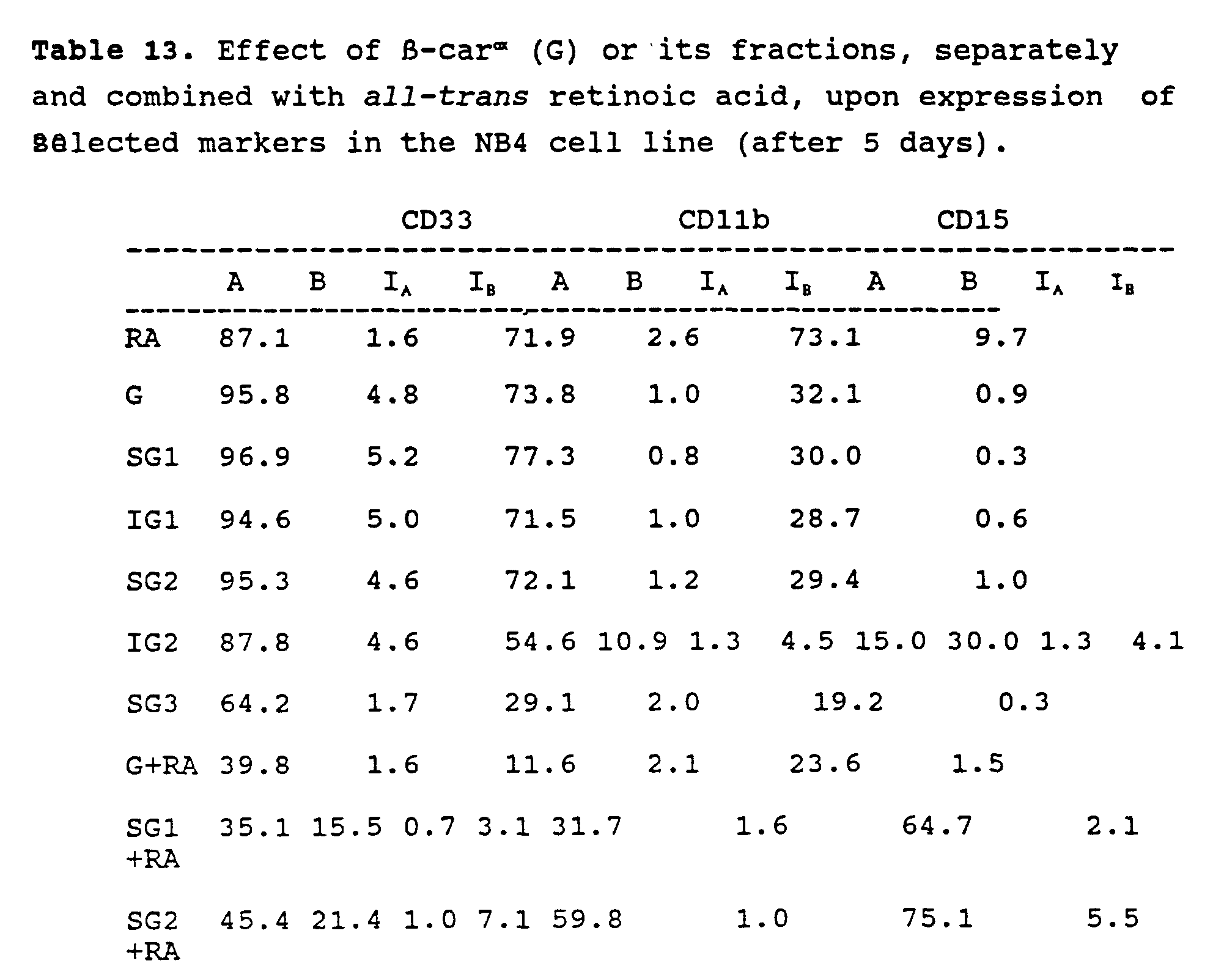

| Effect of β-carox (G), separately and combined with all-trans retinoic acid, upon expression of selected markers in the IMR32 cell line (after 48 h). | ||||||
| RA [µM] | β-carox [µM] | NF | GFAP | |||
| % | | % | intensity | |||
| 0 | 0 | 96.0 | 4.3 | 65.0 | 1.7 | |
| 1 | 0 | 96.4 | 14.2 | 94.4 | 18.4 | |
| 2.5 | 0 | 97.2 | 11.9 | 90.5 | 6.1 | |
| 5 | 0 | 95.5 | 8.4 | 87.5 | 5.2 | |
| 10 | 0 | 92.0 | 7.9 | 83.1 | 4.0 | |
| 0 | 3 | 90.6 | 7.7 | 91.5 | 5.2 | |
| 0 | 5 | 98.7 | 53.0 | 87.0 | 5.2 | |
| 0 | 7.5 | 96.1 | 8.6 | 94.2 | 8.7 | |
| 0 | 10 | 95.8 | 8.5 | 88.1 | 4.5 | |
| 1 | 5 | 94.1 | 6.4 | 98.2 | 22.4 | |
| 1 | 7.5 | 94.7 | 7.5 | 87.4 | 5.6 | |
| 1 | 10 | 98.2 | 17.6 | 89.7 | 6.2 | |
| 2.5 | 3 | 98.3 | 70.0 | 87.7 | 5.6 | |
| 2.5 | 5 | 94.7 | 7.2 | 97.9 | 21.7 | |
| 2.5 | 7.5 | 98.7 | 22.2 | 92.7 | 10.4 | |
| 2.5 | 10 | 93.0 | 6.1 | 97.6 | 20.4 | |
| 5 | 3 | 97.5 | 17.0 | 79.1 | 3.4 | |
| 5 | 5 | 94.7 | 7.1 | 90.3 | 4.8 | |
| 5 | 7.5 | 95.3 | 9.1 | 96.8 | 18.4 | |
| 5 | 10 | 96.7 | 15.0 | 97.4 | 18.5 | |
| 10 | 3 | 93.4 | 6.6 | 89.6 | 5.3 | |
| 10 | 5 | 94.5 | 7.7 | 89.5 | 7.0 | |
| 10 | 7.5 | 90.6 | 7.7 | 93.3 | 7.9 | |
| 10 | 10 | 95.1 | 7.0 | 97.0 | 15.9 |
| Effect of β-carox on the level of glutathione in the DA-3 mouse mammary cancer cell line. | |
| β-carox [µM] | GSH [nmol/mg protein] |
| 0 | 30 |
| 5 | 38 |
| 10 | 6 |
| 20 | 3 |
| Effect of oxidized β-carotene on IC50 of melphalan in the DA-3 mouse mammary cancer cell line model as estimated by MTT test. | |
| β-Carox (µM) | IC50 of melphalan (µM) |
| 0.0 | 100 |
| 5.0 | 40 |
| 7.5 | 44 |
| 10.0 | 6.6 |
| 20.0 | 6.8 |
| In vivo antitumor activity of β-carox and its fractions in the DA-3 model. | ||
| Sample | Dose [mg/kg body] | Relative Tumor Size (28 days) |
| β- | 25 | 0.36 |
| SG1 | 25 | 0.30 |
| | 25 | 0.49 |
| | 5 | 0.72 |
| 10 | 0.49 | |
| 25 | 0.37 | |
| | 25 | 0.33 |
| F1.1 | 10 | 0.30 |
| F1.2 | 10 | 0.47 |
| F1.3 | 10 | 0.47 |
| Ketoaldehyde | 10 | 0.44 |
| 20% Ethanol | N/A | (1.00) |
| Comparison of in vivo antitumor activity of fractions F1 and F2 delivered i.p and p.o. in the DA-3 model. | |
| Sample at 25 mg/kg body | Relative Tumor Size (21 days) |
| F2 p.o. | 0.50 |
| F2 i.p. | 0.48 |
| F1 p.o. | 0.34 |
| F1 i.p. | 0.43 |
| 20% Ethanol | (1.00) |
Claims (16)
- Use of a product for the manufacture of a medicament for use in treating a tumour or skin cell proliferation in a human or animal, wherein the product is a mixture containing more than one oxidised compound, obtainable by the process of oxidation of a carotenoid compound with oxygen, such that 6 to 8 molecules of oxygen react per molecule of the carotenoid compound.
- Use according to claim 1, wherein the process also comprises fractionation of the oxidised mixture, according to molecular weight.
- Use according to claim 2, wherein the fractionation is effected by solvent precipitation.
- Use according to claim 2, wherein the fractionation is effected by size exclusion chromatography.
- Use according to any of claims 2 to 4, wherein the process comprises further fractionation or isolation, according to polarity.
- Use according to any preceding claim, wherein the oxidation is conducted in an organic solvent.
- Use according to any of claims 1 to 5, wherein the oxidation is conducted on said compound in the solid state.
- Use according to any preceding claim, wherein said compound is β-carotene, lycopene or canthaxanthin.
- Use according to any preceding claim, wherein said compound is β-carotene.
- Use according to claim 8, wherein said compound is lycopene.
- Use according to any preceding claim, wherein the oxidation is conducted in the absence of catalysts, initiators or inhibitors of oxidation.
- Use according to any preceding claim, wherein the mixture contains a polymeric or oligomeric component.
- Use according to any of claims 1 and 9 to 12, wherein each oxidised compound has a molecular weight of less than 700 Da.
- Use according to claim 13, wherein each oxidised compound has a molecular weight of less than 300 Da.
- Use according to any preceding claim, wherein the animal or human is also treated with a conventional anti-cancer agent.
- Use of 2-methyl-6-oxo-2,4-heptadienal, for the manufacture of a medicament for use in treating a tumour in an animal or human.
Applications Claiming Priority (3)
| Application Number | Priority Date | Filing Date | Title |
|---|---|---|---|
| US288315 | 1994-08-10 | ||
| US08/288,315 US5475006A (en) | 1994-08-10 | 1994-08-10 | Extensively oxidized derivatives of carotenoids, retinoids and related conjugated polyenes useful as non-toxic cell-differentiation inducers, anti-proliferative agents, and anti-tumor agents |
| PCT/CA1995/000484 WO1996005160A1 (en) | 1994-08-10 | 1995-08-10 | Oxidized carotenoids, retinoids and related conjugated polyenes, and derived fractions and compounds useful as cell-differentiation inducers, cytostatic agents, and anti-tumor agents |
Publications (2)
| Publication Number | Publication Date |
|---|---|
| EP0722431A1 EP0722431A1 (en) | 1996-07-24 |
| EP0722431B1 true EP0722431B1 (en) | 2001-05-23 |
Family
ID=23106590
Family Applications (1)
| Application Number | Title | Priority Date | Filing Date |
|---|---|---|---|
| EP95927612A Expired - Lifetime EP0722431B1 (en) | 1994-08-10 | 1995-08-10 | Oxidized carotenoids, retinoids and related conjugated polyenes, and derived fractions and compounds useful as cell-differentiation inducers, cytostatic agents, and anti-tumor agents |
Country Status (18)
| Country | Link |
|---|---|
| US (1) | US5475006A (en) |
| EP (1) | EP0722431B1 (en) |
| JP (1) | JP3983800B2 (en) |
| KR (1) | KR100404724B1 (en) |
| CN (1) | CN1131939A (en) |
| AT (1) | ATE201394T1 (en) |
| AU (1) | AU704640B2 (en) |
| CA (1) | CA2171625C (en) |
| CZ (1) | CZ103896A3 (en) |
| DE (1) | DE69521020T2 (en) |
| ES (1) | ES2160172T3 (en) |
| FI (1) | FI961544A7 (en) |
| GB (1) | GB2297325B (en) |
| MX (1) | MX9601175A (en) |
| NZ (1) | NZ290769A (en) |
| PL (1) | PL184296B1 (en) |
| PT (1) | PT722431E (en) |
| WO (1) | WO1996005160A1 (en) |
Families Citing this family (19)
| Publication number | Priority date | Publication date | Assignee | Title |
|---|---|---|---|---|
| US6432711B1 (en) * | 1993-11-03 | 2002-08-13 | Diacrin, Inc. | Embryonic stem cells capable of differentiating into desired cell lines |
| US7132458B2 (en) * | 1994-08-10 | 2006-11-07 | Chemaphor Inc. | Oxidized carotenoid fractions and ketoaldehyde useful as cell-differentiation inducers, cytostatic agents, and anti-tumor agents |
| US20040228871A1 (en) * | 1999-06-03 | 2004-11-18 | Tayyaba Hasan | Treatment and analysis of proliferative disorders |
| IL135335A (en) * | 2000-03-29 | 2013-12-31 | Lycored Natural Prod Ind Ltd | Use of carotenoids in the preparation of medicaments for preventing hormone induced adverse effects and pharmaceutical compositions comprising carotenoids |
| US20020031539A1 (en) * | 2000-08-30 | 2002-03-14 | The Brigham And Women's Hospital, Inc. | Oxidized forms of retinoic acid as ligands for peroxisome proliferator activated receptor gamma |
| US20050037048A1 (en) * | 2003-08-11 | 2005-02-17 | Young-Ho Song | Medical devices containing antioxidant and therapeutic agent |
| US20050064011A1 (en) * | 2003-08-11 | 2005-03-24 | Young-Ho Song | Implantable or insertable medical devices containing phenolic compound for inhibition of restenosis |
| EP1727530A4 (en) * | 2004-02-19 | 2009-10-21 | Chemaphor Inc | TOPICAL FORMULATIONS FOR THE TREATMENT OF DERMATOLOGICAL CONDITIONS |
| MX2007003773A (en) * | 2004-09-28 | 2009-01-19 | Chemaphor Inc | Compositions and methods for promoting weight gain and feed conversion. |
| RU2008115148A (en) * | 2005-10-11 | 2009-11-20 | Ликоред Лтд. (Il) | CAROTINOID OXIDATION PRODUCTS AS CHEMO-PREVENTING AND CHEMOTHERAPEUTIC AGENTS |
| US20090306222A1 (en) * | 2006-04-05 | 2009-12-10 | Chemaphor Inc. | Food supplements and uses thereof |
| KR20100065190A (en) | 2007-09-14 | 2010-06-15 | 닛토덴코 가부시키가이샤 | Drug carriers |
| DK2214656T3 (en) * | 2007-10-26 | 2019-03-25 | Avivagen Inc | COMPOSITIONS AND PROCEDURES TO ENHANCE IMMUNE RESPONSE |
| US10456369B2 (en) * | 2009-04-30 | 2019-10-29 | Avivagen Inc. | Methods and compositions for improving the health of animals |
| US20180139985A1 (en) | 2015-04-28 | 2018-05-24 | Avivagen Inc. | Oxidized carotenoids and components thereof for preventing necrotic enteritis |
| AU2016308076A1 (en) * | 2015-08-20 | 2018-02-01 | I.B.R. Israeli Biotechnology Research Ltd. | Carotenoid compositions having antiviral activities and uses thereof |
| US20190054135A1 (en) * | 2016-02-25 | 2019-02-21 | Avivagen Inc. | Plant or microorganism-derived carotenoid-oxygen copolymer compositions, methods of identifying, quantifying and producing same and uses thereof |
| CN106614301A (en) * | 2016-11-14 | 2017-05-10 | 鄂尔多斯市绿食保山羊食品有限公司 | Organic farming method of goats |
| KR102683182B1 (en) * | 2021-11-12 | 2024-07-11 | 주식회사 에이펙셀 | Pharmaceutical composition for cacner treatment comprising salt of meta arsenite |
Family Cites Families (6)
| Publication number | Priority date | Publication date | Assignee | Title |
|---|---|---|---|---|
| DE2505869C3 (en) * | 1975-02-12 | 1978-05-18 | Basf Ag, 6700 Ludwigshafen | Process for the preparation of symmetrical carotenoids |
| DE2635802C2 (en) * | 1976-08-09 | 1984-03-01 | Basf Ag, 6700 Ludwigshafen | Process for the preparation of symmetrical carotenoids |
| DK595489D0 (en) * | 1989-11-27 | 1989-11-27 | Neurosearch As | HYDROXYCARBONYL DERIVATIVES AND PROCEDURES FOR PREPARING THE SAME |
| US5225604A (en) * | 1989-11-27 | 1993-07-06 | Neurosearch A/S | Hydroxycarbonyl derivatives and process for making the same |
| US5310554A (en) * | 1992-10-27 | 1994-05-10 | Natural Carotene Corporation | High purity beta-carotene |
| US5358915A (en) * | 1993-02-12 | 1994-10-25 | American Colloid Company | Process for regenerating spent acid-activated bentonite clays and smectite catalysts |
-
1994
- 1994-08-10 US US08/288,315 patent/US5475006A/en not_active Expired - Lifetime
-
1995
- 1995-08-10 PT PT95927612T patent/PT722431E/en unknown
- 1995-08-10 PL PL95313907A patent/PL184296B1/en not_active IP Right Cessation
- 1995-08-10 WO PCT/CA1995/000484 patent/WO1996005160A1/en not_active Ceased
- 1995-08-10 CZ CZ961038A patent/CZ103896A3/en unknown
- 1995-08-10 EP EP95927612A patent/EP0722431B1/en not_active Expired - Lifetime
- 1995-08-10 MX MX9601175A patent/MX9601175A/en unknown
- 1995-08-10 GB GB9604444A patent/GB2297325B/en not_active Expired - Fee Related
- 1995-08-10 ES ES95927612T patent/ES2160172T3/en not_active Expired - Lifetime
- 1995-08-10 CA CA002171625A patent/CA2171625C/en not_active Expired - Fee Related
- 1995-08-10 AT AT95927612T patent/ATE201394T1/en active
- 1995-08-10 NZ NZ290769A patent/NZ290769A/en not_active IP Right Cessation
- 1995-08-10 AU AU31600/95A patent/AU704640B2/en not_active Ceased
- 1995-08-10 JP JP50688496A patent/JP3983800B2/en not_active Expired - Fee Related
- 1995-08-10 CN CN95190750A patent/CN1131939A/en active Pending
- 1995-08-10 FI FI961544A patent/FI961544A7/en unknown
- 1995-08-10 KR KR1019960701872A patent/KR100404724B1/en not_active Expired - Fee Related
- 1995-08-10 DE DE69521020T patent/DE69521020T2/en not_active Expired - Lifetime
Also Published As
| Publication number | Publication date |
|---|---|
| AU704640B2 (en) | 1999-04-29 |
| MX9601175A (en) | 1997-06-28 |
| ES2160172T3 (en) | 2001-11-01 |
| KR100404724B1 (en) | 2004-04-03 |
| FI961544A0 (en) | 1996-04-09 |
| WO1996005160A1 (en) | 1996-02-22 |
| NZ290769A (en) | 1998-08-26 |
| GB9604444D0 (en) | 1996-05-01 |
| GB2297325A (en) | 1996-07-31 |
| DE69521020D1 (en) | 2001-06-28 |
| AU3160095A (en) | 1996-03-07 |
| PL184296B1 (en) | 2002-09-30 |
| CA2171625A1 (en) | 1996-02-22 |
| PT722431E (en) | 2001-11-30 |
| FI961544A7 (en) | 1996-04-09 |
| KR960704828A (en) | 1996-10-09 |
| CZ103896A3 (en) | 1996-11-13 |
| CA2171625C (en) | 2008-10-14 |
| JPH09504038A (en) | 1997-04-22 |
| EP0722431A1 (en) | 1996-07-24 |
| GB2297325B (en) | 1999-01-13 |
| PL313907A1 (en) | 1996-08-05 |
| US5475006A (en) | 1995-12-12 |
| ATE201394T1 (en) | 2001-06-15 |
| DE69521020T2 (en) | 2001-11-15 |
| JP3983800B2 (en) | 2007-09-26 |
| CN1131939A (en) | 1996-09-25 |
| HK1013814A1 (en) | 1999-09-10 |
Similar Documents
| Publication | Publication Date | Title |
|---|---|---|
| CA2171625C (en) | Oxidized carotenoids, retinoids and related conjugated polyenes, and derived fractions and compounds useful as cell-differentiation inducers, cytostatic agents, and anti-tumor agents | |
| Bollag | Retinoids and cancer | |
| US20030153538A1 (en) | Antitumor agent | |
| MXPA01004152A (en) | Lipoic acid derivatives and their use in treatment of disease. | |
| Maehr et al. | Microbial products. 9. Roxaticin, a new oxo pentaene antibiotic | |
| Bi et al. | The immunomodulatory role of all-trans retinoic acid in tumor microenvironment | |
| TWI406942B (en) | Lycogen extract and composition thereof | |
| Lin et al. | Agaricus blazei murrill and Enteromorpha prolifera-derived polysaccharides gain therapeutic effects on esophageal squamous cell carcinoma | |
| Gonçalves et al. | Morphological and growth alterations in Vero cells transformed by cisplatin | |
| Halter | Vitamin A: its role in the chemoprevention and chemotherapy of cancer | |
| EP2441433B1 (en) | Olleya marilimosa and its use in a method for the preparation of a composition comprising zeaxanthin | |
| EP0815858B1 (en) | Pharmaceutical composition for the treatment of leukemia containing 9-cis-retinoic acid-alpha-tocopherol ester | |
| KR20010100235A (en) | 9-acetylarteminolide compound having activity for inhibiting farnesyl protein transferase, controlling cell cycle and inhibiting angiogenesis, method for isolating same and pharmaceutical composition comprising same | |
| Abdelazim et al. | Impact of Paracoccus sp. EGY7 carotenoids on triple-negative breast cancer cells: invitro study | |
| US20120095108A1 (en) | Composition containing microbial zeaxanthin and preparation thereof | |
| WO2002067950A1 (en) | Vascular regeneration promoters | |
| Lan et al. | Exploring the anti-invasive effects of Ganoderma tsugae (Songshan Lingzhi) against metastatic melanoma: Insights from an in vivo study on oxidative stress modulation | |
| CN114751863B (en) | A kind of terpestacin derivatives, preparation method thereof and application in preparing hypoxic factor inhibitors | |
| CA3078175C (en) | Method for preparing cell extract component or composition having cytocidal activity | |
| JP3795075B2 (en) | Carotenoid agents for inhibiting tumorigenesis of epithelial cells | |
| KR20230025220A (en) | Composition for preventing or treating respiratory diseases caused by excessive production of respiratory mucus containing coenzymeQ9 as an active ingredient | |
| KR20050109958A (en) | Gm-95-containing antitumor effect potentiator, combined antitumor preparation and antitumor agent | |
| Sidella et al. | Evidence for the Ability of Retinoic Acid to Regulate the Phenotypic Expression of Human Neuroblastoma¹ | |
| CN110934877A (en) | Perergosterol and EGFR target antibody composition and application thereof in head and neck squamous cell carcinoma | |
| KR20020090672A (en) | Method of Extracting Eupatilin and Jaceosidin from Artemisia Sylvatica Maximowicz and Composition containing the same |
Legal Events
| Date | Code | Title | Description |
|---|---|---|---|
| PUAI | Public reference made under article 153(3) epc to a published international application that has entered the european phase |
Free format text: ORIGINAL CODE: 0009012 |
|
| AK | Designated contracting states |
Kind code of ref document: A1 Designated state(s): AT BE CH DE DK ES FR GR IE IT LI NL PT SE |
|
| 17P | Request for examination filed |
Effective date: 19960809 |
|
| 17Q | First examination report despatched |
Effective date: 19970805 |
|
| GRAG | Despatch of communication of intention to grant |
Free format text: ORIGINAL CODE: EPIDOS AGRA |
|
| GRAG | Despatch of communication of intention to grant |
Free format text: ORIGINAL CODE: EPIDOS AGRA |
|
| GRAH | Despatch of communication of intention to grant a patent |
Free format text: ORIGINAL CODE: EPIDOS IGRA |
|
| GRAH | Despatch of communication of intention to grant a patent |
Free format text: ORIGINAL CODE: EPIDOS IGRA |
|
| GRAA | (expected) grant |
Free format text: ORIGINAL CODE: 0009210 |
|
| AK | Designated contracting states |
Kind code of ref document: B1 Designated state(s): AT BE CH DE DK ES FR GR IE IT LI NL PT SE |
|
| PG25 | Lapsed in a contracting state [announced via postgrant information from national office to epo] |
Ref country code: GR Free format text: LAPSE BECAUSE OF NON-PAYMENT OF DUE FEES Effective date: 20010523 |
|
| REF | Corresponds to: |
Ref document number: 201394 Country of ref document: AT Date of ref document: 20010615 Kind code of ref document: T |
|
| REG | Reference to a national code |
Ref country code: CH Ref legal event code: EP |
|
| REF | Corresponds to: |
Ref document number: 69521020 Country of ref document: DE Date of ref document: 20010628 |
|
| REG | Reference to a national code |
Ref country code: IE Ref legal event code: FG4D |
|
| PG25 | Lapsed in a contracting state [announced via postgrant information from national office to epo] |
Ref country code: IE Free format text: LAPSE BECAUSE OF NON-PAYMENT OF DUE FEES Effective date: 20010810 |
|
| ITF | It: translation for a ep patent filed | ||
| PG25 | Lapsed in a contracting state [announced via postgrant information from national office to epo] |
Ref country code: SE Free format text: LAPSE BECAUSE OF FAILURE TO SUBMIT A TRANSLATION OF THE DESCRIPTION OR TO PAY THE FEE WITHIN THE PRESCRIBED TIME-LIMIT Effective date: 20010823 Ref country code: DK Free format text: LAPSE BECAUSE OF FAILURE TO SUBMIT A TRANSLATION OF THE DESCRIPTION OR TO PAY THE FEE WITHIN THE PRESCRIBED TIME-LIMIT Effective date: 20010823 |
|
| REG | Reference to a national code |
Ref country code: CH Ref legal event code: NV Representative=s name: ISLER & PEDRAZZINI AG |
|
| ET | Fr: translation filed | ||
| REG | Reference to a national code |
Ref country code: ES Ref legal event code: FG2A Ref document number: 2160172 Country of ref document: ES Kind code of ref document: T3 |
|
| REG | Reference to a national code |
Ref country code: PT Ref legal event code: SC4A Free format text: AVAILABILITY OF NATIONAL TRANSLATION Effective date: 20010821 |
|
| PLBE | No opposition filed within time limit |
Free format text: ORIGINAL CODE: 0009261 |
|
| STAA | Information on the status of an ep patent application or granted ep patent |
Free format text: STATUS: NO OPPOSITION FILED WITHIN TIME LIMIT |
|
| 26N | No opposition filed | ||
| REG | Reference to a national code |
Ref country code: IE Ref legal event code: MM4A |
|
| REG | Reference to a national code |
Ref country code: CH Ref legal event code: PCAR Free format text: ISLER & PEDRAZZINI AG;POSTFACH 1772;8027 ZUERICH (CH) |
|
| PGFP | Annual fee paid to national office [announced via postgrant information from national office to epo] |
Ref country code: NL Payment date: 20100824 Year of fee payment: 16 Ref country code: ES Payment date: 20100826 Year of fee payment: 16 Ref country code: CH Payment date: 20100825 Year of fee payment: 16 |
|
| PGFP | Annual fee paid to national office [announced via postgrant information from national office to epo] |
Ref country code: IT Payment date: 20100826 Year of fee payment: 16 Ref country code: FR Payment date: 20100831 Year of fee payment: 16 Ref country code: DE Payment date: 20100827 Year of fee payment: 16 Ref country code: AT Payment date: 20100803 Year of fee payment: 16 |
|
| PGFP | Annual fee paid to national office [announced via postgrant information from national office to epo] |
Ref country code: PT Payment date: 20100805 Year of fee payment: 16 |
|
| PGFP | Annual fee paid to national office [announced via postgrant information from national office to epo] |
Ref country code: BE Payment date: 20100825 Year of fee payment: 16 |
|
| REG | Reference to a national code |
Ref country code: PT Ref legal event code: MM4A Free format text: LAPSE DUE TO NON-PAYMENT OF FEES Effective date: 20120210 |
|
| BERE | Be: lapsed |
Owner name: *NATIONAL RESEARCH COUNCIL OF CANADA Effective date: 20110831 |
|
| REG | Reference to a national code |
Ref country code: NL Ref legal event code: V1 Effective date: 20120301 |
|
| REG | Reference to a national code |
Ref country code: CH Ref legal event code: PL |
|
| PG25 | Lapsed in a contracting state [announced via postgrant information from national office to epo] |
Ref country code: CH Free format text: LAPSE BECAUSE OF NON-PAYMENT OF DUE FEES Effective date: 20110831 Ref country code: LI Free format text: LAPSE BECAUSE OF NON-PAYMENT OF DUE FEES Effective date: 20110831 |
|
| REG | Reference to a national code |
Ref country code: FR Ref legal event code: ST Effective date: 20120430 |
|
| PG25 | Lapsed in a contracting state [announced via postgrant information from national office to epo] |
Ref country code: BE Free format text: LAPSE BECAUSE OF NON-PAYMENT OF DUE FEES Effective date: 20110831 Ref country code: NL Free format text: LAPSE BECAUSE OF NON-PAYMENT OF DUE FEES Effective date: 20120301 Ref country code: PT Free format text: LAPSE BECAUSE OF NON-PAYMENT OF DUE FEES Effective date: 20120210 Ref country code: IT Free format text: LAPSE BECAUSE OF NON-PAYMENT OF DUE FEES Effective date: 20110810 |
|
| REG | Reference to a national code |
Ref country code: DE Ref legal event code: R119 Ref document number: 69521020 Country of ref document: DE Effective date: 20120301 |
|
| PG25 | Lapsed in a contracting state [announced via postgrant information from national office to epo] |
Ref country code: FR Free format text: LAPSE BECAUSE OF NON-PAYMENT OF DUE FEES Effective date: 20110831 |
|
| REG | Reference to a national code |
Ref country code: AT Ref legal event code: MM01 Ref document number: 201394 Country of ref document: AT Kind code of ref document: T Effective date: 20110810 |
|
| PG25 | Lapsed in a contracting state [announced via postgrant information from national office to epo] |
Ref country code: AT Free format text: LAPSE BECAUSE OF NON-PAYMENT OF DUE FEES Effective date: 20110810 |
|
| REG | Reference to a national code |
Ref country code: ES Ref legal event code: FD2A Effective date: 20130531 |
|
| PG25 | Lapsed in a contracting state [announced via postgrant information from national office to epo] |
Ref country code: DE Free format text: LAPSE BECAUSE OF NON-PAYMENT OF DUE FEES Effective date: 20120301 |
|
| PG25 | Lapsed in a contracting state [announced via postgrant information from national office to epo] |
Ref country code: ES Free format text: LAPSE BECAUSE OF NON-PAYMENT OF DUE FEES Effective date: 20110811 |