EP0719328B1 - Grb3-3 gene, variants and uses thereof - Google Patents
Grb3-3 gene, variants and uses thereof Download PDFInfo
- Publication number
- EP0719328B1 EP0719328B1 EP94915598A EP94915598A EP0719328B1 EP 0719328 B1 EP0719328 B1 EP 0719328B1 EP 94915598 A EP94915598 A EP 94915598A EP 94915598 A EP94915598 A EP 94915598A EP 0719328 B1 EP0719328 B1 EP 0719328B1
- Authority
- EP
- European Patent Office
- Prior art keywords
- grb3
- grb2
- vector
- protein
- sequence
- Prior art date
- Legal status (The legal status is an assumption and is not a legal conclusion. Google has not performed a legal analysis and makes no representation as to the accuracy of the status listed.)
- Expired - Lifetime
Links
Images


Classifications
-
- C—CHEMISTRY; METALLURGY
- C07—ORGANIC CHEMISTRY
- C07K—PEPTIDES
- C07K14/00—Peptides having more than 20 amino acids; Gastrins; Somatostatins; Melanotropins; Derivatives thereof
- C07K14/435—Peptides having more than 20 amino acids; Gastrins; Somatostatins; Melanotropins; Derivatives thereof from animals; from humans
- C07K14/475—Growth factors; Growth regulators
-
- A—HUMAN NECESSITIES
- A61—MEDICAL OR VETERINARY SCIENCE; HYGIENE
- A61P—SPECIFIC THERAPEUTIC ACTIVITY OF CHEMICAL COMPOUNDS OR MEDICINAL PREPARATIONS
- A61P31/00—Antiinfectives, i.e. antibiotics, antiseptics, chemotherapeutics
- A61P31/12—Antivirals
- A61P31/14—Antivirals for RNA viruses
- A61P31/18—Antivirals for RNA viruses for HIV
-
- A—HUMAN NECESSITIES
- A61—MEDICAL OR VETERINARY SCIENCE; HYGIENE
- A61P—SPECIFIC THERAPEUTIC ACTIVITY OF CHEMICAL COMPOUNDS OR MEDICINAL PREPARATIONS
- A61P35/00—Antineoplastic agents
-
- A—HUMAN NECESSITIES
- A61—MEDICAL OR VETERINARY SCIENCE; HYGIENE
- A61P—SPECIFIC THERAPEUTIC ACTIVITY OF CHEMICAL COMPOUNDS OR MEDICINAL PREPARATIONS
- A61P37/00—Drugs for immunological or allergic disorders
-
- C—CHEMISTRY; METALLURGY
- C07—ORGANIC CHEMISTRY
- C07K—PEPTIDES
- C07K14/00—Peptides having more than 20 amino acids; Gastrins; Somatostatins; Melanotropins; Derivatives thereof
- C07K14/435—Peptides having more than 20 amino acids; Gastrins; Somatostatins; Melanotropins; Derivatives thereof from animals; from humans
- C07K14/46—Peptides having more than 20 amino acids; Gastrins; Somatostatins; Melanotropins; Derivatives thereof from animals; from humans from vertebrates
- C07K14/47—Peptides having more than 20 amino acids; Gastrins; Somatostatins; Melanotropins; Derivatives thereof from animals; from humans from vertebrates from mammals
- C07K14/4701—Peptides having more than 20 amino acids; Gastrins; Somatostatins; Melanotropins; Derivatives thereof from animals; from humans from vertebrates from mammals not used
- C07K14/4702—Regulators; Modulating activity
- C07K14/4705—Regulators; Modulating activity stimulating, promoting or activating activity
-
- A—HUMAN NECESSITIES
- A61—MEDICAL OR VETERINARY SCIENCE; HYGIENE
- A61K—PREPARATIONS FOR MEDICAL, DENTAL OR TOILETRY PURPOSES
- A61K38/00—Medicinal preparations containing peptides
Definitions
- the present invention relates to a new gene, designated Grb3-3, its variants, and their uses, in particular in anti-cancer gene therapy.
- oncogenes and suppressor genes are involved in the control of cell division.
- the ras genes, and their products generally referred to as p21 proteins, play a key role in controlling the cell proliferation in all eukaryotic organisms where they have been sought.
- certain specific modifications of these proteins cause them to lose their normal control and lead them to become oncogenic.
- a large number of human tumors have been associated with the presence of ras genes modified.
- overexpression of these p21 proteins can lead to disruption of cell proliferation. Understanding the exact role of these p21 proteins in cells, how they work and their characteristics is therefore a major issue for understanding and approach therapeutic of carcinogenesis.
- Grb2 gene which codes for a protein of 23-25 kDa having a structure SH3-SH2-SH3 (Lowenstein et al., Cell 70 (1992) 431; Matuoka et al., PNAS 89 (1992) 9015).
- the Grb2 gene product seems to interact with phosphorylated tyrosine proteins, by its SH2 domain, and with a factor exchange of SOS class GDP by its SH3 domain (Egan et al., Nature 363 (1993) 45). It would thus be one of the components of the transforming activity of the product of ras gene.
- the present invention follows from the identification, cloning and characterization of an isoform of the Grb2 gene, designated Grb3-3, having a deletion in the SH2 domain.
- This gene is expressed in adult tissues: mRNA correspondent is present under a single band of 1.5 kb, and is translated into a 19 kDa protein. Due to its deletion in the SH2 domain, the gene product Grb3-3 is no longer able to interact with phosphorylated tyrosine proteins (phosphorylated EGF receptor), but it retains the ability to interact with proline-rich areas of SOS proteins. Due to its deletion, the product of Grb3-3 gene is thus capable of opposing the cellular effects of the gene product Grb2. The transfer of this gene in vivo, or variants thereof, including antisense sequences, therefore makes it possible to interfere with the proliferation processes, differentiation and / or cell death.
- a first object of the invention therefore relates to a nucleotide sequence comprising all or part of the Grb3-3 gene (sequence SEQ ID No. 1).
- Another subject of the invention relates to a nucleotide sequence derived from the sequence SEQ ID No. 1 and capable of at least partially inhibiting the expression of the protein Grb2 or Grb3-3.
- the invention relates to the antisense sequences, whose expression in a target cell makes it possible to control the transcription of mRNA cellular.
- Such sequences can for example be transcribed, in the cell target, in RNA complementary to the cellular mRNAs Grb2 or Grb3-3 and thus block their translation into protein, according to the technique described in patent EP 140,308.
- sequences may consist of all or part of the nucleic sequence SEQ ID No. 1, transcribed in reverse orientation.
- Grb2 is an at least bi-functional protein, anchored by its SH2 domain to specific phosphorylated sequences in tyrosine, and, by its two SH3 domains, to the exchange factors of the SOS family.
- Grb3-3 having lost its ability to associate with phosphorylated proteins in tyrosine can therefore only form a complex with the SOS proteins.
- Grb3-3 can therefore oppose the recruitment of the Grb2-SOS complex by receptors for factor growth autophosphorylated or by associated proteins also phosphorylated into tyrosine such as SHC or IRS1.
- Grb3-3 being able to block this recruitment, it is able to block mitogenic pathways and induce cell death.
- Grb3-3 protein is expressed during certain physiological processes such as the maturation of the thymus in the rat.
- the applicant has also shown that Grb3-3 is capable of inducing death cell by apoptosis of different cell types. These properties quite have been demonstrated (i) by injection of prorine recombinant in fibroblasts 3T3 and (ii) by transfer of the coding sequence for Grb3-3 in 3T3 cells (Example 4).
- Grb3-3 is therefore capable of inducing cell death of viable cells such as immortalized, cancerous cells or embryonic.
- Grb2 is capable of opposing to the effects of Grb3-3.
- the nucleic acid sequences according to the invention can be used as such, for example after injection into humans or animals, to induce protection or treat cancer.
- they can be injected in the form of naked DNA. according to the technique described in application WO 90/11092. They can also be administered in complexed form, for example with DEAE-dextran (Pagano et al., J. Virol. 1 (1967) 891), with nuclear proteins (Kaneda et al., Science 243 (1989) 375), with lipids (Felgner et al., PNAS 84 (1987) 7413), in the form of liposomes (Fraley et al., J. Biol. Chem. 255 (1980) 10431), etc.
- the nucleic acid sequences according to the invention form part of a vector.
- a vector in fact makes it possible to improve the administration of the acid. nucleic acid in the cells to be treated, and also to increase its stability in said cells cells, which provides a lasting therapeutic effect.
- the vector used can be of diverse origin, since it is capable of transform animal cells, preferably human tumor cells.
- a viral vector is used, which can be chosen from adenoviruses, retroviruses, adeno-associated viruses (AAV), herpes virus, cytomegalovirus (CMV), vaccinia virus, etc.
- Vectors derived from adenoviruses, retroviruses, or AAVs incorporating sequences of heterologous nucleic acids have been described in the literature [Akli et al., Nature Genetics 3 (1993) 224; Stratford-Perricaudet et al., Human Gene Therapy 1 (1990) 241; EP 185,573, Levrero et al., Gene 101 (1991) 195; Le Gal la Salle et al., Science 259 (1993) 988; Roemer and Friedmann, Eur. J. Biochem. 208 (1992) 211; Dobson and al., Neuron 5 (1990) 353; Chiocca et al., New Biol. 2 (1990) 739; Miyanohara et al., New Biol. 4 (1992) 238; WO91 / 18088].
- the present invention therefore also relates to any recombinant virus comprising, inserted into its genome, a nucleic sequence as defined before.
- the recombinant virus according to the invention is a defective virus "Defective virus” means a virus that cannot replicate in the cell target.
- the genome of defective viruses used in the context of this invention therefore lacks at least the sequences necessary for the replication of said invention virus in the infected cell. These regions can either be eliminated (in whole or in part), either made non-functional, or substituted by other sequences and in particular by the nucleic acid of the invention.
- the defective virus nevertheless retains the sequences of its genome which are necessary for packaging of the viral particles.
- nucleic acid sequences of the invention in the form incorporated into an adenovirus, an AAV or a retrovirus defective recombinant.
- adenoviruses there are different serotypes, including the structure and the properties vary somewhat, but are not pathogenic to humans, and especially non-immunosuppressed subjects. In addition, these viruses do not integrate in the genome of the cells they infect, and can incorporate fragments important exogenous DNA.
- type 2 or 5 adenoviruses Ad 2 or Ad 5
- Ad 5 adenoviruses the sequences necessary for replication are the regions E1A and E1B.
- the defective recombinant viruses of the invention can be prepared by homologous recombination between a defective virus and a plasmid carrying inter alia the nucleotide sequence as defined above (Levrero et al., Gene 101 (1991) 195; Graham, EMBO J. 3 (12) (1984) 2917). Homologous recombination occurs after co-transfection of said viruses and plasmid into an appropriate cell line.
- the cell line used must preferably (i) be transformable by said elements, and (ii), include the sequences capable of complementing the part of the genome of the defective virus, preferably in integrated form to avoid the risk of recombination.
- a line usable for the preparation of adenovirus defective recombinants mention may be made of the human embryonic kidney line 293 (Graham et al., J. Gen. Virol. 36 (1977) 59) which contains in particular, integrated in its genome, the left part of the genome of an Ad5 adenovirus (12%).
- line usable for the preparation of defective recombinant retroviruses it is possible to mention the CRIP line (Danos and Mulligan, PNAS 85 (1988) 6460).
- viruses that have multiplied are recovered and purified according to the classical molecular biology techniques.
- the present invention also relates to a composition pharmaceutical comprising at least one recombinant virus or a sequence nucleotide as defined above.
- compositions of the invention can be formulated as view of topical, oral, parenteral, intranasal administration, intravenous, intramuscular, subcutaneous, intraocular, etc.
- the pharmaceutical compositions contain pharmaceutically acceptable vehicles for an injectable formulation, possibly directly in the tumor to be treated.
- pharmaceutically acceptable vehicles for an injectable formulation may in particular be saline solutions (monosodium phosphate, disodium, sodium chloride, potassium, calcium or magnesium, etc., or mixtures of such salts), sterile, isotonic, or dry compositions, in particular lyophilized, which, by addition according to the case of water sterilized or physiological saline, allow the constitution of injectable solutions.
- nucleic acids used for the administration can be adapted as a function of various parameters, and in particular as a function of the mode of administration used, of the pathology concerned, of the nucleic acid to be expressed, or still the duration of the treatment sought.
- these are formulated and administered in the form of doses of between 10 4 and 10 14 pfu / ml, and preferably 10 6 to 10 10 pfu / ml.
- pfu plaque forming unit
- plaque forming unit corresponds to the infectious power of a virus solution, and is determined by infection of an appropriate cell culture, and measures, generally after 48 hours, the number of plaques of infected cells. The techniques for determining the pfu titer of a viral solution are well documented in the literature.
- Such pharmaceutical compositions can be used in humans, for the treatment and / or prevention of cancers.
- the products of the invention being capable of modulating the activity of ras proteins, they make it possible to intervene in the process of development of cancers, and in particular, they can inhibit the activity of oncogenes whose transforming activity passes by a functional p21-GAP interaction.
- Many cancers have in fact been associated with the presence of ras oncogenic proteins.
- adenocarcinomas of the pancreas 90% of which have a Ki-ras oncogene mutated on the twelfth codon (Almoguera et al., Cell 53 (1988) 549), colon adenocarcinomas and thyroid cancers (50%), or lung carcinomas and myeloid leukemias (30%, Bos, JL Cancer Res. 49 (1989) 4682).
- compositions according to the invention can be used to treat any type of pathology in which an abnormal cell proliferation is observed, by induction of apoptosis, as well as any pathology characterized by cell death by apoptosis (AIDS, chorea of Huntington, Parkinson), by means of compounds blocking the effects of Grb3-3 (antisense in particular).
- the plasmids of type pBR322, pUC and the phages of the M13 series are of commercial origin (Bethesda Research Laboratories).
- DNA fragments can be separated according to their size by electrophoresis in agarose or acrylamide gels, phenol extracts or by a phenol / chloroform mixture, precipitated with ethanol and then incubated in the presence of DNA phage T4 ligase (Biolabs) according to the supplier's recommendations.
- the filling of the protruding 5 ′ ends can be carried out by the Klenow fragment of DNA Polymerase I from E. coli (Biolabs) according to specifications from the supplier.
- the destruction of the protruding 3 'ends is carried out in the presence of phage T4 DNA Polymerase (Biolabs) used according to the recommendations of the maker. Destruction of the prominent 5 'ends is effected by treatment formed by nuclease S1.
- Mutagenesis directed in vitro by synthetic oligodeoxynucleotides can be carried out according to the method developed by Taylor et al. [Nucleic Acids Res. 13 (1985) 8749-8764] using the kit distributed by Amersham.
- PCR P olymerase-catalyzed C hain R eaction, Saiki RK et al., Science 230 (1985) 1350-1354; Mullis KB and Faloona FA, Meth. Enzym. 155 (1987) 335-350]
- PCR P olymerase-catalyzed C hain R eaction, Saiki RK et al., Science 230 (1985) 1350-1354; Mullis KB and Faloona FA, Meth. Enzym. 155 (1987) 335-350
- DNA thermal cycler Perkin Elmer Cetus
- Verification of the nucleotide sequences can be carried out by the method developed by Sanger et al. [Proc. Natl. Acad. Sci. USA, 74 (1977) 5463-5467] using the kit distributed by Amersham.
- the Grb3-3 gene was isolated by screening a human DNA library at using a probe derived from the Grb2 gene sequence.
- the Grb2 protein is the mediator of the interaction between phosphorylated growth factor receptors and SOS factors.
- This example demonstrates that the Grb3-3 protein is unable to interact with the phosphorylated EGF, but that it retains its ability to interact with a peptide rich in proline derived from the human factor SOS1 sequence.
- the underlined part corresponds to the BamHI site created, followed or preceded by start and stop codons.
- the genes thus amplified were then cloned in the form of BamHI fragments into the vector pGEX 2T (Pharmacia) linearized by the same enzyme, in 3 ′ and in the phase of a cDNA coding for GST.
- the vectors thus obtained were then used to transform the E.coli TG1 strain.
- the cells thus transformed were precultivated overnight at 37 ° C, diluted 1 / 10th in LB medium, added with IPTG to induce expression (2 hours, 25 ° C), then cultured for approximately 21 hours at 25 ° vs.
- the cells were then lysed, and the fusion proteins produced purified by affinity on an Agarose-GSH column.
- the bacterial lysate is incubated in the presence of the gel (prepared and balanced with the lysis buffer) for 15 minutes at 4 ° C. After 3 washes with a Tris-HCl buffer pH 7.4, the proteins are eluted in the presence of a Tris-HCl buffer pH 7.7 containing an excess of GST. The supernatant is collected and centrifuged.
- Grb2G203R glycine 203 is replaced by an arginine
- Grb3-3G162R a mutant of Grb3-3 in which glycine 162 is replaced by arginine
- the mutant Grb2G203R has been described as having no more activity in a reinitiation test for DNA synthesis (Lowenstein and cited above).
- the mutant Grb3-3G162R carries the same mutation on the same position, and therefore should also be inactive
- the fragments thus amplified were then eluted, re-amplified by PCR with using oligonucleotides I and II, then cloned into the vector pGEX 2T. Mutants were then produced as described above.
- GST fusion proteins (GST-Grb2, GST-Grb3-3, GST-Grb3-3G162R and GST) were then biotinylated by known conventional techniques skilled in the art (see general molecular biology techniques as well as Mayer et al., PNAS 88 (1991) 627), and used as probes to determine the binding to the immobilized phosphorylated EGF receptor (2.1.) then to a peptide derived from hSOS1 (2.2.).
- the activity of the Grb3-3 protein was studied by determining its capacity to cooperate with ras for the transactivation of a promoter having elements of response to ras (RRE), and governing the expression of embarrassment reporter.
- the promoter used is a synthetic promoter composed of the murine promoter of the thymidine kinase gene and of 4 repeated PEA1 elements derived from the polyome enhancer (Wasylyk et al., EMBO J. 7 (1988) 2475): Py-TK promoter.
- This promoter directs the expression of the reporter gene, in this case the bacterial gene of chloramphenicol acetyl transferase (CAT): vector Py-TK-CAT.
- CAT chloramphenicol acetyl transferase
- the expression vectors of the genes tested were constructed by inserting said genes, in the form of BamHI fragments, at the BglII site of the plasmid pSV2. This site allows genes to be placed under the control of the SV40 early promoter.
- ER22 cells at 40% confluence were transfected with 0.5 ⁇ g of the Py-TK-CAT vector alone (Py) or in the presence of the expression vector carrying, under control of the SV40 early promoter, the gene: Grb2, 2 ⁇ g, Grb3-3, 2 ⁇ g, Grb2 (G203R) 2 ⁇ g, Grb3-3 (G162R) 2 ⁇ g, or Grb3-3, 2 ⁇ g + Grb2, 2 ⁇ g.
- the total amount of DNA was adjusted to 5 ⁇ g with an expression vector without insert.
- the transfection was carried out in the presence of lipospermine (Transfectam, IBF-Sepracor).
- the cells were maintained for 48 hours in culture in a medium DMEM supplemented with 0.5% fetal calf serum.
- CAT activity transactivation of the RRE was then determined as described by Wasylyk et al (PNAS 85 (1988) 7952).
- This example demonstrates the direct involvement of Grb3-3 in apoptosis cellular. This property offers particularly advantageous applications for the treatment of pathologies resulting from cell proliferation (cancers, restenosis, etc).
- Recombinant Grb3-3 protein was prepared as protein fusion with GST according to the protocol described in Example 2.
- the fusion protein has then treated with thrombin (0.25%, Sigma) to separate the GST part, then purified by ion exchange chromatography on monoQ colone.
- Fractions containing the recombinant protein were then concentrated using Microsep (Filtron) microconcentrators in 20 mM phosphate buffer (pH 7) containing 100 mM NaCl.
- the purified protein thus obtained was injected (1 to 3 mg / ml) to 3T3 cells in culture using an automatic microinjector Eppendorf.
- the cells were then incubated at 34 ° C and photographed at regular intervals to follow morphological transformations.
- the results obtained show that 5 hours after the injection of Grb3-3 most of the cells died while injecting under the same conditions of Grb2 or mutant Grb3-3 (G162R) has no effect on cell viability.
- a plasmid was constructed comprising the sequence SEQ ID No. 1 coding for the Grb3-3 protein under the control of the SV40 virus early promoter.
- 3T3 fibroblasts at 40% confluence were transfected in the presence of lipospermine (Transfectam, IBF-Sepracor) with 0.5 or 2 ⁇ g of this plasmid of expression. 48 hours after transfection, 50% of the cells were in suspension in the medium, and the remaining cells, adhering to the wall, exhibited very significant morphological changes (Figure 4). An analysis by agarose gel electrophoresis also showed that the cells had a oligo-nucleosomal DNA fragmentation pattern characteristic of dead cells (figure 4).
- Grb3-3 constitutes a killer gene. capable of inducing cellular apoptosis.
- this property offers particularly advantageous applications for the treatment of pathologies resulting from cell proliferation such as in particular cancers, restenosis, etc.
- This example shows that during the cycle of infection of T lymphocytes by the HIV virus, the relative proportion of the Grb2 and Grb3-3 mRNAs is modified, and that the Grb3-3 messenger is overexpressed at the time of massive viral production and cell death.
Abstract
Description
La présente invention concerne un nouveau gêne, désigné Grb3-3, ses variants, et leurs utilisations, notamment en thérapie génique anti-cancéreuse.The present invention relates to a new gene, designated Grb3-3, its variants, and their uses, in particular in anti-cancer gene therapy.
Différents gènes, appelés oncogènes et gènes suppresseurs, sont impliqués dans le contrôle de la division cellulaire. Parmi ceux-ci, les gènes ras, et leurs produits généralement désignés protéines p21, jouent un rôle clé dans le contrôle de la prolifération cellulaire chez tous les organismes eucaryotes où ils ont été recherchés. Notamment, il a été montré que certaines modifications spécifiques de ces protéines leur font perdre leur contrôle normal et les conduisent à devenir oncogénique. Ainsi, un grand nombre de tumeurs humaines a été associé à la présence de gènes ras modifiés. De même, une surexpression de ces protéines p21 peut conduire à un dérèglement de la prolifération cellulaire. La compréhension du rôle exact de ces protéines p21 dans les cellules, de leur mode de fonctionnement et de leurs caractéristiques constitue donc un enjeu majeur pour la compréhension et l'approche thérapeutique de la cancérogénèse.Different genes, called oncogenes and suppressor genes, are involved in the control of cell division. Among these, the ras genes, and their products generally referred to as p21 proteins, play a key role in controlling the cell proliferation in all eukaryotic organisms where they have been sought. In particular, it has been shown that certain specific modifications of these proteins cause them to lose their normal control and lead them to become oncogenic. So, a large number of human tumors have been associated with the presence of ras genes modified. Likewise, overexpression of these p21 proteins can lead to disruption of cell proliferation. Understanding the exact role of these p21 proteins in cells, how they work and their characteristics is therefore a major issue for understanding and approach therapeutic of carcinogenesis.
Différents facteurs impliqués dans la voie de signalisation ras dépendante ont été identifiés. Parmi ceux-ci figure le gène Grb2, qui code pour une protéine de 23-25 kDa ayant une structure SH3-SH2-SH3 (Lowenstein et al., Cell 70 (1992) 431; Matuoka et al., PNAS 89 (1992) 9015). Le produit du gène Grb2 semble interagir avec les protéines tyrosine phosphorylées, par son domaine SH2, et avec un facteur d'échange du GDP de la classe SOS, par son domaine SH3 (Egan et al., Nature 363 (1993) 45). Il serait ainsi l'un des composants de l'activité transformante du produit du gène ras. La présente invention découle de la mise en évidence, du clonage et de la caractérisation d'un isoforme du gène Grb2, désigné Grb3-3, possédant une délétion dans le domaine SH2. Ce gène est exprimé dans les tissus adultes : l'ARNm correspondant est présent sous une bande unique de 1,5 kb, et est traduit en une protéine de 19 kDa. En raison de sa délétion dans le domaine SH2, le produit du gène Grb3-3 n'est plus capable d'interagir avec les protéines tyrosine phosphorylées (récepteur EGF phosphorylé), mais il conserve la capacité d'interagir avec les domaines riche en proline des protéines SOS. En raison de sa délétion, le produit du gène Grb3-3 est ainsi capable de s'opposer aux effets cellulaires du produit du gène Grb2. Le transfert de ce gène in vivo, ou de variants de celui-ci, y compris de séquences antisens, permet donc d'interférer avec les processus de prolifération, de différenciation et/ou de mort cellulaire.Different factors involved in the ras dependent signaling pathway have been identified. Among these is the Grb2 gene, which codes for a protein of 23-25 kDa having a structure SH3-SH2-SH3 (Lowenstein et al., Cell 70 (1992) 431; Matuoka et al., PNAS 89 (1992) 9015). The Grb2 gene product seems to interact with phosphorylated tyrosine proteins, by its SH2 domain, and with a factor exchange of SOS class GDP by its SH3 domain (Egan et al., Nature 363 (1993) 45). It would thus be one of the components of the transforming activity of the product of ras gene. The present invention follows from the identification, cloning and characterization of an isoform of the Grb2 gene, designated Grb3-3, having a deletion in the SH2 domain. This gene is expressed in adult tissues: mRNA correspondent is present under a single band of 1.5 kb, and is translated into a 19 kDa protein. Due to its deletion in the SH2 domain, the gene product Grb3-3 is no longer able to interact with phosphorylated tyrosine proteins (phosphorylated EGF receptor), but it retains the ability to interact with proline-rich areas of SOS proteins. Due to its deletion, the product of Grb3-3 gene is thus capable of opposing the cellular effects of the gene product Grb2. The transfer of this gene in vivo, or variants thereof, including antisense sequences, therefore makes it possible to interfere with the proliferation processes, differentiation and / or cell death.
Un premier objet de l'invention concerne donc une séquence nucléotidique comprenant tout ou partie du gêne Grb3-3 (séquence SEQ ID n° 1).A first object of the invention therefore relates to a nucleotide sequence comprising all or part of the Grb3-3 gene (sequence SEQ ID No. 1).
Un autre objet de l'invention concerne une séquence nucléotidique dérivée de la séquence SEQ ID n° 1 et capable d'inhiber au moins en partie l'expression de la protéine Grb2 ou Grb3-3. En particulier, l'invention concerne les séquences antisens, dont l'expression dans une cellule cible permet de contrôler la transcription d'ARNm cellulaires. De telles séquences peuvent par exemple être transcrites, dans la cellule cible, en ARN complémentaires des ARNm cellulaires Grb2 ou Grb3-3 et bloquer ainsi leur traduction en protéine, selon la technique décrite dans le brevet EP 140 308. De telles séquences peuvent être constituées par tout ou partie de la séquence nucléiques SEQ ID n° 1, transcrites dans l'orientation inverse.Another subject of the invention relates to a nucleotide sequence derived from the sequence SEQ ID No. 1 and capable of at least partially inhibiting the expression of the protein Grb2 or Grb3-3. In particular, the invention relates to the antisense sequences, whose expression in a target cell makes it possible to control the transcription of mRNA cellular. Such sequences can for example be transcribed, in the cell target, in RNA complementary to the cellular mRNAs Grb2 or Grb3-3 and thus block their translation into protein, according to the technique described in patent EP 140,308. such sequences may consist of all or part of the nucleic sequence SEQ ID No. 1, transcribed in reverse orientation.
Comme indiqué ci-avant, Grb2 est une protéine au moins bi-fonctionnelle, s'ancrant par son domaine SH2 à des séquences particulières phosphorylées en tyrosine, et, par ses deux domaines SH3, aux facteurs d'échange de la famille SOS. Grb3-3 ayant perdu sa capacité à s'associer à des protéines phosphorylées en tyrosine peut donc uniquement former un complexe avec les protéines SOS. Grb3-3 peut donc s'opposer au recrutement du complexe Grb2-SOS par les récepteurs des facteurs de croissance autophosphorylés ou par des protéines associées également phosphorylées en tyrosine telles que SHC ou IRS1. Grb3-3 étant capable de bloquer ce recrutement, il est capable de bloquer des voies mitogènes et d'induire la mort cellulaire. La demanderesse a en effet démontré que la protéine Grb3-3 était exprimée au cours de certains processus physiologiques comme par exemple la maturation du thymus chez le rat. La demanderesse a également montré que Grb3-3 est capable d'induire la mort cellulaire par apoptose de différents types cellulaires. Ces propriétés tout à fait avantageuses ont pu être mises en évidence (i) par injection de la proréine recombinante dans les fibroblastes 3T3 et (ii) par par transfert de la séquence codant pour Grb3-3 dans les cellules 3T3 (exemple 4). Grb3-3 est donc capable d'induire la mort cellulaire de cellules viables telles que des cellules immortalisées, cancéreuses ou embryonnaires. Comme montré dans les exemples, Grb2 est capable de s'opposer aux effets de Grb3-3. As indicated above, Grb2 is an at least bi-functional protein, anchored by its SH2 domain to specific phosphorylated sequences in tyrosine, and, by its two SH3 domains, to the exchange factors of the SOS family. Grb3-3 having lost its ability to associate with phosphorylated proteins in tyrosine can therefore only form a complex with the SOS proteins. Grb3-3 can therefore oppose the recruitment of the Grb2-SOS complex by receptors for factor growth autophosphorylated or by associated proteins also phosphorylated into tyrosine such as SHC or IRS1. Grb3-3 being able to block this recruitment, it is able to block mitogenic pathways and induce cell death. The Applicant has in fact demonstrated that the Grb3-3 protein is expressed during certain physiological processes such as the maturation of the thymus in the rat. The applicant has also shown that Grb3-3 is capable of inducing death cell by apoptosis of different cell types. These properties quite have been demonstrated (i) by injection of prorine recombinant in fibroblasts 3T3 and (ii) by transfer of the coding sequence for Grb3-3 in 3T3 cells (Example 4). Grb3-3 is therefore capable of inducing cell death of viable cells such as immortalized, cancerous cells or embryonic. As shown in the examples, Grb2 is capable of opposing to the effects of Grb3-3.
Par ailleurs, une recherche de l'expression de Grb3-3 réalisée au cours de l'infection de cellules lymphocytaires par le virus HIV a permis de montrer que la production virale massive observée 7 jours après l'infection est corrélée avec une surexpression de l'ARNm de Grb3-3 par les cellules infectées (exemple 5). Cette expérience montre que éliminer ou contrecarrer les effets cellulaires de Grb3-3 peut également permettre de maintenir en vie des cellules infectées, notamment par le VIH, et ainsi, permettre aux lymphocytes T4 de continuer à remplir un rôle de défense immunitaire. A cet égard, l'invention concerne également l'utilisation de composés capables d'éliminer ou de contrecarrer au moins partiellement les effets cellulaires de Grb3-3 pour la préparation d'une composition pharmaceutique destinée traitement du Sida. Plus particulièrement, les composés utilisés peuvent être :
- des séquences antisens génétiques telles que définies ci-dessus,
- des oligonucléotides spécifiques de Grb3-3, modifiés ou non pour une meilleure stabilité ou biodisponibilité (phosphorothioates, agents intercalants, etc). Il peut s'agir de préférence d'oligonucléotides recouvrant la séquence codante localisée entre le domaine SH3 N-terminal et le domaine SH2 résiduel.
- toute séquence dont le transfert dans les cellules infectées induit une surexpression de Grb2.
- genetic antisense sequences as defined above,
- oligonucleotides specific for Grb3-3, modified or not for better stability or bioavailability (phosphorothioates, intercalating agents, etc.). It may preferably be oligonucleotides covering the coding sequence located between the N-terminal SH3 domain and the residual SH2 domain.
- any sequence whose transfer into infected cells induces an overexpression of Grb2.
Les séquences nucléiques selon l'invention peuvent être utilisées telles quelles, par exemple après injection à l'homme ou l'animal, pour induire une protection ou traiter les cancers. En particulier, elles peuvent être injectées sous forme d'ADN nu selon la technique décrite dans la demande WO 90/11092. Elles peuvent également être administrées sous forme complexée, par exemple avec du DEAE-dextran (Pagano et al., J.Virol. 1 (1967) 891), avec des protéines nucléaires (Kaneda et al., Science 243 (1989) 375), avec des lipides (Felgner et al., PNAS 84 (1987) 7413), sous forme de liposomes (Fraley et al., J.Biol.Chem. 255 (1980) 10431), etc.The nucleic acid sequences according to the invention can be used as such, for example after injection into humans or animals, to induce protection or treat cancer. In particular, they can be injected in the form of naked DNA. according to the technique described in application WO 90/11092. They can also be administered in complexed form, for example with DEAE-dextran (Pagano et al., J. Virol. 1 (1967) 891), with nuclear proteins (Kaneda et al., Science 243 (1989) 375), with lipids (Felgner et al., PNAS 84 (1987) 7413), in the form of liposomes (Fraley et al., J. Biol. Chem. 255 (1980) 10431), etc.
Préférentiellement, les séquences nucléiques selon l'invention font partie d'un vecteur. L'emploi d'un tel vecteur permet en effet d'améliorer l'administration de l'acide nucléique dans les cellules à traiter, et également d'augmenter sa stabilité dans lesdites cellules, ce qui permet d'obtenir un effet thérapeutique durable. De plus, il est possible d'introduire plusieurs séquences d'acide nucléique dans un même vecteur, ce qui augmente également l'efficacité du traitement. Preferably, the nucleic acid sequences according to the invention form part of a vector. The use of such a vector in fact makes it possible to improve the administration of the acid. nucleic acid in the cells to be treated, and also to increase its stability in said cells cells, which provides a lasting therapeutic effect. In addition, it is possible to introduce several nucleic acid sequences into the same vector, which also increases the effectiveness of treatment.
Le vecteur utilisé peut être d'origine diverse, dès lors qu'il est capable de transformer les cellules animales, de préférence les cellules tumorales humaines. Dans un mode préféré de mise en oeuvre de l'invention, on utilise un vecteur viral, qui peut être choisi parmi les adénovirus, les rétrovirus, les virus adéno-associés (AAV), le virus de l'herpès, le cytomégalovirus (CMV), le virus de la vaccine, etc. Des vecteurs dérivés des adénovirus, des rétrovirus, ou des AAV incorporant des séquences d'acides nucléiques hétérologues ont été décrits dans la littérature [Akli et al., Nature Genetics 3 (1993) 224 ; Stratford-Perricaudet et al., Human Gene Therapy 1 (1990) 241 ; EP 185 573, Levrero et al., Gene 101 (1991) 195 ; Le Gal la Salle et al., Science 259 (1993) 988 ; Roemer et Friedmann, Eur. J. Biochem. 208 (1992) 211 ; Dobson et al., Neuron 5 (1990) 353 ; Chiocca et al., New Biol. 2 (1990) 739 ; Miyanohara et al., New Biol. 4 (1992) 238 ; WO91/18088].The vector used can be of diverse origin, since it is capable of transform animal cells, preferably human tumor cells. In a preferred embodiment of the invention, a viral vector is used, which can be chosen from adenoviruses, retroviruses, adeno-associated viruses (AAV), herpes virus, cytomegalovirus (CMV), vaccinia virus, etc. Vectors derived from adenoviruses, retroviruses, or AAVs incorporating sequences of heterologous nucleic acids have been described in the literature [Akli et al., Nature Genetics 3 (1993) 224; Stratford-Perricaudet et al., Human Gene Therapy 1 (1990) 241; EP 185,573, Levrero et al., Gene 101 (1991) 195; Le Gal la Salle et al., Science 259 (1993) 988; Roemer and Friedmann, Eur. J. Biochem. 208 (1992) 211; Dobson and al., Neuron 5 (1990) 353; Chiocca et al., New Biol. 2 (1990) 739; Miyanohara et al., New Biol. 4 (1992) 238; WO91 / 18088].
La présente invention concerne donc également tout virus recombinant comprenant, insérée dans son génome, une séquence nucléique telle que définie avant.The present invention therefore also relates to any recombinant virus comprising, inserted into its genome, a nucleic sequence as defined before.
Avantageusement, le virus recombinant selon l'invention est un virus défectif Le terme "virus défectif" désigne un virus incapable de se répliquer dans la cellule cible. Généralement, le génome des virus défectifs utilisés dans le cadre de la présente invention est donc dépourvu au moins des séquences nécessaires à la réplication dudit virus dans la cellule infectée. Ces régions peuvent être soit éliminées (en tout ou en partie), soit rendues non-fonctionnelles, soit substituées par d'autres séquences et notamment par l'acide nucléique de l'invention. Préférentiellement, le virus défectif conserve néanmoins les séquences de son génome qui sont nécessaires à l'encapsidation des particules virales.Advantageously, the recombinant virus according to the invention is a defective virus "Defective virus" means a virus that cannot replicate in the cell target. Generally, the genome of defective viruses used in the context of this invention therefore lacks at least the sequences necessary for the replication of said invention virus in the infected cell. These regions can either be eliminated (in whole or in part), either made non-functional, or substituted by other sequences and in particular by the nucleic acid of the invention. Preferably, the defective virus nevertheless retains the sequences of its genome which are necessary for packaging of the viral particles.
Il est particulièrement avantageux d'utiliser les séquences nucléiques de l'invention sous forme incorporée à un adénovirus, un AAV ou un rétrovirus recombinant défectif.It is particularly advantageous to use the nucleic acid sequences of the invention in the form incorporated into an adenovirus, an AAV or a retrovirus defective recombinant.
Concernant les adénovirus, il en existe différents sérotypes, dont la structure et
les propriétés varient quelque peu, mais qui ne sont pas pathogènes pour l'homme, et
notamment les sujets non immuno-déprimés. Par ailleurs, ces virus ne s'intègrent pas
dans le génome des cellules qu'ils infectent, et peuvent incorporer des fragments
importants d'ADN exogène. Parmi les différents sérotypes, on préfère utiliser dans le
cadre de la présente invention les adénovirus de type 2 ou 5 (Ad 2 ou Ad 5). Dans le
cas des adénovirus Ad 5, les séquences nécessaires à la réplication sont les régions
E1A et E1B. Regarding adenoviruses, there are different serotypes, including the structure and
the properties vary somewhat, but are not pathogenic to humans, and
especially non-immunosuppressed subjects. In addition, these viruses do not integrate
in the genome of the cells they infect, and can incorporate fragments
important exogenous DNA. Among the different serotypes, it is preferred to use in the
framework of the
Les virus recombinants défectifs de l'invention peuvent être préparés par recombinaison homologue entre un virus défectif et un plasmide portant entre autre la séquence nucléotidique telle que définie ci-avant (Levrero et al., Gene 101 (1991) 195; Graham, EMBO J. 3(12) (1984) 2917). La recombinaison homologue se produit après co-transfection desdits virus et plasmide dans une lignée cellulaire appropriée. La lignée cellulaire utilisée doit de préférence (i) être transformable par lesdits éléments, et (ii), comporter les séquences capables de complémenter la partie du génome du virus défectif, de préférence sous forme intégrée pour éviter les risques de recombinaison. A titre d'exemple de lignée utilisable pour la préparation d'adénovirus recombinants défectifs, on peut mentionner la lignée de rein embryonnaire humain 293 (Graham et al., J. Gen. Virol. 36 (1977) 59) qui contient notamment, intégrée dans son génome, la partie gauche du génome d'un adénovirus Ad5 (12 %). A titre d'exemple de lignée utilisable pour la préparation de rétrovirus recombinants défectifs, on peut mentionner la lignée CRIP (Danos et Mulligan, PNAS 85 (1988) 6460).The defective recombinant viruses of the invention can be prepared by homologous recombination between a defective virus and a plasmid carrying inter alia the nucleotide sequence as defined above (Levrero et al., Gene 101 (1991) 195; Graham, EMBO J. 3 (12) (1984) 2917). Homologous recombination occurs after co-transfection of said viruses and plasmid into an appropriate cell line. The cell line used must preferably (i) be transformable by said elements, and (ii), include the sequences capable of complementing the part of the genome of the defective virus, preferably in integrated form to avoid the risk of recombination. As an example of a line usable for the preparation of adenovirus defective recombinants, mention may be made of the human embryonic kidney line 293 (Graham et al., J. Gen. Virol. 36 (1977) 59) which contains in particular, integrated in its genome, the left part of the genome of an Ad5 adenovirus (12%). For exemple of line usable for the preparation of defective recombinant retroviruses, it is possible to mention the CRIP line (Danos and Mulligan, PNAS 85 (1988) 6460).
Ensuite, les virus qui se sont multipliés sont récupérés et purifiés selon les techniques classiques de biologie moléculaire.Then, the viruses that have multiplied are recovered and purified according to the classical molecular biology techniques.
La présente invention a également pour objet une composition pharmaceutique comprenant au moins un virus recombinant ou une séquence nucléotidique tels que définis ci-dessus.The present invention also relates to a composition pharmaceutical comprising at least one recombinant virus or a sequence nucleotide as defined above.
Les compositions pharmaceutiques de l'invention peuvent être formulées en vue d'une administration par voie topique, orale, parentérale, intranasale, intraveineuse, intramusculaire, sous-cutanée, intraoculaire, etc.The pharmaceutical compositions of the invention can be formulated as view of topical, oral, parenteral, intranasal administration, intravenous, intramuscular, subcutaneous, intraocular, etc.
Préférentiellement, les compositions pharmaceutiques contiennent des véhicules pharmaceutiquement acceptables pour une formulation injectable, éventuellement directement dans la tumeur à traiter. Il peut s'agir en particulier de solutions salines (phosphate monosodique, disodique, chlorure de sodium, potassium, calcium ou magnésium, etc, ou des mélanges de tels sels), stériles, isotoniques, ou de compositions sèches, notamment lyophilisées, qui, par addition selon le cas d'eau stérilisée ou de sérum physiologique, permettent la constitution de solutés injectables.Preferably, the pharmaceutical compositions contain pharmaceutically acceptable vehicles for an injectable formulation, possibly directly in the tumor to be treated. It may in particular be saline solutions (monosodium phosphate, disodium, sodium chloride, potassium, calcium or magnesium, etc., or mixtures of such salts), sterile, isotonic, or dry compositions, in particular lyophilized, which, by addition according to the case of water sterilized or physiological saline, allow the constitution of injectable solutions.
Les doses d'acides nucléiques (séquence ou vecteur) utilisées pour l'administration peuvent être adaptées en fonction de différents paramètres, et notamment en fonction du mode d'administration utilisé, de la pathologie concernée, de l'acide nucléique à exprimer, ou encore de la durée du traitement recherchée. D'une manière générale, concernant les virus recombinants selon l'invention, ceux-ci sont formulés et administrés sous forme de doses comprises entre 104 et 1014 pfu/ml, et de préférence 106 à 1010 pfu/ml. Le terme pfu ("plaque forming unit") correspond au pouvoir infectieux d'une solution de virus, et est déterminé par infection d'une culture cellulaire appropriée, et mesure, généralement après 48 heures, du nombre de plages de cellules infectées. Les techniques de détermination du titre pfu d'une solution virale sont bien documentées dans la littérature.The doses of nucleic acids (sequence or vector) used for the administration can be adapted as a function of various parameters, and in particular as a function of the mode of administration used, of the pathology concerned, of the nucleic acid to be expressed, or still the duration of the treatment sought. In general, concerning the recombinant viruses according to the invention, these are formulated and administered in the form of doses of between 10 4 and 10 14 pfu / ml, and preferably 10 6 to 10 10 pfu / ml. The term pfu ("plaque forming unit") corresponds to the infectious power of a virus solution, and is determined by infection of an appropriate cell culture, and measures, generally after 48 hours, the number of plaques of infected cells. The techniques for determining the pfu titer of a viral solution are well documented in the literature.
De telles compositions pharmaceutiques peuvent être utilisées chez l'homme, pour le traitement et/ou la prévention des cancers. En particulier, les produits de l'invention étant capables de moduler l'activité des protéines ras, ils permettent d'intervenir dans le processus de développement des cancers, et notamment, ils peuvent inhiber l'activité des oncogènes dont l'activité transformante passe par une interaction p21-GAP fonctionnelle. De nombreux cancers ont en effet été associés à la présence de protéines ras oncogéniques. Parmi les cancers renfermant le plus souvent des gènes ras mutés, on peut citer notamment les adénocarcinomes du pancréas, dont 90 % ont un oncogène Ki-ras muté sur le douzième codon (Almoguera et coll., Cell 53 (1988) 549), les adénocarcinomes du colon et les cancers de la thyroïde (50 %), ou les carcinomes du poumon et les leucémies myéloïdes (30 %, Bos, J.L. Cancer Res. 49 (1989) 4682). Plus généralement, les compositions selon l'invention peuvent être utilisées pour traiter tout type de pathologie dans lesquelles un prolifèration cellulaire anormale est observée, par induction de l'apoptose, ainsi que toute pathologie caractérisée par une mort cellulaire par apoptose (sida, chorée de Huntington, Parkinson), au moyen de composés bloquant les effets de Grb3-3 (antisens notamment).Such pharmaceutical compositions can be used in humans, for the treatment and / or prevention of cancers. In particular, the products of the invention being capable of modulating the activity of ras proteins, they make it possible to intervene in the process of development of cancers, and in particular, they can inhibit the activity of oncogenes whose transforming activity passes by a functional p21-GAP interaction. Many cancers have in fact been associated with the presence of ras oncogenic proteins. Among the cancers most often containing mutated ras genes, mention may be made in particular of adenocarcinomas of the pancreas, 90% of which have a Ki-ras oncogene mutated on the twelfth codon (Almoguera et al., Cell 53 (1988) 549), colon adenocarcinomas and thyroid cancers (50%), or lung carcinomas and myeloid leukemias (30%, Bos, JL Cancer Res. 49 (1989) 4682). More generally, the compositions according to the invention can be used to treat any type of pathology in which an abnormal cell proliferation is observed, by induction of apoptosis, as well as any pathology characterized by cell death by apoptosis (AIDS, chorea of Huntington, Parkinson), by means of compounds blocking the effects of Grb3-3 (antisense in particular).
La présente invention sera plus complètement décrite à l'aide des exemples qui suivent, qui doivent être considérés comme illustratifs et non limitatifs.The present invention will be more fully described with the aid of the examples. which follow, which should be considered as illustrative and not limiting.
Les méthodes classiquement utilisées en biologie moléculaire telles que les extractions préparatives d'ADN plasmidique, la centrifugation d'ADN plasmidique en gradient de chlorure de césium, l'électropborèse sur gels d'agarose ou d'acrylamide, la purification de fragments d'ADN par électroélution, les extraction de protéines au phénol ou au phénol-chloroforme, la précipitation d'ADN en milieu salin par de l'éthanol ou de l'isopropanol, la transformation dans Escherichia coli, etc... sont bien connues de l'homme de métier et sont abondament décrites dans la littérature [Maniatis T. et al., "Molecular Cloning, a Laboratory Manual", Cold Spring Harbor Laboratory, Cold Spring Harbor, N.Y., 1982 ; Ausubel F.M. et al. (eds), "Current Protocols in Molecular Biology", John Wiley & Sons, New York, 1987].The methods conventionally used in molecular biology such as preparative plasmid DNA extractions, centrifugation of plasmid DNA into cesium chloride gradient, electropboresis on agarose or acrylamide gels, purification of DNA fragments by electroelution, protein extraction at phenol or phenol-chloroform, the precipitation of DNA in a saline medium by ethanol or isopropanol, transformation in Escherichia coli, etc ... are good known to those skilled in the art and are abundantly described in the literature [Maniatis T. et al., "Molecular Cloning, a Laboratory Manual", Cold Spring Harbor Laboratory, Cold Spring Harbor, N.Y., 1982; Ausubel F.M. et al. (eds), "Current Protocols in Molecular Biology ", John Wiley & Sons, New York, 1987].
Les plasmides de type pBR322, pUC et les phages de la série M13 sont d'origine commerciale (Bethesda Research Laboratories).The plasmids of type pBR322, pUC and the phages of the M13 series are of commercial origin (Bethesda Research Laboratories).
Pour les ligatures, les fragments d'ADN peuvent être séparés selon leur taille par électrophorèse en gels d'agarose ou d'acrylamide, extraits au phénol ou par un mélange phénol/chloroforme, précipités à l'éthanol puis incubés en présence de l'ADN ligase du phage T4 (Biolabs) selon les recommandations du fournisseur.For ligations, DNA fragments can be separated according to their size by electrophoresis in agarose or acrylamide gels, phenol extracts or by a phenol / chloroform mixture, precipitated with ethanol and then incubated in the presence of DNA phage T4 ligase (Biolabs) according to the supplier's recommendations.
Le remplissage des extrémités 5' proéminentes peut être effectué par le fragment de Klenow de l'ADN Polymérase I d'E. coli (Biolabs) selon les spécifications du fournisseur. La destruction des extrémités 3' proéminentes est effectuée en présence de l'ADN Polymérase du phage T4 (Biolabs) utilisée selon les recommandations du fabricant. La destruction des extrémités 5' proéminentes est effectuée par un traitement ménagé par la nucléase S1.The filling of the protruding 5 ′ ends can be carried out by the Klenow fragment of DNA Polymerase I from E. coli (Biolabs) according to specifications from the supplier. The destruction of the protruding 3 'ends is carried out in the presence of phage T4 DNA Polymerase (Biolabs) used according to the recommendations of the maker. Destruction of the prominent 5 'ends is effected by treatment formed by nuclease S1.
La mutagénèse dirigée in vitro par oligodéoxynucléotides synthétiques peut être effectuée selon la méthode développée par Taylor et al. [Nucleic Acids Res. 13 (1985) 8749-8764] en utilisant le kit distribué par Amersham.Mutagenesis directed in vitro by synthetic oligodeoxynucleotides can be carried out according to the method developed by Taylor et al. [Nucleic Acids Res. 13 (1985) 8749-8764] using the kit distributed by Amersham.
L'amplification enzymatique de fragments d'ADN par la technique dite de PCR [Polymérase-catalyzed Chain Reaction, Saiki R.K. et al., Science 230 (1985) 1350-1354; Mullis K.B. et Faloona F.A., Meth. Enzym. 155 (1987) 335-350] peut être effectuée en utilisant un "DNA thermal cycler" (Perkin Elmer Cetus) selon les spécifications du fabricant.The enzymatic amplification of DNA fragments by the technique called PCR [ P olymerase-catalyzed C hain R eaction, Saiki RK et al., Science 230 (1985) 1350-1354; Mullis KB and Faloona FA, Meth. Enzym. 155 (1987) 335-350] can be performed using a "DNA thermal cycler" (Perkin Elmer Cetus) according to the manufacturer's specifications.
La vérification des séquences nucléotidiques peut être effectuée par la méthode développée par Sanger et al. [Proc. Natl. Acad. Sci. USA, 74 (1977) 5463-5467] en utilisant le kit distribué par Amersham.Verification of the nucleotide sequences can be carried out by the method developed by Sanger et al. [Proc. Natl. Acad. Sci. USA, 74 (1977) 5463-5467] using the kit distributed by Amersham.
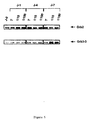
Le gène Grb3-3 a été isolé par criblage d'une banque d'ADN humain au moyen d'une sonde dérivée de la séquence du gène Grb2.The Grb3-3 gene was isolated by screening a human DNA library at using a probe derived from the Grb2 gene sequence.
500 000 phages recombinants Lambda gt11 portant des fragments d'ADN
issus d'une banque de placenta humaine (Clontech) ont été criblés au moyen d'une
sonde dérivée de la séquence du gène Grb2. La sonde utilisée correspond aux 8
premiers acides aminés de la protéine Grb2, et possède la séquence suivante:
10 clones positifs ont ainsi été identifiés. L'insert de ces 10 clones a été isolé
sous forme de fragments EcoRI, cloné dans le plasmide M13mp18 et séquencé. Parmi
ces 10 clones, 9 portaient des inserts identiques à la séquence Grb2. Un seul d'entreeux
portait un insert d'une taille inférieure au gène Grb2, en raison d'une délétion dans
le domaine SH2 (Figure 1). L'analyse de la séquence restante a révélé une identité
parfaite avec les régions correspondantes de Grb2, y compris dans les régions 5' et 3'
non codantes. La phase de lecture ouverte de ce clone code pour une protéine de 177
acides aminés (SEQ ID n° 1), comprenant 2 domaines SH3 bordant un domaine SH2
incomplet (figure 1). Les acides aminés délétés dans le domaine SH2 (résidus 60 à 100
de la protéine Grb2) correspondent aux résidus impliqués dans la liaison de Grb2 aux
peptides contenant des tyrosines phosphorylées.10 positive clones were thus identified. The insert of these 10 clones has been isolated
in the form of EcoRI fragments, cloned into the plasmid M13mp18 and sequenced. Among
of these 10 clones, 9 carried inserts identical to the Grb2 sequence. Only one of them
carried an insert smaller in size than the Grb2 gene, due to a deletion in
the SH2 domain (Figure 1). Analysis of the remaining sequence revealed an identity
perfect with the corresponding regions of Grb2, including in the 5 'and 3' regions
non-coding. The open reading phase of this clone codes for a protein of 177
amino acids (SEQ ID No. 1), comprising 2 SH3 domains bordering an SH2 domain
incomplete (Figure 1). Amino acids deleted in the SH2 domain (
Comme indiqué plus haut, la protéine Grb2 est le médiateur de l'interaction entre les récepteurs de facteurs de croissance phosphorylés et les facteurs SOS. Cet exemple démontre que la protéine Grb3-3 est incapable d'interagir avec le récepteur à l'EGF phosphorylé, mais qu'elle conserve sa faculté d'interagir avec un peptide riche en proline dérivé de la séquence du facteur SOS1 humain. As indicated above, the Grb2 protein is the mediator of the interaction between phosphorylated growth factor receptors and SOS factors. This example demonstrates that the Grb3-3 protein is unable to interact with the phosphorylated EGF, but that it retains its ability to interact with a peptide rich in proline derived from the human factor SOS1 sequence.
La capacité de liaison de Grb3-3 a été étudiée en utilisant des protéines de
fusion à la Glutathion-S-Transférase (GST), biotinylées. Ce type de fusion permet une
purification rapide et efficace des produits recombinants. Pour cela, les séquences de
l'invention ont été exprimées dans la souche d'E.coli TG1 sous forme de protéines de
fusion avec la GST selon la technique décrite par Smith et Johnson [Gene 67 (1988)
31]. Brièvement, les gènes Grb2 et Grb3-3 ont tout d'abord été modifiés par
introduction de part et d'autre des codons start et stop d'un site BamHI. Pour cela, les
phases ouvertes de lecture de ces gènes ont été amplifiées par PCR au moyen des
oligonucléotides suivants :
GAATTCGGATCCATGGAAGCCATCGCCAAATATGACTTC
GAATTCGGATCCTTAGACGTTCCGGTTCACGGGGGTGAC
GAATTC GGATCCATG GAAGCCATCGCCAAATATGACTTC
GAATTC GGATCCTTA GACGTTCCGGTTCACGGGGGTGAC
La partie soulignée correspond au site BamHI crée, suivi ou précédé des codons start et stop.The underlined part corresponds to the BamHI site created, followed or preceded by start and stop codons.
Les gènes ainsi amplifiés ont ensuite été clonés sous forme de fragments BamHI dans le vecteur pGEX 2T (Pharmacia) linéarisé par le même enzyme, en 3' et en phase d'un ADNc codant pour la GST. Les vecteurs ainsi obtenus ont ensuite été utilisés pour transformer la souche E.coli TG1. Les cellules ainsi transformées ont été précultivées une nuit à 37°C, diluées au 1/10e dans du milieu LB, ajoutées d'IPTG pour induire l'expression (2 heures, 25°C), puis cultivées 21 heures environ à 25°C. Les cellules ont ensuite été lysées, et les protéines de fusions produites purifiées par affinité sur colonne Agarose-GSH. Pour cela, le lysat bactérien est incubé en présence du gel (préparé et équilibré avec le tampon de lyse) pendant 15 minutes à 4°C. Après 3 lavages avec un tampon Tris-HCl pH 7,4, les protéines sont éluées en présence d'un tampon Tris-HCl pH 7,7 contenant un excès de GST. Le surnageant est récolté et centrifugé.The genes thus amplified were then cloned in the form of BamHI fragments into the vector pGEX 2T (Pharmacia) linearized by the same enzyme, in 3 ′ and in the phase of a cDNA coding for GST. The vectors thus obtained were then used to transform the E.coli TG1 strain. The cells thus transformed were precultivated overnight at 37 ° C, diluted 1 / 10th in LB medium, added with IPTG to induce expression (2 hours, 25 ° C), then cultured for approximately 21 hours at 25 ° vs. The cells were then lysed, and the fusion proteins produced purified by affinity on an Agarose-GSH column. For this, the bacterial lysate is incubated in the presence of the gel (prepared and balanced with the lysis buffer) for 15 minutes at 4 ° C. After 3 washes with a Tris-HCl buffer pH 7.4, the proteins are eluted in the presence of a Tris-HCl buffer pH 7.7 containing an excess of GST. The supernatant is collected and centrifuged.
Le même protocole a été utilisé pour préparer un mutant de Grb2 dans lequel la glycine 203 est remplacée par une arginine (Grb2G203R) et un mutant de Grb3-3 dans lequel la glycine 162 est remplacée par une arginine (Grb3-3G162R). Le mutant Grb2G203R a été décrit comme n'ayant plus d'activité dans un test de réinitiation de la synthèse d'ADN (Lowenstein et ai précitée). Le mutant Grb3-3G162R porte la même mutation sur la même position, et devrait donc également être inactif The same protocol was used to prepare a Grb2 mutant in which glycine 203 is replaced by an arginine (Grb2G203R) and a mutant of Grb3-3 in which glycine 162 is replaced by arginine (Grb3-3G162R). The mutant Grb2G203R has been described as having no more activity in a reinitiation test for DNA synthesis (Lowenstein and cited above). The mutant Grb3-3G162R carries the same mutation on the same position, and therefore should also be inactive
Ces mutants ont été préparés par mutagénèse par PCR sur les gènes Grb2 et
Grb3-3 en utilisant, en 5', l'oligonucléotide I décrit ci-dessus, et en 3', l'oligonucléotide
III suivant sur lequel le codon muté est souligné :
GACGTTCCGGTTCACGGGGGTGACATAATTGCGGGGAAACATGCGGGTC
GACGTTCCGGTTCACGGGGGTGACATAATT GCG GGGAAACATGCGGGTC
Les fragments ainsi amplifiés ont ensuite été élués, réamplifiés par PCR au moyen des oligonucléotides I et II, puis clonés dans le vecteur pGEX 2T. Les mutants ont ensuite été produits comme décrit ci-dessus.The fragments thus amplified were then eluted, re-amplified by PCR with using oligonucleotides I and II, then cloned into the vector pGEX 2T. Mutants were then produced as described above.
Les protéines de fusion à la GST (GST-Grb2, GST-Grb3-3, GST-Grb3-3G162R et la GST) ont ensuite été biotinylées par les techniques classiques connues de l'homme du métier (Cf techniques générales de biologie moléculaire ainsi que Mayer et al., PNAS 88 (1991) 627), et utilisées comme sondes pour déterminer la liaison au récepteur à l'EGF phosphorylé immobilisé (2.1.) puis à un peptide dérivé de hSOS1 (2.2.).GST fusion proteins (GST-Grb2, GST-Grb3-3, GST-Grb3-3G162R and GST) were then biotinylated by known conventional techniques skilled in the art (see general molecular biology techniques as well as Mayer et al., PNAS 88 (1991) 627), and used as probes to determine the binding to the immobilized phosphorylated EGF receptor (2.1.) then to a peptide derived from hSOS1 (2.2.).
Cet exemple démontre que, en dépit de sa délétion dans le domaine SH2, la protéine Grb3-3 possède un effet fonctionnel.This example demonstrates that, despite its deletion in the SH2 domain, the protein Grb3-3 has a functional effect.
L'activité de la protéine Grb3-3 a été étudié par détermination de sa capacité à coopérer avec ras pour la transactivation d'un promoteur possédant des éléments de réponse à ras (RRE), et gouvernant l'expression d'un gêne reporteur.The activity of the Grb3-3 protein was studied by determining its capacity to cooperate with ras for the transactivation of a promoter having elements of response to ras (RRE), and governing the expression of embarrassment reporter.
Le protocole utilisé a été décrit par exemple dans Schweighoffer et al., Science 256 (1992) 825. Brièvement, le promoteur utilisé est un promoteur synthétique composé du promoteur murin du gène de la thymidine kinase et de 4 éléments PEA1 répétés dérivés de l'enhancer du polyôme (Wasylyk et al., EMBO J. 7 (1988) 2475): promoteur Py-TK. Ce promoteur dirige l'expression du gène reporteur, en l'occurrence du gène bactérien de la chloramphénicol acétyl transférase (CAT) : vecteur Py-TK-CAT. Les vecteurs d'expression des gènes testés ont été construits par insertion desdits gènes, sous forme de fragments BamHI, au site BglII du plasmide pSV2. Ce site permet de placer les gènes sous controle du promoteur précoce SV40.The protocol used has been described for example in Schweighoffer et al., Science 256 (1992) 825. Briefly, the promoter used is a synthetic promoter composed of the murine promoter of the thymidine kinase gene and of 4 repeated PEA1 elements derived from the polyome enhancer (Wasylyk et al., EMBO J. 7 (1988) 2475): Py-TK promoter. This promoter directs the expression of the reporter gene, in this case the bacterial gene of chloramphenicol acetyl transferase (CAT): vector Py-TK-CAT. The expression vectors of the genes tested were constructed by inserting said genes, in the form of BamHI fragments, at the BglII site of the plasmid pSV2. This site allows genes to be placed under the control of the SV40 early promoter.
Des cellules ER22 à 40% de confluence ont été transfectées avec 0,5 µg du vecteur Py-TK-CAT seul (Py) ou en présence de du vecteur d'expression portant, sous contrôle du promoteur précoce de SV40, le gène : Grb2, 2 µg, Grb3-3, 2 µg, Grb2(G203R) 2 µg, Grb3-3(G162R) 2 µg, ou Grb3-3, 2 µg + Grb2, 2 µg. Dans chaque cas, la quantité totale d'ADN a été ajustée à 5 µg avec un vecteur d'expression sans insert. La transfection a été effectuée en présence de lipospermine (Transfectam, IBF-Sepracor). Les cellules ont été maintenues 48 heures en culture dans un milieu DMEM supplémenté par 0,5 % serum de veau fétal. L'activité CAT (transactivation du RRE) a ensuite été déterminée comme décrit par Wasylyk et al (PNAS 85 (1988) 7952).ER22 cells at 40% confluence were transfected with 0.5 μg of the Py-TK-CAT vector alone (Py) or in the presence of the expression vector carrying, under control of the SV40 early promoter, the gene: Grb2, 2 µg, Grb3-3, 2 µg, Grb2 (G203R) 2 µg, Grb3-3 (G162R) 2 µg, or Grb3-3, 2 µg + Grb2, 2 µg. In each case, the total amount of DNA was adjusted to 5 μg with an expression vector without insert. The transfection was carried out in the presence of lipospermine (Transfectam, IBF-Sepracor). The cells were maintained for 48 hours in culture in a medium DMEM supplemented with 0.5% fetal calf serum. CAT activity (transactivation of the RRE) was then determined as described by Wasylyk et al (PNAS 85 (1988) 7952).
Les résultats obtenus sont présentés sur la figure 3. Ils montrent clairement que l'expression de la protéine Grb3-3 s'oppose aux effets de l'activation d'un récepteur à facteur de croissance. Ils montrent également que Grb2 en excès s'oppose aux effets de Grb3-3 sur la réponse au facteur de croissance.The results obtained are presented in Figure 3. They clearly show that the expression of the protein Grb3-3 is opposed to the effects of the activation of a receptor growth factor. They also show that excess Grb2 opposes the effects of Grb3-3 on the response to the growth factor.
Cet exemple démontre l'implication directe de Grb3-3 dans l'apoptose cellulaire. Cette propriété offre des applications particulièrement avantageuses pour le traitement des pathologies résultant d'une prolifération cellulaire (cancers, resténose, etc).This example demonstrates the direct involvement of Grb3-3 in apoptosis cellular. This property offers particularly advantageous applications for the treatment of pathologies resulting from cell proliferation (cancers, restenosis, etc).
L'induction de l'apoptose cellulaire par Grb3-3 a été démontrée (i) par injection de la protéine recombinante dans les fibroblastes 3T3 et (ii) par par transfert de la séquence codant pour Grb3-3 dans les cellules 3T3.Induction of cellular apoptosis by Grb3-3 has been demonstrated (i) by injection of the recombinant protein into fibroblasts 3T3 and (ii) by transfer of the sequence encoding Grb3-3 in 3T3 cells.
La protéine Grb3-3 recombinante a été préparée sous forme de protéine de fusion avec la GST selon le protocole décrit dans l'exemple 2. La protéine de fusion a ensuite été traitée par la thrombine (0,25%, Sigma) pour séparer la partie GST, puis purifiée par chromatographie d'échange d'ions sur colone monoQ. Les fractions contenant la protéine recombinante ont ensuite été concentrées au moyen de microconcentreurs Microsep (Filtron) dans un tampon phosphate 20 mM (pH 7) contenant 100 mM NaCl. La protéine purifiée ainsi obtenue a été injectée (1 à 3 mg/ml) à des cellules 3T3 en culture au moyen d'un microinjecteur automatique Eppendorf. Les cellules ont ensuite été incubées à 34°C et photographiées à intervalles réguliers pour suivre les transformations morphologiques. Les résultats obtenus montrent que 5 heures après l'injection de Grb3-3 la majeure partie des cellules sont mortes alors que l'injection dans les mêmes conditions de Grb2 ou du mutant Grb3-3 (G162R) n'a aucun effet sur la viabilité des cellules.Recombinant Grb3-3 protein was prepared as protein fusion with GST according to the protocol described in Example 2. The fusion protein has then treated with thrombin (0.25%, Sigma) to separate the GST part, then purified by ion exchange chromatography on monoQ colone. Fractions containing the recombinant protein were then concentrated using Microsep (Filtron) microconcentrators in 20 mM phosphate buffer (pH 7) containing 100 mM NaCl. The purified protein thus obtained was injected (1 to 3 mg / ml) to 3T3 cells in culture using an automatic microinjector Eppendorf. The cells were then incubated at 34 ° C and photographed at regular intervals to follow morphological transformations. The results obtained show that 5 hours after the injection of Grb3-3 most of the cells died while injecting under the same conditions of Grb2 or mutant Grb3-3 (G162R) has no effect on cell viability.
Un plasmide a été construit comprenant la séquence SEQ ID n° 1 codant pour la protéine Grb3-3 sous le controle du promotyeur précoce du virus SV40.A plasmid was constructed comprising the sequence SEQ ID No. 1 coding for the Grb3-3 protein under the control of the SV40 virus early promoter.
Les fibroblastes 3T3 à 40 % de confluence ont été transfectés en présence de lipospermine (Transfectam, IBF-Sepracor) avec 0,5 ou 2 µg de ce plasmide d'expression. 48 heures après la transfection, 50% des cellules étaient en suspension dans le milieu, et les cellules restantes, adhérant à la paroi, présentaient des changements morphologiques très importants (figure 4). Une analyse par électrophorèse en gel d'agarose a montré par ailleurs que les cellules présentaient un pattern de fragmentation d'ADN oligo-nucléosomal caractéristique des cellules mortes (figure 4). En revanche, les cellules transfectées dans les mêmes conditions par un plasmide d'expression de Grb2, Grb3-3 (G162R) ou Grb2 (G203R) conservent une morphologie normale, sont toujours viables et ne présentent aucune fragmentation d'ADN. Comme le montre la figure 4, la co-expression de Grb2 permet de s'opposer aux effets de Grb3-3.3T3 fibroblasts at 40% confluence were transfected in the presence of lipospermine (Transfectam, IBF-Sepracor) with 0.5 or 2 µg of this plasmid of expression. 48 hours after transfection, 50% of the cells were in suspension in the medium, and the remaining cells, adhering to the wall, exhibited very significant morphological changes (Figure 4). An analysis by agarose gel electrophoresis also showed that the cells had a oligo-nucleosomal DNA fragmentation pattern characteristic of dead cells (figure 4). On the other hand, cells transfected under the same conditions by a Grb2, Grb3-3 (G162R) or Grb2 (G203R) expression plasmid retain a normal morphology, are still viable and show no fragmentation DNA. As shown in Figure 4, the co-expression of Grb2 makes it possible to oppose to the effects of Grb3-3.
Ces résultats montrent donc clairement que Grb3-3 constitue un gène tueur capable d'induire l'apoptose cellulaire. Comme indiqué ci-avant, cette propriété offre des applications particulièrement avantageuses pour le traitement des pathologies résultant d'une prolifération cellulaire telles que notamment les cancers, la resténose, etc.These results therefore clearly show that Grb3-3 constitutes a killer gene. capable of inducing cellular apoptosis. As stated above, this property offers particularly advantageous applications for the treatment of pathologies resulting from cell proliferation such as in particular cancers, restenosis, etc.
Cet exemple montre que, au cours du cycle d'infection des lymphocytes T par le virus HIV, la proportion relative des ARNm de Grb2 et Grb3-3 est modifiée, et que le messager de Grb3-3 est surexprimé au moment de la production virale massive et de la mort cellulaire.This example shows that during the cycle of infection of T lymphocytes by the HIV virus, the relative proportion of the Grb2 and Grb3-3 mRNAs is modified, and that the Grb3-3 messenger is overexpressed at the time of massive viral production and cell death.
Des lymphocytes du sang périphérique ont été infectés par le virus HIV-1 à
deux dilutions (1/10 et 1/100) pendant 1, 4 ou 7 jours. Les ARNm des cellules ont
ensuite été analysés par reverse-PCR au moyen d'oligonucléotides spécifiques de Grb2
et Grb3-3 pour déterminer la proportion relative des messagers de Grb2 et Grb3-3.
Les oligonucléotides spécifiques de Grb3-3 utilisés sont les suivants :
Les résultats obtenus sont présentés sur la figure 5. Ils montrent clairement
que 7 jours après l'infection par le virus HIV, l'ARNm de Grb3-3 est surexprimé.
Comme montré par dosage de la protéine p24 et de la transcriptase inverse du virus, le
jour 7 correspond également à la période à laquelle une production virale massive est
observée.


Claims (11)
- Nucleotide sequence represented in sequence SEQ ID No. 1.
- Nucleotide sequence represented in sequence SEQ ID No. 1 and capable of inhibiting, at least partially, the expression of the Grb2 or Grb3-3 protein.
- Polypeptide represented in sequence SEQ ID No. 1.
- Vector comprising a nucleotide sequence according to Claims 1 or 2.
- Vector according to Claim 4, characterized in that it is a viral vector.
- Vector according to Claim 5, characterized in that it is a vector derived from adenoviruses, retroviruses, AAVs, HSV virus, CMV or vaccinia Virus.
- Vector according to Claim 5 or 6, characterized in that it is a virus defective for replication.
- Pharmaceutical composition comprising one or more vectors according to one of Claims 4 to 7.
- Pharmaceutical composition comprising one or more nucleotide sequences according to Claims 1 or 2, in a form complexed with DEAE-dextran, with nuclear proteins or with lipids, in crude form or alternatively incorporated into liposomes.
- Use of a sequence according to Claim 1 or 2 or of a vector containing the said sequence for the preparation of a pharmaceutical composition intended for the treatment of cancers.
- Use of a compound capable of eliminating or blocking, at least partially, the cellular effects of Grb3-3 for the preparation of a pharmaceutical composition intended for the treatment of AIDS.
Applications Claiming Priority (3)
| Application Number | Priority Date | Filing Date | Title |
|---|---|---|---|
| FR9310971 | 1993-09-15 | ||
| FR9310971A FR2710074B1 (en) | 1993-09-15 | 1993-09-15 | GRB3-3 gene, its variants and their uses. |
| PCT/FR1994/000542 WO1995007981A1 (en) | 1993-09-15 | 1994-05-09 | Grb3-3 gene, variants and uses thereof |
Publications (2)
| Publication Number | Publication Date |
|---|---|
| EP0719328A1 EP0719328A1 (en) | 1996-07-03 |
| EP0719328B1 true EP0719328B1 (en) | 2000-10-11 |
Family
ID=9450882
Family Applications (1)
| Application Number | Title | Priority Date | Filing Date |
|---|---|---|---|
| EP94915598A Expired - Lifetime EP0719328B1 (en) | 1993-09-15 | 1994-05-09 | Grb3-3 gene, variants and uses thereof |
Country Status (27)
| Country | Link |
|---|---|
| US (2) | US5831048A (en) |
| EP (1) | EP0719328B1 (en) |
| JP (1) | JP3794697B2 (en) |
| KR (1) | KR100330477B1 (en) |
| CN (1) | CN1098930C (en) |
| AT (1) | ATE196926T1 (en) |
| AU (1) | AU698819B2 (en) |
| BR (1) | BR9407693A (en) |
| CA (1) | CA2169938A1 (en) |
| CZ (1) | CZ288882B6 (en) |
| DE (1) | DE69426121T2 (en) |
| DK (1) | DK0719328T3 (en) |
| ES (1) | ES2152313T3 (en) |
| FI (1) | FI120499B (en) |
| FR (1) | FR2710074B1 (en) |
| GR (1) | GR3034600T3 (en) |
| HU (1) | HU221516B (en) |
| IL (2) | IL126756A (en) |
| NO (1) | NO319144B1 (en) |
| NZ (1) | NZ266162A (en) |
| PL (1) | PL178402B1 (en) |
| PT (1) | PT719328E (en) |
| RU (1) | RU2159815C2 (en) |
| SK (1) | SK281123B6 (en) |
| UA (1) | UA46715C2 (en) |
| WO (1) | WO1995007981A1 (en) |
| ZA (1) | ZA947059B (en) |
Families Citing this family (14)
| Publication number | Priority date | Publication date | Assignee | Title |
|---|---|---|---|---|
| FR2732348B1 (en) * | 1995-03-31 | 1997-04-30 | Rhone Poulenc Rorer Sa | CONDITIONAL EXPRESSION SYSTEM |
| US6423824B1 (en) | 1995-06-07 | 2002-07-23 | Biogen, Inc. | CAIP-like gene family |
| US5837844A (en) * | 1995-06-07 | 1998-11-17 | Biogen, Inc. | CAIP-like gene family |
| US6171800B1 (en) | 1995-06-07 | 2001-01-09 | Biogen, Inc. | Method of making and binding CAIP polypeptides |
| US5855911A (en) * | 1995-08-29 | 1999-01-05 | Board Of Regents, The University Of Texas System | Liposomal phosphodiester, phosphorothioate, and P-ethoxy oligonucleotides |
| AU6342696A (en) * | 1996-06-27 | 1998-01-14 | Biogen, Inc. | The caip-like gene family |
| US7309692B1 (en) * | 1996-07-08 | 2007-12-18 | Board Of Regents, The University Of Texas System | Inhibition of chronic myelogenous leukemic cell growth by liposomal-antisense oligodeoxy-nucleotides targeting to GRB2 or CRK1 |
| US6977244B2 (en) * | 1996-10-04 | 2005-12-20 | Board Of Regents, The University Of Texas Systems | Inhibition of Bcl-2 protein expression by liposomal antisense oligodeoxynucleotides |
| US7285288B1 (en) | 1997-10-03 | 2007-10-23 | Board Of Regents, The University Of Texas System | Inhibition of Bcl-2 protein expression by liposomal antisense oligodeoxynucleotides |
| US7704962B1 (en) | 1997-10-03 | 2010-04-27 | Board Of Regents, The University Of Texas System | Small oligonucleotides with anti-tumor activity |
| EP0998945A1 (en) * | 1998-09-30 | 2000-05-10 | Transgene S.A. | Use of magnesium (Mg2+) for the enhancement of gene delivery in gene therapy |
| NZ522045A (en) | 2000-03-30 | 2007-05-31 | Whitehead Biomedical Inst | RNA sequence-specific mediators of RNA interference |
| ES2728168T3 (en) | 2000-12-01 | 2019-10-22 | Max Planck Gesellschaft | Small RNA molecules that mediate RNA interference |
| US20040043950A1 (en) * | 2002-01-03 | 2004-03-04 | Board Of Regents, The University Of Texas System | WT1 antisense oligos for the inhibition of breast cancer |
Family Cites Families (2)
| Publication number | Priority date | Publication date | Assignee | Title |
|---|---|---|---|---|
| JP3462498B2 (en) | 1991-01-18 | 2003-11-05 | ニューヨーク・ユニバーシティ | CDNA cloning method of receptor tyrosine kinase target protein and hGRB protein |
| WO1994007913A1 (en) * | 1992-09-25 | 1994-04-14 | Warner-Lambert Company | Peptide antagonists of sh2 binding and therapeutic uses thereof |
-
1993
- 1993-09-15 FR FR9310971A patent/FR2710074B1/en not_active Expired - Fee Related
-
1994
- 1994-05-09 WO PCT/FR1994/000542 patent/WO1995007981A1/en active IP Right Grant
- 1994-05-09 PT PT94915598T patent/PT719328E/en unknown
- 1994-05-09 CA CA002169938A patent/CA2169938A1/en not_active Abandoned
- 1994-05-09 CZ CZ1996758A patent/CZ288882B6/en not_active IP Right Cessation
- 1994-05-09 KR KR1019960701323A patent/KR100330477B1/en not_active IP Right Cessation
- 1994-05-09 NZ NZ266162A patent/NZ266162A/en not_active IP Right Cessation
- 1994-05-09 UA UA96030994A patent/UA46715C2/en unknown
- 1994-05-09 AT AT94915598T patent/ATE196926T1/en not_active IP Right Cessation
- 1994-05-09 EP EP94915598A patent/EP0719328B1/en not_active Expired - Lifetime
- 1994-05-09 BR BR9407693A patent/BR9407693A/en not_active Application Discontinuation
- 1994-05-09 RU RU96107888/13A patent/RU2159815C2/en active
- 1994-05-09 PL PL94313445A patent/PL178402B1/en not_active IP Right Cessation
- 1994-05-09 SK SK345-96A patent/SK281123B6/en not_active IP Right Cessation
- 1994-05-09 HU HU9600668A patent/HU221516B/en not_active IP Right Cessation
- 1994-05-09 JP JP50901595A patent/JP3794697B2/en not_active Expired - Fee Related
- 1994-05-09 US US08/612,857 patent/US5831048A/en not_active Ceased
- 1994-05-09 US US09/641,640 patent/USRE37952E1/en not_active Expired - Fee Related
- 1994-05-09 ES ES94915598T patent/ES2152313T3/en not_active Expired - Lifetime
- 1994-05-09 CN CN94193814A patent/CN1098930C/en not_active Expired - Fee Related
- 1994-05-09 DK DK94915598T patent/DK0719328T3/en active
- 1994-05-09 AU AU67247/94A patent/AU698819B2/en not_active Ceased
- 1994-05-09 DE DE69426121T patent/DE69426121T2/en not_active Expired - Fee Related
- 1994-09-12 IL IL126756A patent/IL126756A/en not_active IP Right Cessation
- 1994-09-12 IL IL11094294A patent/IL110942A/en not_active IP Right Cessation
- 1994-09-13 ZA ZA947059A patent/ZA947059B/en unknown
-
1996
- 1996-03-08 NO NO19960965A patent/NO319144B1/en not_active IP Right Cessation
- 1996-03-14 FI FI961202A patent/FI120499B/en active IP Right Grant
-
2000
- 2000-10-12 GR GR20000401514T patent/GR3034600T3/en not_active IP Right Cessation
Also Published As
Similar Documents
| Publication | Publication Date | Title |
|---|---|---|
| EP0719328B1 (en) | Grb3-3 gene, variants and uses thereof | |
| JP4351896B2 (en) | Pharmaceutical composition for cancer treatment comprising p38 / JTV-1 as active ingredient and screening method for pharmaceutical composition for cancer treatment | |
| Bissonnette et al. | The apoptotic and transcriptional transactivation activities of p53 can be dissociated | |
| WO1994029446A2 (en) | Intracellular binding proteins and use thereof | |
| WO2000025804A9 (en) | A method for inhibition of phospholamban activity for the treatment of cardiac disease and heart failure | |
| EP1049775B1 (en) | Human btrcp protein | |
| CA2190293C (en) | Method of cancer treatment by p53 protein control | |
| AU722973B2 (en) | Grb3-3 gene, variants and uses thereof | |
| US5770720A (en) | Ubiquitin conjugating enzymes having transcriptional repressor activity | |
| KR102566745B1 (en) | Runx3 modified protein for prevention or treatment of cancer | |
| EP0828832A2 (en) | $g(D)P62, ITS VARIANTS, NUCLEIC ACID SEQUENCES ENCODING THEM, AND THEIR USES IN ANTI-CANCER GENE THERAPY | |
| EP4313110A1 (en) | K-ras inhibitor | |
| Ibarra-Sánchez | Biochemical and cellular function of t-cell protein tyrosine phosphatase | |
| Jayaraman | Regulation of the DNA binding function of the tumor suppressor proteinp53 | |
| WO1996010636A1 (en) | Novel compounds useful for preparing anti-infective agents |
Legal Events
| Date | Code | Title | Description |
|---|---|---|---|
| PUAI | Public reference made under article 153(3) epc to a published international application that has entered the european phase |
Free format text: ORIGINAL CODE: 0009012 |
|
| 17P | Request for examination filed |
Effective date: 19960305 |
|
| AK | Designated contracting states |
Kind code of ref document: A1 Designated state(s): AT BE CH DE DK ES FR GB GR IE IT LI LU NL PT SE |
|
| 17Q | First examination report despatched |
Effective date: 19980821 |
|
| GRAG | Despatch of communication of intention to grant |
Free format text: ORIGINAL CODE: EPIDOS AGRA |
|
| GRAG | Despatch of communication of intention to grant |
Free format text: ORIGINAL CODE: EPIDOS AGRA |
|
| GRAH | Despatch of communication of intention to grant a patent |
Free format text: ORIGINAL CODE: EPIDOS IGRA |
|
| GRAH | Despatch of communication of intention to grant a patent |
Free format text: ORIGINAL CODE: EPIDOS IGRA |
|
| GRAA | (expected) grant |
Free format text: ORIGINAL CODE: 0009210 |
|
| AK | Designated contracting states |
Kind code of ref document: B1 Designated state(s): AT BE CH DE DK ES FR GB GR IE IT LI LU NL PT SE |
|
| REF | Corresponds to: |
Ref document number: 196926 Country of ref document: AT Date of ref document: 20001015 Kind code of ref document: T |
|
| REG | Reference to a national code |
Ref country code: CH Ref legal event code: NV Representative=s name: KIRKER & CIE SA Ref country code: CH Ref legal event code: EP |
|
| REG | Reference to a national code |
Ref country code: IE Ref legal event code: FG4D Free format text: FRENCH |
|
| REG | Reference to a national code |
Ref country code: DK Ref legal event code: T3 |
|
| REF | Corresponds to: |
Ref document number: 69426121 Country of ref document: DE Date of ref document: 20001116 |
|
| ITF | It: translation for a ep patent filed |
Owner name: BARZANO' E ZANARDO MILANO S.P.A. |
|
| GBT | Gb: translation of ep patent filed (gb section 77(6)(a)/1977) |
Effective date: 20001205 |
|
| REG | Reference to a national code |
Ref country code: ES Ref legal event code: FG2A Ref document number: 2152313 Country of ref document: ES Kind code of ref document: T3 |
|
| REG | Reference to a national code |
Ref country code: PT Ref legal event code: SC4A Free format text: AVAILABILITY OF NATIONAL TRANSLATION Effective date: 20001115 |
|
| REG | Reference to a national code |
Ref country code: FR Ref legal event code: CD |
|
| PLBE | No opposition filed within time limit |
Free format text: ORIGINAL CODE: 0009261 |
|
| STAA | Information on the status of an ep patent application or granted ep patent |
Free format text: STATUS: NO OPPOSITION FILED WITHIN TIME LIMIT |
|
| 26N | No opposition filed | ||
| REG | Reference to a national code |
Ref country code: GB Ref legal event code: IF02 |
|
| PGFP | Annual fee paid to national office [announced via postgrant information from national office to epo] |
Ref country code: NL Payment date: 20090504 Year of fee payment: 16 Ref country code: IE Payment date: 20090521 Year of fee payment: 16 Ref country code: ES Payment date: 20090605 Year of fee payment: 16 Ref country code: DK Payment date: 20090514 Year of fee payment: 16 |
|
| PGFP | Annual fee paid to national office [announced via postgrant information from national office to epo] |
Ref country code: SE Payment date: 20090512 Year of fee payment: 16 Ref country code: PT Payment date: 20090429 Year of fee payment: 16 Ref country code: LU Payment date: 20090616 Year of fee payment: 16 Ref country code: IT Payment date: 20090518 Year of fee payment: 16 Ref country code: FR Payment date: 20090515 Year of fee payment: 16 Ref country code: DE Payment date: 20090511 Year of fee payment: 16 Ref country code: AT Payment date: 20090514 Year of fee payment: 16 |
|
| PGFP | Annual fee paid to national office [announced via postgrant information from national office to epo] |
Ref country code: BE Payment date: 20090525 Year of fee payment: 16 |
|
| PGFP | Annual fee paid to national office [announced via postgrant information from national office to epo] |
Ref country code: CH Payment date: 20090513 Year of fee payment: 16 |
|
| PGFP | Annual fee paid to national office [announced via postgrant information from national office to epo] |
Ref country code: GR Payment date: 20090421 Year of fee payment: 16 Ref country code: GB Payment date: 20090506 Year of fee payment: 16 |
|
| REG | Reference to a national code |
Ref country code: PT Ref legal event code: MM4A Free format text: LAPSE DUE TO NON-PAYMENT OF FEES Effective date: 20101109 |
|
| BERE | Be: lapsed |
Owner name: S.A. *AVENTIS PHARMA Effective date: 20100531 |
|
| REG | Reference to a national code |
Ref country code: NL Ref legal event code: V1 Effective date: 20101201 |
|
| REG | Reference to a national code |
Ref country code: CH Ref legal event code: PL |
|
| REG | Reference to a national code |
Ref country code: DK Ref legal event code: EBP |
|
| GBPC | Gb: european patent ceased through non-payment of renewal fee |
Effective date: 20100509 |
|
| PG25 | Lapsed in a contracting state [announced via postgrant information from national office to epo] |
Ref country code: AT Free format text: LAPSE BECAUSE OF NON-PAYMENT OF DUE FEES Effective date: 20100509 |
|
| EUG | Se: european patent has lapsed | ||
| REG | Reference to a national code |
Ref country code: FR Ref legal event code: ST Effective date: 20110131 |
|
| PG25 | Lapsed in a contracting state [announced via postgrant information from national office to epo] |
Ref country code: PT Free format text: LAPSE BECAUSE OF NON-PAYMENT OF DUE FEES Effective date: 20101109 Ref country code: LI Free format text: LAPSE BECAUSE OF NON-PAYMENT OF DUE FEES Effective date: 20100531 Ref country code: CH Free format text: LAPSE BECAUSE OF NON-PAYMENT OF DUE FEES Effective date: 20100531 |
|
| REG | Reference to a national code |
Ref country code: IE Ref legal event code: MM4A |
|
| PG25 | Lapsed in a contracting state [announced via postgrant information from national office to epo] |
Ref country code: SE Free format text: LAPSE BECAUSE OF NON-PAYMENT OF DUE FEES Effective date: 20100510 Ref country code: IT Free format text: LAPSE BECAUSE OF NON-PAYMENT OF DUE FEES Effective date: 20100509 Ref country code: GR Free format text: LAPSE BECAUSE OF NON-PAYMENT OF DUE FEES Effective date: 20101202 Ref country code: BE Free format text: LAPSE BECAUSE OF NON-PAYMENT OF DUE FEES Effective date: 20100531 Ref country code: NL Free format text: LAPSE BECAUSE OF NON-PAYMENT OF DUE FEES Effective date: 20101201 |
|
| PG25 | Lapsed in a contracting state [announced via postgrant information from national office to epo] |
Ref country code: DE Free format text: LAPSE BECAUSE OF NON-PAYMENT OF DUE FEES Effective date: 20101201 Ref country code: IE Free format text: LAPSE BECAUSE OF NON-PAYMENT OF DUE FEES Effective date: 20100510 Ref country code: DK Free format text: LAPSE BECAUSE OF NON-PAYMENT OF DUE FEES Effective date: 20100531 |
|
| PG25 | Lapsed in a contracting state [announced via postgrant information from national office to epo] |
Ref country code: FR Free format text: LAPSE BECAUSE OF NON-PAYMENT OF DUE FEES Effective date: 20100531 |
|
| REG | Reference to a national code |
Ref country code: ES Ref legal event code: FD2A Effective date: 20110715 |
|
| PG25 | Lapsed in a contracting state [announced via postgrant information from national office to epo] |
Ref country code: GB Free format text: LAPSE BECAUSE OF NON-PAYMENT OF DUE FEES Effective date: 20100509 Ref country code: ES Free format text: LAPSE BECAUSE OF NON-PAYMENT OF DUE FEES Effective date: 20110705 |
|
| PG25 | Lapsed in a contracting state [announced via postgrant information from national office to epo] |
Ref country code: ES Free format text: LAPSE BECAUSE OF NON-PAYMENT OF DUE FEES Effective date: 20100510 |
|
| PG25 | Lapsed in a contracting state [announced via postgrant information from national office to epo] |
Ref country code: LU Free format text: LAPSE BECAUSE OF NON-PAYMENT OF DUE FEES Effective date: 20100509 |