EP0711577A2 - Verbessertes System zur Herzmuskelstimulierung mit Defibrillator - Google Patents
Verbessertes System zur Herzmuskelstimulierung mit Defibrillator Download PDFInfo
- Publication number
- EP0711577A2 EP0711577A2 EP95307975A EP95307975A EP0711577A2 EP 0711577 A2 EP0711577 A2 EP 0711577A2 EP 95307975 A EP95307975 A EP 95307975A EP 95307975 A EP95307975 A EP 95307975A EP 0711577 A2 EP0711577 A2 EP 0711577A2
- Authority
- EP
- European Patent Office
- Prior art keywords
- heart
- muscle
- stimulating
- defibrillating
- pacing
- Prior art date
- Legal status (The legal status is an assumption and is not a legal conclusion. Google has not performed a legal analysis and makes no representation as to the accuracy of the status listed.)
- Withdrawn
Links
- 210000003205 muscle Anatomy 0.000 claims abstract description 53
- 230000004936 stimulating effect Effects 0.000 claims abstract description 27
- 230000035939 shock Effects 0.000 claims abstract description 23
- 239000008280 blood Substances 0.000 claims abstract description 20
- 210000004369 blood Anatomy 0.000 claims abstract description 20
- 230000001862 defibrillatory effect Effects 0.000 claims abstract description 16
- 230000004217 heart function Effects 0.000 claims abstract description 14
- 210000005242 cardiac chamber Anatomy 0.000 claims abstract description 9
- 206010061592 cardiac fibrillation Diseases 0.000 claims abstract description 7
- 230000002600 fibrillogenic effect Effects 0.000 claims abstract description 7
- 210000002027 skeletal muscle Anatomy 0.000 claims description 45
- 230000000638 stimulation Effects 0.000 claims description 33
- 230000000747 cardiac effect Effects 0.000 claims description 20
- 238000002560 therapeutic procedure Methods 0.000 claims description 19
- 238000004146 energy storage Methods 0.000 claims description 8
- 230000004044 response Effects 0.000 claims description 8
- 230000003190 augmentative effect Effects 0.000 claims description 3
- 230000003213 activating effect Effects 0.000 claims 3
- 230000008878 coupling Effects 0.000 claims 1
- 238000010168 coupling process Methods 0.000 claims 1
- 238000005859 coupling reaction Methods 0.000 claims 1
- 208000003663 ventricular fibrillation Diseases 0.000 abstract description 11
- 210000004165 myocardium Anatomy 0.000 abstract description 4
- 230000002763 arrhythmic effect Effects 0.000 abstract description 2
- 206010003119 arrhythmia Diseases 0.000 description 12
- 238000000034 method Methods 0.000 description 12
- 230000006793 arrhythmia Effects 0.000 description 11
- 230000006870 function Effects 0.000 description 10
- 230000002861 ventricular Effects 0.000 description 9
- 206010019280 Heart failures Diseases 0.000 description 7
- 230000001746 atrial effect Effects 0.000 description 5
- 230000001684 chronic effect Effects 0.000 description 4
- 238000001514 detection method Methods 0.000 description 4
- 230000000694 effects Effects 0.000 description 4
- 230000001360 synchronised effect Effects 0.000 description 4
- 206010007559 Cardiac failure congestive Diseases 0.000 description 3
- 230000002159 abnormal effect Effects 0.000 description 3
- 230000003416 augmentation Effects 0.000 description 3
- 238000010586 diagram Methods 0.000 description 3
- 239000000835 fiber Substances 0.000 description 3
- 206010011906 Death Diseases 0.000 description 2
- 238000004891 communication Methods 0.000 description 2
- 230000008602 contraction Effects 0.000 description 2
- 210000002837 heart atrium Anatomy 0.000 description 2
- 210000005240 left ventricle Anatomy 0.000 description 2
- 210000005036 nerve Anatomy 0.000 description 2
- 230000001590 oxidative effect Effects 0.000 description 2
- 238000005086 pumping Methods 0.000 description 2
- 230000033764 rhythmic process Effects 0.000 description 2
- 210000005241 right ventricle Anatomy 0.000 description 2
- 230000001225 therapeutic effect Effects 0.000 description 2
- 210000001519 tissue Anatomy 0.000 description 2
- 208000020446 Cardiac disease Diseases 0.000 description 1
- PMATZTZNYRCHOR-CGLBZJNRSA-N Cyclosporin A Chemical compound CC[C@@H]1NC(=O)[C@H]([C@H](O)[C@H](C)C\C=C\C)N(C)C(=O)[C@H](C(C)C)N(C)C(=O)[C@H](CC(C)C)N(C)C(=O)[C@H](CC(C)C)N(C)C(=O)[C@@H](C)NC(=O)[C@H](C)NC(=O)[C@H](CC(C)C)N(C)C(=O)[C@H](C(C)C)NC(=O)[C@H](CC(C)C)N(C)C(=O)CN(C)C1=O PMATZTZNYRCHOR-CGLBZJNRSA-N 0.000 description 1
- 108010036949 Cyclosporine Proteins 0.000 description 1
- 208000013875 Heart injury Diseases 0.000 description 1
- 206010042434 Sudden death Diseases 0.000 description 1
- 230000005856 abnormality Effects 0.000 description 1
- 230000003288 anthiarrhythmic effect Effects 0.000 description 1
- 239000003416 antiarrhythmic agent Substances 0.000 description 1
- 238000010009 beating Methods 0.000 description 1
- 230000009286 beneficial effect Effects 0.000 description 1
- 230000002051 biphasic effect Effects 0.000 description 1
- 208000006218 bradycardia Diseases 0.000 description 1
- 230000036471 bradycardia Effects 0.000 description 1
- 239000003990 capacitor Substances 0.000 description 1
- 238000013194 cardioversion Methods 0.000 description 1
- 229960001265 ciclosporin Drugs 0.000 description 1
- 229930182912 cyclosporin Natural products 0.000 description 1
- 230000006378 damage Effects 0.000 description 1
- 230000009977 dual effect Effects 0.000 description 1
- 238000007667 floating Methods 0.000 description 1
- 210000000600 glycolytic muscle fiber Anatomy 0.000 description 1
- 230000010247 heart contraction Effects 0.000 description 1
- 208000019622 heart disease Diseases 0.000 description 1
- 230000001744 histochemical effect Effects 0.000 description 1
- 239000007943 implant Substances 0.000 description 1
- 238000007918 intramuscular administration Methods 0.000 description 1
- 238000002955 isolation Methods 0.000 description 1
- 230000007774 longterm Effects 0.000 description 1
- 230000028161 membrane depolarization Effects 0.000 description 1
- 208000031225 myocardial ischemia Diseases 0.000 description 1
- 210000004126 nerve fiber Anatomy 0.000 description 1
- 230000001537 neural effect Effects 0.000 description 1
- 230000001991 pathophysiological effect Effects 0.000 description 1
- 230000010412 perfusion Effects 0.000 description 1
- 230000000737 periodic effect Effects 0.000 description 1
- 230000000750 progressive effect Effects 0.000 description 1
- 230000004083 survival effect Effects 0.000 description 1
- 238000002054 transplantation Methods 0.000 description 1
- 238000011282 treatment Methods 0.000 description 1
- 206010047302 ventricular tachycardia Diseases 0.000 description 1
Images



Classifications
-
- A—HUMAN NECESSITIES
- A61—MEDICAL OR VETERINARY SCIENCE; HYGIENE
- A61N—ELECTROTHERAPY; MAGNETOTHERAPY; RADIATION THERAPY; ULTRASOUND THERAPY
- A61N1/00—Electrotherapy; Circuits therefor
- A61N1/18—Applying electric currents by contact electrodes
- A61N1/32—Applying electric currents by contact electrodes alternating or intermittent currents
- A61N1/36—Applying electric currents by contact electrodes alternating or intermittent currents for stimulation
- A61N1/36042—Applying electric currents by contact electrodes alternating or intermittent currents for stimulation of grafted tissue, e.g. skeletal muscle
-
- A—HUMAN NECESSITIES
- A61—MEDICAL OR VETERINARY SCIENCE; HYGIENE
- A61N—ELECTROTHERAPY; MAGNETOTHERAPY; RADIATION THERAPY; ULTRASOUND THERAPY
- A61N1/00—Electrotherapy; Circuits therefor
- A61N1/18—Applying electric currents by contact electrodes
- A61N1/32—Applying electric currents by contact electrodes alternating or intermittent currents
- A61N1/36—Applying electric currents by contact electrodes alternating or intermittent currents for stimulation
- A61N1/362—Heart stimulators
- A61N1/3621—Heart stimulators for treating or preventing abnormally high heart rate
- A61N1/3622—Heart stimulators for treating or preventing abnormally high heart rate comprising two or more electrodes co-operating with different heart regions
-
- A—HUMAN NECESSITIES
- A61—MEDICAL OR VETERINARY SCIENCE; HYGIENE
- A61N—ELECTROTHERAPY; MAGNETOTHERAPY; RADIATION THERAPY; ULTRASOUND THERAPY
- A61N1/00—Electrotherapy; Circuits therefor
- A61N1/18—Applying electric currents by contact electrodes
- A61N1/32—Applying electric currents by contact electrodes alternating or intermittent currents
- A61N1/38—Applying electric currents by contact electrodes alternating or intermittent currents for producing shock effects
- A61N1/39—Heart defibrillators
- A61N1/3956—Implantable devices for applying electric shocks to the heart, e.g. for cardioversion
- A61N1/3962—Implantable devices for applying electric shocks to the heart, e.g. for cardioversion in combination with another heart therapy
-
- A—HUMAN NECESSITIES
- A61—MEDICAL OR VETERINARY SCIENCE; HYGIENE
- A61N—ELECTROTHERAPY; MAGNETOTHERAPY; RADIATION THERAPY; ULTRASOUND THERAPY
- A61N1/00—Electrotherapy; Circuits therefor
- A61N1/18—Applying electric currents by contact electrodes
- A61N1/32—Applying electric currents by contact electrodes alternating or intermittent currents
- A61N1/38—Applying electric currents by contact electrodes alternating or intermittent currents for producing shock effects
- A61N1/39—Heart defibrillators
- A61N1/3956—Implantable devices for applying electric shocks to the heart, e.g. for cardioversion
- A61N1/3962—Implantable devices for applying electric shocks to the heart, e.g. for cardioversion in combination with another heart therapy
- A61N1/39622—Pacing therapy
-
- A—HUMAN NECESSITIES
- A61—MEDICAL OR VETERINARY SCIENCE; HYGIENE
- A61N—ELECTROTHERAPY; MAGNETOTHERAPY; RADIATION THERAPY; ULTRASOUND THERAPY
- A61N1/00—Electrotherapy; Circuits therefor
- A61N1/18—Applying electric currents by contact electrodes
- A61N1/32—Applying electric currents by contact electrodes alternating or intermittent currents
- A61N1/36—Applying electric currents by contact electrodes alternating or intermittent currents for stimulation
- A61N1/362—Heart stimulators
- A61N1/3627—Heart stimulators for treating a mechanical deficiency of the heart, e.g. congestive heart failure or cardiomyopathy
Definitions
- This invention relates to a combined muscle stimulation, pacing and defibrillation apparatus and its method of operation for detecting abnormalities of a patient's heart and, in response to such detection, administering defibrillation therapy. More particularly, this invention relates to a pacing apparatus and method that employs defibrillation therapy which comprises the coordination, control and generation of stimulation pulses to both the patient's heart and to a muscle graft that has been previously wrapped around the heart, with means being provided to insure that the defibrillation therapy is most efficient achieved.
- congestive heart failure is a pathophysiological state in which cardiac output is inadequate to meet physiological requirements of the body.
- the mortality rate for congestive heart failure is greater than 50% within 5 years of onset.
- Treatments for severe chronic cardiac insufficiency include heart transplants, artificial heart implants and cardiomyoplasty.
- Cardiac transplantation, using cyclosporine to inhibit tissue rejection is a very successful technique for prolonging a cardiac patient's life, improving the survival rate to 80% at 1 year.
- the transplant operation is very expensive and heart availability is limited.
- the artificial heart has had very limited success.
- Dynamic cardiomyoplasty is a surgical and electrical therapeutic technique used to overcome or at least alleviate cardiac insufficiency. This technique consists of using a skeletal muscle flap which is dissected from a patient, while maintaining its neural tissues and neurovascular structures, surgically placed around the patient's heart.
- An electrical stimulation device with an electrical pulse generator and intramuscular electrodes, is implanted for performing muscle electrical stimulation in synchrony with ventricular systole to support cardiac pumping.
- Repeated stimulation of the skeletal muscle transforms it into a fatigue-resistant muscle suitable for chronic ventricular assistance, enabling dynamic cardiomyoplasty.
- the skeletal muscle is then trained to function so as to assist cardiac muscle to increase the patient's cardiac output.
- Sequential and progressive skeletal muscle electrical stimulation causes glycolytic muscle fibers, predominant in skeletal muscle, to take the form of oxidative fibers. Oxidative fibers are resistant to fatigue and have histochemical and biochemical characteristics of myocardium.
- cardiomyoplasty patients suffering from ventricular fibrillations are treated by one of several methods, depending on the circumstances at the time of the onset of fibrillation.
- Each of these methods has several disadvantages. Ventricular fibrillations occurring in the hospital must be first confirmed by doctors or paramedical personnel. Standard high energy defibrillation shocks are then applied to the patient. Of course, these shocks must be applied by people having special training. If the ventricular fibrillation occurs outside a hospital, the patient must wait for trained medical help to arrive and apply the high energy shocks as discussed above.
- An objective is to provide a defibrillation method and apparatus which requires defibrillation shocks of lower amplitude than in prior art devices resulting in a safer procedure and reduced power consumption.
- a further objective is to provide a method and apparatus for performing a defibrillation function for a cardiomyoplasty patient efficiently by the stimulation of a skeletal muscle disposed about the heart.
- Yet another objective is to provide a combined apparatus for performing a plurality of cardiac functions including normal rate responsive pacing, arrhythmic pacing for terminating arrhythmia, cardiac pumping assisted by cardiomyoplasty and effective low power defibrillation.
- the present inventors have realized that a problem with existing defibrillation therapies is that they require large electric defibrillation shocks to compensate for dissipation of the shocks in the blood contained in the heart.
- This problem is resolved in the present invention by providing a time period just prior to the application of defibrillation shocks, during which the skeletal muscle is stimulated to cause the muscle to contract around the heart and thereby eject the blood contained in the heart's chambers.
- a pacemaker system for stimulating a patient's heart.
- the patient is also provided with a skeletal muscle grafted onto the heart to assist its cardiac functions.
- the pacemaker system includes means for analyzing signals from the heart for detecting, classifying and correcting abnormal episodes or conditions of the heart such as arrhythmia.
- the pacemaker system further includes heart stimulating means for generating and delivering stimulating pulses of various amplitudes to the heart. These stimulating means may include pacing electrodes, as well as defibrillating electrodes, receiving the various pulses. These pulses may be characteristic of standard pacemaker stimulation, cardioversion or defibrillation shocks.
- the pacemaker system also includes at least one muscle stimulation electrode which is adapted to be placed in electrical contact with the muscle graft.
- This electrode receives muscle stimulating pulses from a muscle pulse stimulating means.
- the muscle pulse stimulating means generates stimulating pulse trains which are delivered to the muscle and have an amplitude, duration and frequency to effect a desired contraction of the muscle for assisting the heart in its cardiac functions.
- the muscle stimulating pulses are synchronized with the natural or paced periodic heart contractions.
- the pacemaker system activates muscle stimulation and fibrillation control means which controls and synchronizes operations of the heart stimulating means and the muscle pulse stimulating means to apply a defibrillation therapy.
- the skeletal muscle is stimulated first to expel blood from the heart chambers and then defibrillation shocks are applied to the heart.
- the device is also adapted to perform other therapeutic functions to correct other such as arrhythmia, ventricular tachycardia, and so on. In these modes of operations, pacing and muscle stimulation are combined synchronously corresponding to the classification of abnormal condition determined by a detecting and classifying means.
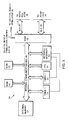
- FIG. 1 there is depicted a block diagram of a pacemaker system 10.
- the system 10 is designed to be implanted within a patient and includes a pulse module or pacemaker 12 and leads for connecting module 10 to a patient's heart 14 and skeletal muscle 16.
- These leads include an atrial cardiac lead 18, a ventricular cardiac lead 20 extending to the atrium and the ventricle of the patient's heart 14, respectively, as well as a defibrillation electrode lead 22 and a skeletal muscle lead 23.
- the pulse module 12 generally includes an interface 24, a microprocessor 26, a defibrillator 28, a skeletal muscle simulator 30 and a power supply 32.
- the interface 24 is provided for the detection of analog signals representing cardiac electrical activity and for the delivery of pacing pulses to the heart over leads 18 and 20.
- the microprocessor 26, in response to various inputs received from the interface 24 as well as from a defibrillator 16, performs various operations so as to generate different control and data outputs to pacemaker interface 24, skeletal muscle stimulator 30 and defibrillator 28.
- the power supply 32 provides reliable voltage to the other components of the pulse module. When the power supply 32 is nearly exhausted it generates an End-Of-Life (EOL) signal on line 33 to the microprocessor 26.
- EOL End-Of-Life
- Skeletal muscle stimulator 30 generates electrical pulses on a skeletal muscle lead 23 for stimulating the skeletal muscle 16.
- the pulses to muscle 16 are generated according to timed control signals from microprocessor 26 received via skeletal muscle stimulation control bus 32.
- the defibrillator 28 has an energy storage device 29 which may consist of one or more capacitors (not shown), and is used to produce high voltage pulses responsive to control signals from microprocessor 26 received on bus 40.
- the defibrillator electrode lead 22 transmits the defibrillator shocks from the implanted pacemaker 12 to the heart 14.
- the microprocessor 26 is connected to a Random Access /Read Only memory unit 34 by an address and data bus 36.
- the pacemaker 12 also includes a telemetry circuit 46 over which control signals and cardiac signals can be exchanged with the outside world.
- the telemetry circuit 46 is coupled to interface 24 by a bus 48.
- microprocessor 26 and interface 24 are connected by a data and communication bus 38 for exchanging various data.
- Microprocessor 26 transmits control signals, according to the description below, over skeletal muscle stimulation control bus 32 to the skeletal muscle stimulator 30. As also more fully described below, microprocessor 26 is connected to defibrillator 16 by a bus 40.
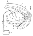
- FIG. 2 illustrates an example of a cardiac assist arrangement 200 for performing cardiac pacing, long-term stimulation of skeletal muscles using systolic augmentation, and defibrillation of the heart.
- a latissimus dorsi skeletal muscle 16 is positioned over the right ventricle 204 and left ventricle 206 of a patient's heart 14.
- the longitudinal fibers 210 of the latissimus dorsi are oriented generally parallel to the longitudinal axes of the ventricles 204 and 206 and interventricular septum 212 of the heart.
- the skeletal muscle 16 is positioned in this manner so that when it is stimulated, it compresses the ventricles, particularly the left ventricle 206, and augments the force of right and left ventricular contractions.
- the ventricular cardiac lead 20 is implanted in the heart's right ventricle 204 and the skeletal muscle lead 24 extends from the muscle stimulator 30 (shown in Figure 1) to the latissimus dorsi muscle 16.
- the skeletal muscle lead 24 may be placed directly on a nerve or placed near nerve branches within the latissimus dorsi muscle 16 to provide for selective depolarization of intact motor nerve fibers. In this manner the muscle 16 is forced selectively to contract about the heart 14, for either systolic augmentation or defibrillation.
- microprocessor 26 comprises to timers 51 and 52, a CPU 53, a vectored interrupts block 54, a ROM 55, a RAM 56, an external memory 57, a port 42 and an internal communications bus 44.
- RAM 56 acts as a scratch pad and active memory during execution of the various programs stored in ROM 55 and used by microprocessor 26.
- These programs include system supervisory programs, detection algorithms for detecting and confirming various arrhythmias, and programming for implementing the flow chart of Figure 4 as well as storage programs for storing, in external memory 57, data concerning the functioning of module 12 and the electrogram provided by ventricular cardiac lead 20 (FIG. 1).
- Timers 51 and 52, and associated control software implement timing functions required by microprocessor 26.
- Microprocessor 26 receives various status and control inputs from the interface 24 and defibrillator 16 on buses 38 and 40 and produces the control signals on these buses and bus 32 necessary for various functions including normal rate responsive pacing of the atria and/or ventricle, skeletal muscle stimulation and defibrillation.
- the atrial and ventricular pace control inputs derived from the signals received from the heart on leads 18 and 20 determine the respective types of atrial and/or ventricular pacing to be delivered to the heart. Details of this function are described in commonly assigned Patent No. 4,869,252 to Norma Louise Gilli, issued September 26, 1989, and entitled “Apparatus And Method For Controlling Pulse Energy In Antitachyarrhythmia And Bradycardia Pacing Devices," which description is incorporated herein by reference.
- the skeletal muscle stimulator 30 shown in Figure 1 receives input signals from microprocessor 26 over the skeletal muscle stimulation control bus 32.
- the interface 24 supplies to stimulator 30 battery power over two battery leads B+ and B- which provide energy for biphasic skeletal muscle stimulation.
- the battery leads B+, B- are floating with respect to the power supplied by supply 32 to provide signal isolation.
- the signal XCHB_L is a cross channel blanking control signal used to disable cardiac sensing by the interface 24 during generation of a skeletal muscle stimulation pulse. Therefore this signal prevents the microprocessor 26 from incorrectly classifying a skeletal muscle stimulation pulse as an episode of intrinsic cardiac activity. Details of the skeletal muscle stimulator are provided in commonly assigned U.S. Patent No. 5,251,621, incorporated herein by reference.
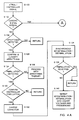
- step S100 the microprocessor receives and analyzes signals form the atrial and ventricle leads.
- step S102 the microprocessor determines from these signals whether the cardiac function of heart 14 is normal, i.e. whether corrective therapy is required.
- the microprocessor determines in step S110 whether the pacing is required. If pacing is required, in step S112 the microprocessor enters a pacing mode wherein it determines the pacing parameters, and in step S114 the pacing signals are generated for atrial and/or ventricle pacing. In step S116 the microprocessor determines if skeletal muscle stimulation is required. Skeletal muscle stimulation signals are generated by the microprocessor in step S118, which signals are synchronous with the pacing signals generated in step S114. These signals are applied to the muscle 16 to augument the functions of the heart.
- step S120 the microprocessor determines if skeletal muscle stimulation is beneficial for augmenting the patient's cardiac functions. If skeletal muscle stimulation is required, than in step S122 the microprocessor generates muscle stimulating signals synchronous with the intrinsic or natural heart beat of the patient.
- step S104 the microprocessor checks for arrhythmia. If arrhythmia is detected, in step S106 the arrhythmia is confirmed and classified. As part of this classification, the microprocessor checks for ventricular fibrillation, in step S124. For other types of arrhythmia, appropriate therapy is provided in step S108. If ventricular fibrillation is detected in step S124, the microprocessor enters into a defibrillation mode. Signals characterizing the operation of pacemaker system 10 in this mode are shown in Figure 5. More specifically, the top row of Figure 6 shows the ECG for heart 14.
- the heart 14 is shown beating normal at its intrinsic or natural sinus rhythm, as indicated by the ECG pulses 72, 74, 76, 78,and 92, 94, 96.
- the pacemaker system goes into the defibrillation mode during which the heart undergoes, and is treated for, ventricular fibrillation.
- the second row of Figure 5 shows the stimulation signals on skeletal muscle lead 24.
- bursts of signal such as at 80 and 82 are applied on skeletal muscle stimulation lead 24 to augment the normal cardiac function of the heart as previously discussed.
- burst 80 is applied synchronously with pulse 72
- burst 82 is applied synchronously with pulse 76 and 80, and so on.
- No bursts are applied during pulses 74 and 78 to allow the muscle 16 to rest.
- the third row of Figure 5 shows activities related to the defibrillation function.
- step S130 the microprocessor confirms that the heart is experiencing ventricular defibrillation. Because fibrillation signals are high energy signals, the defibrillator 28 needs some initialization time during which its energy storage device 29 is charged up. This is accomplished in step S132. After the energy storage element is charged up, in step S134, the microprocessor 26 generates control signals on bus 32, in response to which the skeletal muscle stimulator generates an extended burst of signals 84 on line 24. However as opposed to bursts 80 and 82, burst 84 is not designed to augmented the cardiac function of the heart 14 and is not synchronous with its beats.
- this burst 84 is applied to the muscle 16 to cause the muscle to contract about the heart in order to force the blood from the cardiac chambers to be expelled.
- the amplitude of the signals forming burst 84 is somewhat larger than the amplitude of bursts 80, 82.
- the duration of the burst 84 is also somewhat longer.
- the defibrillator 28 generates a defibrillating high energy shock signal 88 on lead 22. Because the blood was substantially ejected from the heart chambers in step S134, the full impact of shock signal 88 is applied to the heart muscles to stop the ventricular fibrillation.
- step S138 the microprocessor monitors the heart 14 for a preselected period to determine if the fibrillation has stopped and the heart has resumed its normal sinus rhythm. If normal cardiac function is resumed, as indicated by period T3, the microprocessor returns to the normal pacing mode of operation. Otherwise, step S140 orders the defibrillation therapy of period T2 (i.e. steps S130-138) to be repeated. If necessary, the amplitude, and the energy content of the defibrillation shock signal may be increased for this second defibrillation therapy.
- the subject pacemaker system provides a plurality of functions including pacing, arrnythmia therapy, cardiac function augmentation using cardiomyoplasty.
- defibrillation consisting of the steps of expelling the blood from the heart chambers, and then applying defibrillation shocks. Because the shocks are not partially dissipated in extraneous blood within the heart chambers, the defibrillating shocks have a lower amplitude and/or energy content than in other prior art devices. As a result, the defibrillation therapy provided by the subject system is safer for the patient and is more energy efficient.
Landscapes
- Health & Medical Sciences (AREA)
- Cardiology (AREA)
- Radiology & Medical Imaging (AREA)
- Engineering & Computer Science (AREA)
- Biomedical Technology (AREA)
- Nuclear Medicine, Radiotherapy & Molecular Imaging (AREA)
- Life Sciences & Earth Sciences (AREA)
- Animal Behavior & Ethology (AREA)
- General Health & Medical Sciences (AREA)
- Public Health (AREA)
- Veterinary Medicine (AREA)
- Heart & Thoracic Surgery (AREA)
- Rheumatology (AREA)
- Transplantation (AREA)
- Electrotherapy Devices (AREA)
Applications Claiming Priority (2)
| Application Number | Priority Date | Filing Date | Title |
|---|---|---|---|
| US336189 | 1994-11-08 | ||
| US08/336,189 US5500004A (en) | 1994-11-08 | 1994-11-08 | Cardio myostimulation system with defibrillation |
Publications (2)
| Publication Number | Publication Date |
|---|---|
| EP0711577A2 true EP0711577A2 (de) | 1996-05-15 |
| EP0711577A3 EP0711577A3 (de) | 1999-04-21 |
Family
ID=23314955
Family Applications (1)
| Application Number | Title | Priority Date | Filing Date |
|---|---|---|---|
| EP95307975A Withdrawn EP0711577A3 (de) | 1994-11-08 | 1995-11-08 | Verbessertes System zur Herzmuskelstimulierung mit Defibrillator |
Country Status (2)
| Country | Link |
|---|---|
| US (1) | US5500004A (de) |
| EP (1) | EP0711577A3 (de) |
Cited By (1)
| Publication number | Priority date | Publication date | Assignee | Title |
|---|---|---|---|---|
| WO2005035052A1 (en) * | 2003-10-07 | 2005-04-21 | Medtronic, Inc. | Method and apparatus for controlling extra-systolic stimulation (ess) therapy using ischemia detection |
Families Citing this family (17)
| Publication number | Priority date | Publication date | Assignee | Title |
|---|---|---|---|---|
| US5713924A (en) * | 1995-06-27 | 1998-02-03 | Medtronic, Inc. | Defibrillation threshold reduction system |
| US5562595A (en) * | 1995-08-17 | 1996-10-08 | Medtronic, Inc. | Multiple therapy cardiac assist device having battery voltage safety monitor |
| US5697952A (en) * | 1995-08-17 | 1997-12-16 | Medtronic, Inc. | Cardiac assist device having muscle augementation after confirmed arrhythmia and method |
| US5716379A (en) * | 1995-08-17 | 1998-02-10 | Medtronic, Inc. | Cardiac assist device having muscle augmentation prior to defibrillation |
| US5658237A (en) * | 1995-08-17 | 1997-08-19 | Medtronic, Inc. | Cardiac assist device having improved muscle stimulation |
| US5954752A (en) * | 1997-04-30 | 1999-09-21 | Medtronic, Inc. | Cardioversion energy reduction system |
| US5897575A (en) * | 1997-10-24 | 1999-04-27 | Pacesetter, Inc. | Arrhythmia classification system with reliability indication that allows for low quality input signals in pacemakers |
| US5928131A (en) * | 1997-11-26 | 1999-07-27 | Vascor, Inc. | Magnetically suspended fluid pump and control system |
| US6091989A (en) * | 1998-04-08 | 2000-07-18 | Swerdlow; Charles D. | Method and apparatus for reduction of pain from electric shock therapies |
| US20040116969A1 (en) | 2002-08-26 | 2004-06-17 | Owen James M. | Pulse detection using patient physiological signals |
| US20040220628A1 (en) * | 2003-04-11 | 2004-11-04 | Wagner Darrell Orvin | Subcutaneous defibrillation timing correlated with induced skeletal muscle contraction |
| US20150364861A1 (en) * | 2014-06-17 | 2015-12-17 | Minnetronix, Inc. | Implantable connection mechanisms for continuous high power delivery |
| DE102015112098A1 (de) | 2014-07-25 | 2016-01-28 | Minnetronix, Inc. | Spulenparameter und Steuerung |
| US9855376B2 (en) | 2014-07-25 | 2018-01-02 | Minnetronix, Inc. | Power scaling |
| US10342908B2 (en) | 2015-01-14 | 2019-07-09 | Minnetronix, Inc. | Distributed transformer |
| US10406267B2 (en) | 2015-01-16 | 2019-09-10 | Minnetronix, Inc. | Data communication in a transcutaneous energy transfer system |
| US10193395B2 (en) | 2015-04-14 | 2019-01-29 | Minnetronix, Inc. | Repeater resonator |
Citations (5)
| Publication number | Priority date | Publication date | Assignee | Title |
|---|---|---|---|---|
| US4796630A (en) | 1987-03-23 | 1989-01-10 | Telectronics N.V. | Cardiac pacemaker with combined defibrillation and electrosurgery protection |
| US4869252A (en) | 1988-01-11 | 1989-09-26 | Telectronics N.V. | Apparatus and method for controlling pulse energy in antitachyarrhythmia and bradycardia pacing device |
| US5178140A (en) | 1991-09-05 | 1993-01-12 | Telectronics Pacing Systems, Inc. | Implantable medical devices employing capacitive control of high voltage switches |
| US5215083A (en) | 1991-10-07 | 1993-06-01 | Telectronics Pacing Systems, Inc. | Apparatus and method for arrhythmia induction in arrhythmia control system |
| US5251621A (en) | 1991-12-18 | 1993-10-12 | Telectronics Pacing Systems, Inc. | Arrhythmia control pacer using skeletal muscle cardiac graft stimulation |
Family Cites Families (3)
| Publication number | Priority date | Publication date | Assignee | Title |
|---|---|---|---|---|
| US5098369A (en) * | 1987-02-27 | 1992-03-24 | Vascor, Inc. | Biocompatible ventricular assist and arrhythmia control device including cardiac compression pad and compression assembly |
| US4925443A (en) * | 1987-02-27 | 1990-05-15 | Heilman Marlin S | Biocompatible ventricular assist and arrhythmia control device |
| US5390679A (en) * | 1993-06-03 | 1995-02-21 | Eli Lilly And Company | Continuous cardiac output derived from the arterial pressure waveform using pattern recognition |
-
1994
- 1994-11-08 US US08/336,189 patent/US5500004A/en not_active Expired - Fee Related
-
1995
- 1995-11-08 EP EP95307975A patent/EP0711577A3/de not_active Withdrawn
Patent Citations (5)
| Publication number | Priority date | Publication date | Assignee | Title |
|---|---|---|---|---|
| US4796630A (en) | 1987-03-23 | 1989-01-10 | Telectronics N.V. | Cardiac pacemaker with combined defibrillation and electrosurgery protection |
| US4869252A (en) | 1988-01-11 | 1989-09-26 | Telectronics N.V. | Apparatus and method for controlling pulse energy in antitachyarrhythmia and bradycardia pacing device |
| US5178140A (en) | 1991-09-05 | 1993-01-12 | Telectronics Pacing Systems, Inc. | Implantable medical devices employing capacitive control of high voltage switches |
| US5215083A (en) | 1991-10-07 | 1993-06-01 | Telectronics Pacing Systems, Inc. | Apparatus and method for arrhythmia induction in arrhythmia control system |
| US5251621A (en) | 1991-12-18 | 1993-10-12 | Telectronics Pacing Systems, Inc. | Arrhythmia control pacer using skeletal muscle cardiac graft stimulation |
Cited By (1)
| Publication number | Priority date | Publication date | Assignee | Title |
|---|---|---|---|---|
| WO2005035052A1 (en) * | 2003-10-07 | 2005-04-21 | Medtronic, Inc. | Method and apparatus for controlling extra-systolic stimulation (ess) therapy using ischemia detection |
Also Published As
| Publication number | Publication date |
|---|---|
| US5500004A (en) | 1996-03-19 |
| EP0711577A3 (de) | 1999-04-21 |
Similar Documents
| Publication | Publication Date | Title |
|---|---|---|
| US5735876A (en) | Electrical cardiac output forcing method and apparatus for an atrial defibrillator | |
| US6185457B1 (en) | Method and apparatus for electrically forcing cardiac output in an arrhythmia patient | |
| US5500004A (en) | Cardio myostimulation system with defibrillation | |
| US5978704A (en) | Method and apparatus for treating cardiac arrhythmia | |
| US5954752A (en) | Cardioversion energy reduction system | |
| US5987354A (en) | Dual shock atrial defibrillation apparatus | |
| US5782883A (en) | Suboptimal output device to manage cardiac tachyarrhythmias | |
| EP0060117B1 (de) | Synchronisiertes innerherzliches Herzrythmuskorrekturgerät | |
| US5814079A (en) | Cardiac arrhythmia management by application of adnodal stimulation for hyperpolarization of myocardial cells | |
| EP0928214B1 (de) | Vorhofdefibrillationsanordnung mit zwei stromwegen | |
| US5562595A (en) | Multiple therapy cardiac assist device having battery voltage safety monitor | |
| US20050065563A1 (en) | Paced ventilation therapy by an implantable cardiac device | |
| US6230056B1 (en) | Method and apparatus for preventing clotting during a trial fibrillation | |
| US6002962A (en) | Implantable triphasic waveform defibrillator | |
| JP2007503257A (ja) | 心臓蘇生のための方法及び装置 | |
| US5632716A (en) | Apparatus and method of muscle training in dynamic cardiomyoplasty | |
| EP0759308A2 (de) | Vorrichtung zur Herzunterstützung | |
| US5697952A (en) | Cardiac assist device having muscle augementation after confirmed arrhythmia and method | |
| US6167306A (en) | Method and apparatus for electrically forcing cardiac output in an arrhythmia patient | |
| US7596410B1 (en) | Tiered antitachycardia pacing and pre-pulsing therapy | |
| WO1997015351A1 (en) | Method and apparatus for temporarily electrically forcing cardiac output as a backup for tachycardia patients | |
| US6853859B1 (en) | Electrical cardiac output forcer | |
| US7751887B1 (en) | Tiered antitachycardia pacing and pre-pulsing therapy | |
| WO2000013748A1 (en) | A method and apparatus for preventing clotting during atrial fibrillation |
Legal Events
| Date | Code | Title | Description |
|---|---|---|---|
| PUAI | Public reference made under article 153(3) epc to a published international application that has entered the european phase |
Free format text: ORIGINAL CODE: 0009012 |
|
| AK | Designated contracting states |
Kind code of ref document: A2 Designated state(s): DE FR GB |
|
| PUAL | Search report despatched |
Free format text: ORIGINAL CODE: 0009013 |
|
| AK | Designated contracting states |
Kind code of ref document: A3 Designated state(s): DE FR GB |
|
| STAA | Information on the status of an ep patent application or granted ep patent |
Free format text: STATUS: THE APPLICATION IS DEEMED TO BE WITHDRAWN |
|
| 18D | Application deemed to be withdrawn |
Effective date: 19991022 |