EP0705103B1 - Use of notoginsenoside r1 for the treatment of endotoxin shock or heart diseases - Google Patents
Use of notoginsenoside r1 for the treatment of endotoxin shock or heart diseases Download PDFInfo
- Publication number
- EP0705103B1 EP0705103B1 EP95911144A EP95911144A EP0705103B1 EP 0705103 B1 EP0705103 B1 EP 0705103B1 EP 95911144 A EP95911144 A EP 95911144A EP 95911144 A EP95911144 A EP 95911144A EP 0705103 B1 EP0705103 B1 EP 0705103B1
- Authority
- EP
- European Patent Office
- Prior art keywords
- pai
- tpa
- cells
- huvecs
- activity
- Prior art date
- Legal status (The legal status is an assumption and is not a legal conclusion. Google has not performed a legal analysis and makes no representation as to the accuracy of the status listed.)
- Expired - Lifetime
Links
Images


Classifications
-
- A—HUMAN NECESSITIES
- A61—MEDICAL OR VETERINARY SCIENCE; HYGIENE
- A61K—PREPARATIONS FOR MEDICAL, DENTAL OR TOILETRY PURPOSES
- A61K31/00—Medicinal preparations containing organic active ingredients
- A61K31/70—Carbohydrates; Sugars; Derivatives thereof
- A61K31/7028—Compounds having saccharide radicals attached to non-saccharide compounds by glycosidic linkages
- A61K31/7034—Compounds having saccharide radicals attached to non-saccharide compounds by glycosidic linkages attached to a carbocyclic compound, e.g. phloridzin
- A61K31/704—Compounds having saccharide radicals attached to non-saccharide compounds by glycosidic linkages attached to a carbocyclic compound, e.g. phloridzin attached to a condensed carbocyclic ring system, e.g. sennosides, thiocolchicosides, escin, daunorubicin
-
- A—HUMAN NECESSITIES
- A61—MEDICAL OR VETERINARY SCIENCE; HYGIENE
- A61P—SPECIFIC THERAPEUTIC ACTIVITY OF CHEMICAL COMPOUNDS OR MEDICINAL PREPARATIONS
- A61P31/00—Antiinfectives, i.e. antibiotics, antiseptics, chemotherapeutics
- A61P31/04—Antibacterial agents
-
- A—HUMAN NECESSITIES
- A61—MEDICAL OR VETERINARY SCIENCE; HYGIENE
- A61P—SPECIFIC THERAPEUTIC ACTIVITY OF CHEMICAL COMPOUNDS OR MEDICINAL PREPARATIONS
- A61P7/00—Drugs for disorders of the blood or the extracellular fluid
-
- A—HUMAN NECESSITIES
- A61—MEDICAL OR VETERINARY SCIENCE; HYGIENE
- A61P—SPECIFIC THERAPEUTIC ACTIVITY OF CHEMICAL COMPOUNDS OR MEDICINAL PREPARATIONS
- A61P7/00—Drugs for disorders of the blood or the extracellular fluid
- A61P7/02—Antithrombotic agents; Anticoagulants; Platelet aggregation inhibitors
-
- A—HUMAN NECESSITIES
- A61—MEDICAL OR VETERINARY SCIENCE; HYGIENE
- A61P—SPECIFIC THERAPEUTIC ACTIVITY OF CHEMICAL COMPOUNDS OR MEDICINAL PREPARATIONS
- A61P9/00—Drugs for disorders of the cardiovascular system
- A61P9/08—Vasodilators for multiple indications
-
- A—HUMAN NECESSITIES
- A61—MEDICAL OR VETERINARY SCIENCE; HYGIENE
- A61P—SPECIFIC THERAPEUTIC ACTIVITY OF CHEMICAL COMPOUNDS OR MEDICINAL PREPARATIONS
- A61P9/00—Drugs for disorders of the cardiovascular system
- A61P9/10—Drugs for disorders of the cardiovascular system for treating ischaemic or atherosclerotic diseases, e.g. antianginal drugs, coronary vasodilators, drugs for myocardial infarction, retinopathy, cerebrovascula insufficiency, renal arteriosclerosis
Definitions
- the fibrinolytic system serves as a basal defense mechanism, the deposition of fibrin both in the vascular, as also controlled in extravascular systems.
- the right thing Functioning of the fibrinolytic system is necessary to on the one hand hemorrhagic and on the other hand thrombotic To prevent phenomena, but also to the formation of interstitial Fibrin deposits and subsequent scarring to prevent.
- tissue plasminogen activator (t-PA) play an important role in triggering the (extrinsic) fibrinolytic cascade by the transformation of the Zymogens plasminogen into the active plasmin, the fibrin degrades, plays. It is further assumed that therefore the fibrinolytic Capacity of the plasma significantly from the concentration of circulating t-PA.
- the t-PA in the plasma probably comes mainly from the vessel wall, where he located in the endothelial cell. Furthermore, the urokinase plays Plasminogen activator (u-PA) plays a role in total fibrinolysis. It is believed that this plasminogen activator also - at least partially - comes from the vessel wall.
- plasminogen activator inhibitor 1 PAI-1 is also synthesized by endothelial cells and it exist data showing that the relative quantity ratio between PAs and PAI-1 is important for the fibrinolytic Capacity and thus for the prevention of thrombotic Procedures such as during myocardial infarction.
- the pharmacological Regulation of the synthesis of t-PA, u-PA and PAI-1 therefore useful to inadequate endogenous fibrinolysis to increase.
- t-PA and PAI-1 are produced by endothelial cells be regulates their synthesis and secretion at the level of the endothelial cell a rapid and direct Way to increase the fibrinolytic potential of the blood influence.
- plasminogen activators and inhibitors in different cell types due to a number of factors
- the synthesis of t-PA in endothelial cells becomes by a variety of stimuli, e.g. Thrombin, histamine, Butyrate, retinoic acid and tumor promoters, e.g. 12-o-tetradecanoylphorbol-13-acetate (PMA) increased.
- PMA 12-o-tetradecanoylphorbol-13-acetate
- IL-1 interleukin 1
- TGF ⁇ tumor necrosis factor ⁇
- TGF ⁇ Transforming Growth Factor ⁇
- BFGF Basic Fibroblast Growth Factor
- Bacterial sepsis triggered by the release Bacterial endotoxin (LPS) is a life-threatening one Condition in which LPS induced changes in the Coagulation and fibrinolysis intravascular clot formation and as a result cause organ failure. It is believed that while LPS acts on endothelial cells, in which it Expression of tissue factor (TF) and PAI-1 increases. to Time is not sufficient direct treatment of patients, those due to LPS induced intravascular coagulation suffer, possible and measures to treat by LPS triggered symptoms, such as Hypercoagulation are one hand limited to heparin and on the other hand to the treatment causing bacterial sepsis with antibiotics. In China, Chinese herbal drugs such as Panax Notoginseng or astragalus for millennia of traditional Chinese doctors for pain relief and treatment used by stasis and cardiovascular diseases.
- LPS Bacterial endotoxin
- Notoginsenoside R1 the fibrinolytic Capacity by increasing the t-PA and lowering the PAI-1 in humans and mouse boosts and direct Inhibition of LPS effects with respect to PAI-1 and Tissue Factor Increase leads.
- the subject of this invention is the use of Notoginsenoside R1 (NR1) for the production of a Drug for the treatment of endotoxin shock.
- the Medicines can be either parenterally or orally in the form of Solutions or tablets or capsules.
- the invention relates to the use of Notoginsenoside R1 (NR1) for the manufacture of a medicament for the treatment of coronary heart disease, peripheral Arterial disease as well as myocardial infarction and angina pectoris.
- NR1 Chemically pure Notoginsenoside R1 (NR1) was purchased from the National Institute for the Control of Pharmaceutical and Biological Products (Beijing, China). NR1 are substances having the following formula: NR1 was dissolved in incubation medium and diluted to reach a final concentration of 0.01 to 100 ⁇ g / ml. Lipopolysaccharide (Escherichia coli lipopolysaccharide, serotype 026: B6 prepared by phenol extraction was purchased from Sigma (St. Louis, Mo.) A solution at the concentration of 1 mg / ml in distilled water was stored at -70 ° C.
- Morpholinopropanesulfonic acid (Serva, Germany), guanidine thiocyanate (Fluka, Switzerland), piperazine-N, N'-bis [2-ethane sulfonic acid] (PIPES; Sigma), Seakem LE agarose (FMC Bioproducts, ME, USA), dCTP [Aloha- 32P] (ICN Radiochemicals, CA, USA) were obtained from the companies mentioned The remaining materials described in the methods are detailed in the respective citations.
- Endothelial cells were isolated from fresh human umbilical vein veins with collagenase (Sigma), similar to a protocol described by Jaffe et al., J Clin Invest 1973; 52: 2745-56. Cells from 4-6 umbilical cords were pooled and seeded in 75 cm 2 cell culture flasks (Costar, MA, USA) coated with 1% calf skin gelatine (Sigma).
- the cells were brought to confluency at 37 ° C in a water vapor saturated atmosphere of 95% air and 5% CO 2 in medium 199 (Sigma) supplemented with 20% heat-inactivated supplemented calf serum (SCS, Hyclone, UT, USA), 100 ⁇ g / ml Streptomycin, 100 IU / ml penicillin, 250 ng / ml fungizone, 1 mM glutamine (JHR Biosciences, KS, USA) 2 IU / ml heparin (Liquemin Roche, Hoffmann La Roche, Switzerland), 50 ⁇ g / ml ECGS (Technoclone, Austria ).
- medium 199 Sigma
- SCS heat-inactivated supplemented calf serum
- 100 ⁇ g / ml Streptomycin 100 IU / ml penicillin
- 250 ng / ml fungizone 1 mM glutamine (JHR Biosciences, KS, USA) 2 IU
- the endothelial character of the cells was characterized by their typical cobblestone morphology, by positive immunofluorescence with anti-Von Willebrandt factor VIII antibodies and by uptake of acetylated low density lipoprotein (LDL).
- Primary cultures were harvested at the time of confluence with 0.05% trypsin / 0.02% EDTA (JRH Biosciences) and seeded in a split ratio of 1: 3 in 75cm 2 cell culture flasks. Subconfluent cells were cultured under the same conditions until confluence and were harvested during exponential cell growth with trypsin / EDTA and frozen in 1 ml portions in Medium 199 with 10% dimethylsulfoxide (DMSO) in liquid nitrogen.
- DMSO dimethylsulfoxide
- the cells were thawed at 37 ° C and cultured in 6 well plates (diameter 3.5 cm, Costar) in medium 199 spiked with SCS, ECGS and heparin in the above concentrations until reaching confluency.
- cells were used between the 2nd and 3rd passage. The cells were always fed with fresh medium the day before the experiments. All materials used in the cell culture were determined to be free of endotoxin as determined by the Coatest Endotoxin Kit (Kabi Vitrum, Sweden) (limit of detection of the test 5 pg / ml).
- CM conditioned medium
- ECM extracellular matrix
- the monolayers were 3 times with cold phosphate buffered saline (PBS: 0.01 M sodium phosphate, 0.14 M NaCl, pH 7.4) and the cellular components were incubated for 10 minutes at 37 ° C with PBS extracted with 0.5% Triton X100 extracted.
- PBS cold phosphate buffered saline
- the plates were distilled one more time Water washed for remaining cellular components remove and then by microscopic examination examined for the presence of cell debris. This extraction method removed visible cell components from the plates completely and the ECM was scrapped by in 1 ml PBS mixed with 0.1% SDS after 30 minutes incubation extracted at 37 ° C. The extracts were added overnight 4 ° C dialyzed against PBS.
- tPA antigen, uPA antigen, PAI-1 antigen and concentration of tPA PAI-1 complexes became commercially specific Available Enzyme Linked Immunosorbent Assays (ELISAs) (Technoclone) according to the instructions provided by the manufacturer certainly.
- the test ranges for these tests are for tPA between 0.3 and 2.5 ng / ml for uPA between 0.6 and 10 ng / ml, for PAI between 1.0 and 30 ng / ml and for tPA PAI-1 complexes between 0.2 and 20 ng / ml.
- the tPA ELISA determines free tPA and tPA in complex with PAI-1.
- the uPA ELISA determines free uPA and uPA in complex with PAI-1.
- the PAI-1 ELISA measures free, complexed and latent PAI-1.
- the tPA PAI-1 complex ELISA exclusively measures tPA PAI-1 complexes.
- PAI-1 Activity in the plasma and in the CM was measured with a titration assay (Technoclone) according to the instructions provided by the manufacturer certainly.
- SDS-PAGE Polyacrylamide gel electrophoresis
- FA fibrinautography
- RFA reverse fibrinautography
- SDS polyacrylamide gels and buffer were according to the protocol of Laemmli, Nature 1970; 227: 680-85, produced.
- FA was prepared as described by Granelli-Piperno et al., J Exp Med 1978; 148: 223-34.
- the gels became initially 90 minutes in 250 ml of water mixed with 2.5% Triton X100 (Serva) inserted (the Triton solution became after 45 minutes changed) to neutralize the SDS, then were the gels are added to a fibrin agar indicator film 1.5% agarose type L (Behring, Germany), 2 mg / ml plasminogen-rich Fibrinogen (Organon Teknika, Holland) and 0.2 IU / ml bovine thrombin (Sigma). The gels were added Incubated at 37 ° C in a humid chamber and to different Photographed times.
- RFA was performed by the Gels on a fibrin film, basically as described above but additionally 0.4 IU / ml urokinase (Technoclone) were placed.
- the quantification tPA and PAI-1 activity in a given sample was achieved by using both lysis zones, as well as lysis-resistant Zones were photographed on the indicator film. These Zones were recorded on tracing paper and the Drawn areas were cut out and placed on an analytical Weighed the balance.
- HUVECs were incubated for 6 hours at 37 ° C in medium 199 with LPS and / or NR1.
- the scraped cells were frozen and thawed 3 times.
- the cell lysates were assayed for TF activity in a 1-step clotting assay. 100 ⁇ l of the cell lysates were incubated with 100 ⁇ l of 20 mM CaCl 2 at 37 ° C. for 5 minutes in prewarmed plastic tubes in a coagulometer (H. Amelung GmbH., Germany).
- Coagulation was initiated by the addition of 100 ⁇ l of prewarmed normal human citrated plasma or by addition of Factor X deficient plasma (Sigma).
- TF activity was quantified using a standard curve (Log-Log Plot) constructed with rabbit brain thromboplastin (Sigma).
- 100 mU activity was defined as a coagulation time of 20 seconds in a standard human plasma standard assay. The observed coagulant activity corresponds to the TF activity, since no procoagulant activity of the endothelial cells was detected when Factor X deficient plasma was used instead of normal plasma.
- RNA from endothelial cells was assisted that of Chomzynski and Sacchi, Anal Biochem 1987; 162: 156-9, described acidic guanidine thiocyanate-phenol-chloroform Extraction isolated. The RNA precipitate was 0.5% in 50 ⁇ l. SDS resuspended and the concentration determined at 260 nM.
- the RNA samples were in a Electrophorized 1.2% agarose gel and then the fractionated RNA on a Duralon-UVTM membrane (Stratagene, CA, USA) by capillary action.
- RNA blots were enclosed in seal-a-meal sachets and in 50 mM PIPES, 100 mM NaCl 50 mM NaPhosphate 1 mM EDTA, added with 5% SDS prehybridized for at least 3 hours at 57 ° C.
- the prehybridization buffer was discarded and fresh prehybridization buffer offset with 106 CPM / ml of 32 P labeled cDNA probes for either human tPA, human PAI-1, human TF or rat glyceraldehyde-3-phosphate dehydrogenase, which was used as the internal standard probe replaced.
- the cDNA fragments were randomly labeled with a Prime DNA label Kit (Boehringer Mannheim, Germany) radioactively labeled.
- mice only male Balb C mice were used (18-30 g body weight). All experiments were under ethereal anesthesia. The mice became intravenous via the tail vein LPS (10 ng / g) and / or NR1 (1 ⁇ g / g) in a volume of 5 ⁇ g / L. To the with sodium citrate (0.13 M Final concentration) anticoagulated blood samples. platelets Free Plasma was centrifuged at 2500 g recovered at 4 ° C for 30 minutes and at -70 ° C until testing kept.
- results are as mean ⁇ standard deviation specified. An unpaired student's t-test was used around determine the significance.
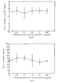
- HUVECs were for 24 hours with different concentrations of NR1 (0.01-100 ⁇ g / ml) and the CM was on tPA antigen, PAI-1 antigen and tPA PAI-1 complexes described under materials and methods.
- the Results are the means of 3 each in 3-fold Arrangement of executed experiments. The values are as Mean values ⁇ S.D. shown in Fig. 1. Significances are in Comparison to control indicated (* p ⁇ 0.05; ** p ⁇ 0.01; *** p ⁇ 0.001). As shown in Fig.
- HUVECs were for 24 hours with different concentrations of NR1 (0.01-100 ⁇ g / ml).
- the ECM was like described and discussed in Materials and Methods PAI-1 antigen examined. The values represent mean ⁇ S.D. out of 6 independent wells.
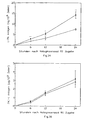
- HUVECs were absent for the specified periods (open circles) or presence of 100 ⁇ g / ml NR1 (full Circles).
- the results represent the mean values of 3, each performed in 3-fold arrangement experiments. Values are mean values ⁇ S.D., * p ⁇ 0.05; ** p ⁇ 0.01 in Comparison to control indicated.
- the tPA antigen increased in the CM of HUVECs for 6, 12 or 24 Hours treated with 100 ⁇ g / ml NR1 were compared to Control time dependent, whereas where the PAI-1 antigen in CM of such kind of treated cells is not significant changed.
- Notoginsenoside R1 affects the uPA antigen secretion of cultivated HUVECs
- Notoginsenoside R1 increases tPA activity and reduces it the PAI-1 activity in cultured HUVECs.
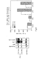
- CM of HUVECs was collected after 24 hours and with SDS PAGE and subsequent FA, as under Material and Methods described examined.
- Lane 1 untreated CM from HUVECs
- Lane 2 CM from HUVECs coupled with monoclonal linked to Sepharose Anti-tPA antibodies was pre-incubated
- Lane 3 CM of HUVECs with monoclonal antibodies coupled to Sepharose Anti-uPA antibodies was pre-incubated
- Lane 4 CM of HUVECs pre-incubated with Sepharose 4B
- Lane 5 purified human tPA
- Lane 6 purified human uPA.
- Lane 1 control; Lane 2: 0.01 ⁇ g / ml NR1; Lane 3: 0.1 ⁇ g / ml NUMBER 1; Lane 4: 1.0 ⁇ g / ml NR1; Lane 5: 10 ⁇ g / ml NR1; Lane 6: 100 / ml NR1. If the CM of HUVECs without or with rising Concentrations of NR1 were cultured for 24 hours, was analyzed with FA or RFA, could be a dose-dependent Increase in the size of the lysis zones can be determined where, however, the size of the lysis-resistant zones with increasing amounts of NR1 decreased ( Figures 5A and B).
- Confluent HUVECs were in the absence for 12 hours (C) or in the presence (T) of NR1 (100 ⁇ g / ml).
- the Northern blot analysis of the RNA extracts of untreated and NR1-treated HUVECs were prepared using 32P-labeled cDNA probes were performed for tPA, PAI-1 and GAPDH mRNA.
- the intensity of the existing on the autoradiogram Gangs was evaluated using densitometry and the specific mRNA for tPA and PAI-1 was against GAPDH mRNA normalized to account for differences in loading. The signal intensity was compared as the ratio of the signal from NR1 treated HUVECs compared to Signal from untreated control cells.
- the mRNA for PAI-1 was expressed in cells that with 1 ⁇ g / ml LPS and / or 100 ⁇ g / ml NR1 were measured.
- the increase induced by LPS to double Amount of PAI-1-specific mRNA (3.2kb) was detected in Presence of both LPS and NR1 at a 1.37-fold Increase reduced (Figure 8).
- the TF activity measured after 6 hours of treatment with 1 ⁇ g / ml LPS in HUVECs was also significant by co-incubation antagonized with NR1 (LPS and NR1 treated cells: 56.0 ⁇ 1,9mU / 106Zellen).
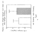
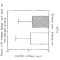
- Figs. 12, 13 and 14 are the effects of NR1-containing Extracts given on tPA, PAI and uPA in healthy volunteers. There is an increase of tPA and a decrease of PAI-1, which analyzes the effects of NR-1 in tissue culture equivalent.
Description
Das fibrinolytische System dient als basaler Abwehrmechanismus, der die Ablagerung von Fibrin sowohl im vaskulären, als auch in extravaskulären Systemen kontrolliert. Das richtige Funktionieren des fibrinolytischen Systems ist notwendig, um einerseits hämorrhagische und andererseits thrombotische Phänomene zu verhindern, aber auch um die Bildung von interstitialen Fibrinablagerungen und darauffolgende Narbenbildung zu verhindern. Man nimmt an, daß der Gewebeplasminogenaktivator (t-PA) eine wichtige Rolle in der Auslösung der (extrinsischen) fibrinolytischen Kaskade durch die Umwandlung des Zymogens Plasminogen in das aktive Plasmin, das Fibrin abbaut, spielt. Man nimmt weiters an, daß daher die fibrinolytische Kapazität des Plasmas signifikant von der Konzentration des zirkulierenden t-PA abhängt. Der t-PA im Plasma stammt wahrscheinlich hauptsächlich aus der Gefäßwand, wo er in der Endothelzelle lokalisiert ist. Weiters spielt der Urokinase Plasminogenaktivator (u-PA) eine Rolle in der Gesamt-Fibrinolyse. Man nimmt an, daß dieser Plasminogenaktivator auch - zumindest teilweise - aus der Gefäßwand stammt. Der Hauptinhibitor der Fibrinolyse, Plasminogenaktivatorinhibitor 1 (PAI-1) wird auch von Endothelzellen synthetisiert und es existieren Daten, die zeigen, daß das relative Mengenverhältnis zwischen PAs und PAI-1 wichtig ist für die fibrinolytische Kapazität und damit für die Verhinderung von thrombotischen Abläufen wie z.B. beim Myokardinfarkt. Die pharmakologische Regulierung der Synthese von t-PA, u-PA und PAI-1 ist daher von Nutzen, um eine unzureichende, endogene Fibrinolyse zu steigern.The fibrinolytic system serves as a basal defense mechanism, the deposition of fibrin both in the vascular, as also controlled in extravascular systems. The right thing Functioning of the fibrinolytic system is necessary to on the one hand hemorrhagic and on the other hand thrombotic To prevent phenomena, but also to the formation of interstitial Fibrin deposits and subsequent scarring to prevent. It is believed that the tissue plasminogen activator (t-PA) play an important role in triggering the (extrinsic) fibrinolytic cascade by the transformation of the Zymogens plasminogen into the active plasmin, the fibrin degrades, plays. It is further assumed that therefore the fibrinolytic Capacity of the plasma significantly from the concentration of circulating t-PA. The t-PA in the plasma probably comes mainly from the vessel wall, where he located in the endothelial cell. Furthermore, the urokinase plays Plasminogen activator (u-PA) plays a role in total fibrinolysis. It is believed that this plasminogen activator also - at least partially - comes from the vessel wall. Of the Main inhibitor of fibrinolysis, plasminogen activator inhibitor 1 (PAI-1) is also synthesized by endothelial cells and it exist data showing that the relative quantity ratio between PAs and PAI-1 is important for the fibrinolytic Capacity and thus for the prevention of thrombotic Procedures such as during myocardial infarction. The pharmacological Regulation of the synthesis of t-PA, u-PA and PAI-1 therefore useful to inadequate endogenous fibrinolysis to increase.
Da sowohl t-PA, als auch PAI-1 von Endothelzellen produziert werden, stellt die Regulation ihrer Synthese und Sekretion auf dem Niveau der Endothelzelle einen raschen und direkten Weg dar, das fibrinolytische Potential des Blutes zu beeinflussen. Kürzlich durchgeführte Studien haben gezeigt, daß die Produktion von Plasminogenaktivatoren und Inhibitoren in verschiedenen Zelltypen durch eine Reihe von Faktoren reguliert wird: Die Synthese von t-PA in Endothelzellen wird durch eine Vielzahl von Stimuli wie z.B. Thrombin, Histamin, Butyrat, Retinolsäure und Tumorpromoteren, wie z.B. Phorbol-12-Myristat-13-Acetat (PMA) gesteigert. Faktoren, die die Expression von PAI-1 regulieren, schließen Lipopolysaccharide, Thrombin, Interleukin 1 (IL-1), Tumornekrosefaktor α (TNF α), Transforming Growth Factor β (TGF β), Basic Fibroblast Growth Factor (BFGF) und Endothelial cell Growth Supplement in Kombination mit Heparin ein. Keine der oben angeführten Substanzen konnte allerdings in vivo erfolgreich angewendet werden.Because both t-PA and PAI-1 are produced by endothelial cells be regulates their synthesis and secretion at the level of the endothelial cell a rapid and direct Way to increase the fibrinolytic potential of the blood influence. Recent studies have shown that the production of plasminogen activators and inhibitors in different cell types due to a number of factors The synthesis of t-PA in endothelial cells becomes by a variety of stimuli, e.g. Thrombin, histamine, Butyrate, retinoic acid and tumor promoters, e.g. 12-o-tetradecanoylphorbol-13-acetate (PMA) increased. Factors that the Regulate expression of PAI-1, include lipopolysaccharides, Thrombin, interleukin 1 (IL-1), tumor necrosis factor α (TNFα), Transforming Growth Factor β (TGFβ), Basic Fibroblast Growth Factor (BFGF) and Endothelial Cell Growth Supplement in combination with heparin. None of the above However, listed substances could be successful in vivo be applied.
Die bakterielle Sepsis, ausgelöst durch die Freisetzung bakteriellen Endotoxins (LPS), ist ein lebensbedrohender Zustand, bei dem durch LPS ausgelöste Veränderungen in der Gerinnung und Fibrinolyse intravaskuläre Gerinnselbildung und in der Folge Organversagen verursachen. Man nimmt an, daß dabei LPS auf Endothelzellen einwirkt, in dem es deren Expression von Tissue Factor (TF) und PAI-1 steigert. Zur Zeit ist keine ausreichende direkte Behandlung von Patienten, die an einer durch LPS ausgelösten intravaskulären Gerinnung leiden, möglich und Maßnahmen zur Behandlung der durch LPS ausgelösten Symptome, wie z.B. Hyperkoagulation sind einerseits beschränkt auf Heparin und andererseits auf die Behandlung der verursachenden bakteriellen Sepsis mit Antibiotika. In China wurden chinesische Kräuterdrogen wie Panax Notoginseng oder Astragalose seit Jahrtausenden von traditionellen chinesischen Ärzten zur Schmerzlinderung und zur Behandlung von Stase und kardiovaskulären Erkrankungen verwendet.Bacterial sepsis, triggered by the release Bacterial endotoxin (LPS), is a life-threatening one Condition in which LPS induced changes in the Coagulation and fibrinolysis intravascular clot formation and as a result cause organ failure. It is believed that while LPS acts on endothelial cells, in which it Expression of tissue factor (TF) and PAI-1 increases. to Time is not sufficient direct treatment of patients, those due to LPS induced intravascular coagulation suffer, possible and measures to treat by LPS triggered symptoms, such as Hypercoagulation are one hand limited to heparin and on the other hand to the treatment causing bacterial sepsis with antibiotics. In China, Chinese herbal drugs such as Panax Notoginseng or astragalus for millennia of traditional Chinese doctors for pain relief and treatment used by stasis and cardiovascular diseases.
Tatsächlich wird zum Beispiel in L. Zechmeister, Progress in the Chemistry of Organic Natural Products, Vol. 46, Springer-Verlag, Wien New York 1984, in dem Kapitel über "Saponins of Ginseng and Related Plants" auf die Eigenschaft von Panax notoginseng als Tonikum, Hämostaticum, Coronartherapeuticum und Antihemorrhagicum hingewiesen. Ebenso wird in Chemical Abstracts 119, 85 683 auf eine antithrombotische Wirkung von Panax notoginseng und in dem Japanischen Patent Abstracts JP Kokai Nr. 55-127 317 auf die antifibrinolytische Wirkung und JP Kokai Nr. 63-198 609 auf die durchblutungsfördernde Wirkung von Panax notoginseng-Präparaten hingewiesen.In fact, for example, in L. Zechmeister, Progress in the Chemistry of Organic Natural Products, Vol. 46, Springer-Verlag, Wien New York 1984, in the chapter on "Saponins of Ginseng and Related Plants "on the property of Panax notoginseng as tonic, hemostatic, coronary therapeutic and Antihemorrhagicum. Similarly, in Chemical Abstracts 119, 85, 683 for an antithrombotic effect of Panax notoginseng and in the Japanese Patent Abstracts JP Kokai No. 55-127 317 on the antifibrinolytic action and JP Kokai No. 63-198 609 on the circulation-promoting effect noted by Panax notoginseng preparations.
H. MATSUDA ET AL.: "Studies of panax japonicus fibrinolysis" PLANTA MEDICA, Bd. 55, Nr. 1, Seiten 18-21, beschreiben eine aktivierende Wirkung von triterpenen Saponinen auf die fibrinolytische Aktivität der Ratte und zur Behandlung der endotoxin induzierten disseminierten intravasalen Gerinnung. Es wird jedoch nur eine globale Aktivierung der Fibrinolyse ohne Angabe der beeinflussten Moleküle beschrieben.H. MATSUDA ET AL .: "Studies of panax japonicus fibrinolysis" PLANTA MEDICA, Vol. 55, No. 1, pages 18-21, describe one activating effect of triterpene saponins on the fibrinolytic activity of the rat and for the treatment of endotoxin induced disseminated intravascular coagulation. However, there will be only one global activation of fibrinolysis without indicating the affected molecules.
In H. MATSUDA ET AL. "Pharmacological study on panax ginseng C.A. Meyer IV. effects of red ginseng on experimental disseminated intravascular coagulation (3) effect of ginsenoide-Ro on the blood coagulative and fibrinolytic systems" CHEM. PHARM. BULL. Bd. 34, Nr. 5, Seiten 2100-2104, werden ebenfalls die Effekte von rotem Ginseng auf Gerinnung und Fibrinolyse bei Ratten beschrieben allerdings ohne nähere Angaben eines molekularen Mechanismus für den globalen Effekt.In H. MATSUDA ET AL. "Pharmacological study on panax ginseng C.A. Meyer IV. Effects of red ginseng on experimental disseminated intravascular coagulation (3) effect of ginsenoid-on the blood coagulative and fibrinolytic CHEM. PHARM. BULL., Vol. 34, No. 5, pp. 2100-2104, Also, the effects of red ginseng on clotting and fibrinolysis in rats, however, described without further details Information of a molecular mechanism for the global Effect.
In einer weiteren Veröffentlichung H. MATSUDA ET AL. "Pharmacological study on panax ginseng C.A. Meyer III. effect of red ginseng on experimental disseminated intravascular coagulation. (2) effects of ginsenoides on blood coagulative and fibrinolytic systems" CHEM. PHARM. BULL., Bd. 34, Nr. 3, Seiten 1153-1157, wird für die stimulierung der fibrinolytischen Aktivität die Urokinase verantworltich gefunden. Auch YOSHIKAWA ET AL. "Structure of two new fibrinolytic saponies from the seed of Luffa cylindra ROEM" CHEM. PHARM. BULL., Bd. 39, Seiten 1185-1188 beschreiben zwar die Struktur von zwei neuen "fibrinolytischen" Saponinen, nicht jedoch auf molekularer Ebene beim Menschen.In another publication H. MATSUDA ET AL. "Pharmacological study on panax ginseng C. A. Meyer III. effect of red ginseng on experimental disseminated intravascular coagulation. (2) effects of ginsenoides on blood coagulative and fibrinolytic systems "CHEM. PHARM. BULL., Vol. 34, No. 3, pages 1153-1157, is for the stimulating the fibrinolytic activity of urokinase found responsible. Also YOSHIKAWA ET AL. "Structure of two new fibrinolytic saponies from the seed of Luffa cylindra ROEM "CHEM. PHARM. BULL., Vol. 39, pages 1185-1188 Although describe the structure of two new "fibrinolytic" saponins, but not molecular ones Level in humans.
BIOSIS PREV199294099689 Abstract - AMERICAN JOURNAL OF CHINESE MEDICINE, Bd. 20, Nr. 2, 1992 Seiten 167-173, KOYAMA N; MORISAKI N; SAITO Y; YOSHIDA 5 "Inhibitory effect of ginsenosides on migration of arterial smooth muscle cells" bezieht sich auf die Verhinderung der Lipidperoxidation und damit auf einen völlig unterschiedlichen Mechanismus der Wirkung von Panax Notoginseng ungereinigten Extrakten.BIOSIS PREV199294099689 Abstract - AMERICAN JOURNAL OF CHINESE MEDICINE, Vol. 20, No. 2, 1992, pages 167-173, KOYAMA N; MORISAKI N; SAITO Y; YOSHIDA 5 "Inhibitory effect of ginsenosides on migration of arterial smooth muscle cells " refers to the prevention of lipid peroxidation and thus to a completely different mechanism of Effect of Panax Notoginseng Unrefined Extracts.
SINICA, SHANGHAI, CN, Bd. 11, Nr. 1, 1990 Seiten 26 - 29, Li X; CHEN J-X; SUN J-J "Protective effects of panax notoginseng saponins on experimental myocardial injury induced by ischemia and reoerfusion in rat" bezieht sich auf die Migration von glatten Muskelzellen, einen weiteren möglichenSINICA, SHANGHAI, CN, Vol. 11, No. 1, 1990, pages 26-29, Li X; CHEN J-X; SUN J-J "Protective effects of panax notoginseng saponins on experimental myocardial injury induced by ischemia and reoerfusion in rat "refers to the Migration of smooth muscle, another possible
Wirkmechanismus.Mechanism of action.
Es wird hier zum ersten Mal gezeigt, dass die Verwendung des Triterpensaponins Notoginsenosid R1 (NR1) die fibrinolytische Kapazität durch Steigerung des t-PA und durch Senkung des PAI-1 beim Menschen und der Maus steigert und zur direkten Hemmung der LPS-Effekte in Bezug auf PAI-1 und Gewebefaktor Anstieg führt. Gegenstand dieser Erfindung ist die Verwendung von Notoginsenosid R1 (NR1) zur Herstellung eines Arzneimittels zur Behandlung von Endotoxin-Schock. Das Arzneimittel kann entweder parenteral oder oral in Form von Lösungen oder Tabletten oder Kapseln verabreicht werden. Insbesondere betrifft die Erfindung die Verwendung von Notoginsenosid R1 (NR1) zur Herstellung eines Arzneimittels zur Behandlung von koronarer Herzerkrankung, peripherer Arterienerkrankung sowie Myocardinfarkt und Angina pectoris.It is shown here for the first time that the use of the Triterpene saponins Notoginsenoside R1 (NR1) the fibrinolytic Capacity by increasing the t-PA and lowering the PAI-1 in humans and mouse boosts and direct Inhibition of LPS effects with respect to PAI-1 and Tissue Factor Increase leads. The subject of this invention is the use of Notoginsenoside R1 (NR1) for the production of a Drug for the treatment of endotoxin shock. The Medicines can be either parenterally or orally in the form of Solutions or tablets or capsules. In particular, the invention relates to the use of Notoginsenoside R1 (NR1) for the manufacture of a medicament for the treatment of coronary heart disease, peripheral Arterial disease as well as myocardial infarction and angina pectoris.
In allen Beispielen wurden die folgenden Materialien und Methoden benutzt.In all examples, the following materials and Methods used.
Chemisch-reines Notoginsenosid R1 (NR1) wurde vom National
Institute for the Control of Pharmaceutical and Biological
Products (Beijing, China) gekauft. NR1 sind Substanzen mit
der folgenden Formel:

Endothelzellen wurden aus frischen humanen Nabelschnurvenen
mit Kollagenase (Sigma) isoliert, ähnlich einem Protokoll,
das von Jaffe et al., J Clin Invest 1973; 52: 2745-56,
beschrieben wurde. Zellen aus 4-6 Nabelschnüren wurden
gepoolt und in 75 cm2 Zellkulturflaschen (Costar, MA, USA)
ausgesät, die mit 1% Gelatine aus Kälberhaut (Sigma)
beschichtet waren. Die Zellen wurden bis zur Konfluenz bei
37°C in einer wasserdampfgesättigten Athmosphäre aus 95% Luft
und 5% CO2 in Medium 199 (Sigma) versetzt mit 20% hitzeinaktivierten
Supplementierten Kälberserum (SCS; Hyclone, UT,
USA), 100 µg/ml Streptomycin, 100 IU/ml Penizillin, 250 ng/ml
Fungizon, 1 mM Glutamin (JHR Biosciences, KS, USA) 2 IU/ml
Heparin (Liquemin Roche; Hoffmann La Roche, Schweiz), 50
µg/ml ECGS (Technoclone, Austria) kultiviert. Der Endothelcharakter
der Zellen wurde durch ihre typische Pflastersteinmorphologie,
durch positive Immunfluoreszens mit
Anti-Von willebrandt Faktor VIII Antikörpern und durch Aufnahme
von azetyllierten Low Density Lipoprotein (LDL) charakterisiert.
Primärkulturen wurden zum Zeitpunkt der Konfluenz
mit 0,05% Trypsin/0,02% EDTA (JRH Biosciences) geerntet und
in einer Split-ratio von 1:3 in 75cm2 Zellkulturflaschen ausgesät.
Subkonfluente Zellen wurden unter den selben
Bedingungen bis zum Erreichen der Konfluenz kultiviert und
wurden während des exponentiellen Zellwachstums mit
Trypsin/EDTA geerntet und in 1 ml Portionen in Medium 199 mit
10% Dimethylsulfoxid (DMSO) im flüssigen Stickstoff
eingefroren. Für die Experimente wurden die Zellen bei 37°C
aufgetaut und in 6 well Platten (Durchmesser 3,5 cm; Costar)
in Medium 199 versetzt mit SCS, ECGS und Heparin in den oben
angeführten Konzentrationen bis zum Erreichen der Konfluenz
kultiviert. In allen Experimenten wurden Zellen zwischen der
2. und 3. Passage verwendet. Die Zellen wurden immer am Tag
vor den Experimenten mit frischem Medium gefüttert. Alle in
der Zellkultur verwendeten Materialien waren wie durch den
Coatest Endotoxin Kit (Kabi Vitrum, Schweden) bestimmt frei
von Endotoxin (Nachweisgrenze des Test 5 pg/ml).Endothelial cells were isolated from fresh human umbilical vein veins with collagenase (Sigma), similar to a protocol described by Jaffe et al., J Clin Invest 1973; 52: 2745-56. Cells from 4-6 umbilical cords were pooled and seeded in 75 cm 2 cell culture flasks (Costar, MA, USA) coated with 1% calf skin gelatine (Sigma). The cells were brought to confluency at 37 ° C in a water vapor saturated atmosphere of 95% air and 5% CO 2 in medium 199 (Sigma) supplemented with 20% heat-inactivated supplemented calf serum (SCS, Hyclone, UT, USA), 100 μg / ml Streptomycin, 100 IU / ml penicillin, 250 ng / ml fungizone, 1 mM glutamine (JHR Biosciences, KS, USA) 2 IU / ml heparin (Liquemin Roche, Hoffmann La Roche, Switzerland), 50 μg / ml ECGS (Technoclone, Austria ). The endothelial character of the cells was characterized by their typical cobblestone morphology, by positive immunofluorescence with anti-Von Willebrandt factor VIII antibodies and by uptake of acetylated low density lipoprotein (LDL). Primary cultures were harvested at the time of confluence with 0.05% trypsin / 0.02% EDTA (JRH Biosciences) and seeded in a split ratio of 1: 3 in 75cm 2 cell culture flasks. Subconfluent cells were cultured under the same conditions until confluence and were harvested during exponential cell growth with trypsin / EDTA and frozen in 1 ml portions in Medium 199 with 10% dimethylsulfoxide (DMSO) in liquid nitrogen. For the experiments, the cells were thawed at 37 ° C and cultured in 6 well plates (diameter 3.5 cm, Costar) in medium 199 spiked with SCS, ECGS and heparin in the above concentrations until reaching confluency. In all experiments cells were used between the 2nd and 3rd passage. The cells were always fed with fresh medium the day before the experiments. All materials used in the cell culture were determined to be free of endotoxin as determined by the Coatest Endotoxin Kit (Kabi Vitrum, Sweden) (limit of detection of the
Herstellung des konditionierten Mediums (CM) und der extrazellulären Matrix (ECM)Preparation of the conditioned medium (CM) and the extracellular matrix (ECM)
Konfluente Kulturen wurden zwei Mal mit Hank's Balanced Salt Solution (HBSS; Sigma) gewaschen und bei 37°C mit 1 ml/Napf von Medium 199 versetzt mit 1,25% SCS und 50 µg/ml ECGS und mit NR1 oder AS IV in den angeführten Konzentrationen inkubiert. Nach der Inkubation wurde der Zellkulturüberstand gesammelt und nach der Zentrifugation - um Zellbruchstücke zu entfernen - bei -70°C bis zum Gebrauch gelagert. Die Gesamtzellzahl der entsprechenden Kulturen wurde nach Trypsinisierung mit der Zählkammer bestimmt. ECM von diesen oder ähnlich behandelten Kulturen wurde nach der Methode von Mimuro et al., Blood 1987; 70: 721-28 präpariert. Die Monolayer wurden 3 Mal mit kalter Phosphat gepufferter Kochsalzlösung (PBS: 0,01 M Natriumphosphat, 0,14 M NaCl, pH 7,4) gewaschen und die zellulären Bestandteile wurden durch 10 minütige Inkubation bei 37°C mit PBS versetzt mit 0,5% Triton X100 extrahiert. Die Platten wurden ein weiteres Mal mit destilliertem Wasser gewaschen um verbleibende zelluläre Bestandteile zu entfernen und anschließend durch mikroskopische Untersuchung auf das Vorhandensein von Zellbruchstücken hin untersucht. Diese Extraktionsmethode entfernte sichtbare Zellbestandteile von den Platten vollständig und die ECM wurde durch Abschaben in 1 ml PBS versetzt mit 0,1% SDS nach 30 minütiger Inkubation bei 37°C extrahiert. Die Extrakte wurden über Nacht bei 4°C gegen PBS dialysiert.Confluent cultures were used twice with Hank's Balanced Salt Solution (HBSS; Sigma) and washed at 37 ° C with 1 ml / well Medium 199 was spiked with 1.25% SCS and 50 μg / ml ECGS and incubated with NR1 or AS IV at the indicated concentrations. After incubation, the cell culture supernatant collected and after centrifugation - to cell debris too Remove - stored at -70 ° C until use. The total cell number The corresponding cultures were after trypsinization determined with the counting chamber. ECM of these or similar treated cultures was determined by the method of Mimuro et al., Blood 1987; 70: 721-28 prepared. The monolayers were 3 times with cold phosphate buffered saline (PBS: 0.01 M sodium phosphate, 0.14 M NaCl, pH 7.4) and the cellular components were incubated for 10 minutes at 37 ° C with PBS extracted with 0.5% Triton X100 extracted. The plates were distilled one more time Water washed for remaining cellular components remove and then by microscopic examination examined for the presence of cell debris. This extraction method removed visible cell components from the plates completely and the ECM was scrapped by in 1 ml PBS mixed with 0.1% SDS after 30 minutes incubation extracted at 37 ° C. The extracts were added overnight 4 ° C dialyzed against PBS.
Tests für tPA Antigen, uPA Antigen, PAI-1 Antigen, PAI-1 Aktivität und tPA PAI-1 Komplexen im CM, in der ECM und im PlasmaAssays for tPA antigen, uPA antigen, PAI-1 antigen, PAI-1 Activity and tPA PAI-1 Complexes in CM, ECM and in the plasma
tPA Antigen, uPA Antigen, PAI-1 Antigen und die Konzentration von tPA PAI-1 Komplexen wurde mit spezifischen kommerziell erhältlichen Enzyme Linked Immunosorbent Assays (ELISAs) (Technoclone) laut den vom Hersteller beigefügten Anleitungen bestimmt. Die Testbereiche für diese Tests liegen für tPA zwischen 0,3 und 2,5 ng/ml für uPA zwischen 0,6 und 10 ng/ml, für PAI zwischen 1,0 und 30 ng/ml und für tPA PAI-1 Komplexe zwischen 0,2 und 20 ng/ml. Der tPA ELISA bestimmt freien tPA und tPA in Komplex mit PAI-1. Der uPA ELISA bestimmt freien uPA und uPA in Komplex mit PAI-1. Der PAI-1 ELISA mißt freien, komplexierten und latenten PAI-1. Der tPA PAI-1 Komplex ELISA mißt ausschließlich tPA PAI-1 Komplexe. PAI-1 Aktivität im Plasma und im CM wurde mit einem Titrationsassay (Technoclone) laut den vom Hersteller beigefügten Anleitungen bestimmt.tPA antigen, uPA antigen, PAI-1 antigen and concentration of tPA PAI-1 complexes became commercially specific Available Enzyme Linked Immunosorbent Assays (ELISAs) (Technoclone) according to the instructions provided by the manufacturer certainly. The test ranges for these tests are for tPA between 0.3 and 2.5 ng / ml for uPA between 0.6 and 10 ng / ml, for PAI between 1.0 and 30 ng / ml and for tPA PAI-1 complexes between 0.2 and 20 ng / ml. The tPA ELISA determines free tPA and tPA in complex with PAI-1. The uPA ELISA determines free uPA and uPA in complex with PAI-1. The PAI-1 ELISA measures free, complexed and latent PAI-1. The tPA PAI-1 complex ELISA exclusively measures tPA PAI-1 complexes. PAI-1 Activity in the plasma and in the CM was measured with a titration assay (Technoclone) according to the instructions provided by the manufacturer certainly.
Die Aktivität von tPA und PAI-1 wurde nach Natriumdodecylsulfat Polyacrylamid Gelelektrophorese (SDS-PAGE) unter Verwendung von Fibrinautographie (FA) und reverser Fibrinautography (RFA) analysiert. SDS Polyacrylamid Gele und Puffer wurden nach dem Protokoll von Laemmli, Nature 1970; 227: 680-85, hergestellt. FA wurde wie von Granelli-Piperno et al., J Exp Med 1978; 148: 223-34 ausgeführt. 100 µl der entsprechenden Proben wurden auf Gele bestehend aus einem 10 cm langen Trenngel mit 10% Acrylamid und aus einem 2 cm langen Obergel mit 4% Acrylamid aufgetragen und bei Raumtemperatur 16 Stunden lang oder bis die Farbfront den Unterrand des Gels erreichte gefahren. Nach der Elektrophorese wurden die Gele vorerst 90 Minuten in 250 ml Wasser versetzt mit 2,5% Triton X100 (Serva) eingelegt (die Tritonlösung wurde nach 45 Minuten gewechselt), um das SDS zu neutralisieren, dann wurden die Gele auf einen Fibrin-Agar-Indikatorfilm versetzt mit 1,5% Agarose Typ L (Behring, Deutschland), 2 mg/ml plasminogenreichem Fibrinogen (Organon Teknika, Holland) und 0,2 IU/ml bovinem Thrombin (Sigma) gelegt. Die Gele wurden bei 37°C in einer feuchten Kammer inkubiert und zu verschiedenen Zeitpunkten fotografiert. RFA wurde durchgeführt, indem die Gele auf einen Fibrinfilm, der grundsätzlich wie oben beschrieben hergestellt wurde, aber zusätzlich 0,4 IU/ml Urokinase (Technoclone) enthielt, gelegt wurden. Die Quantifizierung der tPA und PAI-1 Aktivität in einer bestimmten Probe wurde erreicht, indem sowohl Lysezonen, als auch Lyse-resistente Zonen auf dem Indikatorfilm fotografiert wurden. Diese Zonen wurden auf Transparentpapier aufgezeichnet und die gezeichneten Areale wurden ausgeschnitten und auf einer analytischen Waage gewogen.The activity of tPA and PAI-1 became sodium dodecyl sulfate Polyacrylamide gel electrophoresis (SDS-PAGE) using from fibrinautography (FA) and reverse fibrinautography (RFA) analyzed. SDS polyacrylamide gels and buffer were according to the protocol of Laemmli, Nature 1970; 227: 680-85, produced. FA was prepared as described by Granelli-Piperno et al., J Exp Med 1978; 148: 223-34. 100 μl of the corresponding Samples were on gels consisting of a 10 cm long Release gel with 10% acrylamide and a 2 cm long top gel applied with 4% acrylamide and at room temperature for 16 hours long or until the color front the lower edge of the gel reached dangers. After electrophoresis, the gels became initially 90 minutes in 250 ml of water mixed with 2.5% Triton X100 (Serva) inserted (the Triton solution became after 45 minutes changed) to neutralize the SDS, then were the gels are added to a fibrin agar indicator film 1.5% agarose type L (Behring, Germany), 2 mg / ml plasminogen-rich Fibrinogen (Organon Teknika, Holland) and 0.2 IU / ml bovine thrombin (Sigma). The gels were added Incubated at 37 ° C in a humid chamber and to different Photographed times. RFA was performed by the Gels on a fibrin film, basically as described above but additionally 0.4 IU / ml urokinase (Technoclone) were placed. The quantification tPA and PAI-1 activity in a given sample was achieved by using both lysis zones, as well as lysis-resistant Zones were photographed on the indicator film. These Zones were recorded on tracing paper and the Drawn areas were cut out and placed on an analytical Weighed the balance.
Um den im CM von HUVECs enthaltenen Plasminogenaktivator
immunologisch zu identifizieren, wurden Proben des CM bei 4°C
für 24 Stunden mit entweder an CNBr aktivierte Sepharose
gebundenen monoklonalen Anti-tPA Antikörper (MPW 3 VPA;
Technoclone) oder monoklonalen Anti-uPA Antikörper (MPW 5 UK;
Technoclone) oder als Kontrolle mit Sepharose 4B (Pharmacia,
Schweden) inkubiert. Danach wurde die Sepharose durch Zentrifugation
entfernt und 100 µl der entsprechenden Probe wurde
mit der oben beschriebenen SDS-PAGE und nachfolgender FA
analysiert.To the plasminogen activator contained in the CM of HUVECs
To identify immunologically, samples of the CM were taken at 4 ° C
for 24 hours with either CNBr activated Sepharose
bound monoclonal anti-tPA antibody (
HUVECs wurden für 6 Stunden bei 37°C in Medium 199 mit LPS und/oder NR1 inkubiert. Die Zellen wurden 3 Mal mit Clotting Puffer (130mM NaCl, 8 mM Na-Barbital und 12 mM Na-Acetat, pH = 7,4) gewaschen und in 300 µl Clotting Puffer durch Abschaben aufgenommen. Die abgeschabten Zellen wurden 3 Mal gefroren und getaut. Die Zell-Lysate wurden in einem 1-Schritt-Clotting Assay auf TF-Aktivität hin untersucht. 100 µl der Zellysate wurden mit 100 µl 20 mM CaCl2 bei 37°C für 5 Minuten in vorgewärmten Plastikröhrchen in einem Coagulometer (H. Amelung GmbH., Deutschland) inkubiert. Die Gerinnung wurde durch Zugabe von 100 µl vorgewärmten normalen humanem Zitratplasma oder durch Zugabe von Faktor X Mangelplasma (Sigma) ausgelöst. Die TF-Aktivität wurde mit einer Standardkurve (Log-Log Plot), die mit Kaninchenhirn Thromboplastin (Sigma) konstruiert wurde, quantifiziert. 100 mU Aktivität wurden definiert als eine Gerinnungszeit von 20 Sekunden in einem Standardtest mit normalem humanem Plasma. Die beobachtete coagulatorische Aktivität entspricht der TF-Aktivität, da keine procoagulatorische Aktivität der Endothelzellen festgestellt wurde, wenn Faktor X Mangelplasma anstelle von normalem Plasma verwendet wurde.HUVECs were incubated for 6 hours at 37 ° C in medium 199 with LPS and / or NR1. The cells were washed 3 times with clotting buffer (130 mM NaCl, 8 mM Na-barbital and 12 mM Na-acetate, pH = 7.4) and scrapped in 300 μl of clotting buffer. The scraped cells were frozen and thawed 3 times. The cell lysates were assayed for TF activity in a 1-step clotting assay. 100 μl of the cell lysates were incubated with 100 μl of 20 mM CaCl 2 at 37 ° C. for 5 minutes in prewarmed plastic tubes in a coagulometer (H. Amelung GmbH., Germany). Coagulation was initiated by the addition of 100 μl of prewarmed normal human citrated plasma or by addition of Factor X deficient plasma (Sigma). TF activity was quantified using a standard curve (Log-Log Plot) constructed with rabbit brain thromboplastin (Sigma). 100 mU activity was defined as a coagulation time of 20 seconds in a standard human plasma standard assay. The observed coagulant activity corresponds to the TF activity, since no procoagulant activity of the endothelial cells was detected when Factor X deficient plasma was used instead of normal plasma.
Die gesamte zelluläre RNA aus Endothelzellen wurde mit Hilfe
der von Chomzynski und Sacchi, Anal Biochem 1987; 162: 156-9,
beschriebenen sauren Guanidin Thiozyanat-Phenol-Chloroform
Extraktion isoliert. Der RNA Niederschlag wurde in 50 µl 0,5%
SDS resuspendiert und die Konzentration bei 260 nM bestimmt.
Für die Northern Blot Analyse wurden die RNA Proben in einem
1,2%igen Agarosegel elektrophoretisiert und danach die fraktionierte
RNA auf eine Duralon-UVTM Membran (Stratagene, CA,
USA) durch Kapillarwirkung transferiert. Die RNA Blots wurden
in seal-a-meal Säckchen eingeschlossen und in 50 mM PIPES,
100 mM NaCl 50 mM NaPhosphat 1 mM EDTA, versetzt mit 5% SDS
für zumindest 3 Stunden bei 57°C prähybridisiert. Der Prähybridisationspuffer
wurde verworfen und durch frischen Prähybridisationspuffer
versetzt mit 106 CPM/ml der 32 P markierten
cDNA-Sonden für entweder humanen tPA, humanen PAI-1, humanen
TF oder Ratten-Glyceraldehyd-3-Phosphatdehydrogenase,
die als interne Standardsonde verwendet wurde, ersetzt. Die
cDNA Fragmente wurden mit einem Random Prime DNA Labelling
Kit (Boehringer Mannheim, Deutschland) radioaktiv markiert. Total cellular RNA from endothelial cells was assisted
that of Chomzynski and Sacchi, Anal Biochem 1987; 162: 156-9,
described acidic guanidine thiocyanate-phenol-chloroform
Extraction isolated. The RNA precipitate was 0.5% in 50 μl.
SDS resuspended and the concentration determined at 260 nM.
For Northern blot analysis, the RNA samples were in a
Electrophorized 1.2% agarose gel and then the fractionated
RNA on a Duralon-UVTM membrane (Stratagene, CA,
USA) by capillary action. The RNA blots were
enclosed in seal-a-meal sachets and in 50 mM PIPES,
100
In dieser Studie wurden ausschließlich männliche Balb C Mäuse (18-30 g Körpergewicht) verwendet. Alle Experimente wurden unter Aetherbetäubung durchgeführt. Den Mäusen wurde intravenös über die Schwanzvene LPS (10 ng/g) und/oder NR1 (1 µg/g) in einem Volumen von 5 µg/l injiziert. Zu den angegebenen Zeitpunkten wurden mit Natriumzitrat (0,13 M Endkonzentration) antikoagulierte Blutproben gewonnen. Thrombozytenfreies Plasma wurde durch Zentrifugation bei 2500 g für 30 Minuten bei 4°C gewonnen und bei -70°C bis zur Testung aufbewahrt.In this study, only male Balb C mice were used (18-30 g body weight). All experiments were under ethereal anesthesia. The mice became intravenous via the tail vein LPS (10 ng / g) and / or NR1 (1 μg / g) in a volume of 5 μg / L. To the with sodium citrate (0.13 M Final concentration) anticoagulated blood samples. platelets Free Plasma was centrifuged at 2500 g recovered at 4 ° C for 30 minutes and at -70 ° C until testing kept.
Es wurde bei 6 freiwilligen gesunden Propanden in einem Zeitraum von 24 Stunden 4 x 100 mg-Äquivalent eines Notoginseng R1 (NR1) enthaltenden Extraktes verabreicht. Es erfolgte eine Blutabnahme unmittelbar vor der ersten Einnahme und unmittelbar nach der letzten Einnahme. In diesen Blutproben wurde entsprechend den oben angegebenen Methoden der Gehalt an Plasminogenaktivator Inhibitor I (PAI-1) Antigen, Gewebeplasminogenaktivator (tPA) Antigen und Urokinase-Plasminogenaktivator (uPA) Antigen bestimmt.It was taken at 6 volunteer healthy propands in a period of time of 24 hours 4 x 100 mg equivalent of a notoginseng Administered R1 (NR1) containing extract. There was one Blood sample immediately before the first dose and immediately after the last intake. In these blood samples was according to the methods given above, the content Plasminogen activator Inhibitor I (PAI-1) antigen, tissue plasminogen activator (tPA) Antigen and urokinase plasminogen activator (uPA) antigen.
Die Resultate sind als Mittelwerte ± Standardabweichung angegeben. Ein unpaired Student's t-test wurde verwendet um die Signifikanz festzustellen.The results are as mean ± standard deviation specified. An unpaired student's t-test was used around determine the significance.
HUVECs wurden für 24 Stunden mit verschiedenen Konzentrationen von NR1 (0,01-100 µg/ml) inkubiert und das CM wurde auf tPA Antigen, PAI-1 Antigen und tPA PAI-1 Komplexe hin wie unter Materialien und Methoden beschrieben, untersucht. Die Resultate sind die Mittelwerte von 3 jeweils in 3-facher Anordnung ausgeführten Experimenten. Die Werte sind als Mittelwerte ± S.D. in Fig. 1 gezeigt. Signifikanzen sind im Vergleich zur Kontrolle angegeben (* p<0,05; ** p<0,01; *** p<0,001). Wie in Fig. 1A gezeigt führte die Behandlung von HUVECs mit steigenden Konzentration von NR1 für 24 Stunden zu einem dosisabhänigen Anstieg des tPA Antigens im CM solcher Art behandelter Zellen: Maximale Effekte wurden mit 100 µg/ml NR1 (100mg/ml NR1: 9,6 ± 0,7 ng/105 Zellen/24h; Kontrolle: 5,8± 0,4 ng/105/24h; n=9, p<0,05) erreicht. Wie in Fig. 1B gezeigt, stiegen auch die tPA PAI-1 Komplexe im CM in ähnlicher Weise in Gegenwart zunehmender Konzentrationen von NR1 an (100 µg/ml NR1: 63,5±2,6 ng/105 Zellen/24h; Kontrolle: 40,2 ± 7ng/105 Zellen/24h; n=9, p<0,01.HUVECs were for 24 hours with different concentrations of NR1 (0.01-100 μg / ml) and the CM was on tPA antigen, PAI-1 antigen and tPA PAI-1 complexes described under materials and methods. The Results are the means of 3 each in 3-fold Arrangement of executed experiments. The values are as Mean values ± S.D. shown in Fig. 1. Significances are in Comparison to control indicated (* p <0.05; ** p <0.01; *** p <0.001). As shown in Fig. 1A, the treatment of HUVECs with increasing concentration of NR1 for 24 hours too a dose-dependent increase of the tPA antigen in the CM such Type of treated cells: Maximum effects were at 100 μg / ml NR1 (100mg / ml NR1: 9.6 ± 0.7 ng / 105 cells / 24h; control: 5.8 ± 0.4 ng / 105 / 24h; n = 9, p <0.05). As in Fig. 1B As shown, the tPA PAI-1 complexes also increased more in the CM In the presence of increasing concentrations of NR1 (100 μg / ml NR1: 63.5 ± 2.6 ng / 105 cells / 24h; 40.2 ± 7ng / 105 cells / 24h; n = 9, p <0.01.
HUVECs wurden für 24 Stunden mit verschiedenen Konzentrationen von NR1 (0,01-100µg/ml) inkubiert. Die ECM wurde wie unter Material und Methoden beschrieben, gewonnen und auf PAI-1 Antigen hin untersucht. Die Werte stellen Mittelwerte ± S.D. aus 6 unabhängigen Näpfen dar. PAI-1 Antigen im CM und in der ECM von mit NR1 behandelten HUVECs war nicht signifikant verändert im Vergleich zu den Kontrollen (CM: 100µg NR1/ml: 2,92 ± 0,32 µg/105 Zellen/24h; Kontrolle: 2,78 ± 0,45 µg/105 Zellen/24h; n=9. ECM: 100µg/ml NP1: 42, 55 ± 3,15 ng/ml/24h; Kontrolle: 42,27 ± 1,66 ng/ml/24 h; n=6) (Fig. 1C, Fig. 2).HUVECs were for 24 hours with different concentrations of NR1 (0.01-100μg / ml). The ECM was like described and discussed in Materials and Methods PAI-1 antigen examined. The values represent mean ± S.D. out of 6 independent wells. PAI-1 antigen in CM and in the ECM of NR1-treated HUVECs was not significant changed in comparison to the controls (CM: 100μg NR1 / ml: 2.92 ± 0.32 μg / 105 cells / 24h; Control: 2.78 ± 0.45 μg / 105 cells / 24h; n =. 9 ECM: 100 μg / ml NP1: 42, 55 ± 3.15 ng / ml / 24h; Control: 42.27 ± 1.66 ng / ml / 24 h; n = 6) (Fig. 1C, Fig. 2).
HUVECs wurden für die angegebenen Zeiträume in Abwesenheit (offene Kreise) oder Gegenwart von 100µg/ml NR1 (volle Kreise) inkubiert. Zu den angegebenen Zeitpunkten wurde das entsprechende CM geerntet und wie unter Materialien und Methoden beschrieben auf tPA Antigen und PAI-1 Antigen hin getestet. Die Resultate stellen die Mittelwerte von 3, jeweils in 3-facher Anordnung ausgeführten Experimente dar. Die Werte sind als Mittelwerte ± S.D., *p<0,05; **p<0,01 im Vergleich zur Kontrolle angegeben. Wie in Fig. 3 gezeigt stieg das tPA Antigen im CM von HUVECs, die für 6, 12 oder 24 Stunden mit 100 µg/ml NR1 behandelt wurden im Vergleich zur Kontrolle zeitabhängig an, wo hingegen das PAI-1 Antigen im CM solcher Art behandelter Zellen sich nicht signifikant änderte.HUVECs were absent for the specified periods (open circles) or presence of 100 μg / ml NR1 (full Circles). At the indicated times, the corresponding CM harvested and as under materials and Methods described on tPA antigen and PAI-1 antigen tested. The results represent the mean values of 3, each performed in 3-fold arrangement experiments. Values are mean values ± S.D., * p <0.05; ** p <0.01 in Comparison to control indicated. As shown in Fig. 3 the tPA antigen increased in the CM of HUVECs for 6, 12 or 24 Hours treated with 100 μg / ml NR1 were compared to Control time dependent, whereas where the PAI-1 antigen in CM of such kind of treated cells is not significant changed.
Wenn das CM von HUVECs, die in Gegenwart von 100 µg/ml NR1 inkubiert wurden auf uPA Antigen hin untersucht wurde, fand sich eine signifikante Änderung in der Menge des von diesen Zellen produzierten uPA Antigens im Vergleich zum CM von HUVECs, die unter Kontrollbedingungen kultiviert worden waren (100 µg/ml NR1: 2,9 ± 0,6 ng/106 Zellen/24h; Kontrolle: 2,5 ± 0,8 ng/106 Zellen/24h; n=9When the CM of HUVECs, in the presence of 100 ug / ml NR1 incubated on uPA antigen was examined There is a significant change in the amount of these Cells produced uPA antigen in comparison to CM of HUVECs cultured under control conditions (100 μg / ml NR1: 2.9 ± 0.6 ng / 106 cells / 24h; control: 2.5 ± 0.8 ng / 106 cells / 24h; n = 9
CM von HUVECs wurde nach 24 Stunden gesammelt und mit SDS PAGE und anschließender FA, wie unter Material und Methoden beschrieben untersucht. Bahn 1: unbehandeltes CM von HUVECs; Bahn 2: CM von HUVECs, das mit an Sepharose gekoppelten monoklonalen Anti-tPA Antikörpern vorinkubiert wurde; Bahn 3: CM von HUVECs, das mit an Sepharose gekoppelte monoklonalen Anti-uPA Antikörpern vorinkubiert wurde; Bahn 4: CM von HUVECs, das mit Sepharose 4B vorinkubiert wurde; Bahn 5: gereinigter humaner tPA; Bahn 6: gereinigter humaner uPA. Wenn von HUVECs, die für 24 Stunden unter Kontrollbedingungen inkubiert wurden, gewonnenes CM mit SDS PAGE und anschließender FA analysiert wurde, fanden sich 2 dominante Lysezonen mit einem offensichtlichen Molekulargewicht von 70.000 bzw. 120.000. Diese Lysezonen konnten durch Präinkubation mit monoklonalen Anti-tPA Antikörpern nicht aber durch Präinkubation mit monoklonalen Anti-uPA Antikörpern entfernt werden (Fig. 4). Daher läßt sich schlußfolgern, daß die Lysezone bei 70 kDa von freiem tPA verursacht wurde und die hochmolekulare Lysezone von tPA in Komplex mit PAI-1 stammte.CM of HUVECs was collected after 24 hours and with SDS PAGE and subsequent FA, as under Material and Methods described examined. Lane 1: untreated CM from HUVECs; Lane 2: CM from HUVECs coupled with monoclonal linked to Sepharose Anti-tPA antibodies was pre-incubated; Lane 3: CM of HUVECs with monoclonal antibodies coupled to Sepharose Anti-uPA antibodies was pre-incubated; Lane 4: CM of HUVECs pre-incubated with Sepharose 4B; Lane 5: purified human tPA; Lane 6: purified human uPA. If by HUVECs working for 24 hours under control conditions recovered CM with SDS PAGE and subsequent FA was analyzed, found 2 dominant lysis zones with an apparent molecular weight of 70,000 or 120,000. These lysis zones could by preincubation with monoclonal anti-tPA antibodies but not by preincubation be removed with monoclonal anti-uPA antibodies (Fig. 4). Therefore, it can be concluded that the lysis zone at 70 kDa was caused by free tPA and the high molecular weight Lysis zone of tPA in complex with PAI-1.
Effekt von Notoginsenosid R1(NR1) auf die tPA Aktivität und auf die mit dem tPA PAI-1 Komplex assozierte Aktivität (Feld A) und auf die PAI-1 Aktivität (Feld B) in kultivierten HUVECs nach Analyse mit Fibrinautographie (FA) und reverser Fibrinautographie (RFA): (Fig. 5)Effect of notoginsenoside R1 (NR1) on tPA activity and on the activity associated with the tPA PAI-1 complex (box A) and on the PAI-1 activity (field B) in cultured HUVECs after analysis with fibrinautography (FA) and reverse Fibrin Assay (RFA): (Figure 5)
Nach der Inkubation konfluenter HUVECs für 24 Stunden mit
verschiedenen Konzentrationen von NR1 (0,001-100µg/ml) wurde
das CM mit SDS PAGE und nachfolgender FA oder RFA wie unter
Materialien und Methoden beschrieben, aufbereitet. Die Lysezonen
und die lyseresistenten Zonen in diesen Fig. wurden auf
Transparentpapier übertragen, ausgeschnitten und auf einer
analytischen Waage gewogen. Die Gewichte dieser Transparentpapiere
wurden gegen die Konzentrationen von NR1 aufgetragen
(unteres Feld). Die Daten stellen Resultate eines von 3 unabhängigen
Experimenten, die ähnliche Resultate zeigten, dar.
Bahn 1: Kontrolle; Bahn 2: 0,01µg/ml NR1; Bahn 3: 0,1µg/ml
NR1; Bahn 4: 1,0µg/ml NR1; Bahn 5: 10µg/ml NR1; Bahn 6:
100/ml NR1. Wenn das CM von HUVECs, die ohne oder mit steigenden
Konzentrationen von NR1 für 24 Stunden kultiviert wurden,
mit FA bzw. RFA analysiert wurde, konnte ein dosisabhängiger
Anstieg in der Größe der Lysezonen festgestellt werden,
wo hingegen die Größe der Lyse-resistenten Zonen mit
steigenden Mengen von NR1 abnahmen (Fig. 5A und B). Wenn die
Größe der Lysezonen bzw. der Lyse-resistenten Zonen wie in
den Materialen und Methoden beschrieben quantifiziert wurde,
konnte ein bis zu 3-facher Anstieg in der tPA-abhängigen Lyse
festgestellt werden wo hingegen die PAI-1 abhängige
Lyseresistenz auf 20% in Gegenwart von 100µg/ml NR1 im Vergleich
zur Kontrolle abnahm (Fig. 5A und B).After incubation of confluent HUVECs for 24 hours with
different concentrations of NR1 (0.001-100μg / ml)
the CM with SDS PAGE and subsequent FA or RFA as under
Materials and methods described, prepared. The lysis zones
and the lyseresistenten zones in these figures were on
Translucent paper, cut out and on one
weighed analytical balance. The weights of these transparent papers
were plotted against the concentrations of NR1
(lower field). The data presents results of one of 3 independent
Experiments that showed similar results.
Lane 1: control; Lane 2: 0.01 μg / ml NR1; Lane 3: 0.1 μg /
Konfluente HUVECs wurden für 12. Stunden in Abwesenheit (C) oder in Gegenwart (T) von NR1(100µg/ml) inkubiert. Die Northern Blot Analyse der RNA Extrakte von unbehandelten und NR1 behandelten HUVECs wurde unter Verwendung von 32P-markierten cDNA-Sonden für tPA, PAI-1 und GAPDH mRNA durchgeführt. Die Intensität der auf dem Autoradiogramm vorhandenen Banden wurde mit Hilfe der Densitometrie ausgewertet und die spezifische mRNA für tPA bzw. PAI-1 wurde gegen GAPDH mRNA normalisiert um Unterschiede in der Beladung zu berücksichtigen. Die Signalintensität wurde verglichen als das Verhältnis des Signals von mit NR1 behandelten HUVECs im Vergleich zum Signal von unbehandelten Kontrollzellen. Diese Daten stellen Resultate von 2 unabhängigen Experimenten, die ähnliche Ergebnisse zeigten dar. Wie in Fig. 6 gezeigt, war der stimulierende Effekt von NR1 auf die Sekretion von tPA in HUVECs auch auf dem Niveau der spezifischen mRNA Expression reflektiert. Die tPA spezifische mRNA stieg bis zu einem 2-fachen Wert in für 12 Stunden mit 100µg/ml NR1 behandelten HUVECs an, wo hingegen die PAI-1 spezifische mRNA Expression nicht durch NR1 reguliert wurde (3,2 kb: 82% der Kontrolle, 2,2kb: 86% der Kontrolle). Wenn Northern Blot Experimente in der Gegenwart von 10µg/ml Zycloheximid durchgeführt wurden, wurde der stimulierende Effekt von NR1 auf die tPA spezifische mRNA verhindert (Daten nicht gezeigt). Confluent HUVECs were in the absence for 12 hours (C) or in the presence (T) of NR1 (100 μg / ml). The Northern blot analysis of the RNA extracts of untreated and NR1-treated HUVECs were prepared using 32P-labeled cDNA probes were performed for tPA, PAI-1 and GAPDH mRNA. The intensity of the existing on the autoradiogram Gangs was evaluated using densitometry and the specific mRNA for tPA and PAI-1 was against GAPDH mRNA normalized to account for differences in loading. The signal intensity was compared as the ratio of the signal from NR1 treated HUVECs compared to Signal from untreated control cells. Put this data Results from 2 independent experiments, the similar ones Results showed. As shown in Fig. 6, the stimulatory Effect of NR1 on the secretion of tPA in HUVECs also reflected at the level of specific mRNA expression. The tPA specific mRNA increased up to a 2-fold Value in HUVECs treated with 100μg / ml NR1 for 12 hours where PAI-1 specific mRNA expression is not was regulated by NR1 (3.2 kb: 82% of control, 2.2 kb: 86% of the control). When Northern blot experiments in the Presence of 10μg / ml cycloheximide were performed the stimulatory effect of NR1 on the tPA specific mRNA prevented (data not shown).
Wie in Fig. 7 gezeigt, wird die nach Behandlung der Zellen mit LPS (1µg/ml) für 12 Stunden erfolgte Aufregulation des PAI-1 Antigens durch gleichzeitige Behandlung mit verschiedenen Konzentrationen von NR1 antagonisiert. Das Ausmaß des Antagonismus war bezogen auf die NR1 Konzentration (0,1-100µg/ml) dosisabhängig und die durch LPS induzierte Zunahme des PAI-1 Antigens war durch die Koinkubation der Zellen mit 100µg/ml NR1 signifikant reduziert (Kontrollzellen: 347 ± 34ng/105 Zellen/12h, LPS behandelte Zellen: 946±42ng/105 Zellen/12h, LPS und NR1 behandelte Zellen: 469 ± 29ng/105Zellen/12h). Die Änderung der PAI-1 Aktivität der Zellen war parallel zur Änderung im PAI-1 Antigen (Kontrollzellen: 5,48 ± 0,78U/105Zellen/12h, LPS behandelte Zellen: 8,22±0,18U/105/Zellen/12h, LPS und NR1 behandelte Zellen: 4,77 ± 0,26U/105Zellen/12h, n=6). Die mRNA für PAI-1 wurde in Zellen, die mit einem µg/ml LPS und/oder 100µg/ml NR1 behandelt worden waren gemessen. Der durch LPS induzierte Anstieg zur zweifachen Menge an PAI-1 spezifischer mRNA (3,2kb) wurde in Gegenwart von sowohl LPS, als auch NR1 auf einem 1,37-fachen Anstieg reduziert (Fig. 8).As shown in Fig. 7, the after treatment of the cells with LPS (1 μg / ml) for 12 hours, upregulation of the PAI-1 Antigens by simultaneous treatment with different Concentrations of NR1 antagonized. The extent of Antagonism was related to NR1 concentration (0.1-100μg / ml) dose-dependent and the increase induced by LPS PAI-1 antigen was co-incubated with the cells 100μg / ml NR1 significantly reduced (control cells: 347 ± 34ng / 105 cells / 12h, LPS treated cells: 946 ± 42ng / 105 cells / 12h, LPS and NR1 treated cells: 469 ± 29ng / 10 5 cells / 12h). The change in PAI-1 activity of the cells was parallel to the change in PAI-1 antigen (control cells: 5.48 ± 0.78U / 105 cells / 12h, LPS treated cells: 8.22 ± 0.18U / 105 / cells / 12h, LPS and NR1 treated cells: 4.77 ± 0.26U / 105 cells / 12h, n = 6). The mRNA for PAI-1 was expressed in cells that with 1 μg / ml LPS and / or 100 μg / ml NR1 were measured. The increase induced by LPS to double Amount of PAI-1-specific mRNA (3.2kb) was detected in Presence of both LPS and NR1 at a 1.37-fold Increase reduced (Figure 8).
In vivo Studien zeigten, daß die Injektion von LPS in Mäusen
in einem schnellen Anstieg der Plasma-PAI-1 Aktivität resultierte.
Mit einer LPS Dosis von 10 ng/g (Körpergewicht)
konnte ein signifikanter bis zu 7-facher Anstieg über den
Kontrollwert 2 Stunden nach der Injektion festgestellt werden,
während Höchstwerte 4 Stunden nach der Injektion
erreicht wurden. Zu späteren Zeitpunkten kehrte die PAI-1
Aktivität graduell zu den Normalwerten zurück. Im Gegensatz
dazu kehrte die PAI-1 Aktivität in gleichzeitig mit LPS und
NR1 (1µg/g) behandelten Tieren 4 Stunden nach der Injektion
zu den Kontrollwerten zurück (LPS behandelte Gruppe: 11,3 ±
3,1U/ml, LPS und NR1 behandelte Gruppe: 4,3 ± 1,0 U/ml, Kontrollgruppe
4,9 ± 0, 3U/ml, n=5-8) (Fig. 9).In vivo studies showed that the injection of LPS in mice
resulted in a rapid increase in plasma PAI-1 activity.
With an LPS dose of 10 ng / g (body weight)
could be a significant up to 7-fold increase over the
Control value is detected 2 hours after the injection,
during
In unbehandelten HUVECs wurde nur eine sehr kleine Menge an TF Aktivität gefunden (0,78±0,15mU/106Zellen, n=9). Die TF-Aktivität stieg in HUVECs nach Behandlung mit LPS (1µg/ml für 6h) auf einen Wert von 88,6±6,5mU/106Zellen (n=6) an. Die nach 6 Stunden Behandlung mit 1µg/ml LPS gemessene TF-Aktivität in HUVECs wurde ebenfalls signifikant durch Koinkubation mit NR1 antagonisiert (LPS und NR1 behandelte Zellen: 56,0±1,9mU/106Zellen). Das Ausmaß des Antagonismus war bezogen auf die NR1 Konzentration ebenfalls dosisabhängig, wobei mit 100µg/ml MR1 eine etwa 36,8%ige Hemmung erreicht wurde (Fig. 10). Ein signifikanter Anstieg der TF mRNA wurde nach Behandlung von HUVECs mit LPS beobachtet, der eine 9-fache (2,4±3,1±3,5kb) Steigerung über Kontrollwerte nach 2 Stunden erreichte. Die durch LPS erhöhten TF mRNA Werte wurden in einer ähnlichen Weise durch Koinkubation mit NR1 antagonisiert; die TF mRNA wurde auf einen etwa 4-fach über der Kontrolle liegenden Wert reduziert. Die Behandlung der Zellen mit 100µg/ml NR1 alleine reduzierte die TF mRNA Werte auf 40% der Kontrollwerte (Fig. 11).In untreated HUVECs, only a very small amount was used TF activity was found (0.78 ± 0.15mU / 106 cells, n = 9). The TF activity increased in HUVECs after treatment with LPS (1μg / ml for 6h) to a value of 88.6 ± 6.5mU / 106 cells (n = 6). The TF activity measured after 6 hours of treatment with 1 μg / ml LPS in HUVECs was also significant by co-incubation antagonized with NR1 (LPS and NR1 treated cells: 56.0 ± 1,9mU / 106Zellen). The extent of antagonism was related on the NR1 concentration also dose-dependent, where with approximately 100 μg / ml MR1 an approximately 36.8% inhibition was achieved (Figure 10). A significant increase in TF mRNA was detected Treatment of HUVECs with LPS was observed to be 9-fold (2.4 ± 3.1 ± 3.5kb) increase over control after 2 hours reached. The TF mRNA levels increased by LPS have been reported in similarly antagonized by co-incubation with NR1; The TF mRNA was at about 4-fold above the control reduced value. The treatment of the cells with 100μg / ml NR1 alone reduced the TF mRNA levels to 40% the control values (Figure 11).
In Fig. 12, 13 und 14 sind die Effekte von NR1-enthaltenden Extrakten auf tPA, PAI und uPA bei gesunden Probanden angegeben. Es kommt zu einem Anstieg von tPA und einem Abfall von PAI-1, der den Effekten von NR-1 in der Gewebekultur entspricht.In Figs. 12, 13 and 14 are the effects of NR1-containing Extracts given on tPA, PAI and uPA in healthy volunteers. There is an increase of tPA and a decrease of PAI-1, which analyzes the effects of NR-1 in tissue culture equivalent.
Claims (2)
- The use of notoginsenoside R1 (NR1) for the manufacture of a drug for the treatment of endotoxin shock.
- The use of notoginsenoside R1 (NR1) for the manufacture of a drug for the treatment of coronary heart disease, peripheral arterial disease as well as myocardism and angina pectoris.
Applications Claiming Priority (4)
| Application Number | Priority Date | Filing Date | Title |
|---|---|---|---|
| AT0056194A AT402890B (en) | 1994-03-16 | 1994-03-16 | USE OF NOTOGINSENOSIDE R1 IN THE PRODUCTION OF MEDICINAL PRODUCTS |
| AT561/94 | 1994-03-16 | ||
| AT56194 | 1994-03-16 | ||
| PCT/AT1995/000049 WO1995024905A1 (en) | 1994-03-16 | 1995-03-16 | Use of triterpenesaponins, such as notoginsenoside r1 (r1) and/or astragaloside (iv) for preparing medicaments |
Publications (2)
| Publication Number | Publication Date |
|---|---|
| EP0705103A1 EP0705103A1 (en) | 1996-04-10 |
| EP0705103B1 true EP0705103B1 (en) | 2005-12-28 |
Family
ID=3493868
Family Applications (1)
| Application Number | Title | Priority Date | Filing Date |
|---|---|---|---|
| EP95911144A Expired - Lifetime EP0705103B1 (en) | 1994-03-16 | 1995-03-16 | Use of notoginsenoside r1 for the treatment of endotoxin shock or heart diseases |
Country Status (6)
| Country | Link |
|---|---|
| US (1) | US5770578A (en) |
| EP (1) | EP0705103B1 (en) |
| JP (1) | JPH08510474A (en) |
| AT (1) | AT402890B (en) |
| DE (1) | DE59511032D1 (en) |
| WO (1) | WO1995024905A1 (en) |
Families Citing this family (11)
| Publication number | Priority date | Publication date | Assignee | Title |
|---|---|---|---|---|
| JPH11199598A (en) * | 1998-01-08 | 1999-07-27 | Yokohama Kokusai Bio Kenkyusho:Kk | Ursodeoxycholic acid derivative and its production |
| US6500468B1 (en) * | 2002-01-03 | 2002-12-31 | Farlong Pharmaceutical, Inc. | Panax notoginsenoside composition |
| EP2548880B1 (en) * | 2003-06-23 | 2019-01-09 | Telomerase Activation Sciences, Inc. | Compositions for increasing telomerase activity |
| WO2005000248A2 (en) | 2003-06-25 | 2005-01-06 | Geron Corporation | Compositions and methods for skin conditioning |
| WO2005044179A2 (en) * | 2003-06-27 | 2005-05-19 | Hong Kong University Of Science And Technology | Formulations containing astragalus extracts and uses thereof |
| CN100393319C (en) * | 2005-11-08 | 2008-06-11 | 南京工业大学 | Astragaloside cyclodextrin clathrate, its prepn. and preparing mehtod |
| PT103843B (en) * | 2007-10-04 | 2008-08-12 | Medinfar Produtos Farmaceutico | PRECURSOR CELL ISOLATION METHOD FROM THE HUMAN UMBILICAL CORD |
| CA2795981C (en) | 2009-05-18 | 2017-06-27 | Geron Corporation | Compositions and methods for increasing telomerase activity |
| CN104147030A (en) * | 2013-05-14 | 2014-11-19 | 天士力制药集团股份有限公司 | Protection effects of Astragaloside on heart |
| CN104940810A (en) * | 2015-06-08 | 2015-09-30 | 姜静 | Traditional Chinese medicine composition for treating angina |
| CN105267642A (en) * | 2015-12-03 | 2016-01-27 | 山东卫康生物医药科技有限公司 | Chitosan and chitosan oligosaccharide containing composition with function of preventing and treating coronary heart disease |
Family Cites Families (2)
| Publication number | Priority date | Publication date | Assignee | Title |
|---|---|---|---|---|
| US4708949A (en) * | 1985-09-24 | 1987-11-24 | Yaguang Liu | Therapeutic composition from plant extracts |
| US4795742A (en) * | 1985-09-24 | 1989-01-03 | Yaguang Liu | Therapeutic composition from plant extracts |
-
1994
- 1994-03-16 AT AT0056194A patent/AT402890B/en not_active IP Right Cessation
-
1995
- 1995-03-16 JP JP7523729A patent/JPH08510474A/en active Pending
- 1995-03-16 DE DE59511032T patent/DE59511032D1/en not_active Expired - Fee Related
- 1995-03-16 WO PCT/AT1995/000049 patent/WO1995024905A1/en active IP Right Grant
- 1995-03-16 US US08/553,611 patent/US5770578A/en not_active Expired - Lifetime
- 1995-03-16 EP EP95911144A patent/EP0705103B1/en not_active Expired - Lifetime
Also Published As
| Publication number | Publication date |
|---|---|
| WO1995024905A1 (en) | 1995-09-21 |
| AT402890B (en) | 1997-09-25 |
| ATA56194A (en) | 1997-02-15 |
| US5770578A (en) | 1998-06-23 |
| JPH08510474A (en) | 1996-11-05 |
| EP0705103A1 (en) | 1996-04-10 |
| DE59511032D1 (en) | 2006-02-02 |
Similar Documents
| Publication | Publication Date | Title |
|---|---|---|
| DE10234192B4 (en) | Use of erythropoietin | |
| DE69533550T2 (en) | METHOD FOR INHIBITING CANCER METASTASIS BY ORAL ADMINISTRATION OF SOLUBLE MODIFIED CITRUS PECTIN | |
| Coalson et al. | The pulmonary ultrastructure in septic shock | |
| Tsala et al. | Natural wound healing and bioactive natural products | |
| Henson et al. | Complement fragments, alveolar macrophages, and alveolitis. | |
| EP0705103B1 (en) | Use of notoginsenoside r1 for the treatment of endotoxin shock or heart diseases | |
| Newby et al. | An in vivo model for the assessment of acute fibrinolytic capacity of the endothelium | |
| Souza et al. | Neutrophil migration induced by inflammatory stimuli is reduced by macrophage depletion | |
| DE60028996T2 (en) | TREATMENT OF CANCER BY INCREASING THE MALONYL COA MIRROR | |
| DE69830582T2 (en) | USE OF LACTOFERRINE IN THE TREATMENT OF COMPLAINTS CAUSED BY ALLERGENS | |
| EP0181465A1 (en) | Blood coagulation inhibiting proteins, process for preparing them and their use | |
| DE3024623C2 (en) | ||
| US4778787A (en) | Method for treatment of angina and myocardial infarctions with omental lipids | |
| US20050129618A1 (en) | Drugs for ameliorating itch, rough skin or hypersensitive skin or for whitening via inhibition of the production and release of stem cell factor | |
| Dryjski et al. | Inhibition of intimal hyperplasia after arterial injury by heparins and heparinoid | |
| DE69830842T2 (en) | PEPTIDES AS CALIUM CHANNEL ACTIVATORS | |
| DE69631656T2 (en) | DEBROMHYMENIALDISIN AND RELATED COMPOUNDS FOR TREATING OSTEOARTHRITIS | |
| DE3937607A1 (en) | USE OF AN ANTI-COAGULAR TREATMENT FOR TUMOR DISEASES | |
| DE60207043T2 (en) | HISTIDINE-rich GLYCOPROTEIN (HRGP) FOR INHIBITING ANGIOGENESIS | |
| Schneider et al. | Ultrastructural observations in port wine stains | |
| DE69334102T2 (en) | Compilation for the regulation of cytokine activity | |
| Lecrubier et al. | Study of platelet aggregation induced by platelet activating factor (PAF) after administration of ticlopidine or aspirin | |
| Geratz et al. | Sequential events in the pathogenesis of streptococcal cell wall-induced arthritis and their modulation by bis (5-amidino-2-benzimidazolyl) methane (BABIM). | |
| CH525251A (en) | Aspergillopeptidase ARL 1 - new enzyme preparation with anti blood clotting properties | |
| Borrelli et al. | Lipid peroxidation and lung ultrastructural changes in an experimental model of leukocyte-mediated pulmonary injury |
Legal Events
| Date | Code | Title | Description |
|---|---|---|---|
| PUAI | Public reference made under article 153(3) epc to a published international application that has entered the european phase |
Free format text: ORIGINAL CODE: 0009012 |
|
| 17P | Request for examination filed |
Effective date: 19951214 |
|
| AK | Designated contracting states |
Kind code of ref document: A1 Designated state(s): BE CH DE FR GB IT LI NL |
|
| 17Q | First examination report despatched |
Effective date: 20030417 |
|
| GRAP | Despatch of communication of intention to grant a patent |
Free format text: ORIGINAL CODE: EPIDOSNIGR1 |
|
| RIC1 | Information provided on ipc code assigned before grant |
Ipc: 7A 61P 9/10 B Ipc: 7A 61P 7/00 B Ipc: 7A 61K 31/705 A |
|
| RTI1 | Title (correction) |
Free format text: USE OF NOTOGINSENOSIDE R1 FOR THE TREATMENT OF ENDOTOXIN SHOCK OR HEART DISEASES |
|
| RIC1 | Information provided on ipc code assigned before grant |
Ipc: 7A 61P 9/10 B Ipc: 7A 61P 7/00 B Ipc: 7A 61K 31/705 A |
|
| RIC1 | Information provided on ipc code assigned before grant |
Ipc: 7A 61P 9/10 B Ipc: 7A 61P 7/00 B Ipc: 7A 61K 31/705 A |
|
| GRAS | Grant fee paid |
Free format text: ORIGINAL CODE: EPIDOSNIGR3 |
|
| GRAA | (expected) grant |
Free format text: ORIGINAL CODE: 0009210 |
|
| AK | Designated contracting states |
Kind code of ref document: B1 Designated state(s): BE CH DE FR GB IT LI NL |
|
| PG25 | Lapsed in a contracting state [announced via postgrant information from national office to epo] |
Ref country code: NL Free format text: LAPSE BECAUSE OF FAILURE TO SUBMIT A TRANSLATION OF THE DESCRIPTION OR TO PAY THE FEE WITHIN THE PRESCRIBED TIME-LIMIT Effective date: 20051228 Ref country code: IT Free format text: LAPSE BECAUSE OF FAILURE TO SUBMIT A TRANSLATION OF THE DESCRIPTION OR TO PAY THE FEE WITHIN THE PRESCRIBED TIME-LIMIT;WARNING: LAPSES OF ITALIAN PATENTS WITH EFFECTIVE DATE BEFORE 2007 MAY HAVE OCCURRED AT ANY TIME BEFORE 2007. THE CORRECT EFFECTIVE DATE MAY BE DIFFERENT FROM THE ONE RECORDED. Effective date: 20051228 |
|
| REG | Reference to a national code |
Ref country code: GB Ref legal event code: FG4D Free format text: NOT ENGLISH |
|
| REG | Reference to a national code |
Ref country code: CH Ref legal event code: EP |
|
| REF | Corresponds to: |
Ref document number: 59511032 Country of ref document: DE Date of ref document: 20060202 Kind code of ref document: P |
|
| PGFP | Annual fee paid to national office [announced via postgrant information from national office to epo] |
Ref country code: FR Payment date: 20060320 Year of fee payment: 12 |
|
| PGFP | Annual fee paid to national office [announced via postgrant information from national office to epo] |
Ref country code: GB Payment date: 20060329 Year of fee payment: 12 |
|
| PGFP | Annual fee paid to national office [announced via postgrant information from national office to epo] |
Ref country code: CH Payment date: 20060330 Year of fee payment: 12 |
|
| PG25 | Lapsed in a contracting state [announced via postgrant information from national office to epo] |
Ref country code: BE Free format text: LAPSE BECAUSE OF NON-PAYMENT OF DUE FEES Effective date: 20060331 |
|
| GBT | Gb: translation of ep patent filed (gb section 77(6)(a)/1977) |
Effective date: 20060327 |
|
| PGFP | Annual fee paid to national office [announced via postgrant information from national office to epo] |
Ref country code: DE Payment date: 20060530 Year of fee payment: 12 |
|
| NLV1 | Nl: lapsed or annulled due to failure to fulfill the requirements of art. 29p and 29m of the patents act | ||
| ET | Fr: translation filed | ||
| PLBE | No opposition filed within time limit |
Free format text: ORIGINAL CODE: 0009261 |
|
| STAA | Information on the status of an ep patent application or granted ep patent |
Free format text: STATUS: NO OPPOSITION FILED WITHIN TIME LIMIT |
|
| 26N | No opposition filed |
Effective date: 20060929 |
|
| REG | Reference to a national code |
Ref country code: CH Ref legal event code: PFA Owner name: BERBI GESELLSCHAFT M.B.H. Free format text: BERBI GESELLSCHAFT M.B.H.#FREYUNG 6/6/9#A-1010 WIEN (AT) -TRANSFER TO- BERBI GESELLSCHAFT M.B.H.#FREYUNG 6/6/9#A-1010 WIEN (AT) |
|
| REG | Reference to a national code |
Ref country code: CH Ref legal event code: PL |
|
| GBPC | Gb: european patent ceased through non-payment of renewal fee |
Effective date: 20070316 |
|
| BERE | Be: lapsed |
Owner name: BERBI -G. M.B.H. Effective date: 20060331 |
|
| REG | Reference to a national code |
Ref country code: FR Ref legal event code: ST Effective date: 20071130 |
|
| PG25 | Lapsed in a contracting state [announced via postgrant information from national office to epo] |
Ref country code: DE Free format text: LAPSE BECAUSE OF NON-PAYMENT OF DUE FEES Effective date: 20071002 |
|
| PG25 | Lapsed in a contracting state [announced via postgrant information from national office to epo] |
Ref country code: CH Free format text: LAPSE BECAUSE OF NON-PAYMENT OF DUE FEES Effective date: 20070331 Ref country code: LI Free format text: LAPSE BECAUSE OF NON-PAYMENT OF DUE FEES Effective date: 20070331 |
|
| PG25 | Lapsed in a contracting state [announced via postgrant information from national office to epo] |
Ref country code: GB Free format text: LAPSE BECAUSE OF NON-PAYMENT OF DUE FEES Effective date: 20070316 |
|
| PG25 | Lapsed in a contracting state [announced via postgrant information from national office to epo] |
Ref country code: FR Free format text: LAPSE BECAUSE OF NON-PAYMENT OF DUE FEES Effective date: 20070402 |