EP0698096B1 - Expression of ntrk1-derived oncogenes in pc12 pheochromocytoma cells and use thereof for the screening of anti-tyrosine kinase compounds - Google Patents
Expression of ntrk1-derived oncogenes in pc12 pheochromocytoma cells and use thereof for the screening of anti-tyrosine kinase compounds Download PDFInfo
- Publication number
- EP0698096B1 EP0698096B1 EP94917613A EP94917613A EP0698096B1 EP 0698096 B1 EP0698096 B1 EP 0698096B1 EP 94917613 A EP94917613 A EP 94917613A EP 94917613 A EP94917613 A EP 94917613A EP 0698096 B1 EP0698096 B1 EP 0698096B1
- Authority
- EP
- European Patent Office
- Prior art keywords
- cells
- ngf
- ntrk1
- neuronal
- trk
- Prior art date
- Legal status (The legal status is an assumption and is not a legal conclusion. Google has not performed a legal analysis and makes no representation as to the accuracy of the status listed.)
- Expired - Lifetime
Links
Images



Classifications
-
- C—CHEMISTRY; METALLURGY
- C07—ORGANIC CHEMISTRY
- C07K—PEPTIDES
- C07K14/00—Peptides having more than 20 amino acids; Gastrins; Somatostatins; Melanotropins; Derivatives thereof
- C07K14/435—Peptides having more than 20 amino acids; Gastrins; Somatostatins; Melanotropins; Derivatives thereof from animals; from humans
- C07K14/705—Receptors; Cell surface antigens; Cell surface determinants
- C07K14/71—Receptors; Cell surface antigens; Cell surface determinants for growth factors; for growth regulators
Definitions
- This invention relates to the field of target-derived neurotrophic factors that regulate growth and survival of cells of the nervous system in the developing and adult vertebrate organism.
- this invention focuses on the mechanism of NGF-induced neuronal differentiation.
- the invention provides neuronal cells comprising oncogenic versions of the NGF receptor gene NTRK 1 and displaying neuronal differentiation in the absence of NGF, eukaryotic expression vectors containing the NTRK 1-derived oncogene TRK -T1 , and a method to obtain said neuronal cells by introduction of NTRK 1-derived oncogenes into neuronal host cells.
- the in vitro cell culture system of this invention is useful for research efforts aimed at the treatment of traumatic and neurodegenerative deseases of the central and peripheral nervous system, as well as in the screening of tyrosine-kinase inhibitors.
- Morphogenesis of the nervous system in the vertebrate organism is dependent on a complex interplay of different biochemical and molecular events, that are necessary to insure that, during growth and migration, neural cells will find, and project to, the adequate target regions in the developing tissue.
- target-derived neurotrophic factors that regulate cellular growth and survival play an essential role during these processes.
- maintenance and viability of neuronal connections in the adult organism is believed to be strongly influenced by the availability of, and responsiveness to, neurotrophic factors.
- NGF nerve growth factor
- NGF neurodegenerative diseases of the central and peripheral nervous system
- the cholinergic neurons of the basal forebrain nuclei for instance, known to be important for cognitive functions, consistently undergo degenerative changes in cases of Alzheimer's desease. These neurons are responsive to NGF (Hefti, F., and Weiner, W. J. (1986). Ann. Neurol. 20, 275-281), and their degeneration could be associated with a decrease in NGF responsiveness or limitations in the availability of NGF.
- LNGFR low affinity NGF receptor
- NTRK1 transmembrane glycoprotein of 140 000 daltons
- TrkA transmembrane glycoprotein of 140 000 daltons
- p140 trk a transmembrane glycoprotein of 140 000 daltons
- p140 trk a transmembrane glycoprotein of 140 000 daltons
- p140 protooncogene a member of the tyrosine-kinase family of receptors which is encoded by the NTRK 1 protooncogene and had initially been identified in its rearranged oncogenic version as the transforming principle underlying certain tumors in nonneural tissues
- NTRK1 is the only component of the HNGFR in vivo , or has to act in concert with p75 NGFR or other accessory proteins, it has been shown that the NTRK1 protein alone is sufficient for binding of NGF (although the equilibrium binding constant of this interaction is a matter of controversy; see Klein, R., et al. (1991). Cell 65, 189-197; Kaplan, D., et al. (1991). Science 252, 554-558; Hempstead, B., et al. (1991). Nature 350, 678-683) and for mediating NGF-induced survival and differentiation of neuronal cells (Ibá ⁇ ez, C. F., et al. (1992). Cell 69, 329-341).
- the identification and molecular cloning of functional NGF receptors has opened the possibility to systematically monitor the biochemical events occurring in the signal transduction cascade of the cell that lead to the metabolical and morphological changes observed upon NGF application, starting with the formation of the receptor-ligand complex and ending with long term changes in gene expression and cellular metabolism.
- the most commonly used system to characterize biochemically these events are the rat PC12 pheochromocytoma cells, derived from an adrenal tumor of adrenergic neural crest cell origin (Tischler, A. S., and Greene, L. A. (1975). Nature 258, 341-342) that express both NGF receptor subtypes, the LNGFR and the HNGFR.
- PC12 cells stop dividing, develop excitable membranes, grow neuronal processes and differentiate into a sympathetic neuronal phenotype.
- These features go along with straightforward and well described culture conditions, having made PC12 cells the paradigm for an in vitro cell culture system to study the molecular events required for the induction of neuronal differentiation.
- the rapidly accumulating knowledge about NGF-induced changes in the cellular metabolism has lead into a dilemma which can be best exemplified by a comparison of the effects evoked by NGF and epidermal growth factor (EGF) in PC12 cells.
- EGF epidermal growth factor
- EGF vascular endothelial growth factor
- NGF transmembrane tyrosine kinase receptor
- EGF acts as a mitogen and promotes proliferation.
- the second messenger mechanisms required to mediate such divergent biological effects were expected to be considerably different.
- both factors were found to initiate intracellular signal transduction events and changes in gene expression which are nearly indistinguishable from each other.
- NGF and EGF treatment of PC12 cells leads to the activation of immediate early genes to a comparable level and with a similar time course (Greenberg, M.
- the protein encoded by the v-src oncogene was found to be an inducer of neuronal differentiation (Alema, S., et al. (1985). Nature 316, 557-559.) and it has been suggested that its function is required prior to ras activation (Kremer, N. E., et al. (1991). Neuron 7, 937-946.).
- the function of the src protooncogene product, pp60 c-src is unknown. It has been shown, however, that it is probably also involved in the transduction of signals triggered by the cell adhesion molecules N-CAM, L1 and MAG (Atashi, J. R., et al. (1992). Neuron 8, 831-842.).
- NGF neurotrophic factor
- a novel in vitro systems to study NGF action should be easy to handle and should offer straightforward possibilites to monitor cellular responses to any experimental manipulations.
- the present invention addresses the problems raised in the prior art by providing an in vitro model system for neuronal differentiation that allows to study the basic mechanisms of NGF-specific signal transduction at the level of the NGF receptor and it may also be used to screen compounds having inhibiting action on tyrosine-kinase activity of the protein encoded by TRK-T1 and of the proteins encoded by other oncogenes sharing with TRK-T1 the catalytic site having tyrosine-kinase activity.
- the system of the invention is provided by neuronal cells, characterized in that they comprise a gene coding for an oncogenic derivative of the NGF-receptor NTRK1, said gene causing neuronal differentiation of said cells in the absence of NGF.
- NTRK1 -derived oncogenes have transforming activity in certain tumors, it was found that their introduction into a neuronal cell surprisingly induces neuronal differentiation. This process is independent of the presence of NGF but interestingly displays the major characteristics of the differentiation process observed with normal neuronal cells upon NGF application. In contrast to normal NGF-treated neuronal cells, however, the cells transformed with the NTRK 1-derived oncogenes undergo degenerative processes after a certain period in culture, indicating that NGF-triggered NTRK1 protein might transduce additional signals needed for viability and maintenance of neuronal cells compared to the truncated receptors.
- the in vitro system of this invention therefore allows to compare NTRK1 with its structurally altered oncogenic derivatives with respect to the molecular mechanisms underlying their signalling activity and the recruitment of accessory molecules. Since this invention can be applied to neuronal cell lines, preferably to PC12 cells, establishment of the system is straightforward and the characteristics of the differentiation process can be easily monitored.
- the eukaryotic expression vectors pC24A and pC24B containing the NTRK 1-derived oncogene TRK -T1 in sense and antisense orientation, respectively, are provided, and a method of obtaining neuronal cells displaying NGF-independent neuronal differentiation, characterized by transfection of vectors containing a gene coding for an oncogenic derivative of the NGF-receptor NTRK1 into neuronal host cells.
- the system of the invention providing an useful screening method in the pharmacological search of compound having tyrosine-kinase activity, may be generalized and adapted to other tyrosine-kinase in oncogenic form.
- Fig. 1 shows a schematic representation of vector pC24A used for transfection of PC12 cells.
- the vector contains the cDNA sequence of the TRK -T1 oncogene, indicated by the shaded and hatched boxes designated TPR and NTRK1, respectively, in sense orientation relative to the Cytomegalovirus promotor (CMV) and the SV40 polyadenylation signal (SV40 pA).
- CMV Cytomegalovirus promotor
- SV40 pA SV40 polyadenylation signal
- Bold lines represent polylinker sequences. The positions of cleavage sites for BamHI (B), EcoRI (E) and HindIII (H) are indicated.
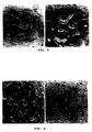
- Fig. 2 shows the morphology of PC12 cells 4 days after transfection with pC24A (A) and pC24B (C), after treatment with NGF (50 ng/ml) for 3 days (B), and under normal culture conditions (D).
- Fig. 3 shows detection of the TRK -T1 gene in transfected PC12 cells by PCR amplification using TRK -T1-specific primers followed by analysis of the amplification products on a 4% Nusieve agarose gel (3:1). Amplification was performed on low molecular weight DNA extracted from PC12 cells transfected with pC24A (lane 1), pC24A (lane 2), pLM6 (lane 3), and from mock transfectants (lane 4). The size of the marker bands (lane 5) is given on the right hand side. The size of the amplified fragments is indicated on the left hand side.
- Fig. 4 shows detection of TRK-T1 and TRK oncoproteins in transfected PC12 cells by Western blotting and detection with an antiserum specific for the carboxy-terminal domain of NTRK1.
- Cells were transfected with pC24A (lane 1) or pDM16 (lane 2) and harvested after 5 days in culture.
- the position of bands representing the TRK-T1 (p55) and TRK (p70) proteins are indicated on the left hand side.
- the molecular weight of marker bands, run on the gel in parallel, is indicated on the right hand side.
- Fig. 5 shows degeneration of PC12 cells, transfected with vector pC24A and grown in the presence of geneticin (400 ⁇ g/ml), after 10 days in culture. Photographs were taken from two independent experiments (A and B).
- Fig. 6 shows the inhibitory effect of K252a on NGF- and TRK-T1-mediated neurite outgrowth of PC12 cells.
- NGF 50 ng/ml
- C, D cells transfected with pC24A
- Fig. 7 shows the detection of the VGF8 gene product on PC12 cells by immunoflourescence staining.
- TRK -T1-transfected cells fixed after three days in culture (A, B) and cells treated with NGF (50 ng/ml) for 1 day (C, D) were stained with preimmune serum (A, C) or aVX (B, D) antiserum.
- Fig. 8 shows the effect of the D11 antibody on PC12 cell differentiation.
- Fig. 9 shows PC12 cell differentiation induced by the TRK oncogene.
- Cells transfected with vector pDM16 were cultured in the absence (A) or presence (B) of K252a (200 nM) for 3 days.
- the in vitro model system for neuronal differentiation of this invention can be set up with any neuronal cell type that is responsive to NGF.
- the rat PC12 pheochromocytoma cell line is used, which can be grown under well described and straightforward culture conditions.
- PC12 cells are widely used and can be obtained from ATCC (Bethesda, Rockville, MD) under code number CRL 1721.
- ATCC Bethesda, Rockville, MD
- CRL 1721 code number
- NGF-responsive human neuroblastoma cell lines SY5Y and LA-N-5 which express NTRK1 and display neurite outgrowth
- phosphorylation of NTRK1 upon NGF treatment Kaplan, D. R., et al. (1991). Science 252, 554-558.).
- the neuronal cells are transfected with eukaryotic expression vectors containing a gene coding for an oncogenic derivative of the NGF-receptor NTRK1.
- a gene coding for an oncogenic derivative of NTRK1 can be any oncogene derived from the NTRK 1 gene by pointmutation, truncation, or fusion to other genes, which includes sequences coding for the carboxy-terminal part of the NTRK1 protein and displays transforming activity in tumors or cell lines.
- Such oncogenes have been isolated from various tumors (Martin-Zanca, D., et al. (1986). Nature 319, 743-748; Bongarzone, I., et al. (1989).
- the capability of these oncogenes to exert similar effects in neuronal cells is plausible, since it has been found that the gene products of the TRK -T1 oncogene (Greco, A., et al. (1992). Oncogene 7, 237-242.), the original TRK oncogene (Coulier, F., et al. (1989). Mol. Cell Biol.
- eukaryotic vectors carrying the appropriate signals for expression of a foreign gene in neuronal cells are used.
- eukaryotic expression vectors designed for transient or stable integration, or vectors derived from viral systems can be employed (according to well known methods).
- episomal vectors usually providing high expression of the inserted gene, can be used; moreover, the presence of a selectable marker allows the selection of tranofected cells.
- plasmid pCEP9B Haambor, J. E., et al. (1988). Proc. Natl. Acad. Sci.
- Oncogene 7, 237-242. containing the same TRK -T1 cDNA insert under the control of the Adenovirus Major Late Promotor and being derived from the 91023B expression vector (Wong, G. G., et al. (1985). Science 228, 810-815.), designed for stable integration. Nevertheless, the effect of TRK-T1 expression on differentiation in cultures transfected with p24EA was still evident. Also, the vector pDM16 (Martin-Zanca, D., et al. (1986).
- the DNA fragments coding for the NTRK1 -derived oncogenes can be derived from genomic DNA or cDNA. These DNA fragments are inserted by conventional molecular cloning procedures known to those skilled in the art in sense and antisense orientation, with respect to the promotor, into the expression vector. Insertion of the TRK-T1 cDNA into vector pCEP9B in antisense orientation relative to the CMV promotor leads to the vector pC24B (see Example A).
- the antisense construct serves as a negative control, which does not not lead to a change in cellular phenotype upon transformation.
- the expression vectors carrying the NTRK1-derived oncogenes are then introduced into neuronal cells. This can be achieved by several well established methods. Because of the high number of cells that can be transformed, the methods of calcium phosphate, DEAE-dextran-mediated, or liposome mediated transfection or electroporation are preferred. When PC12 cells are used, good results are obtained employing electroporation (see Examples C and D). However, the choice of an appropriate transfection method is dependent on the neuronal cell type used and can not be generalized. In case that the expression vector containing the NTRK1 -derived oncogene does not offer the possibility for selection of transformants, a second plasmid expressing a selectable marker protein can be cotransfected.
- a selection marker protein allows to select for transformed cells, facilitating the analysis of the cellular differentiation process, which would otherwise be more difficult to monitor in a culture containing a high percentage of untransfected cells.
- a useful vector for cotransfection is pSV2neo (Southern, P. J., Berg, P. (1982). J. Mol. Appl. Genet. 1, 327-341.) which confers resistance to the antibiotic G418.
- the cells are seeded into tissue culture dishes and grown in selective medium.
- tissue culture dishes Three days after transfection with the vectors containing the NTRK1 -derived oncogenes in sense orientation, differentiated cells comprising long neuritic processes can be identified in the culture, which display a morphology similar to untransfected cells treated with NGF.
- Cultures of cells transfected with the vectors carrying the oncogenes in antisense orientation show a phenotype similar to cultures of untransfected cells.
- the introduction of the vector DNA can be monitored by PCR amplification of oncogene-specific fragments on extrachromosomal DNA (Hirt B., J. Mol. Biol., 26: 365-369, 1967) from cells transfected with episomal vectors and total DNA from cells transfected with integrating vectors.
- selective pressure by an antibiotic agent suited to the used selection marker is provided throughout the entire culture period.
- an antibiotic agent suited to the used selection marker.
- PC12 cells transfected with vector pC24A it was found that, during early stages of the culture (3-6 days), the differentiation of transformants is independent of the presence of G418. However, after this time, in the absence of G418 differentiated cells are no longer detectable. It is likely, that this is due to a loss of the vector DNA from the culture by dilution, which has been reported for episomal replicons in the absence of selective antibiotic pressure (Hambor, J. E., (1988). Proc. Natl. Acad. Sci. USA 85, 4010-4014.).
- the differentiated transformed cells are masked by the proliferation of the untransfected cells.
- the oncogenes of interest can be cloned into inducible expression vectors, under the control of inducible promoters such as the metallothionein promoter whose activity can be turned on by heavy metals. Following transfection, PC12 clones containing the vector can be selected; such clones can be induced to differentiate by stimulating the activity of the promoter.
- the neuronal differentiation observed with the transformed cells is independent of NGF.
- Transformed cells differentiate in cell culture medium which has not been supplemented with NGF.
- the addition of the antibody D11, which is capable of neutralizing the activity of NGF does not interfere with the differentiation observed in cell cultures transformed with the vectors containing the NTRK 1-derived oncogenes.
- conditioned medium from transformed cell cultures does not induce neuronal differentiation in untransfected cells. The two latter observations exclude the possibility, that transfection of the cells induces NGF production in the transfectants, thereby leading to a change in the differentiation state by an autocrine mechanism.
- K252a has the same effect on neuronal cells treated with NGF. Since K252a is a specific inhibitor of the tyrosine kinase activity of the NTRK1 protein, it can be concluded, that the TRK-T1 oncogene product mimicks to quite some extent the action of the NTRK1 protein activated by NGF.
- the transformed neuronal cells undergo degenerative processes.
- the differentiated neuronal cells in the transformed cultures undergo degeneration, which is characterized by an increase in cell volume, the appearance of cytoplasmic vacuoles and detachment of the cells from the culture dish.
- This is in contrast to the behaviour of neuronal cells treated with NGF, in which the differentiated phenotype is stable as long as NGF is provided.
- NGF has a role in the maintenance and viability of the cells, thereby fullfilling tasks which are not covered by the activities of the NTRK1-derived oncoproteins.
- the present invention can be used not only with NTRK1 derived oncogenes, but also with any other oncogene capable to induce PC12 differentiation.
- Vectors pC24A and pC24B are based on the eukaryotic expression vector pCEP9B (Wong, G. G., et al. (1985). Science 228, 810-815.). Briefly, this vector contains the Cytomegalovirus (CMV) promotor, a SV40 polyadenylation site and the neomycin resistance gene, allowing the selection of transformants after transfection with the antibiotic geneticin (G418). This vector can autonomously replicate in eukaryotic cells and is therefore maintained in transfected cells upon G418 selection without the need for being integrated into the host cell genome.
- CMV Cytomegalovirus
- G418 antibiotic geneticin
- Oncogene 7, 237-242. which had been shown to contain a cDNA insert capable of foci formation upon transfection into NIH3T3 cells.
- the cDNA insert was excised from phage 24 DNA by digestion with EcoRI.
- pCEP9B was linearized by digestion with HindIII, which cuts within the polylinker of the plasmid.
- the ends of the insert fragment and the linearized vector were blunted with the Klenow fragment of DNA polymerase I and subsequently ligated using T4 ligase.
- Recombinant plasmids resulting from this ligation reaction were analyzed by restriction analysis.
- the orientation of the TRK-T1 cDNA insert was determined by HindIII/BamHI digestion. Plasmids containing the TRK-T1 cDNA in the sense and antisense orientation relative to the CMV promotor were isolated and designated pC24A (see Fig. 1) and pC24B (not shown), respectively.
- the rat PC12 pheochromocytoma cell line was obtained from ATCC.
- the cells were maintained in RPMI-160 medium supplemented with 5% fetal calf serum, 10% horse serum, penicillin and streptomycin (100 U.I./ml) at 37°C in a humidified atmosphere containing 5% CO 2 .
- the cells were plated onto collagen-coated plastic dishes. NGF was supplied to the cells at a concentration of 50 ng/ml. Transfected cells were seeded onto plastic dishes.
- Exponentially growing PC12 cells were resuspended in RPMI-160 medium containing 50% fetal calf serum at a density of 1.5-3x10 7 cells/ml, mixed with 50 ⁇ g of vector DNA and incubated at 4°C for 15 min. Electroporation was carried out with a Bio-Rad Gene Pulser (Bio-Rad Laboratories, Richmond, CA), with the settings of 960 ⁇ F, 250 Volts and applying 3 pulses.
- the vector DNA used for electroporation was derived from plasmids pC24A and pC24B (see Example A), carrying the TRK-T1 cDNA in sense (pC24A) or in antisense (pC24B) orientation with respect to the promotor.
- the cells were incubated on ice for 20 min., diluted in PC12 conditioned medium and seeded on plastic Petri dishes.
- the antibiotic G418 was added either immediatly after plating or 24-48 hours later at a concentration of 400 ⁇ g/ml.
- the selective medium was renewed weekly.
- DNA derived from plasmids p24EA or p24EB (Greco A., et al. (1992). Oncogene 7, 237-242.), containing the TRK -T1 oncogene in sense (p24EA) or antisense (p24EB) orientation, respectively, or DNA derived from vector pDM16 (Martin-Zanca, D., et al. (1986). Nature 319, 743-748) containing the TRK gene, was mixed with plasmid DNA derived from pSV2neo (Southern, P. J., Berg, P. (1982). J. Mol. Appl. Genet. 1, 327-341.) at a ratio of 5:1.
- the mixture was cotransfected into PC12 cells by electroporation.
- the total amount of plasmid DNA was 50 ⁇ g.
- the conditions for electroporation and the treatment of the cells after introduction of the vector DNA were as described in Example C.
- the cells underwent degenerative processes, characterized by an increase in size, the appearance of vacuoles and detachment from the plate. This behaviour was consistently observed in different experiments performed under identical conditions (Fig. 5 A, B). This is in contrast to NGF-treated untransfected PC12 cell cultures, which do not display degenerative processes. Rather, the cells remain stably differentiated and viable as long as NGF is provided. Moreover, the cells loose their neurites and regain their proliferative capabilities upon removal of NGF (Greene, L. A., and Tischler, A. S. (1976). Proc. Natl. Acad. Sci. USA 73, 2424.)
- TRK -T1 oncogene in PC12 cells transfected with the vector pC24A was verified using the polymerase chain reaction.
- Low molecular weight DNA was extracted 3-5 days after transfection from transfected cells as described (Hirt, B. (1967). J. Mol. Biol. 26, 365-369.).
- An aliquot of the isolated DNA was subjected to PCR amplification using the primers PR1 (5'-GCTGAGAAAAGAGACTTAAT-3') and PR2 (5'-CATCACTGAAGTATTGTG-3'), that encompass a 286 bp fragment including the TPR/NTRK1 junction of the TRK-T1 cDNA (Greco, A., et al. (1992).
- Amplification was carried out in a volume of 100 pl in 1 x PCR buffer (Perkin Elmer) supplemented with dNTPs, each at a concentration of 2.5 mM, primers PR1 and PR2, each at a concentration of 45 pM, 2 nM MgCl 2 and 2.5 units of Taq polymerase (Perkin Elmer). After a denaturation step at 95°C for 30 sec., 30 cycles of amplification were carried out (95°C for 1 min., 55°C for 30 sec., 72°C for 1.5 min.), followed by an extention reaction at 72°C.
- PC12 cells that were either treated with NGF or had been transfected with the vectors pC24A and pDM16 were cultured in the absence or presence of the alkaloid-like kinase inhibitor K252a, which specifically interferes with the phosphorylation of the NTRK1 protein and inhibits NGF action on PC12 cells (Hashimoto, S. (1988). J. Cell Biol. 107, 1531-1539.).
- VGF8 expression as a result of the induction of neuronal differentiation was monitored using immunocytochemistry.
Landscapes
- Health & Medical Sciences (AREA)
- Life Sciences & Earth Sciences (AREA)
- Chemical & Material Sciences (AREA)
- Organic Chemistry (AREA)
- Zoology (AREA)
- Genetics & Genomics (AREA)
- Gastroenterology & Hepatology (AREA)
- Immunology (AREA)
- Biochemistry (AREA)
- Biophysics (AREA)
- General Health & Medical Sciences (AREA)
- Toxicology (AREA)
- Medicinal Chemistry (AREA)
- Molecular Biology (AREA)
- Proteomics, Peptides & Aminoacids (AREA)
- Cell Biology (AREA)
- Micro-Organisms Or Cultivation Processes Thereof (AREA)
- Measuring Or Testing Involving Enzymes Or Micro-Organisms (AREA)
- Investigating Or Analysing Biological Materials (AREA)
- Pharmaceuticals Containing Other Organic And Inorganic Compounds (AREA)
Abstract
Description
- This invention relates to the field of target-derived neurotrophic factors that regulate growth and survival of cells of the nervous system in the developing and adult vertebrate organism. In particular, this invention focuses on the mechanism of NGF-induced neuronal differentiation. The invention provides neuronal cells comprising oncogenic versions of the NGF receptor gene NTRK1 and displaying neuronal differentiation in the absence of NGF, eukaryotic expression vectors containing the NTRK1-derived oncogene TRK-T1, and a method to obtain said neuronal cells by introduction of NTRK1-derived oncogenes into neuronal host cells. The in vitro cell culture system of this invention is useful for research efforts aimed at the treatment of traumatic and neurodegenerative deseases of the central and peripheral nervous system, as well as in the screening of tyrosine-kinase inhibitors.
- Morphogenesis of the nervous system in the vertebrate organism is dependent on a complex interplay of different biochemical and molecular events, that are necessary to insure that, during growth and migration, neural cells will find, and project to, the adequate target regions in the developing tissue. Besides intercellular interactions and interactions of cells with signal-providing components of the extracellular matrix, target-derived neurotrophic factors that regulate cellular growth and survival play an essential role during these processes. Also, maintenance and viability of neuronal connections in the adult organism is believed to be strongly influenced by the availability of, and responsiveness to, neurotrophic factors. The discovery and isolation of nerve growth factor (NGF) that has been shown to control development, differentiation and survival of neurons of the sympathetic nervous system and parts of the sensory and central nervous system (Levi-Montalcini, R. and Angeletti, P. (1968). Physiol. Rev. 48, 534-569; Thoenen, H., and Barde, Y. A. (1980). Physiol. Rev. 60, 1284-1325; Whittemore, S. R., and Seiger, A. (1987). Brain Res. 434, 439-464; Thoenen, H., et al. (1987). Rev. Physiol. Biochem. Pharmacol. 109, 145-178), has provided the first example of a trophic factor active in neural tissue. Meanwhile, additional neurotrophic factors that share structural and functional similarities with NGF, have been added to the growing family of the so-called "neurotrophins", underscoring the relevance of the concept of neurotrophic factors in vivo.
- The possible relationship of NGF to several neurodegenerative diseases of the central and peripheral nervous system makes the investigation of NGF induced signal transduction a subject of main scientific interest. The cholinergic neurons of the basal forebrain nuclei, for instance, known to be important for cognitive functions, consistently undergo degenerative changes in cases of Alzheimer's desease. These neurons are responsive to NGF (Hefti, F., and Weiner, W. J. (1986). Ann. Neurol. 20, 275-281), and their degeneration could be associated with a decrease in NGF responsiveness or limitations in the availability of NGF. Also, the poor capacity of mammalian central nervous system neurons to regenerate upon injury might be in part due to an insufficient supply of target-derived NGF or other neurotrophins and could perhaps be improved by the exogenous application of these factors. A detailed knowledge of the molecular events triggered by NGF in neuronal cells will be important for this field and promises to provide ways for the development of new concepts for the treatment of diseases of the nervous system.
- These perspectives have prompted the search for NGF receptors on neuronal cell surfaces, with the final goal to uncover the molecular mechanisms underlying NGF action. NGF was found to interact with two types of receptors on neuronal cell surfaces which can be discriminated biochemically by their different affinity as the low and the high affinity NGF receptor. The molecular basis of the low affinity NGF receptor (LNGFR) is represented by a transmembrane glycoprotein with a molecular weight of 75 000 daltons that binds NGF with a Kd of 10-9 M (p75 , also designated p75NGFR; Johnson, D., et al. (1986). Cell 47, 545-554; Radeke, M. J., et al. (1987). Nature 325, 593-597) and is expressed on neuronal as well as on nonneuronal cells. However, the low degree of cell type specificity of its expression and the lack of significant homologies with sequences in the intracellular domains of other transmembrane receptors known to mediate signal transduction, make it seem unlikely that p75 can serve as a functional receptor by itself. Furthermore, a transmembrane glycoprotein of 140 000 daltons (NTRK1; also designated Trk, TrkA, p140trk, gp140trk or p140prototrk in the literature), a member of the tyrosine-kinase family of receptors which is encoded by the NTRK1 protooncogene and had initially been identified in its rearranged oncogenic version as the transforming principle underlying certain tumors in nonneural tissues (Martin-Zanca, D., et al. (1986). Nature 319, 743-748), was found to be a receptor for NGF (Kaplan, D., et al. (1991). Science 252, 554-558; Klein, R.,et al. (1991). Cell 65, 189-197), and has been proposed to account for, or be part of, the high affinity NGF receptor (HNGFR). While there is up to now still considerable debate, whether NTRK1 is the only component of the HNGFR in vivo , or has to act in concert with p75NGFR or other accessory proteins, it has been shown that the NTRK1 protein alone is sufficient for binding of NGF (although the equilibrium binding constant of this interaction is a matter of controversy; see Klein, R., et al. (1991). Cell 65, 189-197; Kaplan, D., et al. (1991). Science 252, 554-558; Hempstead, B., et al. (1991). Nature 350, 678-683) and for mediating NGF-induced survival and differentiation of neuronal cells (Ibáñez, C. F., et al. (1992). Cell 69, 329-341).
- The identification and molecular cloning of functional NGF receptors has opened the possibility to systematically monitor the biochemical events occurring in the signal transduction cascade of the cell that lead to the metabolical and morphological changes observed upon NGF application, starting with the formation of the receptor-ligand complex and ending with long term changes in gene expression and cellular metabolism. The most commonly used system to characterize biochemically these events are the rat PC12 pheochromocytoma cells, derived from an adrenal tumor of adrenergic neural crest cell origin (Tischler, A. S., and Greene, L. A. (1975). Nature 258, 341-342) that express both NGF receptor subtypes, the LNGFR and the HNGFR. As a result of NGF treatment for several days, PC12 cells stop dividing, develop excitable membranes, grow neuronal processes and differentiate into a sympathetic neuronal phenotype. These features go along with straightforward and well described culture conditions, having made PC12 cells the paradigm for an in vitro cell culture system to study the molecular events required for the induction of neuronal differentiation. Despite the advantages of this system, however, the rapidly accumulating knowledge about NGF-induced changes in the cellular metabolism has lead into a dilemma which can be best exemplified by a comparison of the effects evoked by NGF and epidermal growth factor (EGF) in PC12 cells.
- Similarly to NGF, EGF exerts its biological effects on PC12 cells by the interaction with a transmembrane tyrosine kinase receptor, encoded by the erbB protooncogene. In contrast to NGF which induces withdrawal from the cell cycle and differentiation, EGF acts as a mitogen and promotes proliferation. The second messenger mechanisms required to mediate such divergent biological effects were expected to be considerably different. Surprisingly, however, both factors were found to initiate intracellular signal transduction events and changes in gene expression which are nearly indistinguishable from each other. For example, with respect to its effects on gene expression, both NGF and EGF treatment of PC12 cells leads to the activation of immediate early genes to a comparable level and with a similar time course (Greenberg, M. E., et al. (1985). Biol. Chem. 260, 14101-14110; Bartel, D. P., et al. M. E. (1989). Genes Dev. 3, 304-313). Also, almost identical increases in cellular reactions like membrane ruffling (Seeley, P. J., et al. L. A. (1984). J. Cell Biol. 98, 417-426), Na+/K+ transport (Boonstra, J., et al. (1983). J. Cell Biol. 97, 92-98), cell adhesion, and glucose uptake (Huff,K., et al. (1981). J. Cell Biol. 88, 189-198) have been observed using either NGF or EGF. Moreover, the pattern of increased tyrosine phosphorylation of cellular proteins is very much alike for EGF- and NGF treated cells (Maher, P. (1988). Proc. Natl. Acad. Sci. USA 85, 6788-6791). Interestingly, NGF, like EGF, triggers phosphorylation of phospholipase C-τ (Kim, U.-H., et al. (1991). J. Biol. Chem. 266, 1359-1362; Vetter, M. L., et al. (1991). Proc. Natl. Acad. Sci. USA 88, 5650-5654), which was initially thought to be a signalling event characteristic for the transduction of mitogenic signals.
- In the course of studies addressing the role of oncogenes in the establishment of the neuronal phenotype, it was shown by introduction of either activated oncogenes or oncoproteins into PC12 cells that the proteins encoded by Ha-ras , N-ras and retroviral ras -derived genes are able to induce PC12 cell differentiation (Guerrero, I., et al. (1986). J. Cell. Physiol. 129, 71-76; Bar-Sagi, D., and Feramisco, J. R. (1985). Cell 42, 841-848; Noda, M., et al. Y. (1985). Nature 318, 73-75). Also, a promoting role of p21ras for survival and neurite outgrowth of freshly dissociated chicken embryonic neurons could be demonstrated (Borasio, G. D., et al. (1989).
Neuron 2, 1087-1096), suggesting an involvement of ras protein in the signalling pathway used by NGF. Further evidence for this notion was provided by the observation that, upon microinjection, anti-p21ras antibodies can block NGF-induced differentiation in PC12 cells (Hagag, N., et al. (1986). Nature 319, 680-682.). Transfection experiments of ras dominant negative mutants in PC12 cells indicated that, on the molecular level, p21ras affects events downstream of the NGF-induced autophosphorylation of the NTRK1 protein, (Thomas, S. M., et al. (1992). Cell 68, 1031-1040; Wood, K. W., et al. (1992). Cell 68, 1041-1050). However, phosphorylation of MAP kinases and RSK is also known to be induced by EGF (Mutoh, T., et al. (1988). J. Biol. Chem. 263, 15853-15856; Miyasaka, T., et al. (1990). J. Biol. Chem. 265, 4730-4735.) and other growth factors. Moreover, activation of cellular ras protein by EGF, as judged by GTP binding and hydrolysis, is indistinguishable from the activation by NGF (Qui, M. S., and Green, S. H. (1991). Neuron 7, 937-946.), indicating that p21ras activity and events downstream of p21ras activation are affected in a comparable manner upon treatment with either NGF or EGF. - Similarly to ras oncogene products, the protein encoded by the v-src oncogene was found to be an inducer of neuronal differentiation (Alema, S., et al. (1985). Nature 316, 557-559.) and it has been suggested that its function is required prior to ras activation (Kremer, N. E., et al. (1991). Neuron 7, 937-946.). The function of the src protooncogene product, pp60c-src, is unknown. It has been shown, however, that it is probably also involved in the transduction of signals triggered by the cell adhesion molecules N-CAM, L1 and MAG (Atashi, J. R., et al. (1992). Neuron 8, 831-842.). The molecular basis of a signalling pathway exclusively used by NGF, if it should exist at all, therefore remains unclear.
- Considering the particular biological effects of NGF on the one side and the absence of any obvious differences to other growth factors in the recruitment of intracellular signalling molecules on the other side, it is reasonable to focus the attention on the first steps of NGF-induced signal transduction. In addition to the above mentioned second messenger systems, activation of signals that have not yet been focused on might be initiated here, possibly by the involvement of accessory proteins acting in concert with NTRK1. This idea is supported by the observation that, in contrast to its effects on neuronal cells, NGF is a powerful mitogen for NIH3T3 fibroblast-like cells that have been manipulated to express NTRK1 (Cordon-Cardo, C., et al. (1991). Cell 66, 173-183.). Also, recent studies have shown a direct interaction of the NTRK1 protein with the serine/threonine kinase ERK1 in PC12 cells (Loeb, D. M., et al. (1992). Neuron 9, 1053-1065.), which possibly results in the subsequent phosphorylation of other intracellular proteins.
- In order to gain more information about the molecular mechanisms of NGF action it is therefore important to investigate the initial decisive steps of NGF-mediated signalling. In this respect, it would be desirable to have an in vitro systems providing novel approaches to study NTRK1 activation and the subsequent early signalling steps on the molecular level. In the last years the relationship between cancer and the biochemical pathway of the signal transduction has become more and more evident.
- It is well-established that the perturbation of said pathways in one of their different passages induces tumoral transformation in this respect, a particular aspectis represented by the deregulation of the genes having tyrosine-kinase activity, mainly representing receptors for growth factors or other factors. This deregulation, which transforms the genes in oncogenes, may be due to different causes, but it usually always exhibits a non -programed a tyrosine-kinase activity which transmits the cells continuos proliferative signals causing tumoral transofrmations it is therefore evident the extreme importance of substances able to interrupt this mechanism by dodulating or eliminating the tyrosine-kinase activity of the above disclosed oncogenes. These substances could also prevent the tumoral transformation.
- A novel in vitro systems to study NGF action should be easy to handle and should offer straightforward possibilites to monitor cellular responses to any experimental manipulations.
- The present invention addresses the problems raised in the prior art by providing an in vitro model system for neuronal differentiation that allows to study the basic mechanisms of NGF-specific signal transduction at the level of the NGF receptor and it may also be used to screen compounds having inhibiting action on tyrosine-kinase activity of the protein encoded by TRK-T1 and of the proteins encoded by other oncogenes sharing with TRK-T1 the catalytic site having tyrosine-kinase activity. The system of the invention is provided by neuronal cells, characterized in that they comprise a gene coding for an oncogenic derivative of the NGF-receptor NTRK1, said gene causing neuronal differentiation of said cells in the absence of NGF. Although these NTRK1-derived oncogenes have transforming activity in certain tumors, it was found that their introduction into a neuronal cell surprisingly induces neuronal differentiation. This process is independent of the presence of NGF but interestingly displays the major characteristics of the differentiation process observed with normal neuronal cells upon NGF application. In contrast to normal NGF-treated neuronal cells, however, the cells transformed with the NTRK1-derived oncogenes undergo degenerative processes after a certain period in culture, indicating that NGF-triggered NTRK1 protein might transduce additional signals needed for viability and maintenance of neuronal cells compared to the truncated receptors. The in vitro system of this invention therefore allows to compare NTRK1 with its structurally altered oncogenic derivatives with respect to the molecular mechanisms underlying their signalling activity and the recruitment of accessory molecules. Since this invention can be applied to neuronal cell lines, preferably to PC12 cells, establishment of the system is straightforward and the characteristics of the differentiation process can be easily monitored. In accord with the present invention, the eukaryotic expression vectors pC24A and pC24B, containing the NTRK1-derived oncogene TRK-T1 in sense and antisense orientation, respectively, are provided, and a method of obtaining neuronal cells displaying NGF-independent neuronal differentiation, characterized by transfection of vectors containing a gene coding for an oncogenic derivative of the NGF-receptor NTRK1 into neuronal host cells. The system of the invention, providing an useful screening method in the pharmacological search of compound having tyrosine-kinase activity, may be generalized and adapted to other tyrosine-kinase in oncogenic form.
- Fig. 1 shows a schematic representation of vector pC24A used for transfection of PC12 cells. The vector contains the cDNA sequence of the TRK-T1 oncogene, indicated by the shaded and hatched boxes designated TPR and NTRK1, respectively, in sense orientation relative to the Cytomegalovirus promotor (CMV) and the SV40 polyadenylation signal (SV40 pA). Bold lines represent polylinker sequences. The positions of cleavage sites for BamHI (B), EcoRI (E) and HindIII (H) are indicated.
- Fig. 2 shows the morphology of
PC12 cells 4 days after transfection with pC24A (A) and pC24B (C), after treatment with NGF (50 ng/ml) for 3 days (B), and under normal culture conditions (D). - Fig. 3 shows detection of the TRK-T1 gene in transfected PC12 cells by PCR amplification using TRK-T1-specific primers followed by analysis of the amplification products on a 4% Nusieve agarose gel (3:1). Amplification was performed on low molecular weight DNA extracted from PC12 cells transfected with pC24A (lane 1), pC24A (lane 2), pLM6 (lane 3), and from mock transfectants (lane 4). The size of the marker bands (lane 5) is given on the right hand side. The size of the amplified fragments is indicated on the left hand side.
- Fig. 4 shows detection of TRK-T1 and TRK oncoproteins in transfected PC12 cells by Western blotting and detection with an antiserum specific for the carboxy-terminal domain of NTRK1. Cells were transfected with pC24A (lane 1) or pDM16 (lane 2) and harvested after 5 days in culture. The position of bands representing the TRK-T1 (p55) and TRK (p70) proteins are indicated on the left hand side. The molecular weight of marker bands, run on the gel in parallel, is indicated on the right hand side.
- Fig. 5 shows degeneration of PC12 cells, transfected with vector pC24A and grown in the presence of geneticin (400 µg/ml), after 10 days in culture. Photographs were taken from two independent experiments (A and B).
- Fig. 6 shows the inhibitory effect of K252a on NGF- and TRK-T1-mediated neurite outgrowth of PC12 cells. Cells grown in the presence of NGF (50 ng/ml; A, B) or cells transfected with pC24A (C, D) were cultured in the presence (A, C) or absence (B, D) of K252a (200 nM) for 4 days.
- Fig. 7 shows the detection of the VGF8 gene product on PC12 cells by immunoflourescence staining. TRK-T1-transfected cells fixed after three days in culture (A, B) and cells treated with NGF (50 ng/ml) for 1 day (C, D) were stained with preimmune serum (A, C) or aVX (B, D) antiserum.
- Fig. 8 shows the effect of the D11 antibody on PC12 cell differentiation. Cells transfected with TRK-T1 (A) or treated with NGF (50 ng/ml; B) were treated with D11 antibodies for 3 days.
- Fig. 9 shows PC12 cell differentiation induced by the TRK oncogene. Cells transfected with vector pDM16 were cultured in the absence (A) or presence (B) of K252a (200 nM) for 3 days.
- The in vitro model system for neuronal differentiation of this invention can be set up with any neuronal cell type that is responsive to NGF. Preferably, the rat PC12 pheochromocytoma cell line is used, which can be grown under well described and straightforward culture conditions. PC12 cells are widely used and can be obtained from ATCC (Bethesda, Rockville, MD) under code number CRL 1721. However, it should be mentioned that any cell type in which NGF-induced neuronal differentiation is mediated by NTRK1, provides a molecular context that, upon transformation with oncogenic versions of NTRK1, leads to cellular responses similar to the effects observed with PC12 cells. Other appropriate cells would be, for example, the NGF-responsive human neuroblastoma cell lines SY5Y and LA-N-5, which express NTRK1 and display neurite outgrowth (Sonnenfeld, K. H., Ishii, D. N. (1982). J. Neurosci. Res. 8, 375-391.) and phosphorylation of NTRK1 upon NGF treatment (Kaplan, D. R., et al. (1991). Science 252, 554-558.).
- The neuronal cells are transfected with eukaryotic expression vectors containing a gene coding for an oncogenic derivative of the NGF-receptor NTRK1. A gene coding for an oncogenic derivative of NTRK1 can be any oncogene derived from the NTRK1 gene by pointmutation, truncation, or fusion to other genes, which includes sequences coding for the carboxy-terminal part of the NTRK1 protein and displays transforming activity in tumors or cell lines. Such oncogenes have been isolated from various tumors (Martin-Zanca, D., et al. (1986). Nature 319, 743-748; Bongarzone, I., et al. (1989).
Oncogene 4, 1457-1462; Barbacid, M., Lamballe, F., Pulido, D., and Klein, R. (1991). Biochim. Biophys. Acta 1072, 115-127; Sozzi, G., et al. (1992). Genes Chrom.Cancer 5, 1-7.). The capability of these oncogenes to exert similar effects in neuronal cells is plausible, since it has been found that the gene products of the TRK-T1 oncogene (Greco, A., et al. (1992). Oncogene 7, 237-242.), the original TRK oncogene (Coulier, F., et al. (1989). Mol. Cell Biol. 1, 15-23) and other NTRK1-derived oncogenes isolated from thyroid tumors (Bongarzone, I., et al. (1989).Oncogene 4, 1457-1462.) are, in contrast to NTRK1, constitutively phosphorylated on tyrosine. In accord with that, transformation of PC12 cells with either the TRK-T1 or the TRK gene both was found to result in indistinguishable neuronal differentiation (see Example E and Fig. 2, 9). - As a vector system to express NTRK1-derived oncogenes, eukaryotic vectors carrying the appropriate signals for expression of a foreign gene in neuronal cells are used. For this purpose, eukaryotic expression vectors designed for transient or stable integration, or vectors derived from viral systems can be employed (according to well known methods). To this purpose, episomal vectors, usually providing high expression of the inserted gene, can be used; moreover, the presence of a selectable marker allows the selection of tranofected cells. Such a vector is represented by plasmid pCEP9B (Hambor, J. E., et al. (1988). Proc. Natl. Acad. Sci. USA 85, 4010-4014.) which contains the Cytomegalovirus promotor (CMV) and is capable of episomal replication (Yates, J. L., et al. (1985). Nature 313, 812-815.). It was found that transfection of the vector pC24A, which was constructed by insertion of a TRK-T1 cDNA fragment into pCEP9B (see Fig. 1 and Example A), was more efficient in producing differentiating cells than the introduction of vector p24EA (Greco, A., et al. (1992). Oncogene 7, 237-242.), containing the same TRK-T1 cDNA insert under the control of the Adenovirus Major Late Promotor and being derived from the 91023B expression vector (Wong, G. G., et al. (1985). Science 228, 810-815.), designed for stable integration. Nevertheless, the effect of TRK-T1 expression on differentiation in cultures transfected with p24EA was still evident. Also, the vector pDM16 (Martin-Zanca, D., et al. (1986). Nature 319, 743-748) which contains the cDNA of the original TRK oncogene under the control of the Moloney murine sarcoma virus long terminal repeat and which is derived from the expression vector pm1SP (Blair, D. G., et al. (1981). Science 212, 941-943.) was found to be effective in converting the phenotype of transfected PC12 cells.
- The DNA fragments coding for the NTRK1-derived oncogenes can be derived from genomic DNA or cDNA. These DNA fragments are inserted by conventional molecular cloning procedures known to those skilled in the art in sense and antisense orientation, with respect to the promotor, into the expression vector. Insertion of the TRK-T1 cDNA into vector pCEP9B in antisense orientation relative to the CMV promotor leads to the vector pC24B (see Example A). The antisense construct serves as a negative control, which does not not lead to a change in cellular phenotype upon transformation.
- The expression vectors carrying the NTRK1-derived oncogenes are then introduced into neuronal cells. This can be achieved by several well established methods. Because of the high number of cells that can be transformed, the methods of calcium phosphate, DEAE-dextran-mediated, or liposome mediated transfection or electroporation are preferred. When PC12 cells are used, good results are obtained employing electroporation (see Examples C and D). However, the choice of an appropriate transfection method is dependent on the neuronal cell type used and can not be generalized. In case that the expression vector containing the NTRK1-derived oncogene does not offer the possibility for selection of transformants, a second plasmid expressing a selectable marker protein can be cotransfected. Expression of a selection marker protein allows to select for transformed cells, facilitating the analysis of the cellular differentiation process, which would otherwise be more difficult to monitor in a culture containing a high percentage of untransfected cells. A useful vector for cotransfection is pSV2neo (Southern, P. J., Berg, P. (1982). J. Mol. Appl. Genet. 1, 327-341.) which confers resistance to the antibiotic G418.
- Subsequently to transfection, the cells are seeded into tissue culture dishes and grown in selective medium. Three days after transfection with the vectors containing the NTRK1-derived oncogenes in sense orientation, differentiated cells comprising long neuritic processes can be identified in the culture, which display a morphology similar to untransfected cells treated with NGF. Cultures of cells transfected with the vectors carrying the oncogenes in antisense orientation, on the other hand, show a phenotype similar to cultures of untransfected cells.
- The introduction of the vector DNA can be monitored by PCR amplification of oncogene-specific fragments on extrachromosomal DNA (Hirt B., J. Mol. Biol., 26: 365-369, 1967) from cells transfected with episomal vectors and total DNA from cells transfected with integrating vectors.
- Preferably, selective pressure by an antibiotic agent suited to the used selection marker, is provided throughout the entire culture period. For PC12 cells transfected with vector pC24A it was found that, during early stages of the culture (3-6 days), the differentiation of transformants is independent of the presence of G418. However, after this time, in the absence of G418 differentiated cells are no longer detectable. It is likely, that this is due to a loss of the vector DNA from the culture by dilution, which has been reported for episomal replicons in the absence of selective antibiotic pressure (Hambor, J. E., (1988). Proc. Natl. Acad. Sci. USA 85, 4010-4014.). This suggests that continous TRK-T1 expression is required to maintain the differentiated phenotype, which is reminiscent of the need for the continous presence of NGF to maintain NGF-induced neuronal differentiation. However, it is also possible that, in the absence of G418, the differentiated transformed cells are masked by the proliferation of the untransfected cells. Alternatively, the oncogenes of interest can be cloned into inducible expression vectors, under the control of inducible promoters such as the metallothionein promoter whose activity can be turned on by heavy metals. Following transfection, PC12 clones containing the vector can be selected; such clones can be induced to differentiate by stimulating the activity of the promoter.
- The neuronal differentiation observed with the transformed cells is independent of NGF. Transformed cells differentiate in cell culture medium which has not been supplemented with NGF. Furthermore, the addition of the antibody D11, which is capable of neutralizing the activity of NGF (Cattaneo, A., et al. (1988). J. Neurochem. 50, 1003-1010), does not interfere with the differentiation observed in cell cultures transformed with the vectors containing the NTRK1-derived oncogenes. Furthermore, conditioned medium from transformed cell cultures does not induce neuronal differentiation in untransfected cells. The two latter observations exclude the possibility, that transfection of the cells induces NGF production in the transfectants, thereby leading to a change in the differentiation state by an autocrine mechanism.
- Besides the morphological similarities between the neuronal differentiation observed upon transformation with NTRK1-derived oncogenes and NGF-induced cellular responses, the two processes also share similar features on the molecular and biochemical level. It was found, that the introduction of NTRK1-derived oncogenes into PC12 cells leads to an expressional upregulation of the protein encoded by the VGF8a gene, which is known also to be specifically induced in PC12 cells upon NGF treatment (Salton, S. R. J., et al. (1991). Mol. Cell. Biol. 11, 2335-2349.). This can be easily monitored by performing immunocytochemistry using the VGF8-specific antiserum aVX (Possenti, R., et al. (1989). EMBO J. 8, 2217-2223.) and secondary antibodies coupled to flourescent dyes. Furthermore, cultivation of the transformed cells in the presence of the inhibitor K252a inhibits the conversion of the transformants to the neuronal phenotype. K252a has the same effect on neuronal cells treated with NGF. Since K252a is a specific inhibitor of the tyrosine kinase activity of the NTRK1 protein, it can be concluded, that the TRK-T1 oncogene product mimicks to quite some extent the action of the NTRK1 protein activated by NGF.
- Interestingly, after a certain time in culture, the transformed neuronal cells undergo degenerative processes. For PC12 cells it was found, that 8 days after transformation, a substantial percentage of the differentiated neuronal cells in the transformed cultures undergo degeneration, which is characterized by an increase in cell volume, the appearance of cytoplasmic vacuoles and detachment of the cells from the culture dish. This is in contrast to the behaviour of neuronal cells treated with NGF, in which the differentiated phenotype is stable as long as NGF is provided. This suggests, that in addition to its activating function of the differentiating pathway, NGF has a role in the maintenance and viability of the cells, thereby fullfilling tasks which are not covered by the activities of the NTRK1-derived oncoproteins.
- The present invention can be used not only with NTRK1 derived oncogenes, but also with any other oncogene capable to induce PC12 differentiation.
- The following examples further illustrate the invention.
- Vectors pC24A and pC24B are based on the eukaryotic expression vector pCEP9B (Wong, G. G., et al. (1985). Science 228, 810-815.). Briefly, this vector contains the Cytomegalovirus (CMV) promotor, a SV40 polyadenylation site and the neomycin resistance gene, allowing the selection of transformants after transfection with the antibiotic geneticin (G418). This vector can autonomously replicate in eukaryotic cells and is therefore maintained in transfected cells upon G418 selection without the need for being integrated into the host cell genome. The cDNA coding for TRK-T1 was obtained from phage 24 (Greco, A., et al. (1992). Oncogene 7, 237-242.) which had been shown to contain a cDNA insert capable of foci formation upon transfection into NIH3T3 cells. The cDNA insert was excised from phage 24 DNA by digestion with EcoRI. pCEP9B was linearized by digestion with HindIII, which cuts within the polylinker of the plasmid. The ends of the insert fragment and the linearized vector were blunted with the Klenow fragment of DNA polymerase I and subsequently ligated using T4 ligase. Recombinant plasmids resulting from this ligation reaction were analyzed by restriction analysis. Taking advantage of two asymmetric HindIII sites present in the TRK-T1 cDNA and a BamHI site localized in the vector (see Fig. 1), the orientation of the TRK-T1 cDNA insert was determined by HindIII/BamHI digestion. Plasmids containing the TRK-T1 cDNA in the sense and antisense orientation relative to the CMV promotor were isolated and designated pC24A (see Fig. 1) and pC24B (not shown), respectively.
- The rat PC12 pheochromocytoma cell line was obtained from ATCC. The cells were maintained in RPMI-160 medium supplemented with 5% fetal calf serum, 10% horse serum, penicillin and streptomycin (100 U.I./ml) at 37°C in a humidified atmosphere containing 5% CO2. For treatment with NGF, the cells were plated onto collagen-coated plastic dishes. NGF was supplied to the cells at a concentration of 50 ng/ml. Transfected cells were seeded onto plastic dishes.
- Exponentially growing PC12 cells were resuspended in RPMI-160 medium containing 50% fetal calf serum at a density of 1.5-3x10⁷ cells/ml, mixed with 50 µg of vector DNA and incubated at 4°C for 15 min. Electroporation was carried out with a Bio-Rad Gene Pulser (Bio-Rad Laboratories, Richmond, CA), with the settings of 960 µF, 250 Volts and applying 3 pulses. The vector DNA used for electroporation was derived from plasmids pC24A and pC24B (see Example A), carrying the TRK-T1 cDNA in sense (pC24A) or in antisense (pC24B) orientation with respect to the promotor. After electroporation, the cells were incubated on ice for 20 min., diluted in PC12 conditioned medium and seeded on plastic Petri dishes. The antibiotic G418 was added either immediatly after plating or 24-48 hours later at a concentration of 400 µg/ml. The selective medium was renewed weekly.
- DNA derived from plasmids p24EA or p24EB (Greco A., et al. (1992). Oncogene 7, 237-242.), containing the TRK-T1 oncogene in sense (p24EA) or antisense (p24EB) orientation, respectively, or DNA derived from vector pDM16 (Martin-Zanca, D., et al. (1986). Nature 319, 743-748) containing the TRK gene, was mixed with plasmid DNA derived from pSV2neo (Southern, P. J., Berg, P. (1982). J. Mol. Appl. Genet. 1, 327-341.) at a ratio of 5:1. Subsequently, the mixture was cotransfected into PC12 cells by electroporation. The total amount of plasmid DNA was 50 µg. The conditions for electroporation and the treatment of the cells after introduction of the vector DNA were as described in Example C.
- Three days after transfection of PC12 cells with the vectors p24EA (not shown) or vector pC24A, containing the TRK-T1 oncogene in sense orientation or with vector pDM16, containing the TRK oncogene in sense orientation, differentiated cells extending long neurites could be observed in culture (Fig. 2 A, 9 A). These cells were indistinguishable in their morphology from untransfected PC12 cells that had been treated with NGF (Fig. 2 B). It was found that the two TRK-T1-encoding vectors differ in their efficiency to promote neuronal differentiation. While transfection of vector pC24A produced 312 differentiated cells/µg of DNA, the number of differentiated cells was about ten fold lower when using the same amount of vector p24EA for transfection. Cultures of cells transfected with the vectors p24EB (not shown) and pC24B (Fig. 2 C), containing the TRK-T1 oncogene in antisense orientation, displayed a morphology similar to untransfected normal PC12 cell cultures (Fig. 2 D). Upon prolonged cultivation, the cultures that were differentiated by the NTRK1-derived oncogenes displayed cellular degeneration. The morphology of PC12 cells transfected with vector pC24A after 10 days in culture is shown in Fig. 5. The cells underwent degenerative processes, characterized by an increase in size, the appearance of vacuoles and detachment from the plate. This behaviour was consistently observed in different experiments performed under identical conditions (Fig. 5 A, B). This is in contrast to NGF-treated untransfected PC12 cell cultures, which do not display degenerative processes. Rather, the cells remain stably differentiated and viable as long as NGF is provided. Moreover, the cells loose their neurites and regain their proliferative capabilities upon removal of NGF (Greene, L. A., and Tischler, A. S. (1976). Proc. Natl. Acad. Sci. USA 73, 2424.)
- The presence of the TRK-T1 oncogene in PC12 cells transfected with the vector pC24A was verified using the polymerase chain reaction. Low molecular weight DNA was extracted 3-5 days after transfection from transfected cells as described (Hirt, B. (1967). J. Mol. Biol. 26, 365-369.). An aliquot of the isolated DNA was subjected to PCR amplification using the primers PR1 (5'-GCTGAGAAAAGAGACTTAAT-3') and PR2 (5'-CATCACTGAAGTATTGTG-3'), that encompass a 286 bp fragment including the TPR/NTRK1 junction of the TRK-T1 cDNA (Greco, A., et al. (1992). Oncogene 7, 237-242.). Amplification was carried out in a volume of 100 pl in 1 x PCR buffer (Perkin Elmer) supplemented with dNTPs, each at a concentration of 2.5 mM, primers PR1 and PR2, each at a concentration of 45 pM, 2 nM MgCl2 and 2.5 units of Taq polymerase (Perkin Elmer). After a denaturation step at 95°C for 30 sec., 30 cycles of amplification were carried out (95°C for 1 min., 55°C for 30 sec., 72°C for 1.5 min.), followed by an extention reaction at 72°C. Fifteen microliters of the samples were subjected to gel electrophoresis on a 4% Nusieve agarose 3:1 gel. Analysis of the reaction products by ethidium bromide staining of the gel revealed the amplification of a 286 bp DNA fragment from material isolated from pC24A-transfected PC12 cells (Fig. 3,
lanes 1, 2). No amplification products could be detected performing PCR on material derived from cells transfected with a control plasmid (pLM6; Fig. 2, lane 3), or from mock transfectants (Fig. 2, lane 4). - The presence of the oncogene products in PC12 cells transfected with the vectors pC24A and pDM16 was monitored by Western blotting and detection of the oncoproteins with an antiserum specifically reacting with the 14 carboxy-terminal amino acid residues of the NTRK1 protein (Greco, A., et al. (1992). Oncogene 7, 237-242.). Preparation of the material from transfected cells used for Western blotting and the detection procedure were carried out as described (Bongarzone, I., Pierotti, M. A., Monzini, N., Mondellini, P., Manenti, G., Donghi, R., Pilotti, S., Grieco, M., Santoro, M., Fusco, A., Vecchio, G., and Della Porta, G. (1989).
Oncogene 4, 1457-1462.). Analysis of the Western blot revealed expression of the 55 kDa TRK-T1 protein (Fig 4, lane 1) and the 70 kDa TRK protein (Fig. 4, lane 2) in cells transfected with the vectors pC24A and pDM16, respectively. - To compare the NGF-induced neuronal differentiation with the events induced by transformation of neuronal cells with NTRK1-derived oncoproteins on the biochemical level, PC12 cells that were either treated with NGF or had been transfected with the vectors pC24A and pDM16 were cultured in the absence or presence of the alkaloid-like kinase inhibitor K252a, which specifically interferes with the phosphorylation of the NTRK1 protein and inhibits NGF action on PC12 cells (Hashimoto, S. (1988). J. Cell Biol. 107, 1531-1539.). PC12 cells cultivated with NGF (50 ng/ml; Fig. 6 A, B) for 4 days revealed normal differentiation in the absence of the inhibitor K252a (Fig. 6 B). However, differentiation was completely blocked by the addition of K252a (200 nM) to the medium (Fig. 6 A). Similarly, PC12 cells transfected with vector pC24A and cultivated for 4 days (Fig. 6 C, D) displayed differentiation to a neuronal phenotype in the absence of K252a (Fig. 6D), but were lacking any characteristics of neuronal differentiation when grown in the presence of K252a (Fig. 6 C). The same effect was also found for PC12 cells that had been transfected with vector pDM16 and grown for 3 days (Fig. 9 A, B). K252a addition completely abolished the differentiation of the transformants (Fig. 9 B), whereas in the absence of the inhibitor the cells were found to bear long neuritic processes and a neuronal morphology (Fig. 9A).
- VGF8 expression as a result of the induction of neuronal differentiation was monitored using immunocytochemistry. PC12 cells that had been transformed with the TRK-T1 oncogene and were subsequently grown in slide chambers (Gibco BRL) for three days (Fig. 7 A, B), or untransfected PC12 cells grown in slide chambers in the presence of NGF (50 ng/ml) for 1 day (Fig. 7 C, D), were fixed with acetone, incubated with the antiserum aVX which is specific for the VGF8 gene product (Possenti, R., et al. (1989). EMBO J. 8, 2217-2223.) or with preimmune serum, repeatedly washed and further incubated with flourescein-labelled goat anti-rabbit antibodies (Meloy, Springfield, VA). TRK-T1-transformed PC12 cells as well as NGF-treated PC12 cells revealed strong immunoreactivity with the aVX antiserum, indicating a high level expression of the VGF8 protein Fig. 7 B, D). No staining could be observed with both cell types when the preimmune serum was used as first antibody (Fig. 7 A, C).
- In order to investigate, whether transfection of neuronal cells might induce the production of NGF by the transformants, thereby creating an autocrine circuit that would be responsible for the observed neuronal differentiation, PC12 cells that had been converted to the neuronal phenotype by transformation with the TRK-T1 gene or by NGF treatment, were cultured for 3 days in the presence of the antibody D11 (Cattaneo, A., et al. (1988). J. Neurochem. 50, 1003-1010; Fig. 8 A, B), which is specific for NGF and blocks its biological activity. Despite the presence of the D11 antibodies, the TRK-T1-transformed cells continued to display a neuronal morphology (Fig. 8 A), whereas cultures treated with NGF were converted to an undifferentiated state (Fig. 8 B). In accord with these observations, no induction of neuronal differentiation could be detected with untransfected PC12 cells that were grown in conditioned medium derived from pC24A-transfected cells (not shown).
Claims (7)
- Neuronal cells characterized in that they comprise a gene coding for an oncogenic derivative of the NGF-receptor NTRK1, said gene causing neuronal differentiation of said cells in the absence of NGF.
- Cells according to claim 1 that are PC12 pheochromocytoma cells.
- Cells according to claim 1 and 2 characterized in that they comprise a gene coding for the NTRK1-related oncoprotein TRK-T1.
- Cells according to claim 1 and 2 characterized in that they comprise the original TRK oncogene.
- A method of obtaining neuronal cells displaying NGF-independent neuronal differentiation characterized by transfection of vectors containing a gene coding for an oncogenic derivative of the NGF-receptor NTRK1 into neuronal host cells.
- A method according to claim 5, wherein the neuronal cells are PC12 pheochromocytoma cells.
- A method for screening compounds having anti-tyrosine kinase activity comprising the culture of the cells of claims 1-4 in the presence of the compound to be tested and the analysis of the cultured cells for the absence of neuronal differentiation.
Applications Claiming Priority (3)
| Application Number | Priority Date | Filing Date | Title |
|---|---|---|---|
| IT93MI000985A IT1264447B1 (en) | 1993-05-14 | 1993-05-14 | EXPRESSION OF ONCOGENS DERIVED FROM NTRKI IN PC12 PHEOCROMOCYTOMA CELLS AND USE OF THIS SYSTEM AS SCREENING OF SUBSTANCES |
| ITMI930985 | 1993-05-14 | ||
| PCT/EP1994/001509 WO1994026889A2 (en) | 1993-05-14 | 1994-05-10 | Expression of ntrk1-derived oncogenes in pc12 pheochromocytoma cells and use thereof for the screening of anti-tyrosine kinase compounds |
Publications (2)
| Publication Number | Publication Date |
|---|---|
| EP0698096A1 EP0698096A1 (en) | 1996-02-28 |
| EP0698096B1 true EP0698096B1 (en) | 1997-03-26 |
Family
ID=11366164
Family Applications (1)
| Application Number | Title | Priority Date | Filing Date |
|---|---|---|---|
| EP94917613A Expired - Lifetime EP0698096B1 (en) | 1993-05-14 | 1994-05-10 | Expression of ntrk1-derived oncogenes in pc12 pheochromocytoma cells and use thereof for the screening of anti-tyrosine kinase compounds |
Country Status (13)
| Country | Link |
|---|---|
| EP (1) | EP0698096B1 (en) |
| JP (1) | JP3549536B2 (en) |
| AT (1) | ATE150790T1 (en) |
| AU (1) | AU689778B2 (en) |
| DE (1) | DE69402313T2 (en) |
| DK (1) | DK0698096T3 (en) |
| ES (1) | ES2101537T3 (en) |
| FI (1) | FI955447L (en) |
| GR (1) | GR3023878T3 (en) |
| IT (1) | IT1264447B1 (en) |
| NO (1) | NO319374B1 (en) |
| NZ (1) | NZ267086A (en) |
| WO (1) | WO1994026889A2 (en) |
Cited By (4)
| Publication number | Priority date | Publication date | Assignee | Title |
|---|---|---|---|---|
| WO2013059740A1 (en) * | 2011-10-21 | 2013-04-25 | Foundation Medicine, Inc. | Novel alk and ntrk1 fusion molecules and uses thereof |
| US10980804B2 (en) | 2013-01-18 | 2021-04-20 | Foundation Medicine, Inc. | Methods of treating cholangiocarcinoma |
| US11230589B2 (en) | 2012-11-05 | 2022-01-25 | Foundation Medicine, Inc. | Fusion molecules and uses thereof |
| US11578372B2 (en) | 2012-11-05 | 2023-02-14 | Foundation Medicine, Inc. | NTRK1 fusion molecules and uses thereof |
Families Citing this family (6)
| Publication number | Priority date | Publication date | Assignee | Title |
|---|---|---|---|---|
| US7696404B2 (en) | 1996-08-19 | 2010-04-13 | Advanced Cell Technology, Inc. | Embryonic or stem-like cell lines produced by cross species nuclear transplantation and methods for enhancing embryonic development by genetic alteration of donor cells or by tissue culture conditions |
| EP1206562A2 (en) * | 1999-08-27 | 2002-05-22 | EC (European Community) | Genetically engineered cell lines, and their uses, in particular for neurotoxicity testing |
| EP1230342A4 (en) | 1999-10-28 | 2003-04-09 | Univ Massachusetts | GYNOGENETIC AND ANDROGENETIC PRODUCTION OF PLURIPOTENT CELLS AND CELL LINES AND THE USE THEREOF FOR THE PRODUCTION OF DIFFERENTIATED CELLS AND TISSUES |
| NZ526888A (en) | 2001-01-02 | 2008-07-31 | Stemron Inc | Isolated homozygous stem (HS) cells and the discovery that these cells may be selected for immunotype and genotype, therefore being useful for diagnosis, transplantation and/or treatment |
| DE10300015A1 (en) | 2003-01-03 | 2004-07-15 | Aventis Pharma Deutschland Gmbh | Imino acid derivatives as inhibitors of matrix metal proteinases |
| CN103361304B (en) * | 2012-03-27 | 2015-09-30 | 中国科学院上海生命科学研究院 | Lonely male monoploid stem cell line and method for making thereof and application |
-
1993
- 1993-05-14 IT IT93MI000985A patent/IT1264447B1/en active IP Right Grant
-
1994
- 1994-05-10 DE DE69402313T patent/DE69402313T2/en not_active Expired - Fee Related
- 1994-05-10 JP JP52495294A patent/JP3549536B2/en not_active Expired - Fee Related
- 1994-05-10 AT AT94917613T patent/ATE150790T1/en not_active IP Right Cessation
- 1994-05-10 WO PCT/EP1994/001509 patent/WO1994026889A2/en not_active Ceased
- 1994-05-10 DK DK94917613.5T patent/DK0698096T3/en active
- 1994-05-10 AU AU69269/94A patent/AU689778B2/en not_active Ceased
- 1994-05-10 EP EP94917613A patent/EP0698096B1/en not_active Expired - Lifetime
- 1994-05-10 FI FI955447A patent/FI955447L/en not_active Application Discontinuation
- 1994-05-10 ES ES94917613T patent/ES2101537T3/en not_active Expired - Lifetime
- 1994-05-10 NZ NZ267086A patent/NZ267086A/en unknown
-
1995
- 1995-11-13 NO NO19954556A patent/NO319374B1/en not_active Application Discontinuation
-
1997
- 1997-06-25 GR GR970401521T patent/GR3023878T3/en unknown
Cited By (10)
| Publication number | Priority date | Publication date | Assignee | Title |
|---|---|---|---|---|
| WO2013059740A1 (en) * | 2011-10-21 | 2013-04-25 | Foundation Medicine, Inc. | Novel alk and ntrk1 fusion molecules and uses thereof |
| US10000814B2 (en) | 2011-10-21 | 2018-06-19 | Foundation Medicine, Inc. | ALK and NTRK1 fusion molecules and uses thereof |
| US11098368B2 (en) | 2011-10-21 | 2021-08-24 | Foundation Medicine, Inc. | ALK and NTRK1 fusion molecules and uses thereof |
| US12522873B2 (en) | 2011-10-21 | 2026-01-13 | Foundation Medicine, Inc. | ALK and NTRK1 fusion molecules and uses thereof |
| US11230589B2 (en) | 2012-11-05 | 2022-01-25 | Foundation Medicine, Inc. | Fusion molecules and uses thereof |
| US11578372B2 (en) | 2012-11-05 | 2023-02-14 | Foundation Medicine, Inc. | NTRK1 fusion molecules and uses thereof |
| US12378302B2 (en) | 2012-11-05 | 2025-08-05 | Foundation Medicine, Inc. | Fusion molecules and uses thereof |
| US10980804B2 (en) | 2013-01-18 | 2021-04-20 | Foundation Medicine, Inc. | Methods of treating cholangiocarcinoma |
| US11771698B2 (en) | 2013-01-18 | 2023-10-03 | Foundation Medicine, Inc. | Methods of treating cholangiocarcinoma |
| US12274699B2 (en) | 2013-01-18 | 2025-04-15 | Foundation Medicine, Inc. | Methods of treating cholangiocarcinoma |
Also Published As
| Publication number | Publication date |
|---|---|
| FI955447A7 (en) | 1995-11-13 |
| NO954556D0 (en) | 1995-11-13 |
| WO1994026889A3 (en) | 1994-12-22 |
| DE69402313D1 (en) | 1997-04-30 |
| EP0698096A1 (en) | 1996-02-28 |
| NZ267086A (en) | 1997-01-29 |
| ES2101537T3 (en) | 1997-07-01 |
| FI955447A0 (en) | 1995-11-13 |
| NO319374B1 (en) | 2005-07-25 |
| ITMI930985A0 (en) | 1993-05-14 |
| JPH08510119A (en) | 1996-10-29 |
| AU6926994A (en) | 1994-12-12 |
| JP3549536B2 (en) | 2004-08-04 |
| FI955447L (en) | 1995-11-13 |
| AU689778B2 (en) | 1998-04-09 |
| ITMI930985A1 (en) | 1994-11-14 |
| DE69402313T2 (en) | 1997-08-21 |
| NO954556L (en) | 1996-01-12 |
| ATE150790T1 (en) | 1997-04-15 |
| IT1264447B1 (en) | 1996-09-23 |
| WO1994026889A2 (en) | 1994-11-24 |
| GR3023878T3 (en) | 1997-09-30 |
| DK0698096T3 (en) | 1997-10-27 |
Similar Documents
| Publication | Publication Date | Title |
|---|---|---|
| Winslow et al. | Cloning of AL-1, a ligand for an Eph-related tyrosine kinase receptor involved in axon bundle formation | |
| Berkemeier et al. | Neurotrophin-5: a novel neurotrophic factor that activates trk and trkB | |
| Carlomagno et al. | Molecular heterogeneity of RET loss of function in Hirschsprung's disease. | |
| US6255454B1 (en) | Expression and use of human fibroblast growth factor receptor | |
| Barbacid et al. | The trk family of tyrosine protein kinase receptors | |
| Klein et al. | The trkB tyrosine protein kinase is a receptor for neurotrophin-4 | |
| Hempstead et al. | Expression of the v-crk oncogene product in PC12 cells results in rapid differentiation by both nerve growth factor-and epidermal growth factor-dependent pathways | |
| Attisano et al. | Identification of human activin and TGFβ type I receptors that form heteromeric kinase complexes with type II receptors | |
| Chang et al. | Ligands for ErbB-family receptors encoded by a neuregulin-like gene | |
| Maness | Nonreceptor Protein Tyrosine Kinases Associated with Neuronal Development (Part 2 of 2) | |
| US5912326A (en) | Cerebellum-derived growth factors | |
| AU689778B2 (en) | Expression of NTRK1-derived oncogenes in PC12 pheochromocytoma cells and use thereof for the screening of anti-tyrosine kinase compounds | |
| Jung et al. | Organization of the human PTK7 gene encoding a receptor protein tyrosine kinase-like molecule and alternative splicing of its mRNA | |
| Robertson et al. | Identification and structure of the nerve growth factor binding site on TrkA | |
| Allsopp et al. | Ectopic trkA expression mediates a NGF survival response in NGF-independent sensory neurons but not in parasympathetic neurons. | |
| AU691915B2 (en) | Biologically active EPH family ligands | |
| US6184349B1 (en) | Cloned peripheral nerve, tetrodotoxin-resistant sodium channel α-subunit | |
| Masiakowski et al. | Changes in PC12 cell morphology induced by transfection with 42C cDNA, coding for a member of the S‐100 protein family | |
| CA2251262C (en) | Nucleic acid encoding a nervous tissue sodium channel | |
| CA2162677C (en) | Expression of ntrk1-derived oncogenes in pc12 pheochromocytoma cells and use thereof for the screening of anti-tyrosine kinase compounds | |
| Hempstead et al. | Molecular analysis of the nerve growth factor receptor | |
| Greco et al. | Expression of TRK-T1 oncogene induces differentiation of PC12 cells | |
| Dubus et al. | Identification of novel trkA variants with deletions in leucine-rich motifs of the extracellular domain | |
| US20020137168A1 (en) | Developmental tyrosine kinases and their ligands | |
| Ponomareva et al. | Stimulation of acetylcholine receptor transcription by neuregulin-2 requires an N-box response element and is regulated by alternative splicing |
Legal Events
| Date | Code | Title | Description |
|---|---|---|---|
| PUAI | Public reference made under article 153(3) epc to a published international application that has entered the european phase |
Free format text: ORIGINAL CODE: 0009012 |
|
| 17P | Request for examination filed |
Effective date: 19951113 |
|
| AK | Designated contracting states |
Kind code of ref document: A1 Designated state(s): AT BE CH DE DK ES FR GB GR IE IT LI LU MC NL PT SE |
|
| GRAG | Despatch of communication of intention to grant |
Free format text: ORIGINAL CODE: EPIDOS AGRA |
|
| 17Q | First examination report despatched |
Effective date: 19960628 |
|
| GRAH | Despatch of communication of intention to grant a patent |
Free format text: ORIGINAL CODE: EPIDOS IGRA |
|
| GRAH | Despatch of communication of intention to grant a patent |
Free format text: ORIGINAL CODE: EPIDOS IGRA |
|
| GRAA | (expected) grant |
Free format text: ORIGINAL CODE: 0009210 |
|
| AK | Designated contracting states |
Kind code of ref document: B1 Designated state(s): AT BE CH DE DK ES FR GB GR IE IT LI LU MC NL PT SE |
|
| REF | Corresponds to: |
Ref document number: 150790 Country of ref document: AT Date of ref document: 19970415 Kind code of ref document: T |
|
| REG | Reference to a national code |
Ref country code: CH Ref legal event code: EP |
|
| REF | Corresponds to: |
Ref document number: 69402313 Country of ref document: DE Date of ref document: 19970430 |
|
| REG | Reference to a national code |
Ref country code: CH Ref legal event code: NV Representative=s name: HUG INTERLIZENZ AG |
|
| ET | Fr: translation filed | ||
| REG | Reference to a national code |
Ref country code: IE Ref legal event code: FG4D Free format text: 72932 |
|
| REG | Reference to a national code |
Ref country code: ES Ref legal event code: FG2A Ref document number: 2101537 Country of ref document: ES Kind code of ref document: T3 |
|
| REG | Reference to a national code |
Ref country code: GR Ref legal event code: FG4A Free format text: 3023878 |
|
| REG | Reference to a national code |
Ref country code: DK Ref legal event code: T3 |
|
| PLBE | No opposition filed within time limit |
Free format text: ORIGINAL CODE: 0009261 |
|
| STAA | Information on the status of an ep patent application or granted ep patent |
Free format text: STATUS: NO OPPOSITION FILED WITHIN TIME LIMIT |
|
| 26N | No opposition filed | ||
| REG | Reference to a national code |
Ref country code: GB Ref legal event code: IF02 |
|
| PGFP | Annual fee paid to national office [announced via postgrant information from national office to epo] |
Ref country code: ES Payment date: 20060426 Year of fee payment: 13 |
|
| PGFP | Annual fee paid to national office [announced via postgrant information from national office to epo] |
Ref country code: PT Payment date: 20060428 Year of fee payment: 13 Ref country code: MC Payment date: 20060428 Year of fee payment: 13 Ref country code: IE Payment date: 20060428 Year of fee payment: 13 Ref country code: FR Payment date: 20060428 Year of fee payment: 13 |
|
| PGFP | Annual fee paid to national office [announced via postgrant information from national office to epo] |
Ref country code: GB Payment date: 20060509 Year of fee payment: 13 Ref country code: BE Payment date: 20060509 Year of fee payment: 13 |
|
| PGFP | Annual fee paid to national office [announced via postgrant information from national office to epo] |
Ref country code: LU Payment date: 20060511 Year of fee payment: 13 |
|
| PGFP | Annual fee paid to national office [announced via postgrant information from national office to epo] |
Ref country code: SE Payment date: 20060512 Year of fee payment: 13 |
|
| PGFP | Annual fee paid to national office [announced via postgrant information from national office to epo] |
Ref country code: NL Payment date: 20060515 Year of fee payment: 13 |
|
| PGFP | Annual fee paid to national office [announced via postgrant information from national office to epo] |
Ref country code: CH Payment date: 20060519 Year of fee payment: 13 |
|
| PGFP | Annual fee paid to national office [announced via postgrant information from national office to epo] |
Ref country code: DE Payment date: 20060523 Year of fee payment: 13 |
|
| PGFP | Annual fee paid to national office [announced via postgrant information from national office to epo] |
Ref country code: DK Payment date: 20060526 Year of fee payment: 13 |
|
| PGFP | Annual fee paid to national office [announced via postgrant information from national office to epo] |
Ref country code: IT Payment date: 20060531 Year of fee payment: 13 Ref country code: GR Payment date: 20060531 Year of fee payment: 13 Ref country code: AT Payment date: 20060531 Year of fee payment: 13 |
|
| REG | Reference to a national code |
Ref country code: PT Ref legal event code: MM4A Free format text: LAPSE DUE TO NON-PAYMENT OF FEES Effective date: 20071112 |
|
| BERE | Be: lapsed |
Owner name: *ISTITUTO NAZIONALE PER LO STUDIO E LA CURA DEI TU Effective date: 20070531 |
|
| EUG | Se: european patent has lapsed | ||
| REG | Reference to a national code |
Ref country code: CH Ref legal event code: PL |
|
| REG | Reference to a national code |
Ref country code: DK Ref legal event code: EBP |
|
| GBPC | Gb: european patent ceased through non-payment of renewal fee |
Effective date: 20070510 |
|
| PG25 | Lapsed in a contracting state [announced via postgrant information from national office to epo] |
Ref country code: PT Free format text: LAPSE BECAUSE OF NON-PAYMENT OF DUE FEES Effective date: 20071112 Ref country code: NL Free format text: LAPSE BECAUSE OF NON-PAYMENT OF DUE FEES Effective date: 20071201 Ref country code: MC Free format text: LAPSE BECAUSE OF NON-PAYMENT OF DUE FEES Effective date: 20070531 |
|
| NLV4 | Nl: lapsed or anulled due to non-payment of the annual fee |
Effective date: 20071201 |
|
| REG | Reference to a national code |
Ref country code: IE Ref legal event code: MM4A |
|
| PG25 | Lapsed in a contracting state [announced via postgrant information from national office to epo] |
Ref country code: AT Free format text: LAPSE BECAUSE OF NON-PAYMENT OF DUE FEES Effective date: 20070510 Ref country code: CH Free format text: LAPSE BECAUSE OF NON-PAYMENT OF DUE FEES Effective date: 20070531 Ref country code: LI Free format text: LAPSE BECAUSE OF NON-PAYMENT OF DUE FEES Effective date: 20070531 |
|
| REG | Reference to a national code |
Ref country code: FR Ref legal event code: ST Effective date: 20080131 |
|
| PG25 | Lapsed in a contracting state [announced via postgrant information from national office to epo] |
Ref country code: BE Free format text: LAPSE BECAUSE OF NON-PAYMENT OF DUE FEES Effective date: 20070531 |
|
| PG25 | Lapsed in a contracting state [announced via postgrant information from national office to epo] |
Ref country code: DK Free format text: LAPSE BECAUSE OF NON-PAYMENT OF DUE FEES Effective date: 20070531 Ref country code: DE Free format text: LAPSE BECAUSE OF NON-PAYMENT OF DUE FEES Effective date: 20071201 |
|
| PG25 | Lapsed in a contracting state [announced via postgrant information from national office to epo] |
Ref country code: IE Free format text: LAPSE BECAUSE OF NON-PAYMENT OF DUE FEES Effective date: 20070510 Ref country code: GB Free format text: LAPSE BECAUSE OF NON-PAYMENT OF DUE FEES Effective date: 20070510 |
|
| PG25 | Lapsed in a contracting state [announced via postgrant information from national office to epo] |
Ref country code: SE Free format text: LAPSE BECAUSE OF NON-PAYMENT OF DUE FEES Effective date: 20070511 |
|
| PG25 | Lapsed in a contracting state [announced via postgrant information from national office to epo] |
Ref country code: FR Free format text: LAPSE BECAUSE OF NON-PAYMENT OF DUE FEES Effective date: 20070531 |
|
| REG | Reference to a national code |
Ref country code: ES Ref legal event code: FD2A Effective date: 20070511 |
|
| PG25 | Lapsed in a contracting state [announced via postgrant information from national office to epo] |
Ref country code: GR Free format text: LAPSE BECAUSE OF NON-PAYMENT OF DUE FEES Effective date: 20071204 Ref country code: ES Free format text: LAPSE BECAUSE OF NON-PAYMENT OF DUE FEES Effective date: 20070511 |
|
| PG25 | Lapsed in a contracting state [announced via postgrant information from national office to epo] |
Ref country code: LU Free format text: LAPSE BECAUSE OF NON-PAYMENT OF DUE FEES Effective date: 20070510 |
|
| PG25 | Lapsed in a contracting state [announced via postgrant information from national office to epo] |
Ref country code: IT Free format text: LAPSE BECAUSE OF NON-PAYMENT OF DUE FEES Effective date: 20070510 |