EP0686262B1 - Methods for the diagnosis of diabetes and prediabetic conditions - Google Patents
Methods for the diagnosis of diabetes and prediabetic conditions Download PDFInfo
- Publication number
- EP0686262B1 EP0686262B1 EP94906796A EP94906796A EP0686262B1 EP 0686262 B1 EP0686262 B1 EP 0686262B1 EP 94906796 A EP94906796 A EP 94906796A EP 94906796 A EP94906796 A EP 94906796A EP 0686262 B1 EP0686262 B1 EP 0686262B1
- Authority
- EP
- European Patent Office
- Prior art keywords
- gad
- iddm
- serum
- preparation
- sera
- Prior art date
- Legal status (The legal status is an assumption and is not a legal conclusion. Google has not performed a legal analysis and makes no representation as to the accuracy of the status listed.)
- Expired - Lifetime
Links
Images


Classifications
-
- C—CHEMISTRY; METALLURGY
- C12—BIOCHEMISTRY; BEER; SPIRITS; WINE; VINEGAR; MICROBIOLOGY; ENZYMOLOGY; MUTATION OR GENETIC ENGINEERING
- C12N—MICROORGANISMS OR ENZYMES; COMPOSITIONS THEREOF; PROPAGATING, PRESERVING, OR MAINTAINING MICROORGANISMS; MUTATION OR GENETIC ENGINEERING; CULTURE MEDIA
- C12N9/00—Enzymes; Proenzymes; Compositions thereof; Processes for preparing, activating, inhibiting, separating or purifying enzymes
- C12N9/88—Lyases (4.)
-
- G—PHYSICS
- G01—MEASURING; TESTING
- G01N—INVESTIGATING OR ANALYSING MATERIALS BY DETERMINING THEIR CHEMICAL OR PHYSICAL PROPERTIES
- G01N33/00—Investigating or analysing materials by specific methods not covered by groups G01N1/00 - G01N31/00
- G01N33/48—Biological material, e.g. blood, urine; Haemocytometers
- G01N33/50—Chemical analysis of biological material, e.g. blood, urine; Testing involving biospecific ligand binding methods; Immunological testing
- G01N33/53—Immunoassay; Biospecific binding assay; Materials therefor
- G01N33/564—Immunoassay; Biospecific binding assay; Materials therefor for pre-existing immune complex or autoimmune disease, i.e. systemic lupus erythematosus, rheumatoid arthritis, multiple sclerosis, rheumatoid factors or complement components C1-C9
-
- G—PHYSICS
- G01—MEASURING; TESTING
- G01N—INVESTIGATING OR ANALYSING MATERIALS BY DETERMINING THEIR CHEMICAL OR PHYSICAL PROPERTIES
- G01N33/00—Investigating or analysing materials by specific methods not covered by groups G01N1/00 - G01N31/00
- G01N33/48—Biological material, e.g. blood, urine; Haemocytometers
- G01N33/50—Chemical analysis of biological material, e.g. blood, urine; Testing involving biospecific ligand binding methods; Immunological testing
- G01N33/53—Immunoassay; Biospecific binding assay; Materials therefor
- G01N33/573—Immunoassay; Biospecific binding assay; Materials therefor for enzymes or isoenzymes
-
- G—PHYSICS
- G01—MEASURING; TESTING
- G01N—INVESTIGATING OR ANALYSING MATERIALS BY DETERMINING THEIR CHEMICAL OR PHYSICAL PROPERTIES
- G01N2800/00—Detection or diagnosis of diseases
- G01N2800/04—Endocrine or metabolic disorders
- G01N2800/042—Disorders of carbohydrate metabolism, e.g. diabetes, glucose metabolism
Definitions
- This invention relates to methods for the identification of persons who have diabetes or a prediabetic status, and in particular it is directed to a diagnostic assay which will provide a simple and effective means of screening large populations of individuals to detect both diabetes and prediabetes (the early stages of diabetes prior to the onset of the clinical disease).
- Diabetes mellitus is a heterogeneous disorder.
- Insulin-dependent (IDDM) and non-insulin-dependent diabetes mellitus (NIDDM) are subtyped physiologically according to patients' dependence for survival on treatment with insulin 1 .
- IDDM is attributed to autoimmunity 2 by reason of disease associations, autoantibodies to pancreatic islet cell antigens, similarities with animal models of IDDM and HLA linkages.
- the disease-relevant autoantigen or antigens in IDDM include a 64,000- M r protein demonstrable by immunoprecipitation from pancreatic islets 3 but hitherto unidentified because of low abundance and assay difficulties.
- Immunoassay for antibody to the 64,000- M r antigen indicated that it was a specific marker for IDDM and preceded symptomatic onset of disease 4 .
- GAD glutamic acid decarboxylase
- Rowley et al . 6 described a high frequency of positive results by radioimmunoprecipitation assay with IDDM sera and a GAD preparation from pig brain and proposed detection of antibodies to GAD as a useful diagnostic test as an autoimmune marker associated with IDDM.
- Insulin-dependent diabetes mellitus is a frequently occurring disease that predominantly affects young individuals, less than age 15 years.
- the disease can, however, have an onset in early or even mid-adult life, hence there is a need for a test that has diagnostic and predictive capacity for IDDM in both childhood and in adult life.
- IDDM occurs at highest frequencies in the developed and economically advanced communities in the world. There is a high incentive to diagnose IDDM at the earliest stage, and even in the preclinical stage before the disease can be recognised by conventional laboratory tests.
- the recognition of antibodies to GAD by a routinely applicable, sensitive and specific immunoassay kit would fulfil this need.
- NIDDM non-insulin dependent form
- LADA latent autoimmune diabetes in adults
- IDDM islet cell antibodies
- ICAs autoantibodies to islet cell antibodies
- 64,000- M r protein now recognised as GAD 3,4
- cytoplasmic ICA demonstrable by immunofluorescence 8
- insulin autoantibodies 9 Antibody to the 64,000- M r molecule has been extremely difficult to assay because of the need to prepare antigen from large quantities of islet cell preparations.
- Rowley et al . 6 showed that with radioiodine-labelled GAD prepared from pig brain, the antibody could be readily and specifically detected by an immunoprecipitation assay.
- the 64,000- M r antigen is an enzyme molecule as are various other disease-relevant autoantigens, most notably thyroid peroxidase in autoimmune thyroiditis 14 , H + K + ATPase in autoimmune gastritis 11 , and pyruvate dehydrogenase in primary biliary cirrhosis 12 .
- GAD is not species specific and, in fact, cDNAs for brain GAD from humans 13 , rat 14 , and cat 15 have such close homology that antibody reactivity could be expected with any mammalian GAD preparation.
- the autoantigen is clearly not organ specific because the source used by Rowley et al . 6 was brain.
- GAD The sequence of GAD from brain and pancreatic islets, the major sites in which GAD is demonstrable, appears to differ only by allelic variations 16,17 .
- GAD exists as at least two isoforms 18 , and the Rowley et al . 6 preparation contained both representatives, as indicated by data by polyacrylamide gel electrophoresis (PAGE).
- GAD ⁇ -aminobutyric acid
- the enzyme has a restricted tissue distribution and is highly expressed in the cytoplasm of GABA-secreting neurons and pancreatic ⁇ -cells.
- the two isoforms of GAD are referred to as GAD 65 and GAD 67 according to their relative molecular masses. These isoforms are the products of two different genes and differ substantially only in their amino-terminal region.
- GAD appears to exist as an integral membrane protein with hydrophobic properties, and is present as a non-covalently linked dimer of MW 96,600 20 .
- This purified membrane form of GAD has been found to be reactive with IDDM sera, however no sequence data are yet available for this integral membrane protein 20 .
- the GAD autoantibody is a valuable predictive marker of the disease
- the predictive capacity for IDDM of a positive test for anti-GAD was remarkably high a relative risk versus a negative test for anti-GAD being 800-fold.
- a study on stored sera from women with gestational diabetes likewise showed that a positive test for anti-GAD was predictive of the occurrence later in life of IDDM in such women 24 .
- PCT/US91/06438 Publication No. WO 92/04632 discloses assays for the detection of diabetes or a prediabetic status relying on exposing patient serum samples to purified ligand capable of binding autoantibodies specific for the 64 kD autoantigen present on pancreatic ⁇ -cells.
- GAD can form monomers, dimers, trimers and tetramers.
- WO 92/19972 discloses assays for the detection of preclinical diabetes using high molecular weight GAD.
- WO 92/14485 relates to the use of GAD to test for autoimmune diseases.
- WO 92/05446 relates to the cloning of GAD cDNA. Kaufman et al., discuss that autoantibodies to 65 kD are associated with diabetes (J. Clin. Invest., 89, 283-292, 1992).
- a method for detecting autoantibodies to glutamic acid decarboxylase (GAD) in the serum of a patient comprising contacting a serum sample from the patient with a GAD preparation and detecting binding of autoantibodies to GAD in said sample by said GAD preparation, characterised in that said GAD preparation comprises an enhanced amount of dimer(s) or oligomer(s) of the 65 kD and/or 67 kD isoforms of GAD, relative to monomeric GAD in preparations of naturally occurring GAD.
- GAD glutamic acid decarboxylase
- the present description of the invention refers specifically to detection of antibodies to GAD in a serum sample, it is to be understood that the term “serum” is used as a non-limiting term, and that the present invention also extends to the detection of autoantibodies to GAD in "blood” samples in general, including blood serum and plasma.
- the method of the invention is carried out under conditions under which the dimeric or oligomeric form of the GAD is retained and is not broken down into monomeric GAD.
- the method of the invention is carried out under non-denaturing conditions, and particularly under non-reducing conditions.
- the detection of binding between the autoantibodies and the GAD antigen is indicative of the presence of the autoantibodies in the serum sample, and hence is diagnostic of a diabetic or prediabetic condition in the patient.
- the method of the present invention uses a non-naturally occurring preparation of GAD antigen which contains an enhanced amount of dimer(s) or oligomer(s) of the 65 kD and/or 67 kD isoforms of GAD.
- the present invention further extends to a diagnostic kit for the detection of autoantibodies to GAD in the serum of a patient, comprising a GAD preparation and means for detecting binding of autoantibodies to GAD in a serum sample from the patient by said GAD preparation, characterised in that said GAD preparation comprises an enhanced amount of dimer(s) or oligomer(s) of the 65 kD and/or 67 kD isoforms of GAD, relative to monomeric GAD in preparations of naturally occurring GAD.
- an "enhanced amount" of dimer(s) or oligomer(s) in the GAD preparation indicate that the amount of dimer(s) or oligomer(s) in the GAD preparation is greater than the amount of dimer(s) or oligomer(s) in preparations of naturally occurring GAD.
- Such greater amounts may be achieved by separation of naturally occurring GAD into fractions having an "enhanced amount” of dimer(s) or oligomer(s), for example by electrophoretic separation on SDS-PAGE gels or by size exclusion chromatography.
- such an "enhanced amount" of dimer(s) or oligomer(s) may be achieved by augmenting the content of dimeric or oligomeric GAD in naturally occurring GAD.
- references herein to GAD preparations having an enhanced amount of dimer(s) or oligomer(s) also include preparations of "substantially pure" GAD having greater than 50% by weight, more particularly greater than 70% by weight or even greater than 90% by weight, of the dimeric or oligomeric form of GAD, with the balance monomeric GAD.
- Such "substantially pure" dimeric or oligomeric GAD may be produced by removal of appropriate amounts of monomeric GAD from naturally occurring GAD using known separation techniques. Alternatively, however, the dimeric or oligomeric GAD may be synthesised by appropriate cross-linking, for example by forming disuiphide bonds or by other means, of naturally occurring or recombinantly produced monomeric GAD.
- a GAD preparation which comprises substantially pure dimer(s) or oligomer(s) of the 65 kD and/or 67 kD isoforms of GAD, and in particular a GAD preparation which comprises substantially pure heterodimer or heterooligomer of the 65 kD and 67 kD isoforms of GAD.
- the dimer(s) or oligomer(s) in the GAD preparation used as antigen in this invention may be an aggregation of one or other of the two isoforms of GAD, or it may be a heterodimer or heterooligomer of both isoforms (65 kD or 67 kD) of GAD.
- the GAD may be either human or non-human mammalian, native or recombinant, and it may be either pancreatic or brain (or other CNS) GAD.
- the GAD preparation used in the work leading to the present invention was obtained from porcine brain and was purified as described in Rowley et al. 6 on an affinity column that contained a monoclonal antibody GAD1 reactive with mammalian GAD 19 .
- This preparation contained predominantly monomeric GAD (65 kD and 67 kD) but also contained some dimeric and oligomeric GAD.
- this preparation may be further processed to provide a preparation containing an enhanced amount of dimeric or oligomeric GAD, for example, by electrophoretic separation.
- GAD When GAD is isolated from natural sources, the major component is monomeric GAD which is not cross-linked. In the work leading to this invention, it was discovered that it is the minor dimeric/oligomeric fraction in such naturally occurring GAD which proves to be the "antigenic" component. Therefore, in order to effectively bind with the autoantibodies present in the sera of diabetic or prediabetic patients, the GAD must exist in the dimeric or oligomeric form, and it has been found that for most diabetic sera there is little or no reactivity with monomeric GAD. This discovery was both unanticipated and unpredictable.
- the essential characteristics of the diagnostic assay for autoantibodies to GAD in accordance with the present invention are the use of an antigen preparation containing an enhanced amount of dimeric or oligomeric GAD instead of monomeric GAD, together with appropriate reaction conditions which avoid the destruction of the dimers or oligomers.
- Enrichment of the dimeric/oligomeric fraction of GAD can be achieved by a number of methods obvious to one skilled in the art of protein biology. These procedures include the purification of the dimeric/oligomeric forms of GAD which can be accomplished by a number of methods including gel filtration, HPLC, affinity chromatography, ion exchange chromatography, etc. The enrichment may also be achieved by the classical cross-linking of monomeric GAD using hetero-bi functional or homo-bi functional reagents known to form co-valent intra-molecular and inter-molecular protein-protein associations.
- the assays of the present invention can be performed using conventional protocols for immunoassays, such as competitive immunoassays, radio-immunoassays, enzyme-linked immunosorbent assays, and the like. Further details of suitable assay protocols in accordance with this invention are described, by way of example, in International Patent Specification No. PCT/US91/06438 (WO 92/04632)
- IDDM sera react preferentially with dimeric or oligomeric forms of molecular GAD.
- any method or procedure that provides a preparation in which the amounts or proportions of dimeric/oligomeric GAD are enhanced would create a more effective reactant for the serological diagnosis of IDDM.
- Such methods for the isolation of GAD include both chemical methods and immunochemical separations such as affinity chromatographic procedures that may be modified to give higher yields of dimers.
- the GAD molecule in the dimeric form may acquire the appropriate "folding" to create the epitope that is optimally reactive with IDDM sera.
- enzymes that react with proteins to produce molecules that are appropriately folded for optimal cellular transport and function of which one example is protein disulphide isomerase (PDI).
- PDI protein disulphide isomerase
- GAD in its monomeric form either as a recombinant molecule or as native monomer, may be susceptible to the folding influences of enzymes such as PDI, so creating an enhanced reactant with IDDM serum.
- both homodimers and heterodimers of GAD 65 and GAD 67 may be prepared, and that the preparation of heterodimers of GAD 65-GAD 67 may provide a reactant of particular utility for serological testing.
- Figure 1 shows the results by autoradiography of 125 I-GAD that was precipitated with three representative IDDM sera and analysed by SDS-PAGE under reducing or non-reducing conditions (see text).
- Lanes 1 and 5 contain non-immunoprecipitated 125 I-GAD and lanes 2, 3, 4 and 6, 7 8 contain 125 I-GAD precipitated with three representative sera from patients with IDDM. It is clearly seen that under reducing conditions there is a strong signal at ⁇ 64 kD, and weak signals at 110-130 kD, whereas under non-reducing conditions there is a weak signal at 64 kD and a strong signal at 110-130 kD.
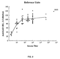
- Figure 2 shows end-point titrations for 68 IDDM sera, including one with Stiff Man Syndrome (SMS), plotted against the value in reference units for the same sera at a 1:2 dilution. Note that a plot of mean values reaches a plateau corresponding to 5-10% of the total GAD added, indicating that not all of the GAD in the preparation can be precipitated, even with high potency sera, as judged by high titre.
- SMS Stiff Man Syndrome
- Figure 3 shows the results by autoradiography of 125 I-GAD that was precipitated with three anomalous sera and analysed by SDS-PAGE under reducing and non-reducing conditions.
- the precipitated 125 I-GAD migrates only as a ⁇ 64 kD component.
- Lanes 1-3 and 5-7 are the anomalous sera.
- Lanes 4 and 8 are a typical IDDM serum.
- the typical IDDM serum (lane 8) has reactivity, as expected, only with the 110-130 kD component under non-reducing conditions, with a weak signal at the exposure time used for this autoradiograph (arrow).
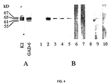
- Figure 4 shows the result by Western blotting after SDS-PAGE electrophoresis of GAD using antibodies to GAD raised experimentally (A) and in humans (B), with serum dilutions of 1:200.
- A Monoclonal mouse antibody GAD-6 raised against GAD 65, and polyclonal rabbit antibody K2 raised against GAD 67;
- B lane 1, healthy person; lane 2, patient with SDS and IDDM; lanes 3-10, patients with IDDM.
- Figure 5 shows the results by dot blotting whereby GAD is spotted onto a nitrocellulose filter and then exposed to the test sera.
- the upper row shows the reaction of IDDM sera with GAD under non-denaturing conditions (see text), wherein all sera show strong reactivity, and in contrast, the lower row (spots 2-10) shows the reaction of the same IDDM sera with GAD treated with ME and boiling wherein only serum 2, and serum 3 (weak) show reactivity.
- Spot 1 represents normal serum.
- the sera depicted here are the same as those depicted in Figure 4.
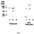
- FIG. 6 shows the results by autoradiography of biosynthetically labelled recombinant GAD ( 35 S-GAD) prepared so as to retain the normal structure of native GAD by expression using rabbit reticulocyte lysate (RRL); precipitation was with a representative positive IDDM serum and a negative serum, and analysed by SDS-PAGE under reducing and non-reducing conditions.
- lane 1 contains the RRL recombinant GAD (19 hour exposure)
- lane 2 contains RRL-expressed GAD precipitated with IDDM serum (7 day exposure)
- lane 3 contains RRL-expressed GAD precipitated with a normal serum (7 day exposure).
- the major signal corresponds to a M.W. of 110-130 kD, indicative of a dimeric form of GAD.
- the nature of the GAD antigen reacting in the radioimmunoprecipitation assay described by Rowley et al . 6 was investigated as follows. The pellet consisting of antigen and antibody that was deposited after immunoprecipitation with Sepharose-protein A was exposed to polyacrylamide gel electrophoresis in the presence of sodium dodecyl sulphate (SDS-PAGE), and the gel was examined by autoradiography. The 125 I-GAD antigen in the precipitate was compared with the GAD labelled with 125 I that had been initially added to the assay.
- Electrophoresis samples were dissolved in a buffer that contained 64 mM Tris-HCl, pH 6.8, 2% SDS, and 10% glycerol with or without 5% 2-mercaptoethanol. Electrophoresis was performed on 10% separating gels. The gels were dried and examined by autoradiography for periods of up to 2 weeks.
- Typical results obtained from autoradiography are shown in Figure 1.
- 125 I-GAD was immunoprecipitated with the IDDM serum and the precipitate was analysed by gel electrophoresis with or without the reducing agent, 2-mercaptoethanol, i.e. reducing or non-reducing conditions. The gel was exposed for autoradiography for 2 weeks.
- lanes 1 and 5 contain non-immunoprecipitated 125 I-GAD and lanes 2, 3, 4 and 6, 7, 8 contain 125 I-GAD precipitated with three representative sera from patients with IDDM.
- the non-precipitated GAD and the immunoprecipitated GAD have a similar migration corresponding to a MW of ⁇ 64 kD.
- the non-precipitated GAD appears as two components that have a migration corresponding to MW of ⁇ 64 kD and ⁇ 110-130 kD, whereas the precipitated GAD has a migration exclusively as a component corresponding to a MW of ⁇ 110-130 kD.
- This Example demonstrates that diabetic sera immuno-precipitate only a small fraction of the total amount of iodinated GAD in a test preparation of affinity-purified porcine brain GAD, irrespective of the amount or potency of serum used for the immunoprecipitation.
- the residual supernatant after immunoprecipitation of 125 I-GAD by IDDM serum regularly contains most of the initial radioactivity added (see above).
- Autoradiography of the residual 125 I-GAD after electrophoresis shows a signal corresponding to a ⁇ 64 kD component, this being precisely comparable with the ⁇ 64 kD signal in the original GAD preparation.
- no other signals corresponding to components of higher molecular weights these components having been completely removed by the first immunoprecipitation.
- the radioimmunoprecipitation assay with 125 I-GAD is carried out under conditions of antibody excess, such that these sera remain reactive at dilutions of 1:100 or 1:1000 or even higher, yet most IDDM sera react with only ⁇ 5-10% of the total iodinated GAD in the preparation.
- GAD preparations derived from porcine brain contain both GAD 65 and GAD 67. This indicates that there would be a substantial amount of the heterodimeric form of GAD, GAD 65-GAD 67 in a porcine brain GAD preparation, given that the monoclonal antibody GAD 1 that is used in the affinity purification is reported to react with only GAD 65. Thus GAD 67 could become bound to the affinity column only through its linkage to GAD 65.
- “Western” transfer immunoblotting is a standard procedure for examining the reactivity of autoimmune sera with protein components, including intracellular proteins. This procedure is usually performed under conditions in which the antigenic protein is denatured by exposure to heat (boiling), a reducing agent (2-mercaptoethanol) and the detergent sodium dodecyl sulphate (SDS).
- the polypeptide epitopes so defined are therefore regarded as “linear” rather than “conformational”, although some degree of renaturation may occur after transfer of the proteins from the gel to the nitrocellulose filter.
- an enzyme is highly dependent on the conformational structure of the molecule since the activity of most enzymes is readily destroyed when the conformational structure is lost, and GAD is completely inactivated by heating for 2 hours at 45° C 20 . Accordingly, an enzymatically active molecule is usually considered to exist folded in its native conformation 30 .
- Recombinant GAD 65 was prepared in the bacterial expression system pMAL (New England Biolabs) 31 .
- the recombinant protein was shown to have strong enzymatic activity as measured by the production of GABA from glutamate labelled with 3 H, according to the procedure described by Rowley et al. 6 , and the specific activity of the recombinant protein was much greater than the specific activity of the purified porcine brain GAD. Nonetheless, when the recombinant GAD antigen was labelled with 125 I and used as antigen in the standard immuno-precipitation assay, it proved to be poorly reactive.
- Recombinant GAD has been used previously with some claimed success in ELISA systems, but the performance of such ELISAs using recombinant GAD is quite inferior to the performance of radioimmunoprecipitation assays using native porcine brain GAD, as judged by published results 32 and results from the First International GAD Antibody Workshop 33 .
- the conformational requirement of the GAD molecule for reactivity with sera from patients with IDDM differs from the conformational requirement for enzymatic activity, indicating that the additional conformational requirement for immunoprecipitation is critically dependent on a dimeric/oligomeric configuration of the GAD molecule.
Landscapes
- Health & Medical Sciences (AREA)
- Life Sciences & Earth Sciences (AREA)
- Engineering & Computer Science (AREA)
- Immunology (AREA)
- Chemical & Material Sciences (AREA)
- Molecular Biology (AREA)
- Biomedical Technology (AREA)
- Hematology (AREA)
- Urology & Nephrology (AREA)
- Biochemistry (AREA)
- Biotechnology (AREA)
- Microbiology (AREA)
- General Health & Medical Sciences (AREA)
- Medicinal Chemistry (AREA)
- Zoology (AREA)
- Genetics & Genomics (AREA)
- Physics & Mathematics (AREA)
- Food Science & Technology (AREA)
- General Physics & Mathematics (AREA)
- Pathology (AREA)
- Organic Chemistry (AREA)
- Cell Biology (AREA)
- Bioinformatics & Cheminformatics (AREA)
- Analytical Chemistry (AREA)
- Wood Science & Technology (AREA)
- Rheumatology (AREA)
- General Engineering & Computer Science (AREA)
- Rehabilitation Therapy (AREA)
- Peptides Or Proteins (AREA)
- Investigating Or Analysing Biological Materials (AREA)
- Medicines Containing Antibodies Or Antigens For Use As Internal Diagnostic Agents (AREA)
- Immobilizing And Processing Of Enzymes And Microorganisms (AREA)
Abstract
Description
- This invention relates to methods for the identification of persons who have diabetes or a prediabetic status, and in particular it is directed to a diagnostic assay which will provide a simple and effective means of screening large populations of individuals to detect both diabetes and prediabetes (the early stages of diabetes prior to the onset of the clinical disease).
- Diabetes mellitus is a heterogeneous disorder. Insulin-dependent (IDDM) and non-insulin-dependent diabetes mellitus (NIDDM) are subtyped physiologically according to patients' dependence for survival on treatment with insulin1. IDDM is attributed to autoimmunity2 by reason of disease associations, autoantibodies to pancreatic islet cell antigens, similarities with animal models of IDDM and HLA linkages. The disease-relevant autoantigen or antigens in IDDM include a 64,000-M r protein demonstrable by immunoprecipitation from pancreatic islets3 but hitherto unidentified because of low abundance and assay difficulties. Immunoassay for antibody to the 64,000-M r antigen indicated that it was a specific marker for IDDM and preceded symptomatic onset of disease4. The recognition in cases of the Stiff Man syndrome of coexisting IDDM and a neural antigen of 64,000-65,000-M r identified as glutamic acid decarboxylase (GAD) led Baekkeskov et al. 5 to establish the coidentity of their 64,000-M r antigen and GAD. Recently, Rowley et al. 6 described a high frequency of positive results by radioimmunoprecipitation assay with IDDM sera and a GAD preparation from pig brain and proposed detection of antibodies to GAD as a useful diagnostic test as an autoimmune marker associated with IDDM.
- Insulin-dependent diabetes mellitus is a frequently occurring disease that predominantly affects young individuals, less than age 15 years. The disease can, however, have an onset in early or even mid-adult life, hence there is a need for a test that has diagnostic and predictive capacity for IDDM in both childhood and in adult life. The disease itself, IDDM, occurs at highest frequencies in the developed and economically advanced communities in the world. There is a high incentive to diagnose IDDM at the earliest stage, and even in the preclinical stage before the disease can be recognised by conventional laboratory tests. The recognition of antibodies to GAD by a routinely applicable, sensitive and specific immunoassay kit would fulfil this need. Moreover, the need for early or preclinical recognition is heightened by the likelihood that specific immunotherapeutic procedures will be developed that will arrest the progress of islet cell destruction that underlies IDDM, noting that such procedures have already been applied successfully in a model in mice of IDDM. Arrest of the natural progression of IDDM could spare patients years of injections with insulin. The development and application of an efficient immunoassay for antibodies to GAD, as a diagnostic procedure for IDDM, would result in an estimated usage of an assay kit in numbers exceeding one million annually, worldwide.
- Apart from this, 5-10% of adults presenting with diabetes are initially diagnosed as having the non-insulin dependent form (NIDDM), however, after a period ranging from months to years, they convert to IDDM. These patients fall into a category called latent autoimmune diabetes in adults (LADA). It has been shown that 75-80% of this group of patients have detectable levels of antibodies to GAD. It is likely that this test will become widely used to discriminate these patients at diagnosis, thus saving them from inappropriate therapy (i.e. diet and tablets) which could result in failure to control their diabetes with the subsequent risk of severe complications. Moreover, the test for antibodies to GAD could have predictive value if applied to individuals at risk by reason of genetic predisposition. The institution of insulin therapy at an early stage, based on the demonstration of antibodies to GAD would avert this scenario.
- The onset of IDDM is preceded by a clinically silent destruction of the pancreatic β-cells usually by an autoimmune process2,7 serologically associated with autoantibodies to islet cell antibodies (ICAs). These include the 64,000-M r protein, now recognised as GAD3,4, the cytoplasmic ICA demonstrable by immunofluorescence8 and insulin autoantibodies9. Antibody to the 64,000-Mr molecule has been extremely difficult to assay because of the need to prepare antigen from large quantities of islet cell preparations. However, Rowley et al.6 showed that with radioiodine-labelled GAD prepared from pig brain, the antibody could be readily and specifically detected by an immunoprecipitation assay. It is of interest that the 64,000-M r antigen is an enzyme molecule as are various other disease-relevant autoantigens, most notably thyroid peroxidase in autoimmune thyroiditis14, H+ K+ ATPase in autoimmune gastritis11, and pyruvate dehydrogenase in primary biliary cirrhosis12. GAD is not species specific and, in fact, cDNAs for brain GAD from humans13, rat14, and cat15 have such close homology that antibody reactivity could be expected with any mammalian GAD preparation. Also, the autoantigen is clearly not organ specific because the source used by Rowley et al. 6 was brain. The sequence of GAD from brain and pancreatic islets, the major sites in which GAD is demonstrable, appears to differ only by allelic variations16,17. GAD exists as at least two isoforms18, and the Rowley et al. 6 preparation contained both representatives, as indicated by data by polyacrylamide gel electrophoresis (PAGE).
- GAD synthesises γ-aminobutyric acid (GABA), the principal inhibitory neurotransmitter in the brain and a putative paracrine signal molecule in pancreatic islets. The enzyme has a restricted tissue distribution and is highly expressed in the cytoplasm of GABA-secreting neurons and pancreatic β-cells. The two isoforms of GAD are referred to as
GAD 65 andGAD 67 according to their relative molecular masses. These isoforms are the products of two different genes and differ substantially only in their amino-terminal region. - In addition, GAD appears to exist as an integral membrane protein with hydrophobic properties, and is present as a non-covalently linked dimer of MW 96,60020. This purified membrane form of GAD has been found to be reactive with IDDM sera, however no sequence data are yet available for this integral membrane protein20.
- The development of simple assays for anti-GAD will provide a valuable tool for diabetes research, particularly as anti-GAD is not a transient reactivity, unlike the sensitivity to the pancreatic islet cytoplasmic antigen. Numerous studies affirm not only that the frequency of anti-GAD is high in IDDM, and in cases of recent onset approaches 70-80%, but also the stability of anti-GAD levels over a period of years6. In fact, even among cases of very long-standing IDDM, the frequency of anti-GAD remains high, at about 60%21.
- Research has also shown that autoantibodies to GAD appear in the bloodstream long before the clinical symptoms of diabetes actually occur4. In a study by the present inventors on individuals selected as being at risk for IDDM by reason of inheritance and positive for antibodies to β-islet cells of the pancreas showed that the presence of anti-GAD in serum correlated strongly with later appearance of IDDM22. In further studies by the present inventors, anti-GAD has been present in the first available serum sample from 80% of prediabetic individuals, and even 10 years before the overt onset of diabetes23. Because it is present years before the onset of diabetes, the GAD autoantibody is a valuable predictive marker of the disease, and in a study in Finland23 based on serum samples obtained from apparently healthy pregnant women who subsequently developed IDDM, the predictive capacity for IDDM of a positive test for anti-GAD was remarkably high a relative risk versus a negative test for anti-GAD being 800-fold. In Melbourne, a study on stored sera from women with gestational diabetes (diabetes appearing during pregnancy) likewise showed that a positive test for anti-GAD was predictive of the occurrence later in life of IDDM in such women24.
- International Patent Specification No. PCT/US91/06438 (Publication No. WO 92/04632) discloses assays for the detection of diabetes or a prediabetic status relying on exposing patient serum samples to purified ligand capable of binding autoantibodies specific for the 64 kD autoantigen present on pancreatic β-cells.
- In an article by Baekkeskov et al. (Nature, Vol 347, p 151-156, 1990), the 64 kD β-cell autoantigen is identified as a target of autoantibodies associated with diabetes. Recombinant MBGAD peptides have been used to detect autoantibodies in an article by Deaizpuna et al., (Diabetes, 41, 1183-1187, 1992). Michelsen et al. (PNAS, 88, 8754-8758, 1991) discuss the 67 kD form of GAD, in conjunction with the 64 kD form, and autoantibodies thereto. Petersen et al. in Diabetes, 1:38A, 148, 1992 disclose that GAD can form monomers, dimers, trimers and tetramers. WO 92/19972 discloses assays for the detection of preclinical diabetes using high molecular weight GAD. WO 92/14485 relates to the use of GAD to test for autoimmune diseases. WO 92/05446 relates to the cloning of GAD cDNA. Kaufman et al., discuss that autoantibodies to 65 kD are associated with diabetes (J. Clin. Invest., 89, 283-292, 1992).
- Research work leading to the present invention has now identified the molecular form in which GAD functions as an antigen, and hence binds efficiently and effectively to autoantibodies in an immunoassay for diabetes or the preclinical stage of diabetes. It is now evident that the monomeric form of GAD is, with rare exceptions, a poorly reactive antigen for IDDM sera, and for an effective assay to demonstrate the reactivity with GAD, the preparation must contain a proportion of GAD in the dimeric or oligomeric configuration. As a result, it is now possible to optimise the form of the antigen used in the detection of autoantibodies to GAD, leading to increased sensitivity in assays for the detection of diabetes or a prediabetic status.
- In accordance with the present invention there is provided a method for detecting autoantibodies to glutamic acid decarboxylase (GAD) in the serum of a patient, comprising contacting a serum sample from the patient with a GAD preparation and detecting binding of autoantibodies to GAD in said sample by said GAD preparation, characterised in that said GAD preparation comprises an enhanced amount of dimer(s) or oligomer(s) of the 65 kD and/or 67 kD isoforms of GAD, relative to monomeric GAD in preparations of naturally occurring GAD.
- Whilst the present description of the invention refers specifically to detection of antibodies to GAD in a serum sample, it is to be understood that the term "serum" is used as a non-limiting term, and that the present invention also extends to the detection of autoantibodies to GAD in "blood" samples in general, including blood serum and plasma.
- As a particularly preferred feature of the present invention, the method of the invention is carried out under conditions under which the dimeric or oligomeric form of the GAD is retained and is not broken down into monomeric GAD.
Preferably, therefore, the method of the invention is carried out under non-denaturing conditions, and particularly under non-reducing conditions. - It will, of course, be appreciated that the detection of binding between the autoantibodies and the GAD antigen is indicative of the presence of the autoantibodies in the serum sample, and hence is diagnostic of a diabetic or prediabetic condition in the patient.
- Advantageously, the method of the present invention uses a non-naturally occurring preparation of GAD antigen which contains an enhanced amount of dimer(s) or oligomer(s) of the 65 kD and/or 67 kD isoforms of GAD.
- The present invention further extends to a diagnostic kit for the detection of autoantibodies to GAD in the serum of a patient, comprising a GAD preparation and means for detecting binding of autoantibodies to GAD in a serum sample from the patient by said GAD preparation, characterised in that said GAD preparation comprises an enhanced amount of dimer(s) or oligomer(s) of the 65 kD and/or 67 kD isoforms of GAD, relative to monomeric GAD in preparations of naturally occurring GAD.
- The references herein to an "enhanced amount" of dimer(s) or oligomer(s) in the GAD preparation indicate that the amount of dimer(s) or oligomer(s) in the GAD preparation is greater than the amount of dimer(s) or oligomer(s) in preparations of naturally occurring GAD. Such greater amounts may be achieved by separation of naturally occurring GAD into fractions having an "enhanced amount" of dimer(s) or oligomer(s), for example by electrophoretic separation on SDS-PAGE gels or by size exclusion chromatography. Alternatively, such an "enhanced amount" of dimer(s) or oligomer(s) may be achieved by augmenting the content of dimeric or oligomeric GAD in naturally occurring GAD.
- It should also be noted that the references herein to GAD preparations having an enhanced amount of dimer(s) or oligomer(s) also include preparations of "substantially pure" GAD having greater than 50% by weight, more particularly greater than 70% by weight or even greater than 90% by weight, of the dimeric or oligomeric form of GAD, with the balance monomeric GAD. Such "substantially pure" dimeric or oligomeric GAD may be produced by removal of appropriate amounts of monomeric GAD from naturally occurring GAD using known separation techniques. Alternatively, however, the dimeric or oligomeric GAD may be synthesised by appropriate cross-linking, for example by forming disuiphide bonds or by other means, of naturally occurring or recombinantly produced monomeric GAD.
- Accordingly, in the methods of the present invention there may be used a GAD preparation which comprises substantially pure dimer(s) or oligomer(s) of the 65 kD and/or 67 kD isoforms of GAD, and in particular a GAD preparation which comprises substantially pure heterodimer or heterooligomer of the 65 kD and 67 kD isoforms of GAD.
- The dimer(s) or oligomer(s) in the GAD preparation used as antigen in this invention may be an aggregation of one or other of the two isoforms of GAD, or it may be a heterodimer or heterooligomer of both isoforms (65 kD or 67 kD) of GAD. Similarly, the GAD may be either human or non-human mammalian, native or recombinant, and it may be either pancreatic or brain (or other CNS) GAD.
- Methods for the isolation of native GAD are well known, and are described, for example, in International Patent Specification No. PCT/US91 /06438 (WO 92/04632). This International specification also describes various methods for the preparation of recombinant GAD.
- Throughout this specification and claims which follow, unless the context requires otherwise, the word "comprise", or variations such as "comprises".or comprising", will be understood to imply the inclusion of a stated integer or group of integers but not the exclusion of any other integer or group of integers.
- The GAD preparation used in the work leading to the present invention was obtained from porcine brain and was purified as described in Rowley et al. 6 on an affinity column that contained a monoclonal antibody GAD1 reactive with mammalian GAD19. This preparation contained predominantly monomeric GAD (65 kD and 67 kD) but also contained some dimeric and oligomeric GAD. As described above, this preparation may be further processed to provide a preparation containing an enhanced amount of dimeric or oligomeric GAD, for example, by electrophoretic separation.
- When GAD is isolated from natural sources, the major component is monomeric GAD which is not cross-linked. In the work leading to this invention, it was discovered that it is the minor dimeric/oligomeric fraction in such naturally occurring GAD which proves to be the "antigenic" component. Therefore, in order to effectively bind with the autoantibodies present in the sera of diabetic or prediabetic patients, the GAD must exist in the dimeric or oligomeric form, and it has been found that for most diabetic sera there is little or no reactivity with monomeric GAD. This discovery was both unanticipated and unpredictable.
- Using a procedure of molecular separation of GAD in gels under non-denaturing conditions which preserve the native conformation of the molecule, it has been shown that the molecular weight of the GAD molecule that binds to the autoantibody in IDDM is almost exactly double that of the monomeric form of the GAD molecule, hitherto believed to be the "diabetes autoantigen". It is emphasised that for almost all the diabetic sera tested so far, little or no monomer is bound by the autoantibody, even in the presence of a 20-fold or greater excess of the monomeric GAD. It therefore follows that the dimeric configuration of the GAD molecule presents an optimal antigen determinant for the autoantibody reaction. In addition, it is evident that any procedure that augments the amount of dimeric GAD vis-a-vis monomeric GAD will increase greatly the sensitivity of the assay, and so optimise the utility of preparations of GAD for diagnostic tests on serum for autoimmune insulin-dependent diabetes mellitus.
- There is prior recognition that dimeric and multimeric forms of the GAD exist naturally in the body, and it has been reported that such dimeric and multimeric forms, stabilised by chemical procedures, can react with the autoantibodies present in serum of persons with diabetes25. However, the standard methods of detecting this serum reactivity, which are carried out in the presence of reducing agents that break disulphide bonds and/or electrostatic or hydrophobic interactions, result in the destruction of dimers and oligomers that are not completely stabilised. Hence, monomeric GAD has been considered hitherto as the major autoantigenic form of GAD.
- As noted above, the essential characteristics of the diagnostic assay for autoantibodies to GAD in accordance with the present invention are the use of an antigen preparation containing an enhanced amount of dimeric or oligomeric GAD instead of monomeric GAD, together with appropriate reaction conditions which avoid the destruction of the dimers or oligomers.
- In the work leading to the present invention, two separate preparations of GAD derived by entirely different procedures were tested, and with each the reactivity of sera from patients with IDDM is only with GAD in its dimeric/oligomeric form. Thus there is solid evidence that the major pancreatic islet autoantigen in IDDM, exists not as hitherto believed as a reactant of 64 kD but rather a s a reactant of 120-130 kD (and greater) GAD, interpretable as a dimeric/oligomeric form of the molecule. Overall, the results set out in Examples 1-8 below establish clearly that GAD is required to be in this dimeric or oligomeric form to be reactive with the autoantibody to GAD in IDDM sera. It therefore follows that any procedure that enhances the amount of dimeric or oligomeric GAD in a GAD preparation would generate an antigenic preparation that would bind even more strongly and more specifically with anti-GAD present in IDDM sera compared with preparations used hitherto. This in turn provides to a more sensitive assay with greatly improved diagnostic utility.
- Enrichment of the dimeric/oligomeric fraction of GAD can be achieved by a number of methods obvious to one skilled in the art of protein biology. These procedures include the purification of the dimeric/oligomeric forms of GAD which can be accomplished by a number of methods including gel filtration, HPLC, affinity chromatography, ion exchange chromatography, etc. The enrichment may also be achieved by the classical cross-linking of monomeric GAD using hetero-bi functional or homo-bi functional reagents known to form co-valent intra-molecular and inter-molecular protein-protein associations.
- Apart from the essential characteristic of use of an antigen preparation containing an enhanced amount of dimeric or
oligomeric GAD 65 and/orGAD 67, the assays of the present invention can be performed using conventional protocols for immunoassays, such as competitive immunoassays, radio-immunoassays, enzyme-linked immunosorbent assays, and the like. Further details of suitable assay protocols in accordance with this invention are described, by way of example, in International Patent Specification No. PCT/US91/06438 (WO 92/04632) - The present description of the invention shows that IDDM sera react preferentially with dimeric or oligomeric forms of molecular GAD. Hence any method or procedure that provides a preparation in which the amounts or proportions of dimeric/oligomeric GAD are enhanced would create a more effective reactant for the serological diagnosis of IDDM. Such methods for the isolation of GAD include both chemical methods and immunochemical separations such as affinity chromatographic procedures that may be modified to give higher yields of dimers. Similarly, methods or procedures that resulted in the creation and stabilisation from monomeric GAD (recombinant or naturally occurring) of dimeric or oligomeric molecules by appropriate cross-linking, or formation of disulphide bonds, or other means, will yield a preparation containing dimeric GAD with an enhanced capacity to react with IDDM sera.
- In this context, the GAD molecule in the dimeric form may acquire the appropriate "folding" to create the epitope that is optimally reactive with IDDM sera. There exist enzymes that react with proteins to produce molecules that are appropriately folded for optimal cellular transport and function, of which one example is protein disulphide isomerase (PDI). GAD in its monomeric form, either as a recombinant molecule or as native monomer, may be susceptible to the folding influences of enzymes such as PDI, so creating an enhanced reactant with IDDM serum. It is further to be noted that both homodimers and heterodimers of
GAD 65 andGAD 67 may be prepared, and that the preparation of heterodimers of GAD 65-GAD 67 may provide a reactant of particular utility for serological testing. - Further details of the present invention will be apparent from the following Examples and the drawings which are included by way of illustration, not by way of limitation, of this invention.
- Figure 1 shows the results by autoradiography of 125I-GAD that was precipitated with three representative IDDM sera and analysed by SDS-PAGE under reducing or non-reducing conditions (see text).
Lanes lanes - Figure 2 shows end-point titrations for 68 IDDM sera, including one with Stiff Man Syndrome (SMS), plotted against the value in reference units for the same sera at a 1:2 dilution. Note that a plot of mean values reaches a plateau corresponding to 5-10% of the total GAD added, indicating that not all of the GAD in the preparation can be precipitated, even with high potency sera, as judged by high titre.
- Figure 3 shows the results by autoradiography of 125I-GAD that was precipitated with three anomalous sera and analysed by SDS-PAGE under reducing and non-reducing conditions. Here, and in the particular contrast to Figure 1, the precipitated 125I-GAD migrates only as a ∼64 kD component. Lanes 1-3 and 5-7 are the anomalous sera.
Lanes - Figure 4 shows the result by Western blotting after SDS-PAGE electrophoresis of GAD using antibodies to GAD raised experimentally (A) and in humans (B), with serum dilutions of 1:200. (A) Monoclonal mouse antibody GAD-6 raised against
GAD 65, and polyclonal rabbit antibody K2 raised againstGAD 67; (B)lane 1, healthy person;lane 2, patient with SDS and IDDM; lanes 3-10, patients with IDDM. - Figure 5 shows the results by dot blotting whereby GAD is spotted onto a nitrocellulose filter and then exposed to the test sera. The upper row (spots 2-10) shows the reaction of IDDM sera with GAD under non-denaturing conditions (see text), wherein all sera show strong reactivity, and in contrast, the lower row (spots 2-10) shows the reaction of the same IDDM sera with GAD treated with ME and boiling wherein
only serum 2, and serum 3 (weak) show reactivity.Spot 1 represents normal serum. The sera depicted here are the same as those depicted in Figure 4. - Figure 6 shows the results by autoradiography of biosynthetically labelled recombinant GAD (35S-GAD) prepared so as to retain the normal structure of native GAD by expression using rabbit reticulocyte lysate (RRL); precipitation was with a representative positive IDDM serum and a negative serum, and analysed by SDS-PAGE under reducing and non-reducing conditions. In each case,
lane 1 contains the RRL recombinant GAD (19 hour exposure),lane 2 contains RRL-expressed GAD precipitated with IDDM serum (7 day exposure), andlane 3 contains RRL-expressed GAD precipitated with a normal serum (7 day exposure). Note that with precipitated RRL-expressed GAD analysed under non-reducing conditions the major signal corresponds to a M.W. of 110-130 kD, indicative of a dimeric form of GAD. - This Example demonstrates that autoantibodies in sera from patients with IDDM react with dimeric/oligomeric forms of porcine brain GAD as shown by autoradiography of immunoprecipitates in SDS-polyacrylamide gels.
- The nature of the GAD antigen reacting in the radioimmunoprecipitation assay described by Rowley et al. 6 was investigated as follows. The pellet consisting of antigen and antibody that was deposited after immunoprecipitation with Sepharose-protein A was exposed to polyacrylamide gel electrophoresis in the presence of sodium dodecyl sulphate (SDS-PAGE), and the gel was examined by autoradiography. The 125I-GAD antigen in the precipitate was compared with the GAD labelled with 125I that had been initially added to the assay.
- For electrophoresis, samples were dissolved in a buffer that contained 64 mM Tris-HCl, pH 6.8, 2% SDS, and 10% glycerol with or without 5% 2-mercaptoethanol. Electrophoresis was performed on 10% separating gels. The gels were dried and examined by autoradiography for periods of up to 2 weeks.
- Typical results obtained from autoradiography are shown in Figure 1. 125I-GAD was immunoprecipitated with the IDDM serum and the precipitate was analysed by gel electrophoresis with or without the reducing agent, 2-mercaptoethanol, i.e. reducing or non-reducing conditions. The gel was exposed for autoradiography for 2 weeks. In the Figure,
lanes lanes - For the immunoprecipitates from sera that gave a positive result for antibodies to GAD, the period required for autoradiography to give a clear signal was 1-2 weeks. Under reducing conditions, i.e. in the presence of 2-mercaptoethanol, the iodinated protein that was precipitated by IDDM sera that contained antibodies to GAD had a molecular weight of - 64 kD. Under non-reducing conditions in which the molecular configuration of dimers and oligomers is not altered, only negligible amounts of iodinated protein with a molecular weight of ∼64 kD were precipitated, whereas there was a strong signal that corresponded to an iodinated protein(s) with a molecular weight of -110-130 kD, together with a weaker signal that corresponded to components of molecular weight greater than 200 kD, presumably oligomeric aggregates of the- 64 kD material. On the other hand, when the same 125I-GAD preparation that had been added to the immunoprecipitation mixture was examined directly using electrophoresis under either reducing or non-reducing conditions, there was a strong signal corresponding to the presence of an iodinated protein of molecular weight ∼64 kD.
- This Example demonstrates that diabetic sera immuno-precipitate only a small fraction of the total amount of iodinated GAD in a test preparation of affinity-purified porcine brain GAD, irrespective of the amount or potency of serum used for the immunoprecipitation.
- In an affinity purified preparation of porcine brain GAD, only a small fraction is in an appropriate autoantigenic configuration for reactivity with IDDM serum, as shown by the consistently repeated finding that it is not possible to immunoprecipitate more than 5-10% of the total 125I-GAD in the radioimmunoprecipitation assay (observed on more than 1000 IDDM sera tested in the present inventors' laboratory). In fact, the total amount of radioiodine-labelled porcine brain GAD immunoprecipitated by any IDDM serum reaches a plateau at a level corresponding to only 5-10% of the total counts of radioactivity added, with no increase obtained by the addition of further amounts of IDDM serum (Figure 2). Moreover, when IDDM sera that give a positive reaction in the radioimmunoprecipitation assay are titrated from the usual 1:2 initial dilution, it is evident that the end-point titration is in many cases beyond a serum dilution of 1:1000 (Figure 2), indicating that the failure to immunoprecipitate most of the 125I-GAD in the preparation is not due to any lack of potency in the precipitating antisera.
- This Example shows that the removal of the dimeric and oligomeric forms of GAD from the GAD preparations abrogates reactivity of IDDM sera with GAD.
- In the radioimmunoprecipitation assay, the residual supernatant after immunoprecipitation of 125I-GAD by IDDM serum regularly contains most of the initial radioactivity added (see above). Autoradiography of the residual 125I-GAD after electrophoresis shows a signal corresponding to a ∼ 64 kD component, this being precisely comparable with the ∼64 kD signal in the original GAD preparation. There are, however, now no other signals corresponding to components of higher molecular weights, these components having been completely removed by the first immunoprecipitation.
- Experiments were performed to determine the degree to which removal of the dimeric/oligomeric forms of GAD from the initial preparation of 125I-GAD would remove the capacity to react with IDDM sera. Accordingly, the 125I-GAD preparation was allowed to react (i.e. absorbed) at 4°C overnight either with a strongly positive IDDM serum, or with pooled normal sera, then immunoprecipitation was performed with protein A-Sepharose to remove all immune complexes of 125I-GAD-anti-GAD. The absorbed supernatant was then used as antigen in the standard immunoprecipitation assay using either IDDM sera or normal human sera. The result was that the supernatant derived after immunoprecipitation (absorption) with a strongly positive IDDM serum generally failed to function as an antigen in the standard radioimmunoprecipitation assay. (There were, however, three anomalous exceptions, discussed in Example 4.) The typical results are shown in Table 1. It should be noted that after the initial precipitation (absorption) of 125I-GAD with IDDM serum, further precipitation with IDDM serum immunoprecipitated no more radioactivity than could be precipitated with a normal serum. On the other hand, after an initial precipitation (absorption) with a normal serum, the number of counts of radioactivity that could be precipitated from the 125I-GAD preparation by an IDDM serum was precisely equivalent to the number of counts of radioactivity precipitated using a fresh non-absorbed preparation of 125I-GAD tested under exactly the same conditions.
Initial Precipitation Second Precipitation (cpm) IDDM serum Normal serum IDDM serum 390 252 Normal serum 2400 97 Nil 2600 78 - In this Example an initial precipitation of 125I-GAD was made either with IDDM serum or normal serum, with a second precipitation likewise, with results compared with those with no initial precipitation (Nil). The results show that the initial precipitation with the IDDM serum removed the reactive capacity of the GAD whereas after initial precipitation with normal serum the reactive capacity of the GAD for IDDM serum was unchanged.
- This Example shows that very exceptional sera from cases of IDDM do react with a monomeric form of purified brain GAD. The identification and characterisation of these anomalously-reactive sera does not derogate from the importance of the dimeric and oligomeric forms of GAD in diagnosis of IDDM. Indeed, the highly exceptional immunoprecipitation reactivity of these sera with GAD in the monomeric rather than dimeric or oligomeric forms provides strong validation of the data herein that in the immunoprecitation assay, IDDM sera react usually and typically with GAD in the dimeric/oligomeric conformation.
- As demonstrated above, for most IDDM sera, the radioimmunoprecipitation assay with 125I-GAD is carried out under conditions of antibody excess, such that these sera remain reactive at dilutions of 1:100 or 1:1000 or even higher, yet most IDDM sera react with only ∼ 5-10% of the total iodinated GAD in the preparation.
- However, although this is the pattern of reactivity observed for the overwhelming majority of IDDM sera tested, in fact well over 99%, an anomalous pattern of reactivity has been observed with three sera. Each of these three sera precipitated more than 10% and up to 30% of the total iodinated GAD in the standard immunoprecipitation assay at a 1:2 dilution. Nonetheless, and despite the apparent potency of these three sera, in each case the reactivity in the immunoprecipitation assay decreased rapidly after dilution of the serum, and thus the potency according to titration was actually low.
- Immunoprecipitation followed by autoradiography under non-reducing conditions, as described above, was applied to detect whether these sera were reacting with dimer/oligomer. The result was that pellets obtained from immunoprecipitation with these three anomalous sera showed a strong reactivity with a component corresponding in MW to monomeric GAD but no apparent reactivity with the dimer/oligomeric forms of GAD (Figure 3).
- In order to determine whether removal of dimeric/oligomeric forms of GAD from the preparation would affect the reactivity of these anomalous sera, the immunoprecipitation was repeated using as the source of 125I-GAD the supernatant obtained after an initial immunoprecipitation (i.e. absorption) with an IDDM serum as described above (see Example 3). In this experiment an initial absorption of the 125I-GAD preparation with an IDDM serum did not remove the reactivity of the 125I-GAD with these three sera although, as expected, and as previously shown, it did significantly reduce the reactivity with all typically reactive IDDM sera. The results of this experiment are shown in Table 2.
Initial Precipitation Second Precipitation (cpm) Atypical Sera Serum 1 Serum 2Serum 3IDDM serum IDDM serum 12,550 8,756 12,580 2,400 Nil 13,100 6,600 7,800 4,800 - In this Example, the results after an initial precipitation of GAD with IDDM serum or no serum (nil) are shown for three anomalous sera that did react with monomeric GAD. For these sera the reactive capacity was retained after the initial precipitation. Note that in contrast the reactive capacity of the GAD for a typical IDDM serum was significantly reduced after the initial precipitation.
- This Example shows that the dimeric/oligomeric forms of GAD that contain both
GAD 65 andGAD 67 react with sera from patients with IDDM. - GAD preparations derived from porcine brain contain both
GAD 65 andGAD 67. This indicates that there would be a substantial amount of the heterodimeric form of GAD, GAD 65-GAD 67 in a porcine brain GAD preparation, given that themonoclonal antibody GAD 1 that is used in the affinity purification is reported to react withonly GAD 65. Thus GAD 67 could become bound to the affinity column only through its linkage toGAD 65. - Evidence that a heterodimer of
GAD 65 andGAD 67 is reactive with sera from patients with IDDM is available from immunoprecipitation studies in which the antigenic material was the supernatant obtained after initially performing the immunoprecipitation assay using either themonoclonal antibody GAD 6 that reacts withGAD 6519, or using the polyclonal antibody K2 raised in the rabbit that is stated to react only withGAD 6726. The results are shown in Table 3. After initial precipitation was performed with K2 to removeGAD 67, the reactivity of IDDM sera with the supernatant was reduced from 2600 cpm obtained without any initial immunoprecipitation, to 1300 cpm, i.e. about 50%. By contrast, after an initial precipitation with themonoclonal antibody GAD 6 that reacts withGAD 65, or with an initial immunoprecipitation with a potent IDDM serum, there was a reduction of the reactivity by virtually 100%, i.e. to the level observed using serum from healthy blood donors.Initial Precipitation Second Precipitation (cpm) IDDM serum Normal Serum GAD 6 197 232 K2 1296 230 IDDM serum 390 252 Normal serum 2427 97 Nil 2590 78 - In this Example, an initial precipitation of a GAD preparation was made with various reactants, the
monoclonal antibody GAD 6 againstGAD 65, the polyclonal rabbit antibody K2 againstGAD 67, IDDM serum or normal serum, and followed by a second precipitation with an IDDM serum or normal serum. The results show that the capacity of GAD to react with IDDM serum was moderately influenced by serum K2 and markedly byGAD 6 and the IDDM serum. From the finding thatGAD 6 and K2 are totally depleting, the inference is that homodimers and heterodimers ofGAD 65/GAD 67 are present in the GAD preparation and both homo- and heterodimeres are reactive. - This Example shows that denaturation of the autoantigenic protein GAD influences its reactivity with IDDM sera.
- "Western" transfer immunoblotting is a standard procedure for examining the reactivity of autoimmune sera with protein components, including intracellular proteins. This procedure is usually performed under conditions in which the antigenic protein is denatured by exposure to heat (boiling), a reducing agent (2-mercaptoethanol) and the detergent sodium dodecyl sulphate (SDS). The polypeptide epitopes so defined are therefore regarded as "linear" rather than "conformational", although some degree of renaturation may occur after transfer of the proteins from the gel to the nitrocellulose filter.
- Sera from patients with IDDM react with preparations of GAD by Western blotting with a component corresponding to a MW of 64-65 kD; however in fact only a small proportion (∼10%) of IDDM sera actually do so according to the experience of the present inventors27 and that of others5,28,29 (Figure 4). This would indicate that IDDM sera usually react with a conformational epitope structure associated with the GAD molecule that is lost under the usual conditions of immunoblotting. Consistent with this is the finding that IDDM sera usually do react with the GAD molecule when this is "spotted" onto a nitrocellulose filter as a dot blot under non-denaturing conditions, these being the absence of 2-mercaptoethanol, heating and sodium dodecyl sulphate27 (Figure 5). However, since the dot blotting procedure does not involve the molecular separation in gels of the reacting protein, there can be no information on the molecular weight of the GAD reactant in the dot blotting procedure. In the dot blotting system, it was found that 2-mercaptoethanol did not interfere with reactivity, whereas boiling of the GAD preparation abrogated its reactivity with IDDM sera.
- As described above, autoradiography of immunoprecipitated pellets (GAD-anti-GAD) after gel electrophoresis exposed to sodium dodecyl sulphate and boiling, but in the absence of 2-mercaptoethanol, shows a component corresponding in molecular weight to a dimeric form of GAD (∼120 kD). It was ascertained that exposure to mild heating, 60°C for 2 minutes, resulted in a loss of the capacity of 125I-GAD to react with IDDM sera, whereas exposure to 2-mercaptoethanol did not result in loss of capacity to react with IDDM sera. Autoradiography under non-reducing conditions of the immunoprecipitated pellets after the 125I-GAD had been treated with 2-mercaptoethanol before use in the immunoprecipitation assay showed that there was retention of the dimeric structure, indicating that the molecule in its dimeric configuration appears to be resistant to reduction in the absence of heating.
It is noted in relation to the results from Western blotting and gel analyses of immunoprecipitates using affinity purified porcine brain GAD, that in the Western blotting procedure the antigen is exposed to denaturing conditions before the reaction with serum antibody, whereas with gel analyses of immunoprecipitates, the exposure to denaturing conditions is after the formation of the antigen-antibody complex. - These results indicate that the reactive epitope presented by the GAD molecule is sensitive to conditions that interfere with the conformational structure of proteins, and are consistent with this epitope being a construct of GAD molecules in a dimeric or oligomeric configuration.
- This Example shows that recombinant GAD produced in a bacterial expression system is inactive in the immuno-precipitation assay, although the recombinant protein has strong enzymatic activity, and hence must retain most of the native configuration of the molecule.
- The action of an enzyme is highly dependent on the conformational structure of the molecule since the activity of most enzymes is readily destroyed when the conformational structure is lost, and GAD is completely inactivated by heating for 2 hours at 45° C20. Accordingly, an enzymatically active molecule is usually considered to exist folded in its native conformation30.
-
Recombinant GAD 65 was prepared in the bacterial expression system pMAL (New England Biolabs)31. The recombinant protein was shown to have strong enzymatic activity as measured by the production of GABA from glutamate labelled with 3H, according to the procedure described by Rowley et al. 6, and the specific activity of the recombinant protein was much greater than the specific activity of the purified porcine brain GAD. Nonetheless, when the recombinant GAD antigen was labelled with 125I and used as antigen in the standard immuno-precipitation assay, it proved to be poorly reactive. - Recombinant GAD has been used previously with some claimed success in ELISA systems, but the performance of such ELISAs using recombinant GAD is quite inferior to the performance of radioimmunoprecipitation assays using native porcine brain GAD, as judged by published results32 and results from the First International GAD Antibody Workshop33.
- Thus, the conformational requirement of the GAD molecule for reactivity with sera from patients with IDDM differs from the conformational requirement for enzymatic activity, indicating that the additional conformational requirement for immunoprecipitation is critically dependent on a dimeric/oligomeric configuration of the GAD molecule.
- In this Example, the use of recombinant GAD derived from a rabbit reticulocyte lysate establishes that a dimeric-oligomeric form of the molecule carries the antigenic reactivity.
- Each of the experiments described in Examples 1-6 was carried out using GAD prepared by immunochemical means from porcine brain. It might be assumed that some of the GAD in the preparation becomes damaged during the extraction process, and that the dimeric or oligomeric structure of the molecule is protective of the antigenicity of the naturally occurring GAD. On this assumption, the exclusive reactivity of IDDM sera with dimeric or oligomeric GAD could represent an artefact of the preparation method. In that case, monomeric GAD prepared by other methods should react with IDDM sera.
- It is now established in various laboratories that recombinant GAD produced in different eukaryotic expression systems can be successfully used to assay for antibodies to GAD29,34,35. If there were an absolute requirement for reactivity that IDDM sera engage a dimeric/oligomeric form of GAD, then with GAD derived from these expression systems the material immunoprecipitated by sera from patients with IDDM should, as for native brain GAD, be in a dimer/oligomer configuration.
- The above presumption was tested as follows. Recombinant GAD biosynthetically labelled with 35S-methionine was prepared using in vitro transcription with SP6 polymerase, followed by in vitro translation using rabbit reticulocyte lysate36. The biosynthetically labelled 35S-GAD was analysed by SDS-PAGE under reducing conditions, followed by autoradiography, and shown to be very pure, consisting almost exclusively of a single protein component with a molecular weight of ∼ 64 kD (Figure 6).
- This purified, eukaryotically expressed 35S-GAD was used as an antigen in the immunoprecipitation assay with IDDM and normal sera as described previously. The result was that, as for affinity purified brain GAD, only a small proportion of the total radioactivity could be immunoprecipitated. The immunoprecipitated pellet (GAD-anti-GAD) was examined by autoradiography after separation of proteins by electrophoresis under non-reducing conditions and, as previously, only the dimeric form of GAD was immunoprecipitated. It is important to note that most of the 35S-GAD in the preparation was in the monomeric form, yet this major proportion proved to be non-precipitable.
-
- 1. World Health Organization: Diabetes Mellitus; Report of WHO Study Group., Geneva, World Health Org., (1985). Tech.Rep.Ser.No.727.
- 2. Ziegler, A.G., Herskowitz, R.D., Jackson, R.A., Soeldner, J.S., Eisenbarth, G.S. Predicting type I diabetes. Diabetes Care 13:762-75, (1990).
- 3. Baekkeskov, S., Nielsen, J.H., Marner, B., Bilde, T., Ludvigsson, J., Lernark, A. Autoantibodies in newly diagnosed diabetic children immunoprecipitate human pancreatic islet cell proteins. Nature (Lond.) 298: 167-69, (1982).
- 4. Atkinson, M.A., Maclaren, N.K., Scharp, D.W., Lacy, P.E., Riley, W.J. 64,000M r autoantibodies as predictors of insulin-dependent diabetes. Lancet 335:1357-60, (1990).
- 5. Baekkeskov, S., Aanstoot, H-J., Christgau, S., Reetz, A., Solimena, M., Cascalhom, Holli, F., Ritcher Olesen, H., DeCamilli, P. Identification of the 64 K autoantigen in insulin-diabetes as the GABA-synthesizing enzyme glutamic acid decarboxylase. Nature (Lond.) 347:151-56, (1990).
- 6. Rowley, M.J., Mackay, I.R., Chen, Q-Y, Knowles, W.J., Zimmet, P.Z. Antibodies to glutamic acid decarboxylase discriminate major types of diabetes mellitus. Diabetes 41:548-51, (1992).
- 7. Maclaren, N.K., Schatz, D.S., Drash, A., Graves, G. Initial pathogenic events in IDDM. Diabetes 38:534-38, (1988).
- 8. Bottazzo, G.F., Florin-Christensen, A., Doniach, D. Islet cell antibodies in diabetes mellitus with autoimmune polyendocrine deficiencies. Lancet 2:1279-83, (1974).
- 9. Palmer, J.P., Asplin, C.M., Clemons, P., Lyen, K., Tarpati, O., Raghu, P.K., Paguette, T.L. Insulin antibodies in insulin-dependent diabetes before insulin treatment. Science 222:1337-39, (1983).
- 10. Czarnocka, B., Ruf, J., Ferrand, M., Carayon, P., Lissitzky, S. Purification of the human thyroid peroxidase and its identification as the microsomal antigen involved in autoimmune thyroid disease. FEBS Lett. 190:147-52, (1985).
- 11. Gleeson, P.A., Toh, B-H. Molecular targets in pernicious anaemia. Immunol. Today, 12:233-39, (1991).
- 12. Gershwin, M.E., Mackay, I.R. Primary biliary cirrhosis; paradigm or paradox for autoimmunity. Gastroenterology 100:822-23, (1991).
- 13. Cram, D.S., Barnett, L.D., Joseph, J.L., Harrison, L.C. Cloning and partial nucleotide sequence of human glutamic acid decarboxylase (GAD) cDNA from brain and pancreatic islets. Biochem. Biophys.Res.Commun. 176:1239-44, (1991).
- 14. Wyborski, R.J., Bond, R.W., Gottlieb, D.I. Characterization of a cDNA coding for rat glutamic acid decarboxylase. Mol.Brain Res. 8:193-98, (1990).
- 15. Kobayashi, Y., Kaufman, D.L., Tobin, A.J. Glutamic acid decarboxylase cDNA; nucleotide sequence encoding an enzymatically active fusion protein. J.Neurosci. 7:2768-72, (1987).
- 16. Karlsen, A.E., Hagoplan, W.A., Grubin, C.E., Dube, S., Disteche, C.M.,
Adler, D.A., Barmeler, H., Mathewes, S., Grant, F.J., Foster, D., Lernark, A.
Cloning and primary structure of a human isoform of glutamic acid
decarboxylase from
chromosome 10. Proc. Natl. Acad. Sci. (USA). 88:8837-41, (1991). - 17. Michelson, B.K., Petersen, J.S., Boel, E., Moldrup, A., Dyrberg, T., Madsen, O.D. Cloning, characterization, and autoimmune recognition of rat islet glutamic acid decarboxylase in insulin-dependent diabetes mellitus. Proc. Natl. Acad. Sci. (USA). 88:8754-58, (1991).
- 18. Kaufman, D.I., McGinnis, J.F., Krieger, N.R., Tobin, A.J. Brain glutamate decarboxylase cloned in γ-aminobutyric acid. Science 232:1138-40, (1986).
- 19. Chang, Y.C., Gottleib, D.I., Characterization of the proteins purified with monoclonal antibodies to glutamic acid decarboxylase. J.Neurosci. 8:2123-30, (1988).
- 20. Nathan, B., Bao, J., Hsu, C-C, Aguilar, P., Wu, R., Yaram, M., Kuo, C-Y., Wu-, J-W. A membrane form of brain 1-glutamate decarboxylase: identification, isolation, and its relation to insulin-dependent diabetes mellitus. Proc. Natl. Acad. Sci. (USA) 91:242-246.
- 21. Tuomi, T., Zimmet, P.Z., Rowley, M.J., Serjeantson, S.W., Mackay, I.R. Persisting antibodies to glutamic acid decarboxylase in Type I (insulin-dependent) diabetes mellitus are not associated with neuropathy. Deabetologia 36:685.
- 22. Zimmet, P.Z., Elliott, R.R., Mackay, I.R., Tuomi, T., Rowley, M.J., Pilcher, C.C., Knowles, W.J. Autoantibodies to glutamic acid decarboxylase and insulin in presymptomatic type I (insulin-dependent) diabetes mellitus: frequency and segregation by age (submitted).
- 23. Tuomilehto, J., Zimmet, P., Mackay, I., Koskela, P., Widgren, G., Toivanen, L., Tuomilehto-Wolf, E., Kohtamaki, K., Stengard, J., Rowley, M. Antibodies to glutamic acid decarboxylase are strong predictors of insulin-dependent diabetes mellitus before the clinical onset of the disease (submitted).
- 24. Beischer, N.A., Wein, P., Mackay, I.R., Rowley, M.J., Zimmet, P., Colman, P.G., Sheedy, M. Prevalence of glutamic acid decarboxylase antibody and islet cell antibody in women who have had gestational diabetes including 19 who now have insulin-requiring diabetes (submitted).
- 25. Petersen, J.S., Hejnaes, K.R., Baekkeskov, S. Characterization of the oligomerization of glutamate decarboxylase. (Abstract). Diabetes 41:38A (1992).
- 26. Kaufmann, D.L., Houser, C.R., Tobin, A.J.. Two forms of the γ-aminobutyric acid synthetic enzyme glutamate decarboxylase have distinct intraneuronal distributions and cofactor interactions. J. Neurochem. 56:720-723 (1991).
- 27. Tuomi, T., Rowley, M.J., Knowles, W., McAnally, T., Chen, Q-Y., Zimmet, P.Z., Mackay, I.R. Autoantigenic properties of native and denatured glutamic acid decarboxylase: evidence for a conformational epitope. Clin. Immunol. Immunopathol. (in press).
- 28. Tuck, A.B., Petley, A.M., Wilkin, T.J. Failure to detect autoantibodies specific for IDDM by Western blotting. Autoimmunity 13:173-174.
- 29. Karlsen, A.E., Hagopian, W.A., Petersen, J.S., Boel, E., Dyrberg, T., Grubion, C.E., Michelsen, B.K., Madsen, O.D., Lernmark, A. Recombinant glutamic acid decarboxylase (representing the single isoform expressed in human islets) detects IDDM-associated 64,000-M r autoantibodies. Diabetes 41: 1355-59 (1992).
- 30. Laver, W.G., Air, G.M., Webster, R.G., Smith-Gill, S.J. Epitopes on protein antigens: misconceptions and realities. Cell 61:553-556 (1990).
- 31. Guan, C., Li, P., Riggs, P.D., Inouye, H. Vectors that facilitate the expression and purification of foreign peptides in Escherichia coli by fusion to maltose-binding protein. Gene 67:21-30 (1987).
- 32. DeAizpurua, H.J., Harrison, L.C., Cram, D.S. An ELISA for antibodies to recombinant glutamic acid decarboxylase in IDDM. Diabetes 41:1182-1187 (1992).
- 33. Schmidli, R.S., Colman, P.G., Bonifacio, E., Boltazzo, G.F., Harrison, L.C. and participation laboratories. High level of concordance between assays for glutamic acid decarboxylase antibodies (GAD Ab): the First International GAD Ab workshop. Diabetes , (submitted).
- 34. Seissler, J., Amann, J., Maunch, L., Haaubruck, H., Wolahrt, S., Bieg, S., Richter, W., Holl, R., Heinze, E., Northemann, W., Scherbaum, W.A. Prevalence of autoantibodies to the 65- and 67-kD isoforms of glutamate decarboxylase in insulin-dependent diabetes mellitus. J.Clin. Invest. 92:1394-1399 (1993).
- 35. Hagopian, W.A., Michelsen, B., Karksen, A.E., Larsen, F., Moody, A., Grubin, C.E., Fowe, R., Petersen, J., McEvoy, R., Lernmark, A. Autoantibodies in IDDM primarily recognize the 65,000-M r rather than the 67,000-M r isoform of glutamic acid decarboxylase. Diabetes 42:631-36 (1993).
- 36. Krieg, P.A., Melton, D.A. Functional messenger RNAs are produced by SP6 in vitro transcription of cloned cDNAs. Nucl. Acids Res. 12:7057-7070 (1984).
-
Claims (12)
- A method for detecting autoantibodies to glutamic acid decarboxylase (GAD) in the serum of a patient, comprising contacting a serum sample from the patient with a GAD preparation and detecting binding of autoantibodies to GAD in said sample by said GAD preparation, characterised in that said GAD preparation comprises an enhanced amount of dimer(s) or oligomer(s) of the 65 kD and/or 67 kD isoforms of GAD, relative to monomeric GAD in preparations of naturally occurring GAD.
- A method as claimed in claim 1 for use in diagnosing a diabetic or prediabetic condition in a patient.
- A method according to claim 1 or claim 2, wherein said method is performed under conditions under which the dimeric or oligomeric form of GAD is retained.
- A method according to claim 3, wherein said method is performed under non-denaturing conditions.
- A method according to claim 4, wherein said method is performed under non-reducing conditions.
- A method according to any one of claims 1 to 5, wherein said GAD preparation is selected from human or non-human mammalian, native or recombinant, and pancreatic or brain or other CNS, GAD.
- A method according to any one of claims 1 to 6, wherein said GAD preparation comprises substantially pure GAD containing greater than 50% by weight of the dimeric or oligomeric forms of GAD.
- A method according to any one of claims 1 to 7, wherein said dimer(s) or oligomer(s) comprise heterodimer(s) or heterooligomer(s) of the 65 kD and 67 kD isoforms of GAD.
- A diagnostic kit for the detection of autoantibodies to GAD in the serum of a patient, comprising a GAD preparation and means for detecting binding of autoantibodies to GAD in a serum sample from the patient by said GAD preparation, characterised in that said GAD preparation comprises an enhanced amount of dimer(s) or oligomer(s) of the 65 kD and/or 67 kD isoforms of GAD, relative to monomeric GAD in preparations of naturally occurring GAD.
- A diagnostic kit according to claim 9 wherein said GAD preparation is selected from human or non-human mammalian, native or recombinant, and pancreatic or brain or other CNS, GAD.
- A diagnostic kit according to claim 9 or claim 10, wherein said GAD preparation comprises substantially pure GAD containing greater than 50% by weight of the dimeric or oligomeric forms of GAD.
- A diagnostic kit according to any one of claims 9 to 11, wherein said GAD preparation comprises dimer(s) or oligomer(s) of one or other of the 65 kD and 67 kD isoforms of GAD, and/or heterodimer(s) or heterooligomer(s) of both isoforms.
Applications Claiming Priority (4)
| Application Number | Priority Date | Filing Date | Title |
|---|---|---|---|
| AUPL7168/93 | 1993-02-09 | ||
| AUPL716893 | 1993-02-09 | ||
| AUPL716893 | 1993-02-09 | ||
| PCT/AU1994/000056 WO1994018568A1 (en) | 1993-02-09 | 1994-02-09 | Methods for the diagnosis of diabetes and prediabetic conditions |
Publications (3)
| Publication Number | Publication Date |
|---|---|
| EP0686262A1 EP0686262A1 (en) | 1995-12-13 |
| EP0686262A4 EP0686262A4 (en) | 1997-06-04 |
| EP0686262B1 true EP0686262B1 (en) | 2002-12-11 |
Family
ID=3776693
Family Applications (1)
| Application Number | Title | Priority Date | Filing Date |
|---|---|---|---|
| EP94906796A Expired - Lifetime EP0686262B1 (en) | 1993-02-09 | 1994-02-09 | Methods for the diagnosis of diabetes and prediabetic conditions |
Country Status (12)
| Country | Link |
|---|---|
| US (1) | US5770381A (en) |
| EP (1) | EP0686262B1 (en) |
| JP (1) | JP3510889B2 (en) |
| AT (1) | ATE229650T1 (en) |
| CA (1) | CA2155677A1 (en) |
| DE (1) | DE69431874D1 (en) |
| IL (1) | IL108587A (en) |
| NZ (1) | NZ261541A (en) |
| SG (1) | SG45161A1 (en) |
| TW (1) | TW305936B (en) |
| WO (1) | WO1994018568A1 (en) |
| ZA (1) | ZA94845B (en) |
Families Citing this family (4)
| Publication number | Priority date | Publication date | Assignee | Title |
|---|---|---|---|---|
| US5821334A (en) * | 1996-01-26 | 1998-10-13 | Vanderbilt University | Insulin-dependent diabetes mellitus-specific chimeric polypeptides |
| FR2759701B1 (en) * | 1997-02-19 | 1999-06-04 | Inst Nat Sante Rech Med | USE OF ULIP PROTEINS IN THE DIAGNOSIS AND THERAPY OF CANCER AND PARANEOPLASTIC NEUROLOGICAL SYNDROMES |
| WO2000073800A1 (en) * | 1999-06-01 | 2000-12-07 | Kyokuto Pharmaceutical Industrial Co., Ltd. | Method for assaying anti-gad antibody and kit |
| IN2014DN09410A (en) | 2012-04-13 | 2015-07-17 | Diabetomics Llc |
Family Cites Families (3)
| Publication number | Priority date | Publication date | Assignee | Title |
|---|---|---|---|---|
| US6682906B1 (en) * | 1990-09-21 | 2004-01-27 | The Regents Of The University Of California | Cloned glutamic acid decarboxylase |
| ATE304050T1 (en) * | 1991-02-22 | 2005-09-15 | Inst Medical W & E Hall | HBGAD AND HIGAD POLYPEPTIDES AND NUCLEIC ACIDS AND THEIR USE IN THE DIAGNOSIS AND TREATMENT OF GAD AUTOANTIGEN-ASSOCIATED DISEASES |
| AU1749692A (en) * | 1991-05-03 | 1992-12-21 | Novo Nordisk A/S | Islet cell autoantigen (gad) for the detection of preclinical diabetes |
-
1994
- 1994-02-08 IL IL108587A patent/IL108587A/en not_active IP Right Cessation
- 1994-02-08 ZA ZA94845A patent/ZA94845B/en unknown
- 1994-02-08 TW TW083101065A patent/TW305936B/zh active
- 1994-02-09 CA CA002155677A patent/CA2155677A1/en not_active Abandoned
- 1994-02-09 WO PCT/AU1994/000056 patent/WO1994018568A1/en active IP Right Grant
- 1994-02-09 AT AT94906796T patent/ATE229650T1/en not_active IP Right Cessation
- 1994-02-09 DE DE69431874T patent/DE69431874D1/en not_active Expired - Lifetime
- 1994-02-09 NZ NZ261541A patent/NZ261541A/en unknown
- 1994-02-09 JP JP51744894A patent/JP3510889B2/en not_active Expired - Fee Related
- 1994-02-09 EP EP94906796A patent/EP0686262B1/en not_active Expired - Lifetime
- 1994-02-09 US US08/495,584 patent/US5770381A/en not_active Expired - Fee Related
- 1994-02-09 SG SG1996000807A patent/SG45161A1/en unknown
Also Published As
| Publication number | Publication date |
|---|---|
| JP3510889B2 (en) | 2004-03-29 |
| JPH08506417A (en) | 1996-07-09 |
| WO1994018568A1 (en) | 1994-08-18 |
| CA2155677A1 (en) | 1994-08-18 |
| NZ261541A (en) | 1996-09-25 |
| SG45161A1 (en) | 1998-01-16 |
| IL108587A (en) | 1998-01-04 |
| ZA94845B (en) | 1994-11-11 |
| DE69431874D1 (en) | 2003-01-23 |
| EP0686262A1 (en) | 1995-12-13 |
| IL108587A0 (en) | 1994-05-30 |
| ATE229650T1 (en) | 2002-12-15 |
| TW305936B (en) | 1997-05-21 |
| US5770381A (en) | 1998-06-23 |
| EP0686262A4 (en) | 1997-06-04 |
Similar Documents
| Publication | Publication Date | Title |
|---|---|---|
| Rowley et al. | Antibodies to glutamic acid decarboxylase discriminate major types of diabetes mellitus | |
| Hawa et al. | Value of Antibodies to Islet Protein Tyrosine Phosphatase–Like Molecule in Predicting Type 1 Diabetes | |
| Tuomi et al. | Antibodies to glutamic acid decarboxylase and insulin-dependent diabetes in patients with autoimmune polyendocrine syndrome type I. | |
| CA2040555C (en) | Cloned glutamic acid decarboxylase | |
| Hagopian et al. | Autoantibodies in IDDM Primarily Recognize the 65,000-M r Rather Than 67,000-M r Isoform of Glutamic Acid Decarboxylase | |
| Christie et al. | Antibodies to GAD and tryptic fragments of islet 64K antigen as distinct markers for development of IDDM: studies with identical twins | |
| US5849506A (en) | Reagents and methods for the diagnosis and treatment of diabetes and stiff man syndrome | |
| Pietropaolo et al. | Combined analysis of GAD65 and ICA512 (IA-2) autoantibodies in organ and non-organ-specific autoimmune diseases confers high specificity for insulin-dependent diabetes mellitus | |
| Miao et al. | Role of autoantibodies in type 1 diabetes | |
| Zimmet et al. | The ethnic distribution of antibodies to glutamic acid decarboxylase: presence and levels in insulin-dependent diabetes mellitus in Europid and Asian subjects | |
| Arif et al. | Identification of 3 beta-hydroxysteroid dehydrogenase as a novel target of steroid cell autoantibodies: association of autoantibodies with endocrine autoimmune disease | |
| US7718386B1 (en) | Methods for the diagnosis of diabetes | |
| Maugendre et al. | Anti-pancreatic autoimmunity and Graves’ disease: study of a cohort of 600 Caucasian patients | |
| US5512447A (en) | Methods for the diagnosis and treatment of diabetes | |
| AU661828B2 (en) | Methods and compositions for early detection and treatment of insulin dependent diabetes mellitus | |
| Ozawa et al. | Detection of autoantibodies to the pancreatic islet heat shock protein 60 in insulin-dependent diabetes mellitus | |
| Zimmet et al. | Autoantibodies to glutamic acid decarboxylase and insulin in islet cell antibody positive presymptomatic type 1 diabetes mellitus: frequency and segregation by age and gender | |
| EP0686262B1 (en) | Methods for the diagnosis of diabetes and prediabetic conditions | |
| Kulmala et al. | Stability of autoantibodies and their relation to genetic and metabolic markers of Type I diabetes in initially unaffected schoolchildren | |
| Gianani et al. | Limited loss of tolerance to islet autoantigens in ICA+ first degree relatives of patients with type I diabetes expressing the HLA DQB1* 0602 allele | |
| US6391651B1 (en) | Materials and methods for detection of insulin dependent diabetes | |
| AU688304B2 (en) | Methods for the diagnosis of diabetes and prediabetic conditions | |
| Bassendine et al. | Primary biliary cirrhosis: nature of autoantigens | |
| Ziegler et al. | Autoantibodies to glutamate decarboxylase detected in diabetes-prone BB/OK rats do not distinguish onset of diabetes | |
| Ng et al. | Tyrosine phosphatase-like protein (IA-2) and glutamic acid decarboxylase (GAD65) autoantibodies: a study of Chinese patients with diabetes mellitus |
Legal Events
| Date | Code | Title | Description |
|---|---|---|---|
| PUAI | Public reference made under article 153(3) epc to a published international application that has entered the european phase |
Free format text: ORIGINAL CODE: 0009012 |
|
| 17P | Request for examination filed |
Effective date: 19950906 |
|
| AK | Designated contracting states |
Kind code of ref document: A1 Designated state(s): AT BE CH DE DK ES FR GB GR IE IT LI LU MC NL PT SE |
|
| A4 | Supplementary search report drawn up and despatched |
Effective date: 19970416 |
|
| AK | Designated contracting states |
Kind code of ref document: A4 Designated state(s): AT BE CH DE DK ES FR GB GR IE IT LI LU MC NL PT SE |
|
| 17Q | First examination report despatched |
Effective date: 20000414 |
|
| GRAG | Despatch of communication of intention to grant |
Free format text: ORIGINAL CODE: EPIDOS AGRA |
|
| GRAG | Despatch of communication of intention to grant |
Free format text: ORIGINAL CODE: EPIDOS AGRA |
|
| GRAH | Despatch of communication of intention to grant a patent |
Free format text: ORIGINAL CODE: EPIDOS IGRA |
|
| GRAH | Despatch of communication of intention to grant a patent |
Free format text: ORIGINAL CODE: EPIDOS IGRA |
|
| GRAA | (expected) grant |
Free format text: ORIGINAL CODE: 0009210 |
|
| AK | Designated contracting states |
Kind code of ref document: B1 Designated state(s): AT BE CH DE DK ES FR GB GR IE IT LI LU MC NL PT SE |
|
| PG25 | Lapsed in a contracting state [announced via postgrant information from national office to epo] |
Ref country code: NL Free format text: LAPSE BECAUSE OF FAILURE TO SUBMIT A TRANSLATION OF THE DESCRIPTION OR TO PAY THE FEE WITHIN THE PRESCRIBED TIME-LIMIT Effective date: 20021211 Ref country code: LI Free format text: LAPSE BECAUSE OF FAILURE TO SUBMIT A TRANSLATION OF THE DESCRIPTION OR TO PAY THE FEE WITHIN THE PRESCRIBED TIME-LIMIT Effective date: 20021211 Ref country code: IT Free format text: LAPSE BECAUSE OF FAILURE TO SUBMIT A TRANSLATION OF THE DESCRIPTION OR TO PAY THE FEE WITHIN THE PRE;WARNING: LAPSES OF ITALIAN PATENTS WITH EFFECTIVE DATE BEFORE 2007 MAY HAVE OCCURRED AT ANY TIME BEFORE 2007. THE CORRECT EFFECTIVE DATE MAY BE DIFFERENT FROM THE ONE RECORDED.SCRIBED TIME-LIMIT Effective date: 20021211 Ref country code: GR Free format text: LAPSE BECAUSE OF FAILURE TO SUBMIT A TRANSLATION OF THE DESCRIPTION OR TO PAY THE FEE WITHIN THE PRESCRIBED TIME-LIMIT Effective date: 20021211 Ref country code: FR Free format text: LAPSE BECAUSE OF NON-PAYMENT OF DUE FEES Effective date: 20021211 Ref country code: CH Free format text: LAPSE BECAUSE OF FAILURE TO SUBMIT A TRANSLATION OF THE DESCRIPTION OR TO PAY THE FEE WITHIN THE PRESCRIBED TIME-LIMIT Effective date: 20021211 Ref country code: BE Free format text: LAPSE BECAUSE OF FAILURE TO SUBMIT A TRANSLATION OF THE DESCRIPTION OR TO PAY THE FEE WITHIN THE PRESCRIBED TIME-LIMIT Effective date: 20021211 Ref country code: AT Free format text: LAPSE BECAUSE OF FAILURE TO SUBMIT A TRANSLATION OF THE DESCRIPTION OR TO PAY THE FEE WITHIN THE PRESCRIBED TIME-LIMIT Effective date: 20021211 |
|
| REF | Corresponds to: |
Ref document number: 229650 Country of ref document: AT Date of ref document: 20021215 Kind code of ref document: T |
|
| REG | Reference to a national code |
Ref country code: GB Ref legal event code: FG4D |
|
| REG | Reference to a national code |
Ref country code: CH Ref legal event code: EP |
|
| REG | Reference to a national code |
Ref country code: IE Ref legal event code: FG4D |
|
| REF | Corresponds to: |
Ref document number: 69431874 Country of ref document: DE Date of ref document: 20030123 |
|
| PG25 | Lapsed in a contracting state [announced via postgrant information from national office to epo] |
Ref country code: LU Free format text: LAPSE BECAUSE OF NON-PAYMENT OF DUE FEES Effective date: 20030209 |
|
| PG25 | Lapsed in a contracting state [announced via postgrant information from national office to epo] |
Ref country code: IE Free format text: LAPSE BECAUSE OF NON-PAYMENT OF DUE FEES Effective date: 20030210 |
|
| PG25 | Lapsed in a contracting state [announced via postgrant information from national office to epo] |
Ref country code: MC Free format text: LAPSE BECAUSE OF NON-PAYMENT OF DUE FEES Effective date: 20030228 |
|
| PG25 | Lapsed in a contracting state [announced via postgrant information from national office to epo] |
Ref country code: SE Free format text: LAPSE BECAUSE OF FAILURE TO SUBMIT A TRANSLATION OF THE DESCRIPTION OR TO PAY THE FEE WITHIN THE PRESCRIBED TIME-LIMIT Effective date: 20030311 Ref country code: PT Free format text: LAPSE BECAUSE OF FAILURE TO SUBMIT A TRANSLATION OF THE DESCRIPTION OR TO PAY THE FEE WITHIN THE PRESCRIBED TIME-LIMIT Effective date: 20030311 Ref country code: GB Free format text: LAPSE BECAUSE OF NON-PAYMENT OF DUE FEES Effective date: 20030311 Ref country code: DK Free format text: LAPSE BECAUSE OF FAILURE TO SUBMIT A TRANSLATION OF THE DESCRIPTION OR TO PAY THE FEE WITHIN THE PRESCRIBED TIME-LIMIT Effective date: 20030311 |
|
| PG25 | Lapsed in a contracting state [announced via postgrant information from national office to epo] |
Ref country code: DE Free format text: LAPSE BECAUSE OF FAILURE TO SUBMIT A TRANSLATION OF THE DESCRIPTION OR TO PAY THE FEE WITHIN THE PRESCRIBED TIME-LIMIT Effective date: 20030312 |
|
| NLV1 | Nl: lapsed or annulled due to failure to fulfill the requirements of art. 29p and 29m of the patents act | ||
| PG25 | Lapsed in a contracting state [announced via postgrant information from national office to epo] |
Ref country code: ES Free format text: LAPSE BECAUSE OF FAILURE TO SUBMIT A TRANSLATION OF THE DESCRIPTION OR TO PAY THE FEE WITHIN THE PRESCRIBED TIME-LIMIT Effective date: 20030627 |
|
| REG | Reference to a national code |
Ref country code: CH Ref legal event code: PL |
|
| PLBE | No opposition filed within time limit |
Free format text: ORIGINAL CODE: 0009261 |
|
| STAA | Information on the status of an ep patent application or granted ep patent |
Free format text: STATUS: NO OPPOSITION FILED WITHIN TIME LIMIT |
|
| GBPC | Gb: european patent ceased through non-payment of renewal fee |
Effective date: 20030311 |
|
| EN | Fr: translation not filed | ||
| 26N | No opposition filed |
Effective date: 20030912 |
|
| REG | Reference to a national code |
Ref country code: IE Ref legal event code: MM4A |
|
| REG | Reference to a national code |
Ref country code: HK Ref legal event code: WD Ref document number: 1013141 Country of ref document: HK |