EP0677533B1 - Antibodies against epitopes with homology to self antigens, methods of preparation and applications thereof - Google Patents
Antibodies against epitopes with homology to self antigens, methods of preparation and applications thereof Download PDFInfo
- Publication number
- EP0677533B1 EP0677533B1 EP95302440A EP95302440A EP0677533B1 EP 0677533 B1 EP0677533 B1 EP 0677533B1 EP 95302440 A EP95302440 A EP 95302440A EP 95302440 A EP95302440 A EP 95302440A EP 0677533 B1 EP0677533 B1 EP 0677533B1
- Authority
- EP
- European Patent Office
- Prior art keywords
- antigen
- cells
- antibodies
- self
- antibody
- Prior art date
- Legal status (The legal status is an assumption and is not a legal conclusion. Google has not performed a legal analysis and makes no representation as to the accuracy of the status listed.)
- Expired - Lifetime
Links
Images


Classifications
-
- C—CHEMISTRY; METALLURGY
- C12—BIOCHEMISTRY; BEER; SPIRITS; WINE; VINEGAR; MICROBIOLOGY; ENZYMOLOGY; MUTATION OR GENETIC ENGINEERING
- C12N—MICROORGANISMS OR ENZYMES; COMPOSITIONS THEREOF; PROPAGATING, PRESERVING, OR MAINTAINING MICROORGANISMS; MUTATION OR GENETIC ENGINEERING; CULTURE MEDIA
- C12N15/00—Mutation or genetic engineering; DNA or RNA concerning genetic engineering, vectors, e.g. plasmids, or their isolation, preparation or purification; Use of hosts therefor
- C12N15/09—Recombinant DNA-technology
- C12N15/63—Introduction of foreign genetic material using vectors; Vectors; Use of hosts therefor; Regulation of expression
- C12N15/79—Vectors or expression systems specially adapted for eukaryotic hosts
- C12N15/85—Vectors or expression systems specially adapted for eukaryotic hosts for animal cells
- C12N15/8509—Vectors or expression systems specially adapted for eukaryotic hosts for animal cells for producing genetically modified animals, e.g. transgenic
-
- A—HUMAN NECESSITIES
- A01—AGRICULTURE; FORESTRY; ANIMAL HUSBANDRY; HUNTING; TRAPPING; FISHING
- A01K—ANIMAL HUSBANDRY; AVICULTURE; APICULTURE; PISCICULTURE; FISHING; REARING OR BREEDING ANIMALS, NOT OTHERWISE PROVIDED FOR; NEW BREEDS OF ANIMALS
- A01K67/00—Rearing or breeding animals, not otherwise provided for; New or modified breeds of animals
- A01K67/027—New or modified breeds of vertebrates
- A01K67/0275—Genetically modified vertebrates, e.g. transgenic
- A01K67/0276—Knock-out vertebrates
-
- C—CHEMISTRY; METALLURGY
- C07—ORGANIC CHEMISTRY
- C07K—PEPTIDES
- C07K16/00—Immunoglobulins [IG], e.g. monoclonal or polyclonal antibodies
-
- C—CHEMISTRY; METALLURGY
- C07—ORGANIC CHEMISTRY
- C07K—PEPTIDES
- C07K16/00—Immunoglobulins [IG], e.g. monoclonal or polyclonal antibodies
- C07K16/18—Immunoglobulins [IG], e.g. monoclonal or polyclonal antibodies against material from animals or humans
- C07K16/28—Immunoglobulins [IG], e.g. monoclonal or polyclonal antibodies against material from animals or humans against receptors, cell surface antigens or cell surface determinants
- C07K16/2803—Immunoglobulins [IG], e.g. monoclonal or polyclonal antibodies against material from animals or humans against receptors, cell surface antigens or cell surface determinants against the immunoglobulin superfamily
-
- C—CHEMISTRY; METALLURGY
- C07—ORGANIC CHEMISTRY
- C07K—PEPTIDES
- C07K16/00—Immunoglobulins [IG], e.g. monoclonal or polyclonal antibodies
- C07K16/42—Immunoglobulins [IG], e.g. monoclonal or polyclonal antibodies against immunoglobulins
- C07K16/4283—Immunoglobulins [IG], e.g. monoclonal or polyclonal antibodies against immunoglobulins against an allotypic or isotypic determinant on Ig
-
- G—PHYSICS
- G01—MEASURING; TESTING
- G01N—INVESTIGATING OR ANALYSING MATERIALS BY DETERMINING THEIR CHEMICAL OR PHYSICAL PROPERTIES
- G01N33/00—Investigating or analysing materials by specific methods not covered by groups G01N1/00 - G01N31/00
- G01N33/48—Biological material, e.g. blood, urine; Haemocytometers
- G01N33/50—Chemical analysis of biological material, e.g. blood, urine; Testing involving biospecific ligand binding methods; Immunological testing
- G01N33/53—Immunoassay; Biospecific binding assay; Materials therefor
- G01N33/569—Immunoassay; Biospecific binding assay; Materials therefor for microorganisms, e.g. protozoa, bacteria, viruses
- G01N33/56966—Animal cells
-
- A—HUMAN NECESSITIES
- A01—AGRICULTURE; FORESTRY; ANIMAL HUSBANDRY; HUNTING; TRAPPING; FISHING
- A01K—ANIMAL HUSBANDRY; AVICULTURE; APICULTURE; PISCICULTURE; FISHING; REARING OR BREEDING ANIMALS, NOT OTHERWISE PROVIDED FOR; NEW BREEDS OF ANIMALS
- A01K2207/00—Modified animals
- A01K2207/15—Humanized animals
-
- A—HUMAN NECESSITIES
- A01—AGRICULTURE; FORESTRY; ANIMAL HUSBANDRY; HUNTING; TRAPPING; FISHING
- A01K—ANIMAL HUSBANDRY; AVICULTURE; APICULTURE; PISCICULTURE; FISHING; REARING OR BREEDING ANIMALS, NOT OTHERWISE PROVIDED FOR; NEW BREEDS OF ANIMALS
- A01K2217/00—Genetically modified animals
-
- A—HUMAN NECESSITIES
- A01—AGRICULTURE; FORESTRY; ANIMAL HUSBANDRY; HUNTING; TRAPPING; FISHING
- A01K—ANIMAL HUSBANDRY; AVICULTURE; APICULTURE; PISCICULTURE; FISHING; REARING OR BREEDING ANIMALS, NOT OTHERWISE PROVIDED FOR; NEW BREEDS OF ANIMALS
- A01K2217/00—Genetically modified animals
- A01K2217/05—Animals comprising random inserted nucleic acids (transgenic)
-
- A—HUMAN NECESSITIES
- A01—AGRICULTURE; FORESTRY; ANIMAL HUSBANDRY; HUNTING; TRAPPING; FISHING
- A01K—ANIMAL HUSBANDRY; AVICULTURE; APICULTURE; PISCICULTURE; FISHING; REARING OR BREEDING ANIMALS, NOT OTHERWISE PROVIDED FOR; NEW BREEDS OF ANIMALS
- A01K2217/00—Genetically modified animals
- A01K2217/07—Animals genetically altered by homologous recombination
- A01K2217/075—Animals genetically altered by homologous recombination inducing loss of function, i.e. knock out
-
- A—HUMAN NECESSITIES
- A01—AGRICULTURE; FORESTRY; ANIMAL HUSBANDRY; HUNTING; TRAPPING; FISHING
- A01K—ANIMAL HUSBANDRY; AVICULTURE; APICULTURE; PISCICULTURE; FISHING; REARING OR BREEDING ANIMALS, NOT OTHERWISE PROVIDED FOR; NEW BREEDS OF ANIMALS
- A01K2227/00—Animals characterised by species
- A01K2227/10—Mammal
- A01K2227/105—Murine
-
- A—HUMAN NECESSITIES
- A01—AGRICULTURE; FORESTRY; ANIMAL HUSBANDRY; HUNTING; TRAPPING; FISHING
- A01K—ANIMAL HUSBANDRY; AVICULTURE; APICULTURE; PISCICULTURE; FISHING; REARING OR BREEDING ANIMALS, NOT OTHERWISE PROVIDED FOR; NEW BREEDS OF ANIMALS
- A01K2267/00—Animals characterised by purpose
- A01K2267/01—Animal expressing industrially exogenous proteins
-
- A—HUMAN NECESSITIES
- A61—MEDICAL OR VETERINARY SCIENCE; HYGIENE
- A61K—PREPARATIONS FOR MEDICAL, DENTAL OR TOILETRY PURPOSES
- A61K38/00—Medicinal preparations containing peptides
-
- C—CHEMISTRY; METALLURGY
- C07—ORGANIC CHEMISTRY
- C07K—PEPTIDES
- C07K2317/00—Immunoglobulins specific features
- C07K2317/20—Immunoglobulins specific features characterized by taxonomic origin
Definitions
- This invention relates to methods of obtaining autologous monoclonal antibodies (AMABs) to self-antigens or homologs thereof, and the use of these antibodies in the analysis of cell populations and in cell separation techniques.
- AMABs autologous monoclonal antibodies
- Antibodies have proven useful in medical applications for both diagnosis and therapy, and in biotechnology applications including cell separation. More generally, their high degree of binding specificity facilitates their use in the identification and localization of any compound to which antibodies can be generated in conjunction with techniques as varied as electron microscopy and enzyme linked immunosorbent assays.
- Antibodies are comprised of both heavy and light chain polypeptides joined by interchain disulfide bonds and other intramolecular interactions. An individual heavy chain is paired with an individual light chain by these disulfide bonds. Of the different classes or isotypes of antibodies, three isotypes (IgD, IgE, and IgG) are comprised of two identical heavy chain/light chain pairs joined by a disulfide bond, and the remaining two isotypes (IgA and IgM) are comprised of more complicated polymers of identical heavy chain/light chain pairs. Each chain contains a constant region and a variable region. The constant regions are peculiar to the animal that generates the antibody and the specific isotype of antibody, while the variable regions conform to the structure of the epitope to which the antibody binds.
- antigen is used herein to refer to a substance, whether an entire molecule or a domain within a molecule, which is capable of eliciting production of antibodies with binding specificity to that substance. Further, the term antigen is applied herein to substances, which in wild type host organisms would not elicit antibody production by virtue of self-recognition, but can elicit such a response in a host animal with the appropriate genetic engineering.
- epitope is used herein to refer to the discrete, three-dimensional sites on an antigen, which are recognized by B lymphocytes. Epitopes are the immunologically active regions on a complex antigen, the regions that actually bind to a B-cell receptor, and that are actually bound by the resulting antibody molecules that are produced by the B-cell. Antigens generally contain at least one epitope and usually more than one epitope. Epitopes on protein antigens can be linear or non-linear. Linear epitopes are those comprised of contiguous amino acid residues in the amino acid sequence of a protein.
- Linear epitopes may or may not require conformational folding to form the native three-dimensional structure and elicit an immune response that produces antibodies with binding specificity to the antigen.
- Non-linear epitopes are comprised of non-contiguous amino acid residues. Thus, non-linear epitopes always require some degree of protein folding to bring the requisite amino acid residues into proximity of one another to form the native three-dimensional structure and elicit an immune response that produces antibodies with binding specificity to the antigen.
- the term "self” is used herein to describe antigens or epitopes which would not be recognized or be only poorly recognized by the B-cell receptors of a wild type member of the host species, by virtue of being included among the substances which are normally biosynthesized by the host species, or to which the host species is normally exposed. Such substances induce tolerance of the host immune system and the host is said to be “tolerized” to the substances.
- the vertebrate immune system is able to discriminate between self-antigens and foreign antigens, mounting an antibody-mediated immune response to the latter and not the former.
- the antibody response is mediated by the B cells.
- Variable-region gene rearrangments occur in an ordered sequence during B-cell maturation in the bone marrow.
- each B cell contains a single, functional variable-region DNA sequence encoding an immunoglobulin heavy chain and a single, functional variable-region DNA sequence encoding an immunoglobulin light chain. This process leads to the generation of mature, immunocompetent B cells each of which is antigenically committed to a single epitope.
- immunologic tolerance to "self” components is accomplished by the selective ablation of B cells with variable regions that are antigentically committed to self-epitopes.
- This self-tolerance precludes production of antibodies specific for antigens or epitopes that are synthesized by a host vertebrate. Thus, only antigens that contain epitopes which are recognized as foreign by the host can be used to generate antibodies.
- EP-A-286447 describes such autoimmune disease, and describes generating monoclonal antibodies to self antigens involved in such autoimmune disease using conventional techniques. Thus, animals with autoimmune diseases are not widely useful in the production of antibodies with binding specificity to self-antigens.
- EP-A-313156 describes a protein antigen which is reactive towards an auto-antibody associated with an auto-immune disease.
- mice In mice, allogeneic differences between strains have allowed the production of mouse anti-mouse antibodies specific for proteins of which such allogeneic differences have been produced. Kessler et al. (1979) J. Immunol. 123:2772-2778; Reif and Allen (1964) J. Exp. Med 120:413-433; Marshak-Rothstein (1979) J. Immunol. 122:2491-2497; and Oi and Herzenberg (1979) Molec. Immunol. 16:1005-1017. Initially, polyclonal antisera and then monoclonal antibodies specific for T cell surface proteins and mouse IgD antibodies were obtained in this manner. These antibodies, however, only recognize the gene product of particular mouse strains.
- Prusiner et al PNAS, 1993, vol 90 no 22, 10608-10612 describe the production of genetically altered prion protein deficient mice. Injection of infectious prion proteins leads to generation of antibodies in these mice.
- FACS fluorescence activated cell sorting
- markers include antibodies, antibodies, and other methods reliant upon antibody affinity to particular cell surface proteins known as "markers”.
- FACS fluorescence activated cell sorting
- Such approaches to cell analysis and separation are especially useful for the determination of cell lineages, the isolation of cells which are capable of synthesizing a particular product, and the treatment of various disease conditions with specific cell types.
- highly purified hematopoietic stem cells are essential for hematopoietic engraftment including, but not limited to, cancer patients and transplantation of other organs in association with hematopoietic engraftment.
- Isolated cell populations are also important targets for gene therapy in the treatment of genetic disorders, AIDS and various forms of cancer.
- efficient purification of cells of such low concentration in the body requires antibodies which recognize and bind to stem cell specific markers with high specificity. Such antibodies are difficult to obtain due to the homology between human and murine stem cell markers.
- This invention seeks to provide novel methods of obtaining (autologous) monoclonal antibodies (AMABs) to a self-antigen or homolog thereof.
- the method can comprise obtaining a genetically engineered host animal that does not biosynthesize or synthesizes an altered form of at least one epitope within the self-antigen and utilizing the host lack of self-tolerance to the at least one epitope to produce antibodies specific to that antigen.
- the invention also encompasses the antibodies or any functional derivative thereof produced by the method.
- the invention further encompasses methods of isolating cells comprising utilizing the antibodies obtained by the method of the invention which are specific to a cell surface antigen.
- the invention seeks to provide novel methods of obtaining AMABs which have binding specificity to self-antigens or homologs thereof.
- the invention may provide a means for overcoming the limitation on producing AMABs to antigens that the host recognizes as self, as well as a method for obtaining "targeted antibodies” that is, AMABs with binding specificity to known, particular and extremely precise epitopes.
- the invention also allows for obtaining AMABs to biological molecules, or epitopes thereof, considered essential for growth and development. As discussed more fully herein, targeted genetic replacement allows for production of functional equivalents of the molecules and thus allows sufficient growth and development of the animal to produce antibodies.
- the term "homolog” is used herein to refer to an antigen with a structure that is so similar to an antigen produced by the host animal, a "self-antigen", as to preclude or seriously hinder the production of antibodies to the homolog.
- the phrase "self-antigen or homolog thereof” is used herein to signify that the invention is not directed solely to obtaining AMABs with binding specificity to self-antigens, but rather the invention is directed to obtaining AMABs to any compound with such a high degree of structural similarity to a self-antigen, that the homolog is sufficiently recognized as self that the host animal does not produce an adequate antibody-mediated immune response.
- An adequate antibody-mediated immune response is one in which either AMABs are not obtained or, if produced, they do not have high affinity for the antigen.
- target antigen is used herein to refer to the composition to which the host animal is exposed and against which an immune response is generated.
- the "target antigen epitope” is a region on the target antigen to which the AMABs bind.
- the term "immune response" is used herein to refer to the production of AMABs by B cells. These antibodies bind to a particular antigen regardless of whether they effect a change in the antigen such as providing immunity to a disease causing agent.
- the AMABs may be associated with the B cell surface and may also be freely circulating.
- structurally non-homologous is used herein as a description to compare self-antigens with antigens biosynthesized by host animals as a result of genetic engineering.
- Two antigens are structurally non-homologous, when an antibody can be generated that binds to one and not the other.
- Structurally non-homologous means that there is some structural difference, perhaps slight, between two or more antigens.
- the structurally non-homologous difference between two antigens can be as small as a single amino acid difference, or the presence or absence of a methyl group.
- the term "functionally equivalent” is used herein as a description to compare self-antigens and target antigens biosynthesized by host animals as a result of genetic engineering. In many cases, disrupting synthesis of a self-antigen can prove lethal, reduce survivorship or the overall health of genetically engineered host animals so as to interfere with obtaining antibodies. Thus, as described herein, genetic engineering can be used to eliminate the biosynthesis of a self-antigen and cause the production of another, functionally equivalent antigen. This functionally equivalent antigen exerts a sufficient amount of the self-antigen function to compensate for the deleterious elimination of the self-antigen by improving the rate of survivorship, overall health or immunocompetence of the host animal to the extent that AMABs may be obtained. As used herein, functionally equivalent antigens are understood to be structurally non-homologous.
- biosynthetic repertoire is used herein to refer to the sum total of all of the compounds biosynthesized by a given host animal.
- wild type is used herein to describe individuals of the host species and strain that have not been subjected to genetic engineering relating to the target antigen and are not descended from such a genetically engineered organism. Thus such individuals will usually produce the self-antigen of interest.
- genetically engineered is used to describe animals that have had one or more genes directly altered or eliminated so as to prevent or alter synthesis of a particular antigen or set of antigens. Such alteration or elimination is at the genetic level, for instance by specific genetic knockout and replacement. Genetic engineering of the target antigen is generally limited to the extent that antibodies can be obtained with enhanced binding specificity to at least one epitope on the antigen or set of antigens, as described herein. The term “genetically engineered” is also applied to the progeny of the originally altered animals.
- domain is used herein to refer to any region or portion of an antigen, including the whole antigen, any portion which is less than the whole antigen, or any region which is more than the whole antigen by virtue of the addition of components which may serve to mask at least one epitope.
- the host animal is genetically engineered so that it does not synthesize a particular self-antigen.
- the genetically modified host is immunized with the self-antigen or a homolog thereof, the host immune system does not recognize the antigen as self, and is thus able to produce an antibody-mediated immune response from which AMABs can be obtained.
- Methods of obtaining such "knock-out" mutants are known in the art and are described for instance, in Mansour et al. Nature 336:348-352 (1986).
- breeding may be used to achieve a homozygous mutant or to obtain multiply genetically engineered host animals.
- the genetic modification to the host can ultimately be acquired through germ-line transmission.
- Progeny of the genetically engineered mice are also encompassed by the invention. Any other method known in the art of creating transgenic animals is suitable for use herein. Suitable methods include, but are not limited to, those described by Kitamura et al. (1991) Nature 350 :423-426; Shinkai et al. (1993) Science 259 :822-825; and Komori et al. (1993) Science 261 :1171-1175. Briefly, in the case of self-antigens of the immune system, embryonic stem (ES) cells homozygous for the gene modification can be implanted into blastocysts of immunodeficient mice such as RAG-deficient mice. The reconstituted animal will, in many cases, lack the immune deficient phenotype and contain the gene modification in its lymphocytes.
- ES embryonic stem
- the host animal is genetically engineered such that the synthesis of a particular self-antigen is replaced by the synthesis of a functionally equivalent antigen.
- antibodies can be obtained for antigens the elimination of which would prove lethal, drastically reduce survivorship, or otherwise hamper efforts to obtain antibodies.
- the self-antigen that is eliminated from the biosynthetic repertoire of the host can be any compound, domain, or epitope thereof, that is normally synthesized by a wild type member of the host species.
- the antibodies obtained by the invention can be directed to any antigen, including but not limited to, proteins, carbohydrates, lipids, nucleic acids, enzyme cofactors or any naturally-occurring aggregates or covalently-linked combinations thereof, or any phosphorylated or sulfonated species thereof.
- the host can be genetically engineered in any way such that the biosynthesis of the antigen is eliminated or altered.
- knocking out the expression of a particular enzyme that is involved in the biosynthesis of the antigen can result in nonproduction of the antigen or the production of a functionally equivalent antigen.
- enzymes include, but are not limited to, enzymes involved in the polymerization and attachment of carbohydrates, phosphate groups, lipids, sulfur-containing groups to proteins could be eliminated, resulting in the elimination or change in structure of the compounds that are covalently attached to proteins, as in the synthesis of glycoproteins.
- antibodies with binding specificity to particular self-glycoproteins or carbohydrate structures on glycoproteins can be obtained by the methods of the invention.
- More global methods of eliminating or changing production of antigens may be utilized. These methods include, but are not limited to, eliminating synthesis of particular genes or gene families and eliminating particular cell types. Examples of suitable genes and gene families include those regulated by the presence of particular factors such as steroids or cytokines or genes which are expressed in a cell type specific manner. An example of such a cell type is brown fat cells.
- the antigen is a protein.
- a knockout mutation for a protein could involve preventing the synthesis of the entire protein (by removing the entire gene or by altering transcriptional or translational control elements) or eliminating the synthesis of just one antigenic domain, while allowing the remaining portion of the protein to be synthesized normally.
- the replacement of the self-antigen with a functionally equivalent antigen can be achieved by at least three distinct types of replacements although any formal replacement known in the art is suitable for use herein.
- the gene encoding the self-antigen can be replaced by a recombinant gene derived from the gene encoding the self-antigen.
- the gene encoding the protein is altered such that an antigenic region is deleted, replaced by an alternative amino acid sequence, or masked by the addition of a novel amino acid sequence.
- the genetically engineered elimination of the antigen can be as minor as the addition, elimination or substitution of a single amino acid.
- such small changes in the structure of the self-protein antigen allow AMABs to extremely precise epitopes to be obtained without severely disrupting the function of the self-antigen.
- a single amino acid change in a suitable domain would result in AMABs to epitopes that include the single amino acid.
- the gene encoding the self-antigen can be replaced by the gene that encodes a homologous protein obtained from an organism that is related to the host species, such that the encoded protein is functionally equivalent but at least partially antigenically non-equivalent. Closely related species usually have a high degree of sequence homology for the same protein, typically greater than 90%.
- This strategy is ideal for practicing the invention in cases in which expression of the antigen is critical to the health of the organism, and AMABs with binding specificity to a small number of epitopes is desired.
- This strategy can also be practiced by replacing only a region of the gene encoding the self-antigen with the corresponding region of the gene encoding the same antigen from a related species.
- the gene encoding the self-antigen can be replaced by a gene encoding a protein that is known to have the same or a similar function, but is structurally non-homologous. This strategy is particularly useful where expression of the antigen is critical to the health of the organism and AMABs with binding specificity to particular epitopes is not required.
- protein antigens to which the AMABs obtained by the invention can have binding specificity include, but are not limited to, cell surface antigens, for example adhesion molecules, MHC class I and class II molecules, integrin, cytokines, selectins, cytokine receptors and immunoglobulins.
- cell surface antigens for example adhesion molecules, MHC class I and class II molecules, integrin, cytokines, selectins, cytokine receptors and immunoglobulins.
- the invention can be practiced using any non-human vertebrate host animal, including fish, reptiles, amphibians, birds, and mammals.
- the host is preferably a mammal and usually belongs to an order including, but not limited to, rodentia , lagomorpha , primates , carnivora , perissodactyla and artiodactyla .
- the host is rodentia or murine and most preferably a mouse.
- AMABs are well known in the art and so are not described in detail herein. Any method known in the art of monoclonal antibody production can be used herein. Such methods include, but are not limited to, separating B cells with cell-surface antibodies of the desired specificity, cloning the DNA expressing the variable regions of the light and heavy chains and expressing the recombinant genes in a suitable host cell. Standard monoclonal antibody generation techniques can be used wherein the AMABs are obtained from immortalized antibody-producing hybridoma cells. These hybridomas can be produced by fusing B lymphocytes, preferably isolated from the immunized host spleen, with compatible immortalized cells, preferably a B cell myeloma.
- compositions of matter comprising the AMABs obtained by the methods described herein.
- the terms "AMAB(s)," “antibody” or “antibodies” include the entire antibody as well as antibody fragments containing functional portions thereof.
- the term “AMABs” includes any monospecific compound comprised of a sufficient portion of the light chain variable region and/or the heavy chain variable region to effect binding to the epitope to which the whole antibody has binding specificity.
- the fragments may include the variable region of at least one heavy or light chain immunoglobulin polypeptide, and include, but are not limited to, Fab fragments, Fab2 fragments, and Fv fragments.
- the monospecific domains may be attached by any method known in the art to another suitable molecule.
- the attachment may be, for instance, chemical or by genetic engineering.
- the AMABs may be produced by any recombinant means known in the art. Such recombinant AMABs include, but are not limited to, fragments produced in bacteria and AMABs in which the majority of the constant regions have been replaced by human antibody constant regions.
- such "humanized" AMABs may be obtained by breeding the genetically engineered host vertebrates described herein with a compatible transgenic animal that expresses functional human Ig loci in place of the wild type Ig loci.
- transgenic animals expressing human Ig loci see WIPO publication number WO 91/10741 and Rajewsky et al. DE P4228162.8. With successive crosses, host animals that do not express a particular self-antigen, but do express humanized Ig proteins can be obtained. When such animals are immunized they will produce humanized or partly humanized AMABs to particular self-antigens. Such humanized AMABs are preferred for use in therapeutic indications.
- the AMABs can be conjugated to other compounds including, but not limited to, enzymes, magnetic beads, colloidal magnetic beads, haptens, fluorochromes, metal compounds, radioactive compounds, drugs or haptens.
- the enzymes that can be conjugated to the AMABs include, but are not limited to, alkaline phosphatase, peroxidase, urease and ⁇ -galactosidase.
- the fluorochromes that can be conjugated to the AMABs include, but are not limited to, fluorescein isothiocyanate, tetramethylrhodamine isothiocyanate, phycoerythrin, allophycocyanins and Texas Red.
- the metal compounds that can be conjugated to the AMABs include, but are not limited to, ferritin, colloidal gold, and particularly, colloidal superparamagnetic beads.
- the haptens that can be conjugated to the AMABs include, but are not limited to, biotin, digoxygenin, oxazalone, and nitrophenol.
- radioactive compounds that can be conjugated or incorporated into the AMABs are known to the art, and include but are not limited to technetium 99m ( 99 TC), 125 I and amino acids comprising any radionuclides, including, but not limited to, 14 C, 3 H and 35 S.
- Any drug known in the art that can be conjugated to protein by any method known in the art is suitable for use in the present invention.
- Such drugs can be conjugated to the AMABs for highly specific delivery to the target molecule.
- the invention further provides methods of using the AMABs, including but not limited to, immunoassays and separating cells.
- Suitable immunoassays are well known in the art and so need not be described in detail herein. These include, but are not limited to, ELISAs and RIAs.
- Methods of cell separation include, but are not limited to, separation based on secretion of molecules and separation based on cell surface molecules. Methods of separating cells based on secretion of molecules are described in Serial No. 07/965,934 and International Application No. PCT/US93/10126.
- Methods for separating cells based on specific cell surface markers generally include the steps of obtaining the AMABs by the methods described herein, contacting the AMABs to a heterogeneous population of cells, and performing a cell separation technique based on the affinity of the AMABs for the cell surface antigen. Any method known in the art may be employed to separate or isolate the cells by initially removing cells with particular cell surface antigens or of particular lineages.
- AMABs are particularly useful for identifying markers associated with particular cell lineages and/or stages of differentiation.
- the AMABs may be attached directly or indirectly to a solid support to allow for separation.
- the separation techniques employed should maximize the retention of viability of the fraction to be collected.
- Various techniques of different efficacy may be employed to obtain separations. Such separations are where up to 10%, usually not more than about 5%, preferably not more than about 1%, of the total cells present not having the marker may remain with the cell population to be retained.
- the particular technique employed will depend upon efficiency of separation, cytotoxicity of the methodology, ease and speed of performance, and necessity for sophisticated equipment and/or technical skill.
- Procedures for cell separation include, but are not limited to, magnetic separation using antibodies linked to colloidal magnetic particles, affinity chromatography and cytotoxic agents joined to a monoclonal antibody or used in conjunction with any antibody-dependent separation techniques known in the art.
- cells may be separated by "panning" with antibody attached to a solid matrix, e.g., a plate.
- Fluorescence activated cell sorting FACS
- FACS Fluorescence activated cell sorting
- Any antibody-dependent separation technique known in the art may be used in conjunction with both positive and negative separation techniques that rely on the physical properties of the cell rather than antibody affinity, including, but not limited to, elutriation and density gradient centrifugation.
- autologous monoclonal antibodies can be directly coupled to magnetic polystyrene particles like Dynal M 450 or similar magnetic particles and used, e.g., for cell separation.
- antibodies can be biotinylated or conjugated with digoxigenin and used in conjunction with avidin or anti-digoxigenin coated affinity columns like SEPARATE LC (Cellpro).
- autologous monoclonal antibodies are used in conjunction with colloidal superparamagnetic microparticles having an organic coating by, e.g., polysaccharides. Miltenyi et al.
- Cytometry 11:231-238 These particles can be used having a size of 10 to 200 nm, preferably between 40 and 100 nm, and can be either directly conjugated to autologous antibodies or used in combination with anti-immunoglobulin, avidin, or anti-hapten-specific microbeads.
- Polysaccharide-coated superparamagnetic particles are commercially available from Miltenyi Biotec GmbH, Germany.
- One procedure which may be used is first incubating the cells for a short period of time at reduced temperatures, generally about 4°C, with saturating levels of AMABs specific for a particular cell surface antigen, and then washing the cells with PBS and a fetal calf serum (FCS) cushion.
- the cells may then be suspended in a buffer medium as described above and separated on the basis of the AMABs for the particular determinants, using various protein(s) specific for the AMABs or AMAB-antigen complex.
- the AMABs may be conjugated with markers, including, but not limited to, magnetic beads, which allow for direct separation, biotin, which can be removed with avidin or streptavidin bound to a support, digoxigenin detected by anti-digoxigenin antibodies and fluorochromes, which can be used with a FACS, or the like, to allow for ease of separation of the particular cell type. Any technique may be employed which is not unduly detrimental to the viability of the remaining cells.
- the cells may then be separated by a FACS or other methodology known to the art.
- Multi-color analyses may be employed, by a variety of the methods including, but not limited to, FACS and fluorescence microscopy.
- the cells may be separated on the basis of the level of staining for the particular antigens.
- the invention further encompasses obtaining AMABs to self-antigens which are receptors by the method described herein and using such AMABs as pharmaceutical agents, wherein the AMABs demonstrate efficacy as receptor agonists or antagonists.
- mice The generation of IgD-deficient mice was performed as described by Roes and Rajewsky (1993) J. Exp. Med. 177:45-55. Briefly, the strategy of C ⁇ gene inactivation and the screening procedure for positive clones was as described in Example 1.
- Targeted ES cell clones were injected into blastocyst isolated from C57BL/6 mice and transferred to (C57BL/6 x BALB/c) fosters.
- Male chimeric offspring were mated with C57BL/6 females for germ-line transmission of the ⁇ T mutation.
- Offspring derived from ES cells were identified by coat color and analyzed for the presence of the mutation which was called ⁇ T, by Southern blotting or phenotypically, by flow cytometry.
- Homozygous mutant mice ( ⁇ T/ ⁇ T) were obtained by the interbreeding of heterozygous offspring.
- poly(A) + RNA isolated from spleens of homozygous mutant ( ⁇ T/ ⁇ T) and wild-type mice was analyzed by Northern blotting.
- mRNA containing C ⁇ exons spliced to the C ⁇ transmembrane exon would be larger than the normal C ⁇ transcripts of 2.4 ( ⁇ s) or 2.7 kb ( ⁇ m), because the 3'-untranslated region of the ⁇ message is 600-bp longer than that of the ⁇ message.
- Hybridization of splenic poly(A) + RNA of homozygous mutant mice with a C ⁇ transmembrane-specific probe reveals bands of 4.8-, 4.0-, 3.8-, and 3.0-kb. However, none of these bands hybridized with the C ⁇ -specific probe.
- the detection limit of the two probes was similar within a factor of two (1.2 x 10 6 copies for the C ⁇ and 0.6 x 10 6 for the ⁇ m probe) as judged by the signals obtained from hybridization to standard plasmid DNA.
- the C ⁇ probe is a cDNA fragment of 1 kb with complete match to the mRNA and the standard plasmid, whereas the ⁇ m probe hybridizes to the mRNA over a stretch of only 480 bp, but 700 bp on the plasmid standard because it contains the ⁇ m1/m2 intron. Consequently, a mRNA representing C ⁇ exons spliced to the ⁇ m exons, if detected with the ⁇ m probe, should also be revealed with the C ⁇ probe. Because the 3.0-, 3.8-, 4.0-, and 4.8-kb bands are clearly above the ⁇ m probe detection limit, they should, if containing C ⁇ sequences, also be detected with the C ⁇ probe. This, however, is not the case.
- mRNA species representing potentially functional chimeric ⁇ / ⁇ molecules were undetectable in Northern blots using as much as 10 ⁇ g of splenic poly(A) + RNA of ⁇ T/ ⁇ T mice. Because mRNA encoding the ⁇ H chain can be detected with the C ⁇ m-specific probe in as little as 300 ng of poly(A) + RNA of spleens of normal mice, a putative mRNA encoding the extracellular domains of ⁇ and the transmembrane portion of ⁇ would be at least 30-fold less abundant than ⁇ H chain message in normal mice, if it exists at all. Thus, mRNA potentially able to encode an IgD-like molecule is undetectable in homozygous mutant mice ( ⁇ T/ ⁇ T) by northern blot analysis.
- IgD-deficient mice were generated by gene-targeting as described in Examples 1 and 2. One animal obtained thereby was immunized intraperitoneally (i.p.) with mouse monoclonal antibody of class IgD (267.7 ⁇ "a" allotype) precipitated in alum.
- Hybridomas obtained were screened for production of anti-IgD antibodies by ELISA using plates coated with B1-8 (IgD), BSA (bovine serum albumin), 267.7 (IgD), R33-24-12 (anti-IgM) and developed using R33-18-10.1-Biotin (rat anti-mouse Kappa) and R33-60-Biotin (anti-IgM) antibodies.
- clones showed reactivity to B1-8 (IgD b ), thirteen of these showed reactivity to 267.7 (IgD a ), one of these was of class IgM. Two other clones showed reactivity to IgM but not to IgD. Hybridomas were further characterized for relative binding affinity and isotype. Fifteen of 21 clones tested showed IgG1 isotype. Additionally, clones were characterized for binding to trypsin-treated and untreated mouse spleen cells using biotinylated anti-mouse IgG1 and streptavidin-phycoerythrin. Trypsin cuts surface IgD and allows localization of the epitope on the IgD molecules.
- Spleen cells obtained from a B6 x 129 mouse were stained with the fluorescein-conjugated AMAB ⁇ 1.3 and phycoerythrin-conjugated anti-mouse IgM antibody (R33-24), washed and analyzed in a FACScan flow cytometer gating on lymphocytes and excluding dead cells by propidium iodide staining.
- the column was washed using one backflush procedure using a 23G flow resistor and the retained cells were eluted. All fractions were analyzed by flow cytometry (FACScan). Dead cells were identified by propidium iodide staining were excluded from analysis.
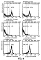
- Figure 4 shows the histograms of a FACS analysis of the separations.
- the first row depicts the cells before separation
- the second row depicts the unmagnetic fraction
- the third row depicts positive cells.
- ⁇ 1.3 results are in the left column
- ⁇ 3.5 results are in the right column.
- the data indicate that at least 95% of the IgD expressing cells are retained by the column, whereas the IgD cells being retained and eluted from the column have a purity of at least 92%. Background staining in this separation and analysis is mainly caused by macrophages taking up antibodies in an unspecific manner.
- NCAMs Neural cell adhesion molecules
- NCAMs are members of the immunoglobulin super family mediating homo- and heterophilic cell-cell interactions. NCAMs appear in various isoforms generated by alternative splicing. Hemperly et al. (1986) Proc. Natl. Acad. Sci. USA 83 :3037-3041; Barthels et al. (1987) EMBO J. 6 :907-914; and Barthels et al. (1992) Eur. J. Neurosci. 4 :327-337. During embryonic development, NCAMs are expressed in derivatives of all three germ layers whereas in the adult animal they are predominantly present in neural tissue.
- Heterozygous animals of two lines were mated and gave rise to 78 offspring. Of these, 38 animals (49%) were heterozygous, 22 (28%) were wild type and 18 (23%) were homozygous for the mutated allele, indicating an almost perfect Mendelian distribution. Homozygous mutant animals are fertile and appear healthy up to four months of age even though they are about 10% smaller by weight than wild type and heterozygous littermates. Isolated brains of a mutant and a heterozygous animal had obvious anatomical differences: the olfactory bulb was reduced in size in mutants compared to +/+ and +/- animals; and the brain weight was reduced by about 10% (after correction for body weight).
- mice described above are inoculated with an antigenic amount of an NCAM suspended in a suitable adjuvant.
- Booster shots are given as required and the antibody titer is measured periodically. Once the titer is sufficient, a suitable method for generation of AMABs is followed.
Landscapes
- Health & Medical Sciences (AREA)
- Life Sciences & Earth Sciences (AREA)
- Chemical & Material Sciences (AREA)
- Immunology (AREA)
- Organic Chemistry (AREA)
- Genetics & Genomics (AREA)
- Engineering & Computer Science (AREA)
- General Health & Medical Sciences (AREA)
- Molecular Biology (AREA)
- Biochemistry (AREA)
- Medicinal Chemistry (AREA)
- Zoology (AREA)
- Biotechnology (AREA)
- Biomedical Technology (AREA)
- Biophysics (AREA)
- Proteomics, Peptides & Aminoacids (AREA)
- Wood Science & Technology (AREA)
- Veterinary Medicine (AREA)
- Bioinformatics & Cheminformatics (AREA)
- Microbiology (AREA)
- General Engineering & Computer Science (AREA)
- Physics & Mathematics (AREA)
- Environmental Sciences (AREA)
- Urology & Nephrology (AREA)
- Hematology (AREA)
- Cell Biology (AREA)
- Virology (AREA)
- Analytical Chemistry (AREA)
- Tropical Medicine & Parasitology (AREA)
- Plant Pathology (AREA)
- Animal Husbandry (AREA)
- Biodiversity & Conservation Biology (AREA)
- Food Science & Technology (AREA)
- Animal Behavior & Ethology (AREA)
- General Physics & Mathematics (AREA)
- Pathology (AREA)
- Peptides Or Proteins (AREA)
- Preparation Of Compounds By Using Micro-Organisms (AREA)
- Micro-Organisms Or Cultivation Processes Thereof (AREA)
- Investigating Or Analysing Biological Materials (AREA)
Abstract
Description
- This invention relates to methods of obtaining autologous monoclonal antibodies (AMABs) to self-antigens or homologs thereof, and the use of these antibodies in the analysis of cell populations and in cell separation techniques.
- Antibodies have proven useful in medical applications for both diagnosis and therapy, and in biotechnology applications including cell separation. More generally, their high degree of binding specificity facilitates their use in the identification and localization of any compound to which antibodies can be generated in conjunction with techniques as varied as electron microscopy and enzyme linked immunosorbent assays.
- Antibodies are comprised of both heavy and light chain polypeptides joined by interchain disulfide bonds and other intramolecular interactions. An individual heavy chain is paired with an individual light chain by these disulfide bonds. Of the different classes or isotypes of antibodies, three isotypes (IgD, IgE, and IgG) are comprised of two identical heavy chain/light chain pairs joined by a disulfide bond, and the remaining two isotypes (IgA and IgM) are comprised of more complicated polymers of identical heavy chain/light chain pairs. Each chain contains a constant region and a variable region. The constant regions are peculiar to the animal that generates the antibody and the specific isotype of antibody, while the variable regions conform to the structure of the epitope to which the antibody binds.
- The term "antigen" is used herein to refer to a substance, whether an entire molecule or a domain within a molecule, which is capable of eliciting production of antibodies with binding specificity to that substance. Further, the term antigen is applied herein to substances, which in wild type host organisms would not elicit antibody production by virtue of self-recognition, but can elicit such a response in a host animal with the appropriate genetic engineering.
- The term "epitope" is used herein to refer to the discrete, three-dimensional sites on an antigen, which are recognized by B lymphocytes. Epitopes are the immunologically active regions on a complex antigen, the regions that actually bind to a B-cell receptor, and that are actually bound by the resulting antibody molecules that are produced by the B-cell. Antigens generally contain at least one epitope and usually more than one epitope. Epitopes on protein antigens can be linear or non-linear. Linear epitopes are those comprised of contiguous amino acid residues in the amino acid sequence of a protein. Linear epitopes may or may not require conformational folding to form the native three-dimensional structure and elicit an immune response that produces antibodies with binding specificity to the antigen. Non-linear epitopes are comprised of non-contiguous amino acid residues. Thus, non-linear epitopes always require some degree of protein folding to bring the requisite amino acid residues into proximity of one another to form the native three-dimensional structure and elicit an immune response that produces antibodies with binding specificity to the antigen.
- The term "self" is used herein to describe antigens or epitopes which would not be recognized or be only poorly recognized by the B-cell receptors of a wild type member of the host species, by virtue of being included among the substances which are normally biosynthesized by the host species, or to which the host species is normally exposed. Such substances induce tolerance of the host immune system and the host is said to be "tolerized" to the substances.
- The vertebrate immune system is able to discriminate between self-antigens and foreign antigens, mounting an antibody-mediated immune response to the latter and not the former. The antibody response is mediated by the B cells. Variable-region gene rearrangments occur in an ordered sequence during B-cell maturation in the bone marrow. At the end of this process, each B cell contains a single, functional variable-region DNA sequence encoding an immunoglobulin heavy chain and a single, functional variable-region DNA sequence encoding an immunoglobulin light chain. This process leads to the generation of mature, immunocompetent B cells each of which is antigenically committed to a single epitope. In a process that is not yet understood, immunologic tolerance to "self" components is accomplished by the selective ablation of B cells with variable regions that are antigentically committed to self-epitopes. This self-tolerance precludes production of antibodies specific for antigens or epitopes that are synthesized by a host vertebrate. Thus, only antigens that contain epitopes which are recognized as foreign by the host can be used to generate antibodies.
- When the self and foreign epitopes are structurally similar, or "homologous", the host immune response is weaker; thus it is virtually impossible to obtain antibodies with high affinity to such epitopes. As a result it is extremely difficult to generate antibodies to highly conserved domains of proteins (e.g. N-CAM, cytokines, and immunoglobulins), because animals that share the conserved domains fail to recognize them as foreign. While antibodies to self-antigens are produced as a result of certain autoimmune diseases, these antibodies have binding specificities to a highly restricted set of self-antigens which cannot be manipulated artificially and generally have low binding affinities. EP-A-286447 describes such autoimmune disease, and describes generating monoclonal antibodies to self antigens involved in such autoimmune disease using conventional techniques. Thus, animals with autoimmune diseases are not widely useful in the production of antibodies with binding specificity to self-antigens. EP-A-313156 describes a protein antigen which is reactive towards an auto-antibody associated with an auto-immune disease.
- In mice, allogeneic differences between strains have allowed the production of mouse anti-mouse antibodies specific for proteins of which such allogeneic differences have been produced. Kessler et al. (1979) J. Immunol. 123:2772-2778; Reif and Allen (1964) J. Exp. Med 120:413-433; Marshak-Rothstein (1979) J. Immunol. 122:2491-2497; and Oi and Herzenberg (1979) Molec. Immunol. 16:1005-1017. Initially, polyclonal antisera and then monoclonal antibodies specific for T cell surface proteins and mouse IgD antibodies were obtained in this manner. These antibodies, however, only recognize the gene product of particular mouse strains. These antibodies can only recognise those epitopes which are not structurally homologous to the self-antigens of the antibody-producing host. Additionally, the epitopes against which these antibodies can be obtained are limimted to the differences between the strains and availability of allotypic strains themselves and thus have little practical utility.
- Prusiner et al PNAS, 1993, vol 90 no 22, 10608-10612, describe the production of genetically altered prion protein deficient mice. Injection of infectious prion proteins leads to generation of antibodies in these mice.
- Numerous methods have been formulated to analyze and sort populations of cells including, but not limited to, fluorescence activated cell sorting (FACS), magnetic separation (using magnetic bead-conjugated antibodies) and other methods reliant upon antibody affinity to particular cell surface proteins known as "markers". Such approaches to cell analysis and separation are especially useful for the determination of cell lineages, the isolation of cells which are capable of synthesizing a particular product, and the treatment of various disease conditions with specific cell types. For example, highly purified hematopoietic stem cells are essential for hematopoietic engraftment including, but not limited to, cancer patients and transplantation of other organs in association with hematopoietic engraftment. Isolated cell populations are also important targets for gene therapy in the treatment of genetic disorders, AIDS and various forms of cancer. Thus, there have been numerous efforts made toward isolating particular varieties of cells in substantially pure or pure form. In instances such as isolation of stem cells, efficient purification of cells of such low concentration in the body requires antibodies which recognize and bind to stem cell specific markers with high specificity. Such antibodies are difficult to obtain due to the homology between human and murine stem cell markers.
- This invention seeks to provide novel methods of obtaining (autologous) monoclonal antibodies (AMABs) to a self-antigen or homolog thereof. The method can comprise obtaining a genetically engineered host animal that does not biosynthesize or synthesizes an altered form of at least one epitope within the self-antigen and utilizing the host lack of self-tolerance to the at least one epitope to produce antibodies specific to that antigen. The invention also encompasses the antibodies or any functional derivative thereof produced by the method. The invention further encompasses methods of isolating cells comprising utilizing the antibodies obtained by the method of the invention which are specific to a cell surface antigen.
- In the accompanying drawings, which are provided to illustrate the invention:
- Figure 1 is a picture depicting anti-IgD antibody staining of mouse spleen tissue in B cell rich areas;
- Figure 2 depicts the results of titration of δ1.3 and δ3.5 AMABs on mouse spleen cells;
- Figure 3 depicts the results of a FACS analysis of doubly stained mouse spleen cells with anti-IgM and anti-IgD antibodies; and
- Figure 4 depicts the results of separation of mouse spleen cells using anti-IgD antibodies conjugated to colloidal superparamagnetic particles.
-
- The invention seeks to provide novel methods of obtaining AMABs which have binding specificity to self-antigens or homologs thereof. The invention may provide a means for overcoming the limitation on producing AMABs to antigens that the host recognizes as self, as well as a method for obtaining "targeted antibodies" that is, AMABs with binding specificity to known, particular and extremely precise epitopes. The invention also allows for obtaining AMABs to biological molecules, or epitopes thereof, considered essential for growth and development. As discussed more fully herein, targeted genetic replacement allows for production of functional equivalents of the molecules and thus allows sufficient growth and development of the animal to produce antibodies.
- The term "homolog" is used herein to refer to an antigen with a structure that is so similar to an antigen produced by the host animal, a "self-antigen", as to preclude or seriously hinder the production of antibodies to the homolog. The phrase "self-antigen or homolog thereof" is used herein to signify that the invention is not directed solely to obtaining AMABs with binding specificity to self-antigens, but rather the invention is directed to obtaining AMABs to any compound with such a high degree of structural similarity to a self-antigen, that the homolog is sufficiently recognized as self that the host animal does not produce an adequate antibody-mediated immune response. An adequate antibody-mediated immune response is one in which either AMABs are not obtained or, if produced, they do not have high affinity for the antigen.
- The term "target antigen" is used herein to refer to the composition to which the host animal is exposed and against which an immune response is generated. The "target antigen epitope" is a region on the target antigen to which the AMABs bind.
- The term "immune response" is used herein to refer to the production of AMABs by B cells. These antibodies bind to a particular antigen regardless of whether they effect a change in the antigen such as providing immunity to a disease causing agent. The AMABs may be associated with the B cell surface and may also be freely circulating.
- The term "structurally non-homologous" is used herein as a description to compare self-antigens with antigens biosynthesized by host animals as a result of genetic engineering. Two antigens are structurally non-homologous, when an antibody can be generated that binds to one and not the other. Structurally non-homologous means that there is some structural difference, perhaps slight, between two or more antigens. The structurally non-homologous difference between two antigens can be as small as a single amino acid difference, or the presence or absence of a methyl group.
- The term "functionally equivalent" is used herein as a description to compare self-antigens and target antigens biosynthesized by host animals as a result of genetic engineering. In many cases, disrupting synthesis of a self-antigen can prove lethal, reduce survivorship or the overall health of genetically engineered host animals so as to interfere with obtaining antibodies. Thus, as described herein, genetic engineering can be used to eliminate the biosynthesis of a self-antigen and cause the production of another, functionally equivalent antigen. This functionally equivalent antigen exerts a sufficient amount of the self-antigen function to compensate for the deleterious elimination of the self-antigen by improving the rate of survivorship, overall health or immunocompetence of the host animal to the extent that AMABs may be obtained. As used herein, functionally equivalent antigens are understood to be structurally non-homologous.
- The term "biosynthetic repertoire" is used herein to refer to the sum total of all of the compounds biosynthesized by a given host animal.
- The term "wild type" is used herein to describe individuals of the host species and strain that have not been subjected to genetic engineering relating to the target antigen and are not descended from such a genetically engineered organism. Thus such individuals will usually produce the self-antigen of interest.
- The term "genetically engineered" is used to describe animals that have had one or more genes directly altered or eliminated so as to prevent or alter synthesis of a particular antigen or set of antigens. Such alteration or elimination is at the genetic level, for instance by specific genetic knockout and replacement. Genetic engineering of the target antigen is generally limited to the extent that antibodies can be obtained with enhanced binding specificity to at least one epitope on the antigen or set of antigens, as described herein. The term "genetically engineered" is also applied to the progeny of the originally altered animals.
- The term "domain" is used herein to refer to any region or portion of an antigen, including the whole antigen, any portion which is less than the whole antigen, or any region which is more than the whole antigen by virtue of the addition of components which may serve to mask at least one epitope.
- In one embodiment of the invention, the host animal is genetically engineered so that it does not synthesize a particular self-antigen. When the genetically modified host is immunized with the self-antigen or a homolog thereof, the host immune system does not recognize the antigen as self, and is thus able to produce an antibody-mediated immune response from which AMABs can be obtained. Methods of obtaining such "knock-out" mutants are known in the art and are described for instance, in Mansour et al. Nature 336:348-352 (1986). As with most genetically engineered host animals used herein, breeding may be used to achieve a homozygous mutant or to obtain multiply genetically engineered host animals. Thus, the genetic modification to the host can ultimately be acquired through germ-line transmission.
- Progeny of the genetically engineered mice are also encompassed by the invention. Any other method known in the art of creating transgenic animals is suitable for use herein. Suitable methods include, but are not limited to, those described by Kitamura et al. (1991) Nature 350:423-426; Shinkai et al. (1993) Science 259:822-825; and Komori et al. (1993) Science 261:1171-1175. Briefly, in the case of self-antigens of the immune system, embryonic stem (ES) cells homozygous for the gene modification can be implanted into blastocysts of immunodeficient mice such as RAG-deficient mice. The reconstituted animal will, in many cases, lack the immune deficient phenotype and contain the gene modification in its lymphocytes.
- In another embodiment of the invention, the host animal is genetically engineered such that the synthesis of a particular self-antigen is replaced by the synthesis of a functionally equivalent antigen. Thus, antibodies can be obtained for antigens the elimination of which would prove lethal, drastically reduce survivorship, or otherwise hamper efforts to obtain antibodies.
- The self-antigen that is eliminated from the biosynthetic repertoire of the host can be any compound, domain, or epitope thereof, that is normally synthesized by a wild type member of the host species. The antibodies obtained by the invention can be directed to any antigen, including but not limited to, proteins, carbohydrates, lipids, nucleic acids, enzyme cofactors or any naturally-occurring aggregates or covalently-linked combinations thereof, or any phosphorylated or sulfonated species thereof. For these embodiments of the invention, the host can be genetically engineered in any way such that the biosynthesis of the antigen is eliminated or altered. For example, knocking out the expression of a particular enzyme that is involved in the biosynthesis of the antigen can result in nonproduction of the antigen or the production of a functionally equivalent antigen. Such enzymes include, but are not limited to, enzymes involved in the polymerization and attachment of carbohydrates, phosphate groups, lipids, sulfur-containing groups to proteins could be eliminated, resulting in the elimination or change in structure of the compounds that are covalently attached to proteins, as in the synthesis of glycoproteins. Thus, antibodies with binding specificity to particular self-glycoproteins or carbohydrate structures on glycoproteins can be obtained by the methods of the invention.
- More global methods of eliminating or changing production of antigens may be utilized. These methods include, but are not limited to, eliminating synthesis of particular genes or gene families and eliminating particular cell types. Examples of suitable genes and gene families include those regulated by the presence of particular factors such as steroids or cytokines or genes which are expressed in a cell type specific manner. An example of such a cell type is brown fat cells.
- In the preferred embodiment of the invention, the antigen is a protein. A knockout mutation for a protein could involve preventing the synthesis of the entire protein (by removing the entire gene or by altering transcriptional or translational control elements) or eliminating the synthesis of just one antigenic domain, while allowing the remaining portion of the protein to be synthesized normally. In the case of a protein antigen, the replacement of the self-antigen with a functionally equivalent antigen, can be achieved by at least three distinct types of replacements although any formal replacement known in the art is suitable for use herein.
- First, the gene encoding the self-antigen can be replaced by a recombinant gene derived from the gene encoding the self-antigen. The gene encoding the protein is altered such that an antigenic region is deleted, replaced by an alternative amino acid sequence, or masked by the addition of a novel amino acid sequence. Thus, in the modification of a self-protein antigen, the genetically engineered elimination of the antigen can be as minor as the addition, elimination or substitution of a single amino acid. Importantly, such small changes in the structure of the self-protein antigen allow AMABs to extremely precise epitopes to be obtained without severely disrupting the function of the self-antigen. A single amino acid change in a suitable domain would result in AMABs to epitopes that include the single amino acid.
- Second, the gene encoding the self-antigen can be replaced by the gene that encodes a homologous protein obtained from an organism that is related to the host species, such that the encoded protein is functionally equivalent but at least partially antigenically non-equivalent. Closely related species usually have a high degree of sequence homology for the same protein, typically greater than 90%. Thus this strategy is ideal for practicing the invention in cases in which expression of the antigen is critical to the health of the organism, and AMABs with binding specificity to a small number of epitopes is desired. This strategy can also be practiced by replacing only a region of the gene encoding the self-antigen with the corresponding region of the gene encoding the same antigen from a related species.
- Third, the gene encoding the self-antigen can be replaced by a gene encoding a protein that is known to have the same or a similar function, but is structurally non-homologous. This strategy is particularly useful where expression of the antigen is critical to the health of the organism and AMABs with binding specificity to particular epitopes is not required.
- Examples of the protein antigens to which the AMABs obtained by the invention can have binding specificity, include, but are not limited to, cell surface antigens, for example adhesion molecules, MHC class I and class II molecules, integrin, cytokines, selectins, cytokine receptors and immunoglobulins.
- The invention can be practiced using any non-human vertebrate host animal, including fish, reptiles, amphibians, birds, and mammals. However, the host is preferably a mammal and usually belongs to an order including, but not limited to, rodentia, lagomorpha, primates, carnivora, perissodactyla and artiodactyla. Preferably the host is rodentia or murine and most preferably a mouse.
- Methods of making AMABs are well known in the art and so are not described in detail herein. Any method known in the art of monoclonal antibody production can be used herein. Such methods include, but are not limited to, separating B cells with cell-surface antibodies of the desired specificity, cloning the DNA expressing the variable regions of the light and heavy chains and expressing the recombinant genes in a suitable host cell. Standard monoclonal antibody generation techniques can be used wherein the AMABs are obtained from immortalized antibody-producing hybridoma cells. These hybridomas can be produced by fusing B lymphocytes, preferably isolated from the immunized host spleen, with compatible immortalized cells, preferably a B cell myeloma.
- The invention further encompasses compositions of matter comprising the AMABs obtained by the methods described herein. As used herein, the terms "AMAB(s)," "antibody" or "antibodies" include the entire antibody as well as antibody fragments containing functional portions thereof. The term "AMABs" includes any monospecific compound comprised of a sufficient portion of the light chain variable region and/or the heavy chain variable region to effect binding to the epitope to which the whole antibody has binding specificity. The fragments may include the variable region of at least one heavy or light chain immunoglobulin polypeptide, and include, but are not limited to, Fab fragments, Fab2 fragments, and Fv fragments.
- In addition, the monospecific domains may be attached by any method known in the art to another suitable molecule. The attachment may be, for instance, chemical or by genetic engineering. The AMABs may be produced by any recombinant means known in the art. Such recombinant AMABs include, but are not limited to, fragments produced in bacteria and AMABs in which the majority of the constant regions have been replaced by human antibody constant regions. In addition, such "humanized" AMABs may be obtained by breeding the genetically engineered host vertebrates described herein with a compatible transgenic animal that expresses functional human Ig loci in place of the wild type Ig loci. For a discussion of transgenic animals expressing human Ig loci, see WIPO publication number WO 91/10741 and Rajewsky et al. DE P4228162.8. With successive crosses, host animals that do not express a particular self-antigen, but do express humanized Ig proteins can be obtained. When such animals are immunized they will produce humanized or partly humanized AMABs to particular self-antigens. Such humanized AMABs are preferred for use in therapeutic indications.
- The AMABs can be conjugated to other compounds including, but not limited to, enzymes, magnetic beads, colloidal magnetic beads, haptens, fluorochromes, metal compounds, radioactive compounds, drugs or haptens. The enzymes that can be conjugated to the AMABs include, but are not limited to, alkaline phosphatase, peroxidase, urease and β-galactosidase. The fluorochromes that can be conjugated to the AMABs include, but are not limited to, fluorescein isothiocyanate, tetramethylrhodamine isothiocyanate, phycoerythrin, allophycocyanins and Texas Red. For additional fluorochromes that can be conjugated to antibodies see Haugland, R. P. Molecular Probes: Handbook of Fluorescent Probes and Research Chemicals (1992-1994). The metal compounds that can be conjugated to the AMABs include, but are not limited to, ferritin, colloidal gold, and particularly, colloidal superparamagnetic beads. The haptens that can be conjugated to the AMABs include, but are not limited to, biotin, digoxygenin, oxazalone, and nitrophenol. The radioactive compounds that can be conjugated or incorporated into the AMABs are known to the art, and include but are not limited to technetium 99m (99TC), 125I and amino acids comprising any radionuclides, including, but not limited to, 14C, 3H and 35S. Any drug known in the art that can be conjugated to protein by any method known in the art is suitable for use in the present invention. Such drugs can be conjugated to the AMABs for highly specific delivery to the target molecule.
- The invention further provides methods of using the AMABs, including but not limited to, immunoassays and separating cells. Suitable immunoassays are well known in the art and so need not be described in detail herein. These include, but are not limited to, ELISAs and RIAs. Methods of cell separation include, but are not limited to, separation based on secretion of molecules and separation based on cell surface molecules. Methods of separating cells based on secretion of molecules are described in Serial No. 07/965,934 and International Application No. PCT/US93/10126. Methods for separating cells based on specific cell surface markers generally include the steps of obtaining the AMABs by the methods described herein, contacting the AMABs to a heterogeneous population of cells, and performing a cell separation technique based on the affinity of the AMABs for the cell surface antigen. Any method known in the art may be employed to separate or isolate the cells by initially removing cells with particular cell surface antigens or of particular lineages.
- AMABs are particularly useful for identifying markers associated with particular cell lineages and/or stages of differentiation. The AMABs may be attached directly or indirectly to a solid support to allow for separation. The separation techniques employed should maximize the retention of viability of the fraction to be collected. Various techniques of different efficacy may be employed to obtain separations. Such separations are where up to 10%, usually not more than about 5%, preferably not more than about 1%, of the total cells present not having the marker may remain with the cell population to be retained. The particular technique employed will depend upon efficiency of separation, cytotoxicity of the methodology, ease and speed of performance, and necessity for sophisticated equipment and/or technical skill.
- Procedures for cell separation include, but are not limited to, magnetic separation using antibodies linked to colloidal magnetic particles, affinity chromatography and cytotoxic agents joined to a monoclonal antibody or used in conjunction with any antibody-dependent separation techniques known in the art. In addition, cells may be separated by "panning" with antibody attached to a solid matrix, e.g., a plate. Fluorescence activated cell sorting (FACS) may also be used and may have varying degrees of sophistication, including, but not limited to, a plurality of color channels, low angle and obtuse light scattering detecting channels, and impedance channels. Any antibody-dependent separation technique known in the art may be used in conjunction with both positive and negative separation techniques that rely on the physical properties of the cell rather than antibody affinity, including, but not limited to, elutriation and density gradient centrifugation.
- Methods to separate cells are commercially available from Dynal, Oslo Norway, Cellpro, Seattle, or Advanced Magnetics, Boston. For example, autologous monoclonal antibodies can be directly coupled to magnetic polystyrene particles like Dynal M 450 or similar magnetic particles and used, e.g., for cell separation. Alternatively, antibodies can be biotinylated or conjugated with digoxigenin and used in conjunction with avidin or anti-digoxigenin coated affinity columns like SEPARATE LC (Cellpro). In a preferred embodiment, however, autologous monoclonal antibodies are used in conjunction with colloidal superparamagnetic microparticles having an organic coating by, e.g., polysaccharides. Miltenyi et al. (1990) Cytometry 11:231-238. These particles can be used having a size of 10 to 200 nm, preferably between 40 and 100 nm, and can be either directly conjugated to autologous antibodies or used in combination with anti-immunoglobulin, avidin, or anti-hapten-specific microbeads. Polysaccharide-coated superparamagnetic particles are commercially available from Miltenyi Biotec GmbH, Germany.
- One procedure which may be used is first incubating the cells for a short period of time at reduced temperatures, generally about 4°C, with saturating levels of AMABs specific for a particular cell surface antigen, and then washing the cells with PBS and a fetal calf serum (FCS) cushion. The cells may then be suspended in a buffer medium as described above and separated on the basis of the AMABs for the particular determinants, using various protein(s) specific for the AMABs or AMAB-antigen complex.
- The AMABs may be conjugated with markers, including, but not limited to, magnetic beads, which allow for direct separation, biotin, which can be removed with avidin or streptavidin bound to a support, digoxigenin detected by anti-digoxigenin antibodies and fluorochromes, which can be used with a FACS, or the like, to allow for ease of separation of the particular cell type. Any technique may be employed which is not unduly detrimental to the viability of the remaining cells.
- After substantial enrichment of the cells containing the cell surface antigen, generally by at least about 50%, preferably at least about 70%, the cells may then be separated by a FACS or other methodology known to the art. Multi-color analyses may be employed, by a variety of the methods including, but not limited to, FACS and fluorescence microscopy. The cells may be separated on the basis of the level of staining for the particular antigens.
- The invention further encompasses obtaining AMABs to self-antigens which are receptors by the method described herein and using such AMABs as pharmaceutical agents, wherein the AMABs demonstrate efficacy as receptor agonists or antagonists.
- The following examples are meant to illustrate, but not limit, the invention.
- Gene targeting of the Cδ gene was performed as described by Roes and Rajewsky J. Exp. Med. 177:45-55 (1993); and Roes and Rajewsky Int . Immunol. 3:1367 (1991). Briefly, a total of 108 E14-1 ES cells were transfected with the targeting vector designed to replace a large part of the Cδ1 exon and to insert frameshift mutations in Cδ3 by filling in restriction sites present in this exon. The introduction of the mutations into the germ-line resulted in functional inactivation of both δ chain Ig domain exons. This was considered important to exclude the possibility of expression of a truncated δH chain that could compete with µ for L chains and be secreted. The presence of the frameshift in the mouse germ-line was indicated by an NheI restriction site resulting from filling in the HindIII site in Cδ3. The Cδ2 exon is a pseudoexon due to a nonfunctional splice acceptor.
- Colonies surviving selection were analyzed by PCR, and positive clones were further analyzed by Southern blotting to confirm the structure of the targeted locus. Homologous recombinants were obtained at a frequency of 1/17 double-resistant or 1/103 G418-resistant clones. Restriction maps of wild-type and mutated Cδ loci as well as restriction analysis of HindIII-digested genomic DNA from five individual candidate recombinants and control cells are shown. A homologous recombination event would result in a 4.4- or 6.0-kb band in addition to the 3.8-kb germ-line band, depending on the presence or absence of the frame shift mutation in Cδ3 that results in loss of the HindIII site. 9 of 10 homologous recombinant clones showed the 6.0-kb fragment, indicating that the breakpoint of recombination was located 3' of the Cδ3 exon. Thus, the Cδ1 and Cδ3 exons were rendered nonfunctional in these clones. One clone retained the HindIII restriction site in the Cδ3 exon, resulting in a diagnostic restriction fragment of 4.4 kb. The structure of the targeted locus was confirmed using a variety of other probes and restriction enzymes. Southern blot analysis was performed as described by Sambrook, Fritsch, and Maniatis, Molecular Cloning. A Laboratory Manual. 2nd ed. Cold Spring Harbor Laboratory, NY (1989).
- The generation of IgD-deficient mice was performed as described by Roes and Rajewsky (1993) J. Exp. Med. 177:45-55. Briefly, the strategy of Cδ gene inactivation and the screening procedure for positive clones was as described in Example 1. Targeted ES cell clones were injected into blastocyst isolated from C57BL/6 mice and transferred to (C57BL/6 x BALB/c) fosters. Male chimeric offspring were mated with C57BL/6 females for germ-line transmission of the δT mutation. Offspring derived from ES cells were identified by coat color and analyzed for the presence of the mutation which was called δT, by Southern blotting or phenotypically, by flow cytometry. Homozygous mutant mice (δT/δT) were obtained by the interbreeding of heterozygous offspring.
- The northern blot analysis of putative (δT/δT) mutant mice was performed as described by Roes and Rajewsky (1993). Briefly, the δT mutation results in functional inactivation of both exons encoding Ig domains of the H chain. The transmembrane and the hinge region exons, however, remain intact and potentially functional. To exclude the possibility that aberrant splicing of precursor RNA encompassing both the Cµ and the Cδ genes resulted in the generation of a significant amount of chimeric Ig transcripts encoding the extracellular domains of the Cµ gene and the transmembrane and cytoplasmic portion of Cδ, poly(A)+ RNA isolated from spleens of homozygous mutant (δT/δT) and wild-type mice was analyzed by Northern blotting. mRNA containing Cµ exons spliced to the Cδ transmembrane exon would be larger than the normal Cµ transcripts of 2.4 (µs) or 2.7 kb (µm), because the 3'-untranslated region of the δ message is 600-bp longer than that of the µ message.
- Hybridization of splenic poly(A)+ RNA of homozygous mutant mice with a Cδ transmembrane-specific probe reveals bands of 4.8-, 4.0-, 3.8-, and 3.0-kb. However, none of these bands hybridized with the Cµ-specific probe. The detection limit of the two probes was similar within a factor of two (1.2 x 10⁶ copies for the Cµ and 0.6 x 10⁶ for the δm probe) as judged by the signals obtained from hybridization to standard plasmid DNA. The Cµ probe is a cDNA fragment of 1 kb with complete match to the mRNA and the standard plasmid, whereas the δm probe hybridizes to the mRNA over a stretch of only 480 bp, but 700 bp on the plasmid standard because it contains the δm1/m2 intron. Consequently, a mRNA representing Cµ exons spliced to the δm exons, if detected with the δm probe, should also be revealed with the Cµ probe. Because the 3.0-, 3.8-, 4.0-, and 4.8-kb bands are clearly above the δm probe detection limit, they should, if containing Cµ sequences, also be detected with the Cµ probe. This, however, is not the case.
- Furthermore, sequential hybridization of the same blot with a neo+ gene and a probe specific for the Cδ3 exon demonstrated that the 3.8-, 4.0-, and 4.8-kb bands also contain sequences derived from the neo+ gene, indicating that they represent aberrant splice products. The 3.0-kb band hybridized with the probe specific for the Cδ3 exon which is nonfunctional in the targeted allele because of frame shift mutations described in Example 1. In addition, with the neo+ probe, mRNA of 2.4 kb, which did not hybridize with the δm probe, was also detected. Low abundance mRNAs of 1.6 and 2.0 kb hybridizing with a Cδm-specific probe are detectable in 10 µg poly(A)+ RNA from both normal and mutant mice. These mRNAs, however, are smaller than the normal Cµ message and therefore, unlikely to encode a functional Ig molecule.
- Taken together, mRNA species representing potentially functional chimeric µ/δ molecules were undetectable in Northern blots using as much as 10 µg of splenic poly(A)+ RNA of δT/δT mice. Because mRNA encoding the δ H chain can be detected with the Cδm-specific probe in as little as 300 ng of poly(A)+ RNA of spleens of normal mice, a putative mRNA encoding the extracellular domains of µ and the transmembrane portion of δ would be at least 30-fold less abundant than δ H chain message in normal mice, if it exists at all. Thus, mRNA potentially able to encode an IgD-like molecule is undetectable in homozygous mutant mice (δT/δT) by northern blot analysis.
- IgD-deficient mice were generated by gene-targeting as described in Examples 1 and 2. One animal obtained thereby was immunized intraperitoneally (i.p.) with mouse monoclonal antibody of class IgD (267.7 δ "a" allotype) precipitated in alum.
- After 6 weeks, the animal was boosted i.p. with soluble B1-8 of "b" allotype to obtain monoclonal antibodies recognizing both allotypes.
- After 3 days, spleen cells of the immunized mouse were fused with X63 Ag8.6.5.3 using a standard PEG fusion protocol. Hybrids were directly cloned into eight 96-well plates and selected using HAT selective medium. Hybridomas obtained were screened for production of anti-IgD antibodies by ELISA using plates coated with B1-8 (IgD), BSA (bovine serum albumin), 267.7 (IgD), R33-24-12 (anti-IgM) and developed using R33-18-10.1-Biotin (rat anti-mouse Kappa) and R33-60-Biotin (anti-IgM) antibodies. Seventeen clones showed reactivity to B1-8 (IgDb), thirteen of these showed reactivity to 267.7 (IgDa), one of these was of class IgM. Two other clones showed reactivity to IgM but not to IgD. Hybridomas were further characterized for relative binding affinity and isotype. Fifteen of 21 clones tested showed IgG1 isotype. Additionally, clones were characterized for binding to trypsin-treated and untreated mouse spleen cells using biotinylated anti-mouse IgG1 and streptavidin-phycoerythrin. Trypsin cuts surface IgD and allows localization of the epitope on the IgD molecules.
- Three clones of highest affinity and IgG1 isotype (δ1.2, δ1.3 and δ3.5) were used for further experiments. Several mg of purified AMABs produced by these clones were produced using roller bottles according to standard methods. 16 clones were tested in a staining assay by incubating mouse spleen cells with culture supernatant. Bound anti-IgD AMAB was detected with a biotinylated anti-mouse-IgG1 and revealed by Streptavidin-PE.
- Frozen spleen sections from a C57BL/6 Mouse were stained with biotinylated mouse anti-mouse IgD AMAB δ1.3 prepared as described in Example 4 and biotinylated using Pierce NHS biotin according to the instructions of the supplier. Staining patterns were developed with peroxidase coupled to Streptavidin using an AEC Staining Kit from Sigma as per the manufacturer's instructions. The results obtained are depicted in Figure 1. The staining pattern is identical to that seen for polyclonal goat anti-mouse IgD antisera, demonstrating that the IgD AMABs obtained by the invention have binding specificity to the desired self-antigen.
- 0.5 mg purified AMAB (δ1.3 and δ3.5) each obtained from Example 4 were rebuffered into 1.5 ml 0.1 M NaHCO3 and reacted for 1 hour with 15 µl carboxylfluorescein-hydroxysuccinimide ester (Boehringer Mannheim) dissolved in DMSO (1 mg/ml). Unbound fluorescein was removed by gel filtration. The F/P ratio was determined to be approximately 3-3.5 for both conjugates. One million mouse spleen cells were incubated for 15 minutes with the different dilutions of the conjugated AMABs shown in Figure 2 and analyzed by Flow cytometry. The results obtained are depicted in Figure 2. The data show that concentrations as low as 1 µg/ml are sufficient for detecting IgD-positive cells and that the AMABs have high affinity.
- Spleen cells obtained from a B6 x 129 mouse were stained with the fluorescein-conjugated AMAB δ1.3 and phycoerythrin-conjugated anti-mouse IgM antibody (R33-24), washed and analyzed in a FACScan flow cytometer gating on lymphocytes and excluding dead cells by propidium iodide staining.
- The results obtained are depicted in Figure 3. The results indicate that most IgM bearing cells coexpress IgD; however, a few IgM+ IgD- cells were obtained. This staining is in good accordance with the expected staining pattern for IgD. It also shows that the AMABs bind to molecules usually present in normal mice, and therefore are autologous monoclonal antibodies.
- Conjugation of antibodies to magnetic particles:
- Purified AMABs δ1.3 and δ3.5 were conjugated to MACS amino microbeads (Miltenyi Biotec GmbH, Bergisch Gladbach, Germany) using SPDP (Pierce) coupling chemistry according to the instructions of the supplier. Approximately 200 µg of activated antibody were conjugated to 1 ml of SPDP modified MACS amino microbeads (OD450 = 10). The conjugated beads were purified twice using a MACS A1 column and rebuffered in PBS to a concentration of OD450 = 1 (see; Miltenyi et al. 1990, Cytometry, 11:231-238).
- 10 million spleen cells in 80 µl were incubated for 15 minutes at 4°C with a 1 to 5 dilution of δ1.3 and δ3.5 conjugated magnetic particles, then the same fluorescein conjugated AMAB was added to a final concentration of 8 µg/ml. Cells were allowed to react with the antibody for 5 minutes then washed once using PBS. Magnetically and fluorescently labelled cells were separated using a MACS magnetic cell sorter (Miltenyi Biotec GmbH) according to the instructions of the supplier. The cells were applied to a prefilled A2 MACS column running with a 25G flow restrictor, the column was washed with 4 ml of buffer and cells passing through the column were collected as the unmagnetic fraction. The column was washed using one backflush procedure using a 23G flow resistor and the retained cells were eluted. All fractions were analyzed by flow cytometry (FACScan). Dead cells were identified by propidium iodide staining were excluded from analysis.
- Figure 4 shows the histograms of a FACS analysis of the separations. In figure 4, the first row depicts the cells before separation, the second row depicts the unmagnetic fraction, and the third row depicts positive cells. δ1.3 results are in the left column, and δ3.5 results are in the right column. The data indicate that at least 95% of the IgD expressing cells are retained by the column, whereas the IgD cells being retained and eluted from the column have a purity of at least 92%. Background staining in this separation and analysis is mainly caused by macrophages taking up antibodies in an unspecific manner.
- Neural cell adhesion molecules (NCAMs) are members of the immunoglobulin super family mediating homo- and heterophilic cell-cell interactions. NCAMs appear in various isoforms generated by alternative splicing. Hemperly et al. (1986) Proc. Natl. Acad. Sci. USA 83:3037-3041; Barthels et al. (1987) EMBO J. 6:907-914; and Barthels et al. (1992) Eur. J. Neurosci. 4:327-337. During embryonic development, NCAMs are expressed in derivatives of all three germ layers whereas in the adult animal they are predominantly present in neural tissue. Processes like neurulation, axonal outgrowth, histogenesis of the retina and development of the olfactory system are correlated with the regulated expression of NCAMs. Crossin et al. (1990) Exp. Neurol. 109:5-15; Tosney et al. (1986) Dev. Biol. 114:437-452; Thiery et al. (1977) J. Biol. Chem. 252:6841-6845; Key et al. (1990) J. Cell Biol. 110:1729-1743; and Chung et al. (1991) J. Comp. Neurol. 314:290-305. Homozygous NCAM-negative mice generated by gene targeting appear healthy and fertile. Adult mutants show a 10% reduction of overall brain weight and a 36% decline in size of the olfactory bulb. NCAM-deficiency coincides with the almost total loss of protein-bound α-(2,8)-linked polysialic acid, a carbohydrate structure thought to be correlated with neural development and plasticity. Theodosis et al. (1991) Proc. Natl. Acad. Sci. USA 88:5494-5498. Testing the animals in the Morris water maze as described by Morris (1981) Learn. Motiv. 12:239-260, showed deficits in spatial learning, whereas activity and motoric abilities of mutant mice appeared normal.
- Gene targeting and generation of homozygous mutant mice were performed using standard protocols and confirmed by Southern blotting and allele-specific PCR. Nuclease SI protection assays, Northern and Western Blotting confirmed the targeted locus as a null-allele. Immunocytochemical analysis of brain sections for NCAM using monoclonal antibodies and polyclonal sera showed most intense staining in the glomeruli and granular cell layer of the olfactory bulb in wild type and heterozygous animals. Overall staining was in good agreement with reports on NCAM-expression in the adult brain. Chung et al. (1991). In homozygous mutant mice there was a total loss of NCAM immunoreactivity as expected.
- Heterozygous animals of two lines were mated and gave rise to 78 offspring. Of these, 38 animals (49%) were heterozygous, 22 (28%) were wild type and 18 (23%) were homozygous for the mutated allele, indicating an almost perfect Mendelian distribution. Homozygous mutant animals are fertile and appear healthy up to four months of age even though they are about 10% smaller by weight than wild type and heterozygous littermates. Isolated brains of a mutant and a heterozygous animal had obvious anatomical differences: the olfactory bulb was reduced in size in mutants compared to +/+ and +/- animals; and the brain weight was reduced by about 10% (after correction for body weight).
- In order to generate high affinity AMABs to NCAMs, the mice described above are inoculated with an antigenic amount of an NCAM suspended in a suitable adjuvant. Booster shots are given as required and the antibody titer is measured periodically. Once the titer is sufficient, a suitable method for generation of AMABs is followed.
- Although the foregoing invention has been described in some detail by way of illustration and example for purposes of clarity and understanding it will be apparent to those skilled in the art that certain modifications may be practiced. Therefore, the description and examples should not be construed as limiting on the invention, which is defined in the appended claims.
Claims (24)
- A method of obtaining from a non-human vertebrate animal an autologous monoclonal antibody to a self-antigen, the method comprising:a) producing monoclonal antibodies using cells or genetic material derived from cells collected from a non-human vertebrate animal, in which the genome of the animal has been altered so that it does not produce at least one epitope of a self-antigen, and in which the animal has been immunized with the self-antigen or a homolog thereof to elicit an immune response to said at least one epitope, wherein the homolog comprises an antigen having a high degree of structural similarity to a self-antigen and does not produce an antibody-mediated immune response to the epitope in the unaltered animal, and wherein said cells are produced in response to, and express antibodies against, the self-antigen or its homolog.
- A method according to claim 1, wherein the animal produces a functional equivalent or a structural homolog of the at least one epitope.
- The method according to any preceding claim wherein the self-antigen is a protein, peptide, carbohydrate, lipid, enzyme cofactor, naturally-occurring aggregate or a covalently-linked combination thereof.
- A method according to claim 3, wherein the self-antigen is a cell surface antigen, a cytokine, a cell adhesion molecule or an immunoglobulin.
- A method according to any preceding claim wherein the self-antigen is an IgD antibody.
- A method according to any preceding claim wherein collection of the cells comprises enriching or isolating antigen-specific B cells.
- A method according to any preceding claim wherein the antibodies are produced using the genetic material by expressing recombinant genes encoding the antibodies.
- A method according to claim 7 wherein the recombiannt genes are isolated using single pre-enriched antigen-specific B cells and single-cell PCR.
- A method according to any preceding claim wherein the antibodies are at least partly humanized.
- A method according to any preceding claim wherein the animal is a mammalian and is from the group rodentia, lagomorpha, primates, carnivora, perissodactyla or artiodactyla.
- A method according to claim 10, wherein the animal is rodentia or murine.
- A method according to any preceding claim wherein the biosynthesis of the self-antigen or homolog thereof is prevented, hindered or eliminated by mutation of a gene encoding an enzyme that affects the metabolic pathway that produces the self-antigen or homolog thereof.
- A monoclonal antibody obtainable by a method according to any preceding claim or a fragment thereof which can bind to the self-antigen.
- An antibody or fragment thereof according to claim 13 wherein the fragment is an Fab fragment.
- An antibody or fragment thereof according to claim 13 or 14 wherein the antibody is conjugated or attached to another substance, label or drug.
- An antibody or fragment thereof according to claim 15 wherein the substance is a reporter enzyme, fluorochrome, metallic compound, toxin, hapten, magnetic particle, solid carrier, liposome, or fluorescent particle.
- An antibody or fragment thereof according to claim 16 wherein the magnetic particle is a colloidal superpara-magnetic particle, the enzyme is alkaline phosphatase, peroxidase or urease, the fluorochrome is a fluorescein isothiocyanate, tetramethylrhodamine isothiocyanate, phycoerythrin, allophycocyanin or Texas Red or the metallic compound is ferritin, colloidal gold, or a magnetic bead.
- An antibody or fragment thereof according to claim 15 wherein the substance comprises a radioactive isotope or is biotin.
- An antibody or fragment thereof according to any of claims 13 to 18 for use in an immunoassay or ELISA.
- An antibody or fragment thereof as defined in any of claims 13 to 18 for use in a method of treatment of the human or animal body by therapy or a diagnostic method practised on the human or animal body.
- A hybridoma producing an antibody as defined in claim 13.
- A method of separating cells on the basis of the presence of cell surface antigens, the method comprising:a) obtaining autologous monoclonal antibodies with a binding specificity to a cell surface antigen by a method according to any one of claims 1 to 13;b) contacting the antibodies with a heterogeneous population of cells, wherein a subpopulation of the cells express the cell surface antigen to which the antibodies have a binding specificity; andc) separating the cells recognized by the antibody from the cells not recognized by the antibody.
- A method according to claim 22 where the antibodies are used in conjunction with colloidal superparamegnetic particles and the cells are isolated using high gradient magnetic separation.
- A method according to claim 23 wherein the antibodies are conjugated to the colloidal superparamagnetic particles, the subpopulation of the cells are bound to the antibodies and the antibodies are recognized by anti-immunoglobulin specific colloidal superparamagnetic particles or the antibodies are modified by a hapten and the colloidal superparamagnetic particles recognize the hapten modification.
Applications Claiming Priority (2)
| Application Number | Priority Date | Filing Date | Title |
|---|---|---|---|
| US08/226,168 US7119248B1 (en) | 1994-04-12 | 1994-04-12 | Antibodies against epitopes with homology to self antigens, methods of preparation and applications thereof |
| US226168 | 1994-04-12 |
Publications (3)
| Publication Number | Publication Date |
|---|---|
| EP0677533A2 EP0677533A2 (en) | 1995-10-18 |
| EP0677533A3 EP0677533A3 (en) | 2000-04-12 |
| EP0677533B1 true EP0677533B1 (en) | 2005-01-12 |
Family
ID=22847844
Family Applications (1)
| Application Number | Title | Priority Date | Filing Date |
|---|---|---|---|
| EP95302440A Expired - Lifetime EP0677533B1 (en) | 1994-04-12 | 1995-04-12 | Antibodies against epitopes with homology to self antigens, methods of preparation and applications thereof |
Country Status (8)
| Country | Link |
|---|---|
| US (2) | US7119248B1 (en) |
| EP (1) | EP0677533B1 (en) |
| JP (3) | JP3797680B2 (en) |
| AT (1) | ATE286913T1 (en) |
| CA (1) | CA2146693C (en) |
| DE (1) | DE69533912T2 (en) |
| DK (1) | DK0677533T3 (en) |
| ES (1) | ES2236698T3 (en) |
Cited By (2)
| Publication number | Priority date | Publication date | Assignee | Title |
|---|---|---|---|---|
| WO2007106448A1 (en) * | 2006-03-10 | 2007-09-20 | Lexicon Genetics Incorporated | Methods for making antibodies in genetically engineered mice |
| US9657097B2 (en) | 1998-01-23 | 2017-05-23 | Hoffman-Laroche, Inc. | Methods of treating autoimmune diseases by administering monoclonal antibodies against human IL-12 |
Families Citing this family (37)
| Publication number | Priority date | Publication date | Assignee | Title |
|---|---|---|---|---|
| US7119248B1 (en) * | 1994-04-12 | 2006-10-10 | Miltenyi Biotec Gmbh | Antibodies against epitopes with homology to self antigens, methods of preparation and applications thereof |
| ATE357459T1 (en) * | 1998-01-23 | 2007-04-15 | Hoffmann La Roche | ANTIBODIES TO HUMAN IL-12 |
| DK1297017T3 (en) * | 2000-06-19 | 2012-09-17 | Dana Farber Cancer Inst Inc | Compositions and Methods with Monoclonal and Polyclonal Antibodies Specific to T Cell Subpopulations |
| WO2003092615A2 (en) * | 2002-05-01 | 2003-11-13 | Beth Israel Deaconess Medical Center | Use of anti-cd1 antibodies for the modulation of immune responses |
| CA2517519A1 (en) * | 2003-03-04 | 2004-09-16 | Alexion Pharmaceuticals, Inc. | Vectors used to create hybrid constant regions |
| US7407767B2 (en) * | 2003-04-16 | 2008-08-05 | Ono Pharmaceutical Co., Ltd. | Method of screening remedy for heart disease and medicinal composition for treating heart disease |
| WO2007080404A2 (en) * | 2006-01-12 | 2007-07-19 | Asterion Limited | Leptin ligand |
| JP5435878B2 (en) * | 2008-02-12 | 2014-03-05 | 富士フイルム株式会社 | Labeled particles with fragmented antibody immobilized on labeling substance |
| WO2010132683A1 (en) | 2009-05-13 | 2010-11-18 | Genzyme Corporation | Methods and compositions for treating lupus |
| EP3683317A3 (en) | 2009-05-13 | 2020-09-30 | Genzyme Corporation | Anti-human cd52 immunoglobulins |
| US8790916B2 (en) | 2009-05-14 | 2014-07-29 | Genestream, Inc. | Microfluidic method and system for isolating particles from biological fluid |
| CN102638971B (en) | 2009-07-08 | 2015-10-07 | 科马布有限公司 | Animal Models and Therapeutic Molecules |
| US9445581B2 (en) | 2012-03-28 | 2016-09-20 | Kymab Limited | Animal models and therapeutic molecules |
| US9796788B2 (en) | 2010-02-08 | 2017-10-24 | Regeneron Pharmaceuticals, Inc. | Mice expressing a limited immunoglobulin light chain repertoire |
| HRP20151188T4 (en) | 2010-02-08 | 2021-10-01 | Regeneron Pharmaceuticals, Inc. | Common light chain mouse |
| US20130045492A1 (en) | 2010-02-08 | 2013-02-21 | Regeneron Pharmaceuticals, Inc. | Methods For Making Fully Human Bispecific Antibodies Using A Common Light Chain |
| US8695618B2 (en) | 2010-12-22 | 2014-04-15 | Carnegie Mellon University | 3D chemical pattern control in 2D fluidics devices |
| PT3865581T (en) | 2011-08-05 | 2024-10-04 | Regeneron Pharma | Humanized universal light chain mice |
| AU2012311286B2 (en) | 2011-09-19 | 2018-07-26 | Kymab Limited | Antibodies, variable domains and chains tailored for human use |
| WO2013045916A1 (en) | 2011-09-26 | 2013-04-04 | Kymab Limited | Chimaeric surrogate light chains (slc) comprising human vpreb |
| EP2755688A4 (en) | 2011-10-27 | 2014-08-06 | Nkt Therapeutics Inc | HUMANIZED ANTIBODIES DIRECTED AGAINST INKT |
| GB2496375A (en) * | 2011-10-28 | 2013-05-15 | Kymab Ltd | A non-human assay vertebrate comprising human antibody loci and human epitope knock-in, and uses thereof |
| US9253965B2 (en) | 2012-03-28 | 2016-02-09 | Kymab Limited | Animal models and therapeutic molecules |
| GB2502127A (en) | 2012-05-17 | 2013-11-20 | Kymab Ltd | Multivalent antibodies and in vivo methods for their production |
| US10251377B2 (en) | 2012-03-28 | 2019-04-09 | Kymab Limited | Transgenic non-human vertebrate for the expression of class-switched, fully human, antibodies |
| AR095199A1 (en) | 2013-03-15 | 2015-09-30 | Genzyme Corp | ANTI-CD52 ANTIBODIES |
| US9788534B2 (en) | 2013-03-18 | 2017-10-17 | Kymab Limited | Animal models and therapeutic molecules |
| US9783618B2 (en) | 2013-05-01 | 2017-10-10 | Kymab Limited | Manipulation of immunoglobulin gene diversity and multi-antibody therapeutics |
| US9783593B2 (en) | 2013-05-02 | 2017-10-10 | Kymab Limited | Antibodies, variable domains and chains tailored for human use |
| US11707056B2 (en) | 2013-05-02 | 2023-07-25 | Kymab Limited | Animals, repertoires and methods |
| US20140331339A1 (en) * | 2013-05-03 | 2014-11-06 | Kymab Limited | Transgenic Non-Human Assay Vertebrates, Assays and Kits |
| CA2925723A1 (en) | 2013-10-01 | 2015-04-09 | Kymab Limited | Animal models and therapeutic molecules |
| CN106255410B (en) | 2014-03-21 | 2020-01-10 | 瑞泽恩制药公司 | Non-human animals producing single domain binding proteins |
| US11111314B2 (en) | 2015-03-19 | 2021-09-07 | Regeneron Pharmaceuticals, Inc. | Non-human animals that select for light chain variable regions that bind antigen |
| SG11201806427WA (en) | 2016-02-16 | 2018-08-30 | Regeneron Pharma | Non-human animals having a mutant kynureninase gene |
| US20170332610A1 (en) | 2016-05-20 | 2017-11-23 | Regeneron Pharmaceuticals, Inc. | Methods for breaking immunological tolerance using multiple guide rnas |
| CN113361141B (en) * | 2021-07-11 | 2022-02-18 | 西南石油大学 | An Improved Test Method for DNA Mapping Algorithm |
Family Cites Families (4)
| Publication number | Priority date | Publication date | Assignee | Title |
|---|---|---|---|---|
| AU650616B2 (en) | 1988-09-21 | 1994-06-30 | Animal Biotechnology Cambridge Limited | Derivation of pluripotential embryonic cell lines from domestic animals |
| ES2191677T3 (en) | 1992-10-21 | 2003-09-16 | Stefan Miltenyi | DIRECT SELECTION OF CELLS THROUGH A SECRETION PRODUCT. |
| GB9308271D0 (en) | 1993-04-21 | 1993-06-02 | Univ Edinburgh | Method of isolating and/or enriching and/or selectively propagating pluripotential animal cells and animals for use in said method |
| US7119248B1 (en) * | 1994-04-12 | 2006-10-10 | Miltenyi Biotec Gmbh | Antibodies against epitopes with homology to self antigens, methods of preparation and applications thereof |
-
1994
- 1994-04-12 US US08/226,168 patent/US7119248B1/en not_active Expired - Fee Related
-
1995
- 1995-04-10 CA CA2146693A patent/CA2146693C/en not_active Expired - Fee Related
- 1995-04-12 DK DK95302440T patent/DK0677533T3/en active
- 1995-04-12 ES ES95302440T patent/ES2236698T3/en not_active Expired - Lifetime
- 1995-04-12 DE DE69533912T patent/DE69533912T2/en not_active Expired - Lifetime
- 1995-04-12 EP EP95302440A patent/EP0677533B1/en not_active Expired - Lifetime
- 1995-04-12 AT AT95302440T patent/ATE286913T1/en active
- 1995-04-12 JP JP08726995A patent/JP3797680B2/en not_active Expired - Fee Related
-
2005
- 2005-09-09 JP JP2005263046A patent/JP2006061158A/en not_active Withdrawn
-
2006
- 2006-01-11 JP JP2006004229A patent/JP4362121B2/en not_active Expired - Fee Related
- 2006-10-10 US US11/546,539 patent/US20070101446A1/en not_active Abandoned
Cited By (2)
| Publication number | Priority date | Publication date | Assignee | Title |
|---|---|---|---|---|
| US9657097B2 (en) | 1998-01-23 | 2017-05-23 | Hoffman-Laroche, Inc. | Methods of treating autoimmune diseases by administering monoclonal antibodies against human IL-12 |
| WO2007106448A1 (en) * | 2006-03-10 | 2007-09-20 | Lexicon Genetics Incorporated | Methods for making antibodies in genetically engineered mice |
Also Published As
| Publication number | Publication date |
|---|---|
| DK0677533T3 (en) | 2005-05-30 |
| JPH0856692A (en) | 1996-03-05 |
| JP3797680B2 (en) | 2006-07-19 |
| CA2146693C (en) | 2010-09-14 |
| DE69533912T2 (en) | 2005-12-08 |
| EP0677533A3 (en) | 2000-04-12 |
| JP4362121B2 (en) | 2009-11-11 |
| DE69533912D1 (en) | 2005-02-17 |
| CA2146693A1 (en) | 1995-10-13 |
| JP2006061158A (en) | 2006-03-09 |
| JP2006151991A (en) | 2006-06-15 |
| US7119248B1 (en) | 2006-10-10 |
| US20070101446A1 (en) | 2007-05-03 |
| EP0677533A2 (en) | 1995-10-18 |
| ATE286913T1 (en) | 2005-01-15 |
| ES2236698T3 (en) | 2005-07-16 |
Similar Documents
| Publication | Publication Date | Title |
|---|---|---|
| US7119248B1 (en) | Antibodies against epitopes with homology to self antigens, methods of preparation and applications thereof | |
| Kitamura et al. | A critical role of λ5 protein in B cell development | |
| JP3523245B1 (en) | Transgenic chromosome-introduced rodents for the production of human antibodies | |
| Baumann et al. | Biology of oligodendrocyte and myelin in the mammalian central nervous system | |
| CA2468565C (en) | Transgenic animals bearing human iglambda light chain genes | |
| AU685811B2 (en) | Mammals lacking expression of CD28 transgenic | |
| AU697523B2 (en) | Transgenic production of oligosaccharides and glycoconjugates | |
| Takeda et al. | Impaired motor coordination in mice lacking neural recognition molecule NB‐3 of the contactin/F3 subgroup | |
| EP0815213A4 (en) | An immortalized mammal hybridoma fusion partner | |
| JPH06500233A (en) | Method for producing heterologous immunoglobulin and transgenic mouse | |
| US5675060A (en) | Transgenic arthritic mice expressing a T-cell receptor transgene | |
| Lamers et al. | Immune status of a μ, χ transgenic mouse line. Deficient response to bacterially related antigens | |
| US6921846B1 (en) | Antibody production methods relating to disruption of peripheral tolerance in B lympho-cytes | |
| EP0789763B1 (en) | Icam-4 materials and methods | |
| Woloschak et al. | Characterization of immunologic and neuropathologic abnormalities in wasted mice. | |
| US5532158A (en) | Interleukin-2 receptor deficient mammals | |
| EP0956508B1 (en) | Icam-4 and diagnostic uses thereof | |
| US20030068659A1 (en) | ICAM-4 materials and methods | |
| WO1999027963A1 (en) | Antibody production methods relating to disruption of peripheral tolerance in b lymphocytes | |
| JPWO2003083110A1 (en) | Novel galactose transferase, peptide thereof and nucleic acid encoding the same | |
| WO1999018441A1 (en) | Icam-4 and diagnostic uses thereof | |
| JPH11313576A (en) | Chimeric animal and method for producing the same |
Legal Events
| Date | Code | Title | Description |
|---|---|---|---|
| PUAI | Public reference made under article 153(3) epc to a published international application that has entered the european phase |
Free format text: ORIGINAL CODE: 0009012 |
|
| AK | Designated contracting states |
Kind code of ref document: A2 Designated state(s): AT BE CH DE DK ES FR GB GR IE IT LI LU MC NL PT SE |
|
| PUAL | Search report despatched |
Free format text: ORIGINAL CODE: 0009013 |
|
| AK | Designated contracting states |
Kind code of ref document: A3 Designated state(s): AT BE CH DE DK ES FR GB GR IE IT LI LU MC NL PT SE |
|
| 17P | Request for examination filed |
Effective date: 20000807 |
|
| 17Q | First examination report despatched |
Effective date: 20001211 |
|
| GRAP | Despatch of communication of intention to grant a patent |
Free format text: ORIGINAL CODE: EPIDOSNIGR1 |
|
| GRAS | Grant fee paid |
Free format text: ORIGINAL CODE: EPIDOSNIGR3 |
|
| GRAA | (expected) grant |
Free format text: ORIGINAL CODE: 0009210 |
|
| AK | Designated contracting states |
Kind code of ref document: B1 Designated state(s): AT BE CH DE DK ES FR GB GR IE IT LI LU MC NL PT SE |
|
| PG25 | Lapsed in a contracting state [announced via postgrant information from national office to epo] |
Ref country code: IT Free format text: LAPSE BECAUSE OF FAILURE TO SUBMIT A TRANSLATION OF THE DESCRIPTION OR TO PAY THE FEE WITHIN THE PRESCRIBED TIME-LIMIT;WARNING: LAPSES OF ITALIAN PATENTS WITH EFFECTIVE DATE BEFORE 2007 MAY HAVE OCCURRED AT ANY TIME BEFORE 2007. THE CORRECT EFFECTIVE DATE MAY BE DIFFERENT FROM THE ONE RECORDED. Effective date: 20050112 |
|
| REG | Reference to a national code |
Ref country code: GB Ref legal event code: FG4D |
|
| REG | Reference to a national code |
Ref country code: CH Ref legal event code: EP |
|
| REF | Corresponds to: |
Ref document number: 69533912 Country of ref document: DE Date of ref document: 20050217 Kind code of ref document: P |
|
| REG | Reference to a national code |
Ref country code: IE Ref legal event code: FG4D |
|
| PG25 | Lapsed in a contracting state [announced via postgrant information from national office to epo] |
Ref country code: SE Free format text: LAPSE BECAUSE OF FAILURE TO SUBMIT A TRANSLATION OF THE DESCRIPTION OR TO PAY THE FEE WITHIN THE PRESCRIBED TIME-LIMIT Effective date: 20050412 Ref country code: LU Free format text: LAPSE BECAUSE OF NON-PAYMENT OF DUE FEES Effective date: 20050412 |
|
| PG25 | Lapsed in a contracting state [announced via postgrant information from national office to epo] |
Ref country code: MC Free format text: LAPSE BECAUSE OF NON-PAYMENT OF DUE FEES Effective date: 20050430 |
|
| REG | Reference to a national code |
Ref country code: DK Ref legal event code: T3 |
|
| REG | Reference to a national code |
Ref country code: CH Ref legal event code: NV Representative=s name: R. A. EGLI & CO. PATENTANWAELTE |
|
| REG | Reference to a national code |
Ref country code: ES Ref legal event code: FG2A Ref document number: 2236698 Country of ref document: ES Kind code of ref document: T3 |
|
| PLBE | No opposition filed within time limit |
Free format text: ORIGINAL CODE: 0009261 |
|
| STAA | Information on the status of an ep patent application or granted ep patent |
Free format text: STATUS: NO OPPOSITION FILED WITHIN TIME LIMIT |
|
| ET | Fr: translation filed | ||
| 26N | No opposition filed |
Effective date: 20051013 |
|
| PG25 | Lapsed in a contracting state [announced via postgrant information from national office to epo] |
Ref country code: PT Free format text: LAPSE BECAUSE OF NON-PAYMENT OF DUE FEES Effective date: 20050614 |
|
| PG25 | Lapsed in a contracting state [announced via postgrant information from national office to epo] |
Ref country code: GR Free format text: LAPSE BECAUSE OF NON-PAYMENT OF DUE FEES Effective date: 20050112 |
|
| PGFP | Annual fee paid to national office [announced via postgrant information from national office to epo] |
Ref country code: IE Payment date: 20110412 Year of fee payment: 17 |
|
| PGFP | Annual fee paid to national office [announced via postgrant information from national office to epo] |
Ref country code: CH Payment date: 20120412 Year of fee payment: 18 Ref country code: NL Payment date: 20120413 Year of fee payment: 18 Ref country code: DK Payment date: 20120411 Year of fee payment: 18 Ref country code: BE Payment date: 20120412 Year of fee payment: 18 |
|
| PGFP | Annual fee paid to national office [announced via postgrant information from national office to epo] |
Ref country code: ES Payment date: 20120510 Year of fee payment: 18 |
|
| REG | Reference to a national code |
Ref country code: IE Ref legal event code: MM4A |
|
| PG25 | Lapsed in a contracting state [announced via postgrant information from national office to epo] |
Ref country code: IE Free format text: LAPSE BECAUSE OF NON-PAYMENT OF DUE FEES Effective date: 20120412 |
|
| PGFP | Annual fee paid to national office [announced via postgrant information from national office to epo] |
Ref country code: AT Payment date: 20120327 Year of fee payment: 18 |
|
| PGFP | Annual fee paid to national office [announced via postgrant information from national office to epo] |
Ref country code: GB Payment date: 20130410 Year of fee payment: 19 Ref country code: DE Payment date: 20130508 Year of fee payment: 19 |
|
| PGFP | Annual fee paid to national office [announced via postgrant information from national office to epo] |
Ref country code: FR Payment date: 20130625 Year of fee payment: 19 |
|
| BERE | Be: lapsed |
Owner name: *MILTENYI STEFAN Effective date: 20130430 |
|
| REG | Reference to a national code |
Ref country code: NL Ref legal event code: V1 Effective date: 20131101 |
|
| REG | Reference to a national code |
Ref country code: CH Ref legal event code: PL |
|
| REG | Reference to a national code |
Ref country code: DK Ref legal event code: EBP Effective date: 20130430 |
|
| REG | Reference to a national code |
Ref country code: AT Ref legal event code: MM01 Ref document number: 286913 Country of ref document: AT Kind code of ref document: T Effective date: 20130430 |
|
| PG25 | Lapsed in a contracting state [announced via postgrant information from national office to epo] |
Ref country code: LI Free format text: LAPSE BECAUSE OF NON-PAYMENT OF DUE FEES Effective date: 20130430 Ref country code: CH Free format text: LAPSE BECAUSE OF NON-PAYMENT OF DUE FEES Effective date: 20130430 Ref country code: BE Free format text: LAPSE BECAUSE OF NON-PAYMENT OF DUE FEES Effective date: 20130430 Ref country code: AT Free format text: LAPSE BECAUSE OF NON-PAYMENT OF DUE FEES Effective date: 20130430 |
|
| PG25 | Lapsed in a contracting state [announced via postgrant information from national office to epo] |
Ref country code: NL Free format text: LAPSE BECAUSE OF NON-PAYMENT OF DUE FEES Effective date: 20131101 |
|
| PG25 | Lapsed in a contracting state [announced via postgrant information from national office to epo] |
Ref country code: DK Free format text: LAPSE BECAUSE OF NON-PAYMENT OF DUE FEES Effective date: 20130430 |
|
| REG | Reference to a national code |
Ref country code: ES Ref legal event code: FD2A Effective date: 20140609 |
|
| PG25 | Lapsed in a contracting state [announced via postgrant information from national office to epo] |
Ref country code: ES Free format text: LAPSE BECAUSE OF NON-PAYMENT OF DUE FEES Effective date: 20130413 |
|
| REG | Reference to a national code |
Ref country code: DE Ref legal event code: R119 Ref document number: 69533912 Country of ref document: DE |
|
| GBPC | Gb: european patent ceased through non-payment of renewal fee |
Effective date: 20140412 |
|
| REG | Reference to a national code |
Ref country code: DE Ref legal event code: R119 Ref document number: 69533912 Country of ref document: DE Effective date: 20141101 |
|
| REG | Reference to a national code |
Ref country code: FR Ref legal event code: ST Effective date: 20141231 |
|
| PG25 | Lapsed in a contracting state [announced via postgrant information from national office to epo] |
Ref country code: DE Free format text: LAPSE BECAUSE OF NON-PAYMENT OF DUE FEES Effective date: 20141101 Ref country code: GB Free format text: LAPSE BECAUSE OF NON-PAYMENT OF DUE FEES Effective date: 20140412 |
|
| PG25 | Lapsed in a contracting state [announced via postgrant information from national office to epo] |
Ref country code: FR Free format text: LAPSE BECAUSE OF NON-PAYMENT OF DUE FEES Effective date: 20140430 |