EP0675729B1 - ACTIVATION OF p53 PROTEIN - Google Patents
ACTIVATION OF p53 PROTEIN Download PDFInfo
- Publication number
- EP0675729B1 EP0675729B1 EP94900267A EP94900267A EP0675729B1 EP 0675729 B1 EP0675729 B1 EP 0675729B1 EP 94900267 A EP94900267 A EP 94900267A EP 94900267 A EP94900267 A EP 94900267A EP 0675729 B1 EP0675729 B1 EP 0675729B1
- Authority
- EP
- European Patent Office
- Prior art keywords
- mutant
- ligand
- terminal
- activated
- amino acids
- Prior art date
- Legal status (The legal status is an assumption and is not a legal conclusion. Google has not performed a legal analysis and makes no representation as to the accuracy of the status listed.)
- Expired - Lifetime
Links
Images


Classifications
-
- C—CHEMISTRY; METALLURGY
- C12—BIOCHEMISTRY; BEER; SPIRITS; WINE; VINEGAR; MICROBIOLOGY; ENZYMOLOGY; MUTATION OR GENETIC ENGINEERING
- C12N—MICROORGANISMS OR ENZYMES; COMPOSITIONS THEREOF; PROPAGATING, PRESERVING, OR MAINTAINING MICROORGANISMS; MUTATION OR GENETIC ENGINEERING; CULTURE MEDIA
- C12N9/00—Enzymes; Proenzymes; Compositions thereof; Processes for preparing, activating, inhibiting, separating or purifying enzymes
- C12N9/10—Transferases (2.)
- C12N9/12—Transferases (2.) transferring phosphorus containing groups, e.g. kinases (2.7)
-
- A—HUMAN NECESSITIES
- A61—MEDICAL OR VETERINARY SCIENCE; HYGIENE
- A61P—SPECIFIC THERAPEUTIC ACTIVITY OF CHEMICAL COMPOUNDS OR MEDICINAL PREPARATIONS
- A61P35/00—Antineoplastic agents
-
- C—CHEMISTRY; METALLURGY
- C07—ORGANIC CHEMISTRY
- C07K—PEPTIDES
- C07K14/00—Peptides having more than 20 amino acids; Gastrins; Somatostatins; Melanotropins; Derivatives thereof
- C07K14/195—Peptides having more than 20 amino acids; Gastrins; Somatostatins; Melanotropins; Derivatives thereof from bacteria
- C07K14/24—Peptides having more than 20 amino acids; Gastrins; Somatostatins; Melanotropins; Derivatives thereof from bacteria from Enterobacteriaceae (F), e.g. Citrobacter, Serratia, Proteus, Providencia, Morganella, Yersinia
- C07K14/245—Escherichia (G)
-
- C—CHEMISTRY; METALLURGY
- C07—ORGANIC CHEMISTRY
- C07K—PEPTIDES
- C07K14/00—Peptides having more than 20 amino acids; Gastrins; Somatostatins; Melanotropins; Derivatives thereof
- C07K14/435—Peptides having more than 20 amino acids; Gastrins; Somatostatins; Melanotropins; Derivatives thereof from animals; from humans
- C07K14/46—Peptides having more than 20 amino acids; Gastrins; Somatostatins; Melanotropins; Derivatives thereof from animals; from humans from vertebrates
- C07K14/47—Peptides having more than 20 amino acids; Gastrins; Somatostatins; Melanotropins; Derivatives thereof from animals; from humans from vertebrates from mammals
- C07K14/4701—Peptides having more than 20 amino acids; Gastrins; Somatostatins; Melanotropins; Derivatives thereof from animals; from humans from vertebrates from mammals not used
- C07K14/4746—Peptides having more than 20 amino acids; Gastrins; Somatostatins; Melanotropins; Derivatives thereof from animals; from humans from vertebrates from mammals not used p53
-
- C—CHEMISTRY; METALLURGY
- C07—ORGANIC CHEMISTRY
- C07K—PEPTIDES
- C07K16/00—Immunoglobulins [IG], e.g. monoclonal or polyclonal antibodies
- C07K16/18—Immunoglobulins [IG], e.g. monoclonal or polyclonal antibodies against material from animals or humans
- C07K16/32—Immunoglobulins [IG], e.g. monoclonal or polyclonal antibodies against material from animals or humans against translation products of oncogenes
-
- A—HUMAN NECESSITIES
- A61—MEDICAL OR VETERINARY SCIENCE; HYGIENE
- A61K—PREPARATIONS FOR MEDICAL, DENTAL OR TOILETRY PURPOSES
- A61K38/00—Medicinal preparations containing peptides
Definitions
- This invention relates to the p53 tumour suppressor protein, and more particularly to mutant forms of the p53 protein which seem to be associated with certain types of tumour, and are usually found at elevated levels in such tumour cells.
- the invention also discloses the conversion of wild type p53 to a constitutively active form.
- Mutation of p53 is a very common genetic alteration in human cancers (1). Loss of the p53 tumour suppressor activity is co-incident with a loss of G1-S checkpoints following DNA damage (2), increases in genomic instability and selectable gene amplication (3,4). Mutant forms of p53 also appear to acquire a dominant growth promoting function (5,6). The region responsible for this transforming activity has been localized to a small C-terminal domain (7). Mice null for the wild type p53 locus develop normally, yet are susceptible to the development of neoplasia at elevated frequencies (8), indicating that wild type p53 is dispensable for the control over normal development and cell differentiation, but is essential to prevent spontaneous tumour formation. Consistent with this data, inherited germline point mutations in p53 lead to a predisposition to cancer in humans (9).
- Biochemical characterization of wild type protein has shown that p53 can function as a sequence-specific DNA binding protein (12,13) and a transcription factor (14-16). These results support a model for wild type p53 in which its function is activated post-translationally after DNA damage to allow DNA repair by controlling the expression of regulatory gene products (18). These may include the DNA damage inducible gene gadd45 (19) and the host protein with oncogenic properties mdm-2 (20).
- p53 Biochemical characterization of p53 has become possible recently owing to the use of protein expression systems which allow for an abundant source of the protein.
- p53 purified using immunoaffinity chromatography has been shown to be a sequence-specific DNA binding protein which recognizes a motif containing two contiguous monomers of the sequence (Pu) 3- C(A/T)(A/T)G(Py), (21).
- Sequence-specific DNA binding activity is manifested in the ability of p53 to bind the SV40 origin of replication (13), and by its ability to activate transcription in vitro from templates harbouring its DNA binding sequence (22).
- Mutant forms of p53 are defective in non-specific DNA binding (23), sequence-specific DNA binding (21) and transcriptional activation (14) suggesting that this activity is normally required to suppress tumour formation.
- Phosphorylation of nuclear DNA binding proteins is an effective mechanism through which gene expression is controlled in response to environmental cues (24). Multi-site phosphorylation of p53 by protein kinases (25-27) suggests its tumour suppressor activity may be tightly coordinated by complex signal transducing pathways.
- Casein kinase II (CasKII) is a highly conserved calcium and nucleotide independent enzyme which phosphorylates broad spectrum of substrates, including transcription factors and DNA binding proteins (28). The activity of the kinase is stimulated in cells exposed to a variety of mutagens and growth factors (29).
- Mouse (25) and human p53 (17) are phosphorylated at the penultimate C-terminal amino acid by Cas KII in vitro. Mutation of this highly conserved C-terminal serine residue of mouse p53 to an alanine abolishes the growth suppressive function of the protein in mammalian cells, suggesting that phosphorylation at this site is one important modification required to activate the tumour suppressor function of p53 (30).
- CasKII is the only known enzyme involved in a direct and positive regulation of the activity of p53.
- sequence specific DNA binding is one activity of p53 required for its tumour suppressor function.
- all mutant forms of p53 eg those encoded by the hotspot alleles, His175, Trp248, and His273, have been shown to be defective in sequence specific DNA binding (21) and in the activation of transcription from templates harbouring its consensus DNA binding site (14).
- Enzymatic modulation of p53 may provide a framework from which to couple signal transducing pathways with the P53 response to DNA damage and ultimately growth control.
- p53 phosphorylation on the activity of the protein, we have purified unmodified recombinant p53 by conventional chromatography from bacteria and shown it to be a multimeric protein which is latent for sequence specific DNA binding (17).
- a motif within the C-terminal 30 amino acids negatively controls p53 function as deletion of this domain constitutively activates p53.
- a set of p53 activating proteins including rabbit muscle casein kinase II, monoclonal antibody PAb421, and E.coli Hsp70, target this C-terminal domain.
- sequence specific DNA binding activity is cryptic, it can be unmasked by enzymes and proteins, including casein kinase II (17). From the observation that p53 is regulated positively by CasKII, we attempted to determine if some mutant forms of p53 are inactive in DNA binding as a result of a defect in conversion from a latent to an activated state.
- Mutant forms of p53 encoded by common 'hotspot' alleles were purified and their activities were characterized at the biochemical level.
- a monoclonal antibody and E.coli DnaK are able to promote sequence-specific DNA binding by these mutant forms of p53, but the most potent physiological activator CasKII is unable to effectively unmask the DNA binding function.
- a new biochemical class of mutant forms of p53 is thus established which is defective in DNA binding after post-translational modification by the growth controlling enzyme CasKII.
- This class of mutant p53 defective in the conversion from the latent to the activated state, was our target for the construction of molecules with the ability to reactivate the tumour suppressor function of mutant p53, and thus potentially lead to selective growth arrest and induction of apoptosis in tumour cells (32,33).
- an in vitro method which comprises activating a mutant p53 protein for specific DNA binding by the action of a ligand which binds to the mutant p53 within the C-terminal 30 amino acids , wherein said mutant has the properties that:
- mutant p53 will be likely to involve amino acid substitution of the wild-type p53 outside the C-terminal 30 amino acids. Examples are mutants in which the substitution is at either or both of positions 273 and 285, typically producing his273 and lys285.
- the activation typically is mediated by the action of a ligand which binds to the mutant p53 within the C-terminal 30 amino acids.
- Suitable such ligands are the monoclonal antibody PAb421 or the bacterial heat shock protein DnaK, but it is contemplated that activation will also be mediated by a ligand which binds effectively to the same site on the mutant p53.
- Such equivalent ligand may comprise a binding peptide fragment of PAb421 or DnaK, or it may be a molecular "mimetic" thereof.
- the designing of mimetics to a known pharmaceutically active compound is a known approach to the development of pharmaceuticals based on a "lead" compound.
- the three-dimensional structure of the ligand and its binding to the receptor is modelled, generally using a computer, and alternative molecules are devised for the ligand which have the same or equivalent binding regions, but which may differ markedly from the original ligand in other respects.
- the original ligand may be a protein or peptide
- the mimetic may be a non-peptide molecule having a quite different skeleton but which has atoms or groups at the receptor binding locations which have the same sort of characteristics, such as size, electric charge, hydrophobicity etc, as the corresponding parts of the original ligand.
- a mimetic can be devised which fulfills the same binding and pharmacologic function as the original ligand, but is for example easier to manufacture and formulate as a pharmaceutical agent.
- a pharmaceutical composition for activating mutant p53 protein for specific DNA binding wherein said mutant p53 has the properties that:
- the ligand used in the composition suitably has the characteristics described above.
- a further aspect of the present invention lies in the use of constitutively activated p53, whether wild type or mutant, for preparing a medicament for therapeutic purposes, as in the tumour or cell growth suppression.
- the present invention provides an activated form of p53 for therapeutic use, in which the activation is obtained by the binding of a ligand within the C-terminal 30 amino acids, or of the p53 molecule deletion within the C-terminal 30 amino acids of the p53 molecule, wherein the activation is effected prior to administration of the p53 activating agent.
- Constitutive activation can be by the binding of a ligand such as PAb421 or DnaK, or the deletion of a C-terminal region, typically within the 30 C-terminal amino acids of the full length molecule; the precise limits of what can be deleted to obtain the effect can of course be determined by routine experimentation.
- the present invention provides the use of an activated form of p53 in the preparation of a medicament for treating conditions ameliorated by activated p53, wherein the activation is obtained by the binding of a ligand to the C-terminal 30 amino acids of the p53 molecule region, or deletion within the C-terminal 30 amino acids of the p53 molecule.
- the present invention provides the use of a ligand in the preparation of a medicament for activating a mutant p53 protein for specific DNA binding by the action of the ligand binding to the mutant p53, wherein said mutant has the properties that:
- PAb421 is a known monoclonal antibody (Harlow et al, J Virol (1981), vol 37, 564-573) and is available from Oncogene Sciences, NY, USA. It has been found to bind the amino acid motif SKKGQSTSRHK in the C-terminal region of p53. Contemplated within the terms of this invention are other antibodies, binding fragments thereof, or non-antibody ligands, natural or synthetic, that are able to compete with PAb421 for binding to that motif and activating mutant p53s.
- PAb122 Another monoclonal antibody that recognises the dame site and can activate mutant p53 is PAb122 (Gurney et al J Virol (1980), vol 34, 752-763), and is available through the ATCC under deposit No ATCC TIB 116.
- DnaK is a known protein (Huang et al, J Biol Chem (1991), vol 266, 7537-7541) and is available from StressGen Biotechnologies Corp, Victoria, BC, Canada. It is a bacterial heat shock protein found in E.coli , and has been found to bind to the amino acid motif RHKKLMFTKTE in the C-terminal region of p53. Contemplated within the terms of this invention are other ligands, natural or synthetic, able to compete with DnaK for binding to that motif and activating mutant p53s.
- DO-1 is a known monoclonal antibody (Vojtisek et al, J Immunol Meth (1992), vol 151, 237-244).
- p53 DNA binding could also be activated by a 30 amino acid C-terminal deletion of p53 (deletion of 90 amino acids inactivated it), mild proteolysis of full length p53 (with trypsin), E coli DnaK (which disrupts protein-protein complexes) or CasKII (and coincident phosphorylation of a C-terminal site on p53). All the foregoing is described in more detail in the Hupp et al paper, to which reference may be made as appropriate.
- the purified mutants encoded by the His 273 and Lys285 alleles (i) appear to exist as latent multi-protein complexes and can be converted to active DNA binding forms by PAb421 and DnaK; (ii) express the N-terminal epitopes recognized by monoclonal antibody DO-1 in the native p53-DNA complex; (iii) require reactive sulfhydryl to promote sequence-specific DNA binding; and (iv) are effectively phosphorylated by CasKII in vitro.
- the E.coli Hsp70 homologue (DnaK) was purified from an overproducing strain by a modification of the published protocol (49). Active fractions of DnaK eluting from MonoQ (49) were applied to an ATP-Agarose column in Buffer P (10% glycerol, 25 mM HEPES (pH 7.6), 0.1 mM EDTA, and 1 mM DTT) containing 10 mM MgCl 2 .
- Buffer P (10% glycerol, 25 mM HEPES (pH 7.6), 0.1 mM EDTA, and 1 mM DTT) containing 10 mM MgCl 2 .
- CasKII from rabbit muscle was purified as described (17) with the following modifications. Muscle from a rabbit was ground in 2 liters of homogenization buffer (4 mM EDTA, 2 mM DTT, 1 mM Benzamidine) and centrifuged at 4000xg for 45 minutes. The soluble supernatant containing 27.5 grams of protein was batch adsorbed to 200 ml of phosphocellulose resin in Buffer P for 2 hours. The column was washed with Buffer P containing 0.35 M KC1, and CasKII activity was step eluted with Buffer P containing 1.2 M KC1 and 0.1% Triton X-100.
- homogenization buffer (4 mM EDTA, 2 mM DTT, 1 mM Benzamidine)
- the soluble supernatant containing 27.5 grams of protein was batch adsorbed to 200 ml of phosphocellulose resin in Buffer P for 2 hours.
- the column was washed with Buffer P containing 0.35 M KC1,
- Oligonucleotides containing the 20-mer p53 consensus DNA binding site (PG) (22) and Hind III compatible ends were: 5' AGC TTA GAC ATG CCT AGA CAT GCC TA 3' and 5' AGC TTA GGC ATG TCT AGG CAT GTC TA 3'.
- Oligonucleotides without the consensus sequence (TL) were: 5' TAT GTC TAA GGG ACC TGC GGT TGG CAT TGA TCT TG 3' and 5' GTG CCA AGA TCA ATG CCA ACC GCA GGT CCC TTA GAC A 3'.
- Complementary oligonucleotides were hybridized and end-labelled with 32 P- ⁇ -ATP as described (Maniatis et al, 1982) and stored at 4°C.
- a 20 ⁇ l reaction volume contained DNA binding buffer (20% glycerol, 25mM HEPES (pH 7.6), 50mM KCl, 1mM DTT, 1mg/ml BSA and 0.1% Triton X-100) and was mixed with PG radiolabelled DNA, unless indicated otherwise (1-5ng), competitor DNA (20-100ng of pBluescript II), the indicated amounts of p53 (Fraction III) and the indicated p53-specific monoclonal antibodies. Incubations followed a 0°C for 30 minutes unless indicated otherwise.
- Reaction products were loaded onto a 4% polyacrylamide gel containing 0.33 X TBE and 0.1% Triton X-100, which had undergone pre-electrophoresis at 100V for 15 minutes at 3°C. Electrophoresis was continued at 200V from 30 to 90 minutes at 3°C. Gels were dried prior to exposure to X-ray film.
- Reactions (10 ⁇ l contained DNA buffer (without DNA) supplemented with 100 ⁇ M ATP containing 32 P- ⁇ -ATP, and 125ng of p53 (Fraction III). CasKII obtained after gel filtration (80ng) was added and after incubations at 30°C for the indicated times, reactions were quenched by the addition of SDS-PAG sample buffer. Products were separated by electrophoresis on a 10% SDS-polyacrylamide gel. p53 was localised by staining with Coomassie Blue and the bands were excised and counted using a liquid scintillation counter (Beckmann LS 1801).
- Reactions (10 ⁇ l) contained DNA binding buffer (without DNA) supplemented with 30ng of p53 (Fraction III), 1mM ATP, and with an ATP regenerating system containing 0.1mg/ml creatine kinase II and 20mM phosphocreatine. After the addition of the indicated amounts of CasKII or purified DnaK, reactions were incubated at 30°C for 30 minutes. A 2-fold concentrated mixture containing radiolabelled DNA, competitor DNA, and the indicated monoclonal antibodies (10 ⁇ l) was added and incubations were continued at 21°C for 10 minutes. Reaction products were analysed as described above.
- the bacterial and mammalian cell expression of p53 exhibiting sequence specific DBA binding, activation of wild-type p53 by Fab421, DnaK or CasKII, and by limited tryptic digestion or by C-terminal deletion.
- mutant forms of p53 in sequence-specific DNA binding assays. Mutant forms of p53 encoded by the His175, Trp248, His273, and Lys285 alleles were purified using Heparin Sepharose and gel filtration chromatography as described for wild type p53. The mutant proteins eluted as a multi-protein complex of approximately 440Kd on gel filtration, similar to that observed with wild type p53 (17). These results indicate that the mutations have not prevented stable associations between monomers.
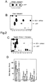
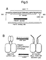
- Wild type p53 did not exhibit sequence-specific DNA binding unless activated by the monoclonal antibody, PAb421 ( Figure 2A, lane 2 vs.lane I). PAb421 binds to the C-terminus of p53 and presumably induces a conformational change that neutralizes the function of a negative regulatory domain ( Figure 5A). Highly purified mutant forms of p53 encoded by the His273 and Lys285 alleles were also unable to bind DNA sequence-specifically, but were effectively activated for DNA binding by PAb421 ( Figure 2A, lanes 4 and 6 vs. lanes 3 and 5). The specific activities for the mutant proteins were 6-8 fold lower than wild types p53 (See Figure 2D and the following Table). Specific activity of activated wild-type and mutant p53s (Fmol DNA bound/ ⁇ g protein) p53 PAb421 DnaK CasKII wild type 166 57 64 His273 55 31 3.8 Lys285 47 14 2.6
- Hsc70 binds to the C-terminus of p53 synthesized in reticulocyte lysates (36), suggesting that this family of proteins targets the same domain as PAb421 and CasKII ( Figure 5A).
- This interaction may have functional significance, since purified recombinant DnaK is able to activate sequence specific DNA binding of wild type P53.
- the His 273 and Lys285 mutant p53s were also activated by DnaK to give rise to products similar in mobility to activated wild type p53 ( Figure 2B, lanes 3, 7, and 11 vs. lanes 1, 5 and 9).
- a monoclonal antibody specific for the N-terminus of p53, DO-1 was able to supershift the DnaK activated wild type and mutant p53s (lanes 4, 8 and 12 vs. lanes 3, 7 and 11).
- DnaK activated mutant p53 bound by DO-1 are 4-6 fold less active than wild type p53 activated by this heat shock protein (Figure 2D).
- the enhancement of DnaK activated mutant p53 DNA binding function was observed upon the inclusion of DO-1 ( Figure 2B).
- the mutant forms of p53, though in an activated state, may yet favour equilibrium towards the latent state ( Figure 5), and the inclusion of DO-1 may help to lock the protein into the high affinity DNA binding conformation.
- Covalent modification of wild type p53 by CasKII in vitro is GTP or ATP dependent and activates the cryptic sequence-specific DNA binding function of P53 ( Figure 1).
- wild type p53 was activated very effectively by CasKII, both latent, mutant forms of p53 ( Figure 2C, lanes 3, 7 and 11 vs.
- the constitutively active p53 ⁇ 30 requires a reactive sulfhydryl for DNA binding
- Diamide which unlike NEM, reversibly oxidizes sulfhydryl residues, also inactivated p53 ⁇ 30 activity (lane 6 vs. lanes 2-4).
- the addition of DTT to diamide-oxidized p53 ⁇ 30 reactivated the function of the protein (lane 7).
- Wild type full length p53 also responded similarly to Diamide.
- the inclusion of increasing amounts of Diamide inactivated the DNA binding activity of PAb421-activated full length p53 ( Figure 4B, lanes 1-4).
- the subsequent treatment of the reactions with an excess of DTT reversed the oxidation promoted by Diamide (lanes 5-8).
- the C-terminal 30 amino acid region of p53 is denoted by amino acid residues 363-393.
- peptide C379-393 at concentrations from 500 ⁇ M to 5 ⁇ M blocked DnaK activation.
- the peptide C369-383 inhibited PAb421 activation by p53, while it was ineffective at inhibiting DnaK or CasKII activation of p53 binding.
- the DnaK targets the C-terminal 15 amino acids of p53, flanking the PAb421 epitope and CasKII phosphorylation sites.
- Peptide C374-388 could block DnaK activation as well as peptide C379-393, suggesting that the C-terminal 5 amino acids, containing the CasKII phosphorylation site, are not recognised by DnaK.
- the peptide C369-383 containing the PAb421 epitope removed important determinants of DnaK binding.
- mutant p53 which is defective in a GTP or ATP dependent post-translational activation of its sequence-specific DNA binding function by casein kinase II.
- these mutants can be converted to activated states by a distinct set of proteins, including PAb421 and E.coli Hsp70 (DnaK). Two important criteria appear necessary for this conversion.
- modifications presumably will be relevant in the cell and include: (i) a specific, high affinity binding of an 'activating' polypeptide (or mimetic) involved in neutralization of the C-terminal negative regulatory domain; and (ii) a highly reduced environment which can maintain p53 in an activated state.
- mutant p53 Activation of mutant p53 from its cryptic state by a monoclonal antibody and a heat shock protein has tremendous therapeutic implications. Given the clear association between the DNA binding activity and tumour suppressor functions of p53, these results imply that in many tumour cells there are high levels of mutant p53 that can potentially be activated to restore significant wild type function.
- Therapeutic applications of the present invention include the administration of an activating ligand for p53, or administration of a constitutively active p53, such as p53 having the C-terminal 30 amino acids deleted.
- a constitutively active p53 such as p53 having the C-terminal 30 amino acids deleted.
- the latter is based on our disclosure in Hupp et al (17) on wild-type p53, but a similar effect may be obtained with mutant p53.
- the C-30 deleted form may be even more active in DNA binding than the PAb421 activated full length molecule.
- these agents could be produced in the target cells by expression from an encoding gene introduced into the cells, eg in a viral vector (a variant of the VDEPT technique - see below).
- the vector could be targeted to the specific cells to be treated, or it could contain regulatory elements which are switched on more or less selectively in the target cells.
- the agent (constitutively active p53 or p53-activating ligand) could be administered in a precursor form, for conversion to the active form by an activating agent produced in, or targeted to, the cells to be treated.
- an activating agent produced in, or targeted to, the cells to be treated.
- This type of approach is sometimes known as ADEPT or VDEPT; the former involving targeting the activating agent to the cells by conjugation to a cell-specific antibody or ligand, while the latter involves producing the activating agent, eg an enzyme, in the target cells by expression from encoding DNA in a viral vector (see for example, EP-A-415731 and WO 90/07936).
- administration may be systemic or targeted, the latter employing direct application of the agent to the target cells or the use of targeting systems such as antibody or cell-specific ligands. Targeting may be desirable for a variety of reasons: for example if the agent is unacceptably toxic, or if it would otherwise require too high a dosage, or if it would not otherwise be able to enter the target cells.
- the normal cellular environment is sufficiently reducing for P53 to be active - see (ii) above - but there may be an enhanced effect from the administration of a reducing agent, as we have found that in tissue culture wild-type p53 can be activated by the extracellular addition of a reducing agent (dithiothreitol) to the culture medium.
- a reducing agent dithiothreitol
Landscapes
- Chemical & Material Sciences (AREA)
- Health & Medical Sciences (AREA)
- Organic Chemistry (AREA)
- Life Sciences & Earth Sciences (AREA)
- Genetics & Genomics (AREA)
- Medicinal Chemistry (AREA)
- General Health & Medical Sciences (AREA)
- Biochemistry (AREA)
- Molecular Biology (AREA)
- Biophysics (AREA)
- Proteomics, Peptides & Aminoacids (AREA)
- Zoology (AREA)
- Gastroenterology & Hepatology (AREA)
- Wood Science & Technology (AREA)
- Engineering & Computer Science (AREA)
- Bioinformatics & Cheminformatics (AREA)
- Microbiology (AREA)
- Biotechnology (AREA)
- Biomedical Technology (AREA)
- Immunology (AREA)
- Oncology (AREA)
- General Engineering & Computer Science (AREA)
- Toxicology (AREA)
- Nuclear Medicine, Radiotherapy & Molecular Imaging (AREA)
- General Chemical & Material Sciences (AREA)
- Chemical Kinetics & Catalysis (AREA)
- Pharmacology & Pharmacy (AREA)
- Animal Behavior & Ethology (AREA)
- Public Health (AREA)
- Veterinary Medicine (AREA)
- Peptides Or Proteins (AREA)
- Medicines Containing Antibodies Or Antigens For Use As Internal Diagnostic Agents (AREA)
- Preparation Of Compounds By Using Micro-Organisms (AREA)
- Medicines That Contain Protein Lipid Enzymes And Other Medicines (AREA)
- Micromachines (AREA)
- Eye Examination Apparatus (AREA)
Abstract
Description
- This invention relates to the p53 tumour suppressor protein, and more particularly to mutant forms of the p53 protein which seem to be associated with certain types of tumour, and are usually found at elevated levels in such tumour cells. The invention also discloses the conversion of wild type p53 to a constitutively active form.
- Mutation of p53 is a very common genetic alteration in human cancers (1). Loss of the p53 tumour suppressor activity is co-incident with a loss of G1-S checkpoints following DNA damage (2), increases in genomic instability and selectable gene amplication (3,4). Mutant forms of p53 also appear to acquire a dominant growth promoting function (5,6). The region responsible for this transforming activity has been localized to a small C-terminal domain (7). Mice null for the wild type p53 locus develop normally, yet are susceptible to the development of neoplasia at elevated frequencies (8), indicating that wild type p53 is dispensable for the control over normal development and cell differentiation, but is essential to prevent spontaneous tumour formation. Consistent with this data, inherited germline point mutations in p53 lead to a predisposition to cancer in humans (9).
- Wild type p53 protein levels rise dramatically in response to the DNA-damaging agents mitomycin C (56) UV light (10,11) and γ irradiation (2). Biochemical characterization of wild type protein has shown that p53 can function as a sequence-specific DNA binding protein (12,13) and a transcription factor (14-16). These results support a model for wild type p53 in which its function is activated post-translationally after DNA damage to allow DNA repair by controlling the expression of regulatory gene products (18). These may include the DNA damage inducible gene gadd45 (19) and the host protein with oncogenic properties mdm-2 (20).
- Biochemical characterization of p53 has become possible recently owing to the use of protein expression systems which allow for an abundant source of the protein. p53 purified using immunoaffinity chromatography has been shown to be a sequence-specific DNA binding protein which recognizes a motif containing two contiguous monomers of the sequence (Pu)3-C(A/T)(A/T)G(Py), (21). Sequence-specific DNA binding activity is manifested in the ability of p53 to bind the SV40 origin of replication (13), and by its ability to activate transcription in vitro from templates harbouring its DNA binding sequence (22). Mutant forms of p53 are defective in non-specific DNA binding (23), sequence-specific DNA binding (21) and transcriptional activation (14) suggesting that this activity is normally required to suppress tumour formation.
- Phosphorylation of nuclear DNA binding proteins is an effective mechanism through which gene expression is controlled in response to environmental cues (24). Multi-site phosphorylation of p53 by protein kinases (25-27) suggests its tumour suppressor activity may be tightly coordinated by complex signal transducing pathways.
- Casein kinase II (CasKII) is a highly conserved calcium and nucleotide independent enzyme which phosphorylates broad spectrum of substrates, including transcription factors and DNA binding proteins (28). The activity of the kinase is stimulated in cells exposed to a variety of mutagens and growth factors (29). Mouse (25) and human p53 (17) are phosphorylated at the penultimate C-terminal amino acid by Cas KII in vitro. Mutation of this highly conserved C-terminal serine residue of mouse p53 to an alanine abolishes the growth suppressive function of the protein in mammalian cells, suggesting that phosphorylation at this site is one important modification required to activate the tumour suppressor function of p53 (30). Based on biochemical and physiological data, CasKII is the only known enzyme involved in a direct and positive regulation of the activity of p53.
- Understanding the regulation of p53 activity is a vital step for the development of therapeutic strategies designed to restore tumour suppressor activity of the protein in transformed cells. Using in vitro systems, it has already been established that wild type p53 activity is negatively regulated in vitro by the viral oncogene, T-antigen (22), and host associated oncogene mdm-2 (31).
- It appears that sequence specific DNA binding is one activity of p53 required for its tumour suppressor function. To date, all mutant forms of p53, eg those encoded by the hotspot alleles, His175, Trp248, and His273, have been shown to be defective in sequence specific DNA binding (21) and in the activation of transcription from templates harbouring its consensus DNA binding site (14).
- Enzymatic modulation of p53 may provide a framework from which to couple signal transducing pathways with the P53 response to DNA damage and ultimately growth control. To study the effects of p53 phosphorylation on the activity of the protein, we have purified unmodified recombinant p53 by conventional chromatography from bacteria and shown it to be a multimeric protein which is latent for sequence specific DNA binding (17). A motif within the C-
terminal 30 amino acids negatively controls p53 function as deletion of this domain constitutively activates p53. A set of p53 activating proteins, including rabbit muscle casein kinase II, monoclonal antibody PAb421, and E.coli Hsp70, target this C-terminal domain. - We have therefore shown that while sequence specific DNA binding activity is cryptic, it can be unmasked by enzymes and proteins, including casein kinase II (17). From the observation that p53 is regulated positively by CasKII, we attempted to determine if some mutant forms of p53 are inactive in DNA binding as a result of a defect in conversion from a latent to an activated state.
- Mutant forms of p53 encoded by common 'hotspot' alleles were purified and their activities were characterized at the biochemical level. We show here that two mutant forms of p53 encoded by the His273 and Lys285 alleles are not inherently defective in sequence-specific DNA binding, but appear to prefer residence in the latent state. A monoclonal antibody and E.coli DnaK are able to promote sequence-specific DNA binding by these mutant forms of p53, but the most potent physiological activator CasKII is unable to effectively unmask the DNA binding function.
- A new biochemical class of mutant forms of p53 is thus established which is defective in DNA binding after post-translational modification by the growth controlling enzyme CasKII. This class of mutant p53, defective in the conversion from the latent to the activated state, was our target for the construction of molecules with the ability to reactivate the tumour suppressor function of mutant p53, and thus potentially lead to selective growth arrest and induction of apoptosis in tumour cells (32,33).
- According to one aspect of the invention, there is provided an in vitro method which comprises activating a mutant p53 protein for specific DNA binding by the action of a ligand which binds to the mutant p53 within the C-
terminal 30 amino acids , wherein said mutant has the properties that: - (i) it occurs at elevated levels in tumours;
- (ii) it does not substantially suppress tumour growth; and,
- (iii) it is not substantially activated by CasKII mediated phosphorylation.
-
- The mutant p53 will be likely to involve amino acid substitution of the wild-type p53 outside the C-
terminal 30 amino acids. Examples are mutants in which the substitution is at either or both of positions 273 and 285, typically producing his273 and lys285. - The activation typically is mediated by the action of a ligand which binds to the mutant p53 within the C-
terminal 30 amino acids. Suitable such ligands are the monoclonal antibody PAb421 or the bacterial heat shock protein DnaK, but it is contemplated that activation will also be mediated by a ligand which binds effectively to the same site on the mutant p53. Such equivalent ligand may comprise a binding peptide fragment of PAb421 or DnaK, or it may be a molecular "mimetic" thereof. - The designing of mimetics to a known pharmaceutically active compound is a known approach to the development of pharmaceuticals based on a "lead" compound. Typically, the three-dimensional structure of the ligand and its binding to the receptor is modelled, generally using a computer, and alternative molecules are devised for the ligand which have the same or equivalent binding regions, but which may differ markedly from the original ligand in other respects. For example, the original ligand may be a protein or peptide, but the mimetic may be a non-peptide molecule having a quite different skeleton but which has atoms or groups at the receptor binding locations which have the same sort of characteristics, such as size, electric charge, hydrophobicity etc, as the corresponding parts of the original ligand. In this way, a mimetic can be devised which fulfills the same binding and pharmacologic function as the original ligand, but is for example easier to manufacture and formulate as a pharmaceutical agent.
- According to another aspect of the invention, there is provided a pharmaceutical composition for activating mutant p53 protein for specific DNA binding, wherein said mutant p53 has the properties that:
- (i) it occurs at elevated levels in tumours;
- (ii) it does not substantially suppress tumour growth; and,
- (iii) it is not substantially activated by CasKII
mediated phosphorylation,
the composition comprising a ligand capable of binding to the mutant p53 within the C-terminal 30 amino acids, and a suitable excipient. -
- The ligand used in the composition suitably has the characteristics described above.
- A further aspect of the present invention lies in the use of constitutively activated p53, whether wild type or mutant, for preparing a medicament for therapeutic purposes, as in the tumour or cell growth suppression.
- Accordingly, the present invention provides an activated form of p53 for therapeutic use, in which the activation is obtained by the binding of a ligand within the C-
terminal 30 amino acids, or of the p53 molecule deletion within the C-terminal 30 amino acids of the p53 molecule, wherein the activation is effected prior to administration of the p53 activating agent. - Constitutive activation can be by the binding of a ligand such as PAb421 or DnaK, or the deletion of a C-terminal region, typically within the 30 C-terminal amino acids of the full length molecule; the precise limits of what can be deleted to obtain the effect can of course be determined by routine experimentation.
- In a further aspect, the present invention provides the use of an activated form of p53 in the preparation of a medicament for treating conditions ameliorated by activated p53, wherein the activation is obtained by the binding of a ligand to the C-
terminal 30 amino acids of the p53 molecule region, or deletion within the C-terminal 30 amino acids of the p53 molecule. - In a further aspect, the present invention provides the use of a ligand in the preparation of a medicament for activating a mutant p53 protein for specific DNA binding by the action of the ligand binding to the mutant p53, wherein said mutant has the properties that:
- (i) it occurs at elevated levels in tumours;
- (ii) it does not substantially suppress tumour growth; and,
- (iii) it is not substantially activated by CasKII
mediated phosphorylation,
wherein the ligand binds to the mutant p53 within the C-terminal 30 amino acids. -
- PAb421 is a known monoclonal antibody (Harlow et al, J Virol (1981), vol 37, 564-573) and is available from Oncogene Sciences, NY, USA. It has been found to bind the amino acid motif SKKGQSTSRHK in the C-terminal region of p53. Contemplated within the terms of this invention are other antibodies, binding fragments thereof, or non-antibody ligands, natural or synthetic, that are able to compete with PAb421 for binding to that motif and activating mutant p53s. Another monoclonal antibody that recognises the dame site and can activate mutant p53 is PAb122 (Gurney et al J Virol (1980), vol 34, 752-763), and is available through the ATCC under deposit No ATCC TIB 116.
- DnaK is a known protein (Huang et al, J Biol Chem (1991), vol 266, 7537-7541) and is available from StressGen Biotechnologies Corp, Victoria, BC, Canada. It is a bacterial heat shock protein found in E.coli, and has been found to bind to the amino acid motif RHKKLMFTKTE in the C-terminal region of p53. Contemplated within the terms of this invention are other ligands, natural or synthetic, able to compete with DnaK for binding to that motif and activating mutant p53s.
- DO-1 is a known monoclonal antibody (Vojtisek et al, J Immunol Meth (1992), vol 151, 237-244).
-
- Figure 1: Rate of phosphorylation of p53 by native and
recombinant casein kinase II. p53 (50 ng) was
phosphorylated at 30°C in DNA binding buffer (in the
absence of an ATP regeneration system and DNA) for the
indicated times using recombinant human casein kinase II
(HuCKII; 125ng) or native rabbit muscle casein kinase II
(RaCKII; MonoQ fraction; 375 ng). Reaction products were
separated by SDS gel electrophoresis. Radiolabeled p53 was
excised from the gel and radioactive phosphate was
quantified by scintillation counting. Activity is
expressed as the moles of phosphate incorporated per mole
of p53 monomer. (B). Rate of activation of p53 DNA
binding by rabbit muscle or recombinant casein kinase II.
p53 (50 ng) was phosphorylated at 30°C for the indicated
times (in the absence of an ATP regeneration system and
DNA) using rabbit muscle (RaCKII; MonoQ fraction, 375 ng)
or recombinant casein kinase II (HuCKII, 125ng).
Radiolabeled DNA was added and reactions were incubated at
0°C for 5 minutes, followed by gel electrophoresis.
Radiolabelled p53-DNA complexes were scanned and quantified
using a phosphoimager. Activity is expressed as the Fmoles
of DNA bound by 50 ng of p53 as a function of the time of
phosphorylation for 40 minutes with different amounts of
rabbit muscle or recombinant casein kinase II as in parts
A and B. Reaction products were separated by
electrophoresis on a 4% polyacrylamide gel; Lane 1(p53 +
1.2ng of HuCKII), lane 2 (p53 + 12.5 ng of HuCKII), lane 3
(p53 + 125 ng of HuCKII), lanes 4-6 (as in lanes 1-3, but
with the addition of DO-1 after DNA binding), lane 7 (p53
+ 37 ng of RaCKII), lane 8 (p53 + 375 ng of RaCKII),
lanes 9 and 10 (as inlanes - Figure 2: Activation of sequence-specific DNA binding of
wild type and mutant p53. Activation reactions were staged
in two parts. In the first stage, wild type p53 (60 ng) or
mutant forms of p53 (180 ng) was incubated with ATP and the
indicated activating protein at 30°C for 30 minutes. In
the second stage, the consensus site oligonucleotide was
added to the reactions to assay for sequence-specific DNA
binding. (A) PAb421;
lane lanes lanes lanes lanes lanes lanes lanes lanes lanes - Figure 3: (A) HEM sensitivity of PAb421 activated p53.
After PAb421 activation of wild type p53 (60 ng; lanes 2-4),
p53 His273 (60 ng; lanes 5-7), and p53 Lys 285 (60 ng;
lanes 8-10) as described in the methods, redox reagents
were added (DTT to 2mM or NEM to 1mM) and incubations were
continued at 30°C for 10 minutes. Lane 1 (no protein),
lanes lanes lanes Lanes 1 and 4 (p53s only),lanes 2 and 5 (DnaK activated p53 with DTT, followed by NEM),lanes 3 and 6 (DnaK activated p53 with NEM followed by DTT). Incubations were then continued with the consensus DNA oligonucleotide to assay for DNA binding as described in the methods p53* marks the position of the activated p53-DNA complex. - Figure 4: p53Δ30 DNA sequence-specific DNA binding activity is sensitive to sulfhydryl modifying reagents. (A) Sensitivity of p53Δ30 to oxidation. p53Δ30 (60 ng) was first treated with redox reagents: lane 1 (full length p53 activated by PAb421), lane 2 (p53Δ30 only), lane 3 (p53Δ30 and 2 mM DTT), lane 4 (p53Δ30 and 2 mM DTT followed by 1 mM NEM), lane 5 (p53Δ30 and 1mM NEM followed by 2 mM DTT), lane 6 (p53Δ30 and 0.5 mM Diamide), and lane 7 (p53Δ30 and 0.5 mM Diamide followed by 2 mM DTT). (B) PAb421-activated full length p53 is sensitive to reversible oxidation by diamide. p53 (60 ng) was first activated by PAb421 as indicated in the methods and then treated with the indicated redox reagents: lane 1-4 (0.1 mM, 0.4 mM, 1.6mM, and 5.4 mM Diamide, respectively) and lanes 5-8 (as in '1-4' but followed by the addition of DTT to 6 mM). After the modifications, p53 was assayed for sequence specific DNA binding.
- Figure 5: (A) C-terminal binding sites of the p53 binding proteins within exon 11(45). The 30 C-terminal amino acids from 363 to 393 are listed (46). Important motifs include: The 01 domain required for higher order oligomerization (38); the CKII phosphorylation site (open headed arrow) (47); the PAb421 binding site (48), the Hsc70 binding site (36), and the endpoint of p53Δ30 (closed arrow) (17). (B) Model describing the equilibrium shifts between dimeric p53 molecules in the latent or the activated states. The solid arrow indicates the direction favoured in the equilibrium for wild type or mutant p53s. Wild type p53 can be effectively converted to the active form by casein kinase II, PAb421, or DnaK. Conformational changes which occur after modification by the activating protein relieve the inhibition of DNA binding dependent upon C-terminal amino acids flanking the PAb421 binding site (stippled rectangle or ellipse). This conformational change activates the sequence specific DNA binding function. In contrast to the wild type protein, p53 mutants may be trapped in the latent state, even in the presence of activating proteins. Conditions that favour the interaction of activating proteins with p53 and that induce conformational changes in the C-terminus may activate the tumour suppressor function of mutant forms of p53.
-
- We have already demonstrated (Reference 17: Hupp et al, Cell, Vol 71, 875-886, November 27, 1992) that the DNA binding activity of p53, required for its tumour suppressor function, is cryptic but can be activated by cellular factors acting on a C-terminal regulatory domain of p53. A gel mobility shift assay demonstrated that recombinant wild-type human p53 binds DNA sequence specifically only weakly, but monoclonal antibody PAb421 binding near the C-terminus activated the cryptic DNA binding activity stoichiometrically. p53 DNA binding could also be activated by a 30 amino acid C-terminal deletion of p53 (deletion of 90 amino acids inactivated it), mild proteolysis of full length p53 (with trypsin), E coli DnaK (which disrupts protein-protein complexes) or CasKII (and coincident phosphorylation of a C-terminal site on p53). All the foregoing is described in more detail in the Hupp et al paper, to which reference may be made as appropriate.
- Using an in vitro system coupling post-translational modification of wild type p53 to the activation of sequence specific DNA binding, we have examined the effect of mutation on the latent DNA binding function of p53. Purification and biochemical characterization of mutant forms of p53 from bacterial expression systems demonstrated that a set of mutants share fundamental properties with the wild type protein.
- Like wild type p53, the purified mutants encoded by the His 273 and Lys285 alleles (i) appear to exist as latent multi-protein complexes and can be converted to active DNA binding forms by PAb421 and DnaK; (ii) express the N-terminal epitopes recognized by monoclonal antibody DO-1 in the native p53-DNA complex; (iii) require reactive sulfhydryl to promote sequence-specific DNA binding; and (iv) are effectively phosphorylated by CasKII in vitro.
- There is however, one noticeable difference between these two mutant proteins and wild type p53. It appears that the mutants are severely defective in the GTP or ATP dependent activation of sequence-specific DNA binding by the cellular enzyme CasKII. Given that wild type p53 is regulated positively by phosphorylation, implicating CasKII involvement in the p53 pathway (52), it is reasonable to expect that defects in this modulation would result in the net loss of p53 tumour suppressor activity. This defect would be made manifest in perturbation of the CasKII signal transducing pathway, in which hypophosphorylated p53 would be inactive as a tumour suppressor. In the selection for mutation during the process of cellular transformation, there could be a selection for: (i) a p53 mutation in the CasKII recognition site at the C-terminus, which would prevent post-translational modification by the kinase, (ii) a dominant p53 mutation in
exon 11 which would strengthen the activity of the negative regulatory domain, effectively locking p53 into the latent state, or (iii) a p53 mutation outside the C-terminal domain which confers an immunity to activation after phosphorylation. The two mutants exemplified herein fall into the latter class. Mutations residing in the C-terminus may exist, but since this region is not highly conserved, little emphasis has been placed on screening p53 alleles for mutation in this negative regulatory domain. - Purified fractions of recombinant human wild type p53 and p53Δ30 from a bacterial expression system, p53 specific monoclonal antibodies PAb421 and DO-1 were obtained as described previously (17). Mutant forms of p53 encoded by the Lys285 and His273 alleles were purified from a bacterial expression system using Heparin Sepharose and Gel filtration chromatography as described for wild type p53. Fractions containing mutant p53s eluting at approximately 440 Kd on gel filtration (compared to protein standards) and which could be activated for sequence-specific DNA binding by PAb421 were concentrated using a Centricon-30 (Amersham) to 0.5 mg/ml and stored frozen at -70°C. Mutant forms of p53 purified by this method were greater than 90% pure when stained with Coomasie Blue in an SDS polyacrylamide gel.
- The E.coli Hsp70 homologue (DnaK) was purified from an overproducing strain by a modification of the published protocol (49). Active fractions of DnaK eluting from MonoQ (49) were applied to an ATP-Agarose column in Buffer P (10% glycerol, 25 mM HEPES (pH 7.6), 0.1 mM EDTA, and 1 mM DTT) containing 10 mM MgCl2. After a 10 column volume wash in Buffer P containing 1.0 M KC1 + 10 mM MgCl2 and a 5 column volume wash with Buffer P containing 10 mM KC1 + 10 mM MgCl2, bound DnaK was eluted with Buffer P containing 10 mM MgCl2 + 5mM ATP. The fractions of DnaK eluting from ATP agarose were dialyzed overnight at 3°C against Buffer P containing 2 mM EDTA and 0.25 M KC1. DnaK was further purified on Superose-12 gel filtration equilibrated in Buffer P containing 0.25 M KC1. NEM and Diamide were from Sigma.
- CasKII from rabbit muscle was purified as described (17) with the following modifications. Muscle from a rabbit was ground in 2 liters of homogenization buffer (4 mM EDTA, 2 mM DTT, 1 mM Benzamidine) and centrifuged at 4000xg for 45 minutes. The soluble supernatant containing 27.5 grams of protein was batch adsorbed to 200 ml of phosphocellulose resin in Buffer P for 2 hours. The column was washed with Buffer P containing 0.35 M KC1, and CasKII activity was step eluted with Buffer P containing 1.2 M KC1 and 0.1% Triton X-100. After Phosphocellulose, fractions containing CasKII were applied to a Heparin Sepharose column and eluted with a linear gradient from 0.05 to 1 M KC1 in Buffer P containing 0.1% Triton X-100. Active fraction were applied to a MonoQ column and eluted with a linear KC1 gradient in Buffer P containing 0.1% Triton X-100. Active CasKII was concentrated to 0.75 mg/ml using Centricon-30 and stored at 4°C. Kinase activity was monitored by assaying for radioactive phosphate incorporation into p53 and casein as described (17). Casein and p53 kinase activities ere co-incident after Heparin Sepharose and MonoQ chromatography. Recombinant human CasKII holoenzyme expressed in E.coli was 95% pure and was obtained from Boehringer Mannheim.
- Oligonucleotides containing the 20-mer p53 consensus DNA binding site (PG) (22) and Hind III compatible ends were: 5' AGC TTA GAC ATG CCT AGA CAT GCC TA 3' and 5' AGC TTA GGC ATG TCT AGG CAT GTC TA 3'. Oligonucleotides without the consensus sequence (TL) were: 5' TAT GTC TAA GGG ACC TGC GGT TGG CAT TGA TCT TG 3' and 5' GTG CCA AGA TCA ATG CCA ACC GCA GGT CCC TTA GAC A 3'. Complementary oligonucleotides were hybridized and end-labelled with 32P-γ -ATP as described (Maniatis et al, 1982) and stored at 4°C.
- A 20µl reaction volume contained DNA binding buffer (20% glycerol, 25mM HEPES (pH 7.6), 50mM KCl, 1mM DTT, 1mg/ml BSA and 0.1% Triton X-100) and was mixed with PG radiolabelled DNA, unless indicated otherwise (1-5ng), competitor DNA (20-100ng of pBluescript II), the indicated amounts of p53 (Fraction III) and the indicated p53-specific monoclonal antibodies. Incubations followed a 0°C for 30 minutes unless indicated otherwise. Reaction products were loaded onto a 4% polyacrylamide gel containing 0.33 X TBE and 0.1% Triton X-100, which had undergone pre-electrophoresis at 100V for 15 minutes at 3°C. Electrophoresis was continued at 200V from 30 to 90 minutes at 3°C. Gels were dried prior to exposure to X-ray film.
- Reactions (10µl contained DNA buffer (without DNA) supplemented with 100µM ATP containing 32P-γ-ATP, and 125ng of p53 (Fraction III). CasKII obtained after gel filtration (80ng) was added and after incubations at 30°C for the indicated times, reactions were quenched by the addition of SDS-PAG sample buffer. Products were separated by electrophoresis on a 10% SDS-polyacrylamide gel. p53 was localised by staining with Coomassie Blue and the bands were excised and counted using a liquid scintillation counter (Beckmann LS 1801).
- Reactions (10µl) contained DNA binding buffer (without DNA) supplemented with 30ng of p53 (Fraction III), 1mM ATP, and with an ATP regenerating system containing 0.1mg/ml creatine kinase II and 20mM phosphocreatine. After the addition of the indicated amounts of CasKII or purified DnaK, reactions were incubated at 30°C for 30 minutes. A 2-fold concentrated mixture containing radiolabelled DNA, competitor DNA, and the indicated monoclonal antibodies (10µl) was added and incubations were continued at 21°C for 10 minutes. Reaction products were analysed as described above.
- Activation of the sequence-specific DNA binding function of wild type and mutant forms of p53 were performed using the assay conditions previously described with the following modifications. In a 10µl reaction containing 20% glycerol, 25 mM HEPES (pH 7.6), 0.05% Triton X-100, 5 mM Mg Cl2, 50 mM KC1, 0.1mM EDTA, 1mg/ml BSA, 0.1 mg/ml creatine kinase, 20 mM phosphocreatine, 1 mM ATP, the indicated amounts of wild type or mutant p53 and the indicated activating protein, reactions were incubated at 30°C for 30 minutes. A 10µl aliquot containing the radiolabeled consensus DNA binding oligonucleotide (5' AG-CTT AGACATGCCT AGACATGCCT A 3' and 5' AGCTTAGGCATGTCT AGGCATGTCT A 3') defined by El-Deiry (21) with competitor plasmid DNA was added and reactions were incubated at 30°C for 5 minutes. Unless indicated otherwise, PAb421 (400 ng), DnaK (2µg), CasKII (240 ng), or DO-1 (400 ng) were added. Reaction products were separated by electropohoresis in a native 4% polyacrylamide gel containing 0.1% Triton X-100 and 0.33 X TBE at 3°C.
- These results, and the details of the experiments, are provided in our paper Hupp et al (17).
- Our recent identification of the latent biochemical phenotype of wild type p53 (17) supports the possibility that some mutant forms of p53 may be inactive due to a relative defect in activation of DNA binding by a post-translational modification (Figure 5). Identification of such a class of mutant p53 would provide suggestive evidence for the importance of a CasKII signalling pathway in the cell and define a class of p53 which could presumably have the capacity to have its tumour suppressor function re-activated by alternative post-translational modifications.
- To test this possibility, we have examined the behaviour of mutant forms of p53 in sequence-specific DNA binding assays. Mutant forms of p53 encoded by the His175, Trp248, His273, and Lys285 alleles were purified using Heparin Sepharose and gel filtration chromatography as described for wild type p53. The mutant proteins eluted as a multi-protein complex of approximately 440Kd on gel filtration, similar to that observed with wild type p53 (17). These results indicate that the mutations have not prevented stable associations between monomers.
- Wild type p53 did not exhibit sequence-specific DNA binding unless activated by the monoclonal antibody, PAb421 (Figure 2A,
lane 2 vs.lane I). PAb421 binds to the C-terminus of p53 and presumably induces a conformational change that neutralizes the function of a negative regulatory domain (Figure 5A). Highly purified mutant forms of p53 encoded by the His273 and Lys285 alleles were also unable to bind DNA sequence-specifically, but were effectively activated for DNA binding by PAb421 (Figure 2A,lanes lanes 3 and 5). The specific activities for the mutant proteins were 6-8 fold lower than wild types p53 (See Figure 2D and the following Table).Specific activity of activated wild-type and mutant p53s (Fmol DNA bound/µg protein) p53 PAb421 DnaK CasKII wild type 166 57 64 His273 55 31 3.8 Lys285 47 14 2.6 - Apparently, these point mutations in p53 do not abolish sequence-specific DNA binding as the cryptic function can be activated (Figure 5B). The other two common mutants studied, His175 and Trp248 could not be activated in DNA binding by PAb421 under these conditions, suggesting that the cryptic activity may be permanently locked into the latent state (Figure 5B).
- p53 can bind to DnaK and Hsc70 in vivo (35). Hsc70 binds to the C-terminus of p53 synthesized in reticulocyte lysates (36), suggesting that this family of proteins targets the same domain as PAb421 and CasKII (Figure 5A). We have found that this interaction may have functional significance, since purified recombinant DnaK is able to activate sequence specific DNA binding of wild type P53.
- The His 273 and Lys285 mutant p53s were also activated by DnaK to give rise to products similar in mobility to activated wild type p53 (Figure 2B,
lanes lanes lanes lanes - The third protein which we have been using to activate wild type p53, and one which is presently the most physiologically relevant, is casein kinase II. The C-terminal CasKII phosphorylation site is required for tumour suppressor function is mammalian cells (30). Covalent modification of wild type p53 by CasKII in vitro is GTP or ATP dependent and activates the cryptic sequence-specific DNA binding function of P53 (Figure 1). Although wild type p53 was activated very effectively by CasKII, both latent, mutant forms of p53 (Figure 2C,
lanes lanes lanes - Independent biochemical analysis of wild type p53 has shown that it requires reactive sulfhydryl for effective sequence specific DNA binding, and it was of interest to determine if the altered conformation of activated mutant forms of p53 could express this biochemical phenotype. In stage activation of wild type and mutant forms of p53 by PAb421, the inclusion of NEM after the activation step inhibited sequence specific DNA binding (Figure 3A,
lanes lanes lanes lanes - A similar analysis was carried out using DnaK activated p53 His273 and wild type p53 (Figure 3B). After the activation by DnaK (
lanes lanes 1 and 4), NEM inhibited sequence specific DNA binding of both mutant and wild type p53s (lanes 3 and 6), while the control reactions containing an excess of DTT were not affected by NEM (lanes 2 and 5). These results suggest that post-translational modulation of the reactive sulfhydryl group through a redox mechanism will greatly affect p53 function and imply that mutant p53 proteins are not defective in DNA binding owing to a propensity of this sulfhydryl group to exist in an oxidized state. - Even the possibility that a reactive sulfhydryl is required at some undetected state during the activation of wild type or mutant forms of p53, we studied the effects of sulfhydryl modifying agents on p53Δ30 activity. This recombinant enzyme is constitutively active for sequence-specific DNA binding as it lacks the C-
terminal 30 amino acids containing the negative regulatory motif which, in an unmodified state, prevents activity. The inclusion of NEM inhibited DNA binding of this protein as it does wild type full length activated p53 (Figure 4A,lane 5 vs. lanes 2-4). Diamide, which unlike NEM, reversibly oxidizes sulfhydryl residues, also inactivated p53Δ30 activity (lane 6 vs. lanes 2-4). The addition of DTT to diamide-oxidized p53Δ30 reactivated the function of the protein (lane 7). - Wild type full length p53 also responded similarly to Diamide. The inclusion of increasing amounts of Diamide inactivated the DNA binding activity of PAb421-activated full length p53 (Figure 4B, lanes 1-4). The subsequent treatment of the reactions with an excess of DTT reversed the oxidation promoted by Diamide (lanes 5-8).
- The C-
terminal 30 amino acid region of p53 is denoted by amino acid residues 363-393. We screened a series of synthetic peptides derived from the C-terminus of p53 for the ability to compete with DnaK activation of the protein. Wild type p53 was first activated for DNA binding by PAb421, DnaK and CasKII in the absence of peptides. Peptide C379-393 was unable to compete effectively with activation by PAb421 and CasKII. The PAb421 epitope is lacking in this peptide, so it was expected that it would not interfere with activation of p53. In contrast, peptide C379-393 at concentrations from 500µM to 5µM blocked DnaK activation. The peptide C369-383 inhibited PAb421 activation by p53, while it was ineffective at inhibiting DnaK or CasKII activation of p53 binding. This suggests that the DnaK targets the C-terminal 15 amino acids of p53, flanking the PAb421 epitope and CasKII phosphorylation sites. Peptide C374-388 could block DnaK activation as well as peptide C379-393, suggesting that the C-terminal 5 amino acids, containing the CasKII phosphorylation site, are not recognised by DnaK. The peptide C369-383 containing the PAb421 epitope removed important determinants of DnaK binding. - From our studies, we have identified a class of mutant p53 which is defective in a GTP or ATP dependent post-translational activation of its sequence-specific DNA binding function by casein kinase II. However, these mutants can be converted to activated states by a distinct set of proteins, including PAb421 and E.coli Hsp70 (DnaK). Two important criteria appear necessary for this conversion. These modifications presumably will be relevant in the cell and include: (i) a specific, high affinity binding of an 'activating' polypeptide (or mimetic) involved in neutralization of the C-terminal negative regulatory domain; and (ii) a highly reduced environment which can maintain p53 in an activated state. Activation of mutant p53 from its cryptic state by a monoclonal antibody and a heat shock protein has tremendous therapeutic implications. Given the clear association between the DNA binding activity and tumour suppressor functions of p53, these results imply that in many tumour cells there are high levels of mutant p53 that can potentially be activated to restore significant wild type function.
- Therapeutic applications of the present invention include the administration of an activating ligand for p53, or administration of a constitutively active p53, such as p53 having the C-
terminal 30 amino acids deleted. The latter is based on our disclosure in Hupp et al (17) on wild-type p53, but a similar effect may be obtained with mutant p53. There is also evidence that the C-30 deleted form may be even more active in DNA binding than the PAb421 activated full length molecule. - Instead of administering these agents directly, they could be produced in the target cells by expression from an encoding gene introduced into the cells, eg in a viral vector (a variant of the VDEPT technique - see below). The vector could be targeted to the specific cells to be treated, or it could contain regulatory elements which are switched on more or less selectively in the target cells.
- Alternatively, the agent (constitutively active p53 or p53-activating ligand) could be administered in a precursor form, for conversion to the active form by an activating agent produced in, or targeted to, the cells to be treated. This type of approach is sometimes known as ADEPT or VDEPT; the former involving targeting the activating agent to the cells by conjugation to a cell-specific antibody or ligand, while the latter involves producing the activating agent, eg an enzyme, in the target cells by expression from encoding DNA in a viral vector (see for example, EP-A-415731 and WO 90/07936).
- Various methods of administration of the therapeutic agent can be used, following known formulations and procedures, Dosages can be determined by routine experimentation. As noted above, administration may be systemic or targeted, the latter employing direct application of the agent to the target cells or the use of targeting systems such as antibody or cell-specific ligands. Targeting may be desirable for a variety of reasons: for example if the agent is unacceptably toxic, or if it would otherwise require too high a dosage, or if it would not otherwise be able to enter the target cells.
- The normal cellular environment is sufficiently reducing for P53 to be active - see (ii) above - but there may be an enhanced effect from the administration of a reducing agent, as we have found that in tissue culture wild-type p53 can be activated by the extracellular addition of a reducing agent (dithiothreitol) to the culture medium.
- The following documents are hereby incorporated by reference into the present disclosure.
- 1. Vogelstein, B. and Kinzler, K. W. (1992) Cell 70, 523-526.
- 2. Kastan, M. B.. Onyekwere, O., Sidransky, D., Vogelstein, B. and Craig, R. W. (1991) Cancer Res. 51, 6304-6311.
- 3. Livingstone, L. R., White, A., Sprouse, J., Livanos, E., Jacks, T. and Tlsty, T. D. (1992) Cell 70, 923-935.
- 4. Yin, Y., Tainsky, M. A., Bischoff, F. Z., Strong, L. C. and Wahl, G. M. (1992) Cell 70, 937-948.
- 5. Lane, D. P. and Benchimol, S. (1990) Genes Dev. 4, 1-8.
- 6. Levine, A. J., Momand, J. and Finlay, C. A. (1991) Nature 351, 453-456.
- 7. Shaulian, E., Zauberman. A., Ginsberg, D. and Oren, M. (1992) Mol. Cell. Biol. 12, 5581-5592.
- 8. Donehower, L. A., Harvey, M., Slagle, B. L., McArthur, M. J., Montgomery Jr, C. A., Butel, J. S. and Bradley, A. (1992) Nature 356, 215-221.
- 9. Malkin, D., Li, F. P., Strong, L. C., Fraumeni, J.F., Nelson, C. M., Kim, D. H., Kassel, J., Gryka, M. A., Bischoff, F. Z., Tainsky, M. A. and Friend, S. H. (1990) Science 250, 1233-1238.
- 10. Maltzman, W. and Czyzyk, L. (1984) Mol. Cell. Biol. 4, 1689-1694.
- 11. Hall, P.A., McKee, P. H., Menage, H. D., Dover, R. and Lane, D. P.
(1992)
Oncogene 8, 203-207. - 12. Kern, S., Kinzler, K., Bruskin, A., Jarosz, D., Friedman, P., Prives, C., and Vogelstein, B. (1991) Science 252, 1708-1711.
- 13. Bargonetti, J. , Friedman, P. N. , Kern, S. E. , Vogelstein. B. and Prives, C. (1991) Cell 65, 1083-1091.
- 14. Kern, S. E., Pietenpol, J. A., Thiagalingam, S., Seymour, A., Kinzler, K. W. and Vogelstein, B. (1992) Science 256, 827-830.
- 15. Scharer, E. and Iggo, R. (1992) Nucl. Acids Res. 20, 1539-1545.
- 16. Funk, W. D., Pak, D. T., Karas, R. H., Wright, W. E. and Shay, J. W. (1992) Mol. Cell Biol. 12, 2866-2871.
- 17. Hupp, T. R., Meek, D. M., Midgley, C. A. and Lane, D. P. (1992) Cell 71, 875-886.
- 18. Lane, D. P. (1992) Nature 358, 15-16.
- 19. Kastan, M. B. , Zhan, Q. , El-deiry, W. S., Carrier, F., Jacks, T., Walsh, W., Plunkett, B. S., Vogelstein. B. and Fornace Jr, A. J. (1992) Cell 71, 587-597.
- 20. Barak, Y., Juven, T., Haffner, R. and Oren, M. (1993) EMBO J. 12, 461-468.
- 21. El-Deiry, W. S., Kern, S. E., Pietenpol, J. A., Kinzler, K. W. and Vogelstein,
B. (1992)
Nature Genetics 1, 45-49. - 22. Farmer, G., Bargonetti. J., Zhu. H., Friedman P.. Prywes, R. and Prives, C. (1992) Nature 358, 83-86.
- 23. Kern, S. E., Kinzler, K. W., Baker, S. J., Nigro, J. M., Rotter, V., Levine,
A. J., Friedman, P., Prives, C. and Vogelstein. B. (1991)
Oncogene 6, 131-136. - 24. Hunter, T. and Karin, M. (1992) Cell 70, 375-387.
- 25. Meek, D. W., Simon, S., Kikkawa. U. and Eckhart, W. (1990) EMBO J. 9, 3253-3260.
- 26. Milne, D. M., Palmer, R. H., Campbell D. G. and Meek, D. W. (1992)
Oncogene 7, 1361-1370. - 27. Lees-Miller, S. P., Chen, Y., and Anderson, C.W. (1990) Mol. Cell. Biol. 10, 6472-6481.
- 28. Meisner, M. and Czech, M. P. (1991) Curr. Opin. Cell Biol. 3, 474-483.
- 29. Carroll, D., Santoro, N. and Marshak, D. R. (1988) Cold Spring Harbor Symp. Quant. Biol. 53, 91-95.
- 30. Milne, D. M., Palmer, R. H. and Meek, D. W. (1992) Nucl. Acids Res. 20, 5565-5570.
- 31. Oliner, J. D., Kinzler, K. W., Meltzer, P. S., George, D. L. and Vogelstein, B. (1992) Nature 358, 80-83.
- 32. Shaw, P., Bovey, R., Tardy, S., Sahli, R., Sordat, B., Costa, J. (1992) Proc. Natl. Acad. Sci USA 89: 4495-4499.
- 33. Yonish-Rouach, E., Grunwald, D., Wilder, S., Kimchi, A., May, E.. Lawrence, J.J., May, P., and Oren, M. (1993) Mol. Cell. Biol. 13, 1415-1423.
- 34. Pinna, L. A. (1990) Biochem. Biophys. Acta 1054, 267-284.
- 35. Clarke, C. F., Cheng, K., Frey, A. B., Stein, R., Hinds, P. W. and Levine, A. J. (1988) Mol. Cell. Biol. 8, 1206-1215.
- 36. Hainaut, P. and Milner, J. (1992) EMBO J. 11, 3513-3520.
- 37. Kraiss, S., Quaiser, A., Oren, M. and Montenarh, M. (1988) J. Virol. 62, 4737-4744.
- 38. Sturzbecher, H.-W., Brain, R., Maimets, T., Addison, C., Rudge, K. and
Jenkins, J. R. (1992)
Oncogene 7, 1515-1523. - 39. Milner, J. and Medcalf, E. A. (1991) Cell 65, 765-774.
- 40. Seto, E., Usheva, A., Zambetti, G. P., Momand, J., Horikoshi, N., Weinmann. R., Levine, A. J. and Shenk, T. (1992) Proc. Natl. Acad. Sci. USA 89, 12028-12032.
- 41. Xanthoudaxis, S., Miao, G., Wang, F., E.Pan, Y. C. and Curran, T. (1992) EMBO J. 11, 3323-3335.
- 42. McBride, A. A., Klausner, R. D. and Howley, P. M. (1992. Proc. Natl. Acad. Sci. USA 89, 7531-7535.
- 43. Abate, C., Patel, L., Rauscher, F.J. III, and Curran, T. (1990) Science 249, 1157-1161.
- 44. Xanthoudakis, S. and Curran, T. (1992) EMBO J. 11, 653-665.
- 45. Lamb, P. and Crawford, L. (1986) Mol. Cell. Biol. 6, 1379-1385.
- 46. Soussi, T., Caron de Fromentel, C., and May, P. (1990)
Oncogene 5, 945-952. - 47. Bischoff, J. R., Casso, D. and Beach, D. (1992) Mol. Cell. Biol. 12, 1405-1411.
- 48. Wade-Evans, A. and Jenkins, J. R. (1985) EMBO J. 4, 699-706.
- 49. Hwang, D.S. and Kaguni, J.M. (1991) J. Biol. Chem. 265, 19244-19248.
- 50. Mulner-Lorillon, O., Cormier, P., Labbe, J., Doree, M., Poulhe, R., Osborne, H., and Belle, R.(1990) Eur. J. Biochem. 93, 529-534.
- 51. Kandror, K., Benumov, A., and Stepanov, A. (1989) Eur. J. Biochem. 180, 441-448.
- 52. Lane, D.P. (1993) Nature 362, 386-387.
- 53. Baudier, J, Delphin, C., Grunwald, D., Khochbin, S., and Lawrence, J.J. (1992) Proc. Natl. Acad. Sci. U.S.A. 89, 11627-11631.
- 54. Friedman, P.N., Chen, X., Bargonetti, J., and Prives, C. (1993) Proc. Natl. Acad. Sci. U.S.A. 90, 3319-3323.
- 55. Borellini, F., and Glazer, R.I. (1993) J. Biol. Chem. 268, 7923-7928.
- 56. Lu, X., Park, S.H., Thompson, T.C., and Lane, D.P. (1992) Cell 70, 153-161.
-
Claims (17)
- An in vitro method which comprises activating a mutant p53 protein for specific DNA binding by the action of a ligand which binds to the mutant p53 within the C-terminal 30 amino acids, , wherein said mutant has the properties that:(i) it occurs at elevated levels in tumours;(ii) it does not substantially suppress tumour growth; and,(iii) it is not substantially activated by CasKII mediated phosphorylation.
- The method of claim 1, wherein the ligand is monoclonal antibody PAb421 or the bacterial heat shock protein DnaK, or a ligand which binds effectively to the same site on the mutant p53.
- The method of claim 1 or claim 2, wherein the mutant p53 involves amino acid substitution of the wild-type p53 outside the C-terminal 30 amino acids.
- The method of claim 3, wherein the substitution is at either or both of positions 273 and 285.
- The method of claim 4, wherein the substitution gives either or both of his273 and lys285.
- A pharmaceutical composition for activating mutant p53 protein for specific DNA binding, wherein said mutant p53 has the properties that:(i) it occurs at elevated levels in tumours;(ii) it does not substantially suppress tumour growth; and,(iii) it is not substantially activated by CasKII mediated phosphorylation,
the composition comprising a ligand capable of binding to the mutant p53 within the C-terminal 30 amino acids, and a suitable excipient. - The composition of claim 6, wherein the ligand is monoclonal antibody PAb421 or the bacterial heat shock protein DnaK, or a ligand which binds effectively to the same site on the mutant p53, or a binding fragment or a molecular mimetic thereof.
- The composition of claim 6 or claim 7, wherein the mutant p53 involves amino acid substitution of the wild-type p53 outside the C-terminal 30 amino acids.
- The composition of claim 8, wherein the substitution is at either or both of positions 273 and 285.
- The composition of claim 9, wherein the substitution gives either or both his273 and lys285.
- An activated form of p53 for therapeutic use, in which the activation is obtained by the binding of a ligand within the C-terminal 30 amino acids of the p53 or a deletion within the C-terminal 30 amino acids of the p53, wherein the activation is effected prior to administration of the p53.
- Use of an activated form of p53 in the preparation of a medicament for treating conditions ameliorated by activated p53, wherein the activation is obtained by the binding of a ligand to the C-terminal 30 amino acids of the p53, or a deletion within the C-terminal 30 amino acids of the p53.
- Use of a ligand in the preparation of a medicament for activating a mutant p53 protein for specific DNA binding by the action of the ligand binding to the mutant p53, wherein said mutant has the properties that:(i) it occurs at elevated levels in tumours;(ii) it does not substantially suppress tumour growth; and,(iii) it is not substantially activated by CasKII mediated phosphorylation,
wherein the ligand binds to the mutant p53 within the C-terminal 30 amino acids. - The use of claim 13, wherein the ligand is monoclonal antibody PAb421 or the bacterial heat shock protein DnaK, or a ligand which binds effectively to the same site on the mutant p53, or a binding fragment or molecular mimetic thereof.
- The use of any one of claims 12 to 14, wherein the mutant p53 involves amino acid substitution of the wild-type p53 outside the C-terminal 30 amino acids.
- The use of claim 15, wherein the substitution is at either or both of positions 273 and 285.
- The use of claim 16, where in the substitution gives either or both of his273 and lys285.
Applications Claiming Priority (3)
| Application Number | Priority Date | Filing Date | Title |
|---|---|---|---|
| GB929224784A GB9224784D0 (en) | 1992-11-26 | 1992-11-26 | Cellular protein |
| GB9224784 | 1992-11-26 | ||
| PCT/GB1993/002438 WO1994012202A1 (en) | 1992-11-26 | 1993-11-26 | ACTIVATION OF p53 PROTEIN |
Publications (2)
| Publication Number | Publication Date |
|---|---|
| EP0675729A1 EP0675729A1 (en) | 1995-10-11 |
| EP0675729B1 true EP0675729B1 (en) | 1999-06-02 |
Family
ID=10725717
Family Applications (1)
| Application Number | Title | Priority Date | Filing Date |
|---|---|---|---|
| EP94900267A Expired - Lifetime EP0675729B1 (en) | 1992-11-26 | 1993-11-26 | ACTIVATION OF p53 PROTEIN |
Country Status (13)
| Country | Link |
|---|---|
| US (1) | US6140058A (en) |
| EP (1) | EP0675729B1 (en) |
| JP (1) | JPH08505607A (en) |
| KR (1) | KR100291964B1 (en) |
| AT (1) | ATE180673T1 (en) |
| AU (1) | AU680216B2 (en) |
| CA (1) | CA2150265C (en) |
| DE (1) | DE69325180T2 (en) |
| DK (1) | DK0675729T3 (en) |
| ES (1) | ES2134336T3 (en) |
| GB (1) | GB9224784D0 (en) |
| GR (1) | GR3030900T3 (en) |
| WO (1) | WO1994012202A1 (en) |
Families Citing this family (24)
| Publication number | Priority date | Publication date | Assignee | Title |
|---|---|---|---|---|
| US5721340A (en) * | 1994-11-28 | 1998-02-24 | The Wistar Institute Of Anatomy & Biology | p53 proteins with altered tetramerization domains |
| US5573925A (en) * | 1994-11-28 | 1996-11-12 | The Wistar Institute Of Anatomy And Biology | P53 proteins with altered tetramerization domains |
| US6169073B1 (en) * | 1995-02-16 | 2001-01-02 | Bayer Corporation | Peptides and peptidomimetics with structural similarity to human p53 that activate p53 function |
| FR2732348B1 (en) * | 1995-03-31 | 1997-04-30 | Rhone Poulenc Rorer Sa | CONDITIONAL EXPRESSION SYSTEM |
| FR2736915B1 (en) | 1995-07-19 | 1997-08-22 | Rhone Poulenc Rorer Sa | P53 PROTEIN VARIANTS AND THERAPEUTIC USES |
| US5847083A (en) * | 1996-08-21 | 1998-12-08 | The Wistar Institute Of Anatomy And Biology | Modified p53 constructs which enhance DNA binding |
| GB9521544D0 (en) * | 1995-10-20 | 1995-12-20 | Univ Dundee | Activation of P53 protein and therapeutic applications thereof |
| FR2755144B1 (en) * | 1996-10-29 | 1998-11-27 | Rhone Poulenc Rorer Sa | SINGLE CHAIN ANTI-P53 ANTIBODY FRAGMENTS AND USE THEREOF |
| GB9714971D0 (en) * | 1997-07-16 | 1997-09-24 | Cancer Res Campaign Tech | Assays,therapeutic methods and means |
| DE19735221C1 (en) * | 1997-08-14 | 1999-05-06 | Wolfgang Willi Prof Dr Deppert | Influencing the binding of p53 to target genes |
| DE19802792A1 (en) * | 1998-01-26 | 1999-07-29 | Wolfgang Willi Prof Dr Deppert | Inhibiting DNA modulation caused by p53 mutant, useful for treatment of cancer |
| US6429298B1 (en) | 1998-10-13 | 2002-08-06 | Board Of Regents, The University Of Texas System | Assays for identifying functional alterations in the p53 tumor suppressor |
| EP1484060A3 (en) * | 1998-12-02 | 2005-02-09 | Pfizer Products Inc. | Methods and compositions for restoring conformational stability of a protein of the p53 family |
| EP1854509A3 (en) * | 1998-12-02 | 2010-08-18 | Pfizer Products Inc. | Methods and compositions for restoring conformational stability of a protein of the p53 family |
| HRP20010414A2 (en) * | 1998-12-02 | 2002-06-30 | Pfizer Prod Inc | METHODS AND COMPOSITIONS FOR RESTORING CONFORMATIONAL STABILITY OF A PROTEIN OF THE p53 FAMILY |
| AUPP932199A0 (en) * | 1999-03-19 | 1999-04-15 | St Vincent's Hospital Sydney Limited | Anti-p53 antibodies |
| US6989239B2 (en) * | 2001-06-20 | 2006-01-24 | R.E.D. Laboratories, N.V./S.A. | Methods for diagnosis and treatment of chronic immune diseases |
| US8067008B2 (en) | 2004-08-23 | 2011-11-29 | Yeda Research And Development Co. | Peptide inhibitors for mediating stress responses |
| US20070224662A1 (en) * | 2004-12-17 | 2007-09-27 | Jun Luo | Post-translational modification of proteins in cell-free expression systems |
| KR100749858B1 (en) | 2005-08-12 | 2007-08-16 | 연세대학교 산학협력단 | p53 variant and its use |
| US20140088017A1 (en) | 2011-05-23 | 2014-03-27 | Yeda Research And Development Co., Ltd. | Use of akt phosphorylation as a biomarker for prognosing neurodegenerative diseases and treating same |
| ES2708556T3 (en) | 2014-09-30 | 2019-04-10 | Diadem S R L | Antibody that binds to a linear epitope of human p53 and its applications for diagnosis |
| GB201617564D0 (en) | 2016-10-17 | 2016-11-30 | Agency For Science Technology And Research And National University Of Singapore And Singapore Health | Anti-p53 antibodies |
| WO2019162974A1 (en) * | 2018-02-23 | 2019-08-29 | Ulisse Biomed S.R.L. | Method to diagnose cancer progression based on prokaryotic biomarkers and molecules for use in the treatment of cancer progression |
Family Cites Families (2)
| Publication number | Priority date | Publication date | Assignee | Title |
|---|---|---|---|---|
| NO312681B1 (en) * | 1990-08-24 | 2002-06-17 | Univ California | Process for the preparation of a pharmaceutical composition with suppressive action / activity |
| CA2118015A1 (en) * | 1992-04-14 | 1993-10-28 | Jeffrey R. Marks | Method of detecting tumors containing complexes of p53 and hsp70 |
-
1992
- 1992-11-26 GB GB929224784A patent/GB9224784D0/en active Pending
-
1993
- 1993-11-26 ES ES94900267T patent/ES2134336T3/en not_active Expired - Lifetime
- 1993-11-26 WO PCT/GB1993/002438 patent/WO1994012202A1/en not_active Ceased
- 1993-11-26 AT AT94900267T patent/ATE180673T1/en not_active IP Right Cessation
- 1993-11-26 DE DE69325180T patent/DE69325180T2/en not_active Expired - Fee Related
- 1993-11-26 KR KR1019950702130A patent/KR100291964B1/en not_active Expired - Fee Related
- 1993-11-26 US US08/446,668 patent/US6140058A/en not_active Expired - Fee Related
- 1993-11-26 AU AU55331/94A patent/AU680216B2/en not_active Ceased
- 1993-11-26 DK DK94900267T patent/DK0675729T3/en active
- 1993-11-26 EP EP94900267A patent/EP0675729B1/en not_active Expired - Lifetime
- 1993-11-26 CA CA002150265A patent/CA2150265C/en not_active Expired - Fee Related
- 1993-11-26 JP JP6512924A patent/JPH08505607A/en active Pending
-
1999
- 1999-08-02 GR GR990401981T patent/GR3030900T3/en unknown
Also Published As
| Publication number | Publication date |
|---|---|
| EP0675729A1 (en) | 1995-10-11 |
| DE69325180T2 (en) | 1999-12-02 |
| DE69325180D1 (en) | 1999-07-08 |
| GR3030900T3 (en) | 1999-11-30 |
| KR100291964B1 (en) | 2001-10-23 |
| DK0675729T3 (en) | 1999-12-06 |
| CA2150265C (en) | 1998-12-29 |
| US6140058A (en) | 2000-10-31 |
| JPH08505607A (en) | 1996-06-18 |
| GB9224784D0 (en) | 1993-01-13 |
| WO1994012202A1 (en) | 1994-06-09 |
| ES2134336T3 (en) | 1999-10-01 |
| AU5533194A (en) | 1994-06-22 |
| CA2150265A1 (en) | 1994-06-09 |
| AU680216B2 (en) | 1997-07-24 |
| ATE180673T1 (en) | 1999-06-15 |
Similar Documents
| Publication | Publication Date | Title |
|---|---|---|
| AU680216B2 (en) | Activation of p53 protein | |
| Hupp et al. | Activation of the cryptic DNA binding function of mutant forms of p53 | |
| Schneider et al. | Regulation of CAK kinase activity by p53 | |
| Lane | p53 and human cancers | |
| Xiong et al. | p21 is a universal inhibitor of cyclin kinases | |
| Lu et al. | The CDK7-cycH-p36 complex of transcription factor IIH phosphorylates p53, enhancing its sequence-specific DNA binding activity in vitro | |
| Leveillard et al. | Functional interactions between p53 and the TFIIH complex are affected by tumour‐associated mutations. | |
| Bischoff et al. | Human p53 inhibits growth in Schizosaccharomyces pombe | |
| Meek | Multisite phosphorylation and the integration of stress signals at p53 | |
| US5688665A (en) | Isolated nucleic acid molecules encoding the p27 KIP-1 protein | |
| Shaulian et al. | Tight DNA binding and oligomerization are dispensable for the ability of p53 to transactivate target genes and suppress transformation. | |
| Murakami et al. | Two nonmuscle myosin II heavy chain isoforms expressed in rabbit brains: filament forming properties, the effects of phosphorylation by protein kinase C and casein kinase II, and location of the phosphorylation sites | |
| WO1997014794A1 (en) | ACTIVATION OF p53 PROTEIN | |
| Elia et al. | Ribosomal protein L22 inhibits regulation of cellular activities by the Epstein‐Barr virus small RNA EBER‐1 | |
| Götz et al. | Protein kinase CK2 interacts with a multi-protein binding domain of p53 | |
| RU2192431C2 (en) | Synthetic mutant gene rb (variants), synthetic mutant gene p53, plasmid (variants), mutant protein (variants), method of inhibition of cellular proliferation (variants), method of induction of apoptosis | |
| Guan et al. | Proline‐directed protein kinase (p34cdc2/p58cyclin A) phosphorylates bovine neurofilaments | |
| Wagner et al. | Fine mapping and regulation of the association of p53 with p34cdc2 | |
| CA2218327A1 (en) | Pcna binding substance | |
| US6635450B1 (en) | Isolated P27 protein, nucleic acid molecules encoding same, methods of identifying agents acting on same, and uses of said agents | |
| Park | Cloning, sequencing, and overexpression of SH2/SH3 adaptor protein Nck from mouse thymus | |
| US6242575B1 (en) | Antibodies for detecting p27 protein | |
| US6355774B1 (en) | Isolated p27 protein | |
| CA2120353C (en) | Cell cycle controlling compositions and methods of using same | |
| Zhang et al. | The DNA-binding and Transcription-Activation Abilities of p53 Are Necessary but not Sufficient for Its Antiproliferation Function’ |
Legal Events
| Date | Code | Title | Description |
|---|---|---|---|
| PUAI | Public reference made under article 153(3) epc to a published international application that has entered the european phase |
Free format text: ORIGINAL CODE: 0009012 |
|
| 17P | Request for examination filed |
Effective date: 19950616 |
|
| AK | Designated contracting states |
Kind code of ref document: A1 Designated state(s): AT BE CH DE DK ES FR GB GR IE IT LI LU MC NL PT SE |
|
| 17Q | First examination report despatched |
Effective date: 19970527 |
|
| GRAG | Despatch of communication of intention to grant |
Free format text: ORIGINAL CODE: EPIDOS AGRA |
|
| GRAG | Despatch of communication of intention to grant |
Free format text: ORIGINAL CODE: EPIDOS AGRA |
|
| GRAH | Despatch of communication of intention to grant a patent |
Free format text: ORIGINAL CODE: EPIDOS IGRA |
|
| RAP1 | Party data changed (applicant data changed or rights of an application transferred) |
Owner name: UNIVERSITY OF DUNDEE |
|
| GRAH | Despatch of communication of intention to grant a patent |
Free format text: ORIGINAL CODE: EPIDOS IGRA |
|
| GRAA | (expected) grant |
Free format text: ORIGINAL CODE: 0009210 |
|
| AK | Designated contracting states |
Kind code of ref document: B1 Designated state(s): AT BE CH DE DK ES FR GB GR IE IT LI LU MC NL PT SE |
|
| REF | Corresponds to: |
Ref document number: 180673 Country of ref document: AT Date of ref document: 19990615 Kind code of ref document: T |
|
| REG | Reference to a national code |
Ref country code: CH Ref legal event code: EP |
|
| REF | Corresponds to: |
Ref document number: 69325180 Country of ref document: DE Date of ref document: 19990708 |
|
| REG | Reference to a national code |
Ref country code: IE Ref legal event code: FG4D |
|
| ITF | It: translation for a ep patent filed | ||
| ET | Fr: translation filed | ||
| REG | Reference to a national code |
Ref country code: ES Ref legal event code: FG2A Ref document number: 2134336 Country of ref document: ES Kind code of ref document: T3 |
|
| REG | Reference to a national code |
Ref country code: PT Ref legal event code: SC4A Free format text: AVAILABILITY OF NATIONAL TRANSLATION Effective date: 19990901 |
|
| REG | Reference to a national code |
Ref country code: DK Ref legal event code: T3 |
|
| PLBE | No opposition filed within time limit |
Free format text: ORIGINAL CODE: 0009261 |
|
| STAA | Information on the status of an ep patent application or granted ep patent |
Free format text: STATUS: NO OPPOSITION FILED WITHIN TIME LIMIT |
|
| 26N | No opposition filed | ||
| REG | Reference to a national code |
Ref country code: GB Ref legal event code: IF02 |
|
| PGFP | Annual fee paid to national office [announced via postgrant information from national office to epo] |
Ref country code: PT Payment date: 20020930 Year of fee payment: 10 |
|
| PGFP | Annual fee paid to national office [announced via postgrant information from national office to epo] |
Ref country code: GR Payment date: 20021003 Year of fee payment: 10 |
|
| PGFP | Annual fee paid to national office [announced via postgrant information from national office to epo] |
Ref country code: MC Payment date: 20021031 Year of fee payment: 10 |
|
| PGFP | Annual fee paid to national office [announced via postgrant information from national office to epo] |
Ref country code: LU Payment date: 20021121 Year of fee payment: 10 |
|
| PGFP | Annual fee paid to national office [announced via postgrant information from national office to epo] |
Ref country code: IE Payment date: 20021125 Year of fee payment: 10 |
|
| PGFP | Annual fee paid to national office [announced via postgrant information from national office to epo] |
Ref country code: SE Payment date: 20021126 Year of fee payment: 10 |
|
| PGFP | Annual fee paid to national office [announced via postgrant information from national office to epo] |
Ref country code: NL Payment date: 20021130 Year of fee payment: 10 |
|
| PG25 | Lapsed in a contracting state [announced via postgrant information from national office to epo] |
Ref country code: LU Free format text: LAPSE BECAUSE OF NON-PAYMENT OF DUE FEES Effective date: 20031126 Ref country code: IE Free format text: LAPSE BECAUSE OF NON-PAYMENT OF DUE FEES Effective date: 20031126 |
|
| PG25 | Lapsed in a contracting state [announced via postgrant information from national office to epo] |
Ref country code: SE Free format text: LAPSE BECAUSE OF NON-PAYMENT OF DUE FEES Effective date: 20031127 |
|
| PG25 | Lapsed in a contracting state [announced via postgrant information from national office to epo] |
Ref country code: MC Free format text: LAPSE BECAUSE OF NON-PAYMENT OF DUE FEES Effective date: 20031130 |
|
| PG25 | Lapsed in a contracting state [announced via postgrant information from national office to epo] |
Ref country code: PT Free format text: LAPSE BECAUSE OF NON-PAYMENT OF DUE FEES Effective date: 20040531 |
|
| PG25 | Lapsed in a contracting state [announced via postgrant information from national office to epo] |
Ref country code: NL Free format text: LAPSE BECAUSE OF NON-PAYMENT OF DUE FEES Effective date: 20040601 |
|
| PG25 | Lapsed in a contracting state [announced via postgrant information from national office to epo] |
Ref country code: GR Free format text: LAPSE BECAUSE OF NON-PAYMENT OF DUE FEES Effective date: 20040603 |
|
| EUG | Se: european patent has lapsed | ||
| NLV4 | Nl: lapsed or anulled due to non-payment of the annual fee |
Effective date: 20040601 |
|
| REG | Reference to a national code |
Ref country code: IE Ref legal event code: MM4A |
|
| REG | Reference to a national code |
Ref country code: PT Ref legal event code: MM4A Free format text: LAPSE DUE TO NON-PAYMENT OF FEES Effective date: 20040531 |
|
| PGFP | Annual fee paid to national office [announced via postgrant information from national office to epo] |
Ref country code: ES Payment date: 20061031 Year of fee payment: 14 |
|
| PGFP | Annual fee paid to national office [announced via postgrant information from national office to epo] |
Ref country code: GB Payment date: 20061120 Year of fee payment: 14 |
|
| PGFP | Annual fee paid to national office [announced via postgrant information from national office to epo] |
Ref country code: FR Payment date: 20061124 Year of fee payment: 14 Ref country code: DK Payment date: 20061124 Year of fee payment: 14 Ref country code: CH Payment date: 20061124 Year of fee payment: 14 Ref country code: AT Payment date: 20061124 Year of fee payment: 14 |
|
| PGFP | Annual fee paid to national office [announced via postgrant information from national office to epo] |
Ref country code: DE Payment date: 20061128 Year of fee payment: 14 |
|
| PGFP | Annual fee paid to national office [announced via postgrant information from national office to epo] |
Ref country code: BE Payment date: 20061129 Year of fee payment: 14 |
|
| PGFP | Annual fee paid to national office [announced via postgrant information from national office to epo] |
Ref country code: IT Payment date: 20061130 Year of fee payment: 14 |
|
| BERE | Be: lapsed |
Owner name: *UNIVERSITY OF DUNDEE Effective date: 20071130 |
|
| REG | Reference to a national code |
Ref country code: DK Ref legal event code: EBP |
|
| GBPC | Gb: european patent ceased through non-payment of renewal fee |
Effective date: 20071126 |
|
| PG25 | Lapsed in a contracting state [announced via postgrant information from national office to epo] |
Ref country code: LI Free format text: LAPSE BECAUSE OF NON-PAYMENT OF DUE FEES Effective date: 20071130 Ref country code: CH Free format text: LAPSE BECAUSE OF NON-PAYMENT OF DUE FEES Effective date: 20071130 |
|
| REG | Reference to a national code |
Ref country code: CH Ref legal event code: PL |
|
| PG25 | Lapsed in a contracting state [announced via postgrant information from national office to epo] |
Ref country code: AT Free format text: LAPSE BECAUSE OF NON-PAYMENT OF DUE FEES Effective date: 20071126 |
|
| PG25 | Lapsed in a contracting state [announced via postgrant information from national office to epo] |
Ref country code: BE Free format text: LAPSE BECAUSE OF NON-PAYMENT OF DUE FEES Effective date: 20071130 |
|
| PG25 | Lapsed in a contracting state [announced via postgrant information from national office to epo] |
Ref country code: DK Free format text: LAPSE BECAUSE OF NON-PAYMENT OF DUE FEES Effective date: 20071130 Ref country code: DE Free format text: LAPSE BECAUSE OF NON-PAYMENT OF DUE FEES Effective date: 20080603 |
|
| REG | Reference to a national code |
Ref country code: FR Ref legal event code: ST Effective date: 20080930 |
|
| PG25 | Lapsed in a contracting state [announced via postgrant information from national office to epo] |
Ref country code: GB Free format text: LAPSE BECAUSE OF NON-PAYMENT OF DUE FEES Effective date: 20071126 |
|
| REG | Reference to a national code |
Ref country code: ES Ref legal event code: FD2A Effective date: 20071127 |
|
| PG25 | Lapsed in a contracting state [announced via postgrant information from national office to epo] |
Ref country code: FR Free format text: LAPSE BECAUSE OF NON-PAYMENT OF DUE FEES Effective date: 20071130 Ref country code: ES Free format text: LAPSE BECAUSE OF NON-PAYMENT OF DUE FEES Effective date: 20071127 |
|
| PG25 | Lapsed in a contracting state [announced via postgrant information from national office to epo] |
Ref country code: IT Free format text: LAPSE BECAUSE OF NON-PAYMENT OF DUE FEES Effective date: 20071126 |