EP0668356A2 - Recombinant infectious bovine rhinotracheitis virus mutant, methods for the production of same, and vaccines containing the same - Google Patents
Recombinant infectious bovine rhinotracheitis virus mutant, methods for the production of same, and vaccines containing the same Download PDFInfo
- Publication number
- EP0668356A2 EP0668356A2 EP95870007A EP95870007A EP0668356A2 EP 0668356 A2 EP0668356 A2 EP 0668356A2 EP 95870007 A EP95870007 A EP 95870007A EP 95870007 A EP95870007 A EP 95870007A EP 0668356 A2 EP0668356 A2 EP 0668356A2
- Authority
- EP
- European Patent Office
- Prior art keywords
- gii
- bhv
- virus
- gene
- dna
- Prior art date
- Legal status (The legal status is an assumption and is not a legal conclusion. Google has not performed a legal analysis and makes no representation as to the accuracy of the status listed.)
- Withdrawn
Links
- 241000701083 Bovine alphaherpesvirus 1 Species 0.000 title claims abstract description 56
- 229960005486 vaccine Drugs 0.000 title claims abstract description 55
- 238000000034 method Methods 0.000 title claims abstract description 49
- 238000004519 manufacturing process Methods 0.000 title abstract description 7
- 108090000623 proteins and genes Proteins 0.000 claims abstract description 135
- 241000700605 Viruses Species 0.000 claims abstract description 128
- 108090000288 Glycoproteins Proteins 0.000 claims abstract description 94
- 102000003886 Glycoproteins Human genes 0.000 claims abstract description 74
- 238000012217 deletion Methods 0.000 claims abstract description 56
- 230000037430 deletion Effects 0.000 claims abstract description 56
- 108090000765 processed proteins & peptides Proteins 0.000 claims abstract description 43
- 229920001184 polypeptide Polymers 0.000 claims abstract description 41
- 102000004196 processed proteins & peptides Human genes 0.000 claims abstract description 41
- 238000003780 insertion Methods 0.000 claims abstract description 38
- 230000037431 insertion Effects 0.000 claims abstract description 38
- 230000000890 antigenic effect Effects 0.000 claims abstract description 26
- 238000006467 substitution reaction Methods 0.000 claims abstract description 11
- 208000005562 infectious bovine rhinotracheitis Diseases 0.000 claims abstract description 6
- 108020004414 DNA Proteins 0.000 claims description 107
- 239000012634 fragment Substances 0.000 claims description 86
- 239000013612 plasmid Substances 0.000 claims description 70
- 108091028043 Nucleic acid sequence Proteins 0.000 claims description 37
- 239000013553 cell monolayer Substances 0.000 claims description 20
- 239000013598 vector Substances 0.000 claims description 20
- 239000002773 nucleotide Substances 0.000 claims description 17
- 125000003729 nucleotide group Chemical group 0.000 claims description 17
- 208000015181 infectious disease Diseases 0.000 claims description 16
- 241000283690 Bos taurus Species 0.000 claims description 9
- 239000013599 cloning vector Substances 0.000 claims description 9
- 238000012216 screening Methods 0.000 claims description 9
- 239000000725 suspension Substances 0.000 claims description 9
- 241000894006 Bacteria Species 0.000 claims description 8
- 108091026890 Coding region Proteins 0.000 claims description 8
- 239000003085 diluting agent Substances 0.000 claims description 8
- 108020004705 Codon Proteins 0.000 claims description 6
- 230000000120 cytopathologic effect Effects 0.000 claims description 6
- 230000002458 infectious effect Effects 0.000 claims description 6
- 244000309466 calf Species 0.000 claims description 5
- 230000000241 respiratory effect Effects 0.000 claims description 5
- 108020005065 3' Flanking Region Proteins 0.000 claims description 4
- 108020005029 5' Flanking Region Proteins 0.000 claims description 4
- 239000002671 adjuvant Substances 0.000 claims description 3
- 230000028993 immune response Effects 0.000 claims description 3
- 230000002779 inactivation Effects 0.000 claims description 3
- WNROFYMDJYEPJX-UHFFFAOYSA-K aluminium hydroxide Chemical compound [OH-].[OH-].[OH-].[Al+3] WNROFYMDJYEPJX-UHFFFAOYSA-K 0.000 claims description 2
- 229910021502 aluminium hydroxide Inorganic materials 0.000 claims description 2
- 239000003795 chemical substances by application Substances 0.000 claims description 2
- 239000003937 drug carrier Substances 0.000 claims description 2
- WSFSSNUMVMOOMR-NJFSPNSNSA-N methanone Chemical compound O=[14CH2] WSFSSNUMVMOOMR-NJFSPNSNSA-N 0.000 claims description 2
- 244000005700 microbiome Species 0.000 claims description 2
- 230000000717 retained effect Effects 0.000 claims description 2
- 150000007949 saponins Chemical class 0.000 claims description 2
- 238000001228 spectrum Methods 0.000 claims description 2
- 229960004854 viral vaccine Drugs 0.000 claims description 2
- 239000000568 immunological adjuvant Substances 0.000 claims 7
- 210000002966 serum Anatomy 0.000 claims 6
- 230000035899 viability Effects 0.000 claims 6
- 229940031551 inactivated vaccine Drugs 0.000 claims 3
- 241000711895 Bovine orthopneumovirus Species 0.000 claims 2
- 241000711573 Coronaviridae Species 0.000 claims 2
- 241000702680 Equine rotavirus Species 0.000 claims 2
- 238000003306 harvesting Methods 0.000 claims 2
- 230000010076 replication Effects 0.000 claims 2
- 239000003053 toxin Substances 0.000 claims 2
- 231100000765 toxin Toxicity 0.000 claims 2
- 108700012359 toxins Proteins 0.000 claims 2
- 235000019687 Lamb Nutrition 0.000 claims 1
- 206010051497 Rhinotracheitis Diseases 0.000 claims 1
- 241000282898 Sus scrofa Species 0.000 claims 1
- 230000037358 bacterial metabolism Effects 0.000 claims 1
- 238000004945 emulsification Methods 0.000 claims 1
- 238000004108 freeze drying Methods 0.000 claims 1
- 230000000415 inactivating effect Effects 0.000 claims 1
- 230000004060 metabolic process Effects 0.000 claims 1
- 239000003381 stabilizer Substances 0.000 claims 1
- 241001465754 Metazoa Species 0.000 abstract description 32
- 238000002360 preparation method Methods 0.000 abstract description 6
- LFQSCWFLJHTTHZ-UHFFFAOYSA-N Ethanol Chemical compound CCO LFQSCWFLJHTTHZ-UHFFFAOYSA-N 0.000 description 49
- 210000004027 cell Anatomy 0.000 description 43
- 239000000872 buffer Substances 0.000 description 30
- 239000000243 solution Substances 0.000 description 30
- FAPWRFPIFSIZLT-UHFFFAOYSA-M Sodium chloride Chemical compound [Na+].[Cl-] FAPWRFPIFSIZLT-UHFFFAOYSA-M 0.000 description 24
- 235000018102 proteins Nutrition 0.000 description 24
- 102000004169 proteins and genes Human genes 0.000 description 24
- TWRXJAOTZQYOKJ-UHFFFAOYSA-L Magnesium chloride Chemical compound [Mg+2].[Cl-].[Cl-] TWRXJAOTZQYOKJ-UHFFFAOYSA-L 0.000 description 22
- 230000029087 digestion Effects 0.000 description 19
- 229920000936 Agarose Polymers 0.000 description 17
- 239000008188 pellet Substances 0.000 description 16
- 108091008146 restriction endonucleases Proteins 0.000 description 16
- 239000000523 sample Substances 0.000 description 16
- 230000003612 virological effect Effects 0.000 description 16
- 108010054576 Deoxyribonuclease EcoRI Proteins 0.000 description 15
- 238000004458 analytical method Methods 0.000 description 15
- ISWSIDIOOBJBQZ-UHFFFAOYSA-N Phenol Chemical compound OC1=CC=CC=C1 ISWSIDIOOBJBQZ-UHFFFAOYSA-N 0.000 description 14
- 241000701093 Suid alphaherpesvirus 1 Species 0.000 description 14
- 102100038126 Tenascin Human genes 0.000 description 14
- 108010008125 Tenascin Proteins 0.000 description 14
- 239000011780 sodium chloride Substances 0.000 description 14
- 239000011543 agarose gel Substances 0.000 description 13
- 230000002950 deficient Effects 0.000 description 13
- XLYOFNOQVPJJNP-UHFFFAOYSA-N water Chemical compound O XLYOFNOQVPJJNP-UHFFFAOYSA-N 0.000 description 13
- 241000700588 Human alphaherpesvirus 1 Species 0.000 description 11
- 229910001629 magnesium chloride Inorganic materials 0.000 description 11
- 241001529453 unidentified herpesvirus Species 0.000 description 11
- QKNYBSVHEMOAJP-UHFFFAOYSA-N 2-amino-2-(hydroxymethyl)propane-1,3-diol;hydron;chloride Chemical compound Cl.OCC(N)(CO)CO QKNYBSVHEMOAJP-UHFFFAOYSA-N 0.000 description 10
- KCXVZYZYPLLWCC-UHFFFAOYSA-N EDTA Chemical compound OC(=O)CN(CC(O)=O)CCN(CC(O)=O)CC(O)=O KCXVZYZYPLLWCC-UHFFFAOYSA-N 0.000 description 10
- VHJLVAABSRFDPM-QWWZWVQMSA-N dithiothreitol Chemical compound SC[C@@H](O)[C@H](O)CS VHJLVAABSRFDPM-QWWZWVQMSA-N 0.000 description 10
- 239000000203 mixture Substances 0.000 description 10
- SCVFZCLFOSHCOH-UHFFFAOYSA-M potassium acetate Chemical compound [K+].CC([O-])=O SCVFZCLFOSHCOH-UHFFFAOYSA-M 0.000 description 10
- 238000012163 sequencing technique Methods 0.000 description 10
- PEDCQBHIVMGVHV-UHFFFAOYSA-N Glycerine Chemical compound OCC(O)CO PEDCQBHIVMGVHV-UHFFFAOYSA-N 0.000 description 9
- 239000000020 Nitrocellulose Substances 0.000 description 9
- 108010076504 Protein Sorting Signals Proteins 0.000 description 9
- HEMHJVSKTPXQMS-UHFFFAOYSA-M Sodium hydroxide Chemical compound [OH-].[Na+] HEMHJVSKTPXQMS-UHFFFAOYSA-M 0.000 description 9
- 238000004113 cell culture Methods 0.000 description 9
- 239000000499 gel Substances 0.000 description 9
- 238000009396 hybridization Methods 0.000 description 9
- 229920001220 nitrocellulos Polymers 0.000 description 9
- HEDRZPFGACZZDS-UHFFFAOYSA-N Chloroform Chemical compound ClC(Cl)Cl HEDRZPFGACZZDS-UHFFFAOYSA-N 0.000 description 8
- 102000004190 Enzymes Human genes 0.000 description 8
- 108090000790 Enzymes Proteins 0.000 description 8
- 238000013461 design Methods 0.000 description 8
- ZMMJGEGLRURXTF-UHFFFAOYSA-N ethidium bromide Chemical compound [Br-].C12=CC(N)=CC=C2C2=CC=C(N)C=C2[N+](CC)=C1C1=CC=CC=C1 ZMMJGEGLRURXTF-UHFFFAOYSA-N 0.000 description 8
- 229960005542 ethidium bromide Drugs 0.000 description 8
- 101150072564 gE gene Proteins 0.000 description 8
- PHTQWCKDNZKARW-UHFFFAOYSA-N isoamylol Chemical compound CC(C)CCO PHTQWCKDNZKARW-UHFFFAOYSA-N 0.000 description 8
- 239000010410 layer Substances 0.000 description 8
- 239000006228 supernatant Substances 0.000 description 8
- 241000588724 Escherichia coli Species 0.000 description 7
- VMHLLURERBWHNL-UHFFFAOYSA-M Sodium acetate Chemical compound [Na+].CC([O-])=O VMHLLURERBWHNL-UHFFFAOYSA-M 0.000 description 7
- 238000005119 centrifugation Methods 0.000 description 7
- 238000006243 chemical reaction Methods 0.000 description 7
- 238000010367 cloning Methods 0.000 description 7
- 239000001632 sodium acetate Substances 0.000 description 7
- 235000017281 sodium acetate Nutrition 0.000 description 7
- 230000029812 viral genome replication Effects 0.000 description 7
- ZHNUHDYFZUAESO-UHFFFAOYSA-N Formamide Chemical compound NC=O ZHNUHDYFZUAESO-UHFFFAOYSA-N 0.000 description 6
- ZMXDDKWLCZADIW-UHFFFAOYSA-N N,N-Dimethylformamide Chemical compound CN(C)C=O ZMXDDKWLCZADIW-UHFFFAOYSA-N 0.000 description 6
- WCUXLLCKKVVCTQ-UHFFFAOYSA-M Potassium chloride Chemical compound [Cl-].[K+] WCUXLLCKKVVCTQ-UHFFFAOYSA-M 0.000 description 6
- 239000007984 Tris EDTA buffer Substances 0.000 description 6
- 230000015572 biosynthetic process Effects 0.000 description 6
- 230000001413 cellular effect Effects 0.000 description 6
- 238000001962 electrophoresis Methods 0.000 description 6
- 239000002609 medium Substances 0.000 description 6
- 230000035772 mutation Effects 0.000 description 6
- 239000000047 product Substances 0.000 description 6
- 238000000746 purification Methods 0.000 description 6
- 238000011282 treatment Methods 0.000 description 6
- JKMHFZQWWAIEOD-UHFFFAOYSA-N 2-[4-(2-hydroxyethyl)piperazin-1-yl]ethanesulfonic acid Chemical compound OCC[NH+]1CCN(CCS([O-])(=O)=O)CC1 JKMHFZQWWAIEOD-UHFFFAOYSA-N 0.000 description 5
- 239000007995 HEPES buffer Substances 0.000 description 5
- DBMJMQXJHONAFJ-UHFFFAOYSA-M Sodium laurylsulphate Chemical compound [Na+].CCCCCCCCCCCCOS([O-])(=O)=O DBMJMQXJHONAFJ-UHFFFAOYSA-M 0.000 description 5
- 239000008049 TAE buffer Substances 0.000 description 5
- HGEVZDLYZYVYHD-UHFFFAOYSA-N acetic acid;2-amino-2-(hydroxymethyl)propane-1,3-diol;2-[2-[bis(carboxymethyl)amino]ethyl-(carboxymethyl)amino]acetic acid Chemical compound CC(O)=O.OCC(N)(CO)CO.OC(=O)CN(CC(O)=O)CCN(CC(O)=O)CC(O)=O HGEVZDLYZYVYHD-UHFFFAOYSA-N 0.000 description 5
- 238000010276 construction Methods 0.000 description 5
- 235000011056 potassium acetate Nutrition 0.000 description 5
- 235000013930 proline Nutrition 0.000 description 5
- 235000019333 sodium laurylsulphate Nutrition 0.000 description 5
- PIEPQKCYPFFYMG-UHFFFAOYSA-N tris acetate Chemical compound CC(O)=O.OCC(N)(CO)CO PIEPQKCYPFFYMG-UHFFFAOYSA-N 0.000 description 5
- 238000011144 upstream manufacturing Methods 0.000 description 5
- UXVMQQNJUSDDNG-UHFFFAOYSA-L Calcium chloride Chemical compound [Cl-].[Cl-].[Ca+2] UXVMQQNJUSDDNG-UHFFFAOYSA-L 0.000 description 4
- DHMQDGOQFOQNFH-UHFFFAOYSA-N Glycine Chemical compound NCC(O)=O DHMQDGOQFOQNFH-UHFFFAOYSA-N 0.000 description 4
- 241000701085 Human alphaherpesvirus 3 Species 0.000 description 4
- 238000000246 agarose gel electrophoresis Methods 0.000 description 4
- 239000001110 calcium chloride Substances 0.000 description 4
- 235000011148 calcium chloride Nutrition 0.000 description 4
- 229910001628 calcium chloride Inorganic materials 0.000 description 4
- 238000010790 dilution Methods 0.000 description 4
- 239000012895 dilution Substances 0.000 description 4
- 201000010099 disease Diseases 0.000 description 4
- 208000037265 diseases, disorders, signs and symptoms Diseases 0.000 description 4
- 238000004520 electroporation Methods 0.000 description 4
- 238000000605 extraction Methods 0.000 description 4
- 230000002209 hydrophobic effect Effects 0.000 description 4
- 210000000987 immune system Anatomy 0.000 description 4
- 238000013507 mapping Methods 0.000 description 4
- 108020004707 nucleic acids Proteins 0.000 description 4
- 102000039446 nucleic acids Human genes 0.000 description 4
- 150000007523 nucleic acids Chemical group 0.000 description 4
- 239000002245 particle Substances 0.000 description 4
- 239000012071 phase Substances 0.000 description 4
- 239000002244 precipitate Substances 0.000 description 4
- NKDFYOWSKOHCCO-YPVLXUMRSA-N 20-hydroxyecdysone Chemical compound C1[C@@H](O)[C@@H](O)C[C@]2(C)[C@@H](CC[C@@]3([C@@H]([C@@](C)(O)[C@H](O)CCC(C)(O)C)CC[C@]33O)C)C3=CC(=O)[C@@H]21 NKDFYOWSKOHCCO-YPVLXUMRSA-N 0.000 description 3
- QTBSBXVTEAMEQO-UHFFFAOYSA-N Acetic acid Chemical compound CC(O)=O QTBSBXVTEAMEQO-UHFFFAOYSA-N 0.000 description 3
- 102000053602 DNA Human genes 0.000 description 3
- 239000003298 DNA probe Substances 0.000 description 3
- 102100038132 Endogenous retrovirus group K member 6 Pro protein Human genes 0.000 description 3
- 101000686824 Enterobacteria phage N4 Virion DNA-directed RNA polymerase Proteins 0.000 description 3
- 101000644628 Escherichia phage Mu Tail fiber assembly protein U Proteins 0.000 description 3
- WQZGKKKJIJFFOK-GASJEMHNSA-N Glucose Natural products OC[C@H]1OC(O)[C@H](O)[C@@H](O)[C@@H]1O WQZGKKKJIJFFOK-GASJEMHNSA-N 0.000 description 3
- 108010086093 Mung Bean Nuclease Proteins 0.000 description 3
- 102000001708 Protein Isoforms Human genes 0.000 description 3
- 108010029485 Protein Isoforms Proteins 0.000 description 3
- 108091081024 Start codon Proteins 0.000 description 3
- 108020005202 Viral DNA Proteins 0.000 description 3
- 239000008346 aqueous phase Substances 0.000 description 3
- 238000011161 development Methods 0.000 description 3
- 239000012153 distilled water Substances 0.000 description 3
- 230000004927 fusion Effects 0.000 description 3
- 238000010438 heat treatment Methods 0.000 description 3
- 238000011534 incubation Methods 0.000 description 3
- 238000002372 labelling Methods 0.000 description 3
- 238000002844 melting Methods 0.000 description 3
- 230000008018 melting Effects 0.000 description 3
- 230000003472 neutralizing effect Effects 0.000 description 3
- 229920003023 plastic Polymers 0.000 description 3
- 239000004033 plastic Substances 0.000 description 3
- 230000008488 polyadenylation Effects 0.000 description 3
- 239000001103 potassium chloride Substances 0.000 description 3
- 235000011164 potassium chloride Nutrition 0.000 description 3
- 150000003148 prolines Chemical class 0.000 description 3
- 239000007787 solid Substances 0.000 description 3
- 238000007711 solidification Methods 0.000 description 3
- 230000008023 solidification Effects 0.000 description 3
- 238000003786 synthesis reaction Methods 0.000 description 3
- 108091003079 Bovine Serum Albumin Proteins 0.000 description 2
- 102000012410 DNA Ligases Human genes 0.000 description 2
- 108010061982 DNA Ligases Proteins 0.000 description 2
- 108020003215 DNA Probes Proteins 0.000 description 2
- 108010067770 Endopeptidase K Proteins 0.000 description 2
- 101001095863 Enterobacteria phage T4 RNA ligase 1 Proteins 0.000 description 2
- 101900120730 Equine herpesvirus 1 Envelope glycoprotein E Proteins 0.000 description 2
- 108060002716 Exonuclease Proteins 0.000 description 2
- 239000004471 Glycine Substances 0.000 description 2
- 101000829958 Homo sapiens N-acetyllactosaminide beta-1,6-N-acetylglucosaminyl-transferase Proteins 0.000 description 2
- 241000701074 Human alphaherpesvirus 2 Species 0.000 description 2
- 206010020751 Hypersensitivity Diseases 0.000 description 2
- KFZMGEQAYNKOFK-UHFFFAOYSA-N Isopropanol Chemical compound CC(C)O KFZMGEQAYNKOFK-UHFFFAOYSA-N 0.000 description 2
- ONIBWKKTOPOVIA-BYPYZUCNSA-N L-Proline Chemical compound OC(=O)[C@@H]1CCCN1 ONIBWKKTOPOVIA-BYPYZUCNSA-N 0.000 description 2
- 108090000364 Ligases Proteins 0.000 description 2
- 102000003960 Ligases Human genes 0.000 description 2
- 102100023315 N-acetyllactosaminide beta-1,6-N-acetylglucosaminyl-transferase Human genes 0.000 description 2
- 108091034117 Oligonucleotide Proteins 0.000 description 2
- 108091005804 Peptidases Proteins 0.000 description 2
- ONIBWKKTOPOVIA-UHFFFAOYSA-N Proline Natural products OC(=O)C1CCCN1 ONIBWKKTOPOVIA-UHFFFAOYSA-N 0.000 description 2
- 239000004365 Protease Substances 0.000 description 2
- 108091081062 Repeated sequence (DNA) Proteins 0.000 description 2
- 108020004682 Single-Stranded DNA Proteins 0.000 description 2
- 108700026226 TATA Box Proteins 0.000 description 2
- 102000006601 Thymidine Kinase Human genes 0.000 description 2
- 108020004440 Thymidine kinase Proteins 0.000 description 2
- 108090000631 Trypsin Proteins 0.000 description 2
- 102000004142 Trypsin Human genes 0.000 description 2
- 101150031479 US9 gene Proteins 0.000 description 2
- XSQUKJJJFZCRTK-UHFFFAOYSA-N Urea Chemical compound NC(N)=O XSQUKJJJFZCRTK-UHFFFAOYSA-N 0.000 description 2
- 206010000210 abortion Diseases 0.000 description 2
- 231100000176 abortion Toxicity 0.000 description 2
- 235000004279 alanine Nutrition 0.000 description 2
- 230000004075 alteration Effects 0.000 description 2
- ROOXNKNUYICQNP-UHFFFAOYSA-N ammonium persulfate Chemical compound [NH4+].[NH4+].[O-]S(=O)(=O)OOS([O-])(=O)=O ROOXNKNUYICQNP-UHFFFAOYSA-N 0.000 description 2
- AVKUERGKIZMTKX-NJBDSQKTSA-N ampicillin Chemical compound C1([C@@H](N)C(=O)N[C@H]2[C@H]3SC([C@@H](N3C2=O)C(O)=O)(C)C)=CC=CC=C1 AVKUERGKIZMTKX-NJBDSQKTSA-N 0.000 description 2
- 229960000723 ampicillin Drugs 0.000 description 2
- 108010070396 bovine herpesvirus type-1 glycoproteins Proteins 0.000 description 2
- UDSAIICHUKSCKT-UHFFFAOYSA-N bromophenol blue Chemical compound C1=C(Br)C(O)=C(Br)C=C1C1(C=2C=C(Br)C(O)=C(Br)C=2)C2=CC=CC=C2S(=O)(=O)O1 UDSAIICHUKSCKT-UHFFFAOYSA-N 0.000 description 2
- 210000004899 c-terminal region Anatomy 0.000 description 2
- AIYUHDOJVYHVIT-UHFFFAOYSA-M caesium chloride Chemical compound [Cl-].[Cs+] AIYUHDOJVYHVIT-UHFFFAOYSA-M 0.000 description 2
- 229940041514 candida albicans extract Drugs 0.000 description 2
- 239000006285 cell suspension Substances 0.000 description 2
- ZYWFEOZQIUMEGL-UHFFFAOYSA-N chloroform;3-methylbutan-1-ol;phenol Chemical compound ClC(Cl)Cl.CC(C)CCO.OC1=CC=CC=C1 ZYWFEOZQIUMEGL-UHFFFAOYSA-N 0.000 description 2
- 230000000295 complement effect Effects 0.000 description 2
- 238000005520 cutting process Methods 0.000 description 2
- 238000001514 detection method Methods 0.000 description 2
- 238000002405 diagnostic procedure Methods 0.000 description 2
- BNIILDVGGAEEIG-UHFFFAOYSA-L disodium hydrogen phosphate Chemical compound [Na+].[Na+].OP([O-])([O-])=O BNIILDVGGAEEIG-UHFFFAOYSA-L 0.000 description 2
- 208000002854 epidermolysis bullosa simplex superficialis Diseases 0.000 description 2
- 102000013165 exonuclease Human genes 0.000 description 2
- 238000009472 formulation Methods 0.000 description 2
- 239000001963 growth medium Substances 0.000 description 2
- 239000000710 homodimer Substances 0.000 description 2
- 230000006801 homologous recombination Effects 0.000 description 2
- 238000002744 homologous recombination Methods 0.000 description 2
- 230000001900 immune effect Effects 0.000 description 2
- 230000002163 immunogen Effects 0.000 description 2
- 208000000509 infertility Diseases 0.000 description 2
- 230000036512 infertility Effects 0.000 description 2
- BPHPUYQFMNQIOC-NXRLNHOXSA-N isopropyl beta-D-thiogalactopyranoside Chemical compound CC(C)S[C@@H]1O[C@H](CO)[C@H](O)[C@H](O)[C@H]1O BPHPUYQFMNQIOC-NXRLNHOXSA-N 0.000 description 2
- 210000003734 kidney Anatomy 0.000 description 2
- 235000005772 leucine Nutrition 0.000 description 2
- 239000003550 marker Substances 0.000 description 2
- 230000007246 mechanism Effects 0.000 description 2
- 230000001404 mediated effect Effects 0.000 description 2
- 238000010369 molecular cloning Methods 0.000 description 2
- 239000000178 monomer Substances 0.000 description 2
- 230000003287 optical effect Effects 0.000 description 2
- LJCNRYVRMXRIQR-OLXYHTOASA-L potassium sodium L-tartrate Chemical compound [Na+].[K+].[O-]C(=O)[C@H](O)[C@@H](O)C([O-])=O LJCNRYVRMXRIQR-OLXYHTOASA-L 0.000 description 2
- 235000019419 proteases Nutrition 0.000 description 2
- 235000021251 pulses Nutrition 0.000 description 2
- 230000002285 radioactive effect Effects 0.000 description 2
- 230000000306 recurrent effect Effects 0.000 description 2
- 239000002356 single layer Substances 0.000 description 2
- 239000001476 sodium potassium tartrate Substances 0.000 description 2
- 235000011006 sodium potassium tartrate Nutrition 0.000 description 2
- DAEPDZWVDSPTHF-UHFFFAOYSA-M sodium pyruvate Chemical compound [Na+].CC(=O)C([O-])=O DAEPDZWVDSPTHF-UHFFFAOYSA-M 0.000 description 2
- 238000002798 spectrophotometry method Methods 0.000 description 2
- 239000007858 starting material Substances 0.000 description 2
- 239000000126 substance Substances 0.000 description 2
- 229940031626 subunit vaccine Drugs 0.000 description 2
- 235000000346 sugar Nutrition 0.000 description 2
- 238000013518 transcription Methods 0.000 description 2
- 230000035897 transcription Effects 0.000 description 2
- 230000009466 transformation Effects 0.000 description 2
- 239000001226 triphosphate Substances 0.000 description 2
- 235000011178 triphosphate Nutrition 0.000 description 2
- UNXRWKVEANCORM-UHFFFAOYSA-N triphosphoric acid Chemical compound OP(O)(=O)OP(O)(=O)OP(O)(O)=O UNXRWKVEANCORM-UHFFFAOYSA-N 0.000 description 2
- 239000012588 trypsin Substances 0.000 description 2
- 238000005406 washing Methods 0.000 description 2
- 239000012138 yeast extract Substances 0.000 description 2
- QRXMUCSWCMTJGU-UHFFFAOYSA-L (5-bromo-4-chloro-1h-indol-3-yl) phosphate Chemical compound C1=C(Br)C(Cl)=C2C(OP([O-])(=O)[O-])=CNC2=C1 QRXMUCSWCMTJGU-UHFFFAOYSA-L 0.000 description 1
- 108091032973 (ribonucleotides)n+m Proteins 0.000 description 1
- OPIFSICVWOWJMJ-AEOCFKNESA-N 5-bromo-4-chloro-3-indolyl beta-D-galactoside Chemical compound O[C@@H]1[C@@H](O)[C@@H](O)[C@@H](CO)O[C@H]1OC1=CNC2=CC=C(Br)C(Cl)=C12 OPIFSICVWOWJMJ-AEOCFKNESA-N 0.000 description 1
- NALREUIWICQLPS-UHFFFAOYSA-N 7-imino-n,n-dimethylphenothiazin-3-amine;hydrochloride Chemical compound [Cl-].C1=C(N)C=C2SC3=CC(=[N+](C)C)C=CC3=NC2=C1 NALREUIWICQLPS-UHFFFAOYSA-N 0.000 description 1
- HRPVXLWXLXDGHG-UHFFFAOYSA-N Acrylamide Chemical compound NC(=O)C=C HRPVXLWXLXDGHG-UHFFFAOYSA-N 0.000 description 1
- 102000002260 Alkaline Phosphatase Human genes 0.000 description 1
- 108020004774 Alkaline Phosphatase Proteins 0.000 description 1
- 244000153158 Ammi visnaga Species 0.000 description 1
- 235000010585 Ammi visnaga Nutrition 0.000 description 1
- 241000972773 Aulopiformes Species 0.000 description 1
- 101100148606 Caenorhabditis elegans pst-1 gene Proteins 0.000 description 1
- 208000035473 Communicable disease Diseases 0.000 description 1
- 206010010741 Conjunctivitis Diseases 0.000 description 1
- 230000006820 DNA synthesis Effects 0.000 description 1
- 102000016928 DNA-directed DNA polymerase Human genes 0.000 description 1
- 108010014303 DNA-directed DNA polymerase Proteins 0.000 description 1
- SHIBSTMRCDJXLN-UHFFFAOYSA-N Digoxigenin Natural products C1CC(C2C(C3(C)CCC(O)CC3CC2)CC2O)(O)C2(C)C1C1=CC(=O)OC1 SHIBSTMRCDJXLN-UHFFFAOYSA-N 0.000 description 1
- 208000004232 Enteritis Diseases 0.000 description 1
- 101710121417 Envelope glycoprotein Proteins 0.000 description 1
- 101710198774 Envelope protein US9 Proteins 0.000 description 1
- 241000230501 Equine herpesvirus sp. Species 0.000 description 1
- 241000701533 Escherichia virus T4 Species 0.000 description 1
- 229920001917 Ficoll Polymers 0.000 description 1
- 108700039691 Genetic Promoter Regions Proteins 0.000 description 1
- CEAZRRDELHUEMR-URQXQFDESA-N Gentamicin Chemical compound O1[C@H](C(C)NC)CC[C@@H](N)[C@H]1O[C@H]1[C@H](O)[C@@H](O[C@@H]2[C@@H]([C@@H](NC)[C@@](C)(O)CO2)O)[C@H](N)C[C@@H]1N CEAZRRDELHUEMR-URQXQFDESA-N 0.000 description 1
- 229930182566 Gentamicin Natural products 0.000 description 1
- QNAYBMKLOCPYGJ-REOHCLBHSA-N L-alanine Chemical compound C[C@H](N)C(O)=O QNAYBMKLOCPYGJ-REOHCLBHSA-N 0.000 description 1
- ROHFNLRQFUQHCH-YFKPBYRVSA-N L-leucine Chemical compound CC(C)C[C@H](N)C(O)=O ROHFNLRQFUQHCH-YFKPBYRVSA-N 0.000 description 1
- COLNVLDHVKWLRT-QMMMGPOBSA-N L-phenylalanine Chemical compound OC(=O)[C@@H](N)CC1=CC=CC=C1 COLNVLDHVKWLRT-QMMMGPOBSA-N 0.000 description 1
- ROHFNLRQFUQHCH-UHFFFAOYSA-N Leucine Natural products CC(C)CC(N)C(O)=O ROHFNLRQFUQHCH-UHFFFAOYSA-N 0.000 description 1
- 108010090054 Membrane Glycoproteins Proteins 0.000 description 1
- 102000012750 Membrane Glycoproteins Human genes 0.000 description 1
- 201000009906 Meningitis Diseases 0.000 description 1
- 241000204031 Mycoplasma Species 0.000 description 1
- KWYHDKDOAIKMQN-UHFFFAOYSA-N N,N,N',N'-tetramethylethylenediamine Chemical compound CN(C)CCN(C)C KWYHDKDOAIKMQN-UHFFFAOYSA-N 0.000 description 1
- LRHPLDYGYMQRHN-UHFFFAOYSA-N N-Butanol Chemical class CCCCO LRHPLDYGYMQRHN-UHFFFAOYSA-N 0.000 description 1
- 230000004988 N-glycosylation Effects 0.000 description 1
- 241001644525 Nastus productus Species 0.000 description 1
- 108700026244 Open Reading Frames Proteins 0.000 description 1
- 229910019142 PO4 Inorganic materials 0.000 description 1
- 229930040373 Paraformaldehyde Natural products 0.000 description 1
- 108091000080 Phosphotransferase Proteins 0.000 description 1
- -1 QuilA) Chemical class 0.000 description 1
- 102000009572 RNA Polymerase II Human genes 0.000 description 1
- 108010009460 RNA Polymerase II Proteins 0.000 description 1
- 108020004518 RNA Probes Proteins 0.000 description 1
- 239000003391 RNA probe Substances 0.000 description 1
- 241000700159 Rattus Species 0.000 description 1
- 101000702488 Rattus norvegicus High affinity cationic amino acid transporter 1 Proteins 0.000 description 1
- 108091028664 Ribonucleotide Proteins 0.000 description 1
- 240000004808 Saccharomyces cerevisiae Species 0.000 description 1
- VYPSYNLAJGMNEJ-UHFFFAOYSA-N Silicium dioxide Chemical class O=[Si]=O VYPSYNLAJGMNEJ-UHFFFAOYSA-N 0.000 description 1
- UIIMBOGNXHQVGW-DEQYMQKBSA-M Sodium bicarbonate-14C Chemical compound [Na+].O[14C]([O-])=O UIIMBOGNXHQVGW-DEQYMQKBSA-M 0.000 description 1
- 238000002105 Southern blotting Methods 0.000 description 1
- 101150003725 TK gene Proteins 0.000 description 1
- 229920004890 Triton X-100 Polymers 0.000 description 1
- 239000013504 Triton X-100 Substances 0.000 description 1
- 108010003533 Viral Envelope Proteins Proteins 0.000 description 1
- 108700005077 Viral Genes Proteins 0.000 description 1
- 208000036142 Viral infection Diseases 0.000 description 1
- 230000010530 Virus Neutralization Effects 0.000 description 1
- JLCPHMBAVCMARE-UHFFFAOYSA-N [3-[[3-[[3-[[3-[[3-[[3-[[3-[[3-[[3-[[3-[[3-[[5-(2-amino-6-oxo-1H-purin-9-yl)-3-[[3-[[3-[[3-[[3-[[3-[[5-(2-amino-6-oxo-1H-purin-9-yl)-3-[[5-(2-amino-6-oxo-1H-purin-9-yl)-3-hydroxyoxolan-2-yl]methoxy-hydroxyphosphoryl]oxyoxolan-2-yl]methoxy-hydroxyphosphoryl]oxy-5-(5-methyl-2,4-dioxopyrimidin-1-yl)oxolan-2-yl]methoxy-hydroxyphosphoryl]oxy-5-(6-aminopurin-9-yl)oxolan-2-yl]methoxy-hydroxyphosphoryl]oxy-5-(6-aminopurin-9-yl)oxolan-2-yl]methoxy-hydroxyphosphoryl]oxy-5-(6-aminopurin-9-yl)oxolan-2-yl]methoxy-hydroxyphosphoryl]oxy-5-(6-aminopurin-9-yl)oxolan-2-yl]methoxy-hydroxyphosphoryl]oxyoxolan-2-yl]methoxy-hydroxyphosphoryl]oxy-5-(5-methyl-2,4-dioxopyrimidin-1-yl)oxolan-2-yl]methoxy-hydroxyphosphoryl]oxy-5-(4-amino-2-oxopyrimidin-1-yl)oxolan-2-yl]methoxy-hydroxyphosphoryl]oxy-5-(5-methyl-2,4-dioxopyrimidin-1-yl)oxolan-2-yl]methoxy-hydroxyphosphoryl]oxy-5-(5-methyl-2,4-dioxopyrimidin-1-yl)oxolan-2-yl]methoxy-hydroxyphosphoryl]oxy-5-(6-aminopurin-9-yl)oxolan-2-yl]methoxy-hydroxyphosphoryl]oxy-5-(6-aminopurin-9-yl)oxolan-2-yl]methoxy-hydroxyphosphoryl]oxy-5-(4-amino-2-oxopyrimidin-1-yl)oxolan-2-yl]methoxy-hydroxyphosphoryl]oxy-5-(4-amino-2-oxopyrimidin-1-yl)oxolan-2-yl]methoxy-hydroxyphosphoryl]oxy-5-(4-amino-2-oxopyrimidin-1-yl)oxolan-2-yl]methoxy-hydroxyphosphoryl]oxy-5-(6-aminopurin-9-yl)oxolan-2-yl]methoxy-hydroxyphosphoryl]oxy-5-(4-amino-2-oxopyrimidin-1-yl)oxolan-2-yl]methyl [5-(6-aminopurin-9-yl)-2-(hydroxymethyl)oxolan-3-yl] hydrogen phosphate Polymers Cc1cn(C2CC(OP(O)(=O)OCC3OC(CC3OP(O)(=O)OCC3OC(CC3O)n3cnc4c3nc(N)[nH]c4=O)n3cnc4c3nc(N)[nH]c4=O)C(COP(O)(=O)OC3CC(OC3COP(O)(=O)OC3CC(OC3COP(O)(=O)OC3CC(OC3COP(O)(=O)OC3CC(OC3COP(O)(=O)OC3CC(OC3COP(O)(=O)OC3CC(OC3COP(O)(=O)OC3CC(OC3COP(O)(=O)OC3CC(OC3COP(O)(=O)OC3CC(OC3COP(O)(=O)OC3CC(OC3COP(O)(=O)OC3CC(OC3COP(O)(=O)OC3CC(OC3COP(O)(=O)OC3CC(OC3COP(O)(=O)OC3CC(OC3COP(O)(=O)OC3CC(OC3COP(O)(=O)OC3CC(OC3COP(O)(=O)OC3CC(OC3CO)n3cnc4c(N)ncnc34)n3ccc(N)nc3=O)n3cnc4c(N)ncnc34)n3ccc(N)nc3=O)n3ccc(N)nc3=O)n3ccc(N)nc3=O)n3cnc4c(N)ncnc34)n3cnc4c(N)ncnc34)n3cc(C)c(=O)[nH]c3=O)n3cc(C)c(=O)[nH]c3=O)n3ccc(N)nc3=O)n3cc(C)c(=O)[nH]c3=O)n3cnc4c3nc(N)[nH]c4=O)n3cnc4c(N)ncnc34)n3cnc4c(N)ncnc34)n3cnc4c(N)ncnc34)n3cnc4c(N)ncnc34)O2)c(=O)[nH]c1=O JLCPHMBAVCMARE-UHFFFAOYSA-N 0.000 description 1
- ZOIORXHNWRGPMV-UHFFFAOYSA-N acetic acid;zinc Chemical compound [Zn].CC(O)=O.CC(O)=O ZOIORXHNWRGPMV-UHFFFAOYSA-N 0.000 description 1
- 150000001295 alanines Chemical class 0.000 description 1
- 230000000172 allergic effect Effects 0.000 description 1
- 235000001014 amino acid Nutrition 0.000 description 1
- 125000000539 amino acid group Chemical group 0.000 description 1
- 150000001413 amino acids Chemical class 0.000 description 1
- 229910001870 ammonium persulfate Inorganic materials 0.000 description 1
- 230000003321 amplification Effects 0.000 description 1
- 235000009697 arginine Nutrition 0.000 description 1
- 150000001484 arginines Chemical class 0.000 description 1
- 230000002238 attenuated effect Effects 0.000 description 1
- 230000001580 bacterial effect Effects 0.000 description 1
- 239000012472 biological sample Substances 0.000 description 1
- 230000000903 blocking effect Effects 0.000 description 1
- 229940098773 bovine serum albumin Drugs 0.000 description 1
- 244000309464 bull Species 0.000 description 1
- AXCZMVOFGPJBDE-UHFFFAOYSA-L calcium dihydroxide Chemical compound [OH-].[OH-].[Ca+2] AXCZMVOFGPJBDE-UHFFFAOYSA-L 0.000 description 1
- 239000000920 calcium hydroxide Substances 0.000 description 1
- 229910001861 calcium hydroxide Inorganic materials 0.000 description 1
- 239000004202 carbamide Substances 0.000 description 1
- 230000007969 cellular immunity Effects 0.000 description 1
- 230000036755 cellular response Effects 0.000 description 1
- 210000003169 central nervous system Anatomy 0.000 description 1
- 238000012512 characterization method Methods 0.000 description 1
- 239000003153 chemical reaction reagent Substances 0.000 description 1
- BKHZIBWEHPHYAI-UHFFFAOYSA-N chloroform;3-methylbutan-1-ol Chemical compound ClC(Cl)Cl.CC(C)CCO BKHZIBWEHPHYAI-UHFFFAOYSA-N 0.000 description 1
- YTRQFSDWAXHJCC-UHFFFAOYSA-N chloroform;phenol Chemical compound ClC(Cl)Cl.OC1=CC=CC=C1 YTRQFSDWAXHJCC-UHFFFAOYSA-N 0.000 description 1
- 210000000349 chromosome Anatomy 0.000 description 1
- 230000004154 complement system Effects 0.000 description 1
- 230000001276 controlling effect Effects 0.000 description 1
- 230000009089 cytolysis Effects 0.000 description 1
- 230000007402 cytotoxic response Effects 0.000 description 1
- SUYVUBYJARFZHO-RRKCRQDMSA-N dATP Chemical compound C1=NC=2C(N)=NC=NC=2N1[C@H]1C[C@H](O)[C@@H](COP(O)(=O)OP(O)(=O)OP(O)(O)=O)O1 SUYVUBYJARFZHO-RRKCRQDMSA-N 0.000 description 1
- SUYVUBYJARFZHO-UHFFFAOYSA-N dATP Natural products C1=NC=2C(N)=NC=NC=2N1C1CC(O)C(COP(O)(=O)OP(O)(=O)OP(O)(O)=O)O1 SUYVUBYJARFZHO-UHFFFAOYSA-N 0.000 description 1
- 230000006378 damage Effects 0.000 description 1
- 238000010908 decantation Methods 0.000 description 1
- 230000003111 delayed effect Effects 0.000 description 1
- 239000005549 deoxyribonucleoside Substances 0.000 description 1
- 239000005547 deoxyribonucleotide Substances 0.000 description 1
- 125000002637 deoxyribonucleotide group Chemical group 0.000 description 1
- 239000003599 detergent Substances 0.000 description 1
- UREBDLICKHMUKA-CXSFZGCWSA-N dexamethasone Chemical compound C1CC2=CC(=O)C=C[C@]2(C)[C@]2(F)[C@@H]1[C@@H]1C[C@@H](C)[C@@](C(=O)CO)(O)[C@@]1(C)C[C@@H]2O UREBDLICKHMUKA-CXSFZGCWSA-N 0.000 description 1
- 229960003957 dexamethasone Drugs 0.000 description 1
- 229960000633 dextran sulfate Drugs 0.000 description 1
- 230000004069 differentiation Effects 0.000 description 1
- QONQRTHLHBTMGP-UHFFFAOYSA-N digitoxigenin Natural products CC12CCC(C3(CCC(O)CC3CC3)C)C3C11OC1CC2C1=CC(=O)OC1 QONQRTHLHBTMGP-UHFFFAOYSA-N 0.000 description 1
- SHIBSTMRCDJXLN-KCZCNTNESA-N digoxigenin Chemical compound C1([C@@H]2[C@@]3([C@@](CC2)(O)[C@H]2[C@@H]([C@@]4(C)CC[C@H](O)C[C@H]4CC2)C[C@H]3O)C)=CC(=O)OC1 SHIBSTMRCDJXLN-KCZCNTNESA-N 0.000 description 1
- ZPWVASYFFYYZEW-UHFFFAOYSA-L dipotassium hydrogen phosphate Chemical compound [K+].[K+].OP([O-])([O-])=O ZPWVASYFFYYZEW-UHFFFAOYSA-L 0.000 description 1
- 235000019797 dipotassium phosphate Nutrition 0.000 description 1
- 229910000396 dipotassium phosphate Inorganic materials 0.000 description 1
- 229910000397 disodium phosphate Inorganic materials 0.000 description 1
- 235000019800 disodium phosphate Nutrition 0.000 description 1
- 230000002500 effect on skin Effects 0.000 description 1
- 239000003623 enhancer Substances 0.000 description 1
- 238000009585 enzyme analysis Methods 0.000 description 1
- 230000008029 eradication Effects 0.000 description 1
- DNJIEGIFACGWOD-UHFFFAOYSA-N ethyl mercaptane Natural products CCS DNJIEGIFACGWOD-UHFFFAOYSA-N 0.000 description 1
- 239000012894 fetal calf serum Substances 0.000 description 1
- 230000037433 frameshift Effects 0.000 description 1
- 239000008103 glucose Substances 0.000 description 1
- 230000013595 glycosylation Effects 0.000 description 1
- 239000000833 heterodimer Substances 0.000 description 1
- 230000005745 host immune response Effects 0.000 description 1
- 230000008348 humoral response Effects 0.000 description 1
- 230000003053 immunization Effects 0.000 description 1
- 238000002649 immunization Methods 0.000 description 1
- 238000007901 in situ hybridization Methods 0.000 description 1
- 230000006698 induction Effects 0.000 description 1
- 230000001524 infective effect Effects 0.000 description 1
- 231100000535 infertility Toxicity 0.000 description 1
- 208000021267 infertility disease Diseases 0.000 description 1
- 230000009027 insemination Effects 0.000 description 1
- 230000002452 interceptive effect Effects 0.000 description 1
- 150000002614 leucines Chemical class 0.000 description 1
- 210000004698 lymphocyte Anatomy 0.000 description 1
- UEGPKNKPLBYCNK-UHFFFAOYSA-L magnesium acetate Chemical compound [Mg+2].CC([O-])=O.CC([O-])=O UEGPKNKPLBYCNK-UHFFFAOYSA-L 0.000 description 1
- 239000011654 magnesium acetate Substances 0.000 description 1
- 235000011285 magnesium acetate Nutrition 0.000 description 1
- 229940069446 magnesium acetate Drugs 0.000 description 1
- 239000012528 membrane Substances 0.000 description 1
- 210000004779 membrane envelope Anatomy 0.000 description 1
- MYWUZJCMWCOHBA-VIFPVBQESA-N methamphetamine Chemical compound CN[C@@H](C)CC1=CC=CC=C1 MYWUZJCMWCOHBA-VIFPVBQESA-N 0.000 description 1
- 235000013336 milk Nutrition 0.000 description 1
- 239000008267 milk Substances 0.000 description 1
- 210000004080 milk Anatomy 0.000 description 1
- 239000002480 mineral oil Substances 0.000 description 1
- 239000007758 minimum essential medium Substances 0.000 description 1
- 230000037230 mobility Effects 0.000 description 1
- 230000004048 modification Effects 0.000 description 1
- 238000012986 modification Methods 0.000 description 1
- 238000004264 monolayer culture Methods 0.000 description 1
- 229910000403 monosodium phosphate Inorganic materials 0.000 description 1
- 235000019799 monosodium phosphate Nutrition 0.000 description 1
- 230000000877 morphologic effect Effects 0.000 description 1
- ZIUHHBKFKCYYJD-UHFFFAOYSA-N n,n'-methylenebisacrylamide Chemical compound C=CC(=O)NCNC(=O)C=C ZIUHHBKFKCYYJD-UHFFFAOYSA-N 0.000 description 1
- 210000004897 n-terminal region Anatomy 0.000 description 1
- 210000000653 nervous system Anatomy 0.000 description 1
- 230000002276 neurotropic effect Effects 0.000 description 1
- 230000005156 neurotropism Effects 0.000 description 1
- JPXMTWWFLBLUCD-UHFFFAOYSA-N nitro blue tetrazolium(2+) Chemical compound COC1=CC(C=2C=C(OC)C(=CC=2)[N+]=2N(N=C(N=2)C=2C=CC=CC=2)C=2C=CC(=CC=2)[N+]([O-])=O)=CC=C1[N+]1=NC(C=2C=CC=CC=2)=NN1C1=CC=C([N+]([O-])=O)C=C1 JPXMTWWFLBLUCD-UHFFFAOYSA-N 0.000 description 1
- 244000309711 non-enveloped viruses Species 0.000 description 1
- 238000003199 nucleic acid amplification method Methods 0.000 description 1
- 238000011330 nucleic acid test Methods 0.000 description 1
- 229920002866 paraformaldehyde Polymers 0.000 description 1
- 230000036961 partial effect Effects 0.000 description 1
- 230000007170 pathology Effects 0.000 description 1
- 230000035515 penetration Effects 0.000 description 1
- 238000011458 pharmacological treatment Methods 0.000 description 1
- COLNVLDHVKWLRT-UHFFFAOYSA-N phenylalanine Natural products OC(=O)C(N)CC1=CC=CC=C1 COLNVLDHVKWLRT-UHFFFAOYSA-N 0.000 description 1
- 239000010452 phosphate Substances 0.000 description 1
- NBIIXXVUZAFLBC-UHFFFAOYSA-K phosphate Chemical compound [O-]P([O-])([O-])=O NBIIXXVUZAFLBC-UHFFFAOYSA-K 0.000 description 1
- 102000020233 phosphotransferase Human genes 0.000 description 1
- 238000007747 plating Methods 0.000 description 1
- 239000002243 precursor Substances 0.000 description 1
- 239000003755 preservative agent Substances 0.000 description 1
- 230000008569 process Effects 0.000 description 1
- 238000012545 processing Methods 0.000 description 1
- 230000002062 proliferating effect Effects 0.000 description 1
- 125000001500 prolyl group Chemical group [H]N1C([H])(C(=O)[*])C([H])([H])C([H])([H])C1([H])[H] 0.000 description 1
- 230000000644 propagated effect Effects 0.000 description 1
- 230000004952 protein activity Effects 0.000 description 1
- 208000009305 pseudorabies Diseases 0.000 description 1
- 238000005086 pumping Methods 0.000 description 1
- 239000011541 reaction mixture Substances 0.000 description 1
- 230000001105 regulatory effect Effects 0.000 description 1
- 230000001850 reproductive effect Effects 0.000 description 1
- 210000003660 reticulum Anatomy 0.000 description 1
- 230000002441 reversible effect Effects 0.000 description 1
- 239000002336 ribonucleotide Substances 0.000 description 1
- 125000002652 ribonucleotide group Chemical group 0.000 description 1
- 235000019515 salmon Nutrition 0.000 description 1
- 150000003839 salts Chemical class 0.000 description 1
- 229930182490 saponin Natural products 0.000 description 1
- 235000017709 saponins Nutrition 0.000 description 1
- 239000013049 sediment Substances 0.000 description 1
- 238000002791 soaking Methods 0.000 description 1
- 239000001509 sodium citrate Substances 0.000 description 1
- NLJMYIDDQXHKNR-UHFFFAOYSA-K sodium citrate Chemical compound O.O.[Na+].[Na+].[Na+].[O-]C(=O)CC(O)(CC([O-])=O)C([O-])=O NLJMYIDDQXHKNR-UHFFFAOYSA-K 0.000 description 1
- AJPJDKMHJJGVTQ-UHFFFAOYSA-M sodium dihydrogen phosphate Chemical compound [Na+].OP(O)([O-])=O AJPJDKMHJJGVTQ-UHFFFAOYSA-M 0.000 description 1
- 229940083575 sodium dodecyl sulfate Drugs 0.000 description 1
- 229940054269 sodium pyruvate Drugs 0.000 description 1
- 230000003381 solubilizing effect Effects 0.000 description 1
- 238000001179 sorption measurement Methods 0.000 description 1
- 241000894007 species Species 0.000 description 1
- 238000010186 staining Methods 0.000 description 1
- 231100000803 sterility Toxicity 0.000 description 1
- 238000003860 storage Methods 0.000 description 1
- 239000000758 substrate Substances 0.000 description 1
- 150000008163 sugars Chemical class 0.000 description 1
- 239000013589 supplement Substances 0.000 description 1
- 208000024891 symptom Diseases 0.000 description 1
- 229940104230 thymidine Drugs 0.000 description 1
- 238000004448 titration Methods 0.000 description 1
- 231100000419 toxicity Toxicity 0.000 description 1
- 230000001988 toxicity Effects 0.000 description 1
- 238000012546 transfer Methods 0.000 description 1
- 238000013519 translation Methods 0.000 description 1
- 239000012137 tryptone Substances 0.000 description 1
- 108700026220 vif Genes Proteins 0.000 description 1
- 230000009385 viral infection Effects 0.000 description 1
- NLIVDORGVGAOOJ-MAHBNPEESA-M xylene cyanol Chemical compound [Na+].C1=C(C)C(NCC)=CC=C1C(\C=1C(=CC(OS([O-])=O)=CC=1)OS([O-])=O)=C\1C=C(C)\C(=[NH+]/CC)\C=C/1 NLIVDORGVGAOOJ-MAHBNPEESA-M 0.000 description 1
- 239000004246 zinc acetate Substances 0.000 description 1
- DGVVWUTYPXICAM-UHFFFAOYSA-N β‐Mercaptoethanol Chemical compound OCCS DGVVWUTYPXICAM-UHFFFAOYSA-N 0.000 description 1
Images


Classifications
-
- C—CHEMISTRY; METALLURGY
- C07—ORGANIC CHEMISTRY
- C07K—PEPTIDES
- C07K14/00—Peptides having more than 20 amino acids; Gastrins; Somatostatins; Melanotropins; Derivatives thereof
- C07K14/005—Peptides having more than 20 amino acids; Gastrins; Somatostatins; Melanotropins; Derivatives thereof from viruses
-
- A—HUMAN NECESSITIES
- A61—MEDICAL OR VETERINARY SCIENCE; HYGIENE
- A61K—PREPARATIONS FOR MEDICAL, DENTAL OR TOILETRY PURPOSES
- A61K39/00—Medicinal preparations containing antigens or antibodies
-
- C—CHEMISTRY; METALLURGY
- C12—BIOCHEMISTRY; BEER; SPIRITS; WINE; VINEGAR; MICROBIOLOGY; ENZYMOLOGY; MUTATION OR GENETIC ENGINEERING
- C12N—MICROORGANISMS OR ENZYMES; COMPOSITIONS THEREOF; PROPAGATING, PRESERVING, OR MAINTAINING MICROORGANISMS; MUTATION OR GENETIC ENGINEERING; CULTURE MEDIA
- C12N2710/00—MICROORGANISMS OR ENZYMES; COMPOSITIONS THEREOF; PROPAGATING, PRESERVING, OR MAINTAINING MICROORGANISMS; MUTATION OR GENETIC ENGINEERING; CULTURE MEDIA dsDNA viruses
- C12N2710/00011—Details
- C12N2710/16011—Herpesviridae
- C12N2710/16711—Varicellovirus, e.g. human herpesvirus 3, Varicella Zoster, pseudorabies
- C12N2710/16722—New viral proteins or individual genes, new structural or functional aspects of known viral proteins or genes
Definitions
- the present invention relates in general to viral vaccines based on mutant viruses and, specifically to the Infectious Bovine Rhinotracheitis Virus (Bovine Herpesvirus type 1), to the mapping, cloning and sequencing of the glycoprotein gII, and the generation of mutants containing deletions or insertions or both in the locus of the glycoprotein gII gene, and its utilization in a vaccine in the form of a defective virus for this glycoprotein, such that no antigenic polypeptide encoded by the viral gene are produced. Animals vaccinated with such mutant viruses do not develop antibodies against the viral glycoprotein gII and can be distinguished serologically from animals infected with field strains of Infectious Bovine Rhinothracheitis virus.
- Bovine Herpesvirus type 1 Bovine Herpesvirus type 1
- the present invention also relates to vaccines for Infectious Bovine Rhinothracheitis containing the same, methods for production, formulation and utilization of the same and provide tools to distinguish serologically animals vaccinated with the same from animals infected with field strains of Infectious Bovine Rhinotracheitis virus.
- Adjacent Any position in the nucleotide sequence located immediately 5' or 3' to a defined sequence.
- Cell culture A proliferating mass of cells which may be in an undifferentiated or differentiated state.
- “cell” and “cell line” are used interchangeably. All such designations include progeny, but not all of the components of the progeny have an identical DNA sequence.
- Coding sequence A deoxyribonucleotide sequence which when transcribed and translated results in the formation of a cellular protein, or a ribonucleotide sequence which when translated results in the formation of a cellular protein.
- Control sequence refers to DNA sequences necessary for the expression of an operative linked coding sequence in a particular host organism.
- the control sequences include promoter, enhancers, terminator signals, polyadenylation signals, and possibly, other still poorly understood sequences.
- Deletion Removal of a fragment from a DNA sequence, being the regions on either side joined together.
- Expression system Refers to DNA sequences containing a specific coding sequence and control sequences operably linked, so that hosts transformed with these sequences are able to produce the encoding protein. The expression system may be included on a vector, although sometimes, the relevant DNA may integrate into a chromosome of the host cell.
- Flanking sequence Refers to DNA sequences adjacent, in 5' and 3', to the coding sequence.
- RNA and/or polypeptide A discrete nucleic acid region which is responsible for a discrete cellular product (RNA and/or polypeptide).
- Glycoprotein Protein that contain covalently bonded sugar molecules.
- gII Glycoprotein II and gene of the glycoprotein gII from Bovine Herpesvirus type 1 (also named gE since the 18th International Herpesvirus Workshop held in Pittsburgh, Pennsylvania, in 1993). Insertion: Introduction of an additional stretch of DNA sequence into another DNA sequence.
- Operative linked refers to the functional association of the components.
- a coding sequence operatively linked to control sequences refers to an association wherein the coding sequence can be expressed under the control of these sequences.
- p.f.u. plaque forming units.
- pb a complementary pair of bases of the double strand DNA.
- kb refers to 1000 bp.
- Reading frame Segment of DNA encoding a polypeptide or polypeptides. Sequence homology: Indicates different nucleotide sequences that are functionally equivalent to one another. The nucleotide differences do not affect the functionality of the products encoded in the different sequences. Substitution: Replacement of a stretch of DNA sequence by another DNA sequence (corresponds to a deletion plus an insertion).
- Transformants Refers to cells that, through different mechanisms, acquire foreign DNA sequences, which can or cannot induce changes in the host cells.
- Vector DNA sequence suitable to carry other DNA sequences. +/-: Refers to the presence/absence of a functionality in a cell or virus.
- Bovine Herpesvirus type 1 (hereinafter "UBHV-1"), also known as Infectious Bovine Rhinotracheitis virus (hereinafter “IBRV”) is an agent associated with a wide spectrum of pathologies such as respiratory problems (the main cause of the loss of growth and vitality in calves), reproductive problems (abortions, infertility and sterility), losses in the milk yield, enteritis, meningitis, conjunctivitis, problems in the central nervous system and dermal infections of cattle (Timoney, J.F. et al. (1988) in Hagan and Bruner's Microbiology and Infectious Diseases of Domestic Animals Comstock Publishing Associates, 8th ed., Ithaca. pp 594-601).
- the severity of the illness resulting from IBRV infections depends on the virus strain and on the age of the affected animal . From the above problems, respiratory problems are the most common. The estimated incidence of the respiratory problems is 2% in adults and 10-30% in calves, being the average of calf mortality about 10%. In calves, the annual worldwide losses are estimated in 3000 million $.(Animal Farm, Supplement 17, May 1992).
- BHV-1 is highly neurotropic, remaining latent in the nervous system of the animal after recovering from infection.
- the animals may present signs of recurrent disease without being reexposed to the virus (Rock, D.L. et al. (1986) J. Gen. Virol., 67: 2515-2520).
- This fact is extremely important for artificial insemination programs since infected bulls produce recurrent shedding of viruses.
- Some pharmacological treatments such as dexamethasone can also provoke nasal shedding of the virus with or without clinical symptoms of active Infectious Bovine Rhinothracheitis (hereinafter "IBR").
- IBR Infectious Bovine Rhinothracheitis
- the genome of BHV-1 consists of a linear, double-stranded, DNA molecule, approximately 135 kb in size. This genome is arranged in two unique sequences, the long one of about 100 kb (named UI) and the short one of about 13 kb (named Us). The Us is bracketed by two inverted repeat sequences: the internal (hereinafeter “Internal Repeat short” or “IRs”) and the terminal (hereinafter “Terminal Repeat short” or "TRs”), both of about 11.5 kb (Hammerschmidt, W. et al. (1986) J. Virol., 58:43-49; Mayfield, J.E. et al. (1983) J.
- IRs Internal Repeat short
- TRs Terminal Repeat short
- This genome structure corresponds to the herpesvirus class D which is also found in the pseudorabies virus (hereinafter "PRV"), equine herpesvirus types 1 and 3 (hereinafter “EHV-1, -3”), and varicellazoster virus (hereinafter "VZV").
- PRV pseudorabies virus
- EHV-1, -3 equine herpesvirus types 1 and 3
- VZV varicellazoster virus
- BHV-1 genome may encode more than 33 structural polypeptides (Misra, V. et al. (1981)) J. Virol., 40:367-378), few of them have been identified and mapped in the IBRV genome.
- sugars play an important role in the generation of the functional glycoprotein, and also in the recognition and immunological properties of such glycoproteins (Olofsson S. (1992) APMIS Suppl 267, 100: 84-95; Van Drunen Littel-van den Hurk, S., et al. (1990) J. Gen. Virol., 71: 2053-2063).
- gI glycoprotein is synthesized as a 105 kDa (relative molecular mass) polypeptide, which in the mature form (fully glycosylated and proteolytically processed) yields a heterodimer of 130 kDa (built from two disulfide-bonded monomers of 75 and 55 kDa respectively).
- the precursor of gII glycoprotein is a nonglycosylated 90 kDa polypeptide, 108 kDa when glycosylated.
- the gIII glycoprotein is isolated in the form of a 180 kDa homodimer from a mature monomer (N- and O-glycosylated) with relative molecular mass of 91 kDa.
- the glV glycoprotein has a relative molecular mass of 71 kDa in a mature form (N- and O-glycosylated), and is also present as a 140 kDa homodimer.
- These proteins can also be found in the surface of BHV-1 infected cells, and react with neutralizing monoclonal antibodies or monospecific antiserum (Van Drunen Littel-van den Hurk, S. et al. (1984) Virology.
- herpesvirus proteins are involved both in the formation of virus-neutralizing antibodies and in lymphocyte-mediated cytotoxic response (Norrild, B. et al. (1979) J. Virol., 32: 741-748; Balachandran, N. et al. (1982) Infect. Immun., 37: 1132-1137; Hampl, H. et al. (1984) J. Virol., 52: 583-590; Ben Porat, T. et al. (1986) Virology, 154: 325-334; and Wathen, L.M.K. et al. (1985) Virus Res., 4: 19-29).
- BHV-1 gI, gIII and gIV are the main glycoproteins involved in eliciting neutralizing antibodies (Babiuk, L.A. et al. (1987) Virology, 159: 57-66). These results are important to design a protein defective viral strain that can be used as a vaccine.
- a defective vaccine allows to distinguish between vaccinated animals and animals infected with field strains of BHV-1, acting the deleted protein as a marker to detect vaccinated animals.
- the protein to be deleted must be carefully selected. Such protein must not be essential for virus replicacation and, when possible, not involved in eliciting neutralizing antibodies.
- IBRV gl and glV glycoproteins are not useful for designing a defective vaccine because their homologous from HSV and PRV (gB, gD, gX and gp50) are either essential proteins for viral replication in cell cultures or play a significant role in the entry of the virus into the cells (Desai, W. et al. (1988) J. Virol., 62: 2596-2604; Ligas, W.L. and Johnson, D.C. (1988) J. Virol., 62: 1486-1494; Liang, X. (1991) J.
- BHV-1 UI region In the BHV-1 UI region is located a gene encoding the gIII glycoprotein which, although non essential for viral replication in cell culture, its deletion involves a lesser cell adsorption and a delayed viral replication (Liang, X. et al. (1991) J. Virol., 65:1124-1132).
- gII glycoprotein from BHV-1 presents additional advantages versus the deletion of other glycoproteins such as gIII: (a) as above mentioned, its homologous from HSV (gE glycoprotein) represents a counter-measure against the host immune system, and (b) an IBRV gII ⁇ strain will present lesser neurovirulence. Other characteristics are shared also with gIII glycoprotein: (a) to be a viable defective viral strain, and (b) to be a protein immunogenic enough to allow the design of a kit to serologically distinguish animals vaccinated with these mutants from animals infected with field strains. For the above reasons, the gII glycoprotein from BHV-1, which is homologous to PRV gI and HSV gE proteins, is the target of the present invention.
- glycoprotein gII (or gE) gene from BHV-1 To map and identify the glycoprotein gII (or gE) gene from BHV-1, it was assumed that gII gene, as in other herpesviruses, would be located in the Us region of the genome (Wirth U. et al. (1989) J. Virol., 65: 4882-4889). All reported data from other Alphaherpesviridae family members indicate the existence of a sequence containing 5 genes laying in conserved positions along the Us region of the genome.
- gII gE-like glycoprotein from BHV-1 or IBRV
- gII ⁇ defective BHV-1 strain it has been demonstrated that the gII gene is not essential for viral replication in cell cultures.
- gII glycoprotein is immunogenic, thus it combines those characteristics required for its use as a marker in the design of a vaccine able to further distinguish serologically between animals vaccinated and animals infected with field strains.
- Vaccines based in modified-live viruses are more effective because they stimulate both the humoral and the cellular components of the immune system. Nevertheless, they do not eliminate latency. There have been reported several cases of reversion to virulent form upon utilization of modified-lived viruses. Moreover, there have been reported hazards of vaccine-induced abortions when administered intramuscularly. Intranasal inoculated vaccines seems to be more safety, but they present administration problems.
- PCT/FR93/0064 reports and claims a patent for the nucleotide sequence of a large portion from the BHV-1 Us region. Moreover, it claims for putative applications derived from the sequence but, to support their claims, they do not present experimental work other than the sequence.
- a third application is PCT/NL92/00097 which describes the design of a double defective strain (gE ⁇ tk ⁇ ) by recombinant procedures. Nevertheless, the procedure described for deleting the gE gene is somewhat different to the described in the present application. Thus, application PCT/NL92/00097 reports a deletion involving the promoter region of the gE gene.
- the defective strain described in the present invention has advantages respect to that described in application PCT/NL92/00097, since a large portion of gE coding sequence has been deleted but maintaining both, the promoter and a short sequence encoding the signal peptide. Such a deletion yields a nonfunctional peptide and does not preclude the expression of downstream genes.
- An object of the present invention is to obtain a BHV-1 mutant virus characterized as BHV-1 FM gII ⁇ .
- Another object of the present invention is to obtain a BHV-1 mutant virus, as a result of a deletion, insertion or substitution (deletion and insertion) in the gII gene from BHV-1.
- Still another object of the invention is to obtain a BHV-1 mutant virus which fails to produce any antigenic gII polypeptide from gII glycoprotein as a result of deletion, insertion or both in the gII gene.
- a further object of the present invention is to provide methods for the production of the mutant virus BHV-1 FM gII ⁇ .
- Another object of the present invention are the vaccines effective in controlling the IBR which contain the BHV-1 FM gII ⁇ virus and the methods of preparation.
- the invention provides tools for further application in diagnostic methods to serologically distinguish animals vaccinated with BHV-1 FM gII ⁇ mutant virus from animals infected with field strains.
- an IBRV mutant which fails to produce any antigenic IBRV gII polypeptide as a result of a deletion or insertion or both in the IBRV gII gene, and a vaccine for IBR disease comprising: (1) a pharmaceutically effective amount of said virus and (2) a pharmaceutically acceptable carrier or diluent.
- an IBRV which does not produce any antigenic gII polypeptide as a result of a deletion in the IBRV gII gene comprising:
- a foreign DNA sequence is inserted in place of the IBRV gII gene sequence deleted in the step (3) in such a way that no antigenic IBRV gII polypeptides are produced and in such a way that IBRV DNA sequences adjacent to each side of the deleted IBRV gII gene sequences are retained.
- IBRV mutants of step (5) do not produce any antigenic IBRV gII polypeptide due to the combination of mutations by deletion and insertion in the IBRV gII gene.
- the steps (1-3) were performed in order to insert a foreign DNA sequence into the plasmid in such a way that no antigenic IBRV gII polypeptides are produced, as well as retaining IBRV DNA sequences adjacent to each side of the insertion point.
- the IBRV mutant from step (5) does not produce any antigenic gII polypeptide due to an insertion in the IBRV gII gene.
- the mutations of the IBRV gII gene of the present invention can be produced, for example, by: (a) deleting an IBRV DNA fragment in such a way that it eliminates or substantially alters the reading frame of the gII gene and the synthesis of the encoded polypeptide; (b) deleting the IBRV DNA to eliminate the nucleotide sequences encoding the epitopes of IBRV gII polypeptide; (c) inserting a foreign DNA sequence in the IBRV gII gene in such a way that it eliminates or substantially alters the reading frame of the gII gene; (d) deleting nucleotide sequences of the IBRV gII gene and at the same time inserting foreign DNA sequences, in such a way that it alters the reading frame of the gII gene and the synthesis of the encoded polypeptide; (e) creating point mutations in the sequence of IBRV gII gene in such a way that it alters the reading frame of the
- the deletions may be produced in different ways, for example by: (a) digesting the IBRV gII gene inserted in a cloning vector with one or more restriction endonucleases to cut the IBRV gII gene at one or more sites, and then religating to remove the IBRV gII nucleotide sequences; (b) cutting the IBRV gII gene in a cloning vector at one site, followed by exonuclease treatment to remove 5' and/or 3' nucleotides to the cut and then religating.
- the deletion mutant BHV-1 FM gII was obtained by eliminating a 1619 bp Sma I-Eco NI fragment which contains substantially all the coding sequences of the IBRV gII gene (see Figure 7).
- the size of this deletion ensured that (a) no polypeptide with antigenic determinants able to elicit antibodies against the IBRV gII glycoprotein in vaccinated animals would be made, or able to react with antiserum raised against IBRV gII glycoprotein; and (b) a reversion to IBRV gII+ by a mutation is virtually impossible.
- deletion or insertion mutants can contain a foreign DNA sequence in place or in addition of the deleted IBRV gII gene sequence.
- a "foreign DNA sequence" means: (1) any DNA sequence which does not encode a gene, regardless of its origin, such as a viral, a prokaryotic or an eukaryotic sequence, or even a synthetic oligonucleotide noncoding sequence; (2) any DNA sequence which encodes a gene other than an IBRV gII gene; and (3) any IBRV DNA sequence which has been translocated from its normal location in the IBRV genome to another location on the IBRV genome.
- the length of the above mentioned foreign DNA sequences is not critical and their size can vary from a few nucleotides (i.e. 4) to genes of large size (i.e. thousands of base pairs).
- the way of inserting foreign DNA sequences is very variable, existing some well-known procedures (Sambrook et al., Molecular Cloning, Cold Spring Harbor Laboratory (1989). Frame shift mutations can be produced in the IBRV gII gene and they also give rise to a IBRV gII ⁇ .
- hybrid plasmids comprising IBRV DNA fragments (IBRV gII gene and flanking sequences).
- the cloning vectors used in the present invention will be detailed below.
- the specific host used to clone the hybrid vectors is not critical to the present invention.
- the specific host used in the preferred form of the invention will be detailed below.
- Alternative vectors and specific hosts other than E. coli can be used (i.e. yeast) to perform the present invention.
- IBRV gII gene sequences that must be present in the hybrid vectors of the previously described steps (1) to (3), will depend on the sequences chosen for deletion and/or insertion, and the restriction endonucleases and exonucleases to be used in the engineering of the deletion and/or insertion mutant. Both the IBRV DNA sequences adjacent to the deletion and/or insertion site in the vector required in the steps (1) and (2) and their length, will depend on the specificities of the deletion and/or insertion to be created. Those that were used will be detailed in the description of the preferred form of the invention.
- the source of IBRV, IBRV gII gene and protein used in the present invention is not critical.
- the strain can be either non-temperature resistant or temperature resistant.
- the specific IBRV strain used was a wild type strain isolate by Laboratorios HIPRA S.A. and named "FM" (hereinafter"BHV-1 FM").
- IBRV host cell strains used in the present invention are not critical as long as they allow the permissive growth of IBRV. Those that were used will be detailed in the below description of the preferred form of the invention.
- the method for screening for IBRV gII mutants in step (5) is not critical in the present invention.
- the screening can be performed by dot-blot hybridization using either non-radioactive or radioactive DNA probes. These probes contain IBRV gII gene DNA sequences which would be expected to be absent from the IBRV gII deletion mutants, or foreign DNA sequences expected to be present in the IBRV gII insertion mutants and deletion/insertion mutants.
- the screening methods can be also performed with RNA probes.
- the screening for IBRV mutants in step (5) can also be done by methods other than those requiring nucleotide probes.
- plaques can be screened by immunological methods for mutant viruses that do not express IBRV gII.
- Another possibility is to use polyclonal, monoclonal or monospecific antibodies against foreign IBRV protein expressed in the IBRV. In this way, IBRV gII deletion and/or insertion mutants can be identified and isolated.
- the gII ⁇ IBRV mutants described in the present invention can be used as a modified-live virus vaccine or as a killed virus vaccine against IBR disease.
- the inactivation of infectivity of the gII ⁇ IBRV mutants is done by chemical or physical treatments (formaldehyde, ultraviolet light, etc.). Any of the vaccine types described above yields a vaccine capable of eliciting both humoral and cellular response in the immunized animal.
- These gII ⁇ IBRV mutants can be exploited to obtain subunit IBRV vaccines, after extraction and purification of their glycoproteins by known methods.
- these gII ⁇ IBRV mutants can be used as a starting material to obtain other IBRV mutants that exhibit additional deletions and/or insertions (for example, in thymidine kinase or other glycoproteins).
- the virus described in the present invention can be used for the preparation of an efficient vaccine against IBR disease.
- an effective amount of virus is combined with a carrier or suitable diluent.
- suitable diluents that can be used in the present invention are: silica salts, aluminium hydroxide, saponins (i.e. QuilA), calcium hydroxide, and other adjuvants whose relevant function is to increase the antigenicity of the vaccine components.
- gII ⁇ IBRV mutants can be emulsified with mineral oils like: Markol® Drakeol® and others under different emulsified formulations (o/w, w/o/w and w/o).
- the preferred storage conditions for the gII ⁇ IBRV mutants of the present invention are either at a titer of 105 to 106 p.f.u./mL and below -70°C in 10% glycerol or lyophilized and stored at -4°C to -20°C.
- the lyophilized virus may be reconstituted with sterile-distilled water or with appropriate buffers, either with or without preservatives.
- the useful dosage of the IBRV gII ⁇ to be administered in the immunization process will vary depending on the age, weight, mode of administration and species. If used without inactivation, a suitable dosage in the vaccine can be about 10 4.5 to 107 p.f.u./animal, preferably about 10 4.5 to 10 5.5 p.f.u./animal. If used inactivated, a suitable dosage in the vaccine can be, for example, about five-fold to ten-fold than that employed before.
- the vaccines that contain gII ⁇ IBRV mutants described in the present invention can be administrated intramuscularly, intradermically, intranasally or subcutaneosly, being intramuscularly the preferred mode of administration.
- the objectives of the present invention are related to the obtention of a mutant strain of BHV-1, by DNA deletion/insertion in the locus of IBRV gII glycoprotein gene. As a result, the virus will not produce or present the gII glycoprotein in its envelope.
- the invention includes the development of a BHV-1 virus strain that can be used as a vaccine against IBR.
- the present invention provides the tools for a differential-diagnostic method allowing the distinction between animals infected with any IBRV gII+ field strain, from animals vaccinated with the IBRV gII ⁇ strain.
- section 4th is divided in the following sections:
- Sections 4.A. to 4.G. Mapping, cloning, sequencing and identifying the structural gene for IBRV gII glycoprotein.
- the cell lines MDBK (Mandi-Darby Bovine Kidney) and GBK (Georgia Bovine Kidney) were used for BHV-1 virus propagation and production; these host cell lines were supplied by Laboratorios HIPRA S.A. These host cells were grown as monolayer cultures in plastic flasks (i.e. Falcon) at 37°C (5% CO2), in Glasgow modified Minimum Essential Medium Eagle (CULTEK S.A.), with 0.2% (w/v) tryptose phosphate broth, 0.5 mM sodium pyruvate and 0.1 mg/mL gentamycin (hereinafter "GMEM”), supplemented with 10% (v/v) fetal calf serum (hereinafter "FCS"). The cell lines were checked for the presence of adventitious viruses and mycoplasma.
- GMEM Glasgow modified Minimum Essential Medium Eagle
- FCS fetal calf serum
- the BHV-1 virus strain used in the present invention was isolated from an infectious outbreak field and named "FM" by Laboratorios HIPRA S.A. As described below, the restriction map of this strain is similar to the BHV-1 Cooper strain. The viruses were stored at -80°C in GMEM containing 10% (v/v) glycerol. The BHV-1 FM was amplified and propagated in MDBK monolayers using the medium and conditions described below. These cultures were infected at a multiplicity of infection (hereinafter "M.O.I.") of 0.1-0.01.
- M.O.I. multiplicity of infection
- a desired virus dilution was made in GMEM supplemented with 1% FCS; the cultures were spread with 0.04 mL of virus dilution per cm2 of cell monolayer and incubated for 2 h at 37°C. Then, GMEM with 1% FCS was added and the incubation was continued until an extensive cytopathic effect was observed (usually 48 h). Alternatively, after 2 h of adsortion the virus dilution was removed and the cell monolayers were spread with GMEM containing 1% FCS.
- the viruses produced by the method of infection above described were purified as follows: the culture medium was collected (30-35 mL per 175 cm2 bottle) and centrifuged at 500-1000xg at 4°C for 20-30 min to eliminate cells and cellular debris. Next, the resulting supernatant was recovered and centrifuged for 3 h at 48000xg at 4°C to obtain a virus pellet. This pellet was stored as mentioned above or used to obtain viral DNA (see step 4.B. ).
- an additional step based on a centrifugation of viruses in a sodium potassium tartrate density gradient, was added to the virus purification protocol; this step was as follows: the virus pellet was resuspended in 0.5 mL of a buffer containing 10 mM Tris-HCl (pH 8.0), 1 mM EDTA, 0.2 M NaCI (hereinafter "STE"), and this suspension was applied on to a discontinuous sodium potassium tartrate gradient: 20, 30, 40 and 50 % (w/v). Next, the gradient was centrifuged for 3h at 100000xg (26,500 rpm Beckman, SW-27 rotor) at 4°C.
- the virus titulation was carried out by infecting cell monolayers in solid media.
- Cells were seeded in 90 mm Petri dishes with GMEM containing 10% FCS and cultured up to obtain a monolayer.
- the medium was aspirated and the cell monolayer was infected with a virus dilution (10 ⁇ 4 to 10 ⁇ 8) in GMEM with 1% FCS, and incubated at 37°C for 2 h.
- the media was removed and the cell monolayer was overlaid with GMEM with 1% FCS and 1% (w/v) low melting agarose (BioRad).
- plaques can be counted by staining the cell monolayer (i.e. Giemsa stain) after removing the agarose layer. Colorless plaques can be seen over a background of stained cells.
- BHV-1 FM DNA was purified from viruses prepared as described in step 4.A. . Viruses were resuspended in STE (2 mL per virus pellet from 30-35 mL of culture medium). Next, 100 ⁇ l of 10% (w/v) SDS and 200 ⁇ g/mL of proteinase K (Boehringer Mannheim) were added to the viral suspension and incubated at 37°C for 1 h. Then, two phenol extractions with an equal volume of phenol-chloroform-isoamyl alcohol (25:24:1 v/v/v) were carried out. The solution was mixed and centrifuged at 1000-2000xg for 3 min.
- the aqueous phase was transferred to a new tube and mixed with an equal volume of chloroform-isoamyl alcohol (24:1 v/v) to eliminate phenol traces.
- 0.1 volumes of 3 M sodium acetate (pH 5.2) were added to the aqueous phase, and the nucleic acids were precipitated by adding 2 volumes of ethanol.
- the solution was mixed and the precipitate was collected by decantation in a 1.5 mL microcentrifuge tube.
- the precipitated DNA was sedimented by centrifugation at 12000xg, washed twice with 70% ethanol and air dried.
- DNA concentration was quantified by optical density at 260 nm and their purity was checked by spectrophotometry scan analysis (320-220 nm) and agarose gel electrophoresis (as detailed in section 4.C. ).
- BHV-1 FM DNA obtained as described in step 4.B.
- the digests were electrophoresed in 0.6% (w/v) agarose gels with 40 mM Tris-acetate (pH 8.1)1 mM EDTA (hereinafter "TAE") at 1 V/cm for 16 hat room temperature.
- TAE Tris-acetate
- DNA was stained by soaking the agarose gels in TAE with 0.5 ⁇ g/mL ethidium bromide for 20 min, visualized on an long wave UV transilluminator and photographed.
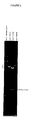
- the BHV-1 gII glycoprotein gene would be located in the Hind III K band ( Figure 1).
- This hypothesis agrees with the reported data on other members of the Alphaherpesviridae family, which presents a sequence of five glycoproteins that are located in conserved positions through the genome: US4 (gG), US5, US6 (gD), US7 (gI) and US8 (gE) from HSV-1 ; US4 (gG2), US5, US6 (gD2), US7 (gI2) and US8 (gE2) from HSV-2 ; gX, gp50, gp63 and gI from PRV and gpIV and gpI from VZV (McGeoch, D. J. (1990) J.Gen. Virol., 71: 2361-2367 ). Therefore, the reported data strongly suggest that the BHV-1 gII glycoprotein gene is located in the Us region of the genome.
- Hind III fragments of BHV-1 FM DNA were cloned at the Hind III site of pTZ1 8u (United States Biochemical, Corp.), by the following procedure: 2 ⁇ g of BHV-1 FM DNA were dissolved in a buffer containing 10 mM Tris-HCl (pH 8.0), 5 mM MgCl2, 100 mM NaCI and 1 mM mercaptoethanol (hereinafter "Hind III digestion buffer"). The DNA was digested at 37°C for 1 h with 2 units of Hind III. The restriction enzyme Hind III was inactivated by heating at 65°C for 20 min.
- Hind III digested pTZ18u DNA was mixed with Hind III digested pTZ18u DNA by the following method: 2 ⁇ g of Hind III digested BHV-1 FM DNA were mixed with 100 ng of Hind III digested pTZ18u, in 20 ⁇ l of a buffer containing 66 mM Tris-HCl (pH 7.5), 5 mM MgCl2, 1 mM dithiothreitol, 1 mM ATP (hereinafter "ligation buffer”), 2 units of phage T4 DNA ligase (Boehringer Mannheim), and were incubated overnight at 16°C.
- ligation buffer 2 units of phage T4 DNA ligase
- Competent E . coli XL1-Blue were prepared for transformation using the CaCl2 procedure described by Sambrook et al. (Sambrook et al. Molecular Cloning, (1989) Cold Spring Harbor Laboratory).
- a 1 mL overnight culture of E. coli XL1-Blue was used to inoculate 200 mL of broth containing 1.6% (w/v) bactotryptone, 1% (w/v) yeast extract and 0.5% (w/v) NaCI (hereinafter "2xYT broth”). The culture was incubated at 37°C for 6-7 h at 300 rpm.
- the bacteria were collected by centrifugation and resuspended in 1/2 volume of ice cold 0.1 M CaCl2. After a 5 min incubation on ice, the bacteria were pelleted and resuspended in 1/15 of the initial culture volume of ice cold 0.1 M CaCl2.
- Samples were plated on Petri dishes with 20 mL of solid media containing 1% (w/v) tryptone, 0.55% (w/v) yeast extract, 1% (w/v) NaCI (hereinafter "LB") and 1,5 % bactoagar, supplemented with 60 ⁇ g/mL ampicillin, 0.04 mg/mL S-bromo-4-chloro-3-indolyl- ⁇ -D-galactopyranoside (hereinafter "X-gal”) and 0.04 mg/mL isopropyl- ⁇ -D-thiogalactopyranoside (hereinafter "IPTG”).
- X-gal S-bromo-4-chloro-3-indolyl- ⁇ -D-galactopyranoside
- IPTG isopropyl- ⁇ -D-thiogalactopyranoside
- miniprep Rapid screening for the hybrid plasmid DNA
- solution II sodium dodecylsulfate
- solution III 3M potassium acetate and 2M acetic acid
- the mixtures were vigorously shaked and centrifuged for 5 min at 13000xg.
- the aqueous phase was transferred to a new tube and the phenol extraction was repeated.
- 2.5 volumes of ethanol were added and the mixture was placed at -20°C for 30 min.
- the precipitated hybrid plasmid DNA was collected by centrifugation at 13000xg for 10 min. and rinsed with 70% (v/v) ice cold ethanol and air dried. Finally, the plasmid DNA was dissolved in 200 ⁇ L of TE buffer.
- the solution was transferred to a centrifuge tube and centrifuged at 65000 rpm for 16 h at 20°C in a Beckman VTi80 rotor.
- the result of the centrifugation was the presence in the middle of the tube of a red fluorescent band corresponding to the supercoiled DNA from the hybrid plasmid.
- the hybrid plasmid was collected by puncturing the tube with a needle.
- the recovered DNA was mixed with 1 volume of water-saturated 1-butanol; the two phases were mixed and centrifuged at 13000xg for 30 s at room temperature. The upper phase containing the ethidium bromide was removed. Such extraction was repeated 8 times until the upper phase did not contain any fluorescent trace.
- DNA concentration was quantified by optical density at 260 nm and their purity was checked by spectrophotometry scan analysis (320-220 nm) and agarose gel electrophoresis (as detailed in section 4.C. ).
- the p1K1 plasmid was linearized with Eco RI restriction enzyme, which did not cut the Hind III K insert. Subsequently, the linearized p1K1 plasmid was digested with the following enzymes: Sal I, Xho I, Pat I and Sma I, in suboptimal conditions respect to those recommended by the supplier and at different times to obtain partial digestions of the Hind III K fragment inserted in the p1K1 hybrid plasmid.
- the digests were electrophoresed in an agarose gel, Southern blotted and hybridized with pUC118 previously linearized with EcoR I and labeled with digoxigenin (hereinafter "DIG") by random primed technique (as detailed in section 4.F. ).
- the hibridization showed a ladder of bands representing the restriction sites in the insert of the hibrid plasmid p1Ki.
- the DNA was stained with 0.5 ⁇ g/mL ethidium bromide in TAE buffer and visualized on a long wave UV transilluminator and photographed.
- the gel was soaked in 1 M NaOH for 30 min at room temperature and then soaked in 1 M Tris-HCI (pH 7.0), 1.5 M NaCI for 60 min at room temperature. Thereafter, the gel was capillary blotted on a nitrocellulose sheet (Schleicher and Schuell), previously hydrated and soaked in 3M NaCI and 0.3 M sodium citrate (hereinafter "20xSSC"). Blotting was performed overnight at room temperature with 10xSSC as transfer buffer.
- the filter was rinsed with 6xSSC, air dried at room temperature and baked in a vacuum oven at 80°C for 2 h. Subsequently, the nitrocellulose filter was placed in a plastic bag and soaked with 20 mL of 5x(0.1% (w/v) Ficoll, 0.1% (w/v) polyvinylpirrolidone and 0.1% (w/v) bovine serum albumin) (hereinafter "Denhardt's solution”), 5xSSC, 50% (v/v) formamide and 100 ⁇ g/mL single stranded DNA (hereinafter "prehybridization solution").
- Denhardt's solution 5x(0.1% (w/v) Ficoll, 0.1% (w/v) polyvinylpirrolidone and 0.1% (w/v) bovine serum albumin)
- 5xSSC 50% (v/v) formamide and 100 ⁇ g/mL single stranded DNA
- the single stranded DNA was prepared as follows: 50 mg of salmon sperm DNA was dissolved in 10 mL of 0.2 M NaOH, denatured by heating at 100°C for 20 min, sheared to about 0.5 kb fragments by pumping with a syringe, and finally neutralized with 0.2 mL of 10 M HCl. The nitrocellulose filter was incubated for 2 h at 42°C; and meanwhile, the DIG-labeled probe (section 4.F.
- hybridization solution a solution containing 5xSSC, 45% (v/v) formamide, 5x Denhardt's solution and 7.5% (w/v) dextran sulfate (hereinafter "hybridization solution”).
- hybridization solution 3 mL of hybridization solution containing 50-100 ng of denatured DIG-labeled probe were added per 100 cm2 nitrocellulose filter and left to hybridize overnight at 42°C.
- the filter was washed twice for 15 min at 42°C with 2xSSC and 0.1% (w/v) SDS solution, and then washed twice for 15 min at 68°C with 0.1xSSC and 0.1% (w/v) SDS solution. Subsequently, the filter was rinsed with 100 mM Tris-HCl (pH 7.5) and 150 mM NaCI (hereinafter "buffer 1") and then incubated for 30 min at room temperature with 100 mL of 0.5% (w/v) blocking reagent in buffer 1 (Boehringer Mannheim) (hereinafter "buffer 2").
- the nitrocellulose filter was briefly rinsed with buffer 1 and incubated for 30 min at room temperature with 20 mL of buffer 2 containing a dilution1:5000 of anti-digoxigenin conjugated to alkaline phosphatase (Boehringer Mannheim). Next, the nitrocellulose filter was briefly rinsed twice for 15 min with buffer 1 and equilibrated for 2 min with 20 mL of 100 mM Tris-HCI (pH 9.5), 100 mM NaCI and 50 mM MgCl2 (hereinafter "buffer 3").
- nitrocellulose filter was incubated with 10 mL of buffer 3 containing 45 ⁇ L of 75 mg/mL nitroblue tetrazolium in 70% (v/v) dimethylformamide and 35 ⁇ L of 50 mg/mL 5-bromo-4-chloro-3-indolyl phosphate, toluidinium salt in 100% (v/v) dimethylformamide (hereinafter "developing buffer").
- developer buffer 50% (v/v) dimethylformamide
- the starting material was linearized-DNA previously purified by phenolchloroform and precipitated with ethanol.
- the DNA was denatured by heating at 95°C for 10 min and quickly ice-cooled.
- the labeling reaction was prepared by adding in an ice-cold microcentrifuge tube: 1 ⁇ g of denatured DNA, 2 ⁇ L of an hexanucleotide mixture (Boehringer Mannheim), 2 ⁇ L of dNTP labeling mixture (1mM ATP, 1mM CTP, 1mM GTP, 0.65 mM TTP, 0.35 mM DIG-dUTP (pH 6.5)), 1 ⁇ L of Klenow enzyme (2U/ ⁇ L) and distilled water to complete a final volume of 20 ⁇ L.
- the tube was incubated at 37°C for 20 h. The reaction was stopped by adding 2 ⁇ L of 0.2 M EDTA (pH 8,0). The labeled DNA was precipitated by adding 2.5 ⁇ L of 3 M sodium acetate (pH 5) and 75 ⁇ L of cold ethanol. The mixture was kept at -80°C for 30 min, and then centrifuged at 12000xg for 10 min. The DNA pellet was washed with 70% (v/v) ethanol, vacuum-dried and redissolved in 50 ⁇ L of TE.
- the agarose gel was stained with ethidium bromide. DNA bands were visualized by long wave UV-light and agarose fragments including the DNA bands of interest were excised. These fragments were placed in a microfuge tube and 2.5 volumes (respect to the weight of agarose fragments -mg/mL-) of 6M Nal solution were added. The microfuge tube was incubated at 55°C for 5 min to melt the agarose. Then, 5 ⁇ L of glassmilk suspension (Bio 101) were added to the microfuge tube and kept for 5 min on ice. The tube was briefly centrifuged at 13000xg and the supernatant carefully removed.
- the pellet was resuspended in 0.7 mL of washing solution (Bio 101), centrifuged at 13000xg for 10 sec and the supernatant discarded; this step was repeated twice more. Finally, the pellet was resuspended in 10 ⁇ L of TE buffer and the suspension incubated at 55°C for 5 min, centrifuged at 13000xg for 10 sec and the supernatant recovered.
- the DNA so obtained can be used directly or further purified by ethanol preciptation.
- the subcloned fragments were those denoted as Sal IA of 1095 bp and Pst IC of 1654 bp ( Figure 3). Then, a second subcloning step was carried out to obtain smaller fragments suitable for sequencing.
- the Sal IA fragment previously purified by Geneclean, was digested with the following restriction enzymes: Pst I to obtain the subclones Sal A1, Sal A4, and Sal A5; Sma I to obtain the subclones Sal A2 and Sal A3; and, Sac I and Pst I to obtain the subclones Sal A31 and Sal A33 ( Figure 5).
- the Pst IC fragment previously purified by Geneclean, was digested with the following restriction enzymes: Sma I to obtain the subclones Pst C1 and Pst C2; and, Sac I to obtain the subclone Pst C3 ( Figure 5).
- the hybrid plasmid DNA of each subclone was obtained from overnight 3mL cultures by the procedure described for the miniprep procedure and further purified by Geneclean.
- Sequencing reactions were carried out following the usual dideoxy chain termination method (Sanger, F. et al. (1977) Proc. Natl. Acad. Sci. USA 74; 5463-5467).
- the reaction mixture contained: an alkali-denatured template corresponding to each one of the above described subclones; the "universal” and “reverse” oligonucleotides corresponding to pUC118 vector were used as primers for DNA synthesis, ( ⁇ -35S)dATP as labeling substrate, Mg2+, deoxyribonucleosides triphosphate, dideoxyribonucleosides triphosphate and T7 DNA polymerase (according to the directions from Pharmacia).
- reaction was stopped by adding a solution containing 0.3% (w/v) bromophenol blue, 0.3% (w/v) xylene cyanol, 10 mM EDTA (pH 7.5) and 97% (v/v) deionyzed formamide.
- Reactions were heated at 90°C for 1 minute and loaded onto a 8% (w/v) sequencing gel, composed by 7.7% (w/v) acrylamide, 0.3% (w/v) bisacrylamide, 32% (w/v) urea, 0.003% (v/v) TEMED and 0.007% (w/v) ammonium persulfate.
- the sequence obtained from the 3' end of the Hind III K fragment does not contain the whole sequence coding for the BHV-1 FM gII glycoprotein.
- a strategy of "walking" along the Us sequence of the viral genome was used.
- the BHV-1 FM DNA was digested with a series of restriction enzymes having restriction sites into the Hind III K fragment and, therefore, they should produce fragments beyond the 3' end of the Hind III K fragment.
- the following enzymes were used: Hind III (as a control), Xho I, Sal I, Sma I, Eco RI/Hpa I, Nco I, Bst EII, and Sac I.
- the viral DNA digests were electrophoresed in 0.8% agarose gels, blotted to nitrocellulose filters and hybridized to the DIG-labeled Sal 1A fragment under high stringent conditions as above described.
- the fragments hybridizing to Sal 1A fragment were used to construct a restriction map for the further cloning and sequencing beyond the Hind III K 3' region. The results of such hybridization are shown in Figure 4.
- the 2400 bp Pst I-Pst I fragment was obtained from the Sal I-Eco RI fragment inserted into the hybrid plasmid pSE1, and subcloned into pUC118 to generate the hybrid plasmid pSE10.
- the Pst I-Pst I fragment was digested with: Xho I to obtain the subclones pSE13 and pSE14; Sac I to obtain the subclone pSE12; Sac I and Nhe I to obtain the subclone pSE121; and, Acc I to obtain the subclone pSE141 ( Figure 5).
- the nucleotide sequence contains an open reading frame (575 codons) coding for a polypeptide with a molecular mass of 65 kDa.
- the translation start codon (ATG) is found 20 nucleotides downstream the first BstB I site.
- the reading frame of 1725 nucleotides is delimited by a stop codon (TAG), which is located into the Eco NI site ( Figure 5 and Table 1).
- TAG stop codon
- No evidence about the existence of a polyadenylation signal downstream the reading frame has been found. This feature is also observed in the homologous genes from HSV-1 and EHV-1, where the polyadenylation signal is shared by the gII homologous gene and the contiguous gene (McGeoch, D. J. et al. (1985) J. Mol. Biol., 181; 1-13; Telford, E. et al.
- a TATA transcription signal has been found 131 nucleotides upstream the start codon. This sequence differs slightly from the consensus (Bucher, P. (1990) J. Mol. Biol., 212; 563-578), although it has also been described for the BHV-1 tk gene (Bello, L. J. et al. (1992) Virology, 189; 407-414) and for various genes of the HSV-1 Us region (McGeoch, D. J. et al. (1985) J. Mol. Biol., 181; 1-13; Bucher, P. (1990) J. Mol. Biol., 212; 563-578).
- aa amino acid sequence
- Kyte-Doolittle algorithm predicts the existence of two hydrophobic regions consistent with sequences generating hidrophobic ⁇ -helices.
- the first domain expands from aa 11 to aa 31, and contains a protease recognition sequence between aa 26 and 27 (von Heinje, G. (1986) Nucleic Acids Res., 4683-4690).
- These data suggest the presence of a signal peptide from aa 1 to aa 26.
- This signal peptide presents, according to the model of Perlman, D. and Halvorson, H. O., 1983 (J. Mol.
- Biol., 167: 391-409 a) a N-terminal region with charged aa residues (3 arginines); b) an hydrophobic-rich aa central region (9 leucines and one phenylalanine over 14 residues) and c) a C-terminal region with short side-chains aa (2 alanines), which includes the processing site of the peptide.
- the signal peptide would allow the transport of the protein to the endoplasmatic reticulum where protein glycosylation occurs.
- the second hydrophobic region expands from aa 413 to aa 443.
- Sequence homology analysis was performed by the FastA program (GCG Sequence Analysis Software Package; Madison, Wisconsin, USA) on a VAX/VMS system; such a program uses the Pearson-Lipman algorithm to search for similarities between a given sequence and a sequence data bank.
- the sequences of the different proteins used to perform the analysis were obtained from the GeneBank. As a whole, the sequence herein obtained shows a high identity degree (around 60%) versus protein sequences putatively homologous to the gII from other herpesviruses (gI from PRV; gE from HSV-1; and, gE from EHV-1). TABLE 2 No. of aas Mol.
- proline residues 63 prolines: 15% in number and 9.9% of the molecular mass
- the aa sequence not only has a similar proline content than EHV-1 gE (the most similar between the reported glycoproteins), but also conserves the positions in which prolines occur.
- EHV-1 gE the most similar between the reported glycoproteins
- Sections 4.H. to 4.J. Design of a hybrid cloning vector containing the 5' and 3' flanking sequences of the gene coding for the gII glycoprotein.
- the object of this step was to select a 1200 bp fragment corresponding wich includes an upstream region of the BHV-1 FM gII gene and the sequence wich codifies the signal peptide of the same gene. Both sequences are contained in the p1K1 insert.
- the objective of this step was to select a hybrid plasmid DNA containing a 4800 bp fragment composed by: (1) a 1200 bp DNA fragment corresponding to the upstream region of the BHV-1 FM gII gene, and the sequence correponding to the signal peptide from BHV-1 FM gII gene, fused to (2) a 3600 bp DNA fragment corresponding to the downstream region of the BHV-1 FM gII gene. This sequence also includes the STOP codon from BHV-1 FM gII gene.
- Two ⁇ g of pSE1 were digested for 5 h at 37°C with 2 units of Eco NI (BioLabs) in 20 ⁇ L of a buffer containing 50 mM potassium acetate, 20 mM Tris-acetate (pH 7.9), 10 mM magnesium acetate, and 1 mM dithiothreitol.
- the digest was subjected to 1% (w/v) agarose gel electrophoresis, the band corresponding to the linearized plasmid was excised from the gel and finally purified by Geneclean. Then, the sticky ends of the linearized plasmid were blunt ended by Mung Bean nuclease (Boehringer Mannheim).
- the linearized pSE1 plasmid was incubated for 1 h at 25°C with 10 units of Mung Bean nuclease in 100 ⁇ L of a solution containing 20 mM sodium acetate (pH 4.6), 5 mM sodium chloride, 1 mM zinc acetate, and 0.001% (v/v) Triton X-100. After this treatment, 0.1 volumes of 3 M sodium acetate were added to the linearized pSE1 plasmid, which was then extracted with an equal volume of phenol/chloroform/isoamylalcohol (25/24/1 v/v/v) and precipitated with 2 volumes of ethanol.
- the redissolved plasmid was digested for 3 h at 37°C with 2 units of Eco RI in 20 ⁇ L of a buffer containing 50 mM Tris-HCl (pH 7.5), 10 mM MgCI2, 100 mM sodium chloride, and 1 mM dithiothreitol.
- a 3600 bp Eco Nl(blunted)-Eco RI fragment from BHV-1 FM was electrophoresed in 1% (w/v) agarose gel, excised from de gel and purified by Geneclean.
- the 3.6 kb Eco Nl(blunted)-Eco RI fragment from BHV-1 FM and the 4.4 kb Sma I-Eco RI fragment from pC1 were mixed and ligated with T4 ligase.
- the ligation product was used to transform competent E. coli XL1-Blue as above described.
- the screened colonies contained a plasmid with a 4.8 kb DNA insert being the size of the total hybrid plasmid DNA of 8 kb, corresponding to the fusion of pC1 plasmid (4.4 kb) with the 3.6 kb Eco Nl(blunted)-Eco RI fragment.
- This plasmid was named pC1SE1a ( Figure 6).
- the object of this step was to select a hybrid plasmid DNA containing a 3 kb fragment composed by: (1) a 1200 pb DNA fragment corresponding to the upstream region of the BHV-1 FM gII gene, and the sequence corresponding to the signal peptide from BHV-1 FM gII gene, fused to (2) a 1.8 kb DNA fragment corresponding to the downstream region of the BHV-1 FM gII gene, This sequence also includes the STOP codon from BHV-1 FM gII gene.
- Two ⁇ g of pC1SE1a were digested for 3 h at 37°C with 2 units of Pst I in 20 ⁇ L of a buffer containing 50 mM Tris-HCl (pH 7.5), 10 mM MgCl2, 100 mM sodium chloride, and 1 mM dithiothreitol. The digestion was then electrophoresed in 1% (w/v) agarose gel and the resulting 3.0 kb Pst I-Pst I fragment was purified by Geneclean as above described.
- Two ⁇ g of pUC118 were digested for 3 h at 37°C with 2 units of Pst I in 20 ⁇ L of a buffer containing 50 mM Tris-HCI (pH 7.5), 10 mM MgCl2, 100 mM sodium chloride, and 1 mM dithiothreitol. After digestion, 0.1 volumes of 3 M sodium acetate were added to the linearized pUC118 plasmid, which was then extracted with an equal volume of phenol/chloroform/isoamylalcohol (25/24/1 v/v/v) and precipitated with 2 volumes of ethanol.
- the 3.0 kb Pst I-Pst I DNA fragment and the linearized pUC118 plasmid were mixed and ligated with T4 DNA ligase, and the ligation product used to transform competent E. coli XL1-Blue, as above described.
- the screened colonies contained a plasmid with a 3.0 kb DNA insert. This plasmid was named p ⁇ gII ( Figure 6).
- Section 4.K. A method to delete the gII glycoprotein gene sequence and to construct a defective gII ⁇ virus strain, by cotransfecting the hybrid vector p ⁇ gII with IBRV DNA from BHV1 FM (gII+) in host cells.
- the BHV-1 FM gII ⁇ which lacks the ability of producing any antigenic polypeptide from gII, was obtained through a deletion in the gII gene from BHV-1 FM. This deletion was obtained by homologous recombination between the hybrid plasmid p ⁇ gII and the genomic BHV-1 FM DNA. For this proupose both DNAs were cotransfected by electroporation of GBK cells.
- p ⁇ gII was obtained by centrifugation in CsCl as described in section 4.D. .
- Ten ⁇ g of p ⁇ gII were digested for 3 h at 37°C with 10 units of Pst I in 100 ⁇ L of a buffer containing 50 mM Tris-HCl (pH 7.5), 10 mM MgCl2, 100 mM sodium chloride, and 1 mM dithiothreitol. After digestion, 0.1 volumes of 3 M sodium acetate were added to the DNA, which was then extracted with an equal volume of phenol/chloroform/isoamylalcohol (25/24/1 v/v/v) and precipitated with 2 volumes of ethanol. The resulting DNA pellet was redisolved in 20 ⁇ L of sterile TE buffer.
- BHV-1 FM gII+ DNA was obtained according to the procedure described in section 4.B. .
- GBK cells were prepared for electroporation as follows: 5.8x10⁶ GBK cells and 35 mL of GMEM supplemented with 10% FCS were added per 175 cm2 culture flask. When a 60-70% confluent cell monolayer was obtained (72 h), the medium was removed and the cells rinsed with a buffer containing 0.04% (w/v) potassium chloride, 0.68% (w/v) sodium chloride, 0.22% (w/v) sodium bicarbonate, 0.014% (w/v) sodium dihydrogenphosphate, 0.1% (w/v) D-glucose, and 0.02% (w/v) EDTA (hereinafter, "EBSS-EDTA").
- EBSS-EDTA 0.04% (w/v) potassium chloride, 0.68% (w/v) sodium chloride, 0.22% (w/v) sodium bicarbonate, 0.014% (w/v) sodium dihydrogenphosphate, 0.1% (w/v) D-glucose, and 0.02% (
- the EBSS-EDTA was removed and the cells detached from each culture flask by 3-5 min treatment with 3.5 mL of a solution containing 0.02% (w/v) EDTA and 0.05% (w/v) trypsin. Trypsin was inhibited by adding 3.5 mL of GMEM. Then, the cell suspension was collected and centrifuged at 800xg for 10 min at 4°C.
- the cell pellet was resuspended in 20 mL of a buffer (cooled at 4°C) containing 20 mM HEPES, 137 mM sodium chloride, 5 mM potassium chloride, 0.7 mM disodium hydrogenphosphate, and 5 mM D-glucose (pH 7.1) (hereinafter, "saline HEPES”), and centrifuged at 800xg for 10 min at 4°C. The supernatant was removed and the cell pellet washed twice with saline HEPES in the conditions above described. Finally, the cell pellet was resuspended in saline HEPES to obtain a suspension of 7x10⁶ cells/mL.
- saline HEPES 20 mM HEPES, 137 mM sodium chloride, 5 mM potassium chloride, 0.7 mM disodium hydrogenphosphate, and 5 mM D-glucose (pH 7.1)
- a volume of 0.8 mL of the GBK cell suspension in saline HEPES, 1 ⁇ g of BHV-1 FM gII+ DNA, and 0.5 ⁇ g of p ⁇ gII digested with Pst I were added to an ice-cold 0.4 cm electroporation cuvette (BioRad).
- the cuvette was kept in ice for 10 min before applying 2 pulses of 280 V and 250 ⁇ F (time constant of 3-5 msec).
- the cuvette was left in ice for 20 sec between each pulse, and for 10 min before plating the electropored cells in a 90 mm diameter Petri dish with 15 mL of GMEM.
- Section 4.L to 4.M. A screening method of the progeny viruses, obtained as described in the step 4K, to identify and isolate IBRV virus particles which have mutated in the locus corresponding to gII glycoprotein gene.
- dishes containing cell monolayers were rinsed twice with 10 mL of a buffer containing 0.8% (w/v) sodium chloride, 0.144% (w/v) sodium hydrogenphosphate, 0.024% (w/v) potassium hydrogenphosphate, and 0.02% (w/v) potassium chloride (hereinafter, "PBS"). Then, the cell monolayer was dehydrated with 10 mL of absolute ethanol for 10 min, and rehydrated with successive washes (10 min per wash) of 70% (v/v) ethanol, 50% (v/v) ethanol, and 30% (v/v) ethanol. Then, the cell monolayer was rinsed twice with PBS containing 5 mM MgCl2 and treated 20 min with 10 mL of 0.2 M HCl.
- the dishes were then incubated for 30 min at 50°C in 2xSSC, 5 mM EDTA.
- the cell monolayer was soaked for 15 min at 37°C in 5 mL of PBS, containing 1 ⁇ g/mL of proteinase K (Boehringer Mannheim) . After protease treatment, the cell monolayer was washed twice for 5 min with 10 mL of PBS containing 0.2% (w/v) glycine, and fixed for 30 min with 10 mL of PBS containing 4% (w/v) paraformaldehyde.
- Cell monolayers were washed twice for 10 min with 10 mL of PBS containing 5 mM MgCl2, and then 10 mL of prehybridization solution were added. Dishes were heated at 95°C for 3 min, and incubated at 42°C for 20 min. Dishes were heated again 3 min at 95°C and ice-cooled. The prehybridization solution was removed and 3 mL of hybridization solution containing the DIG-labeled DNA probe were added onto the cell monolayer.
- the DNA probe was the 570 bp Sal I-Sma I fragment obtained during the subcloning for sequencing and named Sal A3 fragment (section 4.G. ).
- the probe was obtained as follows: 5 ⁇ g of hybrid plasmid DNA from the subclone Sal A3 were incubated at 37°C for 3 h with 2 units of Sal I in 20 ⁇ L of a buffer containing 50 mM Tris-HCl (pH 7.5), 10 mM MgCI2, 100 mM sodium chloride, and 1 mM dithiothreitol. Then, 1/10 volumes of 3 M sodium acetate were added to the above solution, which was extracted with an equal volume of phenol/chloroform/isoamylalcohol (25/24/1 v/v/v) and DNA precipitated with 2 volumes of ethanol.
- the pellet was redissolved in 20 ⁇ L of TE buffer and incubated at 25°C for 3 h with 2 units of Sma I in 20 ⁇ L of a buffer containing 33 mM Tris-acetate (pH 7.9), 10 mM MgCl2, 66 mM potassium acetate, and 0.5 mM dithiothreitol.
- DNA was electrophoresed in a 1% (w/v) agarose gel and the 570 bp fragment was isolated and purified by Geneclean. This fragment (hereinafter "gII ⁇ probe”) was finally labeled with DIG as described in section 4.F.
- plaques derived from the progeny of non-recombinant BHV-1 FM viruses were identified by the presence of a dark precipitate ( Figure 7B). Plaques not surrounded by a dark precipitate ( Figure 7B) were derived from the progeny of mutant BHV-1 FM gII ⁇ (hereinafter “non-hybridized plaques"). We obtained a frequency of 2.2 recombinant (mutant) viruses per 100 pfu.
- Non-hybridized plaques were marked and rescued from agarose layer (kept at 4°C) .
- Pieces of about 0.5x0.5 cm were cut from the agarose corresponding to those non-hybridized plaques.
- Four agarose pieces (named G1, E7, E8, and E9) were placed in microfuge tubes containing 0.5 mL of GMEM, and were gently shaked for 12 h at 4°C.
- Viral suspensions from each non-hybridized plaque were titulated (see section 4.A. ), yielding an average of 60 pfu/ ⁇ L. According to the procedure described in section 4.A.
- a volume of 1 ⁇ L from each viral suspension was used to infect the corresponding GBK cell monolayer on a Petri dish.
- the infected monolayers were incubated for 72 h to allow the apparition of plaques with cytopathic effect.
- One infected monolayer (E7) was used to perform the gII ⁇ probe hybridization analysis above described. The result of the hybridization showed 8 recombinant viruses per 100 pfu, indicating a 3.5-fold enrichment.
- Section 4.N. Analyses of purified BHV-1 gII ⁇ DNA to check that progeny viruses isolated in the step 4.M. are homogeneous and have a deletion in the gII locus.
- Mutant viral DNA was prepared from the recombinant clone E9B-22 according to the procedure described in section 4.B. . The obtained DNA was digested with Hind III and the restriction fragments were electrophoresed in a 0.8% (w/v) agarose gel in TAE buffer at 16 V for 16 h at room temperature. Hind Illdigested DNA from the parental (wild-type) BHV-1 FM virus was included as a control.
- two new bands are generated: (1) a 24.7 kb band corresponding to the fusion of the Hind III K and Hind III C fragments in one of the BHV-1 FM isoforms, and (2) a 21.2 kb band, corresponding to the fusion of the Hind IIIK and Hind IIIF fragments of the second BHV-1 FM isoform.
- the last band comigrates with the Hind III A band.
- the clone E9B-22 has been selected for further studies and named BHV-1 FM gII ⁇ and has been deposited in the Collection Nationale de Cultures de Microorganismes from Institut Pasteur, n o I-1393.
Landscapes
- Chemical & Material Sciences (AREA)
- Organic Chemistry (AREA)
- Health & Medical Sciences (AREA)
- Life Sciences & Earth Sciences (AREA)
- Biophysics (AREA)
- Biochemistry (AREA)
- Gastroenterology & Hepatology (AREA)
- General Health & Medical Sciences (AREA)
- Genetics & Genomics (AREA)
- Medicinal Chemistry (AREA)
- Molecular Biology (AREA)
- Proteomics, Peptides & Aminoacids (AREA)
- Virology (AREA)
- Medicines Containing Antibodies Or Antigens For Use As Internal Diagnostic Agents (AREA)
- Micro-Organisms Or Cultivation Processes Thereof (AREA)
Abstract
Description
- The present invention relates in general to viral vaccines based on mutant viruses and, specifically to the Infectious Bovine Rhinotracheitis Virus (Bovine Herpesvirus type 1), to the mapping, cloning and sequencing of the glycoprotein gII, and the generation of mutants containing deletions or insertions or both in the locus of the glycoprotein gII gene, and its utilization in a vaccine in the form of a defective virus for this glycoprotein, such that no antigenic polypeptide encoded by the viral gene are produced. Animals vaccinated with such mutant viruses do not develop antibodies against the viral glycoprotein gII and can be distinguished serologically from animals infected with field strains of Infectious Bovine Rhinothracheitis virus. The present invention also relates to vaccines for Infectious Bovine Rhinothracheitis containing the same, methods for production, formulation and utilization of the same and provide tools to distinguish serologically animals vaccinated with the same from animals infected with field strains of Infectious Bovine Rhinotracheitis virus.
- The following definitions are provided for a clear and consistent understanding of the specifications stated in the claims.
Adjacent: Any position in the nucleotide sequence located immediately 5' or 3' to a defined sequence.
Cell culture: A proliferating mass of cells which may be in an undifferentiated or differentiated state. As used herein "cell" and "cell line" are used interchangeably. All such designations include progeny, but not all of the components of the progeny have an identical DNA sequence.
Coding sequence: A deoxyribonucleotide sequence which when transcribed and translated results in the formation of a cellular protein, or a ribonucleotide sequence which when translated results in the formation of a cellular protein.
Control sequence: Refers to DNA sequences necessary for the expression of an operative linked coding sequence in a particular host organism. The control sequences include promoter, enhancers, terminator signals, polyadenylation signals, and possibly, other still poorly understood sequences.
Deletion: Removal of a fragment from a DNA sequence, being the regions on either side joined together.
Expression system: Refers to DNA sequences containing a specific coding sequence and control sequences operably linked, so that hosts transformed with these sequences are able to produce the encoding protein. The expression system may be included on a vector, although sometimes, the relevant DNA may integrate into a chromosome of the host cell.
Flanking sequence: Refers to DNA sequences adjacent, in 5' and 3', to the coding sequence. They include the control sequences.
Gene: A discrete nucleic acid region which is responsible for a discrete cellular product (RNA and/or polypeptide).
Glycoprotein: Protein that contain covalently bonded sugar molecules.
gII: Glycoprotein II and gene of the glycoprotein gII from Bovine Herpesvirus type 1 (also named gE since the 18th International Herpesvirus Workshop held in Pittsburgh, Pennsylvania, in 1993).
Insertion: Introduction of an additional stretch of DNA sequence into another DNA sequence.
Operative linked: Refers to the functional association of the components. Thus, a coding sequence operatively linked to control sequences refers to an association wherein the coding sequence can be expressed under the control of these sequences.
p.f.u.: plaque forming units.
pb: a complementary pair of bases of the double strand DNA. kb refers to 1000 bp.
Reading frame: Segment of DNA encoding a polypeptide or polypeptides.
Sequence homology: Indicates different nucleotide sequences that are functionally equivalent to one another. The nucleotide differences do not affect the functionality of the products encoded in the different sequences.
Substitution: Replacement of a stretch of DNA sequence by another DNA sequence (corresponds to a deletion plus an insertion).
Transformants: Refers to cells that, through different mechanisms, acquire foreign DNA sequences, which can or cannot induce changes in the host cells.
Vector: DNA sequence suitable to carry other DNA sequences.
+/-: Refers to the presence/absence of a functionality in a cell or virus. - Bovine Herpesvirus type 1 (hereinafter "UBHV-1"), also known as Infectious Bovine Rhinotracheitis virus (hereinafter "IBRV") is an agent associated with a wide spectrum of pathologies such as respiratory problems (the main cause of the loss of growth and vitality in calves), reproductive problems (abortions, infertility and sterility), losses in the milk yield, enteritis, meningitis, conjunctivitis, problems in the central nervous system and dermal infections of cattle (Timoney, J.F. et al. (1988) in Hagan and Bruner's Microbiology and Infectious Diseases of Domestic Animals Comstock Publishing Associates, 8th ed., Ithaca. pp 594-601). The severity of the illness resulting from IBRV infections depends on the virus strain and on the age of the affected animal . From the above problems, respiratory problems are the most common. The estimated incidence of the respiratory problems is 2% in adults and 10-30% in calves, being the average of calf mortality about 10%. In calves, the annual worldwide losses are estimated in 3000 million $.(Animal Farm, Supplement 17, May 1992).
- As a member of the Alphaherpesviridae family, BHV-1 is highly neurotropic, remaining latent in the nervous system of the animal after recovering from infection. The animals may present signs of recurrent disease without being reexposed to the virus (Rock, D.L. et al. (1986) J. Gen. Virol., 67: 2515-2520). This fact is extremely important for artificial insemination programs since infected bulls produce recurrent shedding of viruses. Some pharmacological treatments such as dexamethasone can also provoke nasal shedding of the virus with or without clinical symptoms of active Infectious Bovine Rhinothracheitis (hereinafter "IBR"). As a consequence, new outbreaks can result from viruses latent in infected but asymptomatic cattle. Thus, the design of vaccines and differential diagnostic systems, specially for eradication programs, could be very useful.
- The genome of BHV-1 consists of a linear, double-stranded, DNA molecule, approximately 135 kb in size. This genome is arranged in two unique sequences, the long one of about 100 kb (named UI) and the short one of about 13 kb (named Us). The Us is bracketed by two inverted repeat sequences: the internal (hereinafeter "Internal Repeat short" or "IRs") and the terminal (hereinafter "Terminal Repeat short" or "TRs"), both of about 11.5 kb (Hammerschmidt, W. et al. (1986) J. Virol., 58:43-49; Mayfield, J.E. et al. (1983) J. Virol., 47: 259-264). This genome structure corresponds to the herpesvirus class D which is also found in the pseudorabies virus (hereinafter "PRV"),
equine herpesvirus types 1 and 3 (hereinafter "EHV-1, -3"), and varicellazoster virus (hereinafter "VZV"). A characteristic of the herpesvirus genome is the ability to invert the orientation of unique sequences flanked by inverted repeated sequences (Us in the above viruses) leading two equimolar isoforms of the BHV-1 genome. - Restriction maps of the genomes from several IBRV strains have been reported (Mayfield, J.E. et al. (1983) J. Virol., 47: 259-264; Simard C. et al. (1990) Arch. Virol., 110: 63-75 ) and three different groups of viral genomes have been established, without any correlation with the epidemiology (Engels, M. et al. (1986) Virus Res., 6:57-73).
- Although BHV-1 genome may encode more than 33 structural polypeptides (Misra, V. et al. (1981)) J. Virol., 40:367-378), few of them have been identified and mapped in the IBRV genome.
- From the encoded structural polypeptides, 11 out of 33 are glycosylated (Misra, V. et al. (1981) J. Virol., 40: 367-378; Van Drunen Littel-van den Hurk, S. et al. (1986) J. Virol., 59: 401-410; Badia & Querol (1988) J. Virol. Meth., 22: 23-29). Glycoproteins play an outstanding role in the recognition, binding and penetration of the virus into the host cell, as well as in the immune response of the infected animal (virus neutralization, destruction of the infected cell and induction of the mechanisms of cell immunity). In addition, sugars play an important role in the generation of the functional glycoprotein, and also in the recognition and immunological properties of such glycoproteins (Olofsson S. (1992) APMIS Suppl 267, 100: 84-95; Van Drunen Littel-van den Hurk, S., et al. (1990) J. Gen. Virol., 71: 2053-2063).
- Monoclonal antibodies raised against BHV-1 have led to the identification and characterization of the four main envelope glycoproteins, gI, gII, gIII and gIV. The gI glycoprotein is synthesized as a 105 kDa (relative molecular mass) polypeptide, which in the mature form (fully glycosylated and proteolytically processed) yields a heterodimer of 130 kDa (built from two disulfide-bonded monomers of 75 and 55 kDa respectively). The precursor of gII glycoprotein is a nonglycosylated 90 kDa polypeptide, 108 kDa when glycosylated. The gIII glycoprotein is isolated in the form of a 180 kDa homodimer from a mature monomer (N- and O-glycosylated) with relative molecular mass of 91 kDa. The glV glycoprotein has a relative molecular mass of 71 kDa in a mature form (N- and O-glycosylated), and is also present as a 140 kDa homodimer. These proteins can also be found in the surface of BHV-1 infected cells, and react with neutralizing monoclonal antibodies or monospecific antiserum (Van Drunen Littel-van den Hurk, S. et al. (1984) Virology. 135: 466-479; Van Drunen Littel-van den Hurk, S. et al. (1986) J. Virol., 59: 401-410; Marshall, R.L. et al., (1986) J. Virol., 57: 745-753; Fitzpatrick, DR., et al. (1988) J. Virol., 62: 4239-4248; Van Drunen Littel-van den Hurk, S. et al. (1989) J. Virol. 63: 2159-2168).
- Several herpesvirus proteins are involved both in the formation of virus-neutralizing antibodies and in lymphocyte-mediated cytotoxic response (Norrild, B. et al. (1979) J. Virol., 32: 741-748; Balachandran, N. et al. (1982) Infect. Immun., 37: 1132-1137; Hampl, H. et al. (1984) J. Virol., 52: 583-590; Ben Porat, T. et al. (1986) Virology, 154: 325-334; and Wathen, L.M.K. et al. (1985) Virus Res., 4: 19-29). It is also described that BHV-1 gI, gIII and gIV are the main glycoproteins involved in eliciting neutralizing antibodies (Babiuk, L.A. et al. (1987) Virology, 159: 57-66). These results are important to design a protein defective viral strain that can be used as a vaccine. A defective vaccine allows to distinguish between vaccinated animals and animals infected with field strains of BHV-1, acting the deleted protein as a marker to detect vaccinated animals. The protein to be deleted must be carefully selected. Such protein must not be essential for virus replicacation and, when possible, not involved in eliciting neutralizing antibodies. A good choice would be the deletion of a protein non essential for viral replication, but acting as a counter-measure against the host immune response. IBRV gl and glV glycoproteins are not useful for designing a defective vaccine because their homologous from HSV and PRV (gB, gD, gX and gp50) are either essential proteins for viral replication in cell cultures or play a significant role in the entry of the virus into the cells (Desai, W. et al. (1988) J. Virol., 62: 2596-2604; Ligas, W.L. and Johnson, D.C. (1988) J. Virol., 62: 1486-1494; Liang, X. (1991) J. Virol., 65: 1124-1132; Card, J.P. et al. (1992) J. Virol., 66: 3032-3041). It has been reported the existence of genes encoding glycoproteins dispensable for viral amplification in cell culture in the US region of several members of the Alphaherpesviridae family (for HSV-1, see Longnecker, R. et al. (1987) Science, 236: 573-576; for PRV see Petrovskis, E.A. et al. (1986) J. Virol., 60: 1166-1169; Petrovskis, E.A. et al. (1987) Virol., 159: 193-195; Mettenleiter, T.C. et al. (1987) J. Virol., 61: 2764-2769; and, Thomsen, D.R. et al. (1987) J. Virol., 61: 229-232). The gene encoding the BHV-1 gII glycoprotein would be homologous to the PRV gl glycoprotein gene, which has already led to a defective pseudorabies strain vaccine (Lomniczi, B. et al. (1984) J. Virol., 49: 970-979; Mettenleiter, T.C. et al. (1985) J. Virol., 53: 52-57). In the BHV-1 UI region is located a gene encoding the gIII glycoprotein which, although non essential for viral replication in cell culture, its deletion involves a lesser cell adsorption and a delayed viral replication (Liang, X. et al. (1991) J. Virol., 65:1124-1132).
- Many viruses, among them the herpesvirus, present proteins that interfere with the host immune system- with cytokynes, with the complement system, etc.- (Gooding L. (1992) Cell, 71; 5-7). It has been reported for HSV that glycoproteins gE and gI bind to the IgG Fc domain, thus interfering with the complement mediated lysis of infected cells (Bell S. et al. (1990) J. Virol., 64; 2181-2186). These protein activities are dispensable for viral replication in cell culture as they act as a counter-measure against the host immune system (Gooding, L. (1992) Cell, 71; 5-7; Post L. and Roizman B. (1981) Cell, 25; 227-232). They represent the best choice for defective vaccine design because their absence does not prevent viral growth in cell culture and impede the interference with the host immune system.
- The deletion of the PRV gI glycoprotein gene, homolugous to gII from BHV-1, alters the neurotropism and reduces the neurovirulence of PRV when injected intraocularly in rats (Card et al., (1992) J. Virol., 66, 3032-3041). This fact could enhance the biosecurity of a vaccine with a deletion in the gII glycoproptein. Considering overall data, the deletion of gII glycoprotein from BHV-1 presents additional advantages versus the deletion of other glycoproteins such as gIII: (a) as above mentioned, its homologous from HSV (gE glycoprotein) represents a counter-measure against the host immune system, and (b) an IBRV gII⁻ strain will present lesser neurovirulence. Other characteristics are shared also with gIII glycoprotein: (a) to be a viable defective viral strain, and (b) to be a protein immunogenic enough to allow the design of a kit to serologically distinguish animals vaccinated with these mutants from animals infected with field strains. For the above reasons, the gII glycoprotein from BHV-1, which is homologous to PRV gI and HSV gE proteins, is the target of the present invention.
- To map and identify the glycoprotein gII (or gE) gene from BHV-1, it was assumed that gII gene, as in other herpesviruses, would be located in the Us region of the genome (Wirth U. et al. (1989) J. Virol., 65: 4882-4889). All reported data from other Alphaherpesviridae family members indicate the existence of a sequence containing 5 genes laying in conserved positions along the Us region of the genome. They are US4(gG), US5, US6(gD), US7(gI) and US8(gE) for HSV-1; US4(gG2), US5, US6(gD2), US7(gI2) and US8(gE2) for HSV-2; and gX, gp50, gp63 and gI for PRV (McGeoch D.J. (1990) J. Gen. Virol., 71: 2361-2367). VZV virus presents only the genes corresponding to US7 (gpIV) and US8 (gpI) because its Us is shorter than in the other herpesviruses. In addition, it has been recently reported the whole sequence of the EHV-1 genome and it corroborates the structure described for HSV-1 (Telford E. et al. (1992) Virology, 189:304-316).
- In the present invention it has been identified, mapped in the Us region, cloned and sequenced the gene coding for the gII (gE-like) glycoprotein from BHV-1 or IBRV, and has been constructed a gII⁻ defective BHV-1 strain. In addition, it has been demonstrated that the gII gene is not essential for viral replication in cell cultures. Moreover, it has been demonstrated that gII glycoprotein is immunogenic, thus it combines those characteristics required for its use as a marker in the design of a vaccine able to further distinguish serologically between animals vaccinated and animals infected with field strains.
- There exist several vaccines against IBR based on chemical or physically attenuated or killed virus. Laboratorios HIPRA S.A. has some of these vaccines. There are also vaccines obtained by attenuation of viruses by serial passages in cell cultures, alteration of the host cell or modification of culture conditions. There are also subunit vaccines prepared by solubilizing components from the viral envelope (i.e., by means of detergents) (Babiuk, L.A. et al. (1987) Virology, 159:57-66).
- All of the above vaccines present disadvantages. Those using whole IBR virus, even killed, can induce allergic or hypersensitivity reactions. Subunit vaccines have a lesser toxicity and are suitable for the serologic differentiation between infected and vaccinated animals, although are expensive in terms of production and must be administered in several doses.
- Vaccines based in modified-live viruses are more effective because they stimulate both the humoral and the cellular components of the immune system. Nevertheless, they do not eliminate latency. There have been reported several cases of reversion to virulent form upon utilization of modified-lived viruses. Moreover, there have been reported hazards of vaccine-induced abortions when administered intramuscularly. Intranasal inoculated vaccines seems to be more safety, but they present administration problems.
- There have been reported vaccines designed from modified-live viruses, thymidine kinase negative (tk⁻) and temperature resistant mutant viruses (for example US patent applications numbers 4.569.840 and 4.703.011). However, these mutants do not solve the problem of the distinction between animals vaccinated with the mutant and animals infected with field strains. A vaccine which overcomes the problem of latency and allows the distinction between animals vaccinated with the mutants and animals infected with field strains is the EP application 88118266.1, which describes the design of a gIII-thymidine kinase double defective strain (gIII⁻tk⁻).
- During the development of the present invention, there have been disclosed three applications for patents which deal on gE (or gII) glycoprotein from BHV-1. They correspond to applications PCT/US92/06034, PCT/FR93/0064 and PCT/NL92/00097. Application PCT/US92/06034 reports and claims the gE glycoprotein gene, its polypeptidic product and additional uses of them. However, the gene sequence has been defined incorrectly. They describe the encoded polypeptide as a sequence of 2038 bp, yielding a protein of 617 amino acid residues. This is not correct since there is a mistake derived from the erroneous choice of the initiation codon, which leads to an extended polypeptide and a promoter lacking the TATA box. This is supported by multialignment analysis of different gE glycoprotein sequences from several herpesvirus (Rebordosa et al., manuscript in preparation). Moreover, the putative 617 aa sequence would lose the N-terminal signal peptide (von Heijne G. (1986) Nucleic Acids Res., 14:4683-4690; and, Perlman D. and Halvorson H. O. (1983) J. Mol. Biol., 167:391-409). Therefore, it is almost impossible that a protein sequence such as the claimed by application PCT/US92/06034 could ever be expressed as a conformationally stable functional protein. Application PCT/FR93/0064 reports and claims a patent for the nucleotide sequence of a large portion from the BHV-1 Us region. Moreover, it claims for putative applications derived from the sequence but, to support their claims, they do not present experimental work other than the sequence. A third application is PCT/NL92/00097 which describes the design of a double defective strain (gE⁻tk⁻) by recombinant procedures. Nevertheless, the procedure described for deleting the gE gene is somewhat different to the described in the present application. Thus, application PCT/NL92/00097 reports a deletion involving the promoter region of the gE gene. This cannot allow the expression of a sequence inserted into the gE locus as they claim in claim 12 (at least under the control of gE promoter). The deletion of gE promoter may also interfere with genes downstream the gE gene (the US9 gene, a 10 kDa protein which may be essential for viral replication). In addition, gE and US9 genes share the termination sequence. In conclusion, the defective strain described in the present invention has advantages respect to that described in application PCT/NL92/00097, since a large portion of gE coding sequence has been deleted but maintaining both, the promoter and a short sequence encoding the signal peptide. Such a deletion yields a nonfunctional peptide and does not preclude the expression of downstream genes. Moreover, the gene construct described in the present application allows the insertion of genes to be expressed under the control of BHV-1 gE promoter. Finally, application PCT/NL92/00097 claims the expression of BHV-1 gE gene into a prokaryotic host. Presented data indicate that such expression is at very low levels and thus probably inadequate to detect BHV-1 in biological samples.
- An object of the present invention is to obtain a BHV-1 mutant virus characterized as BHV-1 FM gII⁻.
- Another object of the present invention is to obtain a BHV-1 mutant virus, as a result of a deletion, insertion or substitution (deletion and insertion) in the gII gene from BHV-1.
- Still another object of the invention is to obtain a BHV-1 mutant virus which fails to produce any antigenic gII polypeptide from gII glycoprotein as a result of deletion, insertion or both in the gII gene.
- A further object of the present invention is to provide methods for the production of the mutant virus BHV-1 FM gII⁻.
- Another object of the present invention are the vaccines effective in controlling the IBR which contain the BHV-1 FM gII⁻ virus and the methods of preparation.
- The invention provides tools for further application in diagnostic methods to serologically distinguish animals vaccinated with BHV-1 FM gII⁻ mutant virus from animals infected with field strains.
-
- Figure 1A shows the restriction enzyme analysis of the BHV-1 FM strain. The relative electrophoretic mobilities of the markers are in kb. On the right lane are the fragments from a Hind III digestion. The resulting molecular masses are compared to those reported by Mayfield et al. (1983) J.Virol., 47: 259-264, for the Cooper strain of BHV-1.1. The table (Figure 1B), shows the molecular masses (in kb) of the fragments from both strains. Both strains show the same Hind III pattern and a very similar EcoRI pattern. Therefore, BHV-1 FM strain belongs to the 1.1. subtype from Bovine herpesvirus.
- Figure 2 shows the HindIII digestion of the hybrid plasmids p1K1, p1L3 and p1J1 compared with a digestion with the same enzyme of the BHV-1 FM genome. The molecular mass of the fragment cloned into the recombinant plasmid p1K1 corresponds to the K-band from the Hind III digestion of BHV-1 FM DNA.
- Figure 3 presents the scheme of the restriction map from the sequence cloned into the recombinant plasmid p1K1. Unique restriction sites (Nco I, BstE II, NheI , Sac I and Bst BI) and multiple restriction sites (Sma I, Sal I, Pst I and Xho I) in the cloned fragment are also indicated. The size in bp of the resulting fragments are shown in the table of this figure.
- Figure 4A shows the electrophoresis of BHV-1 FM DNA digested with Hind III, Xho I, Sal I, Sma I and Eco RI+Hpa I.
- Figure 4B shows the hybridization of fragments from digestion as indicated in Figure 4A with the SaI1a fragment described in Figure 3. The analysis of bands detected by the fragment Sal IA shows the existence of a Sal I-Eco RI (SE1) fragment of 6.2 kb which overlaps with the Hind III K fragment. This fragment was used to determine the 3' sequence of the glycoprotein gII gene and to construct the recombinant plasmid pΔgII.
- Figure 5 depicts a scheme of the regions of recombinant plasmids p1K1 and pSE1 used to sequence the gII glycoprotein gene from BHV-1 FM. The subclones and sequencing reactions are also indicated.
- Figure 6 indicates the constructs designed in the present invention. Specifically, it shows the construction of the pΔgII plasmid cotransfected with DNA from BHV-1 FM gII⁺ to obtain the recombinant virus BHV-1 FM gII⁻. First, the recombinant plasmid p1K1 (described in Figure 2) was digested with restriction enzymes Sma I and Pst I. The 1.2 kb C1 fragment was isolated and subcloned into a pUC118 plasmid previously restricted with the same enzymes. The resulting recombinant plasmid was named pC1. In a similar way, the plasmid pSE1 (obtained by insertion of the Sal I-Eco RI 6.2 kb fragment from BHV-1 FM into a pUC118 vector) was restricted with the enzyme Eco NI and blunt ended with Mung Bean nuclease to eliminate the protruding 5' single stranded end:
 yielding the following blunt end:
 Upon digesting the resulting linearized plasmid with enzyme Eco RI it was further cloned into the plasmid pC1 previously restricted with Sma I and Eco RI, yielding the recombinant plasmid pC1SE1a. Finally, the recombinant plasmid pC1SE1a was digested with the restriction enzyme Pst I and the resulting 3 kb fragment isolated and cloned into a pUC118 vector previously restricted with
Pst 1. The resulting recombinant plasmid was named pΔgII. - Figure 7A shows the restriction pattern of BHV-1 FM gII⁻ DNA compared to that of BHV-1 FM gII⁺, upon digestion of both with the enzyme Hind III. Figure 7B indicates the procedure used to isolate recombinant gII⁻ viruses. Specifically, the figure shows the appearance of GBK cells after being cotransfected with infective BHV-1 FM gII⁺ and pΔgII DNA. After in situ hybridization (see Section 4.L.) there appear dark plaques (Figure 7B(1)) and light plaques (Figure 7B(2)) with cytopathic effect. The probe utilized (see Section 4.L.) detects plaques resulting from the infection by BHV-1 FM gII⁺, therefore light plaques should correspond to gII⁻ viral infection. Viral gII⁻ progeny was isolated (see Section 4.L.) and its DNA analyzed by Hind III digestion. Figure 7A shows an agarose electrophoresis (stained with ethidium bromide) comparing the Hind III digestion of the starting strain BHV-1 FM gII⁺ and the mutant strain BHV-1 FM gII⁻. The mutant strain lacks bands C, F y K, and presents two additional bands of 21.2 and 24.7 kb respectively, which agrees with the deletion of gII glycoprotein gene from BHV-1 FM genome.
- The above described objectives of the present invention have been reached by an IBRV mutant which fails to produce any antigenic IBRV gII polypeptide as a result of a deletion or insertion or both in the IBRV gII gene, and a vaccine for IBR disease comprising: (1) a pharmaceutically effective amount of said virus and (2) a pharmaceutically acceptable carrier or diluent.
- In the preferred embodiment of the present invention, the above described objectives have been reached by obtaining an IBRV which does not produce any antigenic gII polypeptide as a result of a deletion in the IBRV gII gene comprising:
- (1) Constructing a first hybrid vector containing the DNA of the cloning vector and an IBRV DNA fragment containing 1200 bp of the 5' flanking region of the gII glycoprotein gene and 104 nucleotides of the sequence which codes for the same gene;
- (2) Constructing a second hybrid vector containing the DNA of the cloning vector and an IBRV DNA fragment containing 1740 bp of the 3' flanking region of the gII glycoprotein gene and one nucleotide from the stop codon of the same gene;
- (3) Constructing a third hybrid vector containing the DNA of the cloning vector and the IBRV DNA fragment from step (1) fused to the IBRV DNA fragment from step (2). This third hybrid vector bears a deletion of 1619 bp in the gII glycoprotein gene;
- (4) Cotransfecting the hybrid vector from step (3) mixed with infectious gII⁺ IBRV DNA in the IBRV host cells;
and, - (5) Screening the progeny viruses obtained in step (4) to identify and to obtain IBRV mutants which do not produce any antigenic IBRV gII polypeptide as a result of a deletion in the gII glycoprotein gene.
- In a further embodiment of the present invention, a foreign DNA sequence is inserted in place of the IBRV gII gene sequence deleted in the step (3) in such a way that no antigenic IBRV gII polypeptides are produced and in such a way that IBRV DNA sequences adjacent to each side of the deleted IBRV gII gene sequences are retained. As a result, IBRV mutants of step (5) do not produce any antigenic IBRV gII polypeptide due to the combination of mutations by deletion and insertion in the IBRV gII gene.
- In other embodiment form, the steps (1-3) were performed in order to insert a foreign DNA sequence into the plasmid in such a way that no antigenic IBRV gII polypeptides are produced, as well as retaining IBRV DNA sequences adjacent to each side of the insertion point. As a result, the IBRV mutant from step (5) does not produce any antigenic gII polypeptide due to an insertion in the IBRV gII gene.
- The mutations of the IBRV gII gene of the present invention can be produced, for example, by: (a) deleting an IBRV DNA fragment in such a way that it eliminates or substantially alters the reading frame of the gII gene and the synthesis of the encoded polypeptide; (b) deleting the IBRV DNA to eliminate the nucleotide sequences encoding the epitopes of IBRV gII polypeptide; (c) inserting a foreign DNA sequence in the IBRV gII gene in such a way that it eliminates or substantially alters the reading frame of the gII gene; (d) deleting nucleotide sequences of the IBRV gII gene and at the same time inserting foreign DNA sequences, in such a way that it alters the reading frame of the gII gene and the synthesis of the encoded polypeptide; (e) creating point mutations in the sequence of IBRV gII gene in such a way that it alters the reading frame of the gII gene or the synthesis of the encoded polypeptide or the epitopes of IBRV gII polypeptide; (f) altering the expression of the IBRV gII glycoprotein gene by deletion, insertion or point mutation of the regulatory sequences of the IBRV gII gene in such a way that the gene is not transcribed.
- The deletions may be produced in different ways, for example by: (a) digesting the IBRV gII gene inserted in a cloning vector with one or more restriction endonucleases to cut the IBRV gII gene at one or more sites, and then religating to remove the IBRV gII nucleotide sequences; (b) cutting the IBRV gII gene in a cloning vector at one site, followed by exonuclease treatment to remove 5' and/or 3' nucleotides to the cut and then religating.
- In the present invention, the deletion mutant BHV-1 FM gII, described in detail below, was obtained by eliminating a 1619 bp Sma I-Eco NI fragment which contains substantially all the coding sequences of the IBRV gII gene (see Figure 7). The size of this deletion ensured that (a) no polypeptide with antigenic determinants able to elicit antibodies against the IBRV gII glycoprotein in vaccinated animals would be made, or able to react with antiserum raised against IBRV gII glycoprotein; and (b) a reversion to IBRV gII⁺ by a mutation is virtually impossible.
- In other embodiment form, deletion or insertion mutants can contain a foreign DNA sequence in place or in addition of the deleted IBRV gII gene sequence. As used herein, a "foreign DNA sequence" means: (1) any DNA sequence which does not encode a gene, regardless of its origin, such as a viral, a prokaryotic or an eukaryotic sequence, or even a synthetic oligonucleotide noncoding sequence; (2) any DNA sequence which encodes a gene other than an IBRV gII gene; and (3) any IBRV DNA sequence which has been translocated from its normal location in the IBRV genome to another location on the IBRV genome. The length of the above mentioned foreign DNA sequences is not critical and their size can vary from a few nucleotides (i.e. 4) to genes of large size (i.e. thousands of base pairs). The way of inserting foreign DNA sequences is very variable, existing some well-known procedures (Sambrook et al., Molecular Cloning, Cold Spring Harbor Laboratory (1989). Frame shift mutations can be produced in the IBRV gII gene and they also give rise to a IBRV gII⁻.
- Several cloning vectors are used to construct the hybrid plasmids comprising IBRV DNA fragments (IBRV gII gene and flanking sequences). The cloning vectors used in the present invention will be detailed below. The specific host used to clone the hybrid vectors is not critical to the present invention. The specific host used in the preferred form of the invention will be detailed below. Alternative vectors and specific hosts other than E. coli can be used (i.e. yeast) to perform the present invention.
- IBRV gII gene sequences that must be present in the hybrid vectors of the previously described steps (1) to (3), will depend on the sequences chosen for deletion and/or insertion, and the restriction endonucleases and exonucleases to be used in the engineering of the deletion and/or insertion mutant. Both the IBRV DNA sequences adjacent to the deletion and/or insertion site in the vector required in the steps (1) and (2) and their length, will depend on the specificities of the deletion and/or insertion to be created. Those that were used will be detailed in the description of the preferred form of the invention.
- The source of IBRV, IBRV gII gene and protein used in the present invention is not critical. The strain can be either non-temperature resistant or temperature resistant. In the present invention the specific IBRV strain used was a wild type strain isolate by Laboratorios HIPRA S.A. and named "FM" (hereinafter"BHV-1 FM").
- IBRV host cell strains used in the present invention are not critical as long as they allow the permissive growth of IBRV. Those that were used will be detailed in the below description of the preferred form of the invention.
- The method for screening for IBRV gII mutants in step (5) is not critical in the present invention. For example, the screening can be performed by dot-blot hybridization using either non-radioactive or radioactive DNA probes. These probes contain IBRV gII gene DNA sequences which would be expected to be absent from the IBRV gII deletion mutants, or foreign DNA sequences expected to be present in the IBRV gII insertion mutants and deletion/insertion mutants. The screening methods can be also performed with RNA probes. The screening for IBRV mutants in step (5) can also be done by methods other than those requiring nucleotide probes. For example, using monoclonal or monospecific antibodies against the IBRV gII glycoprotein, plaques can be screened by immunological methods for mutant viruses that do not express IBRV gII. Another possibility is to use polyclonal, monoclonal or monospecific antibodies against foreign IBRV protein expressed in the IBRV. In this way, IBRV gII deletion and/or insertion mutants can be identified and isolated.
- The gII⁻ IBRV mutants described in the present invention can be used as a modified-live virus vaccine or as a killed virus vaccine against IBR disease. The inactivation of infectivity of the gII⁻ IBRV mutants is done by chemical or physical treatments (formaldehyde, ultraviolet light, etc.). Any of the vaccine types described above yields a vaccine capable of eliciting both humoral and cellular response in the immunized animal. These gII⁻ IBRV mutants can be exploited to obtain subunit IBRV vaccines, after extraction and purification of their glycoproteins by known methods. Alternatively, these gII⁻ IBRV mutants can be used as a starting material to obtain other IBRV mutants that exhibit additional deletions and/or insertions (for example, in thymidine kinase or other glycoproteins).
- The virus described in the present invention can be used for the preparation of an efficient vaccine against IBR disease. To this purpose, an effective amount of virus is combined with a carrier or suitable diluent. The suitable diluents that can be used in the present invention are: silica salts, aluminium hydroxide, saponins (i.e. QuilA), calcium hydroxide, and other adjuvants whose relevant function is to increase the antigenicity of the vaccine components. In addition, gII⁻ IBRV mutants can be emulsified with mineral oils like: Markol® Drakeol® and others under different emulsified formulations (o/w, w/o/w and w/o).
- The preferred storage conditions for the gII⁻ IBRV mutants of the present invention are either at a titer of 10⁵ to 10⁶ p.f.u./mL and below -70°C in 10% glycerol or lyophilized and stored at -4°C to -20°C. The lyophilized virus may be reconstituted with sterile-distilled water or with appropriate buffers, either with or without preservatives.
- The useful dosage of the IBRV gII⁻ to be administered in the immunization process will vary depending on the age, weight, mode of administration and species. If used without inactivation, a suitable dosage in the vaccine can be about 104.5 to 10⁷ p.f.u./animal, preferably about 104.5 to 105.5 p.f.u./animal. If used inactivated, a suitable dosage in the vaccine can be, for example, about five-fold to ten-fold than that employed before.
- The vaccines that contain gII⁻ IBRV mutants described in the present invention can be administrated intramuscularly, intradermically, intranasally or subcutaneosly, being intramuscularly the preferred mode of administration.
- The following example is provided for illustrative purposes of the development of the present invention, and it is in no way intended to limit the scope of the present invention.
- The objectives of the present invention are related to the obtention of a mutant strain of BHV-1, by DNA deletion/insertion in the locus of IBRV gII glycoprotein gene. As a result, the virus will not produce or present the gII glycoprotein in its envelope. The invention includes the development of a BHV-1 virus strain that can be used as a vaccine against IBR. In addition, the present invention provides the tools for a differential-diagnostic method allowing the distinction between animals infected with any IBRV gII⁺ field strain, from animals vaccinated with the IBRV gII⁻ strain. The detailed description of invention (section 4th) is divided in the following sections:
- 4.A. to 4.G. Mapping, cloning, sequencing and identifying the structural gene for IBRV gII glycoprotein.
- 4.H. to 4.J. The construction of a hybrid vector containing 5' and 3' flanking sequences from the gII glycoprotein gene.
- 4.K. A method to delete the gII glycoprotein gene sequence and to construct a defective gII⁻ virus strain, by cotransfecting the hybrid vector pΔgII with DNA from BHV-1 FM (gII⁺) in host cells.
- 4.L. and 4.M. A screening method of the progeny viruses, obtained as described in the step 4.K., to identify and isolate IBRV virus particles which have a mutation in the locus corresponding to gII glycoprotein gene .
- 4.N. Analyses of purified BHV-1 gII⁻ DNA to check that progeny viruses isolated in the step 4.M. are homogeneous and have a deletion in the gII locus.
- 4.O. List of sequences.
-
- The cell lines MDBK (Mandi-Darby Bovine Kidney) and GBK (Georgia Bovine Kidney) were used for BHV-1 virus propagation and production; these host cell lines were supplied by Laboratorios HIPRA S.A. These host cells were grown as monolayer cultures in plastic flasks (i.e. Falcon) at 37°C (5% CO₂), in Glasgow modified Minimum Essential Medium Eagle (CULTEK S.A.), with 0.2% (w/v) tryptose phosphate broth, 0.5 mM sodium pyruvate and 0.1 mg/mL gentamycin (hereinafter "GMEM"), supplemented with 10% (v/v) fetal calf serum (hereinafter "FCS"). The cell lines were checked for the presence of adventitious viruses and mycoplasma.
- The BHV-1 virus strain used in the present invention was isolated from an infectious outbreak field and named "FM" by Laboratorios HIPRA S.A. As described below, the restriction map of this strain is similar to the BHV-1 Cooper strain. The viruses were stored at -80°C in GMEM containing 10% (v/v) glycerol. The BHV-1 FM was amplified and propagated in MDBK monolayers using the medium and conditions described below. These cultures were infected at a multiplicity of infection (hereinafter "M.O.I.") of 0.1-0.01. More specifically, a desired virus dilution was made in GMEM supplemented with 1% FCS; the cultures were spread with 0.04 mL of virus dilution per cm² of cell monolayer and incubated for 2 h at 37°C. Then, GMEM with 1% FCS was added and the incubation was continued until an extensive cytopathic effect was observed (usually 48 h). Alternatively, after 2 h of adsortion the virus dilution was removed and the cell monolayers were spread with GMEM containing 1% FCS. The viruses produced by the method of infection above described were purified as follows: the culture medium was collected (30-35 mL per 175 cm² bottle) and centrifuged at 500-1000xg at 4°C for 20-30 min to eliminate cells and cellular debris. Next, the resulting supernatant was recovered and centrifuged for 3 h at 48000xg at 4°C to obtain a virus pellet. This pellet was stored as mentioned above or used to obtain viral DNA (see step 4.B.). Alternatively, an additional step, based on a centrifugation of viruses in a sodium potassium tartrate density gradient, was added to the virus purification protocol; this step was as follows: the virus pellet was resuspended in 0.5 mL of a buffer containing 10 mM Tris-HCl (pH 8.0), 1 mM EDTA, 0.2 M NaCI (hereinafter "STE"), and this suspension was applied on to a discontinuous sodium potassium tartrate gradient: 20, 30, 40 and 50 % (w/v). Next, the gradient was centrifuged for 3h at 100000xg (26,500 rpm Beckman, SW-27 rotor) at 4°C. In this way, cellular membranes remain in the upper layer of the gradient, while intact virus particles sediment on the 50% layer. Nonenveloped viruses are pelleted under these conditions. After centrifugation, the band corresponding to the intact virus particles was recovered, diluted four times with STE and centrifuged for 3h at 48000xg at 4°C. Finally, the virus pellet was resuspended in 50 µL of STE.
- The virus titulation was carried out by infecting cell monolayers in solid media. Cells were seeded in 90 mm Petri dishes with GMEM containing 10% FCS and cultured up to obtain a monolayer. Next, the medium was aspirated and the cell monolayer was infected with a virus dilution (10⁻⁴ to 10⁻⁸) in GMEM with 1% FCS, and incubated at 37°C for 2 h. Then, the media was removed and the cell monolayer was overlaid with GMEM with 1% FCS and 1% (w/v) low melting agarose (BioRad). Upon agarose solidification, Petri dishes were incubated at 37°C until infection areas were observed (usually, 3 days). It is noteworthy that extensive cytopathic effect promotes (among other morphologic alterations) the detachment of infected cells from the plastic surface, giving rise to tiny areas (plaques) without cells, which are easily seen. This technique allows the accurate titration of the p.f.u. in a virus sample. Alternatively, the plaques can be counted by staining the cell monolayer (i.e. Giemsa stain) after removing the agarose layer. Colorless plaques can be seen over a background of stained cells.
- BHV-1 FM DNA was purified from viruses prepared as described in step 4.A.. Viruses were resuspended in STE (2 mL per virus pellet from 30-35 mL of culture medium). Next, 100µl of 10% (w/v) SDS and 200 µg/mL of proteinase K (Boehringer Mannheim) were added to the viral suspension and incubated at 37°C for 1 h. Then, two phenol extractions with an equal volume of phenol-chloroform-isoamyl alcohol (25:24:1 v/v/v) were carried out. The solution was mixed and centrifuged at 1000-2000xg for 3 min. The aqueous phase was transferred to a new tube and mixed with an equal volume of chloroform-isoamyl alcohol (24:1 v/v) to eliminate phenol traces. Next, 0.1 volumes of 3 M sodium acetate (pH 5.2) were added to the aqueous phase, and the nucleic acids were precipitated by adding 2 volumes of ethanol. The solution was mixed and the precipitate was collected by decantation in a 1.5 mL microcentrifuge tube. The precipitated DNA was sedimented by centrifugation at 12000xg, washed twice with 70% ethanol and air dried. Then, the DNA was redissolved in 50 µL of 10 mM Tris-HCl (pH 8.0), 1 mM EDTA (hereinafter "TE"). DNA concentration was quantified by optical density at 260 nm and their purity was checked by spectrophotometry scan analysis (320-220 nm) and agarose gel electrophoresis (as detailed in section 4.C.).
- For the genome restriction analysis, 0.4 µg of BHV-1 FM DNA (obtained as described in step 4.B.) were digested separately with the enzymes Hind III and Eco RI (Boehringer Mannheim). The digests were electrophoresed in 0.6% (w/v) agarose gels with 40 mM Tris-acetate (pH 8.1)1 mM EDTA (hereinafter "TAE") at 1 V/cm for 16 hat room temperature. DNA was stained by soaking the agarose gels in TAE with 0.5 µg/mL ethidium bromide for 20 min, visualized on an long wave UV transilluminator and photographed. The digests of BHV-1 FM DNA with the restriction enzymes Hind III and Eco RI are show in Figure 1A. The obtained digestion patterns were compared with those reported in the literature. The restriction patterns of BHV-1 FM strain were very similar to those described for the Cooper strain (Mayfeld, J. E. et al. (1983) J.Virol., 47: 259-264; Whetstone, C. A. et al. (1989) Arch. Virol., 106: 261-279 ) (Figure 1B). The Hind III digestion pattern presents a band of 8.4 kb, corresponding to a fragment which includes most of the BHV-1 Us region, as elsewhere reported (Whetstone, C. A. et al. (1989) Arch. Virol., 106: 261-279; Simard, C. et al. (1990) Arch. Virol., 110: 63-75, Bulach and Studdert, (1990) Arch. Virol., 17-34). This data would indicate that the BHV-1 gII glycoprotein gene is located in this fragment. In the same way, the 16.2 kb band from Eco RI digestion includes the entire BHV-1 Us region. This fragment may also conatin the BHV-1 gII glycoprotein gene.
- According to the starting hypothesis of the present invention, the BHV-1 gII glycoprotein gene would be located in the Hind III K band (Figure 1). This hypothesis agrees with the reported data on other members of the Alphaherpesviridae family, which presents a sequence of five glycoproteins that are located in conserved positions through the genome: US4 (gG), US5, US6 (gD), US7 (gI) and US8 (gE) from HSV-1; US4 (gG2), US5, US6 (gD2), US7 (gI2) and US8 (gE2) from HSV-2; gX, gp50, gp63 and gI from PRV and gpIV and gpI from VZV (McGeoch, D. J. (1990) J.Gen. Virol., 71: 2361-2367 ). Therefore, the reported data strongly suggest that the BHV-1 gII glycoprotein gene is located in the Us region of the genome.
- The Hind III fragments of BHV-1 FM DNA were cloned at the Hind III site of pTZ1 8u (United States Biochemical, Corp.), by the following procedure:
2 µg of BHV-1 FM DNA were dissolved in a buffer containing 10 mM Tris-HCl (pH 8.0), 5 mM MgCl₂, 100 mM NaCI and 1 mM mercaptoethanol (hereinafter "Hind III digestion buffer"). The DNA was digested at 37°C for 1 h with 2 units of Hind III. The restriction enzyme Hind III was inactivated by heating at 65°C for 20 min. The resulting DNA fragments were mixed with Hind III digested pTZ18u DNA by the following method:
2 µg of Hind III digested BHV-1 FM DNA were mixed with 100 ng of Hind III digested pTZ18u, in 20 µl of a buffer containing 66 mM Tris-HCl (pH 7.5), 5 mM MgCl₂, 1 mM dithiothreitol, 1 mM ATP (hereinafter "ligation buffer"), 2 units of phage T4 DNA ligase (Boehringer Mannheim), and were incubated overnight at 16°C. - The resulting hybrid plasmids were used to transform E. coli XL1-Blue as described below:
Competent E. coli XL1-Blue were prepared for transformation using the CaCl₂ procedure described by Sambrook et al. (Sambrook et al. Molecular Cloning, (1989) Cold Spring Harbor Laboratory). In a detailed form, a 1 mL overnight culture of E. coli XL1-Blue was used to inoculate 200 mL of broth containing 1.6% (w/v) bactotryptone, 1% (w/v) yeast extract and 0.5% (w/v) NaCI (hereinafter "2xYT broth"). The culture was incubated at 37°C for 6-7 h at 300 rpm. The bacteria were collected by centrifugation and resuspended in 1/2 volume of ice cold 0.1 M CaCl₂. After a 5 min incubation on ice, the bacteria were pelleted and resuspended in 1/15 of the initial culture volume of ice cold 0.1 M CaCl₂. - Next, 20 µl of the hybrid plasmid DNA was added to 0.2 mL of the CaCl₂- competent bacteria. The mixture was kept on ice for 30 min, incubated for 90 sec at 42°C, and then 0.5 mL of 2xYT broth was added. The mixture was incubated at 37°C for 1h with gentle shaking. Samples were plated on Petri dishes with 20 mL of solid media containing 1% (w/v) tryptone, 0.55% (w/v) yeast extract, 1% (w/v) NaCI (hereinafter "LB") and 1,5 % bactoagar, supplemented with 60 µg/mL ampicillin, 0.04 mg/mL S-bromo-4-chloro-3-indolyl-β-D-galactopyranoside (hereinafter "X-gal") and 0.04 mg/mL isopropyl-β-D-thiogalactopyranoside (hereinafter "IPTG").
- Rapid screening for the hybrid plasmid DNA (hereinafter "miniprep") was made as follows:
Each recombinant clone was inoculated in 5 mL 2xYT broth supplemented with 60 µg/mL ampicillin. The cultures were incubated overnight at 37°C with shaking (300 rpm). One mL of each overnight culture was centrifuged to pellet the bacteria. The bacterial pellet was resupended in 0.2 mL of a buffer containing 50 mM glucose, 25 mM Tris-HCl (pH 8.0), and 10 mM EDTA (hereinafter "solution I"). Thereafter, 0.4 mL of solution containing 0.2 N NaOH and 1% (w/v) sodium dodecylsulfate (hereinafter "SDS") (hereinafter "solution II") were added to each sample and the tubes were gently mixed and kept for 5 min at room temperature. Next, 0.3 mL of solution containing 3M potassium acetate and 2M acetic acid (hereinafter "solution III") were added to each sample and the tubes were gently inverted, kept on ice for 5 min and centrifuged for 10 min at 13000xg. The resulting supernatant was transferred to a new tube and, then, 1 volume of phenol-chloroform-isoamyl alcohol (25:24:1 v/v/v) was added. The mixtures were vigorously shaked and centrifuged for 5 min at 13000xg. The aqueous phase was transferred to a new tube and the phenol extraction was repeated. Then, 2.5 volumes of ethanol were added and the mixture was placed at -20°C for 30 min. The precipitated hybrid plasmid DNA was collected by centrifugation at 13000xg for 10 min. and rinsed with 70% (v/v) ice cold ethanol and air dried. Finally, the plasmid DNA was dissolved in 200 µL of TE buffer. - One µL of 10xHind III cutting buffer and 2 units of Hind III were added to 9 µL-aliquots of hybrid plasmid DNA from each clone. Upon 3 h incubation at 37°C, samples were mixed with 1/10 volume of a solution containing 0.4% (w/v) bromophenol blue, 125 mM EDTA, and 50% (v/v) glycerol. The resulting solution was applied to a 0.6% (w/v) agarose slab gel for electrophoretic analysis. This analysis showed those hybrid plasmids containing a Hind III insert from BHV-1 FM DNA and its size. An 11.26 kb hybrid plasmid DNA containing an 8.4 kb insert, which comigrated with BHV-1 FM Hind III K fragment, was isolated and named p1K1 (Figure 2).
- For large scale preparation of hybrid plasmid DNA, a 100-fold amount of bacteria was processed respect to the miniprep procedure above described, with the exception that the DNA was precipitated with 0.6 volumes of isopropanol from the supernatant obtained after the addition of solution III. Thereafter, the sample was kept for 5 min at room temperature and then centrifuged at 13000xg for 10 min. The precipitate was dissolved in 3 mL of TE buffer and mixed with 3 g of CsCI and 0.4 mL of 10 µg/mL ethidium bromide. The mixture must have a density of 1.55g/mL and a refractive index of 1.3860. The solution was transferred to a centrifuge tube and centrifuged at 65000 rpm for 16 h at 20°C in a Beckman VTi80 rotor. The result of the centrifugation was the presence in the middle of the tube of a red fluorescent band corresponding to the supercoiled DNA from the hybrid plasmid. The hybrid plasmid was collected by puncturing the tube with a needle. To remove ethidium bromide, the recovered DNA was mixed with 1 volume of water-saturated 1-butanol; the two phases were mixed and centrifuged at 13000xg for 30 s at room temperature. The upper phase containing the ethidium bromide was removed. Such extraction was repeated 8 times until the upper phase did not contain any fluorescent trace. Next, to precipitate DNA 3 volumes of distilled water and 2 volumes of ice cold ethanol were added to the lower phase. The solution was centrifuged at 10000xg for 15 min. The hybrid plasmid DNA pellet was dissolved in an adequate volume of TE buffer. DNA concentration was quantified by optical density at 260 nm and their purity was checked by spectrophotometry scan analysis (320-220 nm) and agarose gel electrophoresis (as detailed in section 4.C.).
- In order to locate the structural gII gene into the Hind III K band from BHV-1 genome, it was necessary to determine the position of restriction enzyme sites. This was done as follows:
- The p1K1 plasmid was linearized with Eco RI restriction enzyme, which did not cut the Hind III K insert. Subsequently, the linearized p1K1 plasmid was digested with the following enzymes: Sal I, Xho I, Pat I and Sma I, in suboptimal conditions respect to those recommended by the supplier and at different times to obtain partial digestions of the Hind III K fragment inserted in the p1K1 hybrid plasmid. The digests were electrophoresed in an agarose gel, Southern blotted and hybridized with pUC118 previously linearized with EcoR I and labeled with digoxigenin (hereinafter "DIG") by random primed technique (as detailed in section 4.F.). The hibridization showed a ladder of bands representing the restriction sites in the insert of the hibrid plasmid p1Ki. With this information and knowing the size of the fragments obtained in a complete digestion of p1K1 hybrid plasmid DNA with the above described restriction enzymes, it was possible to construct a detailed restriction map of the 8.4 kb insert cloned in the plasmid p1K1 (Figure 3). This was done in the following way:
Digests were electrophoresed in a 1% (w/v) agarose gel in TAE buffer for 16 h at room temperature at 16V. The DNA was stained with 0.5 µg/mL ethidium bromide in TAE buffer and visualized on a long wave UV transilluminator and photographed. Next, the gel was soaked in 1 M NaOH for 30 min at room temperature and then soaked in 1 M Tris-HCI (pH 7.0), 1.5 M NaCI for 60 min at room temperature. Thereafter, the gel was capillary blotted on a nitrocellulose sheet (Schleicher and Schuell), previously hydrated and soaked in 3M NaCI and 0.3 M sodium citrate (hereinafter "20xSSC"). Blotting was performed overnight at room temperature with 10xSSC as transfer buffer. The filter was rinsed with 6xSSC, air dried at room temperature and baked in a vacuum oven at 80°C for 2 h. Subsequently, the nitrocellulose filter was placed in a plastic bag and soaked with 20 mL of 5x(0.1% (w/v) Ficoll, 0.1% (w/v) polyvinylpirrolidone and 0.1% (w/v) bovine serum albumin) (hereinafter "Denhardt's solution"), 5xSSC, 50% (v/v) formamide and 100 µg/mL single stranded DNA (hereinafter "prehybridization solution"). The single stranded DNA was prepared as follows: 50 mg of salmon sperm DNA was dissolved in 10 mL of 0.2 M NaOH, denatured by heating at 100°C for 20 min, sheared to about 0.5 kb fragments by pumping with a syringe, and finally neutralized with 0.2 mL of 10 M HCl. The nitrocellulose filter was incubated for 2 h at 42°C; and meanwhile, the DIG-labeled probe (section 4.F.), previously heated at 100°C for 10 min and cooled rapidly in ice, was added to a solution containing 5xSSC, 45% (v/v) formamide, 5x Denhardt's solution and 7.5% (w/v) dextran sulfate (hereinafter "hybridization solution"). Next, 3 mL of hybridization solution containing 50-100 ng of denatured DIG-labeled probe were added per 100 cm² nitrocellulose filter and left to hybridize overnight at 42°C. Thereafter, the filter was washed twice for 15 min at 42°C with 2xSSC and 0.1% (w/v) SDS solution, and then washed twice for 15 min at 68°C with 0.1xSSC and 0.1% (w/v) SDS solution. Subsequently, the filter was rinsed with 100 mM Tris-HCl (pH 7.5) and 150 mM NaCI (hereinafter "buffer 1") and then incubated for 30 min at room temperature with 100 mL of 0.5% (w/v) blocking reagent in buffer 1 (Boehringer Mannheim) (hereinafter "buffer 2"). The nitrocellulose filter was briefly rinsed withbuffer 1 and incubated for 30 min at room temperature with 20 mL ofbuffer 2 containing a dilution1:5000 of anti-digoxigenin conjugated to alkaline phosphatase (Boehringer Mannheim). Next, the nitrocellulose filter was briefly rinsed twice for 15 min withbuffer 1 and equilibrated for 2 min with 20 mL of 100 mM Tris-HCI (pH 9.5), 100 mM NaCI and 50 mM MgCl₂ (hereinafter "buffer 3"). Finally, nitrocellulose filter was incubated with 10 mL of buffer 3 containing 45 µL of 75 mg/mL nitroblue tetrazolium in 70% (v/v) dimethylformamide and 35 µL of 50 mg/mL 5-bromo-4-chloro-3-indolyl phosphate, toluidinium salt in 100% (v/v) dimethylformamide (hereinafter "developing buffer"). Once the desired bands were detected, the developing reaction was stopped by washing the nitrocellulose filter with 50 mL of TE. - The starting material was linearized-DNA previously purified by phenolchloroform and precipitated with ethanol. The DNA was denatured by heating at 95°C for 10 min and quickly ice-cooled. The labeling reaction was prepared by adding in an ice-cold microcentrifuge tube: 1 µg of denatured DNA, 2 µL of an hexanucleotide mixture (Boehringer Mannheim), 2 µL of dNTP labeling mixture (1mM ATP, 1mM CTP, 1mM GTP, 0.65 mM TTP, 0.35 mM DIG-dUTP (pH 6.5)), 1 µL of Klenow enzyme (2U/µL) and distilled water to complete a final volume of 20 µL. The tube was incubated at 37°C for 20 h. The reaction was stopped by adding 2 µL of 0.2 M EDTA (pH 8,0). The labeled DNA was precipitated by adding 2.5 µL of 3 M sodium acetate (pH 5) and 75 µL of cold ethanol. The mixture was kept at -80°C for 30 min, and then centrifuged at 12000xg for 10 min. The DNA pellet was washed with 70% (v/v) ethanol, vacuum-dried and redissolved in 50 µL of TE.
- In order to determine the accurate location of the gene coding for the gII glycoprotein, two fragments from the insert of hybrid plasmid p1K1 according to the previously obtained restriction map were subcloned. These fragments were subcloned into the pUC118 plasmid upon digestion of the plasmid p1K1 with the restriction enzymes Sal I and Pst I, and isolated by 1% (w/v) agarose-gel electrophoresis, as above described. The bands of interest were eluted from the gel by using the Geneclean (Bio 101) system, according to the following procedure:
- After electrophoresis in TAE buffer, the agarose gel was stained with ethidium bromide. DNA bands were visualized by long wave UV-light and agarose fragments including the DNA bands of interest were excised. These fragments were placed in a microfuge tube and 2.5 volumes (respect to the weight of agarose fragments -mg/mL-) of 6M Nal solution were added. The microfuge tube was incubated at 55°C for 5 min to melt the agarose. Then, 5 µL of glassmilk suspension (Bio 101) were added to the microfuge tube and kept for 5 min on ice. The tube was briefly centrifuged at 13000xg and the supernatant carefully removed. Next, the pellet was resuspended in 0.7 mL of washing solution (Bio 101), centrifuged at 13000xg for 10 sec and the supernatant discarded; this step was repeated twice more. Finally, the pellet was resuspended in 10 µL of TE buffer and the suspension incubated at 55°C for 5 min, centrifuged at 13000xg for 10 sec and the supernatant recovered. The DNA so obtained can be used directly or further purified by ethanol preciptation.
- The subcloned fragments were those denoted as Sal IA of 1095 bp and Pst IC of 1654 bp (Figure 3). Then, a second subcloning step was carried out to obtain smaller fragments suitable for sequencing.
- The Sal IA fragment, previously purified by Geneclean, was digested with the following restriction enzymes: Pst I to obtain the subclones Sal A1, Sal A4, and Sal A5; Sma I to obtain the subclones Sal A2 and Sal A3; and, Sac I and Pst I to obtain the subclones Sal A31 and Sal A33 (Figure 5).
- The Pst IC fragment, previously purified by Geneclean, was digested with the following restriction enzymes: Sma I to obtain the subclones Pst C1 and Pst C2; and, Sac I to obtain the subclone Pst C3 (Figure 5).
- The hybrid plasmid DNA of each subclone was obtained from overnight 3mL cultures by the procedure described for the miniprep procedure and further purified by Geneclean.
- Sequencing reactions were carried out following the usual dideoxy chain termination method (Sanger, F. et al. (1977) Proc. Natl. Acad. Sci. USA 74; 5463-5467). The reaction mixture contained: an alkali-denatured template corresponding to each one of the above described subclones; the "universal" and "reverse" oligonucleotides corresponding to pUC118 vector were used as primers for DNA synthesis, (α-³⁵S)dATP as labeling substrate, Mg²⁺, deoxyribonucleosides triphosphate, dideoxyribonucleosides triphosphate and T7 DNA polymerase (according to the directions from Pharmacia). The reaction was stopped by adding a solution containing 0.3% (w/v) bromophenol blue, 0.3% (w/v) xylene cyanol, 10 mM EDTA (pH 7.5) and 97% (v/v) deionyzed formamide. Reactions were heated at 90°C for 1 minute and loaded onto a 8% (w/v) sequencing gel, composed by 7.7% (w/v) acrylamide, 0.3% (w/v) bisacrylamide, 32% (w/v) urea, 0.003% (v/v) TEMED and 0.007% (w/v) ammonium persulfate.
- The analysis of the obtained sequences showed a high degree of identity between the sequence corresponding to the Sal IA fragment and those corresponding to the 5' end of the g1 (PRV), gE (HSV-1), and gE (EHV-1) genes.
- The sequence obtained from the 3' end of the Hind III K fragment does not contain the whole sequence coding for the BHV-1 FM gII glycoprotein. In order to obtain the complete 3' end of the gene, a strategy of "walking" along the Us sequence of the viral genome was used. For this purpose, the BHV-1 FM DNA was digested with a series of restriction enzymes having restriction sites into the Hind III K fragment and, therefore, they should produce fragments beyond the 3' end of the Hind III K fragment. The following enzymes were used: Hind III (as a control), Xho I, Sal I, Sma I, Eco RI/Hpa I, Nco I, Bst EII, and Sac I. The viral DNA digests were electrophoresed in 0.8% agarose gels, blotted to nitrocellulose filters and hybridized to the DIG-labeled Sal 1A fragment under high stringent conditions as above described. The fragments hybridizing to Sal 1A fragment were used to construct a restriction map for the further cloning and sequencing beyond the Hind III K 3' region. The results of such hybridization are shown in Figure 4.
- The results from Southern blot made evident the existence of a Sal I-Eco RI fragment (5.2 kb) overlapping the 3' end of the Hind III K fragment.
- Taking into account from these results, it was first decided to isolate the Eco RI C fragment (Figure 1A and B). For this purpose, the viral genome was digested with Eco RI/Hpa I and electrophoresed in a 0.5% (w/v) agarose gel for 48 h in order to resolve the Eco RI C fragment. The additional Hpa I digestion was carried out to remove an undesired 15 kb band which comigrates with the Eco RI C band. This band was withdrawn from the gel and the DNA purified by Geneclean. The resulting fragment was further digested with Sal I to clone the sequence including the 3' end of the BHV-1 FM gII gene. The hybrid plasmid DNA containing the 5.2 kb Sal I-Eco RI insert is herein named pSE1.
- Forthwith, a restriction map of pSE1 was obtained according to the methods above described and the fragments closer to the 5' end of pSE1 were subcloned by using the following restriction enzymes: Nhe I, Pst I, Sac I, and Xho I (Figure 5).
- The 2400 bp Pst I-Pst I fragment was obtained from the Sal I-Eco RI fragment inserted into the hybrid plasmid pSE1, and subcloned into pUC118 to generate the hybrid plasmid pSE10. The Pst I-Pst I fragment was digested with: Xho I to obtain the subclones pSE13 and pSE14; Sac I to obtain the subclone pSE12; Sac I and Nhe I to obtain the subclone pSE121; and, Acc I to obtain the subclone pSE141 (Figure 5).
- These fragments were sequenced as above described. The complete nucleotide sequence of BHV-1 FM gII gene is shown in the section 4.O.. Table 1 shows the restriction sites predicted from the sequence.
- The nucleotide sequence contains an open reading frame (575 codons) coding for a polypeptide with a molecular mass of 65 kDa.
- The translation start codon (ATG) is found 20 nucleotides downstream the first BstB I site. The reading frame of 1725 nucleotides is delimited by a stop codon (TAG), which is located into the Eco NI site (Figure 5 and Table 1). No evidence about the existence of a polyadenylation signal downstream the reading frame has been found. This feature is also observed in the homologous genes from HSV-1 and EHV-1, where the polyadenylation signal is shared by the gII homologous gene and the contiguous gene (McGeoch, D. J. et al. (1985) J. Mol. Biol., 181; 1-13; Telford, E. et al. (1992) Virology, 189; 304-316). A TATA transcription signal has been found 131 nucleotides upstream the start codon. This sequence differs slightly from the consensus (Bucher, P. (1990) J. Mol. Biol., 212; 563-578), although it has also been described for the BHV-1 tk gene (Bello, L. J. et al. (1992) Virology, 189; 407-414) and for various genes of the HSV-1 Us region (McGeoch, D. J. et al. (1985) J. Mol. Biol., 181; 1-13; Bucher, P. (1990) J. Mol. Biol., 212; 563-578). Two GC boxes, related with transcription efficiency, have been found at positions -169 and -226 (Bucher, P. (1990) J. Mol. Biol., 212; 563-578). No CCAT sequence has been found upstream the TATA box, nevertheless the absence of a CCAT box has already been reported for most of the genes from the HSV-1 Us region (McGeoch, D. J. et al. (1985) J. Mol. Biol., 1-13). Finally, a cap box, related with the RNA polymerase II (Bucher, P. (1990) J. Mol. Biol., 212; 563-578), has been identified at position -102.
TABLE 1 RESTRICTION SITES PREDICTED FROM THE gII GENE SEQUENCE OF BHV-1 (FM) Restriction endonucleases Position of the first nucleotide into the sequence (Nucleotide no.) Acc I 507,1083,1425,1785 Bst BI 370,2214 Eco NI 2111 Hind III 1504 Pst I 924,1122,1482 Sac I 728 Sal I 507 Sma I 491,1091 - The analysis of the amino acid (hereinafter "aa") sequence, predicted from the described reading frame, shows that the polypeptide displays all of the essential features of a membrane glycoprotein. Kyte-Doolittle algorithm predicts the existence of two hydrophobic regions consistent with sequences generating hidrophobic α-helices. The first domain expands from
aa 11 to aa 31, and contains a protease recognition sequence between aa 26 and 27 (von Heinje, G. (1986) Nucleic Acids Res., 4683-4690). These data suggest the presence of a signal peptide fromaa 1 to aa 26. This signal peptide presents, according to the model of Perlman, D. and Halvorson, H. O., 1983 (J. Mol. Biol., 167: 391-409): a) a N-terminal region with charged aa residues (3 arginines); b) an hydrophobic-rich aa central region (9 leucines and one phenylalanine over 14 residues) and c) a C-terminal region with short side-chains aa (2 alanines), which includes the processing site of the peptide. The signal peptide would allow the transport of the protein to the endoplasmatic reticulum where protein glycosylation occurs. The second hydrophobic region expands from aa 413 to aa 443. Its location close to the C-terminal end, together with the strong tendency of the segment comprised between the aa 422 and aa 442 to form a hydrophobic α-helix (von Heinje,-G. (1992) J. Mol. Biol., 255; 487-494; Sipos, L. and von Heinje, G. (1993) Eur. J. Biochem, 213; 1333-1340), make this region a good candidate to define a transmembrane domain. All of these features, and the presence of two consensus sites of N-glycosylation (sequence 141:AQLGR NAT GVLIA, and sequence 343:HLRPA NNS VDLVF; see section 4.O.), indicate that the polypeptide coded by the gene object of the present invention is a glycoprotein. - Sequence homology analysis was performed by the FastA program (GCG Sequence Analysis Software Package; Madison, Wisconsin, USA) on a VAX/VMS system; such a program uses the Pearson-Lipman algorithm to search for similarities between a given sequence and a sequence data bank. The sequences of the different proteins used to perform the analysis were obtained from the GeneBank. As a whole, the sequence herein obtained shows a high identity degree (around 60%) versus protein sequences putatively homologous to the gII from other herpesviruses (gI from PRV; gE from HSV-1; and, gE from EHV-1).
TABLE 2 No. of aas Mol. mass (Da) % in number Alanine Glycine Leucine Proline gII (BHV-1) 573 61474 15.0 7.2 9.8 11.0 g1 (PRV) 576 62173 11.8 8.2 8.9 10.8 gE (EHV-1) 550 61143 6.2 5.3 9.6 7.8 gE (HSV-1) 550 59056 10.9 6.9 7.5 11.8 - It is also remarkable the high content of proline residues (63 prolines: 15% in number and 9.9% of the molecular mass) in the present aa sequence (Table 2). This is a typical feature, not only of other homologous herpesviruses glycoproteins, but also of the other BHV-1 glycoproteins described in the literature (gI: 6.5%, gIII: 12.3%, and gIV: 12.5%). On the other hand, the aa sequence, not only has a similar proline content than EHV-1 gE (the most similar between the reported glycoproteins), but also conserves the positions in which prolines occur. Thus, from the 63 prolines, 19 occur at the same position (31.7%), 5 occur in similar positions (8.3%), 13 represent very conservative changes (21.7%), and 23 occur in different positions (38.3%), five of them inside the signal peptide sequence.
- In the same way, the analysis of the obtained aa sequence with the homologous glycoproteins shows very high identity values: 32.5% identity with EHV-1 gE in a 532 aa overlap; 33.4% identity with PRV g1 in a 386 aa overlap, and 24.5% identity with HSV-1 gE in a 502 aa overlap. Therefore, we can conclude that the sequenced gene of the herein invention corresponds to the complete sequence of the gII glycoprotein from BHV-1 FM.
- The object of this step was to select a 1200 bp fragment corresponding wich includes an upstream region of the BHV-1 FM gII gene and the sequence wich codifies the signal peptide of the same gene. Both sequences are contained in the p1K1 insert.
- One µg of p1K1 was digested for 5 h at 37°C with 2 units of Pst I and Sma I in 20 µL of a buffer containing 33 mM Tris-acetate (pH 7.9), 10 mM MgCl₂, 66 mM potassium acetate, and 0.5 mM dithiothreitol. After electrophoresis in 1% (w/v) agarose gel, a 1200 bp Pst I-Sma I fragment was purified by Geneclean as described above. It was also prepared pUC118 digested with Pst I and Sma I using the same conditions. Both DNAs were ligated with T4 ligase, and the resulting product used to transform E. coli XL1-Blue made competent with CaCl₂ treatment, as described above. The screened colonies contained a plasmid with a 1200 bp DNA insert. This hybrid plasmid was named pC1 (Figure 6).
- The objective of this step was to select a hybrid plasmid DNA containing a 4800 bp fragment composed by: (1) a 1200 bp DNA fragment corresponding to the upstream region of the BHV-1 FM gII gene, and the sequence correponding to the signal peptide from BHV-1 FM gII gene, fused to (2) a 3600 bp DNA fragment corresponding to the downstream region of the BHV-1 FM gII gene. This sequence also includes the STOP codon from BHV-1 FM gII gene.
- Two µg of pSE1 were digested for 5 h at 37°C with 2 units of Eco NI (BioLabs) in 20 µL of a buffer containing 50 mM potassium acetate, 20 mM Tris-acetate (pH 7.9), 10 mM magnesium acetate, and 1 mM dithiothreitol. The digest was subjected to 1% (w/v) agarose gel electrophoresis, the band corresponding to the linearized plasmid was excised from the gel and finally purified by Geneclean. Then, the sticky ends of the linearized plasmid were blunt ended by Mung Bean nuclease (Boehringer Mannheim). Thus, the linearized pSE1 plasmid was incubated for 1 h at 25°C with 10 units of Mung Bean nuclease in 100 µL of a solution containing 20 mM sodium acetate (pH 4.6), 5 mM sodium chloride, 1 mM zinc acetate, and 0.001% (v/v) Triton X-100. After this treatment, 0.1 volumes of 3 M sodium acetate were added to the linearized pSE1 plasmid, which was then extracted with an equal volume of phenol/chloroform/isoamylalcohol (25/24/1 v/v/v) and precipitated with 2 volumes of ethanol. The redissolved plasmid was digested for 3 h at 37°C with 2 units of Eco RI in 20 µL of a buffer containing 50 mM Tris-HCl (pH 7.5), 10 mM MgCI₂, 100 mM sodium chloride, and 1 mM dithiothreitol. A 3600 bp Eco Nl(blunted)-Eco RI fragment from BHV-1 FM was electrophoresed in 1% (w/v) agarose gel, excised from de gel and purified by Geneclean.
- At the same time, 2 µg of pC1 were digested for 3 h at 25°C with 2 units of SmaI in 20 µL of a buffer containing 33 mM Tris-acetate (pH 7.9), 10 mM MgCl₂, 66 mM potassium acetate, and 0.5 mM dithiothreitol. The solution was then adjusted to a final concentration of 100 mM sodium chloride in a volume of 40 µL, and was incubated for 2 h at 37°C in the presence of 2 units of Eco RI. The 4.4 kb resulting fragment was isolated by electrophoresis in 1% (w/v) agarose gel and purified by Geneclean. The resulting DNA fragment presented a Sma I (blunt end) and an Eco RI (sticky end).
- Finally, the 3.6 kb Eco Nl(blunted)-Eco RI fragment from BHV-1 FM and the 4.4 kb Sma I-Eco RI fragment from pC1 were mixed and ligated with T4 ligase. The ligation product was used to transform competent E. coli XL1-Blue as above described. The screened colonies contained a plasmid with a 4.8 kb DNA insert being the size of the total hybrid plasmid DNA of 8 kb, corresponding to the fusion of pC1 plasmid (4.4 kb) with the 3.6 kb Eco Nl(blunted)-Eco RI fragment. This plasmid was named pC1SE1a (Figure 6).
- The object of this step was to select a hybrid plasmid DNA containing a 3 kb fragment composed by: (1) a 1200 pb DNA fragment corresponding to the upstream region of the BHV-1 FM gII gene, and the sequence corresponding to the signal peptide from BHV-1 FM gII gene, fused to (2) a 1.8 kb DNA fragment corresponding to the downstream region of the BHV-1 FM gII gene, This sequence also includes the STOP codon from BHV-1 FM gII gene.
- Two µg of pC1SE1a were digested for 3 h at 37°C with 2 units of Pst I in 20 µL of a buffer containing 50 mM Tris-HCl (pH 7.5), 10 mM MgCl₂, 100 mM sodium chloride, and 1 mM dithiothreitol. The digestion was then electrophoresed in 1% (w/v) agarose gel and the resulting 3.0 kb Pst I-Pst I fragment was purified by Geneclean as above described.
- Two µg of pUC118 were digested for 3 h at 37°C with 2 units of Pst I in 20 µL of a buffer containing 50 mM Tris-HCI (pH 7.5), 10 mM MgCl₂, 100 mM sodium chloride, and 1 mM dithiothreitol. After digestion, 0.1 volumes of 3 M sodium acetate were added to the linearized pUC118 plasmid, which was then extracted with an equal volume of phenol/chloroform/isoamylalcohol (25/24/1 v/v/v) and precipitated with 2 volumes of ethanol.
- The 3.0 kb Pst I-Pst I DNA fragment and the linearized pUC118 plasmid were mixed and ligated with T4 DNA ligase, and the ligation product used to transform competent E. coli XL1-Blue, as above described. The screened colonies contained a plasmid with a 3.0 kb DNA insert. This plasmid was named pΔgII (Figure 6).
- The BHV-1 FM gII⁻, which lacks the ability of producing any antigenic polypeptide from gII, was obtained through a deletion in the gII gene from BHV-1 FM. This deletion was obtained by homologous recombination between the hybrid plasmid pΔgII and the genomic BHV-1 FM DNA. For this proupose both DNAs were cotransfected by electroporation of GBK cells.
- Purified pΔgII was obtained by centrifugation in CsCl as described in section 4.D.. Ten µg of pΔgII were digested for 3 h at 37°C with 10 units of Pst I in 100 µL of a buffer containing 50 mM Tris-HCl (pH 7.5), 10 mM MgCl₂, 100 mM sodium chloride, and 1 mM dithiothreitol. After digestion, 0.1 volumes of 3 M sodium acetate were added to the DNA, which was then extracted with an equal volume of phenol/chloroform/isoamylalcohol (25/24/1 v/v/v) and precipitated with 2 volumes of ethanol. The resulting DNA pellet was redisolved in 20 µL of sterile TE buffer.
- BHV-1 FM gII⁺ DNA was obtained according to the procedure described in section 4.B..
- GBK cells were prepared for electroporation as follows: 5.8x10⁶ GBK cells and 35 mL of GMEM supplemented with 10% FCS were added per 175 cm² culture flask. When a 60-70% confluent cell monolayer was obtained (72 h), the medium was removed and the cells rinsed with a buffer containing 0.04% (w/v) potassium chloride, 0.68% (w/v) sodium chloride, 0.22% (w/v) sodium bicarbonate, 0.014% (w/v) sodium dihydrogenphosphate, 0.1% (w/v) D-glucose, and 0.02% (w/v) EDTA (hereinafter, "EBSS-EDTA"). The EBSS-EDTA was removed and the cells detached from each culture flask by 3-5 min treatment with 3.5 mL of a solution containing 0.02% (w/v) EDTA and 0.05% (w/v) trypsin. Trypsin was inhibited by adding 3.5 mL of GMEM. Then, the cell suspension was collected and centrifuged at 800xg for 10 min at 4°C. After removing the supernatant, the cell pellet was resuspended in 20 mL of a buffer (cooled at 4°C) containing 20 mM HEPES, 137 mM sodium chloride, 5 mM potassium chloride, 0.7 mM disodium hydrogenphosphate, and 5 mM D-glucose (pH 7.1) (hereinafter, "saline HEPES"), and centrifuged at 800xg for 10 min at 4°C. The supernatant was removed and the cell pellet washed twice with saline HEPES in the conditions above described. Finally, the cell pellet was resuspended in saline HEPES to obtain a suspension of 7x10⁶ cells/mL. A volume of 0.8 mL of the GBK cell suspension in saline HEPES, 1 µg of BHV-1 FM gII⁺ DNA, and 0.5 µg of pΔgII digested with Pst I were added to an ice-cold 0.4 cm electroporation cuvette (BioRad). The cuvette was kept in ice for 10 min before applying 2 pulses of 280 V and 250 µF (time constant of 3-5 msec). The cuvette was left in ice for 20 sec between each pulse, and for 10 min before plating the electropored cells in a 90 mm diameter Petri dish with 15 mL of GMEM. Cells were incubated at 37°C for 5 h, then the medium removed and the cells overlayed with 15 mL of GMEM , supplemented with 2% (v/v) FCS and 0.8% (w/v) low melting agarose (BioRad) prewarmed at 42°C. Upon agarose solidification, Petri dishes were incubated at 37°C until infection areas were observed (usually, 3 days). The transformation efficiency obtained by the electroporation was approximately 2.5x10³ pfu/µg of BHV-1 FM DNA.
- When plaques on the cell monolayer were seen, 10 mL of 1.8% (w/v) DNA-grade agarose (Ecogen) were added onto the solid agarose-medium in the Petri dishes and kept at room temperature until solidification. This agarose layer confers consistency to the low melting agarose-medium. Then, the Petri dish and the agarose layers were labeled and both layers were carefully detached from the Petri dish and kept at 4°C. Next, dishes containing cell monolayers were rinsed twice with 10 mL of a buffer containing 0.8% (w/v) sodium chloride, 0.144% (w/v) sodium hydrogenphosphate, 0.024% (w/v) potassium hydrogenphosphate, and 0.02% (w/v) potassium chloride (hereinafter, "PBS"). Then, the cell monolayer was dehydrated with 10 mL of absolute ethanol for 10 min, and rehydrated with successive washes (10 min per wash) of 70% (v/v) ethanol, 50% (v/v) ethanol, and 30% (v/v) ethanol. Then, the cell monolayer was rinsed twice with PBS containing 5 mM MgCl₂ and treated 20 min with 10 mL of 0.2 M HCl. The dishes were then incubated for 30 min at 50°C in 2xSSC, 5 mM EDTA. Next, the cell monolayer was soaked for 15 min at 37°C in 5 mL of PBS, containing 1 µg/mL of proteinase K (Boehringer Mannheim) . After protease treatment, the cell monolayer was washed twice for 5 min with 10 mL of PBS containing 0.2% (w/v) glycine, and fixed for 30 min with 10 mL of PBS containing 4% (w/v) paraformaldehyde. Cell monolayers were washed twice for 10 min with 10 mL of PBS containing 5 mM MgCl₂, and then 10 mL of prehybridization solution were added. Dishes were heated at 95°C for 3 min, and incubated at 42°C for 20 min. Dishes were heated again 3 min at 95°C and ice-cooled. The prehybridization solution was removed and 3 mL of hybridization solution containing the DIG-labeled DNA probe were added onto the cell monolayer. In the present invention, the DNA probe was the 570 bp Sal I-Sma I fragment obtained during the subcloning for sequencing and named Sal A3 fragment (section 4.G.).
- The probe was obtained as follows: 5 µg of hybrid plasmid DNA from the subclone Sal A3 were incubated at 37°C for 3 h with 2 units of Sal I in 20 µL of a buffer containing 50 mM Tris-HCl (pH 7.5), 10 mM MgCI₂, 100 mM sodium chloride, and 1 mM dithiothreitol. Then, 1/10 volumes of 3 M sodium acetate were added to the above solution, which was extracted with an equal volume of phenol/chloroform/isoamylalcohol (25/24/1 v/v/v) and DNA precipitated with 2 volumes of ethanol. The pellet was redissolved in 20 µL of TE buffer and incubated at 25°C for 3 h with 2 units of Sma I in 20 µL of a buffer containing 33 mM Tris-acetate (pH 7.9), 10 mM MgCl₂, 66 mM potassium acetate, and 0.5 mM dithiothreitol. DNA was electrophoresed in a 1% (w/v) agarose gel and the 570 bp fragment was isolated and purified by Geneclean. This fragment (hereinafter "gII⁻ probe") was finally labeled with DIG as described in section 4.F.
- After detection according to the procedures described in section 4.E., plaques derived from the progeny of non-recombinant BHV-1 FM viruses (hereinafter "hybridized plaques") were identified by the presence of a dark precipitate (Figure 7B). Plaques not surrounded by a dark precipitate (Figure 7B) were derived from the progeny of mutant BHV-1 FM gII⁻ (hereinafter "non-hybridized plaques"). We obtained a frequency of 2.2 recombinant (mutant) viruses per 100 pfu.
- Non-hybridized plaques were marked and rescued from agarose layer (kept at 4°C) . Pieces of about 0.5x0.5 cm were cut from the agarose corresponding to those non-hybridized plaques. Four agarose pieces (named G1, E7, E8, and E9) were placed in microfuge tubes containing 0.5 mL of GMEM, and were gently shaked for 12 h at 4°C. Viral suspensions from each non-hybridized plaque were titulated (see section 4.A.), yielding an average of 60 pfu/µL. According to the procedure described in section 4.A., a volume of 1 µL from each viral suspension was used to infect the corresponding GBK cell monolayer on a Petri dish. The infected monolayers were incubated for 72 h to allow the apparition of plaques with cytopathic effect. One infected monolayer (E7) was used to perform the gII⁻ probe hybridization analysis above described. The result of the hybridization showed 8 recombinant viruses per 100 pfu, indicating a 3.5-fold enrichment. A total of 96 plaques from two of the remaining dishes (G1 and E9), were selected and numbered. Each plaque was picked with a sterile toothpick and soaked in 200 µL of GMEM into a 96-wells dish. After picking, the agarose layers were detached from the Petri dishes and the cell monolayer was hybridized with the gII⁻ probe according the above described procedure. Four clones (named: G1B-1, G1B-9, E9A-47, and E9B-22) of mutant BHV-1 FM gII⁻ viruses were isolated. Finally, confluent GBK cell monolayers growing in Petri dishes were infected (as described in section 4.A.) with 20 µL of the viral suspensions corresponding to each one of the four clones. After 72 h, the infected monolayers were hybridized with the gII⁻ probe, according to the procedure described in section 4.L. All plaques were not-hybridized, indicating the homogeneity of the viral progeny. The scaled-up virus production and purification was done as described in section 4.B..
- Mutant viral DNA was prepared from the recombinant clone E9B-22 according to the procedure described in section 4.B.. The obtained DNA was digested with Hind III and the restriction fragments were electrophoresed in a 0.8% (w/v) agarose gel in TAE buffer at 16 V for 16 h at room temperature. Hind Illdigested DNA from the parental (wild-type) BHV-1 FM virus was included as a control.
- The gel was stained with 0.5 µg/mL ethidium bromide in TAE buffer and photographed on an UV-transilluminator. Results are shown in Figure 7A. The restriction pattern from E9B-22 shows the lack of the 8.4 kb Hind III K, 15.9 kb Hind III C and 12.8 kb Hind III F bands. This agrees with the removal of the Hind III site in position 1504 (see Table 1) located within the gII gene. At the same time, two new bands are generated: (1) a 24.7 kb band corresponding to the fusion of the Hind III K and Hind III C fragments in one of the BHV-1 FM isoforms, and (2) a 21.2 kb band, corresponding to the fusion of the Hind IIIK and Hind IIIF fragments of the second BHV-1 FM isoform. The last band comigrates with the Hind III A band.
- The above result, together with the fact that plaques derived from E9B-22 clone do not hybridize with the gII⁻ probe, shows in a conclusive manner that the recombinant virus from E9B-22 clone lacks a 1.6 kb fragment from the region coding for the BHV-1 FM gII gene. Moreover a mutant virus has been produced by homologous recombination between the parental BHV-1 FM viral strain and the pΔgII plasmid.
- The clone E9B-22 has been selected for further studies and named BHV-1 FM gII⁻ and has been deposited in the Collection Nationale de Cultures de Microorganismes from Institut Pasteur, nº I-1393.
-


Claims (36)
- Claim 1. An infectious bovine rhinotracheitis virus which fails to produce any antigenic glycoprotein gII polypeptide as a result of a deletion, an insertion or a substitution (deletion and insertion) in the bovine herpesvirus 1 (BHV-1) gII gene and/or in any control sequence of this gene, and whose characteristics identify it as BHV-1 FM gII⁻ (Collection Nationale de Cultures de Microorganismes, of the Institut Pasteur No. I-1393).
- Claim 2. A mutant virus as claimed in Claim 1, wherein said BHV-1 FM gII⁻ virus fails to produce any antigenic glycoprotein gII polypeptide as a result of a deletion, of variable size but always compatible with the virus viability, in the BHV-1 gII gene and/or in any control sequence of this gene.
- Claim 3. A mutant virus as claimed in Claim 2, wherein said deletion is about 1 to 1619 bp in size.
- Claim 4. A mutant virus as claimed in Claim 1,wherein said virus fails to produce any antigenic glycoprotein gII polypeptide as a result of an insertion, of variable size but always compatible with the virus viability, in the BHV-1 gII gene and/or in any control sequence of this gene.
- Claim 5. A mutant virus as claimed in Claim 4, wherein said insertion is about 1 to 1400 bp in size.
- Claim 6. A mutant virus as claimed in Claim 1, wherein said virus fails to produce any antigenic glycoprotein gII polypeptide as a result of a substitution (deletion and insertion) of variable size but always compatible with the virus viability, in the BHV-1 gII gene and/or in any control sequence of this gene.
- Claim 7. A mutant virus as claimed in Claim 6, wherein said substitution is about 1 to 3072 bp in size.
- Claim 8. The infectious bovine rhinotracheitis virus as claimed in Claim 1, wherein said virus is Iyophilized.
- Claim 9. A mutant virus as claimed in Claim 1, wherein said virus fails to produce some of the antigenic determinants of glycoprotein gII as a result of a deletion, an insertion or a substitution (deletion and insertion) in the BHV-1 gII gene.
- Claim 10. A procedure for the obtention of a mutant Bovine Infectious Rhinotracheitis virus which fails to produce any antigenic glycoprotein gII polypeptide as a result of a deletion in the DNA of the BHV-1 gII gene, comprising the following steps:(1) Constructing a first hybrid plasmid comprising the DNA of a cloning vector and a DNA fragment of the BHV-1 which includes 1200 bp of the 5' flanking region of the BHV-1 glycoprotein gII gene and 104 nucleotides of the coding region of the same gene;(2) Constructing a second hybrid plasmid comprising the DNA of a cloning vector and a DNA fragment which includes 1740 bp of the 3' flanking region of the BHV-1 glycoprotein gII gene and the first nucleotide of the stop codon of the same gene;(3) Constructing a third hybrid plasmid comprising the DNA of a cloning vector and the DNA fragment of the BHV-1 described in the step (1) ligated to the DNA fragment of the BHV-1 described in the step (2). This third hybrid vector carries a deletion of 1619 bp in the glycoprotein gII gene;(4) Co-transfecting, in BHV-1 host cells, the hybrid plasmid described in step (3) with BHV-1 DNA;
and(5) Screening the progeny viruses obtained in step (4) so as to identify and produce BHV-1 mutants which fail to produce any antigenic glycoprotein gII polypeptides as a result of a deletion in the glycoprotein gII gene. - Claim 11. A procedure as claimed in Claim 10, wherein the resulting hybrid plasmid of step (3) is pΔgII.
- Claim 12. A procedure as claimed in Claim 10, wherein the resulting virus is lyophilized.
- Claim 13. A procedure as claimed in Claim 10, wherein a foreign sequence is inserted in the place of the deleted BHV-1 glycoprotein gII gene in step (3) such that the BHV-1 DNA sequences adjacent to each side of the deleted BHV-1 gII sequence are retained and such that the resulting BHV-1 mutants of step (4) fail to produce any antigenic BHV-1 glycoprotein gII polypeptide as a result of an insertion in the BHV-1 glycoprotein gll gene.
- Claim 14. A procedure as claimed in Claim 13, wherein the foreign DNA sequence is about 1 to 1400 bp in size.
- Claim 15. A procedure as claimed in Claim 10, wherein all or part of the DNA sequence of glycoprotein gII gene is substituted by a foreign DNA sequence, such that the resulting BHV-1 mutants of step (4) fail to produce any antigenic BHV-1 glycoprotein gII polypeptide as a result of a substitution in the DNA corresponding to the BHV-1 glycoprotein gII gene.
- Claim 16. A procedure as claimed in Claim 15, wherein the foreign DNA sequence is about 1 to 3072 bp in size.
- Claim 17. A vaccine for Infectious Bovine Rhinotracheitis comprising:(1) A pharmaceutically effective amount of BHV-1 FM gII⁻ virus, as claimed in Claim 1, which fails to produce any antigenic glycoprotein gII polypeptide as a result of a deletion, an insertion or a substitution (deletion and insertion) in the DNA corresponding to the BHV-1 glycoprotein gII gene;
and(2) A pharmaceutically acceptable carrier or diluent. - Claim 18. A vaccine as claimed in Claim 17, wherein said BHV-1 FM gII⁻ virus, fails to produce any antigenic glycoprotein gII polypeptide as a result of a deletion, of variable size but always compatible with the virus viability, in the BHV-1 gII gene and/or in any control sequence of this gene.
- Claim 19. A vaccine as claimed in Claim 18, wherein said deletion is about 1 to 1619 bp in size.
- Claim 20: A vaccine as claimed in Claim 17, wherein said BHV-1 FM gII⁻ virus, fails to produce any antigenic glycoprotein gII polypeptide as a result of an insertion, of variable size but always compatible with the virus viability, in the BHV-1 gII gene and/or in any control sequence of this gene.
- Claim 21. A vaccine as claimed in Claim 20, wherein said insertion is about 1 to 1400 bp in size.
- Claim 22. A vaccine as claimed in Claim 17, wherein said BHV-1 FM gII⁻ virus fails to produce any antigenic glycoprotein gII polypeptide as a result of a substitution (deletion and insertion), of variable size but always compatible with the virus viability, in the BHV-1 gII gene and/or in any control sequence of this gene.
- Claim 23. A vaccine as claimed in Claim 22, wherein said substitution is about 1 to 3072 bp in size.
- Claim 24. A vaccine as claimed in Claim 17, wherein said BHV-1 FM gII⁻ virus is lyophilized.
- Claim 25. A vaccine as claimed in Claim 17, wherein said pharmaceutically effective amount of BHV-1 FM gII⁻ virus contained in this vaccine, is comprised between 104.5 to 10⁷⁻⁰ p.f.u. for vaccine dose.
- Claim 26. A vaccine as claimed in Claim 17, wherein the acceptable carrier or diluent is physiologically buffered medium containing 2.5 to 15% (v/v) serum free from antibodies against Infectious Bovine Rhinotracheitis.
- Claim 27. A vaccine as claimed in Claim 26, wherein said serum is one selected from the group consisting of swine serum, horse serum, calf serum and lamb serum.
- Claim 28. A vaccine as claimed in Claim 17, wherein said virus BHV FM gII⁻ is prepared according to the procedures described in Claim 10.
- Claim 29. A live antiviral vaccine to control the infectious bovine rhinotracheitis comprising an effective amount of recombinant virus BHV-1 FM gII⁻ as claimed in Claim 1, and a diluent or immunological adjuvant.
- Claim 30. A universal inactivated vaccine to control the infectious bovine rhinotracheitis, comprising an effective amount of recombinant virus BHV-1 FM gII⁻ as claimed in Claim 1, and a diluent or immunological adjuvant.
- Claim 31. A universal vaccine as claimed in Claim 29 comprising an effective amount of recombinant virus BHV-1 FM gII⁻, mixed with either/both respiratory viruses (i.e. BVD, PI3, BRSV) and/or enteric viruses (i.e. Rotavirus, Coronavirus) both live or inactivated, and mixed or not mixed with bacteria, parts of them, toxins, or another bacterial metabolism product and immunological adjuvants to enhance the immunological response.
- Claim 32. A universal vaccine as claimed in Claim 30 comprising an effective amount of recombinant virus BHV-1 FM gII⁻, mixed with either/both respiratory viruses (i.e. BVD, PI3, BRSV) and/or enteric viruses (i.e. Rotavirus, Coronavirus) both live or inactivated, and mixed or not with bacteria, parts of them, toxins or other metabolism products and immunological adjuvants to enhance the immunological response.
- Claim 33. A method to obtain a live vaccine as claimed in Claim 29, characterized by:a) The replication of BHV-1 FM gII⁻ viruses in any permissive cell line (i.e., MDBK, GBK, or primary cultures) at temperatures between 36°C and 38°C.b) The virus harvesting when the cell monolayer exhibits the highest cytopathic effect (usually between 40-50 hours post-infection).c) The lyophilization of the live virus suspension previously mixed with stabilizing agents and/or immunological adjuvants or when the Iyophilized virus and diluents or immunological adjuvants are in separate vials which are mixed just before the administration.
- Claim 34. A method to obtain an inactivated vaccine as claimed in Claim 30 comprising:a) The replication of BHV-1 FM gII⁻ viruses in any permissive cell line (i.e., MDBK, GBK, or primary cultures) at temperatures between 36°C and 38°C.b) The virus harvesting when the cell monolayer exhibits the highest cytopathic effect (usually between 40-50 hours post-infection).c) The inactivation of the harvested virus by adding inactivating agents (i.e. formaldehyde, aziridins, BPL) at appropriate temperature and time.d) The emulsification of the inactivated harvested virus obtained in c) by using either oily adjuvants, formulated either o/w, w/o, or w/o/w (preferentially Markol® or Drakeol®), or non-oily adjuvants (i.e. saponines, aluminium hydroxide, etc.).
- Claim 35. A method as claimed in Claim 29 wherein said lyophilized virus is dissolved into an inactivated vaccine containing other viruses and/or bacteria in any combination.
- Claim 36. A method as claimed in Claim 30 wherein said inactivated viruses are mixed with other viruses inactivated or not, mixed with immunological adjuvants or not, in order to obtain a vaccine with a broader antigenic spectrum.
Applications Claiming Priority (2)
| Application Number | Priority Date | Filing Date | Title |
|---|---|---|---|
| ES9400172 | 1994-01-31 | ||
| ES09400172A ES2074965B1 (en) | 1994-01-31 | 1994-01-31 | RECOMBINANT MUTANT VIRUSES OF INFECTIOUS BOVINE RHINOTRACHEITIS AND VACCINES THAT CONTAIN IT. |
Publications (2)
| Publication Number | Publication Date |
|---|---|
| EP0668356A2 true EP0668356A2 (en) | 1995-08-23 |
| EP0668356A3 EP0668356A3 (en) | 1998-09-02 |
Family
ID=8285069
Family Applications (1)
| Application Number | Title | Priority Date | Filing Date |
|---|---|---|---|
| EP95870007A Withdrawn EP0668356A3 (en) | 1994-01-31 | 1995-01-31 | Recombinant infectious bovine rhinotracheitis virus mutant, methods for the production of same, and vaccines containing the same |
Country Status (3)
| Country | Link |
|---|---|
| EP (1) | EP0668356A3 (en) |
| CA (1) | CA2141392A1 (en) |
| ES (1) | ES2074965B1 (en) |
Cited By (3)
| Publication number | Priority date | Publication date | Assignee | Title |
|---|---|---|---|---|
| AU703321B2 (en) * | 1994-02-04 | 1999-03-25 | Syntro Corporation | Recombinant infectious bovine rhinotracheitis virus S-IBR-052 and uses thereof |
| WO2000044884A1 (en) * | 1999-01-29 | 2000-08-03 | Kansas State University Research Foundation | Bhv-1 gene-deleted virus vaccine |
| CN101818130B (en) * | 2009-12-24 | 2012-07-25 | 华中农业大学 | gG and TK gene-deleted recombinant infectious bovine rhinotracheitis virus and application |
Family Cites Families (7)
| Publication number | Priority date | Publication date | Assignee | Title |
|---|---|---|---|---|
| GB8305323D0 (en) * | 1983-02-25 | 1983-03-30 | Baylor College Medicine | Infectious bovine rhinotracheitis vaccine |
| JP2559384B2 (en) * | 1985-07-29 | 1996-12-04 | ジ・アップジョン・カンパニ− | Viral vaccine |
| US4703011A (en) * | 1985-11-12 | 1987-10-27 | Novagene, Inc. | Thymidine kinase deletion mutants of bovine herpesvirus-1 |
| FR2693472B1 (en) * | 1992-06-26 | 1994-12-23 | Rhone Merieux | Mutants of the infectious bovine rhinotracheitis virus, deleted in one of the genes of minor glycoproteins, vaccines prepared from these strains, production methods and methods of use. |
| NL9100989A (en) * | 1991-06-07 | 1993-01-04 | Stichting Centr Diergeneeskund | BOVINE HERPESVIRUS TYPE 1 DELETION MUTANTS, VACCINES BASED ON THESE, DIAGNOSTIC KITS FOR DETECTION OF BOVINE HERPESVIRUS TYPE 1. |
| EP0598759A4 (en) * | 1991-07-18 | 1998-06-03 | Syntro Corp | Recombinant infectious bovine rhinotracheitis virus. |
| WO1994024296A2 (en) * | 1993-04-19 | 1994-10-27 | University Of Saskatchewan | Recombinant bovine herpesvirus type 1 vaccines |
-
1994
- 1994-01-31 ES ES09400172A patent/ES2074965B1/en not_active Expired - Fee Related
-
1995
- 1995-01-30 CA CA002141392A patent/CA2141392A1/en not_active Abandoned
- 1995-01-31 EP EP95870007A patent/EP0668356A3/en not_active Withdrawn
Non-Patent Citations (1)
| Title |
|---|
| YANG D. ET AL.: "Immunological analysis of the zinc-binding peptides of surface metalloproteinase (gp63) of Leishmania major", IMMUNOLOGY, vol. 78, no. 4, April 1993 (1993-04-01), pages 582 - 585, XP008003839 * |
Cited By (4)
| Publication number | Priority date | Publication date | Assignee | Title |
|---|---|---|---|---|
| AU703321B2 (en) * | 1994-02-04 | 1999-03-25 | Syntro Corporation | Recombinant infectious bovine rhinotracheitis virus S-IBR-052 and uses thereof |
| US6284251B1 (en) * | 1996-02-26 | 2001-09-04 | Kansas State University Research Foundation | BHV-1 gene-deleted virus vaccine |
| WO2000044884A1 (en) * | 1999-01-29 | 2000-08-03 | Kansas State University Research Foundation | Bhv-1 gene-deleted virus vaccine |
| CN101818130B (en) * | 2009-12-24 | 2012-07-25 | 华中农业大学 | gG and TK gene-deleted recombinant infectious bovine rhinotracheitis virus and application |
Also Published As
| Publication number | Publication date |
|---|---|
| ES2074965B1 (en) | 1996-05-01 |
| CA2141392A1 (en) | 1995-08-01 |
| EP0668356A3 (en) | 1998-09-02 |
| ES2074965A1 (en) | 1995-09-16 |
Similar Documents
| Publication | Publication Date | Title |
|---|---|---|
| Ross* et al. | Nucleotide sequence and characterization of the Marek’s disease virus homologue of glycoprotein B of herpes simplex virus | |
| EP0139417B1 (en) | Vaccines based on membrane bound proteins and process for making them | |
| Utz et al. | Identification of a neutralizing epitope on glycoprotein gp58 of human cytomegalovirus | |
| Crabb et al. | Epitopes of glycoprotein G of equine herpesviruses 4 and 1 located near the C termini elicit type-specific antibody responses in the natural host | |
| IE59087B1 (en) | Virus vaccine | |
| US5352596A (en) | Pseudorabies virus deletion mutants involving the EPO and LLT genes | |
| EP0484382B1 (en) | Feline calicivirus capsid protein and nucleotide sequence | |
| EP0538341B1 (en) | Ehv-4 glycoprotein vaccine | |
| Abdelmagid et al. | Fine mapping of bovine herpesvirus-1 (BHV-1) glycoprotein D (gD) neutralizing epitopes by type-specific monoclonal antibodies and sequence comparison with BHV-5 gD | |
| Holloway et al. | Identification, sequence analysis and characterisation of equine herpesvirus 5 glycoprotein B | |
| Zhang et al. | Protective immunity against equine herpesvirus type-1 (EHV-1) infection in mice induced by recombinant EHV-1 gD | |
| US6224878B1 (en) | Mutants and vaccines of the infectious bovine rhinotracheitis virus | |
| US7264817B1 (en) | Immunogenic composition based on a truncated derivative of a membrane bound protein and process for making it | |
| EP0769056B1 (en) | NON-SPLICING VARIANTS OF gp350/220 | |
| Ghiasi et al. | Expression of herpes simplex virus type 1 glycoprotein I in baculovirus: preliminary biochemical characterization and protection studies | |
| US5716822A (en) | Feline calicivirus capsid gene and protein | |
| EP0668356A2 (en) | Recombinant infectious bovine rhinotracheitis virus mutant, methods for the production of same, and vaccines containing the same | |
| JP3529134B2 (en) | Recombinant feline herpesvirus vector vaccine | |
| US5443831A (en) | Gene encoding glycoprotein B of Infectious Laryngotracheitis Virus | |
| CA2066160A1 (en) | Marek's disease herpesvirus dna segment encoding glycoproteins, gd, gi and ge | |
| US5674499A (en) | Equine herpesvirus gene 15 mutants | |
| EP0471457A2 (en) | Herpesvirus-based viral vector which expresses a foot & mouth disease virus epitope | |
| US5128129A (en) | Infectious bovine rhinotrachetis virus mutants, vaccines containing same, methods for the production of same and methods for the use of same | |
| EP1259258B1 (en) | Gm-negative ehv-mutants | |
| CZ20012589A3 (en) | Vaccine against BHV-1 containing virus with deleted gene |
Legal Events
| Date | Code | Title | Description |
|---|---|---|---|
| PUAI | Public reference made under article 153(3) epc to a published international application that has entered the european phase |
Free format text: ORIGINAL CODE: 0009012 |
|
| AK | Designated contracting states |
Kind code of ref document: A2 Designated state(s): BE DE DK FR GB IE IT NL |
|
| PUAL | Search report despatched |
Free format text: ORIGINAL CODE: 0009013 |
|
| AK | Designated contracting states |
Kind code of ref document: A3 Designated state(s): BE DE DK FR GB IE IT NL |
|
| 17P | Request for examination filed |
Effective date: 19990215 |
|
| 17Q | First examination report despatched |
Effective date: 20010502 |
|
| STAA | Information on the status of an ep patent application or granted ep patent |
Free format text: STATUS: THE APPLICATION IS DEEMED TO BE WITHDRAWN |
|
| 18D | Application deemed to be withdrawn |
Effective date: 20030303 |