The mesophilic filamentous fungus Trichoderma reesei is very efficient
in secreting cellulase enzymes into the growth medium. In optimized
cultivation conditions amounts, up to 40 g/l of extracellular cellulase have
been reported (Durand er al., Enzyme Microb. Technol. 10:341-346 (1988):
Durand er al., in Biochemistry and Genetics of Cellulose Degradation.
Academic Press, 1988, pp. 135-151).
Development of transformation systems for T. reesei (Knowles et al.,
EP244,234; Penttilä et al., Gene 61:155-164 (1987): Berka et al., EP215.594)
has made possible the application of genetic engineering methods to the
fungus. By genetic engineering, production profiles of different cellulase
enzymes have been modulated e.g., to give strains with improved levels of the
endoglucanase I enzyme. The strong cbh1 promoter has been applied to
promote endoglucanase expression (Nevalainen et al., "The molecular biology
of Trichoderma and its application to the expression of both homologous and
heterologous genes," in Molecular lndusrrial Mycology, Leong and Berka,
eds., Marcel Dekker Inc., New York, pp. 129-148 (1991); and Harkki, A.
et al., Enzyme Microb. Technol. 13:227-233 (1991)).
In addition to tailoring the production profiles of homologous proteins,
the production potential of T. reesei has been harnessed to express various
heterologous proteins in the fungus. So far examples are few and include
e.g., calf chymosin (Knowles et al., EP244.234; Berka et al., EP215,594;
Harkki, A. et al., Bio/Technol. 7:596-603 (1989); Uusitalo, J.M. er al., J.
Biotechnol. 17:35-50 (1991)), CBHI-Fab fusion antibodies raised against 2-phenyl-oxazolone
(Nyyssönen et al., WO92/01797) and a fungal ligninolytic
enzyme (Saloheimo, M. and Niku-Paavola, M.-L. Bio/Technol. 9:987-990
(1991)). For improved expression the desired gene has been inserted into a
cbh1 expression cassette and introduced into T. reesei by protoplast
transformation (Harkki, A. et al., Bio/Technol. 7:596-603 (1989); Nyyssönen
et al., WO92/01797; Saloheimo, M. and Niku-Paavola, M.-L. Bio/Technol.
9:987-990 (1991)). Even though heterologous filamentous fungal promoters
such as Aspergillus amdS, argB and glucoamylase (GA) can function in T.
reesei at least to some extent (Penttilä et al., Gene 61:155-164 (1987);
Knowles et al., EP244,234) efficient expression requires the use of a
homologous promoter. In addition, better yields have been obtained in some
cases by producing the desired gene product as a fusion protein (Harkki, A.
et al., Bio/Technol. 7:596-603 (1989); Nyyssönen et al., WO92/01797). The
yields of heterologous proteins obtained from T. reesei have varied between
10 - 150 mg/l.
Phytate, a storage form of phosphorus in plant seeds, is part of human
and animal diets. Phytate phosphorus is poorly available to monogastrics,
because it forms complexes with multivalent metal ions and binds to proteins.
Thus degradation of phytate is of interest. Plant phytin degrading enzymes
phytase and acid phosphatase for the conversion of phytate to inositol and
inorganic phosphorus are produced e.g., by bacteria (Powar, V.K. and
Jagannathan, V.J., J. Bacteriol. 15:1102-1108 (1982); Cosgrove, D.J., Aust.
J. Biol. Sci. 23:1207-1220 (1970) and Cosgrove, D.J. et al., Aust. J. Biol.
Sci. 23:339-343 (1970); yeasts (Nayini, N.R. and Markakis, P., Lebensmittel
Wissenschaft und Technologie, 17:24-26 (1984)) and filamentous fungi
comprising several Aspergillus species such as A. terreus (Yamada et al.,
Agric. Biol. Chem. 32:1275-1282 (1968), A. ficuum (Gibson, D.M.
Biotechnol. Lett. 9:305-310 (1987) and A. niger (Shieh, T.R. and Ware. J.H.,
Appl. Microbiol. 16:1348-1351 (1968)). For complete degradation of plant
phytin, both phytase and pH 2.5 acid phosphatase are needed.
Industrial applications involve remarkable higher production yields than
the amounts produced by the natural reported strains. The gene coding for
phytase has been recently isolated and characterized from A. ficuum (Van
Gorcom et al., EP420.358 or WO91/05053) and the production of phytase has
been improved in A. niger by multiplying the copy number of the gene in an
expression cassette containing a strong homologous Aspergillus promoter e.g.,
GA (Van Gorcom et al., EP420.358 or WO91/05053). A gene coding for
acid phosphatase has been isolated and characterized from A. niger (MacRae
et al., Gene 71:339-348 (1988)).
SUMMARY OF THE INVENTION
Recognizing the need for better production methods of phytase and
pH2.5 acid phosphatase, and for compositions containing the same, the
inventors have developed highly efficient methods for the recombinant
production thereof.
According to the invention, there is provided a method for
obtaining a composition comprising a phytate degrading enzyme and at
least one Thrichoderma enzyme selected from a group consisting of a
β-glucan degrading activity, CBHI, CBHII, EGI and EGII, comprising
the steps of:
There is further provided the use of such compositions in feed and other
such methods comprising food compositions, especially for animals.
BRIEF DESCRIPTION OF THE DRAWINGS
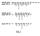
Figure 1. Sequence of peptide #816 ([SEQ ID No. :57:]), oligo PHY-31
([SEQ ID No. :64:]), peptide #1110 ([SEQ ID No. :62:]), oligo PHY-34
([SEQ ID No. :65:]) and oligo PHY-35 ([SEQ ID No. :52:]). pH2.5 acid
phosphatase oligonucleotide PHY-31 is a 17mer mixture with 64 fold
degeneracy and a single inosine. Peptide #816 is derived from an
endoproteinase Lys-C digestion of purified native acid phosphatase. PHY-34
is a 17mer mixture with 128 fold degeneracy. PHY-35 is a 17mer mixture
with 64 fold degeneracy. Both PHY-34 and PHY-35 are necessary for
complete representation of Peptide #1110. Peptide #1110 is derived from a
trypsin digestion of purified native acid phosphatase.
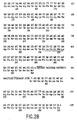
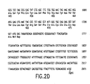
Figure 2. Nucleotide sequence from the 2.1 kb SphI fragment
containing the pH 2.5 acid phosphatase gene [SEQ ID No. :1:] with deduced
amino acid translation [SEQ ID No. :2:]. The intron donor, lariat and
acceptor sequence as determined by cDNA sequencing are overlined. The
nucleotide sequence corresponding to peptides #816 ([SEQ ID No. :57:]) and
#1110 ([SEQ ID No. :62:]) is underlined. The genomic nucleotide sequence
was determined by the M 13-dideoxy method (Sanger, F., et al., Proc. Natl.
Acad. Sci. USA 74:5463-5467 (1977) with the use of the United States
Biochemical Sequencase II kit.
Figure 3. The amino acid sequences of the phytase tryptic peptides
#792 [SEQ ID No. :43:] and #420 [SEQ ID No. :23:] and the deduced
oligonucleotides [SEQ ID Nos. :3:, :4:, :5: and :6:] used in the production of
the phytase probe by nested PCR amplification.
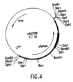
Figure 4. Plasmid pALK169. The map of pALK169 containing the 2.4
kb SphI insert and showing the restriction map of the insert. The location of
the phytase gene is shown by an arrow. The hybridization site for the 350 bp
PCR fragment in the phytase sequence is shown by an intensified line.
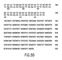
Figure 5. The nucleotide sequence of the phytase gene. [SEQ ID Nos.
:7: (DNA) and :8: (amino acid)].
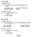
Figure 6. The PCR primers used for making the cbh1-phytase fusion
fragments [SEQ ID Nos. :9: and :10: and :11: and :12:].
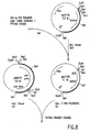
Figure 7. Construction of pALK171. The phytase gene with its own
signal sequence was fused to the cbh1 promoter. Only the relevant restriction
sites are shown.
Figure 8. Construction of pALK 172. The phytase gene was fused to
the cbh1 signal sequence. Only the relevant restriction sites are shown.
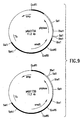
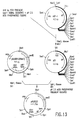
Figure 9. Plasmids pALK173A and pALK173B. The maps of the
plasmids containing the phytase gene with its own promoter and the selection
marker. amdS gene, are shown. In the plasmid, pALK173A the transcriptional
orientation of the phytase and amdS genes is the same; and in the plasmid
pALK173B, the transcriptional orientation of these two genes is opposite to
each other.
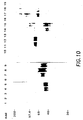
Figure 10. Western blots of the samples from the culture supernatants
of the Trichoderma host strains and transformants producing phytase. Lane 1:
50 ng of purified Aspergillus ALKO 243 phytase; Lane 2: 15 ng of endoF-treated
Aspergillus ALKO 243 phytase; Lanes 3 and 10: T. reesei ALKO 233;
Lanes 4-5 and 11-12: T. reesei ALKO 233 transformant 171FR/ A4 and A13,
respectively; Lanes 6 and 13: T. reesei ALKO 2221; Lanes 7-8 and 14-15:
T. reesei ALKO 2221 transformant 171FR/A5 and A9, respectively: Lane 9:
T. reesei ALKO 2221 transformant D2; Lane 16: T. reesei ATCC56765;
Lanes 17, 18. 19: T. reesei ATCC56765 transformants 171FR/A21, A11, and
A23, respectively. In each case 2 µl of 1:10 dilution of the culture supernatant
were run in the gel, 171FR: the host transformed with the XbaI fragment
from the plasmid pALK171.
Figure 11. The PCR primers used for making the cbh1 - pH 2.5 acid
phosphatase fusion fragments [SEQ ID Nos. :13: and :14: and :15:].
Figure 12. Construction of the plasmid pALK533. The pH 2.5 acid
phosphatase gene with its own signal sequence was fused to the cbh1
promoter.
Figure 13. Construction of the plasmid pALK532. The pH 2.5 acid
phosphatase gene was fused to the cbh1 signal sequence and promoter.
Figure 14. Western blot of the Trichoderma transformants producing
pH 2.5 acid phosphatase. Lane 1: 10ng of purified Aspergillus ALKO 243 pH
2.5 acid phospharase; Lane 2: 10ng of endoF treated Aspergillus ALKO 243
pH 2.5 acid phosphatase; and Lanes 3-9: 60ng of protein from the each of the
culture supernatants of Trichoderma reesei ALKO 2221 transformants SC-9,
KA-31, KA-17, KB-44, KB-18, SB-4 and KA-28, respectively.
I. DEFINITIONS
In the description that follows, a number of terms used in recombinant
DIVA (rDNA) technology are extensively utilized. In order to provide a clear
and consistent understanding of the specification and claims, including the
scope to be given such terms, the following definitions are provided.
Gene. A DNA sequence containing a template for an RNA
polymerase. The RNA transcribed from a gene may or may not code for a
protein. RNA that codes for a protein is termed messenger RNA (mRNA) and,
in eukaryotes, is transcribed by RNA polymerase II. A gene containing a
RNA polymerase II template (as a result of a RNA polymerase II promoter)
wherein an RNA sequence is transcribed which has a sequence complementary
to that of a specific mRNA, but is not normally translated may also be
constructed. Such a gene construct is herein termed an "antisense RNA gene"
and such an RNA transcript is termed an "antisense RNA." Antisense RNAs
are not normally translatable due to the presence of translational stop codons
in the antisense RNA sequence.
A "complementary DNA" or "cDNA" gene includes recombinant genes
synthesized by, for example, reverse transcription of mRNA, thus lacking
intervening sequences (introns). Genes clones from genomic DNA may or may
not contain introns.
Cloning vehicle. A plasmid or phage DNA or other DNA sequence
which is able to carry genetic information, specifically DNA, into a host cell.
A cloning vehicle is often characterized by one or a small number of
endonuclease recognition sites at which such DNA sequences may be cut in
a determinable fashion without loss of an essential biological function of the
vehicle, and into which a desired DNA may be spliced in order to bring about
its cloning into the host cell. The cloning vehicle may further contain a
marker suitable for use in the identification of cells transformed with the
cloning vehicle, and origins of replication that allow for the maintenance and
replication of the vehicle in one or more prokaryotic or eukaryotic hosts.
Markers, for example, are tetracycline resistance or ampicillin resistance. The
word "vector" is sometimes used for "cloning vehicle." A "plasmid" is a
cloning vehicle, generally circular DNA, that is maintained and replicates
autonomously in at least one host cell.
Expression vehicle. A vehicle or vector similar to a cloning vehicle
but which supports expression of a gene that has been cloned into it, after
transformation into a host. The cloned gene is usually placed under the
control of (i.e., operably linked to) certain control sequences such as promoter
sequences, that may be provided by the vehicle or by the recombinant
construction of the cloned gene. Expression control sequences will vary
depending on whether the vector is designed to express the operably linked
gene in a prokaryotic or eukaryotic host and may additionally contain
transcriptional elements such as enhancer elements (upstream activation
sequences) and termination sequences, and/or translational initiation and
termination sites.
Host. A host is a cell, prokaryotic or eukaryotic, that is utilized as the
recipient and carrier of recombinant material.
Eukaryotic host. A "eukaryotic host" may be any cell from a
eukaryotic organism, including, for example, animal, plant, fungi and yeast.
Host of the Invention. The "host of the invention" is a Trichoderma
reesei host that has been engineering to produce recombinant phytase and/or
pH 2.5 acid phosphatase according to the methods of the invention.
Functional Derivative. A "functional derivative" of a protein or
nucleic acid, is a molecule that has been chemically or biochemically derived
from (obtained from) such protein or nucleic acid and which retains a
biological activity (either functional or structural) that is a characteristic of the
native protein or nucleic acid. A "mutant" of a protein or nucleic acid is a
biochemical or chemical derivative of such protein or nucleic acid. The term
"functional derivative" is intended to include "mutants," "fragments,"
"variants," "analogues," or "chemical derivatives" of a molecule that retain a
desired activity of the native molecule.
As used herein, a molecule is said to be a "chemical derivative" of
another molecule when it contains additional chemical moieties not normally
a part of the molecule. Such moieties may improve the molecule's solubility,
absorption, biological half life, etc. The moieties may decrease the toxicity
of the molecule, or eliminate or attenuate any undesirable side effect of the
molecule, etc. Moieties capable of mediating such effects are disclosed in
Remington's Pharmaceutical Sciences (1980). Procedures for coupling such
moieties to a molecule are well known in the art.
Fragment. A "fragment" of a molecule such as a protein or nucleic
acid is meant to refer to a portion of the native amino acid or nucleotide
genetic sequence, and in particular the functional derivatives of the invention.
Variant or Analog. A "variant" or "analog" of a protein or nucleic
acid is meant to refer to a molecule substantially similar in structure and
biological activity to either the native molecule, such as that encoded by a
functional allele.
II. THE HOSTS OF THE INVENTION
T. reesei does not produce endogenous phytase, Instead, other enzyme
components such as β-glucan degrading activity, important in e.g. feed
applications, are produced in high amounts. Thus the use of T. reesei as a
production host for fungal phytase and pH 2.5 acid phosphatase results in
secretion of a totalty different enzyme composition when compared to that
secreted from Aspergillus. In addition, by using Trichoderma reesei as a source of
a composition containing phytate degrading enzymes, some difficult problems
in downstream processing that occur with similar Aspergillus compositions
(e.g., in filtration) are avoided. This is because the mode of growth of the
recombinant T. reesei is different than that of Aspergilli, the mycelium being
most often fluid and easily separable. Thus, by producing these enzymes in
the hosts of the invention, no problems in subsequent filtration of the secreted
material is seen, as is the case with the often slimy and thick mycelium of
Aspergilli.
Improved amounts of phytase and pH 2.5 acid phosphatase (as
compared to synthesis in Aspergillus) can be produced in the T. reesei
expression system by inserting a DNA sequence obtained from A. niger,
coding for phytase or pH 2.5 acid phosphatase activity, into a T. reesei
expression cassette containing the cbh1 promoter and the Aspergillus amd S
gene as a transformation marker. Transformation of the construct to T. reesei
hosts results in stable transformants expressing the phytase or pH 2.5 acid
phosphatase in high amounts in a novel background of accompanying enzyme
activities.
The mixture produced by T. reesei contains high β-glucanase activity
and low glucoamylase activity. Moreover, the amount of phytase produced
by recombinant T. reesei strains in shake flask cultivations is comparable to
the level of which the main cellulase, the endogenous cellobiohydrolase I, is
expressed, more than 1 g/l. The amount of the pH 2.5 acid phosphatase
produced by the recombinant strains in shake flask cultivations is less than 0.5
g/l.
Aspergillus niger var. awamori ALKO 243 (ATCC 38854) (IFO4033)
phytase and acid phosphatase (optimum pH 2.5) were overexpressed in
Trichoderma reesei under the control of the Trichoderma cellobiohydrolase I
(cbh1) promoter. In addition, the phytase gene was expressed from its own
promoter.
For both the genes, two constructions utilizing the cbh1 promoter were
made: in one construction the phytase or acid phosphatase signal sequence was
used and in the other construction the cbh1 signal sequence was used. In all
cases, the fusions were made precise by using PCR and the plasmids were
constructed so that the expression cassette could be separated from the vector
backbone prior to transformations. Thus it was possible to transform strains
with only the desired sequences (and not the entire vector used for maintaining
the sequences) and thus to obtain strains that did not contain any "foreign"
sequences; such strains were suitable for industrial purposes.
Three Trichoderma reesei strains, ATCC 56765 (RutC-30), ALKO 233
(VTT-D-79125) and a low aspartyl protease producing strain ALKO 2221
were used as hosts for phytase expression. For acid phosphatase expression,
only T. reesei ALKO 2221 was transformed. When phytase was expressed
under the cbh1 promoter in Trichoderma, the best transformation with no
E. coli sequences produced in shake flask cultivations about 3,600 fold more
phytase than the nontransformed A. niger ALKO 243. When the phytase
promoter was used, the best yield obtained in shake flask cultivations of T.
reesei transformants was about 120 fold that obtained with A. niger ALKO
243. The best acid phosphatase activities obtained were about 240 fold higher
compared to the levels produced by the A. niger ALKO 243 strain.
The molecular weights (in SDS-PAGE) of the phytase and pH 2.5 acid
phosphatase secreted by Trichoderma were different from those secreted by
Aspergillus. The difference seemed to be due to different glycosylation.
The production level of phytase obtained when the Aspergillus gene
was expressed in Trichoderma under the control of a Trichoderma promoter
was surprisingly high.
The use of T. reesei as a production host for fungal phytase and pH 2.5
acid phosphatase results in totally different enzyme preparations as compared
to that from Aspergillus. When compared to Aspergillus preparations, the
mixtures produced by T. reesei contain substantially higher β-glucanase and
proportionally lower glucoamylase activities thus making T. reesei preparations
preferable to be used e.g. in animal feed.
The hosts of the invention are meant to include all Trichoderma.
Trichoderma are classified on the basis of morphological evidence of
similarity. T. reesei was formerly known as T. viride Pers. or T. koningii
Oudem; sometimes it was classified as a distinct species of the T.
longibrachiatum group. The entire genus Trichoderma, in general, is
characterized by rapidly growing colonies bearing tufted or pustulate,
repeatedly branched conidiophores with lageniform phialides and hyaline or
green conidia borne in slimy heads (Bissett, J., Can. J. Bot. 62:924-931
(1984)).
The fungus called T. reesei is clearly defined as a genetic family
originating from the strain QM6a, that is, a family of strains possessing a
common genetic background originating from a single nucleus of the particular
isolate QM6a. Only those strains are called T. reesei.
Classification by morphological means is problematic and the first
recently published molecular data from DNA-fingerprint analysis and the
hybridization pattern of the cellobiohydrolase 2 (cbh2) gene in T. reesei and
T. longibrachiatum clearly indicates a differentiation of these strains (Meyer,
W. et al., Curr. Genet. 21:27-30 (1992); Morawetz, R. et al., Curr. Genet.
21:31-36 (1992)).
However, there is evidence of similarity between different Trichoderma
species at the molecular level that is found in the conservation of nucleic acid
and amino acid sequences of macromolecular entities shared by the various
Trichoderma species. For example, Cheng, C., et al., Nucl. Acids. Res.
18:5559 (1990), discloses the nucleotide sequence of T. viride cbh1. The gene
was isolated using a probe based on the T. reesei sequence. The authors note
that there is a 95 % homology between the amino acid sequences of the T.
viride and T. reesei gene. Goldman, G.H. et al., Nucl. Acids Res. 18:6717
(1990), discloses the nucleotide sequence of phosphoglycerate kinases from T.
viride and notes that the deduced amino acid sequence is 81 % homologous
with the phosphoglycerate kinase gene from T. reesei. Thus, the species
classified to T. viride and T. reesei must genetically be very close to each
other.
In addition, there is a high similarity of transformation conditions
among the Trichoderma. Although practically all the industrially important
species of Trichoderma can be found in the formerly discussed Trichoderma
section Longibrachiatum, there are some other species of Trichoderma that are
not assigned to this section. Such a species is, for example, Trichoderma
harzianum, which acts as a biocontrol agent against plant pathogens. A
transformation system has also been developed for this Trichoderma species
(Herrera-Estrella, A. et al., Molec. Microbiol. 4:839-843 (1990)) that is
essentially the same as that taught in the application. Thus, even though
Trichoderma harzianum is not assigned to the section Longibrachiatum, the
method used by Herrera-Estrella in the preparation of spheroplasts before
transformation is the same. The teachings of Herrera-Estrella show that there
is not a significant diversity of Trichoderma spp. such that the transformation
system of the invention would not be expected to function in all Trichoderma.
Further, there is a common functionality of fungal transcriptional
control signals among fungal species. At least three A. nidulans promoter
sequences, amdS, argB, and gpd, have been shown to give rise to gene
expression in T. reesei. For amdS and argB, only one or two copies of the
gene are sufficient to being about a selectable phenotypes (Penttilä et al., Gene
61:155-164 (1987). Gruber, F. et al., Curr. Genetic 18:71-76 (1990) also
notes that that fungal genes can often be successfully expressed across different
species.
Many species or Trichoderma are available from a wide variety of
resource centers that contain fungal culture collections. In addition,
Trichoderma species are catalogued in various databases. These resources and
databases are summerized by O'Donnell, K. et al., in Biochemistry of
Filamentous Fungi: Technology and Products, D. B. Finkelstein et al., eds.,
Butterworth-Heinemann, Stoneham, MA. USA. 1992, pp. 3-39.
III. CONSTRUCTION OF THE HOSTS OF THE INVENTION
The process for genetically engineering the hosts of the invention,
according to the invention, is facilitated through the isolation and partial
sequencing of pure protein encoding an enzyme of interest or by the cloning
of genetic sequences which are capable of encoding such protein with
polymerase chain reaction technologies; and through the expression of such
genetic sequences. As used herein, the term "genetic sequences" is intended
to refer to a nucleic acid molecule (preferably DNA). Genetic sequences
which are capable of encoding a protein are derived from a variety of sources.
These sources include genomic DNA, cDNA, synthetic DNA, and
combinations thereof. The preferred source of genomic DNA is a fungal
genomic library. The preferred source of the cDNA is a cDNA library
prepared from fungal mRNA grown in conditions known to induce expression
of the desired mRNA or protein.
The genomic DNA may or may not include naturally
occurring introns. Moreover, such genomic DNA may be obtained in
association with the 5' promoter region of the gene sequences and/or with the
3' transcriptional termination region. Further, such genomic DNA may be
obtained in association with the genetic sequences which encode the 5' non-translated
region of the mRNA and/or with the genetic sequences which
encode the 3' non-translated region. To the extent that a host cell can
recognize the transcriptional and/or translational regulatory signals associated
with the expression of the mRNA and protein, then the 5' and/or 3' non-transcribed
regions of the native gene, and/or, the 5' and/or 3' non-translated
regions of the mRNA may be retained and employed for transcriptional and
translational regulation. Genomic DNA can be extracted and purified from
any host cell, especially a fungal host cell, which naturally expresses the
desired protein by means well known in the art.
For cloning into a vector, such suitable DNA preparations (either
genomic DNA or cDNA) are randomly sheared or enzymatically cleaved,
respectively, and ligated into appropriate vectors to form a recombinant gene
(either genomic or cDNA) library.
A DNA sequence encoding a desired protein
may be inserted into a DNA vector in accordance with
conventional techniques, including blunt-ending or sraggered-ending termini
for ligation, restriction enzyme digestion to provide appropriate termini, filling
in of cohesive ends as appropriate, alkaline phosphatase treatment to avoid
undesirable joining, and ligation with appropriate ligases. Techniques for such
manipulations are disclosed by Maniatis, T., (Maniatis, T. et al., Molecular
Cloning (A Laboratory Manual), Cold Spring Harbor Laboratory, second
edition. 1988) and are well known in the art.
Libraries containing sequences coding for the desired gene may be
screened and the desired gene sequence identified by any means which
specifically selects for a sequence coding for such gene or protein such as, for
example, a) by hybridization with an appropriate nucleic acid probe(s)
containing a sequence specific for the DNA of this protein, or b) by
hybridization-selected translational analysis in which native mRNA which
hybridizes to the clone in question is translated in vitro and the translation
products are further characterized, or, c) if the cloned genetic sequences are
themselves capable of expressing mRNA, by immunoprecipitation of a
translated protein product produced by the host containing the clone.
Oligonucleotide probes specific for a certain protein which can be used
to identify clones to this protein can be designed from the knowledge of the
amino acid sequence of the protein or from the knowledge of the nucleic acid
sequence of the DNA encoding such protein or a related protein.
Alternatively, antibodies may be raised against purified forms of the protein
and used to identify the presence of unique protein determinants in
transformants that express the desired cloned protein. The sequence of amino
acid residues in a peptide is designated herein either through the use of their
commonly employed three-letter designations or by their single-letter
designations. A listing of these three-letter and one-letter designations may be
found in textbooks such as Biochemistry, Lehninger, A., Worth Publishers,
New York, NY (1970). When the amino acid sequence is listed horizontally,
unless otherwise stated, the amino terminus is intended to be on the left end
and the carboxy terminus is intended to be at the right end. Similarly, unless
otherwise stated or apparent from the context, a nucleic acid sequence is
presented with the 5' end on the left.
Because the genetic code is degenerate, more than one codon may be
used to encode a particular amino acid (Watson, J.D., In: Molecular Biology
of the Gene, 3rd Ed., W.A. Benjamin, Inc., Menlo Park, CA (1977), pp. 356-357).
The peptide fragments are analyzed to identify sequences of amino acids
which may be encoded by oligonucleotides having the lowest degree of
degeneracy. This is preferably accomplished by identifying sequences that
contain amino acids which are encoded by only a single codon.
Although occasionally an amino acid sequence may be encoded by only
a single oligonucleotide sequence, frequently the amino acid sequence may be
encoded by any of a set of similar oligonucleotides. Importantly, whereas all
of the members of this set contain oligonucleotide sequences which are capable
of encoding the same peptide fragment and, thus, potentially contain the same
oligonucleotide sequence as the gene which encodes the peptide fragment, only
one member of the set contains the nucleotide sequence that is identical to the
exon coding sequence of the gene. Because this member is present within the
set, and is capable of hybridizing to DNA even in the presence of the other
members of the set, it is possible to employ the unfractionated set of
oligonucleotides in the same manner in which one would employ a single
oligonucleotide to clone the gene that encodes the peptide.
Using the genetic code, one or more different oligonucleotides can be
identified from the amino acid sequence, each of which would be capable of
encoding the desired protein. The probability that a particular oligonucleotide
will, in fact, constitute the actual protein encoding sequence can be estimated
by considering abnormal base pairing relationships and the frequency with
which a particular codon is actually used (to encode a particular amino acid)
in eukaryotic cells. Using "codon usage rules," a single oligonucleotide
sequence, or a set of oligonucleotide sequences, that contain a theoretical
"most probable" nucleotide sequence capable of encoding the protein
sequences is identified.
The suitable oligonucleotide, or set of oligonucleotides, which is
capable of encoding a fragment of a certain gene (or which is complementary
to such an oligonucleotide, or set of oligonucleotides) may be synthesized by
means well known in the art (see, for example, Synthesis and Application of
DNA and RNA, S.A. Narang, ed., 1987, Academic Press, San Diego, CA)
and employed as a probe to identify and isolate a clone to such gene by
techniques known in the art. Techniques of nucleic acid hybridization and
clone identification are disclosed by Maniatis, T., et al., in: Molecular
Cloning, A Laboratory Manual, Cold Spring Harbor Laboratories. Cold Spring
Harbor, NY (1982)), and by Hames, B.D., et al., in: Nucleic Acid
Hvbridization, A Practical Approach, IRL Press, Washington, DC (1985)).
Those members of the above-described gene library which are found to be
capable of such hybridization are then analyzed to determine the extent and
nature of coding sequences which they contain.
To facilitate the detection of a desired DNA coding sequence, the
above-described DNA probe is labeled with a detectable group. Such
detectable group can be any material having a detectable physical or chemical
property. Such materials have been well-developed in the field of nucleic acid
hybridization and in general most any label useful in such methods can be
applied to the present invention. Particularly useful are radioactive labels,
such as 32P, 3H, 14C, 35S, 125I, or the like. Any radioactive label may be
employed which provides for an adequate signal and has a sufficient half-life.
If single stranded, the oligonucleotide may be radioactively labelled using
kinase reactions. Alternatively, polynucleotides are also useful as nucleic acid
hybridization probes when labeled with a non-radioactive marker such as
biotin, an enzyme or a fluorescent group.
Thus, in summary, the elucidation of a partial protein sequence,
permits the identification of a theoretical "most probable" DNA sequence, or
a set of such sequences, capable of encoding such a peptide. By constructing
an oligonucleotide complementary to this theoretical sequence (or by
constructing a set of oligonucleotides complementary to the set of "most
probable" oligonucleotides), one obtains a DNA molecule (or set of DNA
molecules), capable of functioning as a probe(s) for the identification and
isolation of clones containing a gene.
In an alternative way of cloning a gene, a library is prepared using an
expression vector, by cloning DNA or, more preferably cDNA prepared from
a cell capable of expressing the protein into an expression vector. The library
is then screened for members which express the desired protein, for example,
by screening the library with antibodies to the protein.
The above discussed methods are, therefore, capable of identifying
genetic sequences which are capable of encoding a protein or biologically
active or antigenic fragments of this protein. In order to further characterize
such genetic sequences, and, in order to produce the recombinant protein, it
is desirable to express the proteins which these sequences encode. Such
expression identifies those clones which express proteins possessing characteristics
of the desired protein. Such characteristics may include the ability to
specifically bind antibody, the ability to elicit the production of antibody which
are capable of binding to the native, non-recombinant protein, the ability to
provide a enzymatic activity to a cell that is a property of the protein, and the
ability to provide a non-enzymatic (but specific) function to a recipient cell,
among others.
A DNA sequence may be shortened by means known in the art to
isolate a desired gene from a chromosomal region that contains more
information than necessary for the utilization of this gene in the hosts of the
invention. For example, restriction digestion may be utilized to cleave the full-length
sequence at a desired location. Alternatively, or in addition, nucleases
that cleave from the 3'-end of a DNA molecule may be used to digest a certain
sequence to a shortened form, the desired length then being identified and
purified by gel electrophoresis and DNA sequencing. Such nucleases include,
for example, Exonuclease III and Bal31. Other nucleases are well known in
the art.
If the coding sequence and an operably linked promoter are introduced
into a recipient eukaryotic cell as a non-replicating DNA (or RNA), non-integrating
molecule, the expression of the encoded protein may occur through
the transient (nonstable) expression of the introduced sequence.
Preferably the coding sequence is introduced on a DNA (or RNA)
molecule, such as a closed covalent circular molecule that is incapable of
autonomous replication, or preferable a linear molecule that integrates into the
host chromosome. Genetically stable transformants may be constructed with
vector systems, or transformation systems, whereby a desired DNA is
integrated into the host chromosome. Such integration may occur de novo
within the cell or, be assisted by transformation with a vector which
functionally inserts itself into the host chromosome, for example, transposons
or other DNA elements which promote integration of DNA sequences in
chromosomes. A vector is employed which is capable of integrating the
desired gene sequences into a fungal host cell chromosome.
The genes coding for phytase or pH 2.5 acid phosphatase having the amino acid
sequence of SEQ ID NO:2 under the control of suitable promoters may be combined in one
plasmid construction and introduced into the host cells by transformation. The nature of the
plasmid vector will depend on the host organism. In the practical realization
of the invention the filamentous fungus Trichoderma reesei has been employed as a
model. Thus, for T. reesei, vectors
incorporating DNA that provides for integration of the sequences encoding the
phytase or pH 2.5 acid phosphatase genes into the host's chromosome are
preferred. Such targeting to, for example, the cbh1 locus may be achieved by
providing cbh1 coding or flanking sequences on the recombinant construct, in
an amount sufficient to direct integration to this locus at a relevant frequency.
Cells which have stably integrated the introduced DNA into their
chromosomes are selected by also introducing one or more markers which
allow for selection of host cells which contain the expression vector in the
chromosome, for example the marker may provide biocide resistance, e.g.,
resistance to antibiotics, or heavy metals, such as copper, or the like. The
selectable marker gene can either be directly linked to the DNA gene
sequences to be expressed, or introduced into the same cell by co-transfection.
A genetic marker especially for the transformation of the hosts
of the invention is amdS, encoding acetamidase and thus enabling Trichoderma
to grow on acetamide as the only nitrogen source.
To express a desired protein,
transcriptional and translational signals recognizable by an appropriate host are
necessary. The cloned coding sequences, obtained through the methods
described above, and preferably in a double-stranded form, may be operably
linked to sequences controlling transcriptional expression in an expression
vector, and introduced into a host cell to
produce recombinant protein. Depending
upon which strand of the coding sequence is operably linked to the sequences
controlling transcriptional expression, it is also possible to express antisense
RNA.
Expression of the protein in different hosts may result in different post-translational
modifications which may alter the properties of the protein.
A nucleic acid molecule, such as DNA, is said to be "capable of
expressing" a polypeptide if it contains expression control sequences which
contain transcriptional regulatory information and such sequences are
"operably linked" to the nucleotide sequence which encodes the polypeptide.
An operable linkage is a linkage in which a sequence is connected to
a regulatory sequence (or sequences) in such a way as to place expression of
the sequence under the influence or control of the regulatory sequence. Two
DNA sequences (such as a coding sequence and a promoter region sequence
linked to the 5' end of the coding sequence) are said to be operably linked if
induction of promoter function results in the transcription of mRNA encoding
the desired protein and if the nature of the linkage between the two DNA
sequences does not (1) result in the introduction of a frame-shift mutation,
(2) interfere with the ability of the expression regulatory sequences to direct
the expression of the protein, antisense RNA, or (3) interfere with the ability
of the DNA template to be transcribed. Thus, a promoter region would be
operably linked to a DNA sequence if the promoter was capable of effecting
transcription of that DNA sequence.
The precise nature of the regulatory regions needed for gene expression
may vary between species or cell types, but shall in general include, as
necessary, 5' non-transcribing and 5' non-translating (non-coding) sequences
involved with initiation of transcription and translation respectively, such as
the TATA box, capping sequence, CAAT sequence, and the like. Especially,
such 5' non-transcribing control sequences will include a region which
contains a promoter for transcriptional control of the operably linked gene.
Such transcriptional control sequences may also include enhancer sequences
or upstream activator sequences, as desired.
Expression of a protein in eukaryotic hosts such as fungus requires the
use of regulatory regions functional in such hosts, and preferably fungal
regulatory systems. A wide variety of transcriptional and translational regulatory
sequences can be employed, depending upon the nature of the host.
Preferably, these regulatory signals are associated in their native state with a
particular gene which is capable of a high level of expression in the host cell.
In eukaryotes, where transcription is not linked to translation, such
control regions may or may not provide an initiator methionine (AUG) codon,
depending on whether the cloned sequence contains such a methionine. Such
regions will, in general, include a promoter region sufficient to direct the
initiation of RNA synthesis in the host cell. Promoters from filamentous
fungal genes which encode a mRNA product capable of translation are
preferred, and especially, strong promoters can be employed provided they
also function as promoters in the host cell. Preferred strong eukaryotic promoters
for use in Trichoderma include the T. reesei cbh1 gene promoter or a
promoter of another cellulase gene such as that for the cbh2, egl1 or egl2 gene
may be used. In addition to the use of Trichoderma regulatory elements, the
expression of proteins may be placed under the control of regulatory elements
from Aspergillus nidulans (for example, the argB gene promoter and the amdS
gene promoter), Aspergillus niger (for example, the phytase promoter or the
glucoamylase gene promoter) However, expression under non-Trichoderma
regulatory elements such as these may be very low as compared to the use of
Trichoderma elements, and especially those of T. reesei.
As is widely known, translation of eukaryotic mRNA is initiated at the
codon which encodes the first methionine. For this reason, it is preferable to
ensure that the linkage between a eukaryotic promoter and a DNA sequence
which encodes the desired protein does not
contain any intervening codons which are capable of encoding a methionine.
The presence of such codons results either in a formation of a fusion protein
(if the AUG codon is in the same reading frame as the protein-coding DNA
sequence) or a frame-shift mutation (if the AUG codon is not in the same
reading frame as the protein-coding sequence).
It may be desired to construct a fusion product that contains a partial
coding sequence (usually at the amino terminal end) of a protein and a second
coding sequence (partial or complete) of a phytase degrading enzyme of the
invention. The sequence that does not encode the phytase degrading enzyme
may or may not function as a signal sequence for secretion of the protein from
the host cell. For example, the sequence coding for desired protein may be
linked to a signal sequence which will allow secretion of the protein from, or
the compartmentalization of the protein in, a particular host. Such fusion
protein sequences may be designed with or without specific protease sites such
that a desired peptide sequence is amenable to subsequent removal. In a
preferred embodiment, the native signal sequence of a fungal protein is used,
or a functional derivative of that sequence that retains the ability to direct the
secretion of the peptide that is operably linked to it, Aspergillus
leader/secretion signal elements also function in Trichoderma.
Transcriptional initiation regulatory signals can be selected which allow
for repression or activation, so that expression of the operably linked genes
can be modulated. For example, regulatory signals may be temperature-sensitive
so that by varying the temperature, expression can be repressed or
initiated, or are subject to chemical regulation, e.g., metabolite. Translational
signals are not necessary when it is desired to express antisense RNA
sequences.
If desired, the non-transcribed and/or non-translated regions 3' to the
sequence coding for a desired protein can be obtained by the above-described
cloning methods. The 3'-non-transcribed region may be retained for its
transcriptional termination regulatory sequence elements, or for those elements
which direct polyadenylation in eukaryotic cells. Where the native expression
control sequences signals do not function satisfactorily in a host cell, then
sequences functional in the host cell may be substituted.
The vectors of the invention may further comprise other operably
linked regulatory elements such as DNA elements which confer antibiotic
resistance, or origins of replication for maintenance of the vector in one or
more host cells.
In another embodiment, especially for maintenance of the vectors of the
invention in prokaryotic cells, or in yeast S. cerevisiae cells, the introduced
sequence is incorporated into a plasmid or viral vector capable of autonomous
replication in the recipient host. Any of a wide variety of vectors may be
employed for this purpose. In Bacillus hosts, integration of the desired DNA
may be necessary.
Factors of importance in selecting a particular plasmid or viral vector
include: the ease with which recipient cells that contain the vector may be
recognized and selected from those recipient cells which do not contain the
vector; the number of copies of the vector which are desired in a particular
host; and whether it is desirable to be able to "shuttle" the vector between host
cells of different species.
Preferred S. cerevisiae yeast plasmids include those containing the 2-micron
circle, etc., or their derivatives. Such plasmids are well known in the
art (Botstein, D., et al,, Miami Wntr, Symp, 19:265-274 (1982); Broach, J.R.,
in: The Molecular Biology of the Yeast Saccharomyces: Life Cycle and
Inheritance, Cold Spring Harbor Laboratory, Cold Spring Harbor, NY, p.
445-470 (1981); Broach, J.R., Cell 28:203-204 (1982); Bollon, D. P. , et al.,
J, Clin, Hematol, Oncol, 10:39-48 (1980); Maniatis, T., In: Cell Biology:
A Comprehensive Treatise, Vol. 3. Gene Expression, Academic Press, NY,
pp. 563-608 (1980)), and are commercially available.
Once the vector or DNA sequence containing the construct(s) is
prepared for expression, the DNA construct(s) is introduced into an
appropriate host cell by any of a variety of suitable means, including
transformation. After the introduction of the vector, recipient cells are grown
in a selective medium, which selects for the growth of vector-containing cells.
Expression of the cloned gene sequence(s) results in the production of the
desired protein, or in the production of a fragment of this protein. This
expression can take place in a continuous manner in the transformed cells, or
in a controlled manner, for example, by induction of expression.
Fungal transformation is carried out also accordingly to techniques
known in the art, for example, using, for example, homologous recombination
to stably insert a gene into the fungal host and/or to destroy the ability of the
host cell to express a certain protein.
IV. PREPARATION OF ANTIBODIES
In the following description, reference will be made to various
methodologies well-known to those skilled in the an of immunology. Standard
reference works setting forth the general principles of immunology include the
work of Catty, D. (Antibodies, A Practical Approach, Vol. 1, IRL Press,
Washington, DC (1988)); Klein. J. (Immunology: The Science of Cell-Noncell
Discrimination, John Wiley & Sons, New York (1982)); Kennett, R., et al.
in Monoclonal Antibodies, Hybridorna: A New Dimension in Biological
Analyses, Plenum Press, New York (1980)); Campbell, A. ("Monoclonal
Antibody Technology," in: Laboratory Techniques in Biochemistry and
Molecular Biology, Volume 13 (Burdon, R., et al., eds.), Elsevier,
Amsterdam (1984)); and Eisen, H.N., in: Microbiology, 3rd Ed. (Davis,
B.D., et al., Harper & Row, Philadelphia (1980)).
An antibody is said to be "capable of binding" a molecule if it is
capable of specifically reacting with the molecule to thereby bind the molecule
to the antibody. The term "epitope" is meant to refer to that portion of a
hapten which can be recognized and bound by an antibody. An antigen may
have one, or more than one epitope. An "antigen" is capable of inducing an
animal to produce antibody capable of binding to an epitope of that antigen.
The specific reaction referred to above is meant to indicate that the antigen
will react, in a highly selective manner, with its corresponding antibody and
not with the multitude of other antibodies which may be evoked by other
antigens.
The term "antibody" (Ab) or "monoclonal antibody" (Mab) as used
herein is meant to include intact molecules as well as fragments thereof (such
as, for example, Fab and F(ab')2, fragments) which are capable of binding an
antigen, Fab and F(ab')2 fragments lack the Fc fragment of intact antibody,
clear more rapidly from the circulation, and may have less non-specific tissue
binding of an intact antibody (Wahl et al., J. Nucl. Med. 24:316-325 (1983)).
The antibodies of the present invention are prepared by any of a variety
of methods. Preferably, purified phytase or pH 2.5 acid phosphatase protein,
or a fragment thereof, (treated or not treated with endoF or its equivalent to
remove sugar moieties), is administered to an animal in order to induce the
production of sera containing polyclonal antibodies that are capable of binding
sucn phytase or pH 2.5 acid phosphatase.
Cells expressing phytase or pH 2.5 acid phosphatase protein, or a
fragment thereof, or, a mixture of proteins containing phytase or pH 2.5 acid
phosphatase or such fragments, can also be administered to an animal in order
to induce the production of sera containing polyclonal antibodies, some of
which will be capable of binding phytase or pH 2.5 acid phosphatase protein.
If desired, such phytase or pH 2.5 acid phosphatase antibody may be purified
from the other polyclonal antibodies by standard protein purification
techniques and especially by affinity chromatography with purified phytase or
pH 2.5 acid phosphatase or fragments thereof.
A phytase or pH 2.5 acid phosphatase protein fragment may also be
chemically synthesized and purified by HPLC to render it substantially free of
contaminants. Such a preparation is then introduced into an animal in order
to produce polyclonal antisera of high specific activity.
Monoclonal antibodies can be prepared using hybridoma technology
(Kohler et al., Nature 256:495 (1975); Kohler et al., Eur. J. Immunol. 6:511
(1976): Kohler et al., Eur. J. Immunol. 6:292 (1976); Hammerling et al., in:
Monoclonal Antibodies and T-Cell Hvbridomas, Elsevier, N.Y., pp. 563-681
(1981)). In general, such procedures involve immunizing an animal with
phytase or pH 2.5 acid phosphatase protein antigen. The splenocytes of such
animais are extracted and fused with a suitable myeloma cell line. Any
suitable myeloma cell line may be employed in accordance with the present
invention; however, it is preferable to employ the parent myeloma cell line
(SP2O), available from the American Type Culture Collection, Rockville,
Maryland. After fusion, the resulting hybridoma cells are selectively
maintained in HAT medium, and then cloned by limiting dilution as described
by Wands, J.R., et al., Gastroenterology 80:225-232 (1981).
The hybridoma cells obtained through
such a selection are then assayed to identify clones which secrete antibodies
capable of binding the phytase or pH 2.5 acid phosphatase protein antigen.
Through application of the above-described methods, additional cell
lines capable of producing antibodies which recognize epitopes of the phytase
or pH 2.5 acid phosphatase protein can be obtained.
Antibodies against both highly conserved and poorly conserved regions
of the phytase or pH 2.5 acid phosphatase protein are useful for studies on the
control of biosynthesis and catabolism of phytase or pH 2.5 acid phosphatase
protein, and for studies wherein it is necessary to identify or quantitate the
presence and/or of the protein antigen in a composition.
V. PRODUCTION OF PHYTASE AND ACID PHOSPHATASE
The best phytase production levels are obtained when Trichoderma are
transformed with linear DNA using the cbh1 promoter (about 3.800 PNU/ml,
see Table 8). About 3,500 and 3,600 PNU/ml culture medium was obtained
with the best T. reesei ALKO2221 and ALKO233 transformants containing no
E. coli sequences. Both the phytase and the cbh1 signal sequence seemed to
work equally well and the same levels in phytase production could be achieved
when using T. reesei ALKO 2221 or ATCC56765 as a host strain. In T. reesei
ALKO 233 the level of phytase activity produced was higher when the phytase
signal sequence was used.
Phytase is expressed from the Trichoderma reesei hosts of the invention into
the supernatant of the culture medium. The amount of phytase in the culture
medium is generally higher than any hitherto reported amount of a
heterologous protein that was expressed in Trichoderma.
The spectrum of enzymes that accompany phytase in the Trichoderma reesei
strains of the invention is greatly different and advantageous over that of
similar preparations of Aspergillus culture supernatants. Both endoglucanase
and cellobiohydrolase activities are generally substantially higher using the
Trichoderma reesei hosts of the invention. The glycosylation pattern of the phytase
is also different when it is expressed from Trichoderma reesei resulting in a phytase
protein that migrates as multiple bands on Western analysis.
The best production of pH 2.5 acid phosphatase from the Trichoderma reseei
transformants of the invention resulted in 240 APNU/ml culture medium, in
shake flask cultivation and in lactose based medium. As with the phytase, both
the acid phosphatase and the cbh1 signal sequence worked equally well.
The compositions of the invention that contain phytase may be used
directly for the removal of phytic acid, or inositol hexaphosphoric acid, from
raw material, especially phytin-containing raw material, and especially plant
material. Phytase removes the phosphate groups from phytic acid and destroys
its ability to interfere with mineral absorption. When used as an animal feed
additive, the phytase compositions of the invention release phosphate bound
to phytin in grain and thus dramatically reduce the need for doses of additional
phosphate in feed formulations and lessen environmental loads.
The phytase and pH 2.5 acid phosphatase produced according to the
invention may be purified by protein purification methods known in the art.
Having now generally described the invention, the same will become
better understood by reference to certain specific examples that are included
herein for purposes of illustration.
EXAMPLES
EXAMPLE I
Method of Assay of Phytase Activity
Principle Phytase acts on phytate (inositol hexaphosphate) to release
inorganic phosphate. The determination of released inorganic phosphate is
based on the color formed by the reduction of a phosphomolybdate complex.
Unit of Activity One phytase unit (PU) is the amount of enzyme which
liberates, under standard conditions, 1 nmol of inorganic phosphate from
sodium phytate in one minute.
| Assay conditions |
| Substrate | Sodium phytate |
| pH | 5.0 |
| incubation temperature | 37°C ± 0.5°C |
| incubation time |
| | 15 minutes |
| Equipment |
| Water bath | 37°C |
| Water bath |
| | 50°C |
| Spectrophotometer |
| Test tube mixer (vortex) |
| Phosphate Free Glassware |
Reagents All solutions are prepared in deionized water, Milli-Q or
equivalent.
1. Citrate Buffer (0.2 M, pH 5.0). Prepare 0.2 M solutions of
both sodium citrate (C6H8O7Na·2 H2O, 58.8 g/l, Merck 6448) and citric acid
(C6H8O7·H2O, 42.0 g/l, Merck 244) in water. Adjust the pH of the citrate
solution (1 liter) to 5.0 with 0.2 M citric acid (the consumption of citric acid
solution should be about 385 ml). 2. Substrate. Dissolve 1.00 g of sodium phytate (Sigma P-3168)
in about 70 ml citrate buffer. Adjust the pH to 5.0 with 0.2 M citric acid and
adjust the volume to 100 ml with citrate buffer. Fresh substrate solution must
be prepared daily. 3. 15% (w/v) TCA Solution. Prepare from trichloroacetic acid
(Merck 807). 4. 10% (w/v) Ascorbic Acid Solution. Prepare from ascorbic acid
(Merck 127). Store under refrigeration. The solution is stable for seven days. 5. 2.5% (w/v) Ammonium Molybdate Solution. Dissolve 2.5 g
(NH4)6Mo7O24·H2O, Merck 1182) in water and make up to 100 ml. 6. 1 M Sulfuric Acid. Add 55.6 ml of concentrated H2SO4 (Merck
731) to about 800 ml of water, with stirring. Allow to cool and make up to
1000 ml with water. 7. Reagent C. Mix 3 volumes of 1 M sulfuric acid with 1 volume
of 2.5% ammonium molybdate, then add 1 volume of 10% ascorbic acid and
mix well. Fresh reagent C must be prepared daily.
Sample dilution Samples are diluted in citrate buffer. Make duplicate
dilutions of each sample. In case of enzyme powder weigh accurately about
250 mg of sample, dissolve in the buffer and fill to 25 ml in a volumetric
flask, dilute further if necessary.
| Dilution table: |
| Estimated activity PU/ml | Recommended dilution | Dilution factor |
| 2000 | 1 + 19 | 20 |
| 20000 | 1 + 199 | 200 |
| 40000 | 1 + 399 | 400 |
| 100000 | 1 + 999 | 1000 |
| 500000 | 1 + 4999 | 5000 |
Assay
Hydrolysis Pipette 1.0 ml of sample dilution containing 20-190 PU in
two test tubes. Add 2.0 ml of 15% TCA to one of the tubes (blank) and mix.
Put the tubes without TCA in a water bath at 37°C and let them equilibrate for
5 minutes. Using a stopwatch start the hydrolysis by adding sequentially at
proper intervals 1.0 ml of substrate (equilibrated for about 10 minutes at
37°C) to each tube and mix. After exactly 15 minutes incubation stop the
reaction by adding 2.0 ml of TCA to each tube. Mix and cool to room
temperature. Add 1.0 ml substrate to the blank tubes (kept at room
temperature) also and mix. If precipitate occurs it must be separated by
centrifugation for 10 minutes at 2000·g.
Released orthophosphate Pipette 0.4 ml of each sample after
hydrolysis in test tubes. Add 3.6 ml of water to each tube. Add 4.0 ml of
reagent C and mix. Incubate at 50°C for 20 minutes and cool to room
temperature. Measure the absorbance against that of reagent blank (see below)
at 820 nm.
Standard Prepare a 9.0 mM phosphate stock solution. Dissolve and
dilute 612.4 mg KH
2PO
4 (Merck 4873, dried in desiccator with silica) to 500
ml with water in a volumetric flask. Make the following dilutions in water
from the stock solution and use these as standards.
| Dilution | Phosphorus concentration nmol/ml | Phytase activity PU/ml |
| 1:100 | 90 | 240 |
| 1:200 | 45 | 120 |
| 1:400 | 22.5 | 60 |
Pipette 4.0 ml of each dilution to two test tubes. Pipette also 4.0 ml
of water in one tube (reagent blank). Add 4.0 ml of reagent C and mix.
Incubate at 50°C for 20 minutes and cool to room temperature. Measure the
absorbances at 820 nm against that of reagent blank. Prepare a standard curve
by blotting the absorbances against phytase activity (PU/ml). A new standard
line must be constructed with each series of assays.
Calculation Subtract the blank absorbance from the sample absorbance
(the difference should be 0.100 - 1.000). Read the phytase activity (PU/ml)
from the standard line and multiply by the dilution factor. To calculate the
activity (PU/g) of enzyme powders the result (PU/ml) is further multiplied by
25 (ml) and divided by the exact weight of the sample (g).
Preparation of feed and other insoluble samples for phytase analysis
Weigh accurately about 2.5 g of ground sample in two 50 ml beakers. Add
20.0 ml of citrate buffer. Mix using a magnetic stirrer for 30 minutes at room
temperature. Transfer about 10 ml of each in centrifuge tubes and separate
the solid matter by centrifugation for 10 minutes at 2000 g. Apply 2.5 ml of
supernatant on PD-10 gel filtration columns (Sephadex G-25M, Pharmacia 17-0851-01)
equilibrated with 25 ml citrate buffer. Discard the eluate. Then
apply 3.5 ml citrate buffer on the column and collect the eluate in a graduated
cylinder. Fill the volume to 5.0 ml with citrate buffer (dilution factor 2) and
assay for phytase activity. The activity PU/g is obtained by multiplying the
measured activity (PU/ml) by 40 (dilution factor · volume of extraction buffer)
and dividing by the exact weight of sample (g). Reference: Chen et al. Anal.
Chem. 28:1756-1758 (1956).
EXAMPLE 2
Assay of Acid Phosphatase Activity
Principle. Acid phosphatase acts on p-nitrophenyl phosphate to release
inorganic phosphate. The determination of released inorganic phosphate is
based on the color formed by the reduction of phosphomolybdate complex.
Unit of activity. One acid phosphatase unit (HFU) is the amount of
enzyme which liberates, under standard conditions, 1 nmol of inorganic
phosphate from p-nitrophenyl phosphate in one minute.
| Assay conditions. |
| Substrate | p-nitrophenyl phosphate |
| pH | 2.5 |
| Temperature | 37°C ± 0.5°C |
| Incubation time |
| | 15 min |
| Equipment. |
| Water bath | 37°C |
| Water bath |
| | 50°C |
| Spectrophotometer |
| Test tube mixer (vortex) |
| Centrifuge (Hereaus Biofuge 17S. 3090 or equivalent |
| Phosphate Free Glassware |
Reagents. All solutions are prepared in deionized water, Milli-Q or
equivalent.
1. Glycine Buffer (0.2 M, pH 2.5)
Dissolve 15.014 g glycine (Merck 4201) in about 800 ml of water.
Adjust the pH to 2.5 with 1 M hydrochloric acid (consumption should
be about 80 ml) and dilute to 1000 ml with water. 2. Substrate (30 mM)
Dissolve 1.114 g p-nitrophenyl phosphate (Boehringer, 738 352) in
glycine buffer and adjust the volume to 100 ml with the buffer. Fresh
substrate solution must be prepared daily. 3. 15% (w/v) TCA Solution
Prepare from trichloroacetic acid (Merck 807). 4. 10% (w/v) Ascorbic Acid Solution
Prepare from ascorbic acid (Merck 127). Store under refrigeration.
The solution is stable for 7 days. 5. 2.5% (w/v) Ammonium Molybdate Solution
Dissolve 2.5 g (NH4)6MO7O24·4H2O, Merck 1182) in water and make
up to 100 ml. 6. 1 M Sulphuric Acid
Add 55.6 ml of concentrated H2SO4 (Merck 731) to about 800 ml of
water, with stirring. Allow to cool and make up to 1000 ml with
water. 7. Reagent C
Mix 3 volumes of 1 M sulphuric acid with 1 volume of 2.5%
ammonium molybdate, then add 1 volume of 10% ascorbic acid and
mix well. Fresh reagent C must be prepared daily.
Sample dilution. Samples are diluted in glycine buffer. Make
duplicate dilutions of each sample. In case of enzyme powder weigh
accurately about 250 mg of sample, dissolve in the buffer and fill to 25 ml in
a volumetric flask, dilute further if necessary.
Dilution table:
| Estimated activity HFU/ml | Recommended dilution | Dilution factor |
| 20000 | 1 + 19 | 20 |
| 200000 | 1 + 199 | 200 |
| 400000 | 1 + 399 | 400 |
| 1000000 | 1 + 999 | 1000 |
| 5000000 | 1 + 4999 | 5000 |
Hydrolysis: Pipette 1.9 ml of substrate in two test tubes. Add
2.0 ml of 15% TCA to one of the tubes (blank) and mix. Put the tubes
without TCA in a water bath at 37°C and let them equilibrate for 5 min.
Using a stopwatch start the hydrolysis by adding sequentially at proper
intervals 0.1 ml of enzyme dilution to each tube and mix. After exactly 15
min incubation stop the reaction by adding 2.0 ml of TCA to each tube. Mix
and cool to room temperature. Add 0.1 ml of sample to the blank tubes (kept
at room temperature) also and mix. If precipitate occurs it must be separated
by centrifugation for 10 min at 2000 g.
Released orthophosphate: Pipette 0.4 ml of each sample after
hydrolysis in test tubes. Add 3.6 ml of water to each tube. Add 4.0 ml of
reagent C and mix. Incubate at 50°C for 20 min and cool to room
temperature. Measure the absorbance against that of reagent blank (see below)
at 820 nm.
Standard. Prepare a 9.0 mM phosphate stock solution. Dissolve and
dilute 612.4 mg KH
2PO
4 (Merck 4873, dried in dessicator with silica) to 500
ml with water in a volumetric flask. Make the following dilutions in water
from the stock solution and use these as standards.
| Dilution | Phosphorus concentration nmol/ml | Acid phosphatase activity HFU/ml |
| 1:100 | 90 | 2400 |
| 1:200 | 45 | 1200 |
| 1:400 | 22.5 | 600 |
Pipette 4.0 ml of each dilution to two test tubes. Pipette also 4.0 ml
of water in one tube (reagent blank). Add 4.0 ml of reagent C and mix.
Incubate at 50°C for 20 min and cool to room temperature. Measure the
absorbances at 820 nm against that of reagent blank. Prepare a standard curve
by blotting the absorbances against acid phosphatase activity (HFU/ml). A
new standard line must be constructed with each series of assays.
Calculation. Subtract the blank absorbance from the sample absorbance
(the difference should be 0.100-1.000). Read the acid phosphatase activity
(HFU/ml) from the standard line and multiply by the dilution factor. To
calculate the activity (HFU/g) of enzyme powders the result (HFU/ml) is
further multiplied by 25 (ml) and divided by the exact weight of the sample
(g).
Preparation of feed and other insoluble samples for acid phosphatase
analysis. Weigh accurately about 2.5 g of ground sample in two 50 ml
beakers. Add 20.0 ml of glycine buffer. Mix using a magnetic stirrer for 30
min at room temperature. Transfer about 10 ml of each in centrifuge tubes
and separate the solid matter by centrifugation for 10 min at 2000·g. Apply
2.5 ml of supernatant on PD-10 gel filtration columns (Sephadex G-25M.
Pharmacia 17-0851-01) equilibrated with 25 ml glycine buffer. Discard the
eluate. Then apply 3.5 ml of glycine buffer on the column and collect the
eluate in a graduated cylinder. Fill the volume to 5.0 ml with glycine buffer
(dilution factor 2) and assay for acid phosphatase activity. The activity HFU/g
is obtained by, multiplying the measured activity (HFU/ml) by 40 (dilution
factor · volume of extraction buffer) and dividing by the exact weight of
sample (g). Reference: Chen, P.S., et al., Anal. Chem. 28:1756-1758 (1956).
EXAMPLE 3
Purification of Phytase and pH 2.5 Acid Phosphatase
For reference to how the skilled artisan would purify phytase and pH 2.5 acid
phosphatase, the following are provided.
I. PHYTASE
Enzyme purification. Steps were done at 4 to 8 °C unless otherwise
stated. The starting material was the cell-free culture medium concentrate
produced by Aspergillus niger var. awamori ALKO 243.
Ammonium sulphate precipitation. The culture filtrate concentrate (990
ml) was kept on an ice bath and 0.436 g ammonium sulphate per ml was
added (70% saturation). After 30 minutes the precipitate was separated by
centrifugation for 15 minutes at 10000·g and discarded.
Hydrophobic interaction chromatography. The supernatant (1070 ml)
was applied to an Octyl-Sepharose CL-4B (Pharmacia) column (5 cm x 17 cm)
equilibrated with a solution containing 0.436 g (NH4)2SO4 per ml of 20 mM
bis-Tris/HCl (pH 6.2). The column was washed with 500 ml of the
equilibration solution and then developed with a linear gradient of 500 ml
containing 70→0% amonium sulfate in 20 mM bis-Tris/HCl (pH 6.2).
Fractions of 10 ml were collected and analyzed for phytase and acid
phosphatase activity. Most of the phytase activity eluted in the beginning of
the gradient. The fractions were pooled for the next step. The fractions
eluting after phytase activity and containing most of the acid phosphatase
activity were pooled for acid phosphatase purification (see below).
Anion exchange chromatography. The pooled phytase fractions (129
ml) were concentrated by ultrafiltration using an Amicon PM 10 membrane.
The residual ammonium sulphate was removed by PD 10 (Pharmacia) gel
filtration columns equilibrated with 50 mM bis-Tris/HCl (pH 6.2). The
sample, in 24.5 ml, was applied to a DEAE-Sepharose (Pharmacia) column
(5 cm x 7 cm) equilibrated with 50 mM bis-Tris/HCl (pH 6.2). The column
was washed with the equilibrium buffer (100 ml) and developed by a linear
gradient of 200 ml containing 0→0.5 M NaCl in equilibrium buffer.
Gel filtration. The pooled active fractions were concentrated using a
Centricon -30 microconcentrator to a total volume of 600 µl. Portions of 100
µl were run at about 23°C and 0.3 ml/min through a Superose 12-HR 10/30
HPLC column (Pharmacia) equilibrated with 50 mM bis-Tris/HCl (pH 6.2).
Cation exchange. The pooled active fractions were transferred to 50
mM sodium formiate (pH 3.8) using a Centricon -30 microconcentrator. The
sample was applied in two portions of 2 ml to a Mono S HR 5/5 FPLC
column (Pharmacia) equilibrated with 50 mM sodium formiate (pH 3.8) at
about 23°C. The column was washed with the equilibration buffer (10 ml)
and the bound protein was eluted at 60 ml/h with a linear gradient of 20 ml
containing 0→430 mM NaCl in equilibration buffer.
II. ACID PHOSPHATASE
Gel filtration. The pooled fractions containing most of the acid
phosphatase activity from the hydrophobic interaction chromatography step
were concentrated by ultrafiltration using an Amicon PM 10 membrane. The
concentrated sample (25 ml) was run through a Sephacryl S-200 (Pharmacia)
column (2.6 cm x 94 cm) equilibrated with 50 mM bis-Tris/HCl (pH 6.2) at
20 ml/h.
Anion exchange chromatography. The pooled fractions (48 ml) were
applied to a DEAE-Sepharose (Pharmacia) column (5 cm x 7 cm) equilibrated
with 50 mM bis-Tris/HCl (pH 6.2). The column was washed with 100 ml of
equilibration buffer and developed with a linear gradient of 200 ml containing
0→0.5 M NaCl in equilibration buffer.
Anion exchange chromatography. The pooled active fractions were
concentrated and transferred to 20 mM bis-Tris/HCl (pH 6.0) by ultrafiltration
using an Amicon PM 10 membrane. The sample was run in four portions of
3.5 ml on Mono Q HR 5/5 HPLC column (Pharmacia) equilibrated with 20
mM bis-Tris/HCl (pH 6.0) at about 23°C and 60 ml/h. The column was
washed with 10 ml of the equilibrium buffer and the bound protein was eluted
with a linear gradient of 20 ml containing 0→350 mM NaCl in equilibrium
buffer.
Gel filtration. The active fractions were pooled, concentrated and
transferred to 20 mM bis-Tris/HCl (pH 6.2) containing 150 mM NaCl with
Centricon -30 microconcentrator to total volume of 400 µl. Portions of 100
µl were run at about 23°C and 18 ml/h through a Superose 12 HR 10/30
HPLC column (Pharmacia) equilibrated with the sample buffer.
Anion exchange chromatography. The pooled active fractions were
transferred to 20 mM histidin/HCl (pH 5.8) with a
PD 10 gel filtration
column. The sample was run in four portions of 1 ml on
Mono Q HR 5/5
HPLC column (Pharmacia) equilibrated with the sample buffer at about 23°C
and 60 ml/h. The column was washed with 5 ml of the sample buffer and the
bound protein was eluted with a linear gradient of 20 ml containing 0→350
mM NaCl in equilibrium buffer.
| Summary of purification of phytase from Aspergillus niger |
| Step | Total activity (PU) | Total protein (mg) | Specific activity (PU/mg) | Yield (%) | Purification (fold) |
| Culture filtrate | 4486680 | 2119 | 2117 | 100 | 1 |
| Ammonium sulfate supernatant | 3771750 | 1263 | 2986 | 84.1 | 1.4 |
| Octyl Sepharose | 1765881 | 32.3 | 54671 | 39.4 | 26 |
| DEAE-Sepharose | 1453470 | 8.4 | 173032 | 32.4 | 82 |
| Superose 12 | 1010888 | 5.7 | 177349 | 22.5 | 84 |
| Mono S | 827566 | 3.0 | 275885 | 18.4 | 130 |
| Summary of purification of acid phosphatase from Aspergillus niger |
| Step | Total activity (HFU) | Total protein (mg) | Specific activity (HFU/mg) | Yield (%) | Purification (fold) |
| Culture filtrate | 116523000 | 2119 | 54990 | 100 | 1 |
| Ammonium sulphate supernatant | 88275000 | 1263 | 69893 | 75.8 | 1.3 |
| Octyl Sepharose | 68296470 | 583 | 117147 | 58.6 | 2.1 |
| Sephacryl | 52237600 | 97.9 | 533581 | 44.8 | 9.7 |
| DEAE-Sepharose | 46127692 | 54.6 | 844830 | 39.6 | 15.4 |
| Mono Q | 19326753 | 3.28 | 5892303 | 16.6 | 107 |
| Superose | 16876978 | nd | nd | 14.5 | nd |
| Mono Q | 15197050 | 2.2 | 6907750 | 13.0 | 126 |
| nd = not determined |
EXAMPLE 4
Characterization of Purified Phytase and
pH 2.5 Acid Phosphatase Peptide Digestions
Native purified phytase (70 µg) in 50 mM Tris-HCl pH 7.9 was
digested with 2% (w/w) trypsin (TPCK-treated, Sigma) for 2 hours at 37°C
and then with a further 2 % (w/w) trypsin for 21 hours. One lot of native
purified phosphatase in 100 mM Tris-HCl pH 8.0 was treated with 2 % (w/w)
trypsin for 20 hours at 37°C and then with a further 2% (w/w) trypsin for 6
hours. The peptides were purified as described below.
Another lot of purified native phosphatase was alkylated using 4-vinyl
pyridine as follows: To lyophilized phosphatase (75 µg) was added 40 µl 0.5
M Tris-HCl pH 7.5 containing 6 M guanidium hydrochloride, 2 mM EDTA
and 34 mM DTT. After addition of 1 µl 4-vinyl pyridine (Sigma), the
reaction mixture was kept at room temperature (22°C) for 1 hour. The
reaction was stopped by addition of 10 µl 1.4 M DTT. Alkylated phosphatase
was then purified on HPLC with a C-1 reverse-phase column (TSK TMS 250;
0.46 x 4 cm) using a 20% to 70% ACN/0.06% TFA gradient (80% to 30%
0.1% TFA) in 30 minutes. The fractions absorbing at 218 nm were pooled
and evaporated in a Speed-Vac vacuum centrifuge. The dried sample was
resuspended in 60 µl 70 mM Tris-HCl pH 9.1 and digested with 2% (w/w)
lysylendopeptidase C (Wako Chemicals) for 2 hours at 37°C. After addition
of a further 2% (w/w) lysyl endopeptidase C, the incubation at 37°C was
prolonged to 26 hours. The peptides were purified as described below.
Peptide purification and amino terminal sequencing
The peptides obtained by digestions were separated by HPLC on a C-18
reverse-phase column (Vydac 218 TP B5; 0.46 x 25 cm) with a 90 minute
gradient from 0 to 60% ACN/0.06% TFA (100 to 40% of 0.1 % TFA).
Absorbance at 218 nm was used for detection of peptides.
Amino terminal sequencing of the purified peptides, as well as the
native proteins, was done by degrading them in a gas-pulsed-liquid-phase
sequencer (Kalkkinen and Tilgmann, Journal of Protein Chemistry 7:242-243
(1988)). The released PTH-amino acids were analyzed on-line by using
narrow-bore reverse-phase HPLC.
Carboxy terminal sequencing of phytase
One lot of purified phytase (53 µg) was digested with carboxypeptidase
Y (Sigma, 0.6 U) in 50 mM sodium acetate pH 5.6 containing 10% urea and
0.05% SDS at room temperature (22°C). Samples of the digestion were
withdrawn at various time points. These were dried in a Speed-Vac vacuum
centrifuge and derivatized with phenylisothiocyanate (PITC) according to the
amino acid analyzing kit Pico-Tag (Waters association). Analysis of the
derivatized amino acids was performed by reverse-phase HPLC with the Pico-Tag
C-18 column, and quantified by identically derivatized amino acid
standards.
Results and Discussion
Sequences could be extracted from the peptides showing "double
sequences" (for both phytase and phosphatase, Tables 3 and 4) since they were
quantitatively different and/or the other sequence was already known from
peptides sequenced. Native phosphatase seemed to be somewhat resistant to
lysylendopeptidase C digestion. After alkylation however, peptides of
phosphatase were nicely obtained with lysyl-endopeptidase C.
The amino terminal sequence obtained from phytase (Nphy, #1081;
[SEQ. ID NO.:50:]) was similar, but not identical, to the amino terminal
sequence of A. ficuum phytase (LAVPASRNQSSGDT) [SEQ ID No.:17:]
reported by Ullah, A. H.J. Prep. Biochem. 18:459-471 (1988). Peptides
resulting from trypsin digestions are shown in Table 3. One peptide (10 phy)
[SEQ ID NO.:23:] had identical sequences with the internal peptide of A.
ficuum phytase (MMQCQAEQEPLVRVLVNDR); Ullah, A.H.J. Prep.
Biochem. 18:459-471 (1988). Carboxyterminal sequencing of phytase gave the
sequence XSA-OH.
No results were obtained from amino terminal sequencing of native and
alkylated phosphatase (Table 4). One peptide (7Lpho, #817 [SEQ. ID
NO.:53:]) was, however, identical with the amino terminal sequence from
A. ficuum acid phosphatase (pH optimum 2.5: FSYGAAIPQSTQEK
QFSQEFRDG) published by Ullah, A.H.J. and Cummings, B.J., Prep.
Biochem. 17:397-422 (1987) and another (10 Lpho. #941 [SEQ. ID NO.:54:])
seems to be a continuation of this. Peptide 3Tpho (peptide #1106 in Table 4:
SEQ ID No.:61:) could also be a continuation of peptide 11Lpho (peptide
#943-2 in Table 4: SEQ ID No.:60:) since it has four overlapping amino
acids: FSSG.
Peptide 1Lpho (peptide #816 in Table 4; SEQ ID No. :57:) contains
the active site consensus sequence RHGXRXP [SEQ ID No. :18:] of phytases
and phosphatases proposed by Ullah, A.H.J. et al., Biochem. Biophys. Res.
Comun. 178:45-53 (1991). The peptide was highly homologous, but not
identical. One peptide of phytase (#675, [SEQ. ID NO.:37:], LKDPR) again
contained part of the KDPRA [SEQ ID No.:19:] homologic sequence between
A. ficuum phytase and different phosphatases reported by Ullah, A.H.J. et al.,
Biochem. Biophys. Res. Comun. 178:45-53 (1991).
The results indicate that
A.
niger phytase is homologous to
A.
ficuum
phytase, but not identical. The same conclusion is reached in the case of acid
phosphatase (pH optimum 2.5).
The gene for pH 2.5 optimum acid phosphatase has been cloned and
sequenced from Aspergillus niger. Translated nucleotide sequence yielded a
polypeptide of 479 amino acids for the pH 2.5 acid phosphatase. The gene for
this protein was isolated using oligonucleotide probes based on the peptide
sequence of the purified protein.
II. EXPERIMENTAL AND DISCUSSION
A. Design of oligonucleotide probes.
Isolation of the gene encoding pH 2.5 acid phosphatase (AP) was made
through hybridization of degenerate oligonucleotides designed from peptide
sequences. Several internal peptide fragments had been isolated previously
and sequenced from purified pH 2.5 AP from A. niger var. awamori strain
ALKO 243 (ATCC 38854) as described earlier in this patent.
A 17mer degenerate oligonucleotide, PHY-31, was designed from acid
phosphatase peptide #816 (1Lpho in Table 4 [SEQ. NO.:57:]). Through the
incorporation of a neutral inosine, one perfect match out of 64 possible
combinations exists in PHY-31. The nucleotide sequence of oligonucleotide
PHY-31 and corresponding peptide sequence is shown in Figure 1.
B. Hybridization specificity of the oligonucleotide probes
In order to evaluate the specificity of the degenerate oligonucleotides,
they were end labelled with [γ32P]-ATP to a high specific activity using E. coli
polynucleotide T4 kinase (BRL) and used to probe total genomic DNA from
ALKO 243. Genomic DNA was isolated by a neutral lysis method. Briefly,
finely ground frozen dried mycelia was lysed with a 4% SDS-TE buffer. Cell
debris was removed and supernatant was removed and extracted twice with an
equal volumn of Tris-saturated phenol:chloroform (1:1). Genomic DNA was
precipitated with NH4OAC and EtOH. Pelleted DNA was purified by
ultracentrifugation through CsCl and recovered as described by Maniatis et al.
Molecular Cloning, A Laboratory Manual, Cold Spring Harbor Laboratory,
Cold Spring Harbor, NY, USA (1982)). Hybridization to genomic DNA with
[γ32P] ATP labelled degenerate oligonucleotides Maniatis et al. Molecular
Cloning, A Laboratory Manual, Cold Spring Harbor Laboratory, Cold Spring
Harbor, NY, USA (1982) was done at 42°C over night on filters in
oligonucleotide hybridization solution (6X SSPE, 0.5% SDS, 3X Denhardts,
100 µg/ml tRNA). Non-specific binding was removed by washing the filters
twice for 30 minutes at room temperature with 2XSSC, 0.1% SDS, and once
for 5 minutes at 42°C in fresh solution. Overnight exposure on Kodak X-Omat
AR film with intensifying screens revealed positively hybridizing bands.
A. niger ALKO 243 genomic DNA was probed with pH 2.5
oligonucleotide PHY-31. Hybridization was performed as in genomic
hybridization. Among the oligonucleotides for pH 2.5 AP, only PHY-31 gave
relatively specific hybridization to genomic DNA. Hybridization to one
predominant and one minor band indicated sufficient specificity to use for
screening the libraries.
C. Isolation and characterization of the pH 2.5 Acid Phosphatase
gene
Genomic DNA was partially digested with Sau3A in order to produce
fragments 10-23kb in length. Digested DNA was ligated to BamH1-cut
dephosphorylated Lambda Dash II vector arms (Stratagene). The ligated DNA
was packaged in vitro using Gigapack Gold packaging extracts (Stratagene).
Packaged phage was used to infect E. coli strain P2392. The lambda library
was screened with oligonucleotide PHY-31 for the pH 2.5 AP gene under the
conditions established with genomic hybridizations in section (B). Twelve
hybridizing plaques were picked for further characterization. Bacteriophage
DNA isolated from each of the candidates was digested with restriction
endonucleases and probed with either PHY-31 or a mixture of PHY-34 and
PHY-35 which were derived from an independent pH 2.5 AP peptide (Figure
1). One of the clones, AP99, contained a 2.1 kb SphI fragment previously
identified in genomic Southern analysis, that hybridized strongly to both
probes. Strong hybridization to two oligonucleotides derived from different
peptide sequences suggested that AP99 contained pH 2.5 AP coding
sequences. This 2.1 kb SphI fragment was therefore subcloned into M13mp18
and M13mp19 for sequencing. Translation of the nucleotide sequence of this
subclone revealed the peptide sequences including the N-terminal peptide
(Table 4). Immediately upstream of the N-terminal peptide is a typical fungal
secretion signal sequence beginning with a methionine initiation codon at
position 136. All of the peptide sequences were present in a single ORF except
#1108 (peptide 9Tpho in Table 4 [SEQ. ID NO.:63:]) which begins at
nucleotide position 1384. Termination codons were identified in all three
reading frames between nucleotides 1151 and 1384. These results necessitated
the inclusion of an intron(s) in the 3' portion of the gene.
Identification of intron boundaries was made through the isolation and
sequencing of pH 2.5 AP cDNA. The 3' region of pH 2.5 AP gene was
isolated from the corresponding cDNA by PCR amplification using pH 2.5 AP
specific primers. A. niger var. awamori ALKO 243 was grown in RNA broth
media consisting per liter of 2.0% corn starch (Sigma), 1.0% protease peptone
(Difco). 30.0g glucose. 5.0g NH4NO3, 0.5g MgSO4·7H2O, 0.5g KCl, 0.183g
FeSO4·7H2O. Total RNA was isolated essentially by the LiCl precipitation
method of McAda and Douglas (McAda, P.C. et al., Meth. Enzymol. 97:337-344
(1983)). Polyadenylated messenger RNA was affinity purified from total
RNA by the use of oligonucleotide(dT)-cellulose columns (Pharmacia) as
specified by the manufacturer. Oligonucleotide PCR primers UPPHOS
(5'GAATTCCGAGTCCGAGGTCATGGGCGCG-3') [SEQ ID No.:66:] and
DOWNPHOS (5'-GAATTCCCGGGACCTACCCCTCTGCAT-3') [SEQ ID
No.: 16:] were synthesized according to genomic sequences with flanking
EcoRI restriction sites. UPPHOS and DOWNPHOS are inversely oriented
and are separated by 978 bases in the genomic clone. First strand synthesis
was performed with the BMB cDNA kit according to the manufacturer's
recommendations with 1.0 µg mRNA and DOWNPHOS. PCR amplifiction
of the cDNA·mRNA complex with oligonucleotide primers UPPHOS and
DOWNPHOS yielded a specific product of approximately 850 bps. PCR
amplification of pAP-1 plasmid DNA with the same primers yielded the
expected product of 1006 bps. Gel purified cDNA PCR product was cut with
EcoRI and subcloned into pUC-18 for double-stranded sequencing using the
United States Biochemical Sequencase II kit. The primers amplified an 850
bp fragment from the cDNA and the expected 1006 bp fragment from cloned
genomic DNA. Sequencing of the amplified cDNA fragment revealed the
presence of three short introns, each exhibiting consensus fungal donor, lariat
and acceptor sequences. The coding sequence is derived by splicing the
nucleotides 136-916, 971-1088, 1141-1245, and 1305-1740. The resulting
translated sequence codes for a protein of 479 aa as shown in Figure 2.
The pH 2.5 AP polypeptide predicted from the nucleotide sequence has
a calculated Mr of 52,678. The 2.1 kb SphI fragment in pUC18 (pAP-1)
contained 135 bp of upstream pH 2.5 AP sequence.
EXAMPLE 6
Aspergillus niger Phytase Production in Trichoderma reesei
II. EXPERIMENTAL PROTOCOLS
1. Bacterial strains, phage and plasmids
For subcloning and sequencing, the E. coli strains DH5α (Hanahan,
D., "Techniques for transformation of E. coli," in DNA Cloning, vol. 1,
Glover, D.M., ed., IRL Press, Oxford, pp. 109-135 (1985); Bethesda
Research Laboratories, Gaithersburg, MD, USA) and XL-1-Blue (Bullock,
W.O., et al., BioTechniques 5:376-378 (1987); Stratagene, La Jolla, CA.
USA) were used. E. coli Y 1090 (r) (Huynh, D.S., et al., "Constructing and
screening cDNA libraries in λgt1 and λgt11," DNA Cloning, vol. 1, Glover,
D.M., ed., IRL Press, Oxford, pp. 49-57 (1985); Promega Biotec Protoclone
GT System, Madison, WI, USA) was used as a host in λgt11 phage growing.
Aspergillus niger var. awamori ALKO 243 (ATCC 38854) was used
as a donor of the phytase gene. T. reesei strains ATCC56765 (RutC-30),
ALKO 233 (VTT-D-791256, Bailey and Nevalainen, Enzyme Microb. Technol.
3:153-157 (1981)) and ALKO 2221 were used as recipients for the phytase
gene. ALKO 2221 is a low aspartyl protease mutant derived from the strain
ALKO 233 by UV-mutagenesis.
The phage λgt11 (Promega) was used for making the gene library. The
phages were grown by the standard methods described by Silhavy et al.
(Silhavy, T.J., et al., Experiments with Gene Fusions, Cold Spring Harbor
Laboratory, Cold Spring Harbor, NY, USA (1984)).
As vectors for subcloning, pUC9 (Boehringer, Mannheim, FRG) and
pALK307, a derivative of pIBI76 (IBI, New Haven, Conn., USA) were used.
To obtain pALK307, an approximately 940 bp NaeI-PvuI fragment (actually
941 bp) has been deleted from pIBI76. This the only change in pIBI76. The
plasmid pAMH110 (Nevalainen. H., et al., "The molecular biology of
Trichoderma and its application to the expression of both homologous and
heterologous genes," in Molecular Industrial Mycology, Leong and Berka,
eds., Marcel Dekker Inc., New York, pp. 129-148 (1991)) contains the
Trichoderma reesei cbh1 promoter and terminator areas. The plasmid p3SR2
(Kelly and Hynes, EMBO J. 4:475-479 (1985)) contains the Aspergillus
nidulans acetamidase gene. p3SR2 has been kindly donated by Dr. M. Hynes
(University of Melbourne, Australia).
2. Growth media and culture conditions
E. coli cultivations were carried out at 37°C overnight and cultivations
of filamentous fungi at 30°C for 5 to 7 days for enzyme production and for
2 days when mycelia was grown for DNA isolation.
E. coli were grown in L-broth (Maniatis, T., et al., Molecular
Cloning, A Laboratory Manual, Cold Spring Harbor Laboratory, Cold Spring
Harbor, NY, USA (1982)) supplemented with ampicillin (50 - 100 µg/ml)
when needed. PD agar slants (Potato Dextrose broth by Difco, Detroit, MI,
USA) were used for growing the Aspergillus and the Trichoderma strains.
Aspergillus niger ALKO 243 mycelium for DNA extraction was grown in
complete Aspergillus medium containing 2 % (w/v) malt extract (Difco), 0.1%
(w/v) Bacto-peptone (Difco) and 2% (w/v) glucose. The plates and media for
T. reesei transformations were as in Penttilä et al. (Penttilä, M., et al., Gene
61:155-164 (1987)). The transformants were purified on selective acetamide -
CsCl medium (Penttilä, M., et al., Gene 61:155-164 (1987)) before
transferring to PD slants.
For plate screening of high phytase producers, T. reesei clones
transformed with the cbh1 promoter/phytase fusion were grown for 3 to 5 days
on Trichoderma minimal medium plates with no glucose and supplemented
with 1% sodium phytate (Sigma, St. Louis, MO, USA), 1 % Solka Floc and
1% proteose peptone (Difco). When the construct containing the phytase
promoter was used for transformation, screening of high phytase producers
was carried out on plates containing 1% sodium phytate and 1% proteose
peptone but no sodium phosphate.
For phytase production A. niger ALKO 243 was grown for 5 days in
a soy flour medium containing glucose and mineral salts (all from Merck,
Darmstadt, FRG); the pH was adjusted to 5.0. For phytase expression from
the cbh1 promoter. T. reesei transformants were grown for 7 days (250 rpm)
in a lactose based cultivation medium. For growing the T. reesei transformed
with the fragment containing the phytase promoter, Trichoderma minimal
medium was supplemented with 50 g/l soy flour and no sodium phosphate was
added.
The contents of the soy flower medium are as follows (per liter): 50
g of soy flower, 30 g glucose, 5.0 g NH4NO3, 0.5 g MgSO4·7H2O, 0.5 g
KCl, 0.183 g FeSO4·7H2O, at pH 5.0. The lactose based cultivation medium
contains: 4% whey, 1.5 % complex nitrogen source, 1.5 % KH2PO4, 0.5 %
(NH4)2SO4, at pH 5.5.
3. DNA preparations
Plasmid DNA from E. coli (large scale) was isolated by using Qiagen
columns (Diagen GmbH, Dusseldorf, FRG) according to the manufacturer's
protocol. For rapid screening the method of Holmes and Quigley (Anal.
Biochem. 114:193-197 (1981)) was used. Chromosomal DNA from Aspergillus
was isolated from lyophilized mycelia as described in Clements and Roberts
(Curr. Genet. 9:293-298 (1986)) and in Raeder and Broda (Lett. Appl.
Microbiol. 1:17-20 (1985)).
4. Cloning procedures
The standard DNA methods described by Maniatis et al. (Molecular
Cloning, A Laboratory Manual, Cold Spring Harbor Laboratory, Cold Spr
Harbor, NY, USA (1982)) were mainly used. The restriction enzymes, T4-DNA-ligase,
Klenow fragment of DNA polymerase I, T4 DNA polymerase,
polynucleotide kinase and EcoRI methylase used in the DNA manipulations
were from Boehringer (Mannheim, FRG) and New England Biolabs (Beverly,
MA, USA). Mung bean nuclease was from BRL (Gaithersburg, MD. USA)
and ExoIII from Pharmacia (Uppsala, Sweden). Each enzyme was used
according to the supplier's instructions.
For making the gene bank the chromosomal DNA was partially
digested with HaeIII. EcoRI methylase treatment, size fractionation and
packaging were done as in Paloheimo et al. (Paloheimo, M., et al., Appl.
Microbiol. Biotechnol. 36:584-591 (1992)). Fragments of a size of 2-8 kb
were used for construction of the gene bank.
Subcloning into the plasmid vector was done by using standard DNA
methods (Maniatis, T., et al., Molecular Cloning, A Laboratory Manual, Cold
Spring Harbor Laboratory, Cold Spring Harbor, NY, USA (1982)). DNA
fragments for cloning or transformations were isolated from low-melting-point
agarose gels (FMC Bioproducts, Rockland, ME, USA) by the freeze-thaw-phenol
method (Benson, S.A., Bio/Techniques 2:66-68 (1984)) or by using the
GeneClean® or Mermaid™ Kits (BIO 101 Inc., La Jolla, CA, USA) according
to the supplier's instructions.
Sequencing was carried out directly from the plasmids by the Sanger
method (Sanger, F., et al., Proc. Natl. Acad. Sci. USA 74:5463-5467 (1977))
by means of SP6, T7, pUC/M13 primers and extension primers and the
Promega Sequenase sequencing kit (United States Biochemical Corporation,
Cleveland, OH, USA). Fusions between the cbh1 promoter and the phytase
gene were sequenced by automated sequencer (Applied Biosystems 373A,
Foster City, CA, USA) using Taq DyeDeoxy™ Terminator Cycle Sequencing
Kit (Applied Biosystems). The oligonucleotides used were synthesized by an
Applied Biosystems 381A Synthesizer except the pUC primers that were
purchased from the United States Biochemical Corporation.
DNA probes were labeled by using the non radioctive DIG-DNA
Labelling and Detection Kit by Boehringer according to the supplier's
instructions.
Hybridizations were done at 68°C as in supplier's instructions
(Boehringer). Amersham's Hybond N nylon filters were used in the plaque
screenings and in the Southern blot hybridizations.
When the plaques were screened with an antiserum, a phytase specific
polyclonal antiserum KH1236 was used. KH 1236 was made against purified
and deglycosylated phytase preparation (M. Turunen, Alko Ltd.) in the
National Public Health Institute (Helsinki, Finland). Anti-rabbit-IgG alkaline
phosphate conjugate and color development substrates from ProtoBlot™
lmmunoblotting system (Promega) were used to detect the immunocomplexes.
5. Transformations
E. coli strains were transformed according to the method of Hanahan
("Techniques for transformation of E. coli," in DNA Cloning, vol. 1, Glover,
D.M., ed., IRL Press, Oxford, pp. 109-135 (1985)), and T. reesei strains as
in Penttilä et al. (Gene 61:155-164 (1987)). When the ligated fragments were
transformed (the D-transformant in Table 7), the ligation mixture was not
further purified but was used as such in the transformations. Prior to
sporulating on potato dextrose agar (PD) slants T. reesei transformants were
transferred on the selective medium and purified through conidia.
6. Enzyme and protein measurements
For the enzyme assays T. reesei mycelium was separated from the
culture medium by centrifuging for 15 to 30 min at 5,000 to 10,000 rpm
(Sorvall SS-34, Dupont Company, Wilmington, Delaware, USA). A. niger
cultures were centrifuged for 40 min at 10,000 rpm (Sorvall SS-34). The
phytase activity was measured from the culture supernatant as the amount of
inorganic phosphate released by enzymatic hydrolysis of sodium phytate
substrate at 37°C as described earlier. One phytase normalized unit (PNU)
is defined as the amount of phytase activity produced by the A. niger ALKO
243 strain under the cultivation conditions used.
The phytase production on the sodium phytate assay plates was
visualized by pouring the reagent C (3:1:1 ratio of 1 M H2SO4, 2.5%
ammonium molybdate, 10% ascorbic acid) on the plates and incubating them
at 50°C for 15 minutes. The reduction of the phosphomolybdate complex leads
to bluish color.
Amyloglucosidase activity (AGU) was measured by using 1%
Zulkowsky starch (Merck) as a substrate and measuring the amount of the
released glucose units by boiling with DNS reagent (see below) after 10 min
of reaction at 60°C at pH 4.8. Proteases (HUT) were measured at pH 4.7 as
in Food Chemicals Codex (1981) by using 2% haemoglobin (Sigma) as a
substrate. Endoglucanase (ECU) and cellobiohydrolase (FPU) activities were
measured as in IUPAC's Measurement of Cellulase Activities (IUPAC
Commission on Biotechnology, Measurement of Cellulase Activities,
Biochemical Engineering Research Centre, Indian Institute of Technology,
Delhi, India, pp. 5-7 and 10-11 (1981)). 1% hydroxyethylcellulose (Fluka
AG) in 50 mM sodium-citrate buffer (pH4.8) and Whatman no. 1 paper were
used as substrates, respectively. DNS used differed from that described at the
IUPAC's protocol and was made by first diluting 50.0 g 2-hydroxy-3,5-dinitrobenzoic
acid (Merck) into 4 l of deionized water. Then, 80.0 g NaOH
was added slowly by using the magnetic stirrer and 1,500 g sodium-potassium
tartrate (Merck) was added and diluted by heating the solution (maximum
temperature 45°C). The total volume was adjusted to 5 l, the solution was
filtered through Whatman no. 1 and was protected from light.
CBHI protein was measured from the culture supernatant by running
a SDS-polyacrylamide gel and detecting the CBHI protein band (T. reesei
ALKO 233 and ALKO 2221 strains) or by dot blotting samples using the
Schleicher & Schuell's (Dassel, FRG) Minifold™ Micro-Sample Filtration
Manifold (T. reesei ATCC56765). CBHI protein was detected by using CBHI
specific monoclonal antibody CI-89 (Aho, S., et al., Eur. J. Biochem.
200:643-649 (1991)) and anti-mouse-IgG alkaline phosphate conjugate
(Promega). Visualization of the immunocomplexes was done as in the plaque
screening.
7. SDS-PAGE and Western blot analysis
Sodium dodecyl sulphate-polyacrylamide gel electrophoresis (SDS-PAGE)
and Western blot analysis were done as in Laemmli (Laemmli, U.K.,
Nature 227:680-685 (1970)) and as in Towbin et al. (Towbin, H., et al.,
Proc. Natl. Acad. Sci. USA 76:4350-4354 (1979)). Visualization of the phytase
protein in Western blots was done by using the polyclonal rabbit antiserum
KH1236. Visualization of the immunocomplexes was done as in the plaque
screening.
8. Polymerase Chain Reaction (PCR)
The PCR reactions were performed by Techne Thermal Cycler PHC-2
(Techne Ltd., Cambridge, UK) in 100 µl volumes. The reaction mixtures
contained 0.2 mM of each dNTP (3'-deoxynucloside-5'-triphosphate,
Pharmacia) in 10 mM Tris buffer (pH 8.3), 50 mM KCl, 1.5-2.0 mM MgCl2
and 0.01 % (w/v) gelatin. The protocol used was the following: 95°C (plasmid)
or 100°C (chromosomal DNA)/5 min before adding the Taq DNA polymerase
(1-2 units, Cetus Corp., Emeryville, CA, USA) and 100 µl overlay of paraffin
oil, denaturation 95°C/1 min, annealing 60°C/1 min (in the inverse PCR
52°C), extension 72°C/2 min (in the inverse PCR 2.5 min) for 30 cycles. The
final extension time was 9 min to ensure completion of the strand synthesis.
When a plasmid or DNA fragment was used as a template, the amount of the
template used was 5-10 ng and 20-50 pmol of each primer was added. When
chromosomal DNA was used as a template, the corresponding amounts used
were 100 ng and 50-100 pmol. The circular template for the inverse PCR was
done from the digested chromosomal DNA as in Innis et al. (Ochman, H.,
et al., "Amplification of flanking sequences by inverse PCR," in PCR
Protocols, A Guide to Methods and Applications, Innis, M.A., et al., eds.,
Academic Press, San Diego, pp. 219-227 (1990)). PCR fragments were
purified by GeneClean® or Mermaid™ Kit (from an agarose gel if needed) or
by Qiagen tips. The ends of the fragments were filled by using the DNA
polymerase I Klenow fragment.
II. RESULTS
A. Molecular cloning of the Aspergillus niger phytase
1. Production of the phytase probe by
nested PCR amplification
The oligonucleotide primers used in the PCR reactions and the
corresponding amino acid sequences of the phytase ALKO 243 peptides #792
(15 phy) and #420 (10 phy) (Table 3, as described earlier) are shown in
Figure 3 (nucleotides 1409-1480 and 1727-1762 in the phytase sequence, see
Figure 5). Two primary PCR reactions were done. In reaction A, sense
oligonucleotide 1 (#792) and antisense oligonucleotide 4 (#420) were used; in
reaction B, sense oligonucleotide 3 (#420) and antisense oligonucleotide 2
(#792) were used. Primary PCR reaction A gave a single band of about 400
bps while PCR amplification from the reaction B gave no product. Thus it was
concluded that the region coding for the peptide #792 was located on the 5'-side
in the phytase sequence compared to that coding for the peptide #420.
The primary PCR fragment (A) was used as a template for the second PCR
with internal primers from the peptide sequences: sense oligonucleotide 2 and
antisense oligonucleotide 3. PCR amplification from the second PCR reaction
gave a specific product of about 350 bps. The PCR fragment was cloned to
SmaI digested pUC9 and sequenced. The amino acid sequence deduced from
the DNA sequence contained also the known amino acids from the peptide
#792 and #420 that were not coded by the primer sequences. The
approximately 350 bp PCR fragment was used to probe the DNA bank.
2. Screening of the DNA bank
The gene bank contained approximately 1.9 x 10⁶ pfu (plaque forming
units)/ml, of which approximately 99.5% had an insert. From about 80.000
plaques that were screened, two positive clones Hae2-6 and Hae1-5 were
found. The clones were isolated, purified and the inserts (5.6 and 5.2 kb) were
subcloned to EcoRI cut pALK307. The clones also reacted with the phytase
antiserum KH1236. The inserts were restriction mapped and the PCR fragment
was found to hybridize to the about 1 kb BamHI-SphI restriction fragment of
the clones (see Figure 4 for the hybridization area in the phytase sequence).
The sequence of the clone Hae2-6 that contained more of the 5'-sequence
coded for 15 internal tryptic peptide sequences but the N-terminal amino acid
sequence was not found. The N-terminal and the promoter area containing
phage clones were screened from the gene bank by using a 5'-probe made by
inverse PCR (Ochman, H., et al., "Amplification of flanking sequences by
inverse PCR," in PCR Protocols, A Guide to Methods and Applications, Innis,
M.A., et al., eds., Academic Press, San Diego, pp. 219-227 (1990)).
3. Amplification of the 5'-end and the
promoter sequence of the phytase gene
by inverse PCR
Restriction enzyme digestions of the genomic DNA and Southern
hybridizations with the 350 bp PCR probe showed that digestions of the
genomic DNA with SalI produced fragments of suitable size (1-3 kb) for
circularization and amplification with PCR. The primers used for inverse
PCR were from bases 1243-1257 (antisense primer) and 1304-1321 (sense
primer) areas of the phytase sequence (see Figure 5). Inverse PCR with SalI
digested ALKO 243 DNA created a PCR band of about 1.2 kb. The 350 bp
PCR fragment hybridized to the inverse PCR fragment and by sequencing the
subcloned fragment it was confirmed that it contained the upstream pans of the
phytase gene and also the N-terminal peptide sequence was included.
4. Isolation of the complete phytase gene
The 1.2 kb PCR fragment obtained from the inverse PCR was used as
a probe to screen 80.000 plaques from which seven positive plaques could be
identified. The complete phytase gene was isolated on an about 6 kb insert of
a phage clone.
The about 2.4 kb SphI fragment containing the phytase gene and the
promoter area was subcloned into pALK307 (cut with SphI) to give pALK 169
(Figure 4). The restriction map of the phytase containing SphI fragment and
the location of the phytase gene in the fragment are shown in the plasmid map.
The phytase sequence is shown in Figure 5. The sequence coding for
the phytase protein corresponds to the phytase sequence of
Aspergillus ficuum
published in the Gist brocades (Delft, Netherlands) PCT patent application
(EP420,358 or WO91/05053) with 12 differences in the deduced amino acids.
Each difference in the deduced amino acid was due to one nucleotide's change
and might be due to differences between the strains. Also, in the sequence
coding for the structural gene, 33 nucleotide differences were found that did
not lead to differences in the deduced amino acid sequence. In the signal
sequence, there were differences in two nucleotides (the other lead to a
difference in the deduced amino acid) and in the proposed
intron area 8
differences could be found. The overall match per length (nucleotide sequences
from the first ATG to the STOP codon TAG) between the two sequences was
96.3 %. The differences found between the two phytase sequences, Gist-brocades'
and Atko's, are shown in the Table 5.
The fusions between the cbh1 promoter and the phytase signal
sequence. and between the cbh1 signal sequence and the phytase gene were
done by PCR. The phytase signal sequence or the phytase protein N-terminal
sequence start precisely where the corresponding cbh1 sequences would start
(for the cbh1 sequence, see Shoemaker, S., et al., Bio/Technology 1:691-696
(1983)). The primers used for the PCR fragments are shown in Figure 6.
To construct pALK171 (phytase signal sequence), we made use of the SacII
site in the cbh1 promoter area in the 5'-PCR primer. The XhoI site (14
nucleotides from the N-terminal of the phytase gene) was used in the 3'-primer.
The 5'-primer was a 39-mer that contained a "tail" of 19 nucleotides
of the cbh1 promoter sequence (preceding the signal sequence) and 20
nucleotides from the phytase signal sequence. The 3'-primer was a 22-mer.
In the construction of pALK172 (cbh1 signal sequence), we made use of the
SfiI site in the cbh1 signal sequence in the 5'-primer and SalI site of the
phytase (762 nucleotides from the N-terminal) in the 3'-primer. In this case.,
the 5'-primer was a 46-mer containing a "tail" of 28 nucleotides and 18
nucleotides of the phytase N-terminal sequence; the 3'-primer was a 24-mer.
In all the primers, three to five extra nucleotides were added to the ends of the
PCR fragments after the restriction site sequences to ensure a correct cut,
pALK169 was used as a template in the PCR reactions.
Fragments of the expected lengths were obtained from the PCR
reactions: fragment containing the wanted fusion was 202 bps for pALK171
and 800 bps for pALK172.
2. Construction of plasmids with the cbh1
promoter: pALK171 (phytase signal
sequence) and pALK172 (cbh1 signal
sequence)
The plasmid pALK170L was made by cutting the phytase gene from
pALK169 as an SphI-XhoI fragment and ligating it to SphI-XhoI cut pALK307.
The 202 bp PCR fragment containing the cbh1 promoter and phytase signal
sequence was cut with Xho1 and ligated to pALK170L that had been cut with
XbaI, treated with the DNA polymerase I Klenow fragment and cut with XhoI
(pALK170X. Figure 7). The fusion and the PCR fragment areas were
sequenced to ensure that no mistakes had occurred in the PCR amplification.
Phytase fragment containing the fusion was isolated as an SphI (treated with
the T4 DNA polymerase)- SacII fragment and was inserted between the cbh1
promoter and terminator areas of the plasmid pAMH110 previously digested
with NdeI (filled in with the DNA polymerase I Klenow fragment) and SacII.
The plasmid obtained (pALK171X, Figure 7) contains the phytase gene
precisely fused to the cbh1 promoter. To construct pALK171, the amdS gene
(selectable marker) was isolated from p3SR2 as a SphI-KpnI fragment, the
ends were treated by the T4 DNA polymerase, and amdS was then ligated to
the SphI site of pALK171X (treated with the T4 DNA polymerase). The
approximately 7.5 kb linear fragment that contained no bacterial sequences
was isolated from pALK171 by cutting with XbaI and was used for the
transformations.
pALK172 was constructed essentially the same way as pALK171 (see
Figure 8). The 800 bp PCR fragment was cut with SalI and ligated to XbaI
(filled in with the DNA polymerase I Klenow fragment), SalI cut pALK170L.
Also in this case, the fusions and the sequence of the PCR fragment were
checked by sequencing. Phytase-PCR fragment fusion was isolated from
pALK170S as an SphI (treated with the T4 DNA polymerase)-SfiI fragment
and ligated to pAMH 110 that had been cut with NdeI (filled in with the DNA
polymerase I Klenow fragment)-SfiI. To construct pALK172, the fragment
containing the amdS gene was ligated to pALK172S in the same way as when
constructing pALK171. XbaI was used also in this case to cut out from the
vector backbone the linear fragment that was used in the transformations.
3. Construction of the plasmids with the phytase
promoter: pALK173A and pALK173B
The phytase gene with its own promoter was isolated as an SphI
fragment from pALK169 and ligated into the KpnI site of p3SR2 (in both cases
the ends of the fragments were filled in by the T4-polymerase) resulting in
about 11.2 kb plasmids pALK173A and pALK173B (see Figure 4 for
pALK169 and Figures 7 and 8 for p3SR2 map). In pALK173A the two genes,
phytase and amdS are in a parallel orientation, in pALK173B, they are in
opposite orientations (Figure 9). EcoRI was used to linearize pALK173A and
pALK173B when linearized plasmids were used for transformations.
C. Transformation of Trichoderma reesei and screening of the
transformants
T. reesei ATCC56765, ALKO 233 and ALKO 2221 strains were
transformed with circular plasmids and with the XbaI fragments from the
plasmids pALK171 and pALK172 (cbh1 promoter). T. reesei ALKO 233 and
2221 strains were also transformed with the linearized pALK171 and
pALK172 plasmids as well as with circular and linear pALK173A and
pALK173B (phytase promoter) plasmids. Transformation frequencies
(transformants/µg of DNA) varied from 3 to 60 when the fragments isolated
from pALK171 or pALK172 were used. When pALK171 or pALK172
circular plasmids were used in transformations, the frequencies were about
50/µg for T. reesei ALKO 233 and ALKO 2221 and about 100/µg for
T. reesei ATCC56765. Transformation frequencies obtained when linearized
plasmids were used were about 100/µg. When pALK173A or pALK 173B were
used in transformations the frequencies were from 6 to 26 for the linear
plasmid and from 6 to 20 for the circular plasmid.
Regeneration frequency of the sphaeroplasts varied from 4.5% to
13.2% for T. reesei ALKO 233 and ALKO 2221 strains and was 1-2% for the
T. reesei ATCC56765 strain.
The amount of the transformants that were screened for the phytase
production on plates are shown in Table 6. Those clones that clearly produced
a blue colored halo around the colony were counted as positive clones.
| Plate assay positive transformants and total number of tested clones |
| Plasmid | ALKO 233 | ALKO 2221 | ATCC56765 |
| pALK171 |
| fragment | 49% (47/96) | 46% (33/71) | 23% (15/66) |
| circular plasmid | 35% (6/17) | 23% (5/22) | 32% (22/68) |
| linear plasmid | 41% (29/71) | 49% (27/55) | ND |
| pALK172 |
| fragment | 47% (48/103) | 30% (34/113) | 11% (8/72) |
| circular plasmid | 17% (4/23) | 13% (2/15) | 12% (12/104) |
| linear plasmid | 37% (22/59) | 24% (11/45) | ND |
| pALK173A |
| circular plasmid | 75% (9/12) | 70% (14/20) | ND |
| linear plasmid | 67% (10/15) | 64% (14/22) | ND |
| pALK173B |
| circular plasmid | 40% (4/10) | 63% (15/24) | ND |
| linear plasmid | 63% (10/16) | 67% (8/12) | ND |
| Table 6. Plate assay positive transformants and total number of tested clones. The number of the plate assay positive transformants and the total number of phytase plate assay tested transformants are shown. Only those transformant strains that grew well both on the selection slant and on the plate assay are included. As positive phytase producers are counted those strains that clearly showed phytase activity on the plate assay. |
The transformants that seemed to be the best producers on the plate
assay were grown on shake flasks. Inocula were taken either directly from
acetamide slants or from PD slants after purification through conidia. Of the
T. reesei ATCC56765, ALKO 233 and ALKO 2221, transformed with the
fragments from pALK 171 and pALK172, from 7 to 16 clones from each set
of transformants were purified and the phytase production was tested in shake
flask cultivations. When circular plasmids pALK171 or pALK172 had been
used in transformations, 13 and 12 purified T. reesei ATCC56765, and from
four to seven T. reesei ALKO 233 and ALKO 2221 transformant strains were
grown, respectively. When linearized pALK171 or pALK172 plasmids were
used, about 20 transformant strains from each were grown in shake flasks. Of
the T. reesei ALKO 233 clones transformed with the linear pALK173A/B
plasmids, seven pALK173A and two pALK173B (and with the circular
plasmids four pALK173A and three pALK173B) transformants indicating
phytase activity on plates were purified and tested in shake flask cultivations.
For the T. reesei ALKO 2221 transformants the corresponding amounts tested
in shake flask cultivations were as follows: for the linear plasmids. three
pALK173A and five pALK173B and for the circular plasmids six pALK173A
and six pALK173B transformants.
D. Phytase production by the Trichoderma transformants
The best phytase production levels from transformants of
T. reesei
ATCC56765, ALKO 233 and ALKO 2221, without
E. coli sequences, when
the
cbh1 promoter (pALK171 and pALK172 fragments) was used are shown
in Table 7.
About 3600 PNU/ml was obtained with the best transformant. About
the same level of production could be achieved by using both the strains
T. reesei ALKO 233 and ALKO 2221. The best phytase producing T. reesei
ATCC56765 transformant produced about 1.800 PNU/ml. Both the phytase
and the cbh1 signal sequence seemed to work equally well and the same levels
in phytase production could be achieved when using T. reesei ALKO 2221 or
ATCC56765 as a host strain. In T. reesei ALKO 233 the level of phytase
activity produced was higher when the phytase signal sequence was used.
Some of the transformants did not produce any detectable CBHI protein
which most probably indicates integration of the transforming DNA to the
cbh1 locus. The absence of the CBHI protein did not affect the production
levels in the screened transformants, i.e., good producers were found both
among the transformants producing normal amounts of CBHI as well as among
CBHI negative strains (ALKO 233, ALKO 2221).
The best phytase production levels obtained by the use of pALK171
and pALK172 circular and linear plasmid are shown in the Table 8. The best
production yields were obtained with the
T.
reesei ALKO 2221 that had been
transformed with the linear plasmid pALK171 (phytase signal sequence).
| Phytase production by the T. reesei strains transformed with circular or linear plasmid pALK171 or pALK172 |
| Strain | Plasmid | Plasmid form | Transformant | PNU/ml |
| ALKO 233 | pALK171 | circular | C13 | 650 |
| C23 | 530 |
| C4 | 240 |
| linear | A22 | 1,610 |
| A21 | 1,270 |
| A17 | 1,180 |
| pALK172 | circular | C13 | 1,360 |
| C21 | 540 |
| C1 | 500 |
| linear | A20 | 1,420 |
| A27 | 1,330 |
| A32 | 980 |
| ALKO 2221 | pALK171 | circular | C2 | 1,630 |
| C8 | 1,290 |
| C32 | 810 |
| linear | A14 | 3,800 |
| A8 | 3,660 |
| B14 | 3,610 |
| pALK172 | circular | C3 | 480 |
| C4 | 190 |
| C6 | 170 |
| linear | B18 | 2,060 |
| A36 | 1,790 |
| B9 | 1,390 |
| ATCC56765 | pALK171 | circular | A74 | 2,030 |
| A75 | 1,980 |
| B11 | 1,870 |
| pALK172 | circular | B3 | 2,250 |
| B23 | 1,970 |
| B1 | 1,030 |
| Table 8. Phytase production by the T. reesei strains transformed with circular or linear plasmids pALK171 and pALK172. Phytase activities as PNU/ml in the culture supernatants of the three best phytase producing transformants of each type are shown. T. reesei ATCC56765 transformants were purified through conidia before cultivations and the results are averages from two shake flask cultivations. Inocula for cultivations of the T. reesei ALKO 233 and ALKO 2221 transformants were taken from the acetamide slants and the results shown are from one shake flask cultivation. |
Also the
A. niger phytase promoter can promote the expression of the
gene in
Trichoderma. However, the enzyme yields obtained are much lower
than with the
cbh1 promoter homologous to
T. reesei; the activities obtained
from the culture supernatants of transformants containing the phytase's own
promoter were from about 1 to about 14 PNU/ml for the
T. reesei ALKO 233
transformants and from about 6 to about 120 PNU/ml for the
T. reesei ALKO
2221 transformants (Table 9).
| Phytase production by the T. reesei strains transformed with circular or linear plasmid pALK173A or pALK173B |
| Strain | Plasmid | Plasmid form | Transformant | PNU/ml |
| ALKO 233 | pALK173A | circular | C25 | | | 14,2 |
| C29 | 6,6 |
| C23 | 1,2 |
| linear | D4 | | | 8,8 |
| D18 | 5,8 |
| D6 | 3,9 |
| pALK173B | circular | C8 | | | 3,5 |
| C6 | < |
| E10 | < |
| linear | D13 | | | 5,5 |
| D31 | < |
| ALKO 2221 | pALK173A | circular | E3 | | 32,5 |
| E5 | 31,7 |
| C22 | 25,0 |
| linear | D24 | | 37,5 |
| D36 | 8,3 |
| D17 | 5,8 |
| pALK173B | circular | C13 | | | 115,8 |
| C28 | 65,0 |
| C27 | 50,8 |
| linear | D9 | | 36,7 |
| D26 | 22,5 |
| D21 | 18,3 |
| Table 9. Phytase production by T. reesei transformants transformed with circular or linear plasmid pALK173A or pALK173B (phytase promoter). Phytase activities as PNU/ml in the culture supernatants of the transformants are shown. Transformants have been purified through conidia before cultivation. The results shown are from one shake flask cultivation. A "less than" sign ( < ) means that the value was below the detection level. |
Phytase is expressed in the T. reesei strains in high amounts and the
background of other enzyme activities in the supernatants of T. reesei
transformants is different from those in the Aspergillus supernatant (Table 7).
Both endoglucanase and cellobiohydrolase activities are substantially higher
when T. reesei is used as a production host compared to A. niger. The T.
reesei strains used also produced proportionally less glucoamylase activity than
the A. niger ALKO 243 strain.
F. Phytase protein produced by the Trichoderma transformants
Samples from the growth media of the transformants (pALK171
fragment) and the nontransformed T. reesei strains ATCC56765, ALKO 233
and ALKO 2221 were analyzed in Western blots (Figure 10). Briefly, the
following samples were analyzed; Lane 1: 50 ng of purified Aspergillus ALKO
243 phytase; Lane 2: 15 ng of endoF-treated Aspergillus ALKO 243 phytase;
Lanes 3 and 10: T. reesei ALKO 233; Lanes 4-5 and 11-12: T. reesei ALKO
233 transformant 171FR/ A4 and A13, respectively; Lanes 6 and 13: T.
reesei ALKO 2221; Lanes 7-8 and 14-15: T. reesei ALKO 2221 transformant
171FR/A5 and A9, respectively; Lane 9: T. reesei ALKO 2221 transformant
D2; Lane 16: T. reesei ATCC56765; Lanes 17, 18, 19: T. reesei ATCC56765
transformants 171FR/A21, A11, and A23, respectively. In each case, 2 µl of
1:10 dilution of the culture supernatant were run on the gel. 171FR is the
host transformed with the XbaI fragment from the plasmid pALK171.
The molecular weight of the phytase produced by Trichoderma differed
from that produced by Aspergillus and the difference seemed to be due to
differences in the glycosylation level. The phytase secreted by T. reesei ALKO
233 was visible in the Western blots as three and that secreted by T. reesei
ALKO 2221 as 6 to 9 major protein bands of sizes of about 45-65 kDal, the
lowest of which corresponded in size the deglycosylated Aspergillus phytase
(45-48 kDal in SDS-PAGE). The phytase secreted by T. reesei ATCC56765
was of a size of 65-80 kDal and consisted of three to five protein bands. The
molecular weight of the native Aspergillus phytase run in SDS-PAGE is about
80-85 kDal. The phytase protein produced by the transformants that had been
transformed with pALK172 fragment or pALK173A/B showed the same kind
of banding pattern.
III. Conclusions
The production level of Aspergillus phytase obtained when T. reesei
was used as a production host was surprisingly high. By using T. reesei, the
phytase is produced in a novel background differing from that of Aspergillus
and containing enzymes important eg. in feed applications. The molecular
weight of the Aspergillus phytase protein produced in Trichoderma is different
from that of Aspergillus. This difference in size seemed to be due to different
glycosylation but did not affect the enzyme activity.
EXAMPLE 7
Production of Aspergillus niger pH 2.5 Acid
Phosphatase in Trichoderma reesei
I. EXPERIMENTAL PROTOCOLS
1. Strains and plasmids
E. coli strains XL1 -Blue (Bullock, W.O. et al., Biotechniques 5:376-379
(1987); Stratagene, La Jolla, CA, USA) and Sure™ (Greener, A.,
Strategies 3:5-6 (1990); Stratagene, La Jolla, CA, USA) were used as hosts
for constructions made of plasmids pALK601 (Figure 12 and 13) and pAP-1
(Figure 12 and 13). The plasmid pALK601 contains the T. reesei cbh1
promoter and terminator sequences and the Aspergillus nidulans acetamidase
gene. The plasmid pAP-1 contains pH 2.5 acid phosphatase gene from
Aspergillus niger var. awamori ALKO 243 (ATCC 38854).
Trichoderma reesei strain ALKO 2221, a low aspartyl protease mutant
derived from the T. reesei strain ALKO 233 (VTT-D-79125) by UV-mutagenesis
was used as a recipient for the pH 2.5 acid phosphatase gene.
2. Growth media and culture conditions
E. coli strains were grown in L-broth (Maniatis, T., et al., Molecular
Cloning, A Laboratory Manual, Cold Spring Harbor Laboratory, Cold Spring
Harbor, NY, USA ( 1982)) supplemented with ampicillin (50 µg/ml) when
needed, E. coli cultivations were carried out at 37°C overnight.
PD agar slants (Potato Dextrose broth by Difco, Detroit, Michigan,
USA) were used for storing the Trichoderma strains. The plates and media
for T. reesei transformations were essentially as in Penttilä et al. (Penttilä, M.,
et al., Gene 61:155-164(1987)). The transformants were purified on selective
acetamide-CsCI medium (Penttilä, M. et al., Gene 61:155-164 (1987) before
transferring to PD slants, T. reesei transformants were grown in lactose based
medium (see example 6 subparagraph 2) at 30°C (250 rpm) for 7 days for
expression of pH 2.5 acid phosphatase under the control of the cbh1 promoter.
3. Manipulation of DNA
Manipulations of DNA were performed as described above for phytase,
mainly by standard methods (Maniatis, T., et al., Molecular Cloning, A
Laboratory Manual, Cold Spring Harbor Laboratory, Cold Spring Harbor,
NY, USA (1982)). Plasmid DNA from E. coli was isolated by using Qiagen
columns (Diagen GmbH, Dusseldorf, FRG) according to the supplier's
instructions. For rapid screening of the plasmid DNA from E. coli, the
method of Holmes and Quigley, (Holmes and Quigley, Anal. Biochem.
114:193-197 (1981)) was used. The restriction enzymes, T4 DNA ligase,
Klenow fragment of DNA polymerase I and T4 DNA polymerase used in the
DNA manipulations were from Boehringer (Mannheim, FRG) and New
England Biolabs (Beverly, MA, USA). Each enzyme was used according to
the supplier's recommendation. DNA fragments for cloning or
transformations were isolated from low melting point agarose gels (FMC
Bioproducts, Rockland, ME, USA) by the freeze thaw phenol method
(Benson, S.A. Bio/Techniques 2:66-68 (1984)) or by using the Mermaid™ Kit
(BIO 101 Inc., La Jolla, CA, USA) according to the supplier's instructions.
Sequencing of the fusions between the cbh1 promoter and pH 2.5 acid
phosphatase gene was carried out by means of pUC/M13 primers and
extension primers using Taq DyeDeoxy™ Terminator Cycle Sequencing Kit
(Applied Biosystems) and the automated sequencer (Applied Biosystems 373A,
Foster City, CA, USA).
The oligonucleotides used were synthesized by an Applied Biosystems
(Foster City, CA, USA) 381A Synthesizer except the M13 primers that were
purchased from the Applied Biosystems.
4. Transformations
Transformations were also performed as described above for phytase.
Transformation of E. coli strains XL1-Blue or Sure was performed by the
supplier's method (Stratagene La Jolla, CA, USA), T. reesei strains were
transformed essentially according to the method of Penttilä et al. (Penttilä, M.,
et al., Gene 61:155-164 (1987)). Novozym 234 used in fungal protoplast
preparation for transformations was from Novo Industri AS (Copenhagen,
Denmark). Prior sporulating on PD slants T. reesei transformants were
purified through conidia on the selective acetamide medium.
5. Enzyme activity assays
For the enzyme assays the mycelium was separated from the culture
medium by centrifuging for 15 min at 3,000 rpm (Sorvall SS-34, Dupont
Company, Wilmington, Delaware, USA). The pH 2.5 acid phosphatase
enzyme activity was measured from the culture supernatant using
paranitrophenylphosphate (Sigma, St. Louis, USA) as a substrate as described
earlier. One pH 2.5 acid phosphatase activity unit releases 1 nmol of inorganic
phosphate per minute on the substrate p-nitrophenylphosphate in pH 2.5 at
37°C. One acid phosphatase normalized unit (APNU) is defined as the
amount of acid phosphatase activity produced by the A. niger ALKO 243
strain under the cultivation conditions used (see example 6, subparagraph 2).
Amyloglucosidase activity (AGU) was measured by using 1%
Zulkowsky starch (Merck) as a substrate and measuring the amount of the
released glucose units by boiling with DNS reagent (see below) after 10 min
of reaction at 60°C at pH 4.8. Proteases (HUT) were measured at pH 4.7 as
in Food Chemicals Codex (Food Chemicals Codex, National Academy Press,
Washington, DC, USA, pp. 496-497 (1981)) by using 2% haemoglobin
(Sigma) as a substrate. Endoglucanase (ECU) and cellobiohydrolase (FPU)
activities were measured as in IUPAC's Measurement of cellulase activities
(IUPAC Commission on Biotechnology, Measurement of Cellulase Activities,
Biochemical Engineering Research Centre, Indian Institute of Technology,
Delhi, India, pp. 5-7 and 10-11 (1981)). 1% hydroxyethylcellulose (Fluka
AG) in 50 mM Na-citrate buffer (pH 4.8) and Whatman no. 1 paper were
used as substrates, respectively. DNS used differed from that described at the
IUPAC's Measurement of cellulase activities (IUPAC Commission on
Biotechnology, Measurement of Cellulase Activities, Biochemical Engineering
Research Centre, Indian Institute of Technology, Delhi, India, pp. 5-7 and 10-11
(1984)) and was made by first diluting 50.0g 2-hydroxy-3,5-dinitrobentsoicacid
(Merck) into 4 liters of deionized water. Then 80.0g
NaOH was added slowly by using the magnetic stirrer and 1,500g K-Natartrate
(Merck) was added and diluted by heating the solution (maximum
temperature 45°C). The total volume was adjusted to 5 liters, the solution
was filtered through Whatman no. 1 and was protected from light.
6. SDS-page and Western blot analysis
Sodium dodecyl sulphate polyacrylamide gel electrophoresis (SDS-page)
and Western blot analysis were done according to the methods of
Laemmli (Laemmli, U.K., Nature 227:680-685 (1970)) and Towbin et al.
(Towbin, H., et al., Proc. Natl. Acad. Sci. USA 76:4350-4354 (1979)).
Visualization of the pH 2.5 acid phosphatase protein in Western blots was
done by using the polyclonal rabbit antiserum KH1269. KH1269 was made
against purified deglycosylated pH 2.5 acid phosphatase protein (M. Turunen,
Alko Ltd.) and it was supplied by the National Public Health Institute
(Helsinki, Finland). Visualization of the CBHI protein from the pH 2.5 acid
phosphatase transformants in Western blots was done by using the mouse
monoclonal antibody CI-261 (Aho, S. et al., Eur. J. Biochem 200:643-649
(1991)). Anti-rabbit-lgG and anti-mouse-IgG alkaline phosphate conjugate and
color development substrates from ProtoBlot™ Immunoblotting system
(Promega, Madison, USA) were used to detect the immunocomplexes.
7. PCR
The PCR reactions were performed by a Techne thermal cycler PHC-2
(Techne Ltd. , Cambridge, UK) in 100 µl volumes. The reaction mixture
contained 0.2 mM of each dNTP (Pharmacia pH 8.3), 20 - 50 pmol of each
primer and 10 ng of plasmid template in 10 mM Tris buffer (pH 8.3), 50 mM
KCl, 1.5 mM MgCl2 and 100 µg/ml gelatin. The protocol used was the
following: 96°C/ 10 min before adding the Taq DNA polymerase (2 units,
Boehringer Mannheim, FRG) and 100 µl of paraffin oil, denaturation 95°C /
1 min, annealing 60°C /1 min, extension 72°C/ 1 min for 30 cycles. The
final extension time was 9 min to ensure completion of the strand synthesis.
The PCR fragments were purified by Mermaid™ Kit. The ends of the
fragments were filled by using the DNA polymerase I Klenow fragment.
II. RESULTS
A. Vector constructions for overexpression of the pH 2.5 acid
phosphatase gene in Trichoderma reesei ALKO 2221
1. Construction pf plasmid pALK533
The plasmid pALK533 consists of Aspergillus niger var. awamori
ALKO 243 pH2.5 acid phosphatase gene with its own signal sequence inserted
into the Trichoderma reesei expression casette containing the cbh1 promoter
and terminator sequences. pALK533 also contains the Aspergillus nidulans
amdS gene as a selection marker for transformants.
The precise fusion between the cbh1 promoter and the pH 2.5 acid
phosphatase signal sequence was done with PCR. The primers used for the
PCR fragments are shown in Figure 11. The SacII site in the cbh1 promoter
area was used in the 5'-primer and the MluI site of the acid phosphatase gene
(374 nucleotides down from the N-terminal of the acid phosphatase gene) was
used in the 3'-primer. The 5'-primer was a 39-mer containing a tail of 19
nucleotides of the cbh1 promoter sequence preceding the signal sequence
joining exactly to the first 20 nucleotides of the acid phosphatase signal
sequence. The 3'-primer was a 30-mer of pH 2.5 acid phosphatase gene.
pAP-1 (Figure 12) was used as a template in the PCR reactions. A fragment
of the expected length of 466 bps was obtained from the PCR reaction.
The 466 bp PCR fragment containing the pH 2.5 acid phosphatase
signal sequence was digested with MluI and ligated to pAP-1 that had been
digested with HindIII, treated with DNA polymerase I Klenow fragment and
digested with MluI to obtain plasmid p102 (Figure 15). The fusion and the
PCR fragment were sequenced to ensure that no mistakes had occurred in the
PCR amplification.
To construct plasmid pALK533 (Figure 12), a pH 2.5 acid phosphatase
gene containing the fusion was isolated from the plasmid p102 as an Sph1
(filled in with DNA polymerase I Klenow fragment) - SacII fragment and
inserted between the cbh1 promoter and terminator of the plasmid
pALK601(ΔNdeI) that had been digested with NdeI (filled in with DNA
polymerase I Klenow fragment) and SacII. In pALK601(ΔNdeI), the NdeI site
in the intron area of the amdS gene in pALK601 is inactivated using DNA
polymerase I Klenow fragment. The linear fragment used for transformations
was digested out from the vector backbone with XbaI.
2. Construction of the plasmid pALK532
The plasmid pALK532 consists of the Aspergillus niger var. awamori
ALKO 243 pH 2.5 acid phosphatase gene inserted into the Trichoderma reesei
CBHI expression casette containing the cbh1 promoter and signal sequence and
terminator sequences. pALK532 also contains the Aspergillus nidulans amdS
gene as a selection marker for transformants.
The precise fusion between the cbh1 signal sequence and the pH 2.5
acid phosphatase gene was done with PCR. The primers used for PCR
fragments are shown in Figure 11. The SfiI site in the cbhI signal sequence
was used in the 5'-primer. The 5'-primer was a 46-mer containing a tail of
28 nucleotides joining exactly to the first 18 nucleotides of the acid
phosphatase N-terminal sequence. The 3'-primer was the same 30-mer used
in the construction of pALK533. pAP-1 was used as a template in the PCR
reaction. A fragment of the expected length of 418 bps was obtained from the
PCR reaction.
The 418 bp PCR fragment containing the cbh1 signal sequence was
digested with MluI and ligated to pAP-1 that had been digested with HindIII.
treated with DNA polymerase I Klenow fragment and digested with Mlul to
obtain plasmid p51 (Figure 13). The fusion and the PCR fragment were
sequenced to ensure that no mistakes had occurred in the PCR amplification.
To construct the plasmid pALK532 (Figure 13), a pH 2.5 acid phosphatase
fragment containing the fusion was isolated from the plasmid p51 as a SphI
(filled in with DNA polymerase I Klenow fragment) - SfiI fragment and was
inserted between the cbh1 promoter and terminator areas of the plasmid
pALK601(ΔNdeI), pALK601(ΔNdeI) had been digested with NdeI and filled
in with the DNA polymerase I Klenow fragment and digested with SfiI. The
approximately 7.8 kb linear fragment that contained no bacterial sequences
was isolated from pALK532 by restricting with XbaI and was used for
transformations.
B. Transformation of Trichoderma reesei and screening of the
transformants
Trichoderma reesei ALKO 2221 was transformed separately with the
linear XbaI fragments from the plasmid pALK532 and pALK533.
Transformation frequencies (transformants / µg of DNA) varied from 2 to 30.
Forty-four T. reesei ALKO 2221/pALK532 transformants and 103
pALK533 transformants were purified through conidia and were cultivated in
shake flasks.
C. pH 2.5 acid phosphatase production by the Trichoderma
transformants
The best transformants based on the pH 2.5 acid phosphatase
production are shown in the Table 10. The best enzyme activity level was 240
APNU in shake flask cultivation in lactose based medium.
Both the acid phosphatase and the cbh1 signal sequence worked equally
well and about the same level of pH 2.5 acid phosphatase activity could be
achieved. The best pH 2.5 acid phosphatase activity level was produced by
Trichoderma transformant SC-9 and was about 250 fold greater than the levels
produced by native Aspergillus niger var. awamori ALKO 243 strain in
corresponding conditions.
Two out of the nine best producers did not react with the monoclonal
CBHI antibody in Western blot analysis suggesting that the expression casette
had integrated, to the cbh1 locus in those two transformants (Table 10).
D. The enzyme background in the pH 2.5 acid phosphatase
preparations produced by T. reesei
The pH 2.5 acid phosphatase is expressed in the T. reesei transformants
in high amounts and the background of some other enzyme activities in the
supernatants of T. reesei transformants is different from those in the
Aspergillus supernatant (Table 10). Both endoglucanase and cellobiohydrolase
activities are significantly higher when T. reesei is used as a production host.
The T. reesei transformants also produced proportionally less glucoamylase
activity than the A. niger ALKO 243 strain.
E. Identification of the pH 2.5 acid phosphatase produced by the
Trichoderma transformants
Samples from the growth media of the transformants and the T. reesei
ALKO 2221 strain were analyzed in Western blot (Figure 14). The following
samples were analyzed; 10ng of purified Aspergillus ALKO 243 pH 2.5 acid
phosphatase; 10ng of endoF treated Aspergillus ALKO 243 pH 2.5 acid
phosphatase; and 60ng of protein from the each of the culture supernatants of
Trichoderma reesei ALKO 2221 transformants SC-9, KA-31, KA-17, KB-44,
KB-18, SB-4 and KA-28 (Figure 14).
The pH 2.5 acid phosphatase secreted by T. reesei transformants was
seen as four protein bands of sizes of about 50 - 66 kD. This is probably due
to the different level of glycolysation of the protein part of the secreted pH 2.5
acid phosphatase. Compared to the size of the pH 2.5 acid phosphatase
produced by Aspergillus niger var. awamori ALKO 243 strain (66 kD) a
majority of the pH 2.5 acid phosphatase proteins produced by T. reesei are
smaller than that produced by Aspergillus.
SEQUENCE LISTING
(1) GENERAL INFORMATION:
(i) APPLICANT: Nevalainen, Helena K.M.
Paloheimo, Marja T.
Miettinen-Oinonen, Arja S.K.
Torkkeli, Tuula K.
Cantrell, Michael
Piddington, Christopher S.
Rambosek, John A.
Turunen, Marja K.
Fagerström, Richard B. (ii) TITLE OF INVENTION: Production of Phytase Degrading Enzymes
in Trichoderma (iii) NUMBER OF SEQUENCES: 66 (iv) CORRESPONDENCE ADDRESS:
(A) ADDRESSEE: (B) STREET: (C) CITY: (D) STATE: (E) COUNTRY: (F) ZIP: (v) COMPUTER READABLE FORM:
(A) MEDIUM TYPE: Floppy disk (B) COMPUTER: IBM PC compatible (C) OPERATING SYSTEM: PC-DOS/MS-DOS (D) SOFTWARE: PatentIn Release #1.0, Version #1.25 (vi) CURRENT APPLICATION DATA:
(A) APPLICATION NUMBER: (B) FILING DATE: (C) CLASSIFICATION: (vii) PRIOR APPLICATION DATA:
(A) APPLICATION NUMBER: US 07/923,724 (B) FILING DATE: 31-JUL-1992 (viii) ATTORNEY/AGENT INFORMATION:
(A) NAME: (B) REGISTRATION NUMBER: (C) REFERENCE/DOCKET NUMBER: (ix) TELECOMMUNICATION INFORMATION:
(A) TELEPHONE: (B) TELEFAX:
(2) INFORMATION FOR SEQ ID NO:1:
(i) SEQUENCE CHARACTERISTICS:
(A) LENGTH: 2071 base pairs (B) TYPE: nucleic acid (C) STRANDEDNESS: single (D) TOPOLOGY: both (ix) FEATURE:
(A) NAME/KEY: CDS (B) LOCATION: join(136..916, 971..1088, 1141..1245, 1305..1737) (xi) SEQUENCE DESCRIPTION: SEQ ID NO:1:
(2) INFORMATION FOR SEQ ID NO:2:
(i) SEQUENCE CHARACTERISTICS:
(A) LENGTH: 479 amino acids (B) TYPE: amino acid (D) TOPOLOGY: linear (ii) MOLECULE TYPE: protein (xi) SEQUENCE DESCRIPTION: SEQ ID NO:2:
(2) INFORMATION FOR SEQ ID NO:3:
(i) SEQUENCE CHARACTERISTICS:
(A) LENGTH: 17 base pairs (B) TYPE: nucleic acid (C) STRANDEDNESS: single (D) TOPOLOGY: both (xi) SEQUENCE DESCRIPTION: SEQ ID No:3:
(2) INFORMATION FOR SEQ ID NO:4 :
(i) SEQUENCE CHARACTERISTICS:
(A) LENGTH: 17 base pairs (B) TYPE: nucleic acid (C) STRANDEDNESS: single (D) TOPOLOGY: both (xi) SEQUENCE DESCRIPTION: SEQ ID NO:4:
(2) INFORMATION FOR SEQ ID NO:5:
(i) SEQUENCE CHARACTERISTICS:
(A) LENGTH: 20 base pairs (B) TYPE: nucleic acid (C) STRANDEDNESS: single (D) TOPOLOGY: both (xi) SEQUENCE DESCRIPTION: SEQ ID NO:5:
(2) INFORMATION FOR SEQ ID NO:6:
(i) SEQUENCE CHARACTERISTICS:
(A) LENGTH: 23 base pairs (B) TYPE: nucleic acid (C) STRANDEDNESS: single (D) TOPOLOGY: both (xi) SEQUENCE DESCRIPTION: SEQ ID NO:6:
(2) INFORMATION FOR SEQ ID NO:7:
(i) SEQUENCE CHARACTERISTICS:
(A) LENGTH: 2363 base pairs (B) TYPE: nucleic acid (C) STRANDEDNESS: single (D) TOPOLOGY: both (ix) FEATURE:
(A) NAME/KEY: CDS (B) LOCATION: join(404..447, 550..1906) (xi) SEQUENCE DESCRIPTION: SEQ ID NO:7:
(2) INFORMATION FOR SEQ ID NO:8:
(i) SEQUENCE CHARACTERISTICS:
(A) LENGTH: 467 amino acids (B) TYPE: amino acid (D) TOPOLOGY: linear (ii) MOLECULE TYPE: protein (xi) SEQUENCE DESCRIPTION: SEQ ID NO:8:
(2) INFORMATION FOR SEQ ID NO:9:
(i) SEQUENCE CHARACTERISTICS:
(A) LENGTH: 39 base pairs (B) TYPE: nucleic acid (C) STRANDEDNESS: single (D) TOPOLOGY: both (xi) SEQUENCE DESCRIPTION: SEQ ID NO:9:
(2) INFORMATION FOR SEQ ID NO:10:
(i) SEQUENCE CHARACTERISTICS:
(A) LENGTH: 22 base pairs (B) TYPE: nucleic acid (C) STRANDEDNESS: single (D) TOPOLOGY: both (xi) SEQUENCE DESCRIPTION: SEQ ID NO:10:
(2) INFORMATION FOR SEQ ID NO:11:
(i) SEQUENCE CHARACTERISTICS:
(A) LENGTH: 46 base pairs (B) TYPE: nucleic acid (C) STRANDEDNESS: single (D) TOPOLOGY: both (xi) SEQUENCE DESCRIPTION: SEQ ID NO:11:
(2) INFORMATION FOR SEQ ID NO:12:
(i) SEQUENCE CHARACTERISTICS:
(A) LENGTH: 24 base pairs (B) TYPE: nucleic acid (C) STRANDEDNESS: single (D) TOPOLOGY: both (xi) SEQUENCE DESCRIPTION: SEQ ID NO:12:
(2) INFORMATION FOR SEQ ID NO:13:
(i) SEQUENCE CHARACTERISTICS:
(A) LENGTH: 46 base pairs (B) TYPE: nucleic acid (C) STRANDEDNESS: single (D) TOPOLOGY: both (xi) SEQUENCE DESCRIPTION: SEQ ID NO:13:
(2) INFORMATION FOR SEQ ID NO:14:
(i) SEQUENCE CHARACTERISTICS:
(A) LENGTH: 30 base pairs (B) TYPE: nucleic acid (C) STRANDEDNESS: single (D) TOPOLOGY: both (xi) SEQUENCE DESCRIPTION: SEQ ID NO:14:
(2) INFORMATION FOR SEQ ID NO:15:
(i) SEQUENCE CHARACTERISTICS:
(A) LENGTH: 39 base pairs (B) TYPE: nucleic acid (C) STRANDEDNESS: single (D) TOPOLOGY: both (xi) SEQUENCE DESCRIPTION: SEQ ID NO:15:
(2) INFORMATION FOR SEQ ID NO:16:
(i) SEQUENCE CHARACTERISTICS:
(A) LENGTH: 27 base pairs (B) TYPE: nucleic acid (C) STRANDEDNESS: single (D) TOPOLOGY: both (xi) SEQUENCE DESCRIPTION: SEQ ID NO:16:
(2) INFORMATION FOR SEQ ID NO:17:
(i) SEQUENCE CHARACTERISTICS:
(A) LENGTH: 14 amino acids (B) TYPE: amino acid (D) TOPOLOGY: both (xi) SEQUENCE DESCRIPTION: SEQ ID NO:17:
(2) INFORMATION FOR SEQ ID NO:18:
(i) SEQUENCE CHARACTERISTICS:
(A) LENGTH: 7 amino acids (B) TYPE: amino acid (D) TOPOLOGY: both (xi) SEQUENCE DESCRIPTION: SEQ ID NO:18:
(2) INFORMATION FOR SEQ ID NO:19:
(i) SEQUENCE CHARACTERISTICS:
(A) LENGTH: 5 amino acids (B) TYPE: amino acid (D) TOPOLOGY: both (xi) SEQUENCE DESCRIPTION: SEQ ID NO:19:
(2) INFORMATION FOR SEQ ID NO:20:
(i) SEQUENCE CHARACTERISTICS:
(A) LENGTH: 15 amino acids (B) TYPE: amino acid (D) TOPOLOGY: both (xi) SEQUENCE DESCRIPTION: SEQ ID NO:20:
(2) INFORMATION FOR SEO ID NO:21:
(i) SEQUENCE CHARACTERISTICS:
(A) LENGTH: 11 amino acids (B) TYPE: amino acid (D) TOPOLOGY: both (xi) SEQUENCE DESCRIPTION: SEQ ID NO:21:
(2) INFORMATION FOR SEQ ID NO:22:
(i) SEQUENCE CHARACTERISTICS:
(A) LENGTH: 9 amino acids (B) TYPE: amino acid (D) TOPOLOGY: both (ix) FEATURE:
(A) NAME/KEY: Peptide (B) LOCATION: 6..7 (D) OTHER INFORMATION: /label= Peptide
/note= "When deduced from the DNA sequence the
amino acids at positions 6 and 7 were found to be
serine." (xi) SEQUENCE DESCRIPTION: SEQ ID NO:22:
(2) INFORMATION FOR SEQ ID NO:23:
(i) SEQUENCE CHARACTERISTICS:
(A) LENGTH: 16 amino acids (B) TYPE: amino acid (D) TOPOLOGY: both (xi) SEQUENCE DESCRIPTION: SEQ ID NO:23:
(2) INFORMATION FOR SEQ ID NO:24:
(i) SEQUENCE CHARACTERISTICS:
(A) LENGTH: 16 amino acids (B) TYPE: amino acid (D) TOPOLOGY: both (xi) SEQUENCE DESCRIPTION: SEQ ID NO:24:
(2) INFORMATION FOR SEQ ID NO:25:
(i) SEQUENCE CHARACTERISTICS:
(A) LENGTH: 9 amino acids (B) TYPE: amino acid (D) TOPOLOGY: both (xi) SEQUENCE DESCRIPTION: SEQ ID NO:25:
(2) INFORMATION FOR SEQ ID NO:26:
(i) SEQUENCE CHARACTERISTICS:
(A) LENGTH: 9 amino acids (B) TYPE: amino acid (D) TOPOLOGY: both (xi) SEQUENCE DESCRIPTION: SEQ ID NO:26:
(2) INFORMATION FOR SEQ ID NO:27:
(i) SEQUENCE CHARACTERISTICS:
(A) LENGTH: 5 amino acids (B) TYPE: amino acid (D) TOPOLOGY: both (xi) SEQUENCE DESCRIPTION: SEQ ID NO:27:
(2) INFORMATION FOR SEQ ID NO:28:
(i) SEQUENCE CHARACTERISTICS:
(A) LENGTH: 6 amino acids (B) TYPE: amino acid (D) TOPOLOGY: both (xi) SEQUENCE DESCRIPTION: SEQ ID NO:28:
(2) INFORMATION FOR SEQ ID NO:29:
(i) SEQUENCE CHARACTERISTICS:
(A) LENGTH: 7 amino acids (B) TYPE: amino acid (D) TOPOLOGY: both (xi) SEQUENCE DESCRIPTION: SEQ ID NO:29:
(2) INFORMATION FOR SEQ ID NO:30:
(i) SEQUENCE CHARACTERISTICS:
(A) LENGTH: 4 amino acids (B) TYPE: amino acid (D) TOPOLOGY: both (xi) SEQUENCE DESCRIPTION: SEQ ID NO:30:
(2) INFORMATION FOR SEQ ID NO:31:
(i) SEQUENCE CHARACTERISTICS:
(A) LENGTH: 9 amino acids (B) TYPE: amino acid (D) TOPOLOGY: both (xi) SEQUENCE DESCRIPTION: SEQ ID NO:31:
(2) INFORMATION FOR SEQ ID NO:32:
(i) SEQUENCE CHARACTERISTICS:
(A) LENGTH: 5 amino acids (B) TYPE: amino acid (D) TOPOLOGY: both (xi) SEQUENCE DESCRIPTION: SEQ ID NO:32:
(2) INFORMATION FOR SEQ ID NO:33:
(i) SEQUENCE CHARACTERISTICS:
(A) LENGTH: 5 amino acids (B) TYPE: amino acid (D) TOPOLOGY: both (xi) SEQUENCE DESCRIPTION: SEQ ID NO:33:
(2) INFORMATION FOR SEQ ID NO:34:
(i) SEQUENCE CHARACTERISTICS:
(A) LENGTH: 5 amino acids (B) TYPE: amino acid (D) TOPOLOGY: both (xi) SEQUENCE DESCRIPTION: SEQ ID NO:34:
(2) INFORMATION FOR SEQ ID NO:35:
(i) SEQUENCE CHARACTERISTICS:
(A) LENGTH: 6 amino acids (B) TYPE: amino acid (D) TOPOLOGY: both (xi) SEQUENCE DESCRIPTION: SEQ ID NO:35:
(2) INFORMATION FOR SEQ ID NO:36:
(i) SEQUENCE CHARACTERISTICS:
(A) LENGTH: 7 amino acids (B) TYPE: amino acid (D) TOPOLOGY: both (xi) SEQUENCE DESCRIPTION: SEQ ID NO:36:
(2) INFORMATION FOR SEQ ID NO:37:
(i) SEQUENCE CHARACTERISTICS:
(A) LENGTH: 5 amino acids (B) TYPE: amino acid (D) TOPOLOGY: both (xi) SEQUENCE DESCRIPTION: SEQ ID NO:37:
(2) INFORMATION FOR SEQ ID NO:38:
(i) SEQUENCE CHARACTERISTICS:
(A) LENGTH: 7 amino acids (B) TYPE: amino acid (D) TOPOLOGY: both (xi) SEQUENCE DESCRIPTION: SEQ ID NO:38:
(2) INFORMATION FOR SEQ ID NO:39:
(i) SEQUENCE CHARACTERISTICS:
(A) LENGTH: 6 amino acids (B) TYPE: amino acid (D) TOPOLOGY: both (xi) SEQUENCE DESCRIPTION: SEQ ID NO:39:
(2) INFORMATION FOR SEQ ID NO:40:
(i) SEQUENCE CHARACTERISTICS:
(A) LENGTH: 5 amino acids (B) TYPE: amino acid (D) TOPOLOGY: both (xi) SEQUENCE DESCRIPTION: SEQ ID NO:40:
(2) INFORMATION FOR SEQ ID NO:41:
(i) SEQUENCE CHARACTERISTICS:
(A) LENGTH: 10 amino acids (B) TYPE: amino acid (D) TOPOLOGY: both (ix) FEATURE:
(A) NAME/KEY: Peptide (B) LOCATION: 3..8 (D) OTHER INFORMATION: /label= Peptide
/note= "The following are alternative amino acids
at these positions: Proline at 3, Phenylalanine
at 4, Serine at 6, Leucine at 7, and Valine at 8." (xi) SEQUENCE DESCRIPTION: SEQ ID NO:41:
(2) INFORMATION FOR SEQ ID NO:42:
(i) SEQUENCE CHARACTERISTICS:
(A) LENGTH: 11 amino acids (B) TYPE: amino acid (D) TOPOLOGY: both (xi) SEQUENCE DESCRIPTION: SEQ ID NO:42:
(2) INFORMATION FOR SEQ ID NO:43:
(i) SEQUENCE CHARACTERISTICS:
(A) LENGTH: 20 amino acids (B) TYPE: amino acid (D) TOPOLOGY: both (ix) FEATURE:
(A) NAME/KEY: Peptide (B) LOCATION: 17 (D) OTHER INFORMATION: /label= Peptide
/note= "The amino acid at position 17 may also be
Tyrosine." (xi) SEQUENCE DESCRIPTION: SEQ ID NO:43:
(2) INFORMATION FOR SEQ ID NO:44:
(i) SEQUENCE CHARACTERISTICS:
(A) LENGTH: 3 amino acids (B) TYPE: amino acid (D) TOPOLOGY: both (xi) SEQUENCE DESCRIPTION: SEQ ID NO:44:
(2) INFORMATION FOR SEQ ID NO:45:
(i) SEQUENCE CHARACTERISTICS:
(A) LENGTH: 8 amino acids (B) TYPE: amino acid (D) TOPOLOGY: both (xi) SEQUENCE DESCRIPTION: SEQ ID NO:45:
(2) INFORMATION FOR SEQ ID NO:46:
(i) SEQUENCE CHARACTERISTICS:
(A) LENGTH: 8 amino acids (B) TYPE: amino acid (D) TOPOLOGY: both (xi) SEQUENCE DESCRIPTION: SEQ ID NO:46:
(2) INFORMATION FOR SEQ ID NO:47:
(i) SEQUENCE CHARACTERISTICS:
(A) LENGTH: 7 amino acids (B) TYPE: amino acid (D) TOPOLOGY: both (ix) FEATURE:
(A) NAME/KEY: Peptide (B) LOCATION: 1 (D) OTHER INFORMATION: /label= Peptide
/note= "The amino acid at position 1 may also be
Asparagine." (xi) SEQUENCE DESCRIPTION: SEQ ID NO:47:
(2) INFORMATION FOR SEQ ID NO:48:
(i) SEQUENCE CHARACTERISTICS:
(A) LENGTH: 8 amino acids (B) TYPE: amino acid (D) TOPOLOGY: both (xi) SEQUENCE DESCRIPTION: SEQ ID NO:48:
(2) INFORMATION FOR SEQ ID NO:49:
(i) SEQUENCE CHARACTERISTICS:
(A) LENGTH: 6 amino acids (B) TYPE: amino acid (D) TOPOLOGY: both (xi) SEQUENCE DESCRIPTION: SEQ ID NO:49:
(2) INFORMATION FOR SEQ ID NO:50:
(i) SEQUENCE CHARACTERISTICS:
(A) LENGTH: 14 amino acids (B) TYPE: amino acid (D) TOPOLOGY: both (xi) SEQUENCE DESCRIPTION: SEQ ID NO:50:
(2) INFORMATION FOR SEQ ID NO:51:
(i) SEQUENCE CHARACTERISTICS:
(A) LENGTH: 3 amino acids (B) TYPE: amino acid (D) TOPOLOGY: both (ix) FEATURE:
(A) NAME/KEY: Peptide (B) LOCATION: 1 (D) OTHER INFORMATION: /label= Peptide
/note= "When deduced from the DNA sequence the
amino acid at position 1 was found to be
cysteine." (xi) SEQUENCE DESCRIPTION: SEQ ID NO:51:
(2) INFORMATION FOR SEQ ID NO:52:
(i) SEQUENCE CHARACTERISTICS:
(A) LENGTH: 17 base pairs (B) TYPE: nucleic acid (C) STRANDEDNESS: single (D) TOPOLOGY: both (xi) SEQUENCE DESCRIPTION: SEQ ID NO:52:
(2) INFORMATION FOR SEQ ID NO:53:
(i) SEQUENCE CHARACTERISTICS:
(A) LENGTH: 14 amino acids (B) TYPE: amino acid (D) TOPOLOGY: both (xi) SEQUENCE DESCRIPTION: SEQ ID NO:53:
(2) INFORMATION FOR SEQ ID NO:54:
(i) SEQUENCE CHARACTERISTICS:
(A) LENGTH: 10 amino acids (B) TYPE: amino acid (D) TOPOLOGY: both (xi) SEQUENCE DESCRIPTION: SEQ ID NO:54:
(2) INFORMATION FOR SEQ ID NO:55:
(i) SEQUENCE CHARACTERISTICS:
(A) LENGTH: 7 amino acids (B) TYPE: amino acid (D) TOPOLOGY: both (xi) SEQUENCE DESCRIPTION: SEQ ID NO:55:
(2) INFORMATION FOR SEQ ID NO:56:
(i) SEQUENCE CHARACTERISTICS:
(A) LENGTH: 6 amino acids (B) TYPE: amino acid (D) TOPOLOGY: both (xi) SEQUENCE DESCRIPTION: SEQ ID NO:56:
(2) INFORMATION FOR SEQ ID NO:57:
(i) SEQUENCE CHARACTERISTICS:
(A) LENGTH: 13 amino acids (B) TYPE: amino acid (D) TOPOLOGY: both (xi) SEQUENCE DESCRIPTION: SEQ ID NO:57:
(2) INFORMATION FOR SEQ ID NO:58:
(i) SEQUENCE CHARACTERISTICS:
(A) LENGTH: 8 amino acids (B) TYPE: amino acid (D) TOPOLOGY: both (xi) SEQUENCE DESCRIPTION: SEQ ID NO:58:
(2) INFORMATION FOR SEQ ID NO:59:
(i) SEQUENCE CHARACTERISTICS:
(A) LENGTH: 11 amino acids (B) TYPE: amino acid (D) TOPOLOGY: both (xi) SEQUENCE DESCRIPTION: SEQ ID NO:59:
(2) INFORMATION FOR SEQ ID NO:60:
(i) SEQUENCE CHARACTERISTICS:
(A) LENGTH: 8 amino acids (B) TYPE: amino acid (D) TOPOLOGY: both (xi) SEQUENCE DESCRIPTION: SEQ ID NO:60:
(2) INFORMATION FOR SEQ ID NO:61:
(i) SEQUENCE CHARACTERISTICS:
(A) LENGTH: 7 amino acids (B) TYPE: amino acid (D) TOPOLOGY: both (xi) SEQUENCE DESCRIPTION: SEQ ID NO:61:
(2) INFORMATION FOR SEQ ID NO:62:
(i) SEQUENCE CHARACTERISTICS:
(A) LENGTH: 6 amino acids (B) TYPE: amino acid (D) TOPOLOGY: both (xi) SEQUENCE DESCRIPTION: SEQ ID NO:62:
(2) INFORMATION FOR SEQ ID NO:63:
(i) SEQUENCE CHARACTERISTICS:
(A) LENGTH: 7 amino acids (B) TYPE: amino acid (D) TOPOLOGY: both (xi) SEQUENCE DESCRIPTION: SEQ ID NO:63:
(2) INFORMATION FOR SEQ ID NO:64:
(i) SEQUENCE CHARACTERISTICS:
(A) LENGTH: 17 base pairs (B) TYPE: nucleic acid (C) STRANDEDNESS: single (D) TOPOLOGY: both iix) FEATURE:
(A) NAME/KEY: modified_base (B) LOCATION: 12 (D) OTHER INFORMATION: /mod_base= i (xi) SEQUENCE DESCRIPTION: SEQ ID NO:64:
(2) INFORMATION FOR SEQ ID NO:65:
(i) SEQUENCE CHARACTERISTICS:
(A) LENGTH: 17 base pairs (B) TYPE: nucleic acid (C) STRANDEDNESS: single (D) TOPOLOGY: both (xi) SEQUENCE DESCRIPTION: SEQ ID NO:65:
(2) INFORMATION FOR SEQ ID NO:66:
(i) SEQUENCE CHARACTERISTICS:
(A) LENGTH: 28 base pairs (B) TYPE: nucleic acid (C) STRANDEDNESS: single (D) TOPOLOGY: both (xi) SEQUENCE DESCRIPTION: SEQ ID NO:66: