EP0643974A1 - Vaccine against lyme disease - Google Patents
Vaccine against lyme disease Download PDFInfo
- Publication number
- EP0643974A1 EP0643974A1 EP94112143A EP94112143A EP0643974A1 EP 0643974 A1 EP0643974 A1 EP 0643974A1 EP 94112143 A EP94112143 A EP 94112143A EP 94112143 A EP94112143 A EP 94112143A EP 0643974 A1 EP0643974 A1 EP 0643974A1
- Authority
- EP
- European Patent Office
- Prior art keywords
- antigen
- burgdorferi
- ospa
- recombinant
- sequence
- Prior art date
- Legal status (The legal status is an assumption and is not a legal conclusion. Google has not performed a legal analysis and makes no representation as to the accuracy of the status listed.)
- Granted
Links
- 208000016604 Lyme disease Diseases 0.000 title claims abstract description 24
- 229960005486 vaccine Drugs 0.000 title claims abstract description 15
- 239000000427 antigen Substances 0.000 claims abstract description 51
- 102000036639 antigens Human genes 0.000 claims abstract description 51
- 108091007433 antigens Proteins 0.000 claims abstract description 51
- 241000589969 Borreliella burgdorferi Species 0.000 claims abstract description 50
- 238000000034 method Methods 0.000 claims abstract description 13
- 108090000623 proteins and genes Proteins 0.000 claims description 16
- 239000013598 vector Substances 0.000 claims description 16
- 102000004169 proteins and genes Human genes 0.000 claims description 12
- 239000013612 plasmid Substances 0.000 claims description 8
- 102000037865 fusion proteins Human genes 0.000 claims description 6
- 108020001507 fusion proteins Proteins 0.000 claims description 6
- 230000014509 gene expression Effects 0.000 claims description 6
- 230000028993 immune response Effects 0.000 claims description 5
- 230000002163 immunogen Effects 0.000 claims description 5
- 108020004511 Recombinant DNA Proteins 0.000 claims description 4
- 108091028043 Nucleic acid sequence Proteins 0.000 claims description 3
- 239000004480 active ingredient Substances 0.000 claims description 3
- 108010005774 beta-Galactosidase Proteins 0.000 claims description 3
- 102000005936 beta-Galactosidase Human genes 0.000 claims description 3
- 239000000945 filler Substances 0.000 claims description 3
- 239000012050 conventional carrier Substances 0.000 claims description 2
- 230000007850 degeneration Effects 0.000 claims description 2
- 230000002068 genetic effect Effects 0.000 claims description 2
- 238000010353 genetic engineering Methods 0.000 claims description 2
- 125000003275 alpha amino acid group Chemical group 0.000 claims 1
- 150000007523 nucleic acids Chemical group 0.000 claims 1
- 108700006640 OspA Proteins 0.000 abstract description 33
- 241000448699 Borrelia burgdorferi ZS7 Species 0.000 abstract description 23
- 230000001717 pathogenic effect Effects 0.000 abstract description 14
- 206010003246 arthritis Diseases 0.000 abstract description 11
- 206010062746 Carditis Diseases 0.000 abstract description 8
- 238000010171 animal model Methods 0.000 abstract description 7
- 210000004408 hybridoma Anatomy 0.000 abstract description 7
- 208000006454 hepatitis Diseases 0.000 abstract description 6
- 231100000283 hepatitis Toxicity 0.000 abstract description 6
- 230000015572 biosynthetic process Effects 0.000 abstract description 5
- 241000699670 Mus sp. Species 0.000 description 49
- FAPWRFPIFSIZLT-UHFFFAOYSA-M Sodium chloride Chemical compound [Na+].[Cl-] FAPWRFPIFSIZLT-UHFFFAOYSA-M 0.000 description 16
- 239000002953 phosphate buffered saline Substances 0.000 description 14
- 108020004414 DNA Proteins 0.000 description 13
- 210000002966 serum Anatomy 0.000 description 13
- 241000699666 Mus <mouse, genus> Species 0.000 description 12
- 102000007056 Recombinant Fusion Proteins Human genes 0.000 description 12
- 108010008281 Recombinant Fusion Proteins Proteins 0.000 description 12
- LFQSCWFLJHTTHZ-UHFFFAOYSA-N Ethanol Chemical compound CCO LFQSCWFLJHTTHZ-UHFFFAOYSA-N 0.000 description 10
- 239000000872 buffer Substances 0.000 description 9
- 208000015181 infectious disease Diseases 0.000 description 9
- 241001465754 Metazoa Species 0.000 description 8
- XSQUKJJJFZCRTK-UHFFFAOYSA-N Urea Chemical compound NC(N)=O XSQUKJJJFZCRTK-UHFFFAOYSA-N 0.000 description 8
- 210000004027 cell Anatomy 0.000 description 8
- 238000002360 preparation method Methods 0.000 description 8
- 239000011780 sodium chloride Substances 0.000 description 8
- 208000024891 symptom Diseases 0.000 description 8
- 210000001503 joint Anatomy 0.000 description 7
- 244000052769 pathogen Species 0.000 description 7
- 238000012360 testing method Methods 0.000 description 7
- 208000009525 Myocarditis Diseases 0.000 description 6
- 102000003992 Peroxidases Human genes 0.000 description 6
- 241000589970 Spirochaetales Species 0.000 description 6
- 208000021646 inflammation of heart layer Diseases 0.000 description 6
- 108040007629 peroxidase activity proteins Proteins 0.000 description 6
- KCXVZYZYPLLWCC-UHFFFAOYSA-N EDTA Chemical compound OC(=O)CN(CC(O)=O)CCN(CC(O)=O)CC(O)=O KCXVZYZYPLLWCC-UHFFFAOYSA-N 0.000 description 5
- 238000002965 ELISA Methods 0.000 description 5
- 239000007983 Tris buffer Substances 0.000 description 5
- 150000001413 amino acids Chemical group 0.000 description 5
- 210000004369 blood Anatomy 0.000 description 5
- 239000008280 blood Substances 0.000 description 5
- 230000003053 immunization Effects 0.000 description 5
- 239000002609 medium Substances 0.000 description 5
- 230000001681 protective effect Effects 0.000 description 5
- LENZDBCJOHFCAS-UHFFFAOYSA-N tris Chemical compound OCC(N)(CO)CO LENZDBCJOHFCAS-UHFFFAOYSA-N 0.000 description 5
- 238000001262 western blot Methods 0.000 description 5
- HEDRZPFGACZZDS-UHFFFAOYSA-N Chloroform Chemical compound ClC(Cl)Cl HEDRZPFGACZZDS-UHFFFAOYSA-N 0.000 description 4
- 241000588724 Escherichia coli Species 0.000 description 4
- 108010040721 Flagellin Proteins 0.000 description 4
- LRHPLDYGYMQRHN-UHFFFAOYSA-N N-Butanol Chemical compound CCCCO LRHPLDYGYMQRHN-UHFFFAOYSA-N 0.000 description 4
- 239000004202 carbamide Substances 0.000 description 4
- 238000005119 centrifugation Methods 0.000 description 4
- 239000012228 culture supernatant Substances 0.000 description 4
- 238000010790 dilution Methods 0.000 description 4
- 239000012895 dilution Substances 0.000 description 4
- 238000002474 experimental method Methods 0.000 description 4
- 210000002216 heart Anatomy 0.000 description 4
- 238000002649 immunization Methods 0.000 description 4
- 238000011534 incubation Methods 0.000 description 4
- 238000011081 inoculation Methods 0.000 description 4
- 210000003734 kidney Anatomy 0.000 description 4
- 210000004185 liver Anatomy 0.000 description 4
- 239000000243 solution Substances 0.000 description 4
- 241000238876 Acari Species 0.000 description 3
- 241000589968 Borrelia Species 0.000 description 3
- 241000276440 Borrelia burgdorferi B31 Species 0.000 description 3
- WSFSSNUMVMOOMR-UHFFFAOYSA-N Formaldehyde Chemical compound O=C WSFSSNUMVMOOMR-UHFFFAOYSA-N 0.000 description 3
- 108010010803 Gelatin Proteins 0.000 description 3
- PEDCQBHIVMGVHV-UHFFFAOYSA-N Glycerine Chemical compound OCC(O)CO PEDCQBHIVMGVHV-UHFFFAOYSA-N 0.000 description 3
- OKKJLVBELUTLKV-UHFFFAOYSA-N Methanol Chemical compound OC OKKJLVBELUTLKV-UHFFFAOYSA-N 0.000 description 3
- 241000283973 Oryctolagus cuniculus Species 0.000 description 3
- 229920002684 Sepharose Polymers 0.000 description 3
- 210000004556 brain Anatomy 0.000 description 3
- 238000006243 chemical reaction Methods 0.000 description 3
- 230000006378 damage Effects 0.000 description 3
- 201000010099 disease Diseases 0.000 description 3
- 208000037265 diseases, disorders, signs and symptoms Diseases 0.000 description 3
- 230000000694 effects Effects 0.000 description 3
- 239000000284 extract Substances 0.000 description 3
- 239000008273 gelatin Substances 0.000 description 3
- 229920000159 gelatin Polymers 0.000 description 3
- 235000019322 gelatine Nutrition 0.000 description 3
- 235000011852 gelatine desserts Nutrition 0.000 description 3
- 238000000338 in vitro Methods 0.000 description 3
- 210000004072 lung Anatomy 0.000 description 3
- 239000000203 mixture Substances 0.000 description 3
- 210000000056 organ Anatomy 0.000 description 3
- 210000004989 spleen cell Anatomy 0.000 description 3
- IJGRMHOSHXDMSA-UHFFFAOYSA-N Atomic nitrogen Chemical compound N#N IJGRMHOSHXDMSA-UHFFFAOYSA-N 0.000 description 2
- DHMQDGOQFOQNFH-UHFFFAOYSA-N Glycine Chemical compound NCC(O)=O DHMQDGOQFOQNFH-UHFFFAOYSA-N 0.000 description 2
- 108060003951 Immunoglobulin Proteins 0.000 description 2
- 241000238681 Ixodes Species 0.000 description 2
- TWRXJAOTZQYOKJ-UHFFFAOYSA-L Magnesium chloride Chemical compound [Mg+2].[Cl-].[Cl-] TWRXJAOTZQYOKJ-UHFFFAOYSA-L 0.000 description 2
- 102000016943 Muramidase Human genes 0.000 description 2
- 108010014251 Muramidase Proteins 0.000 description 2
- 108010062010 N-Acetylmuramoyl-L-alanine Amidase Proteins 0.000 description 2
- ISWSIDIOOBJBQZ-UHFFFAOYSA-N Phenol Chemical compound OC1=CC=CC=C1 ISWSIDIOOBJBQZ-UHFFFAOYSA-N 0.000 description 2
- PXIPVTKHYLBLMZ-UHFFFAOYSA-N Sodium azide Chemical compound [Na+].[N-]=[N+]=[N-] PXIPVTKHYLBLMZ-UHFFFAOYSA-N 0.000 description 2
- 238000010521 absorption reaction Methods 0.000 description 2
- 239000002253 acid Substances 0.000 description 2
- 239000002671 adjuvant Substances 0.000 description 2
- 238000001042 affinity chromatography Methods 0.000 description 2
- 229960000723 ampicillin Drugs 0.000 description 2
- AVKUERGKIZMTKX-NJBDSQKTSA-N ampicillin Chemical compound C1([C@@H](N)C(=O)N[C@H]2[C@H]3SC([C@@H](N3C2=O)C(O)=O)(C)C)=CC=CC=C1 AVKUERGKIZMTKX-NJBDSQKTSA-N 0.000 description 2
- WHLPIOPUASGRQN-UHFFFAOYSA-N butyl 2-methylprop-2-enoate;methyl 2-methylprop-2-enoate Chemical compound COC(=O)C(C)=C.CCCCOC(=O)C(C)=C WHLPIOPUASGRQN-UHFFFAOYSA-N 0.000 description 2
- 238000012512 characterization method Methods 0.000 description 2
- 238000001514 detection method Methods 0.000 description 2
- 238000011161 development Methods 0.000 description 2
- ZPWVASYFFYYZEW-UHFFFAOYSA-L dipotassium hydrogen phosphate Chemical compound [K+].[K+].OP([O-])([O-])=O ZPWVASYFFYYZEW-UHFFFAOYSA-L 0.000 description 2
- 229910000396 dipotassium phosphate Inorganic materials 0.000 description 2
- 235000019797 dipotassium phosphate Nutrition 0.000 description 2
- LOKCTEFSRHRXRJ-UHFFFAOYSA-I dipotassium trisodium dihydrogen phosphate hydrogen phosphate dichloride Chemical compound P(=O)(O)(O)[O-].[K+].P(=O)(O)([O-])[O-].[Na+].[Na+].[Cl-].[K+].[Cl-].[Na+] LOKCTEFSRHRXRJ-UHFFFAOYSA-I 0.000 description 2
- 230000004927 fusion Effects 0.000 description 2
- 239000000499 gel Substances 0.000 description 2
- 238000005534 hematocrit Methods 0.000 description 2
- 230000001900 immune effect Effects 0.000 description 2
- 210000000987 immune system Anatomy 0.000 description 2
- 238000010166 immunofluorescence Methods 0.000 description 2
- 102000018358 immunoglobulin Human genes 0.000 description 2
- 230000008595 infiltration Effects 0.000 description 2
- 238000001764 infiltration Methods 0.000 description 2
- 210000004698 lymphocyte Anatomy 0.000 description 2
- 229960000274 lysozyme Drugs 0.000 description 2
- 239000004325 lysozyme Substances 0.000 description 2
- 235000010335 lysozyme Nutrition 0.000 description 2
- 238000004519 manufacturing process Methods 0.000 description 2
- 210000005087 mononuclear cell Anatomy 0.000 description 2
- 101150045801 ospA gene Proteins 0.000 description 2
- 229940099789 ospa protein Drugs 0.000 description 2
- 231100000915 pathological change Toxicity 0.000 description 2
- 230000036285 pathological change Effects 0.000 description 2
- YBYRMVIVWMBXKQ-UHFFFAOYSA-N phenylmethanesulfonyl fluoride Chemical compound FS(=O)(=O)CC1=CC=CC=C1 YBYRMVIVWMBXKQ-UHFFFAOYSA-N 0.000 description 2
- 230000000750 progressive effect Effects 0.000 description 2
- 238000000746 purification Methods 0.000 description 2
- 229920006395 saturated elastomer Polymers 0.000 description 2
- 238000012216 screening Methods 0.000 description 2
- 239000007790 solid phase Substances 0.000 description 2
- 210000000952 spleen Anatomy 0.000 description 2
- 238000007920 subcutaneous administration Methods 0.000 description 2
- 239000000758 substrate Substances 0.000 description 2
- 230000008961 swelling Effects 0.000 description 2
- 210000001519 tissue Anatomy 0.000 description 2
- 238000005406 washing Methods 0.000 description 2
- GEYOCULIXLDCMW-UHFFFAOYSA-N 1,2-phenylenediamine Chemical compound NC1=CC=CC=C1N GEYOCULIXLDCMW-UHFFFAOYSA-N 0.000 description 1
- NHBKXEKEPDILRR-UHFFFAOYSA-N 2,3-bis(butanoylsulfanyl)propyl butanoate Chemical compound CCCC(=O)OCC(SC(=O)CCC)CSC(=O)CCC NHBKXEKEPDILRR-UHFFFAOYSA-N 0.000 description 1
- 241001552669 Adonis annua Species 0.000 description 1
- 241000283707 Capra Species 0.000 description 1
- 108091026890 Coding region Proteins 0.000 description 1
- 102000016928 DNA-directed DNA polymerase Human genes 0.000 description 1
- 108010014303 DNA-directed DNA polymerase Proteins 0.000 description 1
- 108010067770 Endopeptidase K Proteins 0.000 description 1
- 102000004190 Enzymes Human genes 0.000 description 1
- 108090000790 Enzymes Proteins 0.000 description 1
- 241000701959 Escherichia virus Lambda Species 0.000 description 1
- GHASVSINZRGABV-UHFFFAOYSA-N Fluorouracil Chemical compound FC1=CNC(=O)NC1=O GHASVSINZRGABV-UHFFFAOYSA-N 0.000 description 1
- 238000002738 Giemsa staining Methods 0.000 description 1
- 239000004471 Glycine Substances 0.000 description 1
- 206010061218 Inflammation Diseases 0.000 description 1
- 239000007836 KH2PO4 Substances 0.000 description 1
- 108010052285 Membrane Proteins Proteins 0.000 description 1
- 102000018697 Membrane Proteins Human genes 0.000 description 1
- CERQOIWHTDAKMF-UHFFFAOYSA-M Methacrylate Chemical compound CC(=C)C([O-])=O CERQOIWHTDAKMF-UHFFFAOYSA-M 0.000 description 1
- 239000000020 Nitrocellulose Substances 0.000 description 1
- 108010090127 Periplasmic Proteins Proteins 0.000 description 1
- 206010035226 Plasma cell myeloma Diseases 0.000 description 1
- 206010036030 Polyarthritis Diseases 0.000 description 1
- 239000002202 Polyethylene glycol Substances 0.000 description 1
- 240000000528 Ricinus communis Species 0.000 description 1
- 239000012506 Sephacryl® Substances 0.000 description 1
- 238000012300 Sequence Analysis Methods 0.000 description 1
- BQCADISMDOOEFD-UHFFFAOYSA-N Silver Chemical compound [Ag] BQCADISMDOOEFD-UHFFFAOYSA-N 0.000 description 1
- VMHLLURERBWHNL-UHFFFAOYSA-M Sodium acetate Chemical compound [Na+].CC([O-])=O VMHLLURERBWHNL-UHFFFAOYSA-M 0.000 description 1
- 229930006000 Sucrose Natural products 0.000 description 1
- CZMRCDWAGMRECN-UGDNZRGBSA-N Sucrose Chemical compound O[C@H]1[C@H](O)[C@@H](CO)O[C@@]1(CO)O[C@@H]1[C@H](O)[C@@H](O)[C@H](O)[C@@H](CO)O1 CZMRCDWAGMRECN-UGDNZRGBSA-N 0.000 description 1
- 210000001744 T-lymphocyte Anatomy 0.000 description 1
- 229920004890 Triton X-100 Polymers 0.000 description 1
- 239000013504 Triton X-100 Substances 0.000 description 1
- SXEHKFHPFVVDIR-UHFFFAOYSA-N [4-(4-hydrazinylphenyl)phenyl]hydrazine Chemical compound C1=CC(NN)=CC=C1C1=CC=C(NN)C=C1 SXEHKFHPFVVDIR-UHFFFAOYSA-N 0.000 description 1
- 238000002835 absorbance Methods 0.000 description 1
- 238000004458 analytical method Methods 0.000 description 1
- 230000000890 antigenic effect Effects 0.000 description 1
- 239000008346 aqueous phase Substances 0.000 description 1
- 230000002917 arthritic effect Effects 0.000 description 1
- 210000003719 b-lymphocyte Anatomy 0.000 description 1
- PXXJHWLDUBFPOL-UHFFFAOYSA-N benzamidine Chemical compound NC(=N)C1=CC=CC=C1 PXXJHWLDUBFPOL-UHFFFAOYSA-N 0.000 description 1
- 230000027455 binding Effects 0.000 description 1
- 230000003115 biocidal effect Effects 0.000 description 1
- 229960002685 biotin Drugs 0.000 description 1
- 239000011616 biotin Substances 0.000 description 1
- 210000000988 bone and bone Anatomy 0.000 description 1
- 210000000845 cartilage Anatomy 0.000 description 1
- 238000004113 cell culture Methods 0.000 description 1
- 230000007910 cell fusion Effects 0.000 description 1
- 239000006285 cell suspension Substances 0.000 description 1
- 210000003169 central nervous system Anatomy 0.000 description 1
- YTRQFSDWAXHJCC-UHFFFAOYSA-N chloroform;phenol Chemical compound ClC(Cl)Cl.OC1=CC=CC=C1 YTRQFSDWAXHJCC-UHFFFAOYSA-N 0.000 description 1
- 238000004587 chromatography analysis Methods 0.000 description 1
- 238000011097 chromatography purification Methods 0.000 description 1
- 230000001684 chronic effect Effects 0.000 description 1
- 239000003541 chymotrypsin inhibitor Substances 0.000 description 1
- 208000019425 cirrhosis of liver Diseases 0.000 description 1
- 238000010367 cloning Methods 0.000 description 1
- 239000011248 coating agent Substances 0.000 description 1
- 238000000576 coating method Methods 0.000 description 1
- 238000001816 cooling Methods 0.000 description 1
- 230000009089 cytolysis Effects 0.000 description 1
- 229940009976 deoxycholate Drugs 0.000 description 1
- KXGVEGMKQFWNSR-LLQZFEROSA-N deoxycholic acid Chemical compound C([C@H]1CC2)[C@H](O)CC[C@]1(C)[C@@H]1[C@@H]2[C@@H]2CC[C@H]([C@@H](CCC(O)=O)C)[C@@]2(C)[C@@H](O)C1 KXGVEGMKQFWNSR-LLQZFEROSA-N 0.000 description 1
- 239000003599 detergent Substances 0.000 description 1
- 238000003113 dilution method Methods 0.000 description 1
- 229940042399 direct acting antivirals protease inhibitors Drugs 0.000 description 1
- 238000001962 electrophoresis Methods 0.000 description 1
- 210000001174 endocardium Anatomy 0.000 description 1
- 229940088598 enzyme Drugs 0.000 description 1
- 230000003628 erosive effect Effects 0.000 description 1
- 210000003743 erythrocyte Anatomy 0.000 description 1
- 238000001400 expression cloning Methods 0.000 description 1
- 239000013604 expression vector Substances 0.000 description 1
- 238000000605 extraction Methods 0.000 description 1
- 210000003495 flagella Anatomy 0.000 description 1
- MHMNJMPURVTYEJ-UHFFFAOYSA-N fluorescein-5-isothiocyanate Chemical compound O1C(=O)C2=CC(N=C=S)=CC=C2C21C1=CC=C(O)C=C1OC1=CC(O)=CC=C21 MHMNJMPURVTYEJ-UHFFFAOYSA-N 0.000 description 1
- 238000000799 fluorescence microscopy Methods 0.000 description 1
- 229960002949 fluorouracil Drugs 0.000 description 1
- 239000012634 fragment Substances 0.000 description 1
- 239000001963 growth medium Substances 0.000 description 1
- 238000009396 hybridization Methods 0.000 description 1
- 230000000521 hyperimmunizing effect Effects 0.000 description 1
- 230000007124 immune defense Effects 0.000 description 1
- 230000008105 immune reaction Effects 0.000 description 1
- 238000003125 immunofluorescent labeling Methods 0.000 description 1
- 229940072221 immunoglobulins Drugs 0.000 description 1
- 230000006698 induction Effects 0.000 description 1
- 230000001939 inductive effect Effects 0.000 description 1
- 230000004054 inflammatory process Effects 0.000 description 1
- 238000003780 insertion Methods 0.000 description 1
- 230000037431 insertion Effects 0.000 description 1
- 230000016507 interphase Effects 0.000 description 1
- 238000011835 investigation Methods 0.000 description 1
- 238000002955 isolation Methods 0.000 description 1
- 229930027917 kanamycin Natural products 0.000 description 1
- 229960000318 kanamycin Drugs 0.000 description 1
- SBUJHOSQTJFQJX-NOAMYHISSA-N kanamycin Chemical compound O[C@@H]1[C@@H](O)[C@H](O)[C@@H](CN)O[C@@H]1O[C@H]1[C@H](O)[C@@H](O[C@@H]2[C@@H]([C@@H](N)[C@H](O)[C@@H](CO)O2)O)[C@H](N)C[C@@H]1N SBUJHOSQTJFQJX-NOAMYHISSA-N 0.000 description 1
- 229930182823 kanamycin A Natural products 0.000 description 1
- 239000007788 liquid Substances 0.000 description 1
- 239000012139 lysis buffer Substances 0.000 description 1
- 229910001629 magnesium chloride Inorganic materials 0.000 description 1
- 210000004379 membrane Anatomy 0.000 description 1
- 239000012528 membrane Substances 0.000 description 1
- 238000010369 molecular cloning Methods 0.000 description 1
- 239000002808 molecular sieve Substances 0.000 description 1
- 229910000402 monopotassium phosphate Inorganic materials 0.000 description 1
- 235000019796 monopotassium phosphate Nutrition 0.000 description 1
- 210000002346 musculoskeletal system Anatomy 0.000 description 1
- 230000035772 mutation Effects 0.000 description 1
- 201000000050 myeloid neoplasm Diseases 0.000 description 1
- 210000004165 myocardium Anatomy 0.000 description 1
- 239000013642 negative control Substances 0.000 description 1
- 229920001220 nitrocellulos Polymers 0.000 description 1
- 229910052757 nitrogen Inorganic materials 0.000 description 1
- 230000009871 nonspecific binding Effects 0.000 description 1
- 229920002113 octoxynol Polymers 0.000 description 1
- 239000012188 paraffin wax Substances 0.000 description 1
- 230000007918 pathogenicity Effects 0.000 description 1
- 239000008188 pellet Substances 0.000 description 1
- 239000000137 peptide hydrolase inhibitor Substances 0.000 description 1
- 210000003516 pericardium Anatomy 0.000 description 1
- 210000001428 peripheral nervous system Anatomy 0.000 description 1
- 238000007747 plating Methods 0.000 description 1
- 208000030428 polyarticular arthritis Diseases 0.000 description 1
- 229920001223 polyethylene glycol Polymers 0.000 description 1
- 210000003240 portal vein Anatomy 0.000 description 1
- GNSKLFRGEWLPPA-UHFFFAOYSA-M potassium dihydrogen phosphate Chemical compound [K+].OP(O)([O-])=O GNSKLFRGEWLPPA-UHFFFAOYSA-M 0.000 description 1
- 238000001556 precipitation Methods 0.000 description 1
- 239000000047 product Substances 0.000 description 1
- 230000002035 prolonged effect Effects 0.000 description 1
- 230000005855 radiation Effects 0.000 description 1
- 238000011084 recovery Methods 0.000 description 1
- 230000004044 response Effects 0.000 description 1
- 238000009589 serological test Methods 0.000 description 1
- 229910052709 silver Inorganic materials 0.000 description 1
- 239000004332 silver Substances 0.000 description 1
- URGAHOPLAPQHLN-UHFFFAOYSA-N sodium aluminosilicate Chemical compound [Na+].[Al+3].[O-][Si]([O-])=O.[O-][Si]([O-])=O URGAHOPLAPQHLN-UHFFFAOYSA-N 0.000 description 1
- 238000002415 sodium dodecyl sulfate polyacrylamide gel electrophoresis Methods 0.000 description 1
- 238000000527 sonication Methods 0.000 description 1
- 241000894007 species Species 0.000 description 1
- 238000010186 staining Methods 0.000 description 1
- 238000010561 standard procedure Methods 0.000 description 1
- 210000003699 striated muscle Anatomy 0.000 description 1
- 238000012916 structural analysis Methods 0.000 description 1
- 239000000126 substance Substances 0.000 description 1
- 239000005720 sucrose Substances 0.000 description 1
- 239000006228 supernatant Substances 0.000 description 1
- 210000002437 synoviocyte Anatomy 0.000 description 1
- 210000004341 tarsal joint Anatomy 0.000 description 1
- 238000002560 therapeutic procedure Methods 0.000 description 1
- 238000009210 therapy by ultrasound Methods 0.000 description 1
- 208000016523 tick-borne infectious disease Diseases 0.000 description 1
- 238000012546 transfer Methods 0.000 description 1
- 230000009466 transformation Effects 0.000 description 1
- 210000003462 vein Anatomy 0.000 description 1
- 238000009423 ventilation Methods 0.000 description 1
- 230000001018 virulence Effects 0.000 description 1
- 210000001835 viscera Anatomy 0.000 description 1
- XLYOFNOQVPJJNP-UHFFFAOYSA-N water Substances O XLYOFNOQVPJJNP-UHFFFAOYSA-N 0.000 description 1
Images


Classifications
-
- C—CHEMISTRY; METALLURGY
- C12—BIOCHEMISTRY; BEER; SPIRITS; WINE; VINEGAR; MICROBIOLOGY; ENZYMOLOGY; MUTATION OR GENETIC ENGINEERING
- C12N—MICROORGANISMS OR ENZYMES; COMPOSITIONS THEREOF; PROPAGATING, PRESERVING, OR MAINTAINING MICROORGANISMS; MUTATION OR GENETIC ENGINEERING; CULTURE MEDIA
- C12N1/00—Microorganisms, e.g. protozoa; Compositions thereof; Processes of propagating, maintaining or preserving microorganisms or compositions thereof; Processes of preparing or isolating a composition containing a microorganism; Culture media therefor
- C12N1/20—Bacteria; Culture media therefor
-
- C—CHEMISTRY; METALLURGY
- C07—ORGANIC CHEMISTRY
- C07K—PEPTIDES
- C07K14/00—Peptides having more than 20 amino acids; Gastrins; Somatostatins; Melanotropins; Derivatives thereof
- C07K14/195—Peptides having more than 20 amino acids; Gastrins; Somatostatins; Melanotropins; Derivatives thereof from bacteria
- C07K14/20—Peptides having more than 20 amino acids; Gastrins; Somatostatins; Melanotropins; Derivatives thereof from bacteria from Spirochaetales (O), e.g. Treponema, Leptospira
-
- C—CHEMISTRY; METALLURGY
- C07—ORGANIC CHEMISTRY
- C07K—PEPTIDES
- C07K16/00—Immunoglobulins [IGs], e.g. monoclonal or polyclonal antibodies
- C07K16/12—Immunoglobulins [IGs], e.g. monoclonal or polyclonal antibodies against material from bacteria
- C07K16/1203—Immunoglobulins [IGs], e.g. monoclonal or polyclonal antibodies against material from bacteria from Gram-negative bacteria
- C07K16/1207—Immunoglobulins [IGs], e.g. monoclonal or polyclonal antibodies against material from bacteria from Gram-negative bacteria from Spirochaetales (O), e.g. Treponema, Leptospira
-
- A—HUMAN NECESSITIES
- A61—MEDICAL OR VETERINARY SCIENCE; HYGIENE
- A61K—PREPARATIONS FOR MEDICAL, DENTAL OR TOILETRY PURPOSES
- A61K38/00—Medicinal preparations containing peptides
-
- A—HUMAN NECESSITIES
- A61—MEDICAL OR VETERINARY SCIENCE; HYGIENE
- A61K—PREPARATIONS FOR MEDICAL, DENTAL OR TOILETRY PURPOSES
- A61K39/00—Medicinal preparations containing antigens or antibodies
-
- Y—GENERAL TAGGING OF NEW TECHNOLOGICAL DEVELOPMENTS; GENERAL TAGGING OF CROSS-SECTIONAL TECHNOLOGIES SPANNING OVER SEVERAL SECTIONS OF THE IPC; TECHNICAL SUBJECTS COVERED BY FORMER USPC CROSS-REFERENCE ART COLLECTIONS [XRACs] AND DIGESTS
- Y02—TECHNOLOGIES OR APPLICATIONS FOR MITIGATION OR ADAPTATION AGAINST CLIMATE CHANGE
- Y02A—TECHNOLOGIES FOR ADAPTATION TO CLIMATE CHANGE
- Y02A50/00—TECHNOLOGIES FOR ADAPTATION TO CLIMATE CHANGE in human health protection, e.g. against extreme weather
- Y02A50/30—Against vector-borne diseases, e.g. mosquito-borne, fly-borne, tick-borne or waterborne diseases whose impact is exacerbated by climate change
Definitions
- Lyme disease is the most common tick-borne infectious disease in the temperate latitudes. It is caused by the spirochete Borrelia burgdorferi, which is transmitted to humans primarily by ticks from the Ixodes tribe. The disease is a chronic progressive infection that affects many organs, such as the skin, the central and peripheral nervous system, the heart, the liver, the kidney, and the musculoskeletal system. Since reliable treatment of this disease by antibiotic therapy is difficult, great efforts are currently being made to investigate the pathogen itself and the host's immune response to infection with B. burgdorferi. A high titer of antibodies against B. burgdorferi is found in people affected by Lyme disease, but this does not provide any protection against the infection. It is assumed that the pathogen passes very quickly from the bloodstream into the tissue and is no longer directly accessible to the immune system. This would mean that protection by antibodies is only possible immediately after the start of the infection, as long as the pathogens are still in the bloodstream.
- the object underlying the invention is to provide an effective vaccine against Lyme disease.
- this requires the development of a suitable animal laboratory model.
- Scid mouse Bosma et al., (1983) Nature 10, 52
- Scid mice can serve as an experimental animal, since Scid mice are infected develop a multisystemic disease with a pathogenic B. burgdorferi isolate, mainly polyarthritis and carditis.
- This animal model makes it possible to test the effects of vaccines against Lyme disease.
- An object of the invention is a passive vaccine against Lyme disease, which has one or more for the 31 kD antigen (OspA) or / and the 34 kD antigen (OspB) from H. burgdorferi, in particular OspA or / and OspB from B. burgdorferi which contains strains B31 (ATCC 35210) and / or ZS7 (DSM 5527) specific monoclonal antibodies.
- Antibodies of the IgG class particularly preferably of the IgG2b or IgG1 subclass, contains.
- the administration of the antibody according to the invention surprisingly, in contrast to the administration of another antibody, for example against the 41 kD surface antigen of B. burgdorferi (flagellin) in immunodeficient experimental animals, preferably Scid mice, which have viable pathogenic B. burgdorferi, preferably B. burgdorferi ZS7 were infected that the formation of arthritis, carditis and hepatitis is completely or at least largely prevented.
- B. burgdorferi fusellin
- the vaccine according to the invention with the antibody as active ingredient can also contain conventional carriers, fillers and auxiliaries.
- the invention also includes a method for obtaining a passive vaccine against Lyme disease from lymphocytes or spleen cells of an experimental animal, preferably a mouse, which is infected with B. burgdorferi organisms or parts thereof, preferably with complete B. burgdorferi B31 or / and ZS7 organisms is immunized, a hybridoma which produces a monoclonal antibody according to the invention being obtained from the lymphocytes or spleen cells of the immunized animal by cell fusion.
- the invention therefore also relates to a hybridoma cell line (ECACC 89 09 1302) which produces an antibody LA-2 according to the invention against OspA (IgG2b).
- Another object of the invention is also the hybridoma cell line ECACC 90050406 producing the antibody LA-26.1 against OspA (IgG1), and the hybridoma cell lines ECACC 90050405 producing the antibodies LA-25.1 and LA-27.1 against OspB (IgG2b or IgG1) or ECACC 90050407.
- the invention also includes the pathogenic B. burgdorferi strain ZS7 (DSM 5527).
- the invention furthermore relates to an antigen which immunoreacts with a monoclonal antibody according to the invention.
- This is to be understood as an antigen which contains the entire amino acid sequence of OspA or OspB or only an immunogenically active partial sequence (immunogenic epitope) of OspA or OspB.
- a person skilled in the art can easily determine potentially immunogenic epitopes of these proteins by means of a structural analysis of the OspA protein (e.g. Chou-Fassmann analysis) and then experimentally test their effectiveness.
- the invention also relates in particular to a recombinant antigen which immunoreacts with the antibody according to the invention, the DNA sequence coding for the antigen being located on a recombinant vector, preferably a prokaryotic vector, which is suitable for protein expression.
- the invention relates to an antigen from B. burgdorferi ZS7, which specifically immunoreacts with the antibody according to the invention and contains the amino acid sequence shown in FIG. 1 or an immunogenic epitope from this sequence.
- the invention also relates to a recombinant DNA, which (1) the one shown in FIG. 1, (2) a ninkleic acid sequence corresponding to it in the context of the degeneration of the genetic code or (3) one with a sequence from (1) or / and (2) contains hybridizing sequence under stringent conditions, which codes for the 31 kD antigen of B. burgdorferi ZS7 or an immunogenic epitope thereof.
- stringent hybridization conditions is as in Maniatis et al., Molecular Cloning. A Laboratory Manual (1982), Cold Spring Harbor Laboratory, New York.
- An antigen according to the invention which is a recombinant non-fusion protein or ⁇ -galactosidase fusion protein is particularly preferred.
- the invention also includes a recombinant vector which contains one or more copies of a recombinant DNA according to the invention.
- the vector according to the invention can be a prokaryotic and / or eukaryotic vector, it is preferably a prokaryotic vector.
- the recombinant vector can be present in the host cell extrachromosomally (e.g. plasmid) or it can also integrate into the genome of the host cell (e.g. bacteriophage lambda).
- the vector according to the invention is preferably a plasmid.
- the recombinant vector pZS-7 / 31-2 (DSM 5528) is particularly preferred.
- the invention also relates to a method for obtaining antigens according to the invention by examining a B. burgdorferi gene bank with one or more antibodies according to the invention, isolating the clones which show a positive immune reaction with the antibodies used.
- an antigen according to the invention can itself also be used for active immunization, ie for inducing antibody formation in the organism, the invention thus also includes an active vaccine against Lyme disease, which as an active ingredient contains an antigen according to the invention, optionally with the usual carrier, filler - and auxiliary substances.
- an active vaccine against Lyme disease which as an active ingredient contains an antigen according to the invention, optionally with the usual carrier, filler - and auxiliary substances.
- a preferred embodiment is if the antigen according to the invention is obtained by genetic engineering.
- the invention further relates to a method for obtaining a passive vaccine against Lyme disease, in which test animals, preferably mice, are immunized with an antigen according to the invention and protective, polyclonal or monoclonal antibodies are obtained from the immunized test animal in the customary manner.
- the invention also includes a method for isolating and recultivating pathogenic B. burgdorferi organisms, which are characterized in that the pathogen is obtained from immunodeficient experimental animals, preferably mice, which have previously been infected with the pathogen, the pathogenicity of the pathogen being maintained remains.
- a method is particularly preferred in which pathogenic B. burgdorferi ZS7 (DSM 5527) organisms are obtained from the blood and / or joints of infected Scid mice.
- mice of the strains C.B-17 Scid (homozygous for the Scid mutation) and C.B-17 were injected with 1x10⁵, 5x10⁵, 1x10⁶ or 1x10⁸ viable or killed (UV radiation) B. burgdorferi organisms subcutaneously into the tail root.
- burgdorferi organisms which were obtained from the midgut of ticks sterilized with ethanol or from the blood of infected mice, were initially cultured in Kelly's medium with the addition of 8 ⁇ g / ml kanamycin and 230 ⁇ g / ml fluorouracil (Johnson et al., ( 1984) J. Clin. Microbiol. 1, 81).
- the detection of B. burgdorferi-specific antibodies was carried out in a conventional ELISA method (Justus et al., (1988) Wehrmed. Mschr. 32, 263).
- the standard curve for the immunoglobulin (Ig) content was obtained by coating a dish with anti-mouse Ig (1: 500 dilution of the serum solution from Paesel, Frankfurt, FRG) and titrating the total mouse IgG or IgM content (Calbiochem., LaJolla, USA). Total serum IgM and IgG were measured similarly.
- the concentration of B. burgdorferi-specific IgM or IgG antibodies is given in ⁇ g Ig / ml serum.
- mice were removed from mice previously infected with B. burgdorferi at different times after infection and either in liquid nitrogen for the preparation of frozen sections or in 5% formaldehyde (in PBS) for embedding in paraffin or methacrylate. Sections of 4 to 7 ⁇ m were made, stained with hematoxylin-eosin and embedded in Entellan (Merck AG, Darmstadt, FRG). Immunohistology was performed using the streptavidin-biotin peroxidase system (Kramer et al., (1989) Eur. J. Immunol. 19, 151).
- Table 1 shows that B. burgdorferi organisms of isolates ZS7 and B31 were detected in the blood of Scid mice which had previously been vaccinated with viable organisms during the entire test period. However, only spirochetes from strain ZS7, but none from strain B31, could be recultivated in vitro. When comparing the recultivated organisms with the primary B. burgdorferi ZS7 isolate, no changes in the protein content or in the plasmid profile were found. No or only extremely low titers of irrelevant antibodies were detected in Scid mice infected with B. burgdorferi during the entire observation period. No IgM or IgG antibodies specific for B. burgdorferi were found in these animals (Table 1).
- mice of the inbred strain BALB / c were immunized with Borrelia homogenized by sonication (B. burgdorferi, strain B31; ATCC 35 210).
- Immunization protocol Day 0: 200 ⁇ g Borrelia antigen in complete Freund's adjuvants subcutaneously Day 21, 35, 49, 63: Challenge with 100 ⁇ g Borrelia antigen in phosphate-buffered saline ip Day 66: Removal of the spleen and preparation of a single cell suspension.
- the immune spleen cells were fused with the Ag8-PAI myeloma cell line according to standard methods using polyethylene glycol (J.H. Peters, H. Baumgarten, M. Schulze “Monoclonal Antibodies” Springer-Verlag, Heidelberg).
- the fusion products were seeded in 96 well tissue culture plates. After 8 days, the cell culture supernatants were examined for the presence of B. burgdorferi- specific monoclonal antibodies using a solid-phase ELISA (JH Peters et al., See above).
- the hybridoma cells from antibody-producing cultures were cloned using the limit dilution method.
- the culture supernatants from individual clones were then characterized again in the solid phase ELISA as well as by Western blot analysis and by immunofluorescence studies.
- the monoclonal antibody LA-2 of the IgG2b subclass is produced and secreted by a monoclonal hybridoma line and reacts in a Western blot with the 31kDa structure (Osp-A) from all B. burgdorferi strains examined (including isolates ZS7 and B31 ) upon contact with B. burgdorferi proteins separated by electrophoresis on an SDS gel and transferred to a membrane by Western blot.
- Osp-A 31kDa structure
- LA-26.1C anti-ospA IgG1
- LA 25.1 anti-OspB (34 kDa antigen)
- IgG2b anti-OspB (34 kDa antigen)
- LA 27.1 anti-OspB (34 kDa antigen)
- mice were subcutaneously infected with 1x10⁸ viable B. burgdorferi ZS7 organisms in the tail root.
- the infected Scid mice were treated with various antisera twice a week.
- One group was treated with NMS (normal mouse serum), a second group with IMS (immune mouse serum) and a third group with the monoclonal antibody LA-2 (against the 31 kD antigen from B. burgdorferi).
- the dose of the administered antisera was 100 ⁇ l or 100 ⁇ g for LA-2 in the first week, 200 ⁇ l or 200 ⁇ g for LA-2 in the second week and 300 ⁇ l or 300 ⁇ g for LA-2 in the third week .
- Table 2 shows that untreated or treated with NMS Scid mice after 12 days clinical and histopathological signs of arthritis or carditis and Hepatitis developed. In contrast, administration of the monoclonal antibody LA-2 in Scid mice brought about a significant reduction in symptoms. Clinically, there was only slight reddening of the joints and histopathological only marginal changes. Mice treated with IMS showed no clinical arthritis findings.
- the B. burgdorferi pathogen could only be detected by in vitro cultivation in mice that were either untreated or treated with NMS. B. burgdorferi could not be detected in the mice treated with LA-2 or IMS (Table 2).
- High molecular DNA from the B. burgdorferi strain ZS7 was purified after cultivation in modified Kelly's medium.
- the spirochetes were pelleted by centrifugation at 10,000 g and washed three times in PBS buffer.
- the dry pellet was resuspended in 10 ml TE (10 mmol / l Tris, 1 mmol / l EDTA, pH 7.4), treated with lysozyme (5 mg / ml) for 15 minutes at 30 ° C. and the DNA by adding 1 ml of 20% SDS released.
- After adding 1.5 ml of NaCl (5 mol / l) the solution was extracted with an equal volume of phenol, followed by extraction with chloroform.
- the DNA was then precipitated by adding 2 volumes of absolute ethanol and incubating at -20 ° C overnight. After centrifugation, the residue was dissolved in 0.5 ml of TE and incubated with DNAse-free RNAse A (20 ⁇ g / ml) for 45 minutes at 55 ° C, followed by treatment with Proteinase K (0.1 ⁇ g / ml) for 1 hour 37 ° C. The solution was adjusted to 0.3 mol / l NaOAc and extracted with phenol-chloroform as described above. After precipitation with ethanol, the DNA was taken up again in TE.
- coli MC 1061
- proportion of recombinant plaque-forming units was determined as follows: randomly selected colonies were picked and saturated until in 2 ml selection medium (LB with 25 ⁇ g / ml ampicillin) grown.
- the plasmid DNA was isolated by the usual alkaline lysis method and then cut with BamHI. More than 50% of the plasmids analyzed contained on average ⁇ 1.5 kb long DNA insertions.
- the cells were plated on 24x24 cm plates at a density of 7,000 pfu per plate and incubated overnight at 30 ° C. After transferring the colonies to nitrocellulose filters (NC), the expression of ⁇ -galactosidase fusion proteins was induced by incubation at 42 ° C. for two hours. The filters were transferred to Whatman 3MM paper treated with 5% SDS and incubated at 95 ° C for about 25 minutes. The proteins were then electroblotted using conventional semi-dry western blotting equipment. After DNAse treatment of the NC filters, immunoreactive clones were identified by expression screening using monoclonal antibodies.
- NC nitrocellulose filters
- NC filters Unspecific binding sites on the NC filters were saturated by incubation with PBS containing 0.2% (weight per volume) gelatin and 3 mmol / l NaN3 at room temperature for four hours. The filters were then incubated with culture supernatants of the anti-31 kD monoclonal antibody clone LA-2 for 18 hours with continuous shaking.
- the inserted DNA of a recombinant E. coli clone with a positive antibody reaction with LA-2 was isolated in the usual way.
- the DNA insert of this clone contained the full-length ospA gene coding for the B. burgdorferi 31 kD antigen.
- the plasmid containing the insert was named pZS-7 / 31-2 and was deposited with DSM (under number DSM 5528) in accordance with the Budapest Treaty.
- the recombinant protein produced by this immunopositive clone was named rZS7 / 31-2.
- the coding DNA sequence of the ospA gene was determined. It is shown together with the deduced amino acid sequence of the OspA protein in Fig. 1.
- Fig. 1 From Fig. 1 it can also be seen that the 31 kD antigen from B. burgdorferi corresponds to a protein with 273 amino acids.
- the recombinant proteins were then purified by affinity chromatography.
- purified monoclonal antibodies LA-2 were covalently bound to activated Sepharose CL 4B.
- the dialyzed urea extract with the recombinant protein was adsorbed on mouse IgG-Sepharose CL 4B and then passed over the LA-2-Sepharose CL 4B column.
- the bound recombinant protein was eluted with 0.1 mol / l glycine / HCl - 0.1 mol / l NaCl, pH 2.5.
- the pH of the collected fractions was neutralized by the immediate addition of 1/10 vol. 0.5 mol / l K2HPO4.
- the protein-containing fractions were concentrated and dialyzed. The degree of purification was determined using SDS polyacrylamide gel electrophoresis.
- the recombinant protein rZS7 / 31-2 was examined immunologically.
- the recombinant protein rB31 / 41-9 (B. burgdorferi 41 kD surface antigen) was used.
- the bound monoclonal antibodies were reacted with peroxidase-labeled species-specific anti-mouse immunoglobulins. Bound peroxidase-labeled antibodies were quantified using the orthophenylenediamine peroxidase substrate. The absorption at 492 nm (A492) was determined directly in the microtiter plate using an automated plate photometer. The strength of the absorption is a measure of the amount of monoclonal antibodies bound.
- the monoclonal antibody LA-2 reacts specifically with rZS7 / 31-2 but not with MC 1061 or rB31 / 41-9.
- the control response of the LA-1 monoclonal antibody is specific for rB31 / 41-9.
- the monoclonal control antibody ACHT-2 (negative control) shows no significant reaction on any of the proteins.
- Figure 2 shows that the antigenic epitope specifically recognized by the monoclonal antibody LA-2 is expressed on the recombinant protein rZS7 / 31-2, which was cloned from the genome of B. burgdorferi ZS7.
- the monoclonal antibodies LA-2 and LA-26.1 recognize the 31 kD antigen OspA and are of the isotype IgG2b and IgG1.
- the monoclonal antibodies LA-25.1 and LA-27.1 recognize the 34 kD antigen OspB and are of the isotype IgG2b and IgG1.
- the monoclonal antibodies LA-10 and LA-21 are specific for the flagella-associated 41 kD periplasmic protein from B. burgdorferi and are of the isotype IgG2a and IgG1. All of the above antibodies were obtained by the method described in Example 2. The aim of this experiment was to determine whether monoclonal antibodies to another B. burgdorferi antigen in Scid mice also protect against the clinical symptoms of Lyme disease.
- the polyclonal anti-B31 immune serum was taken from C57BL / 6 mice 91 days after subcutaneous inoculation with 1x10⁸ B. burgdorferi B31 organisms.
- the polyclonal anti-ZS7 IMS was removed from C57BL / 6 mice 68 days after subcutaneous inoculation with 1x10⁸ B. burgdorferi ZS7. Both sera contained 60 ⁇ g / ml specific antibodies, as determined in an ELISA system (Schaible et al., J. Exp. Med. 170 (1989), 1427-1432).
- the normal mouse serum (NMS) was taken from uninfected C57BL / 6 mice.
- the specified antibodies, the IMS, the NMS or PBS buffer were passively intraperitoneally transferred into Scid mice according to the following protocol: Day 0 and day 3: 100 ⁇ l, Day 7 and day 10: 200 ⁇ l, Day 13 and day 17: 300 ⁇ l.
- Scid mice treated with either anti-ZS7IMS, anti-B31IMS or the monoclonal antibody LA-2 showed no visible clinical symptoms of arthritis, ie there was no reddening and swelling of tibial tarsal joints during the 21 days of observation. There were also no symptoms of carditis and hepatitis. Histopathological examinations showed no changes in the joints, the heart and the liver of Scid mice treated with either anti-ZS7-IMS, anti-B31-IMS or with the monoclonal antibody LA-2.
- the other OspA-specific monoclonal antibody LA-26.1 of the IgG1 isotype and the OspB-specific antibody LA-25.1 and LA-27.1 were also able to alleviate the clinical symptoms of arthritis, carditis and hepatitis. Here, slight pathological changes were seen in the organs examined.
- Scid mice given either PBS buffer, NMS or monoclonal antibodies to flagellin showed clinical signs of arthritis that showed pathological changes typical of untreated Scid mice (Table 3).
- the severity of symptoms in the latter animals increased with time after inoculation and did not decrease during the entire observation period.
- No spirochetes could be isolated from Scid mice that had previously been treated with either anti-ZS7IMS or the LA-2 antibody.
- the detection of spirochetes by immunofluorescence and by cultivation from the blood of Scid mice was possible, using PBS buffer, NMS or the monoclonal antibodies LA-25.1, LA-26.1, LA-27.1, LA-10 or LA -21 had been treated.
- 3.2x10¹⁰ spirochetes are stirred for 2 hours at 4 ° C in 5 ml PBS / 7.5 ml n-butanol in the presence of protease inhibitors (5mmol / l EDTA, 5mmol / l benzamidine and 0.5mmol / l PMSF) on a magnetic stirrer .
- the mixture is then centrifuged for 90 minutes at 10,000 rpm and 4 ° C in the Sorvall centrifuge (fixed-angle rotor).
- the aqueous phase containing the surface proteins is removed and washed three times with chloroform.
- the protein content is determined via the absorbance at 280 nm or with the BCA test.
- C57BL6 or CB-17 mice were given 3 ⁇ at intervals of 7 to 10 days 5 ⁇ g (native OspA from strain B31) or 10 ⁇ g (native OspA from strain ZS7, recombinant OspA from ZS7) in 100 ⁇ l adjuvant (ABM3; Fa. Sebak, Aidenbach, FRG) given subcutaneously in the root of the tail. Serum could be withdrawn for 3 to 4 months at the earliest 3 weeks after the last immunization. The content of specific antibodies is determined in the ELISA system.
Abstract
Die Erfindung betrifft einen passiven Impfstoff gegen die Lyme-Krankheit, der einen oder mehreren für das 31 kD Antigen (OspA) oder/und das 34 kD Antigen (OspB) von B. burgdorferi spezifische monoklonale Antikörper enthält und in immundefizienten Versuchstieren, die mit lebensfähigen pathogenen B. burgdorferi Organismen infiziert wurden, die Ausbildung von Arthritis, Pancarditis und Hepatitis verhindert. Die Erfindung betrifft weiterhin auch ein Verfahren zur Gewinnung des erfindungsgemäßen Impfstoffs, die Hybridoma-Zellinien ECACC 89 09 1302, ECACC 90050405, ECACC 90050406 und ECACC 90050407, welche erfindungsgemäße Antikörper sekretieren und den pathogenen B. burgdorferi Stamm ZS7 (DSM 5527). Ebenfalls Gegenstand der Erfindung ist ein Antigen aus B. burgdorferi, das mit einem erfindungsgemäßen Antikörper immunreagiert, ein Verfahren zur Gewinnung von erfindungsgemäßen Antigenen sowie aktiven Impfstoffen gegen die Lyme-Krankheit, die ein erfindungsgemäßes Antigen als wesentlichen Bestandteil enthalten. Die Erfindung betrifft schließlich ein Verfahren zur Isolierung und Rekultivierung von pathogenen B. burgdorferi Organismen aus immundefizienten Versuchstieren.The invention relates to a passive vaccine against Lyme disease, which contains one or more monoclonal antibodies specific for the 31 kD antigen (OspA) and / or the 34 kD antigen (OspB) from B. burgdorferi and in immunodeficient experimental animals that are viable pathogenic B. burgdorferi organisms were infected, preventing the formation of arthritis, pancarditis and hepatitis. The invention also relates to a method for obtaining the vaccine according to the invention, the hybridoma cell lines ECACC 89 09 1302, ECACC 90050405, ECACC 90050406 and ECACC 90050407, which secrete antibodies according to the invention and the pathogenic B. burgdorferi strain ZS7 (DSM 5527). The invention also relates to an antigen from B. burgdorferi which immunoreacts with an antibody according to the invention, a method for obtaining antigens according to the invention and active vaccines against Lyme disease which contain an antigen according to the invention as an essential component. Finally, the invention relates to a method for isolating and recultivating pathogenic B. burgdorferi organisms from immunodeficient experimental animals.
Description
Die Lyme-Borreliose ist die häufigste, von Zecken übertragene Infektionskrankheit in den gemäßigten Breiten. Sie wird durch die Spirochäte Borrelia burgdorferi hervorgerufen, die vor allem durch Zecken des Stamms Ixodes auf den Menschen übertragen wird. Die Krankheit ist eine chronische progressive Infektion, die viele Organe, wie die Haut, das zentrale und periphere Nervensystem, das Herz, die Leber, die Niere und das musculoskeletale System befällt. Da eine zuverlässige Behandlung dieser Krankheit durch Therapie mit Antibiotika schwierig ist, werden gegenwärtig große Anstrengungen unternommen, den Erreger selbst und die Immunantwort des Wirts auf Infektion mit B. burgdorferi zu erforschen. Bei von der Lyme-Krankheit betroffenen Personen wird zwar ein hoher Titer an Antikörpern gegen B. burgdorferi festgestellt, der aber keinen Schutz gegen die Infektion bewirkt. Es wird angenommen, daß der Erreger sehr rasch aus der Blutbahn in das Gewebe übertritt und dort für das Immunsystem nicht mehr unmittelbar erreichbar ist. Dies würde bedeuten, daß ein Schutz durch Antikörper nur unmittelbar nach Beginn der Infektion, solange also die Erreger sich noch in der Blutbahn befinden, möglich ist.Lyme disease is the most common tick-borne infectious disease in the temperate latitudes. It is caused by the spirochete Borrelia burgdorferi, which is transmitted to humans primarily by ticks from the Ixodes tribe. The disease is a chronic progressive infection that affects many organs, such as the skin, the central and peripheral nervous system, the heart, the liver, the kidney, and the musculoskeletal system. Since reliable treatment of this disease by antibiotic therapy is difficult, great efforts are currently being made to investigate the pathogen itself and the host's immune response to infection with B. burgdorferi. A high titer of antibodies against B. burgdorferi is found in people affected by Lyme disease, but this does not provide any protection against the infection. It is assumed that the pathogen passes very quickly from the bloodstream into the tissue and is no longer directly accessible to the immune system. This would mean that protection by antibodies is only possible immediately after the start of the infection, as long as the pathogens are still in the bloodstream.
Die Tatsache, daß eine natürliche Infektion mit B. burgdorferi in verschiedenen Tierarten gefunden wurde, hat zum Versuch geführt, Labormodelle für die Lyme-Krankheit zu etablieren. Dies gelang auch mit begrenztem Erfolg. So wurde bei Experimenten, welche die Induzierung einer für B. burgdorferi spezifischen Immunantwort in der Maus zum Ziel hatten, gefunden, daß die Infektion von Inzucht-Mausstämmen mit einem schon lange Zeit kultivierten B. burgdorferi-Isolat zu mäßigen, aber signifikanten pathomorphologischen Veränderungen in verschiedenen Organen wie dem Gehirn, dem Herz, den Lungen und den Nieren führte, die vergleichbar mit denjenigen waren, die bei Patienten mit Lyme-Krankheit zu beobachten sind (Schaible et al., (1988) Infect. Immun. 1, 41). Die Ausbildung eines ernsteren Krankheitsbildes bei Tieren wurde vermutlich entweder durch die Immunabwehr des Wirts und/oder durch die reduzierte Virulenz von für einen längeren Zeitraum in vitro kultivierten Spirochäten (Johnson et al., (1984), J. Clin. Microbiol. 20, 747; Schwan et al., (1988), Infect. and Immun. 56, 1837) verhindert.The fact that a natural B. burgdorferi infection has been found in various animal species has led to attempts to establish laboratory models for Lyme disease. This has also been achieved with limited success. Thus, in experiments which aimed to induce an immune response specific to B. burgdorferi in the mouse, it was found that the infection of inbred mouse strains had been cultivated for a long time B. burgdorferi isolate led to moderate but significant pathomorphological changes in various organs such as the brain, heart, lungs and kidneys, which were comparable to those observed in patients with Lyme disease (Schaible et al. , (1988) Infect. Immun. 1, 41). The development of a more serious clinical picture in animals was probably caused either by the immune defense of the host and / or by the reduced virulence of spirochetes cultivated in vitro for a prolonged period (Johnson et al., (1984), J. Clin. Microbiol. 20, 747 ; Schwan et al., (1988) Infect. And Immun. 56, 1837) prevented.
Die der Erfindung zugrundeliegende Aufgabe ist es, einen wirksamen Impfstoff gegen die Lyme-Krankheit bereitzustellen. Dazu ist jedoch zunächst die Entwicklung eines geeigneten tierischen Labormodells erforderlich. Es wird nun vorgeschlagen, daß ein Maus-Stamm ohne funktionsfähige T- und B-Zellen, die sogenannte Scid-Maus (Bosma et al., (1983) Nature 10, 52), als Versuchstier dienen kann, da Scid-Mäuse bei Infektion mit einem pathogenen B. burgdorferi-Isolat eine multisystemische Krankheit und zwar hauptsächlich Polyarthrithis und Carditis, entwickeln. Durch dieses Tiermodell wird es erst möglich, die Wirkung von Impfstoffen gegen die Lyme-Krankheit zu erproben.The object underlying the invention is to provide an effective vaccine against Lyme disease. However, this requires the development of a suitable animal laboratory model. It is now proposed that a mouse strain without functional T and B cells, the so-called Scid mouse (Bosma et al., (1983) Nature 10, 52), can serve as an experimental animal, since Scid mice are infected develop a multisystemic disease with a pathogenic B. burgdorferi isolate, mainly polyarthritis and carditis. This animal model makes it possible to test the effects of vaccines against Lyme disease.
Ein Gegenstand der Erfindung ist ein passiver Impfstoff gegen die Lyme-Krankheit, der einen oder mehrere für das 31kD Antigen (OspA) oder/und das 34 kD Antigen (OspB) von H. burgdorferi, insbesondere OspA oder/und OspB von B. burgdorferi der Stämme B31 (ATCC 35210) oder/und ZS7 (DSM 5527) spezifische monoklonale Antikörper enthält. Bevorzugt ist ein Impfstoff, der einen erfindungsgemäßen Antikörper der Klasse IgG, besonders bevorzugt der Subklasse IgG2b oder IgG1, enthält. Die Verabreichung des erfindungsgemäßen Antikörpers bewirkt überraschenderweise im Gegensatz zur Verabreichung eines anderen Antikörpers, z.H. gegen das 41 kD Oberflächenantigen von B. burgdorferi (Flagellin) bei immundefizienten Versuchstieren, vorzugsweise Scid-Mäusen, die mit lebensfähigen pathogenen B. burgdorferi, vorzugsweise B. burgdorferi ZS7 infiziert wurden, daß die Ausbildung von Arthritis, Carditis und Hepatitis vollständig oder zumindest weitgehend verhindert wird.An object of the invention is a passive vaccine against Lyme disease, which has one or more for the 31 kD antigen (OspA) or / and the 34 kD antigen (OspB) from H. burgdorferi, in particular OspA or / and OspB from B. burgdorferi which contains strains B31 (ATCC 35210) and / or ZS7 (DSM 5527) specific monoclonal antibodies. Preference is given to a vaccine which contains one according to the invention Antibodies of the IgG class, particularly preferably of the IgG2b or IgG1 subclass, contains. The administration of the antibody according to the invention surprisingly, in contrast to the administration of another antibody, for example against the 41 kD surface antigen of B. burgdorferi (flagellin) in immunodeficient experimental animals, preferably Scid mice, which have viable pathogenic B. burgdorferi, preferably B. burgdorferi ZS7 were infected that the formation of arthritis, carditis and hepatitis is completely or at least largely prevented.
Gegebenenfalls kann der erfindungsgemäße Impfstoff mit dem Antikörper als Wirkstoff auch noch übliche Träger-, Füll- und Hilfsstoffe enthalten.If appropriate, the vaccine according to the invention with the antibody as active ingredient can also contain conventional carriers, fillers and auxiliaries.
Weiterhin beinhaltet die Erfindung auch ein Verfahren zur Gewinnung eines passiven Impfstoffs gegen die Lyme-Krankheit aus Lymphozyten oder Milzzellen eines Versuchstieres, vorzugsweise einer Maus, die mit B. burgdorferi Organismen oder Teilen davon, vorzugsweise mit kompletten B. burgdorferi B31 oder/und ZS7 Organismen, immunisiert ist, wobei man aus dem Lymphozyten oder Milzzellen des immunisierten Tieres durch Zellfusion ein Hybridom gewinnt, das einen erfindungsgemäßen monoklonalen Antikörper produziert.Furthermore, the invention also includes a method for obtaining a passive vaccine against Lyme disease from lymphocytes or spleen cells of an experimental animal, preferably a mouse, which is infected with B. burgdorferi organisms or parts thereof, preferably with complete B. burgdorferi B31 or / and ZS7 organisms is immunized, a hybridoma which produces a monoclonal antibody according to the invention being obtained from the lymphocytes or spleen cells of the immunized animal by cell fusion.
Ein Gegenstand der Erfindung ist also auch eine Hybridoma-Zellinie (ECACC 89 09 1302), die einen erfindungsgemäßen Antikörper LA-2 gegen OspA (IgG2b) produziert. Weiterhin ein Gegenstand der Erfindung ist auch die den Antikörper LA-26.1 gegen OspA (IgG1) produzierende Hybridoma-Zellinie ECACC 90050406, sowie die Antikörper LA-25.1 bzw. LA-27.1 gegen OspB (IgG2b bzw. IgG1) produzierenden Hybridoma-Zellinien ECACC 90050405 bzw. ECACC 90050407.The invention therefore also relates to a hybridoma cell line (ECACC 89 09 1302) which produces an antibody LA-2 according to the invention against OspA (IgG2b). Another object of the invention is also the hybridoma cell line ECACC 90050406 producing the antibody LA-26.1 against OspA (IgG1), and the hybridoma cell lines ECACC 90050405 producing the antibodies LA-25.1 and LA-27.1 against OspB (IgG2b or IgG1) or ECACC 90050407.
Ebenso beinhaltet die Erfindung den pathogenen B. burgdorferi Stamm ZS7 (DSM 5527).The invention also includes the pathogenic B. burgdorferi strain ZS7 (DSM 5527).
Ein Gegenstand der Erfindung ist weiterhin ein Antigen, das mit einem erfindungsgemäßen monoklonalen Antikörper immunreagiert. Darunter ist ein Antigen zu verstehen, welches die gesamte Aminosäuresequenz von OspA bzw. OspB oder auch nur eine immunogen wirkende Teilsequenz (immunogenes Epitop) von OspA bzw. OspB enthält. Potentiell immunogene Epitope dieser Proteine kann ein Fachmann ohne Schwierigkeiten durch eine Strukturanalyse des OspA-Protein (z.B. Chou-Fassmann Analyse) ermitteln und dann experimentell auf ihre Wirksamkeit testen.The invention furthermore relates to an antigen which immunoreacts with a monoclonal antibody according to the invention. This is to be understood as an antigen which contains the entire amino acid sequence of OspA or OspB or only an immunogenically active partial sequence (immunogenic epitope) of OspA or OspB. A person skilled in the art can easily determine potentially immunogenic epitopes of these proteins by means of a structural analysis of the OspA protein (e.g. Chou-Fassmann analysis) and then experimentally test their effectiveness.
Ein Gegenstand der Erfindung ist auch insbesondere ein rekombinantes Antigen, das mit dem erfindungsgemäßen Antikörper immunreagiert, wobei sich die für das Antigen kodierende DNA-Sequenz auf einem rekombinanten Vektor, vorzugsweise einem prokaryontischen Vektor befindet, der geeignet zur Proteinexpression ist.The invention also relates in particular to a recombinant antigen which immunoreacts with the antibody according to the invention, the DNA sequence coding for the antigen being located on a recombinant vector, preferably a prokaryotic vector, which is suitable for protein expression.
Insbesondere ein Gegenstand der Erfindung ist ein Antigen aus B. burgdorferi ZS7, das spezifisch mit dem erfindungsgemäßen Antikörper immunreagiert und die in Abb. 1 gezeigte Aminosäuresequenz oder ein immunogenes Epitop aus dieser Sequenz enthält. Demgemäß betrifft die Erfindung auch eine rekombinante DNA, welche (1) die in Abb. 1 gezeigte, (2) eine, ihr im Rahmen der Degeneration des genetischen Codes entsprechende Ninkleinsäuresequenz oder (3) eine mit einer Sequenz aus (1) oder/und (2) unter stringenten Bedingungen hybridisierende Sequenz enthält, die für das 31 kD Antigen von B. burgdorferi ZS7 oder ein immunogenes Epitop davon kodiert. Der Begriff "stringente Hybridisierungsbedinginngen" ist dabei wie in Maniatis et al., Molecular Cloning. A Laboratory Manual (1982), Cold Spring Harbor Laboratory, New York, zu verstehen.In particular, the invention relates to an antigen from B. burgdorferi ZS7, which specifically immunoreacts with the antibody according to the invention and contains the amino acid sequence shown in FIG. 1 or an immunogenic epitope from this sequence. Accordingly, the invention also relates to a recombinant DNA, which (1) the one shown in FIG. 1, (2) a ninkleic acid sequence corresponding to it in the context of the degeneration of the genetic code or (3) one with a sequence from (1) or / and (2) contains hybridizing sequence under stringent conditions, which codes for the 31 kD antigen of B. burgdorferi ZS7 or an immunogenic epitope thereof. The term "stringent hybridization conditions" is as in Maniatis et al., Molecular Cloning. A Laboratory Manual (1982), Cold Spring Harbor Laboratory, New York.
Besonders bevorzugt ist ein erfindungsgemäßes Antigen, das ein rekombinantes Nicht-Fusionsprotein oder β-Galactosidase-Fusionsprotein ist.An antigen according to the invention which is a recombinant non-fusion protein or β-galactosidase fusion protein is particularly preferred.
Weiterhin beinhaltet die Erfindung auch einen rekombinanten Vektor, der eine oder mehrere Kopien einer erfindungsgemäßen rekombinanten DNA enthält. Der erfindungsgemäße Vektor kann ein prokaryontischer oder/und eukaryontischer Vektor sein, er ist vorzugsweise ein prokaryontischer Vektor. Der rekombinante Vektor kann in der Wirtszelle extrachromosomal vorliegen (z.B. Plasmid) oder er kann sich auch in das Genom der Wirtszelle integrieren (z.B. Bakteriophage Lambda). Vorzugsweise ist der erfindungsgemäße Vektor ein Plasmid. Besonders bevorzugt ist der rekombinante Vektor pZS-7/31-2 (DSM 5528).Furthermore, the invention also includes a recombinant vector which contains one or more copies of a recombinant DNA according to the invention. The vector according to the invention can be a prokaryotic and / or eukaryotic vector, it is preferably a prokaryotic vector. The recombinant vector can be present in the host cell extrachromosomally (e.g. plasmid) or it can also integrate into the genome of the host cell (e.g. bacteriophage lambda). The vector according to the invention is preferably a plasmid. The recombinant vector pZS-7 / 31-2 (DSM 5528) is particularly preferred.
Ein Gegenstand der Erfindung ist auch ein Verfahren zur Gewinnung von erfindungsgemäßen Antigenen durch Untersuchung einer B. burgdorferi Genbank mit einem oder mehreren erfindungsgemäßen Antikörpern, wobei man die Klone isoliert, welche mit den verwendeten Antikörpern eine positive Immunreaktion zeigen.The invention also relates to a method for obtaining antigens according to the invention by examining a B. burgdorferi gene bank with one or more antibodies according to the invention, isolating the clones which show a positive immune reaction with the antibodies used.
Da ein erfindungsgemäßes Antigen auch selbst zur aktiven Immunisierung, d.h. zur Induzierung der Antikörperbildung im Organismus, eingesetzt werden kann, beinhaltet die Erfindung somit auch einen aktiven Impfstoff gegen die Lyme-Krankheit, der als Wirkstoff ein erfindungsgemäßes Antigen, gegebenenfalls mit üblichen Träger-, Füll- und Hilfsstoffen, enthält. Eine bevorzugte Ausführungsform ist, wenn man das erfindungsgemäße Antigen auf gentechnologische Weise gewinnt.Since an antigen according to the invention can itself also be used for active immunization, ie for inducing antibody formation in the organism, the invention thus also includes an active vaccine against Lyme disease, which as an active ingredient contains an antigen according to the invention, optionally with the usual carrier, filler - and auxiliary substances. A preferred embodiment is if the antigen according to the invention is obtained by genetic engineering.
Es konnte in der Tat gezeigt werden, daß die Verabreichung von nativem bzw. rekombinantem OspA in normalen Mäusen die Bildung protektiver Antikörper induziert, die nach passivem Transfer in Scid-Mäusen diese gegen die Lyme-Borreliose schützen. Insbesondere findet man, daß rekombinantes OspA eine mit nativem OspA vergleichbare protektive Immunantwort induziert und daher einen vielversprechenden Kandidaten für eine Vakzine gegen die Lyme-Borreliose im Menschen darstellt.Indeed, it could be shown that the administration of native or recombinant OspA in normal mice induces the formation of protective antibodies which, after passive transfer in Scid mice, protect them against Lyme disease. In particular, it is found that recombinant OspA induces a protective immune response comparable to native OspA and is therefore a promising candidate for a vaccine against Lyme disease in humans.
Ein Gegenstand der Erfindung ist weiterhin ein Verfahren zur Gewinnung eines passiven Impfstoffs gegen die Lyme-Krankheit, wobei man Versuchstiere, vorzugsweise Mäuse, mit einem erfindungsgemäßen Antigen immunisiert und aus dem immunisierten Versuchstier auf übliche Weise protektive, polyklonale oder monoklonale Antikörper gewinnt.The invention further relates to a method for obtaining a passive vaccine against Lyme disease, in which test animals, preferably mice, are immunized with an antigen according to the invention and protective, polyclonal or monoclonal antibodies are obtained from the immunized test animal in the customary manner.
Schließlich beinhaltet die Erfindung noch ein Verfahren zur Isolierung und Rekultivierung von pathogenen B. burgdorferi Organismen, die dadurch gekennzeichnet sind, daß man aus immundefizienten Versuchstieren, vorzugsweise Mäusen, die zuvor mit dem Erreger infiziert werden, den Erreger gewinnt, wobei die Pathogenität des Erregers erhalten bleibt. Besonders bevorzugt ist ein Verfahren, bei dem man pathogene B. burgdorferi ZS7 (DSM 5527) Organismen aus Blut oder/und Gelenken von infizierten Scid-Mäusen gewinnt.Finally, the invention also includes a method for isolating and recultivating pathogenic B. burgdorferi organisms, which are characterized in that the pathogen is obtained from immunodeficient experimental animals, preferably mice, which have previously been infected with the pathogen, the pathogenicity of the pathogen being maintained remains. A method is particularly preferred in which pathogenic B. burgdorferi ZS7 (DSM 5527) organisms are obtained from the blood and / or joints of infected Scid mice.
Die Verdeutlichung der Erfindung erfolgt durch nachfolgende Beispiele und die Abbildungen 1 und 2.The invention is illustrated by the following examples and Figures 1 and 2.
Es zeigen:
- Abb. 1
- die DNA- und Aminosäuresequenz des 31 kD Antigens (OspA) aus B. burgdorferi ZS7,
- Abb. 2
- die immunologische Charakterisierung des rekombinanten Proteins rZS7/31-2.
- Fig. 1
- the DNA and amino acid sequence of the 31 kD antigen (OspA) from B. burgdorferi ZS7,
- Fig. 2
- the immunological characterization of the recombinant protein rZS7 / 31-2.
Erwachsenen Mäusen der Stämme C.B-17 Scid (homozygot für die Scid-Mutation) und C.B-17 wurden 1x10⁵, 5x10⁵, 1x10⁶ oder 1x10⁸ lebensfähige oder abgetötete (UV-Strahlung) B. burgdorferi Organismen subkutan in die Schwanzwurzel injiziert.Adult mice of the strains C.B-17 Scid (homozygous for the Scid mutation) and C.B-17 were injected with 1x10⁵, 5x10⁵, 1x10⁶ or 1x10⁸ viable or killed (UV radiation) B. burgdorferi organisms subcutaneously into the tail root.
Die Untersuchungen wurden mit dem schon seit langem kultivierten B. burgdorferi Stamm B31 (ATCC 35210) und dem frischen Isolat B. burgdorferi ZS7 (DSM 5527), das aus einer weiblichen Ixodes rizinus Zecke isoliert wurde, durchgeführt. Alle B. burgdorferi Stämme wurden in modifiziertem Kelly's Medium kultiviert (Barbour et al., (1983) Switzerland. Curr. Microbiol. 8, 123). B. burgdorferi Organismen, die aus dem Mitteldarm von mit Ethanol sterilisierten Zecken oder aus Blut von infizierten Mäusen gewonnen wurden, wurden anfangs in Kelly's Medium unter Zusatz von 8 µg/ml Kanamycin und 230 µg/ml Fluoruracil kultiviert (Johnson et al., (1984) J. Clin. Microbiol. 1, 81).The investigations were carried out with the long-cultivated B. burgdorferi strain B31 (ATCC 35210) and the fresh isolate B. burgdorferi ZS7 (DSM 5527), which was isolated from a female Ixodes rizinus tick. All B. burgdorferi strains were cultivated in modified Kelly's medium (Barbour et al., (1983) Switzerland. Curr. Microbiol. 8, 123). B. burgdorferi organisms, which were obtained from the midgut of ticks sterilized with ethanol or from the blood of infected mice, were initially cultured in Kelly's medium with the addition of 8 µg / ml kanamycin and 230 µg / ml fluorouracil (Johnson et al., ( 1984) J. Clin. Microbiol. 1, 81).
Die Detektion von B. burgdorferi spezifischen Antikörpern wurde in einem konventionellen ELISA-Verfahren durchgeführt (Justus et al., (1988) Wehrmed. Mschr. 32, 263). Die Standardkurve für den Gehalt an Immunglobulin (Ig) wurde durch Beschichtung einer Schale mit Anti-Maus Ig (1:500 Verdünnung der Serumlösung von Paesel, Frankfurt, BRD) und Titration des Gesamt-Maus IgG-oder IgM-Gehalts (Calbiochem., LaJolla, USA) erhalten. Gesamtserum IgM und IgG wurde ähnlich gemessen. Die Konzentration von B. burgdorferi spezifischen IgM- oder IgG-Antikörpern ist in µg Ig/ml Serum angegeben.The detection of B. burgdorferi-specific antibodies was carried out in a conventional ELISA method (Justus et al., (1988) Wehrmed. Mschr. 32, 263). The standard curve for the immunoglobulin (Ig) content was obtained by coating a dish with anti-mouse Ig (1: 500 dilution of the serum solution from Paesel, Frankfurt, FRG) and titrating the total mouse IgG or IgM content (Calbiochem., LaJolla, USA). Total serum IgM and IgG were measured similarly. The concentration of B. burgdorferi-specific IgM or IgG antibodies is given in µg Ig / ml serum.
50 µl Blut wurden in einen Hematocrit-Röhrchen pipettiert (Becton und Dickinson, Heidelberg, BRD) und bei 5.000 g in einer Hematocrit-Zentrifuge (ECCO, BRD) zentrifugiert. Die Röhrchen wurden an der Interphase zwischen Serum und Erythrozyten aufgeschnitten und 5 µl des Serums wurden auf Objektträger aufgetragen (Superior, Bad Mergentheim, BRD). Die mit den Serumproben beladenen Objektträger wurden an der Luft getrocknet und in 100 % Ethanol eine Minute lang bei -20°C fixiert. Nach einstündiger Inkubation mit Kaninchen-Anti B. burgdorferi Hyperimmunserum (1:100 Verdünnung) bei Raumtemperatur wurden die Objektträger fünfmal in PBS gewaschen und dann mit FITC konjugiertem Ziegen-Antikaninchen-Antiserum (1:20 Verdünnung, Jackson Lab., West Grove, USA) eine Stunde lang angefärbt. Die Objektträger wurden gewaschen und in Kaiser's Glycerin-Gelatine (Merck, Darmstadt, BRD) eingebettet und sofort fluoreszenzmikroskopisch untersucht. Unbehandelte Bluttropfen wurden an der Luft getrocknet, in Methanol fixiert, mit Giemsa (0,1 % Merck, Darmstadt, BRD) angefärbt, in PBS entfärbt und in Entellan (Merck, Darmstadt, BRD) eingebettet.50 μl of blood were pipetted into a hematocrit tube (Becton and Dickinson, Heidelberg, FRG) and centrifuged at 5,000 g in a hematocrit centrifuge (ECCO, FRG). The tubes were cut at the interphase between serum and erythrocytes and 5 µl of the serum was applied to slides (Superior, Bad Mergentheim, FRG). The slides loaded with the serum samples were air dried and fixed in 100% ethanol at -20 ° C for one minute. After one hour incubation with rabbit anti B. burgdorferi hyperimmune serum (1: 100 dilution) at room temperature, the slides were washed five times in PBS and then with FITC conjugated goat anti-rabbit antiserum (1:20 dilution, Jackson Lab., West Grove, USA ) stained for an hour. The slides were washed and embedded in Kaiser's glycerol gelatin (Merck, Darmstadt, Germany) and immediately examined by fluorescence microscopy. Untreated blood drops were air-dried, fixed in methanol, stained with Giemsa (0.1% Merck, Darmstadt, FRG), decolorized in PBS and embedded in Entellan (Merck, Darmstadt, FRG).
Verschiedene innere Organe (Gehirn, Herz, Lunge, Leber, Nieren, Milz und Gelenke) wurden von zuvor mit B. burgdorferi infizierten Mäusen zu unterschiedlichen Zeitpunkten nach der Infektion entfernt und entweder in flüssigem Stickstoff zur Präparation von Gefrierschnitten oder in 5 % Formaldehyd (in PBS) zur Einbettung in Paraffin oder Methacrylat aufbewahrt. Schnitte von 4 bis 7 µm wurden hergestellt, mit Hematoxylin-Eosin angefärbt und in Entellan (Merck AG, Darmstadt, BRD) eingebettet. Die Immunhistologie wurde unter Verwendung des Streptavidin-Biotin-Peroxidase-Systems durchgeführt (Kramer et al., (1989) Eur. J. Immunol. 19, 151).Various internal organs (brain, heart, lungs, liver, kidneys, spleen and joints) were removed from mice previously infected with B. burgdorferi at different times after infection and either in liquid nitrogen for the preparation of frozen sections or in 5% formaldehyde (in PBS) for embedding in paraffin or methacrylate. Sections of 4 to 7 µm were made, stained with hematoxylin-eosin and embedded in Entellan (Merck AG, Darmstadt, FRG). Immunohistology was performed using the streptavidin-biotin peroxidase system (Kramer et al., (1989) Eur. J. Immunol. 19, 151).
Tabelle 1 zeigt, daß B. burgdorferi Organismen der Isolate ZS7 und B31 während der gesamten Versuchsdauer im Blut von Scid-Mäusen nachgewiesen wurden, die zuvor mit lebensfähigen Organismen geimpft wurden. Allerdings konnten nur Spirochäten vom Stamm ZS7, aber keine vom Stamm B31 in vitro rekultiviert werden. Bei Vergleich der rekultivierten Organismen mit dem primären B. burgdorferi ZS7 Isolat konnten keine Veränderungen im Proteingehalt oder im Plasmidprofil festgestellt werden. Keine oder nur äußerst geringe Titer an irrelevanten Antikörpern wurden in mit B. burgdorferi infizierten Scid-Mäusen während der gesamten Beobachtungsdauer nachgewiesen. In diesen Tieren wurden keine für B. burgdorferi spezifische IgM- oder IgG-Antikörper gefunden (Tabelle 1). Dagegen exprimierten alle C.B-17-Kontrollmäuse, die mit B. burgdorferi infiziert wurden, große Mengen an Gesamt-Ig und erhöhte Titer an für B. burgdorferi spezifischen IgM- und IgG-Antikörpern. Zwischen 7 und 20 Tagen nach der Infektion mit B. burgdorferi ZS7 zeigten die Scid-Mäuse erste klinische Symptome von Arthritis (Rötung und Schwellung beider Tibiotarsalgelenke), die mit der Zeit zunahmen. Dagegen wurden keine Symptome von Arthritis in Scid-Mäusen, die entweder mit UV-bestrahlten B. burgdorferi ZS7 oder mit lebensfähigen B. burgdorferi B31 Organismen infiziert wurden, und in C.B-17-Kontrollmäusen, die mit lebensfähigen B. burgdorferi ZS7 Organismen infiziert wurden, gefunden.Table 1 shows that B. burgdorferi organisms of isolates ZS7 and B31 were detected in the blood of Scid mice which had previously been vaccinated with viable organisms during the entire test period. However, only spirochetes from strain ZS7, but none from strain B31, could be recultivated in vitro. When comparing the recultivated organisms with the primary B. burgdorferi ZS7 isolate, no changes in the protein content or in the plasmid profile were found. No or only extremely low titers of irrelevant antibodies were detected in Scid mice infected with B. burgdorferi during the entire observation period. No IgM or IgG antibodies specific for B. burgdorferi were found in these animals (Table 1). In contrast, all CB-17 control mice infected with B. burgdorferi expressed large amounts of total Ig and increased titers of IgM and IgG antibodies specific for B. burgdorferi. Between 7 and 20 days after infection with B. burgdorferi ZS7, the Scid mice showed the first clinical symptoms of arthritis (reddening and swelling of both Tibiotarsal joints) that increased over time. In contrast, no symptoms of arthritis were seen in Scid mice infected with either UV-irradiated B. burgdorferi ZS7 or viable B. burgdorferi B31 organisms and in CB-17 control mice infected with viable B. burgdorferi ZS7 organisms , found.
Auch histopathologisch wurden arthritische Gelenksveränderungen in Scid-Mäusen nachgewiesen, die mit lebensfähigen B. burgdorferi ZS7 infiziert wurden (Tabelle 1). Es wurden schwere Gelenkschädigungen festgestellt, gekennzeichnet durch die Anwesenheit von hyperplastisch entzündeten Synovial-Auskleidungszellen, verbunden mit Erosion und Zerstörung von Knorpelgewebe und/oder Knochen. Weiterhin wurde Pancarditis mit Infiltration von Mononuklearzellen in das Endocardium, Myocardium und Pericardium festgestellt. Ebenfalls wurde eine progressive Entzündung der Leber festgestellt, wobei eine Infiltration von Mononuklearzellen, die auf den Pfortaderbereich und die Zentralvene beschränkt war, granulomatöse Reaktionen und schließlich das Auftreten von Leberfibrose beobachtet wurde. Zusätzlich wurden geringere Schäden in den Nieren, der Lunge, dem Gehirn und der gestreiften Muskulatur festgestellt.

Bei Immunisierung einer Maus, die ein intaktes Immunsystem besitzt, mit B. burgdorferi Organismen werden polyklonale Antikörper exprimiert, die für B. burgdorferi spezifisch sind (siehe Tabelle 1).When a mouse with an intact immune system is immunized with B. burgdorferi organisms, polyclonal antibodies are expressed which are specific for B. burgdorferi (see Table 1).
Zehn Wochen alte weibliche Mäuse des Inzuchtstammes BALB/c wurden mit durch Beschallung homogenisierten Borrelien (B. burgdorferi, Stamm B31; ATCC 35 210) immunisiert.Ten-week-old female mice of the inbred strain BALB / c were immunized with Borrelia homogenized by sonication (B. burgdorferi, strain B31; ATCC 35 210).
Immunisierungsprotokoll:
Tag 0: 200 µg Borrelien-Antigen in kompletten Freund's Adjuvants subkutan
Tag 21, 35, 49, 63: Challenge mit 100 µg Borrelien-Antigen in Phosphat-gepufferter Saline i.p.
Tag 66: Entnahme der Milz und Herstellung einer Einzelzellsuspension.Immunization protocol:
Day 0: 200 µg Borrelia antigen in complete Freund's adjuvants subcutaneously
Day 21, 35, 49, 63: Challenge with 100 µg Borrelia antigen in phosphate-buffered saline ip
Day 66: Removal of the spleen and preparation of a single cell suspension.
Die immunen Milzzellen wurden mit der Ag8-PAI Myelom-Zellinie nach Standardmethoden unter Verwendung von Polyethylenglycol fusioniert (J.H. Peters, H. Baumgarten, M. Schulze "Monoklonale Antikörper" Springer-Verlag, Heidelberg).The immune spleen cells were fused with the Ag8-PAI myeloma cell line according to standard methods using polyethylene glycol (J.H. Peters, H. Baumgarten, M. Schulze "Monoclonal Antibodies" Springer-Verlag, Heidelberg).
Die Fusionsprodukte wurden in 96 wells Gewebekulturplatten ausgesät. Nach 8 Tagen wurden die Zellkulturüberstände auf die Anwesenheit von B. burgdorferi spezifischen monoklonalen Antikörpern mit Hilfe eines Festphasen-ELISA, untersucht (J.H. Peters et al., s.o.).The fusion products were seeded in 96 well tissue culture plates. After 8 days, the cell culture supernatants were examined for the presence of B. burgdorferi- specific monoclonal antibodies using a solid-phase ELISA (JH Peters et al., See above).
Die Hybridoma-Zellen aus Antikörper-produzierenden Kulturen wurden nach der Grenzverdünnungsmethode kloniert. Die Kulturüberstände individueller Klone wurden anschließend erneut im Festphasen-ELISA sowie durch Western-Blot Analyse und durch Immunfluoreszenz-Untersuchungen charakterisiert. Der monoklonale Antikörper LA-2 der Subklasse IgG2b wird von einer monoklonalen Hybridoma-Linie produziert und sezerniert und reagiert im Western-Blot mit der 31kDa-Struktur, (Osp-A) von allen untersuchten B. burgdorferi Stämmen (u.a. die Isolate ZS7 und B31) bei Kontakt mit elektrophoretisch über ein SDS-Gel aufgetrennten und mittels Westernblot auf eine Membran transferierten B. burgdorferi Proteinen. Die monoklonalen Antikörper LA-26.1C(anti-ospA IgG1), LA 25.1 (anti-OspB (34 kDa Antigen); IgG2b) und LA 27.1 (anti-OspB (34 kDa Antigen), IgG1) wurden auf analoge Weise hergestellt und charakterisiert.The hybridoma cells from antibody-producing cultures were cloned using the limit dilution method. The culture supernatants from individual clones were then characterized again in the solid phase ELISA as well as by Western blot analysis and by immunofluorescence studies. The monoclonal antibody LA-2 of the IgG2b subclass is produced and secreted by a monoclonal hybridoma line and reacts in a Western blot with the 31kDa structure (Osp-A) from all B. burgdorferi strains examined (including isolates ZS7 and B31 ) upon contact with B. burgdorferi proteins separated by electrophoresis on an SDS gel and transferred to a membrane by Western blot. The monoclonal antibodies LA-26.1C (anti-ospA IgG1), LA 25.1 (anti-OspB (34 kDa antigen); IgG2b) and LA 27.1 (anti-OspB (34 kDa antigen), IgG1) were prepared and characterized in an analogous manner .
C.B-17 Scid-Mäuse wurden mit 1x10⁸ lebensfähigen B. burgdorferi ZS7-Organismen subkutan in die Schwanzwurzel infiziert.C.B-17 Scid mice were subcutaneously infected with 1x10⁸ viable B. burgdorferi ZS7 organisms in the tail root.
Die infizierten Scid-Mäuse wurden zweimal pro Woche mit verschiedenen Antiseren behandelt. Die eine Gruppe wurde mit NMS (normales Mausserum), eine zweite Gruppe mit IMS (immunes Mausserum) und eine dritte Gruppe mit dem monoklonalen Antikörper LA-2 (gegen das 31 kD Antigen von B. burgdorferi) behandelt. Die Dosis der verabreichten Antiseren betrug in der ersten Woche 100 µl bzw. 100 µg bei LA-2, in der zweiten Woche 200 µl bzw. 200 µg bei LA-2 und in der dritten Woche 300 µl bzw. 300 µg bei LA-2.The infected Scid mice were treated with various antisera twice a week. One group was treated with NMS (normal mouse serum), a second group with IMS (immune mouse serum) and a third group with the monoclonal antibody LA-2 (against the 31 kD antigen from B. burgdorferi). The dose of the administered antisera was 100 µl or 100 µg for LA-2 in the first week, 200 µl or 200 µg for LA-2 in the second week and 300 µl or 300 µg for LA-2 in the third week .
Tabelle 2 zeigt, daß unbehandelte oder mit NMS behandelte Scid-Mäuse nach 12 Tagen klinische und histopathologische Anzeichen von Arthrithis bzw. Carditis und Hepatitis entwickelten. Dagegen bewirkte die Verabreichung des monoklonalen Antikörpers LA-2 bei Scid-Mäusen eine deutliche Reduzierung der Symptome. Klinisch waren nur leichte Rötungen der Gelenke und histopathologisch nur marginale Veränderungen festzustellen. Mit IMS behandelte Mäuse zeigten keine klinischen Arthrithisbefunde.Table 2 shows that untreated or treated with NMS Scid mice after 12 days clinical and histopathological signs of arthritis or carditis and Hepatitis developed. In contrast, administration of the monoclonal antibody LA-2 in Scid mice brought about a significant reduction in symptoms. Clinically, there was only slight reddening of the joints and histopathological only marginal changes. Mice treated with IMS showed no clinical arthritis findings.
Ein Nachweis des B. burgdorferi Erregers durch in vitro Kultivierung gelang nur bei Mäusen, die entweder unbehandelt oder mit NMS behandelt wurden. Bei den mit LA-2 oder IMS behandelten Mäusen konnte B. burgdorferi nicht nachgewiesen werden (Tabelle 2).

Hochmolekulare DNA aus dem B. burgdorferi Stamm ZS7 wurde nach Kultivierung in modifiziertem Kelly's Medium gereinigt. Die Spirochäten wurden durch Zentrifugation bei 10.000 g pelletiert und dreimal in PBS-Puffer gewaschen. Das trockene Pellet wurde in 10 ml TE (10 mmol/l Tris, 1 mmol/l EDTA, pH 7,4) resuspendiert, mit Lysozym (5 mg/ml) 15 Minuten lang bei 30°C behandelt und die DNA durch Zugabe von 1 ml 20%igem SDS freigesetzt. Nach Zugabe von 1,5 ml NaCl (5 mol/l) wurde die Lösung mit einem gleichen Volumen an Phenol extrahiert, gefolgt von einer Extraktion mit Chloroform. Die DNA wurde dann durch Zugabe von 2 Volumina absolutem Ethanol und Inkubation bei -20°C über Nacht gefällt. Nach Zentrifugation wurde der Rückstand in 0,5 ml TE gelöst und mit DNAse freier RNAse A (20 µg/ml) 45 Minuten lang bei 55°C inkubiert, gefolgt von einer einstündigen Behandlung mit Proteinase K (0,1 µg/ml) bei 37°C. Die Lösung wurde auf 0,3 mol/l NaOAc eingestellt und mit Phenol-Chloroform wie oben beschrieben extrahiert. Nach Fällung mit Ethanol wurde die DNA wieder in TE aufgenommen.High molecular DNA from the B. burgdorferi strain ZS7 was purified after cultivation in modified Kelly's medium. The spirochetes were pelleted by centrifugation at 10,000 g and washed three times in PBS buffer. The dry pellet was resuspended in 10 ml TE (10 mmol / l Tris, 1 mmol / l EDTA, pH 7.4), treated with lysozyme (5 mg / ml) for 15 minutes at 30 ° C. and the DNA by adding 1 ml of 20% SDS released. After adding 1.5 ml of NaCl (5 mol / l), the solution was extracted with an equal volume of phenol, followed by extraction with chloroform. The DNA was then precipitated by adding 2 volumes of absolute ethanol and incubating at -20 ° C overnight. After centrifugation, the residue was dissolved in 0.5 ml of TE and incubated with DNAse-free RNAse A (20 µg / ml) for 45 minutes at 55 ° C, followed by treatment with Proteinase K (0.1 µg / ml) for 1 hour 37 ° C. The solution was adjusted to 0.3 mol / l NaOAc and extracted with phenol-chloroform as described above. After precipitation with ethanol, the DNA was taken up again in TE.
Hochmolekulare DNA wurde durch drei Sekunden lange Ultraschallbehandlung statistisch zerkleinert. T4-DNA-Polymerase (30 Minuten bei 37°C) und Klenow-Enzym (5 Minuten bei 20°C) wurden dazu verwendet, um die Enden der erzeugten DNA-Fragmente zu glätten. DNA mit glatten Enden wurde in die BamHI-Stelle eines Expressionsvektors pUEX1 unter Verwendung einer Adapter-Klonierungsstrategie ligiert (Bresan und Stanley (1987) Nucl. Acid. Res. S. 1056). Nach einem Größenselektionsschritt durch eine Molekinlarsieb-Chromatographie über Sephacryl S-1000 und Transformation von kompetenten Wirtszellen E. coli (MC 1061) wurde der Anteil an rekombinanten Plaque bildenden Einheiten (pfu) wie folgt bestimmt: zufällig ausgewählte Kolonien wurden gepickt und bis zur Sättigung in 2 ml Selektionsmedium (LB mit 25 µg/ml Ampicillin) angezüchtet. Die Plasmid-DNA wurde nach der üblichen alkalischen Lysis-Methode isoliert und anschließend mit BamHI geschnitten. Mehr als 50 % der analysierten Plasmide enthielten durchschnittlich ≧ 1,5 kb lange DNA-Insertionen.High molecular weight DNA was crushed statistically by three seconds of ultrasound treatment. T4 DNA polymerase (30 minutes at 37 ° C) and Klenow enzyme (5 minutes at 20 ° C) were used to smooth the ends of the DNA fragments generated. Blunt-ended DNA was ligated into the BamHI site of an expression vector pUEX1 using an adapter cloning strategy (Bresan and Stanley (1987) Nucl. Acid. Res. P. 1056). After a size selection step by molecular sieve chromatography on Sephacryl S-1000 and transformation of competent host cells E. coli (MC 1061), the proportion of recombinant plaque-forming units (pfu) was determined as follows: randomly selected colonies were picked and saturated until in 2 ml selection medium (LB with 25 µg / ml ampicillin) grown. The plasmid DNA was isolated by the usual alkaline lysis method and then cut with BamHI. More than 50% of the plasmids analyzed contained on average ≧ 1.5 kb long DNA insertions.
Die Zellen wurden auf 24x24 cm Platten bei einer Dichte von 7.000 pfu pro Platte ausplattiert und über Nacht bei 30°C inkubiert. Nach dem Transfer der Kolonien auf Nitrocellulosefilter (NC) wurde die Expression von β-Galactosidase-Fusionsproteinen durch zweistündige Inkubation bei 42°C induziert. Die Filter wurden auf ein Whatman 3MM-Papier transferiert, das mit 5 % SDS behandelt worden war, und etwa 25 Minuten lang bei 95°C inkubiert. Dann wurden die Proteine elektrogeblottet unter Verwendung einer üblichen Apparatur zum halbtrockenen Westernblotting. Nach DNAse-Behandlung der NC-Filter wurden immunreaktive Klone durch ein Expressions-Screening unter Verwendung monoklonaler Antikörper identifiziert. Unspezifische Bindungsstellen auf den NC-Filtern wurden durch vierstündige Inkubation mit PBS, enthaltend 0,2 % (Gewicht pro Volumen) Gelatine und 3 mmol/l NaN₃ bei Raumtemperatur abgesättigt. Anschließend wurden die Filter mit Kulturüberständen des Anti-31 kD monoklonalen Antikörperklons LA-2 18 Stunden lang unter andauerndem Schütteln inkubiert. Nach gründlichem Waschen (PBS + 1 % (Volumen/Volumen) Triton X-100; PBS + 0,5 mol/l Natriumchlorid; PBS + 1 mol/l Natriumchlorid; jeder Schritt 10 Minuten) wurden die Filter mit der 1:10.000 Verdünnung einer Peroxidase-markierten F(ab)₂-Präparation von Kaninchen-Anti-Maus-IgG-Antikörpern 1,5 Stunden lang bei Raumtemperatur unter andauerndem Schütteln inkubiert. Die Filter wurden wieder, wie oben beschrieben, gewaschen und dann mit Diaminobenzidin als Peroxidasesubstrat inkubiert. Von 10⁴ rekombinanten PFU's reagierten 20 Klone mit dem monoklonalen Antikörper LA-2.The cells were plated on 24x24 cm plates at a density of 7,000 pfu per plate and incubated overnight at 30 ° C. After transferring the colonies to nitrocellulose filters (NC), the expression of β-galactosidase fusion proteins was induced by incubation at 42 ° C. for two hours. The filters were transferred to Whatman 3MM paper treated with 5% SDS and incubated at 95 ° C for about 25 minutes. The proteins were then electroblotted using conventional semi-dry western blotting equipment. After DNAse treatment of the NC filters, immunoreactive clones were identified by expression screening using monoclonal antibodies. Unspecific binding sites on the NC filters were saturated by incubation with PBS containing 0.2% (weight per volume) gelatin and 3 mmol / l NaN₃ at room temperature for four hours. The filters were then incubated with culture supernatants of the anti-31 kD monoclonal antibody clone LA-2 for 18 hours with continuous shaking. After thorough washing (PBS + 1% (Volume / volume) Triton X-100; PBS + 0.5 mol / l sodium chloride; PBS + 1 mol / l sodium chloride; each step 10 minutes), the filters were incubated with the 1: 10,000 dilution of a peroxidase-labeled F (ab) ₂ preparation of rabbit anti-mouse IgG antibodies for 1.5 hours at room temperature with constant shaking. The filters were washed again as described above and then incubated with diaminobenzidine as the peroxidase substrate. Of 10⁴ recombinant PFU's, 20 clones reacted with the monoclonal antibody LA-2.
Die insertierte DNA eines rekombinanten E. coli Klons mit positiver Antikörperreaktion mit LA-2 wurde auf übliche Weise isoliert. Die DNA-Insertion dieses Klons enthielt das für das B. burgdorferi 31 kD Antigen kodierende ospA-Gen in voller Länge. Das Plasmid, das die Insertion enthält, wurde pZS-7/31-2 bezeichnet und wurde gemäß Budapester Vertrag bei DSM (unter der Nummer DSM 5528) hinterlegt.The inserted DNA of a recombinant E. coli clone with a positive antibody reaction with LA-2 was isolated in the usual way. The DNA insert of this clone contained the full-length ospA gene coding for the
Das von diesem immunpositiven Klon produzierte rekombinante Protein wurde als rZS7/31-2 bezeichnet. Die kodierende DNA-Sequenz des ospA-Gens wurde bestimmt. Sie ist zusammen mit der davon abgeleiteten Aminosäuresequenz des OspA Proteins in Abb. 1 dargestellt.The recombinant protein produced by this immunopositive clone was named rZS7 / 31-2. The coding DNA sequence of the ospA gene was determined. It is shown together with the deduced amino acid sequence of the OspA protein in Fig. 1.
Aus Abb. 1 ist auch ersichtlich, daß das 31 kD Antigen von B. burgdorferi einem Protein mit 273 Aminosäuren entspricht.From Fig. 1 it can also be seen that the 31 kD antigen from B. burgdorferi corresponds to a protein with 273 amino acids.
-
a) Der Klon, der das immunreaktive Protein rZS7/31-2 exprimiert, wurde über Nacht bei 30°C in 10 ml LB mit Ampicillin gezüchtet. 1 ml der Kultur wurde in 100 ml Selektionsmedium eingebracht und bei 30°C mit guter Belüftung bis zu einer Dichte von 8x10⁷ Zellen pro ml (A600=0,2) gezüchtet. Die Expression des rekombinanten Proteins wurde durch einen Transfer der Wirtszellen auf 42°C erreicht. Nach Abkühlen und Zentrifugation wurden die Zellen in STE-Puffer (10 mmol/l Tris, 100 mmol/l Natriumchlorid, 1 mmol/l EDTA, pH 8,0) gewaschen und der Rückstand in 0,6 ml Lysispuffer (25 % Sucrose, 50 mmol/l Tris, pH 8,0) resuspendiert. Nach Zugabe von 150 µl Lysozym (10 mg/ml) wurde die Mischung 15 Minuten lang auf Eis inkubiert, gefolgt von einer weiteren Inkubation (15 Minuten auf Eis) mit 18 µl DNAse 1 (10 mg/ml) in Gegenwart von 5 µl 1 mol/l Magnesiumchlorid. Schließlich wurden 250 µl 4x Detergensmix (1 % Triton X 100, 0,5 % Deoxycholat, 0,1 mol/l NaCl, 10 mmol/l Tris, pH 7,4) zugefügt und 5 Minuten lang auf Eis inkubiert. Nach Zentrifugation wurde der Rückstand zweimal mit Puffer A (50 mmol/l Tris, 50 mmol/l NaCl, 1 mmol/l EDTA, pH 8,0) gewaschen und in 9 Volumina Puffer A, zusätzlich 8 M Harnstoff enthaltend, resuspendiert und eine Stunde lang bei Raumtemperatur inkubiert. Die Probe wurde verdünnt mit 9 Teilen Puffer B (50 mmol/l KH₂PO₄/K₂HPO₄, 50 mol/l NaCl, 1 mmol/l EDTA, pH 10,7) und 30 Minuten lang bei Raumtemperatur gerührt, wobei der pH-Wert durch Zugabe von KOH bei 10,7 gehalten wurde. Nach Einstellung eines pH-Werts der Lösung auf 7,0 durch Zugabe von HCl wurde die Probe über Nacht gegen Puffer A dialysiert (bei 4°C) und 10 Minuten lang bei 4°C und 10.000 Umläufen pro Minute (Upm) in einem SS34-Rotor zentrifugiert. Der Überstand, der das rekombinante Protein enthält, wurde bei -20°C aufbewahrt.a) The clone expressing the immunoreactive protein rZS7 / 31-2 was grown overnight at 30 ° C. in 10 ml LB with ampicillin. 1 ml of the culture was poured into 100 ml Selection medium introduced and grown at 30 ° C with good ventilation to a density of 8x10⁷ cells per ml (A600 = 0.2). Expression of the recombinant protein was achieved by transferring the host cells to 42 ° C. After cooling and centrifugation, the cells were washed in STE buffer (10 mmol / l Tris, 100 mmol / l sodium chloride, 1 mmol / l EDTA, pH 8.0) and the residue in 0.6 ml lysis buffer (25% sucrose, 50 mmol / l Tris, pH 8.0) resuspended. After addition of 150 μl lysozyme (10 mg / ml), the mixture was incubated on ice for 15 minutes, followed by a further incubation (15 minutes on ice) with 18 μl DNAse 1 (10 mg / ml) in the presence of 5
μl 1 mol / l magnesium chloride. Finally, 250 ul 4x detergent mix (1% Triton X 100, 0.5% deoxycholate, 0.1 mol / l NaCl, 10 mmol / l Tris, pH 7.4) were added and incubated on ice for 5 minutes. After centrifugation, the residue was washed twice with buffer A (50 mmol / l Tris, 50 mmol / l NaCl, 1 mmol / l EDTA, pH 8.0) and resuspended in 9 volumes of buffer A, additionally containing 8 M urea, and one Incubated for one hour at room temperature. The sample was diluted with 9 parts of buffer B (50 mmol / l KH₂PO₄ / K₂HPO₄, 50 mol / l NaCl, 1 mmol / l EDTA, pH 10.7) and stirred for 30 minutes at room temperature, the pH being increased by addition was kept at 10.7 by KOH. After adjusting the pH of the solution to 7.0 by adding HCl, the sample was dialyzed overnight against Buffer A (at 4 ° C) and for 10 minutes at 4 ° C and 10,000 revolutions per minute (rpm) in an SS34 -Rotor centrifuged. The supernatant, which contains the recombinant protein, was stored at -20 ° C. - b) Da der Klon das imunreaktive Protein rZS7/31-2 auch in das Kulturmedium sezerniert, ist eine Reinigung (Affinitätchromatographie) direkt aus Kulturüberstand möglich.b) Since the clone also secretes the immunoreactive protein rZS7 / 31-2 into the culture medium, purification (affinity chromatography) is possible directly from the culture supernatant.
Die rekombinanten Proteine wurden anschließend Affinitäts-chromatographisch gereinigt.
Zu diesem Zweck wurden gereinigte monoklonale Antikörper LA-2 an aktivierte Sepharose CL 4B kovalent gebunden. Der dialysierte Harnstoff-Extrakt mit dem rekombinanten Protein wurde an Maus IgG-Sepharose CL 4B adsorbiert und anschließend über die LA-2-Sepharose CL 4B Säule gegeben. Nach intensivem Waschen wurde das gebundene rekombinante Protein mit 0,1 mol/l Glycin/HCl - 0,1 mol/l NaCl, pH 2,5 eluiert. Der pH-Wert der gesammelten Fraktionen wurde durch sofortige Zugabe von 1/10 Vol. 0,5 mol/l K₂HPO₄ neutralisiert. Die proteinhaltigen Fraktionen wurden konzentriert und dialysiert. Mit Hilfe der SDS-Polyacrylamid Gel Elektrophorese wurde der Grad der Reinigung bestimmt.The recombinant proteins were then purified by affinity chromatography.
For this purpose, purified monoclonal antibodies LA-2 were covalently bound to activated Sepharose CL 4B. The dialyzed urea extract with the recombinant protein was adsorbed on mouse IgG-Sepharose CL 4B and then passed over the LA-2-Sepharose CL 4B column. After intensive washing, the bound recombinant protein was eluted with 0.1 mol / l glycine / HCl - 0.1 mol / l NaCl, pH 2.5. The pH of the collected fractions was neutralized by the immediate addition of 1/10 vol. 0.5 mol / l K₂HPO₄. The protein-containing fractions were concentrated and dialyzed. The degree of purification was determined using SDS polyacrylamide gel electrophoresis.
Das rekombinante Protein rZS7/31-2 wurde immunologisch untersucht. Zum Vergleich wurde das rekombinante Protein rB31/41-9 (B. burgdorferi 41 kD Oberflächenantigen) herangezogen.The recombinant protein rZS7 / 31-2 was examined immunologically. For comparison, the recombinant protein rB31 / 41-9 (B. burgdorferi 41 kD surface antigen) was used.
Flachboden-Mikrotiterplatten wurden mit Harnstoffextrakten der rekombinanten Proteine rZS7/31-2 und rB31/41-9 bzw. einem Harnstoffextrakt des zur Genexpression verwendeten E. coli Stamms MC 1061 beschichtet. Unspezifische Bindungsstellen wurden mit 0,2 % Gelatine in phosphatgepufferter Kochsalzlösung blockiert. Vertiefungen der so vorbereiteten Mikrotiterplatten wurden mit den angegebenen monoklonalen Antikörpern LA-2 (anti-31 kD, Osp-A), LA-1 (anti-41 kD, Flagellin) bzw. ACHT-2 (anti-α₁-Antichymotrypsin) in Ansatz gebracht.Flat-bottom microtiter plates were coated with urea extracts of the recombinant proteins rZS7 / 31-2 and rB31 / 41-9 or a urea extract of the E.
Die gebundenen monoklonalen Antikörper wurden mit peroxidasemarkierten speziesspezifischen anti-Maus Immunglobulinen zur Reaktion gebracht. Gebundene peroxidasemarkierte Antikörper wurden unter Verwendung des Peroxidasesubstrats Orthophenylendiamin quantifiziert. Die Absorption bei 492 nm (A₄₉₂) wurde direkt in der Mikrotiterplatte mit Hilfe eines automatisierten Plattenphotometers bestimmt. Die Stärke der Absorption ist ein Maß für die Menge gebundener monoklonaler Antikörper.The bound monoclonal antibodies were reacted with peroxidase-labeled species-specific anti-mouse immunoglobulins. Bound peroxidase-labeled antibodies were quantified using the orthophenylenediamine peroxidase substrate. The absorption at 492 nm (A₄₉₂) was determined directly in the microtiter plate using an automated plate photometer. The strength of the absorption is a measure of the amount of monoclonal antibodies bound.
Der monoklonale Antikörper LA-2 reagiert in spezifischer Weise mit rZS7/31-2 aber nicht mit MC 1061 bzw. rB31/41-9. Die Kontrollreaktion des monoklonalen Antikörpers LA-1 ist spezifisch für rB31/41-9. Der monoklonale Kontrollantikörper ACHT-2 (Negativkontrolle) zeigt auf keinem der Proteine eine signifikante Reaktion.The monoclonal antibody LA-2 reacts specifically with rZS7 / 31-2 but not with
Abbildung 2 zeigt, daß das vom monoklonalen Antikörper LA-2 in spezifischer Weise erkannte antigene Epitop auf dem rekombinanten Protein rZS7/31-2 exprimiert ist, das aus dem Genom von B. burgdorferi ZS7 kloniert wurde.Figure 2 shows that the antigenic epitope specifically recognized by the monoclonal antibody LA-2 is expressed on the recombinant protein rZS7 / 31-2, which was cloned from the genome of B. burgdorferi ZS7.
Die monoklonalen Antikörper LA-2 und LA-26.1 erkennen das 31 kD Antigen OspA und sind vom Isotyp IgG2b bzw. IgG1. Die monoklonalen Antikörper LA-25.1 und LA-27.1 erkennen das 34 kD Antigen OspB und sind vom Isotyp IgG2b bzw. IgG1. Die monoklonalen Antikörper LA-10 und LA-21 sind spezifisch für das Flagellen-assoziierte 41 kD periplasmatische Protein von B. burgdorferi und sind vom Isotyp IgG2a bzw. IgG1. Alle obengenannten Antikörper wurden nach dem in Beispiel 2 beschriebenen Verfahren erhalten. Es sollte in diesem Versuch festgestellt werden, ob auch monoklonale Antikörper gegen ein anderes B. burgdorferi Antigen in Scid-Mäusen eine Protektion vor den klinischen Symptomen der Lyme-Borreliose bewirken.The monoclonal antibodies LA-2 and LA-26.1 recognize the 31 kD antigen OspA and are of the isotype IgG2b and IgG1. The monoclonal antibodies LA-25.1 and LA-27.1 recognize the 34 kD antigen OspB and are of the isotype IgG2b and IgG1. The monoclonal antibodies LA-10 and LA-21 are specific for the flagella-associated 41 kD periplasmic protein from B. burgdorferi and are of the isotype IgG2a and IgG1. All of the above antibodies were obtained by the method described in Example 2. The aim of this experiment was to determine whether monoclonal antibodies to another B. burgdorferi antigen in Scid mice also protect against the clinical symptoms of Lyme disease.
Das polyklonale Anti-B31-Immunserum (IMS) wurde aus C57BL/6-Mäusen 91 Tage nach einer subkutanen Inokkulation mit 1x10⁸ B. burgdorferi B31-Organismen entnommen. Das polyklonale Anti-ZS7 IMS wurde aus C57BL/6-Mäusen 68 Tage nach einer subkutanen Inokulation mit 1x10⁸ B. burgdorferi ZS7 entnommen. Beide Sera enthielten 60 µg/ml spezifische Antikörper, wie in einem ELISA-System bestimmt wurde (Schaible et al., J. Exp. Med. 170 (1989), 1427-1432). Das Normal-Maus-Serum (NMS) wurde aus nicht infizierten C57BL/6-Mäusen entnommen.The polyclonal anti-B31 immune serum (IMS) was taken from C57BL / 6 mice 91 days after subcutaneous inoculation with 1x10⁸ B. burgdorferi B31 organisms. The polyclonal anti-ZS7 IMS was removed from C57BL / 6 mice 68 days after subcutaneous inoculation with 1x10⁸ B. burgdorferi ZS7. Both sera contained 60 µg / ml specific antibodies, as determined in an ELISA system (Schaible et al., J. Exp. Med. 170 (1989), 1427-1432). The normal mouse serum (NMS) was taken from uninfected C57BL / 6 mice.
Zum Zeitpunkt der Inokulation und darauffolgend in 4-Tages-Intervallen wurden die angegebenen Antikörper, das IMS, das NMS oder PBS-Puffer passiv intraperitoneal in Scid-Mäuse gemäß folgendem Protokoll transferiert:
Tag 0 und Tag 3: 100 µl,
Tag 7 und Tag 10: 200 µl,
Tag 13 und Tag 17: 300 µl.At the time of inoculation and subsequently at 4-day intervals, the specified antibodies, the IMS, the NMS or PBS buffer were passively intraperitoneally transferred into Scid mice according to the following protocol:
Day 0 and day 3: 100 µl,
Day 13 and day 17: 300 µl.
Scid-Mäuse, die entweder mit Anti-ZS7IMS, Anti-B31IMS oder mit dem monoklonalen Antikörper LA-2 behandelt wurden, zeigten keine sichtbaren klinischen Symptome von Arthritis, d.h. es trat keine Rötung und Schwellung von Tibioltarsalgelenken während der 21 Beobachtungstage auf. Ebenso waren keine Symptome von Carditis und Hepatitis festzustellen. Histopathologische Untersuchungen ergaben keine Veränderungen in den Gelenken, dem Herzen und der Leber von Scid-Mäusen, die entweder mit Anti-ZS7-IMS, Anti-B31-IMS oder mit dem monoklonalen Antikörper LA-2 behandelt wurden.Scid mice treated with either anti-ZS7IMS, anti-B31IMS or the monoclonal antibody LA-2 showed no visible clinical symptoms of arthritis, ie there was no reddening and swelling of tibial tarsal joints during the 21 days of observation. There were also no symptoms of carditis and hepatitis. Histopathological examinations showed no changes in the joints, the heart and the liver of Scid mice treated with either anti-ZS7-IMS, anti-B31-IMS or with the monoclonal antibody LA-2.
Auch der andere OspA-spezifische monoklonale Antikörper LA-26.1 vom Isotyp IgG1 sowie die OspB spezifischen Antikörper LA-25.1 und LA-27.1 waren in der Lage, die klinischen Symptome von Arthritis, Carditis und Hepatitis zu mildern. Hier zeigten sich leichte pathologische Veränderungen in den untersuchten Organen.The other OspA-specific monoclonal antibody LA-26.1 of the IgG1 isotype and the OspB-specific antibody LA-25.1 and LA-27.1 were also able to alleviate the clinical symptoms of arthritis, carditis and hepatitis. Here, slight pathological changes were seen in the organs examined.
Im Gegensatz dazu zeigten Scid-Mäuse, die entweder PBS-Puffer, NMS oder monoklonale Antikörper gegen Flagellin (LA-10 oder LA-21) verabreicht bekommen hatten, klinische Zeichen von Arthritis, die für unbehandelte Scid-Mäuse typischen, pathologischen Veränderungen (Tabelle 3). Die Schwere der Symptome in den letztgenannten Tieren war mit zunehmender Zeitdauer nach der Inokkulation ansteigend und schwächte sich während der gesamten Beobachtungsperiode nicht ab. Aus Scid-Mäusen, die zuvor entweder mit Anti-ZS7IMS oder mit dem Antikörper LA-2 behandelt wurden, konnten keine Spirochäten isoliert werden. Im Gegensatz dazu war der Nachweis von Spirochäten durch Immunfluoreszenz und durch Kultivierung aus dem Blut von Scid-Mäusen möglich, die mit PBS-Puffer, NMS oder den monoklonalen Antikörpern LA-25.1, LA-26.1, LA-27.1, LA-10 oder LA-21 behandelt worden waren.

In diesem Experiment konnte gezeigt werden, daß eine Verabreichung von nativem OspA (isoliert aus B. burgdorferi ZS-7) oder rekombinantem OspA (isoliert aus E. coli Bakterien , die mit dem rekombinanten Plasmid pZS-7/31-2 (DSM 5528) transformiert waren) in normalen Mäusen (Mäusestamm C57BL/6) die Bildung von protektiven polyklonalen Antikörpern induziert. Werden diese Antikörper Scid-Mäusen verabreicht, wird ein Schutz gegen die Lyme-Borreliose bewirkt. Dabei wird festgestellt, daß rekombinantes OspA eine mit nativem OspA vergleichbare protektive Immunantwort induziert. Das Ergebnis und die Details der Versuchsdurchführung bei diesem Experiment sind in Tabelle 4 angegeben.In this experiment it was possible to show that administration of native OspA (isolated from B. burgdorferi ZS-7) or recombinant OspA (isolated from E. coli bacteria which were associated with the recombinant plasmid pZS-7 / 31-2 (DSM 5528) transformed) in normal mice (mouse strain C57BL / 6) induced the formation of protective polyclonal antibodies. If these antibodies are administered to Scid mice, protection against Lyme disease is brought about. It is found that recombinant OspA induces a protective immune response comparable to native OspA. The result and the details of the test implementation in this experiment are given in Table 4.
Die Gewinnung von rekombinanten OspA ist in Beispiel 3 angegeben.The recovery of recombinant OspA is given in Example 3.
Die Gewinnung von nativem OspA sowie die Immunisierung der Mäuse mit OspA geschah wie folgt:Obtaining native OspA and immunizing the mice with OspA were as follows:
3,2x10¹⁰ Spirochäten werden für zwei Stunden bei 4°C in 5 ml PBS/7,5 ml n-Butanol in Gegenwart von Proteaseinhibitoren (5mmol/l EDTA, 5mmol/l Benzamidin und 0,5mmol/l PMSF) auf dem Magnetrührer gerührt. Danach wird das Gemisch für 90 Minuten bei 10000 rpm und 4°C in der Sorvall-Zentrifuge (Festwinkelrotor) zentrifugiert. Die wäßrige Phase, die die Oberflächenproteine enthält, wird abgenommen und dreimal mit Chloroform gewaschen.3.2x10¹⁰ spirochetes are stirred for 2 hours at 4 ° C in 5 ml PBS / 7.5 ml n-butanol in the presence of protease inhibitors (5mmol / l EDTA, 5mmol / l benzamidine and 0.5mmol / l PMSF) on a magnetic stirrer . The mixture is then centrifuged for 90 minutes at 10,000 rpm and 4 ° C in the Sorvall centrifuge (fixed-angle rotor). The aqueous phase containing the surface proteins is removed and washed three times with chloroform.
Der Proteingehalt wird über die Extinktion bei 280 nm bzw. mit dem BCA-Test bestimmt.The protein content is determined via the absorbance at 280 nm or with the BCA test.
Im Silbergel bzw. Westernblot mit anti-B. burgdorferi Kaninchenserum wurden für Stamm ZS7 eine Hauptbande im Molekulargewichtsbereich von 31 kDa sowie schwache Banden bei 20, 34 und 65-68 kDa gefunden. Die Butanol/Wasser Präparation von B. burgdorferi Stamm B31 ergab eine Hauptbande bei 31 kDa, sowie schwache Banden bei 20 und 34 kDa.In silver gel or western blot with anti-B. burgdorferi rabbit serum, a main band in the molecular weight range of 31 kDa and weak bands at 20, 34 and 65-68 kDa were found for strain ZS7. The butanol / water preparation of B. burgdorferi strain B31 showed a main band at 31 kDa and weak bands at 20 and 34 kDa.
C57BL6 bzw. C.B-17 Mäusen wurde 3x im Abstand von 7 bis 10 Tagen 5 µg (natives OspA von Stamm B31) bzw. 10 µg (natives OspA von Stamm ZS7, rekombinantes OspA von ZS7) in 100 µl Adjuvant (ABM3; Fa. Sebak, Aidenbach, BRD) subkutan in die Schwanzwurzel gegeben. Frühestens 3 Wochen nach der letzten Immunisierung konnte für 3 bis 4 Monate Serum abgenommen werden. Der Gehalt an spezifischen Antikörpern wird im ELISA-System bestimmt.


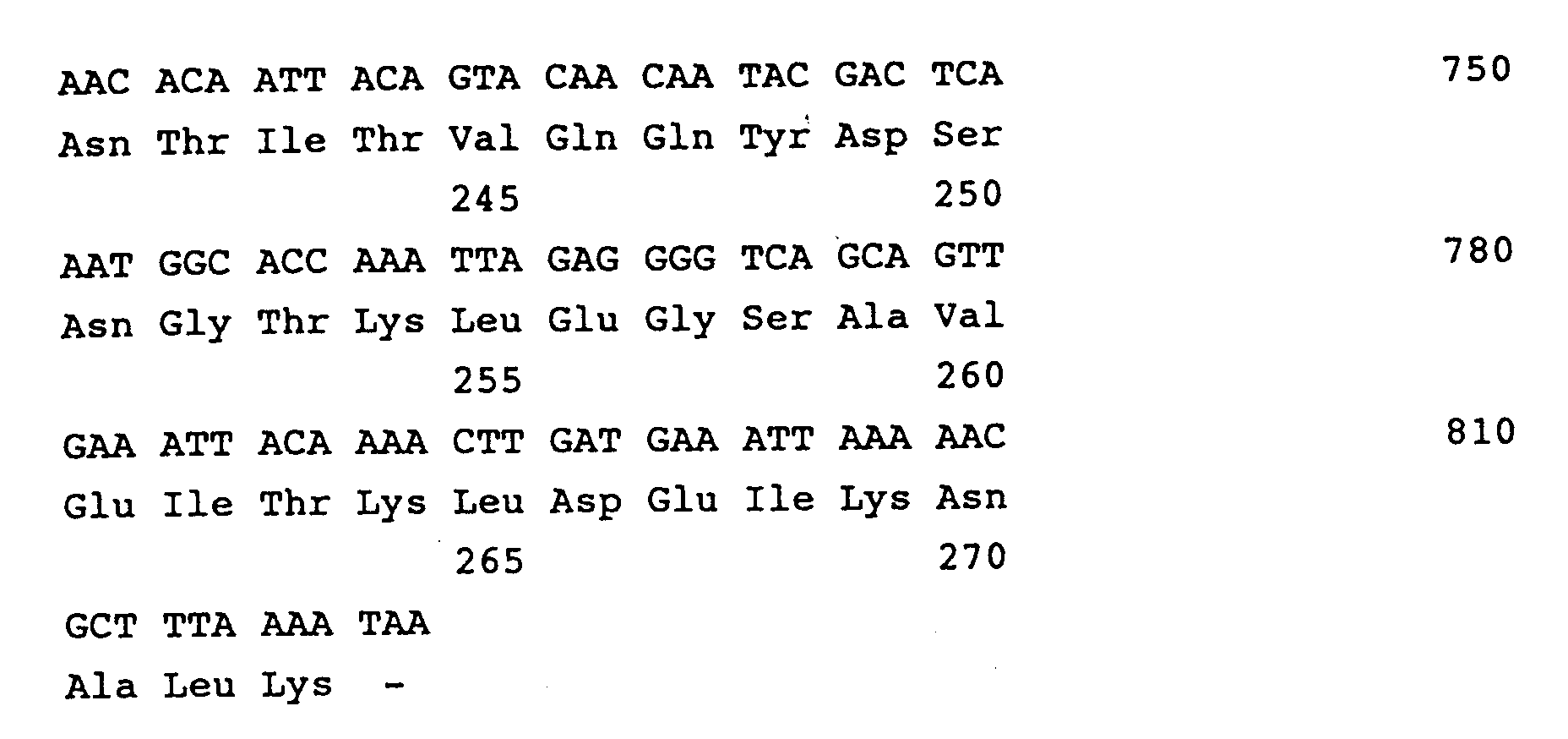
Claims (13)
dadurch gekennzeichnet,
daß es mit einem Antikörper gegen das 31kD Antigen (OspA) von B.burgdorferi immunreagiert.Antigen,
characterized,
that it immunoreacts with an antibody against the 31kD antigen (OspA) from B.burgdorferi.
dadurch gekennzeichnet,
daß es mit einem Antikörper gegen das 34kD Antigen (OspB) von B.burgdorferi immunreagiert.Antigen,
characterized,
that it immunoreacts with an antibody against the 34kD antigen (OspB) from B.burgdorferi.
dadurch gekennzeichnet,
daß sich die dafür kodierende DNA-Sequenz auf einem Vektor, vorzugsweise einem prokaryontischen Vektor befindet, der geeignet zur Proteinexpression ist.Antigen according to claim 1 or 2,
characterized,
that the DNA sequence coding for it is on a vector, preferably a prokaryotic vector, which is suitable for protein expression.
dadurch gekennzeichnet,
daß es folgende Aminosäuresequenz oder ein immunogenes Eptitop aus dieser Sequenz enthält:


characterized,
that it contains the following amino acid sequence or an immunogenic eptitope from this sequence:
dadurch gekennzeichnet,
daß es ein β-Galactosidase-Fusionsprotein oder ein Nicht-Fusionsprotein ist.Antigen according to one of claims 1 to 4,
characterized,
that it is a β-galactosidase fusion protein or a non-fusion protein.
gekennzeichnet, daß sie für ein
Antigen nach einem der Ansprüche 1 , 3, 4 oder 5 kodiert und (1) die im folgenden dargestellte Nukleinsäuresequenz, (2) eine ihr im Rahmen der Degeneration des genetischen Codes entsprechende Sequenz oder (3) eine mit einer Sequenz aus (1) oder/und (2) unter stringenten Bedingungen hybridisierende Sequenz enthält:


characterized that they are for a
Antigen according to one of claims 1, 3, 4 or 5 and (1) the nucleic acid sequence shown below, (2) a sequence corresponding to it in the context of the degeneration of the genetic code or (3) one with a sequence from (1) or / and (2) sequence hybridizing under stringent conditions contains:
dadurch gekennzeichnet,
daß er ein prokaryontischer Vektor ist.Recombinant vector according to claim 7,
characterized,
that it is a prokaryotic vector.
dadurch gekennzeichnet,
daß er ein Plasmid ist.Recombinant vector according to claim 8,
characterized,
that it is a plasmid.
dadurch gekennzeichnet,
daß man eine B.burgdorferi Genbank mit einem oder mehreren monoklonalen Antikörpern, die spezifisch für das 31kD Antigen (OspA) oder das 34kD Antigen (OspB) von B.burgdorferi sind, untersucht und die Klone isoliert, welche mit dem oder den Antikörpern eine positive Immunreaktion zeigen.Method for obtaining antigens according to one of Claims 1 to 5,
characterized,
that a B.burgdorferi gene bank with one or more monoclonal antibodies which are specific for the 31kD antigen (OspA) or the 34kD antigen (OspB) from B.burgdorferi is examined and the clones isolated which are positive with the antibody or antibodies Show immune response.
gekennzeichnet durch
ein oder mehrere Antigene nach einem der Ansprüche 1 bis 5 als Wirkstoff, gegebenenfalls mit üblichen Träger-, Füll- und Hilfsstoffen.Active vaccine against Lyme disease,
marked by
one or more antigens according to any one of claims 1 to 5 as an active ingredient, optionally with conventional carriers, fillers and auxiliaries.
dadurch gekennzeichnet,
daß man das Antigen auf gentechnologische Weise gewinnt.Vaccine according to claim 12,
characterized,
that the antigen is obtained by genetic engineering.
Priority Applications (1)
| Application Number | Priority Date | Filing Date | Title |
|---|---|---|---|
| EP98105444A EP0861664A3 (en) | 1989-09-19 | 1990-09-18 | Vaccine against Lyme disease |
Applications Claiming Priority (5)
| Application Number | Priority Date | Filing Date | Title |
|---|---|---|---|
| DE3931236 | 1989-09-19 | ||
| DE3931236 | 1989-09-19 | ||
| DE4015911 | 1990-05-17 | ||
| DE4015911A DE4015911A1 (en) | 1989-09-19 | 1990-05-17 | VACCINE AGAINST LYME DISEASE |
| EP90117943A EP0418827B1 (en) | 1989-09-19 | 1990-09-18 | Vaccine against Lyme disease |
Related Parent Applications (1)
| Application Number | Title | Priority Date | Filing Date |
|---|---|---|---|
| EP90117943.2 Division | 1990-09-18 |
Related Child Applications (2)
| Application Number | Title | Priority Date | Filing Date |
|---|---|---|---|
| EP98105444A Division EP0861664A3 (en) | 1989-09-19 | 1990-09-18 | Vaccine against Lyme disease |
| EP98105444.8 Division-Into | 1998-03-25 |
Publications (2)
| Publication Number | Publication Date |
|---|---|
| EP0643974A1 true EP0643974A1 (en) | 1995-03-22 |
| EP0643974B1 EP0643974B1 (en) | 1999-01-13 |
Family
ID=25885304
Family Applications (5)
| Application Number | Title | Priority Date | Filing Date |
|---|---|---|---|
| EP94112144A Expired - Lifetime EP0633313B1 (en) | 1989-09-19 | 1990-09-18 | Vaccine against lyme disease |
| EP98105444A Withdrawn EP0861664A3 (en) | 1989-09-19 | 1990-09-18 | Vaccine against Lyme disease |
| EP90117943A Expired - Lifetime EP0418827B1 (en) | 1989-09-19 | 1990-09-18 | Vaccine against Lyme disease |
| EP94112145A Expired - Lifetime EP0633028B1 (en) | 1989-09-19 | 1990-09-18 | Vaccine against lyme disease |
| EP94112143A Expired - Lifetime EP0643974B1 (en) | 1989-09-19 | 1990-09-18 | Vaccine against lyme disease |
Family Applications Before (4)
| Application Number | Title | Priority Date | Filing Date |
|---|---|---|---|
| EP94112144A Expired - Lifetime EP0633313B1 (en) | 1989-09-19 | 1990-09-18 | Vaccine against lyme disease |
| EP98105444A Withdrawn EP0861664A3 (en) | 1989-09-19 | 1990-09-18 | Vaccine against Lyme disease |
| EP90117943A Expired - Lifetime EP0418827B1 (en) | 1989-09-19 | 1990-09-18 | Vaccine against Lyme disease |
| EP94112145A Expired - Lifetime EP0633028B1 (en) | 1989-09-19 | 1990-09-18 | Vaccine against lyme disease |
Country Status (24)
| Country | Link |
|---|---|
| US (6) | US5178859A (en) |
| EP (5) | EP0633313B1 (en) |
| JP (3) | JP3205932B2 (en) |
| KR (2) | KR100205462B1 (en) |
| AT (4) | ATE186646T1 (en) |
| AU (1) | AU651560B2 (en) |
| CA (1) | CA2025597C (en) |
| CZ (2) | CZ284538B6 (en) |
| DE (5) | DE4015911A1 (en) |
| DK (4) | DK0418827T3 (en) |
| ES (4) | ES2141182T3 (en) |
| FI (1) | FI102248B (en) |
| GR (3) | GR3018995T3 (en) |
| HK (2) | HK1002405A1 (en) |
| HR (1) | HRP940545B1 (en) |
| HU (2) | HU212716B (en) |
| IE (1) | IE903377A1 (en) |
| NO (2) | NO311767B1 (en) |
| NZ (1) | NZ235260A (en) |
| PL (2) | PL170229B1 (en) |
| PT (1) | PT95349B (en) |
| SI (1) | SI9011773B (en) |
| SK (3) | SK280285B6 (en) |
| YU (2) | YU48250B (en) |
Cited By (3)
| Publication number | Priority date | Publication date | Assignee | Title |
|---|---|---|---|---|
| DE19632862A1 (en) * | 1996-08-14 | 1998-02-19 | Mikrogen Molekularbiol Entw | Immunologically active proteins from Borrelia burgdorferi, nucleic acids coding for them and their use in test kits and as vaccines |
| US6676942B1 (en) * | 1991-08-15 | 2004-01-13 | Smithkline Beecham Biologicals (S.A.) | Osp a proteins of Borrelia burgdorferi subgroups, encoding genes and vaccines |
| US8137678B2 (en) | 2006-11-03 | 2012-03-20 | Intervet Inc. | Canine lyme disease vaccine |
Families Citing this family (41)
| Publication number | Priority date | Publication date | Assignee | Title |
|---|---|---|---|---|
| DK590288D0 (en) | 1988-10-24 | 1988-10-24 | Symbicom Ab | CHEMICAL COMPOUNDS |
| US5777095A (en) * | 1988-10-24 | 1998-07-07 | Symbicom Aktiebolag | Osp A and B Sequence of Borrelia burgdonferi strains ACA1 and IP90 |
| US7094391B1 (en) * | 1988-10-24 | 2006-08-22 | The University Of Texas System | Compositions and methods for administering Borrelia burgdorferi antigens |
| US6143872A (en) * | 1988-10-24 | 2000-11-07 | Symbicom Aktiebolag | Borrelia burdorferi Osp A and B proteins and immunogenic peptides |
| DE4015911A1 (en) * | 1989-09-19 | 1991-03-28 | Max Planck Gesellschaft | VACCINE AGAINST LYME DISEASE |
| US5582829A (en) * | 1990-04-05 | 1996-12-10 | Rx Technologies, Inc. | Sonicated borrelia burgdorferi vaccine |
| JPH06501382A (en) | 1990-06-15 | 1994-02-17 | エール ユニバーシティ | Compositions and methods for the prevention and diagnosis of Lyme disease |
| EP0465204B1 (en) * | 1990-07-06 | 2000-09-20 | American Home Products Corporation | Vaccine against Lyme disease |
| CA2057536C (en) * | 1990-12-21 | 1999-10-26 | John J. Dunn | Cloning and expression of borrelia lipoproteins |
| US6221363B1 (en) * | 1991-07-11 | 2001-04-24 | Baxter Aktiengesellschaft | Vaccine for the prevention of lyme disease |
| US6872550B1 (en) | 1991-07-11 | 2005-03-29 | Baxter Vaccine Ag | Immunogenic formulation of OspC antigen vaccines for the prevention and treatment of lyme disease and recombinant methods for the preparation of such antigens |
| ATE181572T1 (en) * | 1991-08-15 | 1999-07-15 | Smithkline Beecham Biolog | OSPA PROTEINS OF BORRELIA BURGDORFERI SUBGROUPS, GENES CODING FOR THEM AND VACCINES |
| AU676140B2 (en) * | 1991-10-18 | 1997-03-06 | Connaught Laboratories Inc. | Preparation of recombinant borrelia proteins |
| ATE234365T1 (en) * | 1991-10-22 | 2003-03-15 | Symbicom Ab | IMPROVEMENTS IN DIAGNOSIS AND PROPHYLAXIS OF BORRELIA BURGDORFERI |
| US6303129B1 (en) * | 1992-07-28 | 2001-10-16 | Rx Technologies | Production of borrelia burgdorferi vaccine, product produced thereby and method of use |
| US5656451A (en) * | 1993-07-30 | 1997-08-12 | Yale University | OspE, OspF, and S1 polypeptides in borrelia burgdorferi |
| US6716591B1 (en) | 1993-07-30 | 2004-04-06 | Yale University | B. burgdorferi polypeptides |
| US7008625B2 (en) | 1993-11-01 | 2006-03-07 | Research Foundation Of The State University Of New York | Recombinant constructs of Borrelia burgdorferi |
| US5558993A (en) * | 1994-06-17 | 1996-09-24 | The Regents Of The University Of California | Cloned Borrelia burgdorferi virulence protein |
| GB9511909D0 (en) * | 1995-06-12 | 1995-08-09 | Microbiological Res Authority | Vaccine |
| WO1997031123A1 (en) | 1996-02-21 | 1997-08-28 | Board Of Regents, The University Of Texas System | Vmp-like sequences of pathogenic borrelia |
| US6458555B1 (en) * | 1996-08-21 | 2002-10-01 | Gerd Bach | Cytologic method of examining mucous membranes |
| US6060082A (en) * | 1997-04-18 | 2000-05-09 | Massachusetts Institute Of Technology | Polymerized liposomes targeted to M cells and useful for oral or mucosal drug delivery |
| DE19740735A1 (en) * | 1997-09-16 | 1999-03-18 | Max Planck Gesellschaft | Antibodies specific for Borrelia burgdorferi 24 kD antigen OspC |
| US6306623B1 (en) | 1998-02-24 | 2001-10-23 | The University Of California | Leptospiral major outer membrane protein LipL32 |
| EP1100922B1 (en) | 1998-07-31 | 2008-02-20 | Gundersen Lutheran Medical Foundation, Inc. | Uses of the borreliacidal epitope(s) of borrelia burgdorferi outer surface protein c (ospc) as vaccine |
| WO2000056298A2 (en) * | 1999-03-19 | 2000-09-28 | Luitpold Pharmaceuticals, Inc. | Treatment of lyme disease with polysulfated glycosaminoglycan formulations |
| WO2000078345A1 (en) * | 1999-06-21 | 2000-12-28 | Milkhaus Laboratory, Inc. | Method for treatment of lyme disease |
| PT1311540E (en) | 2000-08-18 | 2008-03-20 | Univ New York State Res Found | Altered ospa of borrelia burgdorferi |
| WO2009003492A1 (en) | 2007-07-03 | 2009-01-08 | Dako Denmark A/S | Mhc multimers, methods for their generation, labeling and use |
| EP1572714A4 (en) | 2002-12-20 | 2006-08-09 | Univ Texas | VMP-LIKE SEQUENCES OF PATHOGENIC i BORRELIA /i SPECIES AND STRAINS |
| EP2155782A2 (en) | 2007-03-26 | 2010-02-24 | Dako Denmark A/S | Mhc peptide complexes and uses thereof in infectious diseases |
| EP2197908A2 (en) | 2007-09-27 | 2010-06-23 | Dako Denmark A/S | Mhc multimers in tuberculosis diagnostics, vaccine and therapeutics |
| DK2254592T3 (en) | 2008-02-28 | 2019-09-09 | Dako Denmark As | MHC multimers for Borrelia diagnostics and disease |
| US10722562B2 (en) | 2008-07-23 | 2020-07-28 | Immudex Aps | Combinatorial analysis and repair |
| GB0817244D0 (en) | 2008-09-20 | 2008-10-29 | Univ Cardiff | Use of a protein kinase inhibitor to detect immune cells, such as T cells |
| US10369204B2 (en) | 2008-10-02 | 2019-08-06 | Dako Denmark A/S | Molecular vaccines for infectious disease |
| CA2781110C (en) * | 2009-11-17 | 2018-07-31 | Abaxis, Inc. | Peptides and methods for the detection of lyme disease antibodies |
| US8758772B2 (en) | 2011-11-04 | 2014-06-24 | Abaxis, Inc. | Peptides and methods for the detection of lyme disease antibodies |
| PL223175B1 (en) | 2012-10-22 | 2016-10-31 | Inst Chemii Bioorganicznej Polskiej Akademii Nauk | Vaccine against Lyme disease, a genetic construct, the recombinant protein, the method for preparing a genetic construct, a method of preparing a vaccine, a method of preparing a recombinant protein, the use of recombinant proteins for the manufacture of a vaccine against Lyme borreliosis |
| US9610336B1 (en) * | 2014-07-16 | 2017-04-04 | Amiram Katz | Immunotherapy for lyme disease |
Citations (4)
| Publication number | Priority date | Publication date | Assignee | Title |
|---|---|---|---|---|
| EP0252641A1 (en) * | 1986-06-26 | 1988-01-13 | BioWhittaker, Inc. | Diagnosis of lyme disease |
| WO1990004411A1 (en) * | 1988-10-24 | 1990-05-03 | Symbicom Aktiebolag | Immunogenically active fractions of borrelia burgdorferi |
| WO1991009870A1 (en) * | 1989-12-22 | 1991-07-11 | Mikrogen Molekularbiologische Entwicklungs-Gmbh | Immunologically active proteines from borrelia burgdorferi, related test kits and vaccine |
| WO1992000055A1 (en) * | 1990-06-15 | 1992-01-09 | Yale University | Compositions and methods for the prevention and diagnosis of lyme disease |
Family Cites Families (10)
| Publication number | Priority date | Publication date | Assignee | Title |
|---|---|---|---|---|
| US4772464A (en) | 1985-08-01 | 1988-09-20 | Miles Laboratories, Inc. | Protective antibodies to serotypic determinants of flagellar antigens |
| US4721617A (en) | 1986-08-14 | 1988-01-26 | Regents Of The University Of Minnesota | Vaccine against lyme disease |
| US5034515A (en) * | 1987-09-22 | 1991-07-23 | Wisconsin Alumni Research Foundation | Staphylococcal fibronectin receptor, monoclonal antibodies thereto and methods of use |
| DE4015911A1 (en) * | 1989-09-19 | 1991-03-28 | Max Planck Gesellschaft | VACCINE AGAINST LYME DISEASE |
| EP0465204B1 (en) * | 1990-07-06 | 2000-09-20 | American Home Products Corporation | Vaccine against Lyme disease |
| CA2057536C (en) * | 1990-12-21 | 1999-10-26 | John J. Dunn | Cloning and expression of borrelia lipoproteins |
| ATE181572T1 (en) * | 1991-08-15 | 1999-07-15 | Smithkline Beecham Biolog | OSPA PROTEINS OF BORRELIA BURGDORFERI SUBGROUPS, GENES CODING FOR THEM AND VACCINES |
| JP3246603B2 (en) * | 1991-08-27 | 2002-01-15 | ノボザイムス アクティーゼルスカブ | Detergent |
| AU676140B2 (en) * | 1991-10-18 | 1997-03-06 | Connaught Laboratories Inc. | Preparation of recombinant borrelia proteins |
| AU681572B2 (en) * | 1991-10-21 | 1997-09-04 | Med Immune, Inc. | Bacterial expression vectors containing DNA encoding secretion signals of lipoproteins |
-
1990
- 1990-05-17 DE DE4015911A patent/DE4015911A1/en not_active Withdrawn
- 1990-09-07 HU HU905816A patent/HU212716B/en not_active IP Right Cessation
- 1990-09-11 NZ NZ235260A patent/NZ235260A/en unknown
- 1990-09-12 AU AU62444/90A patent/AU651560B2/en not_active Ceased
- 1990-09-18 DE DE59009978T patent/DE59009978D1/en not_active Expired - Fee Related
- 1990-09-18 AT AT94112145T patent/ATE186646T1/en not_active IP Right Cessation
- 1990-09-18 PL PL90307358A patent/PL170229B1/en not_active IP Right Cessation
- 1990-09-18 EP EP94112144A patent/EP0633313B1/en not_active Expired - Lifetime
- 1990-09-18 DE DE59010863T patent/DE59010863D1/en not_active Expired - Fee Related
- 1990-09-18 DK DK90117943.2T patent/DK0418827T3/en active
- 1990-09-18 EP EP98105444A patent/EP0861664A3/en not_active Withdrawn
- 1990-09-18 NO NO19904062A patent/NO311767B1/en unknown
- 1990-09-18 AT AT94112144T patent/ATE191499T1/en not_active IP Right Cessation
- 1990-09-18 IE IE337790A patent/IE903377A1/en not_active IP Right Cessation
- 1990-09-18 FI FI904597A patent/FI102248B/en not_active IP Right Cessation
- 1990-09-18 PL PL90286918A patent/PL169804B1/en not_active IP Right Cessation
- 1990-09-18 DE DE59010888T patent/DE59010888D1/en not_active Expired - Fee Related
- 1990-09-18 DE DE59010903T patent/DE59010903D1/en not_active Expired - Fee Related
- 1990-09-18 ES ES94112145T patent/ES2141182T3/en not_active Expired - Lifetime
- 1990-09-18 ES ES94112144T patent/ES2145077T3/en not_active Expired - Lifetime
- 1990-09-18 AT AT94112143T patent/ATE175576T1/en not_active IP Right Cessation
- 1990-09-18 EP EP90117943A patent/EP0418827B1/en not_active Expired - Lifetime
- 1990-09-18 YU YU177390A patent/YU48250B/en unknown
- 1990-09-18 CA CA002025597A patent/CA2025597C/en not_active Expired - Fee Related
- 1990-09-18 ES ES94112143T patent/ES2129551T3/en not_active Expired - Lifetime
- 1990-09-18 DK DK94112144T patent/DK0633313T3/en active
- 1990-09-18 DK DK94112145T patent/DK0633028T3/en active
- 1990-09-18 ES ES90117943T patent/ES2082812T3/en not_active Expired - Lifetime
- 1990-09-18 EP EP94112145A patent/EP0633028B1/en not_active Expired - Lifetime
- 1990-09-18 EP EP94112143A patent/EP0643974B1/en not_active Expired - Lifetime
- 1990-09-18 SI SI9011773A patent/SI9011773B/en not_active IP Right Cessation
- 1990-09-18 DK DK94112143T patent/DK0643974T3/en active
- 1990-09-18 AT AT90117943T patent/ATE131732T1/en not_active IP Right Cessation
- 1990-09-19 CZ CS904560A patent/CZ284538B6/en not_active IP Right Cessation
- 1990-09-19 SK SK4560-90A patent/SK280285B6/en unknown
- 1990-09-19 SK SK1511-98A patent/SK283089B6/en unknown
- 1990-09-19 US US07/585,310 patent/US5178859A/en not_active Expired - Lifetime
- 1990-09-19 JP JP24765090A patent/JP3205932B2/en not_active Expired - Fee Related
- 1990-09-19 SK SK1510-98A patent/SK283088B6/en unknown
- 1990-09-19 KR KR1019900014814A patent/KR100205462B1/en not_active IP Right Cessation
- 1990-09-19 CZ CZ95970A patent/CZ284616B6/en not_active IP Right Cessation
- 1990-09-19 PT PT95349A patent/PT95349B/en active IP Right Grant
-
1993
- 1993-05-27 US US08/068,063 patent/US5434077A/en not_active Expired - Fee Related
-
1994
- 1994-09-15 HR HR940545A patent/HRP940545B1/en not_active IP Right Cessation
-
1995
- 1995-03-20 US US08/407,350 patent/US5686267A/en not_active Expired - Lifetime
- 1995-03-20 US US08/406,623 patent/US5780030A/en not_active Expired - Fee Related
- 1995-04-24 HU HU95P/P00108P patent/HU211228A9/en unknown
-
1996
- 1996-02-14 GR GR960400397T patent/GR3018995T3/en unknown
-
1997
- 1997-03-12 YU YU9097A patent/YU49370B/en unknown
-
1998
- 1998-02-13 HK HK98101147A patent/HK1002405A1/en not_active IP Right Cessation
- 1998-02-23 US US09/027,843 patent/US5856447A/en not_active Expired - Fee Related
- 1998-08-29 KR KR1019980035375A patent/KR100202128B1/en not_active IP Right Cessation
- 1998-12-23 HK HK98114958A patent/HK1013620A1/en not_active IP Right Cessation
-
1999
- 1999-10-07 US US09/413,858 patent/US6613331B1/en not_active Expired - Fee Related
- 1999-11-30 GR GR990403104T patent/GR3032010T3/en unknown
-
2000
- 2000-06-14 GR GR20000401362T patent/GR3033675T3/en not_active IP Right Cessation
- 2000-07-26 JP JP2000225341A patent/JP3328644B2/en not_active Expired - Fee Related
- 2000-12-11 JP JP2000376159A patent/JP3418171B2/en not_active Expired - Fee Related
-
2001
- 2001-09-24 NO NO20014624A patent/NO315905B1/en unknown
Patent Citations (4)
| Publication number | Priority date | Publication date | Assignee | Title |
|---|---|---|---|---|
| EP0252641A1 (en) * | 1986-06-26 | 1988-01-13 | BioWhittaker, Inc. | Diagnosis of lyme disease |
| WO1990004411A1 (en) * | 1988-10-24 | 1990-05-03 | Symbicom Aktiebolag | Immunogenically active fractions of borrelia burgdorferi |
| WO1991009870A1 (en) * | 1989-12-22 | 1991-07-11 | Mikrogen Molekularbiologische Entwicklungs-Gmbh | Immunologically active proteines from borrelia burgdorferi, related test kits and vaccine |
| WO1992000055A1 (en) * | 1990-06-15 | 1992-01-09 | Yale University | Compositions and methods for the prevention and diagnosis of lyme disease |
Non-Patent Citations (1)
| Title |
|---|
| R. WALLICH ET AL.: "CLONING AND SEQUENCING OF THE GENE ENCODING THE OUTER SURFACE PROTEIN A (OSPA) OF A EUROPEAN BORRELIA BURGDORFERI ISOLATE.", NUCLEIC ACIDS RESEARCH, vol. 17, no. 21, 11 November 1989 (1989-11-11), ARLINGTON, VIRGINIA US, pages 8864, XP002088356 * |
Cited By (7)
| Publication number | Priority date | Publication date | Assignee | Title |
|---|---|---|---|---|
| US6676942B1 (en) * | 1991-08-15 | 2004-01-13 | Smithkline Beecham Biologicals (S.A.) | Osp a proteins of Borrelia burgdorferi subgroups, encoding genes and vaccines |
| DE19632862A1 (en) * | 1996-08-14 | 1998-02-19 | Mikrogen Molekularbiol Entw | Immunologically active proteins from Borrelia burgdorferi, nucleic acids coding for them and their use in test kits and as vaccines |
| US6610301B1 (en) | 1996-08-14 | 2003-08-26 | Mikrogen Molekularbiologische Entwicklungs - Gmbh | Immunologically active proteins from Borrelia burgdorferi, nucleic acids which encode them, and their use in test kits and as vaccines |
| US6808711B2 (en) | 1996-08-14 | 2004-10-26 | Mikrogen Molekularbiologische Entwicklungs-Gmbh | Immunologically active proteins from Borrelia burgdorferi, nucleic acids which encode them, and their use in test kits and as vaccines |
| DE19632862B4 (en) * | 1996-08-14 | 2006-08-03 | Mikrogen Molekularbiologische Entwicklungs-Gmbh | Immunologically active proteins of Borrelia burgdorferi, nucleic acids coding for them as well as their use in test kits and as vaccines |
| US8137678B2 (en) | 2006-11-03 | 2012-03-20 | Intervet Inc. | Canine lyme disease vaccine |
| US8414901B2 (en) | 2006-11-03 | 2013-04-09 | Intervet Inc. | Canine Lyme disease vaccine |
Also Published As
Similar Documents
| Publication | Publication Date | Title |
|---|---|---|
| EP0418827B1 (en) | Vaccine against Lyme disease | |
| DE69231619T3 (en) | Method and composition for the prevention of Lyme disease | |
| DE69333036T2 (en) | DNA fragments encoding the subunits of the Neisseria meningitidis receptor | |
| EP0506868B2 (en) | Immunologically active proteines from borrelia burgdorferi, related test kits and vaccine | |
| DE69334002T2 (en) | MEMBRANE-ASSOCIATED IMMUNOGENIC PROTEINS OF MYCOBACTERIA | |
| DE69737125T3 (en) | Streptococcus pneumoniae antigens and vaccines | |
| DE60132893T2 (en) | ANTIGENIC POLYPEPTIDE | |
| DE69632625T2 (en) | Mycrobacterial Proteins, Microorganisms Producing Them and Their Use as Vaccines and Detecting Zubuloses | |
| IE83320B1 (en) | Vaccine against lyme disease | |
| DE69034107T2 (en) | Vaccines against bacteria that lead to blood poisoning | |
| DE69535422T2 (en) | Vaccine for a non-typeable Haemophilus influenzae strain | |
| DE19740735A1 (en) | Antibodies specific for Borrelia burgdorferi 24 kD antigen OspC | |
| DE69629304T2 (en) | LEPTOSPIRA MEMBRANE PROTEINS | |
| DE69333467T2 (en) | BORDETELLA BRONCHISEPTICA VACCINE | |
| DE60024977T2 (en) | CLONING AND EXPRESSION OF HAEMOPHILUS SOMNUS TRANSFERRIN-BINDING PROTEINS | |
| DD295553A5 (en) | VACCINE AGAINST LYME DISEASE | |
| DE69932719T2 (en) | Humanized antibodies for the detection of verotoxin II and cell line for the production thereof | |
| WO2000022134A2 (en) | Test kit for diagnosis of borrelia infection and novel borrelia antigens for the development of vaccines | |
| DE4018988A1 (en) | New Borrelia burgdorferi proteins |
Legal Events
| Date | Code | Title | Description |
|---|---|---|---|
| PUAI | Public reference made under article 153(3) epc to a published international application that has entered the european phase |
Free format text: ORIGINAL CODE: 0009012 |
|
| 17P | Request for examination filed |
Effective date: 19940803 |
|
| AC | Divisional application: reference to earlier application |
Ref document number: 418827 Country of ref document: EP |
|
| AK | Designated contracting states |
Kind code of ref document: A1 Designated state(s): AT BE CH DE DK ES FR GB GR IT LI LU NL SE |
|
| 17Q | First examination report despatched |
Effective date: 19961112 |
|
| GRAG | Despatch of communication of intention to grant |
Free format text: ORIGINAL CODE: EPIDOS AGRA |
|
| GRAG | Despatch of communication of intention to grant |
Free format text: ORIGINAL CODE: EPIDOS AGRA |
|
| GRAH | Despatch of communication of intention to grant a patent |
Free format text: ORIGINAL CODE: EPIDOS IGRA |
|
| GRAH | Despatch of communication of intention to grant a patent |
Free format text: ORIGINAL CODE: EPIDOS IGRA |
|
| RAP1 | Party data changed (applicant data changed or rights of an application transferred) |
Owner name: DEUTSCHES KREBSFORSCHUNGSZENTRUM STIFTUNG DES OEF Owner name: MAX-PLANCK-GESELLSCHAFT ZUR FOERDERUNG DER WISSENS |
|
| GRAA | (expected) grant |
Free format text: ORIGINAL CODE: 0009210 |
|
| AC | Divisional application: reference to earlier application |
Ref document number: 418827 Country of ref document: EP |
|
| AK | Designated contracting states |
Kind code of ref document: B1 Designated state(s): AT BE CH DE DK ES FR GB GR IT LI LU NL SE |
|
| REF | Corresponds to: |
Ref document number: 175576 Country of ref document: AT Date of ref document: 19990115 Kind code of ref document: T |
|
| REG | Reference to a national code |
Ref country code: CH Ref legal event code: EP |
|
| REG | Reference to a national code |
Ref country code: CH Ref legal event code: NV Representative=s name: A. BRAUN, BRAUN, HERITIER, ESCHMANN AG PATENTANWAE |
|
| GBT | Gb: translation of ep patent filed (gb section 77(6)(a)/1977) |
Effective date: 19990113 |
|
| ITF | It: translation for a ep patent filed |
Owner name: JACOBACCI & PERANI S.P.A. |
|
| REF | Corresponds to: |
Ref document number: 59010863 Country of ref document: DE Date of ref document: 19990225 |
|
| ET | Fr: translation filed | ||
| REG | Reference to a national code |
Ref country code: ES Ref legal event code: FG2A Ref document number: 2129551 Country of ref document: ES Kind code of ref document: T3 |
|
| REG | Reference to a national code |
Ref country code: DK Ref legal event code: T3 |
|
| PLBQ | Unpublished change to opponent data |
Free format text: ORIGINAL CODE: EPIDOS OPPO |
|
| PLBI | Opposition filed |
Free format text: ORIGINAL CODE: 0009260 |
|
| PLBF | Reply of patent proprietor to notice(s) of opposition |
Free format text: ORIGINAL CODE: EPIDOS OBSO |
|
| 26 | Opposition filed |
Opponent name: SYMBICOM AB Effective date: 19991013 Opponent name: CONNAUGHT LABORATOTIES, INC. Effective date: 19991013 |
|
| NLR1 | Nl: opposition has been filed with the epo |
Opponent name: SYMBICOM AB Opponent name: CONNAUGHT LABORATOTIES, INC. |
|
| PLBF | Reply of patent proprietor to notice(s) of opposition |
Free format text: ORIGINAL CODE: EPIDOS OBSO |
|
| PLBF | Reply of patent proprietor to notice(s) of opposition |
Free format text: ORIGINAL CODE: EPIDOS OBSO |
|
| PLBF | Reply of patent proprietor to notice(s) of opposition |
Free format text: ORIGINAL CODE: EPIDOS OBSO |
|
| PLBF | Reply of patent proprietor to notice(s) of opposition |
Free format text: ORIGINAL CODE: EPIDOS OBSO |
|
| REG | Reference to a national code |
Ref country code: GB Ref legal event code: IF02 |
|
| PLBL | Opposition procedure terminated |
Free format text: ORIGINAL CODE: EPIDOS OPPC |
|
| PLBM | Termination of opposition procedure: date of legal effect published |
Free format text: ORIGINAL CODE: 0009276 |
|
| STAA | Information on the status of an ep patent application or granted ep patent |
Free format text: STATUS: OPPOSITION PROCEDURE CLOSED |
|
| 27C | Opposition proceedings terminated |
Effective date: 20020405 |
|
| NLR2 | Nl: decision of opposition | ||
| PGFP | Annual fee paid to national office [announced via postgrant information from national office to epo] |
Ref country code: LU Payment date: 20040624 Year of fee payment: 15 |
|
| PGFP | Annual fee paid to national office [announced via postgrant information from national office to epo] |
Ref country code: GR Payment date: 20040727 Year of fee payment: 15 |
|
| PGFP | Annual fee paid to national office [announced via postgrant information from national office to epo] |
Ref country code: NL Payment date: 20040809 Year of fee payment: 15 |
|
| PGFP | Annual fee paid to national office [announced via postgrant information from national office to epo] |
Ref country code: GB Payment date: 20040812 Year of fee payment: 15 |
|
| PGFP | Annual fee paid to national office [announced via postgrant information from national office to epo] |
Ref country code: ES Payment date: 20040923 Year of fee payment: 15 |
|
| PGFP | Annual fee paid to national office [announced via postgrant information from national office to epo] |
Ref country code: BE Payment date: 20040928 Year of fee payment: 15 |
|
| PGFP | Annual fee paid to national office [announced via postgrant information from national office to epo] |
Ref country code: FR Payment date: 20050823 Year of fee payment: 16 |
|
| PGFP | Annual fee paid to national office [announced via postgrant information from national office to epo] |
Ref country code: DE Payment date: 20050831 Year of fee payment: 16 |
|
| PGFP | Annual fee paid to national office [announced via postgrant information from national office to epo] |
Ref country code: SE Payment date: 20050914 Year of fee payment: 16 Ref country code: DK Payment date: 20050914 Year of fee payment: 16 |
|
| PGFP | Annual fee paid to national office [announced via postgrant information from national office to epo] |
Ref country code: CH Payment date: 20050915 Year of fee payment: 16 |
|
| PG25 | Lapsed in a contracting state [announced via postgrant information from national office to epo] |
Ref country code: IT Free format text: LAPSE BECAUSE OF NON-PAYMENT OF DUE FEES;WARNING: LAPSES OF ITALIAN PATENTS WITH EFFECTIVE DATE BEFORE 2007 MAY HAVE OCCURRED AT ANY TIME BEFORE 2007. THE CORRECT EFFECTIVE DATE MAY BE DIFFERENT FROM THE ONE RECORDED. Effective date: 20050918 Ref country code: GB Free format text: LAPSE BECAUSE OF NON-PAYMENT OF DUE FEES Effective date: 20050918 |
|
| PG25 | Lapsed in a contracting state [announced via postgrant information from national office to epo] |
Ref country code: ES Free format text: LAPSE BECAUSE OF NON-PAYMENT OF DUE FEES Effective date: 20050919 |
|
| PGFP | Annual fee paid to national office [announced via postgrant information from national office to epo] |
Ref country code: AT Payment date: 20050919 Year of fee payment: 16 |
|
| PG25 | Lapsed in a contracting state [announced via postgrant information from national office to epo] |
Ref country code: LU Free format text: LAPSE BECAUSE OF NON-PAYMENT OF DUE FEES Effective date: 20050930 Ref country code: BE Free format text: LAPSE BECAUSE OF NON-PAYMENT OF DUE FEES Effective date: 20050930 |
|
| PG25 | Lapsed in a contracting state [announced via postgrant information from national office to epo] |
Ref country code: NL Free format text: LAPSE BECAUSE OF NON-PAYMENT OF DUE FEES Effective date: 20060401 |
|
| GBPC | Gb: european patent ceased through non-payment of renewal fee |
Effective date: 20050918 |
|
| NLV4 | Nl: lapsed or anulled due to non-payment of the annual fee |
Effective date: 20060401 |
|
| PG25 | Lapsed in a contracting state [announced via postgrant information from national office to epo] |
Ref country code: AT Free format text: LAPSE BECAUSE OF NON-PAYMENT OF DUE FEES Effective date: 20060918 |
|
| PG25 | Lapsed in a contracting state [announced via postgrant information from national office to epo] |
Ref country code: SE Free format text: LAPSE BECAUSE OF NON-PAYMENT OF DUE FEES Effective date: 20060919 |
|
| PG25 | Lapsed in a contracting state [announced via postgrant information from national office to epo] |
Ref country code: LI Free format text: LAPSE BECAUSE OF NON-PAYMENT OF DUE FEES Effective date: 20060930 Ref country code: CH Free format text: LAPSE BECAUSE OF NON-PAYMENT OF DUE FEES Effective date: 20060930 |
|
| PG25 | Lapsed in a contracting state [announced via postgrant information from national office to epo] |
Ref country code: DK Free format text: LAPSE BECAUSE OF NON-PAYMENT OF DUE FEES Effective date: 20061002 |
|
| REG | Reference to a national code |
Ref country code: ES Ref legal event code: FD2A Effective date: 20050919 |
|
| PG25 | Lapsed in a contracting state [announced via postgrant information from national office to epo] |
Ref country code: DE Free format text: LAPSE BECAUSE OF NON-PAYMENT OF DUE FEES Effective date: 20070403 |
|
| REG | Reference to a national code |
Ref country code: DK Ref legal event code: EBP |
|
| REG | Reference to a national code |
Ref country code: CH Ref legal event code: PL |
|
| EUG | Se: european patent has lapsed | ||
| REG | Reference to a national code |
Ref country code: FR Ref legal event code: ST Effective date: 20070531 |
|
| BERE | Be: lapsed |
Owner name: DEUTSCHES *KREBSFORSCHUNGSZENTRUM STIFTUNG DES OFF Effective date: 20050930 Owner name: *MAX-PLANCK-GESELLSCHAFT ZUR FORDERUNG DER WISSENS Effective date: 20050930 |
|
| PG25 | Lapsed in a contracting state [announced via postgrant information from national office to epo] |
Ref country code: GR Free format text: LAPSE BECAUSE OF NON-PAYMENT OF DUE FEES Effective date: 19990113 Ref country code: FR Free format text: LAPSE BECAUSE OF NON-PAYMENT OF DUE FEES Effective date: 20061002 |