EP0637631A2 - Highly efficient expression of a polypeptide containing a modified preS1 region of the major hepatitis B virus antigen - Google Patents
Highly efficient expression of a polypeptide containing a modified preS1 region of the major hepatitis B virus antigen Download PDFInfo
- Publication number
- EP0637631A2 EP0637631A2 EP94111872A EP94111872A EP0637631A2 EP 0637631 A2 EP0637631 A2 EP 0637631A2 EP 94111872 A EP94111872 A EP 94111872A EP 94111872 A EP94111872 A EP 94111872A EP 0637631 A2 EP0637631 A2 EP 0637631A2
- Authority
- EP
- European Patent Office
- Prior art keywords
- antigen
- pres1
- cell
- virus
- promoter
- Prior art date
- Legal status (The legal status is an assumption and is not a legal conclusion. Google has not performed a legal analysis and makes no representation as to the accuracy of the status listed.)
- Withdrawn
Links
- 241000700721 Hepatitis B virus Species 0.000 title claims abstract description 127
- 108091007433 antigens Proteins 0.000 title claims description 194
- 239000000427 antigen Substances 0.000 title claims description 191
- 102000036639 antigens Human genes 0.000 title claims description 190
- 230000014509 gene expression Effects 0.000 title description 66
- 108090000765 processed proteins & peptides Proteins 0.000 title description 19
- 102000004196 processed proteins & peptides Human genes 0.000 title description 16
- 229920001184 polypeptide Polymers 0.000 title description 13
- 108090000623 proteins and genes Proteins 0.000 claims abstract description 170
- 241000700618 Vaccinia virus Species 0.000 claims abstract description 106
- 102000004169 proteins and genes Human genes 0.000 claims abstract description 96
- 229960005486 vaccine Drugs 0.000 claims abstract description 47
- 238000000034 method Methods 0.000 claims abstract description 42
- 239000003153 chemical reaction reagent Substances 0.000 claims abstract description 20
- 239000003550 marker Substances 0.000 claims abstract description 12
- 238000002955 isolation Methods 0.000 claims abstract description 7
- 230000008569 process Effects 0.000 claims abstract description 6
- 210000004027 cell Anatomy 0.000 claims description 148
- 241000700605 Viruses Species 0.000 claims description 70
- 239000012634 fragment Substances 0.000 claims description 52
- 206010046865 Vaccinia virus infection Diseases 0.000 claims description 37
- 108091033319 polynucleotide Proteins 0.000 claims description 37
- 102000040430 polynucleotide Human genes 0.000 claims description 37
- 239000002157 polynucleotide Substances 0.000 claims description 37
- 208000007089 vaccinia Diseases 0.000 claims description 37
- 208000015181 infectious disease Diseases 0.000 claims description 28
- 230000000091 immunopotentiator Effects 0.000 claims description 27
- 241000701832 Enterobacteria phage T3 Species 0.000 claims description 22
- 108091026890 Coding region Proteins 0.000 claims description 20
- 210000005229 liver cell Anatomy 0.000 claims description 20
- 210000003527 eukaryotic cell Anatomy 0.000 claims description 15
- 210000001744 T-lymphocyte Anatomy 0.000 claims description 14
- 210000003719 b-lymphocyte Anatomy 0.000 claims description 13
- 101000686777 Escherichia phage T7 T7 RNA polymerase Proteins 0.000 claims description 11
- 101150106093 gpt gene Proteins 0.000 claims description 11
- 238000012258 culturing Methods 0.000 claims description 10
- 210000004962 mammalian cell Anatomy 0.000 claims description 10
- 241000588724 Escherichia coli Species 0.000 claims description 8
- 230000009089 cytolysis Effects 0.000 claims description 8
- 208000003322 Coinfection Diseases 0.000 claims description 7
- 241000124008 Mammalia Species 0.000 claims description 5
- 210000003292 kidney cell Anatomy 0.000 claims description 5
- 230000003053 immunization Effects 0.000 claims description 4
- 239000003937 drug carrier Substances 0.000 claims description 3
- 230000002194 synthesizing effect Effects 0.000 claims description 2
- 238000009007 Diagnostic Kit Methods 0.000 claims 1
- 239000013598 vector Substances 0.000 abstract description 28
- 108010090054 Membrane Glycoproteins Proteins 0.000 abstract description 16
- 102000012750 Membrane Glycoproteins Human genes 0.000 abstract description 15
- 239000000203 mixture Substances 0.000 abstract description 12
- 239000002773 nucleotide Substances 0.000 abstract description 9
- 125000003729 nucleotide group Chemical group 0.000 abstract description 9
- 238000010367 cloning Methods 0.000 abstract description 8
- 230000004048 modification Effects 0.000 abstract description 6
- 238000012986 modification Methods 0.000 abstract description 6
- 238000000746 purification Methods 0.000 abstract description 5
- 238000002560 therapeutic procedure Methods 0.000 abstract description 4
- 235000018102 proteins Nutrition 0.000 description 83
- 239000013612 plasmid Substances 0.000 description 54
- 108020004414 DNA Proteins 0.000 description 40
- 239000006166 lysate Substances 0.000 description 30
- 239000002245 particle Substances 0.000 description 30
- 108700012359 toxins Proteins 0.000 description 21
- 101150082969 SELP gene Proteins 0.000 description 20
- 101150036293 Selenop gene Proteins 0.000 description 20
- 208000002672 hepatitis B Diseases 0.000 description 18
- 239000003053 toxin Substances 0.000 description 18
- 231100000765 toxin Toxicity 0.000 description 18
- 238000001262 western blot Methods 0.000 description 17
- 230000005847 immunogenicity Effects 0.000 description 16
- 239000002609 medium Substances 0.000 description 14
- 238000003752 polymerase chain reaction Methods 0.000 description 14
- 108700026244 Open Reading Frames Proteins 0.000 description 13
- 238000010790 dilution Methods 0.000 description 13
- 239000012895 dilution Substances 0.000 description 13
- 238000004519 manufacturing process Methods 0.000 description 12
- 239000000523 sample Substances 0.000 description 12
- 230000028993 immune response Effects 0.000 description 11
- 240000004808 Saccharomyces cerevisiae Species 0.000 description 10
- 235000014680 Saccharomyces cerevisiae Nutrition 0.000 description 10
- 150000001413 amino acids Chemical class 0.000 description 10
- 230000015572 biosynthetic process Effects 0.000 description 10
- 238000013518 transcription Methods 0.000 description 10
- 230000035897 transcription Effects 0.000 description 10
- 108091034117 Oligonucleotide Proteins 0.000 description 9
- 230000002163 immunogen Effects 0.000 description 9
- 108010053187 Diphtheria Toxin Proteins 0.000 description 8
- 235000001014 amino acid Nutrition 0.000 description 8
- 230000000890 antigenic effect Effects 0.000 description 8
- 238000001514 detection method Methods 0.000 description 8
- 201000010099 disease Diseases 0.000 description 8
- 208000037265 diseases, disorders, signs and symptoms Diseases 0.000 description 8
- 238000009396 hybridization Methods 0.000 description 8
- 108020004999 messenger RNA Proteins 0.000 description 8
- 102000016607 Diphtheria Toxin Human genes 0.000 description 7
- 239000000020 Nitrocellulose Substances 0.000 description 7
- 101710137500 T7 RNA polymerase Proteins 0.000 description 7
- 239000013599 cloning vector Substances 0.000 description 7
- 230000002458 infectious effect Effects 0.000 description 7
- 229920001220 nitrocellulos Polymers 0.000 description 7
- 238000002965 ELISA Methods 0.000 description 6
- 241000282412 Homo Species 0.000 description 6
- 108091028043 Nucleic acid sequence Proteins 0.000 description 6
- 238000010276 construction Methods 0.000 description 6
- 230000000694 effects Effects 0.000 description 6
- 231100000350 mutagenesis Toxicity 0.000 description 6
- 238000002703 mutagenesis Methods 0.000 description 6
- 238000002360 preparation method Methods 0.000 description 6
- 108020003175 receptors Proteins 0.000 description 6
- 230000028327 secretion Effects 0.000 description 6
- 210000002966 serum Anatomy 0.000 description 6
- 239000000126 substance Substances 0.000 description 6
- 238000003786 synthesis reaction Methods 0.000 description 6
- 238000002255 vaccination Methods 0.000 description 6
- 102000002260 Alkaline Phosphatase Human genes 0.000 description 5
- 108020004774 Alkaline Phosphatase Proteins 0.000 description 5
- 241000699670 Mus sp. Species 0.000 description 5
- 108091081024 Start codon Proteins 0.000 description 5
- 239000004480 active ingredient Substances 0.000 description 5
- 238000004113 cell culture Methods 0.000 description 5
- 230000004927 fusion Effects 0.000 description 5
- 239000000499 gel Substances 0.000 description 5
- 230000001965 increasing effect Effects 0.000 description 5
- 230000010354 integration Effects 0.000 description 5
- 239000000463 material Substances 0.000 description 5
- 229920002401 polyacrylamide Polymers 0.000 description 5
- 150000003839 salts Chemical class 0.000 description 5
- 239000007787 solid Substances 0.000 description 5
- 239000011550 stock solution Substances 0.000 description 5
- 230000003612 virological effect Effects 0.000 description 5
- 102000053602 DNA Human genes 0.000 description 4
- 101150106741 Elp4 gene Proteins 0.000 description 4
- 102000004190 Enzymes Human genes 0.000 description 4
- 108090000790 Enzymes Proteins 0.000 description 4
- WQZGKKKJIJFFOK-GASJEMHNSA-N Glucose Natural products OC[C@H]1OC(O)[C@H](O)[C@@H](O)[C@@H]1O WQZGKKKJIJFFOK-GASJEMHNSA-N 0.000 description 4
- 102000007056 Recombinant Fusion Proteins Human genes 0.000 description 4
- 108010008281 Recombinant Fusion Proteins Proteins 0.000 description 4
- 108020004682 Single-Stranded DNA Proteins 0.000 description 4
- DBMJMQXJHONAFJ-UHFFFAOYSA-M Sodium laurylsulphate Chemical compound [Na+].CCCCCCCCCCCCOS([O-])(=O)=O DBMJMQXJHONAFJ-UHFFFAOYSA-M 0.000 description 4
- JLCPHMBAVCMARE-UHFFFAOYSA-N [3-[[3-[[3-[[3-[[3-[[3-[[3-[[3-[[3-[[3-[[3-[[5-(2-amino-6-oxo-1H-purin-9-yl)-3-[[3-[[3-[[3-[[3-[[3-[[5-(2-amino-6-oxo-1H-purin-9-yl)-3-[[5-(2-amino-6-oxo-1H-purin-9-yl)-3-hydroxyoxolan-2-yl]methoxy-hydroxyphosphoryl]oxyoxolan-2-yl]methoxy-hydroxyphosphoryl]oxy-5-(5-methyl-2,4-dioxopyrimidin-1-yl)oxolan-2-yl]methoxy-hydroxyphosphoryl]oxy-5-(6-aminopurin-9-yl)oxolan-2-yl]methoxy-hydroxyphosphoryl]oxy-5-(6-aminopurin-9-yl)oxolan-2-yl]methoxy-hydroxyphosphoryl]oxy-5-(6-aminopurin-9-yl)oxolan-2-yl]methoxy-hydroxyphosphoryl]oxy-5-(6-aminopurin-9-yl)oxolan-2-yl]methoxy-hydroxyphosphoryl]oxyoxolan-2-yl]methoxy-hydroxyphosphoryl]oxy-5-(5-methyl-2,4-dioxopyrimidin-1-yl)oxolan-2-yl]methoxy-hydroxyphosphoryl]oxy-5-(4-amino-2-oxopyrimidin-1-yl)oxolan-2-yl]methoxy-hydroxyphosphoryl]oxy-5-(5-methyl-2,4-dioxopyrimidin-1-yl)oxolan-2-yl]methoxy-hydroxyphosphoryl]oxy-5-(5-methyl-2,4-dioxopyrimidin-1-yl)oxolan-2-yl]methoxy-hydroxyphosphoryl]oxy-5-(6-aminopurin-9-yl)oxolan-2-yl]methoxy-hydroxyphosphoryl]oxy-5-(6-aminopurin-9-yl)oxolan-2-yl]methoxy-hydroxyphosphoryl]oxy-5-(4-amino-2-oxopyrimidin-1-yl)oxolan-2-yl]methoxy-hydroxyphosphoryl]oxy-5-(4-amino-2-oxopyrimidin-1-yl)oxolan-2-yl]methoxy-hydroxyphosphoryl]oxy-5-(4-amino-2-oxopyrimidin-1-yl)oxolan-2-yl]methoxy-hydroxyphosphoryl]oxy-5-(6-aminopurin-9-yl)oxolan-2-yl]methoxy-hydroxyphosphoryl]oxy-5-(4-amino-2-oxopyrimidin-1-yl)oxolan-2-yl]methyl [5-(6-aminopurin-9-yl)-2-(hydroxymethyl)oxolan-3-yl] hydrogen phosphate Polymers Cc1cn(C2CC(OP(O)(=O)OCC3OC(CC3OP(O)(=O)OCC3OC(CC3O)n3cnc4c3nc(N)[nH]c4=O)n3cnc4c3nc(N)[nH]c4=O)C(COP(O)(=O)OC3CC(OC3COP(O)(=O)OC3CC(OC3COP(O)(=O)OC3CC(OC3COP(O)(=O)OC3CC(OC3COP(O)(=O)OC3CC(OC3COP(O)(=O)OC3CC(OC3COP(O)(=O)OC3CC(OC3COP(O)(=O)OC3CC(OC3COP(O)(=O)OC3CC(OC3COP(O)(=O)OC3CC(OC3COP(O)(=O)OC3CC(OC3COP(O)(=O)OC3CC(OC3COP(O)(=O)OC3CC(OC3COP(O)(=O)OC3CC(OC3COP(O)(=O)OC3CC(OC3COP(O)(=O)OC3CC(OC3COP(O)(=O)OC3CC(OC3CO)n3cnc4c(N)ncnc34)n3ccc(N)nc3=O)n3cnc4c(N)ncnc34)n3ccc(N)nc3=O)n3ccc(N)nc3=O)n3ccc(N)nc3=O)n3cnc4c(N)ncnc34)n3cnc4c(N)ncnc34)n3cc(C)c(=O)[nH]c3=O)n3cc(C)c(=O)[nH]c3=O)n3ccc(N)nc3=O)n3cc(C)c(=O)[nH]c3=O)n3cnc4c3nc(N)[nH]c4=O)n3cnc4c(N)ncnc34)n3cnc4c(N)ncnc34)n3cnc4c(N)ncnc34)n3cnc4c(N)ncnc34)O2)c(=O)[nH]c1=O JLCPHMBAVCMARE-UHFFFAOYSA-N 0.000 description 4
- 239000000969 carrier Substances 0.000 description 4
- 210000000170 cell membrane Anatomy 0.000 description 4
- 238000006243 chemical reaction Methods 0.000 description 4
- 210000004978 chinese hamster ovary cell Anatomy 0.000 description 4
- 238000012217 deletion Methods 0.000 description 4
- 230000037430 deletion Effects 0.000 description 4
- 238000011161 development Methods 0.000 description 4
- 238000009472 formulation Methods 0.000 description 4
- 230000002637 immunotoxin Effects 0.000 description 4
- 229940051026 immunotoxin Drugs 0.000 description 4
- 239000002596 immunotoxin Substances 0.000 description 4
- 231100000608 immunotoxin Toxicity 0.000 description 4
- 210000004185 liver Anatomy 0.000 description 4
- 125000002924 primary amino group Chemical group [H]N([H])* 0.000 description 4
- 231100000654 protein toxin Toxicity 0.000 description 4
- 238000010188 recombinant method Methods 0.000 description 4
- 230000001105 regulatory effect Effects 0.000 description 4
- 230000010076 replication Effects 0.000 description 4
- 230000004044 response Effects 0.000 description 4
- 108091008146 restriction endonucleases Proteins 0.000 description 4
- 230000014616 translation Effects 0.000 description 4
- QTBSBXVTEAMEQO-UHFFFAOYSA-N Acetic acid Chemical compound CC(O)=O QTBSBXVTEAMEQO-UHFFFAOYSA-N 0.000 description 3
- 108010049048 Cholera Toxin Proteins 0.000 description 3
- 102000009016 Cholera Toxin Human genes 0.000 description 3
- 241000710188 Encephalomyocarditis virus Species 0.000 description 3
- LFQSCWFLJHTTHZ-UHFFFAOYSA-N Ethanol Chemical compound CCO LFQSCWFLJHTTHZ-UHFFFAOYSA-N 0.000 description 3
- 241000701076 Macacine alphaherpesvirus 1 Species 0.000 description 3
- MUBZPKHOEPUJKR-UHFFFAOYSA-N Oxalic acid Chemical compound OC(=O)C(O)=O MUBZPKHOEPUJKR-UHFFFAOYSA-N 0.000 description 3
- 108010081690 Pertussis Toxin Proteins 0.000 description 3
- 239000004793 Polystyrene Substances 0.000 description 3
- KWYUFKZDYYNOTN-UHFFFAOYSA-M Potassium hydroxide Chemical compound [OH-].[K+] KWYUFKZDYYNOTN-UHFFFAOYSA-M 0.000 description 3
- 239000012722 SDS sample buffer Substances 0.000 description 3
- PBUXMVYWOSKHMF-WDSKDSINSA-N Ser-Met Chemical compound CSCC[C@@H](C(O)=O)NC(=O)[C@@H](N)CO PBUXMVYWOSKHMF-WDSKDSINSA-N 0.000 description 3
- HEMHJVSKTPXQMS-UHFFFAOYSA-M Sodium hydroxide Chemical compound [OH-].[Na+] HEMHJVSKTPXQMS-UHFFFAOYSA-M 0.000 description 3
- QJKMCQRFHJRIPU-XDTLVQLUSA-N Tyr-Ile Chemical compound CC[C@H](C)[C@@H](C(O)=O)NC(=O)[C@@H](N)CC1=CC=C(O)C=C1 QJKMCQRFHJRIPU-XDTLVQLUSA-N 0.000 description 3
- 230000001580 bacterial effect Effects 0.000 description 3
- 230000027455 binding Effects 0.000 description 3
- 210000004369 blood Anatomy 0.000 description 3
- 239000008280 blood Substances 0.000 description 3
- 238000011109 contamination Methods 0.000 description 3
- 239000003814 drug Substances 0.000 description 3
- 210000002472 endoplasmic reticulum Anatomy 0.000 description 3
- 239000013604 expression vector Substances 0.000 description 3
- 230000002068 genetic effect Effects 0.000 description 3
- SPSXSWRZQFPVTJ-ZQQKUFEYSA-N hepatitis b vaccine Chemical compound C([C@H](NC(=O)[C@H]([C@@H](C)O)NC(=O)[C@H]([C@@H](C)O)NC(=O)[C@H](CO)NC(=O)[C@H](CC(N)=O)NC(=O)[C@H](CC=1C2=CC=CC=C2NC=1)NC(=O)[C@H](CCC(N)=O)NC(=O)[C@@H](N)CCSC)C(=O)N[C@@H](CC1N=CN=C1)C(=O)N[C@@H](CCC(N)=O)C(=O)N[C@@H]([C@@H](C)O)C(=O)N[C@@H](CC(C)C)C(=O)N[C@@H](CCC(N)=O)C(=O)N[C@@H](CC(O)=O)C(=O)N1[C@@H](CCC1)C(=O)N[C@@H](CCCNC(N)=N)C(=O)N[C@@H](C(C)C)C(=O)OC(=O)CNC(=O)CNC(=O)[C@H](C)NC(=O)[C@H]1N(CCC1)C(=O)[C@H](CC=1C=CC=CC=1)NC(=O)[C@H](CC=1C=CC(O)=CC=1)NC(=O)[C@H](CC(C)C)NC(=O)CNC(=O)[C@@H](N)CCCNC(N)=N)C1=CC=CC=C1 SPSXSWRZQFPVTJ-ZQQKUFEYSA-N 0.000 description 3
- 229940124736 hepatitis-B vaccine Drugs 0.000 description 3
- 238000000338 in vitro Methods 0.000 description 3
- 238000002347 injection Methods 0.000 description 3
- 239000007924 injection Substances 0.000 description 3
- 239000011159 matrix material Substances 0.000 description 3
- 238000010369 molecular cloning Methods 0.000 description 3
- 231100000219 mutagenic Toxicity 0.000 description 3
- 230000003505 mutagenic effect Effects 0.000 description 3
- 150000007523 nucleic acids Chemical group 0.000 description 3
- 229920002223 polystyrene Polymers 0.000 description 3
- 230000002265 prevention Effects 0.000 description 3
- 108010071207 serylmethionine Proteins 0.000 description 3
- 239000000829 suppository Substances 0.000 description 3
- 238000012546 transfer Methods 0.000 description 3
- 238000013519 translation Methods 0.000 description 3
- 238000011282 treatment Methods 0.000 description 3
- 239000003981 vehicle Substances 0.000 description 3
- QFVHZQCOUORWEI-UHFFFAOYSA-N 4-[(4-anilino-5-sulfonaphthalen-1-yl)diazenyl]-5-hydroxynaphthalene-2,7-disulfonic acid Chemical compound C=12C(O)=CC(S(O)(=O)=O)=CC2=CC(S(O)(=O)=O)=CC=1N=NC(C1=CC=CC(=C11)S(O)(=O)=O)=CC=C1NC1=CC=CC=C1 QFVHZQCOUORWEI-UHFFFAOYSA-N 0.000 description 2
- LRFVTYWOQMYALW-UHFFFAOYSA-N 9H-xanthine Chemical compound O=C1NC(=O)NC2=C1NC=N2 LRFVTYWOQMYALW-UHFFFAOYSA-N 0.000 description 2
- 108010066676 Abrin Proteins 0.000 description 2
- 241000283707 Capra Species 0.000 description 2
- 108020004705 Codon Proteins 0.000 description 2
- 241000702141 Corynephage beta Species 0.000 description 2
- 102000004163 DNA-directed RNA polymerases Human genes 0.000 description 2
- 101710146739 Enterotoxin Proteins 0.000 description 2
- 101710082714 Exotoxin A Proteins 0.000 description 2
- 241000700662 Fowlpox virus Species 0.000 description 2
- PEDCQBHIVMGVHV-UHFFFAOYSA-N Glycerine Chemical compound OCC(O)CO PEDCQBHIVMGVHV-UHFFFAOYSA-N 0.000 description 2
- 102000008100 Human Serum Albumin Human genes 0.000 description 2
- 108091006905 Human Serum Albumin Proteins 0.000 description 2
- VEXZGXHMUGYJMC-UHFFFAOYSA-N Hydrochloric acid Chemical compound Cl VEXZGXHMUGYJMC-UHFFFAOYSA-N 0.000 description 2
- 101710084021 Large envelope protein Proteins 0.000 description 2
- 101710175243 Major antigen Proteins 0.000 description 2
- 102000018697 Membrane Proteins Human genes 0.000 description 2
- 108010052285 Membrane Proteins Proteins 0.000 description 2
- 241000699666 Mus <mouse, genus> Species 0.000 description 2
- NBIIXXVUZAFLBC-UHFFFAOYSA-N Phosphoric acid Chemical compound OP(O)(O)=O NBIIXXVUZAFLBC-UHFFFAOYSA-N 0.000 description 2
- 108700033844 Pseudomonas aeruginosa toxA Proteins 0.000 description 2
- 108020004511 Recombinant DNA Proteins 0.000 description 2
- 108010039491 Ricin Proteins 0.000 description 2
- FAPWRFPIFSIZLT-UHFFFAOYSA-M Sodium chloride Chemical compound [Na+].[Cl-] FAPWRFPIFSIZLT-UHFFFAOYSA-M 0.000 description 2
- 241000191940 Staphylococcus Species 0.000 description 2
- 108010003533 Viral Envelope Proteins Proteins 0.000 description 2
- 239000002671 adjuvant Substances 0.000 description 2
- 239000011543 agarose gel Substances 0.000 description 2
- 238000004458 analytical method Methods 0.000 description 2
- 230000005875 antibody response Effects 0.000 description 2
- 230000008901 benefit Effects 0.000 description 2
- WQZGKKKJIJFFOK-VFUOTHLCSA-N beta-D-glucose Chemical compound OC[C@H]1O[C@@H](O)[C@H](O)[C@@H](O)[C@@H]1O WQZGKKKJIJFFOK-VFUOTHLCSA-N 0.000 description 2
- 239000011230 binding agent Substances 0.000 description 2
- 102000023732 binding proteins Human genes 0.000 description 2
- 108091008324 binding proteins Proteins 0.000 description 2
- 239000000872 buffer Substances 0.000 description 2
- 125000003178 carboxy group Chemical group [H]OC(*)=O 0.000 description 2
- 230000001413 cellular effect Effects 0.000 description 2
- 239000001913 cellulose Substances 0.000 description 2
- 229920002678 cellulose Polymers 0.000 description 2
- 238000012512 characterization method Methods 0.000 description 2
- 238000012412 chemical coupling Methods 0.000 description 2
- 239000003795 chemical substances by application Substances 0.000 description 2
- 238000011969 continuous reassessment method Methods 0.000 description 2
- 230000008878 coupling Effects 0.000 description 2
- 238000010168 coupling process Methods 0.000 description 2
- 238000005859 coupling reaction Methods 0.000 description 2
- 101150044687 crm gene Proteins 0.000 description 2
- 238000005520 cutting process Methods 0.000 description 2
- 230000001086 cytosolic effect Effects 0.000 description 2
- 230000006378 damage Effects 0.000 description 2
- 238000010217 densitometric analysis Methods 0.000 description 2
- 239000008121 dextrose Substances 0.000 description 2
- 230000029087 digestion Effects 0.000 description 2
- 238000001962 electrophoresis Methods 0.000 description 2
- 239000000147 enterotoxin Substances 0.000 description 2
- 231100000655 enterotoxin Toxicity 0.000 description 2
- 230000002255 enzymatic effect Effects 0.000 description 2
- 238000002474 experimental method Methods 0.000 description 2
- 108020001507 fusion proteins Proteins 0.000 description 2
- 102000037865 fusion proteins Human genes 0.000 description 2
- 239000008103 glucose Substances 0.000 description 2
- 230000013595 glycosylation Effects 0.000 description 2
- 238000006206 glycosylation reaction Methods 0.000 description 2
- 125000003630 glycyl group Chemical group [H]N([H])C([H])([H])C(*)=O 0.000 description 2
- 230000012010 growth Effects 0.000 description 2
- 208000006454 hepatitis Diseases 0.000 description 2
- 231100000283 hepatitis Toxicity 0.000 description 2
- FDGQSTZJBFJUBT-UHFFFAOYSA-N hypoxanthine Chemical compound O=C1NC=NC2=C1NC=N2 FDGQSTZJBFJUBT-UHFFFAOYSA-N 0.000 description 2
- 230000036039 immunity Effects 0.000 description 2
- 238000012744 immunostaining Methods 0.000 description 2
- 230000006698 induction Effects 0.000 description 2
- 210000003734 kidney Anatomy 0.000 description 2
- HQKMJHAJHXVSDF-UHFFFAOYSA-L magnesium stearate Chemical compound [Mg+2].CCCCCCCCCCCCCCCCCC([O-])=O.CCCCCCCCCCCCCCCCCC([O-])=O HQKMJHAJHXVSDF-UHFFFAOYSA-L 0.000 description 2
- 230000007246 mechanism Effects 0.000 description 2
- MYWUZJCMWCOHBA-VIFPVBQESA-N methamphetamine Chemical compound CN[C@@H](C)CC1=CC=CC=C1 MYWUZJCMWCOHBA-VIFPVBQESA-N 0.000 description 2
- 201000008968 osteosarcoma Diseases 0.000 description 2
- 238000004806 packaging method and process Methods 0.000 description 2
- 239000004800 polyvinyl chloride Substances 0.000 description 2
- 229920000915 polyvinyl chloride Polymers 0.000 description 2
- 230000004481 post-translational protein modification Effects 0.000 description 2
- 230000001323 posttranslational effect Effects 0.000 description 2
- 238000012545 processing Methods 0.000 description 2
- 230000001681 protective effect Effects 0.000 description 2
- 230000006798 recombination Effects 0.000 description 2
- 238000005215 recombination Methods 0.000 description 2
- 230000022532 regulation of transcription, DNA-dependent Effects 0.000 description 2
- 239000007790 solid phase Substances 0.000 description 2
- 238000001179 sorption measurement Methods 0.000 description 2
- 238000012360 testing method Methods 0.000 description 2
- 230000001225 therapeutic effect Effects 0.000 description 2
- 210000001519 tissue Anatomy 0.000 description 2
- 231100000419 toxicity Toxicity 0.000 description 2
- 230000001988 toxicity Effects 0.000 description 2
- 230000002103 transcriptional effect Effects 0.000 description 2
- 230000005945 translocation Effects 0.000 description 2
- GETQZCLCWQTVFV-UHFFFAOYSA-N trimethylamine Chemical compound CN(C)C GETQZCLCWQTVFV-UHFFFAOYSA-N 0.000 description 2
- 238000011144 upstream manufacturing Methods 0.000 description 2
- 210000003501 vero cell Anatomy 0.000 description 2
- QBYIENPQHBMVBV-HFEGYEGKSA-N (2R)-2-hydroxy-2-phenylacetic acid Chemical compound O[C@@H](C(O)=O)c1ccccc1.O[C@@H](C(O)=O)c1ccccc1 QBYIENPQHBMVBV-HFEGYEGKSA-N 0.000 description 1
- NHBKXEKEPDILRR-UHFFFAOYSA-N 2,3-bis(butanoylsulfanyl)propyl butanoate Chemical compound CCCC(=O)OCC(SC(=O)CCC)CSC(=O)CCC NHBKXEKEPDILRR-UHFFFAOYSA-N 0.000 description 1
- MIJDSYMOBYNHOT-UHFFFAOYSA-N 2-(ethylamino)ethanol Chemical compound CCNCCO MIJDSYMOBYNHOT-UHFFFAOYSA-N 0.000 description 1
- TWJNQYPJQDRXPH-UHFFFAOYSA-N 2-cyanobenzohydrazide Chemical compound NNC(=O)C1=CC=CC=C1C#N TWJNQYPJQDRXPH-UHFFFAOYSA-N 0.000 description 1
- 229920000936 Agarose Polymers 0.000 description 1
- GUBGYTABKSRVRQ-XLOQQCSPSA-N Alpha-Lactose Chemical compound O[C@@H]1[C@@H](O)[C@@H](O)[C@@H](CO)O[C@H]1O[C@@H]1[C@@H](CO)O[C@H](O)[C@H](O)[C@H]1O GUBGYTABKSRVRQ-XLOQQCSPSA-N 0.000 description 1
- VHUUQVKOLVNVRT-UHFFFAOYSA-N Ammonium hydroxide Chemical compound [NH4+].[OH-] VHUUQVKOLVNVRT-UHFFFAOYSA-N 0.000 description 1
- 241000700663 Avipoxvirus Species 0.000 description 1
- 241000894006 Bacteria Species 0.000 description 1
- 231100000699 Bacterial toxin Toxicity 0.000 description 1
- 102100026189 Beta-galactosidase Human genes 0.000 description 1
- 102000004506 Blood Proteins Human genes 0.000 description 1
- 108010017384 Blood Proteins Proteins 0.000 description 1
- 241000120506 Bluetongue virus Species 0.000 description 1
- 101100268670 Caenorhabditis elegans acc-3 gene Proteins 0.000 description 1
- 241000282693 Cercopithecidae Species 0.000 description 1
- 201000006082 Chickenpox Diseases 0.000 description 1
- 241001227713 Chiron Species 0.000 description 1
- 238000011537 Coomassie blue staining Methods 0.000 description 1
- 102100031673 Corneodesmosin Human genes 0.000 description 1
- 101710139375 Corneodesmosin Proteins 0.000 description 1
- 241000557626 Corvus corax Species 0.000 description 1
- 241000186227 Corynebacterium diphtheriae Species 0.000 description 1
- FBPFZTCFMRRESA-KVTDHHQDSA-N D-Mannitol Chemical compound OC[C@@H](O)[C@@H](O)[C@H](O)[C@H](O)CO FBPFZTCFMRRESA-KVTDHHQDSA-N 0.000 description 1
- 102000012410 DNA Ligases Human genes 0.000 description 1
- 108010061982 DNA Ligases Proteins 0.000 description 1
- 102000016928 DNA-directed DNA polymerase Human genes 0.000 description 1
- 108010014303 DNA-directed DNA polymerase Proteins 0.000 description 1
- 108090000626 DNA-directed RNA polymerases Proteins 0.000 description 1
- 229920002307 Dextran Polymers 0.000 description 1
- FEWJPZIEWOKRBE-JCYAYHJZSA-N Dextrotartaric acid Chemical compound OC(=O)[C@H](O)[C@@H](O)C(O)=O FEWJPZIEWOKRBE-JCYAYHJZSA-N 0.000 description 1
- 101800000585 Diphtheria toxin fragment A Proteins 0.000 description 1
- 101710091045 Envelope protein Proteins 0.000 description 1
- SXRSQZLOMIGNAQ-UHFFFAOYSA-N Glutaraldehyde Chemical compound O=CCCCC=O SXRSQZLOMIGNAQ-UHFFFAOYSA-N 0.000 description 1
- DHMQDGOQFOQNFH-UHFFFAOYSA-N Glycine Natural products NCC(O)=O DHMQDGOQFOQNFH-UHFFFAOYSA-N 0.000 description 1
- 239000004471 Glycine Substances 0.000 description 1
- 102000003886 Glycoproteins Human genes 0.000 description 1
- 108090000288 Glycoproteins Proteins 0.000 description 1
- 101710121697 Heat-stable enterotoxin Proteins 0.000 description 1
- 229940124872 Hepatitis B virus vaccine Drugs 0.000 description 1
- UGQMRVRMYYASKQ-UHFFFAOYSA-N Hypoxanthine nucleoside Natural products OC1C(O)C(CO)OC1N1C(NC=NC2=O)=C2N=C1 UGQMRVRMYYASKQ-UHFFFAOYSA-N 0.000 description 1
- 102100034349 Integrase Human genes 0.000 description 1
- 108091029795 Intergenic region Proteins 0.000 description 1
- 102000015696 Interleukins Human genes 0.000 description 1
- 108010063738 Interleukins Proteins 0.000 description 1
- QNAYBMKLOCPYGJ-REOHCLBHSA-N L-alanine Chemical compound C[C@H](N)C(O)=O QNAYBMKLOCPYGJ-REOHCLBHSA-N 0.000 description 1
- HNDVDQJCIGZPNO-YFKPBYRVSA-N L-histidine Chemical compound OC(=O)[C@@H](N)CC1=CN=CN1 HNDVDQJCIGZPNO-YFKPBYRVSA-N 0.000 description 1
- GUBGYTABKSRVRQ-QKKXKWKRSA-N Lactose Natural products OC[C@H]1O[C@@H](O[C@H]2[C@H](O)[C@@H](O)C(O)O[C@@H]2CO)[C@H](O)[C@@H](O)[C@H]1O GUBGYTABKSRVRQ-QKKXKWKRSA-N 0.000 description 1
- 108091026898 Leader sequence (mRNA) Proteins 0.000 description 1
- 108700018351 Major Histocompatibility Complex Proteins 0.000 description 1
- 229930195725 Mannitol Natural products 0.000 description 1
- TUNFSRHWOTWDNC-UHFFFAOYSA-N Myristic acid Natural products CCCCCCCCCCCCCC(O)=O TUNFSRHWOTWDNC-UHFFFAOYSA-N 0.000 description 1
- 235000021360 Myristic acid Nutrition 0.000 description 1
- 206010028980 Neoplasm Diseases 0.000 description 1
- 108091092724 Noncoding DNA Proteins 0.000 description 1
- 239000004677 Nylon Substances 0.000 description 1
- 241000235648 Pichia Species 0.000 description 1
- 208000007452 Plasmacytoma Diseases 0.000 description 1
- 239000002202 Polyethylene glycol Substances 0.000 description 1
- 229920001213 Polysorbate 20 Polymers 0.000 description 1
- 241000288906 Primates Species 0.000 description 1
- 108010076504 Protein Sorting Signals Proteins 0.000 description 1
- 101710188315 Protein X Proteins 0.000 description 1
- IWYDHOAUDWTVEP-UHFFFAOYSA-N R-2-phenyl-2-hydroxyacetic acid Natural products OC(=O)C(O)C1=CC=CC=C1 IWYDHOAUDWTVEP-UHFFFAOYSA-N 0.000 description 1
- 239000012980 RPMI-1640 medium Substances 0.000 description 1
- 101150087119 SAG gene Proteins 0.000 description 1
- 241000235070 Saccharomyces Species 0.000 description 1
- 229920005654 Sephadex Polymers 0.000 description 1
- 239000012507 Sephadex™ Substances 0.000 description 1
- 101710147732 Small envelope protein Proteins 0.000 description 1
- 229920002472 Starch Polymers 0.000 description 1
- 101000874347 Streptococcus agalactiae IgA FC receptor Proteins 0.000 description 1
- 101710137302 Surface antigen S Proteins 0.000 description 1
- 230000005867 T cell response Effects 0.000 description 1
- FEWJPZIEWOKRBE-UHFFFAOYSA-N Tartaric acid Natural products [H+].[H+].[O-]C(=O)C(O)C(O)C([O-])=O FEWJPZIEWOKRBE-UHFFFAOYSA-N 0.000 description 1
- 108090000992 Transferases Proteins 0.000 description 1
- 206010046980 Varicella Diseases 0.000 description 1
- 241000607626 Vibrio cholerae Species 0.000 description 1
- 108020005202 Viral DNA Proteins 0.000 description 1
- 108020000999 Viral RNA Proteins 0.000 description 1
- 208000036142 Viral infection Diseases 0.000 description 1
- 108010027570 Xanthine phosphoribosyltransferase Proteins 0.000 description 1
- 235000011054 acetic acid Nutrition 0.000 description 1
- 239000002253 acid Substances 0.000 description 1
- 239000011149 active material Substances 0.000 description 1
- 235000004279 alanine Nutrition 0.000 description 1
- 229940037003 alum Drugs 0.000 description 1
- XAGFODPZIPBFFR-UHFFFAOYSA-N aluminium Chemical compound [Al] XAGFODPZIPBFFR-UHFFFAOYSA-N 0.000 description 1
- 229910052782 aluminium Inorganic materials 0.000 description 1
- 229910000147 aluminium phosphate Inorganic materials 0.000 description 1
- 125000003277 amino group Chemical group 0.000 description 1
- 239000000908 ammonium hydroxide Substances 0.000 description 1
- 230000003321 amplification Effects 0.000 description 1
- 239000012736 aqueous medium Substances 0.000 description 1
- 238000003556 assay Methods 0.000 description 1
- 238000000376 autoradiography Methods 0.000 description 1
- 239000000688 bacterial toxin Substances 0.000 description 1
- 239000011324 bead Substances 0.000 description 1
- 230000009286 beneficial effect Effects 0.000 description 1
- YTCZZXIRLARSET-VJRSQJMHSA-M beraprost sodium Chemical compound [Na+].O([C@H]1C[C@@H](O)[C@@H]([C@@H]21)/C=C/[C@@H](O)C(C)CC#CC)C1=C2C=CC=C1CCCC([O-])=O YTCZZXIRLARSET-VJRSQJMHSA-M 0.000 description 1
- 108010005774 beta-Galactosidase Proteins 0.000 description 1
- 230000033228 biological regulation Effects 0.000 description 1
- 230000005540 biological transmission Effects 0.000 description 1
- 229960000074 biopharmaceutical Drugs 0.000 description 1
- 210000000601 blood cell Anatomy 0.000 description 1
- 210000004899 c-terminal region Anatomy 0.000 description 1
- AXCZMVOFGPJBDE-UHFFFAOYSA-L calcium dihydroxide Chemical compound [OH-].[OH-].[Ca+2] AXCZMVOFGPJBDE-UHFFFAOYSA-L 0.000 description 1
- 239000000920 calcium hydroxide Substances 0.000 description 1
- 229910001861 calcium hydroxide Inorganic materials 0.000 description 1
- 239000001506 calcium phosphate Substances 0.000 description 1
- 229910000389 calcium phosphate Inorganic materials 0.000 description 1
- 235000011010 calcium phosphates Nutrition 0.000 description 1
- 201000011510 cancer Diseases 0.000 description 1
- 239000002775 capsule Substances 0.000 description 1
- 101150055766 cat gene Proteins 0.000 description 1
- 230000010261 cell growth Effects 0.000 description 1
- 230000022534 cell killing Effects 0.000 description 1
- 239000013592 cell lysate Substances 0.000 description 1
- 239000013553 cell monolayer Substances 0.000 description 1
- 238000005119 centrifugation Methods 0.000 description 1
- 238000010382 chemical cross-linking Methods 0.000 description 1
- 238000001311 chemical methods and process Methods 0.000 description 1
- WIIZWVCIJKGZOK-RKDXNWHRSA-N chloramphenicol Chemical compound ClC(Cl)C(=O)N[C@H](CO)[C@H](O)C1=CC=C([N+]([O-])=O)C=C1 WIIZWVCIJKGZOK-RKDXNWHRSA-N 0.000 description 1
- 229960005091 chloramphenicol Drugs 0.000 description 1
- 238000004140 cleaning Methods 0.000 description 1
- 239000003431 cross linking reagent Substances 0.000 description 1
- 210000004748 cultured cell Anatomy 0.000 description 1
- ATDGTVJJHBUTRL-UHFFFAOYSA-N cyanogen bromide Chemical compound BrC#N ATDGTVJJHBUTRL-UHFFFAOYSA-N 0.000 description 1
- 230000001419 dependent effect Effects 0.000 description 1
- 238000010586 diagram Methods 0.000 description 1
- 239000003085 diluting agent Substances 0.000 description 1
- 210000001840 diploid cell Anatomy 0.000 description 1
- LOKCTEFSRHRXRJ-UHFFFAOYSA-I dipotassium trisodium dihydrogen phosphate hydrogen phosphate dichloride Chemical compound P(=O)(O)(O)[O-].[K+].P(=O)(O)([O-])[O-].[Na+].[Na+].[Cl-].[K+].[Cl-].[Na+] LOKCTEFSRHRXRJ-UHFFFAOYSA-I 0.000 description 1
- 238000009826 distribution Methods 0.000 description 1
- 229940079593 drug Drugs 0.000 description 1
- 239000003995 emulsifying agent Substances 0.000 description 1
- 238000005516 engineering process Methods 0.000 description 1
- 230000002708 enhancing effect Effects 0.000 description 1
- 238000011156 evaluation Methods 0.000 description 1
- 238000001400 expression cloning Methods 0.000 description 1
- 238000000605 extraction Methods 0.000 description 1
- 239000004744 fabric Substances 0.000 description 1
- MSNWSDPPULHLDL-UHFFFAOYSA-K ferric hydroxide Chemical compound [OH-].[OH-].[OH-].[Fe+3] MSNWSDPPULHLDL-UHFFFAOYSA-K 0.000 description 1
- 239000012847 fine chemical Substances 0.000 description 1
- 238000005227 gel permeation chromatography Methods 0.000 description 1
- 238000002523 gelfiltration Methods 0.000 description 1
- 102000054766 genetic haplotypes Human genes 0.000 description 1
- 235000011187 glycerol Nutrition 0.000 description 1
- 239000003102 growth factor Substances 0.000 description 1
- 238000001631 haemodialysis Methods 0.000 description 1
- 230000000322 hemodialysis Effects 0.000 description 1
- 108010004164 hepatitis B surface antigen presurface protein 1 Proteins 0.000 description 1
- 108700017737 hepatitis B virus L Proteins 0.000 description 1
- 210000003494 hepatocyte Anatomy 0.000 description 1
- HNDVDQJCIGZPNO-UHFFFAOYSA-N histidine Natural products OC(=O)C(N)CC1=CN=CN1 HNDVDQJCIGZPNO-UHFFFAOYSA-N 0.000 description 1
- 210000004408 hybridoma Anatomy 0.000 description 1
- 230000001900 immune effect Effects 0.000 description 1
- 210000000987 immune system Anatomy 0.000 description 1
- 208000026278 immune system disease Diseases 0.000 description 1
- 238000002649 immunization Methods 0.000 description 1
- 238000003018 immunoassay Methods 0.000 description 1
- 229940127121 immunoconjugate Drugs 0.000 description 1
- 230000002434 immunopotentiative effect Effects 0.000 description 1
- 230000006872 improvement Effects 0.000 description 1
- 238000010348 incorporation Methods 0.000 description 1
- 238000011534 incubation Methods 0.000 description 1
- 230000001939 inductive effect Effects 0.000 description 1
- 230000000977 initiatory effect Effects 0.000 description 1
- 150000007529 inorganic bases Chemical class 0.000 description 1
- 230000003993 interaction Effects 0.000 description 1
- 229940047122 interleukins Drugs 0.000 description 1
- 210000004020 intracellular membrane Anatomy 0.000 description 1
- 238000005342 ion exchange Methods 0.000 description 1
- JJWLVOIRVHMVIS-UHFFFAOYSA-N isopropylamine Chemical compound CC(C)N JJWLVOIRVHMVIS-UHFFFAOYSA-N 0.000 description 1
- 239000008101 lactose Substances 0.000 description 1
- 239000004816 latex Substances 0.000 description 1
- 229920000126 latex Polymers 0.000 description 1
- 239000010410 layer Substances 0.000 description 1
- 239000003446 ligand Substances 0.000 description 1
- GZQKNULLWNGMCW-PWQABINMSA-N lipid A (E. coli) Chemical compound O1[C@H](CO)[C@@H](OP(O)(O)=O)[C@H](OC(=O)C[C@@H](CCCCCCCCCCC)OC(=O)CCCCCCCCCCCCC)[C@@H](NC(=O)C[C@@H](CCCCCCCCCCC)OC(=O)CCCCCCCCCCC)[C@@H]1OC[C@@H]1[C@@H](O)[C@H](OC(=O)C[C@H](O)CCCCCCCCCCC)[C@@H](NC(=O)C[C@H](O)CCCCCCCCCCC)[C@@H](OP(O)(O)=O)O1 GZQKNULLWNGMCW-PWQABINMSA-N 0.000 description 1
- 239000007788 liquid Substances 0.000 description 1
- 239000006193 liquid solution Substances 0.000 description 1
- 239000006194 liquid suspension Substances 0.000 description 1
- 208000019423 liver disease Diseases 0.000 description 1
- 230000001320 lysogenic effect Effects 0.000 description 1
- ZLNQQNXFFQJAID-UHFFFAOYSA-L magnesium carbonate Chemical compound [Mg+2].[O-]C([O-])=O ZLNQQNXFFQJAID-UHFFFAOYSA-L 0.000 description 1
- 239000001095 magnesium carbonate Substances 0.000 description 1
- 229910000021 magnesium carbonate Inorganic materials 0.000 description 1
- 235000019359 magnesium stearate Nutrition 0.000 description 1
- 230000014759 maintenance of location Effects 0.000 description 1
- 229960002510 mandelic acid Drugs 0.000 description 1
- 239000000594 mannitol Substances 0.000 description 1
- 235000010355 mannitol Nutrition 0.000 description 1
- 239000012528 membrane Substances 0.000 description 1
- HPNSFSBZBAHARI-UHFFFAOYSA-N micophenolic acid Natural products OC1=C(CC=C(C)CCC(O)=O)C(OC)=C(C)C2=C1C(=O)OC2 HPNSFSBZBAHARI-UHFFFAOYSA-N 0.000 description 1
- 150000007522 mineralic acids Chemical class 0.000 description 1
- HPNSFSBZBAHARI-RUDMXATFSA-N mycophenolic acid Chemical compound OC1=C(C\C=C(/C)CCC(O)=O)C(OC)=C(C)C2=C1C(=O)OC2 HPNSFSBZBAHARI-RUDMXATFSA-N 0.000 description 1
- 229960000951 mycophenolic acid Drugs 0.000 description 1
- 210000005170 neoplastic cell Anatomy 0.000 description 1
- 230000007935 neutral effect Effects 0.000 description 1
- 230000003472 neutralizing effect Effects 0.000 description 1
- 238000003199 nucleic acid amplification method Methods 0.000 description 1
- 108020004707 nucleic acids Proteins 0.000 description 1
- 102000039446 nucleic acids Human genes 0.000 description 1
- 235000015097 nutrients Nutrition 0.000 description 1
- 229920001778 nylon Polymers 0.000 description 1
- 150000007524 organic acids Chemical class 0.000 description 1
- 235000005985 organic acids Nutrition 0.000 description 1
- 150000007530 organic bases Chemical class 0.000 description 1
- 235000006408 oxalic acid Nutrition 0.000 description 1
- 239000006179 pH buffering agent Substances 0.000 description 1
- 239000008188 pellet Substances 0.000 description 1
- 238000012510 peptide mapping method Methods 0.000 description 1
- 239000012071 phase Substances 0.000 description 1
- 239000002953 phosphate buffered saline Substances 0.000 description 1
- 239000006187 pill Substances 0.000 description 1
- 229920001223 polyethylene glycol Polymers 0.000 description 1
- 239000000256 polyoxyethylene sorbitan monolaurate Substances 0.000 description 1
- 235000010486 polyoxyethylene sorbitan monolaurate Nutrition 0.000 description 1
- 239000013641 positive control Substances 0.000 description 1
- 230000003389 potentiating effect Effects 0.000 description 1
- 239000000843 powder Substances 0.000 description 1
- MFDFERRIHVXMIY-UHFFFAOYSA-N procaine Chemical compound CCN(CC)CCOC(=O)C1=CC=C(N)C=C1 MFDFERRIHVXMIY-UHFFFAOYSA-N 0.000 description 1
- 229960004919 procaine Drugs 0.000 description 1
- 230000020978 protein processing Effects 0.000 description 1
- 230000012743 protein tagging Effects 0.000 description 1
- 238000000163 radioactive labelling Methods 0.000 description 1
- 239000011535 reaction buffer Substances 0.000 description 1
- 238000003259 recombinant expression Methods 0.000 description 1
- 238000011160 research Methods 0.000 description 1
- CVHZOJJKTDOEJC-UHFFFAOYSA-N saccharin Chemical compound C1=CC=C2C(=O)NS(=O)(=O)C2=C1 CVHZOJJKTDOEJC-UHFFFAOYSA-N 0.000 description 1
- 239000012723 sample buffer Substances 0.000 description 1
- 238000011896 sensitive detection Methods 0.000 description 1
- 239000011780 sodium chloride Substances 0.000 description 1
- 239000000243 solution Substances 0.000 description 1
- 238000000527 sonication Methods 0.000 description 1
- 230000009870 specific binding Effects 0.000 description 1
- 210000004989 spleen cell Anatomy 0.000 description 1
- 238000010561 standard procedure Methods 0.000 description 1
- 239000008107 starch Substances 0.000 description 1
- 235000019698 starch Nutrition 0.000 description 1
- 239000000725 suspension Substances 0.000 description 1
- 238000013268 sustained release Methods 0.000 description 1
- 239000012730 sustained-release form Substances 0.000 description 1
- 239000003826 tablet Substances 0.000 description 1
- 239000011975 tartaric acid Substances 0.000 description 1
- 235000002906 tartaric acid Nutrition 0.000 description 1
- 231100000331 toxic Toxicity 0.000 description 1
- 230000002588 toxic effect Effects 0.000 description 1
- 230000010474 transient expression Effects 0.000 description 1
- 150000003626 triacylglycerols Chemical class 0.000 description 1
- QORWJWZARLRLPR-UHFFFAOYSA-H tricalcium bis(phosphate) Chemical compound [Ca+2].[Ca+2].[Ca+2].[O-]P([O-])([O-])=O.[O-]P([O-])([O-])=O QORWJWZARLRLPR-UHFFFAOYSA-H 0.000 description 1
- 238000000108 ultra-filtration Methods 0.000 description 1
- 229940125575 vaccine candidate Drugs 0.000 description 1
- 229940021648 varicella vaccine Drugs 0.000 description 1
- 229940118696 vibrio cholerae Drugs 0.000 description 1
- 230000009385 viral infection Effects 0.000 description 1
- 239000013603 viral vector Substances 0.000 description 1
- XLYOFNOQVPJJNP-UHFFFAOYSA-N water Substances O XLYOFNOQVPJJNP-UHFFFAOYSA-N 0.000 description 1
- 239000000080 wetting agent Substances 0.000 description 1
- 229940075420 xanthine Drugs 0.000 description 1
- 210000005253 yeast cell Anatomy 0.000 description 1
Images


Classifications
-
- C—CHEMISTRY; METALLURGY
- C07—ORGANIC CHEMISTRY
- C07K—PEPTIDES
- C07K14/00—Peptides having more than 20 amino acids; Gastrins; Somatostatins; Melanotropins; Derivatives thereof
- C07K14/005—Peptides having more than 20 amino acids; Gastrins; Somatostatins; Melanotropins; Derivatives thereof from viruses
-
- G—PHYSICS
- G01—MEASURING; TESTING
- G01N—INVESTIGATING OR ANALYSING MATERIALS BY DETERMINING THEIR CHEMICAL OR PHYSICAL PROPERTIES
- G01N33/00—Investigating or analysing materials by specific methods not covered by groups G01N1/00 - G01N31/00
- G01N33/48—Biological material, e.g. blood, urine; Haemocytometers
- G01N33/50—Chemical analysis of biological material, e.g. blood, urine; Testing involving biospecific ligand binding methods; Immunological testing
- G01N33/53—Immunoassay; Biospecific binding assay; Materials therefor
- G01N33/576—Immunoassay; Biospecific binding assay; Materials therefor for hepatitis
- G01N33/5761—Hepatitis B
- G01N33/5764—Hepatitis B surface antigen
-
- A—HUMAN NECESSITIES
- A61—MEDICAL OR VETERINARY SCIENCE; HYGIENE
- A61K—PREPARATIONS FOR MEDICAL, DENTAL OR TOILETRY PURPOSES
- A61K39/00—Medicinal preparations containing antigens or antibodies
- A61K2039/51—Medicinal preparations containing antigens or antibodies comprising whole cells, viruses or DNA/RNA
- A61K2039/525—Virus
-
- A—HUMAN NECESSITIES
- A61—MEDICAL OR VETERINARY SCIENCE; HYGIENE
- A61K—PREPARATIONS FOR MEDICAL, DENTAL OR TOILETRY PURPOSES
- A61K39/00—Medicinal preparations containing antigens or antibodies
-
- C—CHEMISTRY; METALLURGY
- C12—BIOCHEMISTRY; BEER; SPIRITS; WINE; VINEGAR; MICROBIOLOGY; ENZYMOLOGY; MUTATION OR GENETIC ENGINEERING
- C12N—MICROORGANISMS OR ENZYMES; COMPOSITIONS THEREOF; PROPAGATING, PRESERVING, OR MAINTAINING MICROORGANISMS; MUTATION OR GENETIC ENGINEERING; CULTURE MEDIA
- C12N2730/00—Reverse transcribing DNA viruses
- C12N2730/00011—Details
- C12N2730/10011—Hepadnaviridae
- C12N2730/10111—Orthohepadnavirus, e.g. hepatitis B virus
- C12N2730/10122—New viral proteins or individual genes, new structural or functional aspects of known viral proteins or genes
-
- Y—GENERAL TAGGING OF NEW TECHNOLOGICAL DEVELOPMENTS; GENERAL TAGGING OF CROSS-SECTIONAL TECHNOLOGIES SPANNING OVER SEVERAL SECTIONS OF THE IPC; TECHNICAL SUBJECTS COVERED BY FORMER USPC CROSS-REFERENCE ART COLLECTIONS [XRACs] AND DIGESTS
- Y10—TECHNICAL SUBJECTS COVERED BY FORMER USPC
- Y10S—TECHNICAL SUBJECTS COVERED BY FORMER USPC CROSS-REFERENCE ART COLLECTIONS [XRACs] AND DIGESTS
- Y10S435/00—Chemistry: molecular biology and microbiology
- Y10S435/975—Kit
-
- Y—GENERAL TAGGING OF NEW TECHNOLOGICAL DEVELOPMENTS; GENERAL TAGGING OF CROSS-SECTIONAL TECHNOLOGIES SPANNING OVER SEVERAL SECTIONS OF THE IPC; TECHNICAL SUBJECTS COVERED BY FORMER USPC CROSS-REFERENCE ART COLLECTIONS [XRACs] AND DIGESTS
- Y10—TECHNICAL SUBJECTS COVERED BY FORMER USPC
- Y10S—TECHNICAL SUBJECTS COVERED BY FORMER USPC CROSS-REFERENCE ART COLLECTIONS [XRACs] AND DIGESTS
- Y10S436/00—Chemistry: analytical and immunological testing
- Y10S436/82—Hepatitis associated antigens and antibodies
Definitions
- NP viruses non-passable viruses
- the diseases caused by NP viruses are often very serious, including cancer, immune system disorders and debilitation.
- HBV infection is endemic in many African and Asian countries. Large parts of these populations are carriers of the virus, and it is therefore extremely difficult to control the spread of the disease. In the more developed countries, blood transfusions are a major form of transmission.
- HBV contains three surface glycoproteins of different sizes that are encoded by a single open reading frame. Each uses a different ATG start codon, but all three use the same TAA stop signal; Ganem & Varmus, 1987; Robinson, 1990.
- the small surface antigen S-sAg main antigen or main protein
- the medium surface glycoprotein preS2-S-sAg (medium antigen or medium protein) has an additional 55 amino acids at the amino terminus of S-sAg.
- the large surface glycoprotein preS1-S2-S-sAg large antigen or large protein
- preS1-S2-S-sAg large antigen or large protein
- preS1 region below or "preS2" region refer to the segments with the additional 108 or 55 amino acids found for the large or medium antigen.
- the large protein is myristylated on its N-terminal glycine residue; Persing et al., 1987.
- the molecular weight of the individual antigens depends on the glycosylation and is 24 or 27 kD for the small protein, 30 or 33 kD for the medium protein and 39 or 42 kD for the large protein; Ganem & Varmus, 1987.
- preS regions bind to receptors on the surface of liver cells either via polymerized human serum albumin (preS2 region) or by direct binding of the preS1 region to the receptor molecule; Pontisso et al., 1989a; Neurath et al., 1990; Pontisso et al., 1989b; Petit et al., 1992.
- PreS1-S2-S-sAg remains associated with the endoplasmic reticulum (ER) and leaves the infected cell only as a component of an infectious virus particle (22 nm) or as a component of filamentous, non-infectious particles (42 nm) in the late infection phase; Hollinger, 1990.
- middle antigen and the major antigen are efficiently secreted and associate to non-infectious 22nm particles.
- Mechanisms that mediate translocation of the middle antigen and the major antigen through the membrane of the ER or prevent the secretion of the large antigen have been described; Ou & Rutter, 1987; Masuda et al., 1990; Prange et al., 1992.
- HBV surface proteins induce a B-cell and T-cell-specific immune response in infected individuals. In approximately 90% of cases, these immune responses lead to complete clearance of the infectious virus; Hollinger, 1990.
- discrete B-cell and T-cell-specific neutralizing epitopes were found both in the main antigen and in the preS regions; Neurath et al., 1985; Milich et al., 1985a; Milich et al., 1985b; Neurath et al., 1989.
- the hepatitis B virus antigen consists of a group-specific antigen a and at least two subtype determinants d / y and w / r (Le Bouvier, 1971; Bancroft et al., 1972), resulting in several different antigens leads genomic subtypes: adw, ayw, adr and ayr.
- genomic subtypes adw, ayw, adr and ayr.
- the geographical distribution of the subtypes is from Nishioka et al. (1975).
- a cross-subtype immune response is described by Szmuness et al. (1982).
- Some of the subtype-specific determinants are located in the pres regions (e.g. Milich et al., 1990a & 1990b).
- mice Vaccination studies with different strains of mice have shown that the response to certain S-sAg epitopes depends on the presence of defined molecules in the main histocompatibility complex. However, certain strains that do not respond to the major protein respond to additional epitopes in the preS regions of the large protein. The lack of response of mice to vaccines based on the main antigen alone can therefore be avoided by making the large protein a component of the vaccine; Milich et al., 1985a & 1985b.
- HBV antigens Despite intensive efforts in this regard, the antigenic structure and function of various HBV antigens remains largely unknown.
- One of the obstacles to obtaining information about HBV antigens is the difficulty in obtaining sufficient amounts of these polypeptides.
- Another problem is the isolation of individual HBV components for the development of vaccines and drugs with general effectiveness against a number of infectious HBV strains.
- antigenic components can be isolated from the serum or from tissues of infected people or other primates. However, this strategy leads to unacceptably small amounts. Antigens made from infected cells, blood, or purified viruses also pose a risk of contamination with the active virus. Therefore, isolation and purification of HBV's antigenic components in appropriate amounts appears to be acceptable only by recombinant techniques. It is expected that larger amounts and purer samples of antigenic components can be obtained by recombinant methods compared to those available from tissues of infected individuals. The production of vaccines and the development of therapies should therefore be supported by the improved production and purification options that the in vitro expression of recombinant HBV proteins offers.
- yeast (Dehoux et al., 1986; Imamura et al., 1987; Shiosaki et al., 1991; Kuroda et al., 1992), mammalian cells (Ou & Rutter, 1987; Lee et al., 1989; Youn et al., 1989; Korec et al., 1992) and viral expression systems (McLachlan et al, 1987; Yuasa et al., 1991; Shiraki et al., 1991; Belyaev et al., 1991 ).
- yeast expression systems express over-glycosylated large HBV antigens and non-glycosylated major proteins, none of which of these proteins is secreted.
- Prokaryotic systems do not glycosylate polypeptides and do not secrete recombinant proteins.
- the previously published system for the large-scale expression of HBV surface glycoproteins for the production of vaccines includes a yeast expression system which constitutively expresses these proteins.
- Transformed yeast cells form the small HBV surface glycoprotein with characteristics that correspond to the 22 nm particles found in the blood of naturally infected people.
- the main protein that is formed in yeast is not glycosylated (cf. EP 0 414 374 A2, page 16), while the large antigen is overglycosylated.
- large antigens with deleted glycosylation sites have been constructed (EP 0 414 374 A2, example F.3).
- modifications are likely to reduce the natural immunogenicity of the antigen.
- the large surface glycoprotein is translated by a preS1-specific mRNA, while the medium and small protein are translated by a preS2-specific mRNA (Robinson, 1990).
- the transcriptional promoter elements for the preS1-S2-S gene are upstream of this gene, while the preS2-S gene has its own promoter elements that are within the preS1 region.
- both the 2.4 kb preS1-S2-S mRNA and the 2.1 kb preS2-S mRNA are transcribed, and all three antigens are expressed.
- HBV surface glycoproteins expressed in eukaryotic cells are more efficiently secreted, but the yield of recombinant protein is lower than in prokaryotes or yeasts.
- continuous expression of recombinant HBV surface glycoprotein genes has only been shown in Chinese hamster ovary cells (CHO cells); Ou & Rutter, 1987; Lee et al., 1989; Youn et al., 1989; Korec et al., 1992.
- HBV surface glycoproteins were mainly expressed under the control of the weak vaccinia promoter P7.5. Since the expression yields are low using this promoter, the large antigen could only be detected using sensitive detection methods, such as radioactive labeling and autoradiography; Cheng et al., 1986; Cheng et al., 1987; Kutinivà et al., 1990; Nemeckovà et al., 1991. Expression of the small and medium antigen in the vaccinia virus system has been reported to result in effective secretion of both polypeptides; Cheng et al., 1987. However, the large antigen expressed with vaccinia virus is not secreted but remains associated with intracellular membranes; Cheng et al., 1986.
- Elroy-Stein et al. (1989) have developed yet another variation of the vaccinia virus expression system. Using the hybrid T7 vaccinia system already described, they additionally integrated the 5'-untranslated region (UTR) of the encephalomyocarditis virus in order to improve the translation of the mRNA which is transcribed by the T7 RNA polymerase obtain. The results showed an increased expression of the chloramphenicol transferase gene (cat gene) with protein concentrations that reached 10% of the total cell protein.
- UTR 5'-untranslated region
- Li et al. (U.S. Patent 5,077,213) that a recombinant vaccinia virus (Guang strain) carrying the preS1-S2-S gene from the HBV strain adr, efficiently secreted the small, medium and large antigen from TK- Osteosarcoma cells infected with this recombinant are allowed. It is not clear from Li et al. Which factors are responsible for the results described, especially since other reports show that (i) vaccinia does not secrete a large antigen and (ii) vaccinia is unable to produce the small and medium-sized antigen from the native HBV promoter.
- Li et al. neoplastic cells (osteosarcoma cells) used as the host.
- the unusual characteristics of these cells may account for the different results reported by Li et al. be reported to be responsible.
- the present invention includes a method for producing an HBV preS1-containing antigen using a vaccinia virus expression vector.
- the deletion of the myristylation site at the amino terminus of the large HBV antigen allows a significantly higher expression of HBV preS1-preS2-S-sAg polypeptides modified in this way.
- Vaccinia virus demyristylated antigen can be expressed (i) using a strong vaccinia promoter or (ii) using a Bacteriophage T7 promoter sequence in conjunction with the Bacteriophage T7 RNA polymerase. Demyristylation of the recombinant HBV large antigen results in expression yields that would not have been predicted in the prior art.
- the large HBV antigen can optionally be combined with an immunopotentiator.
- an immunopotentiator In the case of a polypeptide immunopotentiator, the large antigen can be replaced by a chemical linkage or by recombinant methods, ie ligating DNA sequences coding for the immunopotentiator and the large HBV antigen Hybrid gene to be coupled.
- the use of the vaccinia virus expression system allows cells to be used as hosts in which HBV replication normally occurs.
- the resulting availability of large amounts of purified large HBV antigen, which was processed in a "natural" manner enables the establishment of an area of application, for example diagnostic, therapeutic and vaccination applications, which was previously not possible.
- the myristylation site can be destroyed by targeted mutagenesis of the site where the myristylation takes place or by complete deletion of the site.
- the construct can then be cloned into the genome of a first chimeric vaccinia virus vector under the control of a regulatory vaccinia sequence. After infection of cells with the virus, the demyristylated protein is synthesized in large quantities.
- Another object of the present invention is to provide a chimeric vaccinia virus vector that expresses a demyristylated version of a preS-containing antigen under the control of a bacteriophage T7 promoter.
- a second chimeric vaccinia virus vector encodes the bacterial T7 phage RNA polymerase.
- the preS1-containing antigen is formed in large amounts by co-infection of cells with the first and the second virus.
- Another object of the present invention is to provide a preS1-containing antigen according to any of the above objectives, the antigen being synthesized by infection of cells derived from the liver or kidney. The antigen is thus processed in a manner that matches its natural antigenicity and immunogenicity.
- Another object of the present invention is to provide a vaccine containing preS1 according to the above objectives.
- the importance of such a product is demonstrated by the ability of the preS1 region to confer immunity to a certain percentage of people who do not respond to standard HBV vaccines.
- Another object of the present invention is to provide a preS1 containing antigen with more than one copy of the preS1 region.
- a chimeric vaccinia virus which comprises a promoter operably linked to a polynucleotide encoding a region of a large antigen of a hepatitis B virus which at least contains a preS1-B or T cell epitope, the region lacking the myristylation site of the preS1 region.
- a method of synthesizing a hepatitis B virus antigen comprising the steps of: (a) infecting a suitable eukaryotic cell with a first chimeric vaccinia virus which comprises a promoter which is functional with a Linked polynucleotide encoding a region of the preS1 protein region of a large hepatitis B virus antigen that contains at least one preS1 B or T cell epitope, the region lacking the myristylation site of the preS1 region; (b) culturing the coinfected cell; (c) lysis of the coinfected cell; and (d) obtaining the antigen.
- a hepatitis B virus antigen is prepared by a method comprising the following steps: (a) co-infecting a suitable eukaryotic cell with a first chimeric vaccinia virus comprising a promoter that is functional with a Linked polynucleotide encoding a region of the preS1 protein region of a large hepatitis B virus antigen that contains at least one preS1 B or T cell epitope, the region lacking the myristylation site of the preS1 region; (b) culturing the coinfected cell; (c) lysis of the coinfected cell; and (d) obtaining the antigen.
- the hepatitis B virus antigen is linked to an immunopotentiator.
- a vaccine which comprises an antigen of a hepatitis B virus, which was produced by the method described above, in a pharmaceutically acceptable carrier.
- an anti-HBV preS1 antibody is provided, which is produced by immunizing a mammal with an antigen of the hepatitis B virus, which was produced according to the method described above.
- Diagnostic reagent kits are also provided which comprise either a hepatitis B virus antigen or an anti-HBV preS1 antibody.
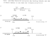
- Fig. 1 Structure of the plasmids pselP-gpt-L2 / preS1 and pselP-gpt-L2 / preS1dMyr.
- the plasmid structures of the gene for the large HBV protein or the demyristylated large HBV protein are given. Arrows show the direction of transcription from the selP and P7.5 promoters.
- FIG. 2 - XbaI restriction pattern of chimeric preS1 or preS1dMyr vaccinia viruses.
- the sizes of the XbaI fragments of the chimeric vaccinia viruses that hybridize with a 32 P-labeled preS1-specific probe are shown. Arrows show the direction of transcription from the selP and P7.5 promoters.
- FIG. 3 SacI restriction pattern of chimeric preS1 or preS1dMyr vaccinia viruses.
- the figure shows the size of the SacI fragments which hybridize with the 32 P-labeled 7.1 kb vaccinia virus SacI fragment "I" from chimeric preS1 and preS1dMyr vaccinia viruses. Arrows show the direction of transcription from the promoters selP and P7.5.
- Fig. 4 Pattern of hybridization with a 32 P-labeled, preS1-specific probe of XbaI cut genomic DNA from CV-1 cells infected with chimeric preS1 or preS1dMyr vaccinia virus clones.
- CV-1 cells were infected with chimeric preS1 or preS1dMyr vaccinia virus clones or wild-type vaccinia viruses.
- Lanes 1 to 10 XbaI cut genomic DNA from CV-1 cells infected with vS1 # 1-10.
- WT XbaI cut genomic DNA from CV-1 cells infected with the wild-type vaccinia virus.
- FIG. 5 Pattern of hybridization with a 32 P-labeled vaccinia virus SacI fragment 'I' specific probe of SacI cut genomic DNA from CV-1 cells infected with chimeric vS1 or vS1dM viruses .
- CV-1 cells were infected with vS1, vS1dM or wild-type vaccinia virus.
- Lanes 1 to 10 SacI-cut genomic DNA from CV-1 cells infected with vS1 # 1-10.
- Lanes 11 to 18 SacI-cut genomic DNA from CV-1 cells infected with vS1dM # 1-6, 9 and 10.
- WT SacI cut genomic DNA from CV-1 cells infected with wild-type vaccinia virus.
- FIG. 7 Construction of plasmid pselP-elp4gpt-L2.
- the vaccinia virus promoter P7.5 (P7.5) or the synthetic early / late vaccinia promoter 4 (elp4) control the xanthine guanine phosphoribosyl transferase gene (gpt gene) from Escherichia coli. Arrows indicate the direction of the transcription.
- Fig. 8 Structure of the plasmids pTZ-L2 / preS1 and pTZ-L2 / preS1dMyr.
- the genes encoding the large HBV protein (preS1) or its demyristylated version (preS1dMyr) were under the control of the bacteriophage T7 promoter, which was matched with a DNA copy of the 5 'non-coding region of the encephalomyocarditis Virus (emc) was linked.
- emc encephalomyocarditis Virus
- Figure 9 Western blot of total proteins extracted from cells coinfected with the T7 polymerase-expressing virus vT7 / selP and the viruses vT7S1 or vT7S1dM.
- Lanes 1 to 4 lysates from CV-1 cells infected with vT7S1dM # 1-4.
- Lanes 5 and 6 lysates from CV-1 cells infected with vT7S1 # 1 and 2.
- Lane 7 lysate from CV-1 cells infected with wild-type vaccinia viruses.
- Lane 8 lysate from uninfected CV-1 cells.
- Lanes 9 to 12 Lysates from CV-1 cells infected with vT7S1 # 3-6. Arrows indicate the monomeric large antigen (lower arrow) and aggregated forms of the antigen (upper arrow).
- Figure 10 Western blots of total proteins from different cell lines infected with chimeric viruses.
- Lanes 1 to 3 Lysates from SK-Hep1 cells (s), Chang liver cells (Ch) or CV-1 cells (C), which were labeled with vS1 # 10 were infected.
- Lanes 4 to 6 Lysates from SK-Hep1 cells, Chang liver cells or CV-1 cells which were infected with vS1 # 9.
- Lane 7 lysate from CV-1 cells infected with vT7S1 # 1.
- Lanes 8 to 10 Lysates from Sk-Hep1 cells, Chang liver cells or CV-1 cells which were infected with wild-type vaccinia viruses.
- Lanes 11 to 13 Lysates from SK-Hep1 cells, Chang liver cells or CV-1 cells which were infected with vS1dM # 10.
- Lanes 14 to 16 Lysates from SK-Hep1 cells, Chang liver cells or CV-1 cells that were infected with vS1dM # 9.
- Lane 17 lysate from CV-1 cells infected with vT71dM # 1.
- FIG. 11 Western blots of total proteins from chimeric virus infected cells expressing myristylated and demyristylated antigens.
- 11A shows the signals of the immunostaining which are specific for preS1 in a Western blot with lysate dilutions of 1:10, 1:20, 1:30, 1:40 and 1:50. Different dilutions were required to determine the immunostaining signal that is suitable for evaluation in a densitometric scanning system.
- Coomassie blue staining at 1:30 dilution of the lysates from CV-1 cells infected with vS1 # 10 and vT7S1 dM is shown in Figure 11B.
- Figure 12 Results of densitometric scanning of Western blots of total proteins from chimeric virus-infected cells expressing myristylated and demyristylated large antigens.
- Computer generated graphics of the results of densitometric scanning of CV-1 cells using vT7S1dM and vS1 # 10 were infected. A 1:50 dilution was used for the T7 / modified construct, and a 1:10 dilution was used for the selP / wild type construct. The shaded area under the curve has been calculated and is shown under "total" at the bottom of each diagram.
- FIG. 13 Structure of plasmids pTZ-L2 / CRM228A, pTZ-L2 / preS1dMyr-CRM228A, pTZ-L2 / preS2-CRM228A and pTZ-L2 / sAg-CRM228A.
- the nucleotide sequence coding for fragment A of the Corynebacteriophage beta tox 228 gene is in the same reading frame as an NcoI / PstI fragment with the complete open reading frames of the HBV ayw-preS1-preS2-S-sAg gene, the preS2- S-sAg gene and the S-sAg gene fused.
- the fusion genes are arranged under the transcriptional control of a modified Bacteriophage T7 promoter. The direction of the transcription is indicated by arrows.
- FIG 14 Construction of plasmid pTZ-L2 / preS1dMyr (x2).
- the nucleotide sequence of the preS1 portion (nucleotides 1-324) of the preS1dMyr preS2 sAg gene was amplified using the plasmid pTZ-L2 / preS1dMyr as a template and specific PCR primer.
- the PCR product was cloned as an NcoI fragment into a pTZ-L2 / preS1dMyr vector cut with NcoI and treated with alkaline phosphatases.
- the preS1-preS1-preS2-sAg gene is arranged under the transcriptional control of a modified Bacteriophage T7 promoter. The position of the PCR primers and the direction of transcription are indicated by arrows.
- a recombinant nucleotide molecule is constructed that includes the sequence encoding the structure of the preS1 region of the HBV large antigen.
- Dane particle DNA is an readily available source from which components of the HBV genome can be isolated using standard methods known to those skilled in the art. In general, construction involves isolating Dane particle DNA, cutting DNA with enzymes, identifying DNA fragments containing the structural gene for the large antigen, ligating the isolated gene to a cloning vector, recombining a portion of the cloning vector including the preS1 region in a vaccinia virus vector and infection of a suitable host with the chimeric vaccinia vector.
- the chimeric vaccinia vector is capable of replication in the host and expression of both vaccinia genes and non-vaccinia genes. Infected cells express a protein containing preS1.
- Oligonucleotides can be obtained using a device such as that from Applied Biosystems, eg Model 380A, or by chemical techniques, such as the method of Matteucci et al., J. Am. Chem. Soc., Vol. 103 (1981), pp. 3185-3191.
- DNA sequences that code for HBV's proteins can be made from Dane particles using methods such as those described by Robinson, Am. J. Med. Sci., Vol. 270 (1975), pp. 151-159.
- the aminoterminal myristylation site of the large HBV antigen is believed to result in the binding of the protein to components of the cell, particularly the cell membrane. It now appears, unexpectedly, that the conversion of G to C on the 5th nucleotide of the region coding for preS1, with which the destruction of the myristylation site was intended, also leads to greater translational efficiency and thus to increased yields on the large antigen; see. Figures 11 and 12 and Example 4.
- the chimeric virus also contains a marker gene, and cells infected with such a virus express a selectable marker protein.
- a marker gene By adding a marker gene to the chimeric vaccinia virus together with further sequences of interest, vectors or cells which carry the sequences can be identified and separated from those cells or vectors without these sequences. This selection is made by culturing the cells in an environment that favors the growth of viruses that have the marker gene.
- the structural gene is placed under the control of a promoter.
- the vaccinia virus has unique transcriptional regulatory signals that are recognized by the viral RNA polymerase. Therefore defined vaccinia promoter sequences are usually ligated to coding sequences of interest.
- a strong vaccinia promoter, such as selP, is preferred in this context.
- the present invention also contemplates the use of non-vaccinia virus promoters.
- the promoter of choice comes from the bacteriophage T7.
- the vaccinia virus bacteriophage T7 hybrid expression system has been shown to significantly increase expression of several foreign genes; Elroy-Stein et al., 1989. Since the transcription machinery of the vaccinia virus does not act on this promoter, the present invention also provides a T7 RNA polymerase.
- the structural gene is thus functionally linked to a regulatory element from the phage, a phage polymerase acting on the regulatory element.
- the promoter / HBV large antigen gene and contain the T7 RNA polymerase gene in different chimeric vaccinia viruses and are contacted by co-infection of the same cell.
- a "cassette” is a covalently linked series of DNA codons that encode several non-overlapping proteins.
- a cassette comprises a gene containing preS1, and can also code for a marker gene.
- the cassette also contains a promoter sequence derived from the T7 phage.
- cassettes are flanked by singular restriction sites that allow clipping from a series of cloning vectors and recombination into a series of cloning vectors, in this case genetically modified vaccinia viruses.
- a cloning vector is used as an intermediate carrier of the isolated gene.
- the cloning vector contains a promoter and a marker gene that can be used to generate a cassette for direct transfer to a recipient, such as the vaccinia virus genome.
- Incorporation of the cassette into the vaccinia sequences requires cutting both the preS1-containing cloning vector and the viral genome, the latter generally being cut at a preferred location relative to the vaccinia virus genes.
- the next step is the ligation of the cut or linearized cassette with the vaccinia genome. This leads to the formation of a linear chimeric viral genome.
- the ligated DNA is then transfected into cells infected with a second vaccinia virus, which aids in packaging the chimeric DNA into an infectious viral particle.
- the formation of a preS1-containing protein comprises the steps of (i) antigenizing a cell culture and (ii) infecting the culture with one or more chimeric vaccinia viruses.
- the cells must be permissive for the replication of vaccinia and can be mammalian kidney cell lines such as Vero (ATCC # CCl81) and CV-1 (ATCC # CCl70), as well as liver cell lines such as SK-HEP-1 (ATCC # HTB52) and Chang ( ATCC # CCl13).
- An advantage of the vaccinia virus system is the wide range of host cells, including liver cells, kidney cells and many other mammalian cell lines. This property has not yet been used for the expression of recombinant HBV surface glycoproteins. Since post-translational processing and modification are important aspects of the immunological properties of an antigen, the cheapest expression system for this type of vaccine includes a mammalian cell, and particularly liver and kidney cells.
- the host cells are not only permissive, but also represent the types of cells in which HBV is normally replicated. They are therefore able to produce HBV antigens that closely resemble those produced during a natural infection.
- the cultured cells should be able to provide post-translational modifications that allow recombinant antigens to show natural, antigenic and immunogenic epitopes. Cells originating in the liver and kidney are most preferred in this regard.
- Cultures are incubated under conditions that optimize the expression of the recombinant proteins.
- Variables that can be optimized include, but are not limited to, temperature, pH, CO2 concentration, and the presence of serum proteins, growth factors, and nutrients.
- a selective factor that favors the growth of viruses that carry the recombinant genes is also included.
- Expression of a protein is usually optimal when a maximum amount of protein has been produced. Infected cells can be broken up mechanically, chemically or thermally to increase the yield of product.
- the recombinant product is further purified as required for a given application.
- Process for cleaning Polypeptides from cell cultures are known to the person skilled in the art. Techniques that can be used include salt extraction, gradient centrifugation, gel filtration, gel chromatography, ultrafiltration, electrophoresis, ion exchange and the like.
- immunopotentiators include oil-based vehicles (Freund's adjuvant), aluminum vehicles (Alum), toxins (Lipid A) and proteins (Interleukins).
- the immunopotentiators are toxins which can be chemically or, in the case of protein toxins, genetically linked to the immunogens.
- Possible toxins for use in such constructs include, but are not limited to, ricin, abrin, pertussis toxin, Shigella-like toxin, cholera toxin, Staphylococcus exotoxin A, heat-stable and heat-labile enterotoxin and Pseudomonas exotoxin A.
- diphtheria toxin is effective in immunopotentiating an immunogen to which it is linked.
- the use of diphtheria toxin in fusion constructs as an immunotoxin has been reported so far; Strom et al., Ann. NY Acad. Sci., Vol. 636 (1981), pp. 233-250; Shaw et al., J. Biol. Chem., Vol. 266 (1991), pp. 21118-21124; Williams et al., J. Biol. Chem., Vol. 265 (1990), pp. 11885-11889.
- immunotoxins differ from immunopotentiators in that the former is a toxin / antibody fusion molecule that selectively destroys target cells, while the latter is a toxin or a toxin / protein fusion molecule, the toxin portion enhancing the immunogenicity of the antigen portion.
- toxins other than diphtheria toxin, as immunopotentiators, have been reported; Cholera toxin: Hussain et al. (1989); Pertussis toxin: Halsov et al., 1991.
- nucleic acid sequences encoding a protein immunogen can be appended to nucleic acid sequences encoding the immunopotentiator in the same reading frame.
- Expression of the "hybrid gene” obtained results in a single polypeptide that contains both immunogen and immunopotentiator domains.
- Another method of increasing the immunogenicity of the preS1 protein sequences is to provide a larger effective dosage of the epitopes contained therein.
- One way in which this can be achieved is to double the preS1 sequences in the large HBV antigen.
- the expressed protein thereof has at least twice as many preS1-B and T-cell epitopes. This higher epitope density can help stimulate the anti-preS1 response.
- HBV-preS1-containing antigens consists in the production of a suitable material for an improved HBV vaccine.
- a significant proportion (7-15%) of the population does not respond to conventional HBV vaccines that do not contain preS1 regions.
- the ability to immunize individuals with antigens that contain this region that has been processed in a manner consistent with the natural infection allows a larger number of individuals to be protected from HBV infections.
- Antibodies directed against the recombinant protein containing preS1 also belong to the present invention.
- the present invention provides large amounts of preS1-containing antigens that are processed by cell types that are normally infected with HBV. Therefore, the possibility of producing both polyclonal and monoclonal antibodies, which are suitable for the detection, therapy and prevention of HBV-related diseases, is significantly improved.
- Immunotoxins are hybrid protein molecules that are generated by the fusion of an antibody and a toxin and can be used to selectively destroy certain cell populations.
- the potent cell-killing effect of a toxin is associated with the excellent specificity of monoclonal antibodies, which causes selective destruction of target cells while other cells are not affected.
- toxins include, but are not limited to, ricin, abrin, pertussis toxin, Shigella-like toxin, cholera toxin, Staphylococcus exotoxin A, heat-stable and heat-stable enterotoxin, Pseudomonas exotoxin A and diphtheria toxin. In some cases it is advantageous to reduce the toxicity of the toxin part by chemical or genetic measures.
- Toxins can be fused to antibodies chemically or, in the case of protein toxins, genetically.
- Protein toxin genes can be fused in frame with a single chain antibody gene (light chain / heavy chain hybrids; see Huston et al., Proc. Natl. Acad. Sci. USA, Vol. 85 (1988), p. 5879- p. 5879- 5883, and Bird et al., Science, Vol. 242 (1988), p. 423-426).
- the single chain antibody part can be replaced by other binding proteins, such as receptors or receptor ligands, for example II-2.
- a vaccine that includes at least the preS1 region of HBV that has been prepared by the recombinant technique described in the present application.
- the vaccine comprises an effective amount of the preS1-containing proteins which are produced by the recombinant expression system in cells infected with chimeric vaccinia viruses and which have a suitable immunogenicity.
- Appropriate immunogenicity means that the polypeptide is capable of eliciting an immune response that protects against natural HBV infection.
- a reagent kit that includes a container and (i) at least one antibody directed against a molecule containing preS1 or (ii) a molecule containing preS is contemplated.
- the antibodies according to the invention are involved in the detection of a molecule containing preS1, which was formed by a natural infection.
- the molecules according to the invention containing preS1 are involved in the detection of preS1-specific antibodies which have been formed by a host against a natural infection or an immunization. In both cases, an antibody that is provided with a chromatographic, fluorometric or other detectable label is used to detect either the preS1-containing protein or a preS1-specific antibody.
- the reagent set preferably also comprises a positive control for the binding of a molecule containing preS1 to a preS1-specific antibody or vice versa, and the detection reaction.
- the present invention also relates to a method for producing proteinaceous particles which show HBV antigenicity and / or HBV immunogenicity. Culturing infected cells leads to the formation of proteinaceous particles. The cells containing the particles can then be collected. According to preferred embodiments, the Particles are isolated from the cells using a number of methods known to those skilled in the art.
- the 1447 base pair fragment NsiI (position 1070 in the HBV ayw genome) / BstEII (position 2817 in the HBV ayw genome) was isolated from the plasmid pTHBV; Christman et al., 1982. Overhangs resulting from restriction endonuclease digestion were removed using T4 DNA polymerase. The blunt-ended fragment was cloned into the SmaI cut and dephosphorylated vector pTM3 (Moss et al., 1990), resulting in the intermediate plasmid pTM3 / preS1A, which carried a correctly oriented insert.
- the bacterial clone carrying the plasmid pTM3 / preS1A was infected with an M13 helper phage (Stratagene, Heidelberg, FRG) to obtain single-stranded (ss) plasmid DNA.
- This ss-DNA was used to generate an NcoI interface on the preS1-ATG start codon by means of targeted mutagenesis (mutagenesis oligonucleotide: 5'-GGA AAG ATT CTG CCC CAT GGT GTA GAT CTT GTT-3 ').
- the intermediate plasmid was named pTM3 / preS1B.
- pselP-gpt-L2 / preS1dMyr Single-stranded DNA of the plasmid pTM3 / preS1A was used to generate an NcoI site at the preS1-ATG start codon by targeted mutagenesis using a mutagenesis oligonucleotide 5'-GGA AAG ATT CTG CGC CAT GGT GTA GAT CTT GTT-3 ', which simultaneously introduced a cytocin at position 2 of the second codon, which changed a glycine at the N-terminus of the open reading frame for preS1-S2-S to an alanine (pTM3 / preS1dMyrA).
- the resulting 40 base pair NcoI fragment containing 5 'non-coding sequences was removed and the remaining one Vector was purified and religated over the NcoI sites to give plasmid pTM3 / preS1dMyrB.
- the 1418 bp NcoI / PstI fragment which contained the fully modified preS1-S2-S gene without 5 'non-coding sequences, was isolated from pTM3 / preS1dMyrB and with the pselP-gpt-L2 vaccinia cut with NcoI / PstI Virus cloning vector ligated; see. US serial no. 07/750 080.
- the plasmid obtained was designated pselP-gpt-L2 / preS1dMyr.
- the NcoI / PstI cassette from pTM-3 / preS1dMyrB was ligated with the pTZ-L2 vector cut with NcoI / PstI; see. US serial no. 07/750 080.
- telomere sequence was determined in a polymerase chain reaction (PCR) from pSV2gpt (ATCC # 37145) using gpt gene-specific oligonucleotides (5'-ATC ATA TGA GGA TCC ATG AGC GAA AAA TAC ATC GTC ACC-3 'and 5 '-ATC CCG GGA CGT CAT AAA AAG ATT AGC GAC CGG AGA TTG GCG G-3') amplified.
- PCR polymerase chain reaction
- NdeI and BamHI sites were introduced at the 5 'end and AatII and SmaI sites at the 3' end of the gpt gene.
- the PCR products were cut with NdeI / SmaI and ligated with a pFS51 vector (EP-A-91 114 300.6) cut with NdeI / SmaI.
- the elp4 sequence was designed as a synthetic NdeI / BamHI linker consisting of the hybridized oligonucleotides oelP4.1 (5'-TAT GTA AAA GTT GAA ATT ATT TTT TTA TGC TGT AAA TAA GTT AAC A-3 ') and oelP4.2 (5'-GAT CTG TTA ACT TAT TTA CTA GCA TAA AAA AAT AAT TTC AAC TTT TAC A-3 '), was introduced upstream of the open reading frame of the gpt gene to give the plasmid pelP4-gpt.
- oelP4.1 5'-TAT GTA AAA GTT GAA ATT ATT TTT TTA TGC TGT AAA TAA GTT A-3 '
- oelP4.2 5'-GAT CTG TTA ACT TAT TTA CTA GCA TAA AAA AAT AAT TTC AAC TTT TAC A-3 '
- the elp4-gpt gene cassette was excised from the plasmid pelP4-gpt with NdeI and AatII and ligated with NdeI / AatII cut and purified pselP-gpt-L2 vector DNA, resulting in the plasmid pselP-elp4gpt-L2.
- pselP-elp4gpt-L2 / T7 The 2.7 kb BamHI fragment with the gene for the bacteriophage T7-RNA polymerase was isolated from the vaccinia virus vTF7 / 3 (Fuerst et al., 1986) and with the BamHI-cut and dephosphorylated pselP-elp4gpt- L2 vector ligated.
- a plasmid clone which carried the T7 RNA polymerase gene in the correct orientation with respect to the selP promoter was isolated. This plasmid was named pselP-elp4gpt-L2 / T7.
- the 5 'non-coding sequences were completely removed from the preS1-S2-S gene.
- An NcoI interface (5'-CCATGG-3 ') was introduced around the ATG start codon by targeted mutagenesis. This manipulation allowed the ATG start codon of the corresponding gene to fuse directly with the early / late vaccinia virus promoter (selP) or the bacteriophage T7 promoter.
- the plasmids pselP-gpt-L2 / preS1 and pselP-gpt-L2 / preS1dMyr Fig.
- the double gene cassettes can be cut out of the plasmids with several restriction enzymes (e.g. NotI, SnaBI, SfiI) and ligated with the corresponding singular interfaces of a suitable vaccinia virus vector.
- the cassettes were cut out with NotI and ligated with vaccinia virus arms, which had also been cut with NotI.
- plasmids pTZ-L2 / preS1 and pTZ-L2 / preS1dMyr (Fig. 8) the large HBV antigen gene or its demyristylated version are under control of the bacteriophage T7 promoter.
- the viruses constructed with these plasmids were referred to as the vT7S1 series and vT7S1dM series.
- the nucleotide sequence encoding fragment A of the Corynebacteriophage beta tox 228 polypeptide (without signal peptide sequence) is in a polymerase chain reaction using the PCR primers 5 'CCT TCA GCC CAT GCC ATG GCT GAT GAT GTT 3' and 5 'A GCC ACC TAC TGC AGG CCT GAC ACG 3 'amplified.
- the 5 'primer introduces an NcoI site, while the 3' primer introduces a PstI site.
- the 606 bp PCR product is cut with NcoI and PstI and cloned into the pTZ-L2 vector cut with NcoI / PstI (cf. US Serial No. 07/914 738, which leads to the plasmid pTZ-L2 / CRM228A.
- pTZ-L2 / preS1dMyr-CRM228A An NcoI site is introduced at the C-terminus of the preS1dMyr open reading frame (ORF) using single-stranded DNA from pTZ-L2 / preS1dMyr as a template and the mutagenic oligonucleotide 5 'TTT TGT TAG GGT TTC CAT GGA TAC CCA AAG ACA 3'. This manipulation changes the last two amino acids of the open reading frame from preS1dMyr from Tyr-Ile to Ser-Met and introduces an additional Glu residue.
- the plasmid obtained is referred to as pTZ-L2 / preS1dMyrdTAA.
- This plasmid is linearized with PstI, partially digested with NcoI and ligated with the NcoI / PstI insert of the plasmid pTZ-L2 / CRM228A.
- a plasmid clone carrying the gene for the CRM228 A fragment is selected and designated pTZ-L2-preS1-CRM 22817.
- pTZ-L2 / preS2-CRM228A An NcoI site is introduced at the C-terminus of the preS2 open reading frame using single-stranded DNA from the plasmid pTZ-L2 / preS2 as a template and the mutagenic oligonucleotide 5 'TTT TGT TAG GGT TTC CAT GGA TAC CCA AAG ACA 3'. This manipulation changes the last two amino acids of the preS2 ORF from Tyr-Ile to Ser-Met and introduces an additional Glu residue.
- the plasmid obtained is referred to as pTZ-L2 / preS2dTAA.
- This plasmid is linearized with PstI, partially digested with NcoI and ligated with the NcoI / PstI insert of the plasmid pTZ-L2 / CRM228A.
- a plasmid clone carrying the gene for the CRM228 A fragment is selected and designated pTZ-L2 / preS2-CRM228 A.
- pTZ-L2 / sAg-CRM228A An NcoI site is introduced at the C-terminus of the HBV-sAg open reading frame using single-stranded DNA from the plasmid pTZ-L2 / sAg as a template and the mutagenic oligonucleotide 5 'TTT TGT TAG GGT TTC CAT GGA TAC CCA AAG ACA 3' .
- This manipulation changes the amino acid sequence of the ORF from HBV sAg from Tyr-Ile to Ser-Met and introduces an additional Glu residue.
- the plasmid obtained is referred to as pTZ-L2 / sAgdTAA.
- This plasmid is linearized with PstI, partially digested with NcoI and ligated with the NcoI / PstI insert of the plasmid pTZ-L2CRM228A.
- a plasmid clone bearing the gene for the CRM228 A fragment is selected and designated pTZ-L2 / sAg-CRM228A.
- pTZ-L2 / preS1dMyr (x2).
- the nucleotide sequence comprising amino acids 1-108 of the ORF from preS1dMyr is generated in a PCR reaction using pTZ-L2 / preS1dMyr as template and the oligonucleotides 5 'TTT TGG AAT ATA AAT AAG GC 3' and 5 'ATT CCA CTC CAT GGC CTG AG 3 'amplified as a PCR primer, the latter primer introducing a second NcoI interface into the PCR product.
- the 360 bp PCR product is cut with NcoI and ligated to the pTZ-L2 / preS1dMyr vector cut with NcoI and treated with alkaline phosphatase.
- a plasmid which carries the PCR product in the correct orientation relative to the old reading frame of the preS1 gene is selected and designated pTZ-L2 / preS1dMyr (x2).
- the NotI gene cassettes from the plasmids containing preS1, preS1dMyr or T7 polymerase (pTZ-L2 / preS1, pTZ-L2 / preS1dMyr, pselP-gpt-L2 / preS1, pselP-gpt-L2 / preS1dMyr, pselP-elp4gpt- L2 / T7) were isolated and ligated with the NotI cut genomic DNA from the vaccinia virus wild-type strain WR, as is essentially described in US serial no. 07/750 080.
- the ligation components consisted of 1 ⁇ g of genomic vaccinia virus DNA cut with NotI, 500 ng NotI cassette which had been isolated from the corresponding plasmids, 5 units of T4 DNA ligase and reaction buffer in a total volume of 50 ⁇ l. After incubation for 16 hours at 16 ° C., the ligation mixture was coprecipitated together with calcium phosphate and added to CV-1 cells that had grown subconfluently. This Cells had been infected with the fowlpox virus strain HP1-441 (Mayr & Malicki, 1996) two hours earlier with a multiplicity of infection, (moi; virus per cell) of 0.1 plaque-forming units (pfu) per cell. 5 to 7 days after the infection, the cell monolayer was completely infected with vaccinia viruses. The cells were harvested and the virus was released from the cell fragments by sonication.
- chimeric vaccinia viruses The selection for chimeric vaccinia viruses was carried out by titrating the virus progeny in the presence of 25 ug / ml mycophenolic acid, 15 ug / ml hypoxanthine and 125 ug / ml xanthine. Viruses grown under these conditions were further plaque-cleaned and used for the preparation of "small" and "large” virus stock solutions. Small virus stock solutions isolated from individual virus plaques were also grown under selective conditions. Infected cells from small stock solutions were divided into two cultures. One culture was used to make large virus stocks. DNA was isolated from the other, digested with various restriction endonucleases and transferred to nitrocellulose filters. These filters were hybridized with specific probes to confirm the orientation of the NotI cassette in the chimeric vaccinia virus genome and the absence of wild-type vaccinia virus.
- the XbaI fragments from genomic DNA of a chimeric preS1 vaccinia virus that hybridize with a 32 P-dCTP-labeled preS1-specific probe should have a size of 1.01 kb and 3.3 kb or 1.9 kb and 2, 41 kb, depending on the orientation of the NotI cassette (Fig. 2), while the SacI fragments from the same chimeric virus as the 32 P-dCTP-labeled 7.1 kb vaccinia virus SacI fragment Hybrid "I" should have a size of 5.35 kb and 4.47 kb or 5.15 kb and 4.68 kb, depending on the orientation (FIG. 3).
- FIG. 2a The singular NotI interface and the flanking XbaI interfaces of the wild-type vaccinia virus genome are shown in FIG. 2a.
- the integration of the NotI cassette into the singular NotI interface in the orientation "a" leads to the formation of two new XbaI fragments with a length of 1.01 kb and 3.3 kb (cf. FIG. 2b).
- the integration in the orientation "b” leads to two new XbaI fragments with a length of 2.41 kb and 1.9 kb (cf. FIG. 2c).
- Fig. 3a the singular NotI interface and the flanking SacI interfaces in the wild-type vaccinia virus genome are shown.
- FIG. 4 The hybridization patterns of the XbaI and SacI cut genomic DNA from 10 chimeric preS1 viruses, vS1 # 1-10, and 8 chimeric preS1dMyr viruses, vS1dM # 1-6, 9 and 10, either with the preS1 probe or the Vaccinia Virus SacI Probe-I are shown in Figures 4 and 5, respectively.
- genomic DNA was isolated from infected cell pellets and cut with XbaI. DNA fragments were separated on a 1% DNA agarose gel and transferred to nitrocellulose filters before probe hybridization. Hybridization of the XbaI fragments showed that vS1 # 1, 2, 4 and 5 (FIG.
- the viruses vS1dM # 1, 2, 5, 6 and 9 had inserted the NotI cassette in the orientation “b”, the virus isolate vS1dM # 10, FIG. 4, lane 18) represents the orientation "a”.
- the chimeric viruses vS1dM # 3 and 4 (Fig. 4, lanes 13 and 14) showed hybridization signals in the high molecular range. This type of signal also occurred in vS1dM # 2 and 5 (Fig. 4, lanes 12 and 15) and in vS1dM # 10 (Fig. 4, lane 18) and may indicate that multiple copies of the NotI cassette in the viral genome has been integrated. The exact structure of these viruses has yet to be determined.
- genomic DNA was isolated from infected cells and cut with SacI.
- the DNA fragments were separated on a 1% DNA agarose gel and transferred to nitrocellulose before probe hybridization.
- Analysis of the SacI fragments that hybridized with SacI Probe-I showed the expected patterns of orientation "a” (4.47 and 5.35 kb) and orientation "b" (4.68 kb and 5.15 kb).
- orientation "a" (4.47 and 5.35 kb)
- orientation "b" (4.68 kb and 5.15 kb
- the 7.1 kb wild-type vaccinia virus SacI fragment "I" (FIG. 5, lane WT) is not present in any of the chimeric viruses. The possibility of contamination of the chimeric viruses with wild-type vaccinia virus could therefore be excluded.
- a chimeric vaccinia virus carrying the T7 RNA polymerase gene, vT7 / selP was constructed by integrating the NotI cassette from the plasmid pselP-elp4gpt-L2 / T7 into the NotI interface of the vaccinia virus genomic DNA.
- This plasmid was constructed from the starting plasmid pselP-elp4gpt-L2, which contains an improved gpt gene cassette.
- the plasmid pselP-gpt-L2 was constructed as shown in FIG. 7.
- the gene which codes for the dominant selection marker is controlled by the vaccinia promoter P7.5, which is located on a 500 base pair vaccinia DNA fragment. This fragment can lead to undesirable recombination events with two endogenous vaccinia virus p7.5 promoter sequences; Venkatesan et al., 1981.
- the P7.5 gpt gene cassette (1.1 kb in size) was replaced by the elp4 gpt gene cassette (in size) of 0.5 kb) replaced.
- the new gene cassette was significantly reduced in size in addition to a weak, non-homologous synthetic early / late promoter.
- the resulting plasmid which had the optimized dominant marker and the strong synthetic early / late promoter followed by a multiple cloning site, was named pselP-elp4gpt-L2.
- CV-1 cells ATCC CCL 70
- SK-Hep1 cells ATCC HTB 52
- Chang liver cells ATCC CCL3
- a moi of 1 with chimeric preS1 vaccinia viruses chimeras preS1dMyr vaccinia viruses or chimeric vaccinia viruses infected with the preS1 or preS1dMyr genes under the control of the bacteriophage T7 promoter.
- Cells infected with the T7 preS1 chimeras were co-infected with a moi of 1 with a vaccinia helper virus chimera which carried the gene for the bacteriophage T7 RNA polymerase under the control of the selP promoter (vT7 / selP).
- vT7 / selP selP promoter
- cells were infected with vT7 / selP alone or after. The cells were harvested 3 days after infection. Infected cells were pelleted and resuspended in 300 ⁇ l sodium dodecyl sulfate sample buffer (SDS sample buffer). 10 to 20 ⁇ l of the lysate were sonicated, boiled and subjected to electrophoresis on a 12% SDS polyacrylamide gel.
- SDS sample buffer sodium dodecyl sulfate sample buffer
- FIG. 6 shows the expression yield in CV-1 cells which were infected with eight vS1dM isolations (FIG. 6, lanes 1 to 8) or 10 vS1 isolations (FIG. 6, lanes 9 to 18).
- vS1dM isolations FIGS. 4, and 5
- FIGS. 4 and 5 influence the expression efficiency of the vS1dM viruses (FIG. 6, lanes 1 to 8).
- the direct comparison of the expression yield of the cells infected with vS1dM viruses (FIG. 6, lanes 1 to 8) with vS1 viruses FIG. 6
- CV-1 cells were co-infected with vT7S1 or vT7S1dM viruses and with the second virus, vT7 / selP, which carried the gene for the bacteriophage T7 RNA polymerase. Lysates from CV-1 cells infected with four different isolates of vT7S1dM (Fig. 9, lanes 1 to 4) and 6 different vT7S1 viruses (Fig. 9, lanes 5 to 6 and 9 to 12) were analyzed . As a control, lysates from post-infected CV-1 cells or from CV-1 cells infected with vT7 / selP alone were also analyzed (FIG. 9, lanes 7 and 8, respectively).
- Example 4 Quantitative determination of the large antigen by Western blot analysis
- the expression yield was compared in a Western blot, which was essentially according to Towbin et al. (1979) with the following modifications.
- the first antibody was either a goat serum specific for hepatitis-associated antigens (HAA) (Axell, catalog number AXL 683), or monoclonal mouse antibodies specific for the preS1 region (Immunotech SA, catalog number 0866), diluted 1: 200 in a Western blot buffer (phosphate buffered saline containing 0.1% Tween 20).
- the second antibody was an alkaline phosphatase-coupled goat anti-mouse serum (Sigma Chemicals, Inc., Catalog No. A-4656) at a 1: 1000 dilution.
- the detection system (NBT and alkaline phosphatase) was from Promega, Inc.
- the quantitative determination of the large HBV antigen was carried out by comparing the Western blot bands.
- purified medium HBV ayw antigen was used in the Vero-cell / Vaccinia virus system was used.
- a series of dilutions was carried out with the purified medium antigen and with preparations of the large antigen.
- the dilutions were electrophoresed on a 12% SDS polyacrylamide gel, transferred to nitrocellulose and in a Western blot using anti-HAA serum as a first antibody and an alkaline phosphatase conjugated anti-goat IgG serum as a second Antibodies carried out.
- anti-HAA serum as a first antibody
- an alkaline phosphatase conjugated anti-goat IgG serum as a second Antibodies carried out.
- 1 to 10 ⁇ g large antigen / 106 cells were detected.
- CV-1 cells a monkey kidney cell line
- SK-Hep1 cells and Chang liver cells both human liver cell lines
- vS1 # 9 orientation: "a”
- vS1 # 10 orientation: "b”
- vS1dM # 9 orientation: "b”
- vS1dM # 10 orientation: "a"
- Figure 10 shows the results of the expression study.
- Lysates from SK-Hep1 cells (S), Chang liver cells (Ch) and CV-1 cells (C) with vS1 # 9 and vS1 # 10, wild-type vaccinia virus and vS1dM # 9 and vS1dM # 10 infected were compared.
- the increased levels of expression of the chimeras with the demyristylated preS1 gene (Fig. 10, lanes 11 to 16) were again clearly visible in comparison with those of the wild-type preS1 gene (lanes 1 to 6). This result shows that this effect is not limited to a single cell line, but could be observed repeatedly in different cell lines.
- the direct comparison of the expression yield starting from the bacteriophage T7 promoter against the vaccinia virus selP promoter was carried out in parallel.
- lysates from CV-1 cells infected with vT7S1 and vT7S1dM were also analyzed in the Western blot shown in FIG. 10.
- the difference in expression yield of the large antigen and its modified version was analyzed in a Western blot for comparison.
- the expression yield of the large antigen or its modified version in the T7 system was at least 5 times higher compared to the expression yields found in the selP promoter system (FIG. 10, lane 6 compared to 7, or lane 16, compared to 17).
- Cultures with 1 x 10⁷ CV-1 cells were infected with vLS1 # 10 with a moi of 2 pfu / cell or with vT7S1dM and the helper virus vT7 / selP with a moi of 1 pfu / cell for each chimeric virus.
- 48 hours after infection the infected cells were harvested and resuspended in 1 ml SDS sample buffer.
- the lysates were sonicated thoroughly and further diluted with SDS sample buffer. 20 ul of the diluted lysate were separated on a 12% SDS polyacrylamide gel. The gels were either used to transfer the proteins to a nitrocellulose filter (Western blot), or the proteins were stained with Coomassie blue.
- FIG. 11A shows the immunologically stained filters.
- the filters were analyzed with a densitometer (Hirschmann Elscript 400).
- the 1:30 dilutions of the lysates from each infection stained with Coomassie blue are shown in Figure 11B.
- the results of the densitometric analysis of the Western blot signals, a suitable dilution (1:10 for the vS1 # 10 lysate; 1:50 for the vT7S1dM lysate) are given in FIG. 12.
- the two peaks correspond to the two preS1 glycoforms.
- Example 5 Enhancement of protein immunogenicity: Construction of hybrid proteins consisting of the HBV surface glycoprotein and the diphtheria toxin fragment A cross-reacting material, and doubling of the HBV preS1-specific epitopes
- a new immunopotentiator namely diphtheria toxin cross-reacting material (CRM)
- CRMs diphtheria toxin cross-reacting material
- a strong immune response is elicited by Corynebacterium diphtheriae, which expresses CRM; "Pharmacology of Bacterial Toxins", in: International Encyclopedia of Pharmacology and Therapeutics, Dorner and Drews (Editor), pp.
- the diphtheria toxin gene or CRM gene is carried by a lysogenic ⁇ phage that is integrated or not integrated into the bacterial genome.
- the intact toxin consists of a fragment "A" with a molecular weight of 22 kD and a fragment "B” with a molecular weight of 40 kD. Fragment A ensures the toxic enzymatic properties, while fragment B is required for the translocation of fragment A across the cell membrane.
- CRM 228 is similar to the wild-type toxin but has no enzymatic activity due to changes in the amino acid sequence; Uchida et al., J. Biol. Chem., Vol. 248 (1973a), pp. 3838-3844; Streeck et al., Science, 221: 855-858.
- the phage carrying the CRM 228 gene is available from the Pasteur Institute, Paris, France.
- the linkage of the DNA coding for the glycoprotein with the nucleic acid coding for the CRM 228 fragment A is achieved by recombinant DNA methods; see. Fig. 13.
- the antigen and the immunopotentiator are therefore genetically linked and are expressed as a fusion protein. Fragment A was chosen because it is smaller compared to CRM 228 fragment B.
- the CRM 228 fragment B or the complete protein CRM 228 exceed the size of the preS1-S2-S-sAg, which may have a negative impact on the immune response directed against the preS1 part.
- the smaller size of the CRM 228 fragment A is advantageous in that it ensures the accessibility of the preS1 epitopes within the fusion protein.
- fragment A's strong immunogenicity is equivalent to that of the native molecule.
- An alternative method of linking HBV antigens and immunopotentiators is to chemically link the highly purified CRM 228 fragment A to purified preS1-preS2-S-sAg (demyristylated version), preS2-S-sAg or S-sAg.
- equimolar amounts of CRM 228 fragment A and antigen to be linked are crosslinked using standard chemical procedures; e.g. Widder and Green, Methods in Enzymology, vol. 112 (1985), p. 207.
- preS1 region of the large HBV antigen amino acids 1-108. This is accomplished by inserting a second copy of the preS1 coding region into a vector encoding all of the large HBV antigen; see. Fig. 14. The expression of this construct results in the new protein (preS1) 2-preS2-S-sAg, which contains two B and T cell-specific preS1 epitopes.
- the proteinaceous particles of the present invention are used in a pharmaceutically acceptable composition which, when administered in an effective amount, is capable of inducing protective immunity to HBV infection.
- the preparation of the vaccines which contain proteins as active ingredients is known to the person skilled in the art.
- such vaccines are manufactured as injectable vaccines, namely as liquid solutions or suspensions; solid forms suitable for dissolving or suspending in a liquid before injection can also be made.
- the preparation can also be emulsified.
- the active immunogenic component is often mixed with a carrier that is pharmaceutically acceptable and compatible with the active component. Suitable carriers are e.g. Water, saline, dextrose, glycerin, ethanol and the like, and combinations thereof.
- the vaccine can optionally contain small amounts of auxiliary substances, such as wetting agents or emulsifiers, pH-buffering agents, adjuvants or immunopotentiators, which increase the effectiveness of the vaccine.
- the vaccines are administered parenterally in a conventional manner by injection, e.g. subcutaneously or intramuscularly.
- Other formulations suitable for other modes of administration include suppositories and in some cases oral formulations.
- conventional binders and carriers e.g. include polyalkalene glucose or triglycerides; Suppositories can be formed from mixtures which contain the active ingredient in a concentration in the range from 0.5% to 10% and preferably in a concentration of 1.2%.
- Oral formulations include typical pharmaceutical grade carriers such as mannitol, lactose, starch, magnesium stearate, sodium saccharin, cellulose, magnesium carbonate and the like.
- the compositions take the form of solutions, suspensions, tablets, pills, capsules, sustained release formulations or powders and contain 10% to 95% and preferably 25 to 70% of active ingredient.
- the proteinaceous particles can be formulated into a vaccine as neutral forms or salt forms.
- Pharmaceutically acceptable salts include acid addition salts formed with the free amino groups of the peptide and inorganic acids such as hydrochloric acid or phosphoric acid, or organic acids such as acetic acid, oxalic acid, tartaric acid, mandelic acid or the like. Salts formed with the free carboxyl groups can also be from inorganic bases such as sodium hydroxide, potassium hydroxide, ammonium hydroxide, calcium hydroxide or iron (III) hydroxide, and organic bases such as isopropylamine, trimethylamine, 2-ethylaminoethanol, histidine, procaine and the like.
- unit dose refers to physically discrete units suitable for use in humans, each unit containing a predetermined amount of active material which has been calculated to provide the desired therapeutic effect in combination with the required diluent, ie the wearer, and a special treatment plan.
- the amount to be administered both in terms of the number of treatments and the amount, depends on the patient being treated, the ability of the patient's immune system to synthesize antibodies and the level of protection desired. Exact amounts of active ingredient that need to be administered depend on the practitioner's judgment and apply specifically to an individual.
- the suitable dosage range is, however, on the order of 1 to several 100 ⁇ g of active ingredient per individual. Appropriate schedules for initial administration and refreshments also vary, but typically an initial administration is followed by 1 or 2 week intervals with one or more subsequent injections or other administrations.
- mice 3 month old female BALB / c mice were vaccinated five times with purified HBV ayw preS1 expressed in Vero cells infected with the appropriate vaccinia virus chimera in a series of vaccinations over a period of 6 weeks ( approximately 1 to 10 ⁇ g protein per vaccination). 3 days after the last vaccination, blood samples were taken from the immunized mice and examined for preS-specific antibodies using an ELISA system with preS1 as the test antigen. Spleen cells from mice with a positive ELISA signal were isolated and plasmacytoma cell line P3X63Ag8 (ATCC TIB 9) using polyethylene glycol according to the method of Lane et al., J. Immunol. Meth. Vol. 81 (1985), pp.
- Hybridoma cell clones that secrete preS-specific monoclonal antibodies were selected in a dilution assay in RPMI 1640 medium (8041-2400, GIBCO). The specificity for the preS1 portion of the monoclonal antibodies was confirmed by synthetic preS1 peptides in a commercially available peptide mapping reagent set (Cambridge Research Biochemicals).
- a diagnostic system preferably in the form of a reagent set, which is suitable for the detection of the presence of preS1-containing antigens or antibodies against preS1-containing antigens in a body sample comprises (a) a protein according to the invention or (b) an antibody according to the invention and (c) a labeled specific binding agent to elicit a signal in the presence of the immune response of a protein containing preS1 and an anti-preS1 antibody.
- label in its various grammatical forms refers to individual atoms and molecules that are involved, either directly or indirectly, in triggering a detectable signal to indicate the presence of an immunoreactant. Any label can be attached to, incorporated into, or used separately from a specific agent, and these atoms or molecules can be used alone or in conjunction with other reagents. Such labels are known as such in the field of immunochemistry and form part of the present invention only insofar as they are used with proteins, methods and / or systems which are new in other respects.
- the diagnostic reagent sets of the invention are typically used in an "ELISA format" to detect the presence or amount of anti-preS1 antibodies or proteins containing preS1 in a body sample, such as serum or plasma.
- ELISA also refers to a solid phase enzyme immunoassay ("enzyme-linked immunosorbent assay") using an antibody or antigen bound to a solid phase and an enzyme-antigen or enzyme-antibody conjugate to determine the amount of antigen or antibody present in a sample to be proven and quantified.
- enzyme-linked immunosorbent assay enzyme-linked immunosorbent assay
- a description of the ELISA technique can be found in Chapter 22 of the 4th edition of Basic and Clinical Immunology, DP Sites et al., Lange Medical Publications, Los Altos, CA (1982) and in U.S. Patents 3,654,090, 3,850 752 and 4 016 043, all of which are made the subject of the present application by appropriate reference.
- the protein containing the preS1 according to the invention or the anti-preS1 antibody according to the invention are fixed to a solid matrix, a solid carrier being formed.
- the molecules are typically fixed to the solid matrix by adsorption from an aqueous medium, although several other types of adsorption and other types of fixation known to those skilled in the art can also be used. Examples of such types are the interaction of proteins with the carboxyl units, which are formed by reaction of cyanogen bromide with matrices containing glucose. These matrices include cross-linked dextrose or cellulose and linkage with glutaraldehyde, as discussed below in connection with latex particles and the like.
- Suitable solid matrices are known to the person skilled in the art. Such materials include the cross-linked dextran available under the trademark SEPHADEX from Pharmacia Fine Chemicals (Piscataway, NJ), agarose, polystyrene beads from about 1 ⁇ m to about 5 mm in diameter (Abbott Laboratories, North Chicago, IL), polyvinyl chloride , Polystyrene, cross-linked polyacrylamide, nitrocellulose or nylon-based fabrics such as layers, strips, blades and tubes.
- SEPHADEX cross-linked dextran available under the trademark SEPHADEX from Pharmacia Fine Chemicals (Piscataway, NJ)
- agarose polystyrene beads from about 1 ⁇ m to about 5 mm in diameter (Abbott Laboratories, North Chicago, IL)
- polyvinyl chloride Polystyrene
- cross-linked polyacrylamide nitrocellulose or nylon-based fabrics such as layers, strips, blades and tubes.
- a preferred matrix is in the form of a
- Hepatitis B virus envelope epitopes gene assembly and expression in Escherichia coli of an immunologically reactive novel multiple-epitope polypeptide 1 (MEP-1). Genes. 110, 137-144. Kuroda, S., Fujisawa, Y., Iino, S., Akahane, Y., Suzuki, H. 1992a. Induction of protection level of anti-pre-S2 antibodies in humans immunized with a novel hepaptitis B vaccine consisting of M (pre-S2 + S) protein particles (a third generation vaccine). Vaccine, 9, 163.
- Milich, DR, Mc Namara, MK, Mc Lachlan, A., Thornthon, GB, Chisari, FV 1985a Distinct H-2-linked regulation of T-cell responses to the pre-S and S regions of the same hepatitis B surface antigen polypeptide allows circumvention of nonresponsiveness to the S region. Proc. Natl. Acad. Sci. USA 82, 8168-8172. Milich, DR, Thornton, GB, Kent, SB, Michel, ML, Tiollais, P. 1985b. Enhanced Immunogenicity of the Pre-S Region of Hepatitis B Surface Antigen. Science, 228, 1195-1199. Milich et al., 1990a.
- Hepatitis B virus contains pre-S gene-encoded domains. Nature 315 ,, 154-156. Neurath, AR, Seto, B., Strick, N. 1989b. Antibodies to synthetic peptides from the preS1 region of the hepatitis B virus (HBV) envelope (env) protein are virus-neutralizing and protective. Vaccine 7, 234-236. Nishioka K., Levin, AG, Simons, MJ 1975. Hepatitis B. antigen, antigen subtypes, and hepatitis B antibody in normal subjects and patients with liver disease. Bulletin of the WHO 52, 293-300.
- Pontisso P., Petit, MA, Bankowski, MJ, Peeples, ME 1989a. Human Liver Plasma Membranes Contain Receptors for the Hepatitis B Virus Pre-S1 Region and, via Polymerized Human Serum Albumin, for the Pre-S2 Region. Journal of Virology, 63, 1981-1988. Pontisso, P., Ruvoletto, MG, Gerlich, WH, Heermann, KH, Bardini, R., Alberti, A. 1989b. Identification of an Attachment Site for Human Liver Plasma Membranes on Hepatitis B Virus Particles. Virology 173, 522-530. Prange, R., Nagel, R., Streek, R. 1992.
- EP 0 198 474 A1 Salstroem et al. Hepatitis B surface antigen formed by recombinant DNA techniques, vaccines, diagnostics cell lines and methods forming same.
- ENDOTRONICS INC. preS1-S2-S protein containing particles produced in mammalian cells (CHO, Vero, mouse L Cells)
- SMITHKLINE BIOLOGICALS. preS1-S2-S protein containing particles produced in Hansenula yeasts US Serial No. 07 / 750.080. Dorner et al. 1991.
- Patent application "Direct molecular cloning of a modified eukaryotic cytoplasmic DNA virus genome”. US Serial No. 07 / 750.080 CIP. Dorner et al. 1992. Continuation in Part of "Direct molecular cloning of a modified eukaryotic cytoplasmic DNA virus genome”. US 5,077,213. Li et al. Recombinant vaccinia virus. SHANGHAI INSTITUTE OF BIOCHEMISTRY. (preS1-S2-S protein containing particles produced in vaccinia) PCT WO 85/03876. Sobczak et al., 1985. Composition useful for. . . INSTITUT PASTEUR.
- preS2-S protein containing particles produced in CHO EP 0 174 444 B1. Valenzuela, PT 1984. Hepatitis surface antigen particle vaccine. CHIRON CORPORATION (preS2-S protein containing particles produced in yeast) EP A 0 312 159. Ellis et al., 1987. Method for producing hepatitis B proteins in yeast. MERCK & CO. (preS2-S protein containing particles produced in Saccharomyces) PCT WO 90/10058. Even-Chen, 1989. Method for production and purification of hepatitis B vaccine. BIO-TECHNOLOGY GENERAL CORP. (preS1-S2-S containing particles produced in CHO cells) US Serial # 07 / 882,768.
Landscapes
- Health & Medical Sciences (AREA)
- Life Sciences & Earth Sciences (AREA)
- Chemical & Material Sciences (AREA)
- Immunology (AREA)
- Engineering & Computer Science (AREA)
- Molecular Biology (AREA)
- General Health & Medical Sciences (AREA)
- Medicinal Chemistry (AREA)
- Hematology (AREA)
- Biochemistry (AREA)
- Biomedical Technology (AREA)
- Urology & Nephrology (AREA)
- Organic Chemistry (AREA)
- Cell Biology (AREA)
- Gastroenterology & Hepatology (AREA)
- Physics & Mathematics (AREA)
- Analytical Chemistry (AREA)
- Microbiology (AREA)
- Biotechnology (AREA)
- General Physics & Mathematics (AREA)
- Pathology (AREA)
- Virology (AREA)
- Food Science & Technology (AREA)
- Biophysics (AREA)
- Genetics & Genomics (AREA)
- Proteomics, Peptides & Aminoacids (AREA)
- Communicable Diseases (AREA)
- Preparation Of Compounds By Using Micro-Organisms (AREA)
- Micro-Organisms Or Cultivation Processes Thereof (AREA)
- Medicines Containing Antibodies Or Antigens For Use As Internal Diagnostic Agents (AREA)
- Peptides Or Proteins (AREA)
Abstract
Description
Die Verhinderung und Behandlung viraler Infektionen sind ein wesentliches medizinisches Ziel. Erkrankungen, die durch nicht-passagierbare Viren (NP-Viren) hervorgerufen werden, sind besonders schwer zu behandeln, da die sie hervorrufenden Agenzien nicht ohne weiteres in Laboratorien für die Untersuchung der die Krankheit hervorrufenden Mechanismen oder für die Entwicklung von Impfstoffen oder Therapien gezüchtet werden können. Die Erkrankungen, die durch NP-Viren hervorgerufen werden, sind oftmals sehr ernsthaft, unter Einschluß von Krebserkrankungen, Störungen des Immunsystems und Entkräftung.The prevention and treatment of viral infections are an essential medical goal. Diseases caused by non-passable viruses (NP viruses) are particularly difficult to treat because the agents that cause them are not readily grown in laboratories for the study of the mechanisms causing the disease or for the development of vaccines or therapies can. The diseases caused by NP viruses are often very serious, including cancer, immune system disorders and debilitation.
Das Hepatitis B-Virus oder HBV ist ein für den Menschen hochgradig infektiöses NP-Virus. In vielen afrikanischen und asiatischen Ländern ist die HBV-Infektion endemisch. Große Teile dieser Populationen sind Träger des Virus, und daher ist eine Ausbreitung der Erkrankung überaus schwer zu bekämpfen. In den entwickelteren Ländern sind Bluttransfusionen eine hauptsächliche Übertragungsart.The hepatitis B virus or HBV is a highly infectious NP virus for humans. HBV infection is endemic in many African and Asian countries. Large parts of these populations are carriers of the virus, and it is therefore extremely difficult to control the spread of the disease. In the more developed countries, blood transfusions are a major form of transmission.
HBV enthält drei Oberflächenglycoproteine unterschiedlicher Größe, die von einem einzelnen offenen Leserahmen codiert werden. Bei jedem wird ein anderes ATG-Startcodon verwendet, alle drei nutzen jedoch das gleiche TAA-Stopsignal; Ganem & Varmus, 1987; Robinson, 1990. Bei dem HBV-Stamm ayw enthält das kleine Oberflächenantigen S-sAg (Hauptantigen bzw. Hauptprotein) 226 Aminosäuren. Das mittlere Oberflächenglycoprotein preS2-S-sAg (mittleres Antigen bzw. mittleres Protein) weist zusätzliche 55 Aminosäuren am Aminoterminus von S-sAg auf. Entsprechend weist das große Oberflächenglycoprotein preS1-S2-S-sAg (großes Antigen bzw. großes Protein) weitere 108 Aminosäuren auf, die mit dem Aminoterminus von preS2-S-sAg verknüpft sind. Die nachstehende Verwendung der Ausdrücke "preS1"-Region oder "preS2"-Region beziehen sich auf die Segmente mit den zusätzlichen 108 bzw. 55 Aminosäuren, die für das große bzw. das mittlere Antigen gefunden werden.HBV contains three surface glycoproteins of different sizes that are encoded by a single open reading frame. Each uses a different ATG start codon, but all three use the same TAA stop signal; Ganem & Varmus, 1987; Robinson, 1990. In the HBV strain ayw, the small surface antigen S-sAg (main antigen or main protein) contains 226 amino acids. The medium surface glycoprotein preS2-S-sAg (medium antigen or medium protein) has an additional 55 amino acids at the amino terminus of S-sAg. Accordingly, the large surface glycoprotein preS1-S2-S-sAg (large antigen or large protein) has a further 108 amino acids which are linked to the amino terminus of preS2-S-sAg. The use of the terms "preS1" region below or "preS2" region refer to the segments with the additional 108 or 55 amino acids found for the large or medium antigen.
Das große Protein ist an seinem N-terminalen Glycinrest myristyliert; Persing et al., 1987. Das Molekulargewicht der einzelnen Antigene hängt von der Glycosylierung ab und beträgt 24 oder 27 kD für das kleine Protein, 30 oder 33 kD für das mittlere Protein und 39 oder 42 kD für das große Protein; Ganem & Varmus, 1987.The large protein is myristylated on its N-terminal glycine residue; Persing et al., 1987. The molecular weight of the individual antigens depends on the glycosylation and is 24 or 27 kD for the small protein, 30 or 33 kD for the medium protein and 39 or 42 kD for the large protein; Ganem & Varmus, 1987.
Die preS-Regionen binden an Rezeptoren auf der Oberfläche von Leberzellen entweder über polymerisiertes menschliches Serumalbumin (preS2-Region) oder durch direkte Bindung der preS1-Region an das Rezeptormolekül; Pontisso et al., 1989a; Neurath et al., 1990; Pontisso et al., 1989b; Petit et al., 1992. PreS1-S2-S-sAg bleibt mit dem endoplasmatischen Reticulum (ER) assoziiert und verläßt die infizierte Zelle nur als Komponente eines infektiösen Viruspartikels (22 nm) oder als Komponente von filamentösen, nicht-infektiösen Partikeln (42 nm) in der späten Infektionsphase; Hollinger, 1990. Im Gegensatz dazu werden das mittlere Antigen und das Hauptantigen effizient sezerniert und assoziieren zu nicht-infektiösen 22nm Partikeln. Mechanismen, die die Translokation des mittleren Antigens und des Hauptantigens durch die Membran des ER vermitteln oder die Sekretion des großen Antigens verhindern, sind beschrieben worden; Ou & Rutter, 1987; Masuda et al., 1990; Prange et al., 1992.The preS regions bind to receptors on the surface of liver cells either via polymerized human serum albumin (preS2 region) or by direct binding of the preS1 region to the receptor molecule; Pontisso et al., 1989a; Neurath et al., 1990; Pontisso et al., 1989b; Petit et al., 1992. PreS1-S2-S-sAg remains associated with the endoplasmic reticulum (ER) and leaves the infected cell only as a component of an infectious virus particle (22 nm) or as a component of filamentous, non-infectious particles (42 nm) in the late infection phase; Hollinger, 1990. In contrast, the middle antigen and the major antigen are efficiently secreted and associate to non-infectious 22nm particles. Mechanisms that mediate translocation of the middle antigen and the major antigen through the membrane of the ER or prevent the secretion of the large antigen have been described; Ou & Rutter, 1987; Masuda et al., 1990; Prange et al., 1992.
Alle drei HBV-Oberflächenproteine induzieren eine B-Zell- und T-Zell-spezifische Immunantwort bei infizierten Individuen. In ungefähr 90 % der Fälle führen diese Immun-Antworten zu einer vollständigen Clearance des infektiösen Virus; Hollinger, 1990. Bei einer ausführlichen Epitop-Analyse der Oberflächenglycoproteine sind diskrete B-Zell- und T-Zell-spezifische neutralisierende Epitope sowohl beim Hauptantigen als auch in den preS-Regionen gefunden worden; Neurath et al., 1985; Milich et al., 1985a; Milich et al., 1985b; Neurath et al., 1989.All three HBV surface proteins induce a B-cell and T-cell-specific immune response in infected individuals. In approximately 90% of cases, these immune responses lead to complete clearance of the infectious virus; Hollinger, 1990. In a detailed epitope analysis of the surface glycoproteins, discrete B-cell and T-cell-specific neutralizing epitopes were found both in the main antigen and in the preS regions; Neurath et al., 1985; Milich et al., 1985a; Milich et al., 1985b; Neurath et al., 1989.
Das Hepatitis B-Virus-Antigen besteht aus einem gruppenspezifischen Antigen a und mindestens zwei Subtyp-Determinanten d/y und w/r (Le Bouvier, 1971; Bancroft et al., 1972), was zu mehreren unterschiedlichen antigenen genomischen Subtypen führt: adw, ayw, adr und ayr. Die geographische Verteilung der Subtypen ist von Nishioka et al. (1975) beschrieben worden. Eine Subtyp übergreifende Immunantwort ist von Szmuness et al. (1982) gezeigt worden. Einige der Subtyp-spezifischen Determinanten befinden sich in den pres-Regionen (z.B. Milich et al., 1990a & 1990b).The hepatitis B virus antigen consists of a group-specific antigen a and at least two subtype determinants d / y and w / r (Le Bouvier, 1971; Bancroft et al., 1972), resulting in several different antigens leads genomic subtypes: adw, ayw, adr and ayr. The geographical distribution of the subtypes is from Nishioka et al. (1975). A cross-subtype immune response is described by Szmuness et al. (1982). Some of the subtype-specific determinants are located in the pres regions (e.g. Milich et al., 1990a & 1990b).
Impfstudien mit unterschiedlichen Mäusestämmen haben gezeigt, daß die Antwort auf bestimmte S-sAg-Epitope vom Vorhandensein definierter Moleküle im Haupthistokompatibilitätskomplex abhängt. Bestimmte Stämme, die nicht auf das Hauptprotein ansprechen, sprechen jedoch auf zusätzliche Epitope in den preS-Regionen des großen Proteins an. Das fehlende Ansprechen von Mäusen auf Impfstoffe, die auf dem Hauptantigen allein basieren, kann daher umgangen werden, indem das große Protein eine Komponente des Impfstoffes darstellt; Milich et al., 1985a & 1985b.Vaccination studies with different strains of mice have shown that the response to certain S-sAg epitopes depends on the presence of defined molecules in the main histocompatibility complex. However, certain strains that do not respond to the major protein respond to additional epitopes in the preS regions of the large protein. The lack of response of mice to vaccines based on the main antigen alone can therefore be avoided by making the large protein a component of the vaccine; Milich et al., 1985a & 1985b.
Bei Menschen variiert die Immunantwort von nicht ansprechend über gering ansprechend zu stark ansprechend. Diese Wirkung scheint Haplotypabhängig zu sein; Egea et al., 1991. Während Impfstoffe, die auf dem Hauptantigen allein basieren, in den meisten Fällen ausreichen, um eine Antikörperantwort beim Menschen hervorzurufen, die zu einem Schutz vor der Krankheit führt (Szmuness et al., 1980), spricht ein bestimmter Prozentsatz der Impfkandidaten nicht auf Impfstoffe an, die auf dem S-sAg alleine basieren. In einer Studie sprachen 7 bis 15 % derjenigen, die mit S-sAg geimpft wurden, nicht durch eine Bildung von Antikörpern an; Nowicki et al., J. Infect. Dis., Bd. 152 (1985), 1245-1258. Impfstoffe, die teilweise, hauptsächlich oder ausschließlich auf dem großen Antigen basieren, sind daher geeignet für die Impfung von Patienten, die schlecht auf Impfstoffe ansprechen, die nur das Hauptantigen enthalten.In humans, the immune response varies from poorly responsive to poorly responsive to highly responsive. This effect appears to be haplotype-dependent; Egea et al., 1991. While vaccines based on the main antigen alone are in most cases sufficient to elicit an antibody response in humans that provides protection against the disease (Szmuness et al., 1980), one speaks Percentage of vaccine candidates did not respond to vaccines based on the S-sAg alone. In one study, 7 to 15% of those vaccinated with S-sAg did not respond by producing antibodies; Nowicki et al., J. Infect. Dis., Vol. 152 (1985), 1245-1258. Vaccines that are partially, mainly, or exclusively based on the large antigen are therefore suitable for the vaccination of patients who respond poorly to vaccines that contain only the main antigen.
In einigen Berichten ist festgestellt worden, daß Kombinationen von Antigenen eine Verbesserung der Ansprechraten zeigen, was darauf hinweist, daß ein Kandidat-Impfstoff, um möglichst wirksam zu sein, die antigenen Komponenten aller drei Oberflächenglycoproteine enthalten sollte. Um die optimalen Kombinationen von Antigenen und die optimalen Verhältnisse dieser Antigene zu bestimmen, ist es erforderlich verschiedene antigene Komponenten von HBV zu isolieren und zu reinigen um ausreichende Mengen zur Durchführung geeigneter Untersuchungen zur Verfügung zu haben.Some reports have found that combinations of antigens show an improvement in response rates, suggesting that, to be as effective as possible, a candidate vaccine should contain the antigenic components of all three surface glycoproteins. In order to determine the optimal combinations of antigens and the optimal ratios of these antigens, various antigenic components are required to isolate and clean from HBV in order to have sufficient quantities available to carry out suitable examinations.
Ungeachtet intensiver Anstrengungen in dieser Hinsicht bleibt die antigene Struktur und Funktion verschiedener HBV-Antigene weitgehend unbekannt. Eines der Hindernisse bei der Gewinnung von Informationen über HBV-Antigene ist die Schwierigkeit, ausreichende Mengen dieser Polypeptide zu erhalten. Ein weiteres Problem ist die Isolierung einzelner HBV-Komponenten für die Entwicklung von Impfstoffen und Arzneistoffen mit allgemeiner Wirksamkeit gegen eine Reihe von infektiösen HBV-Stämmen.Despite intensive efforts in this regard, the antigenic structure and function of various HBV antigens remains largely unknown. One of the obstacles to obtaining information about HBV antigens is the difficulty in obtaining sufficient amounts of these polypeptides. Another problem is the isolation of individual HBV components for the development of vaccines and drugs with general effectiveness against a number of infectious HBV strains.
Einige antigene Komponenten können aus dem Serum oder aus Geweben infizierter Menschen oder anderer Primaten isoliert werden. Diese Strategie führt jedoch zu inakzeptabel kleinen Mengen. Bei Antigenen, die aus infizierten Zellen, Blut oder gereinigten Viren hergestellt werden, besteht auch das Risiko einer Verunreinigung mit dem aktiven Virus. Daher erscheint eine Isolierung und Reinigung der antigenen Komponenten von HBV in geeigneten Mengen nur durch rekombinante Techniken akzeptabel zu sein. Es ist zu erwarten, daß größere Mengen und reinere Proben an antigenen Komponenten im Vergleich zu denjenigen, die aus Geweben infizierter Individuen erhältlich sind, mit rekombinanten Methoden erhalten werden können. Die Herstellung von Impfstoffen und die Entwicklung von Therapien sollte also durch die verbesserten Möglichkeiten der Herstellung und Reinigung, die die in vitro Expression von rekombinanten HBV-Proteinen bietet, unterstützt werden.Some antigenic components can be isolated from the serum or from tissues of infected people or other primates. However, this strategy leads to unacceptably small amounts. Antigens made from infected cells, blood, or purified viruses also pose a risk of contamination with the active virus. Therefore, isolation and purification of HBV's antigenic components in appropriate amounts appears to be acceptable only by recombinant techniques. It is expected that larger amounts and purer samples of antigenic components can be obtained by recombinant methods compared to those available from tissues of infected individuals. The production of vaccines and the development of therapies should therefore be supported by the improved production and purification options that the in vitro expression of recombinant HBV proteins offers.
Die Expression des kleinen, mittleren und großen HBV-Oberflächenglycoproteins in verschiedenen prokaryontischen und eukaryontischen Expressionssystemen ist bereits früher demonstriert worden; Yu et al., 1990; Lin et al., 1991, Schödel et al., 1991; Kumar et al., 1992. Diese Systeme umfassen Hefe (Dehoux et al., 1986; Imamura et al., 1987; Shiosaki et al., 1991; Kuroda et al., 1992), Säugerzellen (Ou & Rutter, 1987; Lee et al., 1989; Youn et al., 1989; Korec et al., 1992) und virale Expressionssysteme (McLachlan et al, 1987; Yuasa et al., 1991; Shiraki et al., 1991; Belyaev et al., 1991). Hefeexpressionssysteme exprimieren jedoch überglycosylierte große HBV-Antigene und nicht-glycosylierte Hauptproteine, wobei keines dieser Proteine sezerniert wird. Prokaryontische Systeme glycosylieren Polypeptide nicht, und sie sezernieren auch keine rekombinanten Proteine ab.The expression of the small, medium and large HBV surface glycoprotein in various prokaryotic and eukaryotic expression systems has been demonstrated earlier; Yu et al., 1990; Lin et al., 1991, Schödel et al., 1991; Kumar et al., 1992. These systems include yeast (Dehoux et al., 1986; Imamura et al., 1987; Shiosaki et al., 1991; Kuroda et al., 1992), mammalian cells (Ou & Rutter, 1987; Lee et al., 1989; Youn et al., 1989; Korec et al., 1992) and viral expression systems (McLachlan et al, 1987; Yuasa et al., 1991; Shiraki et al., 1991; Belyaev et al., 1991 ). However, yeast expression systems express over-glycosylated large HBV antigens and non-glycosylated major proteins, none of which of these proteins is secreted. Prokaryotic systems do not glycosylate polypeptides and do not secrete recombinant proteins.
Das bereits publizierte System für die großtechnische Expression von HBV-Oberflächenglycoproteinen für die Herstellung von Impfstoffen beinhaltet ein Hefeexpressionssystem, das konstitutiv diese Proteine exprimiert. Transformierte Hefezellen bilden das kleine HBV-Oberflächenglycoprotein mit Charakteristika, die den Partikeln von 22 nm entsprechen, die im Blut von natürlichen infizierten Personen gefunden werden. Das Hauptprotein, das in Hefe gebildet wird, ist jedoch nicht glycosyliert (vgl. EP 0 414 374 A2, Seite 16), während das große Antigen überglycosyliert ist. Um diesen Nachteil zu überwinden, sind große Antigene mit deletierten Glycosylierungsstellen konstruiert worden (EP 0 414 374 A2, Beispiel F.3). Es ist jedoch wahrscheinlich, daß derartige Modifikationen die natürliche Immunogenität des Antigens verringern.The previously published system for the large-scale expression of HBV surface glycoproteins for the production of vaccines includes a yeast expression system which constitutively expresses these proteins. Transformed yeast cells form the small HBV surface glycoprotein with characteristics that correspond to the 22 nm particles found in the blood of naturally infected people. However, the main protein that is formed in yeast is not glycosylated (cf. EP 0 414 374 A2, page 16), while the large antigen is overglycosylated. In order to overcome this disadvantage, large antigens with deleted glycosylation sites have been constructed (EP 0 414 374 A2, example F.3). However, such modifications are likely to reduce the natural immunogenicity of the antigen.
Bei natürlichen Infektionen mit HBV wird das große Oberflächenglycoprotein von einer preS1-spezifischen mRNA translatiert, während das mittlere und das kleine Protein von einer preS2-spezifischen mRNA translatiert werden (Robinson, 1990). Die transkriptionalen Promotorelemente für das preS1-S2-S-Gen befinden sich stromaufwärts von diesem Gen, während das preS2-S-Gen seine eigenen Promotorelemente aufweist, die sich innerhalb der preS1-Region befinden. In transformierten Zellinien, die ein integriertes preS1-S2-S-Gen enthalten, werden sowohl die 2,4 kb große preS1-S2-S-mRNA als auch die 2,1 kb große preS2-S-mRNA transkribiert, und alle drei Antigene werden exprimiert. Permanente Zellinien, die das preS1-S2-S-Gen integriert haben, sezernieren Partikel, die hauptsächlich aus dem Haupt-S-Antigen bei variierenden Mengen an M- und L-Antigen bestehen (WO 90/10058). In eukaryontischen Zellen exprimierte HBV-Oberflächenglycoproteine werden wirksamer sezerniert, die Ausbeute an rekombinantem Protein ist jedoch geringer als in Prokaryonten oder Hefen. Bis jetzt ist nur in Chinese Hamster Ovary cells (CHO-Zellen) eine kontinuierliche Expression rekombinanter HBV-Oberflächenglycoprotein-Gene gezeigt worden; Ou & Rutter, 1987; Lee et al., 1989; Youn et al., 1989; Korec et al., 1992.In natural HBV infections, the large surface glycoprotein is translated by a preS1-specific mRNA, while the medium and small protein are translated by a preS2-specific mRNA (Robinson, 1990). The transcriptional promoter elements for the preS1-S2-S gene are upstream of this gene, while the preS2-S gene has its own promoter elements that are within the preS1 region. In transformed cell lines containing an integrated preS1-S2-S gene, both the 2.4 kb preS1-S2-S mRNA and the 2.1 kb preS2-S mRNA are transcribed, and all three antigens are expressed. Permanent cell lines that have integrated the preS1-S2-S gene secrete particles that mainly consist of the main S antigen with varying amounts of M and L antigen (WO 90/10058). HBV surface glycoproteins expressed in eukaryotic cells are more efficiently secreted, but the yield of recombinant protein is lower than in prokaryotes or yeasts. To date, continuous expression of recombinant HBV surface glycoprotein genes has only been shown in Chinese hamster ovary cells (CHO cells); Ou & Rutter, 1987; Lee et al., 1989; Youn et al., 1989; Korec et al., 1992.
Im Vaccinia-Virus-Expressionssystem sind HBV-Oberflächenglycoproteine hauptsächlich unter der Kontrolle des schwachen Vaccinia-Promotors P7.5 exprimiert worden. Da die Expressionsausbeuten unter Verwendung dieses Promotors gering sind, konnte das große Antigen nur unter Anwendung empfindlicher Nachweismethoden, wie radioaktiver Markierung und Autoradiographie, nachgewiesen werden; Cheng et al., 1986; Cheng et al., 1987; Kutinivà et al., 1990; Nemeckovà et al., 1991. Es ist berichtet worden, daß die Expression des kleinen und mittleren Antigens im Vaccinia-Virussystem zu einer wirksamen Sekretion beider Polypeptide führt; Cheng et al., 1987. Das große Antigen, das mit Vaccinia-Virus exprimiert wird, wird jedoch nicht sezerniert, sondern bleibt assoziiert mit intrazellulären Membranen; Cheng et al., 1986.In the vaccinia virus expression system, HBV surface glycoproteins were mainly expressed under the control of the weak vaccinia promoter P7.5. Since the expression yields are low using this promoter, the large antigen could only be detected using sensitive detection methods, such as radioactive labeling and autoradiography; Cheng et al., 1986; Cheng et al., 1987; Kutinivà et al., 1990; Nemeckovà et al., 1991. Expression of the small and medium antigen in the vaccinia virus system has been reported to result in effective secretion of both polypeptides; Cheng et al., 1987. However, the large antigen expressed with vaccinia virus is not secreted but remains associated with intracellular membranes; Cheng et al., 1986.
Zur Verbesserung der Expressionsergebnisse mit Vaccinia-Virus als Expressionsvehikel haben Fuerst et al. (1987) den normalen Promotor P7.5 durch den Bakteriophagen-T7-Promotor ersetzt. Außerdem wurde das T7-RNA-Polymerase-Gen in ein Plasmid oder Vaccinia-Virus integriert, um die Transkription vom heterologen T7-Promotor zu erreichen. Die Ergebnisse zeigten, daß mit drei unterschiedlichen Proteinen, nämlich dem E. coli β-Galactosidase, dem kleinen HBV-Antigen und dem HIV gp160, die Expression von einem T7-Promotor durch eine T7-RNA-Polymerase höher war als die, die mit nativen Vaccinia-Promotoren erreicht wurde.To improve the expression results with vaccinia virus as an expression vehicle, Fuerst et al. (1987) replaced the normal promoter P7.5 with the bacteriophage T7 promoter. In addition, the T7 RNA polymerase gene was integrated into a plasmid or vaccinia virus in order to achieve transcription from the heterologous T7 promoter. The results showed that with three different proteins, namely the E. coli β-galactosidase, the small HBV antigen and the HIV gp160, the expression of a T7 promoter by a T7 RNA polymerase was higher than that with native vaccinia promoters was achieved.
Elroy-Stein et al. (1989) haben noch eine weitere Variation des Vaccinia-Virus-Expressionssystems entwickelt. Unter Verwendung des bereits beschriebenen Hybrid-T7-Vaccinia-Systems haben sie zusätzlich die 5'-nichttranslatierte Region (UTR) des Encephalomyocarditis-Virus integriert, um eine Verbesserung der Translation der mRNA, die durch die T7-RNA-Polymerase transkribiert wird, zu erwirken. Die Ergebnisse zeigten eine gesteigerte Expression des Chloramphenicol-Transferase-Gens (cat-Gens) mit Proteinkonzentrationen, die 10 % des gesamten Zellproteins erreichten.Elroy-Stein et al. (1989) have developed yet another variation of the vaccinia virus expression system. Using the hybrid T7 vaccinia system already described, they additionally integrated the 5'-untranslated region (UTR) of the encephalomyocarditis virus in order to improve the translation of the mRNA which is transcribed by the T7 RNA polymerase obtain. The results showed an increased expression of the chloramphenicol transferase gene (cat gene) with protein concentrations that reached 10% of the total cell protein.
Ein interessantes Ergebnis der Expressionsklonierung des preS1-preS2-S-sAg-Gens mit einen Vaccinia-Expressionsvektor ist die Expression des großen Antigens ohne die gleichzeitige Expression des kleinen oder mittleren Antigens. Dies steht im Gegensatz zu transformierten Zellinien, bei denen alle drei Antigene vom preS1-preS2-S-sAg-Gen exprimiert werden. Die vorliegende Erfindung zeigt also, daß Vaccinia-Systeme die unabhängige Expression des großen Proteins erlauben, was den Anteil des relevanten preS1-Region-Epitops erhöht.An interesting result of the expression cloning of the preS1-preS2-S-sAg gene with a vaccinia expression vector is the expression of the large antigen without the simultaneous expression of the small or medium antigen. This is in contrast to transformed cell lines in which all three antigens are expressed by the preS1-preS2-S-sAg gene. The present invention thus shows that vaccinia systems allow independent expression of the large protein, which increases the proportion of the relevant preS1 region epitope.
Im Gegensatz zu allen anderen Berichten zeigen Li et al. (US-Patent 5 077 213), daß ein rekombinantes Vaccinia-Virus (Stamm Guang), das das preS1-S2-S-Gen aus dem HBV-Stamm adr trägt, die effiziente Sekretion des kleinen, mittleren und großen Antigens aus TK-Osteosarkom-Zellen, die mit dieser Rekombinante infiziert sind, erlaubt. Es ist nicht ersichtlich aus Li et al., welche Faktoren für die beschriebenen Ergebnisse verantwortlich sind, zumal in anderen Berichten gezeigt wird, daß (i) Vaccinia kein großes Antigen sezerniert und (ii) Vaccinia nicht imstande ist, das kleine und das mittlere Antigen vom nativen HBV-Promotor zu exprimieren. Außerdem ist es schwierig, irgendwelche allgemein gültigen Prinzipien in dieser Hinsicht zu erkennen, da Li et al. neoplastische Zellen (Osteosarkom-Zellen) als Wirt verwendeten. Die ungewöhnlichen Charakteristika dieser Zellen mögen für die abweichenden Ergebnisse, die von Li et al. berichtet werden, verantwortlich sein.In contrast to all other reports, Li et al. (U.S. Patent 5,077,213) that a recombinant vaccinia virus (Guang strain) carrying the preS1-S2-S gene from the HBV strain adr, efficiently secreted the small, medium and large antigen from TK- Osteosarcoma cells infected with this recombinant are allowed. It is not clear from Li et al. Which factors are responsible for the results described, especially since other reports show that (i) vaccinia does not secrete a large antigen and (ii) vaccinia is unable to produce the small and medium-sized antigen from the native HBV promoter. In addition, it is difficult to discern any general principles in this regard since Li et al. neoplastic cells (osteosarcoma cells) used as the host. The unusual characteristics of these cells may account for the different results reported by Li et al. be reported to be responsible.
Die vorliegende Erfindung beinhaltet ein Verfahren zur Herstellung eines HBV-preS1-enthaltenden Antigens unter Verwendung eines Vaccinia-Virus-Expressionsvektors. Die Deletion der Myristylierungsstelle am Aminoterminus des großen HBV-Antigens erlaubt eine wesentlich höhere Expression derart modifizierter HBV preS1-preS2-S-sAg Polypeptide. Die Expression des demyristylierten Antigens mit Vaccinia-Virus kann (i) unter Verwendung eines starken Vaccinia-Promotors oder (ii) unter Verwendung einer Bacteriophage T7 -Promotor-Sequenz in Verbindung mit der Bacteriophage T7-RNA-Polymerase erfolgen. Die Demyristylierung des rekombinanten großen HBV-Antigens führt zu Expressionsausbeuten, die nach dem Stand der Technik nicht vorhergesagt worden wären. Wahlweise kann das großen HBV-Antigen mit einem Immunopotentiator kombiniert werden. Im Fall eines Polypeptid-Immunopotentiators kann das große Antigen durch eine chemische Verknüpfung oder durch rekombinante Verfahren, d.h. Ligieren von DNA-Sequenzen, die für den Immunopotentiator und das große HBV-Antigen codieren, um ein neues Hybridgen zu bilden, gekoppelt werden. Schließlich erlaubt die Verwendung des Vaccinia-Virus-Expressionssystems die Verwendung von Zellen als Wirte, in denen die HBV-Replikation normalerweise erfolgt. Die resultierende Verfügbarkeit großer Mengen an gereinigtem großem HBV-Antigen, das in "natürlicher" Weise prozessiert wurde, ermöglicht die Etablierung eines Anwendungsbereiches, z.B. diagnostische, therapeutische und Impfanwendungen, was bisher nicht möglich war.The present invention includes a method for producing an HBV preS1-containing antigen using a vaccinia virus expression vector. The deletion of the myristylation site at the amino terminus of the large HBV antigen allows a significantly higher expression of HBV preS1-preS2-S-sAg polypeptides modified in this way. Vaccinia virus demyristylated antigen can be expressed (i) using a strong vaccinia promoter or (ii) using a Bacteriophage T7 promoter sequence in conjunction with the Bacteriophage T7 RNA polymerase. Demyristylation of the recombinant HBV large antigen results in expression yields that would not have been predicted in the prior art. The large HBV antigen can optionally be combined with an immunopotentiator. In the case of a polypeptide immunopotentiator, the large antigen can be replaced by a chemical linkage or by recombinant methods, ie ligating DNA sequences coding for the immunopotentiator and the large HBV antigen Hybrid gene to be coupled. Finally, the use of the vaccinia virus expression system allows cells to be used as hosts in which HBV replication normally occurs. The resulting availability of large amounts of purified large HBV antigen, which was processed in a "natural" manner, enables the establishment of an area of application, for example diagnostic, therapeutic and vaccination applications, which was previously not possible.
Eine Aufgabe der vorliegenden Erfindung besteht daher darin, eine DNA-Sequenz zur Verfügung zu stellen, die für ein preS1-enthaltendes Antigen codiert, dem die Myristylierungsstelle fehlt, die in der preS1-Region des großen HBV-Antigens vorhanden ist. Die Myristylierungsstelle kann durch eine gezielte Mutagenese der Stelle, an der die Myristylierung stattfindet, oder durch vollständige Deletion der Stelle zerstört werden. Das Konstrukt kann anschließend in das Genom eines ersten chimären Vaccinia-Virus-Vektors unter der Kontrolle einer regulatorischen Vaccinia-Sequenz cloniert werden. Nach der Infektion von Zellen mit dem Virus wird das demyristylierte Protein in großen Mengen synthetisiert.It is therefore an object of the present invention to provide a DNA sequence which codes for a preS1-containing antigen which lacks the myristylation site which is present in the preS1 region of the HBV large antigen. The myristylation site can be destroyed by targeted mutagenesis of the site where the myristylation takes place or by complete deletion of the site. The construct can then be cloned into the genome of a first chimeric vaccinia virus vector under the control of a regulatory vaccinia sequence. After infection of cells with the virus, the demyristylated protein is synthesized in large quantities.
Eine weitere Aufgabe der vorliegenden Erfindung besteht darin, einen chimären Vaccinia-Virus-Vektor bereitzustellen, der eine demyristylierte Version eines preS-enthaltenden Antigens unter der Kontrolle eines Bacteriophage-T7-Promotors exprimiert. Ein zweiter chimärer Vaccinia-Virus-Vektor codiert für die bakterielle T7-Phagen-RNA-Polymerase. Durch Coinfektion von Zellen mit dem ersten und dem zweiten Virus wird das preS1 enthaltende Antigen in großen Mengen gebildet.Another object of the present invention is to provide a chimeric vaccinia virus vector that expresses a demyristylated version of a preS-containing antigen under the control of a bacteriophage T7 promoter. A second chimeric vaccinia virus vector encodes the bacterial T7 phage RNA polymerase. The preS1-containing antigen is formed in large amounts by co-infection of cells with the first and the second virus.
Eine weitere Aufgabe der vorliegenden Erfindung besteht darin, ein preS1 enthaltendes Antigen gemäß einem der vorstehenden Ziele bereitzustellen, wobei das Antigen durch Infektion von Zellen, die aus Leber oder Niere stammen, synthetisiert wird. Das Antigen wird somit in einer Weise prozessiert, die mit seiner natürlichen Antigenität und Immunogenität übereinstimmt.Another object of the present invention is to provide a preS1-containing antigen according to any of the above objectives, the antigen being synthesized by infection of cells derived from the liver or kidney. The antigen is thus processed in a manner that matches its natural antigenicity and immunogenicity.
Es ist weiterhin eine Aufgabe der vorliegenden Erfindung, preS1 enthaltendes Antigen in einer gereinigten oder teilweise gereinigten Form bereitzustellen. Die Gewinnung eines derartigen Antigens wird problemlos in einem auf Vaccinia basierenden Expressionssystem erreicht, da hier der interne Promotor, der für die Synthese des Transkripts des kleinen/mittleren Antigens sorgt, nicht aktiv ist. Daher wird kein kleines oder mittleres Antigen gebildet.It is a further object of the present invention to provide preS1-containing antigen in a purified or partially purified form. Obtaining such an antigen is straightforward in an expression system based on vaccinia, since the internal promoter which is responsible for the synthesis of the transcript of the small / medium antigen is not active here. Therefore, no small or medium-sized antigen is formed.
Eine weitere Aufgabe der vorliegenden Erfindung besteht darin, einen preS1 enthaltenden Impfstoff gemäß den vorstehenden Zielen bereitzustellen. Die Bedeutung eines derartigen Produkts wird durch die Eigenschaft der preS1-Region gezeigt, einem bestimmten Prozentsatz von Personen eine Immunität zu verleihen, die nicht auf Standard-HBV-Impfstoffe ansprechen.Another object of the present invention is to provide a vaccine containing preS1 according to the above objectives. The importance of such a product is demonstrated by the ability of the preS1 region to confer immunity to a certain percentage of people who do not respond to standard HBV vaccines.
Es ist eine weitere Aufgabe der vorliegenden Erfindung, einen preS1 enthaltenden Impfstoff bereitzustellen, der auch einen Immunopotentiator enthält, der durch genetische Fusion der für preS1 und den Immunopotentiator codierenden Sequenzen oder durch chemische Kopplung erhalten wird.It is another object of the present invention to provide a vaccine containing preS1 which also contains an immunopotentiator obtained by genetic fusion of the sequences coding for preS1 and the immunopotentiator or by chemical coupling.
Eine weitere Aufgabe der vorliegenden Erfindung besteht darin, ein preS1 enthaltendes Antigen bereitzustellen, wobei mehr als eine Kopie der preS1-Region vorhanden ist.Another object of the present invention is to provide a preS1 containing antigen with more than one copy of the preS1 region.
Indem die vorstehenden Ziele erreicht werden, wird gemäß einem Aspekt der vorliegenden Erfindung ein chimäres Vaccinia-Virus bereitgestellt, das einen Promotor umfaßt, der funktional mit einem Polynucleotid verknüpft ist, das für einen Bereich eines großen Antigens eines Hepatitis B-Virus codiert, der mindestens ein preS1-B- oder -T-Zell-Epitop enthält, wobei dem Bereich die Myristylierungsstelle der preS1-Region fehlt.By achieving the above objectives, according to one aspect of the present invention there is provided a chimeric vaccinia virus which comprises a promoter operably linked to a polynucleotide encoding a region of a large antigen of a hepatitis B virus which at least contains a preS1-B or T cell epitope, the region lacking the myristylation site of the preS1 region.
Gemäß einem weiteren Aspekt der Erfindung wird ein Verfahren zur Synthese eines Hepatitis B-Virus-Antigens bereitgestellt, das folgende Stufen umfaßt: (a) Infektion einer geeigneten eukaryontischen Zelle mit einem ersten chimären Vaccinia-Virus, das einen Promotor umfaßt, der funktional mit einem Polynucleotid verknüpft ist, das für einen Bereich der preS1-Proteinregion eines großen Hepatitis B-Virus-Antigens codiert, der mindestens ein preS1-B- oder -T-Zell-Epitop enthält, wobei dem Bereich die Myristylierungsstelle der preS1-Region fehlt; (b) Kultivieren der coinfizierten Zelle; (c) Lyse der coinfizierten Zelle; und (d) Gewinnung des Antigens.According to a further aspect of the invention there is provided a method of synthesizing a hepatitis B virus antigen comprising the steps of: (a) infecting a suitable eukaryotic cell with a first chimeric vaccinia virus which comprises a promoter which is functional with a Linked polynucleotide encoding a region of the preS1 protein region of a large hepatitis B virus antigen that contains at least one preS1 B or T cell epitope, the region lacking the myristylation site of the preS1 region; (b) culturing the coinfected cell; (c) lysis of the coinfected cell; and (d) obtaining the antigen.
Gemäß einer bevorzugten Ausführungsform der vorliegenden Erfindung wird ein Hepatitis B-Virus-Antigen nach einem Verfahren hergestellt, das folgende Stufen umfaßt: (a) Coinfizieren einer geeigneten eukaryontischen Zelle mit einem ersten chimären Vaccinia-Virus, das einen Promotor umfaßt, der funktional mit einem Polynucleotid verknüpft ist, das für einen Bereich der preS1-Proteinregion eines großen Hepatitis B-Virus-Antigens codiert, der mindestens ein preS1-B- oder -T-Zell-Epitop enthält, wobei dem Bereich die Myristylierungsstelle der preS1-Region fehlt; (b) Kultivieren der coinfizierten Zelle; (c) Lyse der coinfizierten Zelle; und (d) Gewinnung des Antigens.In accordance with a preferred embodiment of the present invention, a hepatitis B virus antigen is prepared by a method comprising the following steps: (a) co-infecting a suitable eukaryotic cell with a first chimeric vaccinia virus comprising a promoter that is functional with a Linked polynucleotide encoding a region of the preS1 protein region of a large hepatitis B virus antigen that contains at least one preS1 B or T cell epitope, the region lacking the myristylation site of the preS1 region; (b) culturing the coinfected cell; (c) lysis of the coinfected cell; and (d) obtaining the antigen.
Gemäß einer weiteren bevorzugten Ausführungsform ist das Hepatitis B-Virus-Antigen mit einem Immunopotentiator verknüpft.According to a further preferred embodiment, the hepatitis B virus antigen is linked to an immunopotentiator.
Gemäß einer weiteren Ausführungsform wird ein Impfstoff bereitgestellt, der ein Antigen eines Hepatitis B-Virus, das nach dem vorstehend beschriebenen Verfahren hergestellt wurde, in einem pharmazeutisch verträglichen Träger umfaßt.According to a further embodiment, a vaccine is provided which comprises an antigen of a hepatitis B virus, which was produced by the method described above, in a pharmaceutically acceptable carrier.
Gemäß einer weiteren Ausführungsform wird ein anti-HBV-preS1-Antikörper bereitgestellt, der durch Immunisierung eines Säugers mit einem Antigen des Hepatitis B-Virus, das nach dem vorstehend beschriebenen Verfahren hergestellt wurde, hergestellt wird.According to a further embodiment, an anti-HBV preS1 antibody is provided, which is produced by immunizing a mammal with an antigen of the hepatitis B virus, which was produced according to the method described above.
Außerdem werden diagnostische Reagenzsätze bereitgestellt, die entweder ein Hepatitis B-Virus-Antigen oder einen anti-HBV-preS1-Antikörper umfassen.Diagnostic reagent kits are also provided which comprise either a hepatitis B virus antigen or an anti-HBV preS1 antibody.
Weitere Aufgaben, Merkmale und Vorteile der vorliegenden Erfindung sind aus der nachstehenden ausführlichen Beschreibung ersichtlich. Es ist jedoch darauf hinzuweisen, daß die ausführliche Beschreibung und die speziellen Beispiele zwar bevorzugte Ausführungsformen der Erfindung bezeichnen, jedoch nur zur Erläuterung vorgelegt werden, da verschiedene Änderungen und Modifikationen innerhalb des Wesens und des Schutzumfangs der Erfindung für den Fachmann aus der vorliegenden Offenbarung deutlich werden.Further objects, features and advantages of the present invention will be apparent from the detailed description below. It should be understood, however, that the detailed description and specific examples, while indicating preferred embodiments of the invention, are presented for illustration only, as various changes and modifications within the spirit and scope of the invention will become apparent to those skilled in the art from the present disclosure .
Fig. 1 - Struktur der Plasmide pselP-gpt-L2/preS1 und pselP-gpt-L2/preS1dMyr. Die Plasmidstrukturen des Gens für das große HBV-Protein bzw. das demyristylierte große HBV-Protein sind angegeben. Pfeile zeigen die Richtung der Transkription von den Promotoren selP und P7.5.Fig. 1 - Structure of the plasmids pselP-gpt-L2 / preS1 and pselP-gpt-L2 / preS1dMyr. The plasmid structures of the gene for the large HBV protein or the demyristylated large HBV protein are given. Arrows show the direction of transcription from the selP and P7.5 promoters.
Fig. 2 - XbaI-Restriktionsmuster von chimären preS1- oder preS1dMyr-Vaccinia-Viren. Die Größen der XbaI-Fragmente der chimären Vaccinia-Viren, die mit einer ³²P-markierte, preS1-spezifische Sonde hybridisieren, sind gezeigt. Pfeile zeigen die Richtung der Transkription von den Promotoren selP und P7.5.Figure 2 - XbaI restriction pattern of chimeric preS1 or preS1dMyr vaccinia viruses. The sizes of the XbaI fragments of the chimeric vaccinia viruses that hybridize with a 32 P-labeled preS1-specific probe are shown. Arrows show the direction of transcription from the selP and P7.5 promoters.
Fig. 3 - SacI-Restriktionsmuster von chimären preS1- oder preS1dMyr-Vaccinia-Viren. Die Figur zeigt die Größe der SacI-Fragmente, die mit dem ³²P-markierten, 7,1 kb großen Vaccinia-Virus-SacI-Fragment "I" von chimären preS1- und preS1dMyr-Vaccinia-Viren hybridisieren. Pfeile zeigen die Transkriptionsrichtung von den Promotoren selP und P7.5.Figure 3 - SacI restriction pattern of chimeric preS1 or preS1dMyr vaccinia viruses. The figure shows the size of the SacI fragments which hybridize with the 32 P-labeled 7.1 kb vaccinia virus SacI fragment "I" from chimeric preS1 and preS1dMyr vaccinia viruses. Arrows show the direction of transcription from the promoters selP and P7.5.
Fig. 4 - Muster der Hybridisierung mit einer ³²P-markierten, preS1-spezifischen Sonde von XbaI geschnittener genomischer DNA aus CV-1-Zellen, die mit chimären preS1- oder preS1dMyr-Vaccinia-Virus-Clonen infiziert wurden. CV-1-Zellen wurden mit chimären preS1- oder preS1dMyr-Vaccinia-Virus-Clonen oder Wildtyp-Vaccinia-Viren infiziert. Spuren 1 bis 10: mit XbaI geschnittene genomische DNA aus CV-1-Zellen, die mit vS1 #1-10 infiziert wurden. Spuren 11 bis 18: mit XbaI geschnittene genomische DNA aus CV-1-Zellen, die mit vS1dM #1-6, 9 und 10 infiziert wurden. WT = mit XbaI geschnittene genomische DNA aus CV-1-Zellen, die mit dem Wildtyp-Vaccinia-Virus infiziert wurden.Fig. 4 - Pattern of hybridization with a 32 P-labeled, preS1-specific probe of XbaI cut genomic DNA from CV-1 cells infected with chimeric preS1 or preS1dMyr vaccinia virus clones. CV-1 cells were infected with chimeric preS1 or preS1dMyr vaccinia virus clones or wild-type vaccinia viruses.
Fig. 5 - Muster der Hybridisierung mit einer ³²P-markierten, für das Vaccinia-Virus-SacI-Fragment 'I' spezifischen Sonde von SacI gescbnittener genomischer DNA aus CV-1-Zellen, die mit chimären vS1- oder vS1dM-Viren infiziert wurden. CV-1-Zellen wurden mit vS1, vS1dM oder Wildtyp-Vaccinia-Virus infiziert. Spuren 1 bis 10: mit SacI geschnittene genomische DNA aus CV-1-Zellen, die mit vS1 #1-10 infiziert wurden. Spuren 11 bis 18: mit SacI geschnittene genomische DNA aus CV-1-Zellen, die mit vS1dM #1-6, 9 und 10 infiziert wurden. WT = mit SacI geschnittene genomische DNA aus CV-1-Zellen, die mit Wildtyp-Vaccinia-Virus infiziert wurden.Figure 5 - Pattern of hybridization with a 32 P-labeled vaccinia virus SacI fragment 'I' specific probe of SacI cut genomic DNA from CV-1 cells infected with chimeric vS1 or vS1dM viruses . CV-1 cells were infected with vS1, vS1dM or wild-type vaccinia virus.
Fig. 6 - Western Blot-Analyse der Gesamtproteine von CV-1-Zellen, die mit chimären vS1- oder vS1dM-Vaccinia-Viren infiziert wurden. Pfeile bezeichnen die beiden Glycoformen des großen HBV-Proteins mit Molekulargewichten von 39 kDa bzw. 42 kDa. Spuren 1 bis 8: Lysate von CV-1-Zellen, die mit vS1dM #1-6, 9 und 10 infiziert wurden. Spuren 9 bis 18: Lysate von CV-1-Zellen, die mit vS1 #1-10 infiziert wurden. WT = Lysat aus CV-1-Zellen, die mit Wildtyp-Vaccinia-Virus infiziert wurden; un = Lysat aus uninfizierten CV-1-Zellen; M = Molekulargewichtsstandard in Kilodalton (kDa).Figure 6 - Western blot analysis of total proteins from CV-1 cells infected with chimeric vS1 or vS1dM vaccinia viruses. Arrows indicate the two glycoforms of the large HBV protein with molecular weights of 39 kDa and 42 kDa.
Fig. 7 - Konstruktion des Plasmids pselP-elp4gpt-L2. Der Vaccinia-Virus-Promotor P7.5 (P7.5) oder der synthetische frühe/späte Vaccinia-Promotor 4 (elp4) steuern das Xanthin-Guanin-Phosphoribosyltransferase-Gen (gpt-Gen) aus Escherichia coli. Pfeile bezeichnen die Richtung der Transkription.Figure 7 - Construction of plasmid pselP-elp4gpt-L2. The vaccinia virus promoter P7.5 (P7.5) or the synthetic early / late vaccinia promoter 4 (elp4) control the xanthine guanine phosphoribosyl transferase gene (gpt gene) from Escherichia coli. Arrows indicate the direction of the transcription.
Fig. 8 - Struktur der Plasmide pTZ-L2/preS1 und pTZ-L2/preS1dMyr. Die Gene, die für das große HBV-Protein (preS1) oder seine demyristylierte Version (preS1dMyr) codieren, wurden unter die Kontrolle des Bakteriophage-T7-Promotors, der mit einer DNA-Kopie der 5'-nicht-codierenden Region des Encephalomyocarditis-Virus (emc) verknüpft war, gestellt. Für die weiteren Abkürzungen vergleiche die Legende von Fig. 7. Pfeile bezeichnen die Richtung der Transkription.Fig. 8 - Structure of the plasmids pTZ-L2 / preS1 and pTZ-L2 / preS1dMyr. The genes encoding the large HBV protein (preS1) or its demyristylated version (preS1dMyr) were under the control of the bacteriophage T7 promoter, which was matched with a DNA copy of the 5 'non-coding region of the encephalomyocarditis Virus (emc) was linked. For the other abbreviations, see the legend in Fig. 7. Arrows indicate the direction of the transcription.
Fig. 9 - Western Blot der Gesamtproteine, die aus Zellen extrahiert wurden, die mit dem T7-Polymerase exprimierenden Virus vT7/selP und den Viren vT7S1 oder vT7S1dM coinfiziert wurden. Spuren 1 bis 4: Lysate aus CV-1-Zellen, die mit vT7S1dM #1-4 infiziert wurden. Spuren 5 und 6: Lysate aus CV-1-Zellen, die mit vT7S1 #1 und 2 infiziert wurden. Spur 7: Lysat aus CV-1-Zellen, die mit Wildtyp-Vaccinia-Viren infiziert wurden. Spur 8: Lysat aus uninfizierten CV-1-Zellen. Spuren 9 bis 12: Lysate aus CV-1-Zellen, die mit vT7S1 #3-6 infiziert wurden. Pfeile bezeichnen das monomere große Antigen (unterer Pfeil) und aggregierte Formen des Antigens (oberer Pfeil).Figure 9 Western blot of total proteins extracted from cells coinfected with the T7 polymerase-expressing virus vT7 / selP and the viruses vT7S1 or vT7S1dM.
Fig. 10 - Western Blots der Gesamtproteine aus verschiedenen Zellinien, die mit chimären Viren infiziert wurden. Spuren 1 bis 3: Lysate aus SK-Hep1-Zellen (s), Chang-Leberzellen (Ch) oder CV-1-Zellen (C), die mit vS1 #10 infiziert wurden. Spuren 4 bis 6: Lysate aus SK-Hep1-Zellen, Chang-Leberzellen oder CV-1-Zellen die mit vS1 #9 infiziert wurden. Spur 7: Lysat aus CV-1-Zellen, die mit vT7S1 #1 infiziert wurden. Spuren 8 bis 10: Lysate aus Sk-Hep1-Zellen, Chang-Leberzellen oder CV-1-Zellen, die mit Wildtyp-Vaccinia-Viren infiziert wurden. Spuren 11 bis 13: Lysate aus SK-Hep1-Zellen, Chang-Leberzellen oder CV-1-Zellen, die mit vS1dM #10 infiziert wurden. Spuren 14 bis 16: Lysate aus SK-Hep1-Zellen, Chang-Leberzellen oder CV-1-Zellen, die mit vS1dM #9 infiziert wurden. Spur 17: Lysat aus CV-1-Zellen, die mit vT71dM #1 infiziert wurden.Figure 10 Western blots of total proteins from different cell lines infected with chimeric viruses.
Fig. 11 - Western Blots der Gesamtproteine aus mit chimären Viren infizierten Zellen, die myristylierte und demyristylierte Antigene exprimieren. Fig. 11A zeigt die für preS1 spezifischen Signale der Immunanfärbung in einem Western Blot mit Lysat-Verdünnungen von 1:10, 1:20, 1:30, 1:40 und 1:50. Unterschiedliche Verdünnungen waren erforderlich, um das Signal der Immunanfärbung zu bestimmen, das für die Auswertung in einem densitometrischen Abtastsystem geeignet ist. Um gleiche Mengen an Gesamtprotein in den Lysaten zu zeigen, ist eine Anfärbung mit Coomassie-Blau der Verdünnung 1:30 der Lysate von CV-1-Zellen, die mit vS1#10 und vT7S1 dM infiziert waren, in Fig. 11B gezeigt.Figure 11 Western blots of total proteins from chimeric virus infected cells expressing myristylated and demyristylated antigens. 11A shows the signals of the immunostaining which are specific for preS1 in a Western blot with lysate dilutions of 1:10, 1:20, 1:30, 1:40 and 1:50. Different dilutions were required to determine the immunostaining signal that is suitable for evaluation in a densitometric scanning system. To show equal amounts of total protein in the lysates, Coomassie blue staining at 1:30 dilution of the lysates from CV-1 cells infected with
Fig. 12 - Ergebnisse der densitometrischen Abtastung von Western Blots der Gesamtproteine aus mit chimären Viren infizierten Zellen, die myristylierte und demyristylierte große Antigene exprimieren.Mit dem Computer erzeugte Grafiken der Ergebnisse der densitometrischen Abtastung von CV-1-Zellen, die mit vT7S1dM und vS1 #10 infiziert waren. Eine Verdünnung von 1:50 wurde für das T7/modifizierte Konstrukt eingesetzt, und eine Verdünnung von 1:10 wurde für das selP/Wildtyp-Konstrukt eingesetzt. Der schattierte Bereich unter der Kurve wurde berechnet und ist unter "gesamt" unten in jedem Diagramm angegeben.Figure 12 - Results of densitometric scanning of Western blots of total proteins from chimeric virus-infected cells expressing myristylated and demyristylated large antigens. Computer generated graphics of the results of densitometric scanning of CV-1 cells using vT7S1dM and
Fig. 13 - Struktur der Plasmide pTZ-L2/CRM228A, pTZ-L2/preS1dMyr-CRM228A, pTZ-L2/preS2-CRM228A und pTZ-L2/sAg-CRM228A. Die Nucleotidsequenz, die für das Fragment A des Corynebacteriophage beta tox 228-Gens codiert, wird im selben Leseraster als ein NcoI/PstI-Fragment mit den vollständigen offenen Leserahmen des HBV ayw-preS1-preS2-S-sAg-Gens, des preS2-S-sAg-Gens und des S-sAg-Gens fusioniert. Die Fusionsgene werden unter der transkriptionalen Steuerung eines modifizierten Bacteriophage T7-Promotors angeordnet. Die Richtung der Transkription ist durch Pfeile angegeben.Figure 13 - Structure of plasmids pTZ-L2 / CRM228A, pTZ-L2 / preS1dMyr-CRM228A, pTZ-L2 / preS2-CRM228A and pTZ-L2 / sAg-CRM228A. The nucleotide sequence coding for fragment A of the Corynebacteriophage beta tox 228 gene is in the same reading frame as an NcoI / PstI fragment with the complete open reading frames of the HBV ayw-preS1-preS2-S-sAg gene, the preS2- S-sAg gene and the S-sAg gene fused. The fusion genes are arranged under the transcriptional control of a modified Bacteriophage T7 promoter. The direction of the transcription is indicated by arrows.
Fig. 14 - Konstruktion des Plasmids pTZ-L2/preS1dMyr(x2). Die Nucleotidsequenz des preS1-Teils (Nucleotide 1-324) des preS1dMyr-preS2-sAg-Gens wurde unter Verwendung des Plasmids pTZ-L2/preS1dMyr als Matrize und spezifischer PCR-Primer amplifiziert. Das PCR-Produkt wurde als ein NcoI-Fragment in einen mit NcoI geschnittenen und mit alkalischen Phosphatasen behandelten pTZ-L2/preS1dMyr-Vektor cloniert. Das preS1-preS1-preS2-sAg-Gen ist unter der transkriptionalen Steuerung eines modifizierten Bacteriophage T7-Promotors angeordnet. Die Position der PCR-Primer und die Richtung der Transkription sind durch Pfeile angegeben.Figure 14 - Construction of plasmid pTZ-L2 / preS1dMyr (x2). The nucleotide sequence of the preS1 portion (nucleotides 1-324) of the preS1dMyr preS2 sAg gene was amplified using the plasmid pTZ-L2 / preS1dMyr as a template and specific PCR primer. The PCR product was cloned as an NcoI fragment into a pTZ-L2 / preS1dMyr vector cut with NcoI and treated with alkaline phosphatases. The preS1-preS1-preS2-sAg gene is arranged under the transcriptional control of a modified Bacteriophage T7 promoter. The position of the PCR primers and the direction of transcription are indicated by arrows.
Ein rekombinantes Nucleotidmolekül wird konstruiert, das die Sequenz einschließt, die für die Struktur der preS1-Region des großen HBV-Antigens codiert. Zur Isolierung des Strukturgens für das große Antigen stellt Dane-Partikel-DNA eine leicht verfügbare Quelle dar, aus der Komponenten des HBV-Genoms unter Anwendung von Standardmethoden, wie sie dem Fachmann bekannt sind, isoliert werden können. Im allgemeinen umfaßt die Konstruktion die Isolierung der Dane-Partikel-DNA, das Schneiden der DNA mit Enzymen, die Identifizierung von DNA-Fragmenten, die das Strukturgen für das große Antigen enthalten, die Ligation des isolierten Gens mit einem Clonierungsvektor, die Rekombination eines Teils des Clonierungsvektors unter Einschluß der preS1-Region in einen Vaccinia-Virusvektor und die Infektion eines geeigneten Wirts mit dem chimären Vaccinia-Vektor. Der chimäre Vaccinia-Vektor ist zur Replikation in dem Wirt und zur Expression sowohl von Vaccinia-Genen als auch von Nicht-Vaccinia-Genen imstande. Infizierte Zellen exprimieren ein preS1 enthaltendes Protein.A recombinant nucleotide molecule is constructed that includes the sequence encoding the structure of the preS1 region of the HBV large antigen. To isolate the structural gene for the large antigen, Dane particle DNA is an readily available source from which components of the HBV genome can be isolated using standard methods known to those skilled in the art. In general, construction involves isolating Dane particle DNA, cutting DNA with enzymes, identifying DNA fragments containing the structural gene for the large antigen, ligating the isolated gene to a cloning vector, recombining a portion of the cloning vector including the preS1 region in a vaccinia virus vector and infection of a suitable host with the chimeric vaccinia vector. The chimeric vaccinia vector is capable of replication in the host and expression of both vaccinia genes and non-vaccinia genes. Infected cells express a protein containing preS1.
Es kann auch eine chemische Synthese angewandt werden, um die erforderlichen DNA-Sequenzen zu erzeugen. Oligonucleotide können unter Verwendung einer Vorrichtung, wie der von Applied Biosystems, z.B. Modell 380A, oder durch chemische Techniken, wie der Methode von Matteucci et al., J. Am. Chem. Soc., Bd. 103 (1981), S. 3185-3191, synthetisiert werden. DNA-Sequenzen, die für die Proteine von HBV codieren, können aus Dane-Partikeln unter Anwendung von Methoden, wie denjenigen, die von Robinson, Am. J. Med. Sci., Bd. 270 (1975), S. 151-159, beschrieben wurden, isoliert werden.Chemical synthesis can also be used to generate the required DNA sequences. Oligonucleotides can be obtained using a device such as that from Applied Biosystems, eg Model 380A, or by chemical techniques, such as the method of Matteucci et al., J. Am. Chem. Soc., Vol. 103 (1981), pp. 3185-3191. DNA sequences that code for HBV's proteins can be made from Dane particles using methods such as those described by Robinson, Am. J. Med. Sci., Vol. 270 (1975), pp. 151-159.
Man nimmt an, daß die aminoterminale Myristylierungsstelle des großen HBV-Antigens zur Bindung des Proteins an Komponenten der Zelle, insbesondere der Zellmembran, führt. In unerwarteter Weise scheint es nun so, daß die Umwandlung von G in C am 5. Nucleotid der für preS1 codierenden Region, mit der die Zerstörung der Myristylierungsstelle beabsichtigt war, auch zu einem größeren translationalen Wirkungsgrad und damit zu erhöhten Ausbeuten am großen Antigen führt; vgl. Figuren 11 und 12 sowie Beispiel 4.The aminoterminal myristylation site of the large HBV antigen is believed to result in the binding of the protein to components of the cell, particularly the cell membrane. It now appears, unexpectedly, that the conversion of G to C on the 5th nucleotide of the region coding for preS1, with which the destruction of the myristylation site was intended, also leads to greater translational efficiency and thus to increased yields on the large antigen; see. Figures 11 and 12 and Example 4.
Vorzugsweise enthält das chimäre Virus auch ein Markergen, und Zellen, die mit einem derartigen Virus infiziert sind, exprimieren ein selektierbares Markerprotein. Durch Addition eines Markergens an das chimäre Vaccinia-Virus zusammen mit weiteren Sequenzen von Interesse können Vektoren oder Zellen, die die Sequenzen tragen, identifiziert und von denjenigen Zellen oder Vektoren ohne diese Sequenzen getrennt werden. Diese Selektion wird durch Kultivieren der Zellen in einer Umgebung, die das Wachstum von Viren, die das Markergen aufweisen, begünstigt, durchgeführt.Preferably, the chimeric virus also contains a marker gene, and cells infected with such a virus express a selectable marker protein. By adding a marker gene to the chimeric vaccinia virus together with further sequences of interest, vectors or cells which carry the sequences can be identified and separated from those cells or vectors without these sequences. This selection is made by culturing the cells in an environment that favors the growth of viruses that have the marker gene.
Gemäß der vorliegenden Erfindung wird das Strukturgen unter der Steuerung eines Promotors angeordnet. Das Vaccinia-Virus weist einzigartige transkriptionale regulatorische Signale auf, die von der viralen RNA-Polymerase erkannt werden. Daher werden definierte Vaccinia-Promotorsequenzen üblicherweise mit codierenden Sequenzen von Interesse ligiert. Ein starker Vaccinia-Promotor, wie selP, wird in diesem Zusammenhang bevorzugt.According to the present invention, the structural gene is placed under the control of a promoter. The vaccinia virus has unique transcriptional regulatory signals that are recognized by the viral RNA polymerase. Therefore defined vaccinia promoter sequences are usually ligated to coding sequences of interest. A strong vaccinia promoter, such as selP, is preferred in this context.
Die vorliegende Erfindung faßt jedoch auch die Verwendung von Nicht-Vaccinia-Virus-Promotoren ins Auge. Der Promotor der Wahl stammt aus dem Bacteriophagen T7. Es ist gezeigt worden, daß das Vaccinia-Virus-Bacteriophage T7-Hybridexpressionssystem die Expression mehrerer fremder Gene wesentlich verstärkt; Elroy-Stein et al., 1989. Da die transkriptionale Maschinerie des Vaccinia-Virus auf diesen Promotor nicht wirkt, stellt die vorliegende Erfindung auch eine T7-RNA-Polymerase bereit. Das Strukturgen wird also funktionell mit einem regulatorischen Element aus dem Phagen verknüpft, wobei eine Phagen-Polymerase auf das regulatorische Element wirkt. Gemäß einer bevorzugten Ausführungsform sind das Promotor/großes HBV-Antigen-Gen und das T7-RNA-Polymerase-Gen in verschiedenen chimären Vaccinia-Viren enthalten und werden durch Coinfektion der gleichen Zelle in Kontakt gebracht.However, the present invention also contemplates the use of non-vaccinia virus promoters. The promoter of choice comes from the bacteriophage T7. The vaccinia virus bacteriophage T7 hybrid expression system has been shown to significantly increase expression of several foreign genes; Elroy-Stein et al., 1989. Since the transcription machinery of the vaccinia virus does not act on this promoter, the present invention also provides a T7 RNA polymerase. The structural gene is thus functionally linked to a regulatory element from the phage, a phage polymerase acting on the regulatory element. In a preferred embodiment, the promoter / HBV large antigen gene and contain the T7 RNA polymerase gene in different chimeric vaccinia viruses and are contacted by co-infection of the same cell.
Eine "Kassette" ist eine kovalent verknüpfte Reihe von DNA-Codons, die für mehrere nicht-überlappende Proteine codiert. In der vorliegenden Erfindung umfaßt eine Kassette ein preS1 enthaltendes Gen, und sie kann auch für ein Markergen codieren. Die Kassette enthält auch eine Promotor-Sequenz, die von dem T7-Phagen stammt. Normalerweise werden Kassetten von singulären Restriktionsstellen flankiert, die ein Ausschneiden aus einer Reihe von Clonierungsvektoren und eine Rekombination in eine Reihe von Clonierungsvektoren, in diesem Fall gentechnisch veränderte Vaccinia-Viren, erlauben.A "cassette" is a covalently linked series of DNA codons that encode several non-overlapping proteins. In the present invention, a cassette comprises a gene containing preS1, and can also code for a marker gene. The cassette also contains a promoter sequence derived from the T7 phage. Typically, cassettes are flanked by singular restriction sites that allow clipping from a series of cloning vectors and recombination into a series of cloning vectors, in this case genetically modified vaccinia viruses.
Ein Clonierungsvektor wird als intermediärer Träger des isolierten Gens verwendet. Vorzugsweise enthält der Clonierungsvektor einen Promotor und ein Markergen, die verwendet werden können, um eine Kassette für die direkte Übertragung in einen Empfänger, wie das Vaccinia-Virusgenom, zu erzeugen. Die Einverleibung der Kassette in die Vaccinia-Sequenzen erfordert das Schneiden sowohl des preS1 enthaltenden Clonierungsvektors als auch des viralen Genoms, wobei letzteres im allgemeinen an einer bevorzugten Stelle relativ zu den Vaccinia-Virusgenen geschnitten wird. Die nächste Stufe ist die Ligation der ausgeschnittenen oder linearisierten Kassette mit dem Vaccinia-Genom. Dies führt zur Bildung eines linearen chimären viralen Genoms. Sodann wird die ligierte DNA in Zellen transfiziert, die mit einem zweiten Vaccinia-Virus infiziert sind, das das Packen der chimären DNA in ein infektiöses virales Partikel unterstützt.A cloning vector is used as an intermediate carrier of the isolated gene. Preferably, the cloning vector contains a promoter and a marker gene that can be used to generate a cassette for direct transfer to a recipient, such as the vaccinia virus genome. Incorporation of the cassette into the vaccinia sequences requires cutting both the preS1-containing cloning vector and the viral genome, the latter generally being cut at a preferred location relative to the vaccinia virus genes. The next step is the ligation of the cut or linearized cassette with the vaccinia genome. This leads to the formation of a linear chimeric viral genome. The ligated DNA is then transfected into cells infected with a second vaccinia virus, which aids in packaging the chimeric DNA into an infectious viral particle.
Die Bildung eines preS1 enthaltenden Proteins gemäß der vorliegenden Erfindung umfaßt die Stufen des (i) Antigens einer Zellkultur und (ii) Infizierens der Kultur mit einem oder mehreren chimären Vaccinia-Viren. Die Zellen müssen permissiv für die Replikation von Vaccinia sein und können Säuger-Nierenzellinien, wie Vero (ATCC #CCl81) und CV-1 (ATCC #CCl70), sowie Leberzellinien, wie SK-HEP-1 (ATCC #HTB52) und Chang (ATCC #CCl13), umfassen.The formation of a preS1-containing protein according to the present invention comprises the steps of (i) antigenizing a cell culture and (ii) infecting the culture with one or more chimeric vaccinia viruses. The cells must be permissive for the replication of vaccinia and can be mammalian kidney cell lines such as Vero (ATCC # CCl81) and CV-1 (ATCC # CCl70), as well as liver cell lines such as SK-HEP-1 (ATCC # HTB52) and Chang ( ATCC # CCl13).
Ein Vorteil des Vaccinia-Virussystems besteht in dem weiten Wirtszellbereich unter Einschluß von Leberzellen, Nierenzellen und vielen anderen Säugerzellinien. Diese Eigenschaft wird bis jetzt nicht für die Expression von rekombinanten HBV-Oberflächenglycoproteinen genutzt. Da die posttranslationale Prozessierung und Modifikation wichtige Aspekte der immunologischen Eigenschaften eines Antigens sind, beinhaltet das günstigste Expressionssystem für diese Art von Impfstoff eine Säugerzelle und insbesondere Leber- und Nierenzellen.An advantage of the vaccinia virus system is the wide range of host cells, including liver cells, kidney cells and many other mammalian cell lines. This property has not yet been used for the expression of recombinant HBV surface glycoproteins. Since post-translational processing and modification are important aspects of the immunological properties of an antigen, the cheapest expression system for this type of vaccine includes a mammalian cell, and particularly liver and kidney cells.
Bei einer Durchsicht des Standes der Technik muß die "Qualität" des Antigens, das aus bestimmten Wirtszellen erhalten wurde, im Hinblick auf mögliche Impfstoffe in Frage gestellt werden. Ohne eine geeignete Proteinprozessierung sind selbst große Mengen an Antigen von geringem Wert. Daher sind gemäß der vorliegenden Erfindung die Wirtszellen nicht nur permissiv, sondern repräsentieren auch die Arten von Zellen, in denen HBV normalerweise repliziert wird. Sie sind daher imstande, HBV-Antigene zu bilden, die stark denjenigen ähneln, die während einer natürlichen Infektion gebildet werden. Die kultivierten Zellen sollten imstande sein, für posttranslationale Modifikationen zu sorgen, die es rekombinanten Antigenen erlauben, natürliche, antigene und immunogene Epitope zu zeigen. Zellen, die ihren Ursprung in Leber und Niere haben, werden in dieser Hinsicht am stärksten bevorzugt.When reviewing the prior art, the "quality" of the antigen obtained from certain host cells must be questioned with regard to possible vaccines. Without proper protein processing, even large amounts of antigen are of little value. Therefore, according to the present invention, the host cells are not only permissive, but also represent the types of cells in which HBV is normally replicated. They are therefore able to produce HBV antigens that closely resemble those produced during a natural infection. The cultured cells should be able to provide post-translational modifications that allow recombinant antigens to show natural, antigenic and immunogenic epitopes. Cells originating in the liver and kidney are most preferred in this regard.
Kulturen werden unter Bedingungen inkubiert, die die Expression der rekombinanten Proteine optimieren. Variablen, die optimiert werden können, umfassen, ohne Beschränkung hierauf, Temperatur, pH-Wert, CO₂-Konzentration sowie das Vorhandensein von Serumproteinen, Wachstumsfaktoren und Nährstoffen. Üblicherweise ist auch ein selektiver Faktor, der das Wachstum von Viren begünstigt, die die rekombinanten Gene tragen, eingeschlossen. Die Expression eines Proteins ist üblicherweise optimal, wenn eine maximale Menge an Protein gebildet worden ist. Infizierte Zellen können mechanisch, chemisch oder thermisch aufgebrochen werden, um die Ausbeute an Produkt zu erhöhen.Cultures are incubated under conditions that optimize the expression of the recombinant proteins. Variables that can be optimized include, but are not limited to, temperature, pH, CO₂ concentration, and the presence of serum proteins, growth factors, and nutrients. Typically, a selective factor that favors the growth of viruses that carry the recombinant genes is also included. Expression of a protein is usually optimal when a maximum amount of protein has been produced. Infected cells can be broken up mechanically, chemically or thermally to increase the yield of product.
Wahlweise wird das rekombinante Produkt weiter gereinigt, wie es für eine gegebene Anwendung erforderlich ist. Verfahren zur Reinigung von Polypeptiden aus Zellkulturen sind dem Fachmann bekannt. Techniken, die angewandt werden können, umfassen Salzextraktion, Gradientenzentrifugation, Gelfiltration, Gelchromatographie, Ultrafiltration, Elektrophorese, Ionenaustausch und dergl.Optionally, the recombinant product is further purified as required for a given application. Process for cleaning Polypeptides from cell cultures are known to the person skilled in the art. Techniques that can be used include salt extraction, gradient centrifugation, gel filtration, gel chromatography, ultrafiltration, electrophoresis, ion exchange and the like.
Nicht alle Proteine als solche erweisen sich als wirksame Immunogene. Um die Immunogenizität zu erhöhen, welche inhärente Immunogenizität ein spezielles Protein auch immer haben mag, ist es oftmals ratsam, das Protein von Interesse an einen Immunopotentiator zu koppeln. Es gibt mehrere Klassen von Immunopotentiatoren unter Einschluß von auf Öl basierenden Trägern (Freund-Adjuvans), Aluminium-Trägersubstanzen (Alum), Toxinen (Lipid A) und Proteinen (Interleukine).Not all proteins as such prove to be effective immunogens. In order to increase the immunogenicity, whatever the inherent immunogenicity of a particular protein, it is often advisable to couple the protein of interest to an immunopotentiator. There are several classes of immunopotentiators including oil-based vehicles (Freund's adjuvant), aluminum vehicles (Alum), toxins (Lipid A) and proteins (Interleukins).
Gemäß einer speziellen Ausführungsform sind die Immunopotentiatoren Toxine, die chemisch oder, im Fall von Proteintoxinen, genetisch mit den Immunogenen verknüpft werden können. Mögliche Toxine für die Verwendung in derartigen Konstrukten umfassen, ohne Beschränkung hierauf, Ricin, Abrin, Pertussis-Toxin, Shigella-artiges Toxin, Cholera-Toxin, Staphylococcus-Exotoxin A, wärmestabiles und wärmelabiles Enterotoxin und Pseudomonas-Exotoxin A. In einigen Fällen ist es günstig, die Toxizität des Toxinteils durch chemische oder genetische Maßnahmen zu verringern; Revillard et al., Devel. Biol. Stand., Bd. 77 (1992), S. 31; Perentesis et al., Biofactors, Bd. 3 (1992), S. 173; Haslov et al., Immunobiology, Bd. 183 (1991), S. 40-54; Hussain et al., Nat. Immun. Cell Growth Reg., Bd. 8 (1989), S. 231-244.According to a special embodiment, the immunopotentiators are toxins which can be chemically or, in the case of protein toxins, genetically linked to the immunogens. Possible toxins for use in such constructs include, but are not limited to, ricin, abrin, pertussis toxin, Shigella-like toxin, cholera toxin, Staphylococcus exotoxin A, heat-stable and heat-labile enterotoxin and Pseudomonas exotoxin A. In some cases it is beneficial to reduce the toxicity of the toxin part by chemical or genetic measures; Revillard et al., Devel. Biol. Stand., Vol. 77 (1992), p. 31; Perentesis et al., Biofactors, Vol. 3 (1992), p. 173; Haslov et al., Immunobiology, Vol. 183 (1991), pp. 40-54; Hussain et al., Nat. Immune. Cell Growth Reg., Vol. 8 (1989), pp. 231-244.
Es wird hier zum ersten Mal gezeigt, daß Diphtherie-Toxin wirksam hinsichtlich einer Immunopotenzierung eines Immunogens ist, an das es gekoppelt ist. Bisher ist über die Verwendung von Diphtherie-Toxin in Fusionskonstrukten als ein Immunotoxin berichtet worden; Strom et al., Ann. N.Y. Acad. Sci., Bd. 636 (1981), S. 233-250; Shaw et al., J. Biol. Chem., Bd. 266 (1991), S. 21118-21124; Williams et al., J. Biol. Chem., Bd. 265 (1990), S. 11885-11889. Immunotoxine unterscheiden sich von Immunopotentiatoren jedoch dadurch, daß es sich bei ersteren um ein Toxin/Antikörper-Fusionsmolekül handelt, das selektiv Zielzellen zerstört, während letzteres ein Toxin oder ein Toxin/Protein-Fusionsmolekül ist, wobei der Toxinteil die Immunogenizität des Antigenteils verstärkt. Über die Verwendung von Toxinen, die von Diphtherie-Toxin verschieden sind, als Immunopotentiatoren, ist berichtet worden; Cholera-Toxin: Hussain et al. (1989); Pertussis-Toxin: Halsov et al., 1991.It is shown here for the first time that diphtheria toxin is effective in immunopotentiating an immunogen to which it is linked. The use of diphtheria toxin in fusion constructs as an immunotoxin has been reported so far; Strom et al., Ann. NY Acad. Sci., Vol. 636 (1981), pp. 233-250; Shaw et al., J. Biol. Chem., Vol. 266 (1991), pp. 21118-21124; Williams et al., J. Biol. Chem., Vol. 265 (1990), pp. 11885-11889. However, immunotoxins differ from immunopotentiators in that the former is a toxin / antibody fusion molecule that selectively destroys target cells, while the latter is a toxin or a toxin / protein fusion molecule, the toxin portion enhancing the immunogenicity of the antigen portion. About using toxins, other than diphtheria toxin, as immunopotentiators, have been reported; Cholera toxin: Hussain et al. (1989); Pertussis toxin: Halsov et al., 1991.
Zwei Verfahren zur Kopplung sind für Proteintoxine möglich. Erstens können chemische Vernetzungsmittel verwendet werden, um das Proteinimmunogen physikalisch an einen Proteinimmunopotentiator anzuheften. Die Verfahren für das chemische Koppeln sind dem Fachmann bekannt; vgl. z.B. die Artikel von Feeney, Int. J. Peptide Protein Res., Bd. 29 (1987), S. 145-161; Peters und Richards, Ann. Rev. Biochem., Bd. 46 (1977), S. 523 ff; Erlanger, Meth. Enzymol., Bd. 70 (1980), S. 85ff; Han et al., Int. J. Biochem., Bd. 16 (1984), S. 129ff. Zweitens können Nucleinsäuresequenzen, die für ein Proteinimmunogen codieren, an Nucleinsäuresequenzen, die für den Immunopotentiator codieren, im gleichen Leseraster angehängt werden. Die Expression des erhaltenen "Hybrid-Gens" führt zu einem einzelnen Polypeptid, das sowohl Immunogen- als auch Immunopotentiator-Domänen enthält.Two methods of coupling are possible for protein toxins. First, chemical cross-linking agents can be used to physically attach the protein immunogen to a protein immunopotentiator. The methods for chemical coupling are known to the person skilled in the art; see. e.g. the articles by Feeney, Int. J. Peptide Protein Res., Vol. 29 (1987), pp. 145-161; Peters and Richards, Ann. Rev. Biochem., Vol. 46 (1977), pp. 523 ff; Erlanger, Meth. Enzymol., Vol. 70 (1980), pp. 85ff; Han et al., Int. J. Biochem., Vol. 16 (1984), pp. 129ff. Second, nucleic acid sequences encoding a protein immunogen can be appended to nucleic acid sequences encoding the immunopotentiator in the same reading frame. Expression of the "hybrid gene" obtained results in a single polypeptide that contains both immunogen and immunopotentiator domains.
Ein weiteres Verfahren zur Erhöhung der Immunogenizität der preS1-Proteinsequenzen besteht darin, eine größere wirksame Dosierung der Epitope, die darin enthalten sind, bereitzustellen. Ein Weg, auf dem dies erzielt werden kann, ist die Verdopplung der preS1-Sequenzen im großen HBV-Antigen. Durch Insertion mindestens einer zweiten Kopie von preS1-codierenden Sequenzen in das preS1 enthaltende Konstrukt weist das exprimierte Protein davon mindestens zweimal so viele preS1-B- und T-Zell-Epitope auf. Diese höhere Epitopdichte kann dazu beitragen, die anti-preS1-Antwort zu stimulieren.Another method of increasing the immunogenicity of the preS1 protein sequences is to provide a larger effective dosage of the epitopes contained therein. One way in which this can be achieved is to double the preS1 sequences in the large HBV antigen. By inserting at least a second copy of preS1-coding sequences into the construct containing preS1, the expressed protein thereof has at least twice as many preS1-B and T-cell epitopes. This higher epitope density can help stimulate the anti-preS1 response.
Das Gesamtergebnis der vorstehend genannten Verfahren zur Herstellung von HBV-preS1 enthaltenden Antigenen besteht in der Erzeugung eines geeigneten Materials für eine verbesserte HBV-Vaccine. Ein erheblicher Anteil (7 bis 15 %) der Population spricht nicht auf herkömmliche HBV-Impfstoffe an, die keine preS1-Regionen enthalten. Die Fähigkeit, Personen mit Antigenen zu immunisieren, die diese Region, die in einer Weise, die mit der natürlichen Infektion übereinstimmt, prozessiert wurde, enthält, erlaubt es, eine größere Anzahl an Personen vor HBV-Infektionen zu schützen.The overall result of the above-mentioned methods for producing HBV-preS1-containing antigens consists in the production of a suitable material for an improved HBV vaccine. A significant proportion (7-15%) of the population does not respond to conventional HBV vaccines that do not contain preS1 regions. The ability to immunize individuals with antigens that contain this region that has been processed in a manner consistent with the natural infection allows a larger number of individuals to be protected from HBV infections.
Antikörper, die gegen das rekombinante, preS1 enthaltende Protein gerichtet sind, gehören ebenfalls zur vorliegenden Erfindung. Mindestens drei hauptsächliche Hindernisse bestehen, die die Bildung von HBV-preS1-spezifischen Antikörpern erschwert haben. Erstens waren große Mengen dieses Proteins nicht allgemein verfügbar. Zweitens verdünnte das Gemisch aus kleinen, mittleren und großen Antigenen, wenn große Mengen an rekombinant produzierten HBV-Antigenen verfügbar waren, den Anteil der preS1-Regionen. Und drittens wurden rekombinante preS1-Polypeptide bisher nicht hergestellt, die den posttranslationalen Modifikationen entsprachen, die bei natürlich infizierten Zellen festgestellt werden. Die vorliegende Erfindung stellt große Mengen an preS1 enthaltenden Antigenen bereit, die von Zelltypen prozessiert werden, die normalerweise mit HBV infiziert werden. Daher wird die Möglichkeit für die Herstellung sowohl polyklonaler als auch monoklonaler Antikörper, die sich für Nachweis, Therapie und Verhinderung von HBV-bezogenen Erkrankungen eignen, wesentlich verbessert.Antibodies directed against the recombinant protein containing preS1 also belong to the present invention. There are at least three major obstacles that have made the formation of HBV preS1-specific antibodies difficult. First, large amounts of this protein have not been widely available. Second, the mixture of small, medium and large antigens, when large amounts of recombinantly produced HBV antigens were available, diluted the portion of the preS1 regions. And third, recombinant preS1 polypeptides have not yet been made that correspond to the post-translational modifications found in naturally infected cells. The present invention provides large amounts of preS1-containing antigens that are processed by cell types that are normally infected with HBV. Therefore, the possibility of producing both polyclonal and monoclonal antibodies, which are suitable for the detection, therapy and prevention of HBV-related diseases, is significantly improved.
Eine spezielle Verwendung von HBV-spezifischen monoklonalen Antikörpern ist die Verwendung als Immunotoxine. Immunotoxine sind Hybridproteinmoleküle, die durch die Fusion eines Antikörpers und eines Toxins erzeugt werden und verwendet werden können, um selektiv bestimmte Zellpopulationen zu zerstören. Die starke zellabtötende Wirkung eines Toxins wird mit der hervorragenden Spezifität von monoklonalen Antikörpern verbunden, wodurch eine selektive Zerstörung von Zielzellen bewirkt wird, während andere Zellen nicht betroffen sind. Beispiele für derartige Toxine umfassen, ohne Beschränkung hierauf, Ricin, Abrin, Pertussis-Toxin, Shigella-artiges Toxin, Cholera-Toxin, Staphylococcus-Exotoxin A, wärmestabiles und wärmelabiles Enterotoxin, Pseudomonas Exotoxin A und Diphtherie-Toxin. In einigen Fällen ist es vorteilhaft, die Toxizität des Toxinteils durch chemische oder genetische Maßnahmen zu verringern.A special use of HBV-specific monoclonal antibodies is the use as immunotoxins. Immunotoxins are hybrid protein molecules that are generated by the fusion of an antibody and a toxin and can be used to selectively destroy certain cell populations. The potent cell-killing effect of a toxin is associated with the excellent specificity of monoclonal antibodies, which causes selective destruction of target cells while other cells are not affected. Examples of such toxins include, but are not limited to, ricin, abrin, pertussis toxin, Shigella-like toxin, cholera toxin, Staphylococcus exotoxin A, heat-stable and heat-stable enterotoxin, Pseudomonas exotoxin A and diphtheria toxin. In some cases it is advantageous to reduce the toxicity of the toxin part by chemical or genetic measures.
Die Fusion von Toxinen an Antikörper kann chemisch oder, im Fall von Proteintoxinen, genetisch erfolgen. Proteintoxingene können im Leseraster mit einem Einzelketten-Antikörper-Gen fusioniert werden (leichte Kette/schwere Kette-Hybride; vgl. Huston et al., Proc. Natl. Acad. Sci. USA, Bd. 85 (1988), S. 5879-5883, und Bird et al., Science, Bd. 242 (1988), S. 423-426). Alternativ kann der Einzelketten-Antikörper-Teil durch andere Bindungsproteine, wie Rezeptoren oder Rezeptorliganden, z.B. Il-2, ersetzt werden.Toxins can be fused to antibodies chemically or, in the case of protein toxins, genetically. Protein toxin genes can be fused in frame with a single chain antibody gene (light chain / heavy chain hybrids; see Huston et al., Proc. Natl. Acad. Sci. USA, Vol. 85 (1988), p. 5879- p. 5879- 5883, and Bird et al., Science, Vol. 242 (1988), p. 423-426). Alternatively, the single chain antibody part can be replaced by other binding proteins, such as receptors or receptor ligands, for example II-2.
Um eine Prävention einer HBV-Erkrankung zu erreichen, wird ein Impfstoff in Betracht gezogen, der mindestens die preS1-Region von HBV, die nach der in der vorliegenden Anmeldung beschriebenen rekombinanten Technik hergestellt wurde, umfaßt. Der Impfstoff umfaßt in einem pharmazeutisch verträglichen Träger eine wirksame Menge der preS1 enthaltenden Proteine, die durch das rekombinante Expressionssystem in mit chimären Vaccinia-Viren infizierten Zellen hergestellt werden und eine geeignete Immunogenität aufweisen. Eine geeignete Immunogenität bedeutet, daß das Polypeptid imstande ist, eine Immunantwort hervorzurufen, die gegen eine natürliche HBV-Infektion schützt.To achieve prevention of HBV disease, a vaccine is contemplated that includes at least the preS1 region of HBV that has been prepared by the recombinant technique described in the present application. In a pharmaceutically acceptable carrier, the vaccine comprises an effective amount of the preS1-containing proteins which are produced by the recombinant expression system in cells infected with chimeric vaccinia viruses and which have a suitable immunogenicity. Appropriate immunogenicity means that the polypeptide is capable of eliciting an immune response that protects against natural HBV infection.
Zum Nachweis einer HBV-Erkrankung wird ein Reagenzsatz, der einen Behälter und (i) mindestens einen Antikörper, der gegen ein preS1 enthaltendes Molekül gerichtet ist, oder (ii) ein preS enthaltendes Molekül enthält, in Betracht gezogen. Am Nachweis eines preS1 enthaltenden Moleküls, das durch eine natürliche Infektion gebildet wurde, sind die erfindungsgemäßen Antikörper beteiligt. Am Nachweis von preS1-spezifischen Antikörpern, die von einem Wirt gegen eine natürliche Infektion oder eine Immunisierung gebildet wurden, sind die erfindungsgemäßen, preS1 enthaltenden Moleküle beteiligt. In beiden Fällen wird ein Antikörper, der mit einer chromatographischen, fluorometrischen oder sonstigen nachweisbaren Markierung versehen ist, verwendet, um entweder das preS1 enthaltende Protein oder einen preS1-spezifischen Antikörper nachzuweisen. Vorzugsweise umfaßt der Reagenzsatz auch eine positive Kontrolle für die Bindung eines preS1 enthaltenden Moleküls an einen preS1-spezifischen Antikörper oder umgekehrt sowie die Nachweisreaktion.For the detection of HBV disease, a reagent kit that includes a container and (i) at least one antibody directed against a molecule containing preS1 or (ii) a molecule containing preS is contemplated. The antibodies according to the invention are involved in the detection of a molecule containing preS1, which was formed by a natural infection. The molecules according to the invention containing preS1 are involved in the detection of preS1-specific antibodies which have been formed by a host against a natural infection or an immunization. In both cases, an antibody that is provided with a chromatographic, fluorometric or other detectable label is used to detect either the preS1-containing protein or a preS1-specific antibody. The reagent set preferably also comprises a positive control for the binding of a molecule containing preS1 to a preS1-specific antibody or vice versa, and the detection reaction.
Die vorliegende Erfindung betrifft auch ein Verfahren zur Herstellung von proteinartigen Partikeln, die eine HBV-Antigenität und/oder HBV-Immunogenizität zeigen. Das Kultivieren von infizierten Zellen führt zur Bildung von proteinartigen Partikeln. Die Partikel enthaltenden Zellen können dann gesammelt werden. Gemäß bevorzugter Ausführungsformen werden die Partikel aus den Zellen unter Anwendung einer Reihe von Verfahren, die dem Fachmann bekannt sind, isoliert.The present invention also relates to a method for producing proteinaceous particles which show HBV antigenicity and / or HBV immunogenicity. Culturing infected cells leads to the formation of proteinaceous particles. The cells containing the particles can then be collected. According to preferred embodiments, the Particles are isolated from the cells using a number of methods known to those skilled in the art.
pselP-gpt-L2/preS1. Das 1447 Basenpaare umfassende Fragment NsiI (Position 1070 im HBV ayw-Genom)/BstEII (Position 2817 im HBV ayw-Genom) wurde aus dem Plasmid pTHBV isoliert; Christman et al., 1982. Überhänge, die aus dem Restriktionsendonucleaseverdau resultierten, wurden unter Verwendung von T4-DNA-Polymerase entfernt. Das Fragment mit stumpfen Enden wurde in den mit SmaI geschnittenen und dephosphorylierten Vektor pTM3 (Moss et al., 1990) kloniert, was zu dem intermediären Plasmid pTM3/preS1A führte, das ein korrekt orientiertes Insert trug. Der bakterielle Klon, der das Plasmid pTM3/preS1A trug, wurde mit einem M13-Helferphagen (Stratagene, Heidelberg, BRD) infiziert, um einzelsträngige (ss) Plasmid-DNA zu erhalten. Diese ss-DNA wurde verwendet, um eine NcoI-Schnittstelle am preS1-ATG-Startcodon mittels gezielter Mutagenese zu erzeugen (Mutagenese-Oligonucleotid: 5'-GGA AAG ATT CTG CCC CAT GGT GTA GAT CTT GTT-3'). Das intermediäre Plasmid wurde als pTM3/preS1B bezeichnet. Diese Manipulation erlaubte die Entfernung eines 40 bp umfassenden NcoI-Fragments, das 5'-nichtkodierende Sequenzen aus pTM3/preS1B enthielt. Der verbleibende Vektor wurde gereinigt und über die NcoI-Stellen erneut ligiert, was zu pTM3/preS1C führte. Das 1418 bp umfassende NcoI/PstI-Fragment, das das vollständige preS1-S2-S-sAg-Gen ohne 5'-nichtkodierende Sequenzen enthielt, wurde aus dem Plasmid pTM3/preS1C isoliert und mit dem mit NcoI/PstI geschnittenen pselP-gpt-L2-Vaccinia-Virus-Klonierungsvektor ligiert; vgl. US-Seriennr. 07/750 080. Das erhaltene Plasmid wurde als pselP-gpt-L2/preS1 bezeichnet.pselP-gpt-L2 / preS1. The 1447 base pair fragment NsiI (position 1070 in the HBV ayw genome) / BstEII (position 2817 in the HBV ayw genome) was isolated from the plasmid pTHBV; Christman et al., 1982. Overhangs resulting from restriction endonuclease digestion were removed using T4 DNA polymerase. The blunt-ended fragment was cloned into the SmaI cut and dephosphorylated vector pTM3 (Moss et al., 1990), resulting in the intermediate plasmid pTM3 / preS1A, which carried a correctly oriented insert. The bacterial clone carrying the plasmid pTM3 / preS1A was infected with an M13 helper phage (Stratagene, Heidelberg, FRG) to obtain single-stranded (ss) plasmid DNA. This ss-DNA was used to generate an NcoI interface on the preS1-ATG start codon by means of targeted mutagenesis (mutagenesis oligonucleotide: 5'-GGA AAG ATT CTG CCC CAT GGT GTA GAT CTT GTT-3 '). The intermediate plasmid was named pTM3 / preS1B. This manipulation allowed the removal of a 40 bp NcoI fragment containing 5 'non-coding sequences from pTM3 / preS1B. The remaining vector was purified and religated over the NcoI sites, resulting in pTM3 / preS1C. The 1418 bp NcoI / PstI fragment, which contained the complete preS1-S2-S-sAg gene without 5 'non-coding sequences, was isolated from the plasmid pTM3 / preS1C and with the pselP-gpt- cut with NcoI / PstI L2 vaccinia virus cloning vector ligated; see. US serial no. 07/750 080. The plasmid obtained was designated pselP-gpt-L2 / preS1.
pselP-gpt-L2/preS1dMyr. Einzelstrangige DNA des Plasmids pTM3/preS1A wurde verwendet, um eine NcoI-Schnittstelle am preS1-ATG-Startcodon durch gezielte Mutagenese unter Verwendung eines Mutagenese-Oligonucleotids 5'-GGA AAG ATT CTG CGC CAT GGT GTA GAT CTT GTT-3' zu erzeugen, das gleichzeitig ein Cytocin an Position 2 des zweiten Codons einführte, was ein Glycin am N-Terminus des offenen Leserahmens für preS1-S2-S in ein Alanin änderte (pTM3/preS1dMyrA). Das erhaltene 40 Basenpaare umfassende NcoI-Fragment, das 5'-nichtcodierende Sequenzen enthielt, wurde entfernt, und der verbleibende Vektor wurde gereinigt und über die NcoI-Stellen erneut ligiert, wobei man das Plasmid pTM3/preS1dMyrB erhielt. Das 1418 bp große NcoI/PstI-Fragment, das das vollständige modifizierte preS1-S2-S-Gen ohne 5'-nichtkodierende Sequenzen enthielt, wurde aus pTM3/preS1dMyrB isoliert und mit dem mit NcoI/PstI geschnittenen pselP-gpt-L2-Vaccinia-Virus-Klonierungsvektor ligiert; vgl. US-Seriennr. 07/750 080. Das erhaltene Plasmid wurde als pselP-gpt-L2/preS1dMyr bezeichnet.pselP-gpt-L2 / preS1dMyr. Single-stranded DNA of the plasmid pTM3 / preS1A was used to generate an NcoI site at the preS1-ATG start codon by targeted mutagenesis using a mutagenesis oligonucleotide 5'-GGA AAG ATT CTG CGC CAT GGT GTA GAT CTT GTT-3 ', which simultaneously introduced a cytocin at
pTZ-L2/preS1. Um das Plasmid pTZ-L2/preS1 zu erhalten, wurde die NcoI/PstI-Kassette aus pTM-3/preS1C mit dem mit NcoI/PstI geschnittenen pTZ-L2-Vektor ligiert; vgl. US-Seriennr. 07/750 080.pTZ-L2 / preS1. In order to obtain the plasmid pTZ-L2 / preS1, the NcoI / PstI cassette from pTM-3 / preS1C was ligated with the pTZ-L2 vector cut with NcoI / PstI; see. US serial no. 07/750 080.
pTZ-L2/preS1dMyr. Um das Plasmid pTZ-L2/preS1dMyr zu erhalten, wurde die NcoI/PstI-Kassette aus pTM-3/preS1dMyrB mit dem mit NcoI/PstI geschnittenen pTZ-L2-Vektor ligiert; vgl. US-Seriennr. 07/750 080.pTZ-L2 / preS1dMyr. In order to obtain the plasmid pTZ-L2 / preS1dMyr, the NcoI / PstI cassette from pTM-3 / preS1dMyrB was ligated with the pTZ-L2 vector cut with NcoI / PstI; see. US serial no. 07/750 080.
pselP-elp4gpt-L2. Um den Vaccinia-Virus-Promotor P7.5 aus pselP-gpt-L2 durch einen synthetischen schwachen frühen/späten Vaccinia-Virus-Promotor (elp4) zu ersetzen und um 3'-nichtcodierende Sequenzen aus der gpt-Gen-Kassette zu entfernen, wurde der offene Leserahmen des gpt-Gens in einer Polymerasekettenreaktion (PCR) aus pSV2gpt (ATCC #37145) unter Verwendung gpt-Gen spezifischen Oligonucleotiden (5'-ATC ATA TGA GGA TCC ATG AGC GAA AAA TAC ATC GTC ACC-3' und 5'-ATC CCG GGA CGT CAT AAA AAG ATT AGC GAC CGG AGA TTG GCG G-3') amplifiziert. Gleichzeitig wurden NdeI- und BamHI-Stellen am 5'-Ende und AatII- und SmaI-Stellen am 3'-Ende des gpt-Gens eingeführt. Die PCR-Produkte wurden mit NdeI/SmaI geschnitten und mit einem mit NdeI/SmaI geschnittenen pFS51-Vektor (EP-A-91 114 300.6) ligiert. Die elp4-Sequenz wurde als ein synthetischer NdeI/BamHI-Linker, der aus den hybridisierten Oligonukleotiden oelP4.1 (5'-TAT GTA AAA GTT GAA ATT ATT TTT TTA TGC TGT AAA TAA GTT AAC A-3') und oelP4.2 (5'-GAT CTG TTA ACT TAT TTA CTA GCA TAA AAA AAT AAT TTC AAC TTT TAC A-3'), bestand, stromaufwärts zum offenen Leserahmen des gpt-Gens eingeführt, wobei man das Plasmid pelP4-gpt erhielt. Die elp4-gpt-Gen-Kassette wurde aus dem Plasmid pelP4-gpt mit NdeI und AatII ausgeschnitten und mit NdeI/AatII geschnittener und gereinigter pselP-gpt-L2-Vektor-DNA ligiert, was zu dem Plasmid pselP-elp4gpt-L2 führte.pselP-elp4gpt-L2. To replace the vaccinia virus promoter P7.5 from pselP-gpt-L2 with a synthetic weak early / late vaccinia virus promoter (elp4) and to remove 3'-non-coding sequences from the gpt gene cassette, the open reading frame of the gpt gene was determined in a polymerase chain reaction (PCR) from pSV2gpt (ATCC # 37145) using gpt gene-specific oligonucleotides (5'-ATC ATA TGA GGA TCC ATG AGC GAA AAA TAC ATC GTC ACC-3 'and 5 '-ATC CCG GGA CGT CAT AAA AAG ATT AGC GAC CGG AGA TTG GCG G-3') amplified. At the same time, NdeI and BamHI sites were introduced at the 5 'end and AatII and SmaI sites at the 3' end of the gpt gene. The PCR products were cut with NdeI / SmaI and ligated with a pFS51 vector (EP-A-91 114 300.6) cut with NdeI / SmaI. The elp4 sequence was designed as a synthetic NdeI / BamHI linker consisting of the hybridized oligonucleotides oelP4.1 (5'-TAT GTA AAA GTT GAA ATT ATT TTT TTA TGC TGT AAA TAA GTT AAC A-3 ') and oelP4.2 (5'-GAT CTG TTA ACT TAT TTA CTA GCA TAA AAA AAT AAT TTC AAC TTT TAC A-3 '), was introduced upstream of the open reading frame of the gpt gene to give the plasmid pelP4-gpt. The elp4-gpt gene cassette was excised from the plasmid pelP4-gpt with NdeI and AatII and ligated with NdeI / AatII cut and purified pselP-gpt-L2 vector DNA, resulting in the plasmid pselP-elp4gpt-L2.
pselP-elp4gpt-L2/T7. Das 2,7 kb umfassende BamHI-Fragment mit dem Gen für die Bacteriophagen T7-RNA-Polymerase wurde aus dem Vaccinia-Virus vTF7/3 (Fuerst et al., 1986) isoliert und mit dem mit BamHI geschnittenen und dephosphorylierten pselP-elp4gpt-L2-Vektor ligiert. Ein Plasmidklon, der das T7-RNA-Polymerase-Gen in der korrekten Orientierung in Bezug auf den selP-Promotor trug, wurde isoliert. Dieses Plasmid wurde als pselP-elp4gpt-L2/T7 bezeichnet.pselP-elp4gpt-L2 / T7. The 2.7 kb BamHI fragment with the gene for the bacteriophage T7-RNA polymerase was isolated from the vaccinia virus vTF7 / 3 (Fuerst et al., 1986) and with the BamHI-cut and dephosphorylated pselP-elp4gpt- L2 vector ligated. A plasmid clone which carried the T7 RNA polymerase gene in the correct orientation with respect to the selP promoter was isolated. This plasmid was named pselP-elp4gpt-L2 / T7.
Um eine optimale Transkriptionseinheit zu konstruieren, wurden die 5'-nicht codierenden Sequenzen aus dem preS1-S2-S-Gen vollständig entfernt. Eine NcoI-Schnittstelle (5'-CCATGG-3') wurde um ATG-Startcodon durch gezielte Mutagenese eingeführt. Diese Manipulation erlaubte die direkte Fusion des ATG-Startcodons des entsprechenden Gens mit dem frühen/späten Vaccinia-Virus-Promotor (selP) oder dem Bacteriophage T7-Promotor. Die Plasmide pselP-gpt-L2/preS1 und pselP-gpt-L2/preS1dMyr (Fig. 1) wurden konstruiert, um das preS1-S2-S-sAg-Gen oder seine demyristylierte Version unter der Kontrolle des selP-Promotors zusammen mit dem E. coli gpt-Markergen unter der Kontrolle des schwachen Vaccinia-Promotors P7.5 in das Genom des Vaccinia-Virus einzuführen. Die doppelten Gen-Kassetten können aus den Plasmiden mit mehreren Restriktionsenzymen (z.B. NotI, SnaBI, SfiI) ausgeschnitten und mit den entsprechenden singulären Schnittstellen eines geeigneten Vaccinia-Virus-Vektors ligiert werden. Die Kassetten wurden mit NotI ausgeschnitten und mit Vaccinia-Virusarmen, die ebenfalls mit NotI geschnittenen worden waren, ligiert. Da die Not-Kassette, die aus pselP-gpt-L2/preS1 oder pselP-gpt-L2/preS1dMyr isoliert wurde, mit den mit NotI geschnittenen Vektorarmen in zwei verschiedenen Orientierungen ligiert werden kann, wurde ein diagnostischer Restriktionsendonucleaseverdau unter Verwendung der Enzyme XbaI und SacI durchgeführt. Das letztgenannte Enzym erlaubte parallel die Untersuchung auf das Vorhandensein von Vaccinia-Wildtyp-Virus unter Verwendung von SacI-geschnittener Wildtyp-DNA als Referenz. Der starke frühe/späte Promotor ist in US-Seriennr. 07/882 768 beschrieben.In order to construct an optimal transcription unit, the 5 'non-coding sequences were completely removed from the preS1-S2-S gene. An NcoI interface (5'-CCATGG-3 ') was introduced around the ATG start codon by targeted mutagenesis. This manipulation allowed the ATG start codon of the corresponding gene to fuse directly with the early / late vaccinia virus promoter (selP) or the bacteriophage T7 promoter. The plasmids pselP-gpt-L2 / preS1 and pselP-gpt-L2 / preS1dMyr (Fig. 1) were constructed to contain the preS1-S2-S-sAg gene or its demyristylated version under the control of the selP promoter along with that Introduce E. coli gpt marker gene under the control of the weak vaccinia promoter P7.5 into the genome of the vaccinia virus. The double gene cassettes can be cut out of the plasmids with several restriction enzymes (e.g. NotI, SnaBI, SfiI) and ligated with the corresponding singular interfaces of a suitable vaccinia virus vector. The cassettes were cut out with NotI and ligated with vaccinia virus arms, which had also been cut with NotI. Since the emergency cassette isolated from pselP-gpt-L2 / preS1 or pselP-gpt-L2 / preS1dMyr can be ligated with the vector arms cut with NotI in two different orientations, a diagnostic restriction endonuclease digestion was carried out using the enzymes XbaI and SacI performed. The latter enzyme in parallel allowed testing for the presence of wild-type vaccinia virus using SacI-cut wild-type DNA as a reference. The strong early / late promoter is in US serial no. 07/882 768.
In den Plasmiden pTZ-L2/preS1 und pTZ-L2/preS1dMyr (Fig. 8) sind das große HBV-Antigen-Gen oder seine demyristylierte Version unter der Kontrolle des Bakteriophagen T7-Promotors angeordnet. Die mit diesen Plasmiden konstruierten Viren wurden als vT7S1-Reihe bzw. vT7S1dM-Reihe bezeichnet.In the plasmids pTZ-L2 / preS1 and pTZ-L2 / preS1dMyr (Fig. 8) the large HBV antigen gene or its demyristylated version are under control of the bacteriophage T7 promoter. The viruses constructed with these plasmids were referred to as the vT7S1 series and vT7S1dM series.
pTZ-L2/CRM228A. Die Nucleotidsequenz, die für das Fragment A des Corynebacteriophage beta tox 228-Polypeptids (ohne Signalpeptidsequenz) codiert, wird in einer Polymerasekettenreaktion unter Verwendung der PCR-Primer 5' CCT TCA GCC CAT GCC ATG GCT GAT GAT GTT 3' und 5' A GCC ACC TAC TGC AGG CCT GAC ACG 3' amplifiziert. Der 5' Primer führt eine NcoI-Stelle ein, während der 3' Primer eine PstI-Stelle einführt. Das 606 bp umfassende PCR-Produkt wird mit NcoI und PstI geschnitten und in den mit NcoI/PstI geschnittenen pTZ-L2-Vektor (vgl. US-Seriennr. 07/914 738 cloniert, was zum Plasmid pTZ-L2/CRM228A führt.pTZ-L2 / CRM228A. The nucleotide sequence encoding fragment A of the Corynebacteriophage beta tox 228 polypeptide (without signal peptide sequence) is in a polymerase chain reaction using the PCR primers 5 'CCT TCA GCC CAT GCC ATG GCT GAT GAT GTT 3' and 5 'A GCC ACC TAC TGC AGG CCT GAC ACG 3 'amplified. The 5 'primer introduces an NcoI site, while the 3' primer introduces a PstI site. The 606 bp PCR product is cut with NcoI and PstI and cloned into the pTZ-L2 vector cut with NcoI / PstI (cf. US Serial No. 07/914 738, which leads to the plasmid pTZ-L2 / CRM228A.
pTZ-L2/preS1dMyr-CRM228A. Eine NcoI-Stelle wird am C-Terminus des offenen Leserahmens (ORF) von preS1dMyr unter Verwendung einzelsträngiger DNA aus pTZ-L2/preS1dMyr als Matrize und des mutagenen Oligonucleotids 5' TTT TGT TAG GGT TTC CAT GGA TAC CCA AAG ACA 3' eingeführt. Diese Manipulation ändert die beiden letzten Aminosäuren des offenen Leserahmens von preS1dMyr von Tyr-Ile in Ser-Met und führt einen zusätzlichen Glu-Rest ein. Das erhaltene Plasmid wird als pTZ-L2/preS1dMyrdTAA bezeichnet. Dieses Plasmid wird mit PstI linearisiert, partiell mit NcoI verdaut und mit dem NcoI/PstI-Insert des Plasmids pTZ-L2/CRM228A ligiert. Ein Plasmidklon, der das Gen für das CRM228 A-Fragment trägt, wird ausgewählt und als pTZ-L2-preS1-CRM 22817 bezeichnet.pTZ-L2 / preS1dMyr-CRM228A. An NcoI site is introduced at the C-terminus of the preS1dMyr open reading frame (ORF) using single-stranded DNA from pTZ-L2 / preS1dMyr as a template and the mutagenic oligonucleotide 5 'TTT TGT TAG GGT TTC CAT GGA TAC CCA AAG ACA 3'. This manipulation changes the last two amino acids of the open reading frame from preS1dMyr from Tyr-Ile to Ser-Met and introduces an additional Glu residue. The plasmid obtained is referred to as pTZ-L2 / preS1dMyrdTAA. This plasmid is linearized with PstI, partially digested with NcoI and ligated with the NcoI / PstI insert of the plasmid pTZ-L2 / CRM228A. A plasmid clone carrying the gene for the CRM228 A fragment is selected and designated pTZ-L2-preS1-CRM 22817.
pTZ-L2/preS2-CRM228A. Eine NcoI-Stelle wird am C-Terminus des offenen Leserahmens von preS2 unter Verwendung einzelsträngiger DNA aus dem Plasmid pTZ-L2/preS2 als Matrize und des mutagenen Oligonucleotids 5' TTT TGT TAG GGT TTC CAT GGA TAC CCA AAG ACA 3' eingeführt. Diese Manipulation ändert die beiden letzten Aminosäuren des ORF von preS2 von Tyr-Ile zu Ser-Met und führt einen zusätzlichen Glu-Rest ein. Das erhaltene Plasmid wird als pTZ-L2/preS2dTAA bezeichnet. Dieses Plasmid wird mit PstI linearisiert, partiell mit NcoI verdaut und mit dem NcoI/PstI-Insert des Plasmids pTZ-L2/CRM228A ligiert. Ein Plasmidklon, der das Gen für das CRM228 A-Fragment trägt, wird ausgewählt und als pTZ-L2/preS2-CRM228 A bezeichnet.pTZ-L2 / preS2-CRM228A. An NcoI site is introduced at the C-terminus of the preS2 open reading frame using single-stranded DNA from the plasmid pTZ-L2 / preS2 as a template and the mutagenic oligonucleotide 5 'TTT TGT TAG GGT TTC CAT GGA TAC CCA AAG ACA 3'. This manipulation changes the last two amino acids of the preS2 ORF from Tyr-Ile to Ser-Met and introduces an additional Glu residue. The plasmid obtained is referred to as pTZ-L2 / preS2dTAA. This plasmid is linearized with PstI, partially digested with NcoI and ligated with the NcoI / PstI insert of the plasmid pTZ-L2 / CRM228A. A plasmid clone carrying the gene for the CRM228 A fragment is selected and designated pTZ-L2 / preS2-CRM228 A.
pTZ-L2/sAg-CRM228A. Eine NcoI-Stelle wird am C-Terminus des offenen Leserahmens von HBV-sAg unter Verwendung einzelsträngiger DNA aus dem Plasmid pTZ-L2/sAg als Matrize und des mutagenen Oligonucleotids 5' TTT TGT TAG GGT TTC CAT GGA TAC CCA AAG ACA 3' eingeführt. Diese Manipulation ändert die Aminosäuresequenz des ORF von HBV sAg von Tyr-Ile in Ser-Met und führt einen zusätzlichen Glu-Rest ein. Das erhaltene Plasmid wird als pTZ-L2/sAgdTAA bezeichnet. Dieses Plasmid wird mit PstI linearisiert, partiell mit NcoI verdaut und mit dem NcoI/PstI-Insert des Plasmids pTZ-L2CRM228A ligiert. Ein Plasmidklon, der das Gen für das CRM228 A-Fragment trägt, wird ausgewählt und als pTZ-L2/sAg-CRM228A bezeichnet.pTZ-L2 / sAg-CRM228A. An NcoI site is introduced at the C-terminus of the HBV-sAg open reading frame using single-stranded DNA from the plasmid pTZ-L2 / sAg as a template and the mutagenic oligonucleotide 5 'TTT TGT TAG GGT TTC CAT GGA TAC CCA AAG ACA 3' . This manipulation changes the amino acid sequence of the ORF from HBV sAg from Tyr-Ile to Ser-Met and introduces an additional Glu residue. The plasmid obtained is referred to as pTZ-L2 / sAgdTAA. This plasmid is linearized with PstI, partially digested with NcoI and ligated with the NcoI / PstI insert of the plasmid pTZ-L2CRM228A. A plasmid clone bearing the gene for the CRM228 A fragment is selected and designated pTZ-L2 / sAg-CRM228A.
pTZ-L2/preS1dMyr(x2). Die Nucleotidsequenz, die die Aminosäuren 1-108 des ORF von preS1dMyr umfaßt, wird in einer PCR-Reaktion unter Verwendung von pTZ-L2/preS1dMyr als Matrize und der Oligonucleotide 5' TTT TGG AAT ATA AAT AAG GC 3' und 5' ATT CCA CTC CAT GGC CTG AG 3' als PCR-Primer amplifiziert, wobei der letztgenannte Primer eine zweite NcoI-Schnittstelle in das PCR-Produkt einführt. Das 360 bp umfassende PCR-Produkt wird mit NcoI geschnitten und mit dem mit NcoI geschnittenen und mit alkalischer Phosphatase behandelten pTZ-L2/preS1dMyr-Vektor ligiert. Ein Plasmid, das das PCR-Produkt in der korrekten Orientierung relativ zum alten Leseraster des preS1-Gens trägt, wird ausgewählt und als pTZ-L2/preS1dMyr(x2) bezeichnet.pTZ-L2 / preS1dMyr (x2). The nucleotide sequence comprising amino acids 1-108 of the ORF from preS1dMyr is generated in a PCR reaction using pTZ-L2 / preS1dMyr as template and the oligonucleotides 5 'TTT TGG AAT ATA AAT AAG GC 3' and 5 'ATT CCA CTC CAT GGC CTG AG 3 'amplified as a PCR primer, the latter primer introducing a second NcoI interface into the PCR product. The 360 bp PCR product is cut with NcoI and ligated to the pTZ-L2 / preS1dMyr vector cut with NcoI and treated with alkaline phosphatase. A plasmid which carries the PCR product in the correct orientation relative to the old reading frame of the preS1 gene is selected and designated pTZ-L2 / preS1dMyr (x2).
Die NotI-Gen-Kassetten aus den preS1, preS1dMyr oder T7-Polymerase enthaltenden Plasmiden (pTZ-L2/preS1, pTZ-L2/preS1dMyr, pselP-gpt-L2/preS1, pselP-gpt-L2/preS1dMyr, pselP-elp4gpt-L2/T7) wurden isoliert und mit der mit NotI geschnittenen genomischen DNA aus dem Vaccinia-Virus-Wildtypstamm WR ligiert, wie es im wesentlichen in US-Seriennr. 07/750 080 beschrieben ist. Die Ligationskomponenten bestanden aus 1 µg mit NotI geschnittener genomischer Vaccinia-Virus-DNA, 500 ng NotI-Kassette, die aus den entsprechenden Plasmiden isoliert worden war, 5 Units T4-DNA-Ligase und Reaktionspuffer in einem Gesamtvolumen von 50 µl. Nach Inkubation für 16 Stunden bei 16°C wurde das Ligationsgemisch zusammen mit Calciumphosphat kopräzipitiert und zu subkonfluent gewachsenen CV-1-Zellen gegeben. Diese Zellen waren mit dem Geflügelpocken-Virusstamm HP1-441 (Mayr & Malicki, 1996) 2 Stunden zuvor mit einer multiplicity of infection, (moi; Viren pro Zelle) von 0,1 Plaque-bildenden Einheiten (pfu) pro Zelle infiziert worden. 5 bis 7 Tage nach der Infektion war die Zellmonolayer vollständig mit Vaccinia-Viren infiziert. Die Zellen wurden geerntet, und das Virus wurde aus den Zellfragmenten mittels Beschallung freigesetzt.The NotI gene cassettes from the plasmids containing preS1, preS1dMyr or T7 polymerase (pTZ-L2 / preS1, pTZ-L2 / preS1dMyr, pselP-gpt-L2 / preS1, pselP-gpt-L2 / preS1dMyr, pselP-elp4gpt- L2 / T7) were isolated and ligated with the NotI cut genomic DNA from the vaccinia virus wild-type strain WR, as is essentially described in US serial no. 07/750 080. The ligation components consisted of 1 μg of genomic vaccinia virus DNA cut with NotI, 500 ng NotI cassette which had been isolated from the corresponding plasmids, 5 units of T4 DNA ligase and reaction buffer in a total volume of 50 μl. After incubation for 16 hours at 16 ° C., the ligation mixture was coprecipitated together with calcium phosphate and added to CV-1 cells that had grown subconfluently. This Cells had been infected with the fowlpox virus strain HP1-441 (Mayr & Malicki, 1996) two hours earlier with a multiplicity of infection, (moi; virus per cell) of 0.1 plaque-forming units (pfu) per cell. 5 to 7 days after the infection, the cell monolayer was completely infected with vaccinia viruses. The cells were harvested and the virus was released from the cell fragments by sonication.
Die Selektion auf chimäre Vaccinia-Viren wurde durchgeführt, indem die Virusnachkommen in Gegenwart von 25 µg/ml Mycophenolsäure, 15 µg/ml Hypoxanthin und 125 µg/ml Xanthin titriert wurden. Unter diesen Bedingungen gewachsene Viren wurden weiter Plaque-gereinigt und für die Herstellung von "kleinen" und "großen" Virus-Stammlösungen verwendet. Kleine Virus-Stammlösungen, die aus einzelnen Virusplaques isoliert wurden, wurden auch unter selektiven Bedingungen gezüchtet. Infizierte Zellen aus kleinen Stammlösungen wurden in zwei Kulturen geteilt. Eine Kultur wurde zur Herstellung von großen Virus-Stammlösungen verwendet. Aus der anderen wurde DNA isoliert, mit verschiedenen Restriktionsendonucleasen verdaut und auf Nitrocellulosefilter übertragen. Diese Filter wurden mit spezifischen Sonden hybridisiert, um die Orientierung der NotI-Kassette im Genom des chimären Vaccinia-Virus und das Fehlen von Wildtyp-Vaccinia-Virus zu bestätigen.The selection for chimeric vaccinia viruses was carried out by titrating the virus progeny in the presence of 25 ug / ml mycophenolic acid, 15 ug / ml hypoxanthine and 125 ug / ml xanthine. Viruses grown under these conditions were further plaque-cleaned and used for the preparation of "small" and "large" virus stock solutions. Small virus stock solutions isolated from individual virus plaques were also grown under selective conditions. Infected cells from small stock solutions were divided into two cultures. One culture was used to make large virus stocks. DNA was isolated from the other, digested with various restriction endonucleases and transferred to nitrocellulose filters. These filters were hybridized with specific probes to confirm the orientation of the NotI cassette in the chimeric vaccinia virus genome and the absence of wild-type vaccinia virus.
Die XbaI-Fragmente aus genomischer DNA eines chimären preS1-Vaccinia-Virus, die mit einer ³²P-dCTP-markierte preS1-spezifischen Sonde hybridisieren, sollten eine Größe von 1,01 kb und 3,3 kb oder 1,9 kb und 2,41 kb, abhängig von der Orientierung der NotI-Kassette (Fig. 2) aufweisen, während die SacI-Fragmente aus dem gleichen chimären Virus, die mit den ³²P-dCTP-markierten, 7,1 kb großen Vaccinia-Virus-SacI-Fragment "I" hybridisieren, eine Größe von 5,35 kb und 4,47 kb oder 5,15 kb und 4,68 kb, abhängig von der Orientierung (Fig. 3), aufweisen sollten. In Fig. 2a sind die singuläre NotI-Schnittstelle und die flankierenden XbaI-Schnittstellen des Wildtyp-Vaccinia-Virusgenoms gezeigt. Die Integration der NotI-Kassette in die singuläre NotI-Schnittstelle in der Orientierung "a" führt zur Bildung von zwei neuen XbaI-Fragmenten mit einer Länge von 1,01 kb und 3,3 kb (vgl. Fig. 2b). Die Integration in der Orientierung "b" führt zu zwei neuen XbaI-Fragmenten mit einer Länge von 2,41 kb und 1,9 kb (vgl. Fig. 2c). In Fig. 3a sind die singuläre NotI-Schnittstelle und die flankierenden SacI-Schnittstellen im Wildtyp-Vaccinia-Virusgenom gezeigt. Die Integration der NotI-Kassette in die singuläre NotI-Schnittstelle in der Orientierung "a" führt zur Bildung von zwei neuen SacI-Fragmenten mit einer Länge von 5,35 kb und 4,47 kb (vgl. Fig. 3b). Die Integration in der Orientierung "b" erzeugt zwei neue XbaI-Fragmente mit einer Länge von 5,15 kb und 4,68 kb (vgl. Fig. 3c). Im Fall einer Kontamination von chimären Virus-Stammlösungen mit Wildtyp-Vaccinia-Virus-DNA sollte ein 7,1 kb großes SacI-Fragment ebenfalls mit der SacI-Sonde hybridisieren (Fig. 3a).The XbaI fragments from genomic DNA of a chimeric preS1 vaccinia virus that hybridize with a 32 P-dCTP-labeled preS1-specific probe should have a size of 1.01 kb and 3.3 kb or 1.9 kb and 2, 41 kb, depending on the orientation of the NotI cassette (Fig. 2), while the SacI fragments from the same chimeric virus as the 32 P-dCTP-labeled 7.1 kb vaccinia virus SacI fragment Hybrid "I" should have a size of 5.35 kb and 4.47 kb or 5.15 kb and 4.68 kb, depending on the orientation (FIG. 3). The singular NotI interface and the flanking XbaI interfaces of the wild-type vaccinia virus genome are shown in FIG. 2a. The integration of the NotI cassette into the singular NotI interface in the orientation "a" leads to the formation of two new XbaI fragments with a length of 1.01 kb and 3.3 kb (cf. FIG. 2b). The integration in the orientation "b" leads to two new XbaI fragments with a length of 2.41 kb and 1.9 kb (cf. FIG. 2c). In Fig. 3a the singular NotI interface and the flanking SacI interfaces in the wild-type vaccinia virus genome are shown. The integration of the NotI cassette into the singular NotI interface in the orientation "a" leads to the formation of two new SacI fragments with a length of 5.35 kb and 4.47 kb (cf. FIG. 3b). The integration in the orientation "b" generates two new XbaI fragments with a length of 5.15 kb and 4.68 kb (cf. FIG. 3c). In the event of contamination of chimeric virus stock solutions with wild-type vaccinia virus DNA, a 7.1 kb SacI fragment should also hybridize with the SacI probe (FIG. 3a).
Die Hybridisierungsmuster der mit XbaI und SacI geschnittenen genomischen DNA aus 10 chimären preS1-Viren, nämlich vS1 #1-10, und 8 chimären preS1dMyr-Viren, nämlich vS1dM #1-6, 9 und 10, entweder mit der preS1-Sonde oder der Vaccinia-Virus-SacI Sonde-I sind in den Figuren 4 bzw. 5 gezeigt. In Fig. 4 wurde genomische DNA aus infizierten Zellpellets isoliert und mit XbaI geschnitten. DNA-Fragmente wurden auf einem 1 % DNA-Agarosegel aufgetrennt und vor der Hybridisierung mit Sonde auf Nitrocellulosefilter übertragen. Die Hybridisierung der XbaI-Fragmente zeigte, daß vS1 #1, 2, 4 und 5 (Fig. 4, Spuren 1, 2, 4 und 5) die NotI-Kassette in der Orientierung "b" aufwiesen und die Viren vS1 #3, 6-9, (Fig. 4, Spuren 3 und 6-9) in der Orientierung "a". Das Virusisolat vS1 #10 (Spur 10) zeigte ein schwaches Hybridisierungssignal im hochmolekularen Bereich.The hybridization patterns of the XbaI and SacI cut genomic DNA from 10 chimeric preS1 viruses, vS1 # 1-10, and 8 chimeric preS1dMyr viruses, vS1dM # 1-6, 9 and 10, either with the preS1 probe or the Vaccinia Virus SacI Probe-I are shown in Figures 4 and 5, respectively. In Fig. 4, genomic DNA was isolated from infected cell pellets and cut with XbaI. DNA fragments were separated on a 1% DNA agarose gel and transferred to nitrocellulose filters before probe hybridization. Hybridization of the XbaI fragments showed that
Die Viren vS1dM #1, 2, 5, 6 und 9 (Fig. 4, Spuren 11, 12, 15, 16 und 17) hatten die NotI-Kassette in der Orientierung "b" inseriert, das Virusisolat vS1dM #10, Fig. 4, Spur 18) repräsentiert die Orientierung "a". Die chimären Viren vS1dM #3 und 4 (Fig. 4, Spuren 13 und 14) zeigten Hybridisierungssignale im hochmolekularen Bereich. Diese Art von Signal trat auch bei vS1dM #2 und 5 (Fig. 4, Spuren 12 und 15) und bei vS1dM #10 (Fig. 4, Spur 18) auf und deutet möglicherweise darauf hin, daß mehrfache Kopien der NotI-Kassette in das virale Genom integriert worden sind. Die genaue Struktur dieser Viren ist jedoch noch zu bestimmen.The
In Fig. 5 wurde genomische DNA aus infizierten Zellen isoliert und mit SacI geschnitten. Die DNA-Fragmente wurden auf einem 1 % DNA-Agarosegel getrennt und vor der Hybridisierung mit der Sonde auf Nitrocellulose übertragen. Die Analyse der SacI-Fragmente, die mit der SacI Sonde-I hybridisierten, zeigte die erwarteten Muster der Orientierung "a" (4,47 und 5,35 kb) und der Orientierung "b" (4,68 kb und 5,15 kb). Außerdem ist das 7,1 kb umfassende Wildtyp-Vaccinia-Virus-SacI Fragment "I" (Fig. 5, Spur WT) in keinem der chimären Viren vorhanden. Die Möglichkeit einer Kontamination der chimären Viren mit Wildtyp-Vaccinia-Virus konnte daher ausgeschlossen werden.In Fig. 5, genomic DNA was isolated from infected cells and cut with SacI. The DNA fragments were separated on a 1% DNA agarose gel and transferred to nitrocellulose before probe hybridization. Analysis of the SacI fragments that hybridized with SacI Probe-I showed the expected patterns of orientation "a" (4.47 and 5.35 kb) and orientation "b" (4.68 kb and 5.15 kb). In addition, the 7.1 kb wild-type vaccinia virus SacI fragment "I" (FIG. 5, lane WT) is not present in any of the chimeric viruses. The possibility of contamination of the chimeric viruses with wild-type vaccinia virus could therefore be excluded.
Ein chimäres Vaccinia-Virus, das das T7-RNA-Polymerase-Gen trägt, nämlich vT7/selP, wurde durch Integration der NotI-Kassette aus dem Plasmid pselP-elp4gpt-L2/T7 in die NotI-Schnittstelle der Vacciniavirus genomischen DNA konstruiert. Dieses Plasmid wurde aus dem Ausgangsplasmid pselP-elp4gpt-L2 konstruiert, das eine verbesserte gpt-Gen-Kassette enthält. Ausgehend von dem Vaccinia-Virus in vitro-Klonierungsplasmid pselP-gpt-L2 (US-Seriennr. 07/750 080) wurde das Plasmid pselP-elp4gpt-L2 konstruiert, wie es in Fig. 7 gezeigt ist. In den bisherigen Vaccinia-Klonierungsplasmiden, die das gpt-Gen tragen, wird das Gen das für den dominanten Selektionsmarker kodiert durch den Vaccinia-Promotor P7.5 gesteuert, der auf einem 500 Basenpaare großen Vaccinia-DNA-Fragment lokalisiert ist. Dieses Fragment kann zu unerwünschten Rekombinationsereignissen mit zwei endogenen Vaccinia-Virus p7.5 Promotorsequenzen führen; Venkatesan et al., 1981. Um diese mögliche Art von Instabilität zu vermeiden, wurde die P7.5-gpt-Gen-Kassette (mit einer Größe von 1,1 kb) durch die elp4-gpt-Gen-Kassette (mit einer Größe von 0,5 kb) ersetzt. Die neue Gen-Kassette wies zusätzlich zu einem schwachen, nichthomologen synthetischen frühen/späten Promotor eine erheblich verringerte Größe auf. Das resultierende Plasmid, das den optimierten dominanten Marker und den starken synthetischen frühen/späten Promotor, gefolgt von einer multiple cloning site, aufwies, wurde als pselP-elp4gpt-L2 bezeichnet.A chimeric vaccinia virus carrying the T7 RNA polymerase gene, vT7 / selP, was constructed by integrating the NotI cassette from the plasmid pselP-elp4gpt-L2 / T7 into the NotI interface of the vaccinia virus genomic DNA. This plasmid was constructed from the starting plasmid pselP-elp4gpt-L2, which contains an improved gpt gene cassette. Starting from the vaccinia virus in vitro cloning plasmid pselP-gpt-L2 (US serial number 07/750 080), the plasmid pselP-elp4gpt-L2 was constructed as shown in FIG. 7. In the previous vaccinia cloning plasmids which carry the gpt gene, the gene which codes for the dominant selection marker is controlled by the vaccinia promoter P7.5, which is located on a 500 base pair vaccinia DNA fragment. This fragment can lead to undesirable recombination events with two endogenous vaccinia virus p7.5 promoter sequences; Venkatesan et al., 1981. To avoid this possible type of instability, the P7.5 gpt gene cassette (1.1 kb in size) was replaced by the elp4 gpt gene cassette (in size) of 0.5 kb) replaced. The new gene cassette was significantly reduced in size in addition to a weak, non-homologous synthetic early / late promoter. The resulting plasmid, which had the optimized dominant marker and the strong synthetic early / late promoter followed by a multiple cloning site, was named pselP-elp4gpt-L2.
Die Versuche wurden mit titrierten Stammlösungen des chimären Vaccinia-Viren durchgeführt. Zu diesem Zweck wurden CV-1-Zellen (ATCC CCL 70), SK-Hep1-Zellen (ATCC HTB 52) oder Chang-Leberzellen (ATCC CCL3), die bis zur Konfluenz in 10 cm Petrischalen gezüchtet worden waren (5x10⁶ Zellen), mit einer moi von 1 mit chimären preS1-Vaccinia-Viren, chimären preS1dMyr-Vaccinia-Viren oder chimären Vaccinia-Viren mit den preS1- oder preS1dMyr-Genen unter der Kontrolle des Bakteriophagen T7-Promotors infiziert. Mit den T7-preS1-Chimären infizierte Zellen wurden mit einer moi von 1 mit einer Vaccinia-Helfervirus-Chimäre koinfiziert, die das Gen für die Bakteriophagen T7-RNA-Polymerase unter der Kontrolle des selP-Promotors (vT7/selP) trug. Zur Kontrolle wurden Zellen mit vT7/selP allein infiziert, oder nach infiziert. Die Zellen wurden 3 Tage nach der Infektion geerntet. Infizierte Zellen wurden pelletiert und in 300 µl Natriumdodecylsulfat-Probenpuffer (SDS-Probenpuffer) resuspendiert. 10 bis 20 µl des Lysats wurden beschallt, gekocht und einer Elektrophorese auf einem 12 % SDS-Polyacrylamidgel unterworfen.The experiments were carried out with titrated stock solutions of the chimeric vaccinia virus. For this purpose CV-1 cells (ATCC CCL 70), SK-Hep1 cells (ATCC HTB 52) or Chang liver cells (ATCC CCL3), which had been grown to confluence in 10 cm petri dishes (5x10⁶ cells), with a moi of 1 with chimeric preS1 vaccinia viruses, chimeras preS1dMyr vaccinia viruses or chimeric vaccinia viruses infected with the preS1 or preS1dMyr genes under the control of the bacteriophage T7 promoter. Cells infected with the T7 preS1 chimeras were co-infected with a moi of 1 with a vaccinia helper virus chimera which carried the gene for the bacteriophage T7 RNA polymerase under the control of the selP promoter (vT7 / selP). As a control, cells were infected with vT7 / selP alone or after. The cells were harvested 3 days after infection. Infected cells were pelleted and resuspended in 300 µl sodium dodecyl sulfate sample buffer (SDS sample buffer). 10 to 20 μl of the lysate were sonicated, boiled and subjected to electrophoresis on a 12% SDS polyacrylamide gel.
Um die Expressionsausbeute des großen Antigens bei allen ausgewählten Chimären zu bewerten, wurden Lysate von infizierten CV-1-Zellen in einem Western Blot analysiert. Fig. 6 zeigt die Expressionsausbeute in CV-1-Zellen, die mit acht vS1dM-Isolierungen (Fig. 6, Spuren 1 bis 8) oder 10 vS1-Isolierungen (Fig. 6, Spuren 9 bis 18) infiziert waren. Weder die Orientierung der NotI-Gen-Kassetten noch die unerwarteten Integrationsereignisse, die in Figuren 4 und 5 sichtbar sind, beeinflussen den Expressionswirkungsgrad der vS1dM-Viren (Fig. 6, Spuren 1 bis 8). Der direkte Vergleich der Expressionsausbeute der Zellen, die mit vS1dM-Viren (Fig. 6, Spuren 1 bis 8) infiziert wurden, mit vS1-Viren (Fig. 6, Spuren 9 bis 18) zeigt, daß die Modifikation der preS1-Genregion, d.h. die Deletion der Myristylierungsstelle, zu einem signifikanten Anstieg der Expression des großen Antigens führte. Da die gesamten zellulären Proteine in diesem Versuch analysiert wurden, hat diese Wirkung ihre Ursache nicht in einer besseren Löslichkeit des demyristylierten großen Antigens, sondern mit größerer Wahrscheinlichkeit in einer verbesserten Translation der preS1dMyr-Messenger-RNA (mRNA) oder alternativ in verbesserten posttranslationalen Prozessierungs- und Transport-Ereignissen. Das Molekulargewicht des rekombinanten großen HBV-Antigens entsprach dem bei der natürlichen HBV-Infektion synthetisierten. Die beiden Glycoformen mit Molekulargewichten von 39 kDa bzw. 42 kDa konnten klar unterschieden werden.In order to evaluate the expression yield of the large antigen in all selected chimeras, lysates from infected CV-1 cells were analyzed in a Western blot. FIG. 6 shows the expression yield in CV-1 cells which were infected with eight vS1dM isolations (FIG. 6,
Für die Expressionsstudien mit dem Vaccinia-T7-System wurden CV-1-Zellen mit vT7S1- oder vT7S1dM-Viren und mit dem zweiten Virus, nämlich vT7/selP, das das Gen für die Bakteriophagen T7-RNA-Polymerase trug, koinfiziert. Lysate aus CV-1-Zellen, die mit vier verschiedenen Isolaten von vT7S1dM (Fig. 9, Spuren 1 bis 4) und 6 verschiedenen vT7S1-Viren (Fig. 9, Spuren 5 bis 6 und 9 bis 12) infiziert waren, wurden analysiert. Zur Kontrolle wurden auch Lysate von nachinfizierten CV-1-Zellen oder von CV-1-Zellen, die mit vT7/selP alleine infiziert wurden, analysiert (Fig. 9, Spuren 7 bzw. 8). Der direkte Vergleich der Expressionsausbeuten des großen Antigens und seiner demyristylierten Version, die mit diesem System erzielt wurden, bestätigte die mit den herkömmlichen Promotorkonstrukten erhaltenen Ergebnisse. CV-1-Zellen, die mit den vT7S1dM-Viren infiziert worden waren, bildeten signifikant mehr großes Protein als Zellen, die mit den vT7S1-Chimären infiziert wurden. Eine vorsichtige quantitative Bestimmung zeigte eine mindestens 5-fach höhere Produktionsrate des modifizierten großen Antigens gegenüber dem großen Wildtyp-Antigen.For the expression studies with the vaccinia T7 system, CV-1 cells were co-infected with vT7S1 or vT7S1dM viruses and with the second virus, vT7 / selP, which carried the gene for the bacteriophage T7 RNA polymerase. Lysates from CV-1 cells infected with four different isolates of vT7S1dM (Fig. 9,
Die Expressionsausbeute wurden in einem Western Blot verglichen, der im wesentlichen gemäß Towbin et al. (1979) mit den folgenden Modifikationen durchgeführt wurde. Bei dem ersten Antikörper handelte es sich entweder um ein Ziegenserum spezifisch für Hepatitis assoziierte Antigene (HAA), (Axell, Katalognr. AXL 683), oder um für die preS1-Region spezifische monoclonale Mäuse-Antikörper (Immunotech S.A., Katalognr. 0866), die in einer Verdünnung von 1:200 in einem Western Blot-Puffer (phosphatgepufferte Kochsalzlösung mit einem Gehalt an 0,1 % Tween 20) verwendet wurden. Bei dem zweiten Antikörper handelte es sich um ein mit alkalischer Phosphatase gekoppeltes Ziegen anti-Maus-Serum (Sigma Chemicals, Inc., Katalognr. A-4656) in einer Verdünnung von 1:1000. Das Nachweissystem (NBT und alkalische Phosphatase) stammte von Promega, Inc.The expression yield was compared in a Western blot, which was essentially according to Towbin et al. (1979) with the following modifications. The first antibody was either a goat serum specific for hepatitis-associated antigens (HAA) (Axell, catalog number AXL 683), or monoclonal mouse antibodies specific for the preS1 region (Immunotech SA, catalog number 0866), diluted 1: 200 in a Western blot buffer (phosphate buffered saline containing 0.1% Tween 20). The second antibody was an alkaline phosphatase-coupled goat anti-mouse serum (Sigma Chemicals, Inc., Catalog No. A-4656) at a 1: 1000 dilution. The detection system (NBT and alkaline phosphatase) was from Promega, Inc.
Die quantitative Bestimmung des großen HBV-Antigens wurde durch Vergleich der Western Blot-Banden durchgeführt. Als ein interner Standard wurde gereinigtes mittleres HBV ayw-Antigen, das im Vero-Zellen/Vaccinia-Virus-System exprimiert wurde, verwendet. Eine Verdünnungsreihe wurde mit dem gereinigten mittleren Antigen und mit Zubereitungen des großen Antigens durchgeführt. Die Verdünnungen wurden einer Elektrophorese auf einem 12 % SDS-Polyacrylamidgel unterworfen, auf Nitrocellulose übertragen und in einem Western Blot unter Verwendung von anti-HAA-Serum als einem ersten Antikörper und einem mit alkalischer Phosphatase konjugierten anti-Ziegen-IgG-Serum als einem zweiten Antikörper durchgeführt. Abhängig vom Zelltyp, den Zellkulturbedingungen und den verwendeten viralen Vektoren wurden 1 bis 10 µg großes Antigen/10⁶-Zellen nachgewiesen.The quantitative determination of the large HBV antigen was carried out by comparing the Western blot bands. As an internal standard, purified medium HBV ayw antigen was used in the Vero-cell / Vaccinia virus system was used. A series of dilutions was carried out with the purified medium antigen and with preparations of the large antigen. The dilutions were electrophoresed on a 12% SDS polyacrylamide gel, transferred to nitrocellulose and in a Western blot using anti-HAA serum as a first antibody and an alkaline phosphatase conjugated anti-goat IgG serum as a second Antibodies carried out. Depending on the cell type, the cell culture conditions and the viral vectors used, 1 to 10 µg large antigen / 10⁶ cells were detected.
Um die Expressionsgrade des großen HBV-Antigens zu vergleichen, wurden CV-1-Zellen (eine Affen-Nierenzellinie), SK-Hep1-Zellen und Chang-Leberzellen (beides menschliche Leberzellinien) mit vS1 #9 (Orientierung: "a") und vS1 #10 (Orientierung: "b") oder mit vS1dM #9 (Orientierung: "b") und vS1dM #10 (Orientierung: "a", wahrscheinlich mehrere Inserts) infiziert. Parallel dazu wurden alle Zellinien mit Wildtyp-Vaccinia-Virus als Kontrolle infiziert. Fig. 10 zeigt die Ergebnisse der Expressionsstudie. Lysate aus SK-Hep1-Zellen (S), Chang-Leberzellen (Ch) und CV-1-Zellen (C), die mit vS1 #9 und vS1 #10, Wildtyp-Vaccinia-Virus und vS1dM #9 und vS1dM #10 infiziert waren, wurden verglichen. Die erhöhten Expressionsgrade der Chimären mit dem demyristylierten preS1-Gen (Fig. 10, Spuren 11 bis 16) waren erneut im Vergleich mit denjenigen des Wildtyp-preS1-Gens (Spuren 1 bis 6) klar sichtbar. Dieses Ergebnis zeigt, daß diese Wirkung nicht auf eine einzelne Zellinie beschränkt ist, sondern wiederholt in verschiedenen Zellinien beobachtet werden konnte.In order to compare the expression levels of the large HBV antigen, CV-1 cells (a monkey kidney cell line), SK-Hep1 cells and Chang liver cells (both human liver cell lines) with vS1 # 9 (orientation: "a") and vS1 # 10 (orientation: "b") or infected with vS1dM # 9 (orientation: "b") and vS1dM # 10 (orientation: "a", probably several inserts). In parallel, all cell lines were infected with wild-type vaccinia virus as a control. Figure 10 shows the results of the expression study. Lysates from SK-Hep1 cells (S), Chang liver cells (Ch) and CV-1 cells (C) with
Darüber hinaus exprimieren alle Zellinien die beiden authentischen molekularen Formen des großen Antigens, die in Fig. 6 beobachtet werden. Die Expressionsausbeute unterscheiden sich jedoch signifikant. Chang-Leberzellen (Fig. 10, Spuren 2, 5, 12 und 15) synthetisieren das große Antigen wesentlich schlechter als SK-Hep1-Zellen (Spuren 1, 4, 11 und 14) oder CV-1-Zellen (Spuren 3, 6, 13 und 16). Dieser Befund zeigt die Bedeutung der Auswahl der optimalen Zellinie.In addition, all cell lines express the two authentic molecular forms of the large antigen that are observed in FIG. 6. However, the expression yield differs significantly. Chang liver cells (Fig. 10,
Der direkte Vergleich der Expressionsausbeute ausgehend vom Bakteriophagen T7-Promotor gegenüber dem Vaccinia-Virus-selP-Promotor wurde parallel dazu durchgeführt. Zu diesem Zweck wurden Lysate von CV-1-Zellen, die mit vT7S1 bzw. vT7S1dM infiziert waren, ebenfalls in dem Western Blot, der in Fig. 10 gezeigt ist, analysiert. Der Unterschied der Expressionsausbeute des großen Antigens und seiner modifizierten Version wurde in einem Western Blot zu Vergleichszwecken analysiert. Die Expressionsausbeute des großen Antigens oder seiner modifizierten Version im T7-System waren mindestens 5-fach höher im Vergleich zu den Expressionsausbeuten, die im selP-Promotor-System gefunden wurden (Fig. 10, Spur 6, verglichen mit 7, oder Spur 16, verglichen mit 17).The direct comparison of the expression yield starting from the bacteriophage T7 promoter against the vaccinia virus selP promoter was carried out in parallel. For this purpose, lysates from CV-1 cells infected with vT7S1 and vT7S1dM were also analyzed in the Western blot shown in FIG. 10. The difference in expression yield of the large antigen and its modified version was analyzed in a Western blot for comparison. The expression yield of the large antigen or its modified version in the T7 system was at least 5 times higher compared to the expression yields found in the selP promoter system (FIG. 10,
Um die preS1-Expressionsausbeute des chimären Virus, das das preS1-Wildtyp-Gen unter der Steuerung des Vaccinia selP-Promotors trägt (vS1 #10, schwächste preS1-Expression; vgl. Fig. 10, Spur 3) mit dem chimären Virus, das das modifizierte preS1-Gen unter der Steuerung des Bakteriophagen T7-Promotors trägt (vT7S1dM, stärkste preS1-Expression; vgl. Fig. 10, Spur 17), genau zu vergleichen, wurden Verdünnungen von Zellysaten aus beiden Infektionen in einem Western Blot analysiert. Um die offensichtlichen Unterschiede, die in Fig. 11A ersichtlich sind, quantitativ zu bestimmen, wurden die Blots einer densitometrischen Analyse unterzogen.To determine the preS1 expression yield of the chimeric virus which carries the preS1 wild-type gene under the control of the vaccinia selP promoter (
Kulturen mit 1 x 10⁷ CV-1-Zellen wurden mit vLS1 #10 mit einer moi von 2 pfu/Zelle oder mit vT7S1dM und dem Helfervirus vT7/selP einer moi von 1 pfu/Zelle für jedes chimäre Virus infiziert. 48 Stunden nach der Infektion wurden die infizierten Zellen geerntet und in 1 ml SDS-Probenpuffer resuspendiert. Die Lysate wurden gründlich beschallt und weiter mit SDS-Probenpuffer verdünnt. 20 µl des verdünnten Lysats wurden auf einem 12 % SDS-Polyacrylamidgel aufgetrennt. Die Gele wurden entweder für den Transfer der Proteine auf einen Nitrocellulosefilter (Western Blot) verwendet, oder die Proteine wurden mit Coomassie-Blau angefärbt. Fig. 11A zeigt die immunologisch angefärbten Filter. Die Filter wurden mit einem Densitometer analysiert (Hirschmann Elscript 400). Die mit Coomassie-Blau angefärbten 1:30 Verdünnungen der Lysate aus jeder Infektion sind in Fig. 11B gezeigt. Die Ergebnisse der densitometrischen Analyse der Western Blot-Signale, einer geeigneten Verdünnung (1:10 für das vS1#10-Lysat; 1:50 für das vT7S1dM-Lysats), sind in Fig. 12 angegeben. Die beiden Peaks entsprechen den beiden preS1-Glycoformen. Betrachtet man die unterschiedlichen Verdünnungsfaktoren (1:10 gegenüber 1:50) und die Peakflächen (43 198 gegenüber 104 659), so ist klar, daß die Expression des modifizierten preS1-Gens ausgehend vom Bakteriophagen T7-Promotor 12 mal so stark ist wie die Expression des Wildtyp-preS1-Gens, ausgehend vom Vaccinia selP-Promotor.Cultures with 1 x 10⁷ CV-1 cells were infected with
In einigen Fällen mag es wünschenswert sein, die Immunogenizität eines Hepatitis B-Virus-Impfstoffs durch Kopplung von HBV preS1-Peptiden an leistungsfähige Immunopotentiatoren zu verstärken. In diesem Beispiel wird ein neuer Immunopotentiator, nämlich Diphtherie-Toxin-kreuzreagierendes Material (CRM), eingesetzt, um die Immunantwort gegenüber HBV-Oberflächenantigenen zu verstärken. CRMs sind nicht funktionelle Formen des Diphtherie-Toxins, die mit Diphtherie-Toxin-Antiserum kreuzreagieren. Eine starke Immunantwort wird durch Corynebacterium diphtheriae, das CRM exprimiert, hervorgerufen; "Pharmacology of Bacterial Toxins", in: International Encyclopedia of Pharmacology and Therapeutics, Dorner und Drews (Herausgeber), S. 693-708 (1986). Das Diphtherie-Toxin-Gen oder CRM-Gen wird von einem lysogenen β-Phagen getragen, der in das bakterielle Genom integriert oder nicht integriert vorliegt. Das intakte Toxin besteht aus einem Fragment "A" mit einem Molekulargewicht von 22 kD und einem Fragment "B" mit einem Molekulargewicht von 40 kD. Das Fragment A sorgt für die toxischen enzymatischen Eigenschaften, während das Fragment B für die Translokation des Fragments A über die Zellmembran erforderlich ist. CRM 228 ähnelt dem Wildtyp-Toxin, weist jedoch keine enzymatische Aktivität aufgrund von Änderungen in der Aminosäuresequenz auf; Uchida et al., J. Biol. Chem., Bd. 248 (1973a), S. 3838-3844; Streeck et al., Science, Bd. 221 (1983), S. 855-858. Der Phage, der das Gen für CRM 228 trägt, ist vom Institut Pasteur, Paris, Frankreich, erhältlich.In some cases it may be desirable to increase the immunogenicity of a hepatitis B virus vaccine by coupling HBV preS1 peptides to powerful immunopotentiators. In this example, a new immunopotentiator, namely diphtheria toxin cross-reacting material (CRM), is used to increase the immune response to HBV surface antigens. CRMs are non-functional forms of diphtheria toxin that cross-react with diphtheria toxin antiserum. A strong immune response is elicited by Corynebacterium diphtheriae, which expresses CRM; "Pharmacology of Bacterial Toxins", in: International Encyclopedia of Pharmacology and Therapeutics, Dorner and Drews (Editor), pp. 693-708 (1986). The diphtheria toxin gene or CRM gene is carried by a lysogenic β phage that is integrated or not integrated into the bacterial genome. The intact toxin consists of a fragment "A" with a molecular weight of 22 kD and a fragment "B" with a molecular weight of 40 kD. Fragment A ensures the toxic enzymatic properties, while fragment B is required for the translocation of fragment A across the cell membrane. CRM 228 is similar to the wild-type toxin but has no enzymatic activity due to changes in the amino acid sequence; Uchida et al., J. Biol. Chem., Vol. 248 (1973a), pp. 3838-3844; Streeck et al., Science, 221: 855-858. The phage carrying the CRM 228 gene is available from the Pasteur Institute, Paris, France.
Um die Immunogenität des demyristylierten großen Proteins (preS1-S2-S-sAg) oder der beiden kleineren HBV-Oberflächenglycoproteine preS2-S-sAg und S-sAg zu erhöhen, wird CRM 228 A mit diesen Antigenen fusioniert. Die Verknüpfung der für das Glykoprotein kodierenden DNA mit der Nucleinsäure, die für das CRM 228-Fragment A codiert, wird durch rekombinante DNA-Methoden erreicht; vgl. Fig. 13. Das Antigen und der Immunopotentiator sind also genetisch miteinander verbunden und werden als Fusionsprotein exprimiert. Das Fragment A wurde ausgewählt, da es im Vergleich zum CRM 228-Fragment B kleiner ist. Das CRM 228-Fragment B oder das vollständige Protein CRM 228 überschreiten die Größe des preS1-S2-S-sAg, was möglicherweise einen negativen Einfluß auf die Immunantwort, die gegen den preS1-Teil gerichtet ist, hat. Die geringere Größe des CRM 228-Fragments A ist vorteilhaft, indem sie die Zugänglichkeit der preS1-Epitope innerhalb des Fusionsproteins sicherstellt. Die starke Immunogenizität des Fragments A ist jedoch äquivalent zu der des nativen Moleküls.The immunogenicity of the demyristylated large protein (preS1-S2-S-sAg) or the two smaller HBV surface glycoproteins preS2-S-sAg and To increase S-sAg,
Ein alternatives Verfahren zur Verknüpfung von HBV-Antigenen und Immunopotentiatoren ist die chemische Verknüpfung des hochgradig gereinigten CRM 228-Fragments A mit gereinigtem preS1-preS2-S-sAg (demyristylierte Version), preS2-S-sAg oder S-sAg. Zu diesem Zweck werden äquimolare Mengen an CRM 228-Fragment A und Antigen, die miteinander verbunden werden sollen, unter Verwendung von chemischen Standardverfahren vernetzt; z.B. Widder und Green, Methods in Enzymology, Bd. 112 (1985), S. 207.An alternative method of linking HBV antigens and immunopotentiators is to chemically link the highly purified CRM 228 fragment A to purified preS1-preS2-S-sAg (demyristylated version), preS2-S-sAg or S-sAg. To this end, equimolar amounts of CRM 228 fragment A and antigen to be linked are crosslinked using standard chemical procedures; e.g. Widder and Green, Methods in Enzymology, vol. 112 (1985), p. 207.
Ein weiteres Verfahren zur Erhöhung der Immunogenizität von preS1 enthaltenden Antigenen besteht in der Verdopplung der preS1-Region des großen HBV-Antigens (Aminosäuren 1-108). Dies wird durch Insertion einer zweiten Kopie der für preS1 kodierenden Region in einen Vektor, der für das gesamte große HBV-Antigen codiert, erreicht; vgl. Fig. 14. Die Expression dieses Konstrukts ergibt das neue Protein (preS1)₂-preS2-S-sAg, das zwei B-und T-Zell-spezifische preS1-Epitope enthält.Another method of increasing the immunogenicity of preS1-containing antigens is to double the preS1 region of the large HBV antigen (amino acids 1-108). This is accomplished by inserting a second copy of the preS1 coding region into a vector encoding all of the large HBV antigen; see. Fig. 14. The expression of this construct results in the new protein (preS1) ₂-preS2-S-sAg, which contains two B and T cell-specific preS1 epitopes.
Gemäß einer weiteren Ausführungsform werden die proteinhaltigen Partikel der vorliegenden Erfindung in einer pharmazeutisch verträglichen Zusammensetzung verwendet, die bei Verabreichung in einer wirksamen Menge imstande ist, eine schützende Immunität gegen eine HBV-Infektion zu induzieren.In another embodiment, the proteinaceous particles of the present invention are used in a pharmaceutically acceptable composition which, when administered in an effective amount, is capable of inducing protective immunity to HBV infection.
Die Herstellung der Impfstoffe, die Proteine als Wirkstoffe enthalten, ist dem Fachmann bekannt. Typischerweise werden derartige Impfstoffe als injizierbare Impfstoffe, und zwar als flüssige Lösungen oder Suspensionen, hergestellt; feste Formen, die sich zum Lösen oder Suspendieren in einer Flüssigkeit vor der Injektion eignen, können ebenfalls hergestellt werden. Die Zubereitung kann auch emulgiert sein. Der aktive immunogene Bestandteil wird oftmals mit einem Träger gemischt, der pharmazeutisch verträglich und mit dem aktiven Bestandteil kompatibel ist. Geeignete Träger sind z.B. Wasser, Kochsalzlösung, Dextrose, Glycerin, Ethanol und dergl. sowie Kombinationen davon. Darüber hinaus kann der Impfstoff gegebenenfalls geringe Mengen an Hilfssubstanzen, wie Netzmitteln oder Emulgiermitteln, den pH-Wert puffernden Mitteln, Adjuvantien oder Immunopotentiatoren, die die Wirksamkeit des Impfstoffs erhöhen, enthalten.The preparation of the vaccines which contain proteins as active ingredients is known to the person skilled in the art. Typically, such vaccines are manufactured as injectable vaccines, namely as liquid solutions or suspensions; solid forms suitable for dissolving or suspending in a liquid before injection can also be made. The preparation can also be emulsified. The active immunogenic component is often mixed with a carrier that is pharmaceutically acceptable and compatible with the active component. Suitable carriers are e.g. Water, saline, dextrose, glycerin, ethanol and the like, and combinations thereof. In addition, the vaccine can optionally contain small amounts of auxiliary substances, such as wetting agents or emulsifiers, pH-buffering agents, adjuvants or immunopotentiators, which increase the effectiveness of the vaccine.
Die Impfstoffe werden in herkömmlicher Weise parenteral durch Injektion, z.B. subkutan oder intramuskulär, verabreicht. Weitere Formulierungen, die für andere Arten der Verabreichung geeignet sind, umfassen Zäpfchen und in einigen Fällen orale Formulierungen. Für Zäpfchen können herkömmliche Bindemittel und Träger z.B. Polyalkalenglucose oder Triglyceride umfassen; Zäpfchen können aus Mischungen gebildet werden, die den Wirkstoff in einer Konzentration im Bereich von 0,5 % bis 10 % und vorzugsweise in einer Konzentration von 1,2 % enthalten. Orale Formulierungen umfassen typische Träger, wie Mannit, Lactose, Stärke, Magnesiumstearat, Natriumsaccharin, Cellulose, Magnesiumcarbonat und dergl., in pharmazeutischer Qualität. Die Zusammensetzungen nehmen die Form von Lösungen, Suspension, Tabletten, Pillen, Kapseln, Formulierungen für die verzögerte Freisetzung oder Pulvern an und enthalten 10 % bis 95 % und vorzugsweise 25 bis 70 % Wirkstoff.The vaccines are administered parenterally in a conventional manner by injection, e.g. subcutaneously or intramuscularly. Other formulations suitable for other modes of administration include suppositories and in some cases oral formulations. For suppositories, conventional binders and carriers e.g. Include polyalkalene glucose or triglycerides; Suppositories can be formed from mixtures which contain the active ingredient in a concentration in the range from 0.5% to 10% and preferably in a concentration of 1.2%. Oral formulations include typical pharmaceutical grade carriers such as mannitol, lactose, starch, magnesium stearate, sodium saccharin, cellulose, magnesium carbonate and the like. The compositions take the form of solutions, suspensions, tablets, pills, capsules, sustained release formulations or powders and contain 10% to 95% and preferably 25 to 70% of active ingredient.
Die proteinhaltigen Partikel können zu einem Impfstoff als neutrale Formen oder Salzformen formuliert werden. Pharmazeutisch verträgliche Salze umfassen Säureadditionssalze, gebildet mit den freien Aminogruppen des Peptids und anorganischen Säuren, z.B. Salzsäure oder Phosphorsäure, oder organischen Säuren, wie Essigsäure, Oxalsäure, Weinsäure, Mandelsäure oder dergl. Mit den freien Carboxylgruppen gebildete Salze können auch von anorganischen Basen, wie Natriumbydroxid, Kaliumhydroxid, Ammoniumhydroxid, Calciumhydroxid oder Eisen(III)-hydroxid, und organischen Basen, wie Isopropylamin, Trimethylamin, 2-Ethylaminoethanol, Histidin, Procain und dergl., abgeleitet werden.The proteinaceous particles can be formulated into a vaccine as neutral forms or salt forms. Pharmaceutically acceptable salts include acid addition salts formed with the free amino groups of the peptide and inorganic acids such as hydrochloric acid or phosphoric acid, or organic acids such as acetic acid, oxalic acid, tartaric acid, mandelic acid or the like. Salts formed with the free carboxyl groups can also be from inorganic bases such as sodium hydroxide, potassium hydroxide, ammonium hydroxide, calcium hydroxide or iron (III) hydroxide, and organic bases such as isopropylamine, trimethylamine, 2-ethylaminoethanol, histidine, procaine and the like.
Der Ausdruck "Einheitsdosis" bezieht sich auf physikalisch diskrete Einheiten, die sich für die Verwendung bei Menschen eignen, wobei jede Einheit eine vorbestimmte Menge an aktivem Material enthält, die so berechnet wurde, daß sie die gewünschte therapeutische Wirkung in Verbindung mit dem erforderlichen Verdünnungsmittel, d.h. dem Träger, und einem speziellen Behandlungsplan hervorruft. Die zu verabreichende Menge sowohl hinsichtlich der Anzahl der Behandlungen als auch der Menge hängt von dem zu behandelnden Patienten, der Fähigkeit des Immunsystems des Patienten, Antikörper zu synthetisieren, und dem gewünschten Grad des Schutzes ab. Genaue Mengen an Wirkstoff, die verabreicht werden müssen, hängen von der Beurteilung des Praktikers ab und gelten speziell für ein Individuum. Der geeignete Dosierungsbereich liegt jedoch in der Größenordnung von 1 bis mehreren 100 µg Wirkstoff pro Individuum. Geeignete Pläne für die anfängliche Verabreichung und Auffrischungen variieren ebenfalls, typischerweise folgen jedoch einer anfänglichen Verabreichung in Abständen von 1 oder 2 Wochen eine oder mehrere nachfolgende Injektionen oder andere Verabreichungen.The term "unit dose" refers to physically discrete units suitable for use in humans, each unit containing a predetermined amount of active material which has been calculated to provide the desired therapeutic effect in combination with the required diluent, ie the wearer, and a special treatment plan. The amount to be administered, both in terms of the number of treatments and the amount, depends on the patient being treated, the ability of the patient's immune system to synthesize antibodies and the level of protection desired. Exact amounts of active ingredient that need to be administered depend on the practitioner's judgment and apply specifically to an individual. The suitable dosage range is, however, on the order of 1 to several 100 µg of active ingredient per individual. Appropriate schedules for initial administration and refreshments also vary, but typically an initial administration is followed by 1 or 2 week intervals with one or more subsequent injections or other administrations.
3 Monate alte weibliche BALB/c-Mäuse wurden fünfmal mit gereinigtem HBV ayw preS1, das in Vero-Zellen, die mit der geeigneten Vaccinia-Virus-Chimäre infiziert worden waren, exprimiert wurde, in einer Impfversuchsreihe in einer Zeitspanne von 6 Wochen geimpft (ungefähr 1 bis 10 µg Protein pro Impfung). 3 Tage nach der letzten Impfung wurden den immunisierten Mäusen Blutproben entnommen und auf preS-spezifische Antikörper unter Verwendung eines ELISA-Systems mit preS1 als Testantigen untersucht. Milzzellen aus Mäusen mit einem positiven ELISA-Signal wurden isoliert und mit der Plasmacytom-Zellinie P3X63Ag8 (ATCC TIB 9) unter Verwendung von Polyethylenglykol gemäß dem Verfahren von Lane et al., J. Immunol. Meth., Bd. 81 (1985), S. 223-228, fusioniert. Hybridomzellklone, die preS-spezifische monoclonale Antikörper sezernieren, wurden in einem Verdünnungsassay in RPMI 1640-Medium (8041-2400, GIBCO) selektiert. Die Spezifität für den preS1-Teil der monoclonalen Antikörper wurde durch synthetische preS1-Peptide in einem handelsüblichen Peptid-Kartierungsreagenzsatz (Cambridge Research Biochemicals) bestätigt.3 month old female BALB / c mice were vaccinated five times with purified HBV ayw preS1 expressed in Vero cells infected with the appropriate vaccinia virus chimera in a series of vaccinations over a period of 6 weeks ( approximately 1 to 10 µg protein per vaccination). 3 days after the last vaccination, blood samples were taken from the immunized mice and examined for preS-specific antibodies using an ELISA system with preS1 as the test antigen. Spleen cells from mice with a positive ELISA signal were isolated and plasmacytoma cell line P3X63Ag8 (ATCC TIB 9) using polyethylene glycol according to the method of Lane et al., J. Immunol. Meth. Vol. 81 (1985), pp. 223-228. Hybridoma cell clones that secrete preS-specific monoclonal antibodies were selected in a dilution assay in RPMI 1640 medium (8041-2400, GIBCO). The specificity for the preS1 portion of the monoclonal antibodies was confirmed by synthetic preS1 peptides in a commercially available peptide mapping reagent set (Cambridge Research Biochemicals).
Ein diagnostisches System, vorzugsweise in Form eines Reagenzsatzes, das sich für den Nachweis des Vorhandenseins von preS1 enthaltenden Antigenen oder Antikörpern gegen preS1 enthaltende Antigene in einer Körperprobe eignet, umfaßt (a) ein erfindungsgemäßes Protein oder (b) einen erfindungsgemäßen Antikörper und (c) ein markiertes spezifisches Bindungsmittel, um ein Signal beim Vorhandensein der Immunreaktion eines preS1 enthaltenden Proteins und eines anti-preS1 Antikörpers auszulösen.A diagnostic system, preferably in the form of a reagent set, which is suitable for the detection of the presence of preS1-containing antigens or antibodies against preS1-containing antigens in a body sample comprises (a) a protein according to the invention or (b) an antibody according to the invention and (c) a labeled specific binding agent to elicit a signal in the presence of the immune response of a protein containing preS1 and an anti-preS1 antibody.
Der Ausdruck "Markierung" in seinen verschiedenen grammatikalischen Formen bezieht sich auf einzelne Atome und Moleküle, die entweder direkt oder indirekt an der Auslösung eines nachweisbaren Signals beteiligt sind, um das Vorhandensein eines Immunreaktanten anzuzeigen. Beliebige Markierungsmittel können an ein spezifisches Agens gebunden oder diesem einverleibt oder getrennt verwendet werden, und diese Atome oder Moleküle können allein oder zusammen mit weiteren Reagenzien verwendet werden. Derartige Markierungen sind als solche im Bereich der Immunchemie bekannt und stellen einen Teil der vorliegenden Erfindung nur insofern dar, als sie mit in anderer Hinsicht neuen Proteinen, Verfahren und/oder Systemen verwendet werden.The term "label" in its various grammatical forms refers to individual atoms and molecules that are involved, either directly or indirectly, in triggering a detectable signal to indicate the presence of an immunoreactant. Any label can be attached to, incorporated into, or used separately from a specific agent, and these atoms or molecules can be used alone or in conjunction with other reagents. Such labels are known as such in the field of immunochemistry and form part of the present invention only insofar as they are used with proteins, methods and / or systems which are new in other respects.
Die erfindungsgemäßen diagnostischen Reagenzsätze werden typischerweise in einem "ELISA-Format" verwendet, um das Vorhandensein oder die Menge von anti-preS1-Antikörpern oder preS1 enthaltenden Proteinen in einer Körperprobe, wie Serum oder Plasma, nachzuweisen. "ELISA" bezieht sich auch auf einen Festphasen-Enzym-Immunoassay ("enzyme-linked immunosorbent assay"), bei dem ein Antikörper oder Antigen, gebunden an eine feste Phase, und ein Enzym-Antigen- oder Enzym-Antikörper-Konjugat verwendet werden, um die Menge an Antigen oder Antikörper, die in einer Probe vorhanden ist, nachzuweisen und quantitativ zu bestimmen. Eine Beschreibung der ELISA-Technik findet sich in Kapitel 22 der 4. Auflage von Basic and Clinical Immunology, D.P. Sites et al., Lange Medical Publications, Los Altos, CA (1982) und in den US-Patenten 3 654 090, 3 850 752 und 4 016 043, wobei alle diese Druckschriften durch entsprechenden Verweis zum Gegenstand der vorliegenden Anmeldung gemacht werden.The diagnostic reagent sets of the invention are typically used in an "ELISA format" to detect the presence or amount of anti-preS1 antibodies or proteins containing preS1 in a body sample, such as serum or plasma. "ELISA" also refers to a solid phase enzyme immunoassay ("enzyme-linked immunosorbent assay") using an antibody or antigen bound to a solid phase and an enzyme-antigen or enzyme-antibody conjugate to determine the amount of antigen or antibody present in a sample to be proven and quantified. A description of the ELISA technique can be found in Chapter 22 of the 4th edition of Basic and Clinical Immunology, DP Sites et al., Lange Medical Publications, Los Altos, CA (1982) and in U.S. Patents 3,654,090, 3,850 752 and 4 016 043, all of which are made the subject of the present application by appropriate reference.
Gemäß bevorzugter Ausführungsformen sind das erfindungsgemäße preS1 enthaltende Protein oder der erfindungsgemäße anti-preS1-Antikörper an einer festen Matrix fixiert, wobei ein fester Träger gebildet wird. Die Moleküle werden an der festen Matrix typischerweise durch Adsorption aus einem wäßrigen Medium fixiert, wobei jedoch auch mehrere andere Arten der Adsorption sowie andere Arten der Fixierung, die dem Fachmann bekannt sind, herangezogen werden können. Beispielhaft für derartige Arten sind die Wechselwirkung von Proteinen mit den Carboxyleinheiten, die durch Reaktion von Bromcyan mit Glucose enthaltenden Matrices gebildet werden. Diese Matrices umfassen vernetzte Dextrose oder Cellulose und eine Verknüpfung mit Glutaraldehyd, wie es nachstehend im Zusammenhang mit Latexteilchen und dergl. erläutert wird.According to preferred embodiments, the protein containing the preS1 according to the invention or the anti-preS1 antibody according to the invention are fixed to a solid matrix, a solid carrier being formed. The molecules are typically fixed to the solid matrix by adsorption from an aqueous medium, although several other types of adsorption and other types of fixation known to those skilled in the art can also be used. Examples of such types are the interaction of proteins with the carboxyl units, which are formed by reaction of cyanogen bromide with matrices containing glucose. These matrices include cross-linked dextrose or cellulose and linkage with glutaraldehyde, as discussed below in connection with latex particles and the like.
Geeignete feste Matrices sind dem Fachmann bekannt. Derartige Materialien umfassen das vernetzte Dextran, das unter der Warenbezeichnung SEPHADEX von Pharmacia Fine Chemicals (Piscataway, NJ), erhältlich ist, Agarose, Kügelchen aus Polystyrol von etwa 1 µm bis etwa 5 mm Durchmesser (Abbott Laboratories , North Chicago, IL), Polyvinylchlorid, Polystyrol, vernetztes Polyacrylamid, auf Nitrocellulose oder auf Nylon basierende Gewebe, wie Lagen, Streifen, Schaufeln und Rohre. Eine bevorzugte Matrix weist die Form einer Platte oder einer Vertiefung einer Mikrotiterplatte auf, wie diejenigen, die aus Polystyrol oder Polyvinylchlorid gefertigt sind. Eine Mikrotiterplatte und ein oder mehrere Puffer können ebenfalls als Bestandteile dem diagnostischen Reagenzsatz einverleibt werden.Suitable solid matrices are known to the person skilled in the art. Such materials include the cross-linked dextran available under the trademark SEPHADEX from Pharmacia Fine Chemicals (Piscataway, NJ), agarose, polystyrene beads from about 1 µm to about 5 mm in diameter (Abbott Laboratories, North Chicago, IL), polyvinyl chloride , Polystyrene, cross-linked polyacrylamide, nitrocellulose or nylon-based fabrics such as layers, strips, blades and tubes. A preferred matrix is in the form of a plate or a well of a microtiter plate, such as those made from polystyrene or polyvinyl chloride. A microtiter plate and one or more buffers can also be included as components of the diagnostic reagent kit.
Bancroft, W.H., Mundon, F.K., Russell, P.K. 1972. Detection of additional antigenic determinants of hepatitis B antigen. J. Immunol. 109, 842.
Belyacv, A.S., Roy, P. 1992. Presentation of Hepatitis B. Virus preS2 Epitope on Bluetongue Virus Core-like Particles. Virology 190, 840-844.
Cheng, K.C., Moss, B. 1987. Selective Synthesis and Secretion of Particles Composed of the Hepatitis B. Virus Middle Surface Protein Directed by a Recombinant Vaccina Virus: Induction of Antibodies to Pre-S and S Epitopes. J. Virol., 61, 1286-1290.
Cheng, K.C., Smith, G.L., Moss, B. 1986. Hepatitis B Virus Large Surface Protein Is not Secreted but Is Immunogenic when Selectively Expressed by Recombinant Vaccinia Virus. J. Virol., 60, 337-344.
Christman, J.K., Gerber, M., Price, P.M., Flordellis, C., Edelman, J., Ace, G. 1982. Amplification of expression of hepatitis B surface antigen in 3T3 cells cotransfected with a dominant acting gene and cloned viral DNA. Proc. Natl. Acad. Sci. USA 79, 1815-1819.
Dehoux, P., Ribes, V., Sobczak, E., Streeck, R.E. 1986. Expression of the hepatitis B virus large envelope protein in Saccharmoyces cerevisiae. Gene. 48, 155-163.
Egea et al., 1991. The cellular basis for lack of antibody response to hepatitis B vaccine in humans. J. Exp. Med. 173, 531-538.
Elroy-Stein, O., Fuerst, T.R., Moss, B. 1989. Cap-independent translation of mRNA conferred by encephalomyocarditis virus 5' sequence improves the performance of the vaccinia virus/bacteriophage T7 hybrid expression system. Proc. Natl. Acad. Sci. USA 86,, 6126-6130.
Fuerst, T.R., Niles, E.G., Studier, W.F., Moss, B. 1986. Eukaryotic transient expression system based on recombinant vaccinia virus that synthesizes bacteriophage T7 RNA polymerase. Proc. Natl. Acad. Sci. USA 83: 8122-8126.
Ganem, D. and Varmus, H.E. 1987. The molecular biology of Hepatitis B viruses. Ann. Rev. Biochem. 56, 561-693.
Hollinger, F.B. 1990. Hepatitis B virus. In Virology eds. Fields, B.N., Knipe D.M. Raven Press, N.Y., 2171-2236.
Imamura, T., Araki, M., Miyanohara, A., Nakao, J., Yonemura, H., Ohtomo, N., Matsubara, K. 1987. Expression of Hepatitis B Virus Middle and Large Surface Antigen Genes in Saccharomyces cerevisiae. J. Virol. 61, 3543-3549.
Korec, E., Gerlich, W.H. 1992. Expression of large hepatitis B envelope protein mutants using a new expression vector. Arch. Virol., 122, 367-371.
Kumar, V., Bansal, V.J., Rao, K.V.S., Jameel, S. 1992. Hepatitis B virus envelope epitopes: gene assembly and expression in Escherichia coli of an immunologically reactive novel multiple-epitope polypeptide 1 (MEP-1). Gene. 110, 137-144.
Kuroda, S., Fujisawa, Y., Iino, S., Akahane, Y., Suzuki, H. 1992a. Induction of protection level of anti-pre-S2 antibodies in humans immunized with a novel hepaptitis B vaccine consisting of M (pre-S2+S) protein particles (a third generation vaccine). Vaccine, 9, 163.
Kutinova, L., Nemeckova, S., Hamsikova, E., Press, M., Zavadova, H., Hirsch, L., Nemeeck, V., Smrt, J., Slonim, D., Vonka, V. 1990. A recombinant vaccina virus expressing hepatitis B virus middle surface protein. Restricted expression of HBV antigens in human diploid cells. Arch. Virol., 112, 181-193.
Le Bouvier, G.L. 1971. The heterogeneity of the Australia antigen. J. Infect. Dis. 123, 671.
Lee, T., Inokoshi, J., Namiki, M., Takeshima, H., Omura, S. 1989. Production of hepatitis B virus surface antigen containing pre-S1 and pre-S2 domains by Chinese hamster ovary cells. Arch. Virol., 106, 151-158.
Lin, Y., Liu, Y.X., Cislo, T., Mason, B.L., Yu, M.Y.W. 1991. Expression and Characterization of the PreS1 Peptide of Hepatitis B Surface Antigen in Escherichia coli. Journal of Medical Virology, 33, 181-187.
Masuda, M., Yuasa, T., Yoshikura, H. 1990. Effect of the PreS1 RNA Sequence on the Efficiency of the Hepatitis B Virus PreS2 and S Protein Translation. Virology, 174, 320-324.
Mayr, A. and Malicki, K. (1966). Attenuierung von virulentem Hühnerpockenvirus in Zellkulturen und Eigenschaften des attentuierten Virus. Zentralblatt f. Veterinärmedizin. Reihe B, 13; 1-12.
McLachlan, A., Milich, D.R., Raney, A.K., Riggs, M.G., Hughes, J.L., Sorge, J., Chisari, F.V. 1987. Expression of Hepatitis B Virus Surface and core Antigens: Influences of Pre-S and Precore Sequences. J. Virol., 61, 683-692.
Milich, D.R., Mc Namara, M.K., Mc Lachlan, A., Thornthon, G.B., Chisari, F.V. 1985a. Distinct H-2-linked regulation of T-cell responses to the pre-S and S regions of the same hepatitis B surface antigen polypeptide allows circumvention of nonresponsiveness to the S region. Proc. Natl. Acad. Sci. USA 82, 8168-8172.
Milich, D.R., Thornton, G.B., Kent, S.B., Michel, M.L., Tiollais, P. 1985b. Enhanced Immunogenicity of the Pre-S Region of Hepatitis B Surface Antigen. Science, 228, 1195-1199.
Milich et al., 1990a. Importance of subtype in the immune response to the pre-S2 region of the hepatitis B surface antigen. I. T cell fine specificity. J. Immunol. 144, 3535-3543.
Milich et al., 1990b. Importance of subtype in the immune response to the pre-S2 region of the hepatitis B surface antigen. II. Synthetic preS2 immunogen. J. Immunol. 144, 3544-3551.
Moss, B., Elroy-Stein, O., Mizukami T., Alexander, W.A., Fuerst, T.R. Product review: new mammalian expression vectors. Nature 348, 91-92 (1990).
Nemeckova, S., Kutinova, L., Hamsikova, E., Kunke, D., Press, M., Zvadova, H., Smrt, J., Vonka, V. 1991. Synthesis and immunogenicity of hepatitis B virus envelope antigen expressed by recombinant vaccinia virus. Finding of retention signal in the C-terminal portion of the preS1 domain of subtype adyw. Arch. Virol., 121, 29-41.
Neurath, A.R., Kent, S.B.H., Strick, N. 1990. Location and Chemical Synthesis of Pre-S Gene Coded Immunodominant Epitope of Hepatitis B Virus. Science 224, 392-394.
Neurath, A.R., Kent, S.B.H., Strick, N., Taylor, P., Stevens, C.E. 1985. Hepatitis B virus contains pre-S gene-encoded domains. Nature 315,, 154-156.
Neurath, A.R., Seto, B., Strick, N. 1989b. Antibodies to synthetic peptides from the preS1 region of the hepatitis B virus (HBV) envelope (env) protein are virus-neutralizing and protective. Vaccine 7, 234-236.
Nishioka K., Levin, A.G., Simons, M.J. 1975. Hepatitis B. antigen, antigen subtypes, and hepatitis B antibody in normal subjects and patients with liver disease. Bulletin of the WHO 52, 293-300.
Ou, J.-H. Rutter, W.J. 1987. Regulatin of Secretion of the Hepatitis B Virus Major Surface Antigen by the PreS-1 Protein. Journal of Virology 61, 782-786.
Persing, D.H. Varmus, H.E., Ganem, D. 1987. The preS1 protein of hepatitis B virus is acylated at its amino terminus with myristic acid. J. Virol. 61, 1672-1677.
Petit, M.A., Capel, F., Dubanchet, S., Mabit, H. 1992. PreS1-Specific Binding Proteins as Potential Receptors for Hepatitis B Virus in Human Hepatocytes. Virology, 187, 211-222.
Pontisso, P., Petit, M.A., Bankowski, M.J., Peeples, M.E. 1989a. Human Liver Plasma Membranes Contain Receptors for the Hepatitis B Virus Pre-S1 Region and, via Polymerized Human Serum Albumin, for the Pre-S2 Region. Journal of Virology, 63, 1981-1988.
Pontisso, P., Ruvoletto, M.G., Gerlich, W.H., Heermann, K.H., Bardini, R., Alberti, A. 1989b. Identification of an Attachment Site for Human Liver Plasma Membranes on Hepatitis B Virus Particles. Virology 173, 522-530.
Prange, R., Nagel, R., Streek, R. 1992. Deletions in the Hepatitis B Virus Small Envelope Protein: Effect on Assembly and Secretion of Surface Antigen Particles. Journal of Virology, 66, 5832-5841.
Robinson, W.S. 1990. Hepadnaviridac and their replication. In Virology eds. Fields B.N, Knipe D.M. pp. 2137-2169. Raven Press, New York.
Scheiflinger, F., Dorner, F., Falkner, F.G. 1992. Construction of chimeric vaccinia virus by molecular cloning and packaging. Proc. Natl. Acad. Sci. USA 89, 9977-9981.
Schödel, F., Will, H., Johansson, S., Sanchez, J., Holmgren, J. 1991. Synthesis in Vibrio cholerae and secretion of hepatitis B virus antigens fused to Escherichia coli heat-labile enterotoxin subunit B. Gene, Science, 228, 255-259.
Shiosaki, K. Takata, K.I., Nishimura, S.I., Mizokami, H., Mitsubara, K. 1991. Production of hepatitis B virion-like particles in yeast. Gene, 106, 143-149.
Shiraki, K., Hayakawa, Y., Mori, H., Namazue, J., Takamizawa, A., Yoshida, I., Yamanishi, K., Takahashi, M. 1991. Development of immunogenic recombinant Oka varicella vaccine expressing hepatitis B virus surface antigen. Journal of General Virology, 72, 1393-1399.
Szmuness, W., Stevens, C.E., Harley, E.J., et al., 1982. Hepatitis B vaccine in medical staff of hemodialysis units. N. Engl. J. Med., 3077, 1481-1486.
Szmuness, W., Stevens, C.E., Zang, E.A., Harley, E.J., Kellner, A.A. 1980. New England J. Med., 303, 833-841.
Towbin, H., Stachclin, T., Gordon, J. (1979). Electrophorctic transfer of proteins from polyacrylamid gels to nitrocellulose sheets: procedure and some applications. Proc. Natl. Acad. Sci. USA 76, 4350-4354.
Venkatesan, S., Baroudy, B.M., Moss, B. (1981). Distinctive nucleotide sequences adjacent to multiple initiation and termination sites of an early vaccinia virus gene. Cell 25, 805-813.
Youn, B.W., Samanta, H. 1989. Purification and characterization of pre-S-containing hepatitis B surface antigens produced in recombinant mammalian cell culture. Vaccine, 7, 60-68.
Yu, M.W., Shih, J.W.-K-., Bursztyn-Pettegrew, H., Byars, N.F., Allison, A.C., Chan, H.W. 1991. Expression of Pre-S2 Region of Hepatitis B Surface Antigen in Escherichia coli. Journal of Medical Virology 30, 7-13.
Yuasa, T., Kajino, K., Saito, I., Miyamura, T. 1991. Preferential expression of the large hepatitis B virus surface antigen gene by an adenovirus-hepatitis B virus recombinant. Journal of General Virology, 72, 1972-1984.Bancroft, WH, Mundon, FK, Russell, PK 1972. Detection of additional antigenic determinants of hepatitis B antigen. J. Immunol. 109, 842.
Belyacv, AS, Roy, P. 1992. Presentation of Hepatitis B. Virus preS2 Epitope on Bluetongue Virus Core-like Particles. Virology 190, 840-844.
Cheng, KC, Moss, B. 1987. Selective Synthesis and Secretion of Particles Composed of the Hepatitis B. Virus Middle Surface Protein Directed by a Recombinant Vaccina Virus: Induction of Antibodies to Pre-S and S Epitopes. J. Virol., 61, 1286-1290.
Cheng, KC, Smith, GL, Moss, B. 1986. Hepatitis B Virus Large Surface Protein is not Secreted but Is Immunogenic when Selectively Expressed by Recombinant Vaccinia Virus. J. Virol., 60, 337-344.
Christman, JK, Gerber, M., Price, PM, Flordellis, C., Edelman, J., Ace, G. 1982. Amplification of expression of hepatitis B surface antigen in 3T3 cells cotransfected with a dominant acting gene and cloned viral DNA . Proc. Natl. Acad. Sci. USA 79, 1815-1819.
Dehoux, P., Ribes, V., Sobczak, E., Streeck, RE 1986. Expression of the hepatitis B virus large envelope protein in Saccharmoyces cerevisiae. Genes. 48, 155-163.
Egea et al., 1991. The cellular basis for lack of antibody response to hepatitis B vaccine in humans. J. Exp. Med. 173, 531-538.
Elroy-Stein, O., Fuerst, TR, Moss, B. 1989. Cap-independent translation of mRNA conferred by encephalomyocarditis virus 5 'sequence improves the performance of the vaccinia virus / bacteriophage T7 hybrid expression system. Proc. Natl. Acad. Sci. USA 86 ,, 6126-6130.
Fuerst, TR, Niles, EG, Studier, WF, Moss, B. 1986. Eukaryotic transient expression system based on recombinant vaccinia virus that synthesizes bacteriophage T7 RNA polymerase. Proc. Natl. Acad. Sci. USA 83: 8122-8126.
Ganem, D. and Varmus, HE 1987. The molecular biology of hepatitis B viruses. Ann. Rev. Biochem. 56, 561-693.
Hollinger,
Imamura, T., Araki, M., Miyanohara, A., Nakao, J., Yonemura, H., Ohtomo, N., Matsubara, K. 1987. Expression of Hepatitis B Virus Middle and Large Surface Antigen Genes in Saccharomyces cerevisiae . J. Virol. 61, 3543-3549.
Korec, E., Gerlich, WH 1992. Expression of large hepatitis B envelope protein mutants using a new expression vector. Arch. Virol., 122, 367-371.
Kumar, V., Bansal, VJ, Rao, KVS, Jameel, p. 1992. Hepatitis B virus envelope epitopes: gene assembly and expression in Escherichia coli of an immunologically reactive novel multiple-epitope polypeptide 1 (MEP-1). Genes. 110, 137-144.
Kuroda, S., Fujisawa, Y., Iino, S., Akahane, Y., Suzuki, H. 1992a. Induction of protection level of anti-pre-S2 antibodies in humans immunized with a novel hepaptitis B vaccine consisting of M (pre-S2 + S) protein particles (a third generation vaccine). Vaccine, 9, 163.
Kutinova, L., Nemeckova, S., Hamsikova, E., Press, M., Zavadova, H., Hirsch, L., Nemeeck, V., Smrt, J., Slonim, D., Vonka, V. 1990 A recombinant vaccina virus expressing hepatitis B virus middle surface protein. Restricted expression of HBV antigens in human diploid cells. Arch. Virol., 112, 181-193.
Le Bouvier, GL 1971. The heterogeneity of the Australia antigen. J. Infect. Dis. 123, 671.
Lee, T., Inokoshi, J., Namiki, M., Takeshima, H., Omura, S. 1989. Production of hepatitis B virus surface antigen containing pre-S1 and pre-S2 domains by Chinese hamster ovary cells. Arch. Virol., 106, 151-158.
Lin, Y., Liu, YX, Cislo, T., Mason, BL, Yu, MYW 1991. Expression and Characterization of the PreS1 Peptide of Hepatitis B Surface Antigen in Escherichia coli. Journal of Medical Virology, 33, 181-187.
Masuda, M., Yuasa, T., Yoshikura, H. 1990. Effect of the PreS1 RNA Sequence on the Efficiency of the Hepatitis B Virus PreS2 and S Protein Translation. Virology, 174, 320-324.
Mayr, A. and Malicki, K. (1966). Attenuation of virulent chickenpox virus in cell cultures and properties of the attested virus. Zentralblatt f. Veterinary medicine. Row B, 13; 1-12.
McLachlan, A., Milich, DR, Raney, AK, Riggs, MG, Hughes, JL, Sorge, J., Chisari, FV 1987. Expression of Hepatitis B Virus Surface and core Antigens: Influences of Pre-S and Precore Sequences. J. Virol., 61, 683-692.
Milich, DR, Mc Namara, MK, Mc Lachlan, A., Thornthon, GB, Chisari, FV 1985a. Distinct H-2-linked regulation of T-cell responses to the pre-S and S regions of the same hepatitis B surface antigen polypeptide allows circumvention of nonresponsiveness to the S region. Proc. Natl. Acad. Sci. USA 82, 8168-8172.
Milich, DR, Thornton, GB, Kent, SB, Michel, ML, Tiollais, P. 1985b. Enhanced Immunogenicity of the Pre-S Region of Hepatitis B Surface Antigen. Science, 228, 1195-1199.
Milich et al., 1990a. Importance of subtype in the immune response to the pre-S2 region of the hepatitis B surface antigen. I. T cell fine specificity. J. Immunol. 144, 3535-3543.
Milich et al., 1990b. Importance of subtype in the immune response to the pre-S2 region of the hepatitis B surface antigen. II. Synthetic preS2 immunogenic. J. Immunol. 144, 3544-3551.
Moss, B., Elroy-Stein, O., Mizukami T., Alexander, WA, Fuerst, TR Product review: new mammalian expression vectors. Nature 348, 91-92 (1990).
Nemeckova, S., Kutinova, L., Hamsikova, E., Kunke, D., Press, M., Zvadova, H., Smrt, J., Vonka, V. 1991. Synthesis and immunogenicity of hepatitis B virus envelope antigen expressed by recombinant vaccinia virus. Finding of retention signal in the C-terminal portion of the preS1 domain of subtype adyw. Arch. Virol., 121, 29-41.
Neurath, AR, Kent, SBH, Strick, N. 1990. Location and Chemical Synthesis of Pre-S Gene Coded Immunodominant Epitope of Hepatitis B Virus. Science 224, 392-394.
Neurath, AR, Kent, SBH, Strick, N., Taylor, P., Stevens, CE 1985. Hepatitis B virus contains pre-S gene-encoded domains. Nature 315 ,, 154-156.
Neurath, AR, Seto, B., Strick, N. 1989b. Antibodies to synthetic peptides from the preS1 region of the hepatitis B virus (HBV) envelope (env) protein are virus-neutralizing and protective.
Nishioka K., Levin, AG, Simons, MJ 1975. Hepatitis B. antigen, antigen subtypes, and hepatitis B antibody in normal subjects and patients with liver disease. Bulletin of the WHO 52, 293-300.
Ou, J.-H. Rutter, WJ 1987. Regulatin of Secretion of the Hepatitis B Virus Major Surface Antigen by the PreS-1 Protein. Journal of Virology 61, 782-786.
Persing, DH Varmus, HE, Ganem, D. 1987. The preS1 protein of hepatitis B virus is acylated at its amino terminus with myristic acid. J. Virol. 61, 1672-1677.
Petit, MA, Capel, F., Dubanchet, S., Mabit, H. 1992. PreS1-Specific Binding Proteins as Potential Receptors for Hepatitis B Virus in Human Hepatocytes. Virology, 187, 211-222.
Pontisso, P., Petit, MA, Bankowski, MJ, Peeples, ME 1989a. Human Liver Plasma Membranes Contain Receptors for the Hepatitis B Virus Pre-S1 Region and, via Polymerized Human Serum Albumin, for the Pre-S2 Region. Journal of Virology, 63, 1981-1988.
Pontisso, P., Ruvoletto, MG, Gerlich, WH, Heermann, KH, Bardini, R., Alberti, A. 1989b. Identification of an Attachment Site for Human Liver Plasma Membranes on Hepatitis B Virus Particles. Virology 173, 522-530.
Prange, R., Nagel, R., Streek, R. 1992. Deletions in the Hepatitis B Virus Small Envelope Protein: Effect on Assembly and Secretion of Surface Antigen Particles. Journal of Virology, 66, 5832-5841.
Robinson,
Scheiflinger, F., Dorner, F., Falkner, FG 1992. Construction of chimeric vaccinia virus by molecular cloning and packaging. Proc. Natl. Acad. Sci. USA 89, 9977-9981.
Schödel, F., Will, H., Johansson, S., Sanchez, J., Holmgren, J. 1991. Synthesis in Vibrio cholerae and secretion of hepatitis B virus antigens fused to Escherichia coli heat-labile enterotoxin subunit B. Gene, Science, 228, 255-259.
Shiosaki, K. Takata, KI, Nishimura, SI, Mizokami, H., Mitsubara, K. 1991. Production of hepatitis B virion-like particles in yeast. Gene, 106, 143-149.
Shiraki, K., Hayakawa, Y., Mori, H., Namazue, J., Takamizawa, A., Yoshida, I., Yamanishi, K., Takahashi, M. 1991. Development of immunogenic recombinant Oka varicella vaccine expressing hepatitis B virus surface antigen. Journal of General Virology, 72, 1393-1399.
Szmuness, W., Stevens, CE, Harley, EJ, et al., 1982. Hepatitis B vaccine in medical staff of hemodialysis units. N. Engl. J. Med., 3077, 1481-1486.
Szmuness, W., Stevens, CE, Zang, EA, Harley, EJ, Kellner, AA 1980. New England J. Med., 303, 833-841.
Towbin, H., Stachclin, T., Gordon, J. (1979). Electrophorctic transfer of proteins from polyacrylamide gels to nitrocellulose sheets: procedure and some applications. Proc. Natl. Acad. Sci. USA 76, 4350-4354.
Venkatesan, S., Baroudy, BM, Moss, B. (1981). Distinctive nucleotide sequences adjacent to multiple initiation and termination sites of an early vaccinia virus gene. Cell 25, 805-813.
Youn, BW, Samanta, H. 1989. Purification and characterization of pre-S-containing hepatitis B surface antigens produced in recombinant mammalian cell culture. Vaccine, 7, 60-68.
Yu, MW, Shih, JW-K-., Bursztyn-Pettegrew, H., Byars, NF, Allison, AC, Chan, HW 1991. Expression of Pre-S2 Region of Hepatitis B Surface Antigen in Escherichia coli. Journal of
Yuasa, T., Kajino, K., Saito, I., Miyamura, T. 1991. Preferential expression of the large hepatitis B virus surface antigen gene by an adenovirus-hepatitis B virus recombinant. Journal of General Virology, 72, 1972-1984.
EP 0 198 474 A1, Salstroem et al. Hepatitis B surface antigen formed by recombinant DNA techniques, vaccines, diagnostics cell lines and methods forming same. ENDOTRONICS INC.
(preS1-S2-S protein containing particles produced in mammalian cells (CHO, Vero, mouse L Cells))
EP 0 414 374 A2. Comberbach et al. 1990. Novel antigens and methods for their preparation. SMITHKLINE BIOLOGICALS.
(preS1-S2-S protein containing particles produced in Hansenula yeasts)
U.S. Serial No. 07/750,080. Dorner et al. 1991. Patent application "Direct molecular cloning of a modified eukaryotic cytoplasmic DNA virus genome".
U.S. Serial No. 07/750,080 CIP. Dorner et al. 1992. Continuation in Part of "Direct molecular cloning of a modified eukaryotic cytoplasmic DNA virus genome".
U.S. 5,077,213. Li et al. Recombinant vaccinia virus. SHANGHAI INSTITUTE OF BIOCHEMISTRY.
(preS1-S2-S protein containing particles produced in vaccinia)
PCT WO 85/03876. Sobczak et al., 1985. Composition useful for. . . INSTITUT PASTEUR.
(preS2-S protein containing particles produced in CHO)
EP 0 174 444 B1. Valenzuela, P.T. 1984. Hepatitis surface antigen particle vaccine. CHIRON CORPORATION
(preS2-S protein containing particles produced in yeast)
EP A 0 312 159. Ellis et al., 1987. Method for producing hepatitis B proteins in yeast. MERCK & CO.
(preS2-S protein containing particles produced in Saccharomyces)
PCT WO 90/10058. Even-Chen, 1989. Method for production and purification of hepatitis B vaccine. BIO-TECHNOLOGY GENERAL CORP.
(preS1-S2-S containing particles produced in CHO cells)
US Serial# 07/882,768. Falkner et al., 1992. Patent application "Production of isolated proteinaceous materials using recombinant avipox virus vectors".
EP A 91 114 300.-6. Dorner et al., 1991. European patent application "Recombinant Fowlpox Virus (Intergenic region)".EP 0 198 474 A1, Salstroem et al. Hepatitis B surface antigen formed by recombinant DNA techniques, vaccines, diagnostics cell lines and methods forming same. ENDOTRONICS INC.
(preS1-S2-S protein containing particles produced in mammalian cells (CHO, Vero, mouse L Cells))
EP 0 414 374 A2. Comberbach et al. 1990. Novel antigens and methods for their preparation. SMITHKLINE BIOLOGICALS.
(preS1-S2-S protein containing particles produced in Hansenula yeasts)
US Serial No. 07 / 750.080. Dorner et al. 1991. Patent application "Direct molecular cloning of a modified eukaryotic cytoplasmic DNA virus genome".
US Serial No. 07 / 750.080 CIP. Dorner et al. 1992. Continuation in Part of "Direct molecular cloning of a modified eukaryotic cytoplasmic DNA virus genome".
US 5,077,213. Li et al. Recombinant vaccinia virus. SHANGHAI INSTITUTE OF BIOCHEMISTRY.
(preS1-S2-S protein containing particles produced in vaccinia)
PCT WO 85/03876. Sobczak et al., 1985. Composition useful for. . . INSTITUT PASTEUR.
(preS2-S protein containing particles produced in CHO)
EP 0 174 444 B1. Valenzuela, PT 1984. Hepatitis surface antigen particle vaccine. CHIRON CORPORATION
(preS2-S protein containing particles produced in yeast)
EP A 0 312 159. Ellis et al., 1987. Method for producing hepatitis B proteins in yeast. MERCK & CO.
(preS2-S protein containing particles produced in Saccharomyces)
PCT WO 90/10058. Even-Chen, 1989. Method for production and purification of hepatitis B vaccine. BIO-TECHNOLOGY GENERAL CORP.
(preS1-S2-S containing particles produced in CHO cells)
US Serial # 07 / 882,768. Falkner et al., 1992. Patent application "Production of isolated proteinaceous materials using recombinant avipox virus vectors".
EP A 91 114 300.-6. Dorner et al., 1991. European patent application "Recombinant Fowlpox Virus (Intergenic region)".
Claims (60)
und einen pharmazeutisch verträglichen Träger.
and a pharmaceutically acceptable carrier.
und einen Behälter, der das Antigen enthält.
and a container containing the antigen.
und einen Behälter, der den Antikörper enthält.A diagnostic reagent kit comprising an anti-HBV preS1 antibody made by a method comprising the steps of:
and a container containing the antibody.
Applications Claiming Priority (2)
| Application Number | Priority Date | Filing Date | Title |
|---|---|---|---|
| US99351 | 1979-12-03 | ||
| US9935193A | 1993-07-30 | 1993-07-30 |
Publications (2)
| Publication Number | Publication Date |
|---|---|
| EP0637631A2 true EP0637631A2 (en) | 1995-02-08 |
| EP0637631A3 EP0637631A3 (en) | 1996-08-21 |
Family
ID=22274582
Family Applications (1)
| Application Number | Title | Priority Date | Filing Date |
|---|---|---|---|
| EP94111872A Withdrawn EP0637631A3 (en) | 1993-07-30 | 1994-07-29 | Highly efficient expression of a polypeptide containing a modified preS1 region of the major hepatitis B virus antigen. |
Country Status (4)
| Country | Link |
|---|---|
| US (2) | US6077691A (en) |
| EP (1) | EP0637631A3 (en) |
| AT (1) | AT409863B (en) |
| CA (1) | CA2129156A1 (en) |
Cited By (1)
| Publication number | Priority date | Publication date | Assignee | Title |
|---|---|---|---|---|
| EP0753581A1 (en) | 1995-07-10 | 1997-01-15 | Immuno Ag | Improved recombinant eukaryotic cytoplasmic viruses, method for their production and their use as vaccines |
Families Citing this family (2)
| Publication number | Priority date | Publication date | Assignee | Title |
|---|---|---|---|---|
| TWI691598B (en) * | 2015-10-08 | 2020-04-21 | 台灣生物製劑股份有限公司 | Whole expressed cell as antigen carrier, and use thereof in preparing vaccine or diagnostic agent, and screening monoclonal antibodies |
| JP2020108355A (en) * | 2019-01-07 | 2020-07-16 | 公益財団法人ヒューマンサイエンス振興財団 | Strains of hepatitis b virus |
Citations (2)
| Publication number | Priority date | Publication date | Assignee | Title |
|---|---|---|---|---|
| EP0414374A2 (en) * | 1989-07-25 | 1991-02-27 | Smithkline Biologicals S.A. | Novel antigens and methods for their preparation |
| US5077213A (en) * | 1988-11-10 | 1991-12-31 | Shanghai Institute Of Biochemistry, Chinese Academy Of Sciences | Recombinant vaccinia virus |
Family Cites Families (2)
| Publication number | Priority date | Publication date | Assignee | Title |
|---|---|---|---|---|
| US4847080A (en) * | 1984-03-07 | 1989-07-11 | New York Blood Center, Inc. | Pre-S gene coded peptide hepatitis B immunogens, vaccines, diagnostics, and synthetic lipide vesicle carriers |
| EP0304578B1 (en) * | 1987-06-22 | 2001-10-24 | Medeva Holdings Bv | Peptide comprising hepatitis B surface antigen |
-
1994
- 1994-07-29 EP EP94111872A patent/EP0637631A3/en not_active Withdrawn
- 1994-07-29 AT AT0150494A patent/AT409863B/en not_active IP Right Cessation
- 1994-07-29 CA CA002129156A patent/CA2129156A1/en not_active Abandoned
-
1997
- 1997-02-06 US US08/796,415 patent/US6077691A/en not_active Expired - Fee Related
- 1997-02-07 US US08/797,629 patent/US6004561A/en not_active Expired - Fee Related
Patent Citations (2)
| Publication number | Priority date | Publication date | Assignee | Title |
|---|---|---|---|---|
| US5077213A (en) * | 1988-11-10 | 1991-12-31 | Shanghai Institute Of Biochemistry, Chinese Academy Of Sciences | Recombinant vaccinia virus |
| EP0414374A2 (en) * | 1989-07-25 | 1991-02-27 | Smithkline Biologicals S.A. | Novel antigens and methods for their preparation |
Non-Patent Citations (4)
| Title |
|---|
| ARCH. VIROL., Bd. 121, 1991, Seiten 29-41, XP002005924 S. NEMECKOVA ET AL.;: "Synthesis and immunogenicity of hepatitis B virus envelope antigen expressed by recombinant vaccinia virus" * |
| J. VIROL., Bd. 60, 1986, Seiten 337-344, XP002005923 K.-C. CHENG ET AL.;: "Hepatitis B virus large surface protein is not secreted but is immunogenic when selectively expressed by recombinant vaccinia virus" * |
| MOL. CELL. BIOL., Bd. 7, 1987, Seiten 2538-2544, XP002005925 T.R. FUERST ET AL.;: "Use of a hybrid vaccinia virus-T7 RNA polymerase system for expression of target genes" * |
| PROC. NATL. ACAD. SCI. USA, Bd. 86, 1989, Seiten 6126-6130, XP002005926 O. ELROY-STEIN ET AL.;: "Cap-independent translation of mRNA conferred by encephalomyocarditis virus 5' sequence improves the performance of the vaccina virus/bacteriophage T7 hybrid expression system" * |
Cited By (1)
| Publication number | Priority date | Publication date | Assignee | Title |
|---|---|---|---|---|
| EP0753581A1 (en) | 1995-07-10 | 1997-01-15 | Immuno Ag | Improved recombinant eukaryotic cytoplasmic viruses, method for their production and their use as vaccines |
Also Published As
| Publication number | Publication date |
|---|---|
| ATA150494A (en) | 2002-04-15 |
| US6004561A (en) | 1999-12-21 |
| US6077691A (en) | 2000-06-20 |
| EP0637631A3 (en) | 1996-08-21 |
| CA2129156A1 (en) | 1995-01-31 |
| AT409863B (en) | 2002-12-27 |
Similar Documents
| Publication | Publication Date | Title |
|---|---|---|
| DE3788902T3 (en) | Hepatitis delta diagnostics and vaccines, their manufacture and use. | |
| DE69031556T2 (en) | Antigens and processes for their production | |
| DE3650536T2 (en) | Hepatitis B surface antigen, vaccines, diagnostics, cell lines and processes for their production formed by recombinant DNA techniques | |
| DE69433285T2 (en) | HEPATITIS B VIRUS MUTANTS, AND CONNECTIONS AND METHODS FOR THEIR DETERMINATION | |
| DE3072205T2 (en) | DNS TRANSFER VECTOR, TRANSFORMED HOST, VACCINE AND THEIR PRODUCTION. | |
| DE69111801T2 (en) | VACCINE AGAINST HEPATITIS B. | |
| DE68929478T2 (en) | Recombinant CMV neutralization proteins | |
| DE3382659T2 (en) | METHOD FOR THE PRODUCTION OF POXVIRUS RECOMBINANTS FOR THE EXPRESSION OF FOREIGN GENES. | |
| DE69130071T3 (en) | COMPOSITION USED AS A THERAPEUTIC AGENT AGAINST CHRONIC VIRAL LIVER DISEASES | |
| DD285994A5 (en) | METHOD FOR PRODUCING AN HBV VACCINE | |
| US6998252B1 (en) | Recombinant poxviruses having foreign DNA expressed under the control of poxvirus regulatory sequences | |
| DE3587991T2 (en) | RECOMBINANT HERPES SIMPLEX GB-GD VACCINE. | |
| DE69021002T2 (en) | Chimeric Hepadnavirus core antigen proteins. | |
| DE69734585T2 (en) | SYNTHETIC HIV GENES | |
| DE3789399T2 (en) | Production of vaccines against hepatitis B virus. | |
| DE60122300T2 (en) | HEPATITIS B CORE PROTEIN FUSION PROTEINS | |
| DE68923862T2 (en) | Diagnostics for the infectious cat peritonitis virus. | |
| DE69131200T2 (en) | HERPES SIMPLEX VIRUS VP16 VACCINE | |
| JPH01503675A (en) | Hollow virus capsid vaccine | |
| JPH02500409A (en) | Attenuated herpesviruses, herpesviruses containing foreign DNA encoding amino acid sequences, and vaccines containing the same | |
| DE69332045T2 (en) | HEPATISTHERAPEUTIKUM | |
| DE69627526T2 (en) | RECOMBINANT POX VIRUS OF THE INFECTIOUS PERITONITIS OF THE CAT, ITS COMPOSITIONS AND METHODS FOR THE PRODUCTION AND USE | |
| HU196625B (en) | Process for producing hepatitis b virus vaccine | |
| DE10339927A1 (en) | Composition for the prophylaxis / therapy of HBV infections and HBV-mediated diseases | |
| DE69318716T2 (en) | Anti-cat immunodeficiency virus (FIV) vaccines |
Legal Events
| Date | Code | Title | Description |
|---|---|---|---|
| PUAI | Public reference made under article 153(3) epc to a published international application that has entered the european phase |
Free format text: ORIGINAL CODE: 0009012 |
|
| AK | Designated contracting states |
Kind code of ref document: A2 Designated state(s): AT BE CH DE DK ES FR GB IT LI NL SE |
|
| RIN1 | Information on inventor provided before grant (corrected) |
Inventor name: FALKNER, FALKO-GUENTER DR., Inventor name: PFLEIDERER, MICHAEL, DR. Inventor name: DORNER, FRIEDRICH, PROF. |
|
| 17P | Request for examination filed |
Effective date: 19950308 |
|
| PUAL | Search report despatched |
Free format text: ORIGINAL CODE: 0009013 |
|
| AK | Designated contracting states |
Kind code of ref document: A3 Designated state(s): AT BE CH DE DK ES FR GB IT LI NL SE |
|
| RAP1 | Party data changed (applicant data changed or rights of an application transferred) |
Owner name: BAXTER AKTIENGESELLSCHAFT |
|
| 17Q | First examination report despatched |
Effective date: 20000417 |
|
| GRAP | Despatch of communication of intention to grant a patent |
Free format text: ORIGINAL CODE: EPIDOSNIGR1 |
|
| STAA | Information on the status of an ep patent application or granted ep patent |
Free format text: STATUS: THE APPLICATION IS DEEMED TO BE WITHDRAWN |
|
| 18D | Application deemed to be withdrawn |
Effective date: 20040309 |