EP0633930B1 - In vitro generation of human dendritic cells - Google Patents
In vitro generation of human dendritic cells Download PDFInfo
- Publication number
- EP0633930B1 EP0633930B1 EP93908477A EP93908477A EP0633930B1 EP 0633930 B1 EP0633930 B1 EP 0633930B1 EP 93908477 A EP93908477 A EP 93908477A EP 93908477 A EP93908477 A EP 93908477A EP 0633930 B1 EP0633930 B1 EP 0633930B1
- Authority
- EP
- European Patent Office
- Prior art keywords
- cells
- csf
- dendritic cells
- cd1a
- tnf
- Prior art date
- Legal status (The legal status is an assumption and is not a legal conclusion. Google has not performed a legal analysis and makes no representation as to the accuracy of the status listed.)
- Expired - Lifetime
Links
Images


Classifications
-
- A—HUMAN NECESSITIES
- A61—MEDICAL OR VETERINARY SCIENCE; HYGIENE
- A61K—PREPARATIONS FOR MEDICAL, DENTAL OR TOILETRY PURPOSES
- A61K35/00—Medicinal preparations containing materials or reaction products thereof with undetermined constitution
- A61K35/12—Materials from mammals; Compositions comprising non-specified tissues or cells; Compositions comprising non-embryonic stem cells; Genetically modified cells
- A61K35/28—Bone marrow; Haematopoietic stem cells; Mesenchymal stem cells of any origin, e.g. adipose-derived stem cells
-
- A—HUMAN NECESSITIES
- A61—MEDICAL OR VETERINARY SCIENCE; HYGIENE
- A61K—PREPARATIONS FOR MEDICAL, DENTAL OR TOILETRY PURPOSES
- A61K40/00—Cellular immunotherapy
- A61K40/10—Cellular immunotherapy characterised by the cell type used
- A61K40/19—Dendritic cells
-
- A—HUMAN NECESSITIES
- A61—MEDICAL OR VETERINARY SCIENCE; HYGIENE
- A61K—PREPARATIONS FOR MEDICAL, DENTAL OR TOILETRY PURPOSES
- A61K40/00—Cellular immunotherapy
- A61K40/20—Cellular immunotherapy characterised by the effect or the function of the cells
- A61K40/24—Antigen-presenting cells [APC]
-
- A—HUMAN NECESSITIES
- A61—MEDICAL OR VETERINARY SCIENCE; HYGIENE
- A61K—PREPARATIONS FOR MEDICAL, DENTAL OR TOILETRY PURPOSES
- A61K40/00—Cellular immunotherapy
- A61K40/40—Cellular immunotherapy characterised by antigens that are targeted or presented by cells of the immune system
- A61K40/46—Viral antigens
-
- A—HUMAN NECESSITIES
- A61—MEDICAL OR VETERINARY SCIENCE; HYGIENE
- A61P—SPECIFIC THERAPEUTIC ACTIVITY OF CHEMICAL COMPOUNDS OR MEDICINAL PREPARATIONS
- A61P31/00—Antiinfectives, i.e. antibiotics, antiseptics, chemotherapeutics
- A61P31/12—Antivirals
-
- A—HUMAN NECESSITIES
- A61—MEDICAL OR VETERINARY SCIENCE; HYGIENE
- A61P—SPECIFIC THERAPEUTIC ACTIVITY OF CHEMICAL COMPOUNDS OR MEDICINAL PREPARATIONS
- A61P31/00—Antiinfectives, i.e. antibiotics, antiseptics, chemotherapeutics
- A61P31/12—Antivirals
- A61P31/14—Antivirals for RNA viruses
- A61P31/18—Antivirals for RNA viruses for HIV
-
- A—HUMAN NECESSITIES
- A61—MEDICAL OR VETERINARY SCIENCE; HYGIENE
- A61P—SPECIFIC THERAPEUTIC ACTIVITY OF CHEMICAL COMPOUNDS OR MEDICINAL PREPARATIONS
- A61P35/00—Antineoplastic agents
-
- C—CHEMISTRY; METALLURGY
- C12—BIOCHEMISTRY; BEER; SPIRITS; WINE; VINEGAR; MICROBIOLOGY; ENZYMOLOGY; MUTATION OR GENETIC ENGINEERING
- C12N—MICROORGANISMS OR ENZYMES; COMPOSITIONS THEREOF; PROPAGATING, PRESERVING, OR MAINTAINING MICROORGANISMS; MUTATION OR GENETIC ENGINEERING; CULTURE MEDIA
- C12N5/00—Undifferentiated human, animal or plant cells, e.g. cell lines; Tissues; Cultivation or maintenance thereof; Culture media therefor
- C12N5/06—Animal cells or tissues; Human cells or tissues
- C12N5/0602—Vertebrate cells
- C12N5/0634—Cells from the blood or the immune system
- C12N5/0639—Dendritic cells, e.g. Langherhans cells in the epidermis
-
- C—CHEMISTRY; METALLURGY
- C12—BIOCHEMISTRY; BEER; SPIRITS; WINE; VINEGAR; MICROBIOLOGY; ENZYMOLOGY; MUTATION OR GENETIC ENGINEERING
- C12N—MICROORGANISMS OR ENZYMES; COMPOSITIONS THEREOF; PROPAGATING, PRESERVING, OR MAINTAINING MICROORGANISMS; MUTATION OR GENETIC ENGINEERING; CULTURE MEDIA
- C12N2501/00—Active agents used in cell culture processes, e.g. differentation
- C12N2501/20—Cytokines; Chemokines
- C12N2501/22—Colony stimulating factors (G-CSF, GM-CSF)
-
- C—CHEMISTRY; METALLURGY
- C12—BIOCHEMISTRY; BEER; SPIRITS; WINE; VINEGAR; MICROBIOLOGY; ENZYMOLOGY; MUTATION OR GENETIC ENGINEERING
- C12N—MICROORGANISMS OR ENZYMES; COMPOSITIONS THEREOF; PROPAGATING, PRESERVING, OR MAINTAINING MICROORGANISMS; MUTATION OR GENETIC ENGINEERING; CULTURE MEDIA
- C12N2501/00—Active agents used in cell culture processes, e.g. differentation
- C12N2501/20—Cytokines; Chemokines
- C12N2501/23—Interleukins [IL]
-
- C—CHEMISTRY; METALLURGY
- C12—BIOCHEMISTRY; BEER; SPIRITS; WINE; VINEGAR; MICROBIOLOGY; ENZYMOLOGY; MUTATION OR GENETIC ENGINEERING
- C12N—MICROORGANISMS OR ENZYMES; COMPOSITIONS THEREOF; PROPAGATING, PRESERVING, OR MAINTAINING MICROORGANISMS; MUTATION OR GENETIC ENGINEERING; CULTURE MEDIA
- C12N2501/00—Active agents used in cell culture processes, e.g. differentation
- C12N2501/20—Cytokines; Chemokines
- C12N2501/25—Tumour necrosing factors [TNF]
-
- C—CHEMISTRY; METALLURGY
- C12—BIOCHEMISTRY; BEER; SPIRITS; WINE; VINEGAR; MICROBIOLOGY; ENZYMOLOGY; MUTATION OR GENETIC ENGINEERING
- C12N—MICROORGANISMS OR ENZYMES; COMPOSITIONS THEREOF; PROPAGATING, PRESERVING, OR MAINTAINING MICROORGANISMS; MUTATION OR GENETIC ENGINEERING; CULTURE MEDIA
- C12N2502/00—Coculture with; Conditioned medium produced by
- C12N2502/11—Coculture with; Conditioned medium produced by blood or immune system cells
Definitions
- the invention relates generally to an in vitro method of generating human dendritic cells, and, more specifically, to therapeutic and diagnostic uses of the generated cells.
- Dendritic cells are a system of antigen-presenting cells that function to initiate several immune responses such as the sensitization of MHC-restricted T cells, the rejection of organ transplants, and the formation of T cell-dependent antibodies. Dendritic cells are found in many nonlymphoid tissues but can migrate via the afferent lymph or the blood stream to the T cell-dependent areas of lymphoid organs. They are found in the skin, where they are named Langerhans cells, and are also present in the mucosa. They represent the sentinels of the immune system within the peripheral tissues where they can acquire antigens.
- a major impediment to transplantation of allogeneic tissue and organs is graft rejection by the transplant recipient.
- the cell-mediated immune reaction of the recipient, or host, to the donor tissue plays an important role in the rejection process.
- the cell-mediated immune response has two important phases: (i) recognition, when host cells recognize the donor cell as foreign in the context of the major histocompatibility complex (MHC); and (ii) destruction, when the host cells respond by attacking the foreign cells.
- MHC major histocompatibility complex
- destruction when the host cells respond by attacking the foreign cells.
- a number of responder cells undergo proliferation and acquire cytotoxicity - that is, the ability to kill donor cells displaying the appropriate antigens.
- cell-mediated immunity can be described in terms of two measurable functions: proliferation, and cytotoxic activity - see Dubey et al., chapter 131 in Rose et al., Editors, “Manual of Clinical Laboratory Immunology", 3rd edition (American Society of Microbiology, Washington, D.C., 1986).
- MLR mixed lymphocyte response
- the MLR is a relatively simple assay, yet it exists in many variants.
- the assay consists of mixing responder lymphocytes in a suitable culture system with stimulator lymphocytes whose proliferation and/or transcription machinery has been disabled, e.g. by irradiation. After the cells have been cultured for several days, a number of different measurements can be made to quantify the degree of reactivity of the responder cells to the stimulator cells, e.g.
- tritiated thymidine uptake of tritiated thymidine, number of blast cells, number of dividing cells, cytokine production, and the like.
- Other variables in the assay include the source of the responder and stimulator cells, e.g. peripheral blood, spleen, lymph nodes, etc .; whether the responder cells are syngeneic, allogeneic, or xenogenic with respect to the stimulator cells; the method of disabling the stimulator cells, for example irradiation or treatment with a DNA synthesis inhibitor (e.g . mitomycin C) or the like.
- a DNA synthesis inhibitor e.g . mitomycin C
- MLR cell-mediated immune reactivity
- sensitivity Frequently, it is difficult to obtain a strong effect in the MLR, whatever the particular read-out employed. It is believed that antigen-presenting cells in the stimulator population are responsible for stimulating the responder cells; however, in most tissue sources such cells are few in number and/or are of a type that stimulates inefficiently.
- the sensitivity, and hence the utility, of the MLR assay could be greatly enhanced by the availability of more potent stimulator cell populations. Dendritic cells could serve this function, since they are well known as potent antigen-presenting cells, e.g. Steinman, Ann. Rev. Immunol ., Vol. 9, pgs. 271-296 (1991).
- EP 0 455 482 discloses a substantially pure population of human cells comprising pluripotent cells that express the CD34 antigen, but lack expression of the CD38 antigen and other lineage associated antigens. However, the production of human dendritic cells is not disclosed.
- the invention is directed to a method for in vitro generation of human dendritic cells.
- the invention also includes isolated populations of human dendritic cells produced by the method of the invention and applications of the isolated cells, including an improved MLR assay that employs a pure population of dendritic cells as stimulator cells.
- Dendritic cells initiate immunological responses.
- the in vitro data reported herein for the dendritic cells produced according to the invention indicate that these cells are useful as laboratory tools and also may have utility in the in vivo treatment of various diseases by adoptive immunotherapy, including cancer and viral infections.
- cells bearing CD34 antigen are isolated from non-adherent mononuclear fractions through positive selection by indirect immune panning using anti-CD34 monoclonal antibody, e.g . Imu-133.3 available from Immunotech (Marseille, France), anti-My 10 available from Becton Dickinson (Mountain View, California), or the like.
- Panning flasks are prepared as follows: sheep Fab antimouse IgG at a concentration of 25 ⁇ g/mL in Tris buffer (0.05 mol/L, pH 9.4) is distributed (10 mL) in 75-cm 2 tissue culture flasks for overnight coating at 4°C.
- the light-density mononuclear cells (depleted of adherent cells as discussed above) are incubated one hour at 4°C with 5 ⁇ g/mL anti-CD34 antibody at 10 7 cells/mL in RPMI 1640 supplemented with 2% heat-inactivated pooled human AB serum (HABS). Afterwards, cells are washed in cold medium containing 2% HABS, and 10 mL containing about 5 x 10 7 cells are distributed to the flasks previously coated with sheep antimouse IgG, as described above. Following a two-hour incubation at 4°C, non-adherent cells in suspension (i.e. the CD34-depleted fraction) are harvested by gentle pipetting and rinsing several times with medium. The adherent "panned" cells (i.e. the CD34-rich fraction) are then recovered by vigorous pipetting.
- HABS heat-inactivated pooled human AB serum
- the dendritic cells are obtained from the CD34 + cells by culturing these in medium containing GM-CSF or TNF- ⁇ and IL-3.
- the dendritic cells are obtained from the CD34 + cells by culturing these in medium containing TNF- ⁇ as well as GM-CSF.
- TNF- ⁇ , GM-CSF, and IL-3 suitable for use in the invention are commercially available, e.g . from Genzyme Corp. (Cambridge, MA), or can be produced by recombinant expression systems: e.g. as taught by Clark et al. in U.S. patent 4,959,455 (IL-3); Clark et al. in PCT application No. EP85/00326 (publ. no.
- IL-3 and GM-CSF are used at saturating concentration; that is, they are used at a concentration at which all the IL-3 and GM-CSF receptors on the CD34 + cells are occupied by biologically active IL-3 and GM-CSF molecules.
- concentration may depend on the quality of IL-3 and GM-CSF used.
- human IL-3 having a specific activity of at least 5 x 10 6 U/mg is employed, wherein a unit of activity corresponds to the half-maximum proliferative activity as determined by 3 H-thymidine uptake by human bone marrow cells in liquid cultures.
- saturating concentration was 10 ng/mL (or 50 U/mL).
- human GM-CSF having a specific activity of at least 2 x 10 6 U/mg is employed, wherein a unit of activity is as defined for IL-3 above. In the culture systems described below, saturating concentration was 100 ng/mL (or 200 U/mL).
- TNF- ⁇ is used at a concentration in the range of 2 to 3 ng/mL or 40-60 U/mL, most preferably at a concentration of about 2.5 ng/mL or 50 U/mL.
- Units of TNF- ⁇ are defined by Carswell et al., Proc. Natl. Acad. Sci ., Vol. 72, pg. 3666 (1975), and by Aggarwal et al., J. Biol. Chem ., Vol. 260, pg. 2345 (1985).
- the cells can be co-cultured in standard tissue culture medium with standard additives, such as RPMI 1640 supplemented with 10% (v/v) heat inactivated fetal bovine serum, 10 mM Hepes, 2 mM L-glutamine, 5 x 10 -5 M 2-mercaptoethanol, penicillin (100 U/mL) and streptomycin (100 mg/mL).
- standard additives such as RPMI 1640 supplemented with 10% (v/v) heat inactivated fetal bovine serum, 10 mM Hepes, 2 mM L-glutamine, 5 x 10 -5 M 2-mercaptoethanol, penicillin (100 U/mL) and streptomycin (100 mg/mL).
- the CD34 + cells are cultured in the presence of the cytokines for from 8 to 12 days.
- Dendritic cells that form in the culture are isolated by panning as described above, with the exception that anti-CD1a and/or anti-CD14 antibodies are employed (both antibodies being commercially available, e.g . from Becton-Dickinson).
- the MLR method comprises the following steps:
- the sample of responder cells consists of CD4 + T cells from the peripheral blood of a patient who is to be the recipient of a transplant.
- Obtaining T cell populations employs techniques well known in the art which are fully described by DiSabato et al., eds., in Meth. in Enzymol ., Vol. 108 (1984).
- the CD4 + T cells can be isolated as follows: first mononuclear cells are isolated from the peripheral blood and depleted of adherent cells; CD4 + T cells are then purified by depleting other cell types, for example by immunomagnetic depletion ( e.g.
- CD4 + populations having higher than 95% purity are typically achieved after two rounds of immunomagnetic depletion.
- Stimulator cells used in the procedures described herein are dendritic cells derived from CD34 + hematopoietic progenitors cells obtained from a different person from that from whom the responder cells are taken; that is, the stimulator cells are allogeneic with respect to the responder cells. These cells are obtained as described above.
- the stimulator cells are inactivated so that they can still carry out their stimulatory function but are inhibited from any other function that could obscure the response measured from the responder cells.
- the nature of the inactivation depends somewhat on the "read-out" of the assay.
- the read-out, or response measured in the responder cells is cellular proliferation. Other read-outs could also include such phenomena as cytokine production, cytolytic ability, and the like.
- the stimulator cells are treated so that they are incapable of replication, but their antigen-processing machinery remains functional. This is conveniently accomplished by irradiating the cells, e.g. with about 1500 to 5000 R (gamma or X-radiation), preferably 3000 to 4000 R, before mixing with the responder cells.
- proliferation of the responder cells is determined by the uptake of tritiated thymidine using standard protocols. For example, from 10 to 2.5 x 10 4 stimulator cells are added to 2.4 x 10 4 allogeneic CD4 + T cells in 96-well round-bottom tissue-culture plates and are incubated for 4 days in the medium described above. After incubation, the cells are pulsed with 1 ⁇ Ci of tritiated thymidine for 6 hours, and then they are harvested and measured for tritiated thymidine uptake, e.g. by scintillation counting.
- cells generated from CD34 + cord blood hematopoietic precursor cells were processed for two-color fluorescence measurement. Briefly, cells were sequentially incubated with unconjugated monoclonal antibodies, phycoerythrin-conjugated (PE-conjugated) anti-mouse immunoglobulin, normal mouse serum, and monoclonal antibodies OKT6 (anti-CD1a from Ortho) or Leu-M3 (anti-CD14 from Becton-Dickinson), the monoclonal antibodies being directly labelled with fluorescein isothiocyanate (FITC).
- PE-conjugated phycoerythrin-conjugated
- OKT6 anti-CD1a from Ortho
- Leu-M3 anti-CD14 from Becton-Dickinson
- FIG. 1 shows two-color fluorescence intensity from IgG 1 -FITC isotype control versus IgG 2a -PE isotype control.
- Figure 1B shows two-color fluorescence intensity from OKT6-FITC (proportional to CD1a expression) versus Leu-M3-PE (proportional to CD14 expression).
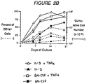
- CD1a + cells were not detected at the onset of the culture and the CD1a antigen could first be observed after 4 days of culture in the presence of TNF- ⁇ , and expression increased until day 20. Whereas less than 5% of CD1a + cells were observed in the presence of IL-3, 5-15% CD1a + cells were detected in GM-CSF. A large proportion of CD1a + cells were detected in IL-3 and TNF- ⁇ (10-25%) and mainly in GM-CSF and TNF- ⁇ (20-60%) (range of 5 experiments after 12 days of culture).
- IL-3 plus TNF- ⁇ is 2-3 times more potent than IL-3 alone
- GM-CSF plus TNF- ⁇ is 3-4 times more potent than GM-CSF alone ( Figure 2).
- GM-CSF allowed the generation of 1-3 x 10 6 CD1a + cells
- IL-3 plus TNF- ⁇ and GM-CSF plus TNF- ⁇ cultures of 2-3 x 10 6 cells were recovered.
- Figure 2 shows the expansion of 10 5 CD34 + cells (i) in the presence of IL-3 (solid triangles), (ii) IL-3 and TNF- ⁇ (open triangles), (iii) GM-CSF (solid squares), and (iv) GM-CSF and TNF- ⁇ (open squares). Solid lines indicate total cell numbers in the three experiments, and dashed lines indicated expression of CD1a antigen. Figures 2A and 2B represent data from two separate sets of experiments.
- CD1a + cells generated by the method of the invention were studied using both light and electron microscopy.
- Adherent cells observed in GM-CSF alone display the classical aspect of regularly shaped macrophages, whereas those obtained in the presence of GM-CSF plus TNF- ⁇ have a typical aspect of dendritic cells with highly ramified dendrites, lobulated nucleus, and a villous surface with dendritic projections.
- Some of the CD1a + cells (about 1 in 5) possess organelles with double membrane joining, recalling the structure of Birbeck granules.
- CD1a + cells generated after 12 days of culture in the presence of GM-CSF and TNF- ⁇ was determined by two-color fluorescence analysis. Depending on the experiment, the percentage of CD1a + cells co-expressing CD14 varied between 10 and 70%.
- Table I The results presented in Table I below were obtained from experiments in which less than 15% of CD1a + cells co-expressed CD14; thus, both CD1a + cells and CD1a - CD14 + cells were characterized.
- CD1a + cells co-expressed CD1c, CD4, and CD40, but did not express CD1b.
- CD1a - CD14 + cells did not express CD1c and only weakly expressed CD4 and CD40.
- CD1a + and CD14 + cells were found to bear Fc ⁇ RII (CD32), Fc ⁇ RIII (CD16), and CR3 (CD11b), whereas only CD1a - CD14 + cells expressed Fc ⁇ RI (CD64) and CR1 (CD35).
- CD1a + and CD1a - CD14 + cells expressed both LFA1 ⁇ (CD11a) and LFA1 ⁇ (CD18), but CD1a + cells exhibited higher levels of ICAM1 (CD54) than CD1a + CD14 + cells.
- CD1a + cells expressed very high levels of HLA-DR (5-10 times more than CD14 + cells).
- CD1a - CD14 + cells expressed high levels of HLA-DQ + .
- CD34 + cells were cultured for 12 hours in accordance with the invention, after which they were irradiated with 4000 Rads to form stimulator cells for the experiment. From 10 to (2.4 x 10 4 ) stimulator cells were seeded for 2.5 x 10 4 resting CD4+ T cells or other responder cells in round-bottomed microtest tissue culture plates in medium supplemented with 10% human AB + serum, as described below.
- Dendritic cells were assayed for their capacity to induce resting allogeneic CD4 + T cells (as responder cells) to proliferate.
- cells cultured in the presence of IL-3 alone (solid triangles) induced marginal allogeneic CD4 + T cell proliferation.
- cells cultured in the presence of GM-CSF alone (solid squares), in the presence of IL-3 plus TNF- ⁇ (open triangles), and in the presence of GM-CSF plus TNF- ⁇ (open squares), induced a strong proliferation of allogeneic CD4 + T cells.
- CD34 + T cells Depending on culture conditions of the CD34 + progenitor cells, the optimal proliferation of the CD34 + T cells was observed for different values of the ratio (stimulator cells)/(allogeneic CD34 + T cells). In comparison with control values (CD34 + T cells without stimulator cells), a 50-fold enhancement of tritiated thymidine uptake by CD4 + T cells was observed at a ratio of 1:3.8 (range 1:3 to 1:25), 1:12.5 (range 1:10 to 1:35), and 1:360 (range 1:100 to 1:400) for cells cultured in the presence of GM-CSF alone, IL-3 plus TNF- ⁇ , and GM-CSF plus TNF- ⁇ respectively (ranges from 5 experiments).
- the CD34 + cells were cultured in the presence of GM-CSF and TNF- ⁇ for all experiments and the responder cells were adult peripheral blood (open squares), cord blood (open and solid triangles), and syngeneic cord blood CD4 + T cells (solid squares).
- Figure 3B shows that allogeneic CD4 + T cells derived either from cord blood or from adult peripheral blood were equally stimulated, and that syngeneic CD4 + T cells were stimulated to a much lower extent (response 20-fold weaker than allogeneic cells).
- CD1a + cells were removed by immunomagnetic depletion ( Figure 4), a strong loss of induction capacity was observed.
- a 50-fold enhancement of CD4 + T cell proliferation was observed for a ratio (stimulator cells)/(allogeneic CD4 + T cells) of 1:200 (range of 3 experiments 1:200 to 1:400) and 1:8 (range of 3 experiments 1:8 to 1:40) before and after CD1a + cells were depleted, respectively.
- CD34 + cells are isolated from the peripheral blood of a cancer patient and grown in the presence of GM-CSF and TNF- ⁇ , as described above. Peripheral blood CD8 cells are cryopreserved. Once dendritic cells are generated, CD8 T cells are thawed and mixed with the patient's cancer cells. After sensitization, CD8 T cells are expanded in the presence of IL-2, e.g . as described by Rosenberg, U.S. patent 4,690,915. Total cells are reinfused into the patient provided that they display tumor-specific cytotoxic activity. The tumor cells may be replaced by specific tumor antigens, e.g . van der Bruggen et al., Science , Vol. 254, pp. 1643 (1991).
- Dendritic cells generated in the presence of GM-CSF and TNF- ⁇ are strong stimulators of resting allogeneic CD8 + T cell proliferation
- Dendritic cells generated from CD34 + cord blood progenitors were cultured for 12 days and irradiated (4000 Rads), and then were used as stimulator cells for resting CD4 + and CD8 + T cells. From 10 to 2.5 x 10 3 stimulator cells were seeded for 2 x 10 4 resting T cells, in round-bottomed microtest tissue-culture plates, in medium supplemented with 10% human AB + serum, with or without 20 U/ml IL-2. After 5 days' incubation, cells were pulsed with 1 ⁇ Ci of 3 H-thymidine for 8 hours, harvested and counted. Tests were carried out in triplicate and results were expressed as mean counts per minute.
- IL-2 The addition of IL-2 to the medium in which the CD4 + and CD8 + T cells were cultured indeed stimulated the proliferation of the CD4 + and CD8 + T cells, as shown by their 3 H-thymidine uptake.
- CD8 + cells cultured in the presence of medium alone showed very little proliferation whereas CD8 + cells cultured in the presence of IL-2 (solid squares) showed much more proliferation; moreover, CD4 + cells cultured in the presence of medium alone (open circles) showed little proliferation whereas CD4 + cells cultured in the presence of IL-2 (solid circles) showed considerably more proliferation.
- SCID-hu mice have allowed secondary responses to recall antigens but have not permitted the establishment of primary responses: e.g. Moller, The SCID-hu Mouse, Vol. 124 (Munksgaard, Copenhagen, 1991); Duchosal et al., Nature , Vol. 355, pp. 258 (1992); and Mosier et al., Curr. Top. Microbiol. Immunol ., Vol. 152, pp. 195 (1989). It is believed that this is due to a lack of reconstitution of the dendritic cell pool. As antigen-pulsed dendritic cells can efficiently induce an antibody response in vivo , e.g. Sornasse et al., J. Exp. Med ., Vol.
- SCID-hu mice are reconstituted with the dendritic cells generated in vitro from the CD34 cells of the donor of human cells or from another donor sharing a compatible MHC. Dendritic cells are pulsed with the appropriate antigen prior to injection. Once the mouse displays antibody to the antigen, B cells are isolated from blood and other reconstituted organs and immortalized, e.g . by culturing in the CD40 system in the presence of Epstein-Barr virus as taught by Banchereau et al., Science , Vol. 251, pp. 70 (1991).
- Dendritic cells are infected by HIV in vitro and HIV is found budding from the surface of the cells after 3-5 days in culture with HIV: e.g. Patterson, J. Gen. Virol ., Vol. 68, pgs. 1177-1181 (1987); Macatonia et al., Immunology , Vol. 71, pgs. 38-45 (1990); and Knight et al., Immunol . Lett ., Vol. 19, pgs. 177-182 (1988).
- Evidence for in vivo infection of dendritic cells by HIV comes from the description of infection of Langerhans cells of the skin and reduction in the amount of MHC class II molecules in cells of the skin: e.g .
- CD34 + cells of HIV patients are used to generate dendritic cells in accordance with the invention.
- the dendritic cells are then incubated with selected antigens and reinfused into the HIV patient.
Landscapes
- Health & Medical Sciences (AREA)
- Life Sciences & Earth Sciences (AREA)
- General Health & Medical Sciences (AREA)
- Engineering & Computer Science (AREA)
- Veterinary Medicine (AREA)
- Public Health (AREA)
- Animal Behavior & Ethology (AREA)
- Chemical & Material Sciences (AREA)
- Epidemiology (AREA)
- Biomedical Technology (AREA)
- Organic Chemistry (AREA)
- Immunology (AREA)
- Zoology (AREA)
- Biotechnology (AREA)
- Genetics & Genomics (AREA)
- Wood Science & Technology (AREA)
- Bioinformatics & Cheminformatics (AREA)
- Cell Biology (AREA)
- Medicinal Chemistry (AREA)
- Hematology (AREA)
- Virology (AREA)
- Pharmacology & Pharmacy (AREA)
- Nuclear Medicine, Radiotherapy & Molecular Imaging (AREA)
- Chemical Kinetics & Catalysis (AREA)
- General Chemical & Material Sciences (AREA)
- General Engineering & Computer Science (AREA)
- Biochemistry (AREA)
- Microbiology (AREA)
- Oncology (AREA)
- Developmental Biology & Embryology (AREA)
- Communicable Diseases (AREA)
- Molecular Biology (AREA)
- Tropical Medicine & Parasitology (AREA)
- AIDS & HIV (AREA)
- Micro-Organisms Or Cultivation Processes Thereof (AREA)
- Medicines Containing Material From Animals Or Micro-Organisms (AREA)
- Immobilizing And Processing Of Enzymes And Microorganisms (AREA)
Abstract
Description
- The invention relates generally to an in vitro method of generating human dendritic cells, and, more specifically, to therapeutic and diagnostic uses of the generated cells.
- Dendritic cells are a system of antigen-presenting cells that function to initiate several immune responses such as the sensitization of MHC-restricted T cells, the rejection of organ transplants, and the formation of T cell-dependent antibodies. Dendritic cells are found in many nonlymphoid tissues but can migrate via the afferent lymph or the blood stream to the T cell-dependent areas of lymphoid organs. They are found in the skin, where they are named Langerhans cells, and are also present in the mucosa. They represent the sentinels of the immune system within the peripheral tissues where they can acquire antigens. As these cells express CD4 and can be infected in vitro by HIV, they are likely to present a port of entry of HIV virus in vivo: e.g. Knight et al., pp. 145 in Racz, et al., editors, "Accessory Cells in HIV and Other Retroviral Infections" (Karger, Basel, 1991); Ramsauer et al., pp. 155 in Racz, et al., editors (cited above). The isolation of human dendritic cells from peripheral blood has only recently been achieved and only small numbers of cells can be generated, e.g. Freudenthal et al., Proc. Natl. Acad. Sci., Vol. 87, pp. 7698 (1990). The in vitro generation of large numbers of human dendritic cells would present an important advantage for priming in vitro human naive CD4 and CD8 T cells, for screening agents that may interfere with HIV infection, for generating primary and secondary in vivo response of human B cells in SCID-hu mice reconstituted with human T and B cells, and for constructing a more sensitive mixed-lymphocyte reaction assay.
- A major impediment to transplantation of allogeneic tissue and organs is graft rejection by the transplant recipient. The cell-mediated immune reaction of the recipient, or host, to the donor tissue plays an important role in the rejection process. The cell-mediated immune response has two important phases: (i) recognition, when host cells recognize the donor cell as foreign in the context of the major histocompatibility complex (MHC); and (ii) destruction, when the host cells respond by attacking the foreign cells. As part of the attacking process, a number of responder cells undergo proliferation and acquire cytotoxicity - that is, the ability to kill donor cells displaying the appropriate antigens. Thus, cell-mediated immunity can be described in terms of two measurable functions: proliferation, and cytotoxic activity - see Dubey et al., chapter 131 in Rose et al., Editors, "Manual of Clinical Laboratory Immunology", 3rd edition (American Society of Microbiology, Washington, D.C., 1986).
- Development of cell culture techniques has led to the establishment of in vitro methods that mimic the in vivo immunization process, thus providing measures for the assessment of cell-mediated immunity in vitro. Of particular utility in regard to transplantation is the mixed lymphocyte response (MLR), or mixed lymphocyte culture. The MLR is a relatively simple assay, yet it exists in many variants. Typically, the assay consists of mixing responder lymphocytes in a suitable culture system with stimulator lymphocytes whose proliferation and/or transcription machinery has been disabled, e.g. by irradiation. After the cells have been cultured for several days, a number of different measurements can be made to quantify the degree of reactivity of the responder cells to the stimulator cells, e.g. uptake of tritiated thymidine, number of blast cells, number of dividing cells, cytokine production, and the like. Other variables in the assay include the source of the responder and stimulator cells, e.g. peripheral blood, spleen, lymph nodes, etc.; whether the responder cells are syngeneic, allogeneic, or xenogenic with respect to the stimulator cells; the method of disabling the stimulator cells, for example irradiation or treatment with a DNA synthesis inhibitor (e.g. mitomycin C) or the like.
- A drawback of the MLR as a routine assay for cell-mediated immune reactivity is sensitivity. Frequently, it is difficult to obtain a strong effect in the MLR, whatever the particular read-out employed. It is believed that antigen-presenting cells in the stimulator population are responsible for stimulating the responder cells; however, in most tissue sources such cells are few in number and/or are of a type that stimulates inefficiently. The sensitivity, and hence the utility, of the MLR assay could be greatly enhanced by the availability of more potent stimulator cell populations. Dendritic cells could serve this function, since they are well known as potent antigen-presenting cells, e.g. Steinman, Ann. Rev. Immunol., Vol. 9, pgs. 271-296 (1991). Unfortunately, it is presently very difficult to obtain them in quantities sufficient for routine MLRs, e.g. Steinman, et al., chapter 49 in Herzenberg et al., Editors, "Cellular Immunology" Vol. 2 (Blackwell Scientific Publications, Oxford, 1986).
- In this regard,
EP 0 455 482 discloses a substantially pure population of human cells comprising pluripotent cells that express the CD34 antigen, but lack expression of the CD38 antigen and other lineage associated antigens. However, the production of human dendritic cells is not disclosed. - Similarly, an article entitled "Tumor Necrosis Factor-alpha Strongly Potentiates Interleukin-3 and Granulocyte-macrophage Colony-Stimulating Factor-Induced Proliferation of Human CD34+ Hematopoietic Progenitor Cells", by Caux, C. et al.; Blood, Vol. 75, No. 12, (June 15), 1990, pp. 2292-2298, discloses the potentiation by TNF-α of the proliferation of human CD34+ cells by IL-3 and GM-CSF. However, the production and recovery of CD1a human dendritic cells is not described or suggested.
- The invention is directed to a method for in vitro generation of human dendritic cells. The invention also includes isolated populations of human dendritic cells produced by the method of the invention and applications of the isolated cells, including an improved MLR assay that employs a pure population of dendritic cells as stimulator cells.
- According to the invention there is provided a process for producing human dendritic cells comprising the steps of:
- (a) culturing human CD34+ hematopoietic cells (i) with GM-CSF, (ii) with TNF-α and IL-3, or (iii) with GM-CSF and TNF-α, thereby inducing the formation of human dendritic cells from said CD34+ hematopoietic cells; and
- (b) recovering said human dendritic cells from said culture.
-
- Dendritic cells initiate immunological responses. The in vitro data reported herein for the dendritic cells produced according to the invention indicate that these cells are useful as laboratory tools and also may have utility in the in vivo treatment of various diseases by adoptive immunotherapy, including cancer and viral infections.
- The invention will now be described in more detail with particular reference to the accompanying drawings of which:
- Figure 1 illustrates flow cytometry data showing the partial co-expression of CD14 by CD1a+ cells.
- Figure 2 illustrates data concerning the growth kinetics of CD1a+ cells under various medium conditions.
- Figure 3 illustrates data of CD4+ cells proliferation after stimulation by dendritic cells.
- Figure 4 illustrates data showing the effect of CD1a+ cells in stimulation of allogeneic CD4+ cell proliferation.
- Figure 5 illustrates data of CD4+ and CD8+ T cell proliferation after stimulation by dendritic cells.
-
- An important aspect of the invention is the generation of dendritic cells from CD34+ hematopoietic cells. CD34+ hematopoietic progenitor cells can be obtained from a variety of tissue sources, e.g. bone marrow, but are preferably obtained from umbilical cord blood samples as follows: Light-density mononuclear cells from the samples are isolated by Ficoll-Hypaque gradient separation (d = 1.077 g/mL) and are depleted of adherent cells, e.g. by overnight incubation at 37°C in RPMI 1640 medium supplemented with 1% w/v tissue-culture grade bovine serum albumin. Preferably, cells bearing CD34 antigen are isolated from non-adherent mononuclear fractions through positive selection by indirect immune panning using anti-CD34 monoclonal antibody, e.g. Imu-133.3 available from Immunotech (Marseille, France), anti-My 10 available from Becton Dickinson (Mountain View, California), or the like. Panning flasks are prepared as follows: sheep Fab antimouse IgG at a concentration of 25 µg/mL in Tris buffer (0.05 mol/L, pH 9.4) is distributed (10 mL) in 75-cm2 tissue culture flasks for overnight coating at 4°C. Separately, the light-density mononuclear cells (depleted of adherent cells as discussed above) are incubated one hour at 4°C with 5 µg/mL anti-CD34 antibody at 107 cells/mL in RPMI 1640 supplemented with 2% heat-inactivated pooled human AB serum (HABS). Afterwards, cells are washed in cold medium containing 2% HABS, and 10 mL containing about 5 x 10⁷ cells are distributed to the flasks previously coated with sheep antimouse IgG, as described above. Following a two-hour incubation at 4°C, non-adherent cells in suspension (i.e. the CD34-depleted fraction) are harvested by gentle pipetting and rinsing several times with medium. The adherent "panned" cells (i.e. the CD34-rich fraction) are then recovered by vigorous pipetting.
- The dendritic cells are obtained from the CD34+ cells by culturing these in medium containing GM-CSF or TNF-α and IL-3. Preferably, the dendritic cells are obtained from the CD34+ cells by culturing these in medium containing TNF-α as well as GM-CSF. TNF-α, GM-CSF, and IL-3 suitable for use in the invention are commercially available, e.g. from Genzyme Corp. (Cambridge, MA), or can be produced by recombinant expression systems: e.g. as taught by Clark et al. in U.S. patent 4,959,455 (IL-3); Clark et al. in PCT application No. EP85/00326 (publ. no. WO86/00639) (GM-CSF); and Mark et al. in U.S. patent 4,677,063 (TNF-α). Preferably, IL-3 and GM-CSF are used at saturating concentration; that is, they are used at a concentration at which all the IL-3 and GM-CSF receptors on the CD34+ cells are occupied by biologically active IL-3 and GM-CSF molecules. Of course, the actual concentration may depend on the quality of IL-3 and GM-CSF used. Preferably, human IL-3 having a specific activity of at least 5 x 10⁶ U/mg is employed, wherein a unit of activity corresponds to the half-maximum proliferative activity as determined by 3H-thymidine uptake by human bone marrow cells in liquid cultures. In the culture systems described below, saturating concentration was 10 ng/mL (or 50 U/mL). Preferably, human GM-CSF having a specific activity of at least 2 x 10⁶ U/mg is employed, wherein a unit of activity is as defined for IL-3 above. In the culture systems described below, saturating concentration was 100 ng/mL (or 200 U/mL). Preferably, TNF-α is used at a concentration in the range of 2 to 3 ng/mL or 40-60 U/mL, most preferably at a concentration of about 2.5 ng/mL or 50 U/mL. Units of TNF-α are defined by Carswell et al., Proc. Natl. Acad. Sci., Vol. 72, pg. 3666 (1975), and by Aggarwal et al., J. Biol. Chem., Vol. 260, pg. 2345 (1985). The cells can be co-cultured in standard tissue culture medium with standard additives, such as RPMI 1640 supplemented with 10% (v/v) heat inactivated fetal bovine serum, 10 mM Hepes, 2 mM L-glutamine, 5 x 10⁻⁵ M 2-mercaptoethanol, penicillin (100 U/mL) and streptomycin (100 mg/mL). Preferably, the CD34+ cells are cultured in the presence of the cytokines for from 8 to 12 days.
- Dendritic cells that form in the culture are isolated by panning as described above, with the exception that anti-CD1a and/or anti-CD14 antibodies are employed (both antibodies being commercially available, e.g. from Becton-Dickinson).
- The MLR method comprises the following steps:
- (1) providing a sample of responder cells; (2) providing a sample of inactivated stimulator cells such that the stimulator cells are allogeneic with respect to the responder cells and such that the stimulator cells consist of dendritic cells produced by the process comprising the steps of (a) treating CD34+ hematopoietic cells with TNF-α and IL-3 or with GM-CSF, and (b) isolating treated CD34+ hematopoietic cells that express the CD1a antigen; (3) co-culturing the responder cells and the inactivated stimulator cells; and (4) measuring a response of the responder cells.
-
- Preferably, the sample of responder cells consists of CD4+ T cells from the peripheral blood of a patient who is to be the recipient of a transplant. Obtaining T cell populations employs techniques well known in the art which are fully described by DiSabato et al., eds., in Meth. in Enzymol., Vol. 108 (1984). For example, the CD4+ T cells can be isolated as follows: first mononuclear cells are isolated from the peripheral blood and depleted of adherent cells; CD4+ T cells are then purified by depleting other cell types, for example by immunomagnetic depletion (e.g. with Dynabeads, Dynal, Oslo, Norway), or the like, using a cocktail of commercially available monoclonal antibodies, e.g. anti-CD14, anti-CD16, anti-CD20, anti-CD8, anti-CD40 (available from Becton-Dickinson and/or Ortho Diagnostic Systems, New Jersey). CD4+ populations having higher than 95% purity are typically achieved after two rounds of immunomagnetic depletion.
- Stimulator cells used in the procedures described herein are dendritic cells derived from CD34+ hematopoietic progenitors cells obtained from a different person from that from whom the responder cells are taken; that is, the stimulator cells are allogeneic with respect to the responder cells. These cells are obtained as described above.
- The stimulator cells are inactivated so that they can still carry out their stimulatory function but are inhibited from any other function that could obscure the response measured from the responder cells. Thus, the nature of the inactivation depends somewhat on the "read-out" of the assay. Preferably, the read-out, or response measured in the responder cells, is cellular proliferation. Other read-outs could also include such phenomena as cytokine production, cytolytic ability, and the like. Preferably, the stimulator cells are treated so that they are incapable of replication, but their antigen-processing machinery remains functional. This is conveniently accomplished by irradiating the cells, e.g. with about 1500 to 5000 R (gamma or X-radiation), preferably 3000 to 4000 R, before mixing with the responder cells.
- Preferably, proliferation of the responder cells is determined by the uptake of tritiated thymidine using standard protocols. For example, from 10 to 2.5 x 10⁴ stimulator cells are added to 2.4 x 10⁴ allogeneic CD4+ T cells in 96-well round-bottom tissue-culture plates and are incubated for 4 days in the medium described above. After incubation, the cells are pulsed with 1 µCi of tritiated thymidine for 6 hours, and then they are harvested and measured for tritiated thymidine uptake, e.g. by scintillation counting.
- The following examples serve to illustrate the present invention. Cell lines, reagents and their concentrations, temperatures, and the values of other variables are only to exemplify the application of the present invention and are not to be considered limitations thereof.
- After 12 days of culture in GM-CSF and TNF-α, cells generated from CD34+ cord blood hematopoietic precursor cells were processed for two-color fluorescence measurement. Briefly, cells were sequentially incubated with unconjugated monoclonal antibodies, phycoerythrin-conjugated (PE-conjugated) anti-mouse immunoglobulin, normal mouse serum, and monoclonal antibodies OKT6 (anti-CD1a from Ortho) or Leu-M3 (anti-CD14 from Becton-Dickinson), the monoclonal antibodies being directly labelled with fluorescein isothiocyanate (FITC). As shown in Figure 1, culturing CD34+ cells for 12 days in the presence of GM-CSF and TNF-α allows the appearance of CD14+ monocytic cells co-expressing the CD1a antigen. In addition, a population of CD14-CD1a+ cells was consistently observed which represented 30-90% of the total CD1a population (range from 5 experiments). Within the monocytic lineage, expression of CD1a antigen is restricted to Langerhans cells. Figure 1A shows two-color fluorescence intensity from IgG1-FITC isotype control versus IgG2a-PE isotype control. Figure 1B shows two-color fluorescence intensity from OKT6-FITC (proportional to CD1a expression) versus Leu-M3-PE (proportional to CD14 expression). As shown in Figure 2, CD1a+ cells were not detected at the onset of the culture and the CD1a antigen could first be observed after 4 days of culture in the presence of TNF-α, and expression increased until day 20. Whereas less than 5% of CD1a+ cells were observed in the presence of IL-3, 5-15% CD1a+ cells were detected in GM-CSF. A large proportion of CD1a+ cells were detected in IL-3 and TNF-α (10-25%) and mainly in GM-CSF and TNF-α (20-60%) (range of 5 experiments after 12 days of culture). In terms of growth efficiency, IL-3 plus TNF-α is 2-3 times more potent than IL-3 alone, and GM-CSF plus TNF-α is 3-4 times more potent than GM-CSF alone (Figure 2). Thus, starting from 105 CD34+ cells, after 12 days in culture, GM-CSF allowed the generation of 1-3 x 10⁶ CD1a+ cells, whereas in IL-3 plus TNF-α and GM-CSF plus TNF-α cultures of 2-3 x 10⁶ cells were recovered. As GM-CSF plus TNF-α appeared to be the most potent combination of factors for generating CD1a+ cells, all the cultures for the characterization of those cells contained GM-CSF plus TNF-α: Figure 2 shows the expansion of 105 CD34+ cells (i) in the presence of IL-3 (solid triangles), (ii) IL-3 and TNF-α (open triangles), (iii) GM-CSF (solid squares), and (iv) GM-CSF and TNF-α (open squares). Solid lines indicate total cell numbers in the three experiments, and dashed lines indicated expression of CD1a antigen. Figures 2A and 2B represent data from two separate sets of experiments.
- The morphology of the CD1a+ cells generated by the method of the invention was studied using both light and electron microscopy. Adherent cells observed in GM-CSF alone display the classical aspect of regularly shaped macrophages, whereas those obtained in the presence of GM-CSF plus TNF-α have a typical aspect of dendritic cells with highly ramified dendrites, lobulated nucleus, and a villous surface with dendritic projections. Some of the CD1a+ cells (about 1 in 5) possess organelles with double membrane joining, recalling the structure of Birbeck granules.
- The phenotype of CD1a+ cells generated after 12 days of culture in the presence of GM-CSF and TNF-α was determined by two-color fluorescence analysis. Depending on the experiment, the percentage of CD1a+ cells co-expressing CD14 varied between 10 and 70%. The results presented in Table I below were obtained from experiments in which less than 15% of CD1a+ cells co-expressed CD14; thus, both CD1a+ cells and CD1a-CD14+ cells were characterized. CD1a+ cells co-expressed CD1c, CD4, and CD40, but did not express CD1b. In contrast, CD1a-CD14+ cells did not express CD1c and only weakly expressed CD4 and CD40. Both CD1a+ and CD14+ cells were found to bear FcγRII (CD32), FcγRIII (CD16), and CR3 (CD11b), whereas only CD1a-CD14+ cells expressed FcγRI (CD64) and CR1 (CD35). CD1a+ and CD1a-CD14+ cells expressed both LFA1α (CD11a) and LFA1β (CD18), but CD1a+ cells exhibited higher levels of ICAM1 (CD54) than CD1a+CD14+ cells. CD1a+ cells expressed very high levels of HLA-DR (5-10 times more than CD14+ cells). In contrast, CD1a-CD14+ cells expressed high levels of HLA-DQ+.
Phenotype of Dendritic Cells Produced According to the Invention Antigen Antibody Reactivity with CD1a cells Reactivity with CD14 cells Source of Antibody () CD1a OKT6, IOT6a, T6, DMC1, L544 ++ - Ort, Hy8, Imu, Cou, CD1b IOT6b - - Imu CD1c IOT6c + - Imu CD14 Leu-M3 +/- +++ BD CD4 IOT4 ++ +/- Imu CD40 Mab 89 ++ +/- EP CD16 Leu11b + + BD CD32 2E1 +/- +/- Imu CD64 197 - + Med CD21 CR2 - - BD CD11b IOM1 ++ ++ Imu CD35 IOT17 - ++ Imu CD54 84H10 +++ + Imu CD11a SPVL7 ++ ++ Hy2 CD18 BL5 ++ ++ Imu HLA-DR L243 ++++ ++ BD HLA-DQ SPVL3 +++ + Imu - CD34+ cells were cultured for 12 hours in accordance with the invention, after which they were irradiated with 4000 Rads to form stimulator cells for the experiment. From 10 to (2.4 x 10⁴) stimulator cells were seeded for 2.5 x 10⁴ resting CD4+ T cells or other responder cells in round-bottomed microtest tissue culture plates in medium supplemented with 10% human AB+ serum, as described below.
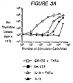
- Dendritic cells were assayed for their capacity to induce resting allogeneic CD4+ T cells (as responder cells) to proliferate. As shown in Figure 3A, cells cultured in the presence of IL-3 alone (solid triangles) induced marginal allogeneic CD4+ T cell proliferation. In contrast, cells cultured in the presence of GM-CSF alone (solid squares), in the presence of IL-3 plus TNF-α (open triangles), and in the presence of GM-CSF plus TNF-α (open squares), induced a strong proliferation of allogeneic CD4+ T cells. Depending on culture conditions of the CD34+ progenitor cells, the optimal proliferation of the CD34+ T cells was observed for different values of the ratio (stimulator cells)/(allogeneic CD34+ T cells). In comparison with control values (CD34+ T cells without stimulator cells), a 50-fold enhancement of tritiated thymidine uptake by CD4+ T cells was observed at a ratio of 1:3.8 (range 1:3 to 1:25), 1:12.5 (range 1:10 to 1:35), and 1:360 (range 1:100 to 1:400) for cells cultured in the presence of GM-CSF alone, IL-3 plus TNF-α, and GM-CSF plus TNF-α respectively (ranges from 5 experiments).
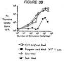
- In the Experiment illustrated in Figure 3B, the CD34+ cells were cultured in the presence of GM-CSF and TNF-α for all experiments and the responder cells were adult peripheral blood (open squares), cord blood (open and solid triangles), and syngeneic cord blood CD4+ T cells (solid squares). Figure 3B shows that allogeneic CD4+ T cells derived either from cord blood or from adult peripheral blood were equally stimulated, and that syngeneic CD4+ T cells were stimulated to a much lower extent (response 20-fold weaker than allogeneic cells).
- When, after 12 days of culture in the presence of GM-CSF and TNF-α, CD1a+ cells were removed by immunomagnetic depletion (Figure 4), a strong loss of induction capacity was observed. A 50-fold enhancement of CD4+ T cell proliferation was observed for a ratio (stimulator cells)/(allogeneic CD4+ T cells) of 1:200 (range of 3 experiments 1:200 to 1:400) and 1:8 (range of 3 experiments 1:8 to 1:40) before and after CD1a+ cells were depleted, respectively. After irradiation, the following were used as stimulator cells: total population (solid squares), population from which CD1a+ cells had been removed by depletion (open squares), and control population from which depletion was carried out with an anti-IgG1 isotype antibody (open triangles).
- CD34+ cells are isolated from the peripheral blood of a cancer patient and grown in the presence of GM-CSF and TNF-α, as described above. Peripheral blood CD8 cells are cryopreserved. Once dendritic cells are generated, CD8 T cells are thawed and mixed with the patient's cancer cells. After sensitization, CD8 T cells are expanded in the presence of IL-2, e.g. as described by Rosenberg, U.S. patent 4,690,915. Total cells are reinfused into the patient provided that they display tumor-specific cytotoxic activity. The tumor cells may be replaced by specific tumor antigens, e.g. van der Bruggen et al., Science, Vol. 254, pp. 1643 (1991).
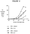
- Dendritic cells generated from CD34+ cord blood progenitors were cultured for 12 days and irradiated (4000 Rads), and then were used as stimulator cells for resting CD4+ and CD8+ T cells. From 10 to 2.5 x 10³ stimulator cells were seeded for 2 x 10⁴ resting T cells, in round-bottomed microtest tissue-culture plates, in medium supplemented with 10% human AB+ serum, with or without 20 U/ml IL-2. After 5 days' incubation, cells were pulsed with 1µCi of 3H-thymidine for 8 hours, harvested and counted. Tests were carried out in triplicate and results were expressed as mean counts per minute.
- The addition of IL-2 to the medium in which the CD4+ and CD8+ T cells were cultured indeed stimulated the proliferation of the CD4+ and CD8+ T cells, as shown by their 3H-thymidine uptake. As shown in Figure 5, CD8+ cells cultured in the presence of medium alone (open squares) showed very little proliferation whereas CD8+ cells cultured in the presence of IL-2 (solid squares) showed much more proliferation; moreover, CD4+ cells cultured in the presence of medium alone (open circles) showed little proliferation whereas CD4+ cells cultured in the presence of IL-2 (solid circles) showed considerably more proliferation.
- SCID-hu mice have allowed secondary responses to recall antigens but have not permitted the establishment of primary responses: e.g. Moller, The SCID-hu Mouse, Vol. 124 (Munksgaard, Copenhagen, 1991); Duchosal et al., Nature, Vol. 355, pp. 258 (1992); and Mosier et al., Curr. Top. Microbiol. Immunol., Vol. 152, pp. 195 (1989). It is believed that this is due to a lack of reconstitution of the dendritic cell pool. As antigen-pulsed dendritic cells can efficiently induce an antibody response in vivo, e.g. Sornasse et al., J. Exp. Med., Vol. 175, pp. 15 (1992), SCID-hu mice are reconstituted with the dendritic cells generated in vitro from the CD34 cells of the donor of human cells or from another donor sharing a compatible MHC. Dendritic cells are pulsed with the appropriate antigen prior to injection. Once the mouse displays antibody to the antigen, B cells are isolated from blood and other reconstituted organs and immortalized, e.g. by culturing in the CD40 system in the presence of Epstein-Barr virus as taught by Banchereau et al., Science, Vol. 251, pp. 70 (1991).
- Dendritic cells are infected by HIV in vitro and HIV is found budding from the surface of the cells after 3-5 days in culture with HIV: e.g. Patterson, J. Gen. Virol., Vol. 68, pgs. 1177-1181 (1987); Macatonia et al., Immunology, Vol. 71, pgs. 38-45 (1990); and Knight et al., Immunol. Lett., Vol. 19, pgs. 177-182 (1988). Evidence for in vivo infection of dendritic cells by HIV comes from the description of infection of Langerhans cells of the skin and reduction in the amount of MHC class II molecules in cells of the skin: e.g. Tschachler et al., J. Invest. Dermatol, Vol. 88, pgs. 233-237 (1987); and Belsito et al., N. Engl. J. Med., Vol. 310, pgs. 1279-1282 (1984). Furthermore, 3-21% of blood dendritic cells from HIV patients contain HIV, with a level of infection two orders of magnitude greater than that seen in other cell types: Macatonia et al., Immunology, Vol. 71, pgs. 38-45 (1990). The infection of dendritic cells by the virus blocks their ability to present antigen to T cells, and thus inhibits the recruitment/proliferation of the T cells which is necessary for a successful immune response.
- CD34+ cells of HIV patients are used to generate dendritic cells in accordance with the invention. The dendritic cells are then incubated with selected antigens and reinfused into the HIV patient.
Claims (6)
- A process for the in vitro generation of human dendritic cells comprising the steps of:(a) culturing human CD34+ hematopoietic cells (i) with GM-CSF, (ii) with TNF-α and IL-3, or (iii) with GM-CSF and TNF-α, thereby inducing the formation of CD1a+ human dendritic cells from said CD34+ hematopoietic cells; and(b) recovering said CD1a+ human dendritic cells from said culture.
- The process of claim 1 wherein the CD34+ cells are cultured in the presence of GM-CSF.
- The process of claim 1 wherein the CD34+ cells are cultured in the presence of GM-CSF and TNF-α.
- The process of claim 1 wherein the CD34+ cells are cultured in the presence of TNF-α and IL-3.
- A process according to any preceding claim wherein the recovered dendritic cells are pulsed with antigen.
- A process according to any preceding claim wherein the CD34+ cells are obtained from an HIV patient.
Priority Applications (2)
| Application Number | Priority Date | Filing Date | Title |
|---|---|---|---|
| EP93908477A EP0633930B1 (en) | 1992-03-30 | 1993-03-29 | In vitro generation of human dendritic cells |
| GR20000401457T GR3033761T3 (en) | 1992-03-30 | 2000-06-23 | In vitro generation of human dendritic cells and uses thereof. |
Applications Claiming Priority (4)
| Application Number | Priority Date | Filing Date | Title |
|---|---|---|---|
| EP92400879 | 1992-03-30 | ||
| EP92400879A EP0563485A1 (en) | 1992-03-30 | 1992-03-30 | In vitro generation of human dendritic cells and uses thereof |
| PCT/US1993/002639 WO1993020186A1 (en) | 1992-03-30 | 1993-03-29 | In vitro generation of human dendritic cells and uses thereof |
| EP93908477A EP0633930B1 (en) | 1992-03-30 | 1993-03-29 | In vitro generation of human dendritic cells |
Publications (2)
| Publication Number | Publication Date |
|---|---|
| EP0633930A1 EP0633930A1 (en) | 1995-01-18 |
| EP0633930B1 true EP0633930B1 (en) | 2000-04-26 |
Family
ID=8211642
Family Applications (2)
| Application Number | Title | Priority Date | Filing Date |
|---|---|---|---|
| EP92400879A Withdrawn EP0563485A1 (en) | 1992-03-30 | 1992-03-30 | In vitro generation of human dendritic cells and uses thereof |
| EP93908477A Expired - Lifetime EP0633930B1 (en) | 1992-03-30 | 1993-03-29 | In vitro generation of human dendritic cells |
Family Applications Before (1)
| Application Number | Title | Priority Date | Filing Date |
|---|---|---|---|
| EP92400879A Withdrawn EP0563485A1 (en) | 1992-03-30 | 1992-03-30 | In vitro generation of human dendritic cells and uses thereof |
Country Status (14)
| Country | Link |
|---|---|
| EP (2) | EP0563485A1 (en) |
| JP (1) | JP3492361B2 (en) |
| KR (1) | KR100223395B1 (en) |
| AT (1) | ATE192189T1 (en) |
| AU (2) | AU682466B2 (en) |
| CA (1) | CA2133316C (en) |
| DE (1) | DE69328481T2 (en) |
| DK (1) | DK0633930T3 (en) |
| ES (1) | ES2145047T3 (en) |
| GR (1) | GR3033761T3 (en) |
| MX (1) | MX9301753A (en) |
| NZ (1) | NZ251585A (en) |
| PT (1) | PT633930E (en) |
| WO (1) | WO1993020186A1 (en) |
Cited By (7)
| Publication number | Priority date | Publication date | Assignee | Title |
|---|---|---|---|---|
| US6797514B2 (en) | 2000-02-24 | 2004-09-28 | Xcyte Therapies, Inc. | Simultaneous stimulation and concentration of cells |
| US6867041B2 (en) | 2000-02-24 | 2005-03-15 | Xcyte Therapies, Inc. | Simultaneous stimulation and concentration of cells |
| US7541184B2 (en) | 2000-02-24 | 2009-06-02 | Invitrogen Corporation | Activation and expansion of cells |
| US7572631B2 (en) | 2000-02-24 | 2009-08-11 | Invitrogen Corporation | Activation and expansion of T cells |
| EP2251418A1 (en) | 2004-10-07 | 2010-11-17 | Argos Therapeutics, Inc. | Mature dendritic cell compositions and methods for culturing same |
| US7977095B2 (en) | 2003-05-08 | 2011-07-12 | Life Technologies Corporation | Generation and isolation of antigen-specific T cells |
| US9528088B2 (en) | 2002-06-28 | 2016-12-27 | Life Technologies Corporation | Methods for eliminating at least a substantial portion of a clonal antigen-specific memory T cell subpopulation |
Families Citing this family (35)
| Publication number | Priority date | Publication date | Assignee | Title |
|---|---|---|---|---|
| DE69333433T2 (en) * | 1992-04-01 | 2004-12-02 | The Rockefeller University | METHOD FOR THE VITRO CULTIVATION OF DENDRITIC PROCUREMENT CELLS AND THE USE THEREOF FOR IMMUNOGENOUS PRODUCTION |
| DE4337396A1 (en) * | 1993-10-26 | 1995-04-27 | Beiersdorf Ag | Human cell lines with Langerhans cell characteristics |
| DE4412794A1 (en) * | 1994-04-14 | 1995-12-14 | Univ Ludwigs Albert | Process for producing dendritic cells, cells thus obtained and containers for carrying out this process |
| US5874560A (en) | 1994-04-22 | 1999-02-23 | The United States Of America As Represented By The Department Of Health And Human Services | Melanoma antigens and their use in diagnostic and therapeutic methods |
| US5670347A (en) | 1994-05-11 | 1997-09-23 | Amba Biosciences Llc | Peptide-mediated gene transfer |
| US6300090B1 (en) | 1994-07-29 | 2001-10-09 | The Rockefeller University | Methods of use of viral vectors to deliver antigen to dendritic cells |
| US5648248A (en) * | 1994-12-30 | 1997-07-15 | Boehringer Ingelheim International Gmbh | Methods for producing differentiated cells from immature hematopoietic cells |
| US5643786A (en) * | 1995-01-27 | 1997-07-01 | The United States Of America As Represented By The Department Of Health And Human Services | Method for isolating dendritic cells |
| US6010905A (en) * | 1995-01-27 | 2000-01-04 | The United States Of America As Represented By The Department Of Health & Human Services | Method for inducing monocytes to exhibit the phenotype of activated myeloid dendritic cells |
| US6340981B1 (en) | 1997-06-30 | 2002-01-22 | Sun Microsystems, Inc. | Method and apparatus for stroke substitution |
| US5871728A (en) | 1995-03-31 | 1999-02-16 | University Of Pittsburgh | Method of regulating dendritic cell maturation |
| US6121044A (en) * | 1995-07-12 | 2000-09-19 | Dendreon Corporation | Potent antigen presenting cell composition |
| US7361330B2 (en) | 1995-10-04 | 2008-04-22 | Immunex Corporation | Methods of using flt3-ligand in the treatment of fibrosarcoma |
| US7150992B1 (en) | 1995-10-04 | 2006-12-19 | Innunex Corporation | Methods of preparing dendritic cells with flt3-ligand and antigen |
| JP2000505650A (en) * | 1996-02-08 | 2000-05-16 | アメリカ合衆国 | Methods and compositions for transforming dendritic cells and activating T cells |
| US6734014B1 (en) | 1996-02-08 | 2004-05-11 | The United States Of America As Represented By The Department Of Health And Human Services | Methods and compositions for transforming dendritic cells and activating T cells |
| US7659119B2 (en) | 1996-02-12 | 2010-02-09 | Argos Therapeutics, Inc. | Method and compositions for obtaining mature dendritic cells |
| US5811297A (en) * | 1996-03-07 | 1998-09-22 | Amba Biosciences, Llc | Immortalized hematopoietic cell lines, cell system thereof with stromal cells, in vitro, ex vivo and in vivo uses, & in vitro generation of dendritic cells and macrophages |
| US6008004A (en) | 1996-10-04 | 1999-12-28 | Becton Dickinson & Company | Identification of a CD34+ bone marrow precursor for dendritic cells in blood and lymphoid tissues |
| JP2001508648A (en) | 1996-11-06 | 2001-07-03 | ザ リージェンツ オブ ザ ユニバーシティ オブ カリフォルニア | Tumor necrosis factor receptor releasing enzyme, composition containing the enzyme, and method of using the same |
| AU9374198A (en) * | 1997-09-08 | 1999-03-29 | Idec Pharmaceuticals Corporation | Methods for producing human antibodies in scid mice using dendritic cells |
| DE69840739D1 (en) | 1997-10-27 | 2009-05-28 | Merix Bioscience Inc | Method and composition for the production of mature dendritic cells |
| AU2872199A (en) | 1998-02-20 | 1999-09-06 | Rockefeller University, The | Apoptotic cell-mediated antigen presentation to dendritic cells |
| EP1016413A1 (en) * | 1998-12-30 | 2000-07-05 | Applied Research Systems ARS Holding N.V. | Human growth hormone to stimulate mobilization of pluripotent hematopoietic stem cells |
| JP2001061469A (en) * | 1999-08-24 | 2001-03-13 | Japan Science & Technology Corp | Method for preparing immune response-inducing dendritic cells |
| US7419957B2 (en) | 2001-08-22 | 2008-09-02 | The United States Of America As Represented By The Secretary Of The Department Of Health And Human Services | Peptides of melanoma antigen and their use in diagnostic, prophylactic and therapeutic methods |
| KR100522526B1 (en) * | 2002-11-28 | 2005-10-19 | 주식회사 바이넥스 | Method of Preparing Dendritic cell for Immunotherapy |
| US7867977B2 (en) | 2005-11-03 | 2011-01-11 | The United States Of America As Represented By The Department Of Health And Human Services | Immunogenic peptides and methods of use for treating and preventing cancer |
| EP3115469B1 (en) | 2007-11-19 | 2020-04-29 | Celera Corporation | Lung cancer markers and uses thereof |
| ES2514590T3 (en) | 2008-09-02 | 2014-10-28 | INSERM (Institut National de la Santé et de la Recherche Médicale) | New melanoma antigen peptide and uses thereof |
| EP2714730A1 (en) | 2011-06-01 | 2014-04-09 | INSERM (Institut National de la Santé et de la Recherche Médicale) | Novel antigen peptide and uses thereof |
| EP2852611B1 (en) | 2012-05-22 | 2019-08-14 | INSERM - Institut National de la Santé et de la Recherche Médicale | Novel melanoma antigen peptide and uses thereof |
| AU2012380681A1 (en) | 2012-05-22 | 2014-10-30 | Centre National De La Recherche Scientifique (Cnrs) | Novel melanoma antigen peptide and uses thereof |
| ES2702676T3 (en) | 2014-04-01 | 2019-03-04 | Inst Nat Sante Rech Med | Isolated donor peptide derived from MHC and uses thereof |
| WO2015150491A1 (en) | 2014-04-01 | 2015-10-08 | INSERM (Institut National de la Santé et de la Recherche Médicale) | An isolated donor mhc-derived peptide and uses thereof |
Family Cites Families (3)
| Publication number | Priority date | Publication date | Assignee | Title |
|---|---|---|---|---|
| WO1991002531A1 (en) * | 1989-08-17 | 1991-03-07 | Peter Maccallum Cancer Institute | Method for the modulation of haemopoiesis in a mammal |
| US5128259A (en) * | 1989-10-27 | 1992-07-07 | Hahnemann University | Factor-dependent hematopoietic cell line exhibiting epo-induced erythrocyte maturation |
| US5622853A (en) * | 1990-05-01 | 1997-04-22 | Becton Dickinson And Company | T lymphocyte precursor |
-
1992
- 1992-03-30 EP EP92400879A patent/EP0563485A1/en not_active Withdrawn
-
1993
- 1993-03-29 WO PCT/US1993/002639 patent/WO1993020186A1/en not_active Ceased
- 1993-03-29 AT AT93908477T patent/ATE192189T1/en not_active IP Right Cessation
- 1993-03-29 DE DE69328481T patent/DE69328481T2/en not_active Expired - Fee Related
- 1993-03-29 CA CA002133316A patent/CA2133316C/en not_active Expired - Fee Related
- 1993-03-29 DK DK93908477T patent/DK0633930T3/en active
- 1993-03-29 EP EP93908477A patent/EP0633930B1/en not_active Expired - Lifetime
- 1993-03-29 MX MX9301753A patent/MX9301753A/en not_active IP Right Cessation
- 1993-03-29 KR KR1019940703412A patent/KR100223395B1/en not_active Expired - Fee Related
- 1993-03-29 PT PT93908477T patent/PT633930E/en unknown
- 1993-03-29 AU AU39289/93A patent/AU682466B2/en not_active Ceased
- 1993-03-29 ES ES93908477T patent/ES2145047T3/en not_active Expired - Lifetime
- 1993-03-29 NZ NZ251585A patent/NZ251585A/en unknown
- 1993-03-29 JP JP51751593A patent/JP3492361B2/en not_active Expired - Fee Related
-
1997
- 1997-07-28 AU AU31558/97A patent/AU3155897A/en not_active Abandoned
-
2000
- 2000-06-23 GR GR20000401457T patent/GR3033761T3/en unknown
Cited By (7)
| Publication number | Priority date | Publication date | Assignee | Title |
|---|---|---|---|---|
| US6797514B2 (en) | 2000-02-24 | 2004-09-28 | Xcyte Therapies, Inc. | Simultaneous stimulation and concentration of cells |
| US6867041B2 (en) | 2000-02-24 | 2005-03-15 | Xcyte Therapies, Inc. | Simultaneous stimulation and concentration of cells |
| US7541184B2 (en) | 2000-02-24 | 2009-06-02 | Invitrogen Corporation | Activation and expansion of cells |
| US7572631B2 (en) | 2000-02-24 | 2009-08-11 | Invitrogen Corporation | Activation and expansion of T cells |
| US9528088B2 (en) | 2002-06-28 | 2016-12-27 | Life Technologies Corporation | Methods for eliminating at least a substantial portion of a clonal antigen-specific memory T cell subpopulation |
| US7977095B2 (en) | 2003-05-08 | 2011-07-12 | Life Technologies Corporation | Generation and isolation of antigen-specific T cells |
| EP2251418A1 (en) | 2004-10-07 | 2010-11-17 | Argos Therapeutics, Inc. | Mature dendritic cell compositions and methods for culturing same |
Also Published As
| Publication number | Publication date |
|---|---|
| AU682466B2 (en) | 1997-10-09 |
| ATE192189T1 (en) | 2000-05-15 |
| GR3033761T3 (en) | 2000-10-31 |
| AU3155897A (en) | 1997-11-20 |
| JPH07505527A (en) | 1995-06-22 |
| DE69328481T2 (en) | 2000-09-07 |
| WO1993020186A1 (en) | 1993-10-14 |
| DK0633930T3 (en) | 2000-09-18 |
| NZ251585A (en) | 1997-07-27 |
| EP0633930A1 (en) | 1995-01-18 |
| EP0563485A1 (en) | 1993-10-06 |
| DE69328481D1 (en) | 2000-05-31 |
| MX9301753A (en) | 1993-09-01 |
| KR100223395B1 (en) | 1999-10-15 |
| ES2145047T3 (en) | 2000-07-01 |
| CA2133316A1 (en) | 1993-10-14 |
| PT633930E (en) | 2000-09-29 |
| KR950700989A (en) | 1995-02-20 |
| CA2133316C (en) | 2005-05-24 |
| JP3492361B2 (en) | 2004-02-03 |
| AU3928993A (en) | 1993-11-08 |
Similar Documents
| Publication | Publication Date | Title |
|---|---|---|
| EP0633930B1 (en) | In vitro generation of human dendritic cells | |
| US6004807A (en) | In vitro generation of human dendritic cells | |
| US7198948B2 (en) | Methods and compositions for obtaining mature dendritic cells | |
| Smit et al. | Generation of dendritic cells expressing bcr-abl from CD34-positive chronic myeloid leukemia precursor cells | |
| Bernhard et al. | Generation of immunostimulatory dendritic cells from human CD34+ hematopoietic progenitor cells of the bone marrow and peripheral blood | |
| US6274378B1 (en) | Methods and compositions for obtaining mature dendritic cells | |
| US5849589A (en) | Culturing monocytes with IL-4, TNF-α and GM-CSF TO induce differentiation to dendric cells | |
| Fujii et al. | Analysis of a chronic myelogenous leukemia patient vaccinated with leukemic dendritic cells following autologous peripheral blood stem cell transplantation | |
| Takamizawa et al. | Dendritic cells that process and present nominal antigens to naive T lymphocytes are derived from CD2+ precursors | |
| JP2002529073A (en) | Generation of lymphoid tissue-specific cells from hematopoietic progenitor cells on a three-dimensional device | |
| WO1997029182A9 (en) | Method and compositions for obtaining mature dendritic cells | |
| US20010026937A1 (en) | Monocyte-derived dendritic cell subsets | |
| AU7112098A (en) | Dendritic cell hybrids | |
| US20040235162A1 (en) | Method of preparing immunoregulatory dendritic cells and the use thereof | |
| Ratta et al. | Efficient presentation of tumor idiotype to autologous T cells by CD83+ dendritic cells derived from highly purified circulating CD14+ monocytes in multiple myeloma patients | |
| Curti et al. | Generation of dendritic cells from positively selected CD14+ monocytes for anti-tumor immunotherapy | |
| Eljaafari et al. | Generation of stable monocyte-derived dendritic cells in the presence of high concentrations of homologous or autologous serum: influence of extra-cellular pH | |
| JPH04325087A (en) | Method for producing CD4+ helper T cells | |
| Gieseler et al. | In-vitro differentiation of mature dendritic cells from human blood monocytes | |
| Lopez et al. | IL-13 induces CD34+ cells isolated from G-CSF mobilized blood to differentiate in vitro into potent antigen presenting cells | |
| JP2001181205A (en) | Method for producing tumor-specific anti-tumor cellular vaccine using dendritic cell and tumor cell | |
| Agger et al. | Characterization of murine dendritic cells derived from adherent blood mononuclear cells in vitro | |
| WO2002040647A1 (en) | Method of establishing cultures of human dendritic cells and use thereof | |
| JP2003521936A5 (en) | ||
| Arpinati et al. | Human CD34+ blood cells induce T-cell unresponsiveness to specific alloantigens only under costimulatory blockade |
Legal Events
| Date | Code | Title | Description |
|---|---|---|---|
| PUAI | Public reference made under article 153(3) epc to a published international application that has entered the european phase |
Free format text: ORIGINAL CODE: 0009012 |
|
| 17P | Request for examination filed |
Effective date: 19940922 |
|
| AK | Designated contracting states |
Kind code of ref document: A1 Designated state(s): AT BE CH DE DK ES FR GB GR IE IT LI LU MC NL PT SE |
|
| 17Q | First examination report despatched |
Effective date: 19960617 |
|
| GRAG | Despatch of communication of intention to grant |
Free format text: ORIGINAL CODE: EPIDOS AGRA |
|
| GRAG | Despatch of communication of intention to grant |
Free format text: ORIGINAL CODE: EPIDOS AGRA |
|
| GRAH | Despatch of communication of intention to grant a patent |
Free format text: ORIGINAL CODE: EPIDOS IGRA |
|
| GRAH | Despatch of communication of intention to grant a patent |
Free format text: ORIGINAL CODE: EPIDOS IGRA |
|
| GRAA | (expected) grant |
Free format text: ORIGINAL CODE: 0009210 |
|
| AK | Designated contracting states |
Kind code of ref document: B1 Designated state(s): AT BE CH DE DK ES FR GB GR IE IT LI LU MC NL PT SE |
|
| REF | Corresponds to: |
Ref document number: 192189 Country of ref document: AT Date of ref document: 20000515 Kind code of ref document: T |
|
| REG | Reference to a national code |
Ref country code: CH Ref legal event code: NV Representative=s name: HUG INTERLIZENZ AG Ref country code: CH Ref legal event code: EP |
|
| REF | Corresponds to: |
Ref document number: 69328481 Country of ref document: DE Date of ref document: 20000531 |
|
| REG | Reference to a national code |
Ref country code: IE Ref legal event code: FG4D |
|
| ITF | It: translation for a ep patent filed | ||
| REG | Reference to a national code |
Ref country code: ES Ref legal event code: FG2A Ref document number: 2145047 Country of ref document: ES Kind code of ref document: T3 |
|
| ET | Fr: translation filed | ||
| REG | Reference to a national code |
Ref country code: DK Ref legal event code: T3 |
|
| REG | Reference to a national code |
Ref country code: PT Ref legal event code: SC4A Free format text: AVAILABILITY OF NATIONAL TRANSLATION Effective date: 20000628 |
|
| PLBE | No opposition filed within time limit |
Free format text: ORIGINAL CODE: 0009261 |
|
| STAA | Information on the status of an ep patent application or granted ep patent |
Free format text: STATUS: NO OPPOSITION FILED WITHIN TIME LIMIT |
|
| PG25 | Lapsed in a contracting state [announced via postgrant information from national office to epo] |
Ref country code: LU Free format text: LAPSE BECAUSE OF NON-PAYMENT OF DUE FEES Effective date: 20010329 Ref country code: IE Free format text: LAPSE BECAUSE OF NON-PAYMENT OF DUE FEES Effective date: 20010329 Ref country code: DK Free format text: LAPSE BECAUSE OF NON-PAYMENT OF DUE FEES Effective date: 20010329 Ref country code: AT Free format text: LAPSE BECAUSE OF NON-PAYMENT OF DUE FEES Effective date: 20010329 |
|
| PG25 | Lapsed in a contracting state [announced via postgrant information from national office to epo] |
Ref country code: SE Free format text: LAPSE BECAUSE OF NON-PAYMENT OF DUE FEES Effective date: 20010330 |
|
| PG25 | Lapsed in a contracting state [announced via postgrant information from national office to epo] |
Ref country code: MC Free format text: LAPSE BECAUSE OF NON-PAYMENT OF DUE FEES Effective date: 20010331 Ref country code: LI Free format text: LAPSE BECAUSE OF NON-PAYMENT OF DUE FEES Effective date: 20010331 Ref country code: GR Free format text: LAPSE BECAUSE OF NON-PAYMENT OF DUE FEES Effective date: 20010331 Ref country code: CH Free format text: LAPSE BECAUSE OF NON-PAYMENT OF DUE FEES Effective date: 20010331 Ref country code: BE Free format text: LAPSE BECAUSE OF NON-PAYMENT OF DUE FEES Effective date: 20010331 |
|
| 26N | No opposition filed | ||
| BERE | Be: lapsed |
Owner name: SCHERING CORP. Effective date: 20010331 |
|
| PG25 | Lapsed in a contracting state [announced via postgrant information from national office to epo] |
Ref country code: PT Free format text: LAPSE BECAUSE OF NON-PAYMENT OF DUE FEES Effective date: 20010930 |
|
| PG25 | Lapsed in a contracting state [announced via postgrant information from national office to epo] |
Ref country code: NL Free format text: LAPSE BECAUSE OF NON-PAYMENT OF DUE FEES Effective date: 20011001 |
|
| EUG | Se: european patent has lapsed |
Ref document number: 93908477.8 |
|
| REG | Reference to a national code |
Ref country code: CH Ref legal event code: PL |
|
| REG | Reference to a national code |
Ref country code: DK Ref legal event code: EBP |
|
| NLV4 | Nl: lapsed or anulled due to non-payment of the annual fee |
Effective date: 20011001 |
|
| REG | Reference to a national code |
Ref country code: GB Ref legal event code: IF02 |
|
| REG | Reference to a national code |
Ref country code: IE Ref legal event code: MM4A |
|
| REG | Reference to a national code |
Ref country code: PT Ref legal event code: MM4A Free format text: LAPSE DUE TO NON-PAYMENT OF FEES Effective date: 20010930 |
|
| PGFP | Annual fee paid to national office [announced via postgrant information from national office to epo] |
Ref country code: FR Payment date: 20050302 Year of fee payment: 13 |
|
| PGFP | Annual fee paid to national office [announced via postgrant information from national office to epo] |
Ref country code: GB Payment date: 20060206 Year of fee payment: 14 |
|
| PGFP | Annual fee paid to national office [announced via postgrant information from national office to epo] |
Ref country code: ES Payment date: 20060317 Year of fee payment: 14 |
|
| PGFP | Annual fee paid to national office [announced via postgrant information from national office to epo] |
Ref country code: DE Payment date: 20060330 Year of fee payment: 14 |
|
| PGFP | Annual fee paid to national office [announced via postgrant information from national office to epo] |
Ref country code: IT Payment date: 20060331 Year of fee payment: 14 |
|
| GBPC | Gb: european patent ceased through non-payment of renewal fee |
Effective date: 20070329 |
|
| REG | Reference to a national code |
Ref country code: FR Ref legal event code: ST Effective date: 20071130 |
|
| PG25 | Lapsed in a contracting state [announced via postgrant information from national office to epo] |
Ref country code: DE Free format text: LAPSE BECAUSE OF NON-PAYMENT OF DUE FEES Effective date: 20071002 |
|
| PG25 | Lapsed in a contracting state [announced via postgrant information from national office to epo] |
Ref country code: GB Free format text: LAPSE BECAUSE OF NON-PAYMENT OF DUE FEES Effective date: 20070329 |
|
| REG | Reference to a national code |
Ref country code: ES Ref legal event code: FD2A Effective date: 20070330 |
|
| PG25 | Lapsed in a contracting state [announced via postgrant information from national office to epo] |
Ref country code: FR Free format text: LAPSE BECAUSE OF NON-PAYMENT OF DUE FEES Effective date: 20070402 Ref country code: ES Free format text: LAPSE BECAUSE OF NON-PAYMENT OF DUE FEES Effective date: 20070330 |
|
| PG25 | Lapsed in a contracting state [announced via postgrant information from national office to epo] |
Ref country code: FR Free format text: LAPSE BECAUSE OF NON-PAYMENT OF DUE FEES Effective date: 20060331 |
|
| PG25 | Lapsed in a contracting state [announced via postgrant information from national office to epo] |
Ref country code: IT Free format text: LAPSE BECAUSE OF NON-PAYMENT OF DUE FEES Effective date: 20070329 |