EP0633472B1 - Immunoassay on a preblocked solid surface - Google Patents
Immunoassay on a preblocked solid surface Download PDFInfo
- Publication number
- EP0633472B1 EP0633472B1 EP94115914A EP94115914A EP0633472B1 EP 0633472 B1 EP0633472 B1 EP 0633472B1 EP 94115914 A EP94115914 A EP 94115914A EP 94115914 A EP94115914 A EP 94115914A EP 0633472 B1 EP0633472 B1 EP 0633472B1
- Authority
- EP
- European Patent Office
- Prior art keywords
- hydrolase
- membrane
- inhibitor
- substrate
- antigen
- Prior art date
- Legal status (The legal status is an assumption and is not a legal conclusion. Google has not performed a legal analysis and makes no representation as to the accuracy of the status listed.)
- Expired - Lifetime
Links
Images
Classifications
-
- G—PHYSICS
- G01—MEASURING; TESTING
- G01N—INVESTIGATING OR ANALYSING MATERIALS BY DETERMINING THEIR CHEMICAL OR PHYSICAL PROPERTIES
- G01N33/00—Investigating or analysing materials by specific methods not covered by groups G01N1/00 - G01N31/00
- G01N33/48—Biological material, e.g. blood, urine; Haemocytometers
- G01N33/50—Chemical analysis of biological material, e.g. blood, urine; Testing involving biospecific ligand binding methods; Immunological testing
- G01N33/58—Chemical analysis of biological material, e.g. blood, urine; Testing involving biospecific ligand binding methods; Immunological testing involving labelled substances
- G01N33/581—Chemical analysis of biological material, e.g. blood, urine; Testing involving biospecific ligand binding methods; Immunological testing involving labelled substances with enzyme label (including co-enzymes, co-factors, enzyme inhibitors or substrates)
-
- G—PHYSICS
- G01—MEASURING; TESTING
- G01N—INVESTIGATING OR ANALYSING MATERIALS BY DETERMINING THEIR CHEMICAL OR PHYSICAL PROPERTIES
- G01N33/00—Investigating or analysing materials by specific methods not covered by groups G01N1/00 - G01N31/00
- G01N33/48—Biological material, e.g. blood, urine; Haemocytometers
- G01N33/50—Chemical analysis of biological material, e.g. blood, urine; Testing involving biospecific ligand binding methods; Immunological testing
- G01N33/53—Immunoassay; Biospecific binding assay; Materials therefor
- G01N33/543—Immunoassay; Biospecific binding assay; Materials therefor with an insoluble carrier for immobilising immunochemicals
- G01N33/54393—Improving reaction conditions or stability, e.g. by coating or irradiation of surface, by reduction of non-specific binding, by promotion of specific binding
-
- G—PHYSICS
- G01—MEASURING; TESTING
- G01N—INVESTIGATING OR ANALYSING MATERIALS BY DETERMINING THEIR CHEMICAL OR PHYSICAL PROPERTIES
- G01N33/00—Investigating or analysing materials by specific methods not covered by groups G01N1/00 - G01N31/00
- G01N33/48—Biological material, e.g. blood, urine; Haemocytometers
- G01N33/50—Chemical analysis of biological material, e.g. blood, urine; Testing involving biospecific ligand binding methods; Immunological testing
- G01N33/53—Immunoassay; Biospecific binding assay; Materials therefor
- G01N33/569—Immunoassay; Biospecific binding assay; Materials therefor for microorganisms, e.g. protozoa, bacteria, viruses
- G01N33/56983—Viruses
-
- Y—GENERAL TAGGING OF NEW TECHNOLOGICAL DEVELOPMENTS; GENERAL TAGGING OF CROSS-SECTIONAL TECHNOLOGIES SPANNING OVER SEVERAL SECTIONS OF THE IPC; TECHNICAL SUBJECTS COVERED BY FORMER USPC CROSS-REFERENCE ART COLLECTIONS [XRACs] AND DIGESTS
- Y10—TECHNICAL SUBJECTS COVERED BY FORMER USPC
- Y10S—TECHNICAL SUBJECTS COVERED BY FORMER USPC CROSS-REFERENCE ART COLLECTIONS [XRACs] AND DIGESTS
- Y10S435/00—Chemistry: molecular biology and microbiology
- Y10S435/975—Kit
Definitions
- This invention relates to assay for a viral antigen and, more particularly, relates to an assay including capture of the viral antigen directly on a membrane precoated with an inert protein.
- Radioimmunoassay (RIA) procedures use radioisotopes as labels, provide high levels of sensitivity and reproducibility, and are amenable to automation for rapid processing of large numbers of samples.
- Fluoroimmunoassay (FIA) uses fluorochromes as labels, and provides direct detection of the label by exciting the dye with excitation radiation of appropriate wavelength and detecting fluorescence therefrom.
- Enzymes have also been used as labels in immunoassay.
- enzyme immunoassay EIA
- the enzyme labeled reagents are cheap to prepare and are highly stable thus giving a long shelf life, yet yield assays which approach the sensitivity of radioimmunoassay and which give objective results that can be determined either visually or with rather simple equipment, such as a spectrophotometer.
- an enzyme is covalently conjugated with one component of a specifically binding antigen-antibody pair, and the resulting enzyme conjugate is reacted with a substrate to produce a color which is measured.
- an unconjugated component such as an antibody is immobilized on a solid support and serves to capture antigen in a specific binding reaction.
- Representative of such conventional EIA is U.S. Patent No. 3,654,090.
- U.S. Patent No. 4,588,680 discloses assay for the M protein of various viruses, including Influenza A,B and C viruses.
- the assay includes disruption of the virus to release the M protein which is absorbed directly onto a polymeric support.
- WO 86/02733 discloses assay for Herpes simplex virus (HSV) in which antigenic glycoproteins gA/B, gC and gD are absorbed directly onto a polymeric, support or captured by specific monoclonal antibodies on the support.
- HSV Herpes simplex virus
- U.S. Patent No. 4,497,899 discloses an assay for Chlamydia antigen in which the antigen is absorbed directly onto a solid support such as a bead, tube, strip, disk or microtiter plate. The absorbed antigen is then assayed by any conventional EIA, RIA or FIA technique.
- EP-A-0 271 731 discloses an immunoassay in which signal amplification is achieved by using at least two enzymes and a blocked modulator, preferably a blocked inhibitor, for one of the enzymes.
- 4,407,943 discloses coating a porous membrane with a water insoluble protein, such a zein, affixing an antigen or antibody to the zein layer, and immobilizing an immunochemically neutral protein on the antigen to prevent nonspecific binding of extraneous protein.
- the present invention relates to a dual enzyme assay for a viral antigen.
- a hydrolase conjugated to a specific antibody bound to antigen on a membrane removes a blocking group from a blocked inhibitor to release an inhibitor.
- the inhibitor inhibits the hydrolysis of an ester substrate to a colored product by an esterase whereby the failure of color to develop is indicative of the presence of the viral antigen in the liquid.
- the term inert protein means a protein which is immunologically unreactive toward any other component of the assay, with the understanding that the inert protein may well be immunologically reactive toward other materials which are not part of the assay of the invention.
- the present invention provides a method for determining a viral antigen in a liquid comprising:
- kits of materials useful in performing an assay in accordance with the method of the invention which comprises a membrane coated with an inert protein, an antibody having a first hydrolase conjugated thereto, a blocked inhibitor as a substrate for said first hydrolase and a housing containing an absorbent material positioned adjacent said membrane.
- the invention provides an assay for a viral antigen in which substantially all binding sites of a solid support are filled with an inert protein. Antigen capture onto the support containing inert protein is accomplished without a specific capture antibody and thereby avoids the time consuming and labor intensive step of producing specific capture antibody.
- the inert protein inhibits substantially all nonspecific binding of other protein which would otherwise reduce assay sensitivity. Since the inert protein is readily available and inexpensive, the invention provides a simplified assay of significant cost savings.
- the immunoassay of the invention may be configured to detect the presence of a viral antigen in the sample or to determine its concentration.
- the viral antigen may be from any source, and may be a viral antigen present in a body fluid, or it may be isolated from a body fluid and subsequently introduced into a different liquid, such as buffer. In other cases, the viral antigen may be from a source other than a body fluid, as, for example, a culture of microorganisms or a cellular extract thereof.
- a source other than a body fluid, as, for example, a culture of microorganisms or a cellular extract thereof.
- viral antigens present in a body fluid such as Adenovirus, Parainfluenza 3 virus and, most preferably, Herpes simplex virus, HSV, Respiratory syncytial virus, RSV, and Influenza A, Flu A.
- the membranes may be of nylon or nitrocellulose.
- a particularly preferred membrane is a modified nylon membrane commercially available from Pall Corp., Glen Cove, New York, under the trade names Immunodyne® and Biodyne® A, B and C.
- any inert protein is coated onto the membrane which does not interfere with the subsequent binding reaction.
- suitable inert proteins are casein and albumin. Coating of the inert protein to the membrane may be carried out by any suitable method, preferably by incubating the membrane with a solution of the protein whereby the protein is physically absorbed into the polymeric matrix of the surface of the membrane.
- the membrane having a coating of inert protein is exposed to the sample suspected of containing the viral antigen.
- the coated membrane is incubated with the sample in a transient, flow-through format for 1 to 15, preferably 5 minutes at a temperature of 0 to 50°C, preferably ambient temperature.
- antigen in the sample is absorbed onto the coated membrane in proportion to its concentration in the sample.
- viral antigen is absorbed preferentially even when the sample contains a large excess of extraneous protein, such as is the case when the sample is a body fluid.
- the viral antigen concentration may be determined by assaying liquid samples having predetermined quantities of antigen therein and comparing color intensities. Measurements may be made either instrumentally, or, preferably with the naked eye.
- Suitable first hydrolases which remove the blocking group from a blocked inhibitor, include phosphatases, peptidases, esterases and glycosidases. Examplary of suitable first enzymes are trypsin, thrombin, mammalian liver esterase, acetylcholinesterase, ⁇ -galactosidase, or most preferably, alkaline phosphatase.
- the blocked inhibitor may be any material which may be converted by the first enzyme to an inhibitor a the second enzyme.
- the preferred blocked inhibitor has two components, the inhibitor and the blocking group and is unreactive toward the second enzyme until its blocking group is removed by the first enzyme and the inhibitor is liberated into the assay medium.
- the blocking group should be one which can be covalently conjugated to the inhibitor by a bond which can be cleaved substantially selectively by the first enzyme, and the inhibitor component should inhibit the activity of the second enzyme while having substantially no effect on the first enzyme.
- the second hydrolase of the dual enzyme assay converts the substrate to a product detectable by a signal associated with color. It is preferred that the second hydrolase is substantially unreactive toward the blocked inhibitor. Suitable second hydrolases are, for example, phosphatases, peptidases such as trypsin, chymotrypsin and pepsin, or preferably esterases such as acetyl cholinesterase (AChE) and butyl cholinesterase.
- the most preferred second hydrolase is a carboxyesterase, such as pig or rabbit liver esterase (RLE), wherein the preferred substrate is an indolyl ester.
- Preferred substrates for hydrolases are indolyl derivatives.
- a preferred substrate is 3-indolyl phosphate.
- preferred substrates are 3-indolyl acetate and butyrate.
- the first hydrolase attached to the antibody cleaves the blocking group from the blocked inhibitor to provide the inhibitor of the second enzyme.
- Suitabel hydrolase inhibitors and blocked hydrolase inhibitors are disclosed in EP-A-0 271 731 and are preferably illustrated by the general formula I set forth below, wherein the nature of group B, as described later, determines whether the compound is an inhibitor or a blocked inhibitor
- R 1 may be H, branched or unbranched C 1-6 alkyl, nitro, alkoxy or halogen;
- X may be O, S or NR 2 wherein R 2 may be H or C 1-6 alkyl; n may be 1-6; A may be F or OF 3 ; and B may be H, a phosphoric acid or salt, a glycosyl group, an amino acid residue, such as a lysine or arginine residue covalently conjugated to X through the amino acid carboxyl group, a C 2-4 acyl group such as an acetyl or butyryl group, or a peptide of the formula II wherein R 3 is (CH 2 ) 4 NH 2 R 4 may be H, branched or unbranched C 1-4 alkyl or hydroxy-C 1-4 alkyl, CH 2 COOH or (CH 2 ) 2 COOH; R 5 may be branched or unbranched C 1-4 alkyl or C 1-4 alkoxy, phenyl
- formula I represents enzyme inhibitors.
- B is any group other than H, formula I represents blocked enzyme inhibitors.
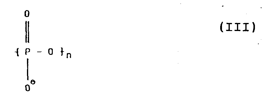
- B is a phosphoric acid or salt thereof, it is intended that B have the formula III wherein P is bonded to X and n may be as described above.
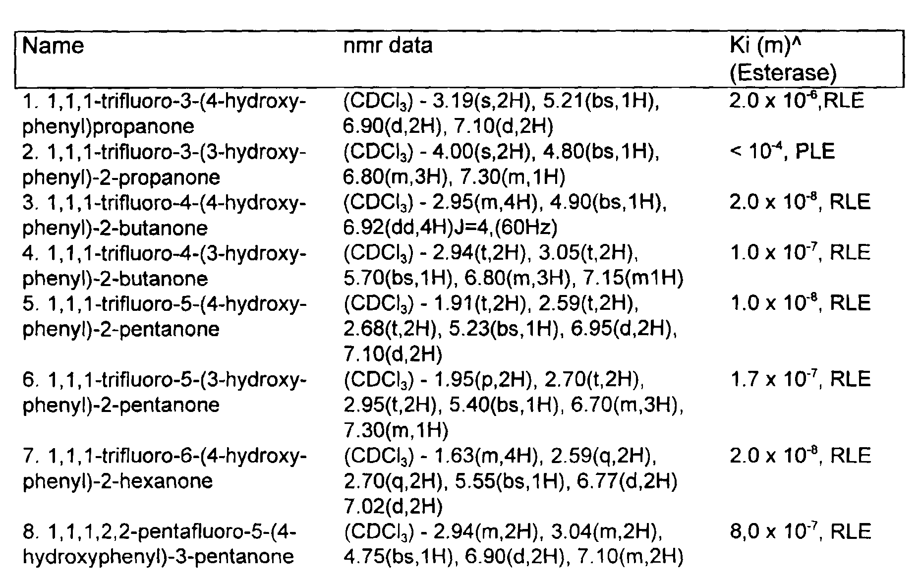
- inhibitor and blocked inhibitor in accordance with formula I may be synthesized by any sequence of conventional chemical reactions as may be envisioned by one skilled in the art. Suitable and convenient methods are given in the Examples, below. The following list of effective enzyme inhibitors is intended to be exemplary only.
- kits or package of materials for performing an assay for a viral antigen in accordance with the method of the invention.
- the kit includes a membrane coated with an inert protein, an antibody having a first hydrolase conjugated thereto, and a blocked inhibitor as a substrate for the first hydrolase.
- the kit may also include a second hydrolase, and an enzyme substrate for the second hydrolase, standards for the ligand, as, for example, one or more viral antigen samples of known concentration, or it may include other reagents, enzyme substrates, or other labeled or unlabeled specific viral antigens, antibodies or complexes thereof useful in carrying out the assay. It may include solutions, such as saline or buffers.
- the components of the kit are assembled in a housing, preferably plastic, containing a material positioned under the membrane, such as absorbent paper, to facilitate flow of assay liquids through the membranes by capillary action.
- HPLC was performed on a Waters 510 two pump system with UV detection using one of two solvent systems on a Brownlee AX- 300 7 x 250 mm column.
- System A initial hold for 5 minutes at 30mM NH 4 OAc pH 6.5 followed by a linear gradient to 2.0 M NH 4 OAc over a 30 minute period followed by a hold at 1.0 M NH 4 OAc for 5 minutes.
- System B used an isocratic buffer system of 30mM NH 4 OAc pH 6.5 for 40 minutes. Flow rates were 1.0 ml/min. Gas chromatography was performed on a H.P.
- a membrane filter stack was assembled with the following configuration:
- the membrane layers were encased in a plastic holder which includes a receiving well formed above the top layer. Within this well was fitted a flow restriction insert which has an aperture more narrow than the receiving well and sits flush against the top membrane.
- An antigen stock was prepared with respiratory syncytial virus (RSV) (Long strain) infected HEp-2 cells diluted in a buffer containing: 250 mM tris(hydroxymethyl)aminomethane hydrochloride (Tris HCl), 150 mM sodium chloride (NaCl), 10 mM ethylenediaminetetraacetate (EDTA), 4% (v/v) polyoxyethylene sorbitan monolaurate (Tween 20), 1% n-acetyl cysteine, 0.2% sodium azide (NaN 3 ), pH 8.5. Control antigen was prepared in a similar manner from uninfected HEp-2 cells.
- RSV respiratory syncytial virus
- a 150 ⁇ l aliquot of this antigen (or control) was applied to the device and allowed to drain through the flow restriction insert and onto the top membrane layer. (Liquid is drawn through the top membrane by the capillary action of the supporting absorbent layers.)
- the flow restriction insert was then removed, and to the device was added 150 ⁇ l of a wash solution consisting of 50 mM Tris HCl, 150 mM NaCl, 0.2% NaN 3 , pH 7.2 (Tris buffered saline (TBS)), additionally containing 1 mg/ml of rabbit IgG.
- TBS Tris buffered saline
- a solution containing 27 ⁇ g/ml of anti-RSV antibody conjugated to alkaline phosphatase was prepared in a buffer containing 50 mM Tris HCl, 100 mM NaCl, 200 mM sodium phosphate, 1% casein, 1 mM magnesium chloride, 0.1 mM zinc chloride, and 1 mM 2-mercaptoethanol, pH 7.5.
- a 150 ⁇ l aliquot of this mixture was added to the device and allowed to absorb into the membrane stack. Following a brief (two minute) incubation, the device was washed with 300 ⁇ l of TBS (without IgG).
- a 150 ⁇ l solution containing 0.33 mg/ml nitroblue tetrazolium, 1% methanol, and 0.2% NaN 3 was added to the device. This was followed by the addition of 150 ⁇ l of a solution containing 12 mM levamisole in 50 mM 2-amino-2-methyl-1-propanol acetate (AMP HOAc), 0.2% NaN 3 , 19 mM magnesium chloride, at pH 9.8. Following a five minute incubation at ambient temperature, the color forming reaction was stopped by the addition of 150 ⁇ l of a solution containing 200 mM potassium phosphate, 10 mM EDTA, 0.2% NaN 3 , pH 7.2.
- AMP HOAc 2-amino-2-methyl-1-propanol acetate
- a dual enzyme cascade assay for RSV is performed in a manner similar to Example II with the following exceptions:
- the color density of the unknown sample is compared with an uninfected control. If the unknown appears significantly lighter than the control (less than half the reflectance density of the control) then it is judged to be a positive result, indicating the presence of viral material.
- the invention provides an assay for a viral antigen which does not include a specific capture antibody for the antigen. Instead, a solid support is precoated with an inert protein and antigen is captured directly on the coated support. Further, the inert protein, even though applied as a precoat prior to antigen capture, prevents substantially all nonspecific binding of other proteins, which would otherwise reduce assay sensitivity.
Landscapes
- Health & Medical Sciences (AREA)
- Life Sciences & Earth Sciences (AREA)
- Immunology (AREA)
- Engineering & Computer Science (AREA)
- Chemical & Material Sciences (AREA)
- Hematology (AREA)
- Biomedical Technology (AREA)
- Urology & Nephrology (AREA)
- Molecular Biology (AREA)
- Food Science & Technology (AREA)
- General Physics & Mathematics (AREA)
- Cell Biology (AREA)
- Biotechnology (AREA)
- Pathology (AREA)
- Microbiology (AREA)
- Medicinal Chemistry (AREA)
- Physics & Mathematics (AREA)
- Analytical Chemistry (AREA)
- Biochemistry (AREA)
- General Health & Medical Sciences (AREA)
- Virology (AREA)
- Tropical Medicine & Parasitology (AREA)
- Chemical Kinetics & Catalysis (AREA)
- Measuring Or Testing Involving Enzymes Or Micro-Organisms (AREA)
- Investigating Or Analysing Biological Materials (AREA)
- Medicines Containing Antibodies Or Antigens For Use As Internal Diagnostic Agents (AREA)
- Pharmaceuticals Containing Other Organic And Inorganic Compounds (AREA)
- Sampling And Sample Adjustment (AREA)
- Analysing Materials By The Use Of Radiation (AREA)
- Peptides Or Proteins (AREA)
Abstract
Description
more particularly, relates to an assay including capture of the viral antigen directly on a membrane precoated with an inert protein.



- Top layer -
- Three µm Immunodyne Immunoaffinity Membrane, (Pall, East Hills, New York, #BIA0030HC5). Precoated by immersion in phosphate buffered saline containing 0.3% casein for 30 minutes at ambient temperature.
- Next layer -
- Non-woven rayon sheet (Schleicher and Schuell, Keene, New Hampshire; #5-S).
- Bottom layer -
- Cellulose absorbent pads (2) (Filtration Sciences, Mount Holly Springs, Pennsylvania; #ED 320-200)
Claims (8)
- A method for determining a viral antigen in a liquid comprising:a) combining a first liquid suspected of containing a viral antigen with a membrane coated with an inert protein and with an antibody specific for said antigen having a first hydrolase conjugated thereto whereby said viral antigen attaches to said coated membrane and binds to said antibody to give a bound fraction including said first hydrolase on said membrane;b) separating said membrane from said first liquid by causing said first liquid to pass through said membrane;c) contacting said coated membrane with a second liquid containing a blocked inhibitor, a second hydrolase substantially unreactive toward said blocked inhibitor and a substrate for said second hydrolase whereby said first hydrolase on said support converts said blocked inhibitor to an inhibitor which inhibits conversion of said substrate to a colored product by said second hydrolase; andd) detecting said viral antigen by a signal associated with the color of said product.
- The method of Claim 1 wherein said inert protein is selected from the group consisting of casein and albumin.
- The method of Claim 1 wherein said first hydrolase is selected from the group consisting of a peptidase, esterase, phosphatase and glycosidase.
- The method of Claim 1 wherein said blocked inhibitor is a blocked fluoroketone.
- The method of claim 1 wherein said second hydrolase is an esterase.
- The method of claim 5 wherein said substrate is an indolyl ester.
- A kit of materials for performing an assay for a viral antigen as defined in claims 1-6, wherein said kit comprises a membrane coated with an inert protein, an antibody having a first hydrolase conjugated thereto, a blocked inhibitor as a substrate for said first hydrolase and a housing containing an absorbent material positioned adjacent said membrane.
- The kit of claim 7 further comprising at least one other reagent selected from a second hydrolase, an enzyme substrate for said second hydrolase, buffer and saline.
Applications Claiming Priority (3)
| Application Number | Priority Date | Filing Date | Title |
|---|---|---|---|
| US27238088A | 1988-11-17 | 1988-11-17 | |
| US272380 | 1988-11-17 | ||
| EP89120945A EP0369361B1 (en) | 1988-11-17 | 1989-11-11 | Immunoassay on a preblocked solid surface |
Related Parent Applications (3)
| Application Number | Title | Priority Date | Filing Date |
|---|---|---|---|
| EP89120945.4 Division | 1989-11-11 | ||
| EP89120945A Division-Into EP0369361B1 (en) | 1988-11-17 | 1989-11-11 | Immunoassay on a preblocked solid surface |
| EP89120945A Division EP0369361B1 (en) | 1988-11-17 | 1989-11-11 | Immunoassay on a preblocked solid surface |
Publications (3)
| Publication Number | Publication Date |
|---|---|
| EP0633472A2 EP0633472A2 (en) | 1995-01-11 |
| EP0633472A3 EP0633472A3 (en) | 1995-02-15 |
| EP0633472B1 true EP0633472B1 (en) | 2000-05-17 |
Family
ID=23039567
Family Applications (2)
| Application Number | Title | Priority Date | Filing Date |
|---|---|---|---|
| EP94115914A Expired - Lifetime EP0633472B1 (en) | 1988-11-17 | 1989-11-11 | Immunoassay on a preblocked solid surface |
| EP89120945A Expired - Lifetime EP0369361B1 (en) | 1988-11-17 | 1989-11-11 | Immunoassay on a preblocked solid surface |
Family Applications After (1)
| Application Number | Title | Priority Date | Filing Date |
|---|---|---|---|
| EP89120945A Expired - Lifetime EP0369361B1 (en) | 1988-11-17 | 1989-11-11 | Immunoassay on a preblocked solid surface |
Country Status (13)
| Country | Link |
|---|---|
| US (1) | US6235464B1 (en) |
| EP (2) | EP0633472B1 (en) |
| JP (1) | JP2589830B2 (en) |
| AT (2) | ATE127589T1 (en) |
| AU (1) | AU625297B2 (en) |
| CA (1) | CA2000685C (en) |
| DE (2) | DE68929213T2 (en) |
| DK (1) | DK175008B1 (en) |
| ES (2) | ES2076948T3 (en) |
| FI (1) | FI96802C (en) |
| GR (1) | GR3018020T3 (en) |
| IE (1) | IE69041B1 (en) |
| MY (1) | MY104234A (en) |
Families Citing this family (19)
| Publication number | Priority date | Publication date | Assignee | Title |
|---|---|---|---|---|
| WO1995023969A1 (en) * | 1994-03-04 | 1995-09-08 | Aquaculture Diagnostics Limited | Rapid immunological test |
| ATE286251T1 (en) | 1999-01-27 | 2005-01-15 | Bayer Ag | IMMUNE TEST USING PARTICLES WITH CASEIN COATINGS |
| AU2001273264A1 (en) * | 2000-07-21 | 2002-02-05 | Cuno, Incorporated | Improved blocking chemistries for nylon membrane |
| US7052831B2 (en) * | 2000-09-29 | 2006-05-30 | Becton Dickinson And Company | Detection of multiple analytes from a single sample using a multi-well, multi-analyte flow-through diagnostic test device |
| US7906276B2 (en) | 2004-06-30 | 2011-03-15 | Kimberly-Clark Worldwide, Inc. | Enzymatic detection techniques |
| US7521226B2 (en) * | 2004-06-30 | 2009-04-21 | Kimberly-Clark Worldwide, Inc. | One-step enzymatic and amine detection technique |
| US7094528B2 (en) * | 2004-06-30 | 2006-08-22 | Kimberly-Clark Worldwide, Inc. | Magnetic enzyme detection techniques |
| WO2006098804A2 (en) | 2005-03-11 | 2006-09-21 | Chembio Diagnostic Systems, Inc. | Dual path immunoassay device |
| US7189522B2 (en) | 2005-03-11 | 2007-03-13 | Chembio Diagnostic Systems, Inc. | Dual path immunoassay device |
| US8003399B2 (en) * | 2005-08-31 | 2011-08-23 | Kimberly-Clark Worldwide, Inc. | Nitrite detection technique |
| US7504235B2 (en) | 2005-08-31 | 2009-03-17 | Kimberly-Clark Worldwide, Inc. | Enzyme detection technique |
| US8758989B2 (en) * | 2006-04-06 | 2014-06-24 | Kimberly-Clark Worldwide, Inc. | Enzymatic detection techniques |
| US7935538B2 (en) * | 2006-12-15 | 2011-05-03 | Kimberly-Clark Worldwide, Inc. | Indicator immobilization on assay devices |
| US7897360B2 (en) | 2006-12-15 | 2011-03-01 | Kimberly-Clark Worldwide, Inc. | Enzyme detection techniques |
| US20100022916A1 (en) | 2008-07-24 | 2010-01-28 | Javanbakhsh Esfandiari | Method and Apparatus for Collecting and Preparing Biological Samples for Testing |
| US20100290948A1 (en) * | 2009-05-15 | 2010-11-18 | Xuedong Song | Absorbent articles capable of indicating the presence of urinary tract infections |
| US8603835B2 (en) | 2011-02-10 | 2013-12-10 | Chembio Diagnostic Systems, Inc. | Reduced step dual path immunoassay device and method |
| CA2944488C (en) | 2014-04-02 | 2023-03-21 | Chembio Diagnostic Systems, Inc. | Immunoassay utilizing trapping conjugate |
| US20160116466A1 (en) | 2014-10-27 | 2016-04-28 | Chembio Diagnostic Systems, Inc. | Rapid Screening Assay for Qualitative Detection of Multiple Febrile Illnesses |
Family Cites Families (19)
| Publication number | Priority date | Publication date | Assignee | Title |
|---|---|---|---|---|
| GB1420916A (en) * | 1971-09-08 | 1976-01-14 | Bagshawe K D | Performance of chemical or biological reactions |
| CA983358A (en) * | 1971-09-08 | 1976-02-10 | Kenneth D. Bagshawe | Performance of chemical or biological reactions |
| US4407943A (en) * | 1976-12-16 | 1983-10-04 | Millipore Corporation | Immobilized antibody or antigen for immunoassay |
| US4241175A (en) * | 1978-12-18 | 1980-12-23 | Merck & Co., Inc. | Assay for hepatitis B core antibody |
| US4372745A (en) * | 1979-12-19 | 1983-02-08 | Electro-Nucleonics, Inc. | Chemical luminescence amplification substrate system for immunochemistry involving microencapsulated fluorescer |
| US4342826A (en) * | 1980-02-04 | 1982-08-03 | Collaborative Research, Inc. | Immunoassay products and methods |
| DE3274017D1 (en) * | 1981-03-07 | 1986-12-04 | Colin Henry Self | Assay and use |
| US4459359A (en) * | 1981-11-19 | 1984-07-10 | New York Blood Center, Inc. | Sensitive immunoassays of antigens or antibodies sequestered within immune complexes |
| US4477576A (en) * | 1982-07-26 | 1984-10-16 | Mex Research Associates | Antigen assay method and kit |
| US4606855A (en) * | 1982-07-26 | 1986-08-19 | Mex Research Associates C/O Leon Reimer | Monoclonal antibody to digoxin |
| US4659678A (en) * | 1982-09-29 | 1987-04-21 | Serono Diagnostics Limited | Immunoassay of antigens |
| EP0126772A1 (en) * | 1982-12-03 | 1984-12-05 | E.I. Du Pont De Nemours And Company | Chromogenic support immunoassay |
| WO1986002733A1 (en) * | 1984-10-31 | 1986-05-09 | Alcon Laboratories, Incorporated | Sandwich immunoassay of antigens involved in ophthalmic disorders |
| ATE75052T1 (en) * | 1986-09-24 | 1992-05-15 | Wako Pure Chem Ind Ltd | METHOD FOR DETERMINING ANTISTREPTOLYSIN-O AND SET OF USE THEREOF. |
| US4835099A (en) * | 1986-11-20 | 1989-05-30 | Becton, Dickinson And Company | Signal enhancement in immunoassay by modulation of enzymatic catalysis |
| US4847199A (en) * | 1987-02-27 | 1989-07-11 | Eastman Kodak Company | Agglutination immunoassay and kit for determination of a multivalent immune species using a buffered salt wash solution |
| DE3889479T2 (en) * | 1987-02-27 | 1994-12-22 | Eastman Kodak Co | Low pI protein coated membrane structure and process for its manufacture and use. |
| US5208143A (en) * | 1988-11-17 | 1993-05-04 | Becton, Dickinson And Company | Immunoassay on a preblocked solid surface |
| CA2023850A1 (en) * | 1989-09-28 | 1991-03-29 | Randal A. Hoke | Stabilizing reagent for membrane immunoassay |
-
1989
- 1989-10-11 MY MYPI89001399A patent/MY104234A/en unknown
- 1989-10-13 CA CA002000685A patent/CA2000685C/en not_active Expired - Lifetime
- 1989-10-17 AU AU42987/89A patent/AU625297B2/en not_active Expired
- 1989-11-01 IE IE351689A patent/IE69041B1/en not_active IP Right Cessation
- 1989-11-11 AT AT89120945T patent/ATE127589T1/en not_active IP Right Cessation
- 1989-11-11 ES ES89120945T patent/ES2076948T3/en not_active Expired - Lifetime
- 1989-11-11 EP EP94115914A patent/EP0633472B1/en not_active Expired - Lifetime
- 1989-11-11 DE DE68929213T patent/DE68929213T2/en not_active Expired - Lifetime
- 1989-11-11 AT AT94115914T patent/ATE193121T1/en not_active IP Right Cessation
- 1989-11-11 EP EP89120945A patent/EP0369361B1/en not_active Expired - Lifetime
- 1989-11-11 ES ES94115914T patent/ES2145084T3/en not_active Expired - Lifetime
- 1989-11-11 DE DE68924129T patent/DE68924129T2/en not_active Expired - Lifetime
- 1989-11-13 JP JP1294750A patent/JP2589830B2/en not_active Expired - Lifetime
- 1989-11-16 DK DK198905759A patent/DK175008B1/en not_active IP Right Cessation
- 1989-11-16 FI FI895472A patent/FI96802C/en not_active IP Right Cessation
-
1994
- 1994-10-20 US US08/326,304 patent/US6235464B1/en not_active Expired - Lifetime
-
1995
- 1995-11-08 GR GR950403133T patent/GR3018020T3/en unknown
Also Published As
| Publication number | Publication date |
|---|---|
| JPH02187662A (en) | 1990-07-23 |
| ATE127589T1 (en) | 1995-09-15 |
| EP0633472A3 (en) | 1995-02-15 |
| EP0369361B1 (en) | 1995-09-06 |
| US6235464B1 (en) | 2001-05-22 |
| AU625297B2 (en) | 1992-07-09 |
| GR3018020T3 (en) | 1996-02-29 |
| JP2589830B2 (en) | 1997-03-12 |
| FI895472A0 (en) | 1989-11-16 |
| DK575989D0 (en) | 1989-11-16 |
| AU4298789A (en) | 1990-05-24 |
| EP0369361A3 (en) | 1991-06-05 |
| DE68929213D1 (en) | 2000-06-21 |
| IE893516L (en) | 1990-05-17 |
| DK575989A (en) | 1990-05-18 |
| MY104234A (en) | 1994-02-28 |
| DE68924129D1 (en) | 1995-10-12 |
| ATE193121T1 (en) | 2000-06-15 |
| CA2000685A1 (en) | 1990-05-17 |
| EP0633472A2 (en) | 1995-01-11 |
| ES2076948T3 (en) | 1995-11-16 |
| EP0369361A2 (en) | 1990-05-23 |
| CA2000685C (en) | 2001-02-20 |
| DE68929213T2 (en) | 2000-12-28 |
| FI96802B (en) | 1996-05-15 |
| FI96802C (en) | 1996-08-26 |
| ES2145084T3 (en) | 2000-07-01 |
| DE68924129T2 (en) | 1996-02-01 |
| DK175008B1 (en) | 2004-04-19 |
| IE69041B1 (en) | 1996-08-07 |
Similar Documents
| Publication | Publication Date | Title |
|---|---|---|
| US5208143A (en) | Immunoassay on a preblocked solid surface | |
| US6235464B1 (en) | Immunoassay on a preblocked solid surface | |
| US4835099A (en) | Signal enhancement in immunoassay by modulation of enzymatic catalysis | |
| US5328831A (en) | Substrate composition for solid phase urease immunoassay | |
| US5279935A (en) | Method of immunossay including deactivation of endogenous alkaline phosphatase | |
| US4904583A (en) | Cascade immunoassay by multiple binding reactions | |
| JPH0756487B2 (en) | Immunoassay for detecting ligands | |
| US5188937A (en) | Layered sandwich assay method for chlamydia and materials therefor | |
| US6013457A (en) | Method for carrying out an immunoassay in a multiphase system | |
| IE893517L (en) | Substrate composition for alkaline phosphatase and method¹for assay using same | |
| US4847194A (en) | Colorimetric detection of delta-5-3-ketosteroid isomerase and immunoassay based thereon | |
| EP0420170A2 (en) | Stabilizing reagent for membrane immunoassay | |
| DK175273B1 (en) | Determining antigen in liq. - using solid support coated with inert protein and tracer including label | |
| EP0369657A2 (en) | Chromogenic substrate for esterase and immunoassay using same | |
| CA2011260A1 (en) | Signal enhancement in magnetic immunoassay by inhibition of enzymatic catalysis |
Legal Events
| Date | Code | Title | Description |
|---|---|---|---|
| PUAI | Public reference made under article 153(3) epc to a published international application that has entered the european phase |
Free format text: ORIGINAL CODE: 0009012 |
|
| PUAL | Search report despatched |
Free format text: ORIGINAL CODE: 0009013 |
|
| 17P | Request for examination filed |
Effective date: 19941008 |
|
| AC | Divisional application: reference to earlier application |
Ref document number: 369361 Country of ref document: EP |
|
| AK | Designated contracting states |
Kind code of ref document: A2 Designated state(s): AT BE CH DE ES FR GB GR IT LI LU NL SE |
|
| AK | Designated contracting states |
Kind code of ref document: A3 Designated state(s): AT BE CH DE ES FR GB GR IT LI LU NL SE |
|
| RIN1 | Information on inventor provided before grant (corrected) |
Inventor name: MCLAURIN, DANIEL A. Inventor name: HOPKINS, ANNE C. Inventor name: HOKE, RANDAL A. Inventor name: HENDERSON, GLENN L. |
|
| 17Q | First examination report despatched |
Effective date: 19990401 |
|
| GRAG | Despatch of communication of intention to grant |
Free format text: ORIGINAL CODE: EPIDOS AGRA |
|
| GRAG | Despatch of communication of intention to grant |
Free format text: ORIGINAL CODE: EPIDOS AGRA |
|
| GRAG | Despatch of communication of intention to grant |
Free format text: ORIGINAL CODE: EPIDOS AGRA |
|
| GRAH | Despatch of communication of intention to grant a patent |
Free format text: ORIGINAL CODE: EPIDOS IGRA |
|
| GRAH | Despatch of communication of intention to grant a patent |
Free format text: ORIGINAL CODE: EPIDOS IGRA |
|
| GRAA | (expected) grant |
Free format text: ORIGINAL CODE: 0009210 |
|
| AC | Divisional application: reference to earlier application |
Ref document number: 369361 Country of ref document: EP |
|
| AK | Designated contracting states |
Kind code of ref document: B1 Designated state(s): AT BE CH DE ES FR GB GR IT LI LU NL SE |
|
| PG25 | Lapsed in a contracting state [announced via postgrant information from national office to epo] |
Ref country code: LI Free format text: LAPSE BECAUSE OF FAILURE TO SUBMIT A TRANSLATION OF THE DESCRIPTION OR TO PAY THE FEE WITHIN THE PRESCRIBED TIME-LIMIT Effective date: 20000517 Ref country code: GR Free format text: LAPSE BECAUSE OF NON-PAYMENT OF DUE FEES Effective date: 20000517 Ref country code: CH Free format text: LAPSE BECAUSE OF FAILURE TO SUBMIT A TRANSLATION OF THE DESCRIPTION OR TO PAY THE FEE WITHIN THE PRESCRIBED TIME-LIMIT Effective date: 20000517 |
|
| REF | Corresponds to: |
Ref document number: 193121 Country of ref document: AT Date of ref document: 20000615 Kind code of ref document: T |
|
| REG | Reference to a national code |
Ref country code: CH Ref legal event code: EP |
|
| REF | Corresponds to: |
Ref document number: 68929213 Country of ref document: DE Date of ref document: 20000621 |
|
| REG | Reference to a national code |
Ref country code: ES Ref legal event code: FG2A Ref document number: 2145084 Country of ref document: ES Kind code of ref document: T3 |
|
| ET | Fr: translation filed | ||
| ITF | It: translation for a ep patent filed | ||
| PG25 | Lapsed in a contracting state [announced via postgrant information from national office to epo] |
Ref country code: LU Free format text: LAPSE BECAUSE OF NON-PAYMENT OF DUE FEES Effective date: 20001111 |
|
| REG | Reference to a national code |
Ref country code: CH Ref legal event code: PL |
|
| PLBE | No opposition filed within time limit |
Free format text: ORIGINAL CODE: 0009261 |
|
| STAA | Information on the status of an ep patent application or granted ep patent |
Free format text: STATUS: NO OPPOSITION FILED WITHIN TIME LIMIT |
|
| 26N | No opposition filed | ||
| REG | Reference to a national code |
Ref country code: GB Ref legal event code: IF02 |
|
| PGFP | Annual fee paid to national office [announced via postgrant information from national office to epo] |
Ref country code: AT Payment date: 20061019 Year of fee payment: 18 |
|
| PGFP | Annual fee paid to national office [announced via postgrant information from national office to epo] |
Ref country code: NL Payment date: 20061121 Year of fee payment: 18 |
|
| PGFP | Annual fee paid to national office [announced via postgrant information from national office to epo] |
Ref country code: SE Payment date: 20061124 Year of fee payment: 18 Ref country code: ES Payment date: 20061124 Year of fee payment: 18 |
|
| PGFP | Annual fee paid to national office [announced via postgrant information from national office to epo] |
Ref country code: IT Payment date: 20061130 Year of fee payment: 18 |
|
| PGFP | Annual fee paid to national office [announced via postgrant information from national office to epo] |
Ref country code: BE Payment date: 20061220 Year of fee payment: 18 |
|
| BERE | Be: lapsed |
Owner name: *BECTON DICKINSON AND CY Effective date: 20071130 |
|
| EUG | Se: european patent has lapsed | ||
| NLV4 | Nl: lapsed or anulled due to non-payment of the annual fee |
Effective date: 20080601 |
|
| PG25 | Lapsed in a contracting state [announced via postgrant information from national office to epo] |
Ref country code: AT Free format text: LAPSE BECAUSE OF NON-PAYMENT OF DUE FEES Effective date: 20071111 |
|
| PG25 | Lapsed in a contracting state [announced via postgrant information from national office to epo] |
Ref country code: BE Free format text: LAPSE BECAUSE OF NON-PAYMENT OF DUE FEES Effective date: 20071130 |
|
| PG25 | Lapsed in a contracting state [announced via postgrant information from national office to epo] |
Ref country code: SE Free format text: LAPSE BECAUSE OF NON-PAYMENT OF DUE FEES Effective date: 20071112 Ref country code: NL Free format text: LAPSE BECAUSE OF NON-PAYMENT OF DUE FEES Effective date: 20080601 |
|
| REG | Reference to a national code |
Ref country code: ES Ref legal event code: FD2A Effective date: 20071112 |
|
| PG25 | Lapsed in a contracting state [announced via postgrant information from national office to epo] |
Ref country code: ES Free format text: LAPSE BECAUSE OF NON-PAYMENT OF DUE FEES Effective date: 20071112 |
|
| PGFP | Annual fee paid to national office [announced via postgrant information from national office to epo] |
Ref country code: FR Payment date: 20081117 Year of fee payment: 20 |
|
| PGFP | Annual fee paid to national office [announced via postgrant information from national office to epo] |
Ref country code: DE Payment date: 20081223 Year of fee payment: 20 |
|
| PGFP | Annual fee paid to national office [announced via postgrant information from national office to epo] |
Ref country code: GB Payment date: 20081128 Year of fee payment: 20 |
|
| PG25 | Lapsed in a contracting state [announced via postgrant information from national office to epo] |
Ref country code: IT Free format text: LAPSE BECAUSE OF NON-PAYMENT OF DUE FEES Effective date: 20071111 |
|
| REG | Reference to a national code |
Ref country code: GB Ref legal event code: PE20 Expiry date: 20091110 |
|
| PG25 | Lapsed in a contracting state [announced via postgrant information from national office to epo] |
Ref country code: GB Free format text: LAPSE BECAUSE OF EXPIRATION OF PROTECTION Effective date: 20091110 |