EP0628639A1 - Reconstituted human antibody against human interleukin 6 receptor - Google Patents
Reconstituted human antibody against human interleukin 6 receptor Download PDFInfo
- Publication number
- EP0628639A1 EP0628639A1 EP92909376A EP92909376A EP0628639A1 EP 0628639 A1 EP0628639 A1 EP 0628639A1 EP 92909376 A EP92909376 A EP 92909376A EP 92909376 A EP92909376 A EP 92909376A EP 0628639 A1 EP0628639 A1 EP 0628639A1
- Authority
- EP
- European Patent Office
- Prior art keywords
- chain
- human
- region
- antibody
- amino acid
- Prior art date
- Legal status (The legal status is an assumption and is not a legal conclusion. Google has not performed a legal analysis and makes no representation as to the accuracy of the status listed.)
- Granted
Links
Images












Classifications
-
- C—CHEMISTRY; METALLURGY
- C07—ORGANIC CHEMISTRY
- C07K—PEPTIDES
- C07K16/00—Immunoglobulins [IG], e.g. monoclonal or polyclonal antibodies
- C07K16/18—Immunoglobulins [IG], e.g. monoclonal or polyclonal antibodies against material from animals or humans
- C07K16/28—Immunoglobulins [IG], e.g. monoclonal or polyclonal antibodies against material from animals or humans against receptors, cell surface antigens or cell surface determinants
- C07K16/2866—Immunoglobulins [IG], e.g. monoclonal or polyclonal antibodies against material from animals or humans against receptors, cell surface antigens or cell surface determinants against receptors for cytokines, lymphokines, interferons
-
- A—HUMAN NECESSITIES
- A61—MEDICAL OR VETERINARY SCIENCE; HYGIENE
- A61P—SPECIFIC THERAPEUTIC ACTIVITY OF CHEMICAL COMPOUNDS OR MEDICINAL PREPARATIONS
- A61P35/00—Antineoplastic agents
-
- A—HUMAN NECESSITIES
- A61—MEDICAL OR VETERINARY SCIENCE; HYGIENE
- A61K—PREPARATIONS FOR MEDICAL, DENTAL OR TOILETRY PURPOSES
- A61K38/00—Medicinal preparations containing peptides
-
- C—CHEMISTRY; METALLURGY
- C07—ORGANIC CHEMISTRY
- C07K—PEPTIDES
- C07K2317/00—Immunoglobulins specific features
- C07K2317/20—Immunoglobulins specific features characterized by taxonomic origin
- C07K2317/24—Immunoglobulins specific features characterized by taxonomic origin containing regions, domains or residues from different species, e.g. chimeric, humanized or veneered
-
- C—CHEMISTRY; METALLURGY
- C07—ORGANIC CHEMISTRY
- C07K—PEPTIDES
- C07K2319/00—Fusion polypeptide
Definitions
- the present invention relates to variable regions (V region) of a mouse monoclonal antibody to the human interleukin-6 receptor (IL-6R), human/mouse chimeric antibody to the human IL-6R, and reshaped human antibody comprising a human antibody wherein the complementarity determining regions (CDRs) of the human light chain (L chain) V region and of the human heavy chain (H chain) V region are grafted with the CDRs of a mouse monoclonal antibody to the human IL-6R.
- the present invention provides DNA coding for the above-mentioned antibodies or part thereof.
- the present invention further provides vectors, especially expression vectors comprising said DNA, and host cells transformed or transfected with said vector.
- the present invention still more provides a process for production of a chimeric antibody to the human IL-6R, and process for production of a reshaped human antibody to the human IL-6R.
- Interleukin-6 is a multi-function cytokine that is produced by a range of cells. It regulates immune responses, acute phase reactions, and hematopoiesis, and may play a central role in host defense mechanisms. It acts on a wide range of tissues, exerting growth-inducing, growth inhibitory, and differentiation-inducing effects, depending on the nature of the target cells.
- the specific receptor for IL-6 (IL-6R) is expressed on lymphoid as well as non-lymphoid cells in accordance with the multifunctional properties of IL-6.
- IL-6 Abnormal expression of the IL-6 gene has been suggested to be involved in the pathogenesis of a variety of diseases, especially autoimmune diseases, mesangial proliferative glomerulonephritis, and plasmacytoma/myeloma (see review by Hirano et al., Immunol. Today 11 , 443-449, 1990).
- Human myeloma cells are observed to produce IL-6 and express IL-6R.
- antibody against IL-6 inhibited the in vitro growth of myeloma cells thus indicating that an autocrine regulatory loop is operating in oncogenesis of human myelomas (Kawano et al., Nature, 332 , 83, 1988).
- the IL-6R is present on the surface of various animal cells, and specifically binds to IL-6, and the number of IL-6R molecules on the cell surface has been reported (Taga et al., J. Exp. Med. 196 , 967, 1987). Further, cDNA coding for a human IL-6R was cloned and a primary structure of the IL-6R was reported (Yamasaki et al., Science, 241 , 825, 1988).
- Mouse antibodies are highly immunogenic in humans and, for this reason, their therapeutic value in humans is limited. The half-life of mouse antibodies in vivo in human is relatively short. In addition, mouse antibodies can not be administered in multiple doses without generating an immune response which not only interferes with the planned efficacy but also risks an adverse allergic response in the patient.
- Mouse antibodies can be humanized in two ways. The more simple method is to construct chimeric antibodies where the V regions are derived from the original mouse monoclonal antibody and the C regions are derived from suitable human antibodies. The resulting chimeric antibody contains the entire V domains of the original mouse antibody and can be expected to bind antigen with the same specificity as the original mouse antibody. In addition, chimeric antibodies have a substantial reduction in the percent of the protein sequence derived from a non-human source and, therefore, are expected to be less immunogenic than the original mouse antibody. Although chimeric antibodies are predicted to bind antigen well and to be less immunogenic, an immune response to the mouse V regions can still occur (LoBuglio et al., Proc. Natl. Acad. Sci. USA 84 , 4220-4224, 1989).
- the second method for humanizing mouse antibodies is more complicated but more extensively reduces the potential immunogenicity of the mouse antibody.
- the complementarity determining regions (CDRs) from the V regions of the mouse antibody are grafted into human V regions to create "reshaped" human V regions. These reshaped human V regions are then joined to human C regions.
- the only portions of the final reshaped human antibody derived from non-human protein sequences are the CDRs.
- CDRs consist of highly variable protein sequences. They do not show species-specific sequences. For these reasons, a reshaped human antibody carrying murine CDRs should not be any more immunogenic than a natural human antibody containing human CDRs.
- the present inventors also found that, when the mouse monoclonal antibodies to the human IL-6R were injected into nude mice transplanted with a human myeloma cell line, the growth of the tumor was remarkably inhibited. This suggests that the anti-human IL-6 receptor antibody is useful as a therapeutic agent for the treatment of myeloma.
- the present invention is intended to provide a less immunogenic antibody to the human IL-6R. Accordingly, the present invention provides reshaped human antibodies to the human IL-6R. The present invention also provides human/mouse chimeric antibodies useful during the construction of the reshaped human antibody. The present invention further provides a part of reshaped human antibody, as well as the expression systems for production of the reshaped human antibody and a part thereof, and of the chimeric antibody.
- the present invention provides L chain V region of mouse monoclonal antibody to the human IL-6R; and H chain V region of a mouse monoclonal antibody to the human IL-6R.
- the present invention also provides a chimeric antibody to the human IL-6R, comprising:
- the present invention also provides CDR of an L chain V region of a mouse monoclonal antibody to the human IL-6R; and CDR of an H chain V region of a mouse monoclonal antibody to the human IL-6R.
- the present invention moreover provides a reshaped human L chain V region of an antibody to the human IL-6R, comprising:
- the present invention also provides a reshaped human L chain of an antibody to the human IL-6R, comprising:
- the present invention still more provides a reshaped human antibody to the human IL-6R, comprising:
- the present invention further provides DNA coding for any one of the above-mentioned antibody polypeptides or parts thereof.
- the present invention also provides vectors, for example, expression vectors comprising said DNA.
- the present invention further provides host cells transformed or transfected with the said vector.
- the present invention still more provide a process for production of a chimeric antibody to the human IL-6R, and a process for production of reshaped human antibody to the human IL-6R.
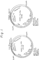
- Fig. 1 represents expression vectors comprising human cytomegalo virus (HCMV) promoter/enhancer system, useful for the expression of the present antibody peptide.
- HCMV human cytomegalo virus
- Fig. 2 is a graph showing a result of ELISA for confirmation of an ability of the present chimeric antibody AUK12-20 to bind to the human IL-6R.
- Fig. 3 is a graph showing a result of measurement of an ability of the present chimeric antibody AUK12-20 to inhibit the binding of IL-6 to the human IL-6R.
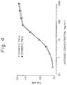
- Fig. 4 is a graph showing a result of ELISA for binding of the present chimeric antibodies PM1a and PM1b to human IL-6R.
- Fig. 5 is a graph showing a result of ELISA testing the ability of the present chimeric antibodies PM1a and PM1b to inhibit IL-6 from binding to the human IL-6R.
- Fig. 6 is a diagram of the construction of the first version of a reshaped human PM-1 H chain V region.
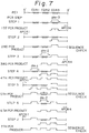
- Fig. 7 is a diagram of the construction of the first version of a reshaped human PM-1 L chain V region.
- Fig. 8 represents a process for construction of an expression plasmid HEF-12h-g ⁇ 1 comprising a human elongation factor 1 ⁇ (HEF-1 ⁇ ) promoter/enhancer, useful for the expression of an H chain.
- HEF-1 ⁇ human elongation factor 1 ⁇
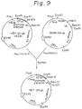
- Fig. 9 represents a process for construction of an expression plasmid HEF-12k-gk comprising the HEF-1 ⁇ promoter/enhancer system, useful for the expression of an L chain.
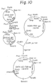
- Fig. 10 represents a process for construction of an expression plasmid DHFR-PMh-g ⁇ 1 comprising HCMV promoter/enhancer and the dihydrofolate reductase (dhfr) gene linked to a defective SV40 promoter/enhancers sequence for amplification, useful for expression of an H chain.
- dhfr dihydrofolate reductase
- Fig. 11 represents a process for the construction of an expression plasmid DHFR- ⁇ E-RVh-PM1-f comprising EF1 ⁇ promoter/enhancer and dhfr gene linked to a defective SV40 promoter/enhancer sequence for amplification, useful for expression of an H chain.
- Fig. 12 is a graph showing an ability of version "a” and "b” of the reshaped human PM-1 L chain V region for binding to the human IL-6R.
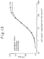
- Fig. 13 is a graph showing an ability of version "f" of the reshaped human PM-1 H chain V region plus version "a” of the reshaped PM-1 L chain L chain V region for binding to the human IL-6R.
- Fig. 14 is a graph showing an ability of vergion "f" of the reshaped PM-1 H chain V region plus version "a" of the reshaped PM-1 L chain V region to inhibit the binding of IL-6 to the human IL-6R.
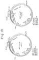
- Fig. 15 represents expression plasmids HEF-V L -gk and HEF-V H -g ⁇ 1 comprising a human EF1- ⁇ promoter/enhancer, useful for expression of an L chain and H chain respectively.
- Fig. 16 shows a process for construction of DNA coding for reshaped human AUK 12-20 antibody L chain V region.
- Fig. 17 is a graph showing results of an ELISA for confirm of an ability of a reshaped human AUK 12-20 antibody L chain V region to bind to human IL-6R.
- Standard AUK 12-20 (chimera) means a result for chimeric AUK 12-20 antibody produced by CHO cells and purified in a large amount.
- Fig. 18 is a graph showing a result of an ELISA for an ability of a reshaped human AUK 12-20 antibody (L chain version "a” + H chain version "b") to bind to human IL-6R.
- Fig. 19 is a graph showing a result of an ELISA for an ability of a reshaped human AUK 12-20 antibody (L chain version "a” + H chain version "d") to bind to the human IL-6R.
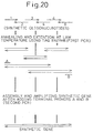
- Fig. 20 shows a process for chemical synthesis of a reshaped human sle 1220 H antibody H chain V region.
- Fig. 21 is a graph showing a result of an ELISA for an ability of a reshaped human sle 1220 antibody (L chain version "a” + H chain version "a”) to inhibit the binding of IL-6 to the human IL-6R.
- Fig. 22 is a graph showing a result of an ELISA for an ability of a reshaped human sle 1220 antibody (L chain version "a” + H chain version "b") to inhibit the binding of IL-6 to the human IL-6R.
- Fig. 23 is a graph showing a result of an ELISA for an ability of a reshaped human sle 1220 antibody (L chain version "a” + H chain version "c") to inhibit the binding of IL-6 to the human LI-6R.
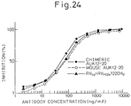
- Fig. 24 is a graph showing a result of an ELISA for an ability of a reshaped human sle 1220 antibody (L chain version "a” + H chain version "d") inhibit the binding of IL-6 to the human LI-6R.
- hybridoma which produces a monoclonal antibody to the human IL-6R
- Japanese Patent Application No. 2-189420 describes a mouse hybridoma PM-1 which produces a monoclonal antibody PM1 and the properties thereof.
- Reference Examples 1 and 2 of the present specification describe the construction process of the hybridoma PM1.
- the present inventors have constructed hybridomas AUK12-20, AUK64-7, and AUK146-15, each producing a mouse monoclonal antibody to the human IL-6R. The construction process of these hybridomas is described in the Reference Examples 3 of this specification.
- hybridoma cells are homogenized and a total RNA is obtained according to a conventional procedure described by Chirgwin et al., Biochemistry 18 , 5294, 1977. Next, the total RNA is used to synthesize single-stranded cDNAs according to the method described by J.W. Larrick et al., Biotechnology, 7 , 934, 1989.
- a specific amplification of a relevant portion of the cDNA is carried out by a polymerase chain reaction (PCR) method.
- PCR polymerase chain reaction
- 11 groups of oligonucleotide primers (Mouse Kappa Variable; MKV) represented in SEQ ID NO: 1 to 11
- an oligonucleotide primer (Mouse Kappa Constant; MKC) represented in SEQ ID NO: 12 are used as 5'-terminal primers and a 3'-terminal primer respectively.
- the MKV primers hybridize with the DNA sequence coding for the mouse ⁇ L chain leader sequence
- the MKC primer hybridizes with the DNA sequence coding for the mouse ⁇ L chain constant region.
- 10 groups of oligonucleotide primers (Mouse Heavy Variable; MHV) represented in SEQ ID NO: 13 to 22, and a oligonucleotide primer (Mouse Heavy Constant MHC) represented in SEQ ID NO: 23 are used as 5'-terminal primers and a 3'-terminal primer, respectively.
- the 5'-terminal primers contain the nucleotide sequence GTCGAC near the 5'-end thereof, which sequence provides a restriction enzyme Sal I cleavage site; and the 3'-terminal primer contains the nucleotide sequence CCCGGG near the 5-end thereof, which sequence provides a restriction enzyme Xma I cleavage site. These restriction enzyme cleavage sites are used to subclone the DNA fragments coding for a variable region into cloning vectors.
- the amplification product is cleaved with restriction enzymes Sal I and Xma I to obtain a DNA fragment coding for a desired V region of a mouse monoclonal antibody.
- an appropriate cloning vector such as plasmid pUC19 is cleaved with the same restriction enzymes Sal I and Xma I and the above DNA fragment is ligated with the cleaved pUC19 to obtain a plasmid incorporating a DNA fragment coding for a desired V region of a mouse monoclonal antibody.
- the sequencing of the cloned DNA can be carried out by any conventional procedure.
- CDRs Complementarity Determining Regions
- the present invention provides hypervariable or complementarity determining regions (CDRs) of each V region of the present invention.
- CDRs hypervariable or complementarity determining regions
- the domains on the L and H chains have the same general structure and each domain comprises four framework regions (FRs), whose sequences are relatively conserved, connected by three CDRs (see Kabat, E.A., Wu, T.T., Bilofsky, H., Reid-Miller, M. and Perry, H., in "Sequences of Proteins of Immunological Interest", US Dept. Health and Human Services 1983).
- the four FRs largely adopt a ⁇ -sheet conformation and the CDRs form loops connecting FRs, and in some cases forming part of, the ⁇ -sheet structure.
- the CDRs are held in close proximity by FRs and, with the CDRs from the other domain, contribute to the formation of the antigen binding site.
- the CDRs are described in Example 4.
- the basic method for constructing chimeric antibodies comprises joining the mouse leader and V region sequences, as found in the PCR-cloned cDNAs, to human C regions-coding sequence already present in mammalian cell expression vectors.
- construction of a chimeric antibody from the monoclonal antibody AUK12-20 is described in Example 5.
- a chimeric antibody from the monoclonal antibody PM-1 is described in Example 6.
- the cDNA coding for the mouse PM-1 ⁇ L chain leader and V region was PCR-subcloned into an expression vector containing a genomic DNA coding for the human kappa C region.
- the cDNA coding for the mouse PM-1 H chain leader and V regions was PCR-subcloned into an expression vector containing a genomic DNA coding for the human gamma-1 C region.
- the cDNA coding for the mouse PM-1 V region were adapted at their 5'- and 3'-ends (1) so that they would be easy to insert into the expression vectors and (2) so that they would function properly in these expression vectors.
- PCR-modified mouse PM-1 V regions were then inserted into HCMV expression vectors already containing the desired human C regions ( Figure 1). These vectors are suitable for either transient or stable expression of genetically-engineered antibodies in a variety of mammalian cell lines.
- a second version of chimeric PM-1 antibody was constructed (version b).
- the amino acid at position 107 in the L chain V region was changed from asparagine to lysine.
- the occurrence of an asparagine at position 107 was an unusual event.
- the most typical amino acid at position 107 is a lysine.
- position 107 was changed to the typical amino acid lysine at this position. This change was achieved using a PCR-mutagenesis method (M. Kamman et al., Nucl. Acids Res. (1989) 17:5404) to make the necessary changes in the DNA sequences coding for the L chain V region.
- the chimeric PM-1 antibody version (a) exhibited an activity to bind to the human IL-6R.
- the chimeric MP-1 antibody version (b) also binds to the human IL-6R as well as version (a).
- chimeric antibodies were constructed. All 4 chimeric antibodies bound well to the human IL-6R thus indicating in a functional assay that the correct mouse V regions had been cloned and sequenced.
- PM-1 antibody was selected as the first candidate for the design and construction of a reshaped human antibody to the human 1L-6R.
- the selection of mouse PM-1 antibody was based largely on results obtained studying the effect of the mouse anti-IL-6R antibodies on human myeloma tumor cells transplanted into nude mice.
- PM-1 antibody showed the strongest anti-tumor cell activity.
- the L chain V region of PM-1 antibody was most similar to the L chain V region of mouse antibody musigkcko (Chen, H.-T. et al., J. Biol. Chem. (1987) 262:13579-13583) with a 93.5% identity.
- the H chain V region of PM-1 antibody was most similar to the H chain V region of mouse antibody musigvhr2 (F.J. Grant et al., Nucl. Acids Res. (1987) 15:5496) with a 84.0% identity.
- the mouse PM-1 V regions show high percents of identity to known mouse V regions thus indicating that the mouse PM-1 V regions are typical mouse V regions.
- the L chain V region of PM-1 antibody was most similar to the L chain V region of human antibody klhure, also referred to as REI (W. Palm et al., Physiol. Chem. (1975) 356:167-191) with a 72.2% identity.
- the H chain V region of PM-1 antibody was most similar to the H chain V region of human antibody humighvap (VAP) (H.W. Schroeder et al., Science (1987) 238:791-793) with a 71.8% identity.
- the comparisons to human V regions are most important for considering how to design reshaped human antibodies from mouse PM-1 antibody.
- the percent identities to human V regions are less than the percent identities to mouse V regions. This is indirect evidence that the mouse PM-1 V regions do look like mouse V regions and not like human V regions. This evidence also indicates that it will be best to humanize mouse PM-1 V regions in order to avoid problems of immunogenicity in human patients.
- V regions from mouse PM-1 antibody were also compared to the consensus sequences for the different subgroups of human V regions as defined by E. A. Kabat et al. ((1987) Sequences of Proteins of Immunological Interest, Forth Edition, U.S. Department of Health and Human servides, U.S. Government Printing Office). The comparisons were made between the FRs of the V regions. The results are shown in Table 1.
- the FRs of mouse PM-1 L chain V region are most similar to the FRs from the consensus sequence for subgroup I (HSGI) of human L chain V regions with 70.1% identity.
- the FRs of mouse PM-1 H chain V region are most similar to the FRs from the consensus sequence for subgroup II (HSGII) of human H chain V regions with 52.9% identity.
- the first step in designing the reshaped human PM-1 V regions was to select the human V regions that would be the basis of the design.
- the FRs in the mouse PM-1 L chain V region were most similar to the FRs in human L chain V regions belonging to subgroup I (Table 1). As discussed above, in comparing the mouse PM-1 L chain V region to known human L chain V regions, it was most similar to the human L chain V region REI, a member of subgroup I of human L chain V regions.
- the FRs from REI were used. Moreover the REI FRs were used as starting material for the construction of reshaped human PM-1 L chain V region.
- position 71 is a tyrosine.
- position 71 was a phenylalanine.
- version "b" of reshaped human PM-1 L chain V region the phenylalanine at position 71 was changed to a tyrosine as found in mouse PM-1 L chain V region.
- Table 2 shows the amino acid sequences of mouse PM-1 L chain V region, the FRs of REI as modified for use in reshaped human CAMPATH-1H antibody (Riechmann et al. , 1988), and the two versions of reshaped human PM-1 L chain V region.
- the FRs in the mouse PM-1 H chain V region were most similar to the FRs in human H chain V regions belonging to subgroup II (Table 1). As discussed above, in comparing the mouse PM-1 H chain V region to known human H chain V regions, it was most similar to the human H chain V region VAP, a member of subgroup II of human H chain V regions. DNA sequences coding for the FRs in human H chain V region NEW, another member of subgroup II of human H chain V regions, were used as starting material for the construction of reshaped human PM-1 H chain V region, and as a base for designing the reshaped human PM-1 H chain V region.
- Residues 27, 28, 29, and 30 are either part of the canonical structure for CDR1 of the H chain V region, as predicted by C. Chothia et al., Nature (1989) 34:877-883, and/or are observed in the model of the mouse PM-1 V regions to form part of the first structural loop of the H chain V region (Chothia, 1987).
- Residue 48 was observed in the model of the mouse PM-1 V regions to be a buried residue. Changes in a buried residue can disrupt the overall structure of the V region and its antigen-binding site.
- Residue 71 is part of the canonical structure for CDR2 of the H chain V region as predicted by Chothia et al. , (1989).
- the six versions of reshaped human PM-1 antibody incorporate different combinations of amino acid changes at these seven positions in the human NEW FRs (see Table 3).
- the first versions of the reshaped human PM-1 L and H chain V regions were each constructed using a novel PCR-based method. Essentially, a plasmid DNA coding for reshaped human V region that already contained suitable human FRs was modified using PCR primers to replace the CDRs present in the starting reshaped human V region with the CDRs from mouse PM-1 antibody.
- the starting material for the construction of the reshaped human PM-1 L chain V region was a plasmid DNA containing the reshaped human D1.3 L chain V region.
- the reshaped human D1.3 L chain V region was constructed based on the FRs present in the human L chain V region of REI.
- the starting material for the construction of the reshaped human PM-1 H chain V region was a plasmid DNA containing the reshaped human D1.3 H chain V region.
- the reshaped human D1.3 H chain V region was constructed based on the FRs present in the human H chain V region of NEW (M. Verhoeyen et al., Science (1988) 239:1534-1536).
- PCR primers were designed to-enable the substitution of the mouse PM-1 CDRs in place of the mouse D1.3 CDRs.
- three primers containing the DNA sequences coding for the mouse PM-1 CDRs and two primers flanking the entire DNA sequence coding for the reshaped human V region were designated and synthesized.
- Using the five PCR primers in a series of PCR reactions yielded a PCR product that consisted of the human FRs present in the starting reshaped human V region and the CDRs present in mouse PM-1 V region (see Example 7, and Figures 7 and 8).
- the PCR products were cloned and sequenced to ensure that the entire DNA sequence of version "a" of reshaped human PM-1 L and H chain V region coded for correct amino acid sequence (SEQ ID NO 55).
- the remaining versions of the reshaped human PM-1 V regions were constructed using slight modifications of published PCR-mutagenesis techniques (Kamman et al. , 1989). As described for the design of the reshaped human PM-1 V regions, one additional version (version “b") of the reshaped human PM-1 L chain V region was constructed and five additional versions (versions "b", “c”, “d”, “e”, and “f") of the reshaped human PM-1 H chain V region were constructed. These additional versions contain a series of minor changes from the first versions. These minor changes in the amino acid sequences were achieved using PCR mutagenesis to make minor changes in the DNA sequences. PCR primers were designed that would introduce the necessary changes into the DNA sequence.
- the reshaped human PM-1 V regions were subcloned into mammalian cell expression vectors already containing human C regions.
- Reshaped human PM-1 L chain V regions were joined to DNA sequences coding for human ⁇ C region.
- Reshaped human PM-1 H chain V regions were joined to DNA sequences coding for human gamma-1 C region.
- the HCMV expression vectors as shown in Figure 1, were modified to replace the HCMV promoter-enhancer region with the human elongation factor (HEF-1 ⁇ ) promoter-enhancer (see Figure 15).
- introns in the leader sequences in both the reshaped human L and H chain V regions SEQ ID NOs: 54 and 55. These introns were originally derived from a mouse mu H chain leader sequence (M.S. Neuberger et al. , Nature 1985 314:268-270) that was used in the construction of reshaped human D1.3 and V regions (Verhoeyen et al. , 1988). Since the reshaped human D1.3 was expressed in a mammalian cell vector that employed a mouse immunoglobulin promoter, the presence of the mouse leader intron was important.
- the leader intron contains sequences that are important for expression from immunoglobulin promoters but not from viral promoters like HCMV (M.S. Neuberger et al., Nucl. Acids Res. (1988) 16:6713-6724).
- the introns in the leader sequences were deleted by PCR cloning cDNAs coding for the reshaped human V regions (see Example 12).
- the reshaped human PM-1 H chain V region was constructed from DNA sequences derived originally from reshaped human B1-8 H chain V region (P.T. Jones et al., Nature (1986) 321:522-525). This first reshaped human V region was constructed from the mouse NP H chain V region (M.S. Neuberger et al., Nature (1985); M.S. Neuberger et al., EMBO J. (1983) 2:1373-1378).
- the "crippled" dhfr gene was introduced into the HEF-1 ⁇ vector expressing human gamma-1 H chains as was described for the HCMV vector expressing human gamma-1 H chains.
- the HEF-1 ⁇ vector expressing reshaped human PM-1 L chains and the HEF-1 ⁇ -dhfr vector expressing reshaped human PM-1 H chains were co-transfected into CHO dhfr(-) cells.
- Stably transformed CHO cell lines were selected in Alpha-Minimum Essential Medium ( ⁇ -MEM) without nucleosides and with 10% FCS and 500 ⁇ g/ml of G418. Prior to any gene amplification steps, CHO cell lines were observed that produced up to 10 ⁇ g/106 cells/day of reshaped human PM-1 antibody.
- the ⁇ L chain V region of the mouse monoclonal, antibody AUK 12-20 is homologous in a similar extent (64 to 68%) with the human ⁇ L chain V region subgroups (HSG) I to IV.
- HSG human ⁇ L chain V region subgroups
- the human antibody REI used for construction of a reshaped human antibody from the mouse monoclonal antibody PM-1 belongs to the HSG I, exhibits a 62% homology with L chain V region of the mouse monoclonal antibody AUK 12-20.
- the CDRs in the AUK 12-20 antibody L chain V region particularly CDR2 corresponded better to canonical structures of the CDRs in REI rather than those in LEN.
- H chain V region of the antibody AUK 12-20 exhibits the highest homology with the HSG I.
- human antibody HAX (Stollar, B.O. et al., J. Immunol. (1987) 139:2496-2501) also belonging to the HSG I exhibits an about 66% homology with the AUK 12-20 antibody H chain V region.
- the FRs of the human antibody HAX belonging to the HSG I, and FRs of humanized 425 antibody H chain V region which has FRs consisting of HSGI consensus sequence (Ketteborough C.A. et al., Protein Engineering (1991) 4:773-783) are used.
- the AUK 12-20 antibody H chain V region exhibits an about 64% homology with version "a" of the humanized 425 antibody H chain V region.
- reshaped human AUK 12-20 antibody L chain V regions is designed as shown in Table 5 using FRs of the REI.
- reshaped human AUK 12-20 antibody H chain V regions are designed using FRs of the reshaped human VHa 425. It is found, however, that nucleotide sequence of DNA coding for reshaped human AUK 12-20 antibody H chain V region thus designed has a sequence well conforming to a splicing donor sequence. Therefore, as in the case of reshaped human PM-1 antibody there is a possibility of an abnormal splicing in the reshaped human AUK 12-20 antibody. Therefore, the nucleotide sequence was partially modified to eliminate the splicing donor-like sequence. The modified sequence is designated as version "a".
- version "b” to “d” of the reshaped human AUK 12-20 antibody H chain V region were designed. Amino acid sequences of the versions “a” to “d” are shown in Table 6.
- version "a” to “d” of reshaped human AUK 12-20 antibody H chain V region are designed as shown in Table 7, using FRs of the human antibody HAX (J. Immunology (1987) 139:2496-2501; an antibody produced by hybridoma 21/28 cells derived from B cells of a SLE patient; its amino acid sequence is described in Fig. 6 and nucleotide sequence of DNA coding for the amino acid sequence is shown in Figs. 4 and 6, of this literature).
- any expression system including eucaryotic cells, for example, animal cells, such as established mammalian cell lines, fungal cells, and yeast cells, as well as procaryotic cells, for example, bacterial cells such as E . coli cells, may be used.
- eucaryotic cells for example, animal cells, such as established mammalian cell lines, fungal cells, and yeast cells, as well as procaryotic cells, for example, bacterial cells such as E . coli cells.
- the present chimeric or reshaped human antibodies are expressed in mammalian cells such as cos cells or CHO cells.
- a conventional promoter useful for the expression in mammalian cells can be used.
- viral expression system such as human cytomegalovirus immediate early (HCMV) promoter is preferably used.
- HCMV human cytomegalovirus immediate early
- the expression vector containing the HCMV promoter include HCMV-V H -HC ⁇ 1, HCMV-V L -HC K , HCMV-12h-g ⁇ 1, HCMV-12k-gk and the like derived from pSV2neo, as shown in Fig. 1.
- HEF-1 ⁇ human elongation factor 1 ⁇
- Expression vectors containing this promotor include HEF-12h-g ⁇ 1 and HEF-12h-g ⁇ (Figs. 8 and 9), as well as HEF-V H -g ⁇ 1 and HEF-V L -g ⁇ (Fig. 15).
- an expression vector may contain a dhfr gene.
- Expression vectors containing the dhfr gene are for example, DHFR- ⁇ E-PMh-g ⁇ 1 (Fig. 10), DHFR- ⁇ E-RVh-PM1-f (Fig. 11) and the like.
- the present invention first provides an L chain V region and an H chain V region of a mouse monoclonal antibody to the human IL-6R, as well as DNA coding for the L chain V region and DNA coding for the H chain V region. These are useful for the construction of a human/mouse chimeric antibody and reshaped human antibody to the human IL-6R.
- the monoclonel antibodies are, for example, AUK12-20, PM-1, AUK64-7 and AUK146-15.
- the L chain V region has an amino acid sequence shown in, for example, SEQ ID NOs: 24, 26, 28 or 30; and the H chain V region has an amino acid sequence shown in SEQ ID NOs: 25, 27, 29, or 31. These amino acid sequences are encoded by nucleotide sequences, for example, shown in SEQ ID NOs: 24 to 31 respectively.
- the present invention also relates to a chimeric antibody to the human IL-6R, comprising:
- two expression vectors i.e., one comprising a DNA coding for a mouse L chain V region and a human L chain C region under the control of an expression control region such as an enhancer/promoter system, and another comprising a DNA coding for a mouse H chain V region and a human H chain C region under the expression control region such as an enhancer/promotor system, are constructed.
- the expression vectors are co-transfected to host cells such as mammalian cells, and the transfected cells are cultured in vitro or in vivo to produce a chimeric antibody.
- a DNA coding for a mouse L chain V region and a human L chain C region and a DNA coding for a mouse H chain V region and a human H chain C region are introduced into a single expression vector, and the vector is used to transfect host cells, which are then cultured in-vivo or in-vitro to produce a desired chimeric antibody.
- the present invention further provides a reshaped antibody to the human IL-6R, comprising:
- the L chain CDRs have amino acid sequences shown in any one of SEQ ID NOs: 24, 26, 28 and 30 wherein the stretches of the amino acid sequences are defined in Table 9; the L chain CDRs have amino acid sequences shown in any one of SEQ ID NOs: 25, 27, 29 and 31 wherein the stretches of the amino acid sequences are defined in Table 9; human L chain FRs are derived from the REI; and human H chain FRs are derived from the NEW or HAX.
- the L chain V region has an amino acid sequence shown in Table 2 as RV L a; and the H chain V region has an amino acid sequence shown in Table 3 as RV H a, RV H b, RV H c, RV H d, RV H e or RV H f.
- the amino acid sequence RV H f is most preferable.
- two expression vectors i.e., one comprising a DNA coding for the reshaped L chain as defined above under the control of an expression control region such as an enhancer/promoter system, and another comprising a DNA coding for the reshaped human H chain as defined above under the expression control region such as an enhancer/promoter system, are constructed.
- the expression vectors are co-transfected to host cells such as mammalian cells, and the transfected cells are cultured in vitro or in-vivo to produce a reshaped human antibody.
- a DNA coding for the reshaped human L chain and a DNA coding for the reshaped H chain are introduced into a single expression vector, and the vector is used to transfect host cells, which are then cultured in vivo or in vitro to produce a desired reshaped human antibody.
- a chimeric antibody of a reshaped human antibody thus produced can be isolated and purified be a conventional processes such as Protein A affinity chromatography, ion exchange chromatography, gel filtration and the like.
- the present chimeric L chain or reshaped human L chain can be combined with an H chain to construct a whole antibody.
- the present chimeric H chain or reshaped human H chain can be combined with an L chain to construct a whole antibody.
- mouse L chain V region, reshaped human L chain V region, mouse H chain V region and reshaped human H chain V region are intrinsically a region which binds to an antigen, human IL-6R, and therefore considered to be useful as such or as a fused protein with other protein, for preparing pharmacenticals or diagnostic agents.
- the present L chain V region CDRs and H chain V region CDRs are intrinsically regions which bind to an antigen, human IL-6R, and therefore considered to be useful as such or as a fused protein with other protein, for preparing pharmacenticals or diagnostic agents.
- DNA coding for a mouse L chain V region of the present invention is useful for construction of a DNA coding for a chimeric L chain or a DNA coding for a reshaped human L chain.
- DNA coding for a mouse H chain V region of the present invention is useful for construction of a DNA coding for a chimeric H chain or a DNA coding for a reshaped human H chain.
- DNA coding for L chain V region CDR of the present invention is useful for construction of a DNA coding for a reshaped human L chain V region and a DNA coding for a reshaped human L chain.
- DNA coding for H chain V region CDR of the present invention is useful for construction of a DNA coding for a reshaped human H chain V region and a DNA coding for a reshaped human H chain.
- a DNA coding for the V region of a mouse monoclonal antibody to a human IL-6R was cloned as follows.
- RNA from hybridoma AUK12-20 was prepared according to a procedure described by Chirgwin et al., Biochemistry 18 , 5294 (1979). Namely, 2.1 ⁇ 108 cells of the hybridoma AUK12-20 were completely homogenized in 20 ml of 4 M guanidine thiocyanate (Fulka). The homogenate was layered over a 5.3 M cesium chloride solution layer in a centrifuge tube, which was then centrifuged in a Beckman SW40 rotor at 31000 rpm at 20°C for 24 hours to precipitate RNA.
- RNA precipitate was washed with 80% ethanol and dissolved in 150 ⁇ l of 10 mM Tris-HCl (pH 7.5) containing 1 mM EDTA and 0.5% SDS, and after adding Protenase (Boehringer) thereon to 0.5 mg/ml, incubated at 37°C for 20 minutes. The mixture was extracted with phenol and chloroform, and RNA was precipitated with ethanol. Next, the RNA precipitate was dissolved in 200 ⁇ l of 10 mM Tris-HCl (pH 7.5) containing 1 mM EDTA.
- RNA prepared as described above was dissolved in 10 ⁇ l of 50 mM Tris-HCl (pH 8.3) buffer solution containing 40 mM KCl, 6 mM MgCl2, 10 mM dithiothreitol, 0.5 mM dATP, 0.5 mM dGTP, 0.5 mM dCTP, 0.5 mM dTTP, 35 ⁇ M oligo dT primer (Amersham), 48 units of RAV-2 reverse transcriptase (RAV-2: Rous associated virus 2; Amersham) and 25 units of human placenta ribonuclease inhibitor (Amersham), and the reaction mixture was incubated at 37°C for 60 minutes and directly used for the subsequent polymerase chain reaction (PCR) method.
- PCR polymerase chain reaction
- the PCR method was carried out using a Thermal Cycler Model PHC-2 (Techne).
- the primers used for the PCR method were MKV (Mouse Kappa Variable) primers represented in SEQ ID NO: 1 to 11, which hybridize with a mouse ⁇ L chain reader sequence (S.T. Jones et al., Biotechnology, 9 , 88, 1991), and an MKC (Mouse Kappa Constant) primer represented in SEQ ID NO: 12, which hybridizes with a mouse ⁇ L chain C region (S.T. Jones et al., Biotechnology, 9 , 88, 1991).
- a PCR solution comprising 10 mM Tris-HCl (pH 8.3), 50 mM KCl, 0.1 mM dATP, 0.1 mM dGTP, 0.1 mM dCTP, 0.1 mM dTTP, 1.5 mM MgCl, 2.5 units of DNA polymerase Ampli Taq (Perkin Elmer Cetus), 0.25 ⁇ M of each group of MKV primer, 3 ⁇ M MKC primer and 1 ⁇ l of the reaction mixture of the single-stranded cDNA synthesis was heated at an initial temperature of 94°C for 1.5 minutes, and then at 94°C for 1 minute, 50°C for 1 minute and 72°C for 1 minute, in this order. After this temperature cycle was repeated 25 times, the reaction mixture was further incubated at 72°C for 10 minutes.
- MHV Mae Heavy Variable primers 1 to 10 represented in SEQ ID NO: 13 to 22 (S.T. Jones et al., Biotechnology, 9 , 88, 1991), and an MHC (Mouse Heavy Constant) primer represented in SEQ ID NO: 23 (S.T. Jones et al., Biotechnology, 9 , 88, 1991) were used.
- Amplification was carried out according to the same procedure as described for the amplification of the ⁇ L chain V region gene in section 3. (1).
- the DNA fragments amplified by the PCR as described above were purified using a QIAGEN PCR product purification kit (QIAGEN Inc. US), and digested with 10 units of restriction enzyme Sal I (GIBCO BRL) in 100 mM Tris-HCl (pH 7.6) containing 10 mM MgCl2 and 150 mM NaCl, at 37°C for three hours.
- the digestion mixture was extracted with phenol and chloroform, and the DNA was recovered by ethanol precipitation.
- the DNA precipitate was digested with 10 units of restriction enzyme Xma I (New England Biolabs), at 37°C for two hours, and resulting DNA fragments were separated by agarose gel electrophoresis using low melting agarose (FMC Bio Products USA).
- a pUC19 vector prepared by digesting plasmid pUC19 by Sal I and Xma I, in a reaction mixture comprising 50 mM Tris-HCl (pH 7.4), 10mM MgCl2, 10 mM dithiothreitol, 1 mM spermidine, 1 mM dATP, 0.1 ⁇ g/ml of bovine serum albumin and 2 units of T4 DNA ligase (New England Biolabs), at 16°C for 16 hours.
- the transformant was cultured in 5 ml of 2xYT medium containing 50 ⁇ g/ml ampicillin, at 37°C overnight, and a plasmid DNA was prepared from the culture according to an alkaline method ( Mlecular Cloning : A Laboratory Manual , Sambrook et al., Cold Spring Habor Laboratory Press, 1989).
- a plasmid containing a gene coding for a mouse H chain V region derived from the hybridoma AUK12-20 was constructed from the Sal I - Xma I DNA fragment, and designated p12-h2.
- plasmids a plasmid pPM-k3 containing a gene coding for a ⁇ L chain V region derived from the hybridoma PM1; a plasmid pPM-h1 containing a gene coding for an H chain V region derived from the hybridoma PM1: a plasmid p64-k4 containing a gene coding for a ⁇ L chain V region derived from the hybridoma AUK64-7; a plasmid p64-h2 containing a gene coding for an H chain V region derived from the hybridoma AUK64-7; a plasmid p146-k3 containing a gene coding for a ⁇ L chain V region derived from the hybridoma AUK146-15; and a plasmid p146-h1 containing
- Nucleotide sequences of a cDNA coding region in the above-mentioned plasmids were determined using a kit, Sequenase (TM) Version 2.0 (U.S. Biochemical Corp. USA).
- plasmid DNA obtained as described above was denatured with 0.2 N NaOH, annealed with a sequencing primer, and labeled with 35S-dATP according to a protocol of the supplier.
- the labeled DNA was applied to 6% polyacrylamide gel containing 8 M urea, and, after electrophoresis, the gel was fixed with 10% methanol and 10% acetic acid, dried, and subjected to autoradiography to determine the nucleotide sequence.
- the nucleotide sequence of cDNA coding region in each plasmid is shown in SEQ ID NOs 24 to 31.
- L chain and H chain V regions are similar each other, wherein 4 frame works (FRs) are linked through 3 super variable regions, i.e., complementarity determining regions (CDRs). While amino acid sequences in the FRs are relatively well conserved, amino acid sequences in CDRs are very highly variable (Kabat, E.A. et al., "Sequences of Proteins of Immunological Interest", US Dept. Heath and Human Services 1983).
- a chimeric L chain/H chain was constructed from PCR-cloned cDNAs coding for V regions ⁇ L chain and H chain of AUK12-20.
- a cDNA coding for the mouse AUK12-20 V region to a DNA coding for a human C region in a mammalian expression vector containing an enhancer and promoter of human cytomegalovirus (HCMV) expression vector, it is necessary to introduce convenient restriction enzyme cleavage sites to the 5'- and 3'- termini of the mouse cDNA.
- HCMV human cytomegalovirus
- This modification of the 5'- and 3'- termini was carried out by PCR method.
- Two sets of primers were designed and synthesized.
- An L chain V region backward primer (SEQ ID NO: 32) and H chain V region backward primer (SEQ ID NO: 33) were designed so that the primers hybridize with a DNA coding for the beginning of the leader sequence, maintain a DNA sequence essential for efficient translation (Kozak, M., J. Mol. Biod. 196 : 947-950, 1987) and form a HindIII site for cloning into the HCMV expression vector.
- An L chain V region forward primer (SEQ ID NO: 34) and an H chain V region forward primer (SEQ ID NO: 35) were designed so that the primers hybridize with a DNA coding for the terminal portion of the J region, maintain a DNA sequence essential for splicing into the C region and form a Bam HI site for joining to the human C region in the HCMV expression vector.
- HCMV-12k-gk HCMV-12h-g ⁇ l.
- V L region may be any mouse L chain V region.
- AUK12-20 ⁇ L chain V region was inserted to obtain the HCMV-12k.
- V H region may be any mouse H chain V region.
- AUK12-20 H chain V region was inserted to obtain HCMV-12h-g ⁇ l.
- the expression vectors constructed as described above were tested in the COS cells.
- the vector DNAs were introduced into COS cells by electroporation using a Gene Pulsar apparatus (Bio Rad). Namely, COS cells were suspended in phosphate-buffered saline (PBS) to a cell concentration of 1 ⁇ 107 cells / ml, and to 0.8 ml aliquot of the suspension was added 10 ⁇ g (per each plasmid) of DNA. Pulses were applied at 1,900 V and 25 ⁇ F.
- PBS phosphate-buffered saline
- the electroporated cells were added to 8 ml of DMEM (GIBCO) containing 10% fetal bovine serum. After incubation for 72 hours, a culture supernatant is collected, centrifuged to eliminate cell debris, and aseptically stored for a short period at 4°C or for a long period at -20°C.
- DMEM fetal bovine serum
- a culture supernatant of the transfected COS cells was assayed by ELISA to confirm the production of chimeric antibody.
- a plate was coated with goat anti-human IgG whole molecule (Sigma). The plate was blocked, and serially diluted supernatant from the COS cell culture was added to each well. After incubation and washing, alkaline phosphatase-linked goat anti-human IgG ( ⁇ -chain specific, Sigma) was added to each well. After incubation and washing, substrate buffer was added thereon. The reaction mixture was incubated, and after termination of the reaction, optical density at 405 mm was measured. As a standard, purified human IgG (Sigma) was used.
- a culture supernatant of the transformed COS cells was assayed by ELISA to determine whether the produced antibody can bind to the antigen.
- a plate was coated with MT18 mouse monoclonal antibody (Reference Example 1), and after blocking with 1% bovine serum albumin (BSA) soluble recombinant human IL-6R (SR 344) was added thereon. After washing, a serially diluted culture supernatant from the COS cells was added to each well. After incubation and washing alkaline phosphatase-linked goat anti-human IgG was added. The reaction mixture was incubated, and after washing a substrate buffer was added. After incubation, the reaction was terminated, and optical density at 405 mm was measured.
- BSA bovine serum albumin
- FIG. 2 A result is shown in Fig. 2.
- Transfection of gene coding for a chimeric antibody AUK12-20 into COS cells was twice repeated. Both the culture supernatant samples exhibited a strong binding to IL-6R, and optical density at 405 mm was changed in a sample dilution (monaclonal antibody concentration) - dependent manner as shown in Fig. 2 by open circles and closed circles revealing the presence of an antibody to IL-6R in the sample.
- a plate was coated with MT18 monoclonal antibody (Reference Example 1). After blocking, soluble recombinant human IL-6R (SR 344) was added thereon. After washing, serially diluted sample from COS cell culture was added to each well with biotinated IL-6.
- alkaline phosphatase-linked streptoavidin was added, and after incubation and washing, a substrate buffer was added.
- the reaction mixture was incubated, and after termination of the reaction, optical density at 405 mm was measured, purified mouse AUK12-20 monoclonal antibody was added as a positive control, and a culture medium from COS cells expressing a non-related antibody was used as a negative control.
- FIG. 3 A result is shown in Fig. 3.
- a culture supernatant of COS cells transfected with genes coding for chimeric antibody AUK 12-20 exhibited the binding of IL-6R with IL-6 at the highest and second highest concentrations. Namely, as shown by closed circles in Fig. 3, optical density at 405 mm changed in a sample dilution (antibody concentration) dependent manner, revealing the inhibition of the binding to IL-6R with IL-6 by an antibody in the sample. This is further confirmed by substantive conformity with antibody concentration dependent change of the positive control (open circles). Note, the negative control did not exhibit inhibition activity (open triangles).
- the cDNA clones pPM-k3 and pPM-h1, coding for the mouse PM-1 ⁇ L chain and the H chain V regions, respectively, were modified by a PCR technique, and then introduced into the HCMV expression vectors (see Figure 1).
- the backward primers pmk-s (SEQ NO: 38) for L chain V region and pmh-s (SEQ NO: 40) for H chain V region were designed to hybridize to the DNA sequences coding for the beginning of the leader sequences, and to have Kozak consensus sequence and a HindIII restriction site.
- the forward primers pmk-a (SEQ No: 36) for L chain V region and pmh-a (SEQ No: 39) for H chain V region were designed to hybridize to the DNA sequences coding for the ends of the J regions, and to have a splice donor sequence and a BamHI restriction site.
- the forward primer pmk-b SEQ NO: 37 was designed to mutate position 107 from an asparagine to a lysine.
- the PCR products were purified, digested with HindIII and BamHI, and subcloned into a pUC19 vector (Yanishe-Perron et al., Gene (1985) 33:103-109).
- the HindIII-BamHI fragments were excised and cloned into the expression vector HCMV-V H -HC ⁇ 1 to obtain HCMV-pmh-g ⁇ l for the chimeric H chain, and into the expression vector HCMV-V L -HC k to obtain HCMV-pmka-gk and HCMV-pmkb-gk for the chimric L chain.
- the vectors were tested in cos cells to look for transient expression of chimeric human PM-1 antibodies.
- the HCMV-pmh-g ⁇ l, and either HCMV-pmka-gk or HCMV-pmkb-gk were co-transfected into the cos cells by electroporation using the Gene Pulsar apparatus (BioRad). DNA (10 ⁇ g of each plasmid) was added to a 0.8 ml aliquot of 1 ⁇ 107 cells/ml in PBS. A pulse was delivered at 1,900 volts, 25 microfarads capacitance.
- DMEM Dulbecco's Modified Eagle Medium
- GEBCO Dulbecco's Modified Eagle Medium
- the medium was collected, centrifuged to remove cellular debris, and stored under sterile conditions at 4°C for short periods of time, or at -20°C for longer periods.
- medium from the cos cells was collected and tested for chimeric PM-1 antibody.
- the medium was first analyzed by ELISA to determine if human-like antibody was being produced by the transfected cos cells.
- ELISA ELISA-binding protein
- human-like antibody in this case, chimeric PM-1 antibody
- plates were coated with goat anti-human IgG (whole molecule, Sigma). Following blocking, the samples from cos cells were serially diluted and added to each well.
- alkaline phosphatase-conjugated goat anti-human IgG (gamma chain specific, Sigma) was added. After incubation and washing, substrate buffer was added. After incubation, the reaction was stopped and the optical density at 405 nm measured. Purified human IgG (Sigma) was used as a standard.
- the medium from the cos cells transfected with the vectors carrying the chimeric PM-1 genes was positive for the expression of a human-like antibody and the approximate amounts were quantified as described.
- the same medium from the cos cells transfected with the vectors carrying the chimeric PM-1 genes was assayed for a an ability to bind to human IL-6R.
- plates were coated with MT18 mouse monoclonal anitbody (Reference Example 1), an antibody to the human IL-6R.
- soluble recombinant human IL-6R (SR344) was added.
- the samples were serially diluted and added to each well.
- alkaline phosphatase-conjugated goat anti-human IgG (gamma chain specific sigma) was added.
- substrate buffer was added. After incubation, the reaction was stopped and the optical density at 405 nm measured. There was no standard available for this assay.
- the chimeric PM-1 antibody with either amino acid at position 107 in the L chain V region, bound well to its antigen, IL-6R. It appears that position 107 in the mouse PM-1 L chain V region is not very critical in antigen-binding and that either an asparagine or a lysine at this position will function satisfactorily. Since the mouse PM-1 antibody has an asparagine at this position in its L chain V region, all future work with chimeric PM-1 antibody was done with version a, the version that has V regions identical to those found in mouse PM-1 antibody.
- a new HCMV expression vector incorporating the dhfr gene was constructed.
- the first step in achieving higher levels of expression of the chimeric PM-1 antibody was to modify the vector HCMV-V H -HC ⁇ 1 ( Figure 1) so that this vector contained a dhfr gene being expressed by a "crippled" SV40 promoter-enhancer.
- the SV40 enhancer elements were deleted from the pSV2-dhfr vector (S. Subramani et al., Mol. Cell. Biol.
- CHO dhfr(-) cells (G. Urlaub et al., Proc. Natl. Acad. Sci. USA (1980) 77:4216-4220) were co-transfected with two plasmid DNAs, the HCMV-V L -HC ⁇ vector for expressing chimeric PM-1a L chain (HCMV-pmka-gk) and the HCMV-V H -HC ⁇ 1 -dhfr vector for expressing chimeric PM-1 H chain (DHFR- ⁇ E PMh-g ⁇ l; Example 10).
- DNA (10 ⁇ g/ml of each plasmid) was added to a 0.8 ml aliquot of 1 ⁇ 107 cells/ml in PBS.
- a pulse was delivered at 1900 volts, 25 microfarads capacitance. After a 10 min recovery period at a room temperature, the electroporated cells were added to 10 ml of Alpha minimum essential medium ( ⁇ -MEM) containing nucleosides and 10% FCS. After overnight incubation, the medium was changed to ⁇ -MEM without nucleosides and with 10% FCS and 500 ⁇ g/ml of G418 (GIBCO) for the selection of dhfr+ and neo+ transformed cells. After selection, the selected clones were used for gene amplification. After one round of amplification in 2 ⁇ 10 ⁇ 8 M methotrexate (MTX), a cell line (PM1k3-7) was selected that produced approximately 3.9 ⁇ g/106 cells/day of chimeric PM-1a antibody.
- MTX methotrexate
- Antibodies produced in transfected cos cells or in stable CHO cell lines were assayed to determine whether the antibodies could compete with biotinylated IL-6 for binding to IL-6R. Plates were coated with MT18 mouse monoclonal antibody. Following blocking, soluble recombinant human IL-6R (SR344) was added. After washing, the samples from the cos cells were serially diluted and added together with biotinylated IL-6 to each well. After washing, alkaline phosphatase-conjugated streptavidin was added. After incubation and washing, substrate buffer was added. After incubation, the reaction was stopped and the optical density at 405 nm measured. The Results are shown in Fig. 5.
- Plasmid DNAs alys11 and F10 code for reshaped human D1.3 L and H chains and contain the FRs from human REI and NEW, respectively.
- An approximately 500 bp NcoI-BamHI fragment containing DNA sequence coding for the reshaped human D1.3 L chain V region was excised from alys11 and subcloned into HindIII-BamHI cleaved- pBR327 to obtain a vector V1-lys-pBR327.
- HindIII-BamHI fragment from the V1-lys-pBR327 was inserted into HindIII-BamHI cleaved pUC19 to obtain a vector V1-lys-pUC19.
- An approximately 700 bp NcoI-BamHI fragment containing DNA sequence coding for the reshaped human D1.3 H chain V region was excised from F10 and subcloned into the HindIII-BamHI site of pBR327 vector, using a HindIII-NcoI adaptor, yielding Vh-lys-pBR327.
- a HindIII-BamHI fragment was then excised from this vector and subcloned into HindIII-BamHI cleaved pUC19 vector yielding Vh-lys-pUC19.
- Figure 6 diagrams the primers and the PCR reactions used in the construction of the first version of reshaped human PM-1 H chain V region.
- a backward primer A (APCR1; SEQ NO: 41) and a forward primer E (APCR4; SEQ NO: 42) hybridize to DNA sequences on the vector.
- APCR1 and APCR4 were specifically designed for pUC19 vector, universal M13 sequence primers could be used.
- the CDR1-grafting/mutagenic primer B (phv-1; SEQ NO: 43), CDR2-grafting primer C (phv-2; SEQ NO: 44) and CDR3-grafting primer D (phv-3; SEQ NO: 45) were 40-60 bp in length, consisting of DNA sequences coding for CDRs from the mouse PM-1 H chain V region and the human FRs in the template DNA that flank the CDR regions.
- the forward primer APCR4 and the backward primer D were used.
- the first PCR product which contains the mouse PM-1 CDR3 sequence, was purified and used in the second PCR reaction as a forward primer with primer C as the backward primer.
- the second and third PCR products which contain mouse PM-1 CDR2 and CDR3, and all three mouse PM-1 CDRs, respectively, were used as primers in the following PCR step.
- the fourth PCR product which has the complete reshaped human PM-1 H chain V region, was purified, digested with HindIII and BamHI, and subcloned into a pUC19 vector for further analysis.
- mutagenic primers phv-1, phv-2, and phv-3 were synthesized for the construction of reshaped human PM-1 H chain V region. They were purified on 12% polyacrylamide gels containing 8M urea.
- the mutagenic primer phv-1 was designed not only for mouse PM-1 CDR1-grafting but also for mutations at positions 27 and 30 in human FR1, Ser to Tyr and Ser to Thr, respectively.
- Each 100 ⁇ l PCR reaction typically contained 10 mM Tris-HCl (pH 8.3), 50 mM KCl, 1.5 mM MgCl2, 250 ⁇ M dNTPs, 50 ng of the template DNA (Vh-lys-pUC19), 2.5 u of AmpliTaq (Perkin Elmer Cetus) and the primers.
- the first PCR reaction containing 1 ⁇ M of each of the phv-3 and APCR4 primers was carried out, after an initial denaturation at 94°C for 1.5 min, for 30 cycles of 94°C for 1 min, 37°C for 1 min and 72°C for 1 min were repeated.
- the ramp time between the annealing and synthesis steps was set for 2.5 min.
- the completion of the last cycle was followed by a final extension at 72°C for 10 min.
- a 523 bp PCR product was purified using a 1.6% low melting temperature agarose gel and then used as a primer in the second PCR reaction.
- the second PCR reaction approximately 1 ⁇ g of the purified first PCR product and 25 pmoles of the mutagenic primer phv-2 were used as primers.
- the PCR conditions were the same as described for the first PCR reaction.
- a 665 bp PCR product from the second PCR reaction and a 737 bp PCR product from the third reaction were used as primers in the third PCR reaction with the primer phv-1, and in the fourth PCR reaction with the primer APCR1, respectively.
- a 1.172 kb PCR product from the fourth PCR reaction was purified, digested with HindIII and BamHI, and then an approximately 700 bp fragment containing the reshaped human PM-1 H chain V region was subcloned into a pUC19 vector. Two of four clones sequenced had the DNA sequence coding for the correct amino acid sequence and were designated pUC-RVh-PM1a.
- mutagenic primers were synthesized. Each PCR reaction was essentially carried out under the same condition as described above.
- version "b” mutagenic primer phv-m4 (Val-71 to Arg-71; the number is according to Kabat et al; see Table 3) (SEQ ID NO: 46) and APCR4 were used in the first PCR reaction with template DNA, pUC-RVh-PM1a.
- the PCR product from this first PCR reaction was purified and was used as a forward primer in the second PCR reaction with the primer APCR1.
- the PCR product from the second PCR reaction was purified using a 1.6% low melting temperature agarose gel, digested with HindIII and BamHI, and subcloned into a pUC19 vector yielding pUC-RVh-PM1b.
- Figure 7 diagrams the primers and the PCR reactions used in the construction of the first version of reshaped human PM-1 L chain V region.
- CDR1-grafting primer pkv-1 SEQ ID NO: 50
- CDR2-grafting primer pkv-2 SEQ ID NO: 51
- CDR3-grafting primer pkv-3 SEQ ID NO: 52
- PCR reactions were carried out as described above.
- the first PCR reaction contained 1 ⁇ M of each of the pkv-3 and APCR4 primers.
- a 350 bp PCR product from the first PCR reaction was purified using a 1.5% low melting temperature agarose gel and used as a forward primer in the second PCR reaction.
- the PCR product from the second PCR reaction was purified, digested with BamHI and HindIII, and the 500 bp fragment containing the CDR3-grafted DNA was subcloned into a pUC19 vector for DNA sequencing.
- a plasmid DNA having the correct sequence was identified and used as the template DNA in the following PCR reaction.
- 25 pmoles of mutagenic primers pkv-2 and APCR4 were used.
- the PCR product from the third PCR reaction was purified and used as a primer, with the primer pkv-1, in the fourth PCR reaction.
- the PCR product from the fourth PCR reaction was used as a primer, with the APCR1 primer, in the fifth PCR reaction.
- a 972 bp PCR product from the fifth PCR reaction was purified, digested with BamHI and HindIII, and subcloned into a pUC19 vector for DNA sequencing. A problem was identified in the CDR2 region. Two additional PCR reactions were necessary. In the sixth and seventh PCR reactions, the PCR product from the fifth PCR reaction, as cloned into pUC19 vector, was used as template DNA. In the sixth PCR reaction, the primers were pkv-2 and APCR4. The PCR product from the sixth PCR reaction was purified and used as a primer, with the APCR1 primer, in the seventh PCR reaction.
- the PCR product of the seventh PCR reaction was purified, digested with BamHI and HindIII, and a 500 bp DNA fragment was subcloned into a pUC19 vector for DNA sequencing. Two of five clones sequenced had the correct DNA sequence. The clone was designated pUC-RV1-PM1a. The sequence is shown in SEQ ID NO: 55.
- a mutagenic primer pvk-m1 (SEQ ID NO: 53) was synthesized.
- the PCR reactions were essentially as described above.
- the mutagenic primer pkv-m1 (Phe-71 to Tyr-71) and the APCR4 primer were used with the template DNA pUC-RV1-PM1a.
- the PCR product of the first PCR reaction was purified and used as a primer, with the APCR1 primer, in the second PCR reaction.
- the PCR product of the second PCR reaction was purified, digested with BamHI and HindIII, and subcloned into a pUC19 vector for DNA sequencing. The clone was designated pUC-RV1-PM1b.
- Example 8 Construction of vectors that employ the human cytomegalovirus immediate early (HCMV) promoter to express genetically-engineered antibodies in mammalian cells (Fig. 1).
- HCMV human cytomegalovirus immediate early
- HCMV-V L -HC K and HCMV-V H -HC ⁇ 1 HCMV vectors designed to express either human kappa L chains or human gamma-1 H chains in mammalian cells.
- HCMV expression vectors HCMV-V L -HC K and HCMV-V H -HC ⁇ 1 designed to express either human kappa L chains or human gamma-1 H chains in mammalian cells.
- HCMV expression vectors is published in Maeda et al., Human Antibodies and Hybridomas (1991) 2:124-134; C. A. Kettleborough et al., Protein Engeneering (1991) 4:773-783. Both vectors are based on pSV2neo (P.J. Southern et al., J. Mol. Appln.
- HCMV human cytomegalovirus
- the L chain expression vector contains genomic DNA coding for the human kappa C region (T. H. Rabbitts et al., Curr. Top. Microbiol. Immunol. (1984) 113:166-171) and the H chain expression vector contains genomic DNA coding for the human gamma-1 C region (N. Takahashi et al. Cell (1982) 29:671-679).
- the HCMV expression vectors are versatile and can be used for both transient and stable expression in a variety of mammalian cell types.
- Example 9 Construction of vectors that employ the human elongation factor 1 ⁇ (HEF-1 ⁇ ) promoter to express genetically-engineered antibodies in mammalian cells (Fig. 8 and Fig. 9)
- the human polypeptide chain elongation factor 1 ⁇ (HEF-1 ⁇ ) is one of the most abundant proteins. It is expressed in most cells.
- the transcriptional activity of the human EF-1 ⁇ promoter-enhancer is about 100-fold stronger than that of the SV40 early promoter-enhancer (D.W. Kim et al., Gene (1990) 91:217-223, and T. Uetsuki et al., J. Biol. Chem. (1989) 264:5791-5798).
- the 2.5 kb HEF-1 ⁇ promoter-enhancer region consists of approximately 1.5 kb of DNA flanking the 5'-end of the gene, 33 bp in the first exon, 943 bp in the first intron, and 10 bp of the first part of the 2nd exon.
- the approximately 2.5 kb HindIII-EcoRI fragment was excised from plasmid DNA pEF321-CAT (D.W. Kim et al., Gene (1990) 91:217-223, and T. Uetsuki et al., J. Biol. Chem. (1989) 264:5791-5798) and cloned into pdKCR vector DNA (M.
- pTEF-1 was digested with EcoRI, filled-in with the Klenow polymerase, and ligated to HindIII linkers. An approximately 1.6 kb HindIII-SmaI fragment was then excised from the modified pTEF-1 vector DNA.
- Plasmid DNA HCMV-12h-g ⁇ 1 ( ⁇ E2) was constructed from the HCMV-12h-g ⁇ 1 constructed in Example 5 by partially digesting HCMV-12h-g ⁇ l with EcoRI, filling-in with klenow polymerase, and self-ligating.
- the plasmid HCMV-12h-g ⁇ 1 ( ⁇ E2) was digested with EcoRI, filled-in with Klenow polymerase, and digested with HindIII. The resulting approximately 7 kb fragment containing the DNA sequence coding for human gamma-1 C region was ligated to the above-prepared 1.6 kb HindIII-SmaI fragment containing the HEF-1 ⁇ promoter-enhancer yielding HEF-12h-g ⁇ 1.
- the HEF-1 ⁇ promoter-enhancer region in this vector was the same as that in pTEF-1 except for 380 bp of DNA flanking the 5'-region.
- the H chain V region present as a HindIII-BamHI fragment, was easily interchangeable with other H chain V regions.
- HindIII-BamHI DNA fragments containing the reshaped H chain V region were excised from the pUC-RVh-PM1a, pUC-RVh-PM1b, pUC-RVh-PM1c, pUC-RVh-PM1d, pUC-RVh-PM1e, and pUC-RVh-PM1f (Example 7), and inserted into the HindIII-BamHI portion of the HEF-12h-g ⁇ 1 to obtain expression vectors RVh-PM1a, RVh-PM1b, RVh-PM1c, RVh-PM1d, RVh-PM1e and RVh-PMhf, respectively.
- the expression vectors RVh-PM1a, RVh-PM1b, RVh-PM1c, RVh-PM1d, RVh-PM1e and RVh-PM1f, as well as HEF-PMh-g ⁇ 1 have the reshaped human PM-1 H chain V regions versions "a”, “b”, “c", “d”, “e” and “f”, as well as the mouse PM-1 H chain V region, respectively.
- HEF-12k-gk an approximately 3.0 kb PvuI-HindIII fragment containing the HEF-1 ⁇ promoter-enhancer region was excised from the HEF-12h-g ⁇ 1 and ligated to an approximately 7.7 kb PvuI-HindIII fragment from the HCMV L chain expression vector HCMV-12k-gk constructed in Example 5 to obtain HEF-12k-gk.
- HEF-12h-g ⁇ 1 the L chain V region in HEF-12k-gk, present as a HindIII-BamHI fragment, is easily interchangeable with other L chain V regions.
- HindIII-BamHI DNA fragments containing the reshaped human L chain V region were excised from the pUC-RV1-PM1a and pUC-RV1-PM1b (Example 7), and inserted into the HindIII-BamHI portion of the HEF-12k-gk to obtain expression vectors RV1-PM1a and RV1-PM1b, respectively.
- the expression vectors RV1-PM1a, RV1-PM1b, and HEF-PMk-gk have the reshaped human L chain V regions "a", "b", and the mouse PM-1 L chain V region, respectively.
- Example 10 Construction of vectors that employ the dihydrofolate reductase (dhfr) gene linked to a defective SV40 promoter-enhancer sequence to achieve high levels of expression of genetically-engineered antibodies in CHO cells (Fig. 10 and Fig. 11).
- dhfr dihydrofolate reductase
- the plasmid DNA pSV2-dhfr (S.Subramani et al., Mol. Cell. Biol. (1981) 1: 854-864) (ATCC 33694) was digested with SphI and PvuII, filled-in with Klenow polymerase, and self-ligated to yield pSV2-dhfr- ⁇ E (see Figure 10).
- An approximately 3.7 kb EcoRI fragment containing the HCMV promoter, the H chain V region, and the human gamma-1 C region was excised from HCMV-PMh-g ⁇ 1 by partially digesting with EcoRI. This fragment was ligated to EcoRI-digested pSV2-dhfr- ⁇ E to yield DHFR- ⁇ E-PMh-g ⁇ 1.
- a similar vector was constructed based on the H chain expression vector that employs the HEF-1 ⁇ promoter-enhancer (see Figure 11).
- An approximately 3.7 kb EcoRI fragment derived from HCMV-12h-g ⁇ 1 was ligated with EcoRI-digested pSV2-dhfr- ⁇ E to yield DHFR- ⁇ E-12h-g ⁇ 1.
- the BamHI site following the dhfr cDNA sequence in DHFR- ⁇ E-12h-g ⁇ 1 was removed by partially digesting with BamHI, filling-in with Klenow polymerase, and self-ligating.
- the improved expression plasmids as prepared above can be used for the production of the reshaped human PH-1 antibodies of the present invention.
- HEF-1 ⁇ vectors expressing reshaped human PM-1 L and H chains were co-transfected into cos cells.
- HEF-1 ⁇ vectors expressing chimeric PM-1 L and H chains were also co-transfected into cos cells. After 3 days the medium from the transfected cos cells was collected and analyzed by ELISA (1) for the amount of human IgG antibody present in the supernatant and (2) for the ability of that human IgG to bind to IL-6R. Later the same samples were also tested by ELISA for the ability of the antibody to inhibit human IL-6 from binding to human IL-6R.
- Version "a” of the reshaped human PM-1 L chain V region was selected as the best version. In subsequent experiments evaluating the different versions of reshaped human PM-1 H chain V regions, version "a" of the reshaped human PM-1 L chain V region was always used.
- Evaluation of the six versions of reshaped human PM-1 H chain V regions were conducted by co-transfecting cos cells with one of the six vectors expressing reshaped human PM-1 H chains (RVh-PM1a, RVh-PM1b, RVh-PM1c, RVh-PM1d, RVh-PM1e or RVh-PM1f) and the vector expressing version "a" of the reshaped human PM-1 L chain (RV1-PM1a).
- Cells were also co-transfected with vectors expressing chimeric PM-1 L and H chains (HEF-PMK-gk and HEF-PMh-g ⁇ 1).
- chimeric and reshaped human PM-1 antibodies were concentrated and purified from cos cell supernatants using Protein A.
- the concentrated media was purified using Protein A agarose (Affi-Gel Protein A MAPSII kit, BioRad). Briefly, the concentrated media was applied to a Protein A agarose column that was equilibrated with five bed volumes of binding buffer. The column was washed with 15 bed volumes of the binding buffer, followed by 5 bed volumes of the elution buffer. The eluate was concentrated and the buffer changed to PBS using a microconcentrator (Centricon 10, Amicon). The purified antibodies were used for further analysis.
- Example 12 Reconstruction of the reshaped human PM-1 V regions to improve the levels of expression.
- the cDNAs coding for the V regions were recloned using the PCR primers.
- the L and H chain expression vectors RV1-PM1a and RVh-PM1f were co-transfected into cos cells. After 48 hrs, total RNA was prepared (Chirgwin et al. , Biochemistry (1979) 18:5294-5299) and 5 ⁇ g of total RNA was used for the first strand cDNA synthesis as described for the PCR cloning of mouse antibody V regions.
- Three PCR primers were designed and synthesized.
- LEV-P1 (SEQ ID NO: 60) and HEV-P1 (SEQ ID NO: 58) contain the splice donor sequence and the BamHI site and were used as forward primers for the L and H chain V regions, respectively.
- HEV-P2 (SEQ ID BO: 59) contains the Kozak consensus sequence before the ATG initiation codon and the HindIII site and was used as a backward primer for both the L and H chain V regions.
- Each 100 ⁇ l PCR reaction contained 20 mM Tris-HCl (pH 8.8), 10 mM KCl, 10 mM (NH4)2SO4, 2 mM MgSO4, 0.1% Triton X-100, 0.1 ⁇ g BSA, 250 ⁇ M dNTPs, 2.5 u of Vent DNA polymerase (Biolabs, U.K.), 50% of the first-strand cDNA synthesis reaction and 100 pmoles each of the forward and backward primers.
- Each PCR tube was overlayed with 50 ⁇ l of mineral oil and then cycled, after an initial melt at 94°C for 1.5 min, at 94°C for 1 min, 50°C for 1 min, and 72°C for 1 min, and then at 72°C for 10 min.
- the 408 bp PCR product containing the L chain V region and the 444 bp PCR product containing the H chain V region were purified using 2.0% low melting temperature agarose gels, digested with BamHI and HindIII, and subcloned into a pUC19 vector to obtain pUC-RV1-PM1a-3 and pUC-RVh-PM1f-3 respectively.
- the DNA sequences of the reshaped human PM-1 L and H chain V regions contain inappropriate splice donor and acceptor sites (see SEQ ID NOs: 54 and 55).
- the sites within the L chain V region are not frequently used (approximately 10% of the mRNA), but the sites within the H chain V region are used frequently (approximately 90% of the mRNA).
- This aberrant splicing resulted in low levels of expression of the reshaped human PM-1 antibody.
- the splice donor sites were removed using a PCR-based method.
- the backward primer NEW-SP1 (SEQ ID NO: 61) and the forward primer NEW-SP2 (SEQ ID NO: 62) were synthesized, changing the DNA sequence TGG GTG AGA to the DNA sequence TGG GTT CGC.
- the conditions for the PCR reactions were as described above for cDNA cloning except that the template DNA was 50 ng of pUC-RVh-PM1f-3 and the primers were either HEV-P2 and NEW-SP2, or HEV-P1 and NEW-SP1.
- the PCR products from the two PCR reactions were purified using a 2.0% low melting temperature agarose gel and used in a PCR joining reaction.
- a 98 ⁇ l PCR reaction containing 0.5 ⁇ g of each of the first PCR products and 5 u of Vent DNA polymerase was incubated at 94°C for 2 min, 50°C for 2 min, and 72°C for 5 min, and then 100 pmoles each of HEV-P1 and HEV-P2 primers were added.
- the PCR tube was overlayed with 30 ⁇ l of mineral oil and subjected to 25 cycles of PCR, at 94°C for 1 min, 50°C for 1 min, and 72°C for 1 min, and then incubated at 72°C for 10 min.
- the splice donor site in the reshaped human PM-1 L chain V region was removed using PCR primers REI-SP1 (SEQ ID NO: 63) and REI-SP2 (SEQ ID NO: 64) that changed the DNA sequence CAG GTA AGG to the DNA sequence CAG GAA AGG (see).
- Both PCR products, a 408 bp DNA fragment for the L chain V region and a 444 bp DNA fragment for the H chain V region were purified using a 2.0% low melting temperature agarose gel, digested with HindIII and BamHI, and subcloned into a pUC19 vector to yield pUC-RV1-PM1a-4 and pUC-RVh-RM1f-4, respectively.
- RVh-PM1f-4 was constructed by replacing the HindIII-BamHI fragment of RVh-PM1f with the HindIII-BamHI fragment excised from pUC-RVh-PM1f-4. Sequence of reshaped human PM-1 antibody L chain V region version "a" wherein introns have been deleted is shown in SEQ ID NO: 57, and sequence of reshaped human PM-1 antibody H chain V region version "f" wherein have been deleted is shown in SEQ ID NO: 56.
- a process for construction of DNA coding for a reshaped human AUK 12-20 antibody L chain V region is shown in Fig. 16.
- a gene coding for a human antibody L chain V region is incorporated into pUC19 vector using restriction enzymes HindIII and BamHI.
- Eight PCR primers (A to H) are prepared, and in the first PCR 4 regions which form a gene coding for the V region are amplified.
- the primers A and H have homology to DNA sequences on the pUC19 vector.
- the primers B, C and D are primers of 40 to 60 bp length each having a gene sequence of CDR to be grafted, respectively.
- the primers E, F and G have homology to DNA sequence of 15 to 20 bp length of the 5'-terminus of the primers B, C and D, respectively.
- Four first PCR use pairs of primers A and E, B and F, C and G, as well as D and H, respectively.
- the PCR product A-E encodes FR1
- the PCR product B-F encodes CDR1 and FR2.
- the 3'-terminal portion of the A-E fragment and the 5'-terminal portion of the B-F fragment have homology in their 15 to 20 bp length, allowing to join there fragments at latter stage.
- the B-F fragment has a homology with the C-G fragment which encodes CDR2 and FR3.
- the C-G fragment further has a homology with the D-H fragment which encodes CDR3 and FR4.
- these 4 fragments can be joined by their mutual homology.
- primers A and H are added thereon in the second PCR to amplify a product formed by correct joining of the 4 fragment.
- the second PCR product thus obtained has three grafted CDRs, and after digestion with HindIII and BamHI, is subcloned into pUC19 vector.
- plasmid pUC-RV1-PM1a-4 constructed by inserting a DNA encoding reshaped human PM-1 antibody L chain V region version "a" into plasmid pUC19 was used.
- primers A to H have the following sequences.
- the backward primers 1220-L1, 1220-L2 and 1220L3 for CDR grafting were purified with 12% polyacrylamide gel containing 8M area prior to using them.
- a 100 ⁇ l PCR reaction mixture contained 20 mM Tris-HCl (pH 8.8), 10 mM KCl, 10 mM (NH4)2SO4, 2 mM MgSO4, 0.1% Triton X-100, 1 ⁇ g BSA, 250 ⁇ m dNTPs, 5 units Vent DNA polymerase (BioLabs. U.K.), 50 ng pUC-RV1-PMla-4 DNA, and 100 p moles each of the forward and backward primers.
- Each PCR tube was overlaid with 50 ⁇ l of mineral oil, and after an initial denaturation at 94°C for 1.5 minutes, 30 cycles of reaction at 94°C for 1 minute, 50°C for 1 minute and 72°C for 1 minute was carried out, followed by an incubation at 72°C for 10 minutes.
- PCR products 252 bp (A-E), 96 bp (B-F), 130 bp (C-G) and 123 bp (D-H) was purified with a 2.0% low melting agarose (FMC, Bio. Products, USA). Namely, an agarose piece containing a DNA fragment was excised, melted at 65°C for 5 minutes, and added to the same volume of 20 mM Tris-HCl (pH 7.5) containing 2 mM EDTA and 200 mM NaCl. The mixture was extracted with phenol and chloroform. The DNA fragment was recovered by an ethanol precipitation, dissolved in 10 mM Tris-HCl (pH 7.5) containing 1 mM EDTA, and used for PCR joining reaction.
- FMC 2.0% low melting agarose
- 98 ⁇ l of a PCR reaction mixture containing 0.2 ⁇ g each of the first PCR products and 5 units of Vent DNA polymerase was incubated at 94°C for 2 minutes, 50°C for 2 minutes and 72°C for 5 minutes for a joining reaction.
- 100 p moles each of the primers A (REVERSE) and H(UNIVERSAL) were added to the reaction mixture to make it to 100 ⁇ l volume, and the reaction mixture was overlaid with 50 ⁇ l of mineral oil and subjected to 30 cycles of a reaction at 94°C for 1 minute, 50°C for 1 minute and 72°C for 1 minute, followed by an incublation at 72°C for 10 minutes.
- the second PCR product of 558 bp length containing an L chain V region into which CDRs of the mouse monoclonal antibody AUK 12-20 L chain had been grafted was purified by a 2.0% low melting agarose gel, and after digestion with BamHI and HindIII, subcloned into a pUC19 vector to obtain pUC-RL L -1220a, and sequenced.
- a resulting amino acid sequence of the L chain V region and a nucleotide sequence encoding the amino acid sequence is shown in SEQ ID NO: 71.
- a HindIII-BamHI DNA fragment containing a reshaped human AUK 12-20 antibody L chain V region was excised from the above-mentioned plasmid pUC-RV L -1220a, and inserted to HindIII-BamHI site of an L chain expression vector HEF-12k-gk to obtain an expression vector RV L -1220a for reshaped human AUK 12-20 antibody L chain V region version "a".
- Example 14 Expression and analysis of reshaped human AUK 12-20 antibody L chain
- the expression vector RV L -1220a for reshaped human AUK 12-20 antibody L chain and the expression vector HEF-12h-g ⁇ 1 for chimeric 12-20 antibody H chain were cotransfected into COS cells to evaluate the reshaped human AUK 1220 antibody L chain version "a". Namely, COS cells were suspended in a phosphate-buffeted saline (PBS) at a concentration of 1 ⁇ 107 cells 1 ml, and to 0.8 ml of the suspension were added the plasmid DNAs (10 ⁇ g for each plasmid). Pulses were applied to the suspension at an electric capacity of 1,900 V, 25 ⁇ F using a Gene Pulser apparatus (Bio Rad).
- PBS phosphate-buffeted saline
- a supernatant of the transfected COS cells was assaied by ELISA and the production of chimeric antibody was confirmed.
- a plate was coated with a goat anti-human IgG (whole molecule) (Sigma). After blocking, the supernatant from COS cells was sequentially diluted and added to each well.
- the plate was incubated and washed, and an alkaline phosphatase-conjugated goat anti-human IgG ( ⁇ -chain specific, Sigma) was added thereon. After incubation and washing, a substrate solution was added. After further incubation, the reaction was terminated and an optical density at 405 nm was measured. As a standard, purified IgG (Sigma) was used.
- a supernatant from the transfected COS cells was assaied by ELISA to determine whether the produced human-like antibody can bind to the antigen, human IL-6R.
- a plate was coated with a mouse monoclonal antibody MT18 (Reference Example 1). After blocking with 1% BSA, soluble recombinant human IL-6R (SR 344) was added to the plate. After washing the plate, supernatant from COS cells was sequentially diluted and added to each well of the plate. After inclusion and washing, alkaline phosphatase-conjugated goat anti-human IgG was added to the wells, and after further incubation and washing, a substrate solution was added thereon. After incubation, the reaction was terminated and optical density at 405 nm was measured.
- FIG. 17 A result is shown in Fig. 17.
- the human-like antibody comprising a combination of a reshaped human AUK 12-20 antibody L chain version "a” and a chimeric 12-20 antibody H chain exhibited a binding ability to IL-6R as strong as chimeric 12-20 antibody.
- Optical density at 405 nm changed in a dilution rate-dependent manner, confirming that the sample contains an antibody to IL-6R.
- this result shows that the reshaped human AUK 12-20 antibody L chain version "a” has an antigen binding ability as high as chimeric AUK 12-20 antibody L chain.
- Example 15 Construction of gene coding for reshaped human AUK 12-20 antibody H chain using HSGI consensus sequence
- PCR primers (A1 to H1) were synthesized.
- the primer 1220-H1 was designed to graft CDR1 and to induce a mutation from T-28 to S-28
- the primer 1220-H3 was designed to graft CDR3 and to induce a mutation from S-94 to R-94.
- the primers 1220-H1, 1220-H2 and 1220-H3 were purified using a 12% polyacrylamide gel containing 8 M urea prior to using them. Nucleotide sequence of each primer was as follow.
- Forward primer SEQ ID NO. A1.
- REVERSE 83 E1. 1220-H1b 73 B1.
- 1220-H1 72 E1.
- 1220-H2 74 G1.
- 1220-H3b 77 D1.
- Condition of PCR was the same as that described in Example 13, except that the pUC-RV H -425a was used as a template DNA and the above-mentioned primers were used for grafting H chain CDRs.
- Primer pairs of A1 and E1, B1 and F1, C1 and G1, as well as D1 and H1 were used to carry out first PCR reactions, and the respective first PCR products, 186 bp (A1-E1), 75 bp (B1-F1), 173 bp (C1-G1) and 105 bp (D1-H1) were purified with 2.0% low melting agarose gel, and used in subsequent second PCR joining reaction.
- each of the first PCR products were used to carry out the second PCR reaction (including PCR joining reaction) to obtain a PCR product of 495 bp containing DNA coding for a human H chain V region into which mouse AUK 12-20 antibody H chain V region CDRs had been grafted, and the PCR product was purified using 2.5% low melting agarose gel. After digesting the PCR product with BamHI and HindIII, resulting BamHI-HindIII DNA fragment was subcloned into pUC19 and sequenced to obtain pUC-RV H -1220a.
- DNA sequence coding for reshaped human AUK 12-20 antibody H chain V region contains a sequence well conforming to a splicing donor sequence, which may cause an abnormal splicing which was troublesome in the production of the reshaped human PM-1 antibody. Therefore, this DNA sequence was modified by PCR.
- Mutagenetic primers, SGI-SP1 (SEQ ID NO: 97) and SGI-SP2 (SEQ ID NO: 98) were synthesized. These primers convert the DNA sequence AAG GTG AGC to the DNA sequence AAA GTC AGC.
- PCR products from two PCR reactions were purified by 2% low melting agarose gel and used in a PCR joining reaction.
- 98 ⁇ l of PCR reaction mixture containing 0.2 ⁇ g each of the first PCR products and 5 units of Vent DNA polymerase was incubated at 94°C for 2 minutes, at 55°C for 2 minutes and at 72°C for 5 minutes for a joining reaction.
- HindIII-BamHI DNA fragment containing DNA coding for reshaped human AUK 12-20 antibody H chain V region was excised from the pUC-RV H -1220a-2, and inserted at HindIII-BamHI sites of an H chain expression vector HEF-12h-g ⁇ 1 to obtain an expression vector RV H -1220a for the reshaped human AUK 12-20 antibody H chain version "a".
- the first PCR products of 202 bp and 323 bp were purified by a 2.0% low melting agarose gel, and used in second PCR (including PCR joining reaction) under the same condition as described above to obtain a 495 bp product (version "b").
- the product was digested with HindIII and BamHI, and subcloned into pUC19 vector to obtain pUC-RV H -1220b.
- mutagenic primer 1220H-m2 (SEQ ID NO: 80), 1220H-m2b (SEQ ID NO: 81) and a template pUC-RV H -1220a were used in a PCR to obtain a PCR product (version "c").
- the product was digested with HindIII-BamHI and inserted at HindIII-BamHI sites of pUC19 vector to obtain pUC-RV H -1220c.
- mutagenic primers 1220H-mla SEQ ID NO: 78
- 1220H-mlb SEQ ID NO: 79
- a template pUC-RV H -1220c was used to obtain a PCR Product (version "d"), which was then digested with HindIII and BamHI and inserted into HindIII-BamHI sites of pUC19 vector to obtain pUC-RV H -1220d.
- an amino acid sequence of the reshaped human AUK 12-20 antibody H chain V region version "b" and a nucleotide sequence coding therefor in the plasmid pUC-RV H -1220b is shown in SEQ No. 84; and an amino acid sequence of the reshaped human AUK 12-20 antibody H chain V region version "d” and a nucleotide sequence coding therefor in the plasmid pUC-RV H -1220d is shown in SEQ ID NO: 85.
- HindIII-BamHI fragments containing a reshaped human AUK 12-20 antibody H chain V region were excised from pUC-RV H -1220b, pUC-RV H -1220c and pUC-RV H -1220d and inserted into HindIII-BamHI sites of H chain expression vector HEF-12h-g ⁇ 1 to obtain RV H -1220b, RV H -1220c and RV H -1220d respectively.
- Example 16 Expression and analysis of various versions of reshaped human AUK 12-20 antibody .
- COS cells were cotransfected with one of 4 expression vectors for reshaped human AUK 12-20 antibody H chain (RV H -1220a, RV H -1220b, RV H -1220c or RV H -1220d) and an expression vector VR L -1220a to evaluate 4 versions of the reshaped human AUK 12-20 antibody H chain V region.
- COS cells were cotransfected with expression vectors for chimeric 12-20 antibody L chain and H chain (HEF-12h-g ⁇ l and FEF-12-gk).
- a reshaped human AUK 12-20 antibody consisting of reshaped human AUK 12-20 antibody L chain and reshaped human AUK 12-20 antibody H chain version "b”
- a reshaped human AUK 12-20 antibody consisting of reshaped human AUK 12-20 antibody L chain and reshaped human AUK 12-20 antibody H chain version "d” shows good binding as well as chimeric 12-20 antibody.
- Example 17 Construction of gene coding for reshaped human sle 1220 antibody H chain using human antibody HAX
- a human antibody having the highest homology with the mouse monoclonal antibody AUK 12-20 H chain V region is HAX (J. Immunology (1987) 139:2496-2501; an antibody produced by hybridoma 21/28 derived from B cells of an SLE patient; its amino acid sequence is shown in Fig. 6, and nucleotide sequence therefor is shown in Figs. 4 and 5 of this literature), according to a protein data base "Leeds".
- Reshaped human sle 1220H antibody H chain V region was constructed using FRs of the antibody HAX and CDRs of mouse monoclonal antibody AUK 12-20 H chain V region.
- DNA coding for a reshaped human sle 1220 H antibody H chain V region version "a” was chemically synthesized.
- DNA coding for sle 1220 H antibody H chain V region of an entire length 439 bp was designed by dividing the DNA into 6 oligonucleotides of 90 to 94 bp length overlapping each other by 21 bp (sle 1220 h 1 to 6; SEQ ID NOs: 86 to 91, respectively).
- SEQ ID NOs: 86 to 91 SEQ ID NOs: 86 to 91, respectively.
- secondary structure was tested and for sites having structural problems the third nucleotide in a codon was changed without change of amino acid encoded thereby.
- the relationship of these oligonucleotides and a process for construction of double-stranded synthetic DNA are shown in Fig. 20.
- the reaction shown in Fig. 20 is carried out using PCR technique. Namely, 6 synthetic oligonucleotides were added to a single PCR reaction tube to carry out the first PCR reaction, thereby two oligonucleotides are anealed and extended, and further 4 oligonucleotides or an entire oligonucleotide are obtained.
- terminal primers A SEQ ID NO: 92
- B SEQ ID NO: 93
- a reaction mixture containing 100 mM tris-HCl (pH 8.5), 50mM KCl, 0.1mM dATP, 0.1mM dGTP, 0.1mM dCTP, 0.1mM dTTP, 1.5mM MgCl2 and 2.5 U of DNA polymerase AmpliTaq (Perkin Elmer Cetus) as well as 5 pmoles each of the oligonucleotides was denaturated at 94°C for 1.5 minutes and subjected to 3 cycles of reaction by incubation at 92°C for 3 minutes, 50°C for 2 minutes and 72°C for 5 minutes, followed by an incubation at 72°C for 10 minutes.
- HindIII-BamHI DNA fragment containing a gene coding for reshaped human 12-20 (sle 1220H) antibody H chain V region was excised from the pUC-RV H -sle 1220Ha and inserted at HindIII-BamHI sites of an H chain expression vector HEF-12h-g ⁇ l to obtain RV H -sle 1220Ha.
- sle 1220Hml SEQ ID NO 95
- sle 1220Hm2 SEQ ID NO: 96
- a reaction mixture containing pUC-RV H -sle 1220Ha as template, 50 pmoles of a mutagenic primer sle 1220Hml or sle 1220Hm2, and 50 pmoles of the terminal primer B was denaturated at 94°C for 1.5 minutes, and subjected to 30 cycles of reaction by incubation at 94°C for 1 minute, at 50°C for 1 minute and at 72°C for 1 minute, followed by an incubation at 72°C for 10 minutes.
- the product of 235 bp or 178 bp was purified by a 1.5% low melting agarose gel to use as a primer in the second PCR reaction.
- the second PCR reaction was carried out using 50 pmoles of the terminal primer A, 0.2 ⁇ g of the PCR product and pUC-RV H -sle 1220Ha as a template, and resulting product of 439 bp was purified by a 1.5% low melting agarose gel, digested with BamHI and HindIII, and subcloned into pUC19 vector to obtain pUC-RV H -sle 1220Hb or pUC-RV H -sle 1220Hc, which encodes reshaped human sle 1220 antibody H chain V region version "b" or "c", respectivity.
- a DNA coding for reshaped human sle 1220 H antibody H chain V region version "d" was constructed also follow.
- pUC-RVh-sle 1220Hb was used as a templete.
- 50 pmoles each of a mutagenic primer sle 1220Hm2 and the terminal primer B was used to carry out 30 cycles of the first PCR reaction.
- Resulting 176bp PCR product was purified on a 1.6% low melting agarose gel to use as a primer in the second PCR.
- This primer and 50p moles of the terminal primer A was used in the second PCR to obtain a 439 bp DNA fragment.
- the PCR product thus obtained was purified, digested with BamHI and HindIII, and subcloned into pUC 19 vector to obtain pUC-RV H -sle 1220Hd.
- BamHI-HindIII fragments containing a DNA encoding reshaped human sle 1220 antibody H chain V region were excised from pUC-RV H -sle 1220Hb, pUC-RV H -sle 122Hc and pUC-RV H -sle 1220Hd, and inserted into HindIII-BamHI sites of the H chain expression vector HEF-12h-g ⁇ l to obtain expression vectors RV H -sle 1220Hb, RV H -sle 1220Hc and RV H -sle 1220Hd respectively.
- Each of four vectors expressing reshaped human sle 1220H antibody H chain (RV H -sle 1220Ha, RV H -sle 1220Hb and RV H -sle 1220Hc or RV H -sle 1220Hd) and the vector RV L -1220a expressing reshaped human AUK 12-20 antibody L chain were cotransfected to COS cells to evaluate the four versions of the reshaped human sle 1220H antibody H chain V region for an ability to inhibit the binding of IL-6 to IL-6R. Results is shown in Figs. 21 to 24. Note, these result were obtained after purifying the produced antibodies by protein A.
- the present invention in a chimeric L chain or a resahped human L chain, or a chimeric H chain or a reshaped human H chain, and especially in RF, one or more than one amino acid can be replaced with other amino acid maintaining an ability to bind to human IL-6R. Therefore, the present invention includes chimeric antibody and reshaped human antibody, chimeric L chain and reshaped human L chain, chimeric H chain and reshaped human H chain, reshaped L chain V region, and reshaped H chain V region, characterized one or more than one amino acid is replaced with other as well as DNA coding therefor, as far as they maintain their native property.
- a mouse T cell line expressing human IL-6R on the cell surface was constructed as follows. Namely, a plasmid pBSF2R.236 disclosed in Japanese Patent Application No. H1-9774 and pSV2neo was transfected into a mouse T cell line CTLL-2 (ATCC TIB214) according to a conventional procedure, and the resultant transformant was screened using G418 according to a conventional procedure to obtain a cell line expressing about 30,000 IL-6Rs per cell. This cell line was designated CTBC3.
- the CTBC3 cells were cultured in RPMI 1640 according to a conventional procedure, the cultured cells were washed four times with PBS buffer, and 1 ⁇ 107 cells were intraperitoneally injected to C57BL/6 mice for immunization. The immunization was carried out once a week for 6 weeks.
- Spleen cells were obtained from the immunized mice and fused with myeloma P3U1 cells using polyethylene glycol according to a conventional procedure, and the fused cells were screened as follows.
- the IL-6R negative human T cell line JURKAT (ATCC CRL 8163) was co-transfected with the plasmids pBSF2R.236 and pSV2neo, and transformed cells were screened to obtain a cell line expressing about 100,000 IL-6Rs per cell.
- the cell line was designated NJBC8.
- a hybridoma cell clone producing an antibody which recognized NP40-lysed NJBC8 but did not recognize NP40-lysed JURKAT was cloned and designated MT18.
- the hybridoma MT18 was deposited with the Fermentation Research Institute Agency of Industrial Science and Technology (FRI), under the Budapest Treaty, as FERM BP-2999 on July 10, 1990.
- human IL-6R was extracted as follows. 3 ⁇ 109 human myeloma U266 cells (IL-6R-producing cells) were lysed in 1 ml of 1% digitonin, 10 mM triethanolamine buffer (pH 7.4), 0.15 M Nacl and 1 mM PMSF (phenylmethylsulfonyl fluoride; Wako Pure Chemicals).
- an MT18 antibody produced by the MT18 hybridoma prepared in Reference Example 1 was bonded to cyanogen bromide-activated Sepharose 4B (Pharmacia) according to a conventional procedure.
- This MT18 antibody-conjugated Sepharose 4B was mixed with the above-prepared cell lysate to bind the solubilized IL-6R to the MT18 antibody on Sepharose 4B. Substances non-specifically bonded to the Sepharose 4B were washed off, and the IL-6R bound to Sepharose 4B via the MT18 antibody was used as an immunogen.
- mice were intraperitoneally immunized with the above-prepared immunogen, once a week for 4 weeks.
- spleen cells were obtained from the immunized mice, and fused with myeloma cells P3U1 using polyethylene glycol according to a conventional procedure.
- the fused cells were screened as follows. First, a culture supernatant and 0.01 ml of Protein G Sepharose (Pharmacia) were mixed to adsorb immunoglobulin in the supernatant to the Protein G Sepharose. On the other hand, 107 U266 cells internally labeled with 35S-methionine were lysed, and the IL-6R was affinity-purified using the MT18-conjugated Sepharose 4B.
- the 35S-methionine-labeled IL-6R was immunoprecipitated with the above-prepared Protein G Sepharose on which an immunoglobulin had been bonded, and the precipitate was analyzed by SDS/PAGE.
- PM1 one hybridoma clone producing antibody which specifically bound to the IL-6R was isolated, and designated PM1.
- the hybridoma PM1 was deposited with the FRI under the Budapest Treaty as FERM BP-2998, on July 10, 1990.
- a soluble IL-6R (SR 344) was prepared according to a procedure described by Yasukawa, K. et al., J. Biochem. 108 , 673-676, 1990. Namely, a plasmid pECEdhfr 344 containing a cDNA coding for IL-6R wherein the 345th codon from the N-terminus had been replaced by a stop codon was transfected to CHO (5E27) cells, the transfected cells were cultured in a serum-free medium (SF-O medium, Sanko Junyaku), and a resulting supernatant was concentrated with an HF-Labl system (Tosoh), and purified by Blue-5PW column and Phenyl-5PW column. The purified soluble IL-6R showed a single band in an SDS-PAGE.
- a female BALB/cAnNCrj mouse (Nippon CREA) was subcutaneously injected with 10 ⁇ g/mouse of the immunogen in Freund's complete adjuvant (Bacto Adjuvant Complete H 37 Ra, Difco), followed by the second and third injections of the same amount of the immunogen in Freund's incomplete adjuvant (Bacto Adjuvant Incomplete Freund, Difco) two and three weeks after the first injection, respectively.
- a final immunization (the fourth injection) was carried out without adjuvant into a tail vein one week after the third injection.
- a serum sample was prepared from the immunized mice, serially diluted with a dilution buffer, and assayed by ELISA according to a procedure described by Goldsmith, P.K., Analytical Biochemistry, 117 , 53-60, 1981. Namely, an SR344 (0.1 ⁇ /ml)-coated plate was blocked with 1% BSA, and the diluted sample was added thereon.
- Mouse IgG bound to the SR344 was measured using goat anti-mouse IgG/alkaline phosphatase (A/P) (ZYMED) and a substrate for alkaline phosphatase (Sigma-104).
- spleen cells were obtained from 5 BALB/c mice three days after the final immunization.
- the spleen cells and myeloma cells (P3U1) were mixed at a ratio of 25 : 1, fused using PEG1500, and cultured in 2000 wells at a cell concentration of 0.7 to 1.1 ⁇ 106 cells/well.
- Supernatants from the wells were screened for their ability to bind SR344 (the first screening designated as R344 recognition assay), and for their ability to inhibit a binding of SR344 with an interleukin-6 by a IL-6/sIL-6R binding inhibition assay (RBIA).
- the first screening provided 240 positive wells, and the second screening provided 36 positive wells.
- R344 recognition assay was carried out as follows: Goat anti-mouse Ig (Cappel) (1 ⁇ g/ml)-coated plate (MaxiSorp, Nunc) was blocked with 1% BSA, and 100 ⁇ l/well of hybridoma culture supernatant was added thereon, followed by an incubation at room temperature for one hour. After washing the plate, 20 ⁇ g/ml of SR344 was added to each well, and incubation was carried out at room temperature for one hour.
- the amount of SR344 captured by the immobilized antibody derived from the supernatant was determined by addition of rabbit anti-SR344 IgG (#2, 5 ⁇ g/ml), goat anti-rabbit IgG-alkaline phosphatase (A/P) (1:3000, Tago), and of a substrate (1 mg/ml, Sigma-104), followed by measurement of the optical dencity at 405-600 nm.
- the above-mentioned RBIA was carried out as follows. MT18 antibody-coated plate was filled with 100 ⁇ g/ml of SR344 (100 ⁇ l/well), and incubation was carried out at a room temperature for one hour. After washing the plate, 50 ⁇ l/well of hybridoma supernatant and 50 ⁇ g/well of biotin-interleukin-6 conjugate (20 ⁇ g/ml) were simultaneously added to each well, and the wells were incubated at room temperature for one hour.
- biotin-IL-6 bound to SR344 was measured by an addition of streptavidin-A/P (1 : 7000, PIERCE) and a corresponding substrate (Sigma-104), followed by a measurement of the optical density at 405-600 nm.
- positive clones were purified by a twice-repeated limiting dilution method, and three hybridoma clones, i.e., AUK12-20, AUK145-15 and AUK64-7, which inhibit the binding of SR344 with the IL-6; and a hybridoma clone AUK181-6, which does not inhibit the binding of SR344 with the IL-6, were obtained.
- the present invention provides a reshaped human antibody to the human IL-6R, comprising a human antibody wherein the CDRs of the human V regions are replaced with the CDRs of a mouse monoclonal antibody to the human IL-6R. Since major portion of the reshaped human antibody is derived from a human antibody and the mouse CDRs which are less antigenic, the present reshaped human antibody is less immunogenic to human, and therefore is promised for therapeutic uses.
Landscapes
- Health & Medical Sciences (AREA)
- Chemical & Material Sciences (AREA)
- Organic Chemistry (AREA)
- Immunology (AREA)
- Life Sciences & Earth Sciences (AREA)
- General Health & Medical Sciences (AREA)
- Medicinal Chemistry (AREA)
- Biochemistry (AREA)
- Biophysics (AREA)
- Genetics & Genomics (AREA)
- Molecular Biology (AREA)
- Proteomics, Peptides & Aminoacids (AREA)
- Chemical Kinetics & Catalysis (AREA)
- General Chemical & Material Sciences (AREA)
- Nuclear Medicine, Radiotherapy & Molecular Imaging (AREA)
- Pharmacology & Pharmacy (AREA)
- Animal Behavior & Ethology (AREA)
- Public Health (AREA)
- Veterinary Medicine (AREA)
- Peptides Or Proteins (AREA)
- Medicines Containing Antibodies Or Antigens For Use As Internal Diagnostic Agents (AREA)
- Preparation Of Compounds By Using Micro-Organisms (AREA)
- Micro-Organisms Or Cultivation Processes Thereof (AREA)
- Medicines That Contain Protein Lipid Enzymes And Other Medicines (AREA)
Abstract
Description
- The present invention relates to variable regions (V region) of a mouse monoclonal antibody to the human interleukin-6 receptor (IL-6R), human/mouse chimeric antibody to the human IL-6R, and reshaped human antibody comprising a human antibody wherein the complementarity determining regions (CDRs) of the human light chain (L chain) V region and of the human heavy chain (H chain) V region are grafted with the CDRs of a mouse monoclonal antibody to the human IL-6R. Moreover, the present invention provides DNA coding for the above-mentioned antibodies or part thereof. The present invention further provides vectors, especially expression vectors comprising said DNA, and host cells transformed or transfected with said vector. The present invention still more provides a process for production of a chimeric antibody to the human IL-6R, and process for production of a reshaped human antibody to the human IL-6R.
- Interleukin-6 (IL-6) is a multi-function cytokine that is produced by a range of cells. It regulates immune responses, acute phase reactions, and hematopoiesis, and may play a central role in host defense mechanisms. It acts on a wide range of tissues, exerting growth-inducing, growth inhibitory, and differentiation-inducing effects, depending on the nature of the target cells. The specific receptor for IL-6 (IL-6R) is expressed on lymphoid as well as non-lymphoid cells in accordance with the multifunctional properties of IL-6. Abnormal expression of the IL-6 gene has been suggested to be involved in the pathogenesis of a variety of diseases, especially autoimmune diseases, mesangial proliferative glomerulonephritis, and plasmacytoma/myeloma (see review by Hirano et al., Immunol. Today 11, 443-449, 1990). Human myeloma cells are observed to produce IL-6 and express IL-6R. In experiments, antibody against IL-6 inhibited the in vitro growth of myeloma cells thus indicating that an autocrine regulatory loop is operating in oncogenesis of human myelomas (Kawano et al., Nature, 332, 83, 1988).
- The IL-6R is present on the surface of various animal cells, and specifically binds to IL-6, and the number of IL-6R molecules on the cell surface has been reported (Taga et al., J. Exp. Med. 196, 967, 1987). Further, cDNA coding for a human IL-6R was cloned and a primary structure of the IL-6R was reported (Yamasaki et al., Science, 241, 825, 1988).
- Mouse antibodies are highly immunogenic in humans and, for this reason, their therapeutic value in humans is limited. The half-life of mouse antibodies in vivo in human is relatively short. In addition, mouse antibodies can not be administered in multiple doses without generating an immune response which not only interferes with the planned efficacy but also risks an adverse allergic response in the patient.
- To resolve these problems methods of producing humanized mouse antibodies were developed. Mouse antibodies can be humanized in two ways. The more simple method is to construct chimeric antibodies where the V regions are derived from the original mouse monoclonal antibody and the C regions are derived from suitable human antibodies. The resulting chimeric antibody contains the entire V domains of the original mouse antibody and can be expected to bind antigen with the same specificity as the original mouse antibody. In addition, chimeric antibodies have a substantial reduction in the percent of the protein sequence derived from a non-human source and, therefore, are expected to be less immunogenic than the original mouse antibody. Although chimeric antibodies are predicted to bind antigen well and to be less immunogenic, an immune response to the mouse V regions can still occur (LoBuglio et al., Proc. Natl. Acad. Sci. USA 84, 4220-4224, 1989).
- The second method for humanizing mouse antibodies is more complicated but more extensively reduces the potential immunogenicity of the mouse antibody. In this method, the complementarity determining regions (CDRs) from the V regions of the mouse antibody are grafted into human V regions to create "reshaped" human V regions. These reshaped human V regions are then joined to human C regions. The only portions of the final reshaped human antibody derived from non-human protein sequences are the CDRs. CDRs consist of highly variable protein sequences. They do not show species-specific sequences. For these reasons, a reshaped human antibody carrying murine CDRs should not be any more immunogenic than a natural human antibody containing human CDRs.
- As seen from the above, it is supposed that reshaped human antibodies are useful for therapeutic purposes, but reshaped human antibodies to the human IL-6R are not known. Moreover, there is no process for construction of a reshaped human antibodies, universally applicable to any particular antibody. Therefore to construct a fully active reshaped human antibody to a particular antigen, various devices are necessary. Even though mouse monoclonal antibodies to the human IL-6R, i.e., PM1 and MT18, were prepared (Japanese Patent Application No. 2-189420), and the present inventors prepared mouse monoclonal antibodies to the human IL-6R, i.e., AUK12-20, AUK64-7 and AUK146-15, the present inventors are not aware of publications which suggest construction of reshaped human antibodies to the human IL-6R.
- The present inventors also found that, when the mouse monoclonal antibodies to the human IL-6R were injected into nude mice transplanted with a human myeloma cell line, the growth of the tumor was remarkably inhibited. This suggests that the anti-human IL-6 receptor antibody is useful as a therapeutic agent for the treatment of myeloma.
- Therefore, the present invention is intended to provide a less immunogenic antibody to the human IL-6R. Accordingly, the present invention provides reshaped human antibodies to the human IL-6R. The present invention also provides human/mouse chimeric antibodies useful during the construction of the reshaped human antibody. The present invention further provides a part of reshaped human antibody, as well as the expression systems for production of the reshaped human antibody and a part thereof, and of the chimeric antibody.
- More specifically, the present invention provides L chain V region of mouse monoclonal antibody to the human IL-6R; and H chain V region of a mouse monoclonal antibody to the human IL-6R.
- The present invention also provides a chimeric antibody to the human IL-6R, comprising:
- (1) an L chain comprising a human L chain C region and an L chain V region of a mouse monoclonal antibody to the IL-6R; and
- (2) an H chain comprising a human H chain C region and an H chain V region of a mouse monoclonal antibody to the human IL-6R.
- The present invention also provides CDR of an L chain V region of a mouse monoclonal antibody to the human IL-6R; and CDR of an H chain V region of a mouse monoclonal antibody to the human IL-6R.
- The present invention moreover provides a reshaped human L chain V region of an antibody to the human IL-6R, comprising:
- (1) framework regions (FRs) of a human L chain V region, and
- (2) CDRs of an L chain V region of a mouse monoclonal antibody to the human IL-6R; and
- (1) FRs of a human H chain V region, and
- (2) CDRs of an H chain V region of a mouse monoclonal antibody to the human IL-6R.
- The present invention also provides a reshaped human L chain of an antibody to the human IL-6R, comprising:
- (1) a human L chain C region; and
- (2) an L chain V region comprising human FRs, and CDRs of a mouse monoclonal antibody to the human IL-6R; and
- (1) a human H chain C region, and
- (2) an H chain V region comprising a human FRs, and CDRs of a mouse monoclonal antibody to the human IL-6R.
- The present invention still more provides a reshaped human antibody to the human IL-6R, comprising:
- (A) an L chain comprising,
- (1) a human L chain C region, and
- (2) an L chain V region comprising human L chain FRs, and L chain CDRs of a mouse monoclonal antibody to the human IL-6R; and
- (B) an H chain comprising,
- (1) a human H chain C region, and
- (2) an H chain V region comprising human H chain FRs, and H chain CDRs of a mouse monoclonal antibody to the human IL-6R.
- The present invention further provides DNA coding for any one of the above-mentioned antibody polypeptides or parts thereof.
- The present invention also provides vectors, for example, expression vectors comprising said DNA.
- The present invention further provides host cells transformed or transfected with the said vector.
- The present invention still more provide a process for production of a chimeric antibody to the human IL-6R, and a process for production of reshaped human antibody to the human IL-6R.
- Fig. 1 represents expression vectors comprising human cytomegalo virus (HCMV) promoter/enhancer system, useful for the expression of the present antibody peptide.
- Fig. 2 is a graph showing a result of ELISA for confirmation of an ability of the present chimeric antibody AUK12-20 to bind to the human IL-6R.
- Fig. 3 is a graph showing a result of measurement of an ability of the present chimeric antibody AUK12-20 to inhibit the binding of IL-6 to the human IL-6R.
- Fig. 4 is a graph showing a result of ELISA for binding of the present chimeric antibodies PM1a and PM1b to human IL-6R.
- Fig. 5 is a graph showing a result of ELISA testing the ability of the present chimeric antibodies PM1a and PM1b to inhibit IL-6 from binding to the human IL-6R.
- Fig. 6 is a diagram of the construction of the first version of a reshaped human PM-1 H chain V region.
- Fig. 7 is a diagram of the construction of the first version of a reshaped human PM-1 L chain V region.
- Fig. 8 represents a process for construction of an expression plasmid HEF-12h-gγ1 comprising a human elongation factor 1α (HEF-1α) promoter/enhancer, useful for the expression of an H chain.
- Fig. 9 represents a process for construction of an expression plasmid HEF-12k-gk comprising the HEF-1α promoter/enhancer system, useful for the expression of an L chain.
- Fig. 10 represents a process for construction of an expression plasmid DHFR-PMh-gγ1 comprising HCMV promoter/enhancer and the dihydrofolate reductase (dhfr) gene linked to a defective SV40 promoter/enhancers sequence for amplification, useful for expression of an H chain.
- Fig. 11 represents a process for the construction of an expression plasmid DHFR-ΔE-RVh-PM1-f comprising EF1α promoter/enhancer and dhfr gene linked to a defective SV40 promoter/enhancer sequence for amplification, useful for expression of an H chain.
- Fig. 12 is a graph showing an ability of version "a" and "b" of the reshaped human PM-1 L chain V region for binding to the human IL-6R.
- Fig. 13 is a graph showing an ability of version "f" of the reshaped human PM-1 H chain V region plus version "a" of the reshaped PM-1 L chain L chain V region for binding to the human IL-6R.
- Fig. 14 is a graph showing an ability of vergion "f" of the reshaped PM-1 H chain V region plus version "a" of the reshaped PM-1 L chain V region to inhibit the binding of IL-6 to the human IL-6R.
- Fig. 15 represents expression plasmids HEF-VL-gk and HEF-VH-gγ1 comprising a human EF1-α promoter/enhancer, useful for expression of an L chain and H chain respectively.
- Fig. 16 shows a process for construction of DNA coding for reshaped human AUK 12-20 antibody L chain V region.
- Fig. 17 is a graph showing results of an ELISA for confirm of an ability of a reshaped human AUK 12-20 antibody L chain V region to bind to human IL-6R. In the Figure, "Standard AUK 12-20 (chimera) means a result for chimeric AUK 12-20 antibody produced by CHO cells and purified in a large amount.
- Fig. 18 is a graph showing a result of an ELISA for an ability of a reshaped human AUK 12-20 antibody (L chain version "a" + H chain version "b") to bind to human IL-6R.
- Fig. 19 is a graph showing a result of an ELISA for an ability of a reshaped human AUK 12-20 antibody (L chain version "a" + H chain version "d") to bind to the human IL-6R.
- Fig. 20 shows a process for chemical synthesis of a reshaped human sle 1220 H antibody H chain V region.
- Fig. 21 is a graph showing a result of an ELISA for an ability of a reshaped human sle 1220 antibody (L chain version "a" + H chain version "a") to inhibit the binding of IL-6 to the human IL-6R.
- Fig. 22 is a graph showing a result of an ELISA for an ability of a reshaped human sle 1220 antibody (L chain version "a" + H chain version "b") to inhibit the binding of IL-6 to the human IL-6R.
- Fig. 23 is a graph showing a result of an ELISA for an ability of a reshaped human sle 1220 antibody (L chain version "a" + H chain version "c") to inhibit the binding of IL-6 to the human LI-6R.
- Fig. 24 is a graph showing a result of an ELISA for an ability of a reshaped human sle 1220 antibody (L chain version "a" + H chain version "d") inhibit the binding of IL-6 to the human LI-6R.
- More specifically, to clone DNA coding for V regions of a mouse monoclonal antibody to a human IL-6R, the construction of hybridoma, which produces a monoclonal antibody to the human IL-6R, is necessary as a gene source. As such a hybridoma, Japanese Patent Application No. 2-189420 describes a mouse hybridoma PM-1 which produces a monoclonal antibody PM1 and the properties thereof. Reference Examples 1 and 2 of the present specification describe the construction process of the hybridoma PM1. The present inventors have constructed hybridomas AUK12-20, AUK64-7, and AUK146-15, each producing a mouse monoclonal antibody to the human IL-6R. The construction process of these hybridomas is described in the Reference Examples 3 of this specification.
- To clone desired DNAs coding for V regions, of a mouse monoclonal antibody, hybridoma cells are homogenized and a total RNA is obtained according to a conventional procedure described by Chirgwin et al., Biochemistry 18, 5294, 1977. Next, the total RNA is used to synthesize single-stranded cDNAs according to the method described by J.W. Larrick et al., Biotechnology, 7, 934, 1989.
- Next, a specific amplification of a relevant portion of the cDNA is carried out by a polymerase chain reaction (PCR) method. For amplification of a κ L chain V region of a mouse monoclonal antibody, 11 groups of oligonucleotide primers (Mouse Kappa Variable; MKV) represented in SEQ ID NO: 1 to 11, and an oligonucleotide primer (Mouse Kappa Constant; MKC) represented in SEQ ID NO: 12 are used as 5'-terminal primers and a 3'-terminal primer respectively. The MKV primers hybridize with the DNA sequence coding for the mouse κ L chain leader sequence, and the MKC primer hybridizes with the DNA sequence coding for the mouse κ L chain constant region. For amplification of the H chain V region of a mouse monoclonal antibody, 10 groups of oligonucleotide primers (Mouse Heavy Variable; MHV) represented in SEQ ID NO: 13 to 22, and a oligonucleotide primer (Mouse Heavy Constant MHC) represented in SEQ ID NO: 23 are used as 5'-terminal primers and a 3'-terminal primer, respectively.
- Note, the 5'-terminal primers contain the nucleotide sequence GTCGAC near the 5'-end thereof, which sequence provides a restriction enzyme Sal I cleavage site; and the 3'-terminal primer contains the nucleotide sequence CCCGGG near the 5-end thereof, which sequence provides a restriction enzyme Xma I cleavage site. These restriction enzyme cleavage sites are used to subclone the DNA fragments coding for a variable region into cloning vectors.
- Next, the amplification product is cleaved with restriction enzymes Sal I and Xma I to obtain a DNA fragment coding for a desired V region of a mouse monoclonal antibody. On the other hand, an appropriate cloning vector such as plasmid pUC19 is cleaved with the same restriction enzymes Sal I and Xma I and the above DNA fragment is ligated with the cleaved pUC19 to obtain a plasmid incorporating a DNA fragment coding for a desired V region of a mouse monoclonal antibody.
- The sequencing of the cloned DNA can be carried out by any conventional procedure.
- The cloning of the desired DNA, and the sequencing thereof, are described in detail in Examples 1 to 3.
- The present invention provides hypervariable or complementarity determining regions (CDRs) of each V region of the present invention. The V domains of each pair of L and H chains from the antigen binding site. The domains on the L and H chains have the same general structure and each domain comprises four framework regions (FRs), whose sequences are relatively conserved, connected by three CDRs (see Kabat, E.A., Wu, T.T., Bilofsky, H., Reid-Miller, M. and Perry, H., in "Sequences of Proteins of Immunological Interest", US Dept. Health and Human Services 1983). The four FRs largely adopt a β-sheet conformation and the CDRs form loops connecting FRs, and in some cases forming part of, the β-sheet structure. The CDRs are held in close proximity by FRs and, with the CDRs from the other domain, contribute to the formation of the antigen binding site. The CDRs are described in Example 4.
- Prior to designing reshaped human V regions of an antibody to the human IL-6R, it is necessary to confirm that the CDRs to be used actually form an effective antigen binding region. For this purpose, chimeric antibodies were constructed. In addition the amino acid sequences of V regions of mouse anti human IL-6R antibodies predicted from the nucleotide sequences of cloned DNAs of the 4 mouse monoclonal antibodies described in Example 1 and 2 were compared to each other and to V regions from known mouse and human antibodies. For each of the 4 mouse monoclonal antibodies, a set of typical, functional mouse L and H chain V regions had been cloned. All four mouse anti-IL-6R antibodies, however, had relatively distinct V regions. The 4 antibodies were not simply minor variations of each other. Using the cloned mouse V regions, 4 chimeric anti-IL-6R antibodies were constructed.
- The basic method for constructing chimeric antibodies comprises joining the mouse leader and V region sequences, as found in the PCR-cloned cDNAs, to human C regions-coding sequence already present in mammalian cell expression vectors. Among said 4 monoclonal antibodies, construction of a chimeric antibody from the monoclonal antibody AUK12-20 is described in Example 5.
- Construction of a chimeric antibody from the monoclonal antibody PM-1 is described in Example 6. The cDNA coding for the mouse PM-1 κ L chain leader and V region was PCR-subcloned into an expression vector containing a genomic DNA coding for the human kappa C region. The cDNA coding for the mouse PM-1 H chain leader and V regions was PCR-subcloned into an expression vector containing a genomic DNA coding for the human gamma-1 C region. Using specially designed PCR primers, the cDNA coding for the mouse PM-1 V region were adapted at their 5'- and 3'-ends (1) so that they would be easy to insert into the expression vectors and (2) so that they would function properly in these expression vectors. The PCR-modified mouse PM-1 V regions were then inserted into HCMV expression vectors already containing the desired human C regions (Figure 1). These vectors are suitable for either transient or stable expression of genetically-engineered antibodies in a variety of mammalian cell lines.
- In addition to constructing a chimeric PM-1 antibody with V regions identical to the V regions present in mouse PM-1 antibody (version a), a second version of chimeric PM-1 antibody was constructed (version b). In chimeric PM-1 antibody (version b), the amino acid at position 107 in the L chain V region was changed from asparagine to lysine. In comparing the L chain V region from mouse PM-1 antibody to other mouse L chain V regions, it was noticed that the occurrence of an asparagine at position 107 was an unusual event. In mouse κ L chain V regions, the most typical amino acid at position 107 is a lysine. In order to evaluate the importance of having the atypical amino acid asparagine at position 107 in the L chain V region of mouse PM-1 antibody, position 107 was changed to the typical amino acid lysine at this position. This change was achieved using a PCR-mutagenesis method (M. Kamman et al., Nucl. Acids Res. (1989) 17:5404) to make the necessary changes in the DNA sequences coding for the L chain V region.
- The chimeric PM-1 antibody version (a) exhibited an activity to bind to the human IL-6R. The chimeric MP-1 antibody version (b) also binds to the human IL-6R as well as version (a). Similarly, from other 2 monoclonal antibodies AUK64-7 and AUK146-15, chimeric antibodies were constructed. All 4 chimeric antibodies bound well to the human IL-6R thus indicating in a functional assay that the correct mouse V regions had been cloned and sequenced.
- From the 4 mouse anti-IL-6R antibodies, PM-1 antibody was selected as the first candidate for the design and construction of a reshaped human antibody to the human 1L-6R. The selection of mouse PM-1 antibody was based largely on results obtained studying the effect of the mouse anti-IL-6R antibodies on human myeloma tumor cells transplanted into nude mice. Of the 4 mouse anti-IL-6R antibodies, PM-1 antibody showed the strongest anti-tumor cell activity.
- To construct a reshaped human antibody wherein the CDRs of a mouse monoclonal antibody are grafted into a human monoclonal antibody, it is desired that there is high homology between FRs of the mouse monoclonal antibody and FRs of the human monoclonal antibody. Therefore, the amino acid sequences of the L and H chain V regions from mouse PM-1 antibody were compared to all known mouse and mouse V regions as found in the OWL (or Leeds) database of protein sequences.
- With respect to V regions from mouse antibodies, the L chain V region of PM-1 antibody was most similar to the L chain V region of mouse antibody musigkcko (Chen, H.-T. et al., J. Biol. Chem. (1987) 262:13579-13583) with a 93.5% identity. The H chain V region of PM-1 antibody was most similar to the H chain V region of mouse antibody musigvhr2 (F.J. Grant et al., Nucl. Acids Res. (1987) 15:5496) with a 84.0% identity. The mouse PM-1 V regions show high percents of identity to known mouse V regions thus indicating that the mouse PM-1 V regions are typical mouse V regions. This provides further indirect evidence that the cloned DNA sequences are correct. There is generally a higher percent identity between the L chain V regions than between the H chain V regions. This is probably due to the lower amount of diversity generally observed in L chain V regions as compared to H chain V regions.
- With respect to V regions from human antibodies, the L chain V region of PM-1 antibody was most similar to the L chain V region of human antibody klhure, also referred to as REI (W. Palm et al., Physiol. Chem. (1975) 356:167-191) with a 72.2% identity. The H chain V region of PM-1 antibody was most similar to the H chain V region of human antibody humighvap (VAP) (H.W. Schroeder et al., Science (1987) 238:791-793) with a 71.8% identity. The comparisons to human V regions are most important for considering how to design reshaped human antibodies from mouse PM-1 antibody. The percent identities to human V regions are less than the percent identities to mouse V regions. This is indirect evidence that the mouse PM-1 V regions do look like mouse V regions and not like human V regions. This evidence also indicates that it will be best to humanize mouse PM-1 V regions in order to avoid problems of immunogenicity in human patients.
- The V regions from mouse PM-1 antibody were also compared to the consensus sequences for the different subgroups of human V regions as defined by E. A. Kabat et al. ((1987) Sequences of Proteins of Immunological Interest, Forth Edition, U.S. Department of Health and Human servides, U.S. Government Printing Office). The comparisons were made between the FRs of the V regions. The results are shown in Table 1.


- The FRs of mouse PM-1 L chain V region are most similar to the FRs from the consensus sequence for subgroup I (HSGI) of human L chain V regions with 70.1% identity. The FRs of mouse PM-1 H chain V region are most similar to the FRs from the consensus sequence for subgroup II (HSGII) of human H chain V regions with 52.9% identity. These results support the results obtained from the comparisons to known human antibodies. The L chain V region in human REI belongs to subgroup I of human L chain V regions and the H chain V region in human VAP belongs to subgroup II of human H chain V regions.
- From these comparisons to the V regions in human antibodies, it is possible to select human V regions that will be the basis for the design of reshaped human PM-1 V regions. It would be best to use a human L chain V region that belongs to subgroup I (SGII) for the design of reshaped human PM-1 L chain V region and a human H chain V region that belongs to subgroup II (SGII) for the design of reshaped human PM-1 H chain V region.
- The first step in designing the reshaped human PM-1 V regions was to select the human V regions that would be the basis of the design. The FRs in the mouse PM-1 L chain V region were most similar to the FRs in human L chain V regions belonging to subgroup I (Table 1). As discussed above, in comparing the mouse PM-1 L chain V region to known human L chain V regions, it was most similar to the human L chain V region REI, a member of subgroup I of human L chain V regions. In designing reshaped human PM-1 L chain V regions, the FRs from REI were used. Moreover the REI FRs were used as starting material for the construction of reshaped human PM-1 L chain V region.
- In these human FRs based on REI, there were five differences from the FRs in the original human REI (positions 39, 71, 104, 105, and 107 according to Kabat et al., 1987; see Table 2). The three changes in FR4 (positions 104, 105, and 107) were based on a J region from another human kappa L chain and, therefore, do not constitute a deviation from human (L. Riechmann et al., Nature (1988) 322:21-25). The two changes at positions 39 and 71 were changes back to the amino acids that occurred in the FRs of rat CAMPATH-1 L chain V region (Riechmann et al., 1988).
- Two versions of reshaped human PM-1 L chain V region were designed. In the first version (version "a"), the human FRs were identical to the REI-based FRs present in reshaped human CAMPATH-1H (Riechmann et al., 1988) and the mouse CDRs were identical to the CDRs in mouse PM-1 L chain V region. The second version (version "b") was based on version "a" with only one amino acid change at position 71 in human FR3. Residue 71 is part of the canonical structure for CDR1 of the L chain V region as defined by C. Chothia et al., (J. Mol. Biol (1987) 196:901-917). The amino acid at this position is predicted to directly influence the structure of the CDR1 loop of the L chain V region and, therefore, may well influence antigen binding. In the mouse PM-1 L chain V region, position 71 is a tyrosine. In the modified REI FRs used in the design of version "a" of reshaped human PM-1 L chain V region, position 71 was a phenylalanine. In version "b" of reshaped human PM-1 L chain V region, the phenylalanine at position 71 was changed to a tyrosine as found in mouse PM-1 L chain V region. Table 2 shows the amino acid sequences of mouse PM-1 L chain V region, the FRs of REI as modified for use in reshaped human CAMPATH-1H antibody (Riechmann et al., 1988), and the two versions of reshaped human PM-1 L chain V region.


- The FRs in the mouse PM-1 H chain V region were most similar to the FRs in human H chain V regions belonging to subgroup II (Table 1). As discussed above, in comparing the mouse PM-1 H chain V region to known human H chain V regions, it was most similar to the human H chain V region VAP, a member of subgroup II of human H chain V regions. DNA sequences coding for the FRs in human H chain V region NEW, another member of subgroup II of human H chain V regions, were used as starting material for the construction of reshaped human PM-1 H chain V region, and as a base for designing the reshaped human PM-1 H chain V region.
- Six versions of reshaped human PM-1 H chain V region were designed. In all six versions, the human FRs were based on the NEW FRs present in reshaped human CAMPATH-1H (Riechmann et al., 1988) and the mouse CDRs were identical to the CDRs in mouse PM-1 H chain V region. Seven amino acid residues in the human FRs (
positions 1, 27, 28, 29, 30, 48, and 71, see Table 3) were identified as having a possible adverse influence on antigen binding. In the model of mouse PM-1 V regions,residue 1 in the H chain V region is a surface residue that is located close to the CDR loops. Residues 27, 28, 29, and 30 are either part of the canonical structure for CDR1 of the H chain V region, as predicted by C. Chothia et al., Nature (1989) 34:877-883, and/or are observed in the model of the mouse PM-1 V regions to form part of the first structural loop of the H chain V region (Chothia, 1987). Residue 48 was observed in the model of the mouse PM-1 V regions to be a buried residue. Changes in a buried residue can disrupt the overall structure of the V region and its antigen-binding site. Residue 71 is part of the canonical structure for CDR2 of the H chain V region as predicted by Chothia et al., (1989). The six versions of reshaped human PM-1 antibody incorporate different combinations of amino acid changes at these seven positions in the human NEW FRs (see Table 3).

- The first versions of the reshaped human PM-1 L and H chain V regions were each constructed using a novel PCR-based method. Essentially, a plasmid DNA coding for reshaped human V region that already contained suitable human FRs was modified using PCR primers to replace the CDRs present in the starting reshaped human V region with the CDRs from mouse PM-1 antibody. The starting material for the construction of the reshaped human PM-1 L chain V region was a plasmid DNA containing the reshaped human D1.3 L chain V region. The reshaped human D1.3 L chain V region was constructed based on the FRs present in the human L chain V region of REI. The starting material for the construction of the reshaped human PM-1 H chain V region was a plasmid DNA containing the reshaped human D1.3 H chain V region. The reshaped human D1.3 H chain V region was constructed based on the FRs present in the human H chain V region of NEW (M. Verhoeyen et al., Science (1988) 239:1534-1536).
- Once the starting plasmid DNAs containing the desired human FRs were selected, PCR primers were designed to-enable the substitution of the mouse PM-1 CDRs in place of the mouse D1.3 CDRs. For each reshaped human PM-1 V region, three primers containing the DNA sequences coding for the mouse PM-1 CDRs and two primers flanking the entire DNA sequence coding for the reshaped human V region were designated and synthesized. Using the five PCR primers in a series of PCR reactions yielded a PCR product that consisted of the human FRs present in the starting reshaped human V region and the CDRs present in mouse PM-1 V region (see Example 7, and Figures 7 and 8). The PCR products were cloned and sequenced to ensure that the entire DNA sequence of version "a" of reshaped human PM-1 L and H chain V region coded for correct amino acid sequence (SEQ ID NO 55).
- The remaining versions of the reshaped human PM-1 V regions were constructed using slight modifications of published PCR-mutagenesis techniques (Kamman et al., 1989). As described for the design of the reshaped human PM-1 V regions, one additional version (version "b") of the reshaped human PM-1 L chain V region was constructed and five additional versions (versions "b", "c", "d", "e", and "f") of the reshaped human PM-1 H chain V region were constructed. These additional versions contain a series of minor changes from the first versions. These minor changes in the amino acid sequences were achieved using PCR mutagenesis to make minor changes in the DNA sequences. PCR primers were designed that would introduce the necessary changes into the DNA sequence. Following a series of PCR reactions, a PCR product was cloned and sequenced to ensure that the changes in the DNA sequence had occurred as planned. Sequence of the reshaped human PM-1 antibody H chain V region version "f" is shown in SEQ ID NO 54).
- Once the DNA sequences of the different versions of reshaped human PM-1 V regions were confirmed by sequencing, the reshaped human PM-1 V regions were subcloned into mammalian cell expression vectors already containing human C regions. Reshaped human PM-1 L chain V regions were joined to DNA sequences coding for human κ C region. Reshaped human PM-1 H chain V regions were joined to DNA sequences coding for human gamma-1 C region. In order to achieve higher levels of expression of the reshaped human PM-1 antibodies, the HCMV expression vectors, as shown in Figure 1, were modified to replace the HCMV promoter-enhancer region with the human elongation factor (HEF-1α) promoter-enhancer (see Figure 15).
- Next, all combinations of the reshaped human L chain versions (a) and (b) with the H chain V region versions (a) to (f) were tested for biding to human IL-6R, and as a result, a reshaped human antibody comprising the L chain version (a) and the H chain version (f) exhibited an ability to bind to IL-6R at a same level as that of chimeric PM-1 (a) (Fig. 13) as described in detail in Example 11.
-
- In reviewing the levels of reshaped human PM-1 antibodies being produced in cos cells, it became apparent that the levels of expression of the reshaped human H chains were always approximately 10-fold lower than the levels of expression of the reshaped human L chains or of the chimeric L or H chains. It appeared that there was a problem in DNA coding for the reshaped human H chain V region that caused low levels of expression. In order to identify whether the lower levels of protein expression were the result of lower levels of transcription, RNA was prepared from cos cells co-transfected with vectors expressing reshaped human PM-1 L and H chains. First-strand cDNA was synthesized as described for the PCR cloning of the mouse PM-1 V regions. Using PCR primers designed to flank the ends of DNA coding for the reshaped human L or H chain V regions, PCR products were generated from the cDNAs that corresponded to reshaped human L chain V region or to reshaped human H chain V region.
- For the reshaped human L chain V region, there were two PCR products, one 408 bp long, as expected, and a shorter PCR product 299 bp long. The correct size PCR product made up approximately 90% of the total yield of PCR product and the shorter PCR product made up approximately 10% of the total yield. For the reshaped human H chain V region, there were also two PCR products, one 444 bp long, as expected, and a shorter PCR product 370 bp long. In this case, however, the incorrect, shorter PCR product made up the majority of the total yield of PCR product, approximately 90%. The correct size PCR product made up only approximately 10% of the total yield of PCR product. These results indicated that some of the RNAs coding for the reshaped human V regions contained deletions.
- In order to determine which sequences were being deleted, the shorter PCR products were cloned and sequenced. From the DNA sequences, it became clear that for both the L and H chain V regions specific sections of DNA were being deleted. Examination of the DNA sequences flanking the deleted sequences revealed that these sequences corresponded to the consensus sequences for splice donor-acceptor sequences (Breathnach, R. et al., Ann. Rev. Biochem (1981) 50:349-383). The explanation for the low levels of expression of the reshaped human H chains was that the design of the reshaped human H chain V regions had inadvertently created a rather efficient set of splice donor-acceptor sites. It also appeared that the design of the reshaped human L chain V regions had inadvertently created a rather inefficient set of splice donor-acceptor sites. In order to remove the splice donor-acceptor sites, minor modifications in the DNA sequences coding for versions "a" and "f", respectively, of the reshaped human PM-1 L and H chain V regions were made using the PCR-mutagenesis methods described earlier.
- Another possible cause of reduced levels of expression was thought to be the presence of introns in the leader sequences in both the reshaped human L and H chain V regions (SEQ ID NOs: 54 and 55). These introns were originally derived from a mouse mu H chain leader sequence (M.S. Neuberger et al., Nature 1985 314:268-270) that was used in the construction of reshaped human D1.3 and V regions (Verhoeyen et al., 1988). Since the reshaped human D1.3 was expressed in a mammalian cell vector that employed a mouse immunoglobulin promoter, the presence of the mouse leader intron was important. The leader intron contains sequences that are important for expression from immunoglobulin promoters but not from viral promoters like HCMV (M.S. Neuberger et al., Nucl. Acids Res. (1988) 16:6713-6724). Where the reshaped human PM-1 L and H chains were being expressed in vectors employing non-immunoglobulin promoters, the introns in the leader sequences were deleted by PCR cloning cDNAs coding for the reshaped human V regions (see Example 12).
- Another possible cause of reduced levels of expression was thought to be the presence of a stretch of approximately 190 bp of non-functional DNA within the intron between the reshaped human PM-1 H chain V region and the human gamma-1 C region. The reshaped human PM-1 H chain V region was constructed from DNA sequences derived originally from reshaped human B1-8 H chain V region (P.T. Jones et al., Nature (1986) 321:522-525). This first reshaped human V region was constructed from the mouse NP H chain V region (M.S. Neuberger et al., Nature (1985); M.S. Neuberger et al., EMBO J. (1983) 2:1373-1378). This stretch of approximately 190 bp occurring in the intron between the reshaped human H chain V region and the BamHI site for joining of the reshaped human V regions to the expression vector was removed during the PCR cloning of cDNAs coding for the reshaped human V regions.
- The DNA and amino acid sequences of the final versions of reshaped human PM-1 L and H chain V regions, as altered to improve expression levels, are shown in SEQ ID NOs: 57 and 56. These DNA sequences code for version "a" of the reshaped human PM-1 L chain V region as shown in Table 2 and version "f" of the reshaped human PM-1 H chain V region as shown in Table 3. When inserted into the HEF-1α expression vectors (Figure 15), these vectors transiently produce approximately 2 µg/ml of antibody in transfected cos cells. In order to stably produce larger amounts of reshaped human PM-1 antibody, a new HEF-1α expression vector incorporating the dhfr gene was constructed (see Example 10, Fig. 11). The "crippled" dhfr gene was introduced into the HEF-1α vector expressing human gamma-1 H chains as was described for the HCMV vector expressing human gamma-1 H chains. The HEF-1α vector expressing reshaped human PM-1 L chains and the HEF-1α-dhfr vector expressing reshaped human PM-1 H chains were co-transfected into CHO dhfr(-) cells. Stably transformed CHO cell lines were selected in Alpha-Minimum Essential Medium (α-MEM) without nucleosides and with 10% FCS and 500 µg/ml of G418. Prior to any gene amplification steps, CHO cell lines were observed that produced up to 10 µg/10⁶ cells/day of reshaped human PM-1 antibody.
- The homology of FRs of κL chain V region of the mouse monoclonal antibody AUK 12-20 with FRs of human κL chain V region subgroup (HSG) I to IV, and the homology of FRs of H chain V region of the mouse monoclonal antibody AUK 12-20 will FRs of human H chain V regions subgroup (HSG) I to III are shown in Table 4.

- As seen from Table 4, the κL chain V region of the mouse monoclonal, antibody AUK 12-20 is homologous in a similar extent (64 to 68%) with the human κL chain V region subgroups (HSG) I to IV. In a search of the Data base "LEEDS" for protein, L chain V region of human antibody Len (M. Schneider et al., Physiol. Chem. (1975) 366:507-557) belonging to the HSG-IV exhibits the highest homology 68%. On the other hand, the human antibody REI, used for construction of a reshaped human antibody from the mouse monoclonal antibody PM-1 belongs to the HSG I, exhibits a 62% homology with L chain V region of the mouse monoclonal antibody AUK 12-20. In addition, the CDRs in the AUK 12-20 antibody L chain V region particularly CDR2, corresponded better to canonical structures of the CDRs in REI rather than those in LEN.
- Considering the above, it is not necessary to choose a human antibody used for humanization of the mouse monoclonal antibody AUK 12-20 L chain V region from those antibodies belonging to the HSG IV. Therefore, as in the case of the humanization of the mouse monoclonal antibody PM-1 L chain V region, the FRs of REI are used for humanization of the mouse monoclonal antibody AUK 12-20 L chain V region.
- As shown in Table 4, H chain V region of the antibody AUK 12-20 exhibits the highest homology with the HSG I. Moreover, in a search of Data base "LEEDS", human antibody HAX (Stollar, B.O. et al., J. Immunol. (1987) 139:2496-2501) also belonging to the HSG I exhibits an about 66% homology with the AUK 12-20 antibody H chain V region. Accordingly, to design reshaped human AUK 12-20 antibody H chain V region, the FRs of the human antibody HAX belonging to the HSG I, and FRs of humanized 425 antibody H chain V region which has FRs consisting of HSGI consensus sequence (Ketteborough C.A. et al., Protein Engineering (1991) 4:773-783) are used. Note, the AUK 12-20 antibody H chain V region exhibits an about 64% homology with version "a" of the humanized 425 antibody H chain V region.
- According to the above reason, reshaped human AUK 12-20 antibody L chain V regions is designed as shown in Table 5 using FRs of the REI.

- According to the above reason, reshaped human AUK 12-20 antibody H chain V regions are designed using FRs of the reshaped human VHa 425. It is found, however, that nucleotide sequence of DNA coding for reshaped human AUK 12-20 antibody H chain V region thus designed has a sequence well conforming to a splicing donor sequence. Therefore, as in the case of reshaped human PM-1 antibody there is a possibility of an abnormal splicing in the reshaped human AUK 12-20 antibody. Therefore, the nucleotide sequence was partially modified to eliminate the splicing donor-like sequence. The modified sequence is designated as version "a".
- In addition, version "b" to "d" of the reshaped human AUK 12-20 antibody H chain V region were designed. Amino acid sequences of the versions "a" to "d" are shown in Table 6.


- Moreover, version "a" to "d" of reshaped human AUK 12-20 antibody H chain V region are designed as shown in Table 7, using FRs of the human antibody HAX (J. Immunology (1987) 139:2496-2501; an antibody produced by hybridoma 21/28 cells derived from B cells of a SLE patient; its amino acid sequence is described in Fig. 6 and nucleotide sequence of DNA coding for the amino acid sequence is shown in Figs. 4 and 6, of this literature).


- For the production of the present chimeric or reshaped human antibodies to the human IL-6R, any expression system, including eucaryotic cells, for example, animal cells, such as established mammalian cell lines, fungal cells, and yeast cells, as well as procaryotic cells, for example, bacterial cells such as E.coli cells, may be used. Preferably the present chimeric or reshaped human antibodies are expressed in mammalian cells such as cos cells or CHO cells.
- In such cases, a conventional promoter useful for the expression in mammalian cells can be used. For example, viral expression system such as human cytomegalovirus immediate early (HCMV) promoter is preferably used. Examples of the expression vector containing the HCMV promoter include HCMV-VH-HCγ1, HCMV-VL-HCK, HCMV-12h-gγ1, HCMV-12k-gk and the like derived from pSV2neo, as shown in Fig. 1.
- Another embodiment of promoter useful for the present invention is the human elongation factor 1α (HEF-1α) promoter. Expression vectors containing this promotor include HEF-12h-gγ1 and HEF-12h-gκ (Figs. 8 and 9), as well as HEF-VH-gγ1 and HEF-VL-gκ (Fig. 15).
- For gene amplification dhfr in a host cell line, an expression vector may contain a dhfr gene. Expression vectors containing the dhfr gene, are for example, DHFR-ΔE-PMh-gγ1 (Fig. 10), DHFR-ΔE-RVh-PM1-f (Fig. 11) and the like.
- In summary, the present invention first provides an L chain V region and an H chain V region of a mouse monoclonal antibody to the human IL-6R, as well as DNA coding for the L chain V region and DNA coding for the H chain V region. These are useful for the construction of a human/mouse chimeric antibody and reshaped human antibody to the human IL-6R. The monoclonel antibodies are, for example, AUK12-20, PM-1, AUK64-7 and AUK146-15. The L chain V region has an amino acid sequence shown in, for example, SEQ ID NOs: 24, 26, 28 or 30; and the H chain V region has an amino acid sequence shown in SEQ ID NOs: 25, 27, 29, or 31. These amino acid sequences are encoded by nucleotide sequences, for example, shown in SEQ ID NOs: 24 to 31 respectively.
- The present invention also relates to a chimeric antibody to the human IL-6R, comprising:
- (1) an L chain comprising a human L chain C region and a mouse L chain V region; and
- (2) an H chain comprising a human H chain C region and a mouse H chain V region. The mouse L chain V region and the mouse H chain V region and DNA encoding them are as described above. The human L chain C region may be any human L chain C region, and for example, is human Cκ. The human H chain C region may be any human H chain C region, and for example human Cγ1.
- For the production of the chimeric antibody, two expression vectors, i.e., one comprising a DNA coding for a mouse L chain V region and a human L chain C region under the control of an expression control region such as an enhancer/promoter system, and another comprising a DNA coding for a mouse H chain V region and a human H chain C region under the expression control region such as an enhancer/promotor system, are constructed. Next, the expression vectors are co-transfected to host cells such as mammalian cells, and the transfected cells are cultured in vitro or in vivo to produce a chimeric antibody.
- Alternatively, a DNA coding for a mouse L chain V region and a human L chain C region and a DNA coding for a mouse H chain V region and a human H chain C region are introduced into a single expression vector, and the vector is used to transfect host cells, which are then cultured in-vivo or in-vitro to produce a desired chimeric antibody.
- The present invention further provides a reshaped antibody to the human IL-6R, comprising:
- (A) an L chain comprising,
- (1) a human L chain C region, and
- (2) an L chain V region comprising a human L chain FRs, and L chain CDRs of a mouse monoclonal antibody to the human IL-6R; and
- (B) an H chain comprising,
- (1) a human H chain C region, and
- (2) an H chain V region comprising human H chain FRs, and H chain CDRs of a mouse monoclonal antibody to the IL-6R.
- In a preferred embodiment, the L chain CDRs have amino acid sequences shown in any one of SEQ ID NOs: 24, 26, 28 and 30 wherein the stretches of the amino acid sequences are defined in Table 9; the L chain CDRs have amino acid sequences shown in any one of SEQ ID NOs: 25, 27, 29 and 31 wherein the stretches of the amino acid sequences are defined in Table 9; human L chain FRs are derived from the REI; and human H chain FRs are derived from the NEW or HAX.
- In the preferred embodiment, the L chain V region has an amino acid sequence shown in Table 2 as RVLa; and the H chain V region has an amino acid sequence shown in Table 3 as RVHa, RVHb, RVHc, RVHd, RVHe or RVHf. The amino acid sequence RVHf is most preferable.
- For the production of the reshaped human antibody, two expression vectors, i.e., one comprising a DNA coding for the reshaped L chain as defined above under the control of an expression control region such as an enhancer/promoter system, and another comprising a DNA coding for the reshaped human H chain as defined above under the expression control region such as an enhancer/promoter system, are constructed. Next, the expression vectors are co-transfected to host cells such as mammalian cells, and the transfected cells are cultured in vitro or in-vivo to produce a reshaped human antibody.
- Alternatively, a DNA coding for the reshaped human L chain and a DNA coding for the reshaped H chain are introduced into a single expression vector, and the vector is used to transfect host cells, which are then cultured in vivo or in vitro to produce a desired reshaped human antibody.
- A chimeric antibody of a reshaped human antibody thus produced can be isolated and purified be a conventional processes such as Protein A affinity chromatography, ion exchange chromatography, gel filtration and the like.
- The present chimeric L chain or reshaped human L chain can be combined with an H chain to construct a whole antibody. Similarly, the present chimeric H chain or reshaped human H chain can be combined with an L chain to construct a whole antibody.
- The present mouse L chain V region, reshaped human L chain V region, mouse H chain V region and reshaped human H chain V region are intrinsically a region which binds to an antigen, human IL-6R, and therefore considered to be useful as such or as a fused protein with other protein, for preparing pharmacenticals or diagnostic agents.
- Moreover, the present L chain V region CDRs and H chain V region CDRs are intrinsically regions which bind to an antigen, human IL-6R, and therefore considered to be useful as such or as a fused protein with other protein, for preparing pharmacenticals or diagnostic agents.
- DNA coding for a mouse L chain V region of the present invention is useful for construction of a DNA coding for a chimeric L chain or a DNA coding for a reshaped human L chain.
- Similarly, DNA coding for a mouse H chain V region of the present invention is useful for construction of a DNA coding for a chimeric H chain or a DNA coding for a reshaped human H chain. Moreover, DNA coding for L chain V region CDR of the present invention is useful for construction of a DNA coding for a reshaped human L chain V region and a DNA coding for a reshaped human L chain. Similarly, DNA coding for H chain V region CDR of the present invention is useful for construction of a DNA coding for a reshaped human H chain V region and a DNA coding for a reshaped human H chain.
- The present invention will be further illustrated by, but is by no means limited to, the following Examples.
- A DNA coding for the V region of a mouse monoclonal antibody to a human IL-6R was cloned as follows.
- Total RNA from hybridoma AUK12-20 was prepared according to a procedure described by Chirgwin et al., Biochemistry 18, 5294 (1979). Namely, 2.1 × 10⁸ cells of the hybridoma AUK12-20 were completely homogenized in 20 ml of 4 M guanidine thiocyanate (Fulka). The homogenate was layered over a 5.3 M cesium chloride solution layer in a centrifuge tube, which was then centrifuged in a Beckman SW40 rotor at 31000 rpm at 20°C for 24 hours to precipitate RNA. The RNA precipitate was washed with 80% ethanol and dissolved in 150 µl of 10 mM Tris-HCl (pH 7.5) containing 1 mM EDTA and 0.5% SDS, and after adding Protenase (Boehringer) thereon to 0.5 mg/ml, incubated at 37°C for 20 minutes. The mixture was extracted with phenol and chloroform, and RNA was precipitated with ethanol. Next, the RNA precipitate was dissolved in 200 µl of 10 mM Tris-HCl (pH 7.5) containing 1 mM EDTA.
- To synthesize single stranded cDNA according to a procedure described by J.W. Larrick et al., Biotechnology, 7, 934 (1989), about 5 µg of the total RNA prepared as described above was dissolved in 10 µl of 50 mM Tris-HCl (pH 8.3) buffer solution containing 40 mM KCl, 6 mM MgCl₂, 10 mM dithiothreitol, 0.5 mM dATP, 0.5 mM dGTP, 0.5 mM dCTP, 0.5 mM dTTP, 35 µM oligo dT primer (Amersham), 48 units of RAV-2 reverse transcriptase (RAV-2: Rous associated
virus 2; Amersham) and 25 units of human placenta ribonuclease inhibitor (Amersham), and the reaction mixture was incubated at 37°C for 60 minutes and directly used for the subsequent polymerase chain reaction (PCR) method. - The PCR method was carried out using a Thermal Cycler Model PHC-2 (Techne).
- The primers used for the PCR method were MKV (Mouse Kappa Variable) primers represented in SEQ ID NO: 1 to 11, which hybridize with a mouse κ L chain reader sequence (S.T. Jones et al., Biotechnology, 9, 88, 1991), and an MKC (Mouse Kappa Constant) primer represented in SEQ ID NO: 12, which hybridizes with a mouse κ L chain C region (S.T. Jones et al., Biotechnology, 9, 88, 1991).
- First, 100 µl of a PCR solution comprising 10 mM Tris-HCl (pH 8.3), 50 mM KCl, 0.1 mM dATP, 0.1 mM dGTP, 0.1 mM dCTP, 0.1 mM dTTP, 1.5 mM MgCl, 2.5 units of DNA polymerase Ampli Taq (Perkin Elmer Cetus), 0.25 µM of each group of MKV primer, 3µM MKC primer and 1 µl of the reaction mixture of the single-stranded cDNA synthesis was heated at an initial temperature of 94°C for 1.5 minutes, and then at 94°C for 1 minute, 50°C for 1 minute and 72°C for 1 minute, in this order. After this temperature cycle was repeated 25 times, the reaction mixture was further incubated at 72°C for 10 minutes.
- As primers for the PCR, MHV (Mouse Heavy Variable)
primers 1 to 10 represented in SEQ ID NO: 13 to 22 (S.T. Jones et al., Biotechnology, 9, 88, 1991), and an MHC (Mouse Heavy Constant) primer represented in SEQ ID NO: 23 (S.T. Jones et al., Biotechnology, 9, 88, 1991) were used. Amplification was carried out according to the same procedure as described for the amplification of the κ L chain V region gene insection 3. (1). - The DNA fragments amplified by the PCR as described above were purified using a QIAGEN PCR product purification kit (QIAGEN Inc. US), and digested with 10 units of restriction enzyme Sal I (GIBCO BRL) in 100 mM Tris-HCl (pH 7.6) containing 10 mM MgCl₂ and 150 mM NaCl, at 37°C for three hours. The digestion mixture was extracted with phenol and chloroform, and the DNA was recovered by ethanol precipitation. Next, the DNA precipitate was digested with 10 units of restriction enzyme Xma I (New England Biolabs), at 37°C for two hours, and resulting DNA fragments were separated by agarose gel electrophoresis using low melting agarose (FMC Bio Products USA).
- An agarose piece containing DNA fragments of about 450 bp in length was excised and melted at 65°C for 5 minutes, and an equal volume of 20 mM Tris-HCl (pH 7.5) containing 2 mM EDTA and 200 mM NaCl was added thereon. The mixture was extracted with phenol and chloroform, and the DNA fragment was recovered by ethanol precipitation and dissolved in 10 µl of 10 mM Tris-HCl (pH 7.5) containing 1 mM EDTA. In this manner, a DNA fragment comprising a gene coding for a mouse κ L chain V region, and a DNA fragment comprising a gene coding for a mouse H chain V region were obtained. Both of the above DNA fragments had a Sal I cohesive end at the 5'-end thereof and an Xma I cohesive end at the 3'-end thereof.
- About 0.3 µg of the Sal I - Xma I DNA fragment comprising a gene coding for a mouse κ L chain V region, prepared as described above, was ligated with about 0.1 µg of a pUC19 vector prepared by digesting plasmid pUC19 by Sal I and Xma I, in a reaction mixture comprising 50 mM Tris-HCl (pH 7.4), 10mM MgCl₂, 10 mM dithiothreitol, 1 mM spermidine, 1 mM dATP, 0.1 µg/ml of bovine serum albumin and 2 units of T4 DNA ligase (New England Biolabs), at 16°C for 16 hours.
- Next, 7 µl of the above ligation mixture was added to 200 µl of competent cells of E. coli DH5α, and the cells were incubated for 30 minutes on ice, for one minute at 42°C, and again for one minute on ice. After adding 800 µl of SOC medium (Mlecular Cloning: A Laboratory Manual, Sambrook et al., Cold Spring Habor Laboratory Press, 1989), the cell suspension was incubated at 37°C for one hour, and inoculated onto an 2xYT agar plate (Mlecular Cloning: A Laboratory Manual, Sambrook et al., Cold Spring Habor Laboratory Press, 1989), which was then incubated at 37°C overnight to obtain an E.coli transformant. The transformant was cultured in 5 ml of 2xYT medium containing 50 µg/ml ampicillin, at 37°C overnight, and a plasmid DNA was prepared from the culture according to an alkaline method (Mlecular Cloning: A Laboratory Manual, Sambrook et al., Cold Spring Habor Laboratory Press, 1989). The thus-obtained plasmid containing a gene coding for a mouse κ L chain V region derived from the hybridoma AUK12-20, was designated p12-k2.
- According to the same procedure as described above, a plasmid containing a gene coding for a mouse H chain V region derived from the hybridoma AUK12-20 was constructed from the Sal I - Xma I DNA fragment, and designated p12-h2.
- Substantially the same procedure as described in Example 1 was applied to the hybridoma PM1, AUK64-7, and AUK146-15, to obtain the following plasmids:
a plasmid pPM-k3 containing a gene coding for a κ L chain V region derived from the hybridoma PM1;
a plasmid pPM-h1 containing a gene coding for an H chain V region derived from the hybridoma PM1:
a plasmid p64-k4 containing a gene coding for a κ L chain V region derived from the hybridoma AUK64-7;
a plasmid p64-h2 containing a gene coding for an H chain V region derived from the hybridoma AUK64-7;
a plasmid p146-k3 containing a gene coding for a κ L chain V region derived from the hybridoma AUK146-15; and
a plasmid p146-h1 containing a gene coding for an H chain V region derived from the hybridoma AUK146-15. - Note E. coli strains containing the above-mentioned plasmid were deposited with the National Collections of Industrial and Marine Bacteria Limited under the Budapest Treaty on February 11, 1991, and were given the accession number shown in Table 8.
Table 8 Plasmid SEQ ID NO Accession No. p12 - k2 24 NCIMB 40367 p12 - h2 25 NCIMB 40363 pPM - k3 26 NCIMB 40366 pPM - h1 27 NCIMB 40362 p64 - k4 28 NCIMB 40368 p64 - h2 29 NCIMB 40364 p146 - k3 30 NCIMB 40369 p146 - h1 31 NCIMB 40365 - Nucleotide sequences of a cDNA coding region in the above-mentioned plasmids were determined using a kit, Sequenase(TM) Version 2.0 (U.S. Biochemical Corp. USA).
- First, about 3 µg of plasmid DNA obtained as described above was denatured with 0.2 N NaOH, annealed with a sequencing primer, and labeled with ³⁵S-dATP according to a protocol of the supplier. Next, the labeled DNA was applied to 6% polyacrylamide gel containing 8 M urea, and, after electrophoresis, the gel was fixed with 10% methanol and 10% acetic acid, dried, and subjected to autoradiography to determine the nucleotide sequence.
- The nucleotide sequence of cDNA coding region in each plasmid is shown in SEQ ID NOs 24 to 31.
- General structures of L chain and H chain V regions are similar each other, wherein 4 frame works (FRs) are linked through 3 super variable regions, i.e., complementarity determining regions (CDRs). While amino acid sequences in the FRs are relatively well conserved, amino acid sequences in CDRs are very highly variable (Kabat, E.A. et al., "Sequences of Proteins of Immunological Interest", US Dept. Heath and Human Services 1983).
- On the basis of the above-determined amino acid sequences of V regions of mouse monoclonal antibodies to human IL-6R, and according to Kabat, E.A. et al., "Sequences of Proteins of Immunological Interest", US Dept. Health and Human Services 1983, CDRs of each V region of mouse monoclonal antibodies to the human IL-6R were determined as shown in Table 9.
Table 9 plasmid SEQ ID NO CDR(1) CDR(2) (Amino acid No.) CDR(3) p12-K2 24 24-38 54-60 93-101 p12-h2 25 31-35 50-66 99-105 pPM-k3 26 24-34 50-56 89-97 pPM-h1 27 31-36 51-66 99-108 p64-k4 28 24-38 54-60 93-101 p64-h2 29 31-35 50-66 99-109 p146-k3 30 24-34 50-56 89-97 p146-h1 31 31-35 50-66 99-106 -
- A chimeric L chain/H chain was constructed from PCR-cloned cDNAs coding for V regions κ L chain and H chain of AUK12-20. In order to easily join a cDNA coding for the mouse AUK12-20 V region to a DNA coding for a human C region in a mammalian expression vector containing an enhancer and promoter of human cytomegalovirus (HCMV) expression vector, it is necessary to introduce convenient restriction enzyme cleavage sites to the 5'- and 3'- termini of the mouse cDNA.
- This modification of the 5'- and 3'- termini was carried out by PCR method. Two sets of primers were designed and synthesized. An L chain V region backward primer (SEQ ID NO: 32) and H chain V region backward primer (SEQ ID NO: 33) were designed so that the primers hybridize with a DNA coding for the beginning of the leader sequence, maintain a DNA sequence essential for efficient translation (Kozak, M., J. Mol. Biod. 196: 947-950, 1987) and form a HindIII site for cloning into the HCMV expression vector. An L chain V region forward primer (SEQ ID NO: 34) and an H chain V region forward primer (SEQ ID NO: 35) were designed so that the primers hybridize with a DNA coding for the terminal portion of the J region, maintain a DNA sequence essential for splicing into the C region and form a Bam HI site for joining to the human C region in the HCMV expression vector.
- Following the amplification by the PCR, the PCR product was digested with Hind III and BamHI, cloned into the HCMV vector containing the human κ and γ1 chain C regions DNA and sequenced to confirm that errors were not introduced during the PCR amplification. The resulting expression vectors are designated as HCMV-12k-gk and HCMV-12h-gγl.
- The structures of the HCMV expression plasmids are shown in Fig. 1. In the plasmid HCMV-VL-HCK, VL region may be any mouse L chain V region. In this example, AUK12-20 κL chain V region was inserted to obtain the HCMV-12k. In the plasmid HCMV-VH-HCγl, VH region may be any mouse H chain V region. In this example, AUK12-20 H chain V region was inserted to obtain HCMV-12h-gγl.
- To observe transient expression of a chimeric AUK12-20 antibody in COS cells, the expression vectors constructed as described above were tested in the COS cells. The vector DNAs were introduced into COS cells by electroporation using a Gene Pulsar apparatus (Bio Rad). Namely, COS cells were suspended in phosphate-buffered saline (PBS) to a cell concentration of 1 × 10⁷ cells / ml, and to 0.8 ml aliquot of the suspension was added 10 µg (per each plasmid) of DNA. Pulses were applied at 1,900 V and 25 µF.
- After recovery period of 10 minutes at a room temperature, the electroporated cells were added to 8 ml of DMEM (GIBCO) containing 10% fetal bovine serum. After incubation for 72 hours, a culture supernatant is collected, centrifuged to eliminate cell debris, and aseptically stored for a short period at 4°C or for a long period at -20°C.
- A culture supernatant of the transfected COS cells was assayed by ELISA to confirm the production of chimeric antibody. To detect the chimeric antibody, a plate was coated with goat anti-human IgG whole molecule (Sigma). The plate was blocked, and serially diluted supernatant from the COS cell culture was added to each well. After incubation and washing, alkaline phosphatase-linked goat anti-human IgG (γ-chain specific, Sigma) was added to each well. After incubation and washing, substrate buffer was added thereon. The reaction mixture was incubated, and after termination of the reaction, optical density at 405 mm was measured. As a standard, purified human IgG (Sigma) was used.
- A culture supernatant of the transformed COS cells was assayed by ELISA to determine whether the produced antibody can bind to the antigen. To detect the binding to the antigen, a plate was coated with MT18 mouse monoclonal antibody (Reference Example 1), and after blocking with 1% bovine serum albumin (BSA) soluble recombinant human IL-6R (SR 344) was added thereon. After washing, a serially diluted culture supernatant from the COS cells was added to each well. After incubation and washing alkaline phosphatase-linked goat anti-human IgG was added. The reaction mixture was incubated, and after washing a substrate buffer was added. After incubation, the reaction was terminated, and optical density at 405 mm was measured.
- A result is shown in Fig. 2. Transfection of gene coding for a chimeric antibody AUK12-20 into COS cells was twice repeated. Both the culture supernatant samples exhibited a strong binding to IL-6R, and optical density at 405 mm was changed in a sample dilution (monaclonal antibody concentration) - dependent manner as shown in Fig. 2 by open circles and closed circles revealing the presence of an antibody to IL-6R in the sample.
- To determine whether an antibody present in a medium inhibits the binding of IL-6R with IL-6, a plate was coated with MT18 monoclonal antibody (Reference Example 1). After blocking, soluble recombinant human IL-6R (SR 344) was added thereon. After washing, serially diluted sample from COS cell culture was added to each well with biotinated IL-6.
- After washing, alkaline phosphatase-linked streptoavidin was added, and after incubation and washing, a substrate buffer was added. The reaction mixture was incubated, and after termination of the reaction, optical density at 405 mm was measured, purified mouse AUK12-20 monoclonal antibody was added as a positive control, and a culture medium from COS cells expressing a non-related antibody was used as a negative control.
- A result is shown in Fig. 3. A culture supernatant of COS cells transfected with genes coding for chimeric antibody AUK 12-20 exhibited the binding of IL-6R with IL-6 at the highest and second highest concentrations. Namely, as shown by closed circles in Fig. 3, optical density at 405 mm changed in a sample dilution (antibody concentration) dependent manner, revealing the inhibition of the binding to IL-6R with IL-6 by an antibody in the sample. This is further confirmed by substantive conformity with antibody concentration dependent change of the positive control (open circles). Note, the negative control did not exhibit inhibition activity (open triangles).
- In order to construct vectors expressing chimeric PM-1 antibody, the cDNA clones pPM-k3 and pPM-h1, coding for the mouse PM-1 κL chain and the H chain V regions, respectively, were modified by a PCR technique, and then introduced into the HCMV expression vectors (see Figure 1). The backward primers pmk-s (SEQ NO: 38) for L chain V region and pmh-s (SEQ NO: 40) for H chain V region were designed to hybridize to the DNA sequences coding for the beginning of the leader sequences, and to have Kozak consensus sequence and a HindIII restriction site. The forward primers pmk-a (SEQ No: 36) for L chain V region and pmh-a (SEQ No: 39) for H chain V region were designed to hybridize to the DNA sequences coding for the ends of the J regions, and to have a splice donor sequence and a BamHI restriction site.
- For the kappa L chain V region, two forward primers were synthesized. Although in most kappa L chains lysine at position 107 is conserved, in mouse PM-1 kappa L chain position 107 is an asparagine. In order to investigate the effect of this change on the antigen-binding activity of the chimeric PM-1 antibody, the forward primer pmk-b (SEQ NO: 37) was designed to mutate position 107 from an asparagine to a lysine. Following the PCR reaction, the PCR products were purified, digested with HindIII and BamHI, and subcloned into a pUC19 vector (Yanishe-Perron et al., Gene (1985) 33:103-109). After DNA sequencing, the HindIII-BamHI fragments were excised and cloned into the expression vector HCMV-VH-HCγ1 to obtain HCMV-pmh-gγl for the chimeric H chain, and into the expression vector HCMV-VL-HCk to obtain HCMV-pmka-gk and HCMV-pmkb-gk for the chimric L chain.
- The vectors were tested in cos cells to look for transient expression of chimeric human PM-1 antibodies. The HCMV-pmh-gγl, and either HCMV-pmka-gk or HCMV-pmkb-gk were co-transfected into the cos cells by electroporation using the Gene Pulsar apparatus (BioRad). DNA (10 µg of each plasmid) was added to a 0.8 ml aliquot of 1 × 10⁷ cells/ml in PBS. A pulse was delivered at 1,900 volts, 25 microfarads capacitance. After a 10 min recovery period at a room temperature, the electroporated cells were added to 20 ml of Dulbecco's Modified Eagle Medium (DMEM) (GIBCO) containing 10% gamma-globulin-free fetal calf serum. After 72 h incubation, the medium was collected, centrifuged to remove cellular debris, and stored under sterile conditions at 4°C for short periods of time, or at -20°C for longer periods.
- After 3 days of transient expression, medium from the cos cells was collected and tested for chimeric PM-1 antibody. The medium was first analyzed by ELISA to determine if human-like antibody was being produced by the transfected cos cells. By using known amounts of purified human IgG as a standard in this assay, it is also possible to estimate an amount of human-like antibody (in this case, chimeric PM-1 antibody) present in the medium from the cos cells. For the detection of human antibody, plates were coated with goat anti-human IgG (whole molecule, Sigma). Following blocking, the samples from cos cells were serially diluted and added to each well. After incubation and washing, alkaline phosphatase-conjugated goat anti-human IgG (gamma chain specific, Sigma) was added. After incubation and washing, substrate buffer was added. After incubation, the reaction was stopped and the optical density at 405 nm measured. Purified human IgG (Sigma) was used as a standard.
- The medium from the cos cells transfected with the vectors carrying the chimeric PM-1 genes was positive for the expression of a human-like antibody and the approximate amounts were quantified as described.
- Next, the same medium from the cos cells transfected with the vectors carrying the chimeric PM-1 genes was assayed for a an ability to bind to human IL-6R. For the detection of binding to the antigen, plates were coated with MT18 mouse monoclonal anitbody (Reference Example 1), an antibody to the human IL-6R. Following blocking, soluble recombinant human IL-6R (SR344) was added. After washing, the samples were serially diluted and added to each well. After incubation and washing, alkaline phosphatase-conjugated goat anti-human IgG (gamma chain specific sigma) was added. After incubation and washing, substrate buffer was added. After incubation, the reaction was stopped and the optical density at 405 nm measured. There was no standard available for this assay.
- Two samples were from transfection with genes coding for a chimeric antibody with V regions identical to those found in mouse PM-1 antibody (chimeric PM-1a antibody, Figure 4). One sample was from transfection with genes coding for a chimeric antibody with a single amino acid change at position 107 in the L chain V region as described above (chimeric PM-1b antibody, Figure 4). All samples showed strong binding to IL-6R that decreased with dilution of the sample. Thus, the chimeric PM-1 antibody, as constructed, is functional and can bind well to its antigen. Most importantly, the demonstration of a functional chimeric PM-1 is direct evidence that the correct mouse PM-1 V regions have been cloned and sequenced. The chimeric PM-1 antibody, with either amino acid at position 107 in the L chain V region, bound well to its antigen, IL-6R. It appears that position 107 in the mouse PM-1 L chain V region is not very critical in antigen-binding and that either an asparagine or a lysine at this position will function satisfactorily. Since the mouse PM-1 antibody has an asparagine at this position in its L chain V region, all future work with chimeric PM-1 antibody was done with version a, the version that has V regions identical to those found in mouse PM-1 antibody.
- In order to stably produce larger amounts of chimeric PM-1 antibody, a new HCMV expression vector incorporating the dhfr gene was constructed. The first step in achieving higher levels of expression of the chimeric PM-1 antibody was to modify the vector HCMV-VH-HCγ1 (Figure 1) so that this vector contained a dhfr gene being expressed by a "crippled" SV40 promoter-enhancer. The SV40 enhancer elements were deleted from the pSV2-dhfr vector (S. Subramani et al., Mol. Cell. Biol. (1981) 1:854-864) and the dhfr gene being expressed by the "crippled" SV40 promoter was inserted into the HCMV-VH-HCγ1 vector in place of the neo gene being expressed by the SV40 promoter-enhancer. The mouse PM-1 V region was then inserted into this new HCMV-VH-HCγ1-dhfr vector. Construction of the improved expression vector is described in Example 10 in detail.
- CHO dhfr(-) cells (G. Urlaub et al., Proc. Natl. Acad. Sci. USA (1980) 77:4216-4220) were co-transfected with two plasmid DNAs, the HCMV-VL-HCκ vector for expressing chimeric PM-1a L chain (HCMV-pmka-gk) and the HCMV-VH-HCγ1-dhfr vector for expressing chimeric PM-1 H chain (DHFR-ΔE PMh-gγl; Example 10). DNA (10 µg/ml of each plasmid) was added to a 0.8 ml aliquot of 1 × 10⁷ cells/ml in PBS. A pulse was delivered at 1900 volts, 25 microfarads capacitance. After a 10 min recovery period at a room temperature, the electroporated cells were added to 10 ml of Alpha minimum essential medium (α-MEM) containing nucleosides and 10% FCS. After overnight incubation, the medium was changed to α-MEM without nucleosides and with 10% FCS and 500 µg/ml of G418 (GIBCO) for the selection of dhfr⁺ and neo⁺ transformed cells. After selection, the selected clones were used for gene amplification. After one round of amplification in 2 × 10⁻⁸ M methotrexate (MTX), a cell line (PM1k3-7) was selected that produced approximately 3.9 µg/10⁶ cells/day of chimeric PM-1a antibody.
- Antibodies produced in transfected cos cells or in stable CHO cell lines were assayed to determine whether the antibodies could compete with biotinylated IL-6 for binding to IL-6R. Plates were coated with MT18 mouse monoclonal antibody. Following blocking, soluble recombinant human IL-6R (SR344) was added. After washing, the samples from the cos cells were serially diluted and added together with biotinylated IL-6 to each well. After washing, alkaline phosphatase-conjugated streptavidin was added. After incubation and washing, substrate buffer was added. After incubation, the reaction was stopped and the optical density at 405 nm measured. The Results are shown in Fig. 5.
- In order to achieve CDR-grafting more rapidly and efficiently, a method for sequential CDR-grafting by PCR was developed. This method is based on PCR-mutagenesis methods (Kamman et al., 1989).
- In order to prepare the template DNAs containing the selected human FRs for CDR-grafting, it was necessary to reclone suitable reshaped human V regions into convenient vectors. Plasmid DNAs alys11 and F10 code for reshaped human D1.3 L and H chains and contain the FRs from human REI and NEW, respectively. An approximately 500 bp NcoI-BamHI fragment containing DNA sequence coding for the reshaped human D1.3 L chain V region was excised from alys11 and subcloned into HindIII-BamHI cleaved- pBR327 to obtain a vector V1-lys-pBR327. HindIII-BamHI fragment from the V1-lys-pBR327 was inserted into HindIII-BamHI cleaved pUC19 to obtain a vector V1-lys-pUC19. An approximately 700 bp NcoI-BamHI fragment containing DNA sequence coding for the reshaped human D1.3 H chain V region was excised from F10 and subcloned into the HindIII-BamHI site of pBR327 vector, using a HindIII-NcoI adaptor, yielding Vh-lys-pBR327. A HindIII-BamHI fragment was then excised from this vector and subcloned into HindIII-BamHI cleaved pUC19 vector yielding Vh-lys-pUC19.
- Note the construction of the plasmid alysll and the DNA sequence coding for the reshaped human D1.3 L chain V region used in a template is described. The DNA sequence coding for the reshaped human D1.3 H chain V region in the plasmid F10 used as a template is described in V. Verhoey et al., Science 237:1534-1536 (1988) Fig. 2.
- Figure 6 diagrams the primers and the PCR reactions used in the construction of the first version of reshaped human PM-1 H chain V region. A backward primer A (APCR1; SEQ NO: 41) and a forward primer E (APCR4; SEQ NO: 42) hybridize to DNA sequences on the vector. Although APCR1 and APCR4 were specifically designed for pUC19 vector, universal M13 sequence primers could be used.
- The CDR1-grafting/mutagenic primer B (phv-1; SEQ NO: 43), CDR2-grafting primer C (phv-2; SEQ NO: 44) and CDR3-grafting primer D (phv-3; SEQ NO: 45) were 40-60 bp in length, consisting of DNA sequences coding for CDRs from the mouse PM-1 H chain V region and the human FRs in the template DNA that flank the CDR regions. In the first PCR reaction, the forward primer APCR4 and the backward primer D were used. The first PCR product, which contains the mouse PM-1 CDR3 sequence, was purified and used in the second PCR reaction as a forward primer with primer C as the backward primer. In the same manner, the second and third PCR products, which contain mouse PM-1 CDR2 and CDR3, and all three mouse PM-1 CDRs, respectively, were used as primers in the following PCR step. The fourth PCR product, which has the complete reshaped human PM-1 H chain V region, was purified, digested with HindIII and BamHI, and subcloned into a pUC19 vector for further analysis.
- Three mutagenic primers phv-1, phv-2, and phv-3 were synthesized for the construction of reshaped human PM-1 H chain V region. They were purified on 12% polyacrylamide gels containing 8M urea. The mutagenic primer phv-1 was designed not only for mouse PM-1 CDR1-grafting but also for mutations at positions 27 and 30 in human FR1, Ser to Tyr and Ser to Thr, respectively. Each 100 µl PCR reaction typically contained 10 mM Tris-HCl (pH 8.3), 50 mM KCl, 1.5 mM MgCl₂, 250 µM dNTPs, 50 ng of the template DNA (Vh-lys-pUC19), 2.5 u of AmpliTaq (Perkin Elmer Cetus) and the primers. The first PCR reaction containing 1 µM of each of the phv-3 and APCR4 primers was carried out, after an initial denaturation at 94°C for 1.5 min, for 30 cycles of 94°C for 1 min, 37°C for 1 min and 72°C for 1 min were repeated. The ramp time between the annealing and synthesis steps was set for 2.5 min. The completion of the last cycle was followed by a final extension at 72°C for 10 min. A 523 bp PCR product was purified using a 1.6% low melting temperature agarose gel and then used as a primer in the second PCR reaction.
- In the second PCR reaction approximately 1 µg of the purified first PCR product and 25 pmoles of the mutagenic primer phv-2 were used as primers. The PCR conditions were the same as described for the first PCR reaction. In the same manner, a 665 bp PCR product from the second PCR reaction and a 737 bp PCR product from the third reaction were used as primers in the third PCR reaction with the primer phv-1, and in the fourth PCR reaction with the primer APCR1, respectively. A 1.172 kb PCR product from the fourth PCR reaction was purified, digested with HindIII and BamHI, and then an approximately 700 bp fragment containing the reshaped human PM-1 H chain V region was subcloned into a pUC19 vector. Two of four clones sequenced had the DNA sequence coding for the correct amino acid sequence and were designated pUC-RVh-PM1a.
- In order to construct other versions of reshaped PM-1 H chain V region, five mutagenic PCR primers were synthesized. Each PCR reaction was essentially carried out under the same condition as described above. For version "b", mutagenic primer phv-m4 (Val-71 to Arg-71; the number is according to Kabat et al; see Table 3) (SEQ ID NO: 46) and APCR4 were used in the first PCR reaction with template DNA, pUC-RVh-PM1a. The PCR product from this first PCR reaction was purified and was used as a forward primer in the second PCR reaction with the primer APCR1. The PCR product from the second PCR reaction was purified using a 1.6% low melting temperature agarose gel, digested with HindIII and BamHI, and subcloned into a pUC19 vector yielding pUC-RVh-PM1b. In the same manner, version "c" (pUC-RVh-PM1c) was obtained using a mutagenic primer phv-nm (Asp-1 to Gln-1) (SEQ ID NO: 47) and a template pUC-RVh-PM1b; version "d" (pUC-RVh-PM1d) was obtained using a mutagenic primer phv-m6 (Ile-48 to Met-48) (SEQ ID NO: 48) and a template pUC-RVh-PM1b; version "e" (pUC-RVh-PM1e) was obtained using the mutagenic primer phv-m6 and a template pVC-RVh-PM1c; and "version f" (pUC-RVh-PM1f) was obtained using a mutagenic primer phv-m7 (Thr-28 to Ser-28, and Phe-29 to Ile-29) (SEQ ID NO: 49) and a template pUC-RVh-PM1b. Amino acid sequence of the version "f" of the reshaped human H chain V region, and a nucleotide sequence codin therefor is shown in SEQ ID NO: 54.
- Figure 7 diagrams the primers and the PCR reactions used in the construction of the first version of reshaped human PM-1 L chain V region. For the construction of the first version of reshaped human PM-1 L chain V region, CDR1-grafting primer pkv-1 (SEQ ID NO: 50), CDR2-grafting primer pkv-2 (SEQ ID NO: 51) and CDR3-grafting primer pkv-3 (SEQ ID NO: 52) were synthesized and purified on a 12% polyacrylamide gel containing 8M urea. PCR reactions were carried out as described above. The first PCR reaction contained 1 µM of each of the pkv-3 and APCR4 primers. A 350 bp PCR product from the first PCR reaction was purified using a 1.5% low melting temperature agarose gel and used as a forward primer in the second PCR reaction. The PCR product from the second PCR reaction was purified, digested with BamHI and HindIII, and the 500 bp fragment containing the CDR3-grafted DNA was subcloned into a pUC19 vector for DNA sequencing. A plasmid DNA having the correct sequence was identified and used as the template DNA in the following PCR reaction. In the third PCR reaction, 25 pmoles of mutagenic primers pkv-2 and APCR4 were used. The PCR product from the third PCR reaction was purified and used as a primer, with the primer pkv-1, in the fourth PCR reaction. In the same manner, the PCR product from the fourth PCR reaction was used as a primer, with the APCR1 primer, in the fifth PCR reaction.
- A 972 bp PCR product from the fifth PCR reaction was purified, digested with BamHI and HindIII, and subcloned into a pUC19 vector for DNA sequencing. A problem was identified in the CDR2 region. Two additional PCR reactions were necessary. In the sixth and seventh PCR reactions, the PCR product from the fifth PCR reaction, as cloned into pUC19 vector, was used as template DNA. In the sixth PCR reaction, the primers were pkv-2 and APCR4. The PCR product from the sixth PCR reaction was purified and used as a primer, with the APCR1 primer, in the seventh PCR reaction. The PCR product of the seventh PCR reaction was purified, digested with BamHI and HindIII, and a 500 bp DNA fragment was subcloned into a pUC19 vector for DNA sequencing. Two of five clones sequenced had the correct DNA sequence. The clone was designated pUC-RV1-PM1a. The sequence is shown in SEQ ID NO: 55.
- For the construction of the other version of reshaped human PM-1 L chain V region, a mutagenic primer pvk-m1 (SEQ ID NO: 53) was synthesized. The PCR reactions were essentially as described above. In the first PCR reaction, the mutagenic primer pkv-m1 (Phe-71 to Tyr-71) and the APCR4 primer were used with the template DNA pUC-RV1-PM1a. The PCR product of the first PCR reaction was purified and used as a primer, with the APCR1 primer, in the second PCR reaction. The PCR product of the second PCR reaction was purified, digested with BamHI and HindIII, and subcloned into a pUC19 vector for DNA sequencing. The clone was designated pUC-RV1-PM1b.
- The DNA fragments coding for the chimeric PM-1 L and H chain V regions were initially inserted into HCMV vectors (HCMV-VL-HCK and HCMV-VH-HCγ1) designed to express either human kappa L chains or human gamma-1 H chains in mammalian cells (see Figure 1). A detailed description of the construction of the HCMV expression vectors is published in Maeda et al., Human Antibodies and Hybridomas (1991) 2:124-134; C. A. Kettleborough et al., Protein Engeneering (1991) 4:773-783. Both vectors are based on pSV2neo (P.J. Southern et al., J. Mol. Appln. Genet. (1982) 1:327-341) and contain the human cytomegalovirus (HCMV) promoter and enhancer (M. Boshart et al., Cell (1985) 41:521-530) for high level transcription of the immunoglobulin L and H chains. The L chain expression vector contains genomic DNA coding for the human kappa C region (T. H. Rabbitts et al., Curr. Top. Microbiol. Immunol. (1984) 113:166-171) and the H chain expression vector contains genomic DNA coding for the human gamma-1 C region (N. Takahashi et al. Cell (1982) 29:671-679). The HCMV expression vectors are versatile and can be used for both transient and stable expression in a variety of mammalian cell types.
- The human polypeptide chain elongation factor 1α (HEF-1α) is one of the most abundant proteins. It is expressed in most cells. The transcriptional activity of the human EF-1α promoter-enhancer is about 100-fold stronger than that of the SV40 early promoter-enhancer (D.W. Kim et al., Gene (1990) 91:217-223, and T. Uetsuki et al., J. Biol. Chem. (1989) 264:5791-5798). The 2.5 kb HEF-1α promoter-enhancer region consists of approximately 1.5 kb of DNA flanking the 5'-end of the gene, 33 bp in the first exon, 943 bp in the first intron, and 10 bp of the first part of the 2nd exon. The approximately 2.5 kb HindIII-EcoRI fragment was excised from plasmid DNA pEF321-CAT (D.W. Kim et al., Gene (1990) 91:217-223, and T. Uetsuki et al., J. Biol. Chem. (1989) 264:5791-5798) and cloned into pdKCR vector DNA (M. Tsuchiya et al., EMBO J. (1987) 6:611-616) (K. O'Hare et al., Proc. Natl. Acod. Sci USA Vol. 78, No. 3, 1527-1531, 1981) to replace an approximately 300 bp HindIII-EcoRI fragment containing the SV40 early promoter-enhancer sequence thus yielding pTEF-1. pTEF-1 was digested with EcoRI, filled-in with the Klenow polymerase, and ligated to HindIII linkers. An approximately 1.6 kb HindIII-SmaI fragment was then excised from the modified pTEF-1 vector DNA.
- Plasmid DNA HCMV-12h-gγ1 (ΔE2) was constructed from the HCMV-12h-gγ1 constructed in Example 5 by partially digesting HCMV-12h-gγl with EcoRI, filling-in with klenow polymerase, and self-ligating.
- The plasmid HCMV-12h-gγ1 (ΔE2) was digested with EcoRI, filled-in with Klenow polymerase, and digested with HindIII. The resulting approximately 7 kb fragment containing the DNA sequence coding for human gamma-1 C region was ligated to the above-prepared 1.6 kb HindIII-SmaI fragment containing the HEF-1α promoter-enhancer yielding HEF-12h-gγ1. The HEF-1α promoter-enhancer region in this vector was the same as that in pTEF-1 except for 380 bp of DNA flanking the 5'-region. The H chain V region, present as a HindIII-BamHI fragment, was easily interchangeable with other H chain V regions.
- HindIII-BamHI DNA fragments containing the reshaped H chain V region were excised from the pUC-RVh-PM1a, pUC-RVh-PM1b, pUC-RVh-PM1c, pUC-RVh-PM1d, pUC-RVh-PM1e, and pUC-RVh-PM1f (Example 7), and inserted into the HindIII-BamHI portion of the HEF-12h-gγ1 to obtain expression vectors RVh-PM1a, RVh-PM1b, RVh-PM1c, RVh-PM1d, RVh-PM1e and RVh-PMhf, respectively. The expression vectors RVh-PM1a, RVh-PM1b, RVh-PM1c, RVh-PM1d, RVh-PM1e and RVh-PM1f, as well as HEF-PMh-gγ1 have the reshaped human PM-1 H chain V regions versions "a", "b", "c", "d", "e" and "f", as well as the mouse PM-1 H chain V region, respectively.
- To construct the L chain expression vector, HEF-12k-gk, an approximately 3.0 kb PvuI-HindIII fragment containing the HEF-1α promoter-enhancer region was excised from the HEF-12h-gγ1 and ligated to an approximately 7.7 kb PvuI-HindIII fragment from the HCMV L chain expression vector HCMV-12k-gk constructed in Example 5 to obtain HEF-12k-gk. As for the H chain expression vector HEF-12h-gγ1, the L chain V region in HEF-12k-gk, present as a HindIII-BamHI fragment, is easily interchangeable with other L chain V regions.
- HindIII-BamHI DNA fragments containing the reshaped human L chain V region were excised from the pUC-RV1-PM1a and pUC-RV1-PM1b (Example 7), and inserted into the HindIII-BamHI portion of the HEF-12k-gk to obtain expression vectors RV1-PM1a and RV1-PM1b, respectively. The expression vectors RV1-PM1a, RV1-PM1b, and HEF-PMk-gk have the reshaped human L chain V regions "a", "b", and the mouse PM-1 L chain V region, respectively.
- In order to remove the enhancer sequence from the SV40 early promoter, the plasmid DNA pSV2-dhfr (S.Subramani et al., Mol. Cell. Biol. (1981) 1: 854-864) (ATCC 33694) was digested with SphI and PvuII, filled-in with Klenow polymerase, and self-ligated to yield pSV2-dhfr-ΔE (see Figure 10). An approximately 3.7 kb EcoRI fragment containing the HCMV promoter, the H chain V region, and the human gamma-1 C region was excised from HCMV-PMh-gγ1 by partially digesting with EcoRI. This fragment was ligated to EcoRI-digested pSV2-dhfr-ΔE to yield DHFR-ΔE-PMh-gγ1.
- A similar vector was constructed based on the H chain expression vector that employs the HEF-1α promoter-enhancer (see Figure 11). An approximately 3.7 kb EcoRI fragment derived from HCMV-12h-gγ1 was ligated with EcoRI-digested pSV2-dhfr-ΔE to yield DHFR-ΔE-12h-gγ1. The BamHI site following the dhfr cDNA sequence in DHFR-ΔE-12h-gγ1 was removed by partially digesting with BamHI, filling-in with Klenow polymerase, and self-ligating. An approximately 4 kb PvuI-BamHI fragment containing the dhfr cDNA was excised from the modified DHFR-ΔE-12h-gγ1 DNA and ligated to an approximately 3 kb PvuI-BamHI fragment from RVh-PM1f-4 (constructed in Example 12) to yield DHFR-ΔE-RVh-PM1f.
- The improved expression plasmids as prepared above can be used for the production of the reshaped human PH-1 antibodies of the present invention.
- The HEF-1α vectors expressing reshaped human PM-1 L and H chains were co-transfected into cos cells. As a standard control, HEF-1α vectors expressing chimeric PM-1 L and H chains were also co-transfected into cos cells. After 3 days the medium from the transfected cos cells was collected and analyzed by ELISA (1) for the amount of human IgG antibody present in the supernatant and (2) for the ability of that human IgG to bind to IL-6R. Later the same samples were also tested by ELISA for the ability of the antibody to inhibit human IL-6 from binding to human IL-6R.
- Evaluation of the two versions of reshaped human PM-1 L chain V regions were conducted by co-transfecting cos cells with one of the two vectors expressing reshaped human PM-1 L chains (RV1-PM1a or RV1-PM1b) and the vector expressing chimeric PM-1 H chain (HCMV-PMh-gγ1). Cells were also co-transfected with vectors expressing chimeric PM-1 L and H chains (HCMV-PMka-gk and HCMV-PMh-gγ1). Data using unpurified cos cell supernatants showed that version "a" of reshaped human PM-1 L chain was equivalent to chimeric PM-1 L chain in assays for binding to IL-6R. Version "b" of reshaped human PM-1 L chain, however, virtually abolished binding to IL-6R (Figure 12). From these results, it was concluded that the change at position 71 in FR3 from phenylalanine (as present in the human REI as modified for CAMPATH-1H) to tyrosine (as present in natural human REI and in mouse PM-1) was very detrimental to the formation of a functional antigen-binding site.
- Version "a" of the reshaped human PM-1 L chain V region was selected as the best version. In subsequent experiments evaluating the different versions of reshaped human PM-1 H chain V regions, version "a" of the reshaped human PM-1 L chain V region was always used.
- Evaluation of the six versions of reshaped human PM-1 H chain V regions were conducted by co-transfecting cos cells with one of the six vectors expressing reshaped human PM-1 H chains (RVh-PM1a, RVh-PM1b, RVh-PM1c, RVh-PM1d, RVh-PM1e or RVh-PM1f) and the vector expressing version "a" of the reshaped human PM-1 L chain (RV1-PM1a). Cells were also co-transfected with vectors expressing chimeric PM-1 L and H chains (HEF-PMK-gk and HEF-PMh-gγ1). Preliminary data using unpurified cos cell supernatants showed that version "a" of reshaped human PM-1 L chain and version "f" of reshaped human PM-1 H chain were equivalent to chimeric PM-1 L and H chains in assays for binding to IL-6R.
- To confirm this preliminary data, chimeric and reshaped human PM-1 antibodies were concentrated and purified from cos cell supernatants using Protein A. Namely the media from cos cells was concentrated using a 100 kd cut-off ultrafiltration device (Amicon). The concentrated media was purified using Protein A agarose (Affi-Gel Protein A MAPSII kit, BioRad). Briefly, the concentrated media was applied to a Protein A agarose column that was equilibrated with five bed volumes of binding buffer. The column was washed with 15 bed volumes of the binding buffer, followed by 5 bed volumes of the elution buffer. The eluate was concentrated and the buffer changed to PBS using a microconcentrator (
Centricon 10, Amicon). The purified antibodies were used for further analysis. - The analysis of purified samples of chimeric PM-1 antibody, and reshaped human PM-1 antibodies with version "a" of the L chain V region and versions "a", "b", "c", "d", "e", and "f" of the reshaped human H chain V region was carried out. Version "a" of the L chain plus version "f" of the H chain is clearly the best reshaped human PM-1 antibody. It binds to IL-6R as well as chimeric PM-1 antibody does (Figure 13). It also inhibits human IL-6 from binding to the IL-6R as well as both the mouse and chimeric PM-1 antibodies do (Figure 14).
- In order to remove the introns within the DNA sequences coding for the leader sequences of the reshaped human PM-1 L and H chain V regions (see SEQ ID Nos: 54 and 55), the cDNAs coding for the V regions were recloned using the PCR primers. The L and H chain expression vectors RV1-PM1a and RVh-PM1f were co-transfected into cos cells. After 48 hrs, total RNA was prepared (Chirgwin et al., Biochemistry (1979) 18:5294-5299) and 5 µg of total RNA was used for the first strand cDNA synthesis as described for the PCR cloning of mouse antibody V regions. Three PCR primers were designed and synthesized. LEV-P1 (SEQ ID NO: 60) and HEV-P1 (SEQ ID NO: 58) contain the splice donor sequence and the BamHI site and were used as forward primers for the L and H chain V regions, respectively. HEV-P2 (SEQ ID BO: 59) contains the Kozak consensus sequence before the ATG initiation codon and the HindIII site and was used as a backward primer for both the L and H chain V regions. Each 100 µl PCR reaction contained 20 mM Tris-HCl (pH 8.8), 10 mM KCl, 10 mM (NH₄)₂SO₄, 2 mM MgSO₄, 0.1% Triton X-100, 0.1 µg BSA, 250 µM dNTPs, 2.5 u of Vent DNA polymerase (Biolabs, U.K.), 50% of the first-strand cDNA synthesis reaction and 100 pmoles each of the forward and backward primers. Each PCR tube was overlayed with 50 µl of mineral oil and then cycled, after an initial melt at 94°C for 1.5 min, at 94°C for 1 min, 50°C for 1 min, and 72°C for 1 min, and then at 72°C for 10 min. The 408 bp PCR product containing the L chain V region and the 444 bp PCR product containing the H chain V region were purified using 2.0% low melting temperature agarose gels, digested with BamHI and HindIII, and subcloned into a pUC19 vector to obtain pUC-RV1-PM1a-3 and pUC-RVh-PM1f-3 respectively.
- It was revealed that the DNA sequences of the reshaped human PM-1 L and H chain V regions contain inappropriate splice donor and acceptor sites (see SEQ ID NOs: 54 and 55). The sites within the L chain V region are not frequently used (approximately 10% of the mRNA), but the sites within the H chain V region are used frequently (approximately 90% of the mRNA). This aberrant splicing resulted in low levels of expression of the reshaped human PM-1 antibody. In order to avoid aberrant splicing in the V regions, the splice donor sites were removed using a PCR-based method. For the H chain V region, the backward primer NEW-SP1 (SEQ ID NO: 61) and the forward primer NEW-SP2 (SEQ ID NO: 62) were synthesized, changing the DNA sequence TGG GTG AGA to the DNA sequence TGG GTT CGC. The conditions for the PCR reactions were as described above for cDNA cloning except that the template DNA was 50 ng of pUC-RVh-PM1f-3 and the primers were either HEV-P2 and NEW-SP2, or HEV-P1 and NEW-SP1.
- The PCR products from the two PCR reactions were purified using a 2.0% low melting temperature agarose gel and used in a PCR joining reaction. A 98 µl PCR reaction containing 0.5 µg of each of the first PCR products and 5 u of Vent DNA polymerase was incubated at 94°C for 2 min, 50°C for 2 min, and 72°C for 5 min, and then 100 pmoles each of HEV-P1 and HEV-P2 primers were added. The PCR tube was overlayed with 30 µl of mineral oil and subjected to 25 cycles of PCR, at 94°C for 1 min, 50°C for 1 min, and 72°C for 1 min, and then incubated at 72°C for 10 min.
- In the same manner, the splice donor site in the reshaped human PM-1 L chain V region was removed using PCR primers REI-SP1 (SEQ ID NO: 63) and REI-SP2 (SEQ ID NO: 64) that changed the DNA sequence CAG GTA AGG to the DNA sequence CAG GAA AGG (see). Both PCR products, a 408 bp DNA fragment for the L chain V region and a 444 bp DNA fragment for the H chain V region, were purified using a 2.0% low melting temperature agarose gel, digested with HindIII and BamHI, and subcloned into a pUC19 vector to yield pUC-RV1-PM1a-4 and pUC-RVh-RM1f-4, respectively.
- RVh-PM1f-4 was constructed by replacing the HindIII-BamHI fragment of RVh-PM1f with the HindIII-BamHI fragment excised from pUC-RVh-PM1f-4. Sequence of reshaped human PM-1 antibody L chain V region version "a" wherein introns have been deleted is shown in SEQ ID NO: 57, and sequence of reshaped human PM-1 antibody H chain V region version "f" wherein have been deleted is shown in SEQ ID NO: 56.
- A process for construction of DNA coding for a reshaped human AUK 12-20 antibody L chain V region is shown in Fig. 16. A gene coding for a human antibody L chain V region is incorporated into pUC19 vector using restriction enzymes HindIII and BamHI. Eight PCR primers (A to H) are prepared, and in the
first PCR 4 regions which form a gene coding for the V region are amplified. The primers A and H have homology to DNA sequences on the pUC19 vector. The primers B, C and D are primers of 40 to 60 bp length each having a gene sequence of CDR to be grafted, respectively. The primers E, F and G have homology to DNA sequence of 15 to 20 bp length of the 5'-terminus of the primers B, C and D, respectively. Four first PCR use pairs of primers A and E, B and F, C and G, as well as D and H, respectively. - The PCR product A-E encodes FR1, and the PCR product B-F encodes CDR1 and FR2. The 3'-terminal portion of the A-E fragment and the 5'-terminal portion of the B-F fragment have homology in their 15 to 20 bp length, allowing to join there fragments at latter stage. Similarly, the B-F fragment has a homology with the C-G fragment which encodes CDR2 and FR3. The C-G fragment further has a homology with the D-H fragment which encodes CDR3 and FR4. Thus, these 4 fragments can be joined by their mutual homology. After joining these 4 fragments in a PCR reaction mixture, primers A and H are added thereon in the second PCR to amplify a product formed by correct joining of the 4 fragment. The second PCR product thus obtained has three grafted CDRs, and after digestion with HindIII and BamHI, is subcloned into pUC19 vector.
- More specifically, as a template, plasmid pUC-RV1-PM1a-4 constructed by inserting a DNA encoding reshaped human PM-1 antibody L chain V region version "a" into plasmid pUC19 was used.
- The above-mentioned primers A to H have the following sequences.
Backward Primer SEQ ID NO. Forward primer SEQ ID NO. A. REVERSE 83 1220-L16 66 B. 1220-L1 65 1220-L2b 68 C. 1220-L2 67 1220L3b 70 D. 1220-L3 69 UNIVERSAL 82 - The backward primers 1220-L1, 1220-L2 and 1220L3 for CDR grafting were purified with 12% polyacrylamide gel containing 8M area prior to using them.
- A 100 µl PCR reaction mixture contained 20 mM Tris-HCl (pH 8.8), 10 mM KCl, 10 mM (NH₄)₂SO₄, 2 mM MgSO₄, 0.1% Triton X-100, 1 µg BSA, 250 µm dNTPs, 5 units Vent DNA polymerase (BioLabs. U.K.), 50 ng pUC-RV1-PMla-4 DNA, and 100 p moles each of the forward and backward primers. Each PCR tube was overlaid with 50 µl of mineral oil, and after an initial denaturation at 94°C for 1.5 minutes, 30 cycles of reaction at 94°C for 1 minute, 50°C for 1 minute and 72°C for 1 minute was carried out, followed by an incubation at 72°C for 10 minutes.
- Each of the PCR products, 252 bp (A-E), 96 bp (B-F), 130 bp (C-G) and 123 bp (D-H) was purified with a 2.0% low melting agarose (FMC, Bio. Products, USA). Namely, an agarose piece containing a DNA fragment was excised, melted at 65°C for 5 minutes, and added to the same volume of 20 mM Tris-HCl (pH 7.5) containing 2 mM EDTA and 200 mM NaCl. The mixture was extracted with phenol and chloroform. The DNA fragment was recovered by an ethanol precipitation, dissolved in 10 mM Tris-HCl (pH 7.5) containing 1 mM EDTA, and used for PCR joining reaction.
- Next, 98 µl of a PCR reaction mixture containing 0.2 µg each of the first PCR products and 5 units of Vent DNA polymerase was incubated at 94°C for 2 minutes, 50°C for 2 minutes and 72°C for 5 minutes for a joining reaction. Next, 100 p moles each of the primers A (REVERSE) and H(UNIVERSAL) were added to the reaction mixture to make it to 100 µl volume, and the reaction mixture was overlaid with 50 µl of mineral oil and subjected to 30 cycles of a reaction at 94°C for 1 minute, 50°C for 1 minute and 72°C for 1 minute, followed by an incublation at 72°C for 10 minutes.
- The second PCR product of 558 bp length containing an L chain V region into which CDRs of the mouse monoclonal antibody AUK 12-20 L chain had been grafted was purified by a 2.0% low melting agarose gel, and after digestion with BamHI and HindIII, subcloned into a pUC19 vector to obtain pUC-RLL-1220a, and sequenced. A resulting amino acid sequence of the L chain V region and a nucleotide sequence encoding the amino acid sequence is shown in SEQ ID NO: 71.
- Next, for construction of an L chain expression vector, a HindIII-BamHI DNA fragment containing a reshaped human AUK 12-20 antibody L chain V region was excised from the above-mentioned plasmid pUC-RVL-1220a, and inserted to HindIII-BamHI site of an L chain expression vector HEF-12k-gk to obtain an expression vector RVL-1220a for reshaped human AUK 12-20 antibody L chain V region version "a".
- The expression vector RVL-1220a for reshaped human AUK 12-20 antibody L chain and the expression vector HEF-12h-gγ1 for chimeric 12-20 antibody H chain (Example 5) were cotransfected into COS cells to evaluate the reshaped human AUK 1220 antibody L chain version "a". Namely, COS cells were suspended in a phosphate-buffeted saline (PBS) at a concentration of 1 × 10⁷
cells 1 ml, and to 0.8 ml of the suspension were added the plasmid DNAs (10 µg for each plasmid). Pulses were applied to the suspension at an electric capacity of 1,900 V, 25 µF using a Gene Pulser apparatus (Bio Rad). - After restoraction at a room temperature for 10 minutes, electroporated cells were added to 8 ml of DMEM medium (GIBCO) containing 10% bovine fetal serum. After incubation for 72 hours, supernatant was collected, centrifuged to eliminate cell debris, and stored in an aseptic condition at 4°C for short period or at -20°C for longer period.
- A supernatant of the transfected COS cells was assaied by ELISA and the production of chimeric antibody was confirmed. To detect human-like antibody, a plate was coated with a goat anti-human IgG (whole molecule) (Sigma). After blocking, the supernatant from COS cells was sequentially diluted and added to each well.
- The plate was incubated and washed, and an alkaline phosphatase-conjugated goat anti-human IgG (α-chain specific, Sigma) was added thereon. After incubation and washing, a substrate solution was added. After further incubation, the reaction was terminated and an optical density at 405 nm was measured. As a standard, purified IgG (Sigma) was used.
- A supernatant from the transfected COS cells was assaied by ELISA to determine whether the produced human-like antibody can bind to the antigen, human IL-6R. A plate was coated with a mouse monoclonal antibody MT18 (Reference Example 1). After blocking with 1% BSA, soluble recombinant human IL-6R (SR 344) was added to the plate. After washing the plate, supernatant from COS cells was sequentially diluted and added to each well of the plate. After inclusion and washing, alkaline phosphatase-conjugated goat anti-human IgG was added to the wells, and after further incubation and washing, a substrate solution was added thereon. After incubation, the reaction was terminated and optical density at 405 nm was measured.
- A result is shown in Fig. 17. The human-like antibody comprising a combination of a reshaped human AUK 12-20 antibody L chain version "a" and a chimeric 12-20 antibody H chain exhibited a binding ability to IL-6R as strong as chimeric 12-20 antibody. Optical density at 405 nm changed in a dilution rate-dependent manner, confirming that the sample contains an antibody to IL-6R. In addition, this result shows that the reshaped human AUK 12-20 antibody L chain version "a" has an antigen binding ability as high as chimeric AUK 12-20 antibody L chain.
- According to the same procedure as described in Example 13. CDRs of AUK 12-20 antibody H chain V region were grafted into the reshaped human VHa425 containing HSG I consensus sequences as its FRs (Kettleborough et al., Protein Engineering (1991) 4:773-783). Fist, a HindIII-BamHI DNA fragment encoding the reshaped human VHa425 (Fig. 3 in the literature) was excised from a plasmid HCMV-RVHa-425-γ1 and subcloned at HindIII-BamHI sites in
pUC 19 vector to obtain pUC-RVH-425a, which was then used as a template. 8 PCR primers (A1 to H1) were synthesized. The primer 1220-H1 was designed to graft CDR1 and to induce a mutation from T-28 to S-28, and the primer 1220-H3 was designed to graft CDR3 and to induce a mutation from S-94 to R-94. The primers 1220-H1, 1220-H2 and 1220-H3 were purified using a 12% polyacrylamide gel containing 8 M urea prior to using them. Nucleotide sequence of each primer was as follow.Backward primer SEQ ID NO. Forward primer SEQ ID NO. A1. REVERSE 83 E1. 1220-H1b 73 B1. 1220-H1 72 E1. 1220-H2b 75 C1. 1220-H2 74 G1. 1220-H3b 77 D1. 1220-H3 76 H1. UNIVERSAL 82 - Condition of PCR was the same as that described in Example 13, except that the pUC-RVH-425a was used as a template DNA and the above-mentioned primers were used for grafting H chain CDRs. Primer pairs of A1 and E1, B1 and F1, C1 and G1, as well as D1 and H1 were used to carry out first PCR reactions, and the respective first PCR products, 186 bp (A1-E1), 75 bp (B1-F1), 173 bp (C1-G1) and 105 bp (D1-H1) were purified with 2.0% low melting agarose gel, and used in subsequent second PCR joining reaction. According to the condition described in Example 13, 0.2 µg each of the first PCR products were used to carry out the second PCR reaction (including PCR joining reaction) to obtain a PCR product of 495 bp containing DNA coding for a human H chain V region into which mouse AUK 12-20 antibody H chain V region CDRs had been grafted, and the PCR product was purified using 2.5% low melting agarose gel. After digesting the PCR product with BamHI and HindIII, resulting BamHI-HindIII DNA fragment was subcloned into pUC19 and sequenced to obtain pUC-RVH-1220a.
- It was revealed that DNA sequence coding for reshaped human AUK 12-20 antibody H chain V region contains a sequence well conforming to a splicing donor sequence, which may cause an abnormal splicing which was troublesome in the production of the reshaped human PM-1 antibody. Therefore, this DNA sequence was modified by PCR. Mutagenetic primers, SGI-SP1 (SEQ ID NO: 97) and SGI-SP2 (SEQ ID NO: 98) were synthesized. These primers convert the DNA sequence AAG GTG AGC to the DNA sequence AAA GTC AGC. Condition of PCR reaction was same as described above, except that 50 ng of pUC-RVH-1220a was used as a template DNA, and the SGI-SP1 and UNIVERSAL (SEQ ID NO: 82), or the SGI-SP2 and REVERS (SEQ ID NO: 83) were used as primers.
- PCR products from two PCR reactions were purified by 2% low melting agarose gel and used in a PCR joining reaction. 98 µl of PCR reaction mixture containing 0.2 µg each of the first PCR products and 5 units of Vent DNA polymerase was incubated at 94°C for 2 minutes, at 55°C for 2 minutes and at 72°C for 5 minutes for a joining reaction. Next, 100 pmoles each of UNIVERSAL and REVERSE primers were added to the reaction mixture, which was then overlaid with 50 µl of mineral oil and subjected to 30 cycles of second PCR reaction consisting of incubations at 94°C for 1 minutes, at 50°C for 1 minute and at 72°C for 1 minute, followed by an incubations at 72°C for 10 minutes. DNA fragment of 495 bp obtained in the second PCR was purified by a 2.0% low melting agarose gel, and subcloned into pUC19 vector and sequenced to obtain pUC-RVH-1220a-2.
- Next, HindIII-BamHI DNA fragment containing DNA coding for reshaped human AUK 12-20 antibody H chain V region was excised from the pUC-RVH-1220a-2, and inserted at HindIII-BamHI sites of an H chain expression vector HEF-12h-gγ1 to obtain an expression vector RVH-1220a for the reshaped human AUK 12-20 antibody H chain version "a".
- For construction of genes coding for reshaped human AUK 12-20 antibody H chain V region versions "b" to "d", two paires of mutagenic primers were synthesized. Each PCR reaction was carried out under substantially the same condition as described above. For construction of version "b", in two first PCR reactions, either UNIVERSAL primer (SEQ ID NO: 82) and mutagenic primer 120H-ml (SEQ ID NO: 78), or REVERSE primer (SEQ ID NO: 83) and mutagenic primer 1220H-mlb (SEQ ID NO: 79), as well as pUC-RVH-1220a as a template were used. The first PCR products of 202 bp and 323 bp were purified by a 2.0% low melting agarose gel, and used in second PCR (including PCR joining reaction) under the same condition as described above to obtain a 495 bp product (version "b"). The product was digested with HindIII and BamHI, and subcloned into pUC19 vector to obtain pUC-RVH-1220b.
- Similarly, mutagenic primer 1220H-m2 (SEQ ID NO: 80), 1220H-m2b (SEQ ID NO: 81) and a template pUC-RVH-1220a were used in a PCR to obtain a PCR product (version "c"). The product was digested with HindIII-BamHI and inserted at HindIII-BamHI sites of pUC19 vector to obtain pUC-RVH-1220c. Moreover, mutagenic primers 1220H-mla (SEQ ID NO: 78), 1220H-mlb (SEQ ID NO: 79), and a template pUC-RVH-1220c were used to obtain a PCR Product (version "d"), which was then digested with HindIII and BamHI and inserted into HindIII-BamHI sites of pUC19 vector to obtain pUC-RVH-1220d.
- Note, an amino acid sequence of the reshaped human AUK 12-20 antibody H chain V region version "b" and a nucleotide sequence coding therefor in the plasmid pUC-RVH-1220b is shown in SEQ No. 84; and an amino acid sequence of the reshaped human AUK 12-20 antibody H chain V region version "d" and a nucleotide sequence coding therefor in the plasmid pUC-RVH-1220d is shown in SEQ ID NO: 85.
- Next, to construct the expression vectors, HindIII-BamHI fragments containing a reshaped human AUK 12-20 antibody H chain V region were excised from pUC-RVH-1220b, pUC-RVH-1220c and pUC-RVH-1220d and inserted into HindIII-BamHI sites of H chain expression vector HEF-12h-gγ1 to obtain RVH-1220b, RVH-1220c and RVH-1220d respectively.
- COS cells were cotransfected with one of 4 expression vectors for reshaped human AUK 12-20 antibody H chain (RVH-1220a, RVH-1220b, RVH-1220c or RVH-1220d) and an expression vector VRL-1220a to evaluate 4 versions of the reshaped human AUK 12-20 antibody H chain V region. For reference, COS cells were cotransfected with expression vectors for chimeric 12-20 antibody L chain and H chain (HEF-12h-gγl and FEF-12-gk). In an assay for binding to the human IL-6R, a reshaped human AUK 12-20 antibody consisting of reshaped human AUK 12-20 antibody L chain and reshaped human AUK 12-20 antibody H chain version "b", and a reshaped human AUK 12-20 antibody consisting of reshaped human AUK 12-20 antibody L chain and reshaped human AUK 12-20 antibody H chain version "d" shows good binding as well as chimeric 12-20 antibody. These results are shown in Figs. 18 and 19.
- A human antibody having the highest homology with the mouse monoclonal antibody AUK 12-20 H chain V region is HAX (J. Immunology (1987) 139:2496-2501; an antibody produced by hybridoma 21/28 derived from B cells of an SLE patient; its amino acid sequence is shown in Fig. 6, and nucleotide sequence therefor is shown in Figs. 4 and 5 of this literature), according to a protein data base "Leeds". Reshaped human sle 1220H antibody H chain V region was constructed using FRs of the antibody HAX and CDRs of mouse monoclonal antibody AUK 12-20 H chain V region.
- An entire DNA coding for a reshaped human sle 1220 H antibody H chain V region version "a" was chemically synthesized. DNA coding for sle 1220 H antibody H chain V region of an entire length 439 bp was designed by dividing the DNA into 6 oligonucleotides of 90 to 94 bp length overlapping each other by 21 bp (sle 1220
h 1 to 6; SEQ ID NOs: 86 to 91, respectively). In designing the oligonucleotides, secondary structure was tested and for sites having structural problems the third nucleotide in a codon was changed without change of amino acid encoded thereby. The relationship of these oligonucleotides and a process for construction of double-stranded synthetic DNA are shown in Fig. 20. - The reaction shown in Fig. 20 is carried out using PCR technique. Namely, 6 synthetic oligonucleotides were added to a single PCR reaction tube to carry out the first PCR reaction, thereby two oligonucleotides are anealed and extended, and further 4 oligonucleotides or an entire oligonucleotide are obtained.
- Next, terminal primers A (SEQ ID NO: 92) and B (SEQ ID NO: 93) are added to carry out the second PCR reaction, wherein only a correct oligonucleotide having an entire length can be amplified. The resulting product is digested with BamHI and HindIII, and subcloned into pUC19 vector, followed by sequencing.
- More specifically, 98 µl of a reaction mixture containing 100 mM tris-HCl (pH 8.5), 50mM KCl, 0.1mM dATP, 0.1mM dGTP, 0.1mM dCTP, 0.1mM dTTP, 1.5mM MgCl₂ and 2.5 U of DNA polymerase AmpliTaq (Perkin Elmer Cetus) as well as 5 pmoles each of the oligonucleotides was denaturated at 94°C for 1.5 minutes and subjected to 3 cycles of reaction by incubation at 92°C for 3 minutes, 50°C for 2 minutes and 72°C for 5 minutes, followed by an incubation at 72°C for 10 minutes. One µl each of 50 mM terminal primers A and B were added to the reaction mixture, which was then overlaid with 80 µl of mineral oil, and after denaturation of 94°C for 1.5 minutes, subjected to 30 cycles of reaction by incubation at 94°C for 1 minute, 50°C for 1 minute and at 72°C for 1 minute, followed by an incubation at 72°C for 10 minutes. The PCR product of 439 bp was purified by a 1.5% low melting agarose gel, digested with restriction enzymes BamHI HindIII, and subcloned into pUC19 vector, followed by confirmation of sequence. A clone thus obtained was designated pUC-RVH-sle 1220Ha. An amino acid sequence of reshaped human sle 1220H antibody H chain V region version "a" and a nucleotide coding therefor in the plasmid pUC-RVH-sle 1220Ha are shown in SEQ ID NO: 94.
- Next, HindIII-BamHI DNA fragment containing a gene coding for reshaped human 12-20 (sle 1220H) antibody H chain V region was excised from the pUC-RVH-sle 1220Ha and inserted at HindIII-BamHI sites of an H chain expression vector HEF-12h-gγl to obtain RVH-sle 1220Ha.
- For construction of version "b" to "d" of reshaped human sle 1220H antibody H chain V region, two mutagenic primers sle 1220Hml (SEQ ID NO 95) and sle 1220Hm2 (SEQ ID NO: 96) were synthesized. In each PCR, Vent DNA polymerase and reaction mixture composition described in Example 13 were use. In each PCR reaction, a reaction mixture containing pUC-RVH-sle 1220Ha as template, 50 pmoles of a mutagenic primer sle 1220Hml or sle 1220Hm2, and 50 pmoles of the terminal primer B was denaturated at 94°C for 1.5 minutes, and subjected to 30 cycles of reaction by incubation at 94°C for 1 minute, at 50°C for 1 minute and at 72°C for 1 minute, followed by an incubation at 72°C for 10 minutes. The product of 235 bp or 178 bp was purified by a 1.5% low melting agarose gel to use as a primer in the second PCR reaction. Namely the second PCR reaction was carried out using 50 pmoles of the terminal primer A, 0.2 µg of the PCR product and pUC-RVH-sle 1220Ha as a template, and resulting product of 439 bp was purified by a 1.5% low melting agarose gel, digested with BamHI and HindIII, and subcloned into pUC19 vector to obtain pUC-RVH-sle 1220Hb or pUC-RVH-sle 1220Hc, which encodes reshaped human sle 1220 antibody H chain V region version "b" or "c", respectivity.
- A DNA coding for reshaped human sle 1220 H antibody H chain V region version "d" was constructed also follow. As a templete pUC-RVh-sle 1220Hb was used. 50 pmoles each of a mutagenic primer sle 1220Hm2 and the terminal primer B was used to carry out 30 cycles of the first PCR reaction. Resulting 176bp PCR product was purified on a 1.6% low melting agarose gel to use as a primer in the second PCR. This primer and 50p moles of the terminal primer A was used in the second PCR to obtain a 439 bp DNA fragment. The PCR product thus obtained was purified, digested with BamHI and HindIII, and subcloned into
pUC 19 vector to obtain pUC-RVH-sle 1220Hd. - Next, to construct expression vectors for various versions of reshaped human sle 1220H antibody H chain V region, BamHI-HindIII fragments containing a DNA encoding reshaped human sle 1220 antibody H chain V region were excised from pUC-RVH-sle 1220Hb, pUC-RVH-sle 122Hc and pUC-RVH-sle 1220Hd, and inserted into HindIII-BamHI sites of the H chain expression vector HEF-12h-gγl to obtain expression vectors RVH-sle 1220Hb, RVH-sle 1220Hc and RVH-sle 1220Hd respectively.
- Each of four vectors expressing reshaped human sle 1220H antibody H chain (RVH-sle 1220Ha, RVH-sle 1220Hb and RVH-sle 1220Hc or RVH-sle 1220Hd) and the vector RVL-1220a expressing reshaped human AUK 12-20 antibody L chain were cotransfected to COS cells to evaluate the four versions of the reshaped human sle 1220H antibody H chain V region for an ability to inhibit the binding of IL-6 to IL-6R. Results is shown in Figs. 21 to 24. Note, these result were obtained after purifying the produced antibodies by protein A.
- As seen from the above, according to the present invention, in a chimeric L chain or a resahped human L chain, or a chimeric H chain or a reshaped human H chain, and especially in RF, one or more than one amino acid can be replaced with other amino acid maintaining an ability to bind to human IL-6R. Therefore, the present invention includes chimeric antibody and reshaped human antibody, chimeric L chain and reshaped human L chain, chimeric H chain and reshaped human H chain, reshaped L chain V region, and reshaped H chain V region, wehrein one or more than one amino acid is replaced with other as well as DNA coding therefor, as far as they maintain their native property.
- Starting hybridomas used in the present invention were constructed as follows.
- To construct a hybridoma producing monoclonal antibody to human IL-6R, as an immunogen, a mouse T cell line expressing human IL-6R on the cell surface was constructed as follows. Namely, a plasmid pBSF2R.236 disclosed in Japanese Patent Application No. H1-9774 and pSV2neo was transfected into a mouse T cell line CTLL-2 (ATCC TIB214) according to a conventional procedure, and the resultant transformant was screened using G418 according to a conventional procedure to obtain a cell line expressing about 30,000 IL-6Rs per cell. This cell line was designated CTBC3.
- The CTBC3 cells were cultured in RPMI 1640 according to a conventional procedure, the cultured cells were washed four times with PBS buffer, and 1 × 10⁷ cells were intraperitoneally injected to C57BL/6 mice for immunization. The immunization was carried out once a week for 6 weeks.
- Spleen cells were obtained from the immunized mice and fused with myeloma P3U1 cells using polyethylene glycol according to a conventional procedure, and the fused cells were screened as follows. The IL-6R negative human T cell line JURKAT (ATCC CRL 8163) was co-transfected with the plasmids pBSF2R.236 and pSV2neo, and transformed cells were screened to obtain a cell line expressing about 100,000 IL-6Rs per cell. The cell line was designated NJBC8. A hybridoma cell clone producing an antibody which recognized NP40-lysed NJBC8 but did not recognize NP40-lysed JURKAT was cloned and designated MT18. The hybridoma MT18 was deposited with the Fermentation Research Institute Agency of Industrial Science and Technology (FRI), under the Budapest Treaty, as FERM BP-2999 on July 10, 1990.
- To construct a hybridoma producing monoclonal antibody to the IL-6R, as an antigen, human IL-6R was extracted as follows. 3 × 10⁹ human myeloma U266 cells (IL-6R-producing cells) were lysed in 1 ml of 1% digitonin, 10 mM triethanolamine buffer (pH 7.4), 0.15 M Nacl and 1 mM PMSF (phenylmethylsulfonyl fluoride; Wako Pure Chemicals). On the other hand, an MT18 antibody produced by the MT18 hybridoma prepared in Reference Example 1 was bonded to cyanogen bromide-activated Sepharose 4B (Pharmacia) according to a conventional procedure. This MT18 antibody-conjugated Sepharose 4B was mixed with the above-prepared cell lysate to bind the solubilized IL-6R to the MT18 antibody on Sepharose 4B. Substances non-specifically bonded to the Sepharose 4B were washed off, and the IL-6R bound to Sepharose 4B via the MT18 antibody was used as an immunogen.
- BALB/c mice were intraperitoneally immunized with the above-prepared immunogen, once a week for 4 weeks. Next, spleen cells were obtained from the immunized mice, and fused with myeloma cells P3U1 using polyethylene glycol according to a conventional procedure. The fused cells were screened as follows. First, a culture supernatant and 0.01 ml of Protein G Sepharose (Pharmacia) were mixed to adsorb immunoglobulin in the supernatant to the Protein G Sepharose. On the other hand, 10⁷ U266 cells internally labeled with ³⁵S-methionine were lysed, and the IL-6R was affinity-purified using the MT18-conjugated Sepharose 4B. Next, the ³⁵S-methionine-labeled IL-6R was immunoprecipitated with the above-prepared Protein G Sepharose on which an immunoglobulin had been bonded, and the precipitate was analyzed by SDS/PAGE. As a result, one hybridoma clone producing antibody which specifically bound to the IL-6R was isolated, and designated PM1. The hybridoma PM1 was deposited with the FRI under the Budapest Treaty as FERM BP-2998, on July 10, 1990.
- As an immunogen, a soluble IL-6R (SR 344) was prepared according to a procedure described by Yasukawa, K. et al., J. Biochem. 108, 673-676, 1990. Namely, a plasmid pECEdhfr 344 containing a cDNA coding for IL-6R wherein the 345th codon from the N-terminus had been replaced by a stop codon was transfected to CHO (5E27) cells, the transfected cells were cultured in a serum-free medium (SF-O medium, Sanko Junyaku), and a resulting supernatant was concentrated with an HF-Labl system (Tosoh), and purified by Blue-5PW column and Phenyl-5PW column. The purified soluble IL-6R showed a single band in an SDS-PAGE.
- A female BALB/cAnNCrj mouse (Nippon CREA) was subcutaneously injected with 10 µg/mouse of the immunogen in Freund's complete adjuvant (Bacto Adjuvant Complete H 37 Ra, Difco), followed by the second and third injections of the same amount of the immunogen in Freund's incomplete adjuvant (Bacto Adjuvant Incomplete Freund, Difco) two and three weeks after the first injection, respectively. A final immunization (the fourth injection) was carried out without adjuvant into a tail vein one week after the third injection. A serum sample was prepared from the immunized mice, serially diluted with a dilution buffer, and assayed by ELISA according to a procedure described by Goldsmith, P.K., Analytical Biochemistry, 117, 53-60, 1981. Namely, an SR344 (0.1 µ/ml)-coated plate was blocked with 1% BSA, and the diluted sample was added thereon. Mouse IgG bound to the SR344 was measured using goat anti-mouse IgG/alkaline phosphatase (A/P) (ZYMED) and a substrate for alkaline phosphatase (Sigma-104).
- After confirming an increase of the anti-SR344 antibody in the serum, spleen cells were obtained from 5 BALB/c mice three days after the final immunization. The spleen cells and myeloma cells (P3U1) were mixed at a ratio of 25 : 1, fused using PEG1500, and cultured in 2000 wells at a cell concentration of 0.7 to 1.1 × 10⁶ cells/well. Supernatants from the wells were screened for their ability to bind SR344 (the first screening designated as R344 recognition assay), and for their ability to inhibit a binding of SR344 with an interleukin-6 by a IL-6/sIL-6R binding inhibition assay (RBIA). The first screening provided 240 positive wells, and the second screening provided 36 positive wells.
- The above-mentioned R344 recognition assay was carried out as follows: Goat anti-mouse Ig (Cappel) (1 µg/ml)-coated plate (MaxiSorp, Nunc) was blocked with 1% BSA, and 100 µl/well of hybridoma culture supernatant was added thereon, followed by an incubation at room temperature for one hour. After washing the plate, 20 µg/ml of SR344 was added to each well, and incubation was carried out at room temperature for one hour. The amount of SR344 captured by the immobilized antibody derived from the supernatant was determined by addition of rabbit anti-SR344 IgG (#2, 5 µg/ml), goat anti-rabbit IgG-alkaline phosphatase (A/P) (1:3000, Tago), and of a substrate (1 mg/ml, Sigma-104), followed by measurement of the optical dencity at 405-600 nm.
- The above-mentioned RBIA was carried out as follows. MT18 antibody-coated plate was filled with 100 µg/ml of SR344 (100 µl/well), and incubation was carried out at a room temperature for one hour. After washing the plate, 50 µl/well of hybridoma supernatant and 50 µg/well of biotin-interleukin-6 conjugate (20 µg/ml) were simultaneously added to each well, and the wells were incubated at room temperature for one hour. An amount of biotin-IL-6 bound to SR344 was measured by an addition of streptavidin-A/P (1 : 7000, PIERCE) and a corresponding substrate (Sigma-104), followed by a measurement of the optical density at 405-600 nm.
- Finally, positive clones were purified by a twice-repeated limiting dilution method, and three hybridoma clones, i.e., AUK12-20, AUK145-15 and AUK64-7, which inhibit the binding of SR344 with the IL-6; and a hybridoma clone AUK181-6, which does not inhibit the binding of SR344 with the IL-6, were obtained.
- The present invention provides a reshaped human antibody to the human IL-6R, comprising a human antibody wherein the CDRs of the human V regions are replaced with the CDRs of a mouse monoclonal antibody to the human IL-6R. Since major portion of the reshaped human antibody is derived from a human antibody and the mouse CDRs which are less antigenic, the present reshaped human antibody is less immunogenic to human, and therefore is promised for therapeutic uses.
-
- Depository Authority:
- National Collections of Industrial and Marine Bacteria Limited
- Address:
- 23 St Macher Drive, Aberdeen AB2 IRY, UNITED KINGDOM
-
Identification of Microorganism Deposition No. Deposition Date E. Coli DH5α, pPM-h1 NCIMB 40362 Feb. 12, 1991 E. Coli DH5α, p12-h2 NCIMB 40363 Feb. 12, 1991 E. Coli DH5α, p64-h2 NCIMB 40364 Feb. 12, 1991 E. Coli DH5α, p146-h1 NCIMB 40365 Feb. 12, 1991 E. Coli DH5α, pPM-k3 MCIMB 40366 Feb. 12, 1991 E. Coli DH5α, p12-k2 NCIMB 40367 Feb. 12, 1991 E. Coli DH5α, p64-k4 NCIMB 40368 Feb. 12, 1991 E. Coli DH5α, p146-k3 NCIMB 40369 Feb. 12, 1991 - Depository Authority:
- Fermentation Research Institute, Agency of industrial Science and Technology
- Address:
- 103, Higashi 1-chome Tsukuba-shi Ibaraki Japan
| Identification of Microorganism | Deposition No. | Deposition Date |
| MT 18 | FERM BP-2999 | July 10, 1990 |
| |
FERM BP-2998 | July 10, 1990 |


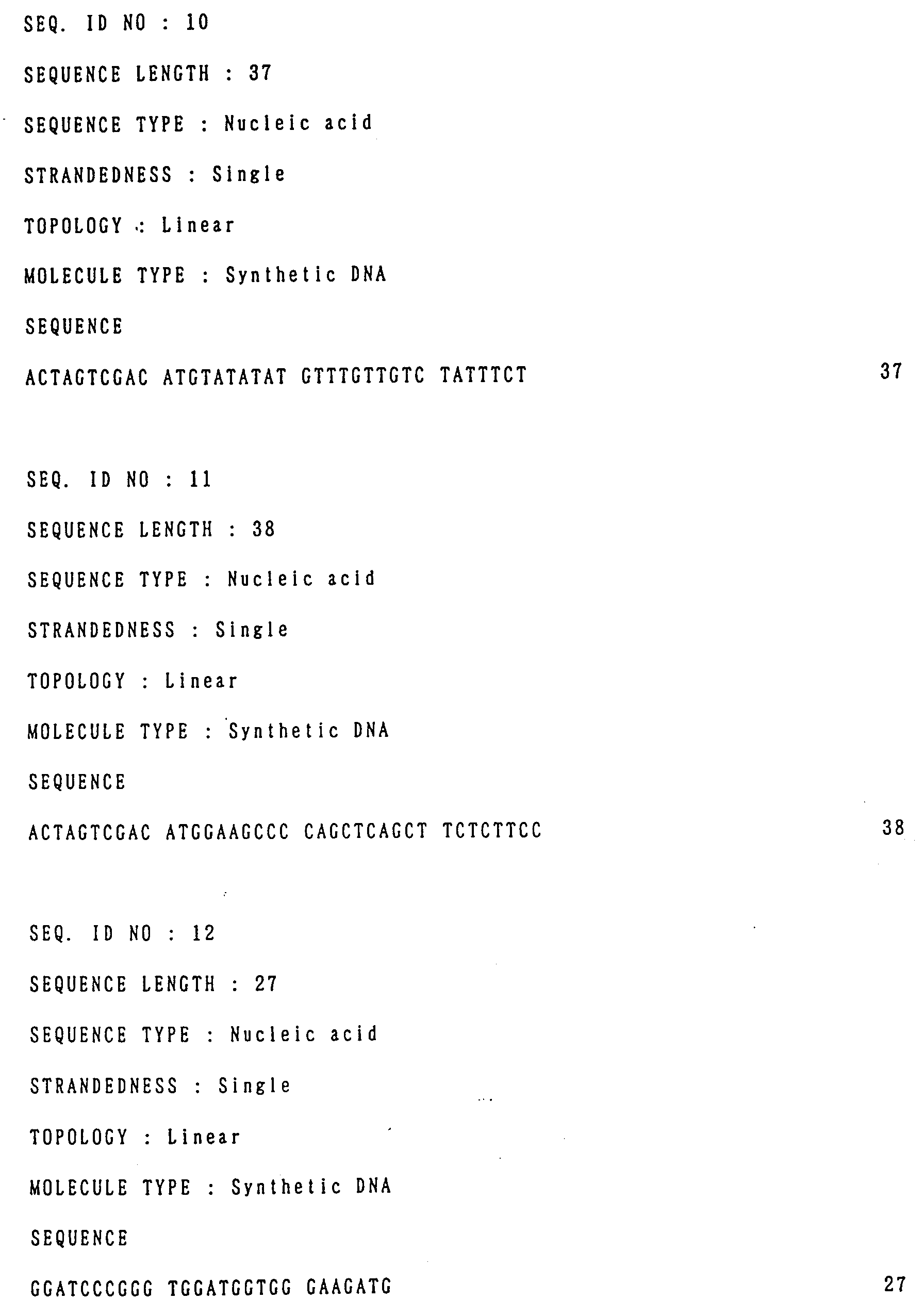


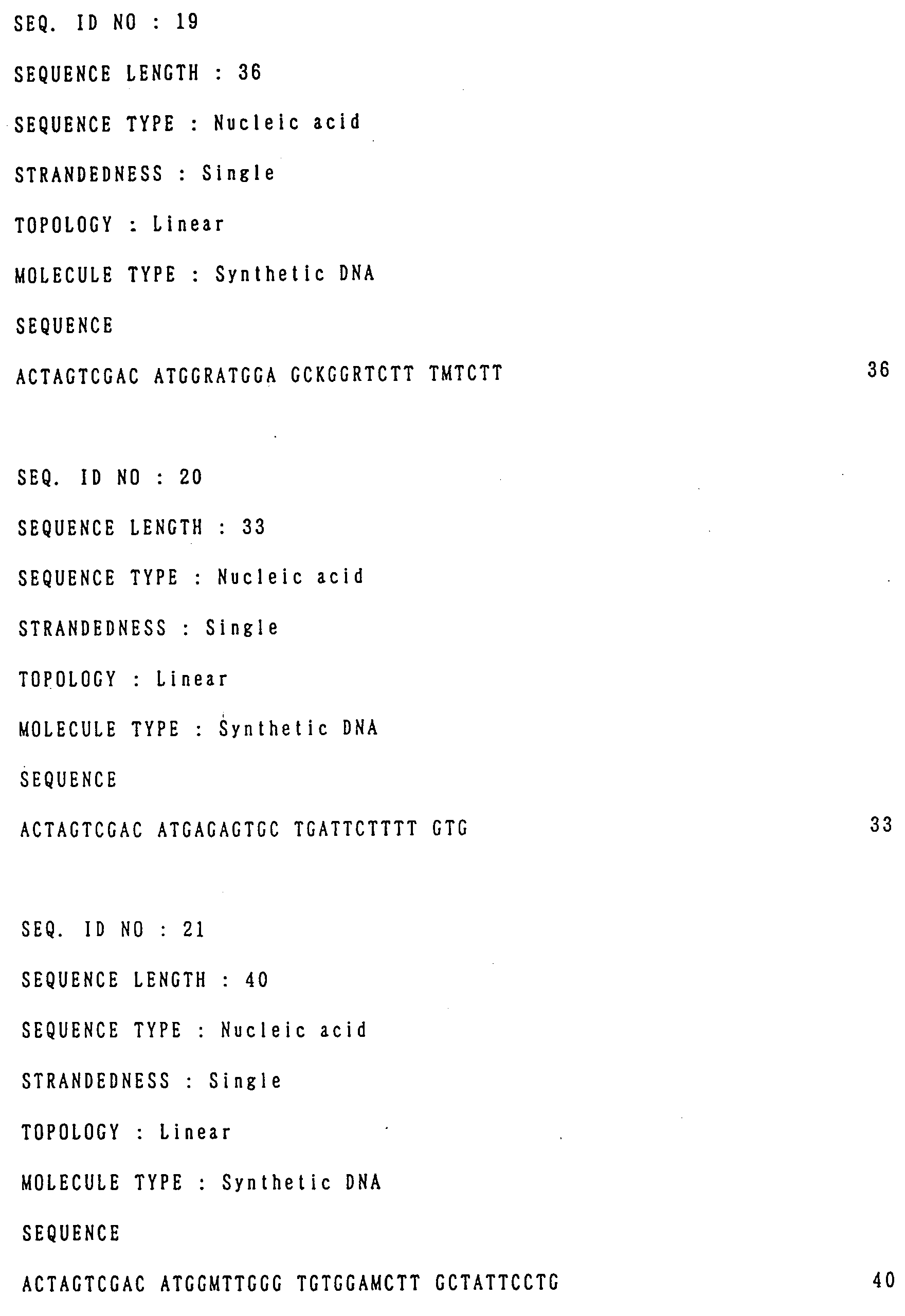


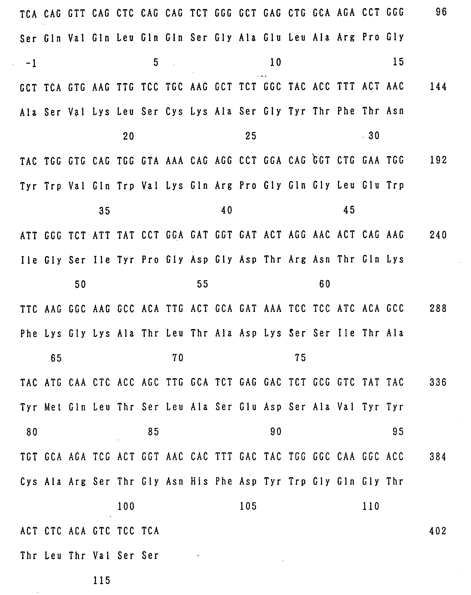


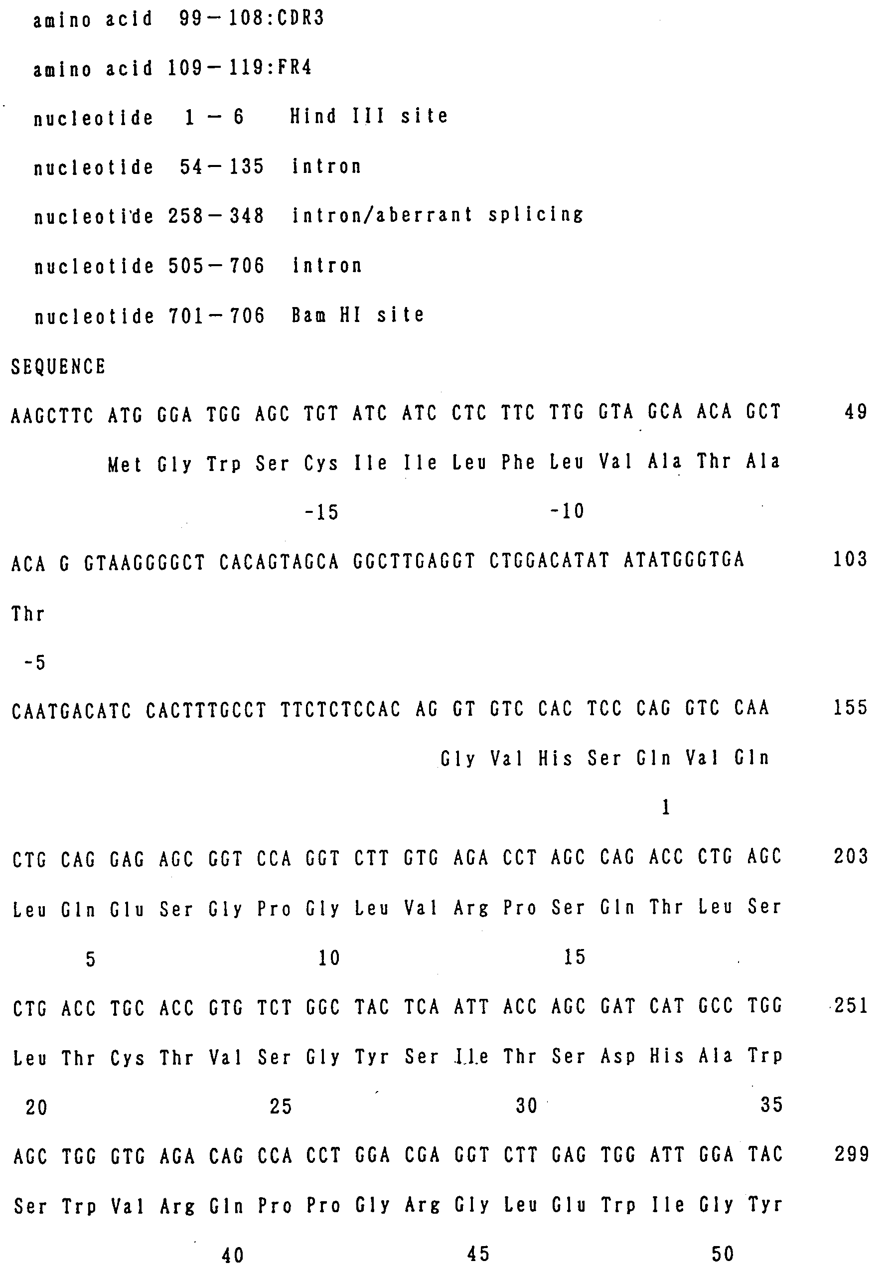


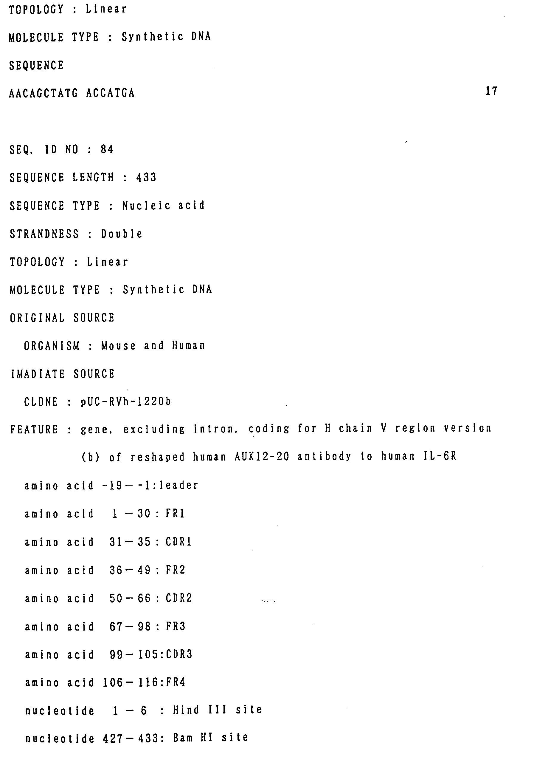


Claims (66)
- A light chain (L chain) variable region (V region) of mouse monoclonal antibody to the human interleukin-6 receptor (IL-6R).
- An L chain V region according to claim 1, having an amino acid sequence shown in any one of SEQ ID NOs: 24, 26, 28 and 30.
- A heavy chain (H chain) V region of a mouse monoclonal antibody to the human IL-6R.
- An H chain V region according to claim 3, having an amino acid sequence shown in SEQ ID NOs: 25, 27, 29 and 31.
- A chimeric antibody to the human IL-6R, comprising:(1) an L chain comprising a human L chain constant region (C region) and an L chain V region of a mouse monoclonal antibody to the human IL-6R; and(2) an H chain comprising a human H chain C region and an H chain V region of a mouse monoclonal antibody to the human IL-6R.
- A chimeric antibody according to claim 5, wherein the mouse L chain V region has an amino acid sequence shown in any one of SEQ ID NOs: 24, 26, 28 and 30; and the mouse H chain V region has an amino acid sequence shown in any one of SEQ ID NOs: 25, 27, 29 and 31.
- Complementarity determining regions (CDRs) of an L chain V region of a mouse monoclonal antibody to the human IL-6R.
- CDR according to claim 7, having amino acid sequence shown in any one of SEQ ID NOs: 24, 26, 28 and 30 wherein the stretch of the amino acid sequence is defined in Table 9.
- CDR of an H chain V region of a mouse monoclonal antibody to the human IL-6R.
- CDR according to claim 9, having amino acid sequence shown in any one of SEQ ID NOs: 25, 27, 29, and 31 wherein the stretch of the amino acid sequence is defined in Table 9.
- A reshaped human L chain V region of an antibody to the human IL-6R, comprising:(1) framework regions (FRs) of a human L chain V region, and(2) CDRs of an L chain V region of a mouse monoclonal antibody to the human IL-6R.
- A reshaped human L chain V region according to claim 11, wherein the CDRs have amino acid sequences shown in any one of SEQ ID Nos.: 24, 26, 28 and 30 wherein the stretches of the amino acid sequences are defined in Table 9.
- A reshaped human L chain V region according to claim 11, wherein the FRs are derived from the human antibody REI.
- A reshaped human L chain V region according to claim 11, having an amino acid sequence shown as RVLa or RVLb in Table 2.
- A reshaped human L chain V region according to claim 11, having an amino acid sequence shown as RVL in Table 5.
- A reshaped human H chain V region of an antibody to the human IL-6R, comprising:(1) FRs of a human H chain V region, and(2) CDRs of an H chain V region of a mouse monoclonal antibody to the human IL-6R.
- A reshaped human H chain V region according to claim 16, wherein the CDRs have amino acid sequences shown in SEQ ID NOs: 25, 27, 29 and 31 wherein the stretches of the amino acid sequences are defined in Table 9.
- A reshaped human H chain V region according to claim 16, wherein the FRs are derived from the human antibody NEW or HAX.
- A reshaped human H chain V region according to claim 16, having an amino acid sequence shown in Table 3 as RVHa, RVHb, RVHc, RVHd, RVHe or RVHf.
- A reshaped human H chain V region according to claim 17, having an amino acid sequence shown as RVHa, RVHb, RVHc or RVHd in Table 7.
- An L chain of a reshaped human antibody to human IL-6R comprising:(1) a human L chain C region; and(2) an L chain V region comprising human L chain FRs and L chain CDRs of mouse monoclonal antibody to human IL-6R.
- A reshaped human antibody L chain according to claim 21, wherein the human L chain C region is a human γ-1C region, the human L chain FRs are derived from REI, and the L chain CDRs have amino acid sequences shown in SEQ ID Nos. 24, 26, 28 and 30 wherein the streches of the amino acid sequences are defined in Table 9.
- A reshaped human antibody L chain according to claim 21, wehrein the L chain V region has an amino acid sequence shown as RVLa or RVLb in Table 2.
- A reshaped human antibody L chain according to claim 21, wherein the L chain V region has an amino acid sequence shown as RVL in Table 5.
- An H chain of a reshaped human antibody to human IL-6R comprising:(1) a human H chain C region, and(2) an H chain V region comprising human H chain FRs, and H chain CRDs of mouse monoclonal antibody to human IL-6.
- A reshaped human antibody H chain according to claim 25, wherein the human H chain C region is human κc region, the human H chain FRs are derived from NEW or HAX, the H chain CDRs have amino acid sequences shown in SEQ ID NOs: 25, 27, 29 or 31 wherein the streckes of the amino acid sequences are defined in Table 9.
- A reshaped human antibody H chain according to claim 25, wherein the H chain V region has an amino acid sequence shown as RVHa, RVHb, RVHc or RVHd in Table 3.
- A reshaped human antibody H chain according claim 25, wherein the H chain V region has an amino acid sequence shown as RVHa, RVHb, RVHc or RVHd in Table 6, or RVHa, RVHb, RVHc or RVHc in Table 7.
- A reshaped antibody to the human IL-6R, comprising:(A) an L chain comprising,(1) a human L chain C region, and(2) an L chain V region comprising human L chain FRs, and L chain CDRs of a mouse monoclonal antibody to the human IL-6R; and(B) an H chain comprising,(1) a human H chain C region, and(2) an H chain V region comprising human H chain FRs, and H chain CDRs of a mouse monoclonal antibody to the human IL-6R.
- A reshaped human antibody according to claim 29, wherein the L chain CDRs have amino acid sequences shown in any one of SEQ ID NOs: 24, 26, 28 and 30 wherein the stretches of the amino acid sequences are defined in Table 9; the H chain CDRs have amino acid sequence shown in any one of SEQ ID NOs: 25, 27, 29 and 31 wherein the stretches of the amino acid sequences are defined in Table 9; the human L chain C region and human L chain FRs are derived from the REI; and the human H chain C region and human FRs are derived from the NEW or HAX.
- A reshaped human antibody according to claim 29, wherein the L chain V region has an amino acid sequence shown as RVLa or RVLb in Table 2.
- A reshaped human antibody according to claim 29, wherein the L chain V region has an amino acid sequence shown as RVL in Table 5.
- A reshaped human antibody according to claim 29, wherein the H chain V region has an amino acid sequence shown in Table 3 as RVHa, RVHb, RVHc, RVHd, RVHe or RVHf.
- A reshaped human antibody according to claim 29, wherein the H chain V region has an amino acid sequence shown as RVHa, RVHb, RVHc or RVHd in Table 6, or RVHa, RVHb, RVHd in Table 7.
- A DNA coding for an L chain V region of a mouse monoclonal antibody to the human IL-6R.
- A DNA according to claim 35, wherein the L chain V region has an amino acid sequence shown in any one of SEQ ID NOs: 24, 26, 28 and 30.
- A DNA coding for an H chain V region of a mouse monoclonal antibody to the human IL-6R.
- A DNA according to claim 37, wherein the H chain V region has an amino acid sequence shown in any one of SEQ ID NOs: 25, 27, 29 and 31.
- A DNA coding for CDR of an L chain V region of a mouse monoclonal antibody to the human IL-6R.
- A DNA coding for CDR according to claim 39, wherein the CDR has an amino acid sequence in any one of SEQ ID NOs: 24, 26, 28 and 30 wherein the stretch of the amino acid sequence is defined in Table 9.
- A DNA coding for CDR of an H chain V region of a mouse monoclonal antibody to the human IL-6R.
- A DNA coding for CDR according to claim 41, wherein the CDR has an amino acid sequence shown in any one of SEQ ID NOs: 25, 27, 29 and 31 wherein the stretch of the amino acid sequence in defined in Table 9.
- A DNA coding for a reshaped human L chain V region of an antibody to the human IL-6R, wherein the reshaped human L chain V region comprises:(1) FRs of a human L chain V region, and(2) CDRs of a mouse L chain V region of a monoclonal antibody to the human IL-6R.
- A DNA coding for a reshaped human L chain V region according to claim 43, wherein the CDRs have amino acid sequences shown in any one of SEQ ID NOs: 24, 26, 28 and 30 wherein the stretches of the amino acid sequences are defined in Table 9.
- A DNA coding for a reshaped human L chain V region according to claim 43, wherein the FRs are derived from the REI.
- A DNA according to claim 43, wherein the L chain V region has an amino acid sequence shown as RVLa or RVLb in Table 2.
- A DNA according to claim 43, wehrein the L chain V region has an amino acid region shown as RVL in Table 5.
- A DNA according to claim 43, having a nucleotide sequence shown in SEQ ID No: 57.
- A DNA coding for a reshaped human H chain V region of an antibody to the human IL-6R, wherein the reshaped Human V region comprises:(1) FRs of a human H chain V region, and(2) CDRs of an H chain V region of a mouse monoclonal antibody to the human IL-6R.
- A DNA coding for a reshaped human H chain V region according to claim 49, wherein the CDRs have amino acid sequences shown in SEQ ID NOs: 25, 27, 29 and 31 wherein the stretches of the amino acid sequences are defined in Table 9.
- A DNA coding for a reshaped human H chain V region according to claim 49, wherein the FRs are derived from the NEW or HAX.
- A DNA coding for a reshaped human H chain V region according to claim 49, wherein the H chain V region has an amino acid sequence shown as RVHa, RVHb, RVHc, RVHd, RVHe or RVHf in Table 3.
- A DNA according to claim 49, wherein the H chain V region has an amino acid sequence shown as RVLa, RVHb, RVHc or RVHd in Table 6, or RVHa, RVHb, RVHc or RVHd in Table 7.
- A DNA according to claim 49, having a nucleotide sequence shown in SEQ ID NO: 56.
- A DNA coding for a reshaped human L chain of an antibody to the human IL-6R, wherein the reshaped human L chain comprises:(1) a human L chain C region; and(2) an L chain V region comprising a human FRs, and CDRs of a mouse monoclonal antibody to the human IL-6R.
- A DNA according to claim 55, wherein the L chain V region has the nucleotide sequence shown in SEQ ID NO: 57.
- A DNA coding for a reshaped human H chain of an antibody to the human IL-6R, wherein the reshaped human H chain comprises:(1) a human H chain C region, and(2) a H chain V region comprising human FRs, and CDRs of a mouse monoclonal antibody to the human IL-6R.
- A DNA according to claim 57, wherein the H chain V region has the nucleotide sequence shown in SEQ ID NO: 56.
- A vector comprising a DNA according to any one of claims 35, 37, 39, 41, 43, 49, 55 and 57.
- A host cell transformed or transfected with a vector comprising a DNA according to any one of claims 35, 37, 39, 41, 43, 49, 55 and 57.
- A DNA coding for a chimeric L chain of an antibody to the human IL-6R, wherein the chimeric L chain comprises:(1) a human L chain C region; and(2) an L chain V region of a mouse monoclonal antibody to the human IL-6R.
- A DNA coding for a chimeric H chain of an antibody to the human IL-6R wherein the chimeric H chain comprises:(1) a human H chain C region; and(2) an H chain V region of a mouse monoclonal antibody to the human IL-6R.
- A process for production of a chimeric antibody to the human IL-6R, comprising the steps of:
culturing host cells cotransfected with an expression vector comprising a DNA according to claim 61 and with an expression vector comprising a DNA according to claim 62; and
recovering a desired antibody. - A process for production of a reshaped human antibody to the human IL-6R, comprising the steps of:
culturing host cells cotransfected with an expression vector comprising a DNA according to claim 55 and with an expression vector comprising a DNA according to claim 57; and recovering desired antibody. - A DNA according to claim 49, having a nucleotide sequence shown in SEQ ID NO: 85, 86 or 94.
- A DNA according to claim 44, having a nucleotide sequence shown in SEQ IN NO: 71.
Applications Claiming Priority (7)
| Application Number | Priority Date | Filing Date | Title |
|---|---|---|---|
| JP3095476A JPH05236966A (en) | 1991-04-25 | 1991-04-25 | DNA encoding a variable region of a mouse monoclonal antibody against human interleukin-6 receptor |
| JP9547691 | 1991-04-25 | ||
| JP95476/91 | 1991-04-25 | ||
| JP4032084A JPH05227970A (en) | 1992-02-19 | 1992-02-19 | Reconstituted human antibody against human interleukin-6 receptor |
| JP3208492 | 1992-02-19 | ||
| JP32084/92 | 1992-02-19 | ||
| PCT/JP1992/000544 WO1992019759A1 (en) | 1991-04-25 | 1992-04-24 | Reconstituted human antibody against human interleukin 6 receptor |
Publications (3)
| Publication Number | Publication Date |
|---|---|
| EP0628639A1 true EP0628639A1 (en) | 1994-12-14 |
| EP0628639A4 EP0628639A4 (en) | 1994-12-28 |
| EP0628639B1 EP0628639B1 (en) | 1999-06-23 |
Family
ID=26370596
Family Applications (1)
| Application Number | Title | Priority Date | Filing Date |
|---|---|---|---|
| EP92909376A Expired - Lifetime EP0628639B1 (en) | 1991-04-25 | 1992-04-24 | Reconstituted human antibody against human interleukin 6 receptor |
Country Status (16)
| Country | Link |
|---|---|
| US (3) | US5795965A (en) |
| EP (1) | EP0628639B1 (en) |
| JP (6) | JP3370324B2 (en) |
| KR (1) | KR100249937B1 (en) |
| AT (1) | ATE181575T1 (en) |
| AU (2) | AU668349B2 (en) |
| DE (2) | DE122009000019I1 (en) |
| DK (1) | DK0628639T3 (en) |
| ES (1) | ES2134212T3 (en) |
| GR (1) | GR3031174T3 (en) |
| HU (1) | HU218140B (en) |
| LU (1) | LU91556I2 (en) |
| NL (1) | NL300383I2 (en) |
| RU (1) | RU2139351C1 (en) |
| TW (1) | TW205553B (en) |
| WO (1) | WO1992019759A1 (en) |
Cited By (62)
| Publication number | Priority date | Publication date | Assignee | Title |
|---|---|---|---|---|
| EP0783893A4 (en) * | 1994-10-07 | 1998-04-22 | Chugai Pharmaceutical Co Ltd | Rheumatoid arthritis remedy containing il-6 antagonist as active ingredient |
| US6121423A (en) * | 1993-05-31 | 2000-09-19 | Chugai Seiyaku Kabushiki Kaisha | Reshaped human antibody to human interleukin-6 |
| EP0997152A4 (en) * | 1997-02-12 | 2001-04-18 | Chugai Pharmaceutical Co Ltd | REMEDIES FOR LYMPHOCITARY TUMORS |
| EP0791359A4 (en) * | 1994-10-21 | 2002-09-11 | Chugai Pharmaceutical Co Ltd | Remedy for diseases caused by il-6 production |
| EP0811384A4 (en) * | 1995-02-13 | 2002-09-18 | Chugai Pharmaceutical Co Ltd | MUSCLE PROTEIN DECOMPOSITION INHIBITOR CONTAINING AN INTERLEUKIN-6 RECEPTOR ANTIBODY |
| WO2003068259A1 (en) * | 2002-02-14 | 2003-08-21 | Chugai Seiyaku Kabushiki Kaisha | Antibody-containing solution pharmaceuticals |
| US6699974B2 (en) | 1996-10-04 | 2004-03-02 | Chugai Seiyaku Kabushiki Kaisha | Reshaped human anti-HM 1.24 antibody |
| EP1513879A4 (en) * | 2002-06-03 | 2008-06-11 | Genentech Inc | PHAGE LIBRARIES AND SYNTHETIC ANTIBODIES |
| EP2057191A1 (en) * | 2006-08-18 | 2009-05-13 | Ablynx N.V. | Amino acid sequences directed against il-6r and polypeptides comprising the same for the treatment of deseases and disorders associated with il-6-mediated signalling |
| US7566453B2 (en) | 1996-06-27 | 2009-07-28 | Chugai Seiyaku Kabushiki Kaisha | Methods for treating melanomas |
| EP1728801A4 (en) * | 2004-03-24 | 2009-10-21 | Chugai Pharmaceutical Co Ltd | HUMANIZED ANTIBODY SUBTYPE AGAINST AN INTERLEUKIN-6 RECEPTOR |
| US20100129355A1 (en) * | 2007-07-26 | 2010-05-27 | Osaka University | Therapeutic agents for ocular inflammatory disease comprising interleukin 6 receptor inhibitor as active ingredient |
| US7785903B2 (en) | 2004-04-09 | 2010-08-31 | Genentech, Inc. | Variable domain library and uses |
| US7824674B2 (en) | 1998-03-17 | 2010-11-02 | Chugai Seiyaku Kabushiki Kaisha | Preventive or therapeutic agent for inflammatory bowel disease comprising IL-6 antagonist as an active ingredient |
| EP2261230A1 (en) * | 2002-09-11 | 2010-12-15 | Chugai Seiyaku Kabushiki Kaisha | Protein purification method |
| WO2011051231A1 (en) | 2009-10-26 | 2011-05-05 | F. Hoffmann-La Roche Ag | Method for the production of a glycosylated immunoglobulin |
| US7955598B2 (en) | 2001-04-02 | 2011-06-07 | Chugai Seiyaku Kabushiki Kaisha | Therapeutic agent for chronic arthritides diseases of childhood-related diseases |
| US8017121B2 (en) | 1994-06-30 | 2011-09-13 | Chugai Seiyaku Kabushika Kaisha | Chronic rheumatoid arthritis therapy containing IL-6 antagonist as effective component |
| US8043617B2 (en) | 2006-06-02 | 2011-10-25 | Regeneron Pharmaceuticals, Inc. | Human antibodies to human IL-6 receptor |
| US8080248B2 (en) | 2006-06-02 | 2011-12-20 | Regeneron Pharmaceuticals, Inc. | Method of treating rheumatoid arthritis with an IL-6R antibody |
| WO2013081612A1 (en) * | 2011-11-30 | 2013-06-06 | Abbvie Biotherapeutics Inc. | Vectors and host cells comprising a modified sv40 promoter for protein expression |
| US8562990B2 (en) | 2000-10-25 | 2013-10-22 | Chugai Seiyaku Kabushiki Kaisha | Method of treating psoriatic arthritis with an IL-6 receptor antibody |
| US8617550B2 (en) | 2003-12-19 | 2013-12-31 | Chugai Seiyaku Kabushiki Kaisha | Treatment of vasculitis with IL-6 antagonist |
| US8632778B2 (en) | 2000-08-11 | 2014-01-21 | Chugai Seiyaku Kabushiki Kaisha | Stabilized anti-interleukin-6 antibody-containing preparations |
| US8709409B2 (en) | 2003-04-28 | 2014-04-29 | Chugai Seiyaku Kabushiki Kaisha | Method for treating rheumatoid arthritis by administering an anti-IL-6 antibody and methotrexate |
| US8748581B2 (en) | 2009-04-10 | 2014-06-10 | Ablynx N.V. | Anti-IL-6R polypeptides and pharmaceutical compositions thereof |
| US8771686B2 (en) | 2006-01-27 | 2014-07-08 | Chugai Seiyaku Kabushiki Kaisha | Methods for treating a disease involving choroidal neovascularization by administering an IL-6 receptor antibody |
| US8802092B2 (en) | 2003-10-17 | 2014-08-12 | Chugai Seiyaku Kabushiki Kaisha | Mesothelioma therapeutic agent |
| US8834876B2 (en) | 2001-02-07 | 2014-09-16 | Chugai Seiyaku Kabushiki Kaisha | Therapeutic agent for hematopoietic tumors |
| US8945558B2 (en) | 2005-10-21 | 2015-02-03 | Chugai Seiyaku Kabushiki Kaisha | Methods for treating myocardial infarction comprising administering an IL-6 inhibitor |
| US9260516B2 (en) | 2006-04-07 | 2016-02-16 | Osaka University | Method for promoting muscle regeneration by administering an antibody to the IL-6 receptor |
| EP3009518A4 (en) * | 2013-06-11 | 2017-01-04 | National Center of Neurology and Psychiatry | Method for predicting post-therapy prognosis of relapsing-remitting multiple sclerosis (rrms) patient, and method for determining applicability of novel therapy |
| US9539322B2 (en) | 2010-05-28 | 2017-01-10 | National University Corporation Hokkaido University | Method of enhancing an antitumor T cell response by administering an anti-IL-6 receptor antibody |
| US9725514B2 (en) | 2007-01-23 | 2017-08-08 | Shinshu University | Chronic rejection inhibitor |
| US9975966B2 (en) | 2014-09-26 | 2018-05-22 | Chugai Seiyaku Kabushiki Kaisha | Cytotoxicity-inducing theraputic agent |
| US10168326B2 (en) | 2013-07-04 | 2019-01-01 | F. Hoffmann-La Roche Inc. | Interference-suppressed immunoassay to detect anti-drug antibodies in serum samples |
| US10400029B2 (en) | 2011-06-28 | 2019-09-03 | Inhibrx, Lp | Serpin fusion polypeptides and methods of use thereof |
| EP3569243A1 (en) | 2011-06-28 | 2019-11-20 | Inhibrx, LP | Serpin fusion polypeptides and methods of use thereof |
| US10519232B2 (en) | 2014-01-22 | 2019-12-31 | Sorbonne Universite | Agents for use in the treatment of retinal inflammation |
| US10662245B2 (en) | 2008-09-26 | 2020-05-26 | Chugai Seiyaku Kabushiki Kaisha | Methods of reducing IL-6 activity for disease treatment |
| US10697883B2 (en) | 2015-05-19 | 2020-06-30 | National Center Of Neurology And Psychiatry | Method for determining application of therapy to multiple sclerosis (MS) patient |
| US10717781B2 (en) | 2008-06-05 | 2020-07-21 | National Cancer Center | Neuroinvasion inhibitor |
| US10774148B2 (en) | 2015-02-27 | 2020-09-15 | Chugai Seiyaku Kabushiki Kaisha | Composition for treating IL-6-related diseases |
| US10968278B2 (en) | 2015-11-03 | 2021-04-06 | Sanofi Biotechnology | Compositions comprising IL6R antibodies for the treatment of uveitis and macular edema and methods of using same |
| US11008394B2 (en) | 2007-12-27 | 2021-05-18 | Chugai Seiyaku Kabushiki Kaisha | High concentration antibody-containing liquid formulation |
| US11046784B2 (en) | 2006-03-31 | 2021-06-29 | Chugai Seiyaku Kabushiki Kaisha | Methods for controlling blood pharmacokinetics of antibodies |
| EP2202307B2 (en) † | 2007-10-15 | 2021-07-14 | Chugai Seiyaku Kabushiki Kaisha | Method for production of antibody |
| US11098127B2 (en) | 2010-01-08 | 2021-08-24 | Regeneron Pharmaceuticals, Inc. | Stabilized formulations containing anti-interleukin-6 receptor (IL-6R) antibodies |
| US11174317B2 (en) | 2015-06-04 | 2021-11-16 | National Center Of Neurology And Psychiatry | Therapeutic agent for mental illness comprising IL-6 inhibitor as active ingredient |
| US11248053B2 (en) | 2007-09-26 | 2022-02-15 | Chugai Seiyaku Kabushiki Kaisha | Method of modifying isoelectric point of antibody via amino acid substitution in CDR |
| US11285208B2 (en) | 2010-05-28 | 2022-03-29 | National Cancer Center | Therapeutic agents for pancreatic cancer |
| US11332533B2 (en) | 2007-09-26 | 2022-05-17 | Chugai Seiyaku Kabushiki Kaisha | Modified antibody constant region |
| US11359194B2 (en) | 2008-04-11 | 2022-06-14 | Chugai Seiyaku Kabushiki Kaisha | Antigen-binding molecule capable of binding two or more antigen molecules repeatedly |
| US11498969B2 (en) | 2019-01-31 | 2022-11-15 | Sanofi Biotechnology | Compositions and methods for treating juvenile idiopathic arthritis |
| US11612562B2 (en) | 2010-01-20 | 2023-03-28 | Chugai Seiyaku Kabushiki Kaisha | Solution preparation containing stabilized antibody |
| US11851486B2 (en) | 2017-05-02 | 2023-12-26 | National Center Of Neurology And Psychiatry | Method for predicting and evaluating therapeutic effect in diseases related to IL-6 and neutrophils |
| US11891434B2 (en) | 2010-11-30 | 2024-02-06 | Chugai Seiyaku Kabushiki Kaisha | Antigen-binding molecule capable of binding to plurality of antigen molecules repeatedly |
| US12297279B2 (en) | 2019-06-04 | 2025-05-13 | Sanofi Biotechnology | Compositions and methods for treating pain in subjects with rheumatoid arthritis |
| US12440561B2 (en) | 2019-04-24 | 2025-10-14 | Sanofi Biotechnology | Method of diagnosis and treatment of rheumatoid arthritis |
| US12558423B2 (en) | 2020-05-29 | 2026-02-24 | Chugai Seiyaku Kabushiki Kaisha | Antibody-containing formulation |
| US12583918B2 (en) | 2019-04-17 | 2026-03-24 | Tokyo Women's Medical University | Therapeutic agent for urological cancer which is characterized by being administered with IL-6 inhibitor and CCR2 inhibitor in combination |
| US12600772B2 (en) | 2018-01-31 | 2026-04-14 | Motokazu Kato | Therapeutic agent for asthma containing IL-6 inhibitor |
Families Citing this family (279)
| Publication number | Priority date | Publication date | Assignee | Title |
|---|---|---|---|---|
| US5472693A (en) * | 1993-02-16 | 1995-12-05 | The Dow Chemical Company | Family of anti-carcinoembryonic antigen chimeric antibodies |
| KR100304333B1 (en) * | 1993-08-24 | 2001-11-30 | 안도 고끼 | Recombinant humanized anti-human immunodeficiency virus antibody |
| US5914110A (en) * | 1993-09-07 | 1999-06-22 | Smithkline Beecham Corporation | Recombinant IL4 antibodies useful in treatment of IL4 mediated disorders |
| AU7949394A (en) * | 1993-11-19 | 1995-06-06 | Chugai Seiyaku Kabushiki Kaisha | Reconstituted human antibody against human medulloblastomatous cell |
| WO1995029690A1 (en) * | 1994-04-29 | 1995-11-09 | The Trustees Of The University Of Pennsylvania | Biologically active peptides and methods of identifying the same |
| CZ106299A3 (en) | 1996-09-26 | 1999-08-11 | Chugai Seiyaku Kabushiki Kaisha | Antibodies to human parathyroid hormone related protein |
| ES2312184T3 (en) | 1997-03-21 | 2009-02-16 | Chugai Seiyaku Kabushiki Kaisha | THERAPEUTIC PREVENTIVE AGENTS FOR THE TREATMENT OF MULTIPLE SCLEROSIS, CONTAINING ANTI-RECEIVING ANTIBODIES OF IL-6 ANTAGONISTS. |
| TR199902800T2 (en) | 1997-05-15 | 2000-04-21 | Chugai Seiyaku Kabushiki Kaisha | A substance that treats cacaxia. |
| KR20100068498A (en) * | 1997-08-15 | 2010-06-23 | 츄가이 세이야꾸 가부시키가이샤 | Preventives and/or remedies for systemic lupus erythematosus containing anti-il-6 receptor antibody as the active ingredient |
| US20020187150A1 (en) * | 1997-08-15 | 2002-12-12 | Chugai Seiyaku Kabushiki Kaisha | Preventive and/or therapeutic agent for systemic lupus erythematosus comprising anti-IL-6 receptor antibody as an active ingredient |
| TR200002885T2 (en) | 1998-04-03 | 2000-12-21 | Chugai Seiyaku Kabushiki Kaisha | Humanized antibody against human tissue factor (TF) |
| ES2276525T3 (en) | 1998-08-24 | 2007-06-16 | Chugai Seiyaku Kabushiki Kaisha | PREVENTIVES OR REMEDIES FOR PANCREATITIS CONTAINING ANTI-RECEPTOR ANTIBODIES IL-6 AS ACTIVE INGREDIENT. |
| JP4813660B2 (en) | 1998-11-27 | 2011-11-09 | ダーウィン ディスカバリー リミテッド | Compositions and methods for increasing bone mineralization |
| US7696325B2 (en) | 1999-03-10 | 2010-04-13 | Chugai Seiyaku Kabushiki Kaisha | Polypeptide inducing apoptosis |
| WO2001016166A2 (en) * | 1999-08-27 | 2001-03-08 | The United States Of America, Represented By The Secretary, Department Of Health And Human Services | Polypeptides, comprising il-6 ligand-binding receptor domains, and related nucleic acids, antibodies, compositions, and methods of use |
| DK1222854T3 (en) | 1999-10-01 | 2011-03-14 | Chugai Pharmaceutical Co Ltd | Prevention and treatment of diseases associated with blood coagulation |
| GB0002666D0 (en) | 2000-02-04 | 2000-03-29 | Univ London | Blockade of voltage dependent sodium channels |
| ATE420661T1 (en) | 2000-04-28 | 2009-01-15 | Chugai Pharmaceutical Co Ltd | CELL PROLIFERATION INHIBITORS |
| AU2002224417A1 (en) * | 2000-10-18 | 2002-04-29 | Immunex Corporation | Methods for treating il-18 mediated disorders |
| CN1308448C (en) | 2000-10-20 | 2007-04-04 | 中外制药株式会社 | Low molecular weight TPO agonist antibody |
| AU1091802A (en) | 2000-10-20 | 2002-04-29 | Chugai Pharmaceutical Co Ltd | Degraded agonist antibody |
| JP4261907B2 (en) | 2000-10-20 | 2009-05-13 | 中外製薬株式会社 | Low molecular weight agonist antibody |
| WO2002036165A1 (en) * | 2000-10-27 | 2002-05-10 | Chugai Seiyaku Kabushiki Kaisha | Blood mmp-3 level-lowering agent containing il-6 antgonist as the active ingredient |
| WO2002036164A1 (en) * | 2000-10-27 | 2002-05-10 | Chugai Seiyaku Kabushiki Kaisha | Blooe vegf level-lowering agent containing il-6 antagonist as the active ingredient |
| WO2002098370A2 (en) * | 2001-03-02 | 2002-12-12 | Medimmune, Inc. | Methods of administering/dosing cd2 antagonists for the prevention and treatment of autoimmune disorders or inflammatory disorders |
| EP3088412B1 (en) | 2001-03-09 | 2021-05-05 | Chugai Seiyaku Kabushiki Kaisha | Protein purification method |
| EP1382964A4 (en) | 2001-04-17 | 2009-06-10 | Chugai Pharmaceutical Co Ltd | METHOD FOR QUANTIFYING SURFACTANT |
| US20100056762A1 (en) * | 2001-05-11 | 2010-03-04 | Old Lloyd J | Specific binding proteins and uses thereof |
| US7589180B2 (en) * | 2001-05-11 | 2009-09-15 | Abbott Laboratories Inc. | Specific binding proteins and uses thereof |
| GB0208041D0 (en) * | 2002-04-08 | 2002-05-22 | Lonza Biologics Plc | Method of culturing animal cells |
| WO2004022597A1 (en) | 2002-09-04 | 2004-03-18 | Chugai Seiyaku Kabushiki Kaisha | Antibody against blood-solubilized n-terminal peptide in gpc3 |
| EP1498137A1 (en) * | 2002-04-25 | 2005-01-19 | Chugai Seiyaku Kabushiki Kaisha | Remedy for lung cancer |
| AU2003257536A1 (en) | 2002-08-27 | 2004-03-19 | Chugai Seiyaku Kabushiki Kaisha | Method of stabilizing protein solution preparation |
| CA2516945A1 (en) * | 2003-02-24 | 2004-09-02 | Chugai Seiyaku Kabushiki Kaisha | Therapeutic agent for spinal cord injury comprising interleukin-6 antagonist |
| JP2004279086A (en) | 2003-03-13 | 2004-10-07 | Konica Minolta Holdings Inc | Radiation image conversion panel and method for manufacturing it |
| US7381409B2 (en) | 2003-06-16 | 2008-06-03 | Celltech R&D, Inc. | Antibodies specific for sclerostin and methods of screening and use therefor |
| EP2204190A1 (en) | 2003-07-15 | 2010-07-07 | Chugai Seiyaku Kabushiki Kaisha | IgM production by transformed cells and methods for quantifying said IgM production |
| EP1707629B1 (en) | 2003-12-03 | 2011-06-08 | Chugai Seiyaku Kabushiki Kaisha | Expression system using mammalian beta-actin promoter |
| DE602005020743D1 (en) * | 2004-02-11 | 2010-06-02 | Warner Lambert Co | PROCESS FOR TREATING OSTEOARTHRITIS WITH ANTI-IL-6 ANTIBODIES |
| AU2005214988A1 (en) * | 2004-02-17 | 2005-09-01 | Absalus, Inc. | Super-humanized antibodies against respiratory syncytial virus |
| US7767792B2 (en) * | 2004-02-20 | 2010-08-03 | Ludwig Institute For Cancer Research Ltd. | Antibodies to EGF receptor epitope peptides |
| AR048335A1 (en) * | 2004-03-24 | 2006-04-19 | Chugai Pharmaceutical Co Ltd | THERAPEUTIC AGENTS FOR INTERNAL EAR DISORDERS CONTAINING AN IL-6 ANTAGONIST AS AN ACTIVE INGREDIENT |
| GB0410627D0 (en) * | 2004-05-12 | 2004-06-16 | Scancell Ltd | Specific binding members |
| EP1810980A1 (en) * | 2004-10-28 | 2007-07-25 | Osaka University | Interleukin-6 inhibitors |
| JP4818936B2 (en) | 2005-01-05 | 2011-11-16 | 中外製薬株式会社 | Cell culture method and use thereof |
| JPWO2006090930A1 (en) * | 2005-02-28 | 2008-07-24 | エーザイ・アール・アンド・ディー・マネジメント株式会社 | New combination of sulfonamide compounds |
| CN101198698B (en) | 2005-03-31 | 2014-03-19 | 中外制药株式会社 | Process for production of polypeptide by regulation of assembly |
| EP1870458B1 (en) | 2005-03-31 | 2018-05-09 | Chugai Seiyaku Kabushiki Kaisha | sc(Fv)2 STRUCTURAL ISOMERS |
| PA8672101A1 (en) * | 2005-04-29 | 2006-12-07 | Centocor Inc | ANTI-IL-6 ANTIBODIES, COMPOSITIONS, METHODS AND USES |
| US7592429B2 (en) * | 2005-05-03 | 2009-09-22 | Ucb Sa | Sclerostin-binding antibody |
| US8003108B2 (en) | 2005-05-03 | 2011-08-23 | Amgen Inc. | Sclerostin epitopes |
| CN101237890A (en) | 2005-06-10 | 2008-08-06 | 中外制药株式会社 | Stabilizer and Utilization of Protein Preparations Containing Meglumine |
| AU2006256030B2 (en) | 2005-06-10 | 2012-06-21 | Chugai Seiyaku Kabushiki Kaisha | Pharmaceutical compositions containing sc(Fv)2 |
| GB2429013C (en) * | 2005-08-11 | 2012-11-28 | Arpi Matossian-Rogers | Peptides for treatment and diagnosis of autoimmunedisease |
| AU2006300234B2 (en) | 2005-10-14 | 2013-01-10 | Chugai Seiyaku Kabushiki Kaisha | Agents for suppressing damage to transplanted islets after islet transplantation |
| EP1957531B1 (en) | 2005-11-07 | 2016-04-13 | Genentech, Inc. | Binding polypeptides with diversified and consensus vh/vl hypervariable sequences |
| WO2007055378A1 (en) | 2005-11-14 | 2007-05-18 | Cell Signals Inc. | Method for treatment or prevention of disease associated with functional disorder of regulatory t cell |
| AR057582A1 (en) * | 2005-11-15 | 2007-12-05 | Nat Hospital Organization | AGENTS TO DELETE INDUCTION OF CYTOTOXIC T LYMPHOCYTES |
| WO2007061029A1 (en) | 2005-11-25 | 2007-05-31 | Keio University | Therapeutic agent for prostate cancer |
| JP5231810B2 (en) | 2005-12-28 | 2013-07-10 | 中外製薬株式会社 | Antibody-containing stabilized preparation |
| EP1997505A4 (en) | 2006-03-22 | 2013-02-13 | Chugai Pharmaceutical Co Ltd | PREPARATION OF ERYTHROPOIETIN SOLUTION |
| EP2009101B1 (en) | 2006-03-31 | 2017-10-25 | Chugai Seiyaku Kabushiki Kaisha | Antibody modification method for purifying bispecific antibody |
| US8741643B2 (en) | 2006-04-28 | 2014-06-03 | Lifescan, Inc. | Differentiation of pluripotent stem cells to definitive endoderm lineage |
| AU2007267213B2 (en) * | 2006-05-25 | 2012-03-29 | Glaxo Group Limited | Modified humanised anti-interleukin-18 antibodies |
| WO2007145840A2 (en) * | 2006-06-13 | 2007-12-21 | Oncomed Pharmaceuticals, Inc. | Compositions and methods for diagnosing and treating cancer |
| EP2044123B1 (en) | 2006-06-21 | 2013-03-13 | Oncotherapy Science, Inc. | Tumor-targeting monoclonal antibodies to fzd10 and uses thereof |
| US20080081041A1 (en) * | 2006-09-29 | 2008-04-03 | Jeffrey Nemeth | Method of Using IL6 Antagonists with Mitoxantrone for Prostate Cancer |
| AU2008209482B2 (en) | 2007-01-24 | 2014-05-01 | Oncomed Pharmaceuticals, Inc. | Compositions and methods for diagnosing and treating cancer |
| EP2126127B1 (en) | 2007-01-25 | 2016-09-28 | Dana-Farber Cancer Institute, Inc. | Use of anti-egfr antibodies in treatment of egfr mutant mediated disease |
| CL2008000719A1 (en) | 2007-03-12 | 2008-09-05 | Univ Tokushima Chugai Seiyaku | THERAPEUTIC AGENT FOR CANCER RESISTANT TO CHEMOTHERAPEUTIC AGENTS THAT UNDERSTAND AN ANTIBODY THAT RECOGNIZES IT CLASS I AS ACTIVE INGREDIENT; PHARMACEUTICAL COMPOSITION THAT INCLUDES SUCH ANTIBODY; AND METHOD TO TREAT CANCER RESISTANT TO |
| US8178101B2 (en) | 2007-05-21 | 2012-05-15 | Alderbio Holdings Inc. | Use of anti-IL-6 antibodies having specific binding properties to treat cachexia |
| KR20100028571A (en) * | 2007-05-21 | 2010-03-12 | 앨더 바이오파마슈티컬즈, 인코포레이티드 | Novel rabbit antibody humanization methods and humanized rabbit antibodies |
| US8252286B2 (en) | 2007-05-21 | 2012-08-28 | Alderbio Holdings Llc | Antagonists of IL-6 to prevent or treat thrombosis |
| US8062864B2 (en) | 2007-05-21 | 2011-11-22 | Alderbio Holdings Llc | Nucleic acids encoding antibodies to IL-6, and recombinant production of anti-IL-6 antibodies |
| US20090238825A1 (en) * | 2007-05-21 | 2009-09-24 | Kovacevich Brian R | Novel rabbit antibody humanization methods and humanized rabbit antibodies |
| US7906117B2 (en) * | 2007-05-21 | 2011-03-15 | Alderbio Holdings Llc | Antagonists of IL-6 to prevent or treat cachexia, weakness, fatigue, and/or fever |
| US9701747B2 (en) | 2007-05-21 | 2017-07-11 | Alderbio Holdings Llc | Method of improving patient survivability and quality of life by anti-IL-6 antibody administration |
| CN101754772B (en) | 2007-05-21 | 2015-08-19 | 奥尔德生物控股有限责任公司 | Antibodies against IL-6 and uses thereof |
| US8404235B2 (en) * | 2007-05-21 | 2013-03-26 | Alderbio Holdings Llc | Antagonists of IL-6 to raise albumin and/or lower CRP |
| JP5142265B2 (en) * | 2007-05-28 | 2013-02-13 | 独立行政法人産業技術総合研究所 | Paratopes and epitopes of anti-mortalin antibodies |
| US9080145B2 (en) | 2007-07-01 | 2015-07-14 | Lifescan Corporation | Single pluripotent stem cell culture |
| US9096832B2 (en) | 2007-07-31 | 2015-08-04 | Lifescan, Inc. | Differentiation of human embryonic stem cells |
| US9283276B2 (en) | 2007-08-14 | 2016-03-15 | Ludwig Institute For Cancer Research Ltd. | Monoclonal antibody 175 targeting the EGF receptor and derivatives and uses thereof |
| CL2008002775A1 (en) | 2007-09-17 | 2008-11-07 | Amgen Inc | Use of a sclerostin binding agent to inhibit bone resorption. |
| CN101939425B (en) | 2007-09-26 | 2014-05-14 | 中外制药株式会社 | Anti-IL-6 receptor antibody |
| AU2008308163B2 (en) | 2007-10-02 | 2013-03-21 | Chugai Seiyaku Kabushiki Kaisha | Therapeutic agents for graft-versus-host disease comprising interleukin 6 receptor inhibitor as active ingredient |
| ATE523585T1 (en) | 2007-11-27 | 2011-09-15 | Lifescan Inc | DIFFERENTIATION OF HUMAN EMBRYONAL STEM CELLS |
| BRPI0821110B8 (en) * | 2007-12-05 | 2021-05-25 | Chugai Pharmaceutical Co Ltd | anti-nr10/il31ra neutralizing antibody, pharmaceutical composition comprising said antibody and use thereof |
| WO2009075344A1 (en) | 2007-12-12 | 2009-06-18 | Japan As Represented By Director General Of Agency Of National Cancer Center | Therapeutic agent for mll leukemia and moz leukemia of which molecular target is m-csf receptor, and use thereof |
| WO2009095489A2 (en) * | 2008-02-01 | 2009-08-06 | Ablynx N.V. | Improved amino acid sequences directed against il-6r and polypeptides comprising the same for the treatment of diseases and disorders associated with il-6-mediated signalling |
| CA2959401C (en) | 2008-02-21 | 2021-12-07 | Centocor Ortho Biotech Inc. | Methods, surface modified plates and compositions for cell attachment, cultivation and detachment |
| US8623648B2 (en) | 2008-04-24 | 2014-01-07 | Janssen Biotech, Inc. | Treatment of pluripotent cells |
| JP5646457B2 (en) * | 2008-04-29 | 2014-12-24 | アッヴィ・インコーポレイテッド | Dual variable domain immunoglobulins and uses thereof |
| CA2724279C (en) * | 2008-05-13 | 2017-03-07 | Novimmune S.A. | Anti-il-6/il-6r antibodies and methods of use thereof |
| EP3002299A1 (en) | 2008-06-03 | 2016-04-06 | AbbVie Inc. | Dual variable domain immunoglobulins and uses thereof |
| PE20100054A1 (en) | 2008-06-03 | 2010-03-03 | Abbott Lab | DUAL VARIABLE DOMAIN IMMUNOGLOBULIN |
| US8188235B2 (en) * | 2008-06-18 | 2012-05-29 | Pfizer Inc. | Antibodies to IL-6 and their uses |
| KR101651661B1 (en) | 2008-06-30 | 2016-08-26 | 얀센 바이오테크 인코포레이티드 | Differentiation of pluripotent stem cells |
| CA2729602A1 (en) | 2008-07-08 | 2010-01-14 | Oncomed Pharmaceuticals, Inc. | Notch1 receptor binding agents and methods of use thereof |
| US9132189B2 (en) | 2008-07-08 | 2015-09-15 | Oncomed Pharmaceuticals, Inc. | Notch1 binding agents and methods of use thereof |
| MX2010014574A (en) | 2008-07-08 | 2011-04-27 | Abbott Lab | Prostaglandin e2 dual variable domain immunoglobulins and uses thereof. |
| CN102272291B (en) | 2008-10-31 | 2018-01-16 | 詹森生物科技公司 | Differentiation of human embryonic stem cells to the pancreatic endocrine lineage |
| PL2366021T3 (en) | 2008-11-20 | 2017-12-29 | Janssen Biotech, Inc. | Pluripotent stem cell culture on micro-carriers |
| PL2366022T3 (en) | 2008-11-20 | 2016-11-30 | Methods and compositions for cell attachment and cultivation on planar substrates | |
| US8337847B2 (en) | 2008-11-25 | 2012-12-25 | Alderbio Holdings Llc | Methods of treating anemia using anti-IL-6 antibodies |
| US9452227B2 (en) | 2008-11-25 | 2016-09-27 | Alderbio Holdings Llc | Methods of treating or diagnosing conditions associated with elevated IL-6 using anti-IL-6 antibodies or fragments |
| US9212223B2 (en) | 2008-11-25 | 2015-12-15 | Alderbio Holdings Llc | Antagonists of IL-6 to prevent or treat thrombosis |
| US8323649B2 (en) | 2008-11-25 | 2012-12-04 | Alderbio Holdings Llc | Antibodies to IL-6 and use thereof |
| US8420089B2 (en) | 2008-11-25 | 2013-04-16 | Alderbio Holdings Llc | Antagonists of IL-6 to raise albumin and/or lower CRP |
| US8992920B2 (en) | 2008-11-25 | 2015-03-31 | Alderbio Holdings Llc | Anti-IL-6 antibodies for the treatment of arthritis |
| JO3672B1 (en) | 2008-12-15 | 2020-08-27 | Regeneron Pharma | High Affinity Human Antibodies to PCSK9 |
| WO2010107110A1 (en) | 2009-03-19 | 2010-09-23 | 中外製薬株式会社 | Antibody constant region variant |
| TWI682995B (en) | 2009-03-19 | 2020-01-21 | 日商中外製藥股份有限公司 | Antibody constant region alteration |
| US10618964B2 (en) | 2009-04-10 | 2020-04-14 | Ablynx N.V. | Nanobody against IL-6R |
| MX2011011670A (en) * | 2009-05-01 | 2011-11-18 | Abbott Lab | IMMUNOGLOBULINAS OF DUAL VARIABLE DOMAIN AND USES OF THE SAME. |
| RU2011151069A (en) | 2009-05-15 | 2013-06-20 | Чугаи Сейяку Кабусики Кайся | ANTI-AXL ANTIBODY |
| KR20170118969A (en) | 2009-07-20 | 2017-10-25 | 얀센 바이오테크 인코포레이티드 | Differentiation of human embryonic stem cells |
| TW201600110A (en) * | 2009-07-31 | 2016-01-01 | Shin Maeda | Cancer Metastasis Inhibitor |
| JP5837821B2 (en) | 2009-09-24 | 2015-12-24 | 中外製薬株式会社 | Antibody constant region variants |
| TW201118166A (en) | 2009-09-24 | 2011-06-01 | Chugai Pharmaceutical Co Ltd | HLA class I-recognizing antibodies |
| MX2012004415A (en) * | 2009-10-15 | 2012-05-08 | Abbott Lab | Dual variable domain immunoglobulins and uses thereof. |
| UY32979A (en) * | 2009-10-28 | 2011-02-28 | Abbott Lab | IMMUNOGLOBULINS WITH DUAL VARIABLE DOMAIN AND USES OF THE SAME |
| US9775921B2 (en) | 2009-11-24 | 2017-10-03 | Alderbio Holdings Llc | Subcutaneously administrable composition containing anti-IL-6 antibody |
| EP2504032A4 (en) | 2009-11-24 | 2013-06-19 | Alderbio Holdings Llc | Antagonists of il-6 to prevent or treat thrombosis |
| DK2519542T3 (en) | 2009-12-28 | 2019-01-14 | Onco Therapy Science Inc | ANTI-CDH3 ANTIBODIES AND APPLICATIONS THEREOF. |
| WO2011088215A2 (en) * | 2010-01-13 | 2011-07-21 | Oncomed Pharmaceuticals, Inc. | Notch1 binding agents and methods of use thereof |
| CN107189979B (en) | 2010-03-01 | 2021-05-04 | 詹森生物科技公司 | Methods of purifying cells derived from pluripotent stem cells |
| EP2543388A4 (en) | 2010-03-04 | 2013-11-27 | Dainippon Sumitomo Pharma Co | MEDICATION FOR INFLAMMATORY BOWEL DISEASE |
| US10435458B2 (en) | 2010-03-04 | 2019-10-08 | Chugai Seiyaku Kabushiki Kaisha | Antibody constant region variants with reduced Fcgammar binding |
| EP4450523A3 (en) | 2010-04-02 | 2025-03-12 | Amunix Pharmaceuticals, Inc. | Binding fusion proteins, binding fusion protein-drug conjugates, xten-drug conjugates and methods of making and using same |
| WO2011128096A1 (en) | 2010-04-16 | 2011-10-20 | Roche Diagnostics Gmbh | Polymorphism markers for predicting response to interleukin-6 receptor-inhibiting monoclonal antibody drug treatment |
| KR101986176B1 (en) | 2010-05-12 | 2019-06-05 | 얀센 바이오테크 인코포레이티드 | Differentiation of human embryonic stem cells |
| SMT202000095T1 (en) | 2010-05-14 | 2020-03-13 | Amgen Inc | High concentration anti-sclerostin antibody formulations |
| WO2011152503A1 (en) | 2010-06-02 | 2011-12-08 | 大日本住友製薬株式会社 | Treatment drug for autoimmune diseases and allergic diseases |
| CA2801107A1 (en) | 2010-06-07 | 2011-12-15 | F. Hoffman-La Roche Ag | Gene expression markers for predicting response to interleukin-6 receptor-inhibiting monoclonal antibody drug treatment |
| JO3330B1 (en) * | 2010-06-10 | 2019-03-13 | Lilly Co Eli | Cgrp antibodies |
| US8735546B2 (en) | 2010-08-03 | 2014-05-27 | Abbvie Inc. | Dual variable domain immunoglobulins and uses thereof |
| CA2807552A1 (en) | 2010-08-06 | 2012-02-09 | Moderna Therapeutics, Inc. | Engineered nucleic acids and methods of use thereof |
| PH12013500337A1 (en) | 2010-08-26 | 2017-08-23 | Abbvie Inc | Dual variable domain immunoglobulins and uses thereof |
| AU2011296381B2 (en) | 2010-08-31 | 2016-03-31 | Janssen Biotech, Inc. | Differentiation of human embryonic stem cells |
| ES3005233T3 (en) | 2010-10-01 | 2025-03-14 | Modernatx Inc | Modified nucleosides, nucleotides, and nucleic acids, and uses thereof |
| CN103328505B (en) | 2010-10-29 | 2015-12-02 | 伊缪诺金公司 | Non-antagonistic EGFR binding molecules and immunoconjugates thereof |
| KR20140008308A (en) | 2010-10-29 | 2014-01-21 | 이뮤노젠 아이엔씨 | Novel egfr-binding molecules and immunoconjugates thereof |
| EP2787007A3 (en) | 2010-11-08 | 2015-03-11 | F. Hoffmann-La Roche AG | Subcutaneously administered ANTI-IL-6 receptor antibody |
| KR101962483B1 (en) | 2010-11-17 | 2019-03-29 | 추가이 세이야쿠 가부시키가이샤 | Multi-specific antigen-binding molecule having alternative function to function of blood coagulation factor VIII |
| CA2818814A1 (en) | 2010-11-23 | 2012-05-31 | Alder Biopharmaceuticals, Inc. | Anti-il-6 antibodies for the treatment of anemia |
| MX385311B (en) | 2011-01-28 | 2025-03-18 | Sanofi Biotechnology | HUMAN ANTIBODIES AGAINST PCSK9 FOR USE IN METHODS OF TREATMENT OF SPECIFIC GROUPS OF PATIENTS. |
| CA2827923C (en) | 2011-02-25 | 2021-11-23 | Chugai Seiyaku Kabushiki Kaisha | Fc.gamma.riib-specific fc antibody |
| EP2681242B1 (en) | 2011-03-01 | 2018-01-24 | Amgen Inc. | Sclerostin and dkk-1 bispecific binding agents |
| EP2688910B1 (en) | 2011-03-25 | 2019-05-29 | Amgen Inc. | Anti-sclerostin antibody crystals and formulations thereof |
| JP2014511687A (en) | 2011-03-31 | 2014-05-19 | モデルナ セラピューティクス インコーポレイテッド | Engineered nucleic acid delivery and formulation |
| WO2012167346A1 (en) | 2011-06-10 | 2012-12-13 | Her Majesty The Queen In Right Of Canada, As Represented By The Minister Of National Defence | Anti-ricin antibodies and uses thereof |
| WO2013012022A1 (en) | 2011-07-19 | 2013-01-24 | 中外製薬株式会社 | Stable protein-containing preparation containing argininamide or analogous compound thereof |
| AR087305A1 (en) | 2011-07-28 | 2014-03-12 | Regeneron Pharma | STABILIZED FORMULATIONS CONTAINING ANTI-PCSK9 ANTIBODIES, PREPARATION METHOD AND KIT |
| WO2013017656A1 (en) | 2011-08-02 | 2013-02-07 | Medizinische Universität Wien | Antagonists of ribonucleases for treating obesity |
| EP2739652B1 (en) | 2011-08-04 | 2017-04-05 | Medizinische Universität Innsbruck | Cahgt1p inhibitors for use in the treatment of candidiasis |
| SI2739311T1 (en) | 2011-08-04 | 2018-07-31 | Amgen Inc. | Method for treating bone gap defects |
| KR101993488B1 (en) | 2011-09-01 | 2019-06-26 | 추가이 세이야쿠 가부시키가이샤 | Method for preparing a composition comprising highly concentrated antibodies by ultrafiltration |
| US9464124B2 (en) | 2011-09-12 | 2016-10-11 | Moderna Therapeutics, Inc. | Engineered nucleic acids and methods of use thereof |
| EP2747782B1 (en) | 2011-09-23 | 2018-01-17 | Ablynx NV | Prolonged inhibition of interleukin-6 mediated signaling |
| DE102011083811A1 (en) * | 2011-09-30 | 2013-04-04 | Airbus Operations Gmbh | Seat device for an aircraft or spacecraft |
| TW201817744A (en) | 2011-09-30 | 2018-05-16 | 日商中外製藥股份有限公司 | Therapeutic antigen-binding molecule having an FcRn binding domain that promotes antigen clearance |
| US20150050269A1 (en) | 2011-09-30 | 2015-02-19 | Chugai Seiyaku Kabushiki Kaisha | Antigen-binding molecule promoting disappearance of antigens having plurality of biological activities |
| EP2763701B1 (en) | 2011-10-03 | 2018-12-19 | Moderna Therapeutics, Inc. | Modified nucleosides, nucleotides, and nucleic acids, and uses thereof |
| TWI589299B (en) | 2011-10-11 | 2017-07-01 | 再生元醫藥公司 | Composition for treating rheumatoid arthritis and method of use thereof |
| WO2013059548A1 (en) | 2011-10-19 | 2013-04-25 | Sanofi | Compositions and methods for treating cancer using jak2 inhibitor |
| WO2013059885A2 (en) | 2011-10-28 | 2013-05-02 | Cephalon Australia Pty Ltd | Polypeptide constructs and uses thereof |
| JO3370B1 (en) | 2011-11-10 | 2019-03-13 | Regeneron Pharma | Methods of inhibiting tumor growth by antagonizing il-6 receptor |
| KR20140105765A (en) | 2011-11-21 | 2014-09-02 | 이뮤노젠 아이엔씨 | Method of treatment of tumors that are resistant to egfr therapies by egfr antibody cytotoxic agent conjugate |
| KR20140103140A (en) * | 2011-12-13 | 2014-08-25 | 노르딕 나노벡터 에이에스 | Chimeric Therapeutic Anti-CD37 Antibody HH1 |
| JP2015501844A (en) | 2011-12-16 | 2015-01-19 | モデルナ セラピューティクス インコーポレイテッドModerna Therapeutics,Inc. | Modified nucleosides, nucleotides and nucleic acid compositions |
| US9388386B2 (en) | 2011-12-22 | 2016-07-12 | Janssen Biotech, Inc. | Differentiation of human embryonic stem cells into single hormonal insulin positive cells |
| EP2797953B1 (en) | 2011-12-28 | 2020-06-03 | Amgen Inc. | Method of treating alveolar bone loss through the use of anti-sclerostin antibodies |
| US9120870B2 (en) | 2011-12-30 | 2015-09-01 | Abbvie Inc. | Dual specific binding proteins directed against IL-13 and IL-17 |
| BR122020002414B1 (en) | 2012-03-15 | 2022-03-03 | Janssen Biotech, Inc | USE OF HUMAN ANTI-CD27 ANTIBODIES |
| WO2013151663A1 (en) | 2012-04-02 | 2013-10-10 | modeRNA Therapeutics | Modified polynucleotides for the production of membrane proteins |
| US9283287B2 (en) | 2012-04-02 | 2016-03-15 | Moderna Therapeutics, Inc. | Modified polynucleotides for the production of nuclear proteins |
| US9254311B2 (en) | 2012-04-02 | 2016-02-09 | Moderna Therapeutics, Inc. | Modified polynucleotides for the production of proteins |
| US9572897B2 (en) | 2012-04-02 | 2017-02-21 | Modernatx, Inc. | Modified polynucleotides for the production of cytoplasmic and cytoskeletal proteins |
| CN114107352A (en) | 2012-04-17 | 2022-03-01 | 弗·哈夫曼-拉罗切有限公司 | Methods of expressing polypeptides using modified nucleic acids |
| KR101510831B1 (en) * | 2012-05-11 | 2015-04-10 | 가톨릭대학교 산학협력단 | A stem cell medicine comprising minicircle vector expressing bispecific protein comprising anti IL-6R and TNFR2 |
| KR101510830B1 (en) | 2012-05-11 | 2015-04-10 | 가톨릭대학교 산학협력단 | The therapy using a stem cell comprising minicircle vector expressing physiologically active protein |
| KR101510302B1 (en) * | 2012-05-11 | 2015-04-10 | 가톨릭대학교 산학협력단 | A stem cell transformed by minicircle vector expressing antibody directed to IL-6 receptor |
| WO2013184888A1 (en) | 2012-06-08 | 2013-12-12 | Janssen Biotech, Inc. | Differentiation of human embryonic stem cells into pancreatic endocrine cells |
| US9925260B2 (en) | 2012-07-05 | 2018-03-27 | Ucb Pharma S.A. | Treatment for bone diseases |
| SG11201501924UA (en) | 2012-09-13 | 2015-05-28 | Chugai Pharmaceutical Co Ltd | Gene knock-in non-human animal |
| US9487590B2 (en) | 2012-09-25 | 2016-11-08 | Cytomx Therapeutics, Inc. | Activatable antibodies that bind interleukin-6 receptor and methods of use thereof |
| AU2013337775B2 (en) | 2012-11-01 | 2017-03-30 | Abbvie Inc. | Anti-VEGF/DLL4 dual variable domain immunoglobulins and uses thereof |
| UY35148A (en) | 2012-11-21 | 2014-05-30 | Amgen Inc | HETERODIMERIC IMMUNOGLOBULINS |
| ES2921623T3 (en) | 2012-11-26 | 2022-08-30 | Modernatx Inc | terminally modified RNA |
| WO2014105543A1 (en) | 2012-12-31 | 2014-07-03 | Janssen Biotech, Inc. | Culturing of human embryonic stem cells at the air-liquid interface for differentiation into pancreatic endocrine cells |
| RU2684215C2 (en) | 2012-12-31 | 2019-04-04 | Янссен Байотек, Инк. | Method for obtaining pancreatic endocrine cells (versions) and method for increasing output of beta cells |
| RU2687379C2 (en) | 2012-12-31 | 2019-05-13 | Янссен Байотек, Инк. | Suspending and clustering of human pluripotent cells with the purpose of their differentiation in pancreatic endocrine cells |
| US10370644B2 (en) | 2012-12-31 | 2019-08-06 | Janssen Biotech, Inc. | Method for making human pluripotent suspension cultures and cells derived therefrom |
| EP2970459A2 (en) | 2013-03-15 | 2016-01-20 | AbbVie Inc. | Dual specific binding proteins directed against il-1beta and il-17 |
| US8980864B2 (en) | 2013-03-15 | 2015-03-17 | Moderna Therapeutics, Inc. | Compositions and methods of altering cholesterol levels |
| US11117975B2 (en) | 2013-04-29 | 2021-09-14 | Teva Pharmaceuticals Australia Pty Ltd | Anti-CD38 antibodies and fusions to attenuated interferon alpha-2B |
| CA2909952C (en) | 2013-04-29 | 2021-10-12 | Teva Pharmaceuticals Australia Pty Ltd. | Anti-cd38 antibodies and fusions to attenuated interferon alpha-2b |
| CN105764922B (en) | 2013-09-27 | 2020-07-17 | 中外制药株式会社 | Method for preparing polypeptide heteromultimer |
| WO2015048744A2 (en) | 2013-09-30 | 2015-04-02 | Moderna Therapeutics, Inc. | Polynucleotides encoding immune modulating polypeptides |
| KR20160067219A (en) | 2013-10-03 | 2016-06-13 | 모더나 세라퓨틱스, 인코포레이티드 | Polynucleotides encoding low density lipoprotein receptor |
| EP3068803B1 (en) | 2013-11-12 | 2021-01-20 | Sanofi Biotechnology | Dosing regimens for use with pcsk9 inhibitors |
| DE202014010499U1 (en) | 2013-12-17 | 2015-10-20 | Kymab Limited | Targeting of human PCSK9 for cholesterol treatment |
| US9017678B1 (en) | 2014-07-15 | 2015-04-28 | Kymab Limited | Method of treating rheumatoid arthritis using antibody to IL6R |
| EP3088517B1 (en) | 2013-12-26 | 2023-11-01 | Mitsubishi Tanabe Pharma Corporation | Human anti-il-33 neutralizing monoclonal antibody |
| KR102344170B1 (en) | 2013-12-27 | 2021-12-27 | 추가이 세이야쿠 가부시키가이샤 | Method for purifying antibody having low isoelectric point |
| WO2015116852A1 (en) | 2014-01-29 | 2015-08-06 | Regeneron Pharmaceuticals, Inc. | Methods for treating rheumatoid arthritis by administering an il-6r antibody |
| TW202214691A (en) | 2014-03-21 | 2022-04-16 | 美商艾伯維有限公司 | Anti-egfr antibodies and antibody drug conjugates |
| UA119352C2 (en) | 2014-05-01 | 2019-06-10 | Тева Фармасьютикалз Острейліа Пті Лтд | COMBINATION OF LENALIDOMIDE OR POMALIDOMIDE AND STRUCTURES OF ANTI-CD38 ANTIBODY-ATHENED INTERPHERONE ALPHA-2B AND METHOD OF TREATMENT38 |
| JP6588969B2 (en) | 2014-05-16 | 2019-10-09 | ヤンセン バイオテツク,インコーポレーテツド | Use of small molecules to enhance MAFA expression in pancreatic endocrine cells |
| US20170362304A1 (en) | 2014-08-20 | 2017-12-21 | Chugai Seiyaku Kabushiki Kaisha | Method for measuring viscosity of protein solution |
| JP2017534617A (en) | 2014-10-21 | 2017-11-24 | アブリンクス エン.ヴェー. | Treatment of IL-6R related diseases |
| UA125637C2 (en) | 2014-10-29 | 2022-05-11 | Тева Фармасьютікалз Острейліа Пті Лтд | MERGER INTERFERON POLYPEPTIDE <font face = "Symbol"> a2 </font> b |
| US10093733B2 (en) | 2014-12-11 | 2018-10-09 | Abbvie Inc. | LRP-8 binding dual variable domain immunoglobulin proteins |
| MA41142A (en) | 2014-12-12 | 2017-10-17 | Amgen Inc | ANTI-SCLEROSTINE ANTIBODIES AND THE USE OF THEM TO TREAT BONE CONDITIONS AS PART OF THE TREATMENT PROTOCOL |
| CA3008162A1 (en) | 2014-12-15 | 2016-06-23 | The Regents Of The University Of California | Bispecific or-gate chimeric antigen receptor responsive to cd19 and cd20 |
| US11384350B2 (en) | 2014-12-15 | 2022-07-12 | The Regents Of The University Of California | Cytotoxic molecules responsive to intracellular ligands for selective T cell mediated killing |
| SG10201710322VA (en) | 2014-12-19 | 2018-02-27 | Chugai Pharmaceutical Co Ltd | Anti-c5 antibodies and methods of use |
| NZ730607A (en) | 2014-12-19 | 2022-07-01 | Chugai Pharmaceutical Co Ltd | Anti-myostatin antibodies, polypeptides containing variant fc regions, and methods of use |
| WO2016125495A1 (en) | 2015-02-05 | 2016-08-11 | Chugai Seiyaku Kabushiki Kaisha | Antibodies comprising an ion concentration dependent antigen-binding domain, fc region variants, il-8-binding antibodies, and uses therof |
| AR104050A1 (en) | 2015-03-26 | 2017-06-21 | Chugai Pharmaceutical Co Ltd | PRODUCTION PROCESS WITH CONTROLLED COPPER IONS |
| US11142587B2 (en) | 2015-04-01 | 2021-10-12 | Chugai Seiyaku Kabushiki Kaisha | Method for producing polypeptide hetero-oligomer |
| TW201710286A (en) | 2015-06-15 | 2017-03-16 | 艾伯維有限公司 | Binding proteins against VEGF, PDGF, and/or their receptors |
| KR20240172758A (en) | 2015-08-18 | 2024-12-10 | 리제너론 파아마슈티컬스, 인크. | Anti-pcsk9 inhibitory antibodies for treating patients with hyperlipidemia undergoing lipoprotein apheresis |
| DK3368571T5 (en) | 2015-10-30 | 2024-09-30 | Univ California | Transforming growth factor-beta responsive polypeptides and methods of use thereof |
| WO2017110981A1 (en) | 2015-12-25 | 2017-06-29 | Chugai Seiyaku Kabushiki Kaisha | Anti-myostatin antibodies and methods of use |
| SG11201803989WA (en) | 2015-12-28 | 2018-06-28 | Chugai Pharmaceutical Co Ltd | Method for promoting efficiency of purification of fc region-containing polypeptide |
| US11484591B2 (en) | 2016-02-22 | 2022-11-01 | Ohio State Innovation Foundation | Chemoprevention using controlled-release formulations of anti-interleukin 6 agents, synthetic vitamin A analogues or metabolites, and estradiol metabolites |
| GB201604124D0 (en) | 2016-03-10 | 2016-04-27 | Ucb Biopharma Sprl | Pharmaceutical formulation |
| CA3016424A1 (en) | 2016-03-14 | 2017-09-21 | Chugai Seiyaku Kabushiki Kaisha | Cell injury inducing therapeutic drug for use in cancer therapy |
| JP6705911B2 (en) | 2016-04-14 | 2020-06-03 | ヤンセン バイオテツク,インコーポレーテツド | Differentiation of pluripotent stem cells into midgut endoderm cells |
| ES3055961T3 (en) | 2016-04-28 | 2026-02-17 | Chugai Pharmaceutical Co Ltd | Antibody-containing preparation |
| BR112019001902A2 (en) | 2016-08-05 | 2019-07-09 | Chugai Seiyaku Kabushiki Kaisha | prophylaxis or treatment composition for il-8-related diseases |
| JP2019530440A (en) | 2016-09-02 | 2019-10-24 | ザ リージェンツ オブ ザ ユニバーシティ オブ カリフォルニア | Methods and compositions comprising interleukin-6 receptor alpha-binding single chain variable fragments |
| SG10201607778XA (en) | 2016-09-16 | 2018-04-27 | Chugai Pharmaceutical Co Ltd | Anti-Dengue Virus Antibodies, Polypeptides Containing Variant Fc Regions, And Methods Of Use |
| AU2017333367A1 (en) | 2016-09-27 | 2019-04-04 | Fresenius Kabi Deutschland Gmbh | Liquid pharmaceutical composition |
| KR102051177B1 (en) * | 2016-10-19 | 2019-12-17 | 주식회사 엘지화학 | Apparatus and methode for switch diagnosis using voltage distribution |
| WO2018078162A1 (en) | 2016-10-31 | 2018-05-03 | Ares Trading S.A. | Liquid pharmaceutical composition |
| US10093731B2 (en) | 2017-02-24 | 2018-10-09 | Kindred Biosciences, Inc. | Anti-IL31 antibodies for veterinary use |
| EP3596175A4 (en) | 2017-03-17 | 2021-01-13 | The Ohio State Innovation Foundation | NANOPARTICLE FOR THE DELIVERY OF CHEMOPREVENTIVE AGENTS |
| EP3603670A4 (en) | 2017-03-31 | 2021-03-10 | Public University Corporation Nara Medical University | MEDICINAL COMPOSITION FOR USE FOR PREVENTING AND / OR TREATING ANOMALY OF BLOOD COAGULATION FACTOR IX, INCLUDING A MULTISPECIFIC ANTIGEN BINDING MOLECULE REPLACING THE FUNCTION OF BLOOD COAGULATION FACTOR VIII |
| CN107098969B (en) * | 2017-06-28 | 2018-10-12 | 武汉波睿达生物科技有限公司 | A kind of recombination structure of Chimeric antigen receptor that treating HIV infection and its application |
| AU2018325645C1 (en) | 2017-08-31 | 2025-01-30 | Hyogo College Of Medicine | IL-33 antagonist-containing therapeutic agent for endometriosis |
| MX2020002750A (en) | 2017-09-13 | 2020-07-20 | Jiangsu Hengrui Medicine Co | Il-6r antibody and antigen binding fragment thereof and medical use. |
| US11692037B2 (en) | 2017-10-20 | 2023-07-04 | Hyogo College Of Medicine | Anti-IL-6 receptor antibody-containing medicinal composition for preventing post-surgical adhesion |
| TW202600611A (en) | 2017-11-01 | 2026-01-01 | 日商中外製藥股份有限公司 | Antibody variants and isoforms with reduced biological activity |
| CA3093729A1 (en) | 2018-03-15 | 2019-09-19 | Chugai Seiyaku Kabushiki Kaisha | Anti-dengue virus antibodies having cross-reactivity to zika virus and methods of use |
| HRP20260414T1 (en) | 2018-03-30 | 2026-05-08 | Amgen Inc. | C-terminal antibody variants |
| US20210196568A1 (en) | 2018-05-21 | 2021-07-01 | Chugai Seiyaku Kabushiki Kaisha | Lyophilized formulation sealed in glass container |
| CN112243423B (en) | 2018-05-28 | 2022-11-04 | 中外制药株式会社 | filling nozzle |
| MA53481A (en) | 2018-08-29 | 2021-07-07 | Regeneron Pharma | METHODS AND COMPOSITIONS FOR TREATING SUBJECTS HAVING RHEUMATOID ARTHRITIS |
| CN113677356A (en) * | 2019-01-07 | 2021-11-19 | 湖南思为康医药有限公司 | Modified immune cells co-expressing chimeric antigen receptors and IL-6 antagonists for reduced toxicity and their use in adoptive cell therapy |
| JPWO2020202839A1 (en) | 2019-03-29 | 2020-10-08 | ||
| US20220177978A1 (en) | 2019-04-02 | 2022-06-09 | INSERM (Institut National de la Santé et de la Recherche Médicale) | Methods of predicting and preventing cancer in patients having premalignant lesions |
| AU2020273072B2 (en) | 2019-04-10 | 2026-01-22 | Chugai Seiyaku Kabushiki Kaisha | Method for purifying Fc region-modified antibody |
| EP3976644A4 (en) | 2019-05-31 | 2022-12-14 | The Johns Hopkins University | Bispecific binding agents |
| EP3986481A2 (en) | 2019-06-20 | 2022-04-27 | Takeda Pharmaceutical Company Limited | Method of treatment with viral-based gene therapy |
| CN110483640B (en) * | 2019-07-16 | 2020-05-01 | 北京汇智和源生物技术有限公司 | Humanized monoclonal antibody against interleukin-6R, its encoding gene and application |
| JP2023517539A (en) | 2020-03-03 | 2023-04-26 | レドナ・セラピューティクス,インコーポレーテッド | Compounds targeting RNA binding proteins or RNA modifying proteins |
| JP2023518812A (en) | 2020-03-23 | 2023-05-08 | ジェネンテック, インコーポレイテッド | Combination of tocilizumab and remdesivir to treat COVID19 pneumonia |
| US20240025991A1 (en) | 2020-03-23 | 2024-01-25 | Genentech, Inc. | Method for treating pneumonia, including covid-19 pneumonia, with an il6 antagonist |
| JP2023518814A (en) | 2020-03-23 | 2023-05-08 | ジェネンテック, インコーポレイテッド | Biomarkers for predicting response to IL-6 antagonists in COVID-19 pneumonia |
| WO2021211006A1 (en) | 2020-04-13 | 2021-10-21 | Federal State Budgetary Institution Center Of Biomedical Technologies Of Federal Medical And Biological Agency | Inhaled hexapeptide for treating interleukin-6 related respiratory diseases |
| JP2023520249A (en) | 2020-05-15 | 2023-05-16 | エフ. ホフマン-ラ ロシュ アーゲー | Method for preventing visible particle formation in parenteral protein solutions |
| US20230167182A1 (en) * | 2020-05-18 | 2023-06-01 | Biosion Inc. | Antibodies binding il6r and uses thereof |
| WO2022005321A1 (en) | 2020-06-29 | 2022-01-06 | Federal State Budgetary Institution Scientific center of biomedical technologies of Federal Medical and Biological Agency | Inhaled tocilizumab for treating interleukin-6 related respiratory diseases |
| KR20220028972A (en) | 2020-08-31 | 2022-03-08 | (주)셀트리온 | Stable Pharmaceutical Formulation |
| EP4213877A1 (en) | 2020-09-17 | 2023-07-26 | Genentech, Inc. | Results of empacta: a randomized, double-blind, placebo-controlled, multicenter study to evaluate the efficacy and safety of tocilizumab in hospitalized patients with covid-19 pneumonia |
| AR124681A1 (en) | 2021-01-20 | 2023-04-26 | Abbvie Inc | ANTI-EGFR ANTIBODY-DRUG CONJUGATES |
| WO2022165419A1 (en) | 2021-02-01 | 2022-08-04 | Kyverna Therapeutics, Inc. | Methods for increasing t-cell function |
| CA3211328A1 (en) | 2021-03-12 | 2022-09-15 | Takatoshi Ozawa | Pharmaceutical composition for treatment or prevention of myasthenia gravis |
| CN117980333A (en) | 2021-06-11 | 2024-05-03 | 基因泰克公司 | Methods for treating chronic obstructive pulmonary disease using ST2 antagonists |
| MX2024004117A (en) | 2021-10-08 | 2024-04-19 | Chugai Pharmaceutical Co Ltd | Method for preparing prefilled syringe formulation. |
| TW202400262A (en) | 2022-04-26 | 2024-01-01 | 日商中外製藥股份有限公司 | Filtered syringes containing pharmaceutical preparations |
| WO2024107752A2 (en) | 2022-11-15 | 2024-05-23 | Onestone Therapeutics Llc | Compositions and methods for immunomodulatory bifunctional fusion molecules |
| EP4692802A1 (en) | 2023-03-31 | 2026-02-11 | The University of Osaka | Biomarker for predicting prognosis in septic patient and use thereof |
| WO2024214811A1 (en) | 2023-04-14 | 2024-10-17 | 中外製薬株式会社 | Method for stabilizing protein-containing pharmaceutical preparation |
| WO2025198912A1 (en) | 2024-03-21 | 2025-09-25 | Hoffmann-La Roche Inc. | Methods of treating myasthenia gravis |
| HUP2400216A1 (en) | 2024-04-17 | 2025-10-28 | Richter Gedeon Nyrt | Methods for purifying immunoglobulins containing N-terminal glutamine |
Family Cites Families (11)
| Publication number | Priority date | Publication date | Assignee | Title |
|---|---|---|---|---|
| US4304583A (en) * | 1980-06-02 | 1981-12-08 | Corning Glass Works | Process for drying optical waveguide preforms |
| JPS6147500A (en) * | 1984-08-15 | 1986-03-07 | Res Dev Corp Of Japan | Chimera monoclonal antibody and its preparation |
| GB8422238D0 (en) * | 1984-09-03 | 1984-10-10 | Neuberger M S | Chimeric proteins |
| GB8607679D0 (en) * | 1986-03-27 | 1986-04-30 | Winter G P | Recombinant dna product |
| US5082782A (en) | 1988-12-27 | 1992-01-21 | Worcester Polytechnic Institute | Production of horseshoe crab amebocytes in vitro |
| IL162181A (en) * | 1988-12-28 | 2006-04-10 | Pdl Biopharma Inc | A method of producing humanized immunoglubulin, and polynucleotides encoding the same |
| US5216128A (en) * | 1989-06-01 | 1993-06-01 | Yeda Research And Development Co., Ltd. | IFN-β2/IL-6 receptor its preparation and pharmaceutical compositions containing it |
| ATE144713T1 (en) * | 1989-07-20 | 1996-11-15 | Tadamitsu Kishimoto | ANTIBODIES TO HUMAN INTERLEUKIN-6 RECEPTOR |
| GB8928874D0 (en) * | 1989-12-21 | 1990-02-28 | Celltech Ltd | Humanised antibodies |
| FR2943539B1 (en) | 2009-03-31 | 2011-07-22 | Ethypharm Sa | PHARMACEUTICAL COMPOSITION COMPRISING A IMMUNOSUPPRESSIVE MACROLIDE OF THE LIMUS FAMILY. |
| EP2409607A1 (en) | 2010-07-20 | 2012-01-25 | Josef Kurz | Shoehorn |
-
1992
- 1992-04-24 ES ES92909376T patent/ES2134212T3/en not_active Expired - Lifetime
- 1992-04-24 HU HU9303002A patent/HU218140B/en active Protection Beyond IP Right Term
- 1992-04-24 EP EP92909376A patent/EP0628639B1/en not_active Expired - Lifetime
- 1992-04-24 TW TW081103237A patent/TW205553B/zh active
- 1992-04-24 DE DE122009000019C patent/DE122009000019I1/en active Pending
- 1992-04-24 AT AT92909376T patent/ATE181575T1/en active
- 1992-04-24 DK DK92909376T patent/DK0628639T3/en active
- 1992-04-24 DE DE69229482T patent/DE69229482T2/en not_active Expired - Lifetime
- 1992-04-24 RU RU93055358A patent/RU2139351C1/en active Protection Beyond IP Right Term
- 1992-04-24 KR KR1019930703229A patent/KR100249937B1/en not_active Expired - Lifetime
- 1992-04-24 JP JP50889892A patent/JP3370324B2/en not_active Expired - Lifetime
- 1992-04-24 WO PCT/JP1992/000544 patent/WO1992019759A1/en not_active Ceased
- 1992-04-24 US US08/137,117 patent/US5795965A/en not_active Expired - Lifetime
- 1992-04-24 AU AU16740/92A patent/AU668349B2/en not_active Expired
-
1995
- 1995-05-08 US US08/436,717 patent/US5817790A/en not_active Expired - Lifetime
-
1996
- 1996-06-26 AU AU56213/96A patent/AU692100B2/en not_active Expired
-
1999
- 1999-09-08 GR GR990402267T patent/GR3031174T3/en unknown
- 1999-11-29 JP JP33828699A patent/JP3176598B2/en not_active Expired - Lifetime
-
2000
- 2000-07-28 JP JP2000229748A patent/JP3913965B2/en not_active Expired - Lifetime
-
2003
- 2003-10-06 JP JP2003347675A patent/JP3616087B2/en not_active Expired - Lifetime
-
2004
- 2004-05-04 US US10/837,904 patent/US7479543B2/en not_active Expired - Fee Related
-
2006
- 2006-12-15 JP JP2006338832A patent/JP2007082560A/en not_active Withdrawn
-
2007
- 2007-09-28 JP JP2007255248A patent/JP2008073049A/en active Pending
-
2009
- 2009-04-02 NL NL300383C patent/NL300383I2/en unknown
- 2009-04-24 LU LU91556C patent/LU91556I2/en unknown
Non-Patent Citations (2)
| Title |
|---|
| NATURE vol. 332 , 24 March 1988 , LONDON, GB pages 323 - 327 L. RIECHMANN ET AL. 'Reshaping human antibodies for therapy.' * |
| See also references of WO9219759A1 * |
Cited By (121)
| Publication number | Priority date | Publication date | Assignee | Title |
|---|---|---|---|---|
| US6121423A (en) * | 1993-05-31 | 2000-09-19 | Chugai Seiyaku Kabushiki Kaisha | Reshaped human antibody to human interleukin-6 |
| US8017121B2 (en) | 1994-06-30 | 2011-09-13 | Chugai Seiyaku Kabushika Kaisha | Chronic rheumatoid arthritis therapy containing IL-6 antagonist as effective component |
| EP0783893A4 (en) * | 1994-10-07 | 1998-04-22 | Chugai Pharmaceutical Co Ltd | Rheumatoid arthritis remedy containing il-6 antagonist as active ingredient |
| EP1884524A3 (en) * | 1994-10-21 | 2008-06-25 | Chugai Seiyaku Kabushiki Kaisha | Pharmaceutical composition for treatment of diseases caused by IL-6 production |
| EP0791359A4 (en) * | 1994-10-21 | 2002-09-11 | Chugai Pharmaceutical Co Ltd | Remedy for diseases caused by il-6 production |
| EP2319535A3 (en) * | 1994-10-21 | 2011-08-31 | Chugai Seiyaku Kabushiki Kaisha | Pharmaceutical composition for treatment of diseases caused by IL-6 production |
| CZ296979B6 (en) * | 1994-10-21 | 2006-08-16 | Chugai Seiyaku Kabushiki Kaisha | Pharmaceutical composition for prevention or treatment of Castleman's disease |
| EP0811384A4 (en) * | 1995-02-13 | 2002-09-18 | Chugai Pharmaceutical Co Ltd | MUSCLE PROTEIN DECOMPOSITION INHIBITOR CONTAINING AN INTERLEUKIN-6 RECEPTOR ANTIBODY |
| US7771723B2 (en) | 1996-06-27 | 2010-08-10 | Chugai Seiyaku Kabushiki Kaisha | Remedies for myeloma to be used together with nitrogen mustard antitumor agents |
| US7566453B2 (en) | 1996-06-27 | 2009-07-28 | Chugai Seiyaku Kabushiki Kaisha | Methods for treating melanomas |
| US7892543B2 (en) | 1996-10-04 | 2011-02-22 | Chugai Seiyako Kabushiki Kaisha | Reshaped human anti-HM 1.24 antibody |
| US6699974B2 (en) | 1996-10-04 | 2004-03-02 | Chugai Seiyaku Kabushiki Kaisha | Reshaped human anti-HM 1.24 antibody |
| EP0997152A4 (en) * | 1997-02-12 | 2001-04-18 | Chugai Pharmaceutical Co Ltd | REMEDIES FOR LYMPHOCITARY TUMORS |
| US6503510B2 (en) | 1997-02-12 | 2003-01-07 | Chugai Seiyaku Kabushiki Kaisha | Remedies for lymphocytic tumors |
| US7824674B2 (en) | 1998-03-17 | 2010-11-02 | Chugai Seiyaku Kabushiki Kaisha | Preventive or therapeutic agent for inflammatory bowel disease comprising IL-6 antagonist as an active ingredient |
| US8632778B2 (en) | 2000-08-11 | 2014-01-21 | Chugai Seiyaku Kabushiki Kaisha | Stabilized anti-interleukin-6 antibody-containing preparations |
| US8562990B2 (en) | 2000-10-25 | 2013-10-22 | Chugai Seiyaku Kabushiki Kaisha | Method of treating psoriatic arthritis with an IL-6 receptor antibody |
| US8597644B2 (en) | 2000-10-25 | 2013-12-03 | Chugai Seiyaku Kabushiki Kaisha | Method for treating psoriasis by administering an antibody to interleukin-6 receptor |
| US8834876B2 (en) | 2001-02-07 | 2014-09-16 | Chugai Seiyaku Kabushiki Kaisha | Therapeutic agent for hematopoietic tumors |
| US7955598B2 (en) | 2001-04-02 | 2011-06-07 | Chugai Seiyaku Kabushiki Kaisha | Therapeutic agent for chronic arthritides diseases of childhood-related diseases |
| US9255145B2 (en) | 2001-04-02 | 2016-02-09 | Chugai Seiyaku Kabushiki Kaisha | Therapeutic agent for chronic arthritides diseases of childhood-related diseases |
| WO2003068259A1 (en) * | 2002-02-14 | 2003-08-21 | Chugai Seiyaku Kabushiki Kaisha | Antibody-containing solution pharmaceuticals |
| US9051384B2 (en) | 2002-02-14 | 2015-06-09 | Chugai Seiyaku Kabushiki Kaisha | Antibody-containing solution formulations |
| EP3192528A1 (en) * | 2002-02-14 | 2017-07-19 | Chugai Seiyaku Kabushiki Kaisha | Formulation of anti-il6r antibody-containing solutions comprising a sugar as a stabilizer |
| WO2003068260A1 (en) * | 2002-02-14 | 2003-08-21 | Chugai Seiyaku Kabushiki Kaisha | Antibody-containing solution pharmaceuticals |
| AU2003211991B2 (en) * | 2002-02-14 | 2008-08-21 | Chugai Seiyaku Kabushiki Kaisha | Antibody-containing solution formulations |
| EP2311489A3 (en) * | 2002-02-14 | 2013-08-21 | Chugai Seiyaku Kabushiki Kaisha | Formulation of antibody-containing solutions comprising a sugar as a stabilizer |
| US8840884B2 (en) | 2002-02-14 | 2014-09-23 | Chugai Seiyaku Kabushiki Kaisha | Antibody-containing solution pharmaceuticals |
| EP1513879A4 (en) * | 2002-06-03 | 2008-06-11 | Genentech Inc | PHAGE LIBRARIES AND SYNTHETIC ANTIBODIES |
| US7985840B2 (en) | 2002-06-03 | 2011-07-26 | Genentech, Inc | Synthetic antibody phage libraries |
| EP2261230A1 (en) * | 2002-09-11 | 2010-12-15 | Chugai Seiyaku Kabushiki Kaisha | Protein purification method |
| EP1561756B1 (en) | 2002-09-11 | 2015-12-23 | Chugai Seiyaku Kabushiki Kaisha | Method of purifying protein |
| US8420789B2 (en) | 2002-09-11 | 2013-04-16 | Chugai Seiyaku Kabushiki Kaisha | Method for removing DNA contaminants from a protein-containing sample |
| US10654888B2 (en) | 2002-09-11 | 2020-05-19 | Chugai Seiyaku Kabushiki Kaisha | Method for removing viruses in a physiologically-active protein-containing sample |
| EP2261230B1 (en) | 2002-09-11 | 2017-05-10 | Chugai Seiyaku Kabushiki Kaisha | Protein purification method |
| EP3225625A1 (en) * | 2002-09-11 | 2017-10-04 | Chugai Seiyaku Kabushiki Kaisha | Protein purification method |
| US8809509B2 (en) | 2002-09-11 | 2014-08-19 | Chugai Seiyaku Kabushiki Kaisha | Protein purification method |
| US10744201B2 (en) | 2003-04-28 | 2020-08-18 | Chugai Seiyaku Kabushiki Kaisha | Method for treating rheumatoid arthritis with a human IL-6 receptor antibody and methotrexate |
| EP3167901A1 (en) * | 2003-04-28 | 2017-05-17 | Chugai Seiyaku Kabushiki Kaisha | Methods for treating interleukin-6 related diseases |
| US8709409B2 (en) | 2003-04-28 | 2014-04-29 | Chugai Seiyaku Kabushiki Kaisha | Method for treating rheumatoid arthritis by administering an anti-IL-6 antibody and methotrexate |
| US8802092B2 (en) | 2003-10-17 | 2014-08-12 | Chugai Seiyaku Kabushiki Kaisha | Mesothelioma therapeutic agent |
| US8617550B2 (en) | 2003-12-19 | 2013-12-31 | Chugai Seiyaku Kabushiki Kaisha | Treatment of vasculitis with IL-6 antagonist |
| EP1728801A4 (en) * | 2004-03-24 | 2009-10-21 | Chugai Pharmaceutical Co Ltd | HUMANIZED ANTIBODY SUBTYPE AGAINST AN INTERLEUKIN-6 RECEPTOR |
| US8398980B2 (en) | 2004-03-24 | 2013-03-19 | Chugai Seiyaku Kabushiki Kaisha | Subtypes of humanized antibody against interleuken-6 receptor |
| EP3736295A1 (en) * | 2004-03-24 | 2020-11-11 | Chugai Seiyaku Kabushiki Kaisha | Subtypes of humanized antibody against interleukin-6 receptor |
| US8734800B2 (en) | 2004-03-24 | 2014-05-27 | Chugai Seiyaku Kabushiki Kaisha | Subtypes of humanized antibody against interleukin-6 receptor |
| EP3269738A1 (en) * | 2004-03-24 | 2018-01-17 | Chugai Seiyaku Kabushiki Kaisha | Subtypes of humanized antibody against interleukin-6 receptor |
| US7785903B2 (en) | 2004-04-09 | 2010-08-31 | Genentech, Inc. | Variable domain library and uses |
| US8945558B2 (en) | 2005-10-21 | 2015-02-03 | Chugai Seiyaku Kabushiki Kaisha | Methods for treating myocardial infarction comprising administering an IL-6 inhibitor |
| US8771686B2 (en) | 2006-01-27 | 2014-07-08 | Chugai Seiyaku Kabushiki Kaisha | Methods for treating a disease involving choroidal neovascularization by administering an IL-6 receptor antibody |
| US12473375B2 (en) | 2006-03-31 | 2025-11-18 | Chugai Seiyaku Kabushiki Kaisha | Methods for controlling blood pharmacokinetics of antibodies |
| US11046784B2 (en) | 2006-03-31 | 2021-06-29 | Chugai Seiyaku Kabushiki Kaisha | Methods for controlling blood pharmacokinetics of antibodies |
| US9260516B2 (en) | 2006-04-07 | 2016-02-16 | Osaka University | Method for promoting muscle regeneration by administering an antibody to the IL-6 receptor |
| US9308256B2 (en) | 2006-06-02 | 2016-04-12 | Regeneron Pharmaceuticals, Inc. | Method of treating rheumatoid arthritis with an anti-IL-6R antibody |
| US8080248B2 (en) | 2006-06-02 | 2011-12-20 | Regeneron Pharmaceuticals, Inc. | Method of treating rheumatoid arthritis with an IL-6R antibody |
| US8043617B2 (en) | 2006-06-02 | 2011-10-25 | Regeneron Pharmaceuticals, Inc. | Human antibodies to human IL-6 receptor |
| US10584173B2 (en) | 2006-06-02 | 2020-03-10 | Regeneron Pharmaceuticals, Inc. | Nucleic acids encoding high affinity antibodies to human IL-6 receptor |
| US11370843B2 (en) | 2006-06-02 | 2022-06-28 | Regeneron Pharmaceuticals, Inc. | High affinity antibodies to human IL-6 receptor |
| US8183014B2 (en) | 2006-06-02 | 2012-05-22 | Regeneron Pharmaceuticals, Inc. | High affinity antibodies to human IL-6 receptor |
| US8192741B2 (en) | 2006-06-02 | 2012-06-05 | Regeneron Pharmaceuticals, Inc. | Method of treating rheumatoid arthritis with an anti-IL-6R antibody |
| US8568721B2 (en) | 2006-06-02 | 2013-10-29 | Regeneron Pharmaceuticals, Inc. | Method of treating rheumatoid arthritis with an anti-IL-6R antibody |
| US9884916B2 (en) | 2006-06-02 | 2018-02-06 | Regeneron Pharmacueuticals, Inc. | High affinity antibodies to human IL-6 receptor |
| EP2057191A1 (en) * | 2006-08-18 | 2009-05-13 | Ablynx N.V. | Amino acid sequences directed against il-6r and polypeptides comprising the same for the treatment of deseases and disorders associated with il-6-mediated signalling |
| US9725514B2 (en) | 2007-01-23 | 2017-08-08 | Shinshu University | Chronic rejection inhibitor |
| US20100129355A1 (en) * | 2007-07-26 | 2010-05-27 | Osaka University | Therapeutic agents for ocular inflammatory disease comprising interleukin 6 receptor inhibitor as active ingredient |
| US20160326255A1 (en) * | 2007-07-26 | 2016-11-10 | Osaka University | Therapeutic agents for ocular inflammatory disease comprising interleukin 6 receptor inhibitor as active ingredient |
| US12116414B2 (en) | 2007-09-26 | 2024-10-15 | Chugai Seiyaku Kabushiki Kaisha | Method of modifying isoelectric point of antibody via amino acid substitution in CDR |
| US12122840B2 (en) | 2007-09-26 | 2024-10-22 | Chugai Seiyaku Kabushiki Kaisha | Method of modifying isoelectric point of antibody via amino acid substitution in CDR |
| US11248053B2 (en) | 2007-09-26 | 2022-02-15 | Chugai Seiyaku Kabushiki Kaisha | Method of modifying isoelectric point of antibody via amino acid substitution in CDR |
| US11332533B2 (en) | 2007-09-26 | 2022-05-17 | Chugai Seiyaku Kabushiki Kaisha | Modified antibody constant region |
| US12600789B2 (en) | 2007-09-26 | 2026-04-14 | Chugai Seiyaku Kabushiki Kaisha | Modified antibody constant region |
| EP2202307B2 (en) † | 2007-10-15 | 2021-07-14 | Chugai Seiyaku Kabushiki Kaisha | Method for production of antibody |
| US11008394B2 (en) | 2007-12-27 | 2021-05-18 | Chugai Seiyaku Kabushiki Kaisha | High concentration antibody-containing liquid formulation |
| US11359026B2 (en) | 2007-12-27 | 2022-06-14 | Chugai Seiyaku Kabushiki Kaisha | High concentration antibody-containing liquid formulation |
| US11584798B2 (en) | 2007-12-27 | 2023-02-21 | Hoffmann-La Roche Inc. | High concentration antibody-containing liquid formulation |
| US11767363B2 (en) | 2007-12-27 | 2023-09-26 | Chugai Seiyaku Kabushiki Kaisha | High concentration antibody-containing liquid formulation |
| US11359194B2 (en) | 2008-04-11 | 2022-06-14 | Chugai Seiyaku Kabushiki Kaisha | Antigen-binding molecule capable of binding two or more antigen molecules repeatedly |
| US11371039B2 (en) | 2008-04-11 | 2022-06-28 | Chugai Seiyaku Kabushiki Kaisha | Antigen-binding molecule capable of binding to two or more antigen molecules repeatedly |
| US10717781B2 (en) | 2008-06-05 | 2020-07-21 | National Cancer Center | Neuroinvasion inhibitor |
| US10662245B2 (en) | 2008-09-26 | 2020-05-26 | Chugai Seiyaku Kabushiki Kaisha | Methods of reducing IL-6 activity for disease treatment |
| US8748581B2 (en) | 2009-04-10 | 2014-06-10 | Ablynx N.V. | Anti-IL-6R polypeptides and pharmaceutical compositions thereof |
| EP3461844A2 (en) | 2009-04-10 | 2019-04-03 | Ablynx N.V. | Improved amino acid sequences directed against il-6r and polypeptides comprising the same for the treatment of il-6r related diseases and disorders |
| US10392440B2 (en) | 2009-04-10 | 2019-08-27 | Ablynx N.V. | Methods for the preparation of compounds directed against interleukin-6 receptor (IL-6R) |
| EP4406615A2 (en) | 2009-10-26 | 2024-07-31 | F. Hoffmann-La Roche AG | Method for the production of a glycosylated immunoglobulin |
| WO2011051231A1 (en) | 2009-10-26 | 2011-05-05 | F. Hoffmann-La Roche Ag | Method for the production of a glycosylated immunoglobulin |
| EP3202785A1 (en) | 2009-10-26 | 2017-08-09 | F. Hoffmann-La Roche AG | Method for the production of a glycosylated immunoglobulin |
| US11021728B2 (en) | 2009-10-26 | 2021-06-01 | Hoffmann-La Roche Inc. | Method for the production of a glycosylated immunoglobulin |
| US10501769B2 (en) | 2009-10-26 | 2019-12-10 | Hoffmann-La Roche Inc. | Method for the production of a glycosylated immunoglobulin |
| US11136610B2 (en) | 2009-10-26 | 2021-10-05 | Hoffmann-La Roche Inc. | Method for the production of a glycosylated immunoglobulin |
| US11377678B2 (en) | 2009-10-26 | 2022-07-05 | Hoffman-La Roche Inc. | Method for the production of a glycosylated immunoglobulin |
| US12077593B2 (en) | 2010-01-08 | 2024-09-03 | Regeneron Pharmaceuticals, Inc. | Stabilized formulations containing anti-interleukin-6 receptor (IL-6R) antibodies |
| US11098127B2 (en) | 2010-01-08 | 2021-08-24 | Regeneron Pharmaceuticals, Inc. | Stabilized formulations containing anti-interleukin-6 receptor (IL-6R) antibodies |
| US11612562B2 (en) | 2010-01-20 | 2023-03-28 | Chugai Seiyaku Kabushiki Kaisha | Solution preparation containing stabilized antibody |
| US9539322B2 (en) | 2010-05-28 | 2017-01-10 | National University Corporation Hokkaido University | Method of enhancing an antitumor T cell response by administering an anti-IL-6 receptor antibody |
| US11285208B2 (en) | 2010-05-28 | 2022-03-29 | National Cancer Center | Therapeutic agents for pancreatic cancer |
| US11891434B2 (en) | 2010-11-30 | 2024-02-06 | Chugai Seiyaku Kabushiki Kaisha | Antigen-binding molecule capable of binding to plurality of antigen molecules repeatedly |
| US12617847B2 (en) | 2010-11-30 | 2026-05-05 | Chugai Seiyaku Kabushiki Kaisha | Antigen-binding molecule capable of binding to plurality of antigen molecules repeatedly |
| US10400029B2 (en) | 2011-06-28 | 2019-09-03 | Inhibrx, Lp | Serpin fusion polypeptides and methods of use thereof |
| US10723785B2 (en) | 2011-06-28 | 2020-07-28 | Inhibrx, Inc. | Serpin fusion polypeptides and methods of use thereof |
| EP3569243A1 (en) | 2011-06-28 | 2019-11-20 | Inhibrx, LP | Serpin fusion polypeptides and methods of use thereof |
| WO2013081612A1 (en) * | 2011-11-30 | 2013-06-06 | Abbvie Biotherapeutics Inc. | Vectors and host cells comprising a modified sv40 promoter for protein expression |
| US10053720B2 (en) | 2011-11-30 | 2018-08-21 | AbbView Biotherapeutics Inc. | Vectors and host cells comprising a modified SV40 promoter for protein expression |
| EP3009518A4 (en) * | 2013-06-11 | 2017-01-04 | National Center of Neurology and Psychiatry | Method for predicting post-therapy prognosis of relapsing-remitting multiple sclerosis (rrms) patient, and method for determining applicability of novel therapy |
| US10782290B2 (en) | 2013-06-11 | 2020-09-22 | National Center Of Neurology And Psychiatry | Method for predicting post-therapy prognosis of relapsing-remitting multiple sclerosis (RRMS) patient, and method for determining applicability of novel therapy |
| US10168326B2 (en) | 2013-07-04 | 2019-01-01 | F. Hoffmann-La Roche Inc. | Interference-suppressed immunoassay to detect anti-drug antibodies in serum samples |
| US10761091B2 (en) | 2013-07-04 | 2020-09-01 | Hoffmann-La Roche, Inc. | Interference-suppressed immunoassay to detect anti-drug antibodies in serum samples |
| US10519232B2 (en) | 2014-01-22 | 2019-12-31 | Sorbonne Universite | Agents for use in the treatment of retinal inflammation |
| US9975966B2 (en) | 2014-09-26 | 2018-05-22 | Chugai Seiyaku Kabushiki Kaisha | Cytotoxicity-inducing theraputic agent |
| US11001643B2 (en) | 2014-09-26 | 2021-05-11 | Chugai Seiyaku Kabushiki Kaisha | Cytotoxicity-inducing therapeutic agent |
| US10774148B2 (en) | 2015-02-27 | 2020-09-15 | Chugai Seiyaku Kabushiki Kaisha | Composition for treating IL-6-related diseases |
| US10697883B2 (en) | 2015-05-19 | 2020-06-30 | National Center Of Neurology And Psychiatry | Method for determining application of therapy to multiple sclerosis (MS) patient |
| US11174317B2 (en) | 2015-06-04 | 2021-11-16 | National Center Of Neurology And Psychiatry | Therapeutic agent for mental illness comprising IL-6 inhibitor as active ingredient |
| US10968278B2 (en) | 2015-11-03 | 2021-04-06 | Sanofi Biotechnology | Compositions comprising IL6R antibodies for the treatment of uveitis and macular edema and methods of using same |
| US11851486B2 (en) | 2017-05-02 | 2023-12-26 | National Center Of Neurology And Psychiatry | Method for predicting and evaluating therapeutic effect in diseases related to IL-6 and neutrophils |
| US12509511B2 (en) | 2017-05-02 | 2025-12-30 | National Center Of Neurology And Psychiatry | Method for predicting and evaluating therapeutic effect in diseases related to IL-6 and neutrophils |
| US12600772B2 (en) | 2018-01-31 | 2026-04-14 | Motokazu Kato | Therapeutic agent for asthma containing IL-6 inhibitor |
| US11498969B2 (en) | 2019-01-31 | 2022-11-15 | Sanofi Biotechnology | Compositions and methods for treating juvenile idiopathic arthritis |
| US12583918B2 (en) | 2019-04-17 | 2026-03-24 | Tokyo Women's Medical University | Therapeutic agent for urological cancer which is characterized by being administered with IL-6 inhibitor and CCR2 inhibitor in combination |
| US12440561B2 (en) | 2019-04-24 | 2025-10-14 | Sanofi Biotechnology | Method of diagnosis and treatment of rheumatoid arthritis |
| US12297279B2 (en) | 2019-06-04 | 2025-05-13 | Sanofi Biotechnology | Compositions and methods for treating pain in subjects with rheumatoid arthritis |
| US12558423B2 (en) | 2020-05-29 | 2026-02-24 | Chugai Seiyaku Kabushiki Kaisha | Antibody-containing formulation |
Also Published As
Similar Documents
| Publication | Publication Date | Title |
|---|---|---|
| EP0628639B1 (en) | Reconstituted human antibody against human interleukin 6 receptor | |
| US5856135A (en) | Reshaped human antibody to human interleukin-6 | |
| US6569430B1 (en) | Antibodies to the antigen Campath-1 | |
| EP1098909B1 (en) | Hybrid human/rodent igg antibody to cd3, and methods of its construction | |
| US6068840A (en) | Recombinant CDR-grafted and chimeric anti-IL-8 antibodies containing CDRs or the variable region from the mouse monoclonal antibody, WS-4 | |
| JP3854306B2 (en) | Humanized and chimeric monoclonal antibodies | |
| JPWO1992019759A1 (en) | Reshaped human antibody against human interleukin-6 receptor | |
| EP0328404B1 (en) | Modified antibodies | |
| AU763491B2 (en) | Antibodies to CD23, derivatives thereof, and their therapeutic uses | |
| NZ249677A (en) | Monoclonal antibodies and compositions useful for treating il-4 diseases and intermediates for making such antibodies | |
| WO1996016990A1 (en) | Humanized antibodies to cd38 | |
| EP0729976A1 (en) | Reconstituted human antibody against human medulloblastomatous cell | |
| AU741338B2 (en) | Chimeric, humanized and single chain antibodies to alpha-2-antiplasmin | |
| JP3614183B2 (en) | Reconstituted human antibody against human interleukin-6 | |
| JPH05227970A (en) | Reconstituted human antibody against human interleukin-6 receptor | |
| CA2232828A1 (en) | Humanized anti-human fas antibody |
Legal Events
| Date | Code | Title | Description |
|---|---|---|---|
| PUAI | Public reference made under article 153(3) epc to a published international application that has entered the european phase |
Free format text: ORIGINAL CODE: 0009012 |
|
| 17P | Request for examination filed |
Effective date: 19931117 |
|
| AK | Designated contracting states |
Kind code of ref document: A1 Designated state(s): AT BE CH DE DK ES FR GB GR IT LI LU MC NL SE |
|
| A4 | Supplementary search report drawn up and despatched |
Effective date: 19941031 |
|
| AK | Designated contracting states |
Kind code of ref document: A4 Designated state(s): AT BE CH DE DK ES FR GB GR IT LI LU MC NL SE |
|
| A4 | Supplementary search report drawn up and despatched | ||
| AK | Designated contracting states |
Kind code of ref document: A4 Designated state(s): AT BE CH DE DK ES FR GB GR IT LI LU MC NL SE |
|
| 17Q | First examination report despatched |
Effective date: 19961004 |
|
| GRAG | Despatch of communication of intention to grant |
Free format text: ORIGINAL CODE: EPIDOS AGRA |
|
| GRAG | Despatch of communication of intention to grant |
Free format text: ORIGINAL CODE: EPIDOS AGRA |
|
| GRAG | Despatch of communication of intention to grant |
Free format text: ORIGINAL CODE: EPIDOS AGRA |
|
| GRAH | Despatch of communication of intention to grant a patent |
Free format text: ORIGINAL CODE: EPIDOS IGRA |
|
| GRAH | Despatch of communication of intention to grant a patent |
Free format text: ORIGINAL CODE: EPIDOS IGRA |
|
| GRAA | (expected) grant |
Free format text: ORIGINAL CODE: 0009210 |
|
| AK | Designated contracting states |
Kind code of ref document: B1 Designated state(s): AT BE CH DE DK ES FR GB GR IT LI LU MC NL SE |
|
| REF | Corresponds to: |
Ref document number: 181575 Country of ref document: AT Date of ref document: 19990715 Kind code of ref document: T |
|
| REG | Reference to a national code |
Ref country code: CH Ref legal event code: NV Representative=s name: PATENTANWAELTE SCHAAD, BALASS, MENZL & PARTNER AG Ref country code: CH Ref legal event code: EP |
|
| REF | Corresponds to: |
Ref document number: 69229482 Country of ref document: DE Date of ref document: 19990729 |
|
| ET | Fr: translation filed | ||
| REG | Reference to a national code |
Ref country code: ES Ref legal event code: FG2A Ref document number: 2134212 Country of ref document: ES Kind code of ref document: T3 |
|
| REG | Reference to a national code |
Ref country code: DK Ref legal event code: T3 |
|
| PLBE | No opposition filed within time limit |
Free format text: ORIGINAL CODE: 0009261 |
|
| 26N | No opposition filed | ||
| REG | Reference to a national code |
Ref country code: DE Ref legal event code: 8364 Ref document number: 69229482 Country of ref document: DE Kind code of ref document: A |
|
| REG | Reference to a national code |
Ref country code: GB Ref legal event code: IF02 |
|
| REG | Reference to a national code |
Ref country code: CH Ref legal event code: SPCF Free format text: PRODUCT NAME: TOCILIZUMAB; REGISTRATION NUMBER/DATE: SWISSMEDIC 58868 02.12.2008 Filing date: 20090212 |
|
| REG | Reference to a national code |
Ref country code: FR Ref legal event code: CP Free format text: PRODUCT NAME: TOCILIZUMAB; REGISTRATION NO/DATE: EU/1/08/492/001 A 006 20090116 Spc suppl protection certif: 09C0009 Filing date: 20090305 |
|
| REG | Reference to a national code |
Ref country code: DE Ref legal event code: V448 Free format text: PRODUCT NAME: TOCILIZUMAB IN ALLEN UNTER DEN SCHUTZBEREICH DES GRUNDPATENTS FALLENDEN FORMEN; REGISTRATION NO/DATE: EU/1/08/492/001 - EU/1/08/492/006; 20090116 Spc suppl protection certif: 12 2009 000 019 Filing date: 20090406 |
|
| REG | Reference to a national code |
Ref country code: IT Ref legal event code: SPCF Ref document number: 501999900778517 Country of ref document: IT Free format text: AUTHORISATION NUMBER(S) AND DATE(S): EU/1/08/492/001-2-3-4-5-6, 20090116 Spc suppl protection certif: 132009901726025 |
|
| REG | Reference to a national code |
Ref country code: SE Ref legal event code: SPCF Free format text: PRODUCT NAME: TOCILIZUMAB (HUMANISERAD ANTI-HUMAN IL-6 RECEPTOR MONOKLONAL ANTIKROPP; NAT REG. NO/DATE: EG EU/1/08/492/001-006 20090116; FIRST REG.: EG EU/1/08/492/001-006 20090116 Spc suppl protection certif: 09900069.L Ref country code: SE Ref legal event code: SPCF Free format text: 0990006-9, 20090116 Spc suppl protection certif: 0990006-9 Filing date: 20090116 |
|
| REG | Reference to a national code |
Ref country code: GB Ref legal event code: CTFF Free format text: PRODUCT NAME: ROACTEMRA (INN; TOCILIZUMAB), WHICH IS A RECOMBINANT HUMANISED MONOCLONAL ANTIBODY OF THE IMMUNOGLOBIN G1 (IGG1) SUBCLASS AND WHICH IS DIRECED AGAINST THE SOLUBLE AND MEMBRANE-BOUND FORMS OF THE INTERLEUKIN 6 RECEPTOR (IL-6R); REGISTERED: UK EU/1/08/492/001 20090116; UK EU/1/08/492/002 20090116; UK EU/1/08/492/003 20090116; UK EU/1/08/492/004 20090116; UK EU/1/08/492/005 20090116; UK EU/1/08/492/006 20090116 Spc suppl protection certif: SPC/GB09/014 Filing date: 20090403 |
|
| REG | Reference to a national code |
Ref country code: AT Ref legal event code: ESZA Ref document number: 181575 Country of ref document: AT Kind code of ref document: T Free format text: PRODUCT NAME: TOCILIZUMAB Spc suppl protection certif: SZ 7/2009 Filing date: 20090401 |
|
| REG | Reference to a national code |
Ref country code: NL Ref legal event code: AC1 Spc suppl protection certif: 300383 Filing date: 20090402 |
|
| REG | Reference to a national code |
Ref country code: IT Ref legal event code: SPCG Ref document number: 501999900778517 Country of ref document: IT Free format text: AUTHORISATION NUMBER(S) AND DATE(S): EU/1/08/492/001-2-3-4-5-6, 20090116 Spc suppl protection certif: 132009901726025 Extension date: 20170424 |
|
| REG | Reference to a national code |
Ref country code: DK Ref legal event code: CTFF Spc suppl protection certif: CA 2009 00007 Filing date: 20090408 Expiry date: 20170424 |
|
| REG | Reference to a national code |
Ref country code: LU Ref legal event code: CCP Free format text: 91556, EXPIRES: 20170424 Spc suppl protection certif: 91556 Expiry date: 20170424 |
|
| REG | Reference to a national code |
Ref country code: FR Ref legal event code: CR Free format text: PRODUCT NAME: TOCILIZUMAB; REGISTRATION NO/DATE IN FRANCE: EU/1/08/492/001 DU 20090116; REGISTRATION NO/DATE AT EEC: EU/1/08/492/001-006 DU 20090116 Spc suppl protection certif: 09C0009 Filing date: 20090305 |
|
| REG | Reference to a national code |
Ref country code: SE Ref legal event code: SPCG Spc suppl protection certif: 09900069.L Ref country code: SE Ref legal event code: SPCG Free format text: 0990006-9 Spc suppl protection certif: 0990006-9 |
|
| REG | Reference to a national code |
Ref country code: AT Ref legal event code: EEZF Ref document number: 181575 Country of ref document: AT Kind code of ref document: T Free format text: PRODUCT NAME: TOCILIZUMAB Spc suppl protection certif: SZ 7/2009 Filing date: 20090401 Extension date: 20170424 Effective date: 20090911 |
|
| REG | Reference to a national code |
Ref country code: FR Ref legal event code: CP |
|
| REG | Reference to a national code |
Ref country code: FR Ref legal event code: CY Free format text: PRODUCT NAME: TOCILIZUMAB; REGISTRATION NO/DATE IN FRANCE: EU/1/08/492/001 DU 20090116; REGISTRATION NO/DATE AT EEC: EU/1/08/492/001-006 DU 20090116 Spc suppl protection certif: 09C0009 Filing date: 20090305 Extension date: 20170423 Ref country code: FR Ref legal event code: AV Spc suppl protection certif: 09C0009 Ref country code: FR Ref legal event code: AV |
|
| REG | Reference to a national code |
Ref country code: NL Ref legal event code: KC1 Free format text: 300383, 20120424, EXPIRES: 20170423 Spc suppl protection certif: 300383 Filing date: 20120424 Expiry date: 20170423 |
|
| REG | Reference to a national code |
Ref country code: CH Ref legal event code: SPCG Free format text: PRODUCT NAME: TOCILIZUMAB; REGISTRATION NUMBER/DATE: SWISSMEDIC 58868 02.12.2008 Spc suppl protection certif: C00628639/01 Filing date: 20090212 Extension date: 20170423 |
|
| REG | Reference to a national code |
Ref country code: CH Ref legal event code: SPCG Free format text: PRODUCT NAME: TOCILIZUMAB; REGISTRATION NUMBER/DATE: SWISSMEDIC 58868 02.12.2008 Spc suppl protection certif: C00628639/01 Filing date: 20090212 Extension date: 20170423 |
|
| PGFP | Annual fee paid to national office [announced via postgrant information from national office to epo] |
Ref country code: LU Payment date: 20110415 Year of fee payment: 20 |
|
| PGFP | Annual fee paid to national office [announced via postgrant information from national office to epo] |
Ref country code: GR Payment date: 20110317 Year of fee payment: 20 |
|
| REG | Reference to a national code |
Ref country code: GB Ref legal event code: CTFG Free format text: PRODUCT NAME: TOCILIZUMAB, WHICH IS A RECOMBINANT HUMANISED MONOCLONAL ANTIBODY OF THE IMMUNOGLOBULIN G1 (IGG1) SUBCLASS AND WHICH IS DIRECTED AGAINST THE SOLUBLE AND MEMBRANE-BOUND FORMS OF THE INTERLEUKIN 6 RECEPTOR (IL-6R); REGISTERED: UK EU/1/08/492/001 20090116; UK EU/1/08/492/002 20090116; UK EU/1/08/492/003 20090116; UK EU/1/08/492/004 20090116; UK EU/1/08/492/005 20090116; UK EU/1/08/492/006 20090116 Spc suppl protection certif: SPC/GB09/014 Filing date: 20090403 Extension date: 20170423 Effective date: 20110621 |
|
| PGFP | Annual fee paid to national office [announced via postgrant information from national office to epo] |
Ref country code: MC Payment date: 20110420 Year of fee payment: 20 Ref country code: SE Payment date: 20110412 Year of fee payment: 20 Ref country code: CH Payment date: 20110412 Year of fee payment: 20 Ref country code: DE Payment date: 20110420 Year of fee payment: 20 Ref country code: FR Payment date: 20110426 Year of fee payment: 20 Ref country code: ES Payment date: 20110427 Year of fee payment: 20 |
|
| PGFP | Annual fee paid to national office [announced via postgrant information from national office to epo] |
Ref country code: NL Payment date: 20110426 Year of fee payment: 20 Ref country code: BE Payment date: 20110411 Year of fee payment: 20 Ref country code: DK Payment date: 20110412 Year of fee payment: 20 Ref country code: GB Payment date: 20110420 Year of fee payment: 20 Ref country code: AT Payment date: 20110328 Year of fee payment: 20 |
|
| PGFP | Annual fee paid to national office [announced via postgrant information from national office to epo] |
Ref country code: IT Payment date: 20110420 Year of fee payment: 20 |
|
| REG | Reference to a national code |
Ref country code: DE Ref legal event code: R071 Ref document number: 69229482 Country of ref document: DE |
|
| BE20 | Be: patent expired |
Owner name: *CHUGAI SEIYAKU K.K. Effective date: 20120424 |
|
| REG | Reference to a national code |
Ref country code: CH Ref legal event code: PL Ref country code: DK Ref legal event code: CTFG Spc suppl protection certif: CA 2009 00007 Filing date: 20090408 Expiry date: 20170424 |
|
| REG | Reference to a national code |
Ref country code: NL Ref legal event code: V4 Effective date: 20120424 Ref country code: NL Ref legal event code: KC1 Free format text: PRODUCT NAME: TOCILIZUMAB; REGISTRATION NO/DATE: EU/1/08/492/001-006 20090116 Spc suppl protection certif: C300383 Filing date: 20090402 Extension date: 20170423 |
|
| REG | Reference to a national code |
Ref country code: DK Ref legal event code: EUP |
|
| REG | Reference to a national code |
Ref country code: GB Ref legal event code: PE20 Expiry date: 20120423 Ref country code: GB Ref legal event code: CTFE Free format text: PRODUCT NAME: TOCILIZUMAB, WHICH IS A RECOMBINANT HUMANISED MONOCLONAL ANTIBODY OF THE IMMUNOGLOBULIN G1 (IGG1) SUBCLASS AND WHICH IS DIRECTED AGAINST THE SOLUBLE AND MEMBRANE-BOUND FORMS OF THE INTERLEUKIN 6 RECEPTOR (IL-6R); REGISTERED: UK EU/1/08/492/001 20090116; UK EU/1/08/492/002 20090116; UK EU/1/08/492/003 20090116; UK EU/1/08/492/004 20090116; UK EU/1/08/492/005 20090116; UK EU/1/08/492/006 20090116 Spc suppl protection certif: SPC/GB09/014 Filing date: 20090403 Expiry date: 20120424 Extension date: 20170423 |
|
| REG | Reference to a national code |
Ref country code: GR Ref legal event code: MA Ref document number: 990402267 Country of ref document: GR Effective date: 20120425 |
|
| REG | Reference to a national code |
Ref country code: SE Ref legal event code: EUG |
|
| REG | Reference to a national code |
Ref country code: DE Ref legal event code: R067 Ref document number: 69229482 Country of ref document: DE Free format text: PRODUCT NAME: TOCILIZUMAB; REGISTRATION NO/DATE: EU/1/08/492/001-006 20090116 Spc suppl protection certif: 122009000019 Filing date: 20090406 Expiry date: 20120425 Extension date: 20170424 |
|
| PG25 | Lapsed in a contracting state [announced via postgrant information from national office to epo] |
Ref country code: DE Free format text: LAPSE BECAUSE OF EXPIRATION OF PROTECTION Effective date: 20120425 |
|
| PG25 | Lapsed in a contracting state [announced via postgrant information from national office to epo] |
Ref country code: GB Free format text: LAPSE BECAUSE OF EXPIRATION OF PROTECTION Effective date: 20120423 |
|
| REG | Reference to a national code |
Ref country code: AT Ref legal event code: MK07 Ref document number: 181575 Country of ref document: AT Kind code of ref document: T Effective date: 20120424 |
|
| REG | Reference to a national code |
Ref country code: DE Ref legal event code: R069 Ref document number: 69229482 Country of ref document: DE Free format text: PRODUCT NAME: TOCILIZUMAB; REGISTRATION NO/DATE: EU/1/08/492/001-006 20090116 Spc suppl protection certif: 122009000019 Filing date: 20090406 Expiry date: 20120425 Extension date: 20170424 Effective date: 20120907 |
|
| REG | Reference to a national code |
Ref country code: ES Ref legal event code: FD2A Effective date: 20130726 |
|
| PG25 | Lapsed in a contracting state [announced via postgrant information from national office to epo] |
Ref country code: ES Free format text: LAPSE BECAUSE OF EXPIRATION OF PROTECTION Effective date: 20120425 |
|
| REG | Reference to a national code |
Ref country code: FR Ref legal event code: SPAY Free format text: PRODUCT NAME: TOCILIZUMAB; REGISTRATION NO/DATE: EU/1/08/492/001 A 006 20090116 Spc suppl protection certif: 09C0009 Filing date: 20090305 Year of fee payment: 4 Extension date: 20170423 |
|
| REG | Reference to a national code |
Ref country code: FR Ref legal event code: SPAY Free format text: PRODUCT NAME: TOCILIZUMAB; REGISTRATION NO/DATE: EU/1/08/492/001 A 006 20090116 Spc suppl protection certif: 09C0009 Filing date: 20090305 Year of fee payment: 5 Extension date: 20170423 |
|
| REG | Reference to a national code |
Ref country code: DE Ref legal event code: R071 Ref document number: 69229482 Country of ref document: DE Free format text: PRODUCT NAME: TOCILIZUMAB; REGISTRATION NO/DATE: EU/1/08/492/001-006 20090116 Spc suppl protection certif: 122009000019 Filing date: 20090406 Expiry date: 20120425 Extension date: 20170424 |
|
| REG | Reference to a national code |
Ref country code: NL Ref legal event code: SPCX Free format text: PRODUCT NAME: TOCILIZUMAB; REGISTRATION NO/DATE: EU/1/08/492/001-006 20090116 Spc suppl protection certif: 300383 Filing date: 20090402 Expiry date: 20120423 Extension date: 20170423 |
|
| REG | Reference to a national code |
Ref country code: CH Ref legal event code: SPCL Spc suppl protection certif: C00628639/01 |
|
| REG | Reference to a national code |
Ref country code: GB Ref legal event code: SPCE Free format text: PRODUCT NAME: TOCILIZUMAB, WHICH IS A RECOMBINANT HUMANISED MONOCLONAL ANTIBODY OF THE IMMUNOGLOBULIN G1 (IGG1) SUBCLASS AND WHICH IS DIRECTED AGAINST THE SOLUBLE AND MEMBRANE-BOUND FORMS OF THE INTERLEUKIN 6 RECEPTOR (IL-6R); REGISTERED: UK EU/1/08/492/001 20090116; UK EU/1/08/492/002 20090116; UK EU/1/08/492/003 20090116; UK EU/1/08/492/004 20090116; UK EU/1/08/492/005 20090116; UK EU/1/08/492/006 20090116 Spc suppl protection certif: SPC/GB09/014 Filing date: 20090403 Expiry date: 20120424 Extension date: 20170423 |
|
| REG | Reference to a national code |
Ref country code: AT Ref legal event code: SPCX Ref document number: 181575 Country of ref document: AT Kind code of ref document: T Free format text: PRODUCT NAME: TOCILIZUMAB; REGISTRATION NO/DATE: EU/1/08/492/001-006 20090116 Spc suppl protection certif: 7/2009 Filing date: 20090401 Expiry date: 20120424 Extension date: 20170424 Effective date: 20170424 |