EP0623169B1 - Hepatitis e virus vaccine and method - Google Patents
Hepatitis e virus vaccine and method Download PDFInfo
- Publication number
- EP0623169B1 EP0623169B1 EP93911555A EP93911555A EP0623169B1 EP 0623169 B1 EP0623169 B1 EP 0623169B1 EP 93911555 A EP93911555 A EP 93911555A EP 93911555 A EP93911555 A EP 93911555A EP 0623169 B1 EP0623169 B1 EP 0623169B1
- Authority
- EP
- European Patent Office
- Prior art keywords
- hev
- sequence
- antigen
- infection
- human
- Prior art date
- Legal status (The legal status is an assumption and is not a legal conclusion. Google has not performed a legal analysis and makes no representation as to the accuracy of the status listed.)
- Expired - Lifetime
Links
Images




Classifications
-
- C—CHEMISTRY; METALLURGY
- C07—ORGANIC CHEMISTRY
- C07K—PEPTIDES
- C07K14/00—Peptides having more than 20 amino acids; Gastrins; Somatostatins; Melanotropins; Derivatives thereof
- C07K14/005—Peptides having more than 20 amino acids; Gastrins; Somatostatins; Melanotropins; Derivatives thereof from viruses
-
- A—HUMAN NECESSITIES
- A61—MEDICAL OR VETERINARY SCIENCE; HYGIENE
- A61P—SPECIFIC THERAPEUTIC ACTIVITY OF CHEMICAL COMPOUNDS OR MEDICINAL PREPARATIONS
- A61P1/00—Drugs for disorders of the alimentary tract or the digestive system
- A61P1/16—Drugs for disorders of the alimentary tract or the digestive system for liver or gallbladder disorders, e.g. hepatoprotective agents, cholagogues, litholytics
-
- A—HUMAN NECESSITIES
- A61—MEDICAL OR VETERINARY SCIENCE; HYGIENE
- A61P—SPECIFIC THERAPEUTIC ACTIVITY OF CHEMICAL COMPOUNDS OR MEDICINAL PREPARATIONS
- A61P31/00—Antiinfectives, i.e. antibiotics, antiseptics, chemotherapeutics
- A61P31/12—Antivirals
-
- A—HUMAN NECESSITIES
- A61—MEDICAL OR VETERINARY SCIENCE; HYGIENE
- A61P—SPECIFIC THERAPEUTIC ACTIVITY OF CHEMICAL COMPOUNDS OR MEDICINAL PREPARATIONS
- A61P31/00—Antiinfectives, i.e. antibiotics, antiseptics, chemotherapeutics
- A61P31/12—Antivirals
- A61P31/14—Antivirals for RNA viruses
-
- A—HUMAN NECESSITIES
- A61—MEDICAL OR VETERINARY SCIENCE; HYGIENE
- A61P—SPECIFIC THERAPEUTIC ACTIVITY OF CHEMICAL COMPOUNDS OR MEDICINAL PREPARATIONS
- A61P31/00—Antiinfectives, i.e. antibiotics, antiseptics, chemotherapeutics
- A61P31/12—Antivirals
- A61P31/20—Antivirals for DNA viruses
-
- C—CHEMISTRY; METALLURGY
- C07—ORGANIC CHEMISTRY
- C07K—PEPTIDES
- C07K16/00—Immunoglobulins [IG], e.g. monoclonal or polyclonal antibodies
- C07K16/08—Immunoglobulins [IG], e.g. monoclonal or polyclonal antibodies against material from viruses
- C07K16/10—RNA viruses
-
- G—PHYSICS
- G01—MEASURING; TESTING
- G01N—INVESTIGATING OR ANALYSING MATERIALS BY DETERMINING THEIR CHEMICAL OR PHYSICAL PROPERTIES
- G01N33/00—Investigating or analysing materials by specific methods not covered by groups G01N1/00 - G01N31/00
- G01N33/48—Biological material, e.g. blood, urine; Haemocytometers
- G01N33/50—Chemical analysis of biological material, e.g. blood, urine; Testing involving biospecific ligand binding methods; Immunological testing
- G01N33/53—Immunoassay; Biospecific binding assay; Materials therefor
- G01N33/576—Immunoassay; Biospecific binding assay; Materials therefor for hepatitis
- G01N33/5767—Immunoassay; Biospecific binding assay; Materials therefor for hepatitis non-A, non-B hepatitis
-
- A—HUMAN NECESSITIES
- A61—MEDICAL OR VETERINARY SCIENCE; HYGIENE
- A61K—PREPARATIONS FOR MEDICAL, DENTAL OR TOILETRY PURPOSES
- A61K38/00—Medicinal preparations containing peptides
-
- A—HUMAN NECESSITIES
- A61—MEDICAL OR VETERINARY SCIENCE; HYGIENE
- A61K—PREPARATIONS FOR MEDICAL, DENTAL OR TOILETRY PURPOSES
- A61K39/00—Medicinal preparations containing antigens or antibodies
-
- C—CHEMISTRY; METALLURGY
- C07—ORGANIC CHEMISTRY
- C07K—PEPTIDES
- C07K2319/00—Fusion polypeptide
-
- C—CHEMISTRY; METALLURGY
- C07—ORGANIC CHEMISTRY
- C07K—PEPTIDES
- C07K2319/00—Fusion polypeptide
- C07K2319/20—Fusion polypeptide containing a tag with affinity for a non-protein ligand
- C07K2319/23—Fusion polypeptide containing a tag with affinity for a non-protein ligand containing a GST-tag
-
- C—CHEMISTRY; METALLURGY
- C07—ORGANIC CHEMISTRY
- C07K—PEPTIDES
- C07K2319/00—Fusion polypeptide
- C07K2319/40—Fusion polypeptide containing a tag for immunodetection, or an epitope for immunisation
-
- C—CHEMISTRY; METALLURGY
- C07—ORGANIC CHEMISTRY
- C07K—PEPTIDES
- C07K2319/00—Fusion polypeptide
- C07K2319/61—Fusion polypeptide containing an enzyme fusion for detection (lacZ, luciferase)
-
- C—CHEMISTRY; METALLURGY
- C12—BIOCHEMISTRY; BEER; SPIRITS; WINE; VINEGAR; MICROBIOLOGY; ENZYMOLOGY; MUTATION OR GENETIC ENGINEERING
- C12N—MICROORGANISMS OR ENZYMES; COMPOSITIONS THEREOF; PROPAGATING, PRESERVING, OR MAINTAINING MICROORGANISMS; MUTATION OR GENETIC ENGINEERING; CULTURE MEDIA
- C12N2770/00—MICROORGANISMS OR ENZYMES; COMPOSITIONS THEREOF; PROPAGATING, PRESERVING, OR MAINTAINING MICROORGANISMS; MUTATION OR GENETIC ENGINEERING; CULTURE MEDIA ssRNA viruses positive-sense
- C12N2770/00011—Details
- C12N2770/28011—Hepeviridae
- C12N2770/28022—New viral proteins or individual genes, new structural or functional aspects of known viral proteins or genes
-
- C—CHEMISTRY; METALLURGY
- C12—BIOCHEMISTRY; BEER; SPIRITS; WINE; VINEGAR; MICROBIOLOGY; ENZYMOLOGY; MUTATION OR GENETIC ENGINEERING
- C12N—MICROORGANISMS OR ENZYMES; COMPOSITIONS THEREOF; PROPAGATING, PRESERVING, OR MAINTAINING MICROORGANISMS; MUTATION OR GENETIC ENGINEERING; CULTURE MEDIA
- C12N2770/00—MICROORGANISMS OR ENZYMES; COMPOSITIONS THEREOF; PROPAGATING, PRESERVING, OR MAINTAINING MICROORGANISMS; MUTATION OR GENETIC ENGINEERING; CULTURE MEDIA ssRNA viruses positive-sense
- C12N2770/00011—Details
- C12N2770/28011—Hepeviridae
- C12N2770/28111—Hepevirus, e.g. hepatitis E virus
- C12N2770/28122—New viral proteins or individual genes, new structural or functional aspects of known viral proteins or genes
Definitions
- This invention relates to antigen and antibody vaccine compositions related to enterically transmitted nonA/nonB hepatitis viral agent, also referred to herein as hepatitis E virus (HEV), and to vaccine methods.
- HEV hepatitis E virus
- Enterically transmitted non-A/non-B hepatitis viral agent (ET-NANB, also referred to herein as hepatitis E virus or HEV) is the reported cause of hepatitis in several epidemics and sporadic cases in Asia, Africa, Europe, Mexico, and the Indian subcontinent. Infection is caused usually by water contaminated with feces, although the virus may also spread by close physical contact. The virus does not seem to cause chronic infection.
- the viral etiology in HEV has been demonstrated by infection of volunteers with pooled fecal isolates; immune electron microscopy (IEM) studies have shown virus particles with 27-34 nm diameters in stools from infected individuals. The virus particles reacted with antibodies in serum from infected individuals from geographically distinct regions, suggesting that a single viral agent or class is responsible for the majority of HEV hepatitis seen worldwide. No antibody reaction was seen in serum from individuals infected with parenterally transmitted NANB virus (also known as hepatitis C virus or HCV), indicating a different specificity between the two NANB types.
- NANB virus also known as hepatitis C virus or HCV
- HEV is characteristically an acute infection, often associated with fever and arthralgia, and with portal inflammation and associated bile stasis in liver biopsy specimens (Arankalle). Symptoms are usually resolved within six weeks. HCV, by contrast, produces a chronic infection in about 50% of the cases. Fever and arthralgia are rarely seen, and inflammation has a predominantly parenchymal distribution (Khuroo, 1980).
- the two viral agents can also be distinguished on the basis of primate host susceptibility.
- HEV but not HCV, can be transmitted to cynomolgus monkeys. HCV is more readily transmitted to chimpanzees than is HEV (Bradley, 1987).
- HEV clones and the sequence of the entire HEV genome sequence were disclosed. From HEV clones, recombinant peptides derived from HEV genomic coding region were produced.
- WO91/15603 provides the sequence for ORF1, 2 and 3. Further, this earlier application provides the sequence for two identified HEV clones, namely, 406.4-2 and 406.3-2. It is suggested that proteins expressed by these clones may be used in the preparation of vaccines.
- the invention includes a peptide vaccine composition for immunizing an individual against hepatitis E virus (HEV).
- the composition includes a pharmacologically acceptable carrier, and a peptide containing the C-terminal 42 amino acids of the putative capsid protein encoded by the second open reading frame of the HEV genome.
- the peptide preferably includes the amino acid sequence identified by one of the following sequences:
- the invention includes a method of inhibiting infection of an individual by HEV, by administering to the subject, by parenteral injection, such as intramuscular or intravenous injection, the above peptide vaccine composition.
- the invention includes an antibody vaccine composition effective in neutralizing hepatitis E virus (HEV) infection, as evidenced by the ability of the composition to block HEV infection of primary human hepatocyte cells in culture.
- HEV hepatitis E virus
- the antibody composition preferably contains an antibody which is immunoreactive with a peptide containing one of the above (i)-(xii) sequences, and preferably with a peptide corresponding to sequences (i)-(iii) and (iv-vi).
- the invention includes a method for preventing or treating HEV infection in an individual, by administering to the subject, by parenteral injection, the above antibody composition.
- HEV genomic clones and sequences corresponding to the entire HEV genome for different HEV strains were obtained according to published methods (Tam, Yarbough) and as described in the parent applications referenced above. Briefly, RNA isolated from the bile of a cynomolgus monkey having a known HEV infection was cloned, as CDNA fragments, to form a fragment library, and the library was screened by differential hybridization to radiolabeled CDNAS from infected and non-infected bile sources.
- the purified fragment was inserted into the Nco I/ Bam HI site of a pGEX vector, and expressed in an E. coli expression system as described in Example 3.
- the SG3(M) peptide can be prepared similarly, using the SEQ ID No. 8 in place of the SEQ ID No. 7.
- HEV capsid antigens HEV capsid antigens
- Figure 2A shows the change in liver ALT levels period following infection with Burma-strain HEV virus, in one of the animals which received a third dose of trpE-C2. As seen, there was no evidence of elevated ALT levels in the 7 and 1/2 week period following infection. The liver biopsy samples also showed no evidence of HEV antigen.
- Antibodies which are immunoreactive with larger capsid peptides or proteins containing the carboxy terminal of SEQ ID No. 13 or 14 are also preferred. These may include, specifically Sequence ID No. 15; Sequence ID No. 16; and internally consistent variations between Sequence ID Nos. 15 and 16. As will be seen below, human sera which are effective to prevent HEV infection of human primary hepatocyes are immunoreactive with the SG3 peptides defined by these sequences.
- Antibodies which are immunoreactive with the trpE-C2 peptides defined by Sequence ID No. 17; Sequence ID No. 18; and internally consistent variations between Sequence ID Nos. 17 and 18 are also preferred, as are antibodies immunoreactive with the entire capsid protein, as defined by Sequence ID No. 19; Sequence ID No. 20; internally consistent variations between Sequence ID Nos. 19 and 20.
- the anti-HEV antibodies are formulated in a suitable solution for injection, typically by intramuscular, subcutaneous or intravenous route, to form the vaccine composition.
- antibodies against the 406.3-2 (B) antigen were tested for their abilities to block HEV infection in human primary hepatocytes in culture.
- the cultured cells were inoculated with either normal human sera or a cynomolgus stool preparation. To demonstrate HEV infection in the cells, the cells were examined on days 1-11 after infection for the presence of HEV RNA, using a combination of reverse transcriptase, to form cDNA's, and polymerase chain reaction (PCR) to amplify HEV-specific cDNA. The amplified fragment is expected to have a 551 basepair length.
- Figure 4 shows Southern blots of the amplified material, using an HEV ORF2 radiolabled probe for detecting amplified HEV sequence.
- Lanes 1-3 are controls. Lane 4 is total amplified material from cells inoculated with normal (non-infected) sera. Lanes 5-11 show amplified material 3 hours, 1 day, 3 days, 5 days, 7 days, 9 days, and 11 days after infection with the cyno stool sample, respectively.
- the results show that HEV propagated in human primary hepatocytes within one day after initial infection (lane 6). There was a time-dependent increase at the level of HEV replication up to 5 days post infection (lanes 7 and 8), which appeared to decrease thereafter (lanes 9-11). There was no evidence of HEV in total cellular RNA isolated from uninfected primary cells.
- Lanes 1 and 3 in this figure show amplified RNA from cells infected with cyno stool sample previously incubated with human preimmune serum.
- the 448 basepair band in the figure indicates HEV infection.
- the second lane corresponds to cells which were exposed to anti-406.3-2 (B) rabbit antisera, and indicates virtually complete absence of HEV infection.
- Lane 4 shows amplified material from cells exposed to anti-406.4-2 (M) rabbit antisera. The antibody showed little or no protective effect against HEV infection.
- HEV antisera To examine the neutralizing antibody characteristics of human HEV antisera, a panel of human antisera were tested for the ability to block HEV infection of cultured hepatocytes, substantially as described above. The ten HEV positive human antisera are shown in Table 1 below, and are from patients who developed HEV infection in India, Pakistan, and Mexico. The antisera were not tested for strain type.
- HEV-positive cyno stool treated with samel (Burma strain) treated with normal pooled serum or HEV antiserum, and tested for the presence of HEV-specific nucleic acid sequences, by PCR amplification and Southern blotting with an HEV radiolabled probe.
- the Southern blots are shown in Figure 6.
- the lane numbers of the 12 serum samples are given in parentheses in Table 1 below.
- the antisera which were effective in neutralizing HEV were India 10 (lane 2), India 18 (lane 3), India 210 (lane 5), India 265 (lane 8), Pak 143 (lane 9), and Pak 336 (lane 10).
- human HEV antisera which provide a suitable source of neutralizing antibodies are those characterized by (a) immunoreactivity with a 406.3-2 antigen, and (b) the SG3 antigen, both as evidenced by immunoreactivity in a Western blot, i.e., where the antigen is in an exposed, accessible configuration.
- a preferred vaccine composition of the invention contains antibodies immunospecific against the 406.3-2 antigenic and against the SG3 antigenic peptide.
- the vaccine composition includes the immunospecific antibodies in a suitable carrier for parenteral injection.
- the antibody vaccine composition is used, according to another aspect of the invention, for preventing or treating HEV infection in humans.
- the liver capsule was removed using fine forceps, and hepatocytes were dislodged by gentle shaking in collagenase solution.
- the hepatocyte suspension was filtered through several layers of gauze and mixed with an equal volume of WMW containing 10% fetal bovine serum (FBS). Hepatocytes were sedimented by centrifugation at 50 Xg for 5 minutes and resuspended in WME containing 5% FBS. Hepatocytes were sedimented and resuspended in the manner for 2 additional times.
- the final cell preparation was further filtered through several layers of gauze before examining for viability using trypan blue. The cells were plated at a density of 2 x 10 6 cells per 60-mm Primaria plates (Falcon) pre-coated with collagen (Collaborative Research).
- the HEV-infected cynomolgus monkey #73 stool pool (fourth passage) was used as an inoculum for infections of primary human hepatocytes.
- Various amounts of inoculum was diluted in 1 ml of serum-free medium (SFM) and applied to the culture during a 3 hr incubation period. This solution was then supplemented with 2 ml of fresh SFM and the entire mixture was incubated overnight. The next day, cell monolayers were washed with WME (10 mM HEPES, pH7.4) for three times and changed to fresh SFM, which was changed at two day intervals thereafter.
- SFM serum-free medium
- cynomolgus monkey hepatocytes were isolated and plated in tissue culture plates with collagen-coated coverslips as described. Cells on coverslips were infected with either the HEV-infected cynomolgus monkey #73 stool pool or the NIH normal human serum three days after initial plating. The infections were allowed to proceed for 2 weeks.
- RT/PCR Reverse transcription/polymerase chain reaction
- HEV infection of primary cynomolgus macaque hepatocytes was evaluated by RT/PCR assays.
- the primers for cDNA synthesis and PCR were based on the nucleotide sequences of the full-length HEV cDNA (A. Tam et al.).
- Primers HEV3.2SF1 (nt 6578-6597) and HEV3.2SF2 (nt 6650-6668) are of sense polarity from the ORF2 region of the viral genome and HEV3.2SR1 (nt 7108-7127) and HEV3.2SR2 (nt 7078-7097) are antisense primers within the region.
- RNA samples were heat-denatured at 95°C for 5 minutes and subjected to reverse transcription at room temperature for 5 minutes and 42°C for 60 minutes using 200 units per reaction of MMLV-reverse transcriptase (BRL) in a 20 ul reaction volume containing 20 units of RNasin (Promega), 1x PCR buffer (Perkin-Elmer Cetus), with a concentration of 1 mM each deoxyribonucleotide (Perkin-Elmer Cetus), and 2.5 uM of HEV3.2SR1 primer. The reaction mixture was then heat-treated at 95°C for 5 minutes to denature the MML V-reverse transcriptase.
- BTL MMLV-reverse transcriptase
- Ten microliters of the cDNA synthesis product was used for PCR in a final volume of 50 ul with 0.5 uM HEV3.2SF1 primer, 1.25 units Taq DNA polymerase (AmpliTaq, Perkin-Elmer Cetus), and 1x PCR buffer, overlayed with 50 ul of mineral oil, and subjected to 40 cycles of PCR in a Perkin-Elmer thermocycler (95°C x 1 minute; 52°C x 2 minutes; 72°C x 30 seconds).
- a TZKF1 plasmid (ET1.1), ATCC deposit number 67717, was digested with Eco RI to release the 1.33 kb HEV insert which was purified from the linearized plasmid by gel electrophoresis.
- the purified fragment was suspended in a standard digest buffer (0.5M Tris HCl, pH 7.5; 1 mg/ml BSA; 10mM MnC12) to a concentration of about 1 mg/ml and digested with DNAse I at room temperature for about 5 minutes.
- a standard digest buffer 0.5M Tris HCl, pH 7.5; 1 mg/ml BSA; 10mM MnC12
- the fragments in the digest mixture were blunt-ended and ligated with EcoRI linkers.
- the resultant fragments were analyzed by electrophoresis (5-1OV/cm) on 1.2% agarose gel, using PhiX174/HaeIII and lambda/HindIII size markers.
- the 100-300 bp fraction was eluted onto NA45 strips (Schleicher and Schuell), which were then placed into 1.5 ml microtubes with eluting solution (1 M NaCl, 50 mM arginine, pH 9.0), and incubated at 67°C for 30-60 minutes.
- the eluted DNA was phenol/chloroform extracted and then precipitated with two volumes of ethanol.
- the pellet was resuspended in 20 ml TE (0.01 M Tris HCl, pH 7.5, 0.001 M EDTA).
- Lambda gt11 phage vector (Huynh) was obtained from Promega Biotec (Madison, WI). This cloning vector has a unique EcoRI cloning site 53 base pairs upstream from the beta-galactosidase translation termination codon.
- the packaged phage were used to infect E. coli strain KM392, obtained from Dr. Kevin Moore, DNAX (Palo Alto, CA).
- E. Coli strain Y1090 available from the American Type Culture Collection (ATCC #37197), could be used.
- the infected bacteria were plated and the resultant colonies were checked for loss of beta-galactosidase activity-(clear plaques) in the presence of X-gal using a standard X-gal substrate plaque assay method (Maniatis). About 50% of the phage plaques showed loss of beta-galactosidase enzyme activity (recombinants).
- HEV convalescent antiserum was obtained from patients infected during documented HEV outbreaks in Mexico, Borneo, Pakistan, Somalia, and Burma. The sera were immunoreactive with VLPs in stool specimens from each of several other patients with ET-NANB hepatitis.
- a lawn of E. coli KM392 cells infected with about 104 pfu of the phage stock from above was prepared on a 150 mm plate and incubated, inverted, for 5-8 hours at 37°C.
- the lawn was overlaid with a nitrocellulose sheet, causing transfer of expressed HEV recombinant protein from the plaques to the paper.
- the plate and filter were indexed for matching corresponding plate and filter positions.
- the filter was developed in a substrate medium containing 33 ml NBT (50 mg/ml stock solution maintained at 4°C) mixed with 16 ml BCIP (50 mg/ml stock solution maintained at 4°C) in 5 ml of alkaline phosphatase buffer (100 mM Tris, 9.5, 100 mM NaCl, 5 mM MgC12). Purple color appeared at points of antigen production, as recognized by the antiserum.
- NBT 50 mg/ml stock solution maintained at 4°C
- BCIP 50 mg/ml stock solution maintained at 4°C
- alkaline phosphatase buffer 100 mM Tris, 9.5, 100 mM NaCl, 5 mM MgC12
- the areas of antigen production determined in the previous step were repeated at about 100-200 pfu on an 82 mm plate.
- the identified plaques were picked and eluted in phage buffer (Maniatis, p. 443).
- the Table also shows reactivity of the various human sera with the non structural peptide Y2. Only one of the sera reacted with the polypeptide expressed by this clone. No immunoreactivity was seen for normal expression products of the gt11 vector.
- Y2 represents a sequence encoded by the HEV sequence 157 basepair sequence from the first open reading frame of the HEV genome.
- the 406.3-2 gtl1 plasmid from above was digested with Eco RI and the released HEV fragment was amplified by PCR in the presence of linkers which added an Nco I site at the 5' fragment end, and a Bam HI site at the 3' fragment end.
- the amplified material was digested with Nco I and Bam HI and inserted into the Nco I/ Bam HI site of the glutathione S-transferase vector pGEX expression vector, according to the manufacturer's instructions.
- the pGEX plasmid was used to transform E.coli host cells, and cells which were successfully transformed with the pGEX vector were identified by immunofluorescence, using anti-HEV human antisera.
- the 406.4-2 gt11 plasmid from above was digested with Eco RI and the released HEV fragment was amplified by PCR, and the amplified fragment was inserted into the Nco I/ Bam HI site of the pGEX expression vector, as above.
- Peptide expression of the 406.4-2 peptide was similar to that described for the 406.3.2 fusion peptide.
- each rabbit antiserum was used at a final dilution of 1:20 with the viral inoculum for HEV infection of primary human hepatocytes. The diluted antibody and viral inoculum were incubated together prior to infection of the cultured cells. Rabbit anti-3-2(M) exhibited a high level of neutralizing activity against HEV infection ( Figure 5, lane 2 versus lane 1). Very little neutralizing activity was observed in rabbit anti-4-2 (M) (lane 4 versus lane 3).
- HEV 3-2(M) but not HEV 4-2(M) or 4-2(B) recombinant protein encoded a neutralizing epitope capable of eliciting protective antibody or antibodies against HEV infection.
- the pBET1 plasmid containing a 2.3 kb insert, corresponding to the 1.8 kb 3' end portion of HEV has been described (Tam).
- the plasmid was digested with Eco RI, releasing two HEV fragments having sizes of about 600 bp and 1400 bp of which 1210 bp contain coding sequence.
- the larger fragment was purified by electrophoresis and inserted into the Eco RI site of the pATH10 trpE fusion vector, obtained from T.J. Koerner et al. (Department of Microbiology, UCLA)
- the recombinant vector was used to transform E. coli DH5 ⁇ F'host.
- the recombinant typE-C2 fusion protein from pATH C2 was isolated by a modification of the procedure of Dieckmann et al.
- the bacterium containing the pATH C2 plasmid was grown overnight in growth media containing tryptophane.
- Two ml of the overnight culture was inoculated into 100 ml of fresh growth media and grown at 37°C for an additional four hours.
- the bacterial broth was added to one liter of fresh growth media without tryptophane and allowed to grow at 30°C for 1.5 hours.
- Ten ml indoleacrylic acid (1 mg/ml) was added and growth was continued for an additional 5 to 10 hours at 30°C.
- the bacterial cells were collected by centrifugation.
- the cell pellet was resuspended in a hypotonic solution containing lysozyme to degrade the bacterial cell wall.
- Sodium chloride and the detergent NP-40 were added to the suspension to cause hypertonic lysis of the cells.
- the lysed cell solution was sonicated.
- the solution was centrifuged.
- the resulting protein pellet was resuspended in about 5 ml of 10 mM Tris pH 7.5 using a dounce homogenizer. Approximately 75% of the protein in solution was composed of the trpE-C2 protein.
- Converted alum adjuvant was prepared according to standard methods. Briefly, a 10% alum suspension was titrated to pH 6.6 with IN NaOH, then stirred overnight at 4°C. The suspension is clarified by low-speed centrifugation, and the supernatant decanted. A small amount of 0.9% NaCI + 1:20,000 formalin was added to each pellet, and suspended by vortexing. To prepare an antigen vaccine composition, trpE-C2 fusion protein from above is added in a 0.9% NaCl solution to a desired final antigen concentration.
- a non-adjuvanted insoluble trpE-C2 peptide was prepared as above in section A.
- 8901, 8902, 8903, 8910, 9902, and 9904 Six cynomolgus monkeys, designated 8901, 8902, 8903, 8910, 9902, and 9904, were used in the vaccination study.
- Four of the monkeys, 8901, and 8902 8903, and 8910 were immunized by intramuscular injection with 1.0 ml of the alum adjuvanted trpE-C2 composition (containing about 50 ⁇ g of C2 peptide). The other two animals received adjuvant only.
- mice 8901 and 8902 each received a third vaccination with the non-adjuvanted, insoluble trpE-C2 composition (a total IV dose of about 80 ⁇ g trpE-C2 peptide each), and both animals showed anti-HEV by Western blotting 4 weeks later.
- Animals 8901, 8903, and 9002 were each challenged IV with 1 ml each of a 10% third passage cyno stool (Burma strain) previously shown to be highly infectious. Animals 8902, 8910, and 9004 were each challenged IV with 1 ml of a proven infectious human stool isolate, Mexican #14, known to cause severe disease in cynos and moderate disease in chimpanzees. The results are shown in Figures 2A, 2B, and 3A, and 3B, discussed above.
Landscapes
- Health & Medical Sciences (AREA)
- Life Sciences & Earth Sciences (AREA)
- Chemical & Material Sciences (AREA)
- Organic Chemistry (AREA)
- Virology (AREA)
- Molecular Biology (AREA)
- General Health & Medical Sciences (AREA)
- Medicinal Chemistry (AREA)
- Engineering & Computer Science (AREA)
- Immunology (AREA)
- Biochemistry (AREA)
- Communicable Diseases (AREA)
- Biomedical Technology (AREA)
- Nuclear Medicine, Radiotherapy & Molecular Imaging (AREA)
- Pharmacology & Pharmacy (AREA)
- Animal Behavior & Ethology (AREA)
- General Chemical & Material Sciences (AREA)
- Public Health (AREA)
- Veterinary Medicine (AREA)
- Chemical Kinetics & Catalysis (AREA)
- Proteomics, Peptides & Aminoacids (AREA)
- Genetics & Genomics (AREA)
- Hematology (AREA)
- Biophysics (AREA)
- Urology & Nephrology (AREA)
- Gastroenterology & Hepatology (AREA)
- Biotechnology (AREA)
- Oncology (AREA)
- Microbiology (AREA)
- General Physics & Mathematics (AREA)
- Pathology (AREA)
- Food Science & Technology (AREA)
- Physics & Mathematics (AREA)
- Cell Biology (AREA)
- Analytical Chemistry (AREA)
- Bioinformatics & Cheminformatics (AREA)
- Medicines Containing Antibodies Or Antigens For Use As Internal Diagnostic Agents (AREA)
- Peptides Or Proteins (AREA)
- Preparation Of Compounds By Using Micro-Organisms (AREA)
- Medicines Containing Material From Animals Or Micro-Organisms (AREA)
Abstract
Description
- This invention relates to antigen and antibody vaccine compositions related to enterically transmitted nonA/nonB hepatitis viral agent, also referred to herein as hepatitis E virus (HEV), and to vaccine methods.
-
- Arankalle, V.A., et al., The Lancet, 550 (March 12, 1988).
- Bradley, D.W., et al., J Gen. Virol., 69:1 (1988).
- Bradley, D.W. et al., Proc. Nat. Acad. Sci., USA, 84:6277 (1987).
- Dieckmann, C.L., et al., J. Biol. Chem. 260:1513 (1985).
- Engleman, E.G., et al., eds., Human Hybridomas and Monoclonal Antibodies, Plenum Press, 1985.
- Gravelle, C.R. et al., J. Infect. Diseases, 131:167 (1975).
- Kane, M.A., et al., JAMA, 252:3140 (1984).
- Khuroo, M.S., Am. J. Med., 48:818 (1980).
- Khuroo, M.S., et al., Am. J. Med., 68:818 (1983).
- Lanford, R.E., et al., In Vitro Cellular and Devel Biol, 25 (2):174 (1989).
- Larrick, J.W. and Fry, K., Huam Antibod Hybrid, 2:172 (1991).
- Maniatis, T., et al. Molecular Cloning: A Laboratory Manual, Cold Spring Harbor Laboratory (1982).
- Saiki, R.K., et al., Science, 239:487 (1988).
- Seto, B., et al., Lancet, 11:941 (1984).
- Sreenivasan, M.A., et al., J. Gen. Virol., 65:1005 (1984).
- Tabor, E., et al., J. Infect. Dis., 140:789 (1979).
- Tam, A., et al., Virology, 185:120 (1991).
- Yarbough, P.O., J. Virology, 65(11):5790 (1991).
- Zola, H., Monoclonal Antibodies: A Manual of Techniques, CRC Press, Boca Raton, LA, 1987.
-
- Enterically transmitted non-A/non-B hepatitis viral agent (ET-NANB, also referred to herein as hepatitis E virus or HEV) is the reported cause of hepatitis in several epidemics and sporadic cases in Asia, Africa, Europe, Mexico, and the Indian subcontinent. Infection is caused usually by water contaminated with feces, although the virus may also spread by close physical contact. The virus does not seem to cause chronic infection.
- The viral etiology in HEV has been demonstrated by infection of volunteers with pooled fecal isolates; immune electron microscopy (IEM) studies have shown virus particles with 27-34 nm diameters in stools from infected individuals. The virus particles reacted with antibodies in serum from infected individuals from geographically distinct regions, suggesting that a single viral agent or class is responsible for the majority of HEV hepatitis seen worldwide. No antibody reaction was seen in serum from individuals infected with parenterally transmitted NANB virus (also known as hepatitis C virus or HCV), indicating a different specificity between the two NANB types.
- In addition to serological differences, the two types of NANB infection show distinct clinical differences. HEV is characteristically an acute infection, often associated with fever and arthralgia, and with portal inflammation and associated bile stasis in liver biopsy specimens (Arankalle). Symptoms are usually resolved within six weeks. HCV, by contrast, produces a chronic infection in about 50% of the cases. Fever and arthralgia are rarely seen, and inflammation has a predominantly parenchymal distribution (Khuroo, 1980).
- The course of HEV is generally uneventful in healthy individuals, and the vast majority of those infected recover without the chronic sequelae seen with HCV. One peculiar epidemiologic feature of this disease, however, is the markedly high mortality observed in pregnant women; this is reported in numerous studies to be on the order of 10-20%. This finding has been seen in a number of epidemiologic studies but at present remains unexplained. Whether this reflects viral pathogenicity, the lethal consequence of the interaction of virus and immune suppressed (pregnant) host, or a reflection of the debilitated prenatal health of a susceptible malnourished population remains to be clarified.
- The two viral agents can also be distinguished on the basis of primate host susceptibility. HEV, but not HCV, can be transmitted to cynomolgus monkeys. HCV is more readily transmitted to chimpanzees than is HEV (Bradley, 1987).
- In the earlier-filed parent applications, HEV clones, and the sequence of the entire HEV genome sequence were disclosed. From HEV clones, recombinant peptides derived from HEV genomic coding region were produced.
- In particular, WO91/15603 provides the sequence for ORF1, 2 and 3. Further, this earlier application provides the sequence for two identified HEV clones, namely, 406.4-2 and 406.3-2. It is suggested that proteins expressed by these clones may be used in the preparation of vaccines.
- In one aspect, the invention includes a peptide vaccine composition for immunizing an individual against hepatitis E virus (HEV). The composition includes a pharmacologically acceptable carrier, and a peptide containing the C-terminal 42 amino acids of the putative capsid protein encoded by the second open reading frame of the HEV genome. The peptide preferably includes the amino acid sequence identified by one of the following sequences:
- (i) Sequence ID No. 13
- (ii) Sequence ID No. 14,
- (iii) internally consistent variations between Sequence ID Nos. 13 and 14,
- (iv) Sequence ID No. 15
- (v) Sequence ID No. 16,
- (vi) internally consistent variations between Sequence ID Nos. 15 and 16,
- (vii) Sequence ID No. 17
- (viii) Sequence ID No. 18,
- (ix) internally consistent variations between Sequence ID Nos. 17 and 18,
- (x) Sequence ID No. 19
- (xi) Sequence ID No. 20, and
- (xii) Internally consistent variations between Sequence ID Nos. 19 and 20.
-
- In a related aspect, the invention includes a method of inhibiting infection of an individual by HEV, by administering to the subject, by parenteral injection, such as intramuscular or intravenous injection, the above peptide vaccine composition.
- In another aspect, the invention includes an antibody vaccine composition effective in neutralizing hepatitis E virus (HEV) infection, as evidenced by the ability of the composition to block HEV infection of primary human hepatocyte cells in culture.
- The antibody composition preferably contains an antibody which is immunoreactive with a peptide containing one of the above (i)-(xii) sequences, and preferably with a peptide corresponding to sequences (i)-(iii) and (iv-vi). In a related aspect, the invention includes a method for preventing or treating HEV infection in an individual, by administering to the subject, by parenteral injection, the above antibody composition.
- These and other objects and features of the invention will become more fully apparent when the following detailed description of the invention is read in conjunction with the accompanying drawings.
-
- Figure 1 shows the HEV genome, the arrangement of open reading frames in the genome, and the approximate coding regions for peptides 406.3-2, GS3, and trpE-C2;
- Figures 2A and 2B show the blood ALT levels observed after infection of cynomolgus monkeys with a Burma-strain HEV stool sample in animals which were previously immunized with a trpE-C2 HEV antigen (2A) or an alum control (2B);
- Figures 3A and 3B show the blood ALT levels observed after infection of cynomolgus monkeys with a Mexico-strain HEV stool sample in animals which were previously immunized with the trpE-C2 HEV antigen (3A) or an alum control (3B);
- Figure 4 shows Southern blots of PCR-amplified RNA from non-infected human primary hepatocytes (lane 4) and primary hepatocytes infected with HEV for increasing times from 3 hours to 11 days (lanes 5-11);
- Figure 5 shows Southern blots of PCR-amplified RNA from HEV-infected human
primary hepatocytes in which the infective virus is preincubated with normal pre-immune rabbit
serum (
lanes 1 and 3) or rabbit antiserum against the HEV antigen HEV 406.3-2 (B) (lane 2) and HEV 406.4-2 (M) (lane 4); - Figure 6 shows Southern blots of PCR-amplified RNA from HEV-infected human primary hepatocytes preincubated with normal human serum (lane 1) and one of a number of different HEV-positive immune human sera (lanes 2-12);
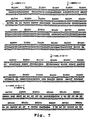
- Figure 7 shows the nucleotide sequences of the HEV ORF2 and ORF3 for Burma (upper line) and Mexico flower line) strains of HEV;
- Figure 8 shows the amino acid sequences of the ORF3 peptide for Burma (upper line) and Mexico (lower line) strains of HEV; and
- Figure 9 shows the amino acid sequences of the ORF2 protein for the Burma (upper line) and Mexico flower line) strains of HEV.
-
- The terms defined below have the following meaning herein:
- 1. "Enterically transmitted non-A/non-B hepatitis viral agent", "hepatitis E virus", or "HEV" means a virus, virus type, or virus class which (1) causes water-borne, infectious hepatitis, (ii) is transmissible in cynomolgus monkeys, (iii) is serologically distinct from hepatitis A virus (HAV), hepatitis B virus (HBV), hepatitis C virus (HCV), and hepatitis D virus, and (iv) includes a genomic region which is homologous to the 1.33 kb cDNA insert in plasmid pTZKF1(ET1.1) carried in E. coli strain BB4 identified by ATCC deposit number 67717.
- 2. Two nucleic acid fragments are "homologous" if they are capable of hybridizing to one another under hybridization conditions described in Maniatis et al., op. cit., pp. 320-323. However, using the following wash conditions: 2 x SCC, 0.1 % SDS, room temperature twice, 30 minutes each; then 2 x SCC, 0.1% SDS, 50°C once, 30 minutes; then 2 x SCC, room temperature twice, 10 minutes each, homologous sequences can be identified that contain at most about 25-30% basepair mismatches. More preferably, homologous nucleic acid strands contain 15-25% basepair mismatches, even more preferably 5-15% basepair mismatches. These degrees of homology can be selected by using more stringent wash conditions for identification of clones from gene libraries (or other sources of genetic material), as is well known in the art.
- 3. Two amino acid sequences or two nucleotide sequences (in an alternative definition
for homology between two nucleotide sequences) are considered homologous (as this term is
preferably used in this specification) if they have an alignment score of >5 (in standard
deviation units) using the program ALIGN with the mutation gap matrix and a gap penalty of
6 or greater. See Dayhoff, M.O., in Atlas of Protein Sequence and Structure (1972) Vol. 5,
National Biomedical Research Foundation, pp. 101-110, and
Supplement 2 to this volume, pp. 1-10. The two sequences (or parts thereof, preferably at least 30 amino acids in length) are more preferably homologous if their amino acids are greater than or equal to 50% identical when optimally aligned using the ALIGN program mentioned above. - 4. A DNA fragment is "derived from" an HEV viral agent if it has the same or substantially the same basepair sequence as a region of the viral agent genome.
- 5. A protein is "derived from" an HEV viral agent if it is encoded by an open reading frame of a DNA or RNA fragment derived from an ET-NANB viral agent.
- 6. In two or more known peptide sequences which are more than about 70% homologous in amino acid sequence, a third amino acid sequence will be "internally consistent with the known sequences" if each amino acid in the third sequence is identical to at least one of amino acids in the known sequences.
-
- This section describes methods for preparing and using an HEV antigen vaccine effective, when injected intramuscularly (i.m.), to prevent HEV infection.
- HEV genomic clones, and sequences corresponding to the entire HEV genome for different HEV strains were obtained according to published methods (Tam, Yarbough) and as described in the parent applications referenced above. Briefly, RNA isolated from the bile of a cynomolgus monkey having a known HEV infection was cloned, as CDNA fragments, to form a fragment library, and the library was screened by differential hybridization to radiolabeled CDNAS from infected and non-infected bile sources.
- The basepair sequence of cloned regions of the HEV fragments in identified clones was determined by standard sequencing methods. With reference to Figure 1, HEV is a virus with an approximately 7.5 kilobase (kb) single-stranded and polyadenylated RNA genome of positive-sense polarity. Three open reading frames (ORFs) have been assigned to HEV as ORF1, encoding polypeptides with domains of the RNA-directed RNA polymerase and a helicase, ORF2, encoding the putative capsid protein of the virus, and ORF3.
- The genomic organization of HEV assigns its non-structural gene(s) at the 5' terminus with the structural gene(s) at the 3' end. Two subgenomic polyadenlated transcripts of approximately 2.0 kb and 3.7 kb in sizes are detected in infected liver and co-terminated at their 3' ends with the 7.5 kb full-length genomic transcript. The genomic organization and expression strategy of HEV suggest that it might be the prototype human pathogen for a new class of RNA virus or perhaps a separate genus within the Caliciviridae family.
- The genomic and peptide sequences shown in Figure 7 correspond to the ORF-2 and ORF-3 regions of Burma (B) (upper lines) and Mexico (M) strains flower lines) of HEV. The bases indicated in the middle lines represent conserved nucleotides. The numbering system used in the comparison is based on the Burma sequence. The Burma sequence has SEQ ID No. 1; and the Mexico sequence, SEQ ID No. 2. The region corresponding to ORF2 has SEQ ID nos. 3 and 4 for the Burma and Mexico strains, respectively. The region corresponding to 406.3-2 has SEQ ID Nos. 5 and 6 for the Burma and Mexico strains, respectively. The region corresponding to SG3 has SEQ ID Nos. 7 and 8 for the Burma and Mexican strains,' respectively. The region corresponding to C2 has SEQ ID Nos. 9 and 10 for the Burma and Mexico strains, respectively. The region corresponding to 406.4-2 has SEQ ID Nos. 11 and 12 for the Burma and Mexico strains, respectively.
- The amino acid sequences corresponding to the third and second open reading frames of the Burma and Mexico strains of HEV are given in Figures 8 and 9, respectively. The sequence listings shown are as follows:
- SEQ ID Nos. 13 and 14 correspond to the amino acid sequences for the peptides 406.3-2 (B) and 406.3-2 (M), respectively. Each peptide is a 48 amino acid peptide in the C-terminal end region of capsid protein encoded by the ORF2, as indicated in the ORF2 sequence (Figure 9).
- SEQ ID Nos. 15 and 16 correspond to the amino acid sequences for the peptides SG3 (B) and SG3 (M), respectively. Each peptide includes the carboxyl 327 amino acids of the HEV capsid.
- SEQ ID Nos. 17 and 18 correspond to the amino acid sequences for the peptides C2 (B) and C2 (M), respectively. They include the carboxyl 436 and 435 amino acids of the HEV protein, respectively.
- SEQ ID Nos. 19 and 20 correspond to the amino acid sequences for the entire putative capsid protein encoded by the Burma and Mexico strain ORF2, respectively.
- SEQ ID Nos. 21 and 22 correspond to the amino acid sequences for the 406.4-2 (B) and 406.4-2 (M), respectively (Figure 8). These are 33 amino acid sequences encoded by the ORF3.
-
- Also contemplated are sequences which are internally consistent with the above specified sequences from different strains of HEV antigens. These include Sequence ID No. 13; Sequence ID No. 14, and internally consistent variations between Sequence ID Nos. 13 and 14; Sequence ID No. 15; Sequence ID No. 16; and internally consistent variations between Sequence ID Nos. 15 and 16; Sequence ID No. 17; Sequence ID No. 18; and internally consistent variations between Sequence ID Nos. 17 and 18; Sequence ID No. 19; Sequence ID No. 20; internally consistent variations between Sequence ID Nos. 19 and 20.
- For example, the HEV 406.4-2 antigens have the sequence homology shown below for the Burma (B) and Mexico (M) strains. The single dots in the sequence comparison indicate recognized high-probability or "neutral" amino acid substitutions. The blank spaces indicate a non-neutral substitution.

- A sequence which is internally consistent with these two sequences would have one of the sequences:
- AN(Q/P)P(G/D)H(L/S)APLG(E/V)(I/T)RPSAPPLP(P/H)V(A/V)DLPQ(P/L)G(L/P)RR, where X/Y means either amino acid X or amino acid Y.
-
- The ORF3 amino acid sequences, 124 amino acids in length, for the Burma and Mexico strains have an 87.1% identity in the 124 amino acids. The ORF2 amino acid sequences, having 659 amino acids of overlap, have a 93.0% identity in the 659 amino acids.
- To prepare the 406.3-2 (M) peptide, the lambda gt1 406.3-2 described in Example 3 was subcloned into the glutathione S-transferase vector pGEX to express the 3-2(M) antigen, as detailed in Example 3, and in the Tam reference.
- The 406.3-2(B) antigen can be prepared by PCR amplification of the Burma SEQ ID No. 5 from above by PCR amplification of the pBET1 plasmid (Tam). This plasmid contains a 2.3 kb insert covering the ORF2 and ORF3 for Burma strain HEV sequence. The plasmid is amplified by PCR amplification, using a 5' primer containing an NcoI site and a 3' primer containing a BamHI site (Sakai). The amplified fragment is inserted into the NcoI/BamHI site of a pGEX vector, and expressed in an E. coli expression system as described in Example 3.
- The SG3(B) peptide was prepared by first amplifying the SEQ ID No. 7 sequence with 5' EcoRI-NcoI and 3' BamHI linkers, using a
gt 10 phage BET1 clone plasmid containing the entire ORF2 and ORF3 regions of HEV (B). The amplified fragment was inserted into the EcoRI/BamHI site of a Bluescript™ vector (Stratagene, San Diego, CA), according to the manufacturer's instructions. After vector propagation and harvesting, the cloned insert was released by digestion with NcoI and BamHI, and gel purified. The purified fragment was inserted into the NcoI/BamHI site of a pGEX vector, and expressed in an E. coli expression system as described in Example 3. The SG3(M) peptide can be prepared similarly, using the SEQ ID No. 8 in place of the SEQ ID No. 7. - The C2 (B) peptide is prepared as described in Example 5. Briefly, a gt10 phage BET1 plasmid was digested with EcoRI to release the SEQ ID No. 10 C2 sequence, and this fragment was inserted into a pATH10 trpE fusion vector, and the recombinant fusion protein expressed in an E. coli host.
- The C2 (M) peptide can be prepared, substantially as described above, by PCR amplification of the SEQ ID No. 10, using a 5' primer containing an EcoRI site and a 3' primer containing a BamHI site. The amplified fragment is inserted into the EcoRI/BamHI site of a pGEX vector, and expressed in an E. coli expression system as described in Example 3.
- The capsid protein (B) was prepared substantially as described above by PCR amplification of the SEQ ID No. 3, from a gt10 BET1 plasmid using a 5' primer containing an NcoI site and a 3' primer containing a BamHI site. The amplified fragment was inserted into the NcoI/BamHI site of a pGEX vector, and expressed in an E. coli expression system as described in Example 3. The capsid protein (M) is similarly prepared.
- To prepare the 406.4-2 (M) peptide, the lambda gt11 406.4-2 described in Example 3 was subcloned into the glutathione S-transferase vector pGEX to express the 3-2(M) antigen, as detailed in Example 3.
- The 406.4-2(B) antigen can be prepared by PCR amplification of the Burma SEQ ID No. 11 from above by PCR amplification, using a 5' primer containing an NcoI site and a 3' primer containing a BamHI site. The amplified fragments is inserted into the NcoI/BamHI site of a pGEX vector, and expressed in an E.coli expression system as described in Example 3.
- It will be appreciated that other HEV peptides containing selected portions, and preferably C-terminal portions of the HEV capsid protein containing the 406.3-2 sequence, can similarly be prepared, using the HEV genomic-insert plasmids above, with amplification of the desired sequences and cloning into a suitable expression vector, as outlined above, and detailed in Examples 3 and 5.
- The coding sequences used in producing the recombinant peptides can be derived from the cloning vectors described above and detailed elsewhere (Tam), or from synthetic nucleotide synthesis using PCR slicing methods to join oligonucleotide fragments, according to known methods, in building up nucleotide sequences.
- HEV peptide antigens may also be obtained from purified HEV virus propagated in primary hepatocytes obtained from primate liver, preferably from human or cynomolgus monkey liver. Methods for preparing primary primate hepatocytes for culture, and culture medium conditions effective to preserve liver-specific functions for extended periods in culture are detailed for human hepatocytes in Example 1 below.
- After 3 days of growth in culture, the cells are infected with a pooled inoculum of HEV-infected cynomolgus monkey stool pool (fourth passage), as detailed in Example 2. The presence and level of propagating HEV virus in the cells can be measured by indirect immunofluorescence. Where, for example, the primary cells are cynomolgus cells, the cells can be immunoreacted with human HEV anti-sera, followed by immunoreaction with rabbit anti-human IgG antibodies.
- Alternatively, the HEV virus can be detected and measured by selective amplification methods involving initial cDNA formation, and PCR amplification of HEV cDNA sequences by PCR amplification, as detailed in Example 2. Virus particles can be isolated from HEV infected human hepatocytes in culture medium by pelleing the virus through a 30% sucrose cushion by ultracentrifugation. The pelleted virus may be further purified, if desired, by zonal centrifugation through a 10-40% sucrose gradient, combining peak virus fractions.
- Other methods for separating virus particles from soluble culture-medium components may be used. For example, clarified culture medium can be passed through a size-exclusion matrix, to separate soluble components by size exclusion.
- Alternatively, the clarified culture medium can be passed through an ultrafiltration membrane having a 10-20 nm pore size capable of retaining virus particles, but passing solute (non-particulate) culture medium components.
- The present invention allows glycosylation and other post-translation modifications in intact HEV capsid protein. Capsid isolation from the viral particles can be carried out by standard methods, such as ion exchange and size-exclusion chromatography, and HPLC purification, after solubilization of the virus particles in a solubilizing medium, such as a solution of a non-ionic surfactant. The protein may be purified by affinity chromatography, employing, for example, antibodies purified from anti-HEV antisera.
- The recombinant or intact HEV capsid or capsid fragment peptides (HEV capsid antigens) described above are incorporated into a vaccine composition, according to known procedures, to enhance the antigenicity of the injected antigens.
- In one composition, the HEV antigen is covalently coupled to a carrier protein, such as keyhole limpet hemocyanin, and injected either in solution form or in combination with an adjuvant. Alternatively, where the HEV antigen is prepared as part of a fusion protein, the non-HEV moiety of the protein may serve as the carrier protein.
The derivatized or fusion protein is carried in a pharmaceutically acceptable carrier, such as in solution or in an adjuvant, such as converted alum. - Alternatively, the free peptide itself, e.g., the HEV C2 peptide, may be formulated in alum or used without adjuvant. A suitable adjuvanted vaccine has a preferred antigen concentration of about 1 mg peptide antigen/mg alum, and not to exceed 80 mg of alum per injection.
- In a related aspect, the invention is directed to a method of inhibiting infection of an individual by hepatitis E virus, by administering to the subject, by parenteral injection, e.g., intramuscular or intravenous injection, the vaccine composition of the invention. Preferred vaccine compositions, for use in the method are those in which the HEV antigen includes the sequence in the peptides identified by:
- Sequence ID No. 13; Sequence ID No. 14, and internally consistent variations between Sequence ID Nos. 13 and 14; Sequence ID No. 15; Sequence ID No. 16; and internally consistent variations between Sequence ID Nos. 15 and 16; Sequence ID No. 17; Sequence ID No. 18; and internally consistent variations between Sequence ID Nos. 17 and 18; Sequence ID No. 19; Sequence ID No. 20; internally consistent variations between Sequence ID Nos. 19 and 20.
-
- The antigen vaccine composition is preferably administered intramuscularly in a series of inoculations, for example, two-three injections given at four week intervals.
- In the method detailed in Example 5, cynomolgus monkeys were injected i.m. with the C2 fusion protein trpE-C2 (B), formulated in a converted alum adjuvant or with no adjuvant. Four animals received the alum plus trpE-C2 (B) antigen in two injections, spaced one month apart. Two other animals received alum only on the same vaccination schedule. None of the animals showed the presence of any
anti-HEV serum antibody 4 weeks after the second injection, as judged by Western blotting using a fusionless C2 HEV antigen or by a separate fluorescence antibody blocking assay. - At this stage, two of the four experimental animals received a third inoculation of non-adjuvanted, insoluble trpE-C2 peptide antigen. Four weeks later, these animals showed anti-HEV antibodies, as evidenced by Western blots. These results suggest that the trpE-C2 antigen may be more effective when administered in the absence of alum, possibly because of alumdenaturation of the antigen during the alum co-precipitation procedure.
- One month after the final inoculation, the animals were challenged with an intravenous injection of a third-passage human stool previously shown to be highly infectious for HEV (Burma strain) or with a Mexico-strain human HEV stool sample. At selected intervals after inoculation, serum samples from the animals were used to measure ALT (alanine transferase) levels, as an indication of necrosis and hepatocellular degradation. Liver biopsy samples were also assayed for the presence of HEV antigens by a direct fluorescent antibody assay (FA).
- Figure 2A shows the change in liver ALT levels period following infection with Burma-strain HEV virus, in one of the animals which received a third dose of trpE-C2. As seen, there was no evidence of elevated ALT levels in the 7 and 1/2 week period following infection. The liver biopsy samples also showed no evidence of HEV antigen.
- Figure 2B shows ALT levels measured after HEV (B) infection of a control animal (alum alone injections) which was infected intravenously with the Burma strain HEV. The elevated ALT levels indicate the level of infection which is expected in the absence of vaccine protection. HEV antigen was also detected in the liver biopsy samples. A similar result was observed in the animal which received two injections of trpE-C2 alum composition, but not the third alum-free vaccination, as described above.
- Figure 3A shows the change in liver ALT levels following infection with Mexico-strain HEV virus, in one of the animals which received a third dose of trpE-C2. Again, there was no evidence of elevated ALT levels out to day 32 (The animal died of unrelated causes at day 32). The liver biopsy samples also showed minimal evidence of HEV antigen. This result demonstrates that an antigen vaccine directed against one HEV strain can provide protective immunity against other HEV strains.
- Figure 3B shows ALT levels measured after HEV infection of a control animal (alum alone injections) which was infected intravenously with the Mexico strain of HEV. High levels of infection (ALT activity) were observed. A similar result was observed in the animal which received two injections of trpE-C2 alum composition, but not the third alum-free vaccination, as described above.
- Details of the vaccination method just reported are given in Example 5.
- In another aspect, the invention includes an antibody vaccine composition effective in neutralizing HEV infection, as evidenced by the ability of the composition to block HEV infection in HEV-infectable primary hepatocytes in culture. Two exemplary primary cells are human and cynomolgus monkey cells.
- The antibodies in the composition are preferably immunoreactive with a peptide containing one of the sequences: Sequence ID No. 13; Sequence ID No. 14, and internally consistent variations between Sequence ID Nos. 13 and 14. As will be seen below, antibodies prepared against the 406.3-2 antigen (M) are effective to block HEV infection in human primary hepatocytes.
- Antibodies which are immunoreactive with larger capsid peptides or proteins containing the carboxy terminal of SEQ ID No. 13 or 14 are also preferred. These may include, specifically Sequence ID No. 15; Sequence ID No. 16; and internally consistent variations between Sequence ID Nos. 15 and 16. As will be seen below, human sera which are effective to prevent HEV infection of human primary hepatocyes are immunoreactive with the SG3 peptides defined by these sequences.
- Antibodies which are immunoreactive with the trpE-C2 peptides defined by Sequence ID No. 17; Sequence ID No. 18; and internally consistent variations between Sequence ID Nos. 17 and 18 are also preferred, as are antibodies immunoreactive with the entire capsid protein, as defined by Sequence ID No. 19; Sequence ID No. 20; internally consistent variations between Sequence ID Nos. 19 and 20.
- The antibodies may be obtained as polyclonal antibodies from antisera, prepared for example, by immunization of a suitable animal, such as a rabbit or goat, with one of the HEV antigens specified above. Alternatively, polyclonal antibodies may be obtained from human or other primate HEV antisera. Anti-HEV polyclonal antibodies from the antisera may be purified or partially purified according to standard methods, such as used to obtain partially purified serum IgG fractions (see, e.g., Antibodies: A laboratory Manual, 1988, Cold Springs Harbor Lab). Alternatively anti-HEV antibodies can be obtained in purified form by affinity chromatography, employing a solid support derivatized with one of the capsid antigens described above.
- In another embodiment, the antibodies are monoclonal antibodies secreted by hybridoma cell lines. To prepare the hybridoma cell lines, lymphocytes from an immunized animal, preferably mouse or human, are immortalized with a suitable immortalizing fusion partner, according to established methods (e.g., Engleman, Zola).
- Alternatively, human monoclonal antibodies may be produced by recombinant methods, in which light and heavy human anti-HEV IgG genes obtained from cultured lymphocytes are inserted into suitable expression vectors, and used to co-infect a suitable host. Methods for obtaining and cloning light and heavy genes from human lymphocytes, and for expressing the cloned genes in a co-infected host cell are known (larrick).
- The anti-HEV antibodies are formulated in a suitable solution for injection, typically by intramuscular, subcutaneous or intravenous route, to form the vaccine composition.
- To demonstrate the neutralizing capability of antibodies prepared as above, antibodies against the 406.3-2 (B) antigen were tested for their abilities to block HEV infection in human primary hepatocytes in culture.
- The primary hepatocytes were prepared and cultured according to published procedures and as detailed in Example 1. The unique culture conditions allow for long-term cell growth in culture without loss of specialized hepatocyte function, as evidenced by the cells' continued ability to make and secrete liver-specific proteins, such as serum albumin, up to several months after initial culturing, as described in Example 1.
- The cultured cells were inoculated with either normal human sera or a cynomolgus stool preparation. To demonstrate HEV infection in the cells, the cells were examined on days 1-11 after infection for the presence of HEV RNA, using a combination of reverse transcriptase, to form cDNA's, and polymerase chain reaction (PCR) to amplify HEV-specific cDNA. The amplified fragment is expected to have a 551 basepair length. Figure 4 shows Southern blots of the amplified material, using an HEV ORF2 radiolabled probe for detecting amplified HEV sequence.
- The results are shown in Figure 4. Lanes 1-3 are controls.
Lane 4 is total amplified material from cells inoculated with normal (non-infected) sera. Lanes 5-11 show amplifiedmaterial 3 hours, 1 day, 3 days, 5 days, 7 days, 9 days, and 11 days after infection with the cyno stool sample, respectively. The results show that HEV propagated in human primary hepatocytes within one day after initial infection (lane 6). There was a time-dependent increase at the level of HEV replication up to 5 days post infection (lanes 7 and 8), which appeared to decrease thereafter (lanes 9-11). There was no evidence of HEV in total cellular RNA isolated from uninfected primary cells. - Rabbit antisera against antigen peptides 406.3-2 (B) and 406.4-2 (M) and 406.4-2 (B) were prepared. As noted above, the 406.3-2 peptide is from the carboxy terminal end region of the HEV capsid protein, and the 406.4-2 peptide, from the peptide encoded by the HEV ORF3. Preimmune rabbit serum or rabbit antiserum against one of HEV antigens was added to the cyno stool inoculum, at a 1:20 dilution, and the antibody was incubated with the viral preparation. The antibody-treated stool sample was then used to infect human primary hepatocytes. 14 days later, the cells were examined for HEV infection by the RT/PCR/Southern blot method just described, except employing primers which are expected to yield a 448 basepair amplified fragment.
- The results are shown in Figure 5.
Lanes Lane 4 shows amplified material from cells exposed to anti-406.4-2 (M) rabbit antisera. The antibody showed little or no protective effect against HEV infection. - Another source of neutralizing antibodies, in accordance with the invention, is human HEV antisera which is characterized by immunospecific reaction to the 406.3-2 antigen and the SG3 antigen, both described above.
- To examine the neutralizing antibody characteristics of human HEV antisera, a panel of human antisera were tested for the ability to block HEV infection of cultured hepatocytes, substantially as described above. The ten HEV positive human antisera are shown in Table 1 below, and are from patients who developed HEV infection in India, Pakistan, and Mexico. The antisera were not tested for strain type.
- Briefly, cultured cells were exposed to HEV-positive cyno stool treated with samel (Burma strain) treated with normal pooled serum or HEV antiserum, and tested for the presence of HEV-specific nucleic acid sequences, by PCR amplification and Southern blotting with an HEV radiolabled probe. The Southern blots are shown in Figure 6. The lane numbers of the 12 serum samples are given in parentheses in Table 1 below. As seen from Figure 6, and indicated in Table 1, the antisera which were effective in neutralizing HEV were India 10 (lane 2), India 18 (lane 3), India 210 (lane 5), India 265 (lane 8), Pak 143 (lane 9), and Pak 336 (lane 10). Other human sera, however, showed very little (lane 11, Mex 387C) or no effect (
lane 4, India 29;lane 6, India 242;lane 7, India 259; lane 12, Mex 387C[IgG]) in their ability to neutralize HEV infection. As a negative control, the normal human serum pool revealed absolutely no neutralizing activity against HEV (lane 1).Serum Clinical Neutralizing Activity normal (1) pooled - India 10 (2) - + India 18 (3) acute, import + India 29 (4) acute, import - India 210 (5) acute + India 242 (6) acute, fulminant - India 259 (7) acute, fulminant - India 265 (8) acute + Pak 143 (9) acute + Pak 336 (10) acute + Mexico F387c (11) convalescent - Mexico F387c (IgG) (12) convalescent - - Several of the human antisera were tested for their IgG and IgM immunoreactivity to 406.3-2 (M), 406.4-2(M) and 406.4-2 (B) antigens noted above. Reaction with IgM antibodies tends to indicate early-phase infection, whereas immunoreactivity with IgG is indicative of both early and later stages of infection. Reaction was measured in an ELISA test. The results are shown in Table 2A and 2B, where a "+" sign indicates a positive reaction; numbers in the table indicate dilution titre of IgG against the specific recombinant protein indicated.
Serum
SamplesIgG Neutralizing
ActivityClinical 406.3-2(M) 406.4-2(B) 406.4-2(M) Normal
Human- - - - Pooled
Human
SerumIndia 18 + + + + acute,
importIndia 29 - + - - acute,
importIndia 210+ + + + acute India 242 + + + - acute,
fulminantIndia 259 +
(500)+
(>5000)+
(2000)- acute,
fulminantIndia 265 +
(>5000)+
(>5000)+
(1000)+ acute Serum Samples IgM 406.3-2(M) 406.4-2(B) 406.4-2(M) Normal Human ND ND ND India 18 - - - India 29 - - - India 210 - - - India 242 + + - India 259 + + - India 265 + + - - The data from the table indicates that those human antisera capable of neutralizing were positive by an IgG ELISA for antibodies to the HEV 3-2(M) epitope. India 29 was not positive for IgG(s) to HEV 3-2(M) and did not neutralize HEV infection (lane 4). Although India 242 and India 259 were positive for IgG(s) to HEV 406.3-2(M), they were also positive for IgM to HEV 406.3-2(M), which is indicative of an early stage HEV infection. Therefore in these particular samples, the levels of IgG(s) to HEV 3-2(M) elicited might be sufficient to neutralize HEV infection of primary human hepatocytes.
- To further study the correlation of neutralizing activities of sera of HEV-infected humans with immunoreactivities to HEV3-2 epitope, Western blotting analyses were performed on these human serum samples, with the results shown in Table 3. As seen in this table, India 18, India 265, and especially
India 210, previously shown to be neutralizing for HEV infection, were immunoreactive to HEV406.3-2(M) in these Western blotting analyses and their immunoreactivities correlated with their neutralizing activities. - As a confirmation for the specific immunoreactivities of these sera to HEV406.3-2(M), Western analyses were performed against the fusion protein SG3 (B), which contains the 329 carboxy-terminal amino acids (nucleotides 6146-7129) of ORF-2 of HEV Burma strain. The immunoreactivities of these sera against HEV406.3-2(M) and SG3 [or HEV406.3-2(B)] were perfectly matched (Table 3).
Serum
Samples406.3-2(M)
ELISA Titre406.3-2(M)
Western BlotSG3
Western BlotNeutralizing
ActivityNormal Human - - - - India 18 2000 ++ + + India 29 - - - - India 210100 ++ + + India 242 500 - - - India 259 500 ± - - India 265 5000 +++ +++ + - Thus, human HEV antisera which provide a suitable source of neutralizing antibodies are those characterized by (a) immunoreactivity with a 406.3-2 antigen, and (b) the SG3 antigen, both as evidenced by immunoreactivity in a Western blot, i.e., where the antigen is in an exposed, accessible configuration.
- More generally, a preferred vaccine composition of the invention contains antibodies immunospecific against the 406.3-2 antigenic and against the SG3 antigenic peptide. The vaccine composition includes the immunospecific antibodies in a suitable carrier for parenteral injection.
- The antibody vaccine composition is used, according to another aspect of the invention, for preventing or treating HEV infection in humans.
- The following examples, which illustrate various methods and compositions in the invention, are intended to illustrate, but not limit the scope of the invention.
- Enzymes: DNAse I and alkaline phosphatase were obtained from Boehringer Mannheim Biochemicals (BMB, Indianapolis, IN); EcoRI, EcoRI methylase, DNA ligase, and DNA Polymerase I, from New England Biolabs (NEB, Beverly MA); and RNase A was obtained from Sigma (St. Louis, MO) .
- Other reagents: EcoRI linkers were obtained from NEB; and nitro blue tetrazolium (NBT), S-bromo-4-chloro-3-indolyl phosphate (BCIP) S-bromo-4-chloro-3-indolyl-B-D-galactopyranoside (Xgal) and isopropyl B-D-thiogaiactopyranoside (IPTG) were obtained from Sigma.
cDNA synthesis kit and random priming labeling kits are available from Boehringer-Mannheim Biochemical (BMB, Indianapolis, IN). - Hepatocytes were isolated from human liver obtained from Stanford University Medical Center. The liver was either perfused in situ or excised as a wedge for perfusion in laboratory. The initial perfusion was performed for 10 minutes at 60 ml/min using Ca++-, Mg++-free Hanks' balanced salt solution supplemented with 10 mM HEPES (pH7.4) and 0.5 mM [ethylene bis(oxyethylenenitrillo]-tetraacetic acid. Perfusion was continued for additional 20 minutes using Williams' medium E (WME) supplemented with 10 mM HEPES (pH7.4) and 100 U/ml collagenase (type I, Sigma Chemical Co., St. Louis, MO).
- After perfusion the liver capsule was removed using fine forceps, and hepatocytes were dislodged by gentle shaking in collagenase solution. The hepatocyte suspension was filtered through several layers of gauze and mixed with an equal volume of WMW containing 10% fetal bovine serum (FBS). Hepatocytes were sedimented by centrifugation at 50 Xg for 5 minutes and resuspended in WME containing 5% FBS. Hepatocytes were sedimented and resuspended in the manner for 2 additional times. The final cell preparation was further filtered through several layers of gauze before examining for viability using trypan blue. The cells were plated at a density of 2 x 10⁶ cells per 60-mm Primaria plates (Falcon) pre-coated with collagen (Collaborative Research).
- Cultures were incubated at 37°C in 5% CO2 for 3 hours to allow attachment and the medium was changed to a serum-free formulation and every 48 hrs thereafter. The serum-free formulation was a WME-based medium supplemented with growth factors, hormones, 10 mM HEPES (pH7.4), 100 ug/ml gentamicin, as has been described (Lanford).
- Human hepatocyte cultures were maintained in serum-free medium for various period of time and labeled with [35S]-methionine for 24 hrs. The medium was adjusted to contain 1 mM PMSF, 1 mM EDTA, and 1% NP40. Antibodies specific for the different plasma proteins were bound to protein A-agarose beads, the beads were washed with PBS, and aliquots of the labeled medium were incubated for 16 hrs at 4°C with the antibody-bead complexes. The beads were washed 3 times with a buffer containing 1% NP40, and immunoprecipitated proteins were eluted with gel electrophoresis sample buffer containing 2% SDS and 2% 2-mercaptoethanol. Samples were analyzed by gradient SDS-PAGE (4 to 15%) and autoradiography.
- The HEV-infected cynomolgus monkey #73 stool pool (fourth passage) was used as an inoculum for infections of primary human hepatocytes. Various amounts of inoculum was diluted in 1 ml of serum-free medium (SFM) and applied to the culture during a 3 hr incubation period. This solution was then supplemented with 2 ml of fresh SFM and the entire mixture was incubated overnight. The next day, cell monolayers were washed with WME (10 mM HEPES, pH7.4) for three times and changed to fresh SFM, which was changed at two day intervals thereafter.
- Primary cynomolgus monkey hepatocytes were isolated and plated in tissue culture plates with collagen-coated coverslips as described. Cells on coverslips were infected with either the HEV-infected cynomolgus monkey #73 stool pool or the NIH normal human serum three days after initial plating. The infections were allowed to proceed for 2 weeks.
- Cells on coverslips were fixed in 90% acetone at room temperature for 1 minute. The coverslips were then air-dried. The coverslips were blocked in 1 % goat serum in PBS for 1 hour, washed with PBS for three times, and incubated with a mixture of rabbit antisera against HEV recombinant proteins 1L6, 4-2, and 6-1-4 at room temperature for 3 hours. The coverslips were again washed with PBS for 3 times and reacted with fluorescein isothiocyanate-conjugated (FITC) goat anti-rabbit IgG(H+L) (Zymed) diluted in PBS-I % goat serum for 30 minutes. After the coverslips were washed with PBS for 3 times and air-dried, they were mounted with FITC glycerol solution and examined under a fluorescent microscope.
- HEV infection of primary cynomolgus macaque hepatocytes was evaluated by RT/PCR assays. The primers for cDNA synthesis and PCR were based on the nucleotide sequences of the full-length HEV cDNA (A. Tam et al.). Primers HEV3.2SF1 (nt 6578-6597) and HEV3.2SF2 (nt 6650-6668) are of sense polarity from the ORF2 region of the viral genome and HEV3.2SR1 (nt 7108-7127) and HEV3.2SR2 (nt 7078-7097) are antisense primers within the region. Following extraction of total cellular RNA from HEV-infected cells using one-step guanidinium procedure or HEV-infected supernatants according to the method of Sherker et al., aliquots of RNA samples were heat-denatured at 95°C for 5 minutes and subjected to reverse transcription at room temperature for 5 minutes and 42°C for 60 minutes using 200 units per reaction of MMLV-reverse transcriptase (BRL) in a 20 ul reaction volume containing 20 units of RNasin (Promega), 1x PCR buffer (Perkin-Elmer Cetus), with a concentration of 1 mM each deoxyribonucleotide (Perkin-Elmer Cetus), and 2.5 uM of HEV3.2SR1 primer. The reaction mixture was then heat-treated at 95°C for 5 minutes to denature the MML V-reverse transcriptase.
- Ten microliters of the cDNA synthesis product was used for PCR in a final volume of 50 ul with 0.5 uM HEV3.2SF1 primer, 1.25 units Taq DNA polymerase (AmpliTaq, Perkin-Elmer Cetus), and 1x PCR buffer, overlayed with 50 ul of mineral oil, and subjected to 40 cycles of PCR in a Perkin-Elmer thermocycler (95°C x 1 minute; 52°C x 2 minutes; 72°C x 30 seconds). Ten microliters of the first-round PCR product then underwent another 40 cycles of nested PCR (95°C x 1 minute; 55°C x 2 minutes; 72°C x 30 seconds) in a total volume of 50 ul containing the internal PCR primers HEV3.2SF2 and HEV3.2SR2.
- First- and second-round PCR products were subjected to agarose electrophoresis, ethidium bromide stained and photographed under UV light. The results are shown in Figure 4, discussed above. Southern transfer was performed and filters were hybridized with [32P-dCTP]-labeled internal probe HEVORF2-7 exclusive of the primers (nt 6782-6997), and autoradiography performed.
- A TZKF1 plasmid (ET1.1), ATCC deposit number 67717, was digested with EcoRI to release the 1.33 kb HEV insert which was purified from the linearized plasmid by gel electrophoresis. The purified fragment was suspended in a standard digest buffer (0.5M Tris HCl, pH 7.5; 1 mg/ml BSA; 10mM MnC12) to a concentration of about 1 mg/ml and digested with DNAse I at room temperature for about 5 minutes. These reaction conditions were determined from a prior calibration study, in which the incubation time required to produce predominantly 100-300 basepair fragments was determined. The material was extracted with phenol/chloroform before ethanol precipitation.
- The fragments in the digest mixture were blunt-ended and ligated with EcoRI linkers. The resultant fragments were analyzed by electrophoresis (5-1OV/cm) on 1.2% agarose gel, using PhiX174/HaeIII and lambda/HindIII size markers. The 100-300 bp fraction was eluted onto NA45 strips (Schleicher and Schuell), which were then placed into 1.5 ml microtubes with eluting solution (1 M NaCl, 50 mM arginine, pH 9.0), and incubated at 67°C for 30-60 minutes. The eluted DNA was phenol/chloroform extracted and then precipitated with two volumes of ethanol. The pellet was resuspended in 20 ml TE (0.01 M Tris HCl, pH 7.5, 0.001 M EDTA).
- Lambda gt11 phage vector (Huynh) was obtained from Promega Biotec (Madison, WI). This cloning vector has a unique EcoRI cloning site 53 base pairs upstream from the beta-galactosidase translation termination codon. The genomic fragments from above, provided either directly from coding sequences 5) or after amplification of cDNA, were introduced into the EcoRI site by mixing 0.5-1.0 mg EcoRI-cleaved gt11, 0.3-3 ml of the above sized fragments, 0.5 ml 10X ligation buffer (above), 0.5 ml ligase (200 units), and distilled water to 5 ml. The mixture was incubated overnight at 14°C, followed by in vitro packaging, according to standard methods (Maniatis, pp. 256-268).
- The packaged phage were used to infect E. coli strain KM392, obtained from Dr. Kevin Moore, DNAX (Palo Alto, CA). Alternatively, E. Coli strain Y1090, available from the American Type Culture Collection (ATCC #37197), could be used. The infected bacteria were plated and the resultant colonies were checked for loss of beta-galactosidase activity-(clear plaques) in the presence of X-gal using a standard X-gal substrate plaque assay method (Maniatis). About 50% of the phage plaques showed loss of beta-galactosidase enzyme activity (recombinants).
- HEV convalescent antiserum was obtained from patients infected during documented HEV outbreaks in Mexico, Borneo, Pakistan, Somalia, and Burma. The sera were immunoreactive with VLPs in stool specimens from each of several other patients with ET-NANB hepatitis.
- A lawn of E. coli KM392 cells infected with about 104 pfu of the phage stock from above was prepared on a 150 mm plate and incubated, inverted, for 5-8 hours at 37°C. The lawn was overlaid with a nitrocellulose sheet, causing transfer of expressed HEV recombinant protein from the plaques to the paper. The plate and filter were indexed for matching corresponding plate and filter positions.
- The filter was washed twice in TBST buffer (10 mM Tris, pH 8.0, 150 mM NaCl, 0.05% Tween 20), blocked with AIB (TBST buffer with 1 % gelatin), washed again in TBST, and incubated overnight after addition of antiserum (diluted to 1:50 in AIB, 12-15 ml/plate). The sheet was washed twice in TBST and then contacted with enzyme-labeled anti-human antibody to attach the labeled antibody at filter sites containing antigen recognized by the antiserum. After a final washing, the filter was developed in a substrate medium containing 33 ml NBT (50 mg/ml stock solution maintained at 4°C) mixed with 16 ml BCIP (50 mg/ml stock solution maintained at 4°C) in 5 ml of alkaline phosphatase buffer (100 mM Tris, 9.5, 100 mM NaCl, 5 mM MgC12). Purple color appeared at points of antigen production, as recognized by the antiserum.
- The areas of antigen production determined in the previous step were repeated at about 100-200 pfu on an 82 mm plate. The above steps, beginning with a 5-8 hour incubation, through NBT-BCIP development, were repeated in order to plaque purify phage secreting an antigen capable of reacting with the HEV antibody. The identified plaques were picked and eluted in phage buffer (Maniatis, p. 443).
- Two subclones which were selected are the 406.3-2 and 406.4-2 clones whose sequences are set forth above. These sequences were isolated from an amplified cDNA library derived from a Mexican stool. Using the techniques described in this section, polypeptides expressed by these clones have been tested for immunoreactivity against a number of different human HEV-positive sera obtained from sources around the world. As shown in Table 4 below, 8 sera immunoreactive with the polypeptide expressed by the 406.4-2, and 6 sera immunoreacted with polypeptide expressed by the 406.3-2 clone.
- For comparison, the Table also shows reactivity of the various human sera with the non structural peptide Y2. Only one of the sera reacted with the polypeptide expressed by this clone. No immunoreactivity was seen for normal expression products of the gt11 vector.
Immunoreactivity of HEV Recombinant
Proteins: Human SeraSera Source Stage 406.3-2 406.4-2 Y2 lgt1 FVH-21 Burma A - - - - FVH-8 Burma A - + + - SOM-19 Somalia A + + - - SOM-20 Somalia A + + - - IM-35 Borneo A + + - - IM-36 Borneo A - - - - PAK-1 Pakistan A + + - - FFI-4 Mexico A + + - - FFI-125 Mexico A - + - - F 387 IC Mexico C + + ND - Normal U.S.A. - - - - - - Here Y2 represents a sequence encoded by the HEV sequence 157 basepair sequence from the first open reading frame of the HEV genome.
- The 406.3-2 gtl1 plasmid from above was digested with EcoRI and the released HEV fragment was amplified by PCR in the presence of linkers which added an NcoI site at the 5' fragment end, and a BamHI site at the 3' fragment end. The amplified material was digested with NcoI and BamHI and inserted into the NcoI/BamHI site of the glutathione S-transferase vector pGEX expression vector, according to the manufacturer's instructions.
- The pGEX plasmid was used to transform E.coli host cells, and cells which were successfully transformed with the pGEX vector were identified by immunofluorescence, using anti-HEV human antisera.
- The 406.4-2 gt11 plasmid from above was digested with EcoRI and the released HEV fragment was amplified by PCR, and the amplified fragment was inserted into the NcoI/BamHI site of the pGEX expression vector, as above. Peptide expression of the 406.4-2 peptide was similar to that described for the 406.3.2 fusion peptide.
- The 406.3-2(M) and 406.4-2(M) fusion proteins, prepared as above, were used to immunize rabbits to generate HEV-specific antisera, according to standard procedures.
- To prove that primary human hepatocytes were permissive for HEV infection and replication, cells were exposed to either normal human serum (NIH normal human serum pool) or HEV-infected cynomolgus macaque stool preparation (cyno#73). Fourteen days postinfection, total cellular RNAs were prepared for reverse-transcription (RT)/polymerase chain reaction (PCR) assays to evaluate the infectability of primary human hepatocytes with HEV. The results indicated that primary human hepatocytes were capable of supporting HEV propagation (Figure 4).
- Although quantitative PCR was not applied, total cellular RNA isolated from HEV-infected primary human hepatocytes would indicate a high level of virus replication as suggested by the extent of hybridization with the a-32P-dCTP labeled HEV-specific probe (lane 5). There was no evidence of HEV in total cellular RNA isolated from primary human hepatocytes treated with normal human serum pool (lane 4). As negative controls for RT/PCR assays, no carry-over or cross-contamination was detected (
lanes - To examine the neutralizing activities of anti-3-2(M), -4-2-(M), each rabbit antiserum was used at a final dilution of 1:20 with the viral inoculum for HEV infection of primary human hepatocytes. The diluted antibody and viral inoculum were incubated together prior to infection of the cultured cells. Rabbit anti-3-2(M) exhibited a high level of neutralizing activity against HEV infection (Figure 5,
lane 2 versus lane 1). Very little neutralizing activity was observed in rabbit anti-4-2 (M) (lane 4 versus lane 3). - This result suggests that the HEV 3-2(M) but not HEV 4-2(M) or 4-2(B) recombinant protein encoded a neutralizing epitope capable of eliciting protective antibody or antibodies against HEV infection. The fact that the Mexico clone 3-2(M) and the Burma clone 3-2(B) share 90.5% homology at the amino acid level (79.8% at the nucleotide level) suggested that antibody(ies) raised against 3-2(M) should cross-neutralize or cross-protect HEV of Mexico or Burma strain from infecting permissive cells.
- The pBET1 plasmid containing a 2.3 kb insert, corresponding to the 1.8 kb 3' end portion of HEV has been described (Tam). The plasmid was digested with EcoRI, releasing two HEV fragments having sizes of about 600 bp and 1400 bp of which 1210 bp contain coding sequence. The larger fragment was purified by electrophoresis and inserted into the EcoRI site of the pATH10 trpE fusion vector, obtained from T.J. Koerner et al. (Department of Microbiology, UCLA) The recombinant vector was used to transform E. coli DH5αF'host.
- The recombinant typE-C2 fusion protein from pATH C2 was isolated by a modification of the procedure of Dieckmann et al. The bacterium containing the pATH C2 plasmid was grown overnight in growth media containing tryptophane. Two ml of the overnight culture was inoculated into 100 ml of fresh growth media and grown at 37°C for an additional four hours. The bacterial broth was added to one liter of fresh growth media without tryptophane and allowed to grow at 30°C for 1.5 hours. Ten ml indoleacrylic acid (1 mg/ml) was added and growth was continued for an additional 5 to 10 hours at 30°C. The bacterial cells were collected by centrifugation. The cell pellet was resuspended in a hypotonic solution containing lysozyme to degrade the bacterial cell wall. Sodium chloride and the detergent NP-40 were added to the suspension to cause hypertonic lysis of the cells. The lysed cell solution was sonicated. The solution was centrifuged. The resulting protein pellet was resuspended in about 5 ml of 10 mM Tris pH 7.5 using a dounce homogenizer. Approximately 75% of the protein in solution was composed of the trpE-C2 protein.
- Converted alum adjuvant was prepared according to standard methods. Briefly, a 10% alum suspension was titrated to pH 6.6 with IN NaOH, then stirred overnight at 4°C. The suspension is clarified by low-speed centrifugation, and the supernatant decanted. A small amount of 0.9% NaCI + 1:20,000 formalin was added to each pellet, and suspended by vortexing. To prepare an antigen vaccine composition, trpE-C2 fusion protein from above is added in a 0.9% NaCl solution to a desired final antigen concentration.
- A non-adjuvanted insoluble trpE-C2 peptide was prepared as above in section A.
- Six cynomolgus monkeys, designated 8901, 8902, 8903, 8910, 9902, and 9904, were used in the vaccination study. Four of the monkeys, 8901, and 8902 8903, and 8910 were immunized by intramuscular injection with 1.0 ml of the alum adjuvanted trpE-C2 composition (containing about 50 µg of C2 peptide). The other two animals received adjuvant only. One month later the six animals were given a second vaccination, identical to the first.
- 4 weeks after the second vaccination, sera from the animals was tested for anti-HEV antibodies by Western blotting, using a fusionless C2 protein. At this stage, animals 8901 and 8902 each received a third vaccination with the non-adjuvanted, insoluble trpE-C2 composition (a total IV dose of about 80 µg trpE-C2 peptide each), and both animals showed anti-HEV by
Western blotting 4 weeks later. - Animals 8901, 8903, and 9002 were each challenged IV with 1 ml each of a 10% third passage cyno stool (Burma strain) previously shown to be highly infectious. Animals 8902, 8910, and 9004 were each challenged IV with 1 ml of a proven infectious human stool isolate, Mexican #14, known to cause severe disease in cynos and moderate disease in chimpanzees. The results are shown in Figures 2A, 2B, and 3A, and 3B, discussed above.
Claims (4)
- A vaccine composition for use in immunizing an individual against hepatitis E virus (HEV) comprising, in a pharmacologically acceptable carrier, an HEV capsid antigen(A) consisting of one of the following sequences:and(i) Sequence ID No:17 of 436 amino acids,(ii) Sequence ID No:18 of 435 amino acids, and(iii) internally consistent variations between Sequence ID Nos. 17 and 18,(B) capable of providing protection against HEV infection, as evidenced by the ability of the antigen to protect cynomolgus monkeys inoculated with said antigen from subsequent HEV challenge.
- The composition of claim 1, wherein the peptide antigen is covalently coupled to a carrier protein.
- Use of an HEV capsid antigen consisting of one of the following sequences:for the manufacture of a vaccine against infection of an individual by hepatitis E virus.(i) Sequence ID No:17 of 436 amino acids,(ii) Sequence ID No:18 of 435 amino acids, and(iii) internally consistent variations between Sequence ID Nos. 17 and 18,
- The use of claim 3, wherein the antigen consists of the sequence of Sequence ID No:17.
Applications Claiming Priority (5)
| Application Number | Priority Date | Filing Date | Title |
|---|---|---|---|
| US82233592A | 1992-01-17 | 1992-01-17 | |
| US822335 | 1992-01-17 | ||
| US07/870,985 US6455492B1 (en) | 1988-06-17 | 1992-04-20 | Hepatitis E virus vaccine and method |
| US870985 | 1992-04-20 | ||
| PCT/US1993/000475 WO1993014208A2 (en) | 1992-01-17 | 1993-01-19 | Hepatitis e virus vaccine and method |
Publications (2)
| Publication Number | Publication Date |
|---|---|
| EP0623169A1 EP0623169A1 (en) | 1994-11-09 |
| EP0623169B1 true EP0623169B1 (en) | 2003-04-02 |
Family
ID=27124631
Family Applications (1)
| Application Number | Title | Priority Date | Filing Date |
|---|---|---|---|
| EP93911555A Expired - Lifetime EP0623169B1 (en) | 1992-01-17 | 1993-01-15 | Hepatitis e virus vaccine and method |
Country Status (9)
| Country | Link |
|---|---|
| US (3) | US6455492B1 (en) |
| EP (1) | EP0623169B1 (en) |
| JP (2) | JP3746291B2 (en) |
| AT (1) | ATE236255T1 (en) |
| CA (1) | CA2125701A1 (en) |
| DE (1) | DE69332820T2 (en) |
| DK (1) | DK0623169T3 (en) |
| ES (1) | ES2196008T3 (en) |
| WO (1) | WO1993014208A2 (en) |
Families Citing this family (25)
| Publication number | Priority date | Publication date | Assignee | Title |
|---|---|---|---|---|
| US6291641B1 (en) | 1988-06-17 | 2001-09-18 | Genelabs Technologies, Inc. | Hepatitis E virus antigens and uses therefor |
| US6455492B1 (en) * | 1988-06-17 | 2002-09-24 | Genelabs Technologies, Inc. | Hepatitis E virus vaccine and method |
| US6214970B1 (en) * | 1988-06-17 | 2001-04-10 | Genelabs Technologies, Inc. | Hepatitis E virus antigens and uses therefor |
| US5686239A (en) * | 1988-06-17 | 1997-11-11 | Genelabs Technologies, Inc. | Hepatitis E virus peptides and methods |
| US5789559A (en) * | 1988-06-17 | 1998-08-04 | Genelabs Technologies, Inc. | DNA sequences of enterically transmitted non-A/non-B hepatitis viral agent |
| JPH08504096A (en) | 1992-09-18 | 1996-05-07 | アメリカ合衆国 | Recombinant proteins of hepatitis E Pakistan strains and their use in diagnostics and vaccines |
| US6787145B1 (en) | 1992-09-18 | 2004-09-07 | The United States Of America As Represented By The Secretary Of The Department Of Health And Human Services | Recombinant proteins of a pakistani strain of hepatitis E and their use in diagnostic methods and vaccines |
| US6207416B1 (en) | 1992-09-18 | 2001-03-27 | The United States Of America As Represented By The Department Of Health And Human Services | Recombinant proteins of a Pakistani strain of hepatitis E and their use in diagnostic methods and vaccines |
| US5736315A (en) * | 1992-10-21 | 1998-04-07 | National Institute Of Health | Methods and compositions for detecting anti-hepatitis E virus activity |
| US5563032A (en) * | 1992-10-21 | 1996-10-08 | The United States Of America As Represented By The Department Of Health And Human Services | Mosaic polypeptide and methods for detecting the hepatitis E virus |
| ATE382696T1 (en) * | 1993-09-24 | 2008-01-15 | Macfarlane Burnet Inst For Med | IMMUNOREACTIVE ANTIGENS OF THE HEPATITIS E VIRUSE |
| US5830636A (en) * | 1993-12-22 | 1998-11-03 | Abbott Laboratories | HEV orf-2 monoclonal antibodies and methods for using same |
| CA2179494A1 (en) * | 1993-12-22 | 1995-06-29 | Deborah A. Paul | Monoclonal antibodies to hepatitis e virus and methods for using same |
| WO1995017501A1 (en) * | 1993-12-22 | 1995-06-29 | Abbott Laboratories | Monoclonal antibodies against hev orf-2 and methods for using same |
| US6054567A (en) * | 1997-04-11 | 2000-04-25 | The United States Of America As Represented By The Department Of Health And Human Services | Recombinant proteins of a pakistani strain of hepatitis E and their use in diagnostic methods and vaccines |
| US6458562B1 (en) | 1998-04-09 | 2002-10-01 | The United States Of America As Represented By The Secretary Of Health And Human Services | Recombinant proteins of a Pakistani strain of hepatitis E and their use in diagnostic methods and vaccines |
| CA2283538A1 (en) * | 1999-09-30 | 2001-03-30 | Mun Hon Ng | New hev antigenic peptide and methods |
| US7005130B2 (en) * | 2001-01-05 | 2006-02-28 | Virginia Tech Intellectual Properties, Inc. | Avian hepatitis E virus, vaccines and methods of protecting against avian hepatitis-splenomegaly syndrome and mammalian hepatitis E |
| US20030220475A1 (en) * | 2001-04-03 | 2003-11-27 | Fields Howard A. | Neutralizing immunogenic hev polypepetides |
| US6614586B2 (en) * | 2001-07-30 | 2003-09-02 | Dorsal Networks, Inc. | Methods and systems for high performance, wide bandwidth optical communication systems using Raman amplification |
| US7309589B2 (en) * | 2004-08-20 | 2007-12-18 | Vironix Llc | Sensitive detection of bacteria by improved nested polymerase chain reaction targeting the 16S ribosomal RNA gene and identification of bacterial species by amplicon sequencing |
| RU2493249C1 (en) * | 2012-04-27 | 2013-09-20 | Закрытое акционерное общество научно-производственная компания "Комбиотех" | Recombinant strain of yeast hansenula polymorpha - producent of capsid protein of hepatitis e virus |
| RU2501809C1 (en) * | 2012-04-27 | 2013-12-20 | Закрытое акционерное общество научно-производственная компания "Комбиотех" | Method for obtaining recombinant core protein of hepatitis e virus and recombinant vaccine for prophylaxis of hepatitis e virus |
| RU2501568C1 (en) * | 2012-04-27 | 2013-12-20 | Закрытое акционерное общество научно-производственная компания "Комбиотех" | Recombinant vaccine for prevention of viral hepatitis e in animals |
| EP3820892B1 (en) * | 2018-07-10 | 2024-01-03 | Institut National de la Santé et de la Recherche Médicale (INSERM) | Antibodies having specificity for the orf2i protein of hepatitis e virus and uses thereof for diagnostic purposes |
Family Cites Families (11)
| Publication number | Priority date | Publication date | Assignee | Title |
|---|---|---|---|---|
| US5218099A (en) * | 1986-04-01 | 1993-06-08 | The United States Of America As Represented By The Department Of Health And Human Services | Post-transfusion, non-A, non-B hepatitis virus polynucleotides |
| FR2597606B1 (en) * | 1986-04-16 | 1989-10-06 | Pasteur Institut | NOVEL REAGENT FOR THE DETECTION OF NON TO NON B VIRAL HEPATITIS AND METHOD OF DIAGNOSING SUCH HEPATITIS BY IMMUNOENZYMATICS |
| AU3748489A (en) * | 1988-06-17 | 1990-01-12 | Genelabs Incorporated | Enterically transmitted non-a/non-b hepatitis viral agent |
| US5885768A (en) * | 1988-06-17 | 1999-03-23 | The United States Of America As Represented By The Department Of Health And Human Services | Hepatitis E virus peptide antigen and antibodies |
| US5741490A (en) * | 1988-06-17 | 1998-04-21 | Genelabs Technologies, Inc. | Hepatitis E virus vaccine and method |
| US5686239A (en) * | 1988-06-17 | 1997-11-11 | Genelabs Technologies, Inc. | Hepatitis E virus peptides and methods |
| US6455492B1 (en) * | 1988-06-17 | 2002-09-24 | Genelabs Technologies, Inc. | Hepatitis E virus vaccine and method |
| US5202430A (en) * | 1990-01-16 | 1993-04-13 | University Of Tennessee | Transmissible gastroenteritis virus genes |
| ATE400658T1 (en) * | 1990-04-05 | 2008-07-15 | Genelabs Tech Inc | ENTERIC TRANSMITTED NON-A/NON-B HEPATITIS VIRUS AGENT AND CHARACTERISTIC EPITOPES OF THE SAME |
| JPH04200388A (en) * | 1990-11-29 | 1992-07-21 | Chemo Sero Therapeut Res Inst | Epidemic non-a non-b hepatitis virus antigen peptide and nucleic acid fragment coding the same |
| JPH08504096A (en) * | 1992-09-18 | 1996-05-07 | アメリカ合衆国 | Recombinant proteins of hepatitis E Pakistan strains and their use in diagnostics and vaccines |
-
1992
- 1992-04-20 US US07/870,985 patent/US6455492B1/en not_active Expired - Fee Related
-
1993
- 1993-01-15 AT AT93911555T patent/ATE236255T1/en not_active IP Right Cessation
- 1993-01-15 EP EP93911555A patent/EP0623169B1/en not_active Expired - Lifetime
- 1993-01-15 ES ES93911555T patent/ES2196008T3/en not_active Expired - Lifetime
- 1993-01-15 DK DK93911555T patent/DK0623169T3/en active
- 1993-01-15 CA CA002125701A patent/CA2125701A1/en not_active Abandoned
- 1993-01-15 DE DE69332820T patent/DE69332820T2/en not_active Expired - Lifetime
- 1993-01-19 JP JP51271493A patent/JP3746291B2/en not_active Expired - Fee Related
- 1993-01-19 WO PCT/US1993/000475 patent/WO1993014208A2/en not_active Ceased
-
1995
- 1995-06-07 US US08/484,054 patent/US5770689A/en not_active Expired - Lifetime
-
2002
- 2002-06-06 US US10/165,868 patent/US20030143241A1/en not_active Abandoned
-
2005
- 2005-10-19 JP JP2005305068A patent/JP2006036795A/en not_active Withdrawn
Also Published As
| Publication number | Publication date |
|---|---|
| ES2196008T3 (en) | 2003-12-16 |
| JPH08509201A (en) | 1996-10-01 |
| US5770689A (en) | 1998-06-23 |
| EP0623169A1 (en) | 1994-11-09 |
| DE69332820D1 (en) | 2003-05-08 |
| ATE236255T1 (en) | 2003-04-15 |
| DK0623169T3 (en) | 2003-07-21 |
| US20030143241A1 (en) | 2003-07-31 |
| WO1993014208A2 (en) | 1993-07-22 |
| JP2006036795A (en) | 2006-02-09 |
| JP3746291B2 (en) | 2006-02-15 |
| WO1993014208A3 (en) | 1993-10-14 |
| US6455492B1 (en) | 2002-09-24 |
| CA2125701A1 (en) | 1993-07-22 |
| DE69332820T2 (en) | 2004-01-29 |
Similar Documents
| Publication | Publication Date | Title |
|---|---|---|
| US5770689A (en) | Hepatitis E virus ORF Z peptides | |
| US5885768A (en) | Hepatitis E virus peptide antigen and antibodies | |
| CA2065287C (en) | New hcv isolates | |
| JP2662358B2 (en) | NANBV diagnostics | |
| US5766840A (en) | Hepatitis G virus and molecular cloning thereof | |
| JP4296174B2 (en) | Hepatitis G virus and its molecular cloning | |
| JPH07101986A (en) | Polypeptide and immunoassay of c-hepatitis virus | |
| EP0476130B1 (en) | Enterically transmitted non-a/non-b hepatitis viral agent and characteristic epitopes thereof | |
| US5741490A (en) | Hepatitis E virus vaccine and method | |
| AU639209B2 (en) | Enterically transmitted non-a/non-b hepatitis viral agent | |
| ES2223054T3 (en) | ANTIGENS OF THE HEPATITIS E VIRUS AND ITS USES. | |
| US5686239A (en) | Hepatitis E virus peptides and methods | |
| WO1995032290A2 (en) | Non-a/non-b/non-c/non-d/non-e hepatitis agents and molecular cloning thereof | |
| US5859230A (en) | Non-A/non-B/non-C/non-D/non-E hepatitis agents and molecular cloning thereof | |
| US6379891B1 (en) | Method of detecting HEV infection | |
| JP4390293B2 (en) | Pakistani strain recombinant proteins of hepatitis E and their use in diagnostic methods and vaccines | |
| US5824649A (en) | DNA sequences of enterically transmitted non-A/non-B hepatitis viral agent and characteristic epitopes thereof | |
| HK1001542B (en) | Hepatitis e virus vaccine and method | |
| HK1001542A1 (en) | Hepatitis e virus vaccine and method | |
| AU684177C (en) | Hepatitis G virus and molecular cloning thereof | |
| HK1012412B (en) | Hepatitis g virus and molecular cloning thereof |
Legal Events
| Date | Code | Title | Description |
|---|---|---|---|
| PUAI | Public reference made under article 153(3) epc to a published international application that has entered the european phase |
Free format text: ORIGINAL CODE: 0009012 |
|
| 17P | Request for examination filed |
Effective date: 19940808 |
|
| AK | Designated contracting states |
Kind code of ref document: A1 Designated state(s): AT BE CH DE DK ES FR GB GR IE IT LI LU MC NL PT SE |
|
| 17Q | First examination report despatched |
Effective date: 19950412 |
|
| GRAH | Despatch of communication of intention to grant a patent |
Free format text: ORIGINAL CODE: EPIDOS IGRA |
|
| RIC1 | Information provided on ipc code assigned before grant |
Free format text: 7C 12N 15/51 A, 7A 61K 39/29 B |
|
| GRAH | Despatch of communication of intention to grant a patent |
Free format text: ORIGINAL CODE: EPIDOS IGRA |
|
| GRAA | (expected) grant |
Free format text: ORIGINAL CODE: 0009210 |
|
| AK | Designated contracting states |
Designated state(s): AT BE CH DE DK ES FR GB GR IE IT LI LU MC NL PT SE |
|
| REG | Reference to a national code |
Ref country code: GB Ref legal event code: FG4D |
|
| REG | Reference to a national code |
Ref country code: CH Ref legal event code: EP |
|
| REG | Reference to a national code |
Ref country code: IE Ref legal event code: FG4D |
|
| REF | Corresponds to: |
Ref document number: 69332820 Country of ref document: DE Date of ref document: 20030508 Kind code of ref document: P |
|
| PG25 | Lapsed in a contracting state [announced via postgrant information from national office to epo] |
Ref country code: PT Free format text: LAPSE BECAUSE OF FAILURE TO SUBMIT A TRANSLATION OF THE DESCRIPTION OR TO PAY THE FEE WITHIN THE PRESCRIBED TIME-LIMIT Effective date: 20030702 Ref country code: GR Free format text: LAPSE BECAUSE OF FAILURE TO SUBMIT A TRANSLATION OF THE DESCRIPTION OR TO PAY THE FEE WITHIN THE PRESCRIBED TIME-LIMIT Effective date: 20030702 |
|
| REG | Reference to a national code |
Ref country code: SE Ref legal event code: TRGR |
|
| REG | Reference to a national code |
Ref country code: DK Ref legal event code: T3 |
|
| REG | Reference to a national code |
Ref country code: ES Ref legal event code: FG2A Ref document number: 2196008 Country of ref document: ES Kind code of ref document: T3 |
|
| ET | Fr: translation filed | ||
| PG25 | Lapsed in a contracting state [announced via postgrant information from national office to epo] |
Ref country code: MC Free format text: LAPSE BECAUSE OF NON-PAYMENT OF DUE FEES Effective date: 20040131 |
|
| PLBE | No opposition filed within time limit |
Free format text: ORIGINAL CODE: 0009261 |
|
| STAA | Information on the status of an ep patent application or granted ep patent |
Free format text: STATUS: NO OPPOSITION FILED WITHIN TIME LIMIT |
|
| 26N | No opposition filed |
Effective date: 20040105 |
|
| REG | Reference to a national code |
Ref country code: HK Ref legal event code: GR Ref document number: 1001542 Country of ref document: HK |
|
| PGFP | Annual fee paid to national office [announced via postgrant information from national office to epo] |
Ref country code: SE Payment date: 20091223 Year of fee payment: 18 Ref country code: DK Payment date: 20091211 Year of fee payment: 18 |
|
| PGFP | Annual fee paid to national office [announced via postgrant information from national office to epo] |
Ref country code: LU Payment date: 20100119 Year of fee payment: 18 Ref country code: IE Payment date: 20100114 Year of fee payment: 18 Ref country code: GB Payment date: 20091211 Year of fee payment: 18 Ref country code: ES Payment date: 20100126 Year of fee payment: 18 Ref country code: CH Payment date: 20100218 Year of fee payment: 18 |
|
| PGFP | Annual fee paid to national office [announced via postgrant information from national office to epo] |
Ref country code: IT Payment date: 20100123 Year of fee payment: 18 Ref country code: FR Payment date: 20100125 Year of fee payment: 18 |
|
| PGFP | Annual fee paid to national office [announced via postgrant information from national office to epo] |
Ref country code: DE Payment date: 20100129 Year of fee payment: 18 Ref country code: BE Payment date: 20100201 Year of fee payment: 18 Ref country code: AT Payment date: 20091211 Year of fee payment: 18 |
|
| PGFP | Annual fee paid to national office [announced via postgrant information from national office to epo] |
Ref country code: NL Payment date: 20100118 Year of fee payment: 18 |
|
| BERE | Be: lapsed |
Owner name: *GOVERNMENT OF THE UNITED STATES OF AMERICA AS REP Effective date: 20110131 Owner name: *GENELABS TECHNOLOGIES INC. Effective date: 20110131 |
|
| REG | Reference to a national code |
Ref country code: NL Ref legal event code: V1 Effective date: 20110801 |
|
| REG | Reference to a national code |
Ref country code: DK Ref legal event code: EBP |
|
| REG | Reference to a national code |
Ref country code: CH Ref legal event code: PL |
|
| REG | Reference to a national code |
Ref country code: SE Ref legal event code: EUG |
|
| GBPC | Gb: european patent ceased through non-payment of renewal fee |
Effective date: 20110115 |
|
| REG | Reference to a national code |
Ref country code: FR Ref legal event code: ST Effective date: 20110930 |
|
| REG | Reference to a national code |
Ref country code: IE Ref legal event code: MM4A |
|
| PG25 | Lapsed in a contracting state [announced via postgrant information from national office to epo] |
Ref country code: CH Free format text: LAPSE BECAUSE OF NON-PAYMENT OF DUE FEES Effective date: 20110131 Ref country code: FR Free format text: LAPSE BECAUSE OF NON-PAYMENT OF DUE FEES Effective date: 20110131 Ref country code: LI Free format text: LAPSE BECAUSE OF NON-PAYMENT OF DUE FEES Effective date: 20110131 |
|
| PG25 | Lapsed in a contracting state [announced via postgrant information from national office to epo] |
Ref country code: AT Free format text: LAPSE BECAUSE OF NON-PAYMENT OF DUE FEES Effective date: 20110115 Ref country code: BE Free format text: LAPSE BECAUSE OF NON-PAYMENT OF DUE FEES Effective date: 20110131 Ref country code: GB Free format text: LAPSE BECAUSE OF NON-PAYMENT OF DUE FEES Effective date: 20110115 |
|
| REG | Reference to a national code |
Ref country code: DE Ref legal event code: R119 Ref document number: 69332820 Country of ref document: DE Effective date: 20110802 |
|
| PG25 | Lapsed in a contracting state [announced via postgrant information from national office to epo] |
Ref country code: NL Free format text: LAPSE BECAUSE OF NON-PAYMENT OF DUE FEES Effective date: 20110801 Ref country code: IT Free format text: LAPSE BECAUSE OF NON-PAYMENT OF DUE FEES Effective date: 20110115 |
|
| PG25 | Lapsed in a contracting state [announced via postgrant information from national office to epo] |
Ref country code: IE Free format text: LAPSE BECAUSE OF NON-PAYMENT OF DUE FEES Effective date: 20110117 |
|
| REG | Reference to a national code |
Ref country code: ES Ref legal event code: FD2A Effective date: 20120220 |
|
| PG25 | Lapsed in a contracting state [announced via postgrant information from national office to epo] |
Ref country code: ES Free format text: LAPSE BECAUSE OF NON-PAYMENT OF DUE FEES Effective date: 20110116 |
|
| PG25 | Lapsed in a contracting state [announced via postgrant information from national office to epo] |
Ref country code: SE Free format text: LAPSE BECAUSE OF NON-PAYMENT OF DUE FEES Effective date: 20110116 |
|
| PG25 | Lapsed in a contracting state [announced via postgrant information from national office to epo] |
Ref country code: LU Free format text: LAPSE BECAUSE OF NON-PAYMENT OF DUE FEES Effective date: 20110115 |
|
| PG25 | Lapsed in a contracting state [announced via postgrant information from national office to epo] |
Ref country code: DE Free format text: LAPSE BECAUSE OF NON-PAYMENT OF DUE FEES Effective date: 20110802 |